Note: Descriptions are shown in the official language in which they were submitted.
- 1 -
ANCHOR ELEMENTS, MEDICAL DEVICES INCLUDING
ONE OR MORE ANCHOR ELEMENTS AND
RELATED ASSEMBLIES AND METHODS
10
TECHNICAL FIELD
The disclosure relates generally to the field of medical devices and related
methods.
In particular, the disclosure relates to anchor elements and anchor element
assemblies that
may be utilized to retain at least a portion of a medical device (e.g., a
medical therapy
delivery device) within a subject and related methods.
BACKGROUND
Implantable medical devices (e.g., medical therapy delivery devices), such as
catheters and leads, may be employed for a variety of therapeutic and
diagnostic purposes.
Controlled placement and retention of such therapy delivery elements within a
subject is
highly desirable as precise placement and retention may result in improved
therapeutic
efficacy or reduced side effects. However, the location of the delivery
element may change
in time. For example, as the subject moves, the location of the implanted
delivery element
may move or shift within the subject.
Anchors may be placed about the therapy delivery element and sutured to
subcutaneous tissue of the subject in order to secure the position of a
delivery region of the
therapy delivery element (e.g., an infusion section or electrode of the
delivery element)
relative to a target location of the subject.
DISCLOSURE
Described are anchor elements, anchor element assemblies, and methods of
anchoring at least a portion of a medical device within a subject. Such anchor
elements
may be positioned and/or deployed within the subject while the at least a
portion of the
Date Recue/Date Received 2021-05-27
CA 02931459 2016-05-24
WO 2015/077796 PCT/US2014/067500
- 2 -
medical device is positioned within (e.g., resident in) a subject. For
example, such anchor
elements may be positioned and/or deployed within the subject with an anchor
deployment
device of an anchor element assembly.
In some embodiments, an anchor element assembly comprises at least one anchor
element having a longitudinal axis. This anchor element includes at least one
lobe section
comprising at least one lobe configured to extend transversely or laterally
from the
longitudinal axis of the at least one anchor element when the anchor element
is in a
deployed state and a lumen formed within the at least one anchor element
configured to
receive at least a portion of a medical device in the lumen. The anchor
element assembly
further comprises an anchor deployment device comprising at least one cannula
configured
to receive the at least one anchor element on the at least one cannula. The
anchor
deployment device is configured to secure the anchor deployment device to the
at least a
portion of the medical device.
In certain embodiments, an anchor element comprising at least one protrusion
section comprises at least two circumferentially-spaced protrusions configured
to extend
transversely or laterally from a longitudinal axis of the anchor element when
the anchor
element is in a deployed state and a lumen formed within the at least one
anchor element
configured to receive at least a portion of a medical device in the lumen. The
anchor
element is configured to be secured over the at least a portion of the medical
device while
the at least a portion of the medical device is positioned within a subject.
Also disclosed is a method of anchoring a medical device within a subject. The
method includes positioning at least a portion of the medical device within
the subject,
securing the at least a portion of the medical device within a lumen of the at
least one
anchor element, and deploying at least one protrusion of the at least one
anchor element to
extend transversely or laterally from a longitudinal axis of the at least one
anchor element
while the at least a portion of the medical device is positioned within the
subject.
Also disclosed are medical device assemblies including such anchor elements
and/or
anchor element assemblies.
Also disclosed are methods of forming and utilizing anchor elements and anchor
element assemblies according to the disclosure.
CA 02931459 2016-05-24
WO 2015/077796 PCT/US2014/067500
- 3 -
BRIEF DESCRIPTION OF THE FIGURES
FIG. 1 depicts a medical device assembly including an anchor element
positioned
on a medical device in accordance with an embodiment of the disclosure.
FIGS. 2A and 2B depict an anchor element in accordance with an embodiment
hereof in an initial state and a deployed state, respectively.
FIGS. 3A and 3B depict an anchor element in accordance with an embodiment
hereof in an initial state and a deployed state, respectively.
FIG. 4 depicts an anchor deployment device in accordance with one embodiment.
FIG. 5 depicts a view of the anchor deployment device shown in FIG. 4
beginning
to deploy an anchor element.
FIG. 6 depicts a cross-sectional view of a portion of the anchor deployment
device
shown in FIG. 4 with a medical device received in the anchor deployment
device.
FIG. 7 depicts another cross-sectional view of a portion of the anchor
deployment
device shown in FIG. 4 with the medical device received in the anchor
deployment device
and an anchor element attached to the anchor deployment device.
FIGS. 8A and 8B depict an anchor element in accordance with an embodiment
hereof in an initial state and a deployed state, respectively.
FIGS. 9 and 10 depict a perspective view and a side view, respectively, of an
anchor deployment device in accordance with an embodiment of the disclosure.
MODE(S) FOR CARRYING OUT THE INVENTION
Illustrations presented herein are not necessarily meant to be actual views of
any
particular device, assembly, system, method, or components thereof, but are
merely
idealized representations, which are employed to describe embodiments of the
disclosure.
Additionally, elements common between figures may retain the same numerical
designation.
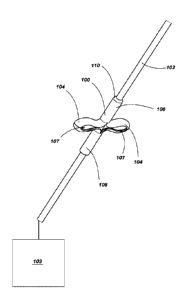
FIG. 1 depicts a medical device assembly including an anchor element 100
positioned on a medical device 102 (e.g., a distal portion of the medical
device 102). Such
medical devices 102 may include a diagnostic device, a monitoring device, a
therapeutic
device, or combinations thereof. For example, the medical device 102 may
comprise a
medical therapy delivery device, a medical device configured to sense a
parameter of the
subject, a medical device configured to diagnose a condition, a medical device
configured
to sample one or more tissues and/or fluids from a subject, or combinations
thereof.
CA 02931459 2016-05-24
WO 2015/077796 PCT/US2014/067500
- 4 -
The medical device 102 may be utilized alone to provide a medical service
(e.g.,
diagnostic, monitoring, therapeutic, or combinations thereof) to a subject or
may be utilized
with one or more medical devices 103 (e.g., a medical device internal or
external to the
subject that is electrically and/or mechanically coupled to the medical device
102). For
example, the medical device 102 and/or device 103 may comprise devices such as
a
pacemaker, defibrillator, monitoring device, infusion device, neurostimulator,
gastric
stimulator, cochlear device, or any other device that is at least partially
subcutaneously
implanted in a subject.
In some embodiments, at least a portion of the medical device 102 is
positioned
proximate the nervous system of a subject (e.g., proximate the spinal cord or
canal, brain,
and/or peripheral nervous system). The medical device 102 may be a catheter, a
lead, or
lead extension. For example, the medical device 102 may be a lead including
one or more
electrodes on a distal end portion of the lead. Electrical contacts in the
lead may be
electrically coupled (e.g., physically or wirelessly) to a control module
having an electrical
signal generator (e.g., medical device 103 external or internal to the
subject) and signals
generated by the medical device 103 may be delivered to the subject via the
electrodes. In
some embodiments, such leads are utilized as implantable stimulation devices,
which may
be utilized in a variety of treatments and procedures, such as, for example,
spinal cord
stimulation. For example, implantable stimulation devices may be used to
stimulate
nerves, such as the spinal cord, muscles, or other tissue. The stimulator
electrodes of the
leads may be implanted in contact with or near the nerves, muscles, or other
tissue to be
stimulated. A pulse generator of the medical device 103 generates electrical
pulses that are
delivered by the electrodes to body tissue. In such embodiments, the lead is
anchored at
one or more places in the subject to prevent or reduce movement of the lead or
stimulator
electrodes within the subject (e.g., during short-term or long-term placement
of the devices
102, 103 in the subject) that could damage tissue, move the stimulator
electrodes out of the
desired position, or interrupt the connection between the stimulator
electrodes and the
medical device 102, 103.
As shown in FIG. 1, the anchor element 100 is placed over at least a portion
of the
medical device 102 (e.g., a cannula of the medical device 102). For example,
at least a
portion of the medical device 102 may be positioned within a lumen formed by
the tubular
body (e.g., cannula) of the anchor element 100. As depicted, the anchor
element 100 is
shown in a deployed state where one or more protrusions (e.g., one, two,
three, four, or
CA 02931459 2016-05-24
WO 2015/077796 PCT/US2014/067500
- 5 -
more lobes 104, e.g., circumferentially-spaced lobes) extend outwardly from a
portion of
the anchor element 100 (e.g., laterally outward from a longitudinal axis or
centerline of the
anchor element 100). Each lobe 104 extending laterally from the anchor element
100 may
form an opening 107 within the lobe 104.
When attached to the medical device 102, the lobes 104 of the anchor element
100
may anchor the medical device 102 by engaging with one or more portions of the
subject.
For example, the lobes 104 of the anchor element 100 may engage with a portion
of the
subject's tissue (e.g., muscle tissue, nervous tissue, connective tissue,
etc.) to at least
partially retain the medical device 102 in a desired position within the
subject. It is also
believed that, in some embodiments, regrowth of the tissue of the subject
proximate the
lobes 104 may intertwine with at least a portion of the lobes 104 (e.g.,
tissue may extend
through the openings 107) further anchoring the anchor element 100 and medical
device 102 within the subject.
The anchor element 100 may be coupled (e.g., mechanically coupled) to at least
a
portion of the medical device 102 (e.g., an outer portion or exterior surface
of the medical
device 102). For example, the anchor element 100 may be secured to the medical
device 102 through mechanical interference (e.g., utilizing friction,
compression, swaging,
etc.) rather than through adhesion or the use of fasteners. The anchor element
100 may
include one or more portions for retaining the anchor element 100 to the
medical
device 102. For example, engagement portions 106, 108 may be formed on either
side of
the lobes 104 and may act to secure the anchor element 100 to the medical
device 102 (e.g.,
via a mechanical interference fit). In some embodiments, each of the
engagement
portions 106, 108 of the anchor element 100 include an inner dimension (e.g.,
diameter)
that is smaller than an outer dimension (e.g., diameter) of the medical device
102. One or
more portions of the anchor element 100 (e.g., engagement portions 106, 108)
may be
formed from a flexible material (e.g., an elastically deformable material)
such as, for
example, a polymer (e.g., silicone, polyurethane, etc.). The
flexible engagement
portions 106, 108 may be deformed (e.g., elastically deformed) to enlarge a
cross-sectional
area of a lumen formed within each the engagement portions 106, 108. The
enlarged
engagement portions 106, 108 may be positioned over (e.g., around, about) the
medical
device 102. As the enlarged engagement portions 106, 108 are allowed to
contract back to
substantially their original size (e.g., cross-sectional area), the engagement
portions 106,
108 may engage and couple with the medical device 102.
CA 02931459 2016-05-24
WO 2015/077796 PCT/US2014/067500
- 6 -
In some embodiments, one or more ends of the anchor element 100 include a
taper 110 or chamfer to assist in insertion of the anchor element 100 into the
subject.
FIGS. 2A and 2B depict an anchor element (e.g., anchor element 100) in an
initial
state (e.g., a retracted or relaxed state) and a deployed state (e.g., a semi-
distended state of
the inner diameter), respectively. As shown in FIG. 2A, the anchor element 100
includes a
protrusion or lobe portion 105 positioned between the engagement portions 106,
108 of the
anchor element 100. The body of the anchor element may form a lumen 101
therein. The
lobes 104 (e.g., two lobes 104) of the lobe portion 105 are formed about the
anchor element
100 (e.g., at equal circumferential spacing) by slits 112 in the tubular body
of the anchor
element 100. In the initial state, the lobe portion 105 of the anchor element
100 is
substantially parallel to (e.g., coextensive with) a longitudinal axis L100 of
the anchor
element 100.
Referring also to FIG. 2B, the engagement portions 106, 108 may be moved
toward
each other to transition the anchor element 100 to the deployed state. The
slits 112 enable
the lobes 104 to extend outwardly (e.g., in a direction lateral or transverse
(e.g.,
perpendicular) to the longitudinal axis L100 of the anchor element 100) from a
portion of
the anchor element 100 (e.g., from the engagement portions 106, 108).
FIGS. 3A and 3B depict an anchor element 200 in an initial state (e.g., a
retracted
state) and a deployed state, respectively. As shown in FIG. 3A, the anchor
element 200
may be somewhat similar to anchor element 100 discussed above and the body of
the
anchor element 200 may form a lumen 201 therein. However, anchor element 200
may
include more than one lobe portion (e.g., two lobe portions 205, 207)
positioned between
the engagement portions 206, 208 of the anchor element 200. In other
embodiments, the
anchor element 200 includes three, four, or more lobe portions. Lobes 204
(e.g., two lobes)
of each lobe portion 205, 207 are formed about the anchor element 200 (e.g.,
at equal
circumferential spacing) by slits 212 in the tubular body of the anchor
element 200. In the
initial state, the lobe portions 205 of the anchor element 200 are
substantially parallel to
(e.g., coextensive with) a longitudinal axis L200 of the anchor element 200.
In some embodiments, the anchor element 200 includes an additional engagement
portion 209 positioned between the lobe portions 205, 207.
Referring also to FIG. 3B, the engagement portions 206, 208 may be moved
toward
each other (e.g., toward the additional engagement portion 209) to transition
the anchor
element 200 to the deployed state. The slits 212 enable the lobes 204 to
extend outwardly
CA 02931459 2016-05-24
WO 2015/077796 PCT/US2014/067500
- 7 -
(e.g., in a direction lateral or transverse (e.g., perpendicular) to a
longitudinal axis L200 of
the anchor element 200) from a portion of the anchor element 200 (e.g., from
the
engagement portions 206, 208). As depicted, the lobe portions 205, 207 may be
offset
from one another (e.g., offset 90 degrees about the longitudinal axis L200 of
the anchor
element 200).
FIG. 4 depicts an anchor deployment device 300 that may be utilized with an
anchor element (e.g., anchor elements 100, 200 discussed above with reference
to FIGS. 1
through 3B). As shown in FIG. 4, the anchor deployment device 300 includes a
first
cannula (e.g., deployment cannula 302) and a second cannula (e.g., anchor
cannula 304)
received at least partially within the deployment cannula 302. For example,
the
deployment cannula 302 may have an inner dimension (e.g., diameter) that is
greater than
an outer dimension (e.g., diameter) of the anchor cannula 304 such that the
anchor
cannula 304 may be received and movable within the deployment cannula 302. The
anchor
deployment device 300 may include a handle 306 having a first portion 308
coupled to the
deployment cannula 302 and a second portion 310 coupled to the anchor cannula
304. The
portions 308, 310 of the handle 310 may be movable relative to one another
(e.g., the
second portion 310 may slide relative to the first portion 308) in order to
move the anchor
cannula 304 within the deployment cannula 302. Each portion 308, 310 of the
handle 306
may include one or more grips 314 enabling a user (e.g., medical practitioner)
to actuate
the handle 306, thereby sliding the second portion 310 relative to the first
portion 308 along
a common axis.
As depicted, the anchor cannula 304 may be sized to receive an anchor element
(e.g., anchor element 100) on the anchor cannula 304 at distal portion 312 of
the anchor
deployment device 300. The outer dimension (e.g., diameter) of the anchor
cannula 304
may be greater than the inner dimension (e.g., diameter) of the anchor element
100. Such a
diameter of the anchor cannula 304 may act to enlarge a cross-sectional area
of a
lumen 101 formed within a portion of the anchor element 100 (e.g., at each of
the
engagement portions 106, 108 (FIG. 1)) to form an initial dimension to an
enlarged
dimension. For example, the anchor cannula 304 may deform (e.g., elastically
deform) the
anchor element 100 to a dimension (e.g., diameter) that is greater than a
dimension (e.g.,
diameter) of the medical device 102 (FIG. 1) on which the anchor element 100
is to be
placed.
CA 02931459 2016-05-24
WO 2015/077796 PCT/US2014/067500
- 8 -
FIG. 5 depicts a view of the anchor deployment device 300 shown in FIG. 4
beginning to deploy an anchor element (e.g., anchor element 100 in a distended
state of the
inner diameter). As shown in FIG. 5, at least a portion of a medical device
(e.g., medical
device 102) may be received within a portion of the anchor deployment device
300. For
example, the anchor cannula 304 may have an inner dimension (e.g., diameter)
that is sized
to enable at least a portion of the medical device 102 to be received within
the anchor
cannula 304. In some embodiments, a proximal portion 316 of the anchor
deployment
device 300 is configured such that the medical device 102 extends through the
anchor
deployment device 300 and out of the of the anchor deployment device 300 at
the proximal
portion 316. Such a configuration may enable a user to position the anchor
deployment
device 300 along and through the medical device 102 in order to secure an
anchor element
100 to the anchor deployment device 300 at any desired position. For example,
the
medical device 102 may be placed within a subject and the anchor deployment
device 300
may be slid along the medical device 102. A portion of the anchor deployment
device 300
(e.g., the distal portion 312) may be inserted within the subject to secure
the anchor
element 100 within the subject while the medical device 102 resides within the
subject.
Actuation of the handle 306 may bring the anchor element 100, which is
positioned
on the anchor cannula 304 (e.g., in a radially enlarged or stretched state),
into contact with
the deployment cannula 302 (e.g., a leading end 318 of the deployment cannula
302). The
deployment cannula 302 may act to force (e.g., slide) at least a portion of
the anchor
element 100 along the anchor cannula 304. For example, the deployment cannula
302 may
force the first engagement portion 106 toward the second engagement portion
108, thereby
deploying the lobes 104 of the anchor element 100. As the anchor cannula 304
is slid
within the deployment cannula 302, the leading end 318 of the deployment
cannula 302
may force the anchor element 100 off of the anchor cannula 304 and onto the
medical
device 102 (e.g., into the position shown in FIG. 1).
FIG. 6 depicts a cross-sectional view of a portion of the anchor deployment
device 300 shown in FIG. 4 with the medical device 102 received in the anchor
deployment
device 300. As shown in FIG. 6, the inner diameter ID304 of the anchor cannula
304 is
sized to enable the medical device 102 to be received within the anchor
cannula 304. The
inner diameter ID302 of the deployment cannula 302 may be greater than an
outer
dimension 0D304 of the anchor cannula 304 such that the anchor cannula 304 may
be
received and movable within the deployment cannula 302.
CA 02931459 2016-05-24
WO 2015/077796 PCT/US2014/067500
- 9 -
FIG. 7 depicts another cross-sectional view of a portion of the anchor
deployment
device 300 shown in FIG. 4 with the medical device 102 received in the anchor
deployment
device 300 and the anchor element 100 attached to the anchor deployment device
300. The
outer diameter 0D304 of the anchor cannula 304 may be greater than an inner
diameter of
the anchor element 100 such that the anchor cannula 304 acts to enlarge a
cross-sectional
area of the lumen formed within a portion of the anchor element 100 to form an
enlarged
inner diameter IDI 00 of the anchor element 100 that is substantially equal to
the outer
diameter 0D304 of the anchor cannula 304. The enlarged inner diameter IDioo of
the anchor
element 100 may be greater than an outer diameter 0D102 of the medical device
102 such
that the enlarged inner diameter 'Dm of the anchor element 100 may be deployed
over the
outer diameter 01730102 of the medical device 102. When the anchor element 100
is removed
from the anchor cannula 304 (e.g., by the deployment cannula 302 as discussed
above), the
anchor element 100 may contract toward the initial diameter to the anchor
element 100
(e.g., where the initial diameter of the anchor element 100 is less than the
outer diameter
0D102 of the medical device 102) in order to secure the anchor element 100 to
the medical
device 102.
FIGS. 8A and 8B depict an anchor element 400 in an initial state and a
deployed
state, respectively. The anchor element 400 may be similar to and include one
or more of
the same features and functioning as the anchor elements 100, 200 discussed
above with
reference to FIGS. 1 through 3B. As shown in FIG. 8A, the anchor element 400
includes a
lobe portion 405 positioned between the engagement portions 406, 408 of the
anchor
element 400. The body of the anchor element 400 may form a lumen 401 therein.
Lobes
404 (e.g., two lobes) of the lobe portion 405 are formed about the anchor
element 400 (e.g.,
at equal circumferential spacing) by slits 412 in the tubular body of the
anchor element
400. In the initial state, the lobe portion 405 of the anchor element 400 is
substantially
parallel to (e.g., coextensive with) a longitudinal axis L400 of the anchor
element 400.
Referring also to FIG. 8B, the engagement portions 406, 408 may be moved
toward
each other to transition the anchor element 400 to the deployed state. The
slits 412 enable
the lobes 404 to extend outwardly (e.g., in a direction lateral or transverse
(e.g.,
perpendicular) to the longitudinal axis L400 of the anchor element 400) from a
portion of
the anchor element 400 (e.g., from the engagement portions 406, 408).
As depicted, the anchor element 400 may include a biasing feature (e.g., a
radial
biasing feature). For example, the anchor element 400 may include one or more
springs
CA 02931459 2016-05-24
WO 2015/077796 PCT/US2014/067500
- 10 -
414 extending about at least a portion of the anchor element 400 (e.g., the
engagement
portions 406, 408). In some embodiments, the springs 414 are disposed on an
exterior
portion of the anchor element 400. In other embodiments, the springs 414 may
be disposed
within the anchor element 400. The springs 414 may act to bias the anchor
element 400 in
(e.g., toward) an initial state. For example, the springs 414 may act to
radially bias the
engagement portions 406, 408 of the anchor element 400 inward in a direction
toward the
lumen 401 (e.g., constricting the lumen 401) such that the springs 414 bias
the engagement
portions 406, 408 to or toward an initial state (e.g., an unstretched inner
diameter of the
anchor element 400). In some embodiments, the springs 414 act to relatively
more rapidly
tighten the anchor element 400 around a medical device 102 (see, e.g., FIG.
5).
It is noted that any anchor element disclosed herein (e.g., anchor elements
100, 200)
may include a radial biasing feature (e.g., springs). In other embodiments,
the anchor
element may include an axial biasing feature.
FIGS. 9 and 10 depict a perspective view and a side view, respectively, of an
anchor deployment device 500. The anchor deployment device 500 may be similar
to and
include one or more of the same features and functioning as the anchor
deployment device
300 discussed above with reference to FIGS. 4 through 7. As shown in FIGS. 9
and 10, the
anchor deployment device 500 includes a first cannula (e.g., deployment
cannula 502) and
a second cannula (e.g., anchor cannula 504) received at least partially within
the
deployment cannula 502. The anchor deployment device 500 may include a handle
506
(e.g., formed as a hub) coupled to the anchor cannula 504 such that the handle
506 and the
anchor cannula 504 may be moved relative to another portion of the anchor
deployment
device 500 (e.g., a body 501 of the anchor deployment device 500). For
example, the body
501 of the anchor deployment device 500 may define an opening or chamber 507
in which
the handle 506 is at least partially disposed. In some embodiments, the body
501 of the
anchor deployment device 500 defines a track 509 in the chamber 507 upon which
a
portion of the handle 506 (e.g., a complementary portion) may move along
(e.g., slide) to
guide (e.g., and retain) the handle 506 and the anchor cannula 504 relative to
the body 501
and the deployment cannula 502. Movement of the handle 506 relative to the
body 501
enables a user (e.g., medical practitioner) to slide the anchor cannula 504
relative to the
deployment cannula 502 along a common axis.
As depicted, the anchor deployment device 500 is shown with an anchor element
(e.g., anchor element 400 in a distended state of the inner diameter)
positioned on the
CA 02931459 2016-05-24
WO 2015/077796 PCT/US2014/067500
- 11 -
anchor cannula 504 of the anchor deployment device 500. As above, the anchor
deployment device 500 may have an inner dimension (e.g., diameter) that is
sized to enable
at least a portion of a medical device 102 (FIG. 5) to be received within the
anchor cannula
504. As also described above, the handle 506, the anchor cannula 504, and the
deployment
cannula 502 may be utilized to deploy one or more anchor elements on a medical
device
(e.g., anchor elements 100, 200, 400 on medical device 102 as shown and
described
above).
As further depicted in FIGS. 9 and 10, the anchor deployment device 500 may
include upper handle 510. A first end of upper handle 510 may include a
locking
mechanism 512 that holds (e.g., locks, clamps, etc.) the medical device 102
(FIG. 5). For
example, the locking mechanism 512 may secure the medical device 102 when an
anchor
element is being deployed on the medical device 102 (e.g., when at least a
portion of the
medical device 102 is resident in a subject).
A second end of upper handle 510 may include a protrusion or elongated member
514 that engages with the handle 506 to secure the handle 506 and the anchor
cannula 504.
For example, the elongated member 514 of the upper handle 510 may retain the
handle 506
and the anchor cannula 504 and prevent the handle 506 and the anchor cannula
504 from
sliding relative to the body 501 of anchor deployment device 500.
The upper handle 510 may be configured such that the first end and the second
end
move (e.g., pivot) relative to each other. For example, when the locking
mechanism 512 is
securing the medical device 102 (FIG. 5), the elongated member 514 is
disengaged with the
handle 506, thereby enabling the handle 506 and the anchor cannula 504 to move
relative
to the body 501. Similarly, when the elongated member 514 is engaged with the
handle
506 and restricting the handle 506 and the anchor cannula 504 from moving
relative to the
body 501, the locking mechanism 512 is disengaged from the medical device 102,
thereby
enabling the anchor deployment device 500 to move (e.g., slide) along the
medical device
102. Such a configuration may enable the anchor deployment device 500 to be
secured to
the medical device 102 while an anchor element is being deployed and,
likewise, secure the
anchor deployment device 500 from any unwanted movement of the anchor cannula
504
relative to the deployment cannula 502 when the anchor deployment device 500
is being
moved and positioned along the medical device 102.
The anchor deployment device 500 may include rear handle 516 that enables a
user
to move and position the anchor deployment device 500 along the medical device
102.
CA 02931459 2016-05-24
WO 2015/077796 PCT/US2014/067500
- 12 -
It is noted that to the extent that the anchor deployment devices are
described in use
with a particular anchor element, in other embodiments, the anchor deployment
devices
may be utilized with any suitable anchor element (e.g., anchor elements 100,
200, 400).
It is further noted that while the anchor elements and components of the
anchor
deployment device are primarily discussed herein as having a diameter, these
elements are
not necessarily limited to circular cross sections. For example, the anchor
elements and
components of the anchor deployment device, and the lumens formed therein, may
have a
square, circular, oval, rectangular, or any other suitable cross-sectional
shape.
Referring to FIGS. 1 through 10, in operation, a lumen of an anchor element
(e.g.,
lumen 101, 201, 401 of anchor element 100, 200, 400) is enlarged to position
the anchor
element 100, 200, 400 on the anchor cannula 304 of the anchor deployment
device 300. A
medical device 102 (e.g., a medical device that has already been inserted and
positioned
within a subject) is positioned within the anchor element 302 and the anchor
deployment
device 300 and anchor element 100, 200, 400 are moved along the medical device
102 to
position the anchor element 100, 200, 400 within the subject. The anchor
element 100,
200, 400 may then be deployed within the subject utilizing the handle 306 of
the anchor
deployment device 300 to deploy the lobes 104, 204, 404 of the anchor element
100, 200,
400 and to force the anchor element 100, 200, 400 onto (e.g., about, around)
the medical
device 102 with the deployment cannula 302. Constriction of the anchor element
100, 200,
400 about the medical device 102 as the anchor element 100, 200, 400 contracts
toward the
initial lumen size of the anchor element 100, 200, 400 acts to secure the
anchor element
100, 200, 400 about the medical device 102 while both the anchor element 100,
200, 400
and the medical device 102 are positioned within the subject. For example, the
anchor
element 100, 200, 400 may contract to the initial size of the lumen 101, 201,
401 of the
anchor element 100, 200, 400 or to a cross-sectional area between the initial
size and the
enlarged (e.g., deformed) size of the lumen 101, 201, 401 of the anchor
element 100, 200,
400. In some embodiments, the constriction of the anchor element 100, 200, 400
may also
constrict or compress a portion of the medical device 102 (e.g., a cannula).
Once the anchor element 100, 200, 400 is placed over the medical device 102
.. within the subject, the lobes 104, 204, 404 of the anchor element 100, 200,
400 may anchor
the medical device 102 by engaging with one or more portions of the subject's
tissue to at
least partially retain the medical device 102 in a desired position within the
subject.
CA 02931459 2016-05-24
WO 2015/077796 PCT/US2014/067500
- 13 -
Once being apprised of the instant disclosure, one of ordinary skill in the
art will be
able to make and use the devices and assemblies disclosed herein. For example,
the anchor
elements may be formed from a polymer (e.g., a polyurethane such as
CARBOTHANEO)
and springs may be formed from a metal material (e.g., 316 stainless steel).