Note: Descriptions are shown in the official language in which they were submitted.
84366760
FABS-IN-TANDEM IMM'UNOGLOBULIN AND USES THEREOF
CROSS-REFERENCE TO RELATED APPLICATION
[0001] This application claims priority to International Application
No. PCT/CN2013/090923, filed December 30, 2013.
FIELD OF INVENTION
[0002] The present invention relates to multivalent and multispecific binding
proteins, and to
methods of making and using multivalent and multispecific binding proteins.
[0003]
BACKGROUND OF THE INVETION
[0004] Bispecific or multispecific antibodies have been generated in attempts
to prepare
molecules useful for the treatment of various inflammatory diseases, cancers,
and other
disorders.
10005] Bispecific antibodies have been produced using the quadroma technology
(see
Milstein, C. and A.C. Cuello, Nature, 1983. 305(5934): p. 537-40) based on the
somatic
fusion of two different hybridoma cell lines expressing murine monoclonal
antibodies with
the desired specificities of the bispecific antibody. Bispecific antibodies
can also be
produced by chemical conjugation, of two different mAbs (see Staerz, U.D., et
al., Nature,
1985. 314(6012): p. 628-31). Other approaches have used chemical conjugation
of two
different monoclonal antibodies or smaller antibody fragments (see Brennan,
M., et al.,
Science, 1985. 229(4708): p. 81-3).
1
CA 2931641 2020-02-18
CA 02931641 2016-05-25
WO 2015/103072 PCT/US2014/072336
[0006] Another method is the coupling of two parental antibodies with a hetero-
bifunctional
crosslinker. In particular, two different Fab fragments have been chemically
crosslinked at
their hinge cysteine residues in a site-directed manner (see Glennie, M.J., et
al., J Immunol,
1987. 139(7): p. 2367-75).
[0007] Other recombinant bispecific antibody formats have been developed in
the recent past
(see Kriangkum, J., et al., Biomol Eng, 2001. 18(2): p. 31-40). Amongst them
tandem single-
chain Fv molecules and diabodies, and various derivatives thereof, have been
used for the
construction of recombinant bispecific antibodies. Normally, construction of
these molecules
starts from two single-chain Fv (scFv) fragments that recognize different
antigens (see
Economides, A.N., et al., Nat Med, 2003. 9(1): p. 47-52). Tandem scFv
molecules (taFv)
represent a straightforward format simply connecting the two scFv molecules
with an
additional peptide linker. The two scFv fragments present in these tandem scFv
molecules
fotoi separate folding entities. Various linkers can be used to connect the
two scFv fragments
and linkers with a length of up to 63 residues (see Nakanishi, K., et al..
Annu Rev Immunol,
2001. 19: p. 423-74).
[0008] In a recent study, in vivo expression by transgenic rabbits and cattle
of a tandem scFv
directed against CD28 and a melanoma-associated proteoglycan was reported (see
Gracie,
J.A., et al., J Clin Invest, 1999. 104(10): p. 1393-401). In this construct
the two scFv
molecules were connected by a CH1 linker and serum concentrations of up to 100
mg/L of
the bispecific antibody were found. A few studies have now reported expression
of soluble
tandem scFv molecules in bacteria (see Leung, B.P., et al., J Immunol, 2000.
164(12): p.
6495-502; Ito, A., et al., J Immunol, 2003. 170(9): p. 4802-9; Karni, A., et
al., J
Neuroimmunol, 2002. 125(1-2): p. 134-40) using either a very short Ala3 linker
or long
glycine/serine-rich linkers.
[0009] In a recent study, phage display of a tandem scFv repertoire containing
randomized
middle linkers with a length of 3 or 6 residues enriched those molecules which
are produced
in soluble and active form in bacteria. This approach resulted in the
isolation of a preferred
tandem scFv molecule with a 6 amino acid residue linker (see Arndt, M. and J.
Krauss,
Methods Mol Biol, 2003. 207: p. 305-21).
[0010] Bispecific diabodies (Db) utilize the diabody format for expression.
Diabodies are
produced from scFv fragments by reducing the length of the linker connecting
the VH and
VL domain to approximately 5 residues (see Peipp, M. and T. Valerius, Biochem
Soc Trans,
2002. 30(4): p. 507-11). This reduction of linker size facilitates
dimerization of two
2
CA 02931641 2016-05-25
WO 2015/103072 PCT/US2014/072336
polypeptide chains by crossover pairing of the VH and VL domains. Bispecific
diabodies are
produced by expressing two polypeptide chains with either the structure VHA-
VLB and
VHB-VLA (VH-VL configuration) or VLA-VHB and VLB-VHA (VL-VH configuration)
within the same cell. A recent comparative study demonstrates that the
orientation of the
variable domains can influence expression and formation of active binding
sites (see Mack,
M., G. Riethmuller, and P. Kufer, Proc Natl Acad Sci U S A, 1995. 92(15): p.
7021-5).
[0011] One approach to force the generation of bispecific diabodies is the
production of
knob-into-hole diabodies (see Holliger, P., T. Prospero, and G. Winter, Proc
Natl Acad Sci U
S A, 1993. 90(14): p. 6444-8.18). This was demonstrated for a bispecific
diabody directed
against HER2 and CD3. A large knob was introduced in the VH domain by
exchanging
Va137 with Phe and Leu45 with Trp and a complementary hole was produced in the
VL
domain by mutating Phe98 to Met and 'Tyr87 to Ala, either in the anti- HER2 or
the anti-CD3
variable domains. By using this approach the production of bispecific
diabodies could be
increased from 72% by the parental diabody to over 90% by the knob-into-hole
diabody.
[0012] Single-chain diabodies (scDb) represent an alternative strategy to
improve the
formation of bispecific diabody-like molecules (see Holliger, P. and G.
Winter, Cancer
Immunol Immunother, 1997. 45(3-4): p. 128-30; Wu, A.M., et al.,
Immunotechnology, 1996.
2(1): p. 21-36). Bispecific single-chain diabodies are produced by connecting
the two
diabody-forming polypeptide chains with an additional middle linker with a
length of
approximately 15 amino acid residues. Consequently, all molecules with a
molecular weight
corresponding to monomeric single-chain diabodies (50-60 kDa) are bispecific.
Several
studies have demonstrated that bispecific single chain diabodies are expressed
in bacteria in
soluble and active form with the majority of purified molecules present as
monomers (see
Holliger, P. and G. Winter, Cancer Immunol Immunother, 1997. 45(3-4): p. 128-
30; Wu,
A.M., et al., Immunotechnology, 1996. 2(1): p. 21-36; Pluckthun, A. and P.
Pack,
Immunotechnology, 1997. 3(2): p. 83-105; Ridgway, J.B., et al., Protein Eng,
1996. 9(7): p.
617-21).
[0013] Diabody have been fused to Fe to generate more Ig-like molecules, named
di-diabody
(see Lu, D., et al., J Biol Chem, 2004. 279(4): p. 2856-65). In addition,
multivalent antibody
construct comprising two Fab repeats in the heavy chain of an IgG and capable
of binding
four antigen molecules has been described (see US patent 8,722,859 B2, and
Miller, K., et al.,
J Immunol, 2003. 170(9): p. 4854-61).
3
CA 02931641 2016-05-25
WO 2015/103072 PCT/US2014/072336
[0014] The most recent examples are tetravalent IgG¨single-chain variable
fragment (scFv)
fusions (Doug J, et al. 2011 MAbs 3:273-288; CoLoma MJ, Morrison SL 1997 Nat
Biotechnol 15:159-163; Lu D, et al. 2002 J Immunol Methods 267:213-226),
catumaxomab,
a trifunctional rat,/mouse hybrid bispecificepithelialcelladhesionmolecule-
CD3antibody
(Lindhofer H, et al 1995 J Immunol 155:219-225), the bispecific CD19-CD3 scFy
antibody
blinatumomab (Bargou R, et at. 2008 Science 321:974-977), "dual- acting Fab"
(DAF)
antibodies (BostromJ, et al. 2009 Science 323:1610-1614), covalently linked
pharmacophore
peptides to catalytic anti- bodies (Doppalapudi VR, et al. 2010 Proc Natl Acad
Sci USA
107:22611-22616), use of the dynamic exchange between half IgG4 molecules to
generate
bispecific antibodies (van der Neut Kolfschoten M, et al. 2007 Science
317:1554-1557;
Stubenrauch K, et al. 2010 Drug Metab Dispos 38:84-91), or by exchange of
heavy-chain
and light-chain domains within the antigen binding fragment (Fab) of one half
of the
bispecific antibody (CrossMab format) (Schaefer Wet al 2011Proc Natl Acad Sci
108:11187-
92).
[0015] There is a need in the art for single molecular entities with dual
antigen binding
function, and for methods of generating such multivalent and multispecific
binding proteins.
The present invention addresses these and other needs.
SUMMARY OF THE INVENTION
[0016] The present invention provides multivalent and multispecific binding
proteins, and
methods of making and using such binding proteins. In one embodiment, the
multivalent and
multispccific binding proteins provided herein arc Fabs-in-tandem
immunoglobulins (FIT-
Ig), and are capable of binding two or more antigens, or two or more epitopes
of the same
antigen, or two or more copies of the same epitope. The multivalent and
multispecific binding
proteins provided herein are useful for treatment and/or prevention of acute
and chronic
inflammatory diseases and disorders, autoimmune diseases, cancers, spinal cord
injuries,
sepsis, and other diseases, disorders, and conditions. Pharmaceutical
compositions
comprising the multivalent and multispecific binding proteins are provided
herein. In
addition, nucleic acids, recombinant expression vectors, and host cells for
making such FIT-
Igs are provided herein. Methods of using the FIT-Igs of the invention to
detect specific
antigens, in vivo or in vitro, are also encompassed by the invention.
4
CA 02931641 2016-05-25
WO 2015/103072 PCT/US2014/072336
[0017] The present invention provides a family of binding proteins that are
capable of
binding two or more antigens, e.g., with high affinity. In one aspect, the
present invention
provides an approach to construct a bispecific binding protein using two
parental monoclonal
antibodies: mAb A, which binds to antigen A, and mAb B, which binds to antigen
B. The
binding proteins disclosed herein, in one embodiment, are capable of binding
antigens,
cytokines, chemokines, cytokine receptors, chemokine receptors, cytokine- or
chemokine-
related molecules, or cell surface proteins.
[0018] Thus, in one aspect, binding proteins capable of binding two or more
antigens are
provided. In one embodiment, the present invention provides a binding protein
comprising at
least two polypeptide chains, wherein the polypeptidc chains pair to form IgG-
like molecules
capable of binding two or more antigens. In one embodiment, the binding
protein comprises
two, three, four, five, or more polypeptide chains. In one embodiment, the
binding protein
comprises at least one VLA, at least one VLB, at least one VHA, at least one
VHB, at least one
CL, and at least one CHL wherein VL is a light chain variable domain, VH is a
heavy chain
variable domain, CL is a light chain constant domain, CH1 is the first
constant domain of the
heavy chain, A is a first antigen, and B is a second antigen. In a further
embodiment, the first
polypeptide chain comprises a VLA, a CL, a VHB, and a CHL In a further
embodiment, the
binding protein further comprises an Fe. In another embodiment, the Fe region
is a variant Fe
region. In a further embodiment, the variant Fe region exhibits modified
effector function,
such as ADCC or CDC. In another embodiment, the variant Fe region exhibits
modified
affinity or avidity for one or more FcyR.
[0019] In one embodiment, the binding protein comprises three polypeptide
chains, wherein
the first polypeptide chain comprises a VLA, a CL, a VHB, and a CH 1, the
second polypeptide
chain comprises VHA and CHI, and the third polypeptide chain comprises VLB and
CL. In a
further embodiment, the first polypeptide chain of the binding protein further
comprises an
Fe. In another embodiment, the binding protein comprises two polypeptide
chains, wherein
the first polypeptide chain comprises a VLA, a CL, a VHB, and a CHI, the
second polypeptide
chain comprises VHA, CHL VLB, and CL. In a further embodiment, the first
polypeptide
chain further comprises an Fe.
[0020] In one embodiment, the binding protein comprises three polypeptide
chains, and their
corresponding cDNA during co-transfection are present at a molar ratio of
first:second:third
of 1:1:1, 1:1.5:1, 1:3:1, 1:1:1.5, 1:1:3, 1:1.5:1.5, 1:3:1.5, 1:1.5:3, or
1:3:3. In another
embodiment, the binding protein comprises two polypeptide chains, and their
corresponding
CA 02931641 2016-05-25
WO 2015/103072 PCT/US2014/072336
cDNA during co-transfection are present at a molar ratio of first:second of
1:1, 1:1.5, or 1:3,
or any other ratios, through optimization, in an effort to maximize the
monomeric FIT-Ig
fraction in any given transfection.
[0021] In one embodiment, the binding protein of the present invention does
not comprise a
peptide linker. In one embodiment, the binding protein of the present
invention comprises at
least one amino acid or polypeptide linker. In a further embodiment, the
linker is selected
from the group consisting of G,GS, SG,GGS, GSG, SGG, GGG, GGGS, SGGG, GGGGS,
GGGGSGS, GGGGSGS, GGGGSGGS, GGGGSGGGGS, GGGGSGGGGSGGGGS,
AKTTPKLEEGEFSEAR, AKTTPKLEEGEFSEARV, AKTTPKLGG, SAKTTPKLGG,
AKTTPKLEEGEFSEARV, SAKTTP, SAKTTPKLGG, RADAAP, RADAAPTVS,
RADAAAAGGPGS, RADAAAA(G4S)4, SAKTTP,
SAKTTPKLGG,
SAKTTPKLEEGEFSEARV, ADAAP, ADAAPTVSIFPP, TVAAP, TVAAPSVFIFPP,
QPKAAP, QPKAAPSVTLFPP, AKTTPP, AKTTPPSVTPLAP, AKTTAP,
AKTTAPSVYPLAP, AS TKGP, ASTKGPSVFPLAP, GENKVEYAPALMALS ,
GPAKELTPLKEAKVS, and GHEAAAVMQVQYPAS. The linkers can also be in vivo
cleavable peptide linkers, protease (such as MMPs) sensitive linkers,
disulfide bond-based
linkers that can be cleaved by reduction, etc., as previously described
(Fusion Protein
Technologies for Biopharmaceuticals: Applications and Challenges, edited by
Stefan R.
Schmidt), or any cleavable linkers known in the art. Such cleavable linkers
can be used to
release the top Fab in vivo for various purposes, in order to improve
tissue/cell penetration
and distribution, to enhance binding to targets, to reduce potential side
effect, as well as to
modulate in vivo functional and physical half-life of the 2 different Fab
regions.
[0022] In one embodiment, the binding protein comprises a first polypeptide
comprising,
from amino to carboxyl terminus, VLA-CL-VHB-CH1-Fc, a second polypeptide chain
comprising, from amino to carboxyl terminus, VHA-CH1, and a third polypeptide
chain
comprising, from amino to carboxyl terminus, VLB-CL; wherein VL is a light
chain variable
domain, CL is a light chain constant domain, VH is a heavy chain variable
domain, CH1 is
the first constant domain of the heavy chain, A is a first epitope or antigen,
and B is a second
epitope or antigen. In one embodiment, the Fc region is human IgGl. In another
embodiment,
the Fc region is a variant Fc region. In a further embodiment, the amino acid
sequence of the
Fc region is at least 65%, at least 70%, at least 75%, at least 80%, at least
85%, at least 90%,
at least 95%, at least 99%, or 100% identical to SEQ ID NO: 20. In a further
embodiment, the
CL of the first polypeptide chain is fused directly to VHB. In another
embodiment, the CL of
6
CA 02931641 2016-05-25
WO 2015/103072 PCT/1JS2014/072336
the first polypeptide chain is linked to VH B via an amino acid or an
oligopeptide linker. In a
further embodiment, the linker is GSG (SEQ ID NO: 26) or GGGGSGS (SEQ ID NO:
28).
[0023] In another embodiment, the binding protein comprises a first
polypeptide comprising,
from amino to carboxyl terminus, VHB-CH1-VLA-CL-Fc, a second polypeptide chain
comprising, from amino to carboxyl terminus, VHA-CH1, and a third polypeptide
chain
comprising, from amino to carboxyl terminus, VLB-CL; wherein VL is a light
chain variable
domain, CL is a light chain constant domain, VH is a heavy chain variable
domain, CH1 is
the first constant domain of the heavy chain, A is a first epitope or antigen,
and B is a second
epitope or antigen. In one embodiment, the Fc region is human IgG1 . In
another embodiment,
the Fc region is a variant Fc region. In a further embodiment, the amino acid
sequence of the
Fc region is at least 65%, at least 70%, at least 75%, at least 80%, at least
85%, at least 90%,
at least 95%, at least 99%, or 100% identical to SEQ ID NO: 20. In one
embodiment, the
CH1 of the first polypeptide chain is fused directly to VLA. In another
embodiment, the CH1
of the first polypeptide chain is linked to VLA via an amino acid or an
oligopeptide linker. In
a further embodiment, the linker is GSG (SEQ ID NO: 26) or GGGGSGS (SEQ ID NO:
28).
[0024] In another embodiment, the binding protein comprises a first
polypeptide comprising,
from amino to carboxyl terminus, VLA-CL-VHB-CH1-Fc, and a second polypeptide
chain
comprising, from amino to carboxyl terminus, VHA-CH1-VLB-CL; wherein VL is a
light
chain variable domain, CL is a light chain constant domain, VH is a heavy
chain variable
domain, CH1 is the first constant domain of the heavy chain, A is a first
epitope or antigen,
and B is a second epitope or antigen. In one embodiment, the Fc region is
human IgGl. In
another embodiment, the Fc region is a variant Fc region. In a further
embodiment, the amino
acid sequence of the Fc region is at least 65%, at least 70%, at least 75%, at
least 80%, at
least 85%, at least 90%, at least 95%, at least 99%, or 100% identical to SEQ
ID NO: 20. In a
further embodiment, the CL of the first polypeptide chain is fused directly to
VHS. In another
embodiment, the CL of the first polypeptide chain is linked to VHB via an
amino acid or an
oligopeptide linker. In a further embodiment, the linker is GSG (SEQ ID NO:
26) or
GGGGSGS (SEQ ID NO: 28).
[0025] In another embodiment, binding protein comprises a first polypeptide
comprising,
from amino to carboxyl terminus, VHB-CH1-VLA-CL-Fc, and a second polypeptide
chain
comprising, from amino to carboxyl terminus, VLB-CL-VHA-CH1; wherein VL is a
light
chain variable domain, CL is a light chain constant domain, VH is a heavy
chain variable
domain, CH1 is the first constant domain of the heavy chain, A is a first
epitope or antigen,
7
CA 02931641 2016-05-25
WO 2015/103072 PCT/US2014/072336
and B is a second epitope or antigen. In one embodiment, the Fe region is
human IgG1 . In
another embodiment, the Fe region is a variant Fe region. In a further
embodiment, the amino
acid sequence of the Fe region is at least 65%, at least 70%, at least 75%, at
least 80%, at
least 85%, at least 90%, at least 95%, at least 99%, or 100% identical to SEQ
ID NO: 20. In
one embodiment, the CH1 of the first polypeptide chain is fused directly to
VLA. In another
embodiment, the CH1 of the first polypeptide chain is linked to VLA via an
amino acid or an
oligopeptide linker. In a further embodiment, the linker is GSG (SEQ ID NO:
26) or
GGGGSGS (SEQ ID NO: 28).
[0026] The binding proteins of the present invention arc capable of binding
pairs of
cytokines. For example, the binding proteins of the present invention arc
capable of binding
pairs of cytokines selected from the group consisting of IL-1a and IL-1(3; IL-
12 and IL-18,
TNFa and IL-23, TNFa and IL-13; TNF and IL-18; TNF and IL-12; TNF and IL-
lbeta; TNF
and MIF; TNF and IL-6, TNF and IL-6 Receptor, TNF and IL-17; IL-17 and IL-20;
IL-17
and IL-23; TNF and IL-15; TNF and VEGF; VEGFR and EGFR; PDGFR and VEGF, IL-13
and 1L-9; IL-13 and 1L-4; IL-13 and 1L-5; 1L-13 and 1L-25; IL-13 and TARC; 1L-
13 and
MDC; IL-13 and MIF; IL-13 and TGF-I3; IL-13 and LHR agonist; IL-13 and CL25;
IL-13
and SPRR2a; IL-13 and SPRR2b; IL-13 and ADAM8; and TNFa and PGE4, IL-13 and
PED2, TNF and PEG2. In one embodiment, the binding proteins of the present
invention are
capable of binding 1L-17 and 1L-20.'the binding proteins of the present
invention, in one
embodiment, are capable of binding 1L-17 and 1L-20 and comprise variable heavy
and light
chains derived from the anti-1L-17 antibody LY and the anti-IL-20 antibody
15D2. In one
embodiment, the binding proteins of the present invention are capable of
binding IL-17 and
TNF. The binding proteins of the present invention, in one embodiment, are
capable of
binding IL-17 and TNF and comprise variable heavy and light chains derived
from the anti-
IL-17 antibody LY and the TNF antibody golinnumab.
[0027] In one embodiment, the binding proteins of the present invention bind
IL-17 and IL-
20 and comprise a first polypeptide comprising, consisting essentially of, or
consisting of an
amino acid sequence selected from the group consisting of SEQ ID NOs: 15, 25,
and 27; a
second polypeptide chain comprising, consisting essentially of, or consisting
of an amino
acid sequence according to SEQ ID NO: 21; and a third polypeptide chain
comprising,
consisting essentially of, or consisting of a sequence according to SEQ ID NO:
23. In another
embodiment, the binding proteins of the present invention bind IL-27 and IL-20
and comprise
8
CA 02931641 2016-05-25
WO 2015/103072 PCT/US2014/072336
a first polypeptide chain comprising, consisting essentially of, or consisting
of an amino acid
sequence selected from the group consisting of SEQ ID NOs: 15, 25, and 27, and
a second
polypeptide chain comprising, consisting essentially of, or consisting of an
amino acid
sequence selected from the group consisting of SEQ ID NOs: 29, 30, and 31.
[0028] In one embodiment, the binding proteins of the present invention bind
TNF and IL-17
and comprise a first polypeptide comprising, consisting essentially of, or
consisting of an
amino acid sequence according to SEQ ID NOs: 87; a second polypeptide chain
comprising,
consisting essentially of, or consisting of an amino acid sequence according
to SEQ ID NO:
89; and a third polypeptide chain comprising, consisting essentially of, or
consisting of a
sequence according to SEQ ID NO: 91.
[0029] In another embodiment, the binding protein is capable of binding pairs
of targets
selected from the group consisting of CD137 and CD20, CD137 and EGFR, CD137
and Her-
2, CD137 and PD-1, CD137 and PDL-1, VEGF and PD-L1, Lag-3 and TIM-3, OX40 and
PD-1, TIM-3 and PD-1, TIM-3 and PDL-1, EGFR and DLL-4, CD138 and CD20; CD138
and CD40; CD19 and CD20; CD20 and CD3; CD3 and CD33; CD3 and CD133; CD47 and
CD20, CD38 and CD138; CD38 and CD20; CD20 and CD22; CD38 and CD40; CD40 and
CD20; CD-8 and IL-6; CSPGs and RGM A; CTLA-4 and BTN02; IGF1 and IGF2; IGF1/2
and Erb2B; IGF-1R and EGFR; EGFR and CD13; IGF-1R and ErbB3; EGFR-2 and IGFR;
VEGFR-2 and Met; VEGF-A and Angiopoietin-2 (Ang-2); IL-12 and TWEAK; IL-13 and
IL-lbeta; PDGFR and VEGFõ EpCAM and CD3, Her2 and CD3, CD19 and CD3, EGFR and
Her3, CD16a and CD30, CD30 and PSMA, EGFR and CD3, CEA and CD3, TROP-2 and
HSG, TROP-2 and CD3, MAG and RGM A; NgR and RGM A; NogoA and RGM A; OMGp
and RGM A; PDL-1 and CTLA-4; CTLA-4 and PD-1; PD-1 and TIM-3; RGM A and RGM
B; Te38 and TNFa; TNFa and Blys; TNFla and CD-22; TNFa and CTLA-4 domain;
TNFla
and GP130; TNFa and IL-12p40; and TNFa and RANK ligand, Factor IXa, Factor X.
In
one embodiment, the binding proteins of the present invention are capable of
binding CD3
and CD20. The binding proteins of the present invention, in one embodiment,
are capable of
binding CD3 and CD20 and comprise variable heavy and light chains derived from
the anti-
CD3 antibody OKT3 and the anti-CD20 antibody ofatumumab. In one embodiment,
the
binding proteins of the present invention are capable of binding CTLA-4 and PD-
1. The
binding proteins of the present invention, in one embodiment, are capable of
binding CTLA-4
and PD-1 and comprise variable heavy and light chains derived from the CTLA-4
antibody
ipilimumab and the PD-1 antibody nivolumab.
9
CA 02931641 2016-05-25
WO 2015/103072 PCT/US2014/072336
[0030] In one embodiment, the binding proteins of the present invention bind
CD3 and CD20
and comprise a first polypeptide chain comprising, consisting essentially of,
or consisting of
an amino acid sequence selected from the group consisting of SEQ ID NOs: 41
and 48; a
second polypeptide chain comprising, consisting essentially of, or consisting
of an amino acid
sequence according to SEQ ID NO: 44; and a third polypeptide chain comprising,
consisting
essentially of, or consisting of an amino acid sequence according to SEQ ID
NO: 46.
[0031] In one embodiment, the binding proteins of the present invention bind
CTLA-4 and
PD-1 and comprise a first polypeptide chain comprising, consisting essentially
of, or
consisting of an amino acid sequence according to SEQ ID NO: 92; a second
polypeptide
chain comprising, consisting essentially of, or consisting of an amino acid
sequence
according to SEQ ID NO: 95; and a third polypeptide chain comprising,
consisting essentially
of, or consisting of an amino acid sequence according to SEQ ID NO: 97.
[0032] In one embodiment, the binding protein provided herein is capable of
binding one or
more epitopes on CTLA-4. In one embodiment, the binding protein provided
herein is
capable of binding one or more peiotpes on PD-1.
[0033] In one embodiment, the binding protein is capable of binding one or
more epitopes on
one or more immune checkpoint protein on T cells such as, for example, TIM-3,
Lag3, ICOS,
BTLA, CD160, 2B4, KIR, CD137, CD27, 0X40, CD4OL, and A2aR. In another
embodiment,
the binding protein is capable of binding one or more epitopes on one or more
tumor cell
surface protein that is involved with immune checkpoint pathways, such as, for
example, PD-
L1, PD-L2, Galectin9, HVEM, CD48, B7-1, B7-2, ICOSL, B7-H3, B7-H4, CD137L,
OX4OL,
CD70, and CD40.
[0034] In one aspect, the present invention provides pharmaceutical
compositions comprising
the binding proteins described herein. In one embodiment, provided herein are
pharmaceutical compositions comprising the binding protein of any one of the
preceding
claims and one or more pharmaceutically acceptable carrier.
[0035] In another aspect, the present invention provides methods of treating
or preventing an
inflammatory disease, autoimmune disease, neurodegenerative disease, cancer,
sepsis, or
spinal cord injury in a subject in need thereof. In one embodiment, the method
comprises
administering to a subject an effective amount of one or more of the binding
proteins
provided herein, or one or more pharmaceutical compositions comprising the
binding
proteins provided herein and a pharmaceutically acceptable carrier. Uses of
the binding
proteins described herein in the manufacture of a medicament for treatment or
prevention of
CA 02931641 2016-05-25
WO 2015/103072 PCT/1JS2014/072336
an inflammatory disease, autoimmune disease, neurodegenerative disease,
cancer, or spinal
cord injury are also provided herein. In one embodiment, the inflammatory
disease,
autoimmune disease, or neurodegenerative disease is selected from the group
consisting of
asthma, rheumatoid arthritis, systemic lupus erythematosus, multiple
sclerosis, Alzheimer's
disease, or Parkinson's disease.
[0036] In one embodiment, the present disclosure provides methods for treating
or preventing
rheumatoid arthritis, psoriasis, osteoporosis, stroke, liver disease, or oral
cancer to a subject
in need thereof, the method comprising adminitering to the subject a FIT-Ig
binding protein
described herein, wherein the binding protein is capable of binding IL-17 and
IL-20. In a
further embodiment, the FIT-Ig binding protein comprises an amino acid
sequence selected
from SEQ ID NOs: 15, 25, and 27; and amino acid sequence according to SEQ ID
NO: 21;
and an amino acid sequence according to SEQ ID NO: 23. In another embodiment,
the FIT-Ig
binding protein comprises an amino acid sequence selected from SEQ ID NOs: 15,
25, and
27; and an amino acid sequence selected from SEQ ID NOs: 29, 30 and 31.
[0037] In one embodiment, the present disclosure provides methods for treating
or preventing
B cell cancer in a subject in need thereof, the method comprising
administering to the
subject a FIT-Ig binding protein, wherein the FIT-Ig binding protein is
capable of binding
one or more B cell antigen. In a further embodiment, the FIT-Ig binding
protein is capable of
binding CD20. In a further embodiment, the FIT-Ig binding protein is capable
of binding
CD20 and another antigen. In a further embodiment, the binding protein is
capable of binding
CD3 and CD20. In a further embodiment, the cancer is a B cell cancer. In a
still further
embodiment, the B cell cancer is selected from the group consisting of
Hodgkin's lymphoma,
non-Hodgkin's lymphoma [NHL], precursor B cell lymphoblastic
leukemia/lymphoma, mature B
cell neoplasms, B cell chronic lymphocytic leukemia/small lymphocytic
lymphoma, B cell
prolymphocytic leukemia, lymphoplasmacytic lymphoma, mantle cell lymphoma,
follicular
lymphoma, cutaneous follicle center lymphoma, marginal zone B cell lymphoma,
hairy cell
leukemia, diffuse large B cell lymphoma, Burkitt's lymphoma, plasmacytoma,
plasma cell
myeloma, post-transplant lymphoproliferative disorder, Waldenstrom's
macroglobulinemia, and
anaplastic large-cell lymphoma. In one embodiment, the present disclosure
provides methods
for treating or preventing a B cell cancer in a subject in need thereof, the
method comprising
administering to the subject a FIT-Ig binding protein, wherein the FIT-1g
binding protein
comprises an amino acid sequence according to SEQ ID NOs: 41 or 48; and amino
acid
11
CA 02931641 2016-05-25
WO 2015/103072 PCT/US2014/072336
sequence according to SEQ ID NO: 44, and an amino acid sequence according to
SEQ ID
NO: 46.
[0038] In one embodiment, the present disclosure provides methods for treating
or preventing
an autoimmune disease, inflammatory disease, or infection in a subject in need
thereof, the
method comprising administering to the subject a FIT-Ig binding protein
described herein,
wherein the binding protein is capable of binding TNF and IL-17. In a further
embodiment, the
FIT-Ig binding protein comprises sequences according to SEQ ID NOs: 87, 89,
and 91. In
another embodiment, the present disclosure provides methods for treating or
preventing an
autoimmune or inflammatory disease, the method comprising administering to the
subject a FIT-
Ig binding protein, wherein the binding protein is capable of binding TNF and
IL-17, and wherein
the autoimmune or inflammatory disease is selected from the group consisting
of Crohn's
disease, psoriasis (including plaque psoriasis), arthritis (including
rheumatoid arthritis, psoratic
arthritis, osteoarthritis, or juvenile idiopathic arthritis), multiple
sclerosis, ankylosing spondylitis,
spondylosing arthropathy, systemic lupus crythematosus, uvcitis, sepsis,
neurodcgcnerative
diseases, neuronal regeneration, spinal cord injury, primary and metastatic
cancers, a respiratory
disorder; asthma; allergic and nonallergic asthma; asthma due to infection;
asthma due to
infection with respiratory syncytial virus (RSV); chronic obstructive
pulmonary disease (COPD);
a condition involving airway inflammation; eosinophilia; fibrosis and excess
mucus production;
cystic fibrosis; pulmonary fibrosis; an atopic disorder; atopic dermatitis;
urticaria; eczema;
allergic rhinitis; allergic enterogastritis; an inflammatory and/or autoimmune
condition of the
skin; an inflammatory and/or autoimmunc condition of gastrointestinal organs;
inflammatory
bowel diseases (IBD); ulcerative colitis; an inflammatory and/or autoimmune
condition of the
liver; liver cirrhosis; liver fibrosis; and liver fibrosis caused by hepatitis
B and/or C virus;
scleroderma. In another embodiment, In another embodiment, the present
disclosure provides
methods for treating or preventing cancer in a subject in need thereof, the
method comprising
adminitering to the subject a FIT-Ig binding protein described herein, wherein
the binding
protein is capable of binding.TNF and IL-17. In a further embodiment, the
cancer is
hepatocellular carcinoma; glioblastoma; lymphoma; or Hodgkin's lymphoma. In
another
embodiment, the present disclosure provides methods for treating or preventing
and infection in a
subject in need thereof, the method comprising administering to the subject a
FIT-Ig binding
protein described herein, wherein the infection is a viral infection, a
bacterial infection, a parasitic
infection, H'TLV-1 infection. In one embodiment, the present disclosure
provides methods for
suppression of expression of protective type 1 immune responses, and
suppression of expression
of a protective type 1 immune response during vaccination.
12
CA 02931641 2016-05-25
WO 2015/103072 PCT/US2014/072336
[0039] In one embodiment, the present disclosure provides methods for treating
rheumatoid
arthritis in a subject in need thereof, the method comprising administering to
the subject a
FIT-Ig binding protein, wherein the binding protein comprises sequences
according to SEQ
ID NOs: 87, 89, and 91.
[0040] In one embodiment, the present disclosure provides methods for treating
or preventing
cancer in a subject in need thereof, the method comprising adminitering to the
subject a FIT-
Ig binding protein described herein, wherein the binding protein is capable of
binding.CTLA-
4 and PD-1. In a further embodiment, the FIT-Ig binding protein comprises an
amino acid
sequence comprising SEQ ID NOs: 92, 95, and 97. In another embodiment, the
present
disclosure provides methods for treating or preventing cancer in a subject in
need thereof,
wherein the bindng protein is capable of binding CTLA-4 and PD-1, and wherein
the cancer
is a cancer typically responsive to immunotherapy. In another embodiment, the
cancer is a cancer
that has not been associated with immunotherapy. In another embodiment, the
cancer is a cancer
that is a refractory or recurring malignancy. In another embodiment, the
binding protein inhibits
the growth or survival of tumor cells. In another embodiment, the cancer is
selected from the
group consisting of melanoma (e.g., metastatic malignant melanoma), renal
cancer (e.g. clear cell
carcinoma), prostate cancer (e.g. hormone refractory prostate adenocarcinoma),
pancreatic
adenocarcinoma, breast cancer, colon cancer, lung cancer (e.g. non-small cell
lung cancer),
esophageal cancer, squamous cell carcinoma of the head and neck, liver cancer,
ovarian cancer,
cervical cancer, thyroid cancer, glioblastoma, glioma, leukemia, lymphoma, and
other neoplastic
malignancies.
[0041] In one embodiment, the present disclosure provides methods for treating
or preventing
melanoma in a subject in need thereof, the method comprising administering to
the subject a
FIT-Ig binding protein described herein, wherein the binding protein is
capable of
binding.CTLA-4 and PD-1. In a further embodiment, the present disclosure
provides methods
for treating or preventing melanoma in a subject in need thereof, wherein the
method comprises
administering to the subject a FIT-Ig binding protein comprising amino acid
sequences according
to SEQ ID NOs: 92, 95, and 97.
[0042] In another embodiment, the present disclosure provides methods for
treating or
preventing infections or infectious disease in a subject in need thereof, the
method comprising
administering to the subject a FIT-Ig binding protein described herein,
wherein the binding
protein is capable of binding CTLA-4 and PD-1. In one embodiment, the FIT-Ig
binding protein
is administered alone, or in combination with vaccines, to stimulate the
immune response to
13
84366760
pathogens, toxins, and self-antigens. Therefore, in one embodiment, the
binding proteins provided
herein can be used to stimulate immune response to viruses infectious to
humans, such as, but not
limited to, human immunodeficiency viruses, hepatitis viruses class A, B and
C, Epstein Barr
virus, human cytomegalovirus, human papilloma viruses, herpes viruses,
bacteria, fungal
parasites, or other pathogens.
[0042a] According to one aspect of the present invention, there is provided a
bispecific binding
protein comprising at least three polypeptide chains, wherein the first
polypeptide chain
comprises, from amino terminus to carboxyl terminus, either (i) VLA-CL-VHB-CH1-
Fc, wherein
CL is fused directly to VHB, or (ii) VHB-CH1-VLA-CL-Fc, wherein CH1 is fused
directly to
VLA, and wherein there are no linkers inserted between variable domains and
constant domains;
wherein the second polypeptide chain comprises, from amino terminus to
carboxyl terminus,
VHA-CH1, wherein there is no linker inserted between VHA and CH1; wherein the
third
polypeptide chain comprises, from amino to carboxyl terminus, VLB-CL, wherein
there is no
linker inserted between VLB and CL; wherein A is a first epitope or antigen,
and B is a second
epitope or antigen, and wherein A and B are different epitopes of the same
antigen or different
antigens; wherein VLA is a light chain variable domain of a first parental
antibody that binds A,
CL is an antibody light chain constant domain, VHB is a heavy chain variable
domain of a
second parental antibody that binds B, CH1 is a first constant domain of an
antibody heavy
chain, VHA is a heavy chain variable domain of said first parental antibody
that binds A, and
VLB is a light chain variable domain of said second parental antibody that
binds B; and wherein
two of said first polypeptide chain, two of said second polypeptide chain, and
two of said third
polypeptide chain are capable of associating to provide a dual specific,
tetravalent binding
protein comprising six polypeptide chains having four Fab binding regions, and
wherein said
binding protein binds both epitope or antigen A and epitope or antigen B.
10042b] According to another aspect of the present invention, there is
provided a
pharmaceutical composition comprising the binding protein as described herein
and one or more
pharmaceutically acceptable carriers.
[0042c] According to still another aspect of the present invention, there is
provided a
pharmaceutical composition comprising the binding protein as described herein
for use in
treating or preventing an inflammatory disease, autoimmune disease,
neurodegenerative
disease, cancer, sepsis, or spinal cord injury, wherein the binding protein
binds different
14
Date Recue/Date Received 2020-12-15
84366760
epitopes of the same antigen or different antigens involved with the
inflammatory disease,
autoimmune disease, neurodegenerative disease, sepsis, or spinal cord injury.
[0042d] According to yet another aspect of the present invention, there is
provided a
pharmaceutical composition comprising the binding protein as described herein
and a
pharmaceutically acceptable carrier for use in treating a metabolic disease,
wherein said binding
protein is capable of binding two different epitopes of one antigen or two
different antigens
selected from the group consisting of: ANGPTL3, ANGPTL4, INFa, IL-6, IL-lbeta,
CCL2,
CCL3, CCL5, NFKB1, and NFKB2; and wherein said metabolic disease is selected
from the
group consisting of: diabetes, insulin dependent diabetes mellitus,
arteriosclerosis,
atherosclerosis, and diabetic arteriosclerotic disease.
[0042e] According to a further aspect of the present invention, there is
provided use of the
binding protein as described herein in the manufacture of a medicament for
treatment or
prevention of an inflammatory disease, autoimmune disease, neurodegenerative
disease, cancer,
or spinal cord injury, wherein the binding protein binds different epitopes of
the same antigen
or different antigens involved in the inflammatory disease, autoimmune
disease,
neurodegenerative disease, sepsis, or spinal cord injury.
1004211 According to yet a further aspect of the present invention, there is
provided a binding
protein as described herein that is capable of binding IL-17 and IL-20 for use
in the treatment
of a disease selected from the group consisting of rheumatoid arthritis,
psoriasis, osteoporosis,
stroke, liver disease, and oral cancer.
[0042g] According to still a further aspect of the present invention, there is
provided a binding
protein as described herein that is capable of binding CD3 and CD20 for use in
the treatment of
a B cell cancer.
BRIEF DESCRIPTION OF THE DRAWINGS
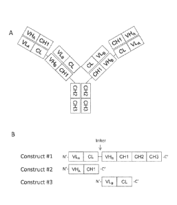
[0043] Figure 1A shows the structure of FIT-Igs that are made up of three
constructs, such as
FIT1-Ig, FIT2-Ig, and FIT3-Ig. Figure 1B shows the three constructs used to
prepare such
FIT1-Igs.
[0044] Figure 2A shows the basic structure of FIT-Igs that are made up of two
constructs.
Figure 2B shows the two constructs used to prepare such FIT-Igs.
14a
Date Recue/Date Received 2020-12-15
84366760
[0045] Figure 3 provides the dual-specific antigen binding of FIT1-Ig as
measured by Biacore.
The top panel of Figure 3 shows the results of the BiacoreTM binding assay in
which FIT1-Ig was
first saturated by IL-17, followed by IL-20. The bottom panel of Figure 3
shows the results of the
Biacore assay in which FIT1-Ig was first saturated by IL-20, followed by IL-
17.
[0046] Figure 4. Shows the solubility at a range of pH of anti-IL-17/IL-20 FIT
Ig or monoclonal
antibody rituximab, as measured by PEG-induced precipitation.
[0047] Figure 5 shows the stability of stable Cl-JO cell line development in
both DG44 (5A and
5B) and CHO-S (SC and 5D) systems.
[0048] Figure 6 shows the binding to CTLA-4 (6A) or PD-1 (Figure 6B) by FIT10-
Ig or the
parental antibodies Ipilimumab and Nivolumab, as assessed by ELISA.
[0049] Figure 7 shows a multiple binding study of FIT10-Ig against both CTLA-4
and PD-1.
Binding to CTLA-4 followed by PD-1; and binding by PD-1 followed by CTLA-4 are
both
shown as indicated in Figure 7.
DETAILED DESCRIPTION
14b
CA 2931641 2020-02-18
CA 02931641 2016-05-25
WO 2015/103072 PCT/US2014/072336
[0050] The present invention relates to multivalent and multispecific binding
proteins,
methods of making the binding proteins, and to their uses in the prevention
and/or treatment
of acute and chronic inflammatory diseases and disorders, cancers, and other
diseases. This
invention pertains to multivalent and/or multispecific binding proteins
capable of binding two
or more antigens. Specifically, the invention relates to Fabs-in-tandem
immunoglobulins
(FIT-Ig), and pharmaceutical compositions thereof, as well as nucleic acids,
recombinant
expression vectors and host cells for making such FIT-Igs. Methods of using
the FIT-Igs of
the invention to detect specific antigens, either in vitro or in vivo are also
encompassed by the
invention.
[0051] The novel family of binding proteins provided herein are capable of
binding two or
more antigens, e.g., with high affinity. Specifically, the present invention
provides an
approach to construct a bispecific binding protein using 2 parental monoclonal
antibodies:
mAb A, which binds to antigen a; and mAb B, which binds to antigen b.
[0052] In one aspect, the present invention provides a binding protein
comprising a variable
light chain specific for a first antigen or epitope, a first light chain
constant domain, a variable
heavy chain specific for a second antigen or epitope, a first heavy chain CH1,
a variable
heavy chain specific for the first antigen or epitope, a second heavy chain
CH1, a variable
heavy chain specific for the second antigen or epitope, and a second light
chain constant
domain. In one embodiment, the binding protein further comprises an Fe region.
The binding
protein may further comprise one or more amino acid or polypeptide linker
linking two or
more of the components of the binding protein. For example, the binding
protein may
comprise a polypeptide linker linking the light chain variable region to the
light chain
constant region.
[0053] In one embodiment, the present disclosure provides a binding protein
comprising a
polypeptide chain comprising VLA-CL-(Xl)n-VHB-CH1-(X2)n, wherein VLA is the
light
chain variable domain of mAb A, CL is a light chain constant domain, X1
represents an
amino acid or an oligopeptide linker, VHB is the heavy chain variable domain
of mAb B,
CH1 is the first constant domain of the heavy chain, X2 represents an Fe
region or a different
dimerization domain, and n is 0 or 1.
[0054] In one embodiment, the invention provides a binding protein comprising
three
different polypeptide chains (Figure 1), wherein the first polypeptide chain
(construct 41)
comprises VLA-CL-(X1)n-VHB-CH1-(X2)n, wherein VLA is the light chain variable
domain
of mAb A, CL is a light chain constant domain, X1 represents an amino acid or
an
CA 02931641 2016-05-25
WO 2015/103072 PCT/US2014/072336
oligopeptide linker, VHB is the heavy chain variable domain of mAb B, CH1 is
the first
constant domain of the heavy chain, X2 represents an Fe region or a different
dimerization
domain, and n is 0 or 1. The second polypeptide chain (construct #2) comprises
VHA-CH1,
wherein VHA is the heavy chain variable domain of mAb A, and CH1 is the first
constant
domain of the heavy chain. The third polypeptide chain (construct #3)
comprises VLB-CL,
wherein VLB is the light chain variable domain of mAb B, and CL is the
constant domain of
the light chain.
[0055] In another embodiment, the invention provides a binding protein
comprising three
different polypeptide chains with the overall molecular design similar to the
previous
embodiment except the order of the variable domains are reversed. In the
embodiment the
first polypeptide chain comprises VHB-CH1-(X1)n-VLA-CL-(X2)n, wherein VLA is a
light
chain variable domain of mAb A, CL is a light chain constant domain, X1
represents an
amino acid or an oligopeptide linker, VHB is the heavy chain variable domain
of mAb B,
CH1 is the first constant domain of the heavy chain, X2 represents an Fe
region or a different
dimerization domain, and n is 0 or 1. The second polypeptide chain comprises
VHA-CH1,
wherein VHA is the heavy chain variable domain of mAb A and CH1 is the first
constant
domain of the heavy chain. The third polypeptide chain comprises VLB-CL,
wherein VLB is
the light chain variable domain of mAb B and CL is the constant domain of the
light chain.
[0056] In another embodiment the invention provides a binding protein
comprising two
different polypeptide chains (Figure 2), wherein the first polypeptide chain
(construct #1)
comprises VLA-CL-(X1)n-VHB-CH1-(X2)n, wherein VLA is a light chain variable
domain of
mAb A, CL is a light chain constant domain, X1 represents an amino acid or an
oligopeptide
linker, VHB is the heavy chain variable domain of mAb B, CH1 is the first
constant domain
of the heavy chain, X2 represents an Fe region or a different dimerization
domain, and n is 0
or 1. The second polypeptide chain (construct #4) comprises VHA-CH1-(X3)n-VLB-
CL,
wherein VHA is the heavy chain variable domain of mAb A, CHI is the first
constant domain
of the heavy chain, X3 represents an amino acid or polypeptide that is not a
constant domain,
n is 0 or 1, VLB is the light chain variable domain of mAb B, and CL is the
constant domain
of the light chain.
[0057] In another embodiment the invention provides a binding protein
comprising two
polypeptide chains with the overall molecular design similar to the previous
embodiment
except the order of the variable domains are reversed. In this embodiment the
first
polypeptide chain comprises VHB-CH1-(Xl)n-VLA-CL-(X2)n, wherein VLA is a light
chain
16
CA 02931641 2016-05-25
WO 2015/103072 PCT/US2014/072336
variable domain of mAb A, CL is a light chain constant domain, X1 represents
an amino acid
or an oligopeptide linker, VHB is the heavy chain variable domain of mAb B,
CH1 is the first
constant domain of the heavy chain, X2 represents an Fe region or a different
dimerization
domain, and n is 0 or 1. The second polypeptide chain comprises VLB-CL-(X3)n-
VHA-CH1,
wherein VHA is the heavy chain variable domain of mAb A, CH1 is the first
constant domain
of the heavy chain, X3 represents an amino acid or an oligopeptide linker, n
is 0 or 1, VLB is
the light chain variable domain of mAb B, and CL is the constant domain of the
light chain.
[0058] In one embodiment, the VH and VL domains in the binding protein are
selected from
the group consisting of murine heavy/light chain variable domains, fully human
heavy/light
chain variable domains, CDR grafted heavy/light chain variable domains,
humanized
heavy/light chain variable domains, and mixtures thereof In a preferred
embodiment
VHANLA and VHBNLB are capable of binding the same antigen. In another
embodiment
VHANLA and VHBNLB are capable of binding different antigens.
[0059] In one embodiment, the first polypeptide chain comprises VLA-CL-VHB-CH1-
Fc, and
the CL and VHB of the first polypeptide chain are directly fused together. In
another
embodiment, the CL and VHB are linked by an amino acid or an oligopeptide
linker. In
another embodiment, the first polypeptide chain comprises VHB-CH1-VLA-CL-Fc,
and the
CH1 and VLA are directly fused together. In another embodiment, the CH1 and
VLA are
linked by an amino acid or an oligopeptide linker. In a further embodiment,
the oligo- or
poly-peptide linker comprises 1 or more amino acids of any reasonable sequence
that
provides flexibility. Preferably the linker is selected from the group
consisting of G,GS, SG,
GGS, GSG, SGG, GGG, GGGS, SGGG, GGGGS, GGGGSGS, GGGGSGS, GGGGSGGS,
GGGGSGGGGS, GGGGSGGGGSGGGGS,
AKTTPKLEE GEF S EAR,
AKTTPKLEEGEFSEARV, AKTTPKLGG, SAKTTPKLGG, AKTTPKLEEGEFSEARV,
SAKTTP, SAKTTPKLGG, RADAAP, RADAAPTVS, RADAAAAGGPGS,
RADAAAA(G4S)4, SAKTTP, SAKTTPKLGG, SAKTTPKLEEGEFSEARV, ADAAP,
ADAAPTVSIFPP, TVAAP, TVAAPSVFIFPP, QPKAAP, QPKAAPSVTLFPP, AKTTPP,
AKTTPPSVTPLAP, AKTTAP, AKTTAPSVYPLAP, ASTKGP, ASTKGPSVFPLAP,
GENKVEYAPALMALS, GPAKELTPLKEAKVS, and GHEAAAVMQVQYPAS. In one
embodiment, the amino acid sequence of the linker may be selected from the
group
consisting of SEQ ID NOs. 26, 28, and 49-86. In one embodiment, the linker is
GSG (SEQ
ID NO: 26) or GGGGSGS (SEQ ID NO: 28). The linkers can also be in vivo
cleavable
peptide linkers, protease (such as MMPs) sensitive linkers, disulfide bond-
based linkers that
17
CA 02931641 2016-05-25
WO 2015/103072 PCT/US2014/072336
can be cleaved by reduction, etc., as previously described (Fusion Protein
Technologies for
Biopharmaceuticals: Applications and Challenges, edited by Stefan R. Schmidt),
or any
cleavable linkers known in the art. Such cleavable linkers can be used to
release the top Fab
in vivo for various purposes, in order to improve tissue/cell penetration and
distribution, to
enhance binding to targets, to reduce potential side effect, as well as to
modulate in vivo
functional and physical half-life of the 2 different Fab regions. In one
embodiment, the
binding protein comprises an Fe region. As used herein, the term "Fe region"
refers to the C-
terminal region of an IgG heavy chain. An example of the amino acid sequence
containing
the human IgG1 Fe region is SEQ ID NO: 20. The Fe region of an IgG comprises
two
constant domains, CH2 and CH3.
[0060] In one embodiment, the Fe region is a variant Fe region. In one
embodiment, the
variant Fe region has one or more amino acid modifications, such as
substitutions, deletions,
or insertions, relative to the parent Fe region. In a further embodiment,
amino acid
modifications of the Fe region alter the effector function activity relative
to the parent Fe
region activity. For example, in one embodiment, the variant Fe region may
have altered (i.e.,
increased or decreased) antibody-dependent cytotoxicity (ADCC), complement-
mediated
cytotoxicity (CDC), phagocytosis, opsonization, or cell binding. In another
embodiment,
amino acid modifications of the Fe region may alter (i.e., increase or
decrease) the affinity of
the variant Fe region for an FcyR relative to the parent Fe region. For
example, the variant Fe
region may alter the affinity for FcyRI, FcyRII, FcyRIII.
[0061] In one preferred embodiment, the binding proteins provided herein are
capable of
binding one or more targets. In one embodiment, the target is selected from
the group
consisting of cytokines, cell surface proteins, enzymes and receptors.
Preferably the binding
protein is capable of modulating a biological function of one or more targets.
More
preferably the binding protein is capable of neutralizing one or more targets.
[0062] In one embodiment, the binding protein of the invention is capable of
binding
cytokines selected from the group consisting of lymphokines, monokines, and
polypeptide
hormones. In a further embodiment, the binding protein is capable of binding
pairs of
cytokines selected from the group consisting of IL-la and IL-113; IL-12 and IL-
18, TNFa and
IL-23, TNFoc and IL-13; TNF and IL-18; TNF and IL-12; TNF and IL-lbeta; TNF
and MIF;
TNF and IL-6, TNF and IL-6 Receptor, TNF and 1L-17; IL-17 and IL-20; 1L-17 and
1L-23;
TNF and IL-15; 'TNF and VEGF; VEGFR and EGFR; IL-13 and IL-9; IL-13 and IL-4;
IL-13
and IL-5; IL-13 and IL-25; IL-13 and TARC; IL-13 and MDC; IL-13 and MIF; IL-13
and
18
CA 02931641 2016-05-25
WO 2015/103072 PCT/US2014/072336
TGF-I3; IL-13 and LHR agonist; IL-13 and CL25; IL-13 and SPRR2a; IL-13 and
SPRR2b;
1L-13 and ADAM8; and TNFa and PGE4, IL-13 and PED2, TNF and PEG2.
[0063] In another embodiment, the binding protein of the invention is capable
of binding
pairs of targets selected from the group consisting of CD137 and CD20, CD137
and EGFR,
CD137 and Her-2, CD137 and PD-1, CD137 and PDL-1, VEGF and PD-L1, Lag-3 and
TIM-
3, 0X40 and PD-1, TIM-3 and PD-1, TIM-3 and PDL-1, EGFR and DLL-4, VEGF and
EGFR, HGF and VEGF, VEGF and VEGF (same or a different epitope), VEGF and
Ang2,
EGFR and cMet, PDGF and VEGF, VEGF and DLL-4, 0X40 and PD-L1, ICOS and PD-1,
ICOS and PD-L1, Lag-3 and PD-1, Lag-3 and PD-L1, Lag-3 and CTLA-4, ICOS and
CTLA-
4, CD138 and CD20; CD138 and CD40; CD19 and CD20; CD20 and CD3; CD3 and CD33;
CD3 and CD133; CD38 & CD138; CD38 and CD20; CD20 and CD22; CD38 and CD40;
CD40 and CD20; CD47 and CD20, CD-8 and IL-6; CSPGs and RGM A; CTLA-4 and
BTN02; CTLA-4 and PD-1; IGF1 and IGF2; IGF1/2 and Erb2B; IGF-1R and EGFR; EGFR
and CD13; IGF-1R and ErbB3; EGFR-2 and IGFR; Her2 and Her2 (same or a
different
epitope); Factor IXa, Factor X ,VEGFR-2 and Met; VEGF-A and Angiopoietin-2
(Ang-2);
IL-12 and TWEAK; IL-13 and IL-lbeta ; MAG and RGM A; NgR and RGM A; NogoA and
RGM A; OMGp and RGM A; PDL-1 and CTLA-4; PD-1 and TIM-3; RGM A and RGM B;
Te38 and TNFa.; TNFa and Blys; TNFa and CD-22; TNFa and CTLA-4 domain; TNFa
and GP130; TNFa and IL-12p40; and TNFa and RANK ligand.
[0064] In one embodiment, the binding protein is capable of binding human IL-
17 and
human IL-20. In a further embodiment, the binding protein is capable of
binding human IL-
17 and human IL-20 and comprises a FIT-Ig polypeptide chain 1 sequence that is
about
65%, about 70%, about 75%, about 80%, about 85%, about 90%, about 95%, about
99%, or
100% identical to a sequence selected from the group consisting of SEQ ID NO.
15, 25, and
27; a polypeptide chain 1t2 sequence that is about 65%, about 70%, about 75%,
about 80%,
about 85%, about 90%, about 95%, about 99%, or 100% identical to SEQ ID NO.
21; and a
polypeptide chain #3 sequence that is about 65%, about 70%, about 75%, about
80%, about
85%, about 90%, about 95%, about 99%, or 100% identical to SEQ ID NO.23. In
another
embodiment, the binding protein is capable of binding human IL-17 and human 1L-
20 and
comprises FIT-Ig polypeptide chain #1 sequence that is about 65%, about 70%,
about 75%,
about 80%, about 85%, about 90%, about 95%, about 99%, or 100% identical to a
sequence
selected from the group consisting of SEQ ID NO. 15, 25, and 27; and a
polypeptide chain #4
19
CA 02931641 2016-05-25
WO 2015/103072 PCT/US2014/072336
that is about 65%, about 70%, about 75%, about 80%, about 85%, about 90%,
about 95%,
about 99%, or 100% identical to a sequence selected from the group consisting
of SEQ ID
NO. 29, 30, and 31.
[0065] In one embodiment, the binding protein is capable of binding human CD3
and human
CD20. In a further embodiment, the binding protein comprises a FIT-Ig
polypeptide chain #1
sequence that is about 65%, about 70%, about 75%, about 80%, about 85%, about
90%,
about 95%, about 99%, or 100% identical to a sequence selected from the group
consisting of
SEQ ID NO. 41 and 48; a polypeptide chain #2 sequence that is about 65%, about
70%, about
75%, about 80%, about 85%, about 90%, about 95%, about 99%, or 100% identical
to SEQ
ID NO.44; and a polypeptide chain #3 sequence that is about 65%, about 70%,
about 75%,
about 80%, about 85%, about 90%, about 95%, about 99%, or 100% identical to
SEQ ID NO.
46.
[0066] In one embodiment, the binding protein is capable of binding human IL-
17 and
human TNF. In a further embodiment, the binding protein is capable of binding
human IL-17
and human TNF and comprises a FIT-Ig polypeptide chain #1 sequence that is
about 65%,
about 70%, about 75%, about 80%, about 85%, about 90%, about 95%, about 99%,
or 100%
identical to SEQ ID NO. 87; a polypeptide chain #2 sequence that is about 65%,
about 70%,
about 75%, about 80%, about 85%, about 90%, about 95%, about 99%, or 100%
identical to
SEQ ID NO. 89, and a polypeptide chain #3 sequence that is about 65%, about
70%, about
75%, about 80%, about 85%, about 90%, about 95%, about 99%, or 100% identical
to SEQ
ID NO. 91.
[0067] In one embodiment, the binding protein is capable of binding human CTLA-
4 and
human PD-1. In a further embodiment, the binding protein is capable of binding
human
CTLA-4 and human PD-1 and comprises a FIT-Ig polypeptide chain #1 sequence
that is
about 65%, about 70%, about 75%, about 80%, about 85%, about 90%, about 95%,
about
99%, or 100% identical to SEQ ID NO. 92 a polypeptide chain #2 sequence that
is about
65%, about 70%, about 75%, about 80%, about 85%, about 90%, about 95%, about
99%, or
100% identical to SEQ ID NO. 95; and a polypeptide chain #3 sequence that is
about 65%,
about 70%, about 75%, about 80%, about 85%, about 90%, about 95%, about 99%,
or 100%
identical to SEQ ID NO. 97.
[0068] In another embodiment, the binding protein of the invention is capable
of binding one
or two cytokines, cytokine-related proteins, and cytokine receptors selected
from the group
CA 02931641 2016-05-25
WO 2015/103072 PCT/US2014/072336
consisting of BMP1, BMP2, BMP3B (GDF10), BMP4, BMP6, BMP8, CSF1 (M-CSF),
CSF2 (GM-CSF), CSF3 (G-CSF), EPO, FGF1 (aFGF), FGF2 (bFGF), FGF3 (int-2), FGF4
(HST), FGF5, FGF6 (HST-2), FGF7 (KGF), FGF9, FGF10, FGF11, FGF12, FGF12B,
FGF14, FGF16, FGF17, FGF19, FGF20, FGF21, FGF23, IGF1, IGF2, IFNA1, IFNA2,
IFNA4, IFNA5, IFNA6, IFNA7, IFNB1, IFNG, IFNW1, FILl, FIL1 (EPSILON), FIL1
(ZETA), IL1A, IL1B, IL2, IL3, IL4, IL5, IL6, IL7, IL8, IL9, IL10, IL11, IL12A,
IL12B,
IL13, IL14, IL15, IL16, IL17, IL17B, IL18, IL19, IL20, IL22, IL23, IL24, IL25,
IL26, IL27,
IL28A, IL28B, IL29, IL30, PDGFA, FGER1, FGFR2, FGFR3, EGFR, ROR1, 2B4, KIR,
CD137, CD27, 0X40, CD4OL, A2aR, CD48, B7-1, B7-2, ICOSL, B7-H3, B7-H4, CD137L,
OX4OL, CD70, CD40, PDGFB, TGFA, TGFB1, TGFB2, TGFB3, LTA (TNF-b), LTB, TNF
(TNF-a ), TNFSF4 (0X40 ligand), TNFSF5 (CD40 ligand), TNFSF6 (FasL), TNFSF7
(CD27 ligand), TNFSF8 (CD30 ligand), TNFSF9 (4-1BB ligand), TNFSF10 (TRAIL),
TNFSF11 (TRANCE), TNFSF12 (APO3L), TNFSF13 (April), TNFSF13B, TNFSF14
(HVEM-L), TNFSF15 (VEGI), TNFSF18, FIGF (VEGFD), VEGF, VEGFB, VEGFC,
IL1R1, IL1R2, IL1RL1, IL1RL2, IL2RA, IL2RB, IL2RG, IL3RA, IL4R, IL5RA, IL6R,
IL7R, IL8RA, IL8RB, IL9R, ILIORA, ILlORB, IL11RA, IL12RB1, IL12RB2, IL13RA1,
IL13RA2, IL15RA, IL17R, IL18R1, IL20RA, IL21R, IL22R, IL1HY1, IL1RAP,
IL1RAPL1,
IL1RAPL2, IL1RN, IL6ST, IL18BP, IL18RAP, IL22RA2, AlF1, HGF, LEP (leptin),
PIN,
and THPO.
[0069] The binding protein of the invention is capable of binding one or more
chemokines,
chemokine receptors, and chemokine-related proteins selected from the group
consisting of
CCL1 (1-309), CCL2 (MCP -1 / MCAF), CCL3 (MIP-1a), CCL4 (MIP-1b), CCL5
(RANTES), CCL7 (MCP-3), CCL8 (mcp-2), CCL11 (eotaxin), CCL13 (MCP-4), CCL15
(MIP-1d), CCL16 (HCC-4), CCL17 (TARC), CCL18 (PARC), CCL19 (MIP-3b), CCL20
(MIP-3a), CCL21 (SLC / exodus-2), CCL22 (MDC / STC-1), CCL23 (MF'IF-1), CCL24
(MPIF-2 / eotaxin-2), CCL25 (TECK), CCL26 (eotaxin-3), CCL27 (CTACK / TLC),
CCL28,
CXCL1 (GRO1), CXCL2 (GRO2), CXCL3 (GRO3), CXCL5 (ENA-78), CXCL6 (GCP-2),
CXCL9 (MIG), CXCL10 (IP 10), CXCL11 (I-TAC), CXCL12 (SDF1), CXCL13, CXCL14,
CXCL16, PF4 (CXCL4), PPBP (CXCL7), CX3CL1 (SCYD1), SCYE1, XCL1
(lymphotactin), XCL2 (SCM-1b), BLR1 (MDR15), CCBP2 (D6 / JAB61), CCR1 (CKR1 /
HM145), CCR2 (mcp-1RB / RA), CCR3 (CKR3 / CMKBR3), CCR4, CCR5 (CMKBR5 /
ChernR13), CCR6 (CMKBR6 / CKR-L3 / STRL22 / DRY6), CCR7 (CKR7 / EBI1), CCR8
(CMKBR8 / TERI / CKR-L1), CCR9 (GPR-9-6), CCRL1 (VSHK1), CCRL2 (L-CCR),
21
CA 02931641 2016-05-25
WO 2015/103072 PCT/US2014/072336
XCR1 (GPR5 / CCXCR1), CMKLR1, CMKOR1 (RDC1), CX3CR1 (V28), CXCR4, GPR2
(CCR10), GPR31, GPR81 (FKSG80), CXCR3 (GPR9/CKR-L2), CXCR6 (TYMSTR
/STRL33 Bonzo), HM74, IL8RA (IL8Ra), IL8RB (IL8Rb), LTB4R (GPR16), TCP10,
CKLFSF2, CKLFSF3, CKLFSF4, CKLFSF5, CKLFSF6, CKLFSF7, CKLFSF8, BDNF,
C5R1, CSF3, GRCC10 (C10), EPO, FY (DARC), GDF5, HIF1A, 1L8, PRL, RGS3, RGS13,
SDF2, SLIT2, TLR2, TLR4, TREM1, TREM2, and VHL.
[0070] In another embodiment, the binding protein of the invention is capable
of binding cell
surface protein such as, for example, integrins. In another embodiment, the
binding protein
of the invention is capable of binding enzymes selected from the group
consisting of kinases
and proteases. In yet another embodiment, the binding protein of the invention
is capable of
binding receptors selected from the group consisting of lymphokine receptors,
monokine
receptors, and polypeptide hormone receptors.
[00711 In one embodiment, the binding protein is multivalent. In another
embodiment, the
binding protein is multispecific. The multivalent and or multispecific binding
proteins
described above have desirable properties particularly from a therapeutic
standpoint. For
instance, the multivalent and or multispecific binding protein may (1) be
internalized (and/or
catabolized) faster than a bivalent antibody by a cell expressing an antigen
to which the
antibodies bind; (2) be an agonist antibody; and/or (3) induce cell death
and/or apoptosis of a
cell expressing an antigen which the multivalent antibody is capable of
binding to. The
"parent antibody" which provides at least one antigen binding specificity of
the multivalent
and or multispecific binding proteins may be one which is internalized (and/or
catabolized)
by a cell expressing an antigen to which the antibody binds; and/or may be an
agonist, cell
death-inducing, and/or apoptosis-inducing antibody, and the multivalent and or
multispecific
binding protein as described herein may display improvement(s) in one or more
of these
properties. Moreover, the parent antibody may lack any one or more of these
properties, but
may be endowed with them when constructed as a multivalent binding protein as
herein
described.
[0072] In another embodiment the binding protein of the invention has an on
rate constant
(Kon) to one or more targets selected from the group consisting of: at least
about 102M-ls-1; at
least about 103M-is-1; at least about 104m-is-i;
at least about 105M-1s-1; and at least about
106m-is-i,
as measured by surface plasmon resonance. Preferably, the binding protein of
the
invention has an on rate constant (Kon) to one or more targets between 102M'
s' to 103M-' s-
22
CA 02931641 2016-05-25
WO 2015/103072 PCT/US2014/072336
1; between 103M-is-1 to 104M-1s-1; between 104M-1s-1 to 105M-ls-1, or between
105M-1s-1 to
106M-1s-1, as measured by surface plasmon resonance.
[0073] In another embodiment the binding protein has an off rate constant
(Koff) for one or
more targets selected from the group consisting of: at most about 10-3s-1; at
most about 10-4s-
; at most about 10-5s-1; and at most about 10-6s-1, as measured by surface
plasmon resonance.
Preferably, the binding protein of the invention has an off rate constant
(Koff) to one or more
targets of 10-3s-1 to 10-4s-1; of 104s'to 10-5s-1; or of 105s'to 10-6s 1, as
measured by surface
plasmon resonance.
[0074] In another embodiment the binding protein has a dissociation constant
(KD) to one or
more targets selected from the group consisting of: at most about le M; at
most about 10-8
M; at most about 10-9 M; at most about 104 M; at most about 10-11M; at most
about 10-12 M;
and at most 10-13 M. Preferably, the binding protein of the invention has a
dissociation
constant (KD) to IL-12 or IL-23 of 10-7 M to 10-8 M; of 10-8 M to 10-9 M; of
10-9 M to 10-10
M; of 10-10 to 10-" M; of 10-11 M to 10-12M; or of 10-12 M to 10-13M.
[0075] In another embodiment, the binding protein described above is a
conjugate further
comprising an agent selected from the group consisting of an immunoadhesion
molecule, an
imaging agent, a therapeutic agent, and a cytotoxic agent. In one embodiment,
the imaging
agent is selected from the group consisting of a radiolabel, an enzyme, a
fluorescent label, a
luminescent label, a bioluminescent label, a magnetic label, and biotin. In a
further
embodiment, the imaging agent is a radiolabel selected from the group
consisting of: 3H, 14C,
35s, 90y, 99Tc, "In, 1251, 1311, 177Lu,
'66Ho, and 153Sm. In one embodiment, the therapeutic or
cytotoxic agent is selected from the group consisting of an immunosuppressive
agent, an
immuno-stimulatory agent, an anti-metabolite, an alkylating agent, an
antibiotic, a growth
factor, a cytokinc, an anti-angiogcnic agent, an anti-mitotic agent, an
anthracyclinc, a toxin,
and an apoptotic agent. In one embodiment, the binding protein is conjugated
directly to the
agent. In another embodiment, the binding protein is conjugated to the agent
via a linker.
Suitable linkers include, but are not limited to, amino acid and polypeptide
linkers disclosed
herein. Linkers may be cleavable or non-cleavable.
[0076] In another embodiment the binding protein described above is a
crystallized binding
protein and exists as a crystal. Preferably the crystal is a carrier-free
pharmaceutical
controlled release crystal. More preferably the crystallized binding protein
has a greater half
life in vivo than the soluble counterpart of said binding protein. Most
preferably the
crystallized binding protein retains biological activity.
23
CA 02931641 2016-05-25
WO 2015/103072 PCT/US2014/072336
[0077] In another embodiment the binding protein described above is
glycosylated.
Preferably, the glycosylation is a human glycosylation pattern.
[0078] One aspect of the invention pertains to an isolated nucleic acid
encoding any one of
the binding protein disclosed above. A further embodiment provides a vector
comprising the
isolated nucleic acid disclosed above wherein said vector is selected from the
group
consisting of pcDNA; pTT (Durocher et al., Nucleic Acids Research 2002, Vol
30, No.2);
pTT3 (pTT with additional multiple cloning site; pEFBOS (Mizushima, S. and
Nagata, S.,
(1990) Nucleic acids Research Vol 18, No. 17); pBV; pJV; pcDNA3.1 TOPO, pEF6
TOPO
and pBJ. The multi-specific binding proteins and methods of making the same
are provided.
The binding protein can be generated using various techniques. Expression
vectors, host cells
and methods of generating the binding proteins are provided in this
disclosure.
[0079] The antigen-binding variable domains of the binding proteins of this
disclosure can be
obtained from parent binding proteins, including polyclonal Abs, monoclonal
Abs, and or
receptors capable of binding antigens of interest. These parent binding
proteins may be
naturally occurring or may be generated by recombinant technology. The person
of ordinary
skill in the art is well familiar with many methods for producing antibodies
and/or isolated
receptors, including, but not limited to using hybridoma techniques, selected
lymphocyte
antibody method (SLAM), use of a phage, yeast, or RNA-protein fusion display
or other
library, immunizing a non-human animal comprising at least some of the human
immunoglobulin locus, and preparation of chimeric, CDR-grafted, and humanized
antibodies.
See, e.g., US Patent Publication No. 20090311253 Al. Variable domains may also
be
prepared using affinity maturation techniques. The binding variable domains of
the binding
proteins can also be obtained from isolated receptor molecules obtained by
extraction
procedures known in the art (e.g., using solvents, detergents, and/or affinity
purifications), or
determined by biophysical methods known in the art (e.g., X-ray
crystallography, NMR,
interferometry, and/or computer modeling).
[0080] An embodiment is provided comprising selecting parent binding proteins
with at least
one or more properties desired in the binding protein molecule. In an
embodiment, the
desired property is one or more of those used to characterize antibody
parameters, such as,
for example, antigen specificity, affinity to antigen, potency, biological
function, epitope
recognition, stability, solubility, production efficiency, immunogenicity,
pharmacokinetics,
bioavailability, tissue cross reactivity, or orthologous antigen binding. See,
e.g., US Patent
Publication No. 20090311253.
24
CA 02931641 2016-05-25
WO 2015/103072 PCT/US2014/072336
[0081] The multi-specific antibodies may also be designed such that one or
more of the
antigen binding domain are rendered non-functional. The variable domains may
be obtained
using recombinant DNA techniques from parent binding proteins generated by any
one of the
methods described herein. In an embodiment, a variable domain is a murine
heavy or light
chain variable domain. In another embodiment, a variable domain is a CDR
grafted or a
humanized variable heavy or light chain domain. In an embodiment, a variable
domain is a
human heavy or light chain variable domain.
[0082] In an embodiment, one or more constant domains are linked to the
variable domains
using recombinant DNA techniques. In an embodiment, a sequence comprising one
or more
heavy chain variable domains is linked to a heavy chain constant domain and a
sequence
comprising one or more light chain variable domains is linked to a light chain
constant
domain. In an embodiment, the constant domains are human heavy chain constant
domains
and human light chain constant domains, respectively. In an embodiment, the
heavy chain is
further linked to an Fc region. The Fc region may be a native sequence Fc
region or a variant
Fc region. In another embodiment, the Fc region is a human Fc region. In
another
embodiment, the Fe region includes Fe region from IgGI , IgG2, IgG3, IgG4,
IgA, IgM, IgE,
or IgD.
[0083] Additionally, the binding proteins provided herein can be employed for
tissue-specific
delivery (target a tissue marker and a disease mediator for enhanced local PK
thus higher
efficacy and/or lower toxicity), including intracellular delivery (targeting
an internalizing
receptor and an intracellular molecule), delivering to inside brain (targeting
transferrin
receptor and a CNS disease mediator for crossing the blood-brain barrier). The
binding
proteins can also serve as a carrier protein to deliver an antigen to a
specific location via
binding to a non-neutralizing epitope of that antigen and also to increase the
half-life of the
antigen. Furthermore, the binding proteins can be designed to either be
physically linked to
medical devices implanted into patients or target these medical devices (see
Burke et al.
(2006) Advanced Drug Deliv. Rev. 58(3): 437-446; Hildebrand et al. (2006)
Surface and
Coatings Technol. 200(22-23): 6318-6324; Drug/device combinations for local
drug therapies
and infection prophylaxis, Wu (2006) Biomaterials 27(142450-2467; Mediation of
the
cytokine network in the implantation of orthopedic devices, Marques (2005)
Biodegradable
Systems in Tissue Engineer. Regen. Med. 377-397). Directing appropriate types
of cell to the
site of medical implant may promote healing and restoring normal tissue
function.
Alternatively, inhibition of mediators (including but not limited to
cytokines), released upon
CA 02931641 2016-05-25
WO 2015/103072 PCT/US2014/072336
device implantation by a receptor antibody fusion protein coupled to or target
to a device is
also provided.
[0084] In one aspect, a host cell is transformed with the vector disclosed
above. In one
embodiment, the host cell is a prokaryotic cell. In a further embodiment, the
host cell is
Escherecia coli. In another embodiment, the host cell is an eukaryotic cell.
In a further
embodiment, the eukaryotic cell is selected from the group consisting of
protist cell, animal
cell, plant cell and fungal cell. In one embodiment, the host cell is a
mammalian cell
including, but not limited to, 293, COS, NSO, and CHO and; or a fungal cell
such as
Saccharomyces cerevisiae; or an insect cell such as Sf9.
[0085] Another aspect of the invention provides a method of producing a
binding protein
disclosed above comprising culturing any one of the host cells also disclosed
above in a
culture medium under conditions sufficient to produce the binding protein.
Preferably 50%-
75% of the binding protein produced by this method is a dual specific
tetravalent binding
protein. More preferably 75%-90% of the binding protein produced by this
method is a dual
specific tetravalent binding protein. Most preferably 90%-95% of the binding
protein
produced is a dual specific tetravalent binding protein.
[0086] Another embodiment provides a binding protein produced according to the
method
disclosed above.
[0087] One embodiment provides a composition for the release of a binding
protein wherein
the composition comprises a formulation which in turn comprises a crystallized
binding
protein, as disclosed above and an ingredient; and at least one polymeric
carrier. Preferably
the polymeric carrier is a polymer selected from one or more of the group
consisting of: poly
(acrylic acid), poly (cyanoacrylates), poly (amino acids), poly (anhydrides),
poly
(depsipeptide), poly (esters), poly (lactic acid), poly (lactic-co-glycolic
acid) or PLGA, poly
(b-hydroxybutryate), poly (caprolactonc), poly (dioxanone); poly (ethylene
glycol), poly
((hydroxypropyl) methacrylamide, poly [(organo)phosphazene], poly (ortho
esters), poly
(vinyl alcohol), poly (vinylpyrrolidone), maleic anhydride- alkyl vinyl ether
copolymers,
pluronic polyols, albumin, alginate, cellulose and cellulose derivatives,
collagen, fibrin,
gelatin, hyaluronic acid, oligosaccharides, glycaminoglycans, sulfated
polyeaccharides,
blends and copolymers thereof. Preferably the ingredient is selected from the
group
consisting of albumin, sucrose, trehalose, lactitol, gelatin, hydroxypropy1-13-
cyclodextrin,
methoxypolyethylene glycol and polyethylene glycol. Another embodiment
provides a
26
CA 02931641 2016-05-25
WO 2015/103072 PCT/US2014/072336
method for treating a mammal comprising the step of administering to the
mammal an
effective amount of the composition disclosed above.
[0088] The invention also provides a pharmaceutical composition comprising a
binding
protein, as disclosed above and a pharmaceutically acceptable carrier.
Pharmaceutically
acceptable carriers include, but are not limited to, phosphate buffer or
saline. Other common
parenteral vehicles include sodium phosphate solutions, Ringer's dextrose,
dextrose and
sodium chloride, lactated Ringer's, or fixed oils. Intravenous vehicles
include fluid and
nutrient replenishers, electrolyte replenishers, such as those based on
Ringer's dextrose, and
the like. Preservatives and other additives may also be present such as for
example,
antimicrobials, antioxidants, chelating agents, and inert gases and the like.
More particularly,
pharmaceutical compositions suitable for injectable use include sterile
aqueous solutions
(where water soluble) or dispersions and sterile powders for the
extemporaneous preparation
of sterile injectable solutions or dispersions. The carrier can be a solvent
or dispersion
medium containing, for example, water, ethanol, polyol (e.g., glycerol,
propylene glycol, and
liquid polyethylene glycol, and the like), and suitable mixtures thereof. In
some cases, it will
be preferable to include isotonic agents, for example, sugars, polyalcohols,
such as mannitol,
sorbitol, or sodium chloride in the composition. Prolonged absorption of the
injectable
compositions can be brought about by including in the composition an agent
which delays
absorption, for example, aluminum monostearate and gelatin.
[0089] In a further embodiment the pharmaceutical composition comprises at
least one
additional therapeutic agent for treating a disorder. In one embodiment, the
additional agent
is selected from the group consisting of: therapeutic agents, imaging agents,
cytotoxic agent,
angiogenesis inhibitors (including but not limited to anti-VEGF antibodies or
VEGF-trap);
kinasc inhibitors (including but not limited to KDR and TIE-2 inhibitors); co-
stimulation
molecule blockers (including but not limited to anti-B7.1, anti-B7.2, CTLA4-
1g, anti-PD-1,
anti-CD20); adhesion molecule blockers (including but not limited to anti-LFA-
1 Abs, anti-
E/L selectin Abs, small molecule inhibitors); anti-cytokine antibody or
functional fragment
thereof (including but not limited to anti-IL-18, anti-TNF, anti-IL-6/cytokine
receptor
antibodies); methotrexate; cyclosporin; rapamycin; FK506; detectable label or
reporter; a
TNF antagonist; an antirheumatic; a muscle relaxant, a narcotic, a non-steroid
anti-
inflammatory drug (NSAID), an analgesic, an anesthetic, a sedative, a local
anesthetic, a
neuromuscular blocker, an antimicrobial, an antipsoriatic, a corticosteriod,
an anabolic
steroid, an erythropoietin, an immunization, an immunoglobulin, an
immunosuppressive, a
27
CA 02931641 2016-05-25
WO 2015/103072 PCT/US2014/072336
growth hormone, a hormone replacement drug, a radiopharmaceutical, an
antidepressant, an
antipsychotic, a stimulant, an asthma medication, a beta agonist, an inhaled
steroid, an
epinephrine or analog, a cytokine, and a cytokine antagonist.
[0090] In another aspect, the invention provides a method for treating a human
subject
suffering from a disorder in which the target, or targets, capable of being
bound by the
binding protein disclosed above is detrimental, comprising administering to
the human
subject a binding protein disclosed above such that the activity of the target
or targets in the
human subject is inhibited and treatment or preventions of the disorder is
achieved. In one
embodiment, the disease or disorder is an inflammatory condition, autoimmune
disease, or
cancer. In one embodiment, the disease or disorder is selected from the group
comprising
arthritis, osteoarthritis, juvenile chronic arthritis, septic arthritis, Lyme
arthritis, psoriatic
arthritis, reactive arthritis, spondyloarthropathy, systemic lupus
erythematosus, Crohn's
disease, ulcerative colitis, inflammatory bowel disease, insulin dependent
diabetes mellitus,
thyroiditis, asthma, allergic diseases, psoriasis, dermatitis scleroderma,
graft versus host
disease, organ transplant rejection, acute or chronic immune disease
associated with organ
transplantation, sarcoidosis, atherosclerosis, disseminated intravascular
coagulation,
Kawasaki's disease, Grave's disease, nephrotic syndrome, chronic fatigue
syndrome,
Wegener's granulomatosis, Henoch-Schoenlein purpurea, microscopic vasculitis
of the
kidneys, chronic active hepatitis, uveitis, septic shock, toxic shock
syndrome, sepsis
syndrome, cachexia, infectious diseases, parasitic diseases, acquired
immunodeficiency
syndrome, acute transverse myelitis, Huntington's chorea, Parkinson's disease,
Alzheimer's
disease, stroke, primary biliary cirrhosis, hemolytic anemia, malignancies,
heart failure,
myocardial infarction, Addison's disease, sporadic, polyglandular deficiency
type I and
polyglandular deficiency type II, Schmidt's syndrome, adult (acute)
respiratory distress
syndrome, alopecia, alopecia arcata, seroncgative arthopathy, arthropathy,
Reiter's disease,
psoriati c arthropathy, ulcerative col itic arthropathy, enteropathi c
synovitis, ch lamydi a,
yersini a and salmonella associated arthropathy, spon dyloarthopathy, ath
eromatous
disease/arteriosclerosis, atopic allergy, autoimmune bullous disease,
pemphigus vulgaris,
pemphigus foliaceus, pemphigoid, linear IgA disease, autoimmune haemolytic
anaemia,
Coombs positive haemolytic anaemia, acquired pernicious anaemia, juvenile
pernicious
anaemia, myalgic encephalitis/Royal Free Disease, chronic mucocutaneous
candidiasis, giant
cell arteritis, primary sclerosing hepatitis, cryptogenic autoimmune
hepatitis, Acquired
Immunodeficiency Disease Syndrome, Acquired Immunodeficiency Related Diseases,
28
CA 02931641 2016-05-25
WO 2015/103072 PCT/US2014/072336
Hepatitis B, Hepatitis C, common varied immunodeficiency (common variable
hypogammaglobulinaemia), dilated cardiomyopathy, female infertility, ovarian
failure,
premature ovarian failure, fibrotic lung disease, cryptogenic fibrosing
alveolitis, post-
inflammatory interstitial lung disease, interstitial pneumonitis, connective
tissue disease
associated interstitial lung disease, mixed connective tissue disease
associated lung disease,
systemic sclerosis associated interstitial lung disease, rheumatoid arthritis
associated
interstitial lung disease, systemic lupus erythematosus associated lung
disease,
dermatomyositis/polymyositis associated lung disease, Sjogren's disease
associated lung
disease, ankylosing spondylitis associated lung disease, vasculitic diffuse
lung disease,
haemosiderosis associated lung disease, drug-induced interstitial lung
disease, fibrosis,
radiation fibrosis, bronchiolitis obliterans, chronic eosinophilic pneumonia,
lymphocytic
infiltrative lung disease, postinfectious interstitial lung disease, gouty
arthritis, autoimmune
hepatitis, type-1 autoimmune hepatitis (classical autoimmune or lupoid
hepatitis), type-2
autoimmune hepatitis (anti-LKM antibody hepatitis), autoimmune mediated
hypoglycaemia,
type B insulin resistance with acanthosis nigricans, hypoparathyroidism, acute
immune
disease associated with organ transplantation, chronic immune disease
associated with organ
transplantation, osteoarthrosis, primary sclerosing cholangitis, psoriasis
type 1, psoriasis type
2, idiopathic leucopaenia, autoimmune neutropaenia, renal disease NOS,
glomerulonephritides, microscopic vasulitis of the kidneys, lyme disease,
discoid lupus
erythematosus, male infertility idiopathic or NOS, sperm autoimmunity,
multiple sclerosis
(all subtypes), sympathetic ophthalmia, pulmonary hypertension secondary to
connective
tissue disease, Goodpasture's syndrome, pulmonary manifestation of
polyarteritis nodosa,
acute rheumatic fever, rheumatoid spondylitis, Still's disease, systemic
sclerosis, Sjorgren's
syndrome, Takayasu's disease/arteritis, autoimmune thrombocytopacnia,
idiopathic
thrombocytopaenia, autoimmune thyroid disease, hyperthyroidism, goitrous
autoimmune
hypothyroidism (Hashimoto's disease), atrophic autoimmune hypothyroidism,
primary
myxoedema, phacogenic uveitis, primary vasculitis, vitiligo acute liver
disease, chronic liver
diseases, alcoholic cirrhosis, alcohol-induced liver injury, choleosatatis,
idiosyncratic liver
disease, Drug-Induced hepatitis, Non-alcoholic Steatohepatitis, allergy and
asthma, group B
streptococci (GBS) infection, mental disorders (e.g., depression and
schizophrenia), Th2
Type and Thl Type mediated diseases, acute and chronic pain (different forms
of pain), and
cancers such as lung, breast, stomach, bladder, colon, pancreas, ovarian,
prostate and rectal
cancer and hematopoietic malignancies (leukemia and lymphoma),
Abetalipoprotemia,
29
CA 02931641 2016-05-25
WO 2015/103072 PCT/US2014/072336
Acrocyanosis, acute and chronic parasitic or infectious processes, acute
leukemia, acute
lymphoblastic leukemia (ALL), acute myeloid leukemia (AML), acute or chronic
bacterial
infection, acute pancreatitis, acute renal failure, adenocarcinomas, aerial
ectopic beats, AIDS
dementia complex, alcohol-induced hepatitis, allergic conjunctivitis, allergic
contact
dermatitis, allergic rhinitis, allograft rejection, alpha-1- antitrypsin
deficiency, amyotrophic
lateral sclerosis, anemia, angina pectoris, anterior horn cell degeneration,
anti cd3 therapy,
antiphospholipid syndrome, anti-receptor hypersensitivity reactions, aordic
and peripheral
aneuryisms, aortic dissection, arterial hypertension, arteriosclerosis,
arteriovenous fistula,
ataxia, atrial Fibrillation (sustained or paroxysmal), atrial flutter,
atrioventricular block, B cell
lymphoma, bone graft rejection, bone marrow transplant (BMT) rejection, bundle
branch
block, Burkitt's lymphoma, Burns, cardiac arrhythmias, cardiac stun syndrome,
cardiac
tumors, cardiomyopathy, cardiopulmonary bypass inflammation response,
cartilage transplant
rejection, cerebellar cortical degenerations, cerebellar disorders, chaotic or
multifocal atrial
tachycardia, chemotherapy associated disorders, chronic myelocytic leukemia
(CML),
chronic alcoholism, chronic inflammatory pathologies, chronic lymphocytic
leukemia (CLL),
chronic obstructive pulmonary disease (COPD), chronic salicylate intoxication,
colorectal
carcinoma, congestive heart failure, conjunctivitis, contact dermatitis, cor
pulmonale,
coronary artery disease, Creutzfeldt-Jakob disease, culture negative sepsis,
cystic fibrosis,
cytokine therapy associated disorders, Dementia pugilistica, demyelinating
diseases, dengue
hemorrhagic fever, dermatitis, dermatologic conditions, diabetes, diabetes
mellitus, diabetic
ateriosclerotic disease, Diffuse Lewy body disease, dilated congestive
cardiomyopathy,
disorders of the basal ganglia, Down's Syndrome in middle age, drug- induced
movement
disorders induced by drugs which block CNS dopamine receptors, drug
sensitivity, eczema,
encephalomyelitis, endocarditis, endocrinopathy, epiglottitis, epstein-ban
virus infection,
erythromelalgia, extrapyramidal and cerebellar disorders, familial
hematophagocytic
lymphohistiocytosis, fetal thymus implant rejection, Friedreich's ataxia,
functional peripheral
arterial disorders, fun gal sepsis, gas gangrene, gastric ulcer, glomeru I ar
nephritis, graft
rejection of any organ or tissue, gram negative sepsis, gram positive sepsis,
granulomas due
to intracellular organisms, hairy cell leukemia, Hallerrorden-Spatz disease,
hashimoto's
thyroiditis, hay fever, heart transplant rejection, hemachromatosis,
hemodialysis, hemolytic
uremic syndrome/thrombolytic thrombocytopenic purpura, hemorrhage, hepatitis
(A), His
bundle anythmias, HIV infection/HIV neuropathy, Hodgkin's disease,
hyperkinetic
movement disorders, hypersensitity reactions, hypersensitivity pneumonitis,
hypertension,
CA 02931641 2016-05-25
WO 2015/103072 PCT/US2014/072336
hypokinetic movement disorders, hypothalamic-pituitary-adrenal axis
evaluation, idiopathic
Addison's disease, idiopathic pulmonary fibrosis, antibody mediated
cytotoxicity, Asthenia,
infantile spinal muscular atrophy, inflammation of the aorta, influenza a,
ionizing radiation
exposure, iridocyclitis/uveitis/optic neuritis, ischemia- reperfusion injury,
ischemic stroke,
juvenile rheumatoid arthritis, juvenile spinal muscular atrophy, Kaposi's
sarcoma, kidney
transplant rejection, legionella, leishmaniasis, leprosy, lesions of the
corticospinal system,
lipedema, liver transplant rejection, lymphederma, malaria, malignamt
Lymphoma, malignant
histiocytosis, malignant melanoma, meningitis, meningococcemia,
metabolic/idiopathic,
migraine headache, mitochondrial multi.system disorder, mixed connective
tissue disease,
monoclonal gammopathy, multiple myeloma, multiple systems degenerations
(Mencel
Dejerine- Thomas Shi-Drager and Machado-Joseph), myasthenia gravis,
mycobacterium
avium intracellul are, mycobacterium tuberculosis, myelodyplastic syndrome,
myocardial
infarction, myocardial ischemic disorders, nasopharyngeal carcinoma, neonatal
chronic lung
disease, nephritis, nephrosis, neurodegenerative diseases, neurogenic I
muscular atrophies,
neutropenic fever, non- Hodgkin's lymphoma, occlusion of the abdominal aorta
and its
branches, occlusive arterial disorders, okt3 therapy, orchitis/epidydimitis,
orchitis/vasectomy
reversal procedures, organomegaly, osteoporosis, pancreas transplant
rejection, pancreatic
carcinoma, paraneoplastic syndrome/hypercalcemia of malignancy, parathyroid
transplant
rejection, pelvic inflammatory disease, perennial rhinitis, pericardial
disease, peripheral
atherlosclerotic disease, peripheral vascular disorders, peritonitis,
pernicious anemia,
pneumocystis carinii pneumonia, pneumonia, POEMS syndrome (polyneuropathy,
organomegaly, endocrinopathy, monoclonal gammopathy, and skin changes
syndrome), post
perfusion syndrome, post pump syndrome, post-MI cardiotomy syndrome,
preeclampsia,
Progressive supranucico Palsy, primary pulmonary hypertension, radiation
therapy,
Raynaud's phenomenon and disease, Raynoud's disease, Refsum's disease, regular
narrow
QRS tachycardia, renovascular hypertension, reperfusion injury, restrictive
cardiomyopathy,
sarcomas, scleroderma, senile chorea, Senile Dementia of Ley body type,
seronegative
arthropathies, shock, sickle cell anemia, skin allograft rejection, skin
changes syndrome,
small bowel transplant rejection, solid tumors, specific arrythmias, spinal
ataxia,
spinocerebellar degenerations, streptococcal myositis, structural lesions of
the cerebellum,
Subacute sclerosing panencephalitis, Syncope, syphilis of the cardiovascular
system,
systemic anaphalaxis, systemic inflammatory response syndrome, systemic onset
juvenile
rheumatoid arthritis, T-cell or FAB ALL, Telangiectasia, thromboangitis
obliterans,
31
CA 02931641 2016-05-25
WO 2015/103072 PCT/US2014/072336
thrombocytopenia, toxicity, transplants, trauma/hemorrhage, type III
hypersensitivity
reactions, type IV hypersensitivity, unstable angina, uremia, urosepsis,
urticaria, valvular
heart diseases, varicose veins, vasculitis, venous diseases, venous
thrombosis, ventricular
fibrillation, viral and fungal infections, vital encephalitis/aseptic
meningitis, vital-associated
hemaphagocytic syndrome, Wernicke- Korsakoff syndrome, Wilson's disease,
xenograft
rejection of any organ or tissue.
[0091] In another aspect the invention provides a method of treating a patient
suffering from
a disorder comprising the step of administering any one of the binding
proteins disclosed
above before, concurrent, or after the administration of a second agent, as
discussed above.
In a preferred embodiment the second agent is selected from the group
consisting of
budenoside, epidermal growth factor, corticosteroids, cyclosporin,
sulfasalazine,
aminosalicylates, 6-mercaptopurine, azathioprine, metronidazole, lipoxygenase
inhibitors,
mesalamine, olsalazine, balsalazide, antioxidants, thromboxane inhibitors, IL-
1 receptor
antagonists, anti-IL-l3 monoclonal antibodies, anti-IL-6 monoclonal
antibodies, growth
factors, elastase inhibitors, pyridinyl-imidazole compounds, antibodies or
agonists of TNF,
LT, IL-1, IL-2, IL-6, IL-7, IL-8, IL-12, IL-13, IL-15, IL-16, IL-18, IL-23,
EMAP-II, GM-
CSF, FGF, and PDGF, antibodies of CD2, CD3, CD4, CD8, CD-19, CD25, CD28, CD30,
CD40, CD45, CD69, CD90 or their ligands, methotrexate, cyclosporin, FK506,
rapamycin,
mycophcnolate mofctil, leflunomide, NSAIDs, ibuprofen, corticostcroids,
prednisolonc,
phosphodiestcrase inhibitors, adensosinc agonists, antithrombotic agents,
complement
inhibitors, adrenergic agents, IRAK, NIK, IKK, p38, MAP kinase inhibitors, IL-
113
converting enzyme inhibitors, TNFa converting enzyme inhibitors, T-cell
signalling
inhibitors, metalloproteinase inhibitors, sulfasalazine, azathioprine, 6-
mercaptopurines,
angiotcnsin converting enzyme inhibitors, soluble cytokinc receptors, soluble
p55 TNF
receptor, soluble p75 TNF receptor, sIL-1R1, sIL-1R11, sIL-6R,
antiinflammatory cytokines,
IL-4, IL-10, IL-11, IL-13 and TGFp.
[0092] In one embodiment, the pharmaceutical compositions disclosed above are
administered to the subject by at least one mode selected from parenteral,
subcutaneous,
intramuscular, intravenous, intrarticular, intrabronchial, intraabdominal,
intracapsular,
intracartilaginous, intracavitary, intracelial, intracerebellar,
intracerebroventricular, intracolic,
intraceryical, intragastric, intrahepatic, intramyocardial, intraosteal,
intrapelvic,
intrapericardiac, intraperitoneal, intrapleural, intrapro static,
intrapulmonary, intrarectal,
32
CA 02931641 2016-05-25
WO 2015/103072 PCT/US2014/072336
intrarenal, intraretinal, intraspinal, intrasynovial, intrathoracic,
intrauterine, intravesical,
bolus, vaginal, rectal, buccal, sublingual, intranasal, and transdermal.
[0093] One aspect of the invention provides at least one anti-idiotype
antibody to at least one
binding protein of the present invention. The anti-idiotype antibody includes
any protein or
peptide containing molecule that comprises at least a portion of an
immunoglobulin molecule
such as, but not limited to, at least one complementarily determining region
(CDR) of a
heavy or light chain or a ligand binding portion thereof, a heavy chain or
light chain variable
region, a heavy chain or light chain constant region, a framework region, or;
any portion
thereof, that can be incorporated into a binding protein of the present
invention.
[0094] In another embodiment the binding proteins of the invention are capable
of binding
one or more targets selected from the group consisting of ABCF1; ACVR1;
ACVR1B;
ACVR2; ACVR2B; ACVRL1; ADORA2A; Aggrecan; AGR2: AICDA; AlF1; AIG1;
AKAP1; AKAP2; AMH; AMHR2; ANGPT1; ANGPT2; ANGPTL3 ANGPTL4; ANPEP;
APC; APOC1; AR; AZGP1 (zinc-a-glycoprotein); B7.1; B7.2; BAD; BAFF; BAG1;
BAIl;
BCL2; BCL6; BDNF; BLNK; BLR1 (MDR15); BlyS; BMPl; BMP2; BMP3B (GDF10);
BMP4; BMP6; BMP8; BMPR1A; BMPR1B; BMPR2; BPAG1 (plectin); BRCAl; C19orf10
(IL27w); C3; C4A; C5; C5R1; CANT1; CASP1; CASP4; CAV1; CCBP2 (D6 / JAB61);
CCL1 (1-309); CCL11 (eotaxin); CCL13 (MCP-4); CCL15 (MIP-1d); CCL16 (HCC-4);
CCL17 (TARC); CCL18 (PARC); CCL19 (MIP-3b); CCL2 (MCP -1); MCAF; CCL20
(MIP-3a); CCL21 (MIP-2); SLC; exodus-2; CCL22 (MDC / STC-1); CCL23 (MPIF-1);
CCL24 (MPIF-2 / eotaxin-2); CCL25 (TECK); CCL26 (eotaxin-3): CCL27 (CTACK /
ILC);
CCL28; CCL3 (MIP-1a); CCL4 (MIP-1b); CCL5 (RANTES); CCL7 (MCP-3); CCL8 (mcp-
2); CCNAl; CCNA2; CCND1; CCNE1; CCNE2; CCR1 (CKR1 / HM145); CCR2 (mcp-1RB
/ RA);CCR3 (CKR3 / CMKBR3); CCR4; CCR5 (CMKBR5 / ChemR13); CCR6 (CMKBR6 /
CKR-L3 / STRL22 / DRY6); CCR7 (CKR7 / EBI1); CCR8 (CMKBR8 / TER1 / CKR-L1);
CCR9 (GPR-9-6); CCRL1 (VSHK1); CCRL2 (L-CCR); CD164; CD19; CD1C; CD20;
CD200; CD-22; CD24; CD28; CD3; CD37; CD38; CD3E; CD3G; CD3Z; CD4; CD40;
CD4OL; CD44; CD45RB; CD47, CD48, CD52; CD69; CD70, CD72; CD74; CD79A;
CD79B; CD8; CD80; CD81; CD83; CD86; CD137, CD138, B7-1, B7-2, ICOSL, B7-H3, B7-
H4, CD137L, OX4OL, CDH1 (E-cadherin); CDH10; CDH12; CDH13; CDH18; CDH19;
CDH20; CDH5; CDH7; CDH8; CDH9; CDK2; CDK3; CDK4, CDK5; CDK6; CDK7;
CDK9; CDKN1A (p21Wapl/Cipl); CDKN1B (p27Kip1); CDKN1C; CDKN2A
(p16INK4a); CDKN2B; CDKN2C; CDKN3; CEBPB; CER1; CHGA; CHGB; Chitinase;
33
CA 02931641 2016-05-25
WO 2015/103072 PCT/US2014/072336
CHST10; CKLFSF2; CKLFSF3; CKLFSF4; CKLFSF5; CKLFSF6; CKLFSF7; CKLFSF8;
CLDN3; CLDN7 (elaudin-7); CLN3; CLU (clusterin); CMKLRI; CMKORI (RDC1);
CNRI; C0L18A1; COL1A1; COL4A3; COL6A1; CR2; CRP; CSF1 (M-CSF); CSF2 (GM-
CSF); CSF3 (GCSF); CTLA-4; CTNNB1 (b-catenin); CTSB (cathepsin B); CX3CL1
(SCYD1); CX3CR1 (V28); CXCLI (GROI); CXCL10 (IP-10); CXCL11 (I-TAC / IP-9);
CXCL12 (SDFI); CXCL13; CXCL14; CXCL16; CXCL2 (GRO2); CXCL3 (GRO3);
CXCL5 (ENA-78 / LIX); CXCL6 (GCP-2); CXCL9 (MIG); CXCR3 (GPR9/CKR-L2);
CXCR4; CXCR6 (TYMSTR /STRL33 / Bonzo); CYB5; CYCI; CYSLTRI; DAB2IP; DES;
DKFZp451J0118; DNCL1; DPP4; E2FI; ECGFI; EDG1; EFNA1; EFNA3; EFNB2; EGF;
EGFR; ELAC2; ENG; EN01; EN02; EN03; EF'HB4; EF'0; ERBB2 (Her-2); EREG; ERK8;
ESR1; ESR2; F3 (TF); FADD; FasL; FASN; FCER1A; FCER2; FCGR3A; FGF; FGF1
(aFGF); FGF10; FGF11; FGF12; FGF12B; FGF13; FGF14; FGF16; FGF17; FGF18; FGF19;
FGF2 (bFGF); FGF20; FGF21; FGF22; FGF23; FGF3 (int-2); FGF4 (HST); FGF5; FGF6
(HST-2); FGF7 (KGF); FGF8; FGF9; FGFR3; FIGF (VEGFD); FILI (EPSILON); FILI
(ZETA); FLJ12584; FLJ25530; FLRTI (fibronectin); FLTI; FOS; FOSLI (FRA-1); FY
(DARC); GABRP (GABAa); GAGEB1; GAGEC1; GALNAC4S-6 ST; GATA3; GDF5 ;
GF11; GGT1; GM-CSF; GNASI; GNRH1; GPR2 (CCR10); GPR31; GPR44; GPR81
(FKSG80); GRCC10 (C10); GRP; GSN (Gelsolin); GSTP1; HAVCR2; HDAC4; HDAC5;
HDAC7A; HDAC9; HGF; HIF1A; HIFI; histamine and histamine receptors; HLA-A; HLA-
DRA; HM74; HMOXI; HUMCYT2A; ICEBERG; ICOSL; ID2; IFN-a; IFNAl; IFNA2;
IFNA4; IFNA5; IFNA6; IFNA7; IFNB1; IFNgamma; IFNWI; IGBPI; IGFI; IGF1R; IGF2;
IGFBP2; IGFBP3; IGFBP6; IL-I; IL10; ILlORA; ILlORB; ILII: IL11RA; IL-12;
IL12A;
IL12B; ILI2RBI; ILI2RB2; IL13; IL13RAI; IL13RA2; IL14; IL15; ILI5RA; IL16;
IL17;
IL17B; IL17C; IL17R; IL18; IL18BP; IL18R1; IL18RAP; IL19; ILIA; IL1B; ILIFIO;
IL1F5; IL1F6; IL1F7; IL1F8; IL1F9; IL1HY1; IL1R1; IL1R2; ILIRAF'; IL1RAPL1;
IL1RAPL2;IL1RL1;IL1RL2 ILIRN; IL2; IL20; IL2ORA; IL21R; IL22; IL22R; IL22RA2;
IL23; IL24; IL25; IL26; 1L27; IL28A; IL28B; IL29; IL2RA; IL2RB; IL2RG; IL3;
IL30;
IL3RA; IL4; IL4R; IL5; IL5RA; IL6; IL6R; IL6ST (glycoprotein 130); IL7; IL7R;
IL8;
IL8RA; IL8RB; IL8RB; IL9; IL9R; ILK; INHA; INHBA; INSL3; INSL4; IRAKI; IRAK2;
ITGAl; ITGA2; ITGA3; ITGA6 (a6 integrin); ITGAV; ITGB3; ITGB4 (b 4 integrin);
JAG1;
JAK1; JAK3; JUN; K6HF; KAIl; KDR; KITLG; KLF5 (GC Box BP); KLF6; KLK10;
KLK12; KLK13; KLK14; KLK15; KLK3; KLK4; KLK5; KLK6; KLK9; KRT1; KRT19
(Keratin 19); KRT2A; KRTHB6 (hair-specific type II keratin); LAMAS; LEP
(leptin);
34
CA 02931641 2016-05-25
WO 2015/103072 PCT/US2014/072336
Lingo-p75; Lingo-Troy; LPS; LTA (TNF-b); LTB; LTB4R (GPR16); LTB4R2; LTBR;
MACMARCKS; MAG or Omgp ; MAP2K7 (c-Jun); MDK; MIB1; midkine; MIF; MIP-2;
MKI67 (Ki-67); MMP2; MMP9; MS4A1; MSMB; MT3 (metallothionectin-III); MTSS1;
MUC1 (mucin); MYC; MYD88; NCK2; neurocan; NFKB1; NFKB2; NGFB (NGF); NGFR;
NgR-Lingo; NgR-Nogo66 (Nogo); NgR-p75; NgR-Troy; NME1 (NM23A); NOX5; NPPB;
NROB1; NROB2; NR1D1; NR1D2; NR1H2; NR1H3; NR1H4; NR1I2; NR1I3; NR2C1;
NR2C2; NR2E1; NR2E3; NR2F1; NR2F2; NR2F6; NR3C1; NR3C2; NR4A1; NR4A2;
NR4A3; NR5A1; NR5A2; NR6A1; NRP1; NRP2; NT5E; NTN4; ODZ1; OPRD1; PCSK9;
P2RX7; PAP; PART1; PATE; PAWR; PCA3; PCNA; PD-1; PD-Li; a1pha4beta7, 0X40,
GITR, TIM-3, Lag-3, B7-H3, B7-H4, GDF8, CGRP, Lingo-1, Factor IXa, Factor X,
ICOS,
GARP, BTLA, CD160, ROR1, 2B4, KIR, CD27, 0X40, CD4OL, A2aR, PDGFA; PDGFB;
PECAM1; PF4 (CXCL4); PGF; PGR; phosphacan; PIAS2; PIK3CG; PLAU (uPA); PLG;
PLXDC1; PPBP (CXCL7); PPID; PR1; PRKCQ; PRI(D1; PRL; PROC; PROK2; PSAP;
PSCA; PTAFR; PTEN; PTGS2 (COX-2); PTN; RAC2 (p21Rac2); RARB; RGS1; RGS13;
RGS3; RNF110 (ZNF144); ROB02; S100A2; SCGB1D2 (lipophilin B); SCGB2A1
(mammaglobin 2); SCGB2A2 (mammaglobin 1); SCYE1 (endothelial Monocyte-
activating
cytokine); SDF2; SERPINAl; SERPINA3; SERPINB5 (maspin); SERPINE1 (PAT-1);
SERPINF1; SHBG; SLA2; SLC2A2; SLC33A1; SLC43A1; SLIT2; SPP1; SPRR1B (Sprl);
ST6GAL1; STABl; STAT6; STEAP; STEAP2; TB4R2; TBX21; TCP10; TDGF1; TEK;
TGFA; TGFB1; TGFB1l1; TGFB2; TGFB3; TGFBI; TGFBR1; TGFBR2; TGFBR3; TH1L;
THBS1 (thrombospondin-1); THBS2; THBS4; THPO; TIE (Tie-1); TIMP3; tissue
factor;
TLR10; TLR2; TLR3; TLR4; TLR5; TLR6; TLR7; TLR8; TLR9; TNF; TNF-a; TNFAIP2
(B94); TNFAIP3; TNFRSF11A; TNFRSF1A; TNFRSF1B; TNFRSF21; TNFRSF5;
TNFRSF6 (Fas); TNFRSF7; TNFRSF8; TNFRSF9; TNFSF10 (TRAIL): TNFSF11
(TRANCE); TNFSF12 (APO3L); TNFSF13 (April); TNFSF13B; TNFSF14 (HVEM-L);
TNFSF15 (VEGI); TNFSF18; TNFSF4 (0X40 ligand); TNFSF5 (CD40 ligand); TNFSF6
(FasL); TNFSF7 (CD27 ligand); TNFSF8 (CD30 ligand); 'TNFSF9 (4-1BB ligand);
TOLLIP;
Toll-like receptors; TOP2A (topoisomerase ha); TP53; TPM1; TPM2; TRADD; TRAF1;
TRAF2; TRAF3; TRAF4, TRAF5; TRAF6; TREM1; TREM2; TRPC6; TSLP; TWEAK;
VEGF; VEGFB; VEGFC; versican; VHL C5; VLA-4; XCL1 (lymphotactin); XCL2 (SCM-
lb); XCR1 (GPR5 / CCXCR1); YY1; and ZFPM2.
[0095] Given their ability to bind to two or more antigens, the binding
proteins of the present
invention can be used to detect the antigens (e.g., in a biological sample,
such as serum or
CA 02931641 2016-05-25
WO 2015/103072 PCT/US2014/072336
plasma), using a conventional immunoassay, such as an enzyme linked
immunosorbent
assays (ELISA), an radioimmunoassay (RIA) or tissue immunohistochemistry. The
FIT-Ig is
directly or indirectly labeled with a detectable substance to facilitate
detection of the bound or
unbound antibody. Suitable detectable substances include various enzymes,
prosthetic
groups, fluorescent materials, luminescent materials and radioactive
materials. Examples of
suitable enzymes include horseradish peroxidase, alkaline phosphatase, *-
galactosidase, or
acetylcholinesterase; examples of suitable prosthetic group complexes include
streptavidin/biotin and avidin/biotin; examples of suitable fluorescent
materials include
umbelliferone, fluorescein, fluorescein isothiocyanate, rhodamine,
dichlorotriazinylamine
fluorescein, dansyl chloride or phycoerythrin; an example of a luminescent
material includes
luminol; and examples of suitable radioactive material include 3H, 14C, 35S,
90Y, 99Tc,
111In, 1251, 1311, 177Lu, 166Ho, or 153Sm.
[00961 The binding proteins of the invention, in one embodiment, are capable
of neutralizing
the activity of the antigens both in vitro and in vivo. Accordingly, such FIT-
Igs can be used
to inhibit antigen activity, e.g., in a cell culture containing the antigens,
in human subjects or
in other mammalian subjects having the antigens with which a binding protein
of the
invention cross-reacts. In another embodiment, the invention provides a method
for reducing
antigen activity in a subject suffering from a disease or disorder in which
the antigen activity
is detrimental. A binding protein of the invention can be administered to a
human subject for
therapeutic purposes.
[0097] As used herein, the term "a disorder in which antigen activity is
detrimental" is
intended to include diseases and other disorders in which the presence of the
antigen in a
subject suffering from the disorder has been shown to be or is suspected of
being either
responsible for the pathophysiology of the disorder or a factor that
contributes to a worsening
of the disorder. Accordingly, a disorder in which antigen activity is
detrimental is a disorder
in which reduction of antigen activity is expected to alleviate the symptoms
and/or
progression of the disorder. Such disorders may be evidenced, for example, by
an increase in
the concentration of the antigen in a biological fluid of a subject suffering
from the disorder
(e.g., an increase in the concentration of antigen in serum, plasma, synovial
fluid, etc. of the
subject). Non-limiting examples of disorders that can be treated with the
binding proteins of
the invention include those disorders discussed below and in the section
pertaining to
pharmaceutical compositions of the antibodies of the invention.
36
84366760
[0098] The FIT-Igs of the invention may bind one antigen or multiple antigens.
Such
antigens include, but are not limited to, the following targets:
= Therapeutic targets
= Cytoldnes and cytokine receptors
= Chemokines
= Chemokine receptors and GPCRs
= Olfactory Receptors
= Receptors
= Cancer targets
= Secreted proteins as potential antibody targets
= Protein kinases, and
= Human CD markers and (Zola H, 2005 CD molecules 2005: human cell
differentiation molecules Blood, 106:3123-6).
[0099] FIT-Igs are useful as therapeutic agents to simultaneously block two
different targets
to enhance efficacy/safety and/or increase patient coverage. Such targets may
include soluble
targets (IL-13 and TNF) and cell surface receptor targets (VEGFR and EGFR). It
can also be
used to induce redirected cytotoxicity between tumor cells and T cells (Her2
and CD3) for
cancer therapy, or between autoreactive cell and effector cells for
autoimmune/transplantation, or between any target cell and effector cell to
eliminate disease-
causing cells in any given disease.
[00100] In addition, FIT-Ig can be used to trigger receptor clustering
and activation
when it is designed to target two different epitopes. on the same receptor.
This may have
benefit in making agonistic and antagonistic anti-GPCR therapeutics. In this
case, FIT-Ig can
be used to target two different epitopes on one cell for clustering/signaling
(two cell surface
molecules) or signaling (on one molecule). Similarly, a FIT-Ig molecule can be
designed to
37
CA 2931641 2020-02-18
CA 02931641 2016-05-25
WO 2015/103072 PCT/US2014/072336
trigger CTLA-4 ligation, and a negative signal by targeting two different
epitopes (or 2
copies of the same epitope) of CTLA-4 extracellular domain, leading to down
regulation of
the immune response. CTLA-4 is a clinically validated target for therapeutic
treatment of a
number of immunological disorders. CTLA-4/B7 interactions negatively regulate
T cell
activation by attenuating cell cycle progression, IL-2 production, and
proliferation of T cells
following activation, and CTLA-4 (CD152) engagement can down-regulate T cell
activation
and promote the induction of immune tolerance. However, the strategy of
attenuating T cell
activation by agonistic antibody engagement of CTLA-4 has been unsuccessful
since CTLA-
4 activation requires ligation. The molecular interaction of CTLA-4/B7 is in
"skewed zipper"
arrays, as demonstrated by crystal structural analysis (Stamper 2001
Nature410:608).
However none of the currently available CTLA-4 binding reagents have ligation
properties,
including anti-CTLA-4 monoclonal antibodies. There have been several attempts
to address
this issue. In one case, a cell member-bound single chain antibody was
generated, and
significantly inhibited allogeneic rejection in mice (Hwang 2002 JI 169:633).
In a separate
case, artificial APC surface-linked single-chain antibody to CTLA-4 was
generated and
demonstrated to attenuate T cell responses (Griffin 2000 JI 164:4433). In both
cases, CTLA-
4 ligation was achieved by closely localized member-bound antibodies in
artificial systems.
While these experiments provide proof-of-concept for immune down-regulation by
triggering
CTLA-4 negative signaling, the reagents used in these reports are not suitable
for therapeutic
use. To this end, CTLA-4 ligation may be achieved by using a FIT-Ig molecule,
which target
two different epitopes (or 2 copies of the same epitope) of CTLA-4
extracellular domain.
The rationale is that the distance spanning two binding sites of an IgG,
approximately 150-
170A, is too large for active ligation of CTLA-4 (30-50 A between 2 CTLA-4
homodimer).
However the distance between the two binding sites on FIT-Ig (one arm) is much
shorter,
also in the range of 30-50 A, allowing proper ligation of CTLA-4.
[00101] Similarly, FIT-Ig can target two different members of a cell
surface receptor
complex (e.g. IL-12R alpha and beta). Furthermore, FIT-Ig can target CR1 and a
soluble
protein/pathogen to drive rapid clearance of the target soluble
protein/pathogen.
[00102] Additionally, FIT-Igs of the invention can be employed for tissue-
specific
delivery (target a tissue marker and a disease mediator for enhanced local PK
thus higher
efficacy and/or lower toxicity), including intracellular delivery (targeting
an internalizing
receptor and a intracellular molecule), delivering to inside brain (targeting
transferrin receptor
and a CNS disease mediator for crossing the blood-brain barrier). FIT-Ig can
also serve as a
38
CA 02931641 2016-05-25
WO 2015/103072 PCT/US2014/072336
carrier protein to deliver an antigen to a specific location via biding to a
non-neutralizing
epitope of that antigen and also to increase the half-life of the antigen.
Furthermore, FIT-Ig
can be designed to either be physically linked to medical devices implanted
into patients or
target these medical devices (Burke, Sandra E.; Kuntz, Richard E.; Schwartz,
Lewis B.
Zotarolimus (ABT-578) eluting stents. Advanced Drug Delivery Reviews (2006),
58(3),
437-446. ; Surface coatings for biological activation and functionalization of
medical
devices.
Hildebrand, H. F.; Blanchemain, N.; Mayer, G.; Chai, F.; Lefebvre, M.;
Boschin,
F. Surface
and Coatings Technology (2006), 200(22-23), 6318-6324. ; Drug/ device
combinations for local drug therapies and infection prophylaxis. Wu,
Peng; Grainger,
David W. Biomaterials (2006), 27(11), 2450-2467. ; Mediation of the cytokine
network in
the implantation of orthopedic devices. Marques,
A. P.; Hunt, J. A.; Reis, Rui L.
Biodegradable Systems in Tissue Engineering and Regenerative Medicine (2005),
377-
397; Page: 52
[00103]
Mediation of the cytokine network in the implantation of orthopedic devices.
Marques, A. P.; Hunt, J. A.; Reis, Rui L. Biodegradable Systems in Tissue
Engineering and
Regenerative Medicine (2005), 377-
397.) Briefly, directing appropriate types of cell to
the site of medical implant may promote healing and restoring normal tissue
function.
Alternatively, inhibition of mediators (including but not limited to
cytokines), released upon
device implantation by a FIT-Ig coupled to or target to a device is also
provided. For
example, Stents have been used for years in interventional cardiology to clear
blocked
arteries and to improve the flow of blood to the heart muscle. However,
traditional bare metal
stents have been known to cause restenosis (re-narrowing of the artery in a
treated area) in
some patients and can lead to blood clots. Recently, an anti-CD34 antibody
coated stent has
been described which reduced restenosis and prevents blood clots from
occurring by
capturing endothelial progenitor cells (EPC) circulating throughout the blood.
Endothelial
cells are cells that line blood vessels, allowing blood to flow smoothly. The
EPCs adhere to
the hard surface of the stent forming a smooth layer that not only promotes
healing but
prevents restenosis and blood clots, complications previously associated with
the use of stents
(Aoji et al . 2005 J Am Coll Cardiol. 45(10):1574-9). In addition to improving
outcomes for
patients requiring stents, there are also implications for patients requiring
cardiovascular
bypass surgery. For example, a prosthetic vascular conduit (artificial artery)
coated with anti-
EPC antibodies would eliminate the need to use arteries from patients legs or
arms for bypass
surgery grafts. This would reduce surgery and anesthesia times, which in turn
will reduce
39
CA 02931641 2016-05-25
WO 2015/103072 PCT/US2014/072336
coronary surgery deaths. FIT-Ig are designed in such a way that it binds to a
cell surface
marker (such as CD34) as well as a protein (or an epitope of any kind,
including but not
limited to lipids and polysaccharides) that has been coated on the implanted
device to
facilitate the cell recruitment. Such approaches can also be applied to other
medical implants
in general. Alternatively, FIT-Igs can be coated on medical devices and upon
implantation
and releasing all FITs from the device (or any other need which may require
additional fresh
FIT-Ig, including aging and denaturation of the already loaded FIT-Ig) the
device could be
reloaded by systemic administration of fresh FIT-Ig to the patient, where the
FIT-Ig is
designed to binds to a target of interest (a cytokine, a cell surface marker
(such as CD34) etc.)
with one set of binding sites and to a target coated on the device (including
a protein, an
epitope of any kind, including but not limited to lipids, polysaccharides and
polymers ) with
the other. This technology has the advantage of extending the usefulness of
coated implants.
[00104] FIT-Ig molecules of the invention are also useful as therapeutic
molecules to
treat various diseases. Such FIT-Ig molecules may bind one or more targets
involved in a
specific disease. Examples of such targets in various diseases are described
below.
[00105] Many proteins have been implicated in general autoimmune and
inflammatory
responses, including C5, CCLI (1-309), CCL11 (eotaxin), CCL13 (mcp-4), CCL15
(MIP-1d),
CCL16 (HCC-4), CCL17 (TARC), CCL18 (PARC), CCL19, CCL2 (mcp-1), CCL20 (MIP-
3a), CCL21 (MIP-2), CCL23 (MPIF-1), CCL24 (MPIF-2 / eotaxin-2), CCL25 (TECK),
CCL26, CCL3 (MIP-1a), CCL4 (MIP-1b), CCL5 (RANTES), CCL7 (mcp-3), CCL8 (mcp-
2), CXCL1, CXCL10 (IP-10), CXCL11 (I-TAC / IP-9), CXCL12 (SDF1), CXCL13,
CXCL14, CXCL2, CXCL3, CXCL5 (ENA-78 / LIX), CXCL6 (GCP-2), CXCL9, IL13, IL8,
CCL13 (mcp-4), CCRI, CCR2, CCR3, CCR4, CCR5, CCR6, CCR7, CCR8, CCR9,
CX3CR1, IL8RA, XCR1 (CCXCRI), IFNA2, ILIO, IL13, ILI7C, ILIA, IL1B, ILIFIO,
IL1F5, IL1F6, IL1F7, IL1F8, IL1F9, 1L22, IL5, IL8, IL9, LTA, LTB, MIF, SCYE1
(endothelial Monocyte-activating cytokine), SPP1, TNF, TNFSF5, IFNA2, IL1 ORA,
IL1 ORB, IL13, IL13RA1, IL5RA, IL9, IL9R, ABCF1, BCL6, C3, C4A, CEBPB, CRP,
ICEBERG, IL1R1, IL1RN, IL8RB, LTB4R, TOLLIP, FADD, IRAKI, IRAK2, MYD88,
NCK2, TNFAIP3, TRADD, TRAFI, TRAF2, TRAF3, TRAF4, TRAF5, TRAF6, ACVR1,
ACVRIB, ACVR2, ACVR2B, ACVRL1, CD28, CD3E, CD3G, CD3Z, CD69, CD80, CD86,
CNR1, CTLA-4, CYSLTRI, FCER1A, FCER2, FCGR3A, GPR44, HAVCR2, OPRD1,
P2RX7, TLR2, TLR3, TLR4, TLR5, TLR6, TLR7, TLR8, TLR9, TLR10, BLR1, CCL1,
CCL2, CCL3, CCL4, CCL5, CCL7, CCL8, CCL11, CCL13, CCL15, CCL16, CCL17,
CA 02931641 2016-05-25
WO 2015/103072 PCT/US2014/072336
CCL18, CCL19, CCL20, CCL21, CCL22, CCL23, CCL24, CCL25, CCR1, CCR2, CCR3,
CCR4, CCR5, CCR6, CCR7, CCR8, CCR9, CX3CL1, CX3CR1, CXCL1, CXCL2, CXCL3,
CXCL5, CXCL6, CXCL10, CXCL11, CXCL12, CXCL13, CXCR4, GPR2, SCYE1, SDF2,
XCL1, XCL2, XCR1, AMH, AMHR2, BMPR1A, BMPR1B, BMPR2, C19orf10 (IL27w),
CER1, CSF1, CSF2, CSF3, DKFZp451J0118, FGF2, GFI1, IFNA1, IFNB1, IFNG, IGF1,
ILIA, IL1B, IL1R1, IL1R2, IL2, IL2RA, IL2RB, IL2RG, IL3, IL4, IL4R, IL5,
IL5RA, IL6,
IL6R, IL6ST, IL7, IL8, IL8RA, IL8RB, IL9, IL9R, IL10, ILlORA, ILlORB, IL11,
IL11RA,
IL12A, IL12B, IL12RB1, IL12RB2, IL13, IL13RAI, IL13RA2, IL15, IL15RA, IL16,
IL17,
IL17R, IL18, IL18R1, IL19, IL20, KITLG, LEP, LTA, LTB, LTB4R, LTB4R2, LTBR,
MIF,
NPPB, PDGFB, TBX21, TDGF1, TGFA, TGFB1, TGFB111, TGFB2, TGFB3, TGFB1,
TGFBR1, TGFBR2, TGFBR3, TH1L, TNF, TNFRSF1A, TNFRSF1B, TNFRSF7,
TNFRSF8, TNFRSF9, TNFRSF11A, TNFRSF21, TNFSF4, TNFSF5, TNFSF6, TNFSF11,
VEGF, ZFPM2, and RNF110 (ZNF144). FIT-Igs capable of binding one or more of
the
targets listed above are also contemplated.
[00106] Allergic asthma is characterized by the presence of eosinophilia,
goblet cell
metaplasia, epithelial cell alterations, airway hyperreactivity (AHR), and Th2
and Thl
cytokine expression, as well as elevated serum IgE levels. It is now widely
accepted that
airway inflammation is the key factor underlying the pathogenesis of asthma,
involving a
complex interplay of inflammatory cells such as T cells, B cells, eosinophils,
mast cells and
macrophages, and of their secreted mediators including cytokines and
chemokines.
Corticosteroids are the most important anti-inflammatory treatment for asthma
today,
however their mechanism of action is non-specific and safety concerns exist,
especially in the
juvenile patient population. The development of more specific and targeted
therapies is
therefore warranted. There is increasing evidence that IL-13 in mice mimics
many of the
features of asthma, including AHR, mucus hypersecrction and airway fibrosis,
independently
of eosinophilic inflammation (Finotto et al., International Immunology (2005),
17(8), 993-
1007; Padilla etal., Journal of Immunology (2005), 174(12), 8097-8105).
[00107] IL-13 has been implicated as having a pivotal role in causing
pathological
responses associated with asthma. The development of anti-IL-13 monoclonal
antibody
therapy to reduce the effects of IL-13 in the lung is an exciting new approach
that offers
considerable promise as a novel treatment for asthma. However other mediators
of
differential immunological pathways are also involved in asthma pathogenesis,
and blocking
these mediators, in addition to IL-13, may offer additional therapeutic
benefit. Such target
41
CA 02931641 2016-05-25
WO 2015/103072 PCT/US2014/072336
pairs include, but are not limited to, IL-13 and a pro-inflammatory cytokine,
such as tumor
necrosis factor-a (TNF-a). TNF-a may amplify the inflammatory response in
asthma and
may be linked to disease severity (McDonnell, et al., Progress in Respiratory
Research
(2001), 31(New Drugs for Asthma, Allergy and COPD), 247-250.). This suggests
that
blocking both IL-13 and TNF-a may have beneficial effects, particularly in
severe airway
disease. In a preferred embodiment the FIT-Ig of the invention binds the
targets IL-13 and
TNFa and is used for treating asthma.
[00108] Animal models such as OVA-induced asthma mouse model, where both
inflammation and AHR can be assessed, arc known in the art and may be used to
determine
the ability of various FIT-1g molecules to treat asthma. Animal models for
studying asthma
are disclosed in Coffman, et al., Journal of Experimental Medicine (2005),
201(12), 1875-
1879; Lloyd, et al., Advances in Immunology (2001), 77, 263-295; Boyce et al.,
Journal of
Experimental Medicine (2005), 201(12), 1869-1873; and Snibson, et al., Journal
of the
British Society for Allergy and Clinical Immunology (2005), 35(2), 146-52. In
addition to
routine safety assessments of these target pairs specific tests for the degree
of
immunosuppression may be warranted and helpful in selecting the best target
pairs (see
Luster et al., Toxicology (1994), 92(1-3), 229-43; Descotes, et al.,
Developments in
biological standardization (1992), 77 99-102; Hart et al., Journal of Allergy
and Clinical
Immunology (2001), 108(2), 250-257).
[00109] Based on the rationale disclosed above and using the same
evaluation model
for efficacy and safety other pairs of targets that FIT-Ig molecules can bind
and be useful to
treat asthma may be determined. Preferably such targets include, but are not
limited to, IL-13
and IL-lbeta, since IL-lbeta is also implicated in inflammatory response in
asthma; IL-13
and cytokines and chemokines that arc involved in inflammation, such as IL-13
and IL-9; IL-
13 and 1L-4; IL-13 and 1L-5; IL-13 and IL-25; IL-13 and TARC; IL-13 and MDC;
IL-13 and
MIF; 1L-13 and TGF-0; IL-13 and LHR agonist; 1L-13 and CL25; IL-13 and SPRR2a;
IL-13
and SPRR2b; and IL-13 and ADAM8. The present invention also contemplates FIT-
Igs
capable of binding one or more targets involved in asthma selected from the
group consisting
of CSF1 (MCSF), CSF2 (GM-CSF), CSF3 (GCSF), FGF2, IFNA1, IFNB1, IFNG,
histamine
and histamine receptors, ILIA, IL1B, 1L2, IL3, IL4, IL5, IL6, IL7, IL8, IL9,
IL10, IL11,
IL12A, IL12B, IL13, IL14, IL15, IL16, IL17, IL18, IL19, KITLG, PDGFB, IL2RA,
IL4R,
IL5RA, IL8RA, IL8RB, IL12RB1, IL12RB2, IL13RA1, IL13RA2, IL18R1, TSLP, CCL1,
CCL2, CCL3, CCL4, CCL5, CCL7, CCL8, CCL13, CCL17, CCL18, CCL19, CCL20,
42
CA 02931641 2016-05-25
WO 2015/103072 PCT/US2014/072336
CCL22, CCL24,CX3CL1, CXCL1, CXCL2, CXCL3, XCL1, CCR2, CCR3, CCR4, CCR5,
CCR6, CCR7, CCR8, CX3CR1, GPR2, XCR1, FOS, GATA3, JAK1, JAK3, STAT6,
TBX21, TGFB1, TNFSF6, YY1, CYSLTR1, FCER1A, FCER2, LTB4R, TB4R2, LTBR, and
Chitinase.
[001101 Rheumatoid arthritis (RA), a systemic disease, is characterized by
a chronic
inflammatory reaction in the synovium of joints and is associated with
degeneration of
cartilage and erosion of juxta-articular bone. Many pro-inflammatory cytokines
including
TNF, chemokines, and growth factors are expressed in diseased joints. Systemic
administration of anti-TNF antibody or sTNFR fusion protein to mouse models of
RA was
shown to be anti-inflammatory and joint protective. Clinical investigations in
which the
activity of TNF in RA patients was blocked with intravenously administered
infliximab
(Harriman G, Harper LK, Schaible TF. 1999 Summary of clinical trials in
rheumatoid
arthritis using infliximab, an anti-TNFalpha treatment. Ann Rheum Dis 58 Suppl
1:161-4. ), a
chimeric anti-TNF monoclonal antibody (mAB), has provided evidence that TNF
regulates
IL-6, IL-8, MCP-1, and VEGF production, recruitment of immune and inflammatory
cells
into joints, angiogenesis, and reduction of blood levels of matrix
metalloproteinases-1 and -3.
A better understanding of the inflammatory pathway in rheumatoid arthritis has
led to
identification of other therapeutic targets involved in rheumatoid arthritis.
Promising
treatments such as interleukin-6 antagonists (MRA), CTLA4Ig (abatacept,
Genovese Mc et al
2005 Abatacept for rheumatoid arthritis refractory to tumor necrosis factor
alpha inhibition.
N Engl J Med. 353:1114-23.), and anti-B cell therapy (rituximab, Okamoto H,
Kamatani N.
2004 Rituximab for rheumatoid arthritis. N Engl J Med. 351:1909) have already
been tested
in randomized controlled trials over the past year. Other cytokines have been
identified and
have been shown to be of benefit in animal models, including interleukin-15,
interleukin-17,
and interleukin-18, and clinical trials of these agents are currently under
way. Dual-specific
antibody therapy, combining anti-TNF and another mediator, has great potential
in enhancing
clinical efficacy andlor patient coverage. For example, blocking both TNF and
VEGF can
potentially eradicate inflammation and angiogenesis, both of which are
involved in
pathophysiology of RA. Blocking other pairs of targets involved in RA
including, but not
limited to, TNF and IL-18; TNF and IL-12; TNF and IL-23; TNF and IL- lbeta;
TNF and
MIF; TNF and IL-17; and TNF and IL-15 with specific FIT-Ig Igs is also
contemplated. In
addition to routine safety assessments of these target pairs, specific tests
for the degree of
immunosuppression may be warranted and helpful in selecting the best target
pairs (see
43
CA 02931641 2016-05-25
WO 2015/103072 PCT/US2014/072336
Luster et al., Toxicology (1994), 92(1-3), 229-43; Descotes, et al.,
Developments in
biological standardization (1992), 77 99-102; Hart et al., Journal of Allergy
and Clinical
Immunology (2001), 108(2), 250-257). Whether a FIT-Ig Ig molecule will be
useful for the
treatment of rheumatoid arthritis can be assessed using pre-clinical animal RA
models such
as the collagen-induced arthritis mouse model. Other useful models are also
well known in
the art (see Brand DD., Comp Med. (2005) 55(2):114-22).
[00111] The immunopathogenic hallmark of systemic lupus erythematosus (SLE)
is
the polyclonal B cell activation, which leads to hyperglobulinemia,
autoantibody production
and immune complex formation. The fundamental abnormality appears to be the
failure of T
cells to suppress the forbidden B cell clones due to generalized T cell
dysregulation. In
addition, B and T-cell interaction is facilitated by several cytokines such as
1L-10 as well as
co-stimulatory molecules such as CD40 and CD4OL, B7 and CD28 and CTLA-4, which
initiate the second signal. These interactions together with impaired
phagocytic clearance of
immune complexes and apoptotic material, perpetuate the immune response with
resultant
tissue injury. The following targets may be involved in SLE and can
potentially be used for
FIT-Ig approach for therapeutic intervention: B cell targeted therapies: CD-
20, CD-22, CD-
19, CD28, CD4, CD80, HLA-DRA, IL10, IL2, IL4, TNFRSF5, TNFRSF6, TNFSF5,
TNFSF6, BLR1, HDAC4, HDAC5, HDAC7A, HDAC9, ICOSL, IGBP1, MS4A1, RGS1,
SLA2, CD81, IFNB1, IL10, TNFRSF5, TNFRSF7, TNFSF5, AICDA, BLNK, GALNAC4S-
6ST, HDAC4, HDAC5, HDAC7A, HDAC9, IL10, IL11, IL4, INHA, INHBA, KLF6,
TNFRSF7, CD28, CD38, CD69, CD80, CD83, CD86, DPP4, FCER2, IL2RA, TNFRSF8,
TNFSF7, CD24, CD37, CD40, CD72, CD74, CD79A, CD79B, CR2, IL1R2, ITGA2, ITGA3,
MS4A1, ST6GAL1, CD1C, CHST 10, HLA-A, HLA-DRA, and NT5E.; co-stimulatory
signals: CTLA-4 or B7.1/B7.2; inhibition of B cell survival: BlyS, BAFF;
Complement
inactivation: C5; Cytokinc modulation: the key principle is that the net
biologic response in
any tissue is the result of a balance between local levels of proinflammatory
or anti-
inflammatory cytokines (see Sfikakis PP et al 2005 Curr Opin Rheumatol 17:550-
7). SLE is
considered to be a Th-2 driven disease with documented elevations in serum IL-
4, IL-6, IL-
10. FIT-Ig Igs capable of binding one or more targets selected from the group
consisting of
IL-4, IL-6, IL-10, IFN-a, and TNF-a are also contemplated. Combination of
targets discussed
above will enhance therapeutic efficacy for SLE which can be tested in a
number of lupus
preclinical models (see Peng SL (2004) Methods Mol Med.;102:227-72).
44
CA 02931641 2016-05-25
WO 2015/103072 PCT/US2014/072336
[00112] Multiple sclerosis (MS) is a complex human autoimmune-type disease
with a
predominantly unknown etiology. Immunologic destruction of myelin basic
protein (MBP)
throughout the nervous system is the major pathology of multiple sclerosis. MS
is a disease
of complex pathologies, which involves infiltration by CD4+ and CD8+ T cells
and of
response within the central nervous system. Expression in the CNS of
cytokines, reactive
nitrogen species and costimulator molecules have all been described in MS. Of
major
consideration are immunological mechanisms that contribute to the development
of
autoimmunity. In particular, antigen expression, cytokine and leukocyte
interactions, and
regulatory T-cells, which help balance/modulate other T-cells such as Thl and
Th2 cells, arc
important areas for therapeutic target identification.
[00113] 1L-12 is a proinflammatory cytokine that is produced by APC and
promotes
differentiation of Thl effector cells. IL-12 is produced in the developing
lesions of patients
with MS as well as in EAE-affected animals. Previously it was shown that
interference in IL-
12 pathways effectively prevents EAE in rodents, and that in vivo
neutralization of IL-12p40
using a anti-IL-12 mAb has beneficial effects in the myelin-induced EAE model
in common
marmosets.
[00114] TWEAK is a member of the TNF family, constitutively expressed in
the
central nervous system (CNS), with pro-inflammatory, proliferative or
apoptotic effects
depending upon cell types. Its receptor, Fn14, is expressed in CNS by
endothelial cells,
reactive astrocytes and neurons. TWEAK and Fn14 mRNA expression increased in
spinal
cord during experimental autoimmune encephalomyelitis (EAE). Anti-TWEAK
antibody
treatment in myelin oligodendrocyte glycoprotein (MOG) induced EAE in C57BL/6
mice
resulted in a reduction of disease severity and leukocyte infiltration when
mice were treated
after the priming phase.
[00115] One aspect of the invention pertains to FIT-Ig 1g molecules capable
of binding
one or more, preferably two, targets selected from the group consisting of IL-
12, TWEAK,
IL-23, CXCL13, CD40, CD4OL, 1L-18, VEGF, VLA-4, TNF, CD45RB, CD200, IFNgamma,
GM-CSF, FGF, C5, CD 52, and CCR2. A preferred embodiment includes a dual-
specific anti-
IL-12/TWEAK FIT-Ig Ig as a therapeutic agent beneficial for the treatment of
MS. Several
animal models for assessing the usefulness of the FIT-Ig molecules to treat MS
are known in
the art (see Steinman L, et al., (2005) Trends Immunol. 26(11):565-71; Lublin
FD., et al.,
(1985) Springer Semin Immunopatho1.8(3):197-208; Genain CP, et al., (1997) J
Mol Med.
75(3):187-97; Tuohy VK, et al., (1999) J Exp Med. 189(7):1033-42; Owens T, et
al., (1995)
CA 02931641 2016-05-25
WO 2015/103072 PCT/1JS2014/072336
Neurol Clin.13(1):51-73; and 't Hart BA, et al., (2005) J Immunol 175(7):4761-
8. In addition
to routine safety assessments of these target pairs specific tests for the
degree of
immunosuppression may be warranted and helpful in selecting the best target
pairs (see
Luster et al., Toxicology (1994), 92(1-3), 229-43; Descotes, et al.,
Developments in
biological standardization (1992), 77 99-102; Jones R. 2000 Rovelizumab (ICOS
Corp).
IDrugs .3 (4) :442-6).
[00116] The pathophysiology of sepsis is initiated by the outer membrane
components
of both gram-negative organisms (lipopolysaccharide [LPS], lipid A, endotoxin)
and gram-
positive organisms (lipoteichoic acid, pcptidoglycan). These outer membrane
components are
able to bind to the CD14 receptor on the surface of monocytes. By virtue of
the recently
described toll-like receptors, a signal is then transmitted to the cell,
leading to the eventual
production of the proinflammatory cytokines tumor necrosis factor-alpha (TNF-
alpha) and
interleukin-1 (IL-I). Overwhelming inflammatory and immune responses are
essential
features of septic shock and play a central part in the pathogenesis of tissue
damage, multiple
organ failure, and death induced by sepsis. Cytokines, especially tumor
necrosis factor (TNF)
and interleukin (IL)-1, have been shown to be critical mediators of septic
shock. These
cytokines have a direct toxic effect on tissues; they also activate
phospholipase A2. These
and other effects lead to increased concentrations of platelet-activating
factor, promotion of
nitric oxide synthase activity, promotion of tissue infiltration by
neutrophils, and promotion
of neutrophil activity.
[00117] The treatment of sepsis and septic shock remains a clinical
conundrum, and
recent prospective trials with biological response modifiers (i.e. anti-TNF,
anti-MIF) aimed at
the inflammatory response have shown only modest clinical benefit. Recently,
interest has
shifted toward therapies aimed at reversing the accompanying periods of immune
suppression. Studies in experimental animals and critically ill patients have
demonstrated that
increased apoptosis of lymphoid organs and some parenchymal tissues contribute
to this
immune suppression, anergy, and organ system dysfunction. During sepsis
syndromes,
lymphocyte apoptosis can be triggered by the absence of IL-2 or by the release
of
glucocorticoids, granzymes, or the so-called 'death' cytokines: tumor necrosis
factor alpha or
Fas ligand. Apoptosis proceeds via auto-activation of cytosolic and/or
mitochondrial
caspases, which can be influenced by the pro- and anti-apoptotic members of
the Bc1-2
family. In experimental animals, not only can treatment with inhibitors of
apoptosis prevent
lymphoid cell apoptosis; it may also improve outcome. Although clinical trials
with anti-
46
CA 02931641 2016-05-25
WO 2015/103072 PCT/US2014/072336
apoptotic agents remain distant due in large part to technical difficulties
associated with their
administration and tissue targeting, inhibition of lymphocyte apoptosis
represents an
attractive therapeutic target for the septic patient. Likewise, a dual-
specific agent targeting
both inflammatory mediator and a apoptotic mediator, may have added benefit.
One aspect
of the invention pertains to FIT-Ig Igs capable of binding one or more targets
involved in
sepsis, preferably two targets, selected from the group consisting TNF, IL-1,
MIF, IL-6, IL-8,
IL-18, IL-12, IL-23, FasL, LPS, Toll-like receptors, TLR-4, tissue factor, MIP-
2,
ADORA2A, CASP1, CASP4, ILIO, IL1B, NFKB1, PROC, TNFRSF1A, CSF3, IL10, IL1B,
IL6, ADORA2A, CCR3, IL10, IL1B, IL1RN, MIF, NFKB1, PTAFR, TLR2, TLR4, GPR44,
HMOX1, midkinc, IRAK1, NFKB2, SERPINA1, SERF'INE1, and TREM1. The efficacy of
such FIT-1g Igs for sepsis can be assessed in preclinical animal models known
in the art (see
Buras JA, et al.,(2005) Nat Rev Drug Discov. 4(10):854-65 and Calandra T, et
al., (2000) Nat
Med. 6(2):164-70).
[00118] Chronic neurodegenerative diseases are usually age-dependent
diseases
characterized by progressive loss of neuronal functions (neuronal cell death,
demyelination),
loss of mobility and loss of memory. Emerging knowledge of the mechanisms
underlying
chronic neurodegenerative diseases (e.g. Alzheimer's disease) show a complex
etiology and a
variety of factors have been recognized to contribute to their development and
progression
e.g. age, glycemic status, amyloid production and multimerization,
accumulation of advanced
glycation-end products (AGE) which bind to their receptor RAGE (receptor for
AGE),
increased brain oxidative stress, decreased cerebral blood flow,
neuroinflammation including
release of inflammatory cytokines and chemokines, neuronal dysfunction and
microglial
activation. Thus these chronic neurodegenerative diseases represent a complex
interaction
between multiple cell types and mediators. Treatment strategies for such
diseases are limited
and mostly constitute either blocking inflammatory processes with non-specific
anti-
inflammatory agents (e.g., corticosteroids, COX inhibitors) or agents to
prevent neuron loss
andlor synaptic functions. These treatments fail to stop disease progression.
Recent studies
suggest that more targeted therapies such as antibodies to soluble A-13
peptide (including the
A-b oligomeric forms) can not only help stop disease progression but may help
maintain
memory as well. These preliminary observations suggest that specific therapies
targeting
more than one disease mediator (e.g. A-13 and a pro-inflammatory cytokinc such
as TNF) may
provide even better therapeutic efficacy for chronic neurodegenerative
diseases than observed
with targeting a single disease mechanism (e.g. soluble A-13a1one) (see C.E.
Shepherd, et al,
47
CA 02931641 2016-05-25
WO 2015/103072 PCT/1JS2014/072336
Neurobiol Aging. 2005 Oct 24; Nelson RB., Curr Pharm Des. 2005;11:3335;
William L.
Klein.; Neurochem Int. 2002 ;41:345; Michelle C Janelsins, et al., J
Neuroinflammation.
2005 ;2:23; Soloman B., Curr Alzheimer Res. 2004;1:149; Igor Klyubin, et al.,
Nat Med.
2005;11:556-61; Arancio 0, et al., EMBO Journal (2004) 1-10; Bornemann KD, et
al., Am J
Pathol. 2001;158:63; Deane R, et al., Nat Med. 2003;9:907-13; and Eliezer
Masliah, et al.,
Neuron. 2005;46:857).
[00119] The FIT-Ig molecules of the invention can bind one or more targets
involved
in Chronic neurodegenerative diseases such as Alzheimer's. Such targets
include, but are not
limited to, any mediator, soluble or cell surface, implicated in AD
pathogenesis e.g. AGE
(S100 A, amphoterin), pro-inflammatory cytokincs (e.g. IL-1), chemokines (e.g.
MCP 1),
molecules that inhibit nerve regeneration (e.g. Nogo, RGM A), molecules that
enhance
neurite growth (neurotrophins). The efficacy of FIT-Ig molecules can be
validated in pre-
clinical animal models such as the transgenic mice that over-express amyloid
precursor
protein or RAGE and develop Alzheimer's disease-like symptoms. In addition,
FIT-Ig
molecules can be constructed and tested for efficacy in the animal models and
the best
therapeutic FIT-Ig can be selected for testing in human patients. FIT-Ig
molecules can also be
employed for treatment of other neurodegenerative diseases such as Parkinson's
disease.
Alpha-Synuclein is involved in Parkinson's pathology. A FIT-Ig capable of
targeting alpha-
synuclein and inflammatory mediators such as INF, IL-1, MCP-1 can prove
effective therapy
for Parkinson's disease and are contemplated in the invention.
[00120] Despite an increase in knowledge of the pathologic mechanisms,
spinal cord
injury (SCI) is still a devastating condition and represents a medical
indication characterized
by a high medical need. Most spinal cord injuries are contusion or compression
injuries and
the primary injury is usually followed by secondary injury mechanisms
(inflammatory
mediators e.g. cytokines and chemokines) that worsen the initial injury and
result in
significant enlargement of the lesion area, sometimes more than 10-fold. These
primary and
secondary mechanisms in SCI are very similar to those in brain injury caused
by other means
e.g. stroke. No satisfying treatment exists and high dose bolus injection of
methylprednisolone (MP) is the only used therapy within a narrow time window
of 8 h post
injury. This treatment, however, is only intended to prevent secondary injury
without causing
any significant functional recovery. It is heavily criticized for the lack of
unequivocal
efficacy and severe adverse effects, like immunosuppression with subsequent
infections and
severe histopathological muscle alterations. No other drugs, biologics or
small molecules,
48
CA 02931641 2016-05-25
WO 2015/103072 PCT/US2014/072336
stimulating the endogenous regenerative potential are approved, but promising
treatment
principles and drug candidates have shown efficacy in animal models of SCI in
recent years.
To a large extent the lack of functional recovery in human SCI is caused by
factors inhibiting
neurite growth, at lesion sites, in scar tissue, in myelin as well as on
injury-associated cells.
Such factors are the myelin-associated proteins NogoA, 0Mgp and MAG, RGM A,
the scar-
associated CSPG (Chondroitin Sulfate Proteoglycans) and inhibitory factors on
reactive
astrocytes (some semaphorins and ephrins). However, at the lesion site not
only growth
inhibitory molecules are found but also neurite growth stimulating factors
like neurotrophins,
laminin, LI and others. This ensemble of neurite growth inhibitory and growth
promoting
molecules may explain that blocking single factors, like NogoA or RGM A,
resulted in
significant functional recovery in rodent SCI models, because a reduction of
the inhibitory
influences could shift the balance from growth inhibition to growth promotion.
However,
recoveries observed with blocking a single neurite outgrowth inhibitory
molecule were not
complete. To achieve faster and more pronounced recoveries either blocking two
neurite
outgrowth inhibitory molecules e.g. Nogo and RGM A, or blocking an neurite
outgrowth
inhibitory molecule and enhancing functions of a neurite outgrowth enhancing
molecule e.g.
Nogo and neurotrophins, or blocking a neurite outgrowth inhibitory molecule
e.g. Nogo and a
pro-inflammatory molecule e.g. TNF, may be desirable (see McGee AW, et al.,
Trends
Neurosci. 2003;26:193; Marco Domeniconi, et at., J Neurol Sci. 2005;233:43;
Milan
Makwanal, et at., FEBS J. 2005;272:2628; Barry J. Dickson, Science.
2002;298:1959;
Felicia Yu Hsuan Teng, et al., J Neurosci Res. 2005;79:273; Tara Karnezis, et
al., Nature
Neuroscience 2004; 7, 736; Gang Xu, et al., J. Neurochem.2004; 91; 1018).
1001211 Other FIT-Igs contemplated are those capable of binding target
pairs such as
NgR and RGM A; NogoA and RGM A; MAG and RGM A; OMGp and RGM A; RGM A
and RGM B; CSPGs and RGM A; aggrecan, midkinc, neurocan, versican, phosphacan,
Te38
and TNF-a; AB globulomer-specific antibodies combined with antibodies
promoting dendrite
& axon sprouting. Dendrite pathology is a very early sign of AD and it is
known that NOGO
A restricts dendrite growth. One can combine such type of ab with any of the
SCI-candidate
(myelin-proteins) Ab. Other FIT-Ig targets may include any combination of NgR-
p75, NgR-
Troy, NgR-Nogo66 (Nogo), NgR-Lingo, Lingo-Troy, Lingo-p75, MAG or Omgp.
Additionally, targets may also include any mediator, soluble or cell surface,
implicated in
inhibition of neurite e.g Nogo, Ompg, MAG, RGM A, semaphorins, ephrins,
soluble A-b,
pro-inflammatory cytokines (e.g. IL-1), chemokines (e.g. MIP la), molecules
that inhibit
49
CA 02931641 2016-05-25
WO 2015/103072 PCT/US2014/072336
nerve regeneration. The efficacy of anti-nogo / anti-RGM A or similar FIT-Ig
molecules can
be validated in pre-clinical animal models of spinal cord injury. In addition,
these FIT-Ig
molecules can be constructed and tested for efficacy in the animal models and
the best
therapeutic FIT-Ig can be selected for testing in human patients. In addition,
FIT-Ig
molecules can be constructed that target two distinct ligand binding sites on
a single receptor
e.g. Nogo receptor which binds three ligand Nogo, Ompg, and MAG and RAGE that
binds
A-b and S100 A. Furthermore, neurite outgrowth inhibitors e.g. nogo and nogo
receptor, also
play a role in preventing nerve regeneration in immunological diseases like
multiple
sclerosis. Inhibition of nogo-nogo receptor interaction has been shown to
enhance recovery
in animal models of multiple sclerosis. Therefore, FIT-Ig molecules that can
block the
function of one immune mediator eg a cytokine like 1L-12 and a neurite
outgrowth inhibitor
molecule eg nogo or RCM may offer faster and greater efficacy than blocking
either an
immune or an neurite outgrowth inhibitor molecule alone.
[00122] Monoclonal antibody therapy has emerged as an important therapeutic
modality for cancer (von Mehren M, et al 2003 Monoclonal antibody therapy for
cancer.
Annu Rev Med.;54:343-69). Antibodies may exert antitumor effects by inducing
apoptosis,
redirected cytotoxicity, interfering with ligand-receptor interactions, or
preventing the
expression of proteins that are critical to the neoplastic phenotype. In
addition, antibodies can
target components of the tumor microenvironment, perturbing vital structures
such as the
formation of tumor-associated vasculature. Antibodies can also target
receptors whose
ligands are growth factors, such as the epidermal growth factor receptor. The
antibody thus
inhibits natural ligands that stimulate cell growth from binding to targeted
tumor cells.
Alternatively, antibodies may induce an anti-idiotype network, complement-
mediated
cytotoxicity, or antibody-dependent cellular cytotoxicity (ADCC). The use of
dual-specific
antibody that targets two separate tumor mediators will likely give additional
benefit
compared to a mono-specific therapy. FIT-Ig Igs capable of binding the
following pairs of
targets to treat oncological disease are also contemplated: IGFl and IGF2;
IGF1/2 and
Erb2B; VEGFR and EGFR; CD20 and CD3, CD138 and CD20, CD38 and CD20, CD38 &
CD138, CD40 and CD20, CD138 and CD40, CD38 and CD40. Other target combinations
include one or more members of the EGF/erb-2/erb-3 family. Other targets (one
or more)
involved in oncological diseases that FIT-Ig Igs may bind include, but are not
limited to those
selected from the group consisting of: CD52, CD20, CD19, CD3, CD4, CD8, BMP6,
IL12A,
ILIA, IL1B, IL2, IL24, INHA, TNF, TNFSF10, BMP6, EGF, FGF1, FGF10, FGF11,
CA 02931641 2016-05-25
WO 2015/103072 PCT/US2014/072336
FGF12, FGF13, FGF14, FGF16, FGF17, FGF18, FGF19, FGF2, FGF20, FGF21, FGF22,
FGF23, FGF3, FGF4, FGF5, FGF6, FGF7, FGF8, FGF9, GRP, IGF1, IGF2, IL12A, IL1A,
ILIB, IL2, INHA, TGFA, TGFB1, TGFB2, TGFB3, VEGF, CDK2, EGF, FGF10, FGF18,
FGF2, FGF4, FGF7, IGF1, IGFIR, IL2, VEGF, BCL2, CD164, CDKN1A, CDKNIB,
CDKN1C, CDKN2A, CDKN2B, CDKN2C, CDKN3, GNRHI, IGFBP6, IL1A, ILIB,
ODZ1, PAWR, PLG, TGFB1I1, AR, BRCA1, CDK3, CDK4, CDK5, CDK6, CDK7, CDK9,
E2F1, EGFR, EN01, ERBB2, ESRI, ESR2, IGFBP3, IGFBP6, IL2, INSL4, MYC, NOX5,
NR6A1, PAP, PCNA, PRKCQ, PRKD1, PRL, TP53, FGF22, FGF23, FGF9, IGFBP3, IL2,
INHA, KLK6, TP53, CHGB, GNRH1, IGF1, IGF2, INHA, INSL3, INSL4, PRL, KLK6,
SHBG, NR1D1, NR1H3, NR1I3, NR2F6, NR4A3, ESR1, ESR2, NROB1, NROB2, NR1D2,
NR1H2, NR1H4, NR1I2, NR2C1, NR2C2, NR2E1, NR2E3, NR2F1, NR2F2, NR3C1,
NR3C2, NR4A1, NR4A2, NR5A1, NR5A2, NR6A1, PGR, RARB, FGF1, FGF2, FGF6,
KLK3, KRT1, APOC1, BRCA1, CHGA, CHGB, CLU, COL1A1, COL6A1, EGF, ERBB2,
ERK8, FGF1, FGF10, FGF11, FGF13, FGF14, FGF16, FGF17, FGF18, FGF2, FGF20,
FGF21, FGF22, FGF23, FGF3, FGF4, FGF5, FGF6, FGF7, FGF8, FGF9, GNRHI, IGF1,
IGF2, IGFBP3, IGFBP6, IL12A, ILIA, IL1B, IL2, IL24, INHA, INSL3, INSL4, KLK10,
KLK12, KLK13, KLK14, KLK15, KLK3, KLK4, KLK5, KLK6, KLK9, MMP2, MMP9,
MSMB, NTN4, ODZI, PAP, PLAU, PRL, PSAP, SERPINA3, SHBG, TGFA, TIMP3,
CD44, CDH1, CDH10, CDH19, CDH20, CDH7, CDH9, CDH1, CDH10, CDH13, CDH18,
CDH19, CDH20, CDH7, CDH8, CDH9, ROB02, CD44, ILK, ITGAI, APC, CD164,
COL6A1, MT S Sl, PAP, TGFB111, AGR2, AIG1, AKAP1, AKAP2, CANT1, CAVI ,
CDH12, CLDN3, CLN3, CYB5, CYCl, DAB2IP, DES, DNCL1, ELAC2, EN02, EN03,
FASN, FLJ12584, FLJ25530, GAGEB1, GAGEC1, GGT1, GSTP1, HIP1, HUMCYT2A,
IL29, K6HF, KAI1, KRT2A, MIB1, PARTI, PATE, PCA3, PIAS2, PIK3CG, PPID, PRI,
PSCA, SLC2A2, SLC33A1, SLC43A1, STEAP, STEAP2, TPMI, TPM2, TRPC6, ANGPTI,
ANGPT2, ANPEP, ECGF1, EREG, FGF1, FGF2, FIGF, FLT1, JAG1, KDR, LAMAS,
NRP1, NRP2, PGF, PLXDC1, STAB1, VEGF, VEGFC, ANGPTL3, BAIL COL4A3, IL8,
LAMM, NRPI, NRP2, STAB1, ANGPTL4, PECAM1, PF4, PROK2, SERPINFI,
TNFAIP2, CCL11, CCL2, CXCLI, CXCL10, CXCL3, CXCL5, CXCL6, CXCL9, IFNAI,
IFNB1, IFNG, IL1B, IL6, MDK, EDGI, EFNAI, EFNA3, EFNB2, EGF, EPHB4, FGFR3,
HGF, IGF1, ITGB3, PDGFA, TEK, TGFA, TGFB1, TGFB2, TGFBR1, CCL2, CDH5,
COL18A1, EDG1, ENG, ITGAV, ITGB3, THBS1, THBS2, BAD, BAG1, BCL2, CCNAI,
CCNA2, CCND1, CCNE1, CCNE2, CDH1 (E-cadherin), CDKN1B (p27Kipl), CDKN2A
51
CA 02931641 2016-05-25
WO 2015/103072 PCT/US2014/072336
(p16INK4a), C0L6A1, CTNNB1 (b-catenin), CTSB (cathepsin B), ERBB2 (Her-2),
ESR1,
ESR2, F3 (IF), FOSL1 (FRA-1), GATA3, GSN (Gelsolin), IGFBP2, IL2RA, IL6, IL6R,
IL6ST (glycoprotein 130), ITGA6 (a6 integrin), JUN, KLK5, KRT19, MAP2K7 (c-
Jun),
MKI67 (Ki-67), NGFB (NGF), NGFR, NME1 (NM23A), PGR, PLAU (uPA), PTEN, CTLA-
4, 0X40, GITR, TIM-3, Lag-3, B7-H3, B7-H4, GDF8, CGRP, Lingo-1, ICOS, GARP,
BTLA, CD160, ROR1, SERPINB5 (maspin), SERPINE1 (PAT-1), TGFA, THBS1
(thrombospondin-1), TIE (Tie-1), TNFRSF6 (Fas), TNFSF6 (FasL), TOP2A
(topoisomerase
ha), TP53, AZGP1 (zinc-a-glycoprotein), BPAG1 (plectin), CDKN1A
(p21Wapl/Cipl),
CLDN7 (claudin-7), CLU (clusterin), ERBB2 (Her-2), FGF1, FLRT1 (fibronectin),
GABRP
(GABAa), GNAS1, ID2, ITGA6 (a6 integrin), 1TGB4 (b 4 integrin), KLF5 (GC Box
BP),
KRT19 (Keratin 19), KRTHB6 (hair-specific type II keratin), MACMARCKS, MT3
(metallothionectin411), MUC1 (mucin), PTGS2 (COX-2), RAC2 (p21Rac2), S100A2,
SCGB1D2 (lipophilin B), SCGB2A1 (mammaglobin 2), SCGB2A2 (mammaglobin 1),
SPRR1B (Sprl), THBS1, THBS2, THBS4, and TNFAIP2 (B94).
[00123] In an embodiment, diseases that can be treated or diagnosed with
the
compositions and methods provided herein include, but are not limited to,
primary and
metastatic cancers, including carcinomas of breast, colon, rectum, lung,
oropharynx,
hypopharynx, esophagus, stomach, pancreas, liver, gallbladder and bile ducts,
small intestine,
urinary tract (including kidney, bladder and urothelium), female genital tract
(including
cervix, uterus, and ovaries as well as choriocarcinoma and gestational
trophoblastic disease),
male genital tract (including prostate, seminal vesicles, testes and germ cell
tumors),
endocrine glands (including the thyroid, adrenal, and pituitary glands), and
skin, as well as
hemangiomas, melanomas, sarcomas (including those arising from bone and soft
tissues as
well as Kaposi's sarcoma), tumors of the brain, nerves, eyes, and meninges
(including
astrocytomas, gliomas, glioblastomas, retinoblastomas, neuromas,
neuroblastomas,
Schwannomas, and meningiomas), solid tumors arising from hematopoietic
malignancies
such as leukemias, and lymphomas (both Hodgkin's and non-Hodgkin's lymphomas).
[00124] In an embodiment, the antibodies provided herein or antigen-binding
portions
thereof, are used to treat cancer or in the prevention of metastases from the
tumors described
herein either when used alone or in combination with radiotherapy and/or other
chemotherapeutic agents.
[00125] According to another embodiment of the invention, the human immune
effector cell is a member of the human lymphoid cell lineage. In this
embodiment, the
52
CA 02931641 2016-05-25
WO 2015/103072 PCT/US2014/072336
effector cell may advantageously be a human T cell, a human B cell or a human
natural killer
(NK) cell. Advantageously, such cells will have either a cytotoxic or an
apoptotic effect on
the target cell. Especially advantageously, the human lymphoid cell is a
cytotoxic T cell
which, when activated, exerts a cytotoxic effect on the target cell. According
to this
embodiment, then, the recruited activity of the human effector cell is this
cell's cytotoxic
activity.
[00126] According to a preferred embodiment, activation of the cytotoxic T
cell may
occur via binding of the CD3 antigen as effector antigen on the surface of the
cytotoxic T cell
by a bispecific antibody of this embodiment of the invention. The human CD3
antigen is
present on both helper T cells and cytotoxic T cells. Human CD3 denotes an
antigen which is
expressed on T cells as part of the multimolecular T cell complex and which
comprises three
different chains: CD3-epsilon, CD3-delta and CD3-gamma.
[00127] The activation of the cytotoxic potential of T cells is a complex
phenomenon
which requires the interplay of multiple proteins. The T cell receptor ("TCR")
protein is a
membrane bound disulfide-linked heterodimer consisting of two different
glycoprotein
subunits. The TCR recognizes and binds foreign peptidic antigen which itself
has been bound
by a member of the highly diverse class of major histocompatibility complex
("MHC")
proteins and has been presented, bound to the MHC, on the surface of antigen
presenting
cells ("APCs").
[00128] Although the variable TCR binds foreign antigen as outlined above,
signaling
to the T cell that this binding has taken place depends on the presence of
other, invariant,
signaling proteins associated with the TCR. These signaling proteins in
associated form are
collectively referred to as the CD3 complex, here collectively referred to as
the CD3 antigen.
[00129] The activation of T cell cytotoxicity, then, normally depends first
on the
binding of the TCR with an MHC protein, itself bound to foreign antigen,
located on a
separate cell. Only when this initial TCR-MHC binding has taken place can the
CD3-
dependent signaling cascade responsible for T cell clonal expansion and,
ultimately, T cell
cytotoxicity ensue.
[00130] However, binding of the human CD3 antigen by the first or second
portion of
a bispecific antibody of the invention activates T cells to exert a cytotoxic
effect on other
cells in the absence of independent TCR-MHC binding. This means that T cells
may be
cytotoxically activated in a clonally independent fashion, i.e., in a manner
which is
53
CA 02931641 2016-05-25
WO 2015/103072 PCT/US2014/072336
independent of the specific TCR clone carried by the T cell. This allows an
activation of the
entire T cell compartment rather than only specific T cells of a certain
clonal identity.
[00131] In light of the foregoing discussion, then, an especially preferred
embodiment
of the invention provides a bispecific antibody in which the effector antigen
is the human
CD3 antigen. The bispecific antibody according to this embodiment of the
invention may
have a total of either two or three antibody variable domains.
[00132] According to further embodiments of the invention, other lymphoid
cell-
associated effector antigens bound by a bispecific antibody of the invention
may be the
human CD16 antigen, the human NKG2D antigen, the human NKp46 antigen, the
human
CD2 antigen, the human CD28 antigen or the human CD25 antigen.
[00133] According to another embodiment of the invention, the human
effector cell is
a member of the human myeloid lineage. Advantageously, the effector cell may
be a human
monocyte, a human neutrophilic granulocyte or a human dendritic cell.
Advantageously, such
cells will have either a cytotoxic or an apoptotic effect on the target cell.
Advantageous
antigens within this embodiment which may be bound by a bispecific antibody of
the
invention may be the human CD64 antigen or the human CD89 antigen.
[00134] According to another embodiment of the invention, the target
antigen is an
antigen which is uniquely expressed on a target cell or effector cell in a
disease condition, but
which remains either non-expressed, expressed at a low level or non-accessible
in a healthy
condition. Examples of such target antigens which might be specifically bound
by a
bispecific antibody of the invention may advantageously be selected from
EpCAM, CCR5,
CD19, HER-2 neu, HER-3, HER-4, EGFR, PSMA, CEA, MUC-1 (mucin), MUC2, MUC3,
MUC4, MUC5AC, MUC5B, MUC7, 13hCG, Lewis-Y, CD20, CD33, CD30, ganglioside
GD3, 9-0-Acetyl-GD3, GM2, Globo H, fucosyl GM1, Poly SA, GD2, Carboanhydrasc
IX
(MN/CA IX), CD44v6, Sonic Hedgehog (Shh), Wue-1, Plasma Cell Antigen,
(membrane-
bound) IgE, Melanoma Chondroitin Sulfate Proteoglycan (MCSP), CCR8, TNF-alpha
precursor, STEAP, mesothelin, A33 Antigen, Prostate Stem Cell Antigen (PSCA),
Ly-6;
desmoglein 4, E-cadherin neoepitope, Fetal Acetylcholine Receptor, CD25, CA19-
9 marker,
CA-125 marker and Muellerian Inhibitory Substance (MIS) Receptor type II, sTn
(sialylated
Tn antigen; TAG-72), FAP (fibroblast activation antigen), endosialin,
EGFRvIII, LG, SAS
and CD63.
[00135] According to a specific embodiment, the target antigen specifically
bound by a
bispecific antibody may be a cancer-related antigen, that is an antigen
related to a malignant
54
CA 02931641 2016-05-25
WO 2015/103072 PCT/US2014/072336
condition. Such an antigen is either expressed or accessible on a malignant
cell, whereas the
antigen is either not present, not significantly present, or is not accessible
on a non-malignant
cell. As such, a bispecific antibody according to this embodiment of the
invention is a
bispecific antibody which recruits the activity of a human immune effector
cell against the
malignant target cell bearing the target antigen, or rendering the target
antigen accessible.
[00136] Gene Therapy: In a specific embodiment, nucleic acid sequences
encoding a
binding protein provided herein or another prophylactic or therapeutic agent
provided herein
are administered to treat, prevent, manage, or ameliorate a disorder or one or
more symptoms
thereof by way of gene therapy. Gene therapy refers to therapy performed by
the
administration to a subject of an expressed or expressible nucleic acid. In
this embodiment,
the nucleic acids produce their encoded antibody or prophylactic or
therapeutic agent
provided herein that mediates a prophylactic or therapeutic effect.
[00137] Any of the methods for gene therapy available in the art can be
used in the
methods provided herein. For general reviews of the methods of gene therapy,
see Goldspiel
et al. (1993) Clin. Pharmacy 12:488-505; Wu and Wu (1991) Biothcrapy 3:87-95;
Tolstoshev
(1993) Ann Rev. Pharmacol. Toxicol. 32:573-596; Mulligan (1993) Science
260:926-932;
Morgan and Anderson (1993) Ann Rev. Biochem. 62:191-217; and May (1993)
TIBTECH
11(5):155-215. Methods commonly known in the art of recombinant DNA technology
which
can be used are described in Ausubel et al. (eds.), Current Protocols in
Molecular Biology,
John Wiley &Sons, NY (1993); and Kriegler, Gene Transfer and Expression, A
Laboratory
Manual, Stockton Press, NY (1990). Detailed description of various methods of
gene therapy
are disclosed in US Patent Publication No. US20050042664.
[00138] Diagnostics: The disclosure herein also provides diagnostic
applications
including, but not limited to, diagnostic assay methods, diagnostic kits
containing one or
more binding proteins, and adaptation of the methods and kits for use in
automated and/or
semi-automated systems. The methods, kits, and adaptations provided may be
employed in
the detection, monitoring, and/or treatment of a disease or disorder in an
individual. This is
further elucidated below.
[00139] A. Method of Assay: The present disclosure also provides a method
for
determining the presence, amount or concentration of an analyte, or fragment
thereof, in a
test sample using at least one binding protein as described herein. Any
suitable assay as is
known in the art can be used in the method. Examples include, but are not
limited to,
CA 02931641 2016-05-25
WO 2015/103072 PCT/US2014/072336
immunoassays and/or methods employing mass spectrometry. Immunoassays provided
by the
present disclosure may include sandwich immunoassays, radioimmunoassay (R1A),
enzyme
immunoassay (EIA), enzyme-linked immunosorbent assay (ELISA), competitive-
inhibition
immunoassays, fluorescence polarization immunoassay (FPIA), enzyme multiplied
immunoassay technique (EMIT), bioluminescence resonance energy transfer
(BRET), and
homogenous chemiluminescent assays, among others. A chemiluminescent
microparticle
immunoassay, in particular one employing the ARCHITECT automated analyzer
(Abbott
Laboratories, Abbott Park, Ill.), is an example of an immunoassay. Methods
employing mass
spectrometry are provided by the present disclosure and include, but are not
limited to
MALD1 (matrix-assisted laser desorption/ionization) or by SELDI (surface-
enhanced laser
desorption/ionization).
[00140] Methods for collecting, handling, processing, and analyzing
biological test
samples using immunoassays and mass spectrometry would be well-known to one
skilled in
the art, are provided for in the practice of the present disclosure (US 2009-
0311253 Al).
[00141] B. Kit: A kit for assaying a test sample for the presence, amount
or
concentration of an analyte, or fragment thereof, in a test sample is also
provided. The kit
comprises at least one component for assaying the test sample for the analyte,
or fragment
thereof, and instructions for assaying the test sample for the analyte, or
fragment thereof. The
at least one component for assaying the test sample for the analyte, or
fragment thereof, can
include a composition comprising a binding protein, as disclosed herein,
and/or an anti-
analyte binding protein (or a fragment, a variant, or a fragment of a variant
thereof), which is
optionally immobilized on a solid phase. Optionally, the kit may comprise a
calibrator or
control, which may comprise isolated or purified analyte. The kit can comprise
at least one
component for assaying the test sample for an analyte by immunoassay and/or
mass
spectrometry. The kit components, including the analyte, binding protein,
and/or anti-analyte
binding protein, or fragments thereof, may be optionally labeled using any art-
known
detectable label. The materials and methods for the creation provided for in
the practice of the
present disclosure would be known to one skilled in the art (US 2009-0311253
Al).
[00142] C. Adaptation of Kit and Method: The kit (or components thereof),
as well as
the method of determining the presence, amount or concentration of an analyte
in a test
sample by an assay, such as an immunoassay as described herein, can be adapted
for use in a
variety of automated and semi-automated systems (including those wherein the
solid phase
56
CA 02931641 2016-05-25
WO 2015/103072 PCT/US2014/072336
comprises a microparticle), as described, for example, in U.S. Pat. Nos.
5,089,424 and
5,006,309, and as commercially marketed, for example, by Abbott Laboratories
(Abbott Park,
Ill.) as ARCHITECT . Other platforms available from Abbott Laboratories
include, but are
not limited to, AxSYMO, IMx0 (see, for example, U.S. Pat. No. 5,294,404, PRISM
, EIA
(bead), and QuantumTM II, as well as other platforms. Additionally, the
assays, kits and kit
components can be employed in other formats, for example, on electrochemical
or other
hand-held or point-of-care assay systems. The present disclosure is, for
example, applicable
to the commercial Abbott Point of Care (i-STAT , Abbott Laboratories)
electrochemical
immunoassay system that performs sandwich immunoassays. Immunosensors and
their
methods of manufacture and operation in single-use test devices arc described,
for example
in, U.S. Pat. Nos. 5,063,081, 7,419,821, and 7,682,833; and US Publication
Nos.
20040018577, 20060160164 and US 20090311253. It will be readily apparent to
those skilled
in the art that other suitable modifications and adaptations of the methods
described herein
are obvious and may be made using suitable equivalents without departing from
the scope of
the embodiments disclosed herein. Having now described certain embodiments in
detail, the
same will be more clearly understood by reference to the following examples,
which are
included for purposes of illustration only and are not intended to be
limiting.
[00143] Although the foregoing invention has been described in some detail
by way of
illustration and example for purposes of clarity of understanding, it will be
readily apparent to
one of ordinary skill in the art in light of the teachings of this invention
that certain changes
and modifications may be made thereto without departing from the spirit or
scope of the
appended claims. The following examples are provided by way of illustration
only and not by
way of limitation. Those of skill in the art will readily recognize a variety
of non-critical
parameters that could be changed or modified to yield essentially similar
results.
EXAMPLES
Example 1. Construction, expression, purification, and analysis of anti-
IL17/IL-20
Fabs-in-tandem immunoglobulin (FIT-12)
[00144] To demonstrate the FIT-Ig technology, we have generated a group of
anti-IL-
17/IL-20 FIT-Ig molecules: FIT1-Ig, FIT2-Ig, and FIT3-Ig, all of which
contains 3 different
57
CA 02931641 2016-05-25
WO 2015/103072
PCT/US2014/072336
polypeptides, as shown in Figure 1, where antigen A is IL-17 and antigen B is
IL-20. The
DNA construct used to generate FIT-Ig capable of binding IL-17 and IL-20 is
illustrated in
FigurelB. Briefly, parental mAbs included two high affinity antibodies, anti-
IL-17 (clone
LY) (U.S. Patent No. 7,838,638) and anti-hIL-20 (clone 15D2) (U.S. Patent
Application
Publication No. U52014/0194599). To generate FIT-1g construct #1, the VL-CL of
LY was
directly (FIT1-Ig), or through a linker of 3 amino acids (FIT2-Ig) or 7 amino
acids (FIT3-Ig)
fused to the N-terminus of the 15D2 heavy chain (as shown in Table 1). The
construct #2 is
VH-CH1 of LY, and the 3rd construct is VL-CL of 15D2. The 3 constructs for
each FIT-Ig
were co-transfected in 293 cells, resulting in the expression and secretion of
FIT-Ig protein.
[00145] We also generated a group of anti-IL-17/IL-20 FIT-Ig molecules:
FIT4-Ig,
FIT5-Ig, and FIT6-Ig, each of which contains 2 different polypeptides, as
shown in Figure 2.
The DNA constructs used to generate FIT-Ig capable of binding 1L-17 and 1L-20
are
illustrated in Figure2B, where antigen A is IL-17 and antigen B is IL-20.
Briefly, parental
mAbs included two high affinity antibodies, anti-IL-17 (clone LY) and anti-hIL-
20 (clone
15D2). To generate FIT-Ig construct #1, the VL-CL of LY was directly (FIT4-
1g), or through
a linker of 3 amino acids (FITS -Ig) or 7 amino acids (FIT6-Ig) fused to the N-
terminus of the
15D2 heavy chain (as shown in Table 1). To generate FIT-Ig construct #4, the
VH-CH1 of
LY was directly (FIT4-Ig), or through a linker of 3 amino acids (FIT5-Ig) or 7
amino acids
(FIT6-Ig) fused to the N-terminus of the 15D2 light chain. The 2 DNA
constructs (construct
#1 and #4) for each FIT-Ig were co-transfected in 293 cells, resulting in the
expression and
secretion of FIT-Ig protein. The detailed procedures of the PCR cloning are
described below.
Example 1.1: Molecular cloning of anti-IL-17/IL-20 FIT-Ig molecules:
[00146] For construct #1 cloning, LY light chain was amplified by PCR using
forward
primers annealing on light chain signal sequence and reverse primers annealing
on C-
terminus of the light chain. 15D2 heavy chain was amplified by PCR using
forward primers
annealing on N-terminus of 15D2 VH and reverse primers annealing on C-terminus
of CH.
These 2 PCR fragments were gel purified and combined by overlapping PCR using
signal
peptide and CH primer pair. The combined PCR product was cloned into a 293
expression
vector, which already contained the human Fc sequence.
Table 1. Anti-IL-17/IL-20 FIT-Ig molecules and DNA constructs.
FIT-Ig Construct Linker Construct Construct Construct
58
CA 02931641 2016-05-25
WO 2015/103072 PCT/US2014/072336
molecule #1 #2 #3 #4
FIT1-Ig VL17-CL-VH20- No linker VH17- VL20-CL
CH1-Fc CH1
FIT2-Ig VLI7-CL-linker- GSG VH17- VL20-CL
VH20-CH1-Fc CH1
FIT3-Ig VL17-CL-linker- GGGGSGS VH17- VL20-CL
VH20-CH1-Fc CH1
FIT4-Ig VL17-CL-VH20- No linker VH17-CH1-V1-20-
CH1-Fc CL
FITS -Ig VL17-CL-linker- GSG VH17-CH1-linker-
VH20-CH1-Fc VL20-CL
FIT6-1g VL17-CL-linker- GGGGSGS VHF-CHI -linker-
VH20-CH1-Fc VL20-CL
[00147] For construct #2 cloning, LY VH-CH1 was amplified by PCR using
forward
primers annealing on heavy chain signal peptide and reverse primer annealing
on C-terminal
of CH1. The PCR product was gel purified before cloning into 293 expression
vector.
[00148] For construct #3, 15D2 light chain was amplified by PCR using
forward
primer annealing on N-terminal of light chain signal peptide and reverse
primer annealing on
the end of CL. The PCR product was gel purified before cloning into 293
expression vector.
[00149] For construct #4 cloning, LY VH-CH1 was amplified by PCR using
forward
primer annealing on N -terminus of heavy chain signal peptide and reverse
primer annealing
on the end of CHI. 15D2 VL was amplified using primers annealing on the end of
15D2 VL.
Both PCR products were gel purified and combined by overlap PCR. The combined
PCR
product was gel purified and cloned in 293 expression vector. Table 2 shows
sequences of
PCR primers used for above molecular cloning.
Table 2. PCR primers used for molecular construction of anti-IL-17/anti-CD20
FIT-Igs
Pl: 5' CAGGTGCAGCTGGTGCAGAGCGGCGCCGAAG 3' SEQ TD
NO. 1
P2: SEQ ID
NO. 2
5'GCTGGACCTGAGAGCCTGAACCGCCACCACCACACTCTCCCCTGT
TGAAGC 3'
P3:5' SEQ ID
NO. 3
GGTGGTGGCGGTTCAGGCTCTCAGGTCCAGCTTGTGCAATCTGGCG
CCGAGG3'
59
CA 02931641 2016-05-25
WO 2015/103072 PCT/US2014/072336
P4: 5' GTCTGCGGCCGCTCATTTACCCGGAGACAGGGAGAG 3' SEQ ID NO. 4
P5: 5' TAAGCGTACGGTGGCTGCACCATCTGTCTTC 3' SEQ ID NO. 5
P6: 5' SEQ ID NO. 6
CGGCGCCAGATTGCACAAGCTGGACCTGGCCTGAACCACACTCTCC
CCTGTTGAAGCTC3'
P7: 5' SEQ ID NO. 7
GCTGGACCTGAGAGCCTGAACCGCCACCACCACACTCTCCCCTGTT
GAAGC3'
P8: 5' SEQ ID NO. 8
GGTGGTGGCGGTTCAGGCTCTCAGGTCCAGCTTGTGCAATCTGGCG
CCGAGG3'
P9: 5' SEQ ID NO. 9
TACCTCGGCGCCAGATTGCACAAGCTGGACCTGACACTCTCCCCTG
'1"I'GAAGC1'C1"1"1'G3'
P10:5' SEQ ID NO.
CATGACACCTTAACAGAGGCCCCAGGTCGTTTTACCTCGGCGCCAG 10
ATTGCACAAG3'
P11:5' CAATAAGCTTTACATGACACCTTAACAGAGGCCCCAG3' SEQ ID NO.
11
P12:5' TCGAGCGGCCGCTCAACAAGATTTGGGCTCAACTTTCTTG3' SEQ ID NO.
12
P13: SEQ ID NO.
5'GCTGCTGCTGTGGTTCCCCGGCTCGCGATGCGCTATACAGTTGAC 13
ACAGTC3'
P14:5' SEQ ID NO.
GAAGATGAAGACAGATGGTGCAGCCACCGTACGCTTGATCTCTACC 14
TTTGTTC
3'
[00150] The final sequences of hIL-17/hIL-20 FIT1-Ig, FIT2-Ig, FIT3-Ig,
FIT4-Ig,
FIT5-Ig, and FIT6-Ig are listed in Table 3.
Table 3. Amino acid sequences of anti-IL-17/IL-20 FIT-Ig molecules
Protein Sequence
______________________ Sequence
Protein region identifier 12 34 5678 9012 345678 90
DIVMTQTPLSLSVTPGQ?AS I S CRS SRSLVHSRGNTY
Anti-IL-17/IL-20 LHWYLQKPGQSPQLLIYKVSNRFIGVPDRFSGSGSGT
FIT1-Ig DFTLKI SRVEAEDVGVYYCSQS THLPFT FGOGTKLE I
POLYPEPTIDE #1 SEQ ID KRTVAAPSVF I FPPS DEQLKSGTASVVCLLNNFYPRE
NO 15 AKVQWKVDNALQ SGNSQESVTEQDSKDS TY SL S S TLT
.:
LS KADYEKHKVYACEVTHQGLS SPVTKSFNRGECQVQ
LVQSGAEVKRPGASVKVSCKASGYTFTNDI I HWVRQA
PGQRLEWMGW INAGYGNTQY SQNFQDRVS I TRDT SAS
TAYMEL SLRSEDTAVYYCAREPLWFG7,, SS PH DYYGM
DVWGQGTTVTVS SAS TKGPSVFPLAPS SKS TS GGTAA
LGCLVKDYFPEPVTVSWNSGALTSGVHTFPAVLQSSG
CA 02931641 2016-05-25
WO 2015/103072 PCT/US2014/072336
Protein Sequence
Sequence
Protein region Identifier 12 34 5678 9012 345 678 90
LYSLSSVVTVPS SSLGTQTY ICNVNHKPSNTKVDKKV
EPKS C DKTHT CP PC PAPELLGGPSVFLFPPKPKDTLM
I S RT PEVTCVVVDVSHEDPEVKFNWYVDGVEVHNAKT
KPREEQYNSTYRVVSVLTVLHQDWLNGKEYKCKVSNK
AL PAP IEKT I SKAKGQPREPQVYTLPPSREEMTKNQV
SL TC LVKGFY PS DI AVEWESNGQPENNYKT TP PVLDS
DGSFELYSKLTVDKSRWQQGNVFSCSVMHEALHNHYT
QKSLSLSPGK*
LY VL SEQ ID DIVMTQTPLSLSVTPGQPAS
IS CRS SR-SLVHSRGNTY
NO.:16 LHWYLQKPGQ SPQLL I YKVSNRFI GVPDRFSGS GS GT
DFTLKISRVEAEDVGVYYCSQS THLPFT FGQGTKLE I
CL SEQ ID RTVAAPSVFI FP PS
DEQLKS GTASVVCLLNNFYPREA
NO.:17 KVQWKVDNALQS GNSQESVTEQ DSKDSTYS LS STLTL
SKADYEKHKVYACEVTHQGLSS PVTKSFNRGEC
Linker None
15D2 VH SEQ ID
QVQLVQSGAEVKP,PGASVKVSCKASGYTFTNDI I HWV
NO.:18 RQAPGQRLEWMGWINAGYGNTQYSQNFQDRVS I TRDT
SASTAYMELI SLRSEDTAVYYCAREPLWFGES SPHDY
YGMDVWGQGTTVTVSS
CHI SEQ ID AS
TKGPSVFPLAPSSKSTSGGTAALGCLVKDYFPEPV
NO.:19 TVSWNS GALT SGVHTFPAVLQS SGLYSLSSVVTVPSS
SLGTQTYICNVNHKPSNTKVDKKVEPKSC
Fe SEQ ID
DKTHTCPPCPAPELLGGPSVFLFPPKPKDTLMISRTP
NO.:20 EVTCVVVDVS HE DPEVKFNWYVDGVEVHNAKT KPREE
QYNS TYRVVSVLTVLHQDWLNGKEYKCKVSNKALPAP
IEKT I SKAKGQPRE PQVYTL PP SREEMTKNQVSLTCL
VKGFYPS DIAVEWE SNGQPENNYKT T PPVL DS DGSFF
LYSKLTVDKSRWQQGNVFSC SVMHEALHNHYTQKS LS
LS PGK*
QVQLVQS GAEVKKPGS SVKVSCKAS GYS FT DYH I HWV
RQAPGQGLEWMGVINPMYGTTDYNQRFKGRVT I TADE
Anti-IL-17/IL-20 ST
STAYMELSSLRSEDTAVYYCARYDYFTGTGVYWGQ
FIT1-Ig SEQ ID
GT LVTVS SAS TKGPSVFPLAPS SKS T SGGTAALGCLV
POLYPEPTIDE #2 NO.:21
KDYFPEPVTVSWNS GALT S GVHTFPAVLQS SGLYSLS
SVVTVPS S SLGTQT Y I CNVNHKPSNTKVDKKVE PKS C
LY VH SEQ ID QVQLVQS GAEVKKPGS
SVKVSCKAS GYS FT DYH HWV
NO.:22 RQAPGQGLEWMGVINPMYGT TDYNQRFHGRVT I TADE
ST STAYMELSSLRSEDTAVYYCARYDYFTGTGVYWGQ
GT LVTVS S
CHI SEQ ID AS
TKGPSVFPLAPSSKSTSGGTAALGCLVKDYFPEPV
NO.:19 TVSWNS GALT SGVHTFPAVLQS SGLYSLSSVVTVPSS
SLGTQTYICNVNHKPSNTKVDKKVEPKSC
SEQ ID AI QL TQS PS S LSASVGDRVT I T CRASQG I S SALAWYQ
NO.:23 QKPGKAPKLL IY DAS SLE S GVP SRFS GS GS GT DFTLT
Anti-IL-17/IL-20 IS SLQPE
DFATYYCQQFNSY PL TFGGGTKVE I KRTVA
FIT1-Ig AP SVFI
FPPSDEQLKSGTASVVCLLNNFYPREAKVQW
POLYPEPT1DE #3 KVDNALQS
GNSQESVTEQDSKDST YSLS ST LT LSKAD
YEKHKVYACEVTHQGLSSPVTKSFNRGEC*
15D2 VL SEQ ID AI QL TQS PS S
LSASVGDRVT T CRASQG S SALAWYQ
61
CA 02931641 2016-05-25
WO 2015/103072 PCT/US2014/072336
Protein Sequence
Sequence
Protein region Identifier 12 34 5678 9012 345 678 90
NO.:24 QKPGKAPKLL TY DAS S LE S GVP SRFS GS GS GT DFTLT
IS SLQPE DFATYYCQQFNSY PL TFGGGTKVE I K
CL SEQ ID RTVAAPSVFI FP PS
DEQLKS GTASVVCLLNNFYPREA
NO.:17 KVQWKVDNALQS GNSQESVTEQ DSKDSTYS LS STLTL
SKADYEKHKVYACEVTHQGLSS PVTKS FNRGE C *
Anti-IL-17/IL-20 SEQ ID
DIVMTQTPLSLSVTPGQPAS IS CRS SRSLVHSRGNTY
FIT2-Ig NO.:25
LHWYLQKPGQ S PQLL I YKVSNRFI GVPDRFSGS GS GT
POLYPEPTIDE #1
DFTLKISRVEAE DVGVYYCS QS THLPFT FGQGTKLE I
KRTVAAPSVF I EPPS DEQLKSGTASVVCLLNNFYPRE
AKVQWKVDNALQ SGNSQE SVTEQDSKDS TY SL S S TLT
LSKADYEKHKVYACEVTHQGLS SPVTKSFNRGECGSG
QVQLVQSCAEVKRPCASVKVSCKASCYTFTNDI I HWV
RQAPGQRLEWMGWINAGYGNTQYSQNFQDRVS I TRDT
SASTAYMELI SLRSEDTAVYYCAREPLWFGES SPHDY
YGMDVWGQGT TVTVS SAS TKGP SVFPLAPS SKS T S GG
TAAL GCLVKDYFPE PVTVSWNS GALT SGVHTFPAVLQ
SS GLYS LS SVVTVPS S SLGTQT Y CNVNHKPSNTKVD
KKVE PKS C DKTHTC PPCPAPELLGGPSVFL FP PKPKD
TLMI SRT PEVTCVVVDVS HE DPEVKFNWYVDGVEVHN
AKTKPREEQYNS TYRVVSVLTVLHQDWLNGKEYKCKV
SNKALPAPIEKT I SKAKGQPRE PQVYTL PP SREEMTK
NQVSLTCLVKGFYPSDIAVEWE SNGQPENNYKTTPPV
LDS DGS FFLY SKLTVDKSRWQQGNVFSC SVMHEALHN
HY TQKS LS LS PGK*
LY VL SEQ ID DIVMTQTPLSLSVTPGQPAS
IS CRS SRSLVHSRGNTY
NO.:16 LHWYLQKPGQ S PQLL TYKVSNREI GVPDRFSGS GS GT
DFTLKI SRVEAE DVGVYYCSQS THLPFT FGQGTKLE I
CL SEQ ID RTVAAPSVFI FP PS
DEQLKS GTASVVCLLNNFYPREA
NO.:17 KVQWKVDNALQS GNSQESVTEQ DSKDSTYS LS STLTL
SKADYEKHKVYACEVTHQGL S S PVTKS FNRGE C
Linker SEQ ID GS G
NO.:26
15D2 VH SEQ ID
QVQLVQSGAEVKRPGASVKVSCKASGYTFTNDI I HWV
NO.:18 RQAPGQRLEWMGWINAGYGNTQYSQNFQDRVS ITRD1
SASTAYMELI SLRSEDTAVYYCAREPLWFGES SPHDY
YGMDVWGQGT TVTVS S
CHI SEQ ID
ASTKGPSVFPLAPSSKSTSGGTAALGCLVKDYFPEPV
NO.:19 TVSWNS GALT SGVHTFPAVLQS SGLYSLSSVVTVPS S
SLGTQTYICNVNHKPSNTKVDKKVEPKSC
Fc SEQ ID DKTHTC PPC
PAPELLGGPSVFL FPPKPKDT LMI SRT P
NO.:20 EVTCVVVDVS HE DPEVKFNWYVDGVEVHNAKTKPREE
QYNS TYRVVSVLTVLHQDWLNGKEYKCKVSNKALPAP
IEKT I SKAKGQPRE PQVYTL PP SREEMTKNQVS LTCL
VKGFYPS DIAVEWE SNGQPENNYKT T PPVL DS DGSFF
LYSKLTVDKSRWQQGNVESC SVMHEALHNHYTQKS LS
LS PGK*
Anti-IL-17/IL-20
QVQLVQSGAEVKKPGS SVKVSCKAS GYS FT DYH I HWV
FIT2-Ig
RQAPGQGLEWMGVINPMYGT TDYNQRFKGRVT ITADE
POLYPEPTIDE #2 ST
STAYMELS SLRSEDTAVYYCARYDYFTGTGVYWGQ
62
CA 02931641 2016-05-25
WO 2015/103072 PCT/US2014/072336
Protein Sequence
Sequence
Protein region Identifier 12 34 5678 9012 345 678 90
SEQ ID GT LVTVS SAS TKGPSVFPLAPS SKS T SGGTAALGCLV
NO.:21 KDYFPEPVTVSWNS GALT S GVHTFPAVLQS SGLYS LS
SVVTVPS S S L GTQT Y I CNVNHKPSNTKVDKKVE PKS C
LY VH SEQ ID QVQLVQSGAEVKKPGS SVKVSCKAS GYS FT DYH I HWV
NO.:22 RQAPGQGLEWMGVINPMYGT TDYNQRFKGRVT I TADE
ST STAYMELS SLRSEDTAVYYCARYDYFTGTGVYWGQ
GT LVTVS S
CH1 SEQ ID AS TKGPSVFPLAPS SKSTSGGTAALGCLVKDYFPEPV
NO.:19 TVSWNS GALT SGVHTFPAVLQS SGLYSLSSVVTVPS S
SLGTQTYICNVNHKPSNTKVDKKVEPKSC
Anti-IL-17/IL-20 SEQ ID AI QL TQS PS SLSASVGDRVT I T CRASQG I S SALAWYQ
FIT2-Ig NO.:23 QKPGKAPKLL IYDAS SLESGVPSRFSGSGS GT DFTLT
POLYPEPTIDE #3 IS SLQPE DFATYYCQQFNSY PL TFGGGTKVE I KRTVA
AP SVFI EPPS DEQLKS GTASVVCLLNNFYPREAKVQW
KVDNALQ S GN SQES VTEQ DS KDST YS LS ST LT LS KAD
YEKHKVYACEVT HQGLS S PVTKS FNRGEC*
15D2 VL SEQ ID AI QLTQS PS SLSASVGDRVT ITCRASQGISSALAWYQ
NO.:24 QKPGKAPKLL TY DAS S LE S GVP SRFS GS GS GT DFTLT
IS SLQPE DFATYYCQQFNSY PL TFGGGTKVE I K
CL SEQ ID RTVAAPSVFI FP PS DEQLKS GTASVVCLLNNFYPREA
NO.:17 KVQWKVDNALQS GNSQESVTEQ DSKDSTYS LS STLTL
SKADYEKHKVYACEVTHQGLSS PVTKS FNRGE C *
Anti-IL-17/IL-20 SEQ ID DIVMTQTPLSLSVTPGQPAS IS CRS SRSLVHSRGNTY
FIT3-Ig NO.:27 LHWYLQKPGQ S PQLL I YKVSNRFI GVPDRFSGS GS GT
POLYPEPTIDE #1 DFTLKI SRVEAE DVGVYYCS QS THLPFT FGQGTKLE I
KRTVAAPSVF I EPPS DEQLKSGTASVVCLLNNFYPRE
AKVQWKVDNALQ SGNSQE SVTEQDSKDS TY SL S STLT
LSKADYEKHKVYACEVTHQGLS SPVTKSFNRGECGGG
GS GSQVQLVQ SGAEVKRPGASVKVSCKASGYT FTNDI
HWVRQAPGQRLEWMGWINAGYGNTQYS QN FQ DRVS I
TRDT SAS TAYME LI SLRSEDTAVYYCAREPLWFGES S
PH DYYGMDVWGQGT TVTVS SAS TKGPSVFPLAPS SKS
TS GGTAALGC LVKDYFPE PVTVSWNS GALT SGVHT FP
AVLQ S S GLYS LS SVVTVPSS SLGTQTYICNVNHKPSN
TKVDKKVEPKSC DKTHTCPPCPAPELLGGPSVFLFPP
KPKDTLMI SRTPEVTCVVVDVS HE DPEVKFNWYVDGV
EVHNAKTKPREEQYNSTYRVVSVLTVLHQDWLNGKEY
KCKVSNKALPAP TEKT I SKAKGQPRE PQVY TL PPSRE
EMTKNQVS LT CLVKGFYPS D IAVEWE SNGQ PENNYKT
TPPVLDSDGSFFLYSKLTVDKSRWQQGNVFSC SVMHE
ALHNHYTQKS LS LS PGK*
LY VL SEQ ID DIVMTQTPLSLSVTPGQPAS IS CRS SRSLVHSRGNTY
N0.:16 LHWYLQKPGQ S POLL I YKVSNRFI GVPDRFSGS GS GT
DFTLKI SRVEAE DVGVYYCS QS THLPFT FGQGTKLE I
CL SEQ ID RTVAAPSVFI FP PS DEQLKS GTASVVCLLNNFYPREA
NO.:17 KVQWKVDNALQS GNSQESVTEQ DSKDSTYS LS STLTL
SKADYEKHKVYACEVTHQGLSS PVTKSFNRGEC
Linker SEQ ID GGGG S GS
NO.:28
63
CA 02931641 2016-05-25
WO 2015/103072 PCT/US2014/072336
Protein Sequence
Sequence
Protein region Identifier 12 34 5678 9012 345 678 90
15D2 VH SEQ ID QVQLVQ S GAEVKRP
GASVKVS C KAS GYT FTND I I HWV
NO.:18 RQAPGQRLEWMGWINAGYGNTQYSQNFQDRVS I TRDT
SASTAYMELI SLRSEDTAVYYCAREPLWFGES SPHDY
YGMDVWGQGT TVTVS S
CH1 SEQ ID AS TKGPSVFPLAPS
SKSTSGGTAALGCLVKDYFPEPV
NO.:19 TVSWNS GALT SGVHTFPAVLQS SGLYSLSSVVTVPS S
SLGTQTYICNVNHKPSNTKVDKKVEPKSC
Fe SEQ ID DKTHTC PPC
PAPELLGGPSVFL FPPKPKDT LMI SRT P
NO.:20 EVTCVVVDVS HE DPEVKFNWYVDGVEVHNAKTKPREE
QYNS TYRVVSVLTVLHQDWLNGKEYKCKVSNKALPAP
IEKT I SKAKGQPRE PQVYTL PP SREEMTKNQVS LTCL
VKGFYPS DIAVEWE SNGQPENNYKT T PPVL DS DGSFF
LYSKLTVDKSRWQQGNVFSC SVMHEALHNHYTQKS LS
LS PGK*
Anti-IL-17/IL-20 SEQ ID
QVQLVQSGAEVKKPGS SVKVSCKAS GYS FT DYE I HWV
FIT3-Ig NO.:21
RQAPGQGLEWMGVINPMYGT TDYNQRFKGRVT I TADE
POLYPEPTIDE ST
STAYMELS SLRSEDTAVYYCARYDYFTGTGVYWGQ
#2 GT LVTVS
SAS TKGPSVFPLAPS SKS T SGGTAALGCLV
KDYFPEPVTVSWNS GALT S GVHTFPAVLQS SGLYS LS
SVVTVPS S S L GTQT Y I CNVNHKPSNTKVDKKVE PKS C
LY VH SEQ ID QVQLVQSGAEVKKPGS
SVKVSCKAS GYS FT DYH I HWV
NO.:22 RQAPGQGLEWMGVI NPMYGT TDYNQRFKGRVT TADE
ST STAYMELS SLRSEDTAVYYCARYDYFTGTGVYWGQ
GT LVTVS S
CH1 SEQ ID AS TKGPSVFPLAPS
SKSTSGGTAALGCLVKDYFPEPV
NO.:19 TVSWNS GALT SGVHTFPAVLQS SGLYSLSSVVTVPS S
SLGTQTYICNVNHKPSNTKVDKKVEPKSC
Anti-IL-17/IL-20 SEQ ID AI
QL TQS PS SLSASVGDRVT I T CRASQG I S SALAWYQ
FIT3-Ig NO.:23
QKPGKAPKLL IYDAS S LE S GVP SRFS GS GS GT DFTLT
POLYPEPTIDE IS SLQPE
DFATYYCQQFNSY PL TFGGGTKVE KRTVA
#3 AP SVFI
FPPSDEQLKSGTASVVCLLNNFYPREAKVQW
KVDNALQ S GN SQES VTEQ DS KDST YS LS ST LT LS KAD
YEKHKVYACEVTHQGLSSPVTKSFNRGEC A
15D2 VL SEQ ID AI QL TQS PS
SLSASVGDRVT I T CRASQG I S SALAWYQ
NO.:24 QKPGKAPKLL I Y DAS S LE S GVP SRFS GSGS GT DFTLT
IS SLQPE DFATYYCQQFNSY PL TFGGGTKVE I K
CL SEQ ID RTVAAPSVFI FP PS
DEQLKS GTASVVCLLNNFYPREA
NO.:17 KVQWKVDNALQS GNSQESVTEQ DSKDSTYS LS STLTL
SKADYEKHKVYACEVTHQGLSS PVTKSFNRGEC*
Anti-IL-17/IL-20 FIT4 SEQ ID DIVMTQTPLSLSVTPGQPAS
IS CRS SRSLVHSRGNTY
-Ig POLYPEPTIDE # NO.:15 LHWYLQKPGQ S PQLL
YKVSNRFI GVPDRFSGS GS GT
1 DFTLKI SRVEAE DVGVYYCS QS
THLPFT FGQGTKLE I
KRTVAAPSVF I FPPS DEQLKSGTASVVCLLNNFYPRE
AKVQWKVDNALQ SGNSQE SVTEQDSKDS TY SL S STLT
LS KADYEKHKVYACEVTHQGLS SPVTKSFNRGECQVQ
LVQS GAEVKRPGASVKVSCKAS GYT FTN DI I HWVRQA
PGQRLEWMGW INAGYGNTQY SQNFQDRVS I TRDT SAS
TAYMEL I SLRSE DTAVYYCARE PLWFGESS PH DYYGM
DVWGQGTTVTVS SAS TKGPSVFPLAPS SKS TS GGTAA
LGCLVKDYFPEPVTVSWNS GAL TS GVHT FPAVLQS SG
64
CA 02931641 2016-05-25
WO 2015/103072 PCT/US2014/072336
Protein Sequence
Sequence
Protein region Identifier 12 34 5678 9012 345 678 90
LYSLSSVVTVPSSSLGTQTYICNVNHKPSNTKVDKKV
EPKSCDKTHTCPPCPAPELLGGPSVFLFPPKPKDTLM
ISRTPEVTCVVVDVSHEDPEVKFNWYVDGVEVHNAKT
KPREEQYNSTYRVVSVLTVLHQDWLNGKEYKCKVSNK
ALPAPIEKTISKAKGQPREPQVYTLPPSREEMTKNQV
SLTCLVKGFYPSDIAVEWESNGQPENNYKTTPPVLDS
DGSFFLYSKLTVDKSRWQQGNVFSCSVMHEALHNHYT
QKSLSLSPGK*
LY VL SEQ ID
DIVMTQTPLSLSVTPGQPASISCRSSRSLVHSRGNTY
NO.:16
LHWYLQKPGQSPQLLIYKVSNRFIGVPDRFSGSGSGT
DFTLKISRVEAEDVGVYYCSQSTHLPFTFGQGTKLEI
CL SEQ ID
RTVAAPSVFIFPPSDEQLKSGTASVVCLLNNFYPREA
NO.:17
KVQWKVDNALQSGNSQESVTEQDSKDSTYSLSSTLTL
SKADYEKHKVYACEVTHQGLSSPVTKSFNRGEC
Linker None
15D2 VH SEQ ID
QVQLVQSGAEVKRPCASVKVSCKASGYTFTNDITHWV
NO.:18 RQAPGQRLEWMGWINAGYGNTQYSQNFQDRVSITRDT
SASTAYMELISLRSEDTAVYYCAREPLWFGESSPHDY
YGMDVWGQGTTVTVSS
C111 SEQ ID
ASTKGPSVFPLAPSSKSTSGGTAALGCLVKDYFPEPV
NO.:19 TVSWNSGALTSGVHTFPAVLOSSGLYSLSSVVIVPSS
SLGTQTYICNVNHKPSNTKVDKKVEPKSC
Fe SEQ ID
DKTHTCPPCPAPELLGGPSVFLEPPKRKDTLMISRTP
NO.:20 EVTCVVVDVSHEDPEVKFNWYVDGVEVHNAKTKPREE
QYNSTYRVVSVLTVLHQDWLNGKEYKCKVSNKALPAP
IEKTISKAKGQPREPQVYTLPPSREEMTKNQVSLTCL
VKGFYPSDIAVEWESNGQPENNYKTTPPVLDSDGSFF
LYSKLTVDKSRWQQGNVESCSVMHEALHNHYTQKSLS
LSPGK*
Anti-IL-17/1L-20 SEQ ID
QVQLVQSGAEVKKPGSSVKVSCKASGYSFTDYHIHWV
FIT4-Ig NO.:29 RQAPGQGLEWMGVINPMYGTTDYNQRFKGRVTITADE
POLYPEPTIDE #4
STSTAYMELSSLRSEDTAVYYCARYDYFTGTGVYWGQ
GTLVTVSSASTKGPSVFPLAPSSKSTSGGTAALGCLV
KDYFPEPVTVSWNSGALTSGVHTFPAVLQSSGLYSLS
SVVTVPSSSLGTQTYICNVNHKPSNTKVDKKVEPKSC
AIQLTQSPSSLSASVGDRVTITCRASQGISSALAWYQ
QKPGKAPKLLIYDASSLESGVPSRFSGSGSGTDFTLT
ISSLQPEDFATYYCQQFNSYPLTFGGGTKVEIKRTVA
APSVFIFPPSDEQLKSGTASVVCLLNNFYPREAKVQW
KVDNALQSGNSQESVTEQDSKDSTYSLSSTLTLSKAD
YEKHKVYACEVTHQGLSSPVTKSFNRGEC*
LY VH SEQ ID
QVQLVQSGAEVKKPGSSVKVSCKASGYSFTDYHIHWV
NO.:22 RQAPGQGLEWMGVINPMYGTTDYNQRFKGRVTITADE
STSTAYMELSSLRSEDTAVYYCARYDYFTGTGVYWGQ
GTLVTVSS
CH1 SEQ ID
ASTKGPSVFPLAPSSKSTSGGTAALGCLVKDYFPEPV
NO.:19 TVSWNSGALTSGVHTFPAVLQSSGLYSLSSVVTVPSS
SLGTQTYICNVNHKPSNTKVDKKVEPKSC
Linker none
CA 02931641 2016-05-25
WO 2015/103072 PCT/US2014/072336
Protein Sequence
Sequence
Protein region Identifier 12 34 5678 9012 345 678 90
15D2 VL SEQ ID AI QL TQS PS S
LSASVGDRVT I T CRASQG I S SALAWYQ
NO.:24 QKPGKAPKLL TY DAS S LE S GVP SRFS GS GS GT DFTLT
IS SLQPE DFATYYCQQFNSY PL TFGGGTKVE I K
CL SEQ ID RTVAAPSVFI FP PS
DEQLKS GTASVVCLLNNFYPREA
NO.:17 KVQWKVDNALQS GNSQESVTEQ DSKDSTYS LS STLTL
SKADYEKHKVYACEVTHQGLSS PVTKS FNRGE C *
Anti-IL-17/1L-20 FITS SEQ ID DIVMTQTPLSLSVTPGQPAS
IS CRS SRS LVHS RGNTY
-Ig POLYPEPTIDE # NO.:25 LHWYLQKPGQ S PQLL I
YKVSNRFI GVPDRFSGS GS GT
DFTLKI SRVEAE DVGVYYCS QS THLPFT FGQGTKLE I
KRTVAAPSVF I FPPS DEQLKSGTASVVCLLNNFYPRE
AKVQWKVDNALQ SGNSQE SVTEQDSKDS TY SL S S TLT
LSKADYEKHKVYACEVTHQGLS SPVTKSFNRGECGSG
QVQLVQSGAEVKRPGASVKVSCKASGYTFTNDI I HWV
RQAPGQRLEWMGWINAGYGNTQYSQNFQDRVS I TRDT
SASTAYMELI SLRSEDTAVYYCAREPLWFGES SPHDY
YGMDVWGQGT TVTVS SAS TKGP SVFPLAPS SKS T S GG
TAAL GC LVKDYF PE PVTVSWNS GALT S GVH T F PAVLQ
SS GLYS LS SVVTVPS S SLGTQT Y CNVNHKPSNTKVD
KKVE PKS C DKTHTC PPCPAPELLGGPSVFL FP PKPKD
TLMI SRT PEVTCVVVDVS HE DPEVKFNWYVDGVEVHN
AKTKPREEQYNS TYRVVSVLTVLHQDWLNGKEYKCKV
SNKALPAPIEKT I SKAKGQPRE PQVYTL PP SREEMTK
NQVSLTCLVKGFYPSDIAVEWE SNGQPENNYKTTPPV
LDS DGS FFLY SKLTVDKSRWQQGNVFSC SVMHEALHN
HY TQKS LS LS PGK*
LY VL SEQ ID DIVMTQTPLSLSVTPGQPAS IS CRS
SRS LVHS RGNTY
NO.:16 LHWYLQKPGQ S PQLL I YKVSNRFI
GVPDRFSGS GS GT
DFTLKI SRVEAE DVGVYYCS QS THLPFT FGQGTKLE I
CL SEQ ID RTVAAPSVFI FP PS DEQLKS
GTASVVCLLNNFYPREA
NO.:17 KVQWKVDNALQS GNSQESVTEQ DSKDSTYS LS
STLTL
SKADYEKHKVYACEVTHQGLSS PVTKSFNRGEC
Linker SEQ ID GS G
NO.:26
15D2 VH SEQ ID
QVQLVQSGAEVKRPGASVKVSCKASGYTFTNDI HWV
NO.:18 RQAPGQRLEWMGWI NAGYGNTQYS QNFQ DRVS I TRDT
SASTAYMELI SLRSEDTAVYYCAREPLWFGES SPHDY
YGMDVWGQGT TVTVSS
CH1 SEQ ID AS
TKGPSVFPLAPSSKSTSGGTAALGCLVKDYFPEPV
NO.:19 TVSWNSGALT SGVHTFPAVLQS SGLYSLSSVVTVPSS
SLGTQTYICNVNHKPSNTKVDKKVEPKSC
Fc SEQ ID DKTHTC PPC
PAPELLGGPSVFL FPPKPKDT LMI SRT P
NO.:20 EVTCVVVDVS HE DPEVKFNWYVDGVEVANAKTKPREE
QYNS TYRVVSVLTVLHQDWLNGKEYKCKVSNKALPAP
IEKT I SKAKGQPRE PQVYTL PP SREEMTKNQVS LTCL
VKGFYPS DIAVEWE SNGQPENNYKT T PPVL DS DGSFF
LYSKLTVDKSRWQQGNVTSC SVMHEALHNHYTQKS LS
LS PGK*
Anti-IL-17/IL-20 SEQ ID QVQLVQS GAEVKKPGS
SVKVSCKAS GYS FT DYH I HWV
66
CA 02931641 2016-05-25
WO 2015/103072 PCT/US2014/072336
Protein Sequence
Sequence
Protein region Identifier 12 34 5678 9012 345 678 90
FIT5-Ig NO.:30
RQAPGQGLEWMGVINPMYGTTDYNQRFKGRVT I TADE
POLYPEPTIDE #4 ST
STAYMELSSLRSEDTAVYYCARYDYFTGTGVYWGQ
GT LVTVS SAS TKGPSVFPLAPS SKS T SGGTAALGCLV
KDYFPEPVTVSWNS GALT SGVHTFPAVLQS SGLYSLS
SVVTVPS S SLGTQTY I CNVNHKPSNTKVDKKVEPKSC
GS GAIQLTQS PS SLSASVGDRVT I TCRASQGI S SALA
WYQQKPGKAPKLLIYDASSLESGVPSRFSGSGSGTDF
TLT I SSLQPEDFATYYCQQFNSYPLTFGGGTKVEIKR
TVAAPSVFI FPPSDEQLKSGTASVVCLLNNFYPREAK
VQWKVDNALQ SGNS QESVTEQDSKDS TY SL SS TLTLS
KADYEKHKVYACEVTHQGLS S PVTKS FNRGEC *
LY VU SEQ ID QVQLVQS GAEVKKPCS
SVKVSCKAS GYS FT DYH I HWV
NO.:22 RQAPGQGLEWMGVINPMYGTTDYNQRFKGRVT I TADE
ST STAYMELSSLRSEDTAVYYCARYDYFTGTGVYWGQ
GT LVTVS S
CHI SEQ ID AS
TKGPSVFPLAPSSKSTSGGTAALGCLVKDYFPEPV
NO.:19 TVSWNSGALT SGVHTFPAVLQS SGLYSLSSVVTVPSS
SLGTQTYICNVNHKPSNTKVDKKVEPKSC
Linker SEQ ID GS G
NO.:26
15D2 VL SEQ ID AI QLTQS PS S
LSASVGDRVT T CRASQG S SALAWYQ
NO.:24 nl<PGKAPKLL TY DAS SLESGVP SRFSGSGS GT DFTLT
IS SLQPEDFATYYCQQFNSYPLTFGGGTKVEIK
CL SEQ ID RTVAAPSVFI FP PS
DEQLKS GTASVVCLLNNFYPREA
NO.:17 KVQWKVDNALQS GNSQESVTEQ DSKDSTYS LS STLTL
SKADYEKHKVYACEVTHQGLSS PVTKSFNRGEC*
Anti-IL-17/IL-20 FIT6 SEQ ID DIVMTQTPLSLSVTPGQPAS
I S CRS SRSLVHSRGNTY
-Ig POLYPEPTIDE # NO.:27
LHWYLQKPGQSPQLLIYKVSNRFIGVPDRFSGSGSGT
DFTLKI SRVEAE DVGVYYCS QS THLPFT FGQGTKLE I
KRTVAAPSVF I FPPS DEQLKSGTASVVCLLNNFYPRE
AKVOWKVDNALOSGNSOESVTEODSKDS TY SL S S TLT
LS KADYEKHKVYACEVTHQGLS SPVTKSFNRGECGGG
GS GS QVQLVQ SGAEVKRPGASVKVS CKASGYT FTNDI
HWVRQAPGQRLEWMGWINAGYGNTQYSQNFQDRVS I
TRDT SAS TAYME LI SLRSEDTAVYYCAREPLWFGESS
PHDYYGMDVWGQGTTVTVSSAS TKGPSVFPLAPS SKS
TS GGTAALGC LVKDYFPE PVTVSWNS GALT SGVHT FP
AVLQSSGLYSLS SVVTVPSS SLGTQTYICNVNHKPSN
TKVDKKVEPKSCDKTHTCPPCPAPELLGGPSVFLFPP
KPKDTLMI SRTPEVTCVVVDVS HE DPEVKFNWYVDGV
EVHNAKTKPREEQYNSTYRVVSVLTVLHQDWLNGKEY
KCKVSNKALPAPIEKT I SKAKGQPREPQVYTL PPSRE
EMTKNQVS LT CLVKGFYPS D TAVEWE SNGQ PENNYKT
TPPVLDSDGSFFLYSKLTVDKSRWQQGNVFSCSVMHE
ALHNHYTQKSLS LS PGK*
LY VL SEQ ID DIVMTQTPLSLSVTPGQPAS I S
CRS SRSLVHSRGNTY
NO.:16
LHWYLQKPGQSPQLLIYKVSNRFIGVPDRFSGSGSGT
DFTLKI SRVEAEDVGVYYCSQS THLPFT FGQGTKLE I
67
CA 02931641 2016-05-25
WO 2015/103072 PCT/US2014/072336
Protein Sequence
Sequence
Protein region Identifier 12 34 5678 9012 345 678 90
CL SEQ ID RTVAAPSVFI FP PS DEQLKS
GTASVVCLLNNFYPREA
NO.:17 KVQWKVDNALQS GNSQESVTEQ DS KDSTYS LS STLTL
SKADYEKHKVYACEVTHQGLSS PVTKSFNRGEC
Linker GGGGSGS
15D2 VH SEQ ID
QVQLVQSGAEVKRPGASVKVSCKASGYTFTNDI I HWV
NO.:18 RQAPGQRLEWMGWINAGYGNTQYSQNFQDRVS I TRDT
SASTAYMELI SLRSEDTAVYYCAREPLWFGES SPHDY
YGMDVWGQGT TVTVS S
CH1 SEQ ID AS TKGPSVFPLAPS
SKSTSGGTAALGCLVKDYFPEPV
NO.:19 TVSWNS GALT SGVHTFPAVLQS SGLYSLSSWTVPS S
SLGTQTYICNVNHKPSNTKVDKKVEPKSC
Fc SEQ ID
DKTHTCPPCPAPELLGGPSVFLFPPKPKDTLMISRTP
NO.:20 EVTCVVVDVS HE DPEVKFNWYVDGVEVHNAKTKPREE
QYNS TYRVVSVLTVLHQDWLNGKEYKCKVSNKALPAP
IEKT I SKAKGQPRE PQVYTL PP SREEMTKNQVS LTCL
VKGFYPS DIAVEWE SNGQPENNYKT T PPVL DS DGSFF
LYSKLTVDKSP.WQQGNVFSC SVMHEALANHYTQKSLS
LS PGK*
Anti-IL-17/IL-20 SEQ ID QVQLVQSGAEVKKPGS
SVKVSCKAS GYS FT DYE I HWV
FIT6-Ig NO.:31 RQAPGQGLEWMGVINPMYGT
TDYNQRFKGRVT I TADE
POLYPEPTIDE #4 ST STAYMELS SLRS E
DTAVYYCARYDYFTGTGVYWGQ
GT LVTVS SAS TKGPSVFPLAPS SKS T SGGTAALGCLV
KDYFPEPVTVSWNS GALT S GVHTFPAVLQS SGLYSLS
SVVTVPS S S L GTQT Y I CNVNHKPSNTKVDKKVE PKS C
GGGGSGSAI QLTQS PS SL SASVGDRVT TCRASQGI S
SALAWYQQKPGKAPKLLIYDAS SLE S GVPS RFS GS GS
GT DFTLT I S S LQ PE DFATYYCQQFNSYPLT FGGGTKV
El KRTVAAPSVF I FPPS DEQLKSGTASVVC LLNNFYP
REAKVQWKVDNALQSGNSQE SVTEQDSKDS TY SLS ST
LT LS KADYEKHKVYACEVTHQGL S SPVTKS FNRGEC *
LY VH SEQ ID QVQLVQS GAEVKKPGS
SVKVSCKASGYS FT DY H I HWV
NO.:22 RQAPGQGLEWMGVINPMYGTTDYNQRFL<GRVT I TADE
ST STAYMELS SLRSEDTAVYYCARYDYFTGTGVYWGQ
GT LVTVS S
CH1 SEQ ID AS TKGPSVFPLAPS
SKSTSGGTAALGCLVKDYFPEPV
NO.:19 TVSWNS GALT SGVHTFPAVLQS SGLYSLSSVVTVPS S
SLGTQTYICNVNHKPSNTKVDKKVEPKSC
Linker SEQ ID GGGG S GS
NO.:28
15D2 VL SEQ ID AI QL TQS PS
SLSASVGDRVT I T CRASQG I S SALAWYQ
NO.:24 QKPGKAPKLL TY DAS S LE S GVP SRFS GS GS GT DFTLT
IS SLQPE DFATYYCQQFNSY PL TFGGGTKVE K
CL SEQ ID RTVAAPSVFI FP PS
DEQLKS GTASVVCLLNNFYPREA
NO.:17 KVQWKVDNALQS GNSQESVTEQ DS KDSTYS LS STLTL
SKADYEKHKVYACEVTHQGLSS PVTKS FNRGE C *
68
CA 02931641 2016-05-25
WO 2015/103072 PCT/US2014/072336
Example 1.2: Expression, purification, and analysis of anti-IL-17/IL-20 FIT-Ig
proteins:
[00151] All DNA
constructs of each FIT-Ig were subcloned into pBOS based vectors,
and sequenced to ensure accuracy. Construct #1, #2, and #3 of each FIT-Ig (1,
2, and 3), or
construct #1 and #4 of each FIT-Ig (4, 5, and 6) were transiently co-expressed
using
Polyethyleneimine (PEI) in 293E cells. Briefly, DNA in FreeStyleTM 293
Expression
Medium was mixed with the PEI with the final concentration of DNA to PEI ratio
of 1:2,
incubated for 15min (no more than 20min) at room temperature, and then added
to the 293E
cells (1.0-1.2 >< 106/ml, cell viability > 95%) at 60iug DNA/120m1 culture.
After 6-24 hours
culture in shaker, peptone was added to the transfectcd cells at a final
concentration of 5%,
with shaking at 125 rpm/min., at 37 C, 8% CO2. On the 6th - 7th day,
supernatant was
harvested by centrifugation and filtration, and FIT-Ig protein was purified
using protein A
chromatography (Pierce, Rockford, IL) according to the manufacturer's
instructions. The
proteins were analyzed by SDS-PAGE and their concentrations determined by A280
and
BCA (Pierce. Rockford, IL).
[00152] For the
expression of FIT1-Ig, FIT2-1g, and FIT3-Ig, different DNA molar
ratios of the 3 constructs were used, including construct #1:#2:#3 = 1:1:1,
construct #1:#2:#3
= 1:1.5:1.5, and construct #1:#2:#3 = 1:3:3 (Table 4). FIT-Ig
proteins were purified by
protein A chromatography. The purification yield (7-16 mg/L) was consistent
with hIgG
quantification of the expression medium for each protein. The composition and
purity of the
purified FIT-Igs were analyzed by SDS-PAGE in both reduced and non-reduced
conditions.
In non-reduced conditions, FIT-Ig migrated as a single band of approximately
250 KDa. In
reducing conditions, each of the FIT-Ig proteins yielded two bands, one higher
MW band is
construct #1 of approximately 75 KDa, and one lower MW band corresponds to
both
construct#2 and #3 overlapped at approximately 25 KDa. The SDS-PAGE showed
that each
FIT-Ig is expressed as a single species, and the 3 polypeptide chains are
efficiently paired to
form an IgG-like molecule. The sizes of the chains as well as the full-length
protein of FIT-
Ig molecules are consistent with their calculated molecular mass based on
amino acid
sequences.
Table 4. Expression and SEC analysis of hIL-17/IL-20 FIT-Ig proteins
FIT-Ig DNA ratio: Expression level % Peak monomeric
Construct 1:2:3 (mg/L) fraction by SEC
protein
69
CA 02931641 2016-05-25
WO 2015/103072 PCT/US2014/072336
FIT1-Ig 1:1:1 15.16 92.07
1:1.5:1.5 14.73 95.49
1:3:3 9.87 97.92
FIT2-1g 1:1:1 15.59 90.92
1:1.5:1.5 12.61 94.73
1:3:3 7.03 97.29
FIT3-Ig 1:1:1 15.59 91.47
1:1.5:1.5 15.16 94.08
1:3:3 7.75 97.57
[00153] To further study the physical properties of FIT-Ig in solution,
size exclusion
chromatography (SEC) was used to analyze each protein. For SEC analysis of the
FIT-Ig,
purified FIT-Ig, in PBS, was applied on a TSKgel SuperSW3000, 300 x 4.6 mm
column
(TOSOH). An HPLC instrument, Model U3000 (DIONEX) was used for SEC. All
proteins
were determined using UV detection at 280 nm and 214 nm. The elution was
isocratic at a
flow rate of 0.25 mL/min. All 3 FIT-Ig proteins exhibited a single major peak,
demonstrating
physical homogeneity as monomeric proteins (Table 4). The ratio of construct
#1:#2:#3 =
1:3:3 showed a better monomeric profile by SEC for all 3 FIT-Ig proteins
(Table 4).
[00154] Table 4 also shows that the expression levels of all the FIT-Ig
proteins are
comparable to that of the regular mAbs, indicating that the FIT-Ig can be
expressed
efficiently in mammalian cells. For the expression of FIT4-Ig, FITS-Ig, and
FIT6-Ig, the
DNA ration of construct #1:#4 = 1:1, and the expression level were in the
range of 1-10 mg/L,
and the % Peak monomeric fraction as determined by SEC was in the range of 58-
76%.
Based on this particular mAb combination (LY and 15D2), the 3-polypepide FIT-
Ig
constructs (FIT1-Ig, FIT2-Ig, and FIT3-Ig) showed better expression profile
than that of the
2-polypeptide FIT-Ig constructs (FIT4-Ig, FITS-Ig, and FIT6-Ig), therefore
FIT1-Ig, FIT2-Ig,
and FIT3-Ig were further analyzed for functional properties
Example 1.3 Determination of antigen binding affinity of anti-IL-17/IL-20 FIT-
Igs
[00155] The kinetics of FIT-Ig binding to rhIL-17 and rhIL-20 was
determined by
surface plasmon resonance (Table 5) with a Biacore X100 instrument (Biacore
AB, Uppsala,
Sweden) using HBS-EP (10 mM HEPES, pH 7.4, 150 mM NaC1, 3 mM EDTA, and 0.005%
surfactant P20) at 25 C. Briefly, goat anti-human IgG Fey fragment specific
polyclonal
antibody (Pierce Biotechnology Inc, Rockford, IL) was directly immobilized
across a CMS
research grade biosensor chip using a standard amine coupling kit according to
CA 02931641 2016-05-25
WO 2015/103072 PCT/US2014/072336
manufacturer's instructions. Purified FIT-Ig samples were diluted in HEPES-
buffered saline
for capture across goat anti-human IgG Fe specific reaction surfaces and
injected over
reaction matrices at a flow rate of 5 ul/min. The association and dissociation
rate constants,
kon (M-ls-1) and koff (s-1) were determined under a continuous flow rate of 30
uL/min.
Rate constants were derived by making kinetic binding measurements at ten
different antigen
concentrations ranging from 1.25 to 1000 nM. The equilibrium dissociation
constant (M) of
the reaction between FIT-Ig and the target proteins was then calculated from
the kinetic rate
constants by the following formula: KD = koff/kon. Aliquots of antigen samples
were also
simultaneously injected over a blank reference and reaction CM surface to
record and
subtract any nonspecific binding background to eliminate the majority of the
refractive index
change and injection noise. Surfaces were regenerated with two subsequent 25
ml injections
of 10 mM Glycine (pH 1.5) at a flow rate of 10 AL/min. The anti-Fe antibody
immobilized
surfaces were completely regenerated and retained their full capture capacity
over twelve
cycles.
Table 5. Functional characterizations of anti-IL-17/IL-20 FIT-Ig molecules
Binding Kinetics by Biacore
Neutralization
mAb or
FIT I Antigen Potency
- g
k.,, kott Kd IC 50 (PM)
s-1)
(s-1) (M)
LY hIL-17 8./4E+5 1.80E-5 2.18E-11 101
FIT1-Ig hIL-17 1.07E+7 3.88E-5 3.64E-12 102
FIT2-Ig hIL-17 9.24E+6 1.53E-5 1.65E-12 137
FIT3 -Ig hIL-17 8.71E+6 9.58E-6 1.10E-12 146
15D2 h1L-20 1.70E+6 8.30E-5 5.00E-11 50
F1T1-1g hIL-20 1.40E+6 3.82E-5 2.73E-11 54
FIT2-Ig hIL-20 1.80E+6 3.50E-5 1.95E-11 50
FIT3 -Ig hIL-20 1.40E+6 3.82E-5 2.73E-11 72
[00156] The Biacore analysis indicated the overall binding parameters of
the three
FIT-Igs to hIL-17 and hIL-20 were similar, with the affinities of the FIT-Igs
being very close
to that of the parental mAb LY and 15D2, and there was no lose of binding
affinities for
either antigen binding domains (Table 5).
[00157] In addition, tetravalent dual-specific antigen binding of FIT-1g
was also
analyzed by Biacore. FIT1-Ig was first captured via a goat anti-human Fe
antibody on the
71
CA 02931641 2016-05-25
WO 2015/103072 PCT/US2014/072336
Biacore sensor chip, and the first antigen was injected and a binding signal
observed. As the
FIT1-Ig was saturated by the first antigen, the second antigen was then
injected and the
second signal observed. This was done either by first injecting IL-17 then IL-
20 or by first
injecting IL-20 followed by IL-17 for FIT2-Ig (Figure 3). In either sequence,
a dual-binding
activity was detected, and both antigen binding was saturated at 25-30 RU.
Similar results
were obtained for FIT2-Ig and FIT3-Ig. Thus each FIT-Ig was able to bind both
antigens
simultaneously as a dual-specific tetravalent molecule.
[00158] The expression profile and dual-binding properties of FIT-Ig
clearly
demonstrated that, within the FIT-Ig molecule, both VL-CL paired correctly
with their
corresponding VH-CH1 to form 2 functional binding domains, and expressed as a
single
monomeric, tetravalent, and bispecific full length FIT-Ig protein. This is in
contrast to the
multivalent antibody type of molecules (Miller and Presta, US Patent 8722859),
which
displayed tetravalent but mono-specific binding activities to one target
antigen.
Example 1.4 Determination of biological activity of anti-IL-17/IL-20 FIT-Ig
[00159] The biological activity of FIT-Ig to neutralize IL-17 function was
measured
using GROa bioassay. Briefly, Hs27 cells were seeded at 10000 cells/504/well
into 96 well
plates. FIT-Ig or anti-IL-17 control antibody (25 4) were added in duplicate
wells, with
starting concentration at 2.5 nM followed by 1:2 serial dilutions until 5 pM.
IL-17A (25 4)
was then added to each well. The final concentration of IL-17A was 0.3 nM.
Cells were
incubated at 37 C for 17h before cell culture supernatant were collected.
Concentrations of
GRO-a in cell culture supernatants were measured by human CACL1/GRO alpha
Quantikine
kit according to the manufacturer's protocol (R&D systems).
[00160] The biological activity of FIT-Ig to neutralize IL-20 function was
measured
using IL-2012 BAF3 cell proliferation assay. Briefly, 24iL of recombinant
human IL-20 at
0.8 nM was added to each well of 96-well plates (the final concentration of IL-
20 is 0.2 nM).
Anti-IL20 antibody or FIT-Ig or other control antibody were diluted to 400 nM
(working
concentration was 100 nM) followed by 5-fold serial dilutions and were added
to 96-well
assay plates (254 per well). BaF3 cells stably transfected with IL-20 receptor
were then
added to each well at concentration of 10000 cell/well in volume of 50 4 RPMI
1640 plus
10% FBS, Hygromycin B at the concentration of 800n/ml, G418 at the
concentration of
800 g/ml. After 48-hr incubation, 1004 CellTiter-Glo Luminescent buffer were
added to
72
CA 02931641 2016-05-25
WO 2015/103072 PCT/US2014/072336
each well. Contents were mixed for 2 minutes on an orbital shaker to induce
cell lysis and
plates were incubated at room temperature for 10 minutes to stabilize
luminescent signal.
Luminescence was recorded by SpectraMax M5.
[00161] As shown in Table 5, all FIT-Igs were able to neutralize both hIL-
20 and hIL-
17, with affinities similar to that of the paternal antibodies. Based on
functional analysis
using both Biacore and cell-based neutralization assays, it appears that all 3
FIT-Igs fully
retain the activities of the parental mAbs. There was no significant
functional differences
among the three FIT-Igs, indicating that the linker was optional, and that FIT-
Ig construct
provided sufficient flexibility and special dimension to allow dual binding in
the absence of a
peptide spacer between the 2 Fab binding regions. This is in contrast to DVD-
Ig type of
molecules, where a linker between the 2 variable domains on each of the 2
polypeptide chain
is required for retaining activities of the lower (2'1) variable domain.
Example 1.5 Stability study of anti-IL-17/IL-20 FIT-Ig
[00162] FIT 1-Ig protein samples in citrate buffer (pH=6.0) were
individually incubated
at constant 4 C, 25 C and 40 C for 1 day, 3 days or 7 days; Similarly, FIT1-
Ig protein
samples were freeze-thawed once, twice or three times. The fractions of intact
full
monomeric protein of all samples was detected by SEC-HPLC, with 10 lug of each
protein
sample injected into Utimate 3000 HPLC equipping Superdex200 5/150 GL at flow
rate 0.3
mL/min for 15 min, and data was recorded and analyzed using Chromeleon
software supplied
by the manufacturer. Table 6 shows that FIT1-Ig and FIT3-Ig remained full
intact
monomeric molecule under these thermo-challenged conditions.
Table 6. Stability analysis of FIT-Ig by measuring % full monomeric fractions
by SEC
Temp. ( C) Time (day) FIT1-Ig FIT3-Ig
0 (Starting) 98.74 98.60
1 98.09 97.78
4
3 97.81 97.45
7 97.63 97.65
1 99.00 98.26
25 3 99.00 98.01
7 98.86 98.53
40 1 98.95 98.50
73
CA 02931641 2016-05-25
WO 2015/103072 PCT/US2014/072336
3 98.94 98.35
7 98.82 98.37
lx freeze-thaw 98.89 98.21
2X freeze-thaw 95.37 98.21
3X freeze-thaw 95.24 98.35
Example 1.6 Solubility study of anti-IL-17/IL-20 FIT-Ig
[00163] The solubility of FIT1-Ig was analyzed by measuring sign of
precipitation in
the presence of increasing concentration of PEG6000 (PEG6000 was purchased
from
Shanghai lingfeng chemical reagent co. Ltd). Briefly, solubility of protein in
the presence of
PEG6000 was obtained as a function of PEG6000 concentration (0, 5%, 10%, 15%,
20%,
25% and 30%). The solubility studies were conducted at a temperature of 25 C
at a solution
pH of 6Ø Briefly, protein was precipitated by mixing appropriate quantities
of buffered stock
solutions of the protein, PEG and the buffer to get the desired concentration
of the
components. The final volume was made up to 200 I and the concentration of
protein was
set at 1.0 mg/mt. The final solutions were mixed well and equilibrated for 16
h. After
equilibration, the solutions were centrifuged at 13000 tpm for 10 min to
separate the protein
precipitate. Protein solubility was measured at 280 nm using Spectra Max
Plus384
(Molecular Device) and obtained from the absorbance of the supernatant, and
calculating the
concentration based on standard curve of protein concentration (Figure 4A). We
also
analyzed a commercial antibody Rituxan using the same experimental method
under 3
different pH conditions (Figure 4B). It appears that the protein solubility is
dependent on the
pH conditions, and that the predicted solubility of FIT-Ig would be in the
range of
monoclonal antibodies.
Example 1.7 Pharmacokinetic study of anti-IL-17/IL-20 FIT-Ig
[00164] Pharmacokinetic properties of FIT1-Ig were assessed in male Sprague-
Dawley
(SD) rats. FIT-Ig proteins were administered to male SD rats at a single
intravenous dose of 5
mg/kg via a jugular cannula or subcutaneously under the dorsal skin Serum
samples were
collected at different time points over a period of 28 days with sampling at
0, 5, 15, and
30min; 1, 2, 4, 8, and 24hr; and 2, 4, 7, 10, 14, 21, and 28 day serial
bleeding via tail vein,
74
CA 02931641 2016-05-25
WO 2015/103072 PCT/US2014/072336
and analyzed by human IL-17 capture and/or human IL-20 capture ELISAs.
Briefly, ELISA
plates were coated with goat anti-biotin antibody (5 [tg/ml, 4 C.,
overnight), blocked with
Superblock (Pierce), and incubated with biotinylated human IL-17 (IL-17
capture ELISA) or
IL-20 (IL-20 capture ELISA) at 50 ng/ml in 10% Superblock TTBS at room
temperature for
2 h. Serum samples were serially diluted (0.5% serum, 10% Superblock in TTBS)
and
incubated on the plate for 30 min at room temperature. Detection was carried
out with HRP-
labeled goat anti human antibody and concentrations were determined with the
help of
standard curves using the four parameter logistic fit. Several animals,
especially in the
subcutaneous group, showed a sudden drop in FIT-Ig concentrations following
day 10,
probably due to developing an anti-human response. These animals were
eliminated from the
final calculations. Values for the pharmacokinetic parameters were determined
by non-
compartmental model using WinNonlin software (Pharsight Corporation, Mountain
View,
Calif.).
[00165] The rat PK study, FIT1-Ig serum concentrations were very similar
when
determined by the two different ELISA methods, indicating that the molecule
was intact, and
capable of binding both antigens in vivo. Upon IV dosing, FIT1-Ig exhibited a
bi-phasic
pharmacokinetic profile, consisting of a distribution phase followed by an
elimination phase,
similar to the PK profile of conventional IgG molecules. The pharmacokinetic
parameters
calculated based on the two different analytical methods were very similar and
are shown in
Table 7. Clearance of FIT-Ig was low (12 mL/day/kg), with low volumes of
distribution
(Vss-130 mL/kg) resulting in a long half-life (T1/2>10 days). Following
subcutaneous
administration, FIT-Ig absorbed slowly, with maximum serum concentrations of
approximately 26.9 Lg/m1 reached at 4 days post-dose. The terminal half-life
was about 11
days and the subcutaneous bioavailability was close to 100%. As demonstrated
by these
results, the properties of FIT1-1g arc very similar to a conventional IgG
molecule in vivo,
indicating a potential for therapeutic applications using comparable dosing
regimens.
[00166] The pharmacokinetics study of FIT-Ig has demonstrated a surprising
breakthrough in the field of multi-specific Ig-like biologics development. The
rat
pharmacokinetic system is commonly used in the pharmaceutical industry for
preclinical
evaluation of therapeutic mAbs, and it well predicts the pharmacokinetic
profile of mAbs in
humans. The long half-life and low clearance of FIT-Ig will enable its
therapeutic utility for
chronic indications with less frequent dosing, similar to a therapeutic mAb.
In addition, FIT-
CA 02931641 2016-05-25
WO 2015/103072 PCT/US2014/072336
Ig, being 100-kDa larger than an IgG, seemed to penetrate efficiently into the
tissues based
on its IgG-like volume of distribution parameter from the PK study.
Table 7. Pharmacokinetics analysis of FIT1-Ig in SD Rats
IV
PK
CL Vss Beta t112 AUC M RT
parameters
Unit mL/day/kg mL/kg Day Day*!ig/mL Day
IL-17 ELISA 12.2 131 10.8 411 10.7
IL-20 ELISA 11.9 128 10.8 421 10.7
SC
PK
parameters Tmax C max t% AUCINF CL/F
Unit Day ug/mL Day day*ug/mL mL/day/kg %
IL-17 ELISA 4.00 26.9 11.0 406 12.4 103.5
IL-20 ELISA 4.00 23.1 10.4 350 14.3 86.4
Example 1.8 Stable CHO cell line development studies of FIT-Ig
[00167] It has been observed that FIT-Ig was efficiently expressed in
transiently-
transfected 293E cells. In order to further determine the manufacturing
feasibility of FIT-Ig,
stable transfections were carried out in both CHO-DG44 and CHO-S cell lines,
and
subsequent clone selections as well as productivity analysis were performed.
Briefly, CHO
cells were transfected by electroporation with 8x10⁶ cells in 400 ill
transfection solution plus
20ug DNA (for CHO DG44 cells) or 25 [ig DNA (for CHO-S cells) subcloned in
Freedom
pCHO vector (Life Technologies). The stable cell line selection was done using
routine
procedures. Briefly, for CHO-DG44 selection, upon transfection, stable pool
was selected (-
HT/2P/400G, where P is pg/mL Puromycin, G is pg/mL G418), and protein
production was
analyzed by IgG ELISA. Top pools were selected and proceed to amplification
for several
rounds with increasing concentration of MTX (50, 100, 200 and 500 nM),
followed by
analysis of protein production by IgG ELISA. The top pools were then selected
for
subcloning. For CHO-S cell selection, the first phase selection was performed
in medium
containing 10P/400G/100M (M is nM MTX), followed by analysis of protein
production.
Then the top pools were selected and proceed to 21 phase selection in either
3013/400G/500M
or 50P/400G/1000M, followed by protein production measurement by ELISA. The
top pools
76
CA 02931641 2016-05-25
WO 2015/103072
PCT/US2014/072336
were then selected for subcloning. For protein productivity analysis, fully
recovered cell
pools (viability >90%) were seeded at 5 x 10⁵ viable cells/mL (CHO DG44) or 3
x 105 viable
cells/mL (CHO-S) using 30 mL fresh medium (CD FortiCHOTM medium supplemented
with
6 mM L-glutamine) in 125-nit shake flasks. The cells were incubated on a
shaking platform
at 37 C, 80% relative humidity, 8% CO2, and 130 rpm. Sample cultures daily or
at regular
intervals (e.g., on day 0, 3, 5, 7, 10, 12, and 14) to determine the cell
density, viability, and
productivity until culture viability drops below 50% or day 14 of culture is
reached. After
sampling, feed the cultures with glucose as needed.
[00168] The overall process of FITI-Ig CHO stable cell line development
showed
features similar to that of a monoclonal antibody development in CHO cells.
For example,
during DG44 pool analysis under 2P/400G, the VCD continued to increase until
day 10-12 up
to about 1.3E7, whereas cell viability remained above 80% up to day 13-14, and
the
productivity reached almost 40 mg/mL on day 14 (Figure 5A). Upon amplification
at
5P/400G/50M, productivity reached above 50 mg/mL on day 14 (Figure 5B). For
CHO-S
cell selection, the titer reached above 200 mg/mL during the phase 1 selection
(Figure 5C),
and above 370 mg/mL at the phase 2 selection (Figure 5D). These levels of
productivity are
similar to what have been previously observed for regular human mAb
development is our
laboratory, suggesting that FIT-Ig display mAb-like manufacturing feasibility
for commercial
applications.
Example 2:
Construction, expression, and purification of anti-CD3/CD20 Fabs-in-
tandem immunoglobulin (FIT-Ig)
[00169] To demonstrate if a FIT-Ig can bind to cell surface antigens, we
have
generated an anti-CD3/CD20 FIT-Ig molecule FIT7-Ig and FIT8-Ig, which is the 3-
polypeptide construct, as shown in Figure 1. The construct used to generate
FIT-Ig capable
of binding cell surface CD3 and CD20 is illustrated in FigurelB. Briefly,
parental mAbs
include two high affinity antibodies, anti-CD3 (OKT3) and anti-CD20
(Ofatumumab). To
generate FIT7-Ig construct #1, the VL-CL of OKT3 was fused directly (FIT7-Ig)
or through a
linker of 7 amino acids linker (FIT8-Ig) to the N-terminus of the Ofatumumab
heavy chain
(as shown in Table 8). The construct #2 is VH-CH1 of OKT3 and the 3rd
construct is VL-CL
of Ofatumumab. The 3 constructs for FIT-Ig were co-transfected in 293 cells,
resulting in the
77
CA 02931641 2016-05-25
WO 2015/103072 PCT/US2014/072336
expression and secretion of' FIT-Ig proteins. The detailed procedures of the
PCR cloning are
described below:
Example 2.1 Molecular cloning of anti-CD3/CD20 FIT-Ig:
[00170] The molecular cloning method is similar as that for anti-hIL-17/hIL-
20 FIT-Ig.
Table 8. Anti-CD3/CD20 FIT-Ig molecules and constructs.
FIT-Ig Construct #1 Linker Construct Construct
molecule #2 #3
FIT 7-1g VL0)3-CL-VHcD2o-CH 1-Fe No linker VI-10)3-CH1 VLcD20-CL
FIT8-Ig VLcD3-CL-linker-VHch2o-CH1- GGGGSGS VI-10)3-CH1 VL0)20-CL
Fe
[00171] Table 9 shows sequences of PCR primers used for molecular
construction
above.
Table 9. PCR primers used for molecular construction of anti-IL-17/1L-20 FIT-
Igs
SEQ ID
NO.
P4: GTCTGCGGCCGCTCATTTACCCGGAGACAGGGAGAG 32
P12: TCGAGCGGCCGCTCAACAAGATTTGGGCTCAACTTTCTTG 33
P20: CAGGTCCAGCTGCAGCAGTCTG 34
P22:GCCTGCGAAGTCACCCATCAGGGCCTGAGCTCGCCCGTCACAAAGA 35
GCTTCAACAGGGG
P23: 36
TACGAGAAACACAAAGTCTACGCCTGCGAAGTCACCCATCAGGGCCTG
AG
P24: 37
TGACGCTGAGCAAAGCAGACTACGAGAAACACAAAGTCTACGCCTGCG
AA
P25 :CTCGCCCGTC A CAAAG A G CTTCAACAGGGG A G AGTGTG AAGTGCA 38
GCTGGTGGAGTCTG
P28: 39
GCTGCTGCTGTGGTTCCCCGGCTCGCGATGCGAAATTGTGTTGACACAG
TC
78
CA 02931641 2016-05-25
WO 2015/103072 PCT/US2014/072336
P29: 40
AAGATGAAGACAGATGGTGCAGCCACCGTACGTTTAATCTCCAGTCGTG
TCC
[00172] The final sequences of anti-CD3/CD20 FIT-Ig are described in Table
10.
Table 10. Amino acid sequences of anti-0O3/CD20 FIT-Ig
Protein Sequence
Sequence
Protein region Identifier 12345678901234567890
OKT3/0fatumumab SEQ ID QIVLTQS PAIMSAS PGEKVTMTC SASS SVSYMNWY
FIT7-Ig NO.:41 QQKSGTS PKRWI Y DT SKLASGVPAHFRGSGSGT SY
POLYPEP TIDE #1 SLT I S GMEAEDAATYYCQQWS SNPFTEGSGTKLEI
NRTVAAPSVFI FP PS DEQLKSGTASVVCLLNNFYP
REAKVQWKVDNALQS GNSQE SVTEQ DS KDS TYS IS
STL TL SKADYEKHKVYACEVTHQGL SS PVTKS FNR
GECEVQLVE S GGGLVQPGRS LRL SCAAS GET FNDY
AMHWVRQAPGKGLEWVS TI SWNS GS IGYADSVKGR
FT I SRDNAKKSLYLQMNSLRAEDTALYYCAKDIQY
GNYYYGMDVWGQGTTVTVS SAS TKGPSVFPLAP S
SKS T SGGTAALGCLVKDYFPEPVTVSWNSGAL
TSGVHTFPAVLQS SGLY SLS SVVTVPS S SLGT
QTY I CNVNHKP SNTKVDKKVE PKS C DKTHT CP P
CPAPELLGGPSVFLEPPKPKDTLMI SRTPEVTCVV
VDVSHEDPEVKFNWYVDGVEVHNAKTKPREEQYNS
TYRVVSVLTVLHQDWLNGKEYKCKVSNKALPAP IE
KT I SKAKGQPRE PQVYT LPPSREEMTKNQVS LT CL
VKGFY PS DI AVEWESNGQPENNYKT TPPVLDS DGS
FFLYS KL TVDKSRWQQGNVFS CSVMHEALHNHYTQ
KSLSLSPGK*
OKT3 VL SEQ ID QIVLTQS PAIMSAS PGEKVTMTC SASS SVSYMNWY
NO.:42 QQKSGTS PKRW I Y DT SKLASGVPAHFRGSGSGT SY
SLT IS GMEAEDAATYYCQQWS SNPFTEGSGTKLEI
CL SEQ ID RTVAAPSVFI FPP S DEQLKS GTASVVCLLNNFY PR
NO.:17 EAKVQWKVDNALQ SGNS QE SVTEQDSKDS TY SL S S
TLTLSKADYEKHKVYACEVTHQGLS SPVTKSFNRG
EC
Linker none
Ofatumumab VH SEQ ID EVQLVES GGGLVQ PGRS LRL S CAAS G FT FNDYAMH
NO.:43 WVRQAPGKGLEWVST I SWNS GS I GYADSVKGRFT I
SRDNAKKSLYLQMNSLRAEDTALYYCAKDIQYCNY
YYGMDVWGQGTTVTVSS
Cu1 SEQ ID AS TKGPSVFPLAP S S KS TSGGTAALGCLVKDYFPE
NO.:19 PVTVSWNS GALT S GVHT FPAVLQ S S GLYS LS SVVT
VPS SS LGTQTY I CNVNHKPSNTKVDKKVEPKSC
Fe SEQ ID DKTHT CP PCPAPELLGGPSVFLEPPKPKDTLMI SR
NO.:20 TPEVTCVVVDVSHEDPEVKFNWYVDGVEVHNAKTK
PREEQYNSTYRVVSVLTVLHQDWLNGKEYKCKVSN
79
CA 02931641 2016-05-25
WO 2015/103072 PCT/US2014/072336
Protein Sequence
Sequence
Protein region Identifier 12345678901234567890
KALPAPIEKT I SKAKGQPREPQVYTLPPSREEMTK
NQVSL TC LVKGFY PS DI AVEWESNGQPENNYKT TP
PVL DS DGS FFLYS KL TVDKSRWQQGNVFS C SVMHE
ALHNHYTQKSLSLSPGK*
OKT3/0fatumumab SEQ ID
QVQLQQSGAELARPGASVKMSCKASGYTFTRYTMH
FIT7-Ig NO.:44 WVKQRPGQGLEWIGYINPSRGYTNYNQKFKDKATL
POLYPEPTIDE t12 TT DKS SS TAYMQLSS LT SE
DSAVYYCARYYDDHYC
LDYWGQGTTLTVS SASTKGPSVFPLAPS SKS TS GG
TAALGCLVKDYFPEPVTVSWNS GAL TS GVHT FPAV
LQSSGLYSLSSVVTVPSSSLGTQTYICNVNHKPSN
TKVDKKVEPKSC
OKT3 VH SEQ ID QVQLQQS
CAELARPCASVKMSCKAS CYTFTRYTMH
NO.:45 WVKQRPGQGLEWIGYINPSRGYTNYNQKFKDKATL
TD KS SS TAYMQL SS LT SEDSAVYYCARYYDDHYC
LDYWGQGTTLTVS S
CH1 SEQ ID AS TKGPSVFPLAP S S KS
TSGGTAALGCLVKDYFPE
NO.:19 PVTVSWN GALT S GVHT FPAVLQ SS GLYSLS SVVT
VPS SS LGTQTY ICNVNHKPSNTKVDKKVEPKSC
OKT3/0fatumumab SEQ ID EIVLTQS
PATLSLSPGERATLSCRASQSVSSYLAW
FIT7-Ig NO.:46
YQQKPGQAPRLLIYDASNRATGI PARFSGSGSGTD
POLYPEPTIDE #3 FTL T I SS LEPEDFAVYYCQQRSNWP
IT FGQGTRLE
IKRTVAAPSVFI FPPSDEQLKSGTASVVCLLNNFY
PREAKVQWKVDNALQ SGNSQE SVTEQDSKDS TY SL
SSTLTLSKADYEKHKVYACEVTHQGLSSPVTKS EN
RGEC*
Ofatumumab VL SEQ ID EIVLTQS
PATLSLSPGERATLSCRASQSVSSYLAW
NO.:47 YQQKPGQAPRLLIYDASNRATGI PARFSGSGSGTD
FTL T I SS LEPEDFAVYYCQQRSNWP IT FGQGTRLE
IK
CL SEQ ID RTVAAPSVFI FPP S
DEQLKS GTASVVCLLNNFY PR
NO.:17 EAKVQWKVDNALQ SGNS QE SVTEQDSKDS TY SL S S
TLTLSKADYEKHKVYACEVTHQGLS SPVTKS FNRG
EC A-
OKT3/0fatumumab SEQ ID QIVLTQS
PAIMSASPGEKVTMTC SASS SVSYMNWY
FIT8-Ig NO.:48 QQKSGTS PKRW I Y DT
SKLASGVPAHERGSGSGT SY
POLYPEPTIDE #1 SLT I S GMEAEDAATYYCQQWS
SNPFTEGSGTKLEI
NRTVAAPSVFI FP PS DEQLKSGTASVVCLLNNFYP
REAKVQWKVDNALQS GNSQESVTEQ DS KDS TYS LS
STL TL SKADYEKHKVYACEVTHQGL SS PVTKS FNR
GECGGGGSGSEVQLVESGGGLVQPGRSLRLSCAAS
GET FNDYAMHWVRQAPGKGLEWVST I SWNS GS I GY
ADSVKGRFT I SRDNAKKSLYLQMNSLRAEDTALYY
CAKDIQYGNYYYGMDVWGQGTTVTVSSAS TKGP SV
FPLAPS SKS T S GG TAAL GCLVKDY F PE PVTVS
WNS GAL SGVHT FPAVLQS SGLYSL S SVVTVP
SS S LGTQT Y CNVNHKP SNTKVDKKVEPKSCDK
THTCP PC PAPELLGGPSVFLEPPKPKDTLMI SRTP
EVT CVVVDVS HE D PEVKPNWYVDGVEVHNAKTK PR
EEQYNSTYRVVSVLTVLHQDWLNGKEYKCKVSNKA
LPAPIEKT I SKAKGQ PRE PQVYT LP FS REEMTKNQ
CA 02931641 2016-05-25
WO 2015/103072 PCT/US2014/072336
Protein Sequence
Sequence
Protein region Identifier 12345678901234567890
VSLTCLVKGFYPS DI AVEWESNGQPENNYKT TP PV
LDS DGSFFLYSKL TVDKSRWQQGNVFS C SVMHEAL
HNHYTQKSLSLSPGK*
OKT3 YL SEQ ID QIVLTQS PAIMSAS
PGEKVTMTC SASS SVSYMNWY
NO.:42 QQKSGTS PKRWI Y DT SKLAS GVPAHFRGS GS GT SY
SLT IS GMEAEDAATYYCQQWS SNPFTFGS GTKLEI
CL SEQ ID RTVAAPSVFI FPP S
DEQLKS GTASVVCLLNNFY PR
NO.:17 EAKVQWKVDNALQ SGNS QE SVTEQDSKDS TY SL S S
TLTLSKADYEKHKVYACEVTHQGLS SPVTKSFNRG
EC
Linker SEQ ID GGGGS GS
NO.:28
Ofatumumab VH EVQLVES GGGLVQ PGRS LRLS CAAS
GET FNDYAMH
WVRQAPGKGLEWVST I SWNS GS I GYADSVKGRFT I
SRDNAKKSLYLQMNSLRAEDTALYYCAKDIQYGNY
YYGMDVWGQGTTVTVSS
C111 SEQ ID AS TKGPSVFPLAP S S KS
TSGGTAALGCLVKDYFPE
NO.:19 PVTVSWNSGALTSGVHTFPAVLQSSGLYSLSSVVT
VPS SS LGTQTY I CNVNHKPSNTKVDKKVEPKSC
Fc SEQ ID DKTHT CP PC
PAPELLGGPSVELEPPKPKDTLMI SR
NO. :20 T PEVT CVVVDVS HE D PEVKFNWYVDGVEVHNAKTK
PREEQYNSTYRVVSVLTVLHQDWLNGKEYKCKVSN
KAL PAPI EKT I SKAKGQPREPQVYTLPPSREEMTK
NQVSL TC LVKGFY PS DI AVEWESNGQPENNYKT TP
PVL DS DGSFFLYSKLTVDKSRWQQGNVFSCSVMHE
ALHNHYTQKSLSLSPGK*
OKT3/Ofatumumab SEQ ID
QVQLQQSGAELARPGASVKMSCKASGYTFTRYTMH
FIT8-Ig NO. :44 WVKQRPGQGLEWIGY
INPSRGYTNYNQKFKDKATL
POLYPEPTIDE #2 TT DKS SS TAYMQL SS LT
SEDSAVYYCARYYDDHYC
LDYWGQGTTLTVS SASTKGPSVFPLAPS SKS TS GG
TAALGCLVKDYFPEPVTVSWNS GAL TS GVHT FPAV
LQSSGLYSLSSVVTVPSSSLGTQTY ICNVNHKPSN
TKVDKKVEPKSC
OKT3 VH SEQ ID
QVQLQQSGAELARPGASVKMSCKASGYTFTRYTMH
NO.:45 WVKQRPGQGLEWIGY INPSRGYTNYNQKFKDKATL
TT DKS SS TAYMQL SS LT SEDSAVYYCARYYDDHYC
LDYINGQG'LTLTVS S
CH1 SEQ ID AS TKGPSVFPLAP S S KS
TSGGTAALGCLVKDYFPE
NO.:19 PVTVSWNS GALT S GVHT FPAVLQ S S GLYS LS SVVT
VPS SS LGTQTY I CNVNHKPSNTKVDKKVEPKSC
OKT3/Ofatumumab SEQ ID EIVLTQS
PATLSLSPGERATLSCRASQSVSSYLAW
FIT8-Ig NO.:46
YQQKPGQAPRLLIYDASNRATGI PARFS GS GSGTD
POLYPEPTIDE #3 FTL T I SS LEPEDFAVYYCQQRSNWP
IT FGQGTRLE
IKRTVAAPSVFI FPPSDEQLKSGTASVVCLLNNFY
PREAKVQWKVDNALQ SGNSQE SVTENSKDS TY SL
SSTLTLSKADYEKHKVYACEVTHQGLSSPVTKS FN
RGEC*
Ofatumumab YL SEQ ID EIVLTQS
PATLSLSPGERATLSCRASQSVSSYLAW
NO.:47 YQQKPGQAPRLLIYDASNRATGI PARES GS GSGTD
81
CA 02931641 2016-05-25
WO 2015/103072 PCT/US2014/072336
Protein Sequence
Sequence
Protein region Identifier 12345678901234567890
FTL T I SS LE PE DFAVYYCQQRSNWP IT FGQGTRLE
IK
CL SEQ ID RTVAAPSVFI FPP S
DEQLKS GTASVVCLLNNFY PR
NO.:17 EAKVQWKVDNALQ SGNS QE SVTEQDSKDS TY SL S S
TLTLSKADYEKHKVYACEVTHQGLS SPVTKSFNRG
EC*
Example 2.2 Expression and purification of anti-CD3/CD20 FIT-Ig:
[00173] All DNA
constructs of each FIT-Ig were subcloned into pBOS based vectors,
and sequenced to ensure accuracy. Construct #1, #2, and #3 of each FIT-Ig were
transiently
co-expressed using Polyethyleneimine (PEI) in 293E cells. Briefly, DNA in
FreeStyleTM 293
Expression Medium was mixed with the PEI with the final concentration of DNA
to PEI ratio
of 1:2, incubated for 15min (no more than 20min) at room temperature, and then
added to the
293E cells (1.0-1.2 x 10⁶/ml, cell viability > 95%) at 60ktg DNA/120m1
culture. After 6-24
hours culture in shaker, add peptone to the transfected cells at a final
concentration of 5%,
with shaking at 125 rpm/min., at 37 C, 8% CO2. On the 6th - 7th day,
supernatant was
harvested by centrifugation and filtration, and FIT-Ig protein purified using
protein A
chromatography (Pierce, Rockford, IL) according to manufacturer's
instructions. The
proteins were analyzed by SDS-PAGE and their concentration determined by A280
and BCA
(Pierce, Rockford, IL) (Table 11).
Table 11. Expression and SEC analysis of anti-CD3/CD20 FIT-Ig proteins
FIT-Ig DNA ratio: Expression level % Peak monomeric
Construct 1:2:3 (mg/L) fraction by SEC
protein
1:3:3 21.3 99.53
1:3:3 25.6 99.16
Example 2.3 Binding activities of anti-CD3/CD20 FIT-Ig molecules:
[00174] Binding
of anti-CD3/CD20 FIT-Igs to both targets were analyzed by PACS,
using Jurkat cells that express CD3 on the cell surface, as well as Raji cells
that express
CD20 on the cell surface. Briefly, 5x10⁵ cells were washed in ice-cold PBS and
blocked with
2% FBS on ice for 1 hr. Cells were incubated with antibody, FIT-Ig (100 nM),
or isotype
control on ice for lhr and washed 3 times with PBS. Secondary antibody (goat
anti-human
82
CA 02931641 2016-05-25
WO 2015/103072 PCT/US2014/072336
IgG labeled with Alexa Fluor 488, Invitrogen) were added and incubated with
cells on ice for
1 hr in dark followed by three times wash with PBS. Samples were analyzed in
FACs calibur.
The cell surface binding shows that both FIT7-Ig and FIT8-Ig were able to
binding to both
cell surface antigens CD3 and CD20 in a concentration dependent manner.
Compared to the
binding activities of the parental mAbs, FIT-Ig showed a reduced binding
intensity to CD3 on
Jurkat cells, but an enhanced binding intensity to CD20 on Raji cells. In all
binding studies,
FIT7-Ig and FIT8-Ig showed similar binding activities to both antigens,
indicating the linker
did not make a significant impact on its binding ability for FIT8-Ig (Table
12).
Table 12. Cell surface antigen binding studies of anti-CD3/CD20 FIT-1g
proteins
FIT-Ig protein Antigen (cell line) .. Binding Intensity by
FACS (MFI)
OKT3 399
FIT7-Ig 159
CD3 (Jurkat)
FIT8-Ig 211
Ofatumumab 181
FIT7-Ig 291
CD20 (Raji)
FIT8-Ig 274
Example 3: Construction, expression, and purification of anti-TNF/II.-17 Fabs-
in-
tandem immunnlobulin (FIT-I2)
[00175] Another FIT-Ig that can bind to human IL-17 and human TNFct (FIT9-
Ig) was
also generated using anti-IL-17 mAb clone LY, and anti-TNF mAb Golimumab, in
the 3-
polypeptide construct, as shown in Figure 1. To generate FIT9-Ig construct #1,
the VL-CL
of Golimumab was fused directly to the N-terminus of LY heavy chain (as shown
in Table
13). The construct #2 is VH-CH1 of Golimumab and the 3I'd construct is VL-CL
of LY. The
3 constructs for FIT9-Ig were co-transfected in 293 cells, resulting in the
expression and
secretion of FIT9-Ig proteins. The final sequences of anti-TNF/IL-17 FIT-Ig
are described in
Table 14.
Example 3.1 Molecular cloning of anti-TNF/IL-17 FIT-1g:
[00176] The molecular cloning method is similar as that for anti-h1L-17/h1L-
20 F1T-Ig.
Table 13. Anti-TNF/IL-17 FIT-Ig molecule and constructs.
83
CA 02931641 2016-05-25
WO 2015/103072 PCT/US2014/072336
FIT-Ig Construct #1 Linker Construct Construct
molecule #2 #3
FIT9-Ig VLF-CL-VHIT _17-CHT-Fc No linker VH-17\ F-CH1 VLIT _17-CL
Table 14: Amino acid sequences of anti-TNF/1L-17 FIT-1g molecules
Protein Sequence
Protein region Sequence 12345678901234567890
Identifier
EIVLTQSPATLSLSPGER
ATLSCRASQSVYSYLA
Anti-IL- WYQQKPGQAPRLLIYD
TNF/IL-17 FIT9- SEQ ID NO. :87 ASNRATGIPARFSGSGS
Ig GTDFTLT1SSLEPEDFAV
POLYPEPTIDE YYCQQRSNWPPFTFGP
#1 GTKVDTKRTVAAPSVFI
FPPSDEQLKSGTASVVC
LLNNFYPREAKVQWKV
DNALQSGNSQESVTEQ
DSKDSTYSLSSTLTLSK
ADYEKHKVYACEVTH
QGLSSPVTKSFNRGECQ
VQLVQSGAEVKKPGSS
VKVSCKASGYSFTDYHI
HWVRQAPGQGT FWMG
VINPMYGTTDYNQRFK
GRVTITADESTSTAYME
LSSLRSEDTAVYYCAR
YDYFTGTGVYWGQGT
LVTVSSASTKGPSVITL
APSSKSTSGGTAALGCL
VKDYFPEPVTVSWNSG
ALTSGVHTFPAVLQSSG
LYSLSSVVTVPSSSLGT
Q I YICN VNHKPSN I KV
DKKVEPKSCDKTHTCP
PCPAPELLGGPSVFLFPP
KPKDTLMISRTPEVTCV
VVDVSHEDPEVKFNWY
VDGVEVHNAKTKPREE
QYNSTYRVVSVLTVLH
QDWLNGKEYKCKVSN
KALPAPIEKTISKAKGQ
PREPQVYTLPPSREEMT
KNQVSLTCLVKGFYPS
D1AVEWESNGQPENNY
84
CA 02931641 2016-05-25
WO 2015/103072 PCT/US2014/072336
Table 14: Amino acid sequences of anti-TNF/IL-17 FIT-I molecules
Protein Sequence
Protein region Sequence 12345678901234567890
Identifier
KTTPPVLDSDGSFFLYS
KLTVDKSRWQQGNVFS
CSVMHEALHNHYTQKS
L SLSPGK*
GOLIMUMAB VL SEQ ID NO. :88 EIVLTQSPATLSLSPGER
ATLSCRASQSVYSYLA
WYQQKPGQAPRLLIYD
ASNRATGIPARFSGSGS
GTDFTLTISSLEPEDFAV
YYCQQRSNWPPFTFGP
GTKVDIK
CL SEQ ID NO.:17 RTVAAPSVFIFPPSDEQL
KSGTASVVCLLNNFYP
REAKVQWKVDNALQS
GNSQESVTEQDSKDST
YSLSSTLTLSKADYEKH
KVYACEVTHQGLSSPV
TKSFNRGEC
Linker None
LY VII SEQ ID NO. :22 QVQLVQSGAEVKKPGS
SVKVSCKASGYSFTDY
HIHWVRQAPGQGLEW
MGVINPMYGTTDYNQR
FKGRVTITADEST STAY
MEL S SLRSEDTAVYYC
ARYDYFTGTGVYWGQ
GTLVTVSS
CH1 SEQ ID NO.:19 ASTKGPSVFPLAP S SKS
TSGGTAALGCLVKDYF
PEPVTVSWNSGALTSG
VHTFPAVLQSSGLYSLS
SVVTVPSSSLGTQTY IC
NVNHKPSNTKVDKKVE
PKSC
Fc SEQ ID NO. :20 DKTHTCPPCPAPELLGG
PSVFLFPPKPKDTLMISR
TPEVTCVVVDVSHEDP
EVKFNWYVDGVEVHN
AKTKPREEQYN STYRV
VSVLTVLHQDWLNGKE
YKCKVSNKALPAPIEKT
ISKAKGQPREPQVYTLP
PSREEMTKNQVSLTCL
CA 02931641 2016-05-25
WO 2015/103072 PCT/US2014/072336
Table 14: Amino acid sequences of anti-TNF/IL-17 FIT-I molecules
Protein Sequence
Protein region Sequence 12345678901234567890
Identifier
VKGFYPSDIAVEWESN
GQPENNYKTTPPVLDS
DGSFFLYSKLTVDKSR
WQQGN VFSCSVMHEA
LHNHYTQKSLSLSPGK*
QVQLVESGGGVVQPGR
SLRLSCAASGFIFSSYA
Anti-TNF/IL-17 MHWVRQAPGNGLEWV
FIT9-Ig SEQ ID NO.: 89 AFMSYDGSNKKYADSV
POLYPEPTIDE KGRFTISRDNSKNTLYL
#2 QMNSLRAEDTAVYYC
ARDRGIAAGGNYYYYG
MDVWGQGTTVTVS SA
STKGPSVFPLAPSSKSTS
GGTAALGCLVKDYFPE
PVTVSWNSGALTSGVH
TFPAVLQSSGLYSLSSV
VTVPSSSLGTQTYICNV
NHKPSNTKVDKKVEPK
Sc
GOLIMUMAB VII SEQ ID NO. :90 QVQLVESGGGVVQPGR
SLRLSCAASGFIFSSYA
MHWVRQAPGNGLEWV
AFMSYDGSNKKYADSV
KGRFTISRDNSKNTLYL
QMNSLRAEDTAVYYC
ARDRGIAAGGNYYYYG
MDVWGQGTTVTVSS
CHI SEQ ID NO.:19 ASTKGPSVFPLAP S SKS
T SG GTAALGCLVKDYF
PEPVTVSWNSGALTSG
VH11-PAVLQSSGLYSLS
SVVTVPSSSLGTQTYIC
NVNHKPSNTKVDKKVE
PKSC
SEQ ID NO.:91 DIVMTQTPLSLSVTPGQ
PAS1SCRSSRSLVHSRG
Anti-IL- NTYLHWYLQKPGQSPQ
TNF/IL-17 FIT9- LLIYKVSNRFIGVPDRFS
Ig GSGSGTDFTLKISRVEA
POLYPEPTIDE EDVGVYYCSQSTHLPF
#3 TFGQGTKLEIKRTVAAP
SVFIFPPSDEQLKSGTAS
86
CA 02931641 2016-05-25
WO 2015/103072 PCT/US2014/072336
Table 14: Amino acid sequences of anti-TNF/IL-17 FIT-I molecules
Protein Sequence
Protein region Sequence 12345678901234567890
Identifier
VVCLLNNFYPREAKVQ
WKVDNALQSGNSQESV
TEQDSKDSTYSLSSTLT
LSKADYEKHKVYACEV
THQGLSSPVTKSFNRGE
C*
LY VL SEQ ID NO.:16 DIVMTQTPLSLSVTPGQ
PASISCRSSRSLVHSRG
NTYLHWYLQKPGQSPQ
LLIYKVSNRFIGVPDRFS
GSGSGTDFTLKISRVEA
EDVGVYYCSQSTHLPF
TFGQGTKLEIK
CL SEQ ID NO.:17 RTVAAPSVFIFPPSDEQL
KSGTASVVCLLNNFYP
REAKVQWKVDNALQS
GNSQESVTEQDSKDST
YSLSSTLTLSKADYEKH
KVYACEVTHQGLSSPV
TKSFNRGEC*
Example 3.2 Expression, purification, and analysis of anti-TNF/IL-17 FIT-Ig
proteins:
[00177] All DNA constructs of each FIT-Ig were subcloned into pBOS based
vectors,
and sequenced to ensure accuracy. Construct #1, #2, and #3 of FIT9-Ig were
transiently co-
expressed using Polyethyleneimine (PEI) in 293E cells as described previously
and FIT9-Ig
proteins were purified by protein A chromatography. The expression level was
10-23 mg/L.
The purified protein was subjected to functional analysis using cell-based
assays for IL-17
(production of GROo. by Hs27 cells) and TNF (production of IL-8 by L929
cells). The
neutralization potency of FIT9-Ig against human TNF was 11.6 pM (compared to
15.9 pM by
Golimumab in the same experiment), as against human IL-17 was 122 pM (compared
to 51.5
pM by LY in the same experiment). Overall FIT9-Ig maintained the biological
activities of
the parental mAbs.
Example 4: Construction, expression, and purification of anti-CTLA-4/PD-1 Fabs-
in-
tandem immunoglobulin (FIT-I2)
87
CA 02931641 2016-05-25
WO 2015/103072 PCT/US2014/072336
[00178] Another
FIT-Ig that can bind to human CTLA-4 and human PD-1 (FIT10-Ig)
was generated using anti-CTLA-4 mAb Ipilimumab, and anti-PD-1 rnAb Nivolumab,
in the
3-polypeptide construct, as shown in Figure 1. To generate FIT10-Ig construct
#1, the VL-
CL of Ipilimumab was fused directly to the N-terminus of Nivolumab heavy chain
(as shown
in Table 15). The construct #2 is VH-CHI of Ipilimumab and the 311 construct
is VL-CL of
Nivolumab. The 3 constructs for FIT10-Ig were co-transfected in 293 cells,
resulting in the
expression and secretion of FIT10-Ig proteins.
Example 4.1 Molecular cloning of anti-CTLA-4/PD-1 FIT-Ig:
[00179] The
molecular cloning method is similar as that for anti-h1L-17/111L-20 FIT-Ig.
The final sequences of anti- CTLA-4/13D-1 FIT-Ig are described in Table 16.
Table 15. Anti-CTLA-4/PD-1 FIT-Ig molecule and constructs.
FIT-Ig Construct #1 Linker Construct Construct
molecule #2 #3
FIT10-Ig VI-cm A-4-CL-VHpn_i-CH1-Fc No linker VHCTIA-4- VLpp_i -CL
CHI
Table 16. Amino acid sequences of anti-CTLA-4/PD-1 FIT-Ig molecules
Protein Sequence
Protein region Sequence 12345678901234567890
Identifier
EIVLTQSPGTLSLSPGER
ATLSCRASQSVGSSYLA
Anti-CTLA- WYQQKPGQAPRLLIYG
4/PD-1 FIT10-Ig SEQ ID NO. :92 AFSRATGIPDRFSGSGS
POLYPEPTIDE GTDFTLT1SRLEPEDFA
#1 VYYCQQYGSSPWTFGQ
GTKVEIKRTVAAPSVFI
FPPSDEQLKSGTASVVC
LLNNFYPREAKVQWKV
DNALQSGNSQESVTEQ
DSKDSTYSLSSTLTLSK
ADYEKHKVYACEVTH
QGLSSPVTKSFNRGECQ
VQLVESGGGVVQPGRS
88
CA 02931641 2016-05-25
WO 2015/103072 PCT/US2014/072336
Table 16. Amino acid sequences of anti-CTLA-4/PD-1 FIT-Ig molecules
Protein Sequence
Protein region Sequence 12345678901234567890
Identifier
LRLDCKASGITFSNSGM
HWVRQAPGKGLEWVA
VIWYDGSKRYYADSVK
GRFT1SRDNSKNTLFLQ
MNSLRAEDTAVYYCAT
NDDYWGQGTLVTVSS
ASTKGPSVFPLAPSSKS
TSGGTAALGCLVKDYF
PEPVTVSWNSGALTSG
VHTFPAVLQSSGLYSLS
SVVTVPSSSLGTQTYIC
NVNHKPSNTKVDKKVE
PKSCDKTHTCPPCPAPE
LLGGPSVFLFPPKPKDT
LMISRTPEVTCVVVDVS
HEDPEVKFNWYVDGV
EVHNAKTKPREEQYNS
TYRVVSVLTVLHQDWL
NGKEYKCKVSNKALPA
PIEKTISKAKGQPREPQ
VYTLPPSREEMTKNQV
SLTCLVKGFYPSDIAVE
WESNGQPENNYKTTPP
VLDSDGSFFLYSKLTVD
KSRWQQGNVFSCSVM
HEALHNHYTQKSLSLSP
GK*
IPILIMUMAB VL SEQ ID NO. :93 EIVLTQSPGTLSLSPGER
ATLSCRASQSVGSSYLA
WYQQKPGQAPRLLIYG
AFSRATGIPDRFSGSGS
GTDFTLTISRLEPEDFA
VYYCQQYGSSPWTFGQ
GTKVEIK
CL SEQ ID NO.:17 RTVAAPSVFIFPPSDEQL
KSGTASVVCLLNNFYP
REAKVQWKVDNALQS
GNSQESVTEQDSKDST
YSLSSTLTLSKADYEKH
KVYACEVTHQGLSSPV
TKSFNRGEC
Linker None
N1VOLUMAB VII SEQ ID NO. :94 QVQLVESGGGVVQPGR
89
CA 02931641 2016-05-25
WO 2015/103072 PCT/US2014/072336
Table 16. Amino acid sequences of anti-CTLA-4/PD-1 FIT-Ig molecules
Protein Sequence
Protein region Sequence 12345678901234567890
Identifier
SLRLDCKASGITFSNSG
MHWVRQAPGKGLEWV
AVIWYDGSKRYYADSV
KGRFT1SRDNSKNTLFL
QMNSLRAEDTAVYYC
ATNDDYWGQGTLVTV
SS
CH1 SEQ ID NO.:19 ASTKGPSVFPLAPSSKS
TSGGTAALGCLVKDYF
PEPVTVSWNSGALTSG
VHTFPAVLQSSGLYSLS
SVVTVPSSSLGTQTYIC
NVNHKPSNTKVDKKVE
PKSC
Fc SEQ ID NO. :20 DKTHTCPPCPAPELLGG
PSVFLFPPKPKDTLMISR
TPEVTCVVVDVSHEDP
EVKFNWYVDGVEVHN
AKTKPREEQYNSTYRV
VSVLTVLHQDWLNGKE
YKCKVSNKALPAPIEKT
ISKAKGQPREPQVYTLP
PSREEMTKNQVSLTCL
VKGFYPSDIAVEWESN
GQPENNYKTTPPVLDS
DGSFFLYSKLTVDKSR
WQQGNVFSCSVMHEA
LHNHYTQKSLSLSPGK*
QVQLVESGGGVVQPGR
SLRLSCAASGFTFSSYT
Anti-CTLA- MHWVRQAPGKGLEWV
4/PD-1 FIT10-Ig SEQ ID NO.:95 TFISYDGN1NKYYADSV
POLYPEPTIDE KGRFTISRDNSKNTLYL
#2 QMNSLRAEDTAIYYCA
RTGWLGPFDYWGQGT
LVTVSSASTKGPSVFPL
APSSKSTSGGTAALGCL
VKDYFPEPVTVSWNSG
ALTSGVHTFPAVLQSSG
LYSLSSVVTVPSSSLGT
QTYICNVNHKPSNTKV
DKKVEPKSC
IPILIMUMAB VII SEQ ID NO. :96 QVQLVESGGGVVQPGR
CA 02931641 2016-05-25
WO 2015/103072 PCT/US2014/072336
Table 16. Amino acid sequences of anti-CTLA-4/PD-1 FIT-Ig molecules
Protein Sequence
Protein region Sequence 12345678901234567890
Identifier
SLRLSCAASGFTFSSYT
MHWVRQAPGKGLEWV
TFISYDGNNKYYADSV
KGRFTISRDNSKNTLYL
QMNSLRAEDTAIYYCA
RTGWLGPFDYWGQGT
LVTVSS
CH1 SEQ ID NO.:19 ASTKGPSVFPLAPSSKS
TSGGTAALGCLVKDYF
PEPVTVSWNSGALTSG
VHTFPAVLQSSGLYSLS
SVVTVPSSSLGTQTYIC
NVNHKPSNTKVDKKVE
PKSC
SEQ ID NO.: 97 EIVLTQSPATLSLSPGER
ATLSCRASQSVSSYLA
Anti-CTLA- WYQQKPGQAPRLLIYD
4/PD-1 FIT10-Ig ASNRATGIPARFSGSGS
POLYPEPTIDE GTDFTLTISSLEPEDFAV
#3 YYCQQSSNWPRTFGQG
TKVEIKRTVAAPSVFIFP
PSDEQLKSGTASVVCLL
NNFYPREAKVQWKVD
NALQSGNSQESVTEQD
SKDSTYSLSSTLTLSKA
DYEKHKVYACEVTHQ
GLSSPVTKSFNRGEC*
Nivolumab VL SEQ ID NO.: 98 EIVLTQSPATLSLSPGER
ATLSCRASQSVSSYLA
WYQQKPGQAPRLLIYD
ASNRATGIPARFSGSGS
GIDF1LIISSLEPEDFAV
YYCQQSSNWPRTFGQG
TKVEIK
CL SEQ ID NO.:17 RTVAAPSVFIFPPSDEQL
KSGTASVVCLLNNFYP
REAKVQWKVDNALQS
GNSQESVTEQDSKDST
YSLSSTLTLSKADYEKH
KVYACEVTHQGLSSPV
TKSFNRGEC*
91
84366760
Example 4.2 Expression, purification, and functional analysis of anti-CTLA-
4/PD-1
FIT-Ig proteins:
1001801 All DNA constructs of each FIT-Ig were subcloned into pBOS based
vectors,
and sequenced to ensure accuracy. Construct #1, #2, and #3 of FIT10-Ig were
transiently co-
expressed using Polyethyleneirnine (PEI) in 293E cells as described previously
and FIT9-Ig
proteins were purified by protein A chromatography to 98% monomeric full
protein. The
expression levels were up to 43 mg/L. The purified protein was subjected to
binding analysis
using ELISA against recombinant CTLA-41g and PD-1. Briefly, for binding to
CTLA-4,
human CTLA-41g (R&D Systems) was immobilized on 96-well plates, followed by
routine
wash and blocking procedures. Then FIT-10-Ig or 1pilimumab at various
concentrations were
added to the plate, followed by incubation and multiple wash steps, and
detected with anti-
human Fab ¨HRP. For binding to PD-1, human PD-1 (with a his tag) (R&D Systems)
was
immobilized on 96-well plates, followed by routine wash and blocking
procedures. Then FIT-
10-Ig or Nivolumab at various concentrations were added to the plate, followed
by incubation
and multiple wash steps, and detected with anti-human Fc ¨HRP (Figure 6). It
appears that
FIT10-Ig was able to bind both CTLA-4 (A) and PD-1 (B) with similar activities
as the
parental mAbs Ipilimumab and Nivolumab, respectively.
[00181] In addition, multiple-antigen binding study was done using
OctetRed to
determine if FIT10-Ig was able to bind recombinant CTLA-4 and PD-1
simultaneously.
Briefly, FIT10-Ig was immobilize on AR2G sensor at concentration of 10 jig/ml,
followed by
binding of CTLA-41g and then PD-1 (or PD-1 first, then CTLA-41g) in assay
buffer (PBS pH
7.4, 0.1% BSA, 0.02% Tween), with concentration at 80 nM. At the end of the
experiment,
the surface was regenerated with 10 mM glycine at pH1.5 five times (Figure 7).
This
experiment shows that FIT10-Ig was able to bind PD-1 when it had already bound
to CTLA-
4, and vice versa, indicating that FIT10-Ig was able to bind both CTLA-41g and
PD-1
simultaneously.
92
CA 2931641 2019-01-21