Note: Descriptions are shown in the official language in which they were submitted.
CA 02931751 2016-05-30
, =
COMPOSITIONS, METHODS AND KITS FOR DETERMININ' G
THE PRESENCE OF MYCOPLASM.4,PNE:UMONL4E.AND1OR
MYCOPLASMA GENITALIUM IN A TEST SAMPLE
FIELD OF TILE INVENTION
The present invention relates to hybridization assay probes, capture probes,
amplification primers, nucleic acid compositions, methods and kits useful for
determining the
presence of Mycoplasma pneumoniae and/or Myeoplasma genital/urn iri-a test
sample.
20
No reference referred to herein is admitted to be prior art to the claimed
invention.
BACKGROVND OF THE INVENTION
Mycoplasmas are small prokaryotic organisms (0.2 to 0.3 izni)belonging to the
class Mollieutes, whose members lack a .cell wall and have a small genome
size. The
mollicutes include at least 100 species of MYeoplasma, 13 of which are known
to infect -
humans. One of these species, M pneumoniae, is a major cause .of community-
acquired
pneumonia (non-pneurnococcal bacterial pneumonia), a type of pneumonia whieh
is responsible
for up to 2 million respiratory tract infections in the United States
annually.. The incidence of
mycoplasma non-pneumonic respiratory infection has been estimated to be 10 to
20 times
higher than this number. See 2 GERALD L. mA,¨ NDELL ET AL., PRINCIPLES AND
PRACTICE OF
-1-
CA 02931751 2016-05-30
WO 03/040689
PCT/US02/35133
INFECTIOUS DISEASES D (5th ed. 2000); see also PATRICK R. MURRAY ET Al.,
MANUAL OF
CLINICAL MICROBIOLOGY V (6(11 ed. 1995).
While M pneumoniae infections are generally mild, as many as 10% of those
infected with the organism progress to bronchopneumonia, requiring treatment
or
hospitalization. The most common early presentation of an M. pnewnoniae
infection is
tracheobronchitis. Radiological findings vary widely and often indicate a more
severe
condition than symptoms typically associated with an.M. pneumoniae infection
(e.g., flu-like
dry cough, pharyngitis, fever, malaise, headache and nasal congestion) would
suggest
Recovery of the organism from extra-pulmonary sites is rare, although
hematologic,
' 10 musculoskeletal, cardiovascular, dermatologic and neurologic
complications have been
reported. See ELMER W. KONEMAN, COLOR ATLAS AND TEXTBOOK OF DIAGNOSTIC
MICROBIOLOGY 862 (5th ed. 1997).
pneumoniae infections occur year round, especially in large populations, but
typically peak in late fall or winter. The incidence of isolation of the
organism increases with
age, and it is second only to Streptococcus pneumoniae as a cause of pneumonia
in the elderly.
pneumoniae-related pneumonia is seen most commonly in children over 5 years of
age and
in young adults. Crowded military populations, camps and schools are
particularly at risk for
community-acquired pneumonia caused by M. pnewnoniae infections. In one study,
over 50%
of pneumonias seen in young military recruits were associated with M.
pneun2oniae. See Gray
et al., "Respiratory diseases among U.S. military personnel: countering
emerging threats,"
Emerg. Infect. Dis., 3:379-387 (1999).
= The incubation period for M. pneumoniae, which typically ranges from 2 to
3
weeks, is significantly longer than most viral respiratory infections. After
clinical
manifestation, symptoms associated with a M pneumonia infection generally last
from 3 to 10
days. Appropriate antibiotic treatments can significantly shorten the duration
of the respiratory
symptoms associated with a M pneumoniae infection. However, since culture and
many
available diagnostic tests are difficult, time consuming and/or not readily
available, ineffective
antibiotic treatments are often prescribed, thereby unnecessarily prolonging
the symptomatic
period.
=
-2-
CA 02931751 2016-05-30
WO 03/040689
PCT/US02/35133
Diagnosis of a M. pneumoniae infection is often based solely on clinical signs
and symptoms. Although culture remains the gold standard for diagnosing a M
pneumoniae
infection, isolation, detection and identification of the fastidious M
pneumoniae organisms is
difficult and can take weeks to complete. Diagnosis has also been based on
demonstrating the
presence of cold agglutinins. However, this test is a nonspecific indicator,
as cold agglutinins
may never develop in some patients infected with M pneumoniae and have also
been observed
with lymphoma and a variety of viral diseases, including mononucleosis caused
by Epstein-Ban:
virus and cytomegalovirus. Assaying for complement-fixing antibodies has also
been used to
confirm infection with M pneumoniae, but is of little practical value in
guiding diagnostic and
therapeutic decisions, as the antibodies arise too late in the infection. An
enzyme
immunoassay has also been developed for detecting IgM and IgG directed against
M
pneumoniae, but is limited in its usefulness since it does not become positive
until 1 to 2 weeks
into infection. In addition, an antigen-capture, indirect immunoassay has been
used to detect
M pneumoniae antigens in sputum samples; however, the reagents of this assay
cross-react
with M genitalium antigens. See, e.g., Bartlett et al., "Community-Acquired
Pneumonia in
Adults: Guidelines for Management," Clin. Infect. Dis., 26:811-838 (1998).
Thus, a need
exists for a sensitive and specific assay which can be used to determine the
presence of M
pneumoniae in a test sample during a clinically relevant period.
Also of clinical relevance is the detection of M genitalium in a test sample.
M
genitalium, which is thought to be a cause of nongonococcal uretluitis (NGU),
a sexually
transmitted disease, has been detected to a significantly greater extent in
symptomatic males
than in asymptomatic males. See Yoshida et al., "Phylogeny-Based Rapid
Identification of
Mycoplasma and Ureaplasmas from Urethritis Patients," .I. Clin. Microbiol,,
40:105-110
(2002). In addition to NGU, M genitalium is thought to be involved in pelvic
inflammatory
disease, which can lead to infertility in women in severe cases. See JACK
MANILOFF ET AL.,
MYCOPLASMAS : MOLECULAR BIOLOGY AND PATHOGENESIS 417 (ASM 1992). M genitalium
may also cause disease in the respiratory tract, making it important for some
assays to
distinguish between the presence of M pneumoniae and M gentialium. See LEE H.
HILBORNE
ET AL., A REVIEW OF THE SCIENTIFIC LITERATURE AS IT PERTAINS TO THE GULF WAR
ILLNESSES, VOL.1: INFECTIOUS DISEASES CH. 3 (Rand 2000). Therefore, it would
be of
-3-
=
CA 02931751 2016-05-30
clinical importance to have an assay for specifically detecting the presence
of M genitalium
LI;
in a test sample which is capable of distinguishing betweenM genitalium and M
pneunioniae.
,;..
summARy.OF THE TWENTION t
The present invention provides a solution t9, the clinical need for a
sensitive
assay specific for M pneumpniae by featuring oligonucleoticles which are
useful for
determining whether M pnewnoniae .is present in a test sample which is
obtained from, for
example, a throat or nasopharyngeal swab taken from an individual suspected of
having
community-acquired pneumonia. More rarely, specimens for determining the
presence of M
pneumoniae may be obtained from joint fluid aspirates, cerebrospinal fluid,
synovial fluid, the
genital tract, as well as experimental solutions, cultures and other sample
media. The present
invention also provides .a solution to the clinical need for an assay specific
forM genitalium
by featuring oligonucleotides which are useful for determining whether M
genitalium is
present in a test sample which is obtained from, for example, the urethra, the
anal canal, the
lower genital tract of a woman or the respiratory tract The featured
oligonucleotides may be
contained in hybridization assay probes, helper probes, capture prolops and/or
amplification
primers which are useful for detecting, innnobilizing .and/or amplifying
target nucleic acid
sequences derived from M pneztmeniqe present in a tpt sample.
-4-
CA 02931751 2016-05-30
CA 2736469
Various aspects of this disclosure relate to an oligonucleotide for use in
amplifying a target
sequence present in Mycoplasma-derived nucleic acid under amplification
conditions, said
oligonucleotide being up to 40 bases in length and comprising a first target
binding region
having a base sequence which is at least 80% homologous to the base sequence
of SEQ ID
NO:29 or SEQ ID NO:30, wherein said oligonucleotide optionally includes a 5'
sequence
which is recognized by an RNA polymerase or which enhances initiation or
elongation by an
RNA polymerase.
Various aspects of this disclosure relate to a set of oligonucleotides for use
in amplifying a
target sequence present in Mycoplasma-derived nucleic acid under amplification
conditions,
said set comprising: a first oligonucleotide up to 40 bases in length and
comprising a first
target binding region having a base sequence which is at least 80% homologous
to the base
sequence of SEQ ID NO:29 or SEQ ID NO:30; and a second oligonucleotide up to
40 bases in
length and comprising a second target binding region having a base sequence
which is at least
80% homologous to a base sequence selected from the group consisting of SEQ ID
NO:21,
SEQ ID NO:22, SEQ ID NO:25 and SEQ ID NO:26, wherein at least one of said
first and
second oligonucleotides optionally includes a 5' sequence which is recognized
by an RNA
polymerase or which enhances initiation or elongation by an RNA polymerase.
Various aspects of this disclosure relate to a method for amplifying a target
sequence present
in Mycoplasma-derived nucleic acid, said method comprising the steps of: a)
contacting a test
sample suspected of containing Mycoplasma-derived nucleic acid with said
oligonucleotide of
this invention; and b) exposing said test sample to conditions sufficient to
amplify said target
sequence.
Various aspects of this disclosure relate to a method for amplifying a target
sequence present
in Mycoplasma-derived nucleic acid, said method comprising the steps of: a)
contacting a test
sample suspected of containing Mycoplasma-derived nucleic acid with said first
and second
oligonucleotides of this invention; and b) exposing said test sample to
conditions sufficient to
amplify said target sequence.
Various aspects of this disclosure relate to a kit for use in determining the
presence of
Mycoplasma pneumoniae in a test sample, said kit comprising: a hybridization
assay probe for
use in determining the presence of Mycoplasma pneumoniae in a test sample,
said probe
-4a-
CA 02931751 2016-05-30
CA 2736469
comprising a first oligonucleotide having a first target binding region,
wherein the base
sequence of said first target binding region consists of or is contained
within a base sequence
selected from the group consisting of SEQ ID NO:1, SEQ ID NO:2, SEQ ID NO:3
and SEQ
ID NO:4, wherein said first target binding region is capable of forming a
detectable hybrid
with nucleic acid derived from Mycoplasma pneumoniae under stringent
hybridization
conditions, wherein said probe is not capable of forming a detectable hybrid
with nucleic acid
derived from Mycoplasma genitalium under said conditions, and wherein said
probe does not
comprise another base sequence region overlapping with or in addition to said
first target
binding region which is capable of forming a stable hybrid with nucleic acid
derived from
Mycoplasma pneumoniae under said conditions; and a second oligonucleotide up
to 40 bases
in length and comprising a second target binding region having a base sequence
which is at
least 80% homologous to the base sequence of SEQ ID NO:29 or SEQ ID NO:30,
wherein
said second oligonucleotide optionally includes a 5' sequence which is
recognized by an RNA
polymerase or which enhances initiation or elongation by an RNA polymerase.
Various aspects of this disclosure relate to a kit for use in determining the
presence of
Mycoplasma genitalium in a test sample, said kit comprising in packaged
combination: a
hybridization assay probe for use in determining the presence of Mycoplasma
genitalium in a
test sample, said probe comprising a first oligonucleotide having a first
target binding region,
wherein the base sequence of said first target binding region consists of or
is contained within
a base sequence selected from the group consisting of SEQ ID NO:5, SEQ ID
NO:6, SEQ ID
NO:7 and SEQ ID NO:8, wherein said first target binding region is capable of
forming a
detectable hybrid with nucleic acid derived from Mycoplasma genitalium under
stringent
hybridization conditions, wherein said probe is not capable of forming a
detectable hybrid
with nucleic acid derived from Mycoplasma pneumoniae under said conditions,
and wherein
said probe does not comprise another base sequence region overlapping with or
in addition to
said first target binding region which is capable of forming a stable hybrid
with nucleic acid
derived from Mycoplasma genitalium under said conditions; and a second
oligonucleotide up
to 40 bases in length and comprising a second target binding region having a
base sequence
which is at least 80% homologous to the base sequence of SEQ ID NO:29 or
-4b-
CA 02931751 2016-05-30
CA 2736469
SEQ ID NO: 30, wherein said second oligonucleotide optionally includes a 5'
sequence which
is recognized by an RNA polymerase or which enhances initiation or elongation
by an RNA
polymerase.
Various aspects of this disclosure relate to an oligonucleotide for use in
amplifying a target sequence present in Mycoplasma-derived nucleic acid under
amplification
conditions, said oligonucleotide being up to 40 bases in length and comprising
a first target
binding region having a base sequence, the base sequence of the first target
binding region
being the base sequence of SEQ ID NO:29 or SEQ ID NO:30, wherein said
oligonucleotide
optionally includes a 5' sequence which is recognized by an RNA polymerase or
which
enhances initiation or elongation by an RNA polymerase, and wherein said
oligonucleotide
does not comprise a base sequence region overlapping with or in addition to
said first target
binding region which is capable of forming a stable hybrid with Mycoplasma-
derived nucleic
acid under said conditions.
Various aspects of this disclosure relate to a set of oligonucleotides for use
in
amplifying a target sequence present in Mycoplasma-derived nucleic acid under
amplification
conditions, said set comprising: a first oligonucleotide, the first
oligonucleotide being the
oligonucleotide described herein; and a second oligonucleotide up to 40 bases
in length and
comprising a second target binding region having a base sequence, the base
sequence of the
second target binding region being the base sequence of SEQ ID NO:21, SEQ ID
NO:22, SEQ
ID NO:25 or SEQ ID NO:26, wherein the second oligonucleotide optionally
includes a 5'
sequence which is recognized by an RNA polymerase or which enhances initiation
or
elongation by an RNA polymerase, and wherein said second oligonucleotide does
not
comprise a base sequence region overlapping with or in addition to said second
target binding
region which is capable of forming a stable hybrid with Mycoplasma-derived
nucleic acid
under said conditions.
Various aspects of the disclosure relate to an oligonucleotide for use in
amplifying a target sequence present in Mycoplasma-derived nucleic acid, the
base sequence
of the oligonucleotide consisting of the base sequence of SEQ ID NO: 29.
Various aspects of the disclosure relate to an oligonucleotide for use in
amplifying a target sequence present in Mycoplasma-derived nucleic acid, the
base
-4c-
CA 02931751 2016-05-30
CA 2736469
sequence of the oligonucleotide consisting of the base sequence of SEQ ID NO:
29 and a 5'
promoter sequence.
Various aspects of the disclosure relate to a set of oligonucleotides for use
in
amplifying a target sequence present in Mycoplasma-derived nucleic acid, the
set comprising:
a first oligonucleotide, the first oligonucleotide being the oligonucleotide
of any one of claims
1 to 3; and a second oligonucleotide, the base sequence of the second
oligonucleotide
consisting of the base sequence of SEQ ID NO:21 or SEQ ID NO:25 and,
optionally, a 5'
promoter sequence.
Various aspects of the disclosure relate to a method for amplifying a target
sequence present in Mycoplasma-derived nucleic acid, said method comprising
the steps of: a)
contacting a test sample suspected of containing Mycoplasma-derived nucleic
acid with said
oligonucleotide described herein; and b) exposing said test sample to
conditions sufficient to
amplify said target sequence.
Various aspects of the disclosure relate to a method for amplifying a target
sequence present in Mycoplasma-derived nucleic acid, said method comprising
the steps of: a)
contacting a test sample suspected of containing Mycoplasma-derived nucleic
acid with said
first and second oligonucleotides described herein; and b) exposing said test
sample to
conditions sufficient to amplify said target sequence.
In one embodiment, hybridization assay probes are provided which hybridize
to a target region present in nucleic acid derived from M pneumoniae to form
detectable
probe: target hybrids indicating the presence of M pneumoniae in a test
sample. The probes
of this embodiment comprise an oligonucleotide having a target binding region,
where the
base sequence of the target binding region consists of or is contained within
a base sequence
selected from the group consisting of:
SEQ ID NO:1 cattggaaactattaatctagagtgtggtagg,
SEQ ID NO :2 cauuggaaacuauuaaucuagagugugguagg,
SEQ ID NO :3 cctaccacactctagattaatagtttccaatg, and
SEQ ID NO:4 ccuaccacacucuagauuaauaguuuccaaug.
Various embodiments of the claimed invention relate to a hybridization assay
-4d-
CA 02931751 2016-05-30
CA 2736469
probe for use in determining the presence of Mycoplasma genitalium in a
sample, said probe
comprising a target binding region, wherein the base sequence of said target
binding region
consists of a base sequence selected from the group consisting of SEQ ID NO:5,
SEQ ID
NO:6, SEQ ID NO:7 and SEQ ID NO:8, and a detectable label joined to a non-
nucleotide
linker positioned between nucleotides 16 and 17 of said target binding region,
wherein said
target binding region is capable of forming a detectable hybrid with nucleic
acid derived from
Mycoplasma genitalium under stringent hybridization conditions, wherein said
probe is not
capable of forming a detectable hybrid with nucleic acid derived from
Mycoplasma
pneumoniae under said conditions, and wherein said probe does not comprise
another base
sequence region overlapping with or in addition to said target binding region
which is capable
of forming a stable hybrid with nucleic acid derived from Mycoplasma
genitalium under said
conditions.
Various embodiments of the claimed invention relate to a composition
comprising said probe as described herein hybridized to nucleic acid derived
from
Mycoplasma genitalium under said conditions.
Various embodiments of the claimed invention relate to a kit comprising: said
probe as described herein; and a first oligonucleotide, wherein the base
sequence of said first
oligonucleotide consists of a base sequence selected from the group consisting
of SEQ ID
NO:29, SEQ ID NO:30, SEQ ID NO:31 and SEQ ID NO:32, and, optionally, a 5'
sequence
which is recognized by an RNA polymerase or which enhances initiation or
elongation by an
RNA polymerase.
Various embodiments of the claimed invention relate to a kit comprising: said
probe as described herein; and an oligonucleotide comprising a target binding
region, wherein
the base sequence of said target binding region consists of a base sequence
selected from the
group consisting of SEQ ID NO:13, SEQ ID NO:14, SEQ ID NO:15 and SEQ ID NO:16,
wherein said target binding region is capable of forming a stable hybrid with
nucleic acid
derived from Mycoplasma genitalium under hybridization conditions, and wherein
said
oligonucleotide does not comprise another base sequence region overlapping
with or in
addition to said target binding region which is capable of forming a stable
hybrid with nucleic
acid derived from Mycoplasma genitalium under said conditions.
-4e-
CA 02931751 2016-05-30
WO 03/040689
PCT/US02/35133
These probes preferentially hybridize to the target nucleic acid over nucleic
acid derived from
non-M pneumoniae orga nisi:us, especially over nucleic acid derived fromM.
genital/urn, under
stringent hybridization assay conditions.
In another embodiment of the present invention, hybridization assay probes are
provided which hybridize to a target region present in nucleic acid derived
fromM genital/urn
to form detectable probe:target hybrids indicating the presence of M
genital/urn in a test a
sample. The probes of this embodiment comprise an oligonucleotide having a
target binding
region, where the base sequence of the target binding region consists of or is
contained within
a base sequence selected from the group consisting of:
SEQ ID NO:5 cattggaaactatcagtctagagtgtggtagg,
SEQ ID NO:6 cauuggaaacuaucagucuagagugugguagg,
SEQ ID NO:7 ectaccacactctagactgatagtaccaatg,
SEQ ID NO :8 ccuaccacacucuagacugauaguuuccaaug,
SEQ ID NO:9 ttggaaactatcagtctagagtgtggtag,
SEQ ID NO:10 uuggaaacuaucagucuagagugugguag,
SEQ ID NO:11 ctaccacactctagactgatagtttccaa, and
SEQ ID NO:12 cuaccacacucuagacugauaguuuccaa.
These probes preferentially hybridize to the target nucleic acid over nucleic
acid derived from
non-M genital/urn organisms, especially over nucleic acid derived from M.
pneumoniae, under
stringent hybridization assay conditions.
The base sequence of the target binding region of a probe for M. pneumoniae
or M genital/urn in the present invention preferably includes at least 12
contiguous bases of
the recited sequence, more preferably is at least about 80% homologous to the
recited
sequence, even more preferably is at least about 90% homologous to the recited
sequence, and
most preferably consists of the recited sequence (excluding internal bulges or
abasic regions
in the target binding region which do not hybridize to the target sequence or
interfere with
distinguishing between the target nucleic acid and non-target nucleic acid).
In the preferred
embodiment, the degree of homology is based upon a contiguous base region
present in the
recited sequence which is available for hybridization to the target sequence.
The target binding
region may consist of DNA, RNA, a combination DNA and RNA, or it may be a
nucleic acid
-5-
CA 02931751 2016-05-30
WO 03/040689
PCT/US02/35133
analog (e.g., a peptide nucleic acid) or contain one or more modified
nucleosides (e.g., a
ribonucleoside having a 2'-0-methy1 substitution to the ribofuranosyl moiety).
Most
preferably, the hybridization assay probes of the present invention are
nucleic acids or nucleic
acid analogs consisting of the recited sequence and optionally include a
detectable label or
reporter group. Probes of the present invention are preferably
oligonucleotides up to 35, 50
or 100 bases in length.
Hybridization assay probes of the present invention may include one or more
base sequences in addition to the base sequence of the target binding region
region which do
not stably bind to nucleic acid derived from the target organism (i.e., M
pneumoniae or M
genitalium) under stringent conditions. An additional base sequence may be
comprised of any
desired base sequence, so long as it does not stably bind to nucleic acid
derived from the target
orgRnism under stringent conditions or prevent stable hybridization of the
probe to the target
nucleic acid. By way of example, an additional base sequence may constitute
the immobilized
probe binding region of a capture probe, where the immobilized probe binding
region is
comprised of, for example, a 3' poly dA (adenine) region which hybridizes
under stringent
conditions to a 5' poly dT (thymine) region of a polynucleotide bound directly
or indirectly to
a solid support. An additional base sequence might also be a 5' sequence
recognized by an
RNA polymerase or which enhances initiation or elongation by an RNA polymerase
(e.g., a
T7 promoter). More than one additional base sequence may be included if the
first sequence
is incorporated into, for example, a "molecular beacon" probe. Molecular
beacons are
disclosed by Tyagi et al., "Detectably Labeled Dual Conformation
Oligonucleotide Probes,
Assays and Kits," U.S. Patent No. 5,925,517, and include a target binding
region which is
bounded by two base sequences having regions which are at least partially
complementary to
each other. A more detailed description of molecular beacons is provided infra
in the section
entitled "Hybridization Assay Probes to M pneumoniae or M. genitalium
Ribosomal Nucleic
Acid." An additional base sequence may be joined directly to the target
binding region or, for
example, by means of a non-nucleotide linker.
While not required, the probes preferably include a detectable label or group
of interacting labels. The label may be any suitable labeling substance,
including but not limited
to a radioisotope, an enzyme, an enzyme cofactor, an enzyme substrate, a dye,
a hapten, a
-6-
CA 02931751 2016-05-30
WO 03/040689
PCT/US02/35133
chemiluminescent molecule, a fluorescent molecule, a phosphorescent molecule,
an
electrochemiluminescent molecule, a chromophore, a base sequence region that
is unable to
stably bind to the target nucleic acid under the stated conditions, and
mixtures of these. In
one particularly preferred embodiment, the label is an acridinium ester (AE),
preferably 4-(2-
succinimidyloxycarbonyl ethyl)-pheny1-10-methylacridinium-9-carboxylate
fluorosulfonate
(hereinafter referred to as "standard AE"). Groups of interacting labels
include, but are not
limited to, enzyme/substrate, enzyme/cofactor, luminescent/quencher,
luminescent/adduct, dye
dimers and Forrester energy transfer pairs.
In another embodiment, the invention contemplates probe mixes that are useful
for determining whether M. pneumoniae organisms are present in a test sample.
For instance,
to determine the presence of these organisms, the probe mix may comprise one
of the above-
described M pneumoniae probes and one or more helper probes. Preferably, the
helper probes
are oligonucleotides up to 100 bases in length, more preferably from 12 to 50
bases in length,
and even more preferably from 18 to 35 bases in length.
The invention also contemplates compositions comprising stable nucleic acid
duplexes formed between the above-described hybridization assay probes and the
target nucleic
acids for the probes under stringent hybridization assay conditions.
In a further embodiment, the present invention provides capture probes
comprising at least one oligonucleotide containing an immobilized probe
binding region and
a target binding region. The immobilized probe binding region of the capture
probes may be
comprised of any base sequence capable of stably hybridizing under stringent
conditions to
oligonucleotides bound to a solid support present in a test sample.
Preferably, the immobilized
probe binding region is a poly dA, homopolymer tail located at the 3' end of
the capture probe.
In this embodiment, oligonucleotides bound to the solid support would include
5' poly dT tails
of sufficient length to stably bind to the poly dA tails of the capture probes
under assay
conditions. In a preferred embodiment, the immobilized probe binding region
includes a poly
dA tail which is about 30 adenines in length, and the capture probe includes a
spacer region
which is about 3 thymines in length for joining target binding region and the
immobilized probe
binding region to each other.
-7-
CA 02931751 2016-05-30
WO 03/040689
PCT/1JS02/35133
The target binding region of the capture probes stably binds to a target
sequence present in nucleic acid derived from Myeoplasma organisms. The target
binding
region of the capture probes of this embodiment comprise a base sequence
region which is at
least about 85% homologous (preferably at least about 90% homologous, more
preferably at
least about 95% homologous, and most preferably 100% homologous) to a base
sequence
selected from the group consisting of:
SEQ ID NO:13 ccttgcaggtcctttcaactttgat,
SEQ ID NO:14 ccuugcagguccuuucaacuuugau,
SEQ ID NO:15 atcaaagttgaaaggacctgcaagg,
SEQ ID NO:16 aucaaaguugaaaggaccugcaagg,
SEQ ID NO:17 caaactctagccattacctgc,
SEQ ID NO:18 caaacucuagccauuaccugc,
SEQ ID NO:19 gcaggtaatggctagagtttg, and
SEQ ID NO:20 gcagguaauggcuagag-uuug.
The invention also features amplification primers useful for detecting the
presence of Mycoplasma organisms in an amplification assay. In one preferred
embodiment,
the invention provides one or more amplification primers for amplifying (e.g.,
which, when
contacted with a nucleic acid polymerase under amplification conditions, will
bind to or cause
extension through a nucleic acid region) nucleic acid derived from a
Mycoplasma organism
present in a test sample, each amplification primer comprising an
oligonucleptide, where the
base sequence of the target binding region has or substantially corresponds to
abase sequence
selected from the group consisting of:
SEQ ID NO :21 captgettaacagttgtatg,
SEQ ID NO:22 cageugcuuaacaguuguaug,
SEQ ID NO:23 catacaactgttaagcagetg,
SEQ ID NO:24 cauacaacuguuaagcagcug,
SEQ ID NO:25 ggattgaaaagtctggtgttaaaggcagetgc,
SEQ ID NO:26 ggauugaaaagucugguguuaaaggcagcugc,
SEQ ID NO:27 gcagctgcctttaacaccagac __ ILL tcaatcc,
SEQ ID NO:28 gcagcugccuuuaacaccagacuuuucaaucc,
-8-
CA 02931751 2016-05-30
WO 03/040689
PCT/US02/35133
SEQ ID NO:29 caccgctccacatgaaattc,
SEQ ID NO:30 caccgcuccacaugaaauuc,
SEQ ID NO:31 gaatttcatgtggagcggtg,
SEQ ID NO:32 gaauuucauguggagcggug,
SEQ ID NO:33 ctacgcatttcaccgctccac,
SEQ ID NO :34 cuacgcauuucaccgcuccac,
SEQ ID NO:35 gtggageggtgaaatgcgtag,
SEQ ID NO:36 guggageggugaaaugcguag,
SEQ ID NO :37 cgccactggtgttccttcatatatctacgc,
SEQ ID NO:38 cgccacugguguuccuucauauaucuacgc,
SEQ ID NO:39 gcgtagatatatgaaggaacaccagtggcg, and
SEQ ID NO :40 gcguagauauaugaaggaacaccaguggcg.
Amplification primers of the present invention do not, however, include an
amplification primer
comprising an oligonucleotide having a target binding region, where the base
sequence of the
target binding region has or substantially corresponds to the base sequence of
SEQ ID NO:25,
SEQ ID NO:26, SEQ ID NO:27, SEQ ID NO:28, SEQ ID NO:37, SEQ ID NO:38, SEQ ID
NO:39 or SEQ ID NO:40, except in combination with an amplification primer
comprising an
oligonucleotide having a target binding region, where the base sequence of the
target binding
region has or substantially corresponds to the base sequence of SEQ ID NO:21,
SEQ ID
NO:22, SEQ ID NO:23, SEQ ID NO:24, SEQ ID NO:29, SEQ ID NO:30, SEQ ID NO:31,
SEQ ID NO:32, SEQ ID NO:33, SEQ ID NO:34, SEQ ID NO:35 or SEQ M NO:36. Tim
amplification primers of the present invention have a target binding region
which is preferably
from 18 to 40 bases in length. The amplification primers of this embodiment
optionally include
a 5' sequence which is recognized by an RNA polymerase or which enhances
initiation or
elongation by an RNA polymerase. If included, a T7 promoter, such as SEQ ID
NO:41
aatttaatacgactcactatagggaga, is preferred.
When the amplification primers of the present invention are not combined in
sets of two or more amplification primers, the amplification primers
preferably comprise an
oligonucleotide having a target binding region, where the base sequence of the
target binding
region is at least about 80% homologous (more preferably at least about 90%
homologous and
-9-
CA 02931751 2016-05-30
WO 03/040689
PCT/US02/35133
most preferably 100% homologous) to the base sequence of SEQ ID NO:21, SEQ ID
NO:22,
SEQ ID NO:23, SEQ ID NO:24, SEQ ID NO:29, SEQ ID NO:30, SEQ ID NO:31, SEQ ID
NO:32, SEQ ID NO:33, SEQ ID NO:34, SEQ ID NO:35 or SEQ ID NO:36. And, with the
exception of an optional 5' sequence recognized by an RNA polymerase or which
enhances
initiation or elongation by an RNA polymerase, the base sequences of the
amplification primers .
are preferably at least about 80% homologous (more preferably at least about
90%
homologous and most preferably 100% homologous) to the base sequence of SEQ ID
NO:21,
SEQ ID NO:22, SEQ ID NO:23, SEQ ID NO:24, SEQ ID NO:29, SEQ ID NO:30, SEQ ID
NO:31, SEQ ID NO:32, SEQ ID NO:33, SEQ ID NO:34, SEQ ID NO:35 or SEQ ID NO:36.
Amplification primers of the present invention are preferably employed in sets
of at least two amplification primers. Preferred sets include a first
amplification primer
comprising an oligonucleotide having a target binding region, where the base
sequence of the
target binding region contains an at least 10 contiguous base region which is
at least about
80% complementary (more preferably at least about 90% complementary and most
preferably
100% complementary) to an at least 10 contiguous base region present in a
target sequence
selected from the group consisting of SEQ ID NO:21, SEQ ID NO:22, SEQ ID
NO:23, SEQ
ID NO:24, SEQ ID NO:25, SEQ ID NO:26, SEQ ID NO:27 and SEQ ID NO:28. The
second
amplification primer of these preferred sets comprises an oligonucleotide
having a target
binding region, where the base sequence of the target binding region contains
an at least 10
contiguous base region which is at least about 80% complementary (more
preferably at least
about 90% complementary and most preferably 100% complementary) to an at least
10
contiguous base region present in a target sequence selected from the group
consisting of SEQ
ID NO:29, SEQ ID NO:30, SEQ ID NO:31, SEQ
NO:32, SEQ ID NO:33, SEQ ID
NO:34, SEQ ID NO:35, SEQ ID NO:36, SEQ ID NO:37, SEQ ID NO:38, SEQ ID NO:39
and SEQ ID NO:40. In particularly preferred embodiments, the base sequence of
the target
binding region of an amplification primer is at least about 80% complementary,
more
preferably at least about 90% complementary and most preferably perfectly
complementary
to the target sequence. And, except for an optional 5' sequence recognized by
an RNA
polymerase or which enhances initiation or elongation by an RNA polymerase,
the base
sequence of an amplification primer in the most preferred embodiment of the
present invention
-10-
CA 02931751 2016-05-30
WO 03/040689
PCT/US02/35133
is at least about 80% homologous (more preferably at least about 90%
homologous and most
preferably 100% homologous) to the base sequence of SEQ ID NO:21, SEQ ID
NO:22, SEQ
ID NO:23, SEQ ID NO:24, SEQ ID NO:29, SEQ ID NO:30, SEQ ID NO:31, SEQ ID
NO:32, SEQ ID NO:33, SEQ ID NO:34, SEQ ID NO:35 or SEQ ID NO:36.
The invention additionally contemplates compositions comprising stable nucleic
acid duplexes formed between the above-described amplification primers and the
target nucleic
acids for the primers under amplification conditions.
The invention further features methods for determining whether M pneumoniae
or, in the alternative, M genitalium is present in a test sample. In one
embodiment, the
invention provides a method for determining whether M pneumoniae or M
genitalium is
present in a test sample, where the method comprises the steps of: (a)
contacting the test
sample with one of the above-described hybridization assay probes for
detecting M
pneumoniae or M genitalium under conditions permitting the probe to
preferentially hybridize
to a target nucleic acid derived from M pneumoniae or M genitalium, thereby
forming a
probe:taxget hybrid stable for detection; and (b) determining whether the
hybrid is present in
the test sample as an indication of the presence or absence of M pneumoniae or
M genitalium
in the test sample. This method may further include the step of quantifying
the amount of
hybrid present in the test sample as a means for estimating the amount of M.
pneumoniae or
M genitalium present in the test sample.
The methods for determining whether M pneumoniae or M genitaliun2 is
present in a test sample, or the amount of these organisms present in a test
sample, may further
include the step of contacting the test sample with at least one helper probe,
as desired. See
Hogan et al., "Means and Method for Enhancing Nucleic Acid Hybridization,"
U.S. Patent No.
5,030,557. In addition to the helper probes, or in the alternative, the
methods may further
include the step of contacting the test sample with at least one of the above-
described
amplification primers appropriate for amplifying a target nucleic acid
sequence present in
nucleic acid derived from Mycoplasma organisms, as desired.
The invention also contemplates methods for amplifying a target nucleic acid
sequence present in nucleic acid derived from Mycoplasma organisms present in
a test sample,
where the method comprises the steps of: (a) contacting the test sample with
at least one of
-11-
.
=
CA 02931751 2016-05-30
WO 03/040689
PCT/US02/35133
the above-described amplification primers under amplification conditions; and
(b) amplifying
the target nucleic acid sequence. Preferred amplification methods will include
a set of at least
two of the above-described amplification primers.
In one embodiment, the method for amplifying a target nucleic acid sequence
present in nucleic acid derived from Mycoplasma organisms will further include
the steps of:
(a) contacting the test sample with a hybridization assay probe which
preferentially hybridins
to the target nucleic acid sequence, or a complement thereof, under stringent
hybridization
conditions, thereby forming a probe:target hybrid stable for detection; and
(b) determining
whether the hybrid is present in the test sample as an indication of the
presence or absence of
M pneumoniae or M genitalium in the test sample. The above-described
hybridization assay
=
probes are especially preferred for this method.
The invention also contemplates kits for determining whether M pneumoniae
or M genitalium is present in a test sample. These kits comprise at least one
of the above-
described hybridization assay probes specific for nucleic acid derived fromM
pneumoniae or
M genitalium and optionally include written instructions for determining the
presence or
amount of M. pneumoniae or M genitalium in a test sample. In another
embodiment, the kits
further comprise at least one helper probe appropriate for nucleic acid
derived from M
pneumoniae and/or M. genitalium. In a further embodiment, the kits comprise,
in addition to
the hybridization assay probes, at least one of the above-described
amplification primers
appropriate for amplifying a target nucleic acid sequence present in nucleic
acid derived from
il/Iycoplasma organisms. In still another embodiment, the kits further
comprise, in addition to
the hybridization assay probes, at least one of the above-described capture
probes. In yet
another embodiment, the kits further comprise, in addition to the
hybridization assay probes,
at least one of the above-described capture probes and at least one of the
above-described
amplification primers. Kits including a capture probe may further include a
solid support
material (e.g., magnetically responsive particles) for immobilizing the
capture probe, either
directly or indirectly, in a test sample.
The invention also contemplates kits for amplifying a target nucleic acid
sequence present in nucleic acid derived from Mycoplasn2a organisms, where the
kits comprise
at least one of the above-described amplification primers and optionally
include written
-12-
CA 02931751 2016-05-30
WO 03/040689
PCT/US02/35133
instructions for amplifying nucleic acid derived from Myeoplasma organisms. In
a further
embodiment, these kits may include, in addition to the amplification primers,
at least one of the
above-described capture probes. Such kits May further include a solid support
material for
immobilizing the capture probe in a test sample.
Those skilled in the art will appreciate that the hybridi7ation assay probes
of the
present invention may be used as amplification primers, helper probes or
capture probes; that
the target binding regions of the amplification primers of the present
invention may be used as
hybridization assay probes, helper probes or capture probes, depending upon
the degree of
specificity required by a particular assay; and that the target binding
regions of the capture
probes of the present invention may be used as hybridization assay probes,
amplification
primers or helper probes, depending upon the degree of specificity required by
a particular
assay. Thus, the present invention contemplates oligonucleotides for use in
determining the
presence or absence of M pneumoniae or M. genitalium in a test sample
comprising,
consisting essentially of or consisting of any of the above-described
nucleotide base sequences
and analogs thereof.
Unless indicated otherwise, the phrases "comprising" may be substituted with
the phrase "consisting essentially of' or "consisting of' in the following
disclosure, thereby
indicating varying degrees of scope contemplated by the present invention.
Each claim,
however, is intended to be limited by the particular transitional phrase
recited.
Other features and advantages of the invention will be apparent from the
following description of the preferred embodiments thereof and from the
claims.
BRIEF DESCRIPTION OF THE DRAWINGS
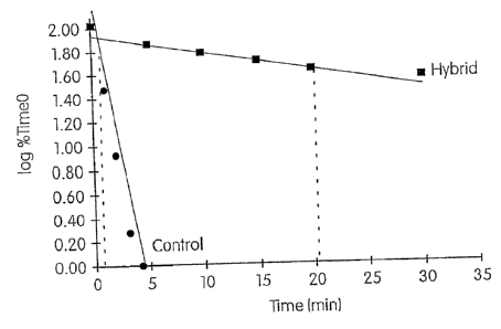
Figures 1 and 2 are graphs which were used to determine the differential
hydrolysis ratios of two M pneumoniae probes disclosed by Hammond et al.,
"Nucleic Acid
Hybridization Assay Probes, Helper Probes and Amplification Oligonucleotides
Targeted to
pneurnoniae Nucleic Acid," U.S. Patent No. 5,656,427. Using the time and
signal data set
forth in Tables 2-5 of Example 2 infra, these graphs plot the data for hybrids
(0) and controls
(0) as the log of the percentage of time zero chemiluminescence on the y-axis
versus time in
minutes on the x-axis. Slopes and associated ti4 values (time required to
hydrolyze 50% of the
-13-
.1
=
CA 02931751 2016-05-30
WO 03/040689
PCT/US02/35133
probe associated acriciinium ester label) were determined for the controls and
hybrids of each
probe using standard linear-regression analysis. See Arnold et al., "Assay
Formats Involving
Acridinitun-Ester-Labeled DNA Probes," Clinical Chemishy, 35:1588-1594 (1989).
Based
on the ty, values determined from these graphs, the differential hydrolysis
ratio for each probe
was calculated by comparing the t% value of the hybrid to the ty, value of the
control.
Figures 3 and 4 are graphs which were used to determine the differential
hydrolysis ratios of two probes according to the present invention. Using the
time and signal
data set forth in Tables 7-10 of Example 2 infra, these graphs plot the data
for hybrids (I) and
controls (0) as the log of the percentage of time zero chemiluminescence on
the y-axis versus
time in minutes on the x-axis. Slopes and associated ty2 values were
determined for the
controls and hybrids of each probe using standard linear-regression analysis.
Based on the t%
values determined from these graphs, the differential hydrolysis ratio for
each probe was
calculated by comparing the ty, value of the hybrid to the t% value of the
control.
FIG. 5 depicts a linking reagent having an extended aminoallcylearboxy linker
arm which can be used to join a detectable label to an oligonucleotide.
DESCRIPTION OF TEE PREFERRED EMBODIMENTS
The present invention describes oligonucleotides. targeted to nucleic acid
derived from Mycoplasma organisms which are useful for determining the
presence or absence
of M pneumoniae or M genitalium in a test sample. The oligonucleotides can aid
in detecting
pneumoniae or M. genitalium in different ways, such as by functioning as
hybridization
assay probes, capture probes and/or amplification primers. Hybridization assay
probes of the
present invention can preferentially hybridize to = a target nucleic acid
sequence present in
nucleic acid derived from M. pneumoniae or M genitalium under stringent
hybridization assay
conditions to form detectable duplexes which indicate the presence of M.
pneumoniae or M
genitalium in a test sample. Some of the probes are believed to be capable of
distinguishing
between the target organism and its known closest phylogenetic neighbors.
Capture probes of
the present invention can hybridize to a target nucleic acid sequence present
in nucleic acid
derived from Mycoplasma organisms under stringent hybridization assay
conditions and can
be used to separate target nucleic acid from clinical specimens. Amplification
primers of the
-14-
CA 02931751 2016-05-30
WO 03/040689
PCT/US02/35133
present invention can hybridize to a target nucleic acid sequence present in
nucleic acid derived
from Myeoplasina organisms under amplification conditions and can be used as
primers in an
amplification reaction to generate M-derived nucleic acid. The probes and
amplification
primers may be used in assays for the detection and/or quantitation of M.
pnewnoniae or M
genitaliwn in a test sample.
A. Definitions.
The following terms have the indicated meanings in the specification unless
expressly indicated to have a different meaning.
By "sample" or "test sample" is meant any substance suspected of containing
a target organism or nucleic acid derived from the target organism. The
substance may be, for
example, an unprocessed clinical specimen, such as a sputum or urethral
specimen, a buffered
medium containing the specimen, a medium containing the specimen and lytic
agents for
releasing nucleic acid belonging to the target organism, or a medium
containing nucleic acid
derived from the target organism which has been isolated and/or purified in a
reaction
receptacle or on a reaction material or device. In the claims, the terms
"sample" and "test
sample" may refer to specimen in its raw form or to any stage of processing to
release, isolate
and purify nucleic acid derived from target organisms in the specimen. Thus,
within a method
of use claim, each reference to a "sample" or "test sample" may refer to a
substance suspected
of containing nucleic acid derived from the target organism or organisms at
different stages of
processing and is not limited to the initial form of the substance in the
claim.
By "target nucleic acid" or "target" is meant a nucleic acid containing a
target
nucleic acid sequence.
By "target nucleic acid sequence," "target nucleotide sequence," "target
sequence" or "target region" is meant a specific deoxyribonucleotide or
ribonucleotide
sequence comprising all or part of the nucleotide sequence of a single-
stranded nucleic acid
molecule, and the deoxyribonucleotide or ribonucleotide sequence complementary
thereto.
(The claims, however, may restrict a target sequence to the particular sense
of the recited
sequence with a proviso excluding complementary sequences thereof.)
-15- =
CA 02931751 2016-05-30
WO 03/040689
PCT/US02/35133
By "polynucleotide," "oligonucleotide" or "oligomer" is meant a polymer made
up of two or more nucleoside subunits or nucleobase subunits coupled together.
The
oligonucleotide may be DNA and/or RNA and analogs thereof. The sugar groups of
the
nucleoside subunits may be ribose, deoxyribose and analogs thereof, including,
for example,
ribonucleosides having a 2'-0-alkyl substitution (e.g., 2'-0-methyl) to the
ribofuranosyl moiety.
(Oligonucleotides including nucleoside subunits having 2' substitutions which
are useful as
hybridization assay probes, capture probes, helper probes and/or amplification
primers are
disclosed by Becker et al., "Method for Amplifying Target Nucleic Acids Using
Modified
Primers," U.S. Patent No. 6,130,038.) The nucleoside subunits may by joined by
linkages such
as phosphodiester linkages, modified linkages or by non-nucleotide moieties
which do not
prevent hybridization of the oligonucleotide to its complementary target
nucleic acid sequence.
Modified linkages include those linkages in which a standard phosphodiester
linkage is
replaced with a different linkage, such as a phosphorothio ate linkage or a
methylphosphonate
linkage. The nucleobase subunits may be joined, for example, by replacing the
natural
deoxyribose phosphate backbone of DNA with a pseuodo peptide backbone, such as
a 2-
aminoethylglycine backbone which couples the nucleobase subunits by means of a
carboxymethyl linker to the central secondary amine. (DNA analogs having a
pseudo peptide =
backbone are referred to as "peptide nucleic acids" or "PNA" and are disclosed
by Nielsen et
al., "Peptide Nucleic Acids," U.S. Patent No. 5,539,082.) Other non-limiting
examples of
oligonucleotides or oligomers contemplated by the. present invention include
nucleic acid
analogs containing bicyclic and tricyclic nucleoside and nucleotide analogs
referred to as
"locked nucleic acids," "locked nucleoside analogues'or "LNA." (Locked nucleic
acids are
disclosed by Wang, "Conformationally Locked Nucleosides and Oligonucleotides,"
U.S. Patent
No. 6,083,482; Imanishi et al., "Bicyclonucleoside and Oligonucleotide
Analogues," U.S.
Patent No. 6,268,490; and Wengel et al., "Oligonucleotide Analogues,"
International
Publication No. WO 99/14226.) Any nucleic acid analog is contemplated by the
present
invention provided the oligonucleotide, as defined above, can stably bind to a
target nucleic
acid :under stringent hybridization assay conditions or amplification
conditions. For
hybridization assay probes, an oligonucleotide must be capable of
preferentially hybridizing to
the target nucleic acid under stringent hybridization assay conditions. For
capture probes, an
-16-
CA 02931751 2016-05-30
WO 03/040689
PCT/US02/35133
oligonucleotide or set of joined oligonucleotides must be capable of
hybridizing to an
immobilized probe and to the target nucleic acid under the same or different
assay conditions
(hybridization to the target sequence is preferably preferential). And for
amplification primers,
an oligonucleotide must be capable of hybridizing to the target nucleic acid
under amplification
conditions and acting as a primer and/or a promoter template for the
initiation of nucleic acid
synthesis.
Oligonucleotides of a defined sequence may be produced by techniques known
to those of ordinary skill in the art, such as by chemical or biochemical
synthesis, and by in
vitro or in vivo expression from recombinant nucleic acid molecules, e.g.,
bacterial or retroviral
vectors. As intended by this disclosure, an oligonucleotide does not consist
of wild-type
chromosomal DNA or the in vivo transcription 'products thereof. One use of an
oligonucleotide is as a hybridization assay probe. Oligonucleotides may also
be used as in vivo
or in vitro therapeutic amplification primers or as antisense agents to block
or inhibit gene
transcription, or translation in diseased, infected, or pathogenic cells.
By "hybridization assay probe" or "probe" is meant an oligonucleotidehaving
a base sequence sufficiently complementary to its target nucleic acid sequence
to form a
probe :target hybrid stable for detection under stringent hybridization assay
conditions. As
would be understood by someone having ordinary skill in the art, a probe is an
isolated nucleic
acid molecule, or an analog thereof, in a form not found in nature without
human intervention
(e.g., recombined with foreign nucleic acid, isolated, or purified to some
extent). The probes
of this invention may have additional nucleosides or nucleobases which are
coupled to the
target complementary sequence so long as such nucleosides or nucleobases do
not prevent
hybridization under stringent hybridization conditions and, in the case of
hybridization assay
probes, do not prevent preferential hybridization to the target nucleic acid.
One or more
sequences which are non-complementary to the target sequence may be included
in a probe
of the present invention, provided these additional sequences do not stably
bind to nucleic acid
derived from any orp nism present in the test sample. Such sequences could
include, by way
of example, a target capture sequence (generally a homopolymer tract, such as
apoly dA, poly
A, poly dT or poly U tail), a promotor sequence, a binding site for RNA
transcription, a
restriction endonuclease recognition site, or sequences which will confer a
desired secondary
-17- '
CA 02931751 2016-05-30
WO 03/040689
PCT/US02/35133
or tertiary structure, such as a catalytic active site or a hairpin structure,
which can be used to
facilitate detection and/or amplification. Probes of a defined sequence may be
produced by
techniques known to those of ordinary skill in the art, such as by chemical
synthesis, and by
in vit7-o or in vivo expression from recombinant nucleic acid molecules,
By "stably," "stable" or "stable for detection" is meant that the temperature
of
a reaction mixture is at least 2 C below the melting temperature of a nucleic
acid duplex. The
temperature of the reaction mixture is more preferably at least 5 C below the
melting
temperature of the nucleic acid duplex, and even more preferably at least 10 C
below the
melting temperature of the reaction mixture.
By "substantially homologous," "substantially corresponding" or "substantially
corresponds" is meant that the subject oligonucleotide has a base sequence
containing an at
least 10 contiguous base region that is at least about 80% homologous,
preferably at least
about 90% homologous, and most preferably 100% homologous to an at least 10
contiguous
base region present in a reference base sequence (excluding RNA and DNA
equivalents).
(Those skilled in the art will readily appreciate modifications that could be
made to the
hybridization assay conditions at various percentages of homology to permit
hybridization of
the oligonucleotide to the target sequence while preventing levels of non-
specific hybridization
sufficient to interfere with detection of the target nucleic acid.) The degree
of similarity is
determined by comparing the order of nucleobases making up the two sequences
and does not
take into consideration other structural differences which may exist between
the two
sequences, provided the structural differences do not prevent hydrogen bonding
with
complementary bases. The degree of homology between two sequences can also be
expressed
in ten-us of the number of base differences between each set of at least 10
contiguous bases
being compared, which may be 0, 1 or 2 base differences.
By "substantially complementary" is meant that the subject oligonucleotide has
a base sequence containing an at least 10 contiguous base region that is at
least 80%
complementary, preferably at least 90% complementary, and most preferably 100%
complementary to an at least 10 contiguous base region present in a target
nucleic acid
sequence (excluding RNA and DNA equivalents). (Those skilled in the art will
readily
appreciate modifications that could be made to the hybridization assay
conditions at various
-18-
CA 02931751 2016-05-30
WO 03/040689
PCT/U502/35133
=
percentages of complementarity to permit hybridization of the oligonucleotide
to the target
sequence while preventing levels of non-specific hybridization sufficient to
interfere with
detection of the target nucleic acid.) The degree of complementarity is
determined by
comparing the order of nucleobases making up the two sequences and does not
take into
consideration other structural differences which may exist between the two
sequences,
provided the structural differences do not prevent hydrogen bonding with
complementary
bases. The degree of complementarily between two sequences can also be
expressed in terms
of the number of base mismatches present in each set of at least 10 contiguous
bases being
compared, which may be 0, 1 or 2 base mismatches.
By "about" is meant the nearest rounded whole number when referring to a
percentage of complementarity or homology (e.g., a lower limit of 24.4 bases
would be 24
bases and a lower limit of 24.5 bases would be 25 bases).
By "RNA and DNA equivalents" is meant RNA and DNA molecules having the
same complementary base pair hybridization properties. RNA and DNA equivalents
have
different sugar moieties (i.e., ribose versus deoxyribose) and may differ by
the presence of
moil in RNA and thymine in DNA. The differences between RNA and DNA
equivalents do
not contribute to differences in homology because the equivalents have the
same degree of
complementarity to a particular sequence.
By "hybridization" is meant the ability of two completely or partially
complementary nucleic acid strands to come together under specified
hybridization assay
conditions in an antiparallel orientation (a parallel orientation may also be
possible) to form a
stable structure having a double-stranded region. The two constituent strands
of this double-
stranded structure, sometimes called a hybrid, are held together by hydrogen
bonds. Although
these hydrogen bonds most commonly form between nucleotides containing the
bases adenine
and thymine or uracil (A and T or U) or cytosine and guanine (C and G) on
single nucleic acid
strands, base pairing can also form between bases that are not members of
these "canonical"
pairs. Non-canonical base pairing is well-known in the art. See, e.g., ROGER
L.P. ADAMS ET
AL., THE BIOCHEMISTRY OF THE NUCLEIC ACIDS (11' ed. 1992).
By "preferentially hybridize" is meant that under stringent hybridi7Ation
assay
conditions, hybridization assay probes can hybridize to their target nucleic
acids to form stable
-19-
CA 02931751 2016-05-30
WO 03/040689
PCT/US02/35133
probe:target hybrids indicating the presence of at least one organism of
interest ("detectable
hybrids"), and there is not formed a sufficient number of stable probe:non-
target hybrids to
indicate the presence of non-targeted organisms ("non-detectable hybrids"),
especially
phylogenetically closely related organisms. Thus, the probe hybridizes to
target nucleic acid
to a sufficiently greater extent than to non-target nucleic acid to enable one
having ordinary
skill in the art to accurately detect the presence (or absence) of nucleic
acid derived from M.
pneumoniae or M. genitaliuin, as appropriate, and distinguish its presence
from that of a
phylogenetically closely related organism in a test sample. In general,
reducing the degree of
complementarity between an oligonucleotide sequence and its target sequence
will decrease
the degree or rate of hybridization of the oligonucleotide to its target
region. However, the
inclusion of one or more non-complementary bases may facilitate the ability of
an
oligonucleotide to discriminate against non-target organisms.
Preferential hybridization can be measured using any of a variety of
techniques
known in the art, including, but not limited to those based on light emission,
mass changes,
changes in conductivity or turbidity. A number of detection means are
described herein, and
one in particular is used in the examples provided below. Preferably, there is
at least a 10-fold
difference between target and non-target hybridization signals in a test
sample, more
preferably at least a 100-fold difference, and most preferably at least a 500-
fold difference.
Preferably, non-target hybridization signals in a test sample are no more than
the background
signal level.
By "stringent hybridization assay conditions," "hybridization assay
conditions," "stringent hybridization conditions," or "stringent conditions"
is meant
conditions permitting a hybridization assay probe to preferentially hybridize
to a target nucleic
acid (preferably rRNA or rDNA derived from M. pneumoniae or M. genitalium)
over nucleic
acid derived from a closely related non-target microorganism. Stringent
hybridization assay
conditions may vary depending upon factors including the GC content and length
of the probe,
the degree of similarity between the probe sequence and sequences of non-
target sequences
which may be present in the test sample, and the target sequence.
Hybridization conditions
include the temperature and the composition of the hybridization reagents or
solutions. While
the Examples section infra provides preferred hybridization assay conditions
for detecting
-20-
CA 02931751 2016-05-30
WO 03/040689
PCT/US02/35133
invention, other stringent conditions could be easily ascertained by someone
having ordinary
skill in the art.
By "assay conditions" is meant conditions permitting stable hybridization of
an
oligonucleotide to a target nucleic acid. Assay conditions do not require
preferential
hybridization of the oligonucleotide to the target nucleic acid.
By "differential hydrolysis" is meant the different rates at which the ester
bond
of an acric-linimn ester (AE) molecule is hydrolyzed in the presence of an
alkaline selection
reagent, which will depend upon whether the AE molecule is associated with a
probe free in
solution or a probe bound to a target nucleic acid. Generally, AE molecules
associated with
probe bound to target nucleic acid will hydrolyze more slowing than AE
molecules associated
with probe free in solution in the presence of a selection reagent. An example
of an alkaline
selection reagent is set forth in the Examples section infra under the
subheading "Reagents."
By "differential hydrolysis ratio" is meant the ratio of the rate of
hydrolysis of
the ester bond of an AE molecule associated with probe bound to a target
nucleic acid to the
rate of hydrolysis of the ester bond of an AE molecule associated with an
identical probe free
in solution in the presence of an alkaline selection reagent. The greater the
differential
hydrolysis ratio of the AE-labeled probe, the greater the sensitivity and
discriminatory capacity
of the AE-labeled probe for the target nucleic acid.
By "consists essentially of' or "consisting essentially of," when used with
reference to a hybridization assay probe herein, is meant an oligonucleotide
comprising a target
binding region, where the base sequence of the target binding region consists
of or is contained
within at least 29 contiguous bases of the base sequence of SEQ ID NO:1, SEQ
ID NO:2,
SEQ ID NO:3, SEQ ID NO:4, SEQ ID NO:5, SEQ ID NO:6, SEQ ID NO:7 or SEQ ID
NO: 8. The base sequence of the target binding region preferably contains the
base sequence
of SEQ ID NO:9, SEQ ID NO:10, SEQ ID NO:11 or SEQ ID NO:12 for detecting the
presence of M genitaliwn in a test sample or the corresponding base sequence
within the base
sequence of SEQ ID NO:1, SEQ ID NO:2, SEQ ID NO:3 or SEQ ID NO:4 for detecting
the
presence of M. pneumoniae in a test sample. Thus, these phrases contain both a
sequence
length limitation and a sequence variation limitation. Any additions or
deletions are non-
material variations of the specified base sequence which do not prevent the
oligonucleotide
-21-
.
CA 02931751 2016-05-30
WO 03/040689 PCT/US02/35133
from having its claimed property (i.e., preferentially hybridizing under
stringent hybridization
assay conditions to the target nucleic acid over non-target nucleic acids).
The oligonucleotide
may include other nucleic acid molecules which do not participate in
hybridization of the probe
to the target nucleic acid and which do not affect such hybridization.
By "nucleic acid duplex," "duplex," "nucleic acid hybrid" or "hybrid" is meant
a stable nucleic acid structure comprising a double-stranded, hydrogen-bonded
region. Such
hybrids include RNA:RNA, RNA:DNA and DNA:DNA duplex molecules and analogs
thereof.
The structure is sufficiently stable to be detectable by any known means,
including means
which do not require a probe associated label. For instance, the detection
method may include
a probe coated substrate which is optically active and sensitive to changes in
mass at its
surface. Mass changes result in different reflective and transmissive
properties' of the optically
active substrate in response to light and serve to indicate the presence or
amount of
=
immobilized target nucleic acid. See, e.g., Nygren et al., "Devices and
Methods for Optical
Detection of Nucleic Acid Hybridization," U.S. Patent No. 6,060,237.
By "amplification primer" or "primer" is meant an oligonucleotide capable of
hybridizing to a target nucleic acid and acting as a primer and/or a promoter
template (e.g., for
synthesis of a complementary strand, thereby forming a functional promoter
sequence) for the
initiation of nucleic acid synthesis. If the amplification primer is designed
to initiate RNA
synthesis, the primer may contain a base sequence which is non-complementary
to the target
sequence but which is recognized by an RNA polymerase, such as a T7, T3 or SP6
RNA
polymerase. An amplification primer may contain a 3' terminus which is
modified to pievent
or lessen the rate or amount of primer extension. (McDonough et al. disclose
primers and
promoter-primers having modified or blocked 3'-ends in U.S. Patent No.
5,766,849, entitled
"Methods of Amplifying Nucleic Acids Using Promoter-Containing Primer
Sequences!)
While the amplification primers of the present invention may be chemically
synthesized or
derived from a vector, they are not naturally-occurring nucleic acid
molecules.
By "nucleic acid amplification," "target amplification" or "amplification" is
meant increasing the number of nucleic acid molecules having at least one
target nucleic acid
sequence. Target amplification according to the present invention may be
either linear or
exponential, although exponential amplification is preferred.
-22-
CA 02931751 2016-05-30
WO 03/040689
PCT/11S02/35133
By "amplification conditions" is meant conditions permitting nucleic acid
amplification. While the Examples section infra provides preferred
amplification conditions
for amplifying target nucleic acid sequences derived from Mycoplasma organisms
using
primers of the present invention in a transcription-mediated amplification
method, other
acceptable amplification conditions could be easily determined by one having
ordinary skill in
the art, depending on the particular method of amplification desired.
By "antisense," "opposite sense" or "negative sense" is meant a nucleic acid
molecule perfectly complementary to a reference, or sense, nucleic acid
molecule.
By "sense," "same-sense" or "positive sense" is meant a nucleic acid molecule
perfectly homologous to a reference nucleic acid molecule.
By "amplicon" is meant a nucleic acid molecule generated in a nucleic acid
amplification reaction and which is derived from a target nucleic acid. An
amplicon contains
a target nucleic acid sequence which may be of the same or opposite sense as
the target nucleic
acid. =
By "derived" is meant that the referred to nucleic acid is obtained directly
from
a target organism or indirectly as the product of a nucleic acid
amplification, which product,
may be, for instance, an antisense RNA molecule which does not exist in the
target organism.
By "capture probe" is meant an oligonucleotide or a set of at least two
oligonucleotides linked together which are capable of hybridizing to a target
nucleic acid and
to an immobilized probe, thereby providing means for immobilizing and
isolating the target
nucleic acid in a test sample. That portion of the capture probe which
hybridizes to the target
nucleic acid is referred to as the "target binding region," and that portion
of the capture probe
which hybrid ins to the immobilized probe is referred :to as the "immobilized
probe binding
region." While the preferred capture probe hybridizes to both the target
nucleic acid and the
immobilized probe under assay conditions, the target binding region and the
immobilized probe
binding region may be designed to hybridize to their respective target
sequences under
different hybridization conditions. In this way, the capture probe may be
designed so that it
first hybridizes to the target nucleic acid under more favorable in solution
kinetics before
adjusting the conditions to permit hybridization of the immobilized probe
binding region to the
immobilized probe. When the target binding and immobilized probe binding
regions are
-23-
CA 02931751 2016-05-30
WO 03/040689
PCT/US02/35133
provided on the same capture probe, they may be directly adjoining each other
on the same
oligonucleotide, they may be separated from each other by one or more
optionally modified
nucleotides, or they may be joined to each other by means of a non-nucleotide
linker,
By "target binding region" is meant that portion of an oligonucleotide which
stably binds to a target sequence present in a target nucleic acid, a DNA or
RNA equivalent
of the target sequence or a complement of the target sequence under assay
conditions. The
assay conditions may be stringent hybridization conditions or amplification
conditions.
By "immobilized probe binding region" is meant that portion of an
oligonucleotide which hybridizes to an immobilized probe under assay
conditions.
By "homopolymer tail" in the claims is meant a contiguous base sequence of
at least 10 identical bases (e.g., 10 contiguous adenines or thymines).
By "immobilized probe" is meant an oligonucleotide for joining a capture probe
to an immobilized support. The immobilized probe is joined either directly or
indirectly to the
solid support by a linkage or interaction which remains stable under the
conditions employed
to hybridize the capture probe to the target nucleic acid and to the
immobilized probe, whether
those conditions are the same or different. The immobilized probe facilitates
separation of the
bound target nucleic acid from unbound materials in a sample.
By "isolate" or "isolating" is meant that at least a portion of the target
nucleic
acid present in a test sample is concentrated within a reaction receptacle or
on a reaction
device or solid carrier (e.g., test tube, cuvette, microtiter plate well,
nitrocellulose filter, slide
or pipette tip) in a fixed or releasable manner so that the target nucleic
acid can be purified
without significant loss of the target nucleic acid from the receptacle,
device or carrier.
By "separate," "separation," "separating" or "purify," "purified" or
"purifying"
is meant that one or more components of a sample contained in or on a
receptacle, device or
carrier are physically removed from one or more other sample components
present in or on the
receptacle, device or carrier. Sample components which may be removed during a
separating
or purifying step include proteins, carbohydrates, lipids, inhibitors, non-
target nucleic acids
and unbound probe. Preferably retained in a sample during a separating or
purifying step are
target nucleic acids bound to immobilized capture probes.
-24-
CA 02931751 2016-05-30
WO 03/040689
PCT/US02/35133
By "helper probe" is meant an oligonucleotide designed to hybridi7e to a
target
nucleic acid at a different locus than that of a hybridization assay probe,
thereby either
increasing the rate of hybridization of the probe to the target nucleic acid,
increasing the
melting temperature of the probe:target hybrid, or both.
By "Mycoplasma organisms" is meant two or more species of Mycoplasma,
including M pneumoniae and M genitalium.
By "phylogenetically closely related" is meant that the organisms are closely
related to each other in an evolutionary sense and therefore would have a
higher total nucleic
acid sequence homology than organisms that are more distantly related.
Organisms occupying
adjacent and next to adjacent positions on the phylogenetic tree are closely
related. Organisms
occupying positions farther away than adjacent or next to adjacent positions
on the
phylogenetic tree will still be closely related if they have significant total
nucleic acid sequence
homology.
By "species-specific" is meant that the referred to hybridization assay probe
is
capable of preferentially hybridizing under stringent hybricli7ation assay
conditions to a target
nucleic acid sequence present in nucleic acid derived from orgnnisms belonging
to the species
M pneumoniae or M genitalium.
B. Hybridization Conditions and Probe Design
Hybridi7ation reaction conditions, most importantly the temperature of
hybridization and the concentration of salt in the hybridization solution, can
be selected to
allow the hybridization assay probes of the present invention to
preferentially hybridize to
nucleic acids having a target nucleic sequence derived from either M
pneumoniae or M
genitalium, and not to non-target nucleic acid which may be present in a test
sample. At
decreased salt concentrations and/or increased temperatures (conditions of
increased
stringency) the extent of nucleic acid hybridization decreases as hydrogen
bonding between
paired nucleotide bases in the double-stranded hybrid molecule is disrupted.
This process is
known as "melting."
Generally speaking, the most stable hybrids are those having the largest
number
of contiguous, perfectly matched (1 . e hydrogen-bonded) nucleotide base
pairs. Such hybrids
-25-
CA 02931751 2016-05-30
WO 03/040689
PCT/US02/35133
would usually be expected to be the last to melt as the stringency of the
hybridization
conditions increases. However, a double-stranded nucleic acid region
containing one or more
mismatched, "non-canonical," or imperfect base pairs (resulting in weaker or
non-existent base
pairing at that position in the nucleotide sequence of a nucleic acid) may
still be sufficiently
stable under conditions of relatively high stringency to allow the nucleic
acid hybrid to be
formed and detected in a hybridization assay without cross-reacting with
other, non-selected
nucleic acids which may be present in a test sample.
Hence, depending on the degree of similarity between the nucleotide sequences
of the target nucleic acid and those of non-target nucleic acids belonging to
phylogenetically
distinct, but closely-related organisms on the one hand, and the degree of
complementarily
between the nucleotide sequences of a particular probe and those of the target
and non-target
nucleic acids on the other, one or more mismatches will not necessarily defeat
the ability of an
oligonucleotide contained in the probe or primer to hybridize to the target
nucleic acid and not
to non-target nucleic acids.
The hybridization assay probes of the present invention were chosen, selected,
and/or designed to maximize the difference between the melting temperatures
(T.) of the
probe:target hybrid (Tm is defined as the temperature at which half of the
potentially double-
stranded molecules in a given reaction mixture are in a single-stranded,
denatured state) and
the T. of a mismatched hybrid formed between the probe and rRNA or rDNA of the
phylogenetically most closely-related organisms expected to be present in the
test sample, but
not sought to be detected. While the unlabeled amplification primers and
capture probes need
not have such an extremely high degree of specificity as the hybridization
assay probe to be
useful in the present invention, they are designed in a similar manner to
preferentially hybridize
to one or more target nucleic acids over other nucleic acids under specified
amplification or
hybridization assay conditions.
To facilitate the identification of nucleic acid sequences to be used in the
design
of probes, 16S rRNA nucleotide sequences from different organisms were first
aligned to
maximize homology. These organisms included Acholeplasn2a laidlawii,
Escherichia coli,
Mycoplasma buccale, Mycoplasma bovis, Mycoplasma capricolum, Mycoplasma
faucium,
Mycoplasn2a fern2entans, Mycoplasma gallisepticum, Mycoplasn2a genitalium,
Mycoplasma
-26-
CA 02931751 2016-05-30
WO 03/040689
PCT/US02/35133
hominis, Mycoplasma iowae, Mycoplasma lipophilun2, Mycoplasma muris,
Mycoplasma
orale, Mycoplasma pneumoniae, Mycoplasma pi711772, Mycoplasma primatum,
Mycoplasma
salivarium, Spiroplasma rnirum and Ureaplasn2a urealyticum. The sequences used
for this
comparison were determined in the laboratory or obtained from published
sources. Sequences
for M pneumoniae and M genitalium were obtained from the GenBank database
under
Accession Nos. M29061 and X77334, respectively.
Within the rRNA molecule there is a close relationship between secondary
structure (caused in part by intra-molecular hydrogen bonding) and function.
This fact
imposes restrictions on evolutionary changes in the primary nucleotide
sequence causing the
secondary structure to be maintained. For example, if a base is changed in one
"strand" of a
double helix (due to intra-molecular hydrogen bonding, both "strands" are part
of the same
rRNA molecule), a compensating substitution usually occurs in the primary
sequence of the
other "strand" in order to preserve complernentarity (this is referred to as
co-variance), and
thus the necessary secondary structure. This allows two very different rRNA
sequences to be
aligned based both on the conserved primary sequence and also on the conserved
secondary
structure elements. Potential target sequences for the hybridization assay
probes described
herein were identified by noting variations in the homology of the aligned.
sequences.
The sequence. evolution at each of the variable regions is mostly divergent.
As
a result of this divergence, corresponding rRNA variable regions of more
distant phylogenetic
relatives of M. pnewnoniae and M genitaliwn show greater differences from the
rRNA of
these organisms than do the rRNAs of phylogenetically closer relatives.
Sufficient variation
between M pneumoniae and M genitalium and other organisms was observed to
identify
preferred target sites and design hybridization assay probes useful for
distinguishing between
M pneumoniae and M genitalium over other closely related organisms.
Merely identifying putatively unique potential target nucleotide sequences
does
not guarantee that a functionally species-specific hybridization assay probe
may be made to
hybridize to M. pneumoniae or M genitalium rRNA or rDNA comprising that
sequence.
Various other factors will determine the suitability of a nucleic acid locus
as a target site for
species-specific probes. Because the extent and specificity of hybridization
reactions, such as
those described herein, are affected by a number of factors, manipulation of
one or more of
-27-
CA 02931751 2016-05-30
WO 03/040689
PCT/11S02/35133
those factors will determine the exact sensitivity and specificity of a
particular oligonucleotide,
whether perfectly complementary to its target or not. The importance and
effect of various
assay conditions are known to those skilled in the art and are disclosed by
the following:
Kohne, "Method for Detection, Identification and Quantitation of Non-Viral
Organisms," U.S
Patent No. 4,851,330; Hogan et al., "Nucleic Acid Probes to Mycobacterium
gordonae," U.S.
Patent No. 5,216,143; and Hogan, "Nucleic Acid Probes for Detection and/or
Quantitation of
Non-Viral Organisms," U. S . Patent Nos. 5,840,488.
The desired temperature of hybridization and the hybridization solution
.
composition (such as salt concentration, detergents and other solutes) can
also greatly affect
the stability of double-stranded hybrids. Conditions such as ionic strength
and the temperature
at which a probe will be allowed to hybridize to a target must be taken into
account in
constructing a species-specific probe. The thermal stability of hybrid nucleic
acids generally
increases with the ionic strength of the reaction mixture. On the other hand,
chemical reagents
which disrupt hydrogen bonds, such as formamide, urea, dimethyl sulfoxide and
alcohols, can
greatly reduce the thermal stability of the hybrids.
To maximize the specificity of a probe for its target, probes of the present
invention were designed to hybridize to their targets under conditions of high
stringency.
Under such conditions only single nucleic acid strands (or regions) having a
high degree of
complementarity will hybridize to each other. Single nucleic acid strands
without such a high
degree of complementarity will not form hybrids. Accordingly, the stringency
of the assay
conditions determines the amount of complementarity which should exist between
two nucleic
acid strands in order to form a hybrid. Stringency is chosen to maximize the
difference in
stability between the hybrid formed between the probe and the target nucleic
acid and potential
hybrids between the probe and any non-target nucleic acids present in a test
sample.
Proper specificity may be achieved by minimizing the length of the
hybridization assay
probe having perfect complementarity to sequences of non-target organisms, by
avoiding G
and C rich regions of complementarity to non-target nucleic acids, and by
constructing the
probe to contain as many destabilizing mismatches to non-target sequences as
possible.
Whether a probe is appropriate for detecting only a specific type of organism
depends largely
on the thermal stability difference between probe:target hybrids versus
probe:non-target
-28-
=
CA 02931751 2016-05-30
WO 03/040689
PCT/US02/35133
hybrids. In designing probes, the differences in these T. values should be as
large as possible
(preferably 2-5 C or more). Manipulation of the T. can be accomplished by
changes to probe
length and probe composition, such as GC content versus AT content or the
inclusion of
nucleotide analogs (e.g., ribonucleotides having a 21-0-methyl substitution to
the ribofuranosyl
moiety).
In general, the optimal hybridization temperature for oligonucleotide probes
is
approximately 5 C below the melting temperature for a given duplex. Incubation
at
temperatures below the optimum temperature may allow mismatched base sequences
to
hybridize and can therefore decrease specificity. The longer the probe, the
more hydrogen
bonding between base pairs and, in general, the higher the T.. Increasing the
percentage of
G and C also increases the T. because G-C base pairs exhibit additional
hydrogen bonding and
therefore greater thermal stability than A-T base pairs. Such considerations
are known in the
art. See, e.g., J. SAMBROOK ET AL, MOLECULAR CLONING: A LABORATORY MANUAL CH.
11 (2d ed. 1989).
A preferred method for determining T. measures hybridization using the well
known hybridization protection assay (HPA) disclosed by Arnold et al.,
"Homogenous
Protection Assay," U.S. Patent No. 5,283,174. The T. can be measured using
FLPA in the
following manner. Probe molecules are labeled with an acridinium ester and
permitted to form
probe:target hybrids in a lithium succinate buffer (0.1 M lithium succinate
buffer, pH 4.7, 20
mM EDTA, 15 m.M aldrithio1-2, 1.2 M LiC1, 3% (v/v) ethanol absolute, 2% (w/v)
lithium
lauryl sulfate) using an excess amount of target. Aliquots of the solution
containing the
probe:target hybrids are then diluted in the lithium succinate buffered
solution and incubated
for five minutes at various temperatures starting below that of the
anticipated T. (typically
55 C) and increasing in 2-5 C increments. This solution is then diluted with a
mild alkaline
borate buffer (600 mM boric acid, 240 rnM Na0H, 1% (v/v) TRITON X-100, pH
8.5) and
incubated at an equal or lower temperature (for example 50 C) for ten minutes.
Under these conditions the acridinium ester attached to the single-stranded
probe is hydrolyzed, while the acridinium ester attached to hybridized probe
is relatively
protected from hydrolysis. Thus, the amount of acridinium ester remaining
after hydrolysis
treatment is proportional to the number of hybrid molecules. The remaining
acridinium ester
-29-
CA 02931751 2016-05-30
WO 03/040689
PCT/US02/35133
can be measured by monitoring the chemiluminescence produced from the
remaining
acridinium ester by adding hydrogen peroxide and alkali to the solution.
Chemiluminescence
can be measured in a luminometer, such as a LEADER 450i lurninometer (Gen-
Probe
Incorporated, San Diego, CA; Cat. No. 32004 The resulting data is plotted as
percent of
maximum signal (usually from the lowest temperature) versus temperature. The
T. is defined
as the temperature at which 50% of the maximum signal remains. In addition to
the method
above, T. may be determined by isotopic methods known to those skilled in the
art (see, e.g.,
U.S. Patent No. 5,840,488).
It should be noted that the Tin for a given hybrid varies depending on the
nature
of the hybridization solution used. Factors such as the salt concentration,
detergents, and other
solutes can affect hybrid stability during thermal denaturation (see, e.g.,
SAMBROOK ET AL.,
supra, ch. 11). Conditions such as ionic strength and the temperature at which
a probe will
be allowed to hybridize to target should be taken into account in probe
construction. (The
thermal stability of a hybrid nucleic acid increases with the ionic strength
of the reaction
mixture.) On the other hand, chemical reagents that disrupt hydrogen bonds,
such as
foi.mamide, urea, dimethyl sulfmdde and alcohols, can greatly reduce hybrid
thermal stability.
To ensure specificity of a hybridization assay probe for its target, it is
preferable
to design probes which hybridize only to target nucleic acid under conditions
of high
stringency. Only highly complementary sequences will form hybrids under
conditions of high
stringency, Accordingly, the stringency of the assay conditions determines the
amount of
complementarity needed between two sequences in order for a stable hybrid to
form.
Stringency should be chosen to maximize the difference in stability between
the probe:target
hybrid and potential probe:non-target hybrids.
Examples of specific stringent hybridization conditions are provided in the
Examples section infi-a (see the subsection entitled "Reagents" for a
description of particular
hybridization and amplification reagents which can be used). Of course,
alternative stringent
hybridization conditions could be determined by those of ordinary skill in the
art based on the
present disclosure. (See, e.g., SAMBROOK ET AL., supra, ch. 11.)
The length of the target nucleic acid sequence region and, accordingly, the
length of the probe sequence can also be important. In some cases, there may
be several
-30-
.
CA 02931751 2016-05-30
WO 03/040689
PCT/US02/35133
sequences from a particular region, varying in location and length, which may
be used In
design probes with the desired hybridization characteristics. In other cases,
one probe may be
significantly better with regard to specificity than another which differs
from it merely by a
single base. While it is possible for nucleic acids that are not perfectly
complementary to
hybridize, the longest stretch of perfectly complementary bases, as well as
the base
compositions, will generally determine hybrid stability.
Regions of rRNA known to form strong internal structures inhibitory to
hybridi7ation are less preferred target regions, especially in assays where
helper probes
disclosed infra are not used. Likewise, probes with extensive self-
complementarity are
generally to be avoided. (It needs to be pointed out, however, that some
degree of self-
complementarity in a probe may be desirable, as in hairpin probes like the
molecular beacons
and molecular torches discussed below.) If .a strand is wholly or partially
involved in an intra-
molecular or inter-molecular hybrid, it will be less able to participate in
the formation of a new
inter-molecular probe:target hybrid without a change in the reaction
conditions. Ribosomal
RNA molecules are known to form very stable intra-molecular helices and
secondary structures
by hydrOgen bonding. By designing a probe to a region of the target nucleic
acid which
remains substantially single-stranded under hybridization conditions, the rate
and extent of
hybridization between probe and target may be increased.
A genomic ribosomal nucleic acid (rDNA) target occurs naturally in a double-
stranded form, as does a product of the polymerase chain reaction (PCR). These
double-
stranded targets are naturally inhibitory to hybridization with a probe and
require denaturation
prior to hybridization. Appropriate denaturation and hybridization conditions
are known in
the art (see, e.g., Southern, E.M., J Mol. Biol., 98:503 (1975)).
A number of formulae are available which will provide an estimate of the
melting temperature for perfectly matched oligonucleotides to their target
nucleic acids. One
such foimula is the following:
Tm = 81.5 + 16.6(1og10[Nal) + 0.41(fraction G+C) - (600/N)
(where N = the length of the oligonucleotide in number of nucleotides)
provides a good
estimate of the Trii for oligonucleotides between 14 and 60 to 70 nucleotides
in length. From
such calculations, subsequent empirical verification or "fine tuning" of the
Tm may be made
-31-.
CA 02931751 2016-05-30
WO 03/040689
PCT/US02/35133
using screening techniques well known in the art. For further information on
hybridization and
oligonucleotide probes reference, may be made to SAMBROOK ET AL., supra, ch.
11. This
reference, among others well known in the art, also provides estimates of the
effect of
mismatches on the Tm of a hybrid. Thus, from the known nucleotide sequence of
a given region
of the ribosomal RNA (or rDNA) of two or more organisms, oligonucleotides may
be designed
which will distinguish these organisms from one another.
C. Preparation of Oligonueleotides
The hybridization assay probes, amplification primers and capture probes of
the present invention can be readily prepared by methods known in the art.
Preferably, the
oligonucleotides are synthesized using solid phase methods. Caruthers, for
example, describes
using standard phosphoramidite solid-phase chemistry to join nucleotides by
phosphodiester
linkages. See Caruthers et al., "Chemical Synthesis of Deoxynucleotides by the
Phosphoramidite Method," Methods Enzymol., 154:287 (1987). Automated solid-
phase
chemical synthesis using cyanoethyl phosphoramidite precursors has been
describedby Barone.
See Barone et al., "In Situ Activation of bis-dialkylaminephosphines -- a New
Method for
Synthesizing Deoxyoligonucleotides on Polymer Supports,"NucIeic Acids Res.,
12(10):4051
(1984). Batt discloses a procedure for synthesizing oligonucleotides
containing
phosphorothioate linkages in U.S. Patent No. 5,449,769, entitled "Method and
Reagent for
Sulfitrization of Organophosphorous Compounds." In addition, Riley et al.
disclose the
synthesis of oligonucleotides having different linkages including
methylphosphonate linkages
in U.S. Patent No. 5,811,538, entitled "Process for the Purification of
Oligomers." Moreover,
methods for the organic synthesis of oligonucleotides are known to those of
skill in the art and
are described in, for example, SAMBROOK ET AL., supra, ch. 10.
Following synthesis and purification of a particular oligonucleotide, several
different procedures may be utilized to purify and control the quality of the
oligonucleotide.
Suitable procedures include polyacrylamide gel electrophoresis or high
pressure liquid
chromatography. Both of these procedures are well known to those skilled in
the art.
All of the oligonucleotides of the present invention, whether hybridization
assay
probes, amplification primers or capture probes, may be modified with chemical
groups to
-32-
=
CA 02931751 2016-05-30
WO 03/040689
PCT/US02/35133
enhance their performance or to facilitate the characterization of
amplification products. For
example, backbone-modified oligonucleotides such as those having
phosphorothioate,
methylphosphonate, 2'-0-alkyl or peptide groups which render the
oligonucleotides resistant
to the nucleolytic activity of certain polymerases or to nuclease enzymes may
allow the use of
such enzymes in an amplification or other reaction. Another example of a
modification involves
using non-nucleotide linkers incorporated between nucleotides in the nucleic
acid chain of a
probe or primer, and which do not prevent hybridization of a probe or
hybridization and
elongation of a primer. See Arnold et al., "Non-Nucleotide Linking Reagents
for Nucleotide
Probes," U.S. Patent No. 6,031,091. The oligonucleotides of the present
invention may also
contain mixtures of the desired modified and natural nucleotides.
The 3' end of an amplification primer may be modified or blocked to prevent
or inhibit initiation of DNA synthesis, as disclosed by Kacian et al. in U.S.
Patent No.
5,554,516. The 3' end of the primer can be modified in a variety of ways well
Icnown in the art
By way of example, appropriate modifications to a primer can include the
addition of
ribonucleotides, 3' deoxynucleotide residues (e.g., cordycepin), 2',3'-
dideoxynucleotide
residues, modified nucleotides such as phosphorothioates, and non-nucleotide
linkages such
as those disclosed by Arnold et al. in U.S. Patent No. 6,031,091 or alkane-
diol modifications
(see Wilk et al., "Backbone-Modified Oligonucleotides Containing a Butanedio1-
1,3 Moiety
as a 'Vicarious Segment' for the Deoxyribosyl Moiety -- Synthesis and Enzyme
Studies,"
Nucleic Acids Res., 18(8):2065 (1990)), or the modification may simply consist
of a region 3'
to the priming sequence that is non-complementary to the target nucleic acid
sequence.
Additionally, a mixture of different 3' blocked primers or of 3' blocked and
unblocked primers
may increase the efficiency of nucleic acid amplification, as disclosed
therein.
The 5' end of primers may be modified to be resistant to the 5'-exonuclease
activity present in some nucleic acid polymerases. Such modifications can be
carried out by
adding anon-nucleotide group to the terminal 5' nucleotide of the primer using
techniques such
as those disclosed by Arnold et al. in U.S. Patent No. 6,031,091. To
facilitate strand
displacement, the 5' end may also be modified to include non-complementary
nucleotides as
disclosed by Dattagupta et al, "Isothermal Strand Displacement Nucleic Acid
Amplification,"
U.S. Patent No. 6,087,133.
-33-
CA 02931751 2016-05-30
WO 03/040689
PCT/US02/35133
Once synthesized, a selected oligonucleotide may be labeled by any of several
well known methods (see, e.g., SAMBROOK, supra, ch. 10). Useful labels include
radioisotopes
as well as non-radioactive reporting groups. Isotopic labels include 31-1,
35S, 32P, 1251, 57Co and
14C. Isotopic labels can be introduced into the oligonucleotide by techniques
known in the art
such as nick translation, end labeling, second strand synthesis, the use of
reverse transcription,
and by chemical methods. When using radiolabeled probes, hybridization can be
detected by
autoradiography, scintillation counting or garnm a counting. The detection
method selected will
depend upon the particular radioisotope used for labeling.
Non-isotopic materials can also be used for labeling and may be introduced
internally into the nucleic acid sequence or at the end of the nucleic acid
sequence. Modified
nucleotides may be incorporated enzymatically or chemically. Chemical
modifications of the
probe may be performed during or after synthesis of the probe, for example,
through the use
of non-nucleotide linker groups, as disclosed by Arnold et al. in U.S. Patent
No. 6,031,091.
Non-isotopic labels include fluorescent molecules (individual labels or
combinations of
interacting labels, such as the fluorescence resonance energy transfer (FRET)
pairs disclosed
by Tyagi et al. in U.S. Patent No. 5,925,517), chemiluminescent molecules,
enzymes,
cofactors, enzyme substrates, haptens or other ligands. With the hybridization
assay probes
of the present invention, the probes are preferably labeled by means of a non-
nucleotide linker
with an acridinium ester (AE), such as standard AE Acridinium ester labeling
may be
performed as disclosed by Arnold et al., "Acridinium Ester Labelling and
Purification of
Nucleotide Probes," U.S. Patent No. 5,185,439.
D. Nucleic Acid Amplification
Preferably, the amplification primers of the present invention are
oligodeoxynucleotides and are sufficiently long to be used as a substrate for
the synthesis of
extension products by a nucleic acid polymerase. Optimal primer length should
take into
account several factors, including the temperature of reaction, the structure
and base
composition of the primer, and how the primer is to be used. For example, for
optimal
specificity the oligonucleotide primer generally should be at least 12 bases
in length, depending
on the complexity of the target nucleic acid sequence. If such specificity is
not essential,
= -34-
CA 02931751 2016-05-30
WO 03/040689
PCT/US02/35133
=
shorter primers may be used. In such a case, it may be desirable to carry out
the reaction at
cooler temperatures in order to form stable hybrid complexes with the template
nucleic acid.
Useful guidelines for designing amplification primers with desired
characteristics are described above in the section entitled "Preparation of
Oligonucleotides."
Optimal sites for amplifying and probing contain at least two, and preferably
three, conserved
regions of M. pneumaniae and/or M. genitalium nucleic acid. These regions are
about 15 to
350 bases in length, and preferably between about 15 and 150 bases in length.
The degree of amplification observed with a set of amplification primers
(primers and/or promoter-primers) depends on several factors, including the
ability of the
primers to hybrillin to their specific target sequences and their ability to
be extended or copied
enzymatically. While amplification primers of different lengths and base
compositions may be
used, amplification primers preferred in this invention have target binding
regions of 18 to 40
bases with a predicted Tm to target above 42 C, preferably at least about 50
C.
Parameters affecting probe hybridization, such as melting temperature,
complementarity and secondary structure of the target sequence, also affect
amplification
primer hybridization and therefore performance of the amplification primers.
The degree of
non-specific extension (primer-dirner or non-target copying) can also affect
amplification
efficiency. Thus, amplification primers are generally selected to have low
self-complementarity
or cross-complementarity, particularly at the 3' ends of their sequences.
Notwithstanding
amplification primers including regions of self-complementaiity may be useful,
such as the self-
reporting "signal primers" disclosed by Nadeau et al., "Detection of Nucleic
Acids by
Fluorescence Quenching," U.S. Patent No. 5,958,700, and the "hairpin primers"
disclosed by
Nazarenko et al., "Nucleic Acid Amplification Oligonucleotides with Molecular
Energy
Transfer Labels and Methods Based Thereon," U.S. Patent No. 5,866,336. Lengthy
homopolymer runs and high GC content are avoided to reduce spurious primer
extension.
Computer programs are available to aid in this aspect of the design, including
Oligo
Tech analysis software available from Oligo Therapeutics, Inc. and accessible
on the World
Wide Web at the following URL: http://www.oligosetc.com.
A nucleic acid polymerase used in conjunction with the amplification primers
of the present invention refers to a chemical, physical or biological agent
which incorporates
-35-
.
CA 02931751 2016-05-30
WO 03/040689
PCT/US02/35133
either ribonucleotides or deoxyribonucleotides, or both, into a nucleic acid
polymer, or strand,
in a template-dependent manner. Examples of nucleic acid polymerases include
DNA-directed
DNA polymerases, RNA-directed DNA polymerases, and RNA-directed RNA
polymerases.
DNA polymerases bring about nucleic acid synthesis in a template-dependent
manner and in
a 5' to 3' direction. Because of the typical anti-parallel orientation of the
two strands in a
double-stranded nucleic acid, this direction is from a 3' region on the
template to a 5' region
on the template. Examples of DNA-directed DNA polymerases include E. coil DNA
polymerase I, the thermostable DNA polymerase from Thermus aquaticus (Tag),
and the large
fragment of DNA polymerase I from Bacillus stearothermophilus (Bst). See,
e.g., Riggs et al.,
"Purified DNA Polymerase from Bacillus stearothermophilus," U.S. Patent No.
6,066,483.
Examples of RNA-directed DNA polymerases include various retroviral reverse
transcriptases,
such as Moloney murine leukemia virus (MMLV) reverse transcriptase or avian
myeloblastosis
virus (AMY) reverse transcriptase.
, During most nucleic acid amplification reactions, a nucleic acid polymerase
adds nucleotide residues to the 3' end of the primer using the target nucleic
acid as a template,
thus synthesizing a second nucleic acid strand having a nucleotide sequence
partially or
completely complementary to a region of the target nucleic acid. In many
nucleic acid
amplification reactions, the two strands comprising the resulting double-
stranded structure
must be separated by chemical or physical means in order to allow the
amplification reaction
to proceed. Alternatively, the newly-synthesized template strand may be made
available for
hybridization with a second primer or promoter-primer by other means, such as
through strand
displacement or the use of a nucleolytic enzyme which digests part or all of
the original target
strand. In this way the process may be repeated through a number of cycles,
resulting ina large
increase in the number of nucleic acid molecules having the target nucleotide
sequence.
Either the first or second amplification primer, or both, may be a promoter-
primer. (In
some applications, the amplification primers may only consist of promoter-
primers which are
complementary to the sense strand, as disclosed by Kacian et al., "Nucleic
Acid Sequence
Amplification Method, Composition and Kit," U.S. Patent No. 5,554,516.) A
promoter-
primer usually contains an oligonucleotide that is not complementary to a
nucleotide sequence
present in the target nucleic acid molecule or primer extension product(s)
(see, e.g., Kacian
-36-
.
=
CA 02931751 2016-05-30
WO 03/040689
PCT/US02/35133
et al., "Nucleic Acid Sequence Amplification Methods," U.S. Patent No.
5,399,491). These
non-complementary sequences may be located 5' to the complementary sequences
on the
amplification primer and may provide a locus for initiation of RNA synthesis
when made
double-stranded through the action of a nucleic acid polymerase. The promoter
thus provided
may allow for the in vitro transcription of multiple RNA copies of the target
nucleic acid
sequence. It will be appreciated that all references to primers herein are
inclusive of primers
and p. romoter-primers, unless the context clearly indicates otherwise.
E. Sample Processing
Sample processing prior to amplification or detection of a target sequence may
be necessary or useful for discriminating a target sequence from non-target
nucleic acid present
in a sample. Sample processing procedures may include, for example, direct or
indirect
immobilization of nucleic acids and/or oligonucleotides from the liquid phase
in a
heterogeneous assay. With some procedures, such immobilization may require
multiple
hybridization events. Ranld et al., "Detection of Microbial Nucleic Acids by a
One-Step
Sandwich Hybridization Test," U.S. Patent Nos. 4,486,539 and 4,563,419, for
example,
disclose a one-step nucleic acid "sandwich" hybridization method involving the
use of a solid-
phase bound nucleic acid having a target complementary sequence and a labeled
nucleic acid
probe which is complementary to a distinct region of the target nucleic acid.
Stabinsky,
"Methods and Kits for Performing Nucleic Acid Hybridization Assays," U.S.
Patent No.
4,751,177, discloses methods including a "mediator" polynucleotide that
reportedly overcomes
sensitivity problems associated with Rank? s method resulting from leakage of
immobilized
probe from the solid support. Instead of directly immobilizing the target
nucleic acid, the
mediator polynucleotides of Stabinsky are used to bind and indirectly
immobilize target
polynucleotide:probe polynucleotide complexes which have formed free in
solution.
Any known solid support may be used for sample processing, such as matrices
and particles free in solution. The solid support may be, for example,
nitrocellulose, nylon,
glass, polyacrylate, mixed polymers, polystyrene, silane polypropylene and,
preferably, particle
having a magnetic charge to facilitate recovering sample and/or removing
unbound nucleic
acids or other sample components. Particularly preferred supports are magnetic
spheres that
-37-
CA 02931751 2016-05-30
WO 03/040689
PCT/US02/35133
are monodisperse (i.e., uniform in size 5%), thereby providing consistent
results, which is
particularly advantageous for use in an automated procedure. One such
automated procedure
is disclosed by Ammann et al., "Automated Process for Isolating and Amplifying
a Target
Nucleic Acid Sequence," U.S.Patent No. 6,335,166.
An oligonucleotide for immobilizing a target nucleic acid on a solid support
may be joined directly or indirectly to the solid support by any linkage or
interaction which is
stable under assay conditions (e.g., conditions for amplification and/or
detection). Referred
to herein as an "immobilized probe," this oligonucleotide may bind directly to
the target nucleic
acid or it may include a base sequence region, such as a homopolymeric tract
(e.g., a poly dT)
or a simple short repeating sequence (e.g., an AT repeat), which hybridizes to
a
complementary base sequence region present on a capture probe. Direct joining
occurs when
the immobilized probe is joined to the solid support in the absence of an
intermediate group.
For example, direct joining may be via a covalent linkage, chelation or ionic
interaction.
Indirect joining occurs when the immobilized probe is joined to the solid
support by one or
more linkers. A "linker" is a means for binding at least two different
molecules into a stable
complex and contains one or more components of a binding partner set.
Members of a binding partner set are able to recognize and bind to each other.
Binding partner sets may be, for example, receptor and ligand, enzyme and
substrate, enzyme
and cofactor, enzyme and coenzyme, antibody and antigen, sugar and lectin,
biotin and
streptavidin, ligand and chelating agent, nickel and histidine, substantially
complementary
oligonucleotides, and complementary homopolymeric nucleic acids or
homopolymeric portions
of polymeric nucleic acids. Components of a binding partner set are the
regions of the
members that participate in binding.
A preferred sample processing system having practical advantages in terms of
its ease of use and rapidity comprises an immobilized probe containing a base
sequence which
is complementary to a base sequence of a capture probe, referred to herein as
an "immobilized
probe binding region." The capture probe additionally contains a base
sequence, referred to
herein as a "target binding region," which may specifically hybridize to a
target sequence
contained in a target nucleic acid under assay conditions. (While specificity
of the target
binding region of the capture probe for a region of the target nucleic acid is
desirable to
-38-
.
CA 02931751 2016-05-30
WO 03/040689
PCT/US02/35133
minimize the number of non-target nucleic acids remaining from the sample
after a separation
step, it is not a requirement of the capture probes of the present invention
if the capture probes
are being used solely to isolate target nucleic acid.) If the capture probe is
not being employed
to isolate a target nucleic acid for subsequent amplification of a target
sequence, the capture
probe may further include a detectable label attached within or near the
target binding region,
such as a substituted or unsubstituted acridinium ester. The labeled capture
probe may be used
in a homogeneous or semi-homogenous assay to specifically detect hybrid
nucleic acids
without detecting single-stranded nucleic acids, such as the capture probe. A
preferred
homogenous assay which could be used with this system is the hybridization
protection assay
(HPA), which is discussed above in the section entitled "Hybridization
Conditions and Probe
Design." Following the HPA format, label associated with capture probes which
have not
hybridized to target nucleic acids. would be hydrolyzed with the addition of a
mild base, while
label associated with capture probe:target hybrids would be protected from
hydrolysis.
An advantage of this latter assay system is that only a single target-specific
hybridization event (capture probe:target) is necessary for target detection,
rather than multiple
such events (e.g., capture probe:target and probe:target or probe:amplicon)
which are required
in other sample processing procedures described herein. Also, fewer
oligonucleotides in an
assay tend to make the assay faster and simpler to optimize, since the overall
rate at which a
target nucleic acid is captured and detected is limited by the slowest
hybridizing
oligonucleotide. 'While the target binding region of a capture probe may be
less specific in
alternative assay systems, it must still be rare enough to avoid significant
saturation of the
capture probe with non-target nucleic acids. Thus, the requirement that two
separate and
specific target sequences be identified in these alternative systems could
place constraints on
the identification of an appropriate target. By contrast, only one such target
sequence is
needed when the capture probe simultaneously functions as the detection probe.
Whichever approach is adopted, the assay needs to include means for detecting
the presence of the target nucleic acid in the test sample. A variety of means
for detecting
target nucleic acids are well known to those skilled in the art of nucleic
acid detection,
including means which do not require the presence of a detectable label.
Nevertheless, probes
including a detectable label are preferred. A labeled probe for detecting the
presence of a
-39-
'
CA 02931751 2016-05-30
WO 03/040689 PCT/US02/35133
=
target nucleic acid would have to include a base sequence which is
substantially complementary
and specifically hybridizes to a target sequence contained in the target
nucleic acid. Once the
probe stably binds to the target nucleic acid, and the resulting target:probe
hybrid has been
directly or indirectly immobilized, unbound probe can be washed away or
inactivated and the
remaining bound probe can be detected and/or measured.
Preferred sample processing systems combine the elements of detection and
nucleic acid amplification. These systems first directly or indirectly
immobilize a target nucleic
acid using a capture probe, the captured target nucleic acid is purified by
removing inter alia -
cellular debris, non-target nucleic acid and amplification inhibitors from the
sample-containing
vessel, which is followed by amplification of a target sequence contained in
the target nucleic
acid. Amplified product is then detected, preferably in solution with a
labeled probe. (The
target nucleic acid may remain in the immobilized state during amplification
or it may be eluted
from the solid support prior to amplification using appropriate conditions,
such as by first
incubating at a temperature above the Tm of the capture probe:target complex
and/or the Tm
of the capture probe:immobilized probe complex.) A preferred embodiment of
this system is
disclosed by Weisburg et aL , "Two-Step Hybridization and Capture of a
Polynucleotide," U.S.
Patent No. 6,110,678. In this system, the capture probe hybridizes to the
target nucleic acid
and an immobilized probe hybridizes to the capture probe:target complex under
different
hybridization conditions. Under a first set of hybridization conditions,
hybridization of the
capture probe to the target nucleic acid is favored over hybridization of the
capture probe to
the immobilized probe. Thus, under this first set of conditions, the capture
probe is in solution
rather than bound to a solid support, thereby maximizing the concentration of
the free capture
probe and utilizing favorable liquid phase kinetics for hybridization to the
target nucleic acid.
After the capture probe has had sufficient time to hybridize to the target
nucleic acid, a second
set of hybridization conditions is imposed permitting in the capture
probe:target complex to
hybridize to the immobilized probe, thereby isolating the target nucleic acid
in the sample
solution. The immobilized target nucleic acid may then be purified, and a
target sequence
present in the target nucleic acid may be amplified and detected. A
purification procedure
which includes one or more wash steps is generally desirable when working with
crude samples
-40-
.
CA 02931751 2016-05-30
WO 03/040689
PCT/US02/35133
(e.g., clinical, environmental, industrial, food, water, etc.) to prevent
enzyme inhibition and/or
nucleic acid degradation due to substances present in the sample.
A preferred amplification method is the transcription-mediated amplification
method disclosed by Kacian et al., "Nucleic Acid Sequence Amplification
Methods," U.S.
Patent No. 5,480,789. In accord with this method, a promoter-primer having a
3' region
complementary to a portion of the target and a 5' promoter region and a primer
having the
same nucleotide sequence as a portion of the target are contacted with a
target RNA molecule.
The primer and promoter-primer define the boundaries of the target region to
be amplified,
including both the sense present on the target molecule and its complement,
and thus the length
and sequence of the ampli con. In this preferred embodiment, the amplification
oligonucleotides and immobilized target RNA are contacted in the presence of
effective
amounts of Moloney murine leukemia virus-derived reverse transcriptase and T7
RNA
polymerase, both ribonucleotide and deoxyribonucleotide triphosphates, and
necessary salts
and cofactors at 42 C. Under these conditions, nucleic acid amplification
occurs, resulting
predominantly in the production of RNA amplicons of a sense opposite to that
of the target
nucleic acid. These amplicons can then be detected in solution by, for
example, using an
acrklinium ester-labeled hybridization assay probe of the same sense as the
target nucleic acid,
employing HPA, as disclosed by Arnold et al. in U.S. Patent No. 5,283,174.
The 3' terminus of the immobilized probe and the capture probe are preferably
"capped" or blocked to prevent or inhibit their use as templates for nucleic
acid polymerase
activity. Capping may involve adding 3' deoxyribonucleotides (such as
cordycepin), 3', 2'-
dideoxynucleotide residues, non-nucleotide linkers, such as those disclosed by
Arnold et al.
in U.S. Patent No. 6,031,091, alkane-diol modifications, or non-complementary
nucleotide
residues at the 3' terminus.
Those skilled in the art will recognize that the above-described methodology
is amenable, either as described or with obvious modifications, to various
other amplification
schemes, including, for example, the polymerase chain reaction (F'CR), Qf3
replicase-mediated
amplification, self-sustained sequence replication (3SR), strand displacement
amplification
(SDA), nucleic acid sequence-based amplification (NASBA), loop-mediated
isothermal
amplification (LAMP), and the ligase chain reaction (LCR).
-41-
CA 02931751 2016-05-30
WO 03/040689
PCT/US02/35133
F. Capture Probes for Isolating M. Ribosomal Nucleic Acid
Capture probes of the present invention are designed to bind to and isolate
nucleic acid derived from the 165 ribosomal nucleic acid of a Mycoplasma
organism in the
presence of non-target nucleic acid. As such, the capture probes include both
a target binding
region and an immobilized probe binding region. The target binding region of
the capture
probes includes a base sequence which hybridizes to a target sequence derived
from 16S
ribosomal nucleic acid from a Mycoplasma organism under assay conditions.
While not
essential, the target binding region preferably exhibits specificity for the
target sequence in the
presence of non-target nucleic acid under assay conditions. The immobilized
probe binding
region has a base sequence which hybridizes to an immobilized probe comprising
a
polynucleotide, or a chimeric containing polynucleotide sequences, which is
joined to a solid
support present in the test sample, either directly or indirectly. The target
binding region and
the immobilized probe binding region may be joined to each other directly or
by means of, for
example, a nucleotide base sequence, an abasie sequence or a non-nucleotide
linker.
In a preferred embodiment, capture probes according to the present invention
include a target binding region comprising a base sequence region which is at
least about 85%
homologous (preferably at least about 90% homologous, more preferably at least
about.95% =
homologous, and most preferably 100% homologous) to a base sequence selected
from the
group consisting of SEQ ID NO:13, SEQ ID NO:14, SEQ ID NO:15, SEQ ID NO:16,
SEQ
ID NO:17, SEQ ID NO:18, SEQ ID NO:19 and SEQ ID NO:20. The immobilized probe
binding region of these preferred capture probes comprises a base sequence
which hybridizes
to an immobilized probe joined directly or indirectly to a solid support
provided to the test
sample under assay conditions. The immobilized probe binding region preferably
comprises
a homopolymeric region (e.g., poly dA) located at the 3' end of the capture
probe which is
complementary to a homopolymeric region (e.g., poly dT) located at the 5' end
of the
immobilized probe. Other base sequences may be incorporated into the
immobilized probe
binding region, including, for example, short repeating sequences.
To prevent undesirable cross-hybridization reactions, the capture probes of
the
present invention preferably exclude nucleotide base sequences, other than the
nucleotide base
sequence of the target binding region, which can stably bind to nucleic acid
derived from any
-42-
CA 02931751 2016-05-30
WO 03/040689
PCT/US02/35133
organism which may be present in the test sample under assay conditions.
Consistent with this
approach, and in order to maximize the immobilization of capture probe:target
complexes
which are formed, the nucleotide base sequence of the immobilized probe
binding region is
preferably designed so that it can stably bind to a nucleotide base sequence
present in the
immobilized probe under assay conditions and not to nucleic acid derived from
any organism
which may be present in the test sample.
The target binding region and the immobilized probe binding region of the
capture probe may be selected so that the capture probe:target complex has a
higher T. than
the T. of the capture probe:immobilized probe complex. In this way, a first
set of conditions
may be imposed which favors hybridization of the capture probe to the target
sequence over
the immobilized probe, thereby providing for optimal liquid phase
hybridization kinetics for
hybridization of the capture probe to the target sequence. Once sufficient
time has passed for
the capture probe to bind to the target sequence, a second set of less
stringent conditions may
be imposed which allows for hybridization of the capture probe to the
immobilized probe. An
example of differing hybridization conditions for capturing a target nucleic
acid on a solid
support is set for in Example 4 infra. Other sets of conditions could be
established by those
skilled in the art without engaging in anything more than routine
experimentation.
Capture probes of the present invention may also include a label or a pair of
interacting labels for direct detection of the target sequence in a test
sample. Non-limiting
examples of labels, combinations of labels and means for labeling probes are
set forth supra
in the section entitled "Preparation of Oligonucleotides" and infra in the
section entitled
"Hybridization Assay Probes to M pneumoniae and/or M. genitaliun2 Ribosomal
Nucleic
Acid." A particularly useful method for detecting the presence of a capture
probe hybridized
to a target nucleic acid is the hybridization protection assay (HPA), which is
described above
in the section entitled "Hybridization Conditions and Probe Design." HPA is a
homogenous
assay which distinguishes between probe hybridized to target nucleic acid and
probe which
remains unhybridized. Signal detected from an HPA reaction vessel provides an
indication of
the presence or amount of target organisms in the test sample.
Despite their application in a direct detection assay, the most common use of
capture probes is in the isolation and purification of target nucleic acid
prior to amplifying a
-43-
CA 02931751 2016-05-30
WO 03/040689 PCT/US02/35133
target sequence contained in the target nucleic acid. By isolating and
purifying the target
nucleic acid prior to amplification, the number of unintended amplification
reactions (i.e.,
amplification of non-target nucleic acid) can be severely limited. And, to
prevent or inhibit the
capture probe itself from functioning as a template for nucleic acid
polymerase activity in the
presence of amplification reagents and under amplification conditions, the 3'
end of the capture
probe may be capped or blocked. Examples of capping agents include 3'
deoxyribonucleotides,
3', 2'-dideoxynucleotide residues, non-nucleotide linkers, alkane-diol
modifications, and non-
complementary nucleotide residues at the 3' temilnus.
G. Amplification of Mycoplasma Ribosomal Nucleic Acid
The amplification primers of the present invention are directed to regions of
16S ribosomal nucleic acid derived from Mycoplasma organisms. The
amplification primers
may flank, overlap or be contained within at least one of the target nucleic
acid sequences of
a hybridization assay probe (or its complement) used to detect the presence of
a Mycoplasina
organism in a nucleic acid amplification assay. As indicated above, the
amplification primers
may also include non-complementary bases at their 5' ends comprising a
promoter sequence
able to bind an RNA polymerase and direct RNA transcription using the target
nucleic acid as
a template. A T7 promoter sequence, such as SEQ ID NO:41, may be used.
Amplification primers of the present invention are capable of amplifying a
target
nucleic acid sequence present in nucleic acid derived from MYcoplasma
organisms under
amplification conditions. These amplification primers comprise an
oligonucleotide having a
target binding region, where the base sequence of the target binding region
has or substantially
. corresponds to the base sequence of SEQ ID NO:21, SEQ ID NO:22, SEQ ID
NO:23, SEQ '
ID NO:24, SEQ ID NO:25, SEQ ID NO:26, SEQ ID NO:27, SEQ ID NO:28, SEQ ID
NO:29, SEQ ID NO:30, SEQ ID NO:31, SEQ ID NO:32, SEQ ID NO:33, SEQ ID NO:34,
SEQ ID NO:35, SEQ ID NO:36, SEQ ID NO:37, SEQ ID NO:38, SEQ TT) NO:39 or SEQ
ID NO:40. Amplification primers of the present invention do not, however,
include an
amplification primer comprising an oligonucleotide having a target binding
region, where the
base sequence of the target binding region has or substantially corresponds to
the base
sequence of SEQ ID NO:25, SEQ ID NO:26, SEQ ID NO:27, SEQ ID NO:28, SEQ ID
-44-
=
CA 02931751 2016-05-30
WO 03/040689
PCT/US02/35133
NO:37, SEQ ID NO:38, SEQ ID NO:39 or SEQ ID NO:40, except in combination with
an
amplification primer comprising an oligonucleotide having a target binding
region, where the
target binding region has or substantially corresponds to the base sequence of
SEQ ID NO:21,
SEQ ID NO:22, SEQ NO:23, SEQ ID NO:24, SEQ ID NO:29, SEQ ID NO:30, SEQ ID
NO:31, SEQ ID NO:32, SEQ ID NO:33, SEQ ID NO:34, SEQ 1D NO:35 and SEQ ID
NO:36. The target binding region of an amplification primer according to the
Present
invention is preferably at least about 80% homologous (more preferably at
least about 90%
homologous and most preferably 100% homologous) to the recited base sequence.
Amplification primers of the present invention have a target binding region
which is preferably
at least 12 bases in length and more preferably from 18 to 40 bases in length.
Where the base sequence of the target binding region has or substantially
corresponds to the base sequence of SEQ ID NO:21, SEQ ID NO:22, SEQ ID NO:23,
SEQ
ID NO:24, SEQ ID NO:29, SEQ ID NO:30, SEQ ID NO:31, SEQ ID NO:32, SEQ ID
NO:33, SEQ ID NO:34, SEQ ID NO:35 or SEQ ID NO:36, the base sequence of the
target
binding region is preferably at least about 80% homologous (more preferably at
least about
90% homologous and most preferably 100% homologous) to the base sequence of
SEQ ID
NO:21, SEQ ID NO:22, SEQ ID NO:23, SEQ ID NO:24, SEQ ID NO:29, SEQ ID NO:30,
SEQ ID NO:31, SEQ ID NO:32, SEQ ID NO:33, SEQ ID NO:34, SEQ ID NO:35 or SEQ
ID NO:36. And, with the exception of an optional 5' sequence recognized by an
RNA
polymerase or which enhances initiation or elongation by an RNA polymerase,
the base
sequence of an amplification primer in the most preferred embodiment of the
present invention
is at least about 80% homologous (more preferably at least about 90%
homologous and most
preferably 100% homologous) to the base sequence of SEQ ID NO:21, SEQ ID
NO:22, SEQ
ID NO:23, SEQ ID NO:24, SEQ ID NO:29, SEQ ID NO:30, SEQ ID NO:31, SEQ ID
NO:32, SEQ ID NO:33, SEQ ID NO:34, SEQ ID NO:35 or SEQ ID NO:36.
In one preferred embodiment, a set of at least two amplification primers for
amplifying nucleic acid from a Mycoplasma organism is provided which includes:
(i) a first
amplification primer comprising an oligonucleotide having a target binding
region, where the
base sequence of the target binding region has or substantially corresponds to
the base
sequence of SEQ ID NO:21, SEQ ID NO:22, SEQ ID NO:23 or SEQ ID NO:24; and (ii)
a
-45-
,
CA 02931751 2016-05-30
WO 03/040689
PCT/US02/35133
second amplification primer comprising an oligonucleotide having a target
binding region,
where the base sequence of the target binding region has or substantially
corresponds to the
base sequence of SEQ ID NO:29, SEQ ID NO:30, SEQ ID NO:31 or SEQ ID NO:32.
Preferably, the base sequence of the target binding region of the first
amplification primer has
or substantially corresponds to the base sequence of SEQ ID NO:21, and the
base sequence
of the target binding region of the second amplification primer has or
substantially corresponds
to the base sequence of SEQ ID NO:29. In a preferred mode, the second
amplification primer
further includes a 5' sequence which is recognized by an RNA polymerase or
which enhances
initiation or elongation by an RNA polymerase.
In another preferred embodiment, a set of at least two amplification primers
for
amplifying nucleic acid from a Mycoplasma organism is provided which includes:
(1) a first
amplification primer comprising an oligonucleotide having a target binding
region, where the
base sequence of the target binding region has or substantially corresponds to
the base
sequence of SEQ ID NO:21, SEQ ID NO:22, SEQ ID NO:23 or SEQ ID NO:24; and (ii)
a
second amplification primer comprising an oligonucleotide having a target
binding region,
where the base sequence of the target binding region has or substantially
corresponds to the
base sequence of SEQ ID NO:33, SEQ ID NO:34, SEQ ID NO:35 or SEQ ID NO:36.
Preferably, the base sequence of the target binding region of the first
amplification primer has
or substantially corresponds to the base sequence of SEQ ID NO:21, and the
base sequence
of the target binding region of the second amplification primer has or
substantially corresponds
to the base sequence of SEQ ID NO:33. In a preferred mode, the second
amplification primer
further includes a 5' sequence which is recognized by an RNA polymerase or
which enhances
initiation or elongation by an RNA polymerase.
In yet another preferred embodiment, a set of at least two amplification
primers
for amplifying nucleic acid from aMycoplasma organism is provided which
includes: (i) a first
amplification primer comprising an oligonucleotide having a target binding
region, where the
base sequence of the target binding region has or substantially corresponds to
the base
sequence of SEQ ID NO:21, SEQ ID NO:22, SEQ ID NO:23 or SEQ ID NO:24; and (ii)
a
second amplification primer comprising an oligonucleotide having a target
binding region,
where the base sequence of the target binding region has or substantially
corresponds to the
-46-
CA 02931751 2016-05-30
WO 03/040689
PCT/US02/35133
base sequence of SEQ ID NO:37, SEQ ID NO:38, SEQ ID NO:39 or SEQ ID NO:40.
Preferably, the base sequence of the target binding region of the first
amplification primer has
or substantially corresponds to the base sequence of SEQ ID NO :21, and the
base sequence
of the target binding region of the second amplification primer has or
substantially corresponds
to the base sequence of SEQ ID NO:37. In a preferred mode, the second
amplification primer
further includes a 5' sequence which is recognized by an RNA polymerase or
which enhances
initiation or elongation by an RNA polymerase.
In still another preferred embodiment, a set of at least two amplification
primers
for amplifying nucleic acid from a Mycoplasma organism is provided which
includes: (i) a first
amplification primer comprising an oligonucleotide having a target binding
region, where the
base sequence of the target binding region has or substantially corresponds to
the base
sequence of SEQ ID NO:25, SEQ ID NO:26, SEQ ID NO:27 or SEQ ID NO:28; and (ii)
a
second amplification primer comprising an oligonucleotide having a target
binding region,
where the base sequence of the target binding region has or substantially
corresponds to the
base sequence of SEQ ID NO:29, SEQ ID NO:30, SEQ ID NO:3.1 or SEQ ID NO:32.
=
Preferably, the base sequence of the target binding region of the first
amplification primer has
or substantially corresponds to the base sequence of SEQ ID NO:25, and the
base sequence
of the target binding region of the second amplification primer has or
substantially corresponds
to the base sequence of SEQ ID NO:29. In a preferred mode, the second
amplification primer
further includes a 5 sequence which is recognized by an RNA polymerase or
which enhances
initiation or elongation by an RNA polymerase.
In a further preferred embodiment, a set of at least two amplification primers
for amplifying nucleic acid from a Mycoplasrna organism is provided which
includes: (1) a first
amplification primer comprising an oligonucleotide having a target binding
region, where the
base sequence of the target binding region has or substantially corresponds to
the base
sequence of SEQ ID NO:25, SEQ ID NO:26, SEQ ID NO:27 or SEQ ID NO:28; and (ii)
a
second amplification primer comprising an oligonucleotide having a target
binding region,
where the base sequence of the target binding region has or substantially
corresponds to the
base sequence of SEQ ID NO:33, SEQ ID NO:34, SEQ ID NO:35 or SEQ ID NO:36.
Preferably, the base sequence of the target binding region of the first
amplification primer has
-47-
CA 02931751 2016-05-30
WO 03/040689
PCT/US02/35133
or substantially corresponds to the base sequence of SEQ ID NO:25, and the
base sequence
of the target binding region of the second amplification primer has or
substantially corresponds
to the base sequence of SEQ ID NO:33. In a preferred mode, the second
amplification primer
further includes a 5' sequence which is recognized by an RNA polymerase or
which enhances
initiation or elongation by an RNA polymerase.
Amplification primers of the present invention may have modifications, such
as blocked 3' and/or 5' termini (as discussed above) or sequence additions
including, but not
limited to, a specific nucleotide sequence recognized by an RNA polymerase
(e.g., the
promoter sequence for T7, T3 or SP6 RNA polymerase), a sequence which enhances
initiation
or elongation of RNA transcription by an RNA polymerase, or a sequence which
may provide
for intra-molecular base pairing and encourage the formation of secondary or
tertiary nucleic
acid structures.
Amplification primers are used in a nucleic acid amplification procedure, such
as the polymerase chain reaction (PCR), QB replicase-mediated amplification,
self-sustained
sequence replication (3SR), transcription-mediated amplification (TMA),
nucleic acid
sequence-based amplification (NASBA), ligase chain reaction (LCR), strand
displacement
amplification (SDA) and loop-mediated isothermal amplification (LAMP), each of
which is
well known in the art. See, e.g., Mullis, "Process for Amplifying Nucleic Acid
Sequences,"
U.S. Patent No. 4,683,202; Erlich et al., "Kits for Amplifying and Detecting
Nucleic Acid
Sequences," U.S. Patent No. 6,197,563; Walker et aL, `.`Strand Displacement
Amplification¨
an Isothermal, In Vitro DNA Amplification Technique," Nucleic Acids Res.,
20(7):1691-1696
(1992); Fahy et al., "Self-sustained Sequence Replication (3SR): An Isothermal
Transcription-
Based Amplification System Alternative to PCR,"PCR Methods and Applications,
1:25-33
(1991); Kacian et al., U.S. Patent No. 5,399,491; Davey et al., "Nucleic Acid
Amplification
Process," U.S. Patent No. 5,554,517; Birkeruneyer et al., "Amplification of
Target Nucleic
Acids Using Gap Filling Ligase Chain Reaction," U.S. Patent No. 5,427,930;
Marshall et al.,
"Amplification of RNA Sequences Using the Ligase Chain Reaction," U.S. Patent
No.
5,686,272; Walker, "Strand Displacement Amplification," U.S. Patent No.
5,712,124; Notomi
et al., "Process for Synthesizing Nucleic Acid," U.S. Patent No. 6,410,278;
Dattagupta et al.,
"Isothermal Strand Displacement Amplification," U.S. Patent No. 6,214,587; and
HELEN H.
-48-
CA 02931751 2016-05-30
WO 03/040689
PCT/US02/35133
LEE ET AL., NUCLEIC ACID AMPLIFICATION TECHNOLOGIES: APPLICATION TO DISEASE
DIAGNOSIS (1997). Other amplification procedures not specifically indicated
but which meet
the definition os "nucleic acid amplification" supra are also contemplated by
the inventors.
Amplification primers of the present invention are preferably unlabeled but
may
include one or more reporter groups to facilitate detection of a target
nucleic acid in
combination with or exclusive of a hybridization assay probe. A wide variety
of methods are
available to directly detect an amplified target sequence. For example, the
nucleotide substrates
or the primers can include a detectable label which is incorporated into newly
synthesized
DNA. The resulting labeled amplification product is then generally separated
from the unused
labeled nucleotides or primers and the label is detected in the separated
product fraction. See,
e.g, Wu, "Detection of Amplified Nucleic Acid Using Secondary Capture
Oligonucleotides
and Test Kit," U.S. Patent No. 5,387,510.
A separation step is not required if the primer is modified by, for example,
linking it to two dyes which form a donor/acceptor dye pair. The modified
primer can be
designed so that the fluorescence of one dye pair member remains quenched by
the other dye
pair member, so long as the primer does not hybrid in to target nucleic acid,
thereby physically
separating the two dyes. Moreover, the primer can be further modified to
include a restriction
endonuclease recognition site positioned between the two dyes so that when a
hybrid is formed
between the modified primer and target nucleic acid, the restriction
endonuclease recognition
site is rendered double-stranded and available for cleavage or nicking by the
appropriate
restriction endonuclease. Cleavage or nicking of the hybrid then separates the
two dyes,
resulting in a change in fluorescence due to decreased quenching which can be
detected as an
indication of the presence of the target organism or organisms in the test
sample. Such
modified primers are disclosed by Nadeau et al., "Detection of Nucleic Acids
by Fluorescence
Quenching," U.S. Patent Nos. 5,958,700 and 6,054,279.
Substances which can serve as useful detectable labels are well known in the
art and include radioactive isotopes, fluorescent molecules, chemiluminescent
molecules,
chromophores, as well as ligands such as biotin and haptens which, while not
directly
detectable, can be readily detected by a reaction with labeled forms of their
specific binding
partners, e.g., avidin and antibodies, respectively.
-49-
,
CA 02931751 2016-05-30
WO 03/040689
PCT/US02/35133
Another approach is to detect the amplification product by hybridization with
a detectably labeled probe and measuring the resulting hybrids in any
conventionalmanner. In
particular, the product can be assayed by hybridizing a chemilutninescent,
acridinium ester-
labeled probe to the target sequence, selectively hydrolyzing the acridinium
ester present on
unhybridized probe, and measuring the chemiluminescenee produced from the
remaining
acridinium ester in a luminometer. See, e.g., Arnold et al., U.S. Patent No.
5,283,174, and
NORMAN C. NELSON ET AL., NONISOTOPIC PROBING, BLOTTING, AND SEQUENCING, ch. 17
(Larry J. Kricka ed., 2d ed. 1995).
H. Hybridization Assay Probes to M. pneumoniae or
M. genitalium Ribosomal Nucleic Acid
This embodiment of the invention relates to novel hybridization assay probes.
Hybridization is the association of two single strands of complementary
nucleic acid to form
a hydrogen bonded double strand. A nucleic acid sequence able to hybridize to
a nucleic acid
sequence sought to be detected ("target sequence") can serve as a probe for
the target
sequence. Hybridization may occur between complementary nucleic acid strands,
including
DNA/DNA, DNA/RNA, and RNAJRNA. Two single strands of deoxyribo-(DNA) or ribo-
(RNA) nucleic acid, formed from nucleotides (including the bases adenine (A),
cytosine (C),
thymidine (T), guanine (G), uracil (U), inosine (I), and analogs thereof, may
hybridize to form
a double-stranded structure in which the two strands are held together by
hydrogen bonds
between pairs of complementary bases. Generally, A is hydrogen-bonded to T or
U, while G
is hydrogen-bonded to C. At any point along the hybridized strands, therefore,
the classical
base pairs AT or AU, TA or UA, GC or CG may be found. Thus, when a first
single strand
of nucleic acid contains sufficient contiguous complementary bases to a
second, and those two
strands are brought together under conditions that will promote their
hybridization, double-
stranded nucleic acid will result. Under appropriate cOnditions, DNA/DNA,
RNA/DNA, or
RNA/RNA hybrids may be formed.
The rate and extent of hybridization is influenced by a number of factors. For
instance, it is implicit that if one of the two strands is wholly or partially
involved in a hybrid,
it will be less able to participate in the follnation of a new hybrid. By
designing a probe so that
a substantial portion of the sequence of interest is single-stranded, the rate
and extent of
-50-
CA 02931751 2016-05-30
WO 03/040689
PCT/US02/35133
hybridization may be greatly increased. Also, if the target is an integrated
genomic sequence
it will naturally occur in a double-stranded form, as is the case with a
product of PCR. These
double-stranded targets are naturally inhibitory to hybridization with a probe
and require
denaturation prior to the hybridization step. In addition, there can be intra-
molecular hybrids
formed within a probe if there is sufficient self-complementarity. Regions of
the nucleic acid
which are known to form strong internal structures inhibitory to hybridization
are typically less
preferred. Examples of such structures include hairpin loops. Undesirable
secondary structure
in a hybridization assay probe can be avoided through careful probe design,
and commercial
computer programs are available to search for these types of interactions,
such as the Oligo
Tech analysis software available from Oligo Therapeutics, Inc.
In some applications, such as homogenous assays, probes exhibiting at least
some degree of self-complementarity may be desirable to facilitate detection
of probe:target
duplexes in a test sample. Such probes include "molecular torches" which are
designed to
include distinct regions of self-complementarity referred to as the "target
binding domain" and
the "target closing domain." These two domains are, connected by a joining
region in the
molecular torch and hybridize to each other under hybridization assay
conditions. The joining
region can be a non-nucleotide linker, such as polyethylene glycol. Molecular
torches are
disclosed by Becker et al., "Molecular Torches," U.S. Patent No. 6,361,945.
When exposed to denaturing conditions, the two complementary regions (which
may be fully or partially complementary) of the molecular torch melt, leaving
the target binding
domain available for hybridization to a target sequence when the original
hybridization assay
conditions are restored. Molecular torches are designed so that the target
binding domain
favors hybridization to the target sequence over the target closing domain.
The target binding
domain and the target closing domain of a molecular torch include interacting
labels (e.g.,
luminescent/quencher) positioned so that a different signal is produced when
the molecular
torch is self-hybridized than when the molecular torch is hybridized to a
target nucleic acid,
thereby peiniitting detection of probe:target duplexes in a test sample in the
presence of
unhybridized probe having viable labels associated therewith.
In accordance with the teachings of Becker et al. in U.S. Patent No.
6,361,945,
hybridization assay probes of the present invention may be designed and
constructed to include,
-51-
.
CA 02931751 2016-05-30
WO 03/040689
PCMIS02/35133
in addition to a "target binding domain" able to distinguish between nucleic
acid derived from
M pneumoniae and M genitalium, a "target closing domain," a "joining region"
and
interacting labels characteristic of a molecular torch.
Another example of a self-complementary hybridization assay probe is a
"molecular beacon." Molecular beacons include nucleic acid molecules having a
target
complement sequence, an affinity pair (or nucleic acid arms) holding the probe
in a closed
conformation in the absence of a target nucleic acid sequence, and a label
pair that interacts
when the probe is in a closed conformation. Hybridization of the target
nucleic acid and the
target complement sequence separates the members of the affinity pair, thereby
shifting the
probe to an open conformation. The shift to the open conformation is
detectable due to
reduced interaction of the label pair, which may be, for example, a
fluorophore and a quencher
(e.g., DABCYL and EDANS). Examples of various molecular beacon configurations
and
applications are disclosed by Tyagi et al. in U.S. Patent No. 5,925,517. In
accordance with
the teachings of Tyagi et al., probes according to the present invention may
be designed and
constructed to include, in addition to a "target complement sequence" able to
distinguish
between nucleic acid derived from M pneumoniae and M genitalium, an "affinity
pair" and
dual labels characteristic of a molecular beacon.
The rate at which a probe hybridizes to its target is one measure of the
thermal
stability of the target secondary structure in the probe region. The standard
measurement of
hybridization rate is the Cotta, which is measured as moles of nucleotides per
liter times
seconds. Thus, it is the concentration of probe times the time at which 50% of
maximal
hybridization occurs at that concentration. This value is determined by
hybridizing various
amounts of probe to a constant amount of target for a fixed time. The CatIn is
found
graphically by standard procedure. The probe:target hybrid melting temperature
may be
determined by isotopic methods well-known to those skilled in the art. The
melting
temperature for a given hybrid will vary depending on the hybridization
solution being used.
Thus, in a first aspect, the invention features hybridization assay probes
able to
distinguish between nucleic acid derived from M pneumoniae and M genitalium,
by virtue of
the ability of the probe to preferentially hybridize to nucleic acid derived
from either M
pneumoniae or M genitalium under stringent hybricli7ation assay conditions.
Specifically, ti&
-52-
CA 02931751 2016-05-30
WO 03/040689
PCT/US02/35133
probes contain an oligonucleotide having a base sequence that is substantially
complementary
to a target sequence present in nucleic acid derived from M pneumoniae or M
genitalium.
A probe according to the present invention may detect less than all members of
the species
targeted, either M pneun2oniae or M genitalium, and still be characterized as
either a M
pneumoniae or M genitalium probe, provided the probe is capable of detecting
the presence
of at least one strain belonging to the species targeted under stringent
hybridization assay
conditions. Notwithstanding, it is believed that the probes of the present
invention are able to
detect all strains of M pneumoniae or M genitalium.
In the case of a hybridization assay, the length of the target nucleic acid
sequence and, accordingly, the length of the probe sequence can be important.
In some cases,
there may be several sequences from a particular region, varying in location
and length, which
will yield probes with the desired hybridization characteristics. In other
cases, one sequence
may have better hybridization characteristics than another that differs merely
by a single base.
While it is possible for nucleic acids that are not perfectly complementary to
hybridize, the
longest stretch of perfectly homologous base sequence will normally primarily
determine hybrid
stability. While probes of different lengths and base composition may be used,
the probes
preferred in this invention have oligonucleotides that are up to 100 bases in
length, more
preferably from 12 to 50 bases in length, and even more preferably from 18 to
35 bases in
length.
The hybridization assay probes include a base sequence that is substantially
complementary to a 16S rRNA or rDNA target sequence present in or derived from
the nucleic
acid of M pneumoniae or M genitalium. Thus, the probes are able to stably bind
to a M.
pneumoniae or M. genitalium target sequence under stringent hybridization
assay conditions.
As discussed above, the hybridization assay probes may have additional base
sequences which
do not stably bind to the target nucleic acid.
In addition to self-complementary probes, probes of the present invention may
be designed and constructed to include an immobilized probe binding region of
a capture
probe, where the immobilized probe binding region is comprised of a nucleotide
base sequence
which can hybridize under predetermined hybridization conditions to a
substantially
complementary nucleotide base sequence contained in an immobilized probe
joined directly or
-53-
CA 02931751 2016-05-30
WO 03/040689
PCT/US02/35133
indirectly to a solid support. (Examples of solid supports and means for
joining
oligonucleotides to solid supports are described supra in the section entitled
"Sample
Processing".) The immobilized probe binding region is preferably selected so
that it will not
stably bind under the predetermined hybridization conditions to nucleic acid
from any organism
which may be present in the test sample, including M pneumoniae or M
genitalium. Thus,
a preferred nucleotide base sequence for the immobilized probe binding region
of a capture
probe according to the present invention is a homopolymer tail, such as a 3'
poly dA tail
matched to a 5' poly dT tail on the immobilized probe. These tails may be of
any length
sufficient to facilitate stable hybridization under predetermined
hybridization conditions and
are preferably about 30 bases in length.
The immobilized probe is preferably joined to a magnetically charged particle
which can be isolated in a reaction vessel during a purification step once the
probe has had
sufficient time to hybridize to target nucleic acid present in the sample.
(Acosta et al., "Assay
Work Station," U.S. Patent No. 6,254,826, disclose an instrument for
performing such a
purification step.) The capture probe is preferably designed so that the
melting temperature
of the capture probe:target hybrid is greater than the melting temperature of
the capture
probe:immobilized probe hybrid. In this way, different sets of hybridi7atiaa
assay conditions
can be employed to facilitate hybridi7ation of the capture probe to the target
nucleic acid prior
to hybridization of the capture probe to the immobilized oligonucleotide,
thereby maximizing
the concentration of free probe and providing favorable liquid phase
hybridization kinetics.
This "two-step" target capture method is discussed above and disclosed by
Weisburg et al.,
U.S. Patent No. 6,110,678. Other target capture schemes which could be readily
adapted to
the present invention are well known in the art and include, without
limitation, those disclosed
by the following: Dunn et al., Methods in Enzymology, "Mapping viral mRNAs by
sandwich
hybridization," 65(1):468-478 (1980); Ranld et al., U.S. Patent No. 4,486,539;
Stabinsky, U.S.
Patent No. 4,751,177; and Becker et al., U.S. Patent No. 6,130,038.
For M pneumoniae probes, the terms "target nucleic acid sequence," "target
nucleotide sequence," "target sequence" and "target region" all refer to a
nucleic acid sequence
present in M pneumoniae rRNA or rDNA, or a sequence complementary thereto,
which is not
present in the nucleic acid of a closely related non-M pneumoniae species. And
for M
-54-
CA 02931751 2016-05-30
WO 03/040689
PCT/US02/35133
=
genitalium, these same terms refer to a nucleic acid sequence present in M
genitalium rRNA
or rDNA, or a sequence complementary thereto, which is not present in the
nucleic acid of a
closely related non-M genitalium species. Nucleic acids having nucleotide
sequences
complementary to a target sequence may be generated by target amplification
techniques, such
as those discussed supra in the section entitled "Amplification of Mycoplasma
Ribosomal
Nucleic Acid."
Organisms that might be present in certain test samples containing M
pneumoniae
or M genitalium include, for example, M buccale, M faucium, M hominis, M orale
and M
salivarium. Of these, M pneumoniae and M genitalium are the most closely
related. This
list of organisms is by no means intended to be fully representative of the
organisms that the
M pneun7oniae and M genitalium probes of the present invention can be used to
distinguish
over. In general, it is expected that the M. pneumoniae probes of the present
invention can be
used to distinguish nucleic acid derived from M pneumoniae over nucleic acid
derived from
of any non-M pneumoniae organism present in a test sample, and the M
genitalium probes
of the present invention can be used to distinguish nucleic acid derived from
M genitalium
over nucleic acid derived from any non-M genitalium organism present in a test
sample.
A M pneumoniae probe of the present invention comprises an oligonucleotide
having a target binding region, where the base sequence of the target binding
region consists
of or is contained within a base sequence selected from the group consisting
of SEQ ID NO:1,
SEQ JD NO:2, SEQ ID NO:3 and SEQ ID NO:4. The probe preferentially hybridizes
under
stringent hybridization conditions to a target nucleic acid derived from M
pneumoniae over
nucleic acid derived from non-M pneumoniae organisms, especially nucleic acid
from M
genitalium, which may be present in the test sample. The probe does not
include any other
target complementary base sequence region overlapping with or in addition to
the target
binding region which is capable of forming a stable hybrid with nucleic acid
derived from M
pneumoniae under the same conditions.
A M genitalium probe of the present invention comprises an oligonucleotide
having a target binding region, where the base sequence of the target binding
region is
contained within a base sequence selected from the group consisting of SEQ ID
NO:5, SEQ
-55-
.
CA 02931751 2016-05-30
WO 03/040689
PCT/US02/35133
ID NO:6, SEQ ID NO:7, SEQ ID NO:8, SEQ ID NO:9, SEQ ID NO:10, SEQ ID NO:11 and
SEQ ID NO:12. The probe preferentially hybridizes under stringent
hybiidization conditions
to a target nucleic acid derived from M genital/urn over nucleic acid derived
from non-M
genital/urn organisms, especially nucleic acid fronaM. pneurnoniae, which may
be present in
the test sample. The probe does not include any other target complementary
base sequence
region overlapping with or in addition to the target binding region which is
capable of forming
a stable hybrid with nucleic acid derived from M. genital/urn under the same
conditions.
Once synthesized, the probes may be labeled with a detectable label or
reporter
group by any well-known method. See, e.g., SANIBROOK ET AL., supra, ch. 10.
The probe may
be labeled with a detectable moiety such as a radioisotope, antigen or
chemiluminescent moiety
to facilitate detection of the target sequence. Useful labels include
radioisotopes as well as non-
radioactive reporting groups. Isotopic labels include 3H, 35S, 32p, 1251, 57Co
and 14C. Isotopic
labels can be introduced into an oligonucleotide by techniques known in the
art such as nick
translation, end labeling, second strand synthesis, reverse transcription and
by chemical
methods. When using radiolabeled probes, hybridization can be detected by
techniques such
as autoradiography, scintillation counting or gamma counting. The chosen
detection method
depends on the particular radioisotope used for labeling.
Non-isotopic materials can also be used for labeling and may be introduced
internally between nucleotides or at an end of the oligonucleotide. Modified
nucleotides may
be incorporated enzymatically or chemically. Chemical modifications of the
oligonucleotide
may be performed during or after synthesis of the oligonucleotide using
techniques known in
the art. For example, through use of non-nucleotide linker groups disclosed by
Arnold et al.
in U.S. Patent No. 6,031,091. Non-isotopic labels include fluorescent
molecules,
chemiluminescent molecules, fluorescent chemiluminescent molecules,
phosphorescent
molecules, electrochemiluminescent molecules, chromophores, enzymes, enzyme
cofactors,
enzyme substrates, dyes and haptens or other ligands. Another useful labeling
technique is a
base sequence that is unable to stably bind to the target nucleic acid under
stringent conditions.
Probes of the present invention are preferably labeled with an acridinium
ester, particularly
standard AE, which is joined to the probe by means of a non-nucleotide linker,
such as the
linking reagent depicted in FIG. 5. (Acridinium ester labeling techniques are
disclosed by
-56-
CA 02931751 2016-05-30
WO 03/040689
PCT/US02/35133
=
Arnold et al. in U.S. Patent No. 5,185,439, and linking reagents are disclosed
by Arnold et al.
in U.S. Patent No. 6,031,091.)
In a particularly preferred embodiment, M pneumoniae probes according to the
present invention comprise an oligonucleotide having a target binding region,
where the base
sequence of the target binding region consists of or is contained within the
base sequence of
SEQ ID NO:1 or SEQ ID NO:2, and an acridinium ester label joined to the probe
by means
of a non-nucleotide linker positioned between, optionally modified nucleotides
14 and 15
(reading 5' to 3') of SEQ ID NO:1 or SEQ ID NO:2. Where the base sequence of
the target
binding region consists of or is contained within the base sequence of SEQ ID
NO:3 or SEQ
ID NO :4, an acridinium ester label is preferably joined to the probe by means
of a non-
nucleotide linker positioned between optionally modified nucleotides 18 and 19
(reading 5' to
3') of SEQ ID NO:3 or SEQ ID NO:4. Joining the acridinium ester labels to the
probes is
preferably carried out in accordance with the teachings of Arnold et al. in
U.S. Patent Nos. =
5,185,434 and 6,031,091 using the non-nucleotide linker arm illustrated in
FIG. 5.
In another particularly preferred embodiment, M genitalium probes according to
the present invention comprise an oligonucleotide having a target binding
region, where the
base sequence of the target binding region consists of or is contained within
the base sequence
SEQ ID NO:5 or SEQ ID NO:6, and an acridinium ester label joined to the probe
by means
of a non-nucleotide linker positioned between optionally modified nucleotides
16 and 17
(reading 5' to 3') of SEQ ID NO:5 or SEQ ID NO:6. Where the base sequence of
the target
binding region consists of or is contained within the base sequence of SEQ ID
NO :7 or SEQ
ID NO:8, an acridinium ester label is preferably joined to the probe by means
of a non-
nucleotide linker positioned between optionally modified nucleotides 16 and 17
(reading 5' to
3') of SEQ ID NO:7 or SEQ ID NO:S. Joining the acridinium ester labels to the
probes is
preferably carried out in accordance with the teachings of Arnold et al. in
U.S. Patent Nos.
5,185,439 and 6,031,091 using the non-nucleotide linker arm illustrated in
FIG. 5.
The selected hybridization assay probe can then be contacted with a test
sample
suspected of containing M pnewnoniae or M genitalium. Generally, the test
sample is from
a source which also contains unknown organisms. After bringing the probe into
contact with
the test sample, the test sample can be incubated under conditions permitting
preferential
-57-
CA 02931751 2016-05-30
WO 03/040689
PCT/US02/35133
hybridization of the probe to a target nucleic acid derived from M. pneumoniae
or M.
genitalium over nucleic acid derived from non-target organisms in the test
sample.
The probe may also be combined with one or more unlabeled helper probes to
facilitate binding to target nucleic acid derived from M. pneumoniae or M
genitalium. After
the probe has hybridized to target nucleic acid present in the test sample,
the resulting hybrid
may be separated and detected by various techniques well known in the art,
such as
hydroxyapatite adsorption and radioactive monitoring. Other techniques include
those which
involve selectively degrading label associated with unhybridized probe and
thenmeasuring the
amount of remaining label associated with hybridized probe, as disclosed by
Arnold et al. in
U.S. Patent No. 5,283,174. This latter technique is particularly preferred.
I. Helper Probes Used in the Detection of M. pneumoniae and M. genitalium
Another embodiment of this invention relates to helper probes. As mentioned
above, helper probes can be used to facilitate hybridization of hybridization
assay probes to
their intended target nucleic acids, so that the hybridization assay probes
more readily form
probe :target nucleic acid duplexes than they would in the absence of helper
probes. (Helper
probes are disclosed by Hogan et al. in U.S. Patent No. 5,030,557.) Each
helper probe
contains an oligonucleotide that is sufficiently complementary to a target
nucleic acid sequence
to form a helper probe:target nucleic acid duplex under stringent
'hybridization assay
conditions. The stringent hybridization assay conditions employed with a given
helper probe
are determined by the conditions used for preferentially hybridizing the
associated hybridization
assay probe to the target nucleic acid.
Regions of single stranded RNA and DNA can be involved in secondary and
tertiary structures even under stringent hybridization assay conditions. Such
structures can
sterically inhibit or block hybridization of a hybridization assay probe to a
target nucleic acid.
Hybridization of the helper probe to the target nucleic acid alters the
secondary and tertiary
structures of the target nucleic acid, thereby rendering the target region
more accessible by the
hybridization assay probe. As a result, helper probes enhance the kinetics
and/or the melting
-58-
CA 02931751 2016-05-30
WO 03/040689
PCT/US02/35133
temperature of the hybridization assay probe:target nucleic acid duplex.
Helper probes are
generally selected to hybridize to nucleic acid sequences located near the
target region of the
hybridization assay probe.
Helper probes which may be used with the M. pneumoniae and/or M. genitaliun2
hybridization assay probes of the present invention would be targeted to
nucleic acid sequences
within target nucleic acid derived from M. pneumoniae and/or M genitalium.
Each helper
probe would preferably contain an at least 10 contiguous base region which is
at least 80%
complementary to an at least 10 contiguous base region present in a target
sequence present
in the target nucleic acid derived from M. pneumonlae. and/or M genitallum.
Helper probes
and their associated hybridization assay probes have different target
sequences contained within
the same target nucleic acid. Helper probes which may be used with the present
invention are
preferably oligonucleotides up to 100 bases in length, more preferably from 12
to 50 bases in
length, and even more preferably from 18 to 35 bases in length. Alternatively,
the helper
probes may be at least 90% complementary, or even perfectly complementary, to
their target
regions.
J. Nucleic Acid Compositions
In another related aspect, the present invention features compositions
comprising
a nucleic acid hybrid formed between a hybridization assay probe and a target
nucleic acid
("probe:target") under stringent hybridization assay conditions. One use of
the hybrid fonned
between a probe and a target nucleic acid is to provide an indication of the
presence or amount
of a target organism or group of organisms in a test sample. For example,
acridinium ester
(AE) present. in nucleic acid hybrids is resistant to hydrolysis in an alkali
solution, whereas AE
present in single-stranded nucleic acid is susceptible to hydrolysis in an
alkali solution (see
Arnold et al., U.S. Patent No. 5,283,174). Thus, the presence of target
nucleic acids can be
detected, after the hydrolysis of the unbound AE-labeled probe, by measuring
chemiluminescence of acridinium ester remaining associated with the nucleic
acid hybrid.
The present invention also contemplates compositions comprising nucleic acid
hybrids formed between a capture probe and a target nucleic acid ("capture
probe:target")
under stringent hybridization assay conditions. One use of the hybrid formed
between a
=
-59-
CA 02931751 2016-05-30
WO 03/040689
PCT/US02/35133
capture probe and a target nucleic acid is to isolate and purify the target
nucleic acid in a test
sample prior to amplification of a target sequence contained in the target
nucleic acid or
detection of the target nucleic acid in, for example, a heterogenous assay. By
isolating and
purifying target nucleic acid prior to amplification or detection, the
opportunities for non-
specific binding or amplification are significantly minimized.
The present invention further contemplates compositions comprising nucleic
acid
hybrids formed between a helper probe and a target nucleic acid ("helper
probe:target") under
stringent hybridintion assay conditions. One use of the hybrid formed between
a helper probe
and a target nucleic acid is to make available a particular nucleic acid
sequence for
hybridization. For example, a hybrid formed between a helper probe and a
target nucleic acid
may render a nucleic acid sequence available for hybridization with a
hybridization assay probe.
Hogan et al, provide a description of helper probes in U.S. Patent No.
5,030,557.
The present invention additionally features compositions comprising a nucleic
acid
formed between an amplification primer and a target nucleic acid
("primer:target") under
amplification conditions. One use of the hybrid formed between a primer and a
target nucleic
acid is to provide an initiation site for a nucleic acid polymerase at the 3'
end of the
amplification primer. For example, a hybrid may form an initiation site for
reverse
transcriptase, DNA polymerases such as Tag polymerase or T4 DNA polymerase,
and RNA
polymerases such as T7 polymerase, SP6 polymerase, T3 polymerase and the like.
Compositions of the present invention include compositions for determining the
presence or amount ofM. pneumoniae in a test sample comprising a nucleic acid
hybrid formed
between a target nucleic acid derived from M. pneumoniae and a probe
comprising an
oligonucleotide having a target binding region, where the base sequence of the
target binding
region consists of or is contained within the base sequence of SEQ ID NO:1,
SEQ ID NO:2,
SEQ ID NO:3 or SEQ ID NO:4. The oligonucleotides of these compositions may
include at
least one additional nucleotide base sequence region which does not stably
bind to nucleic acid
derived from M pneumoniae under stringent hybridization conditions. In another
embodiment,
these probe:target compositions may further comprise at least one helper probe
hybridized to
the M pnewnoniae-derived target nucleic acid.
-60-
CA 02931751 2016-05-30
WO 03/040689
PCT/US02/35133
Compositions of the present invention may also include compositions for
determining the presence or amount of M genitalium in a test sample comprising
a nucleic acid
hybrid formed between a target nucleic acid derived from M. genitalium and a
probe
comprising an oligonucleotide having a target binding region, where the base
sequence of the
target binding region consists of or is contained within the base sequence of
SEQ ID NO:5,
SEQ ID NO:6, SEQ ID NO:7, SEQ ID NO:8, SEQ ID NO:9, SEQ ID NO:10, SEQ ID NO:11
or SEQ ID NO:12. The oligonucleotides of these compositions may include at
least one
additional nucleotide base sequence region which does not stably bind to
nucleic acid derived
from M. genitalium under stringent hybridization conditions. In another
embodiment, these
probe :target compositions may further comprise at least one helper probe
hybridized to the M.
genitalium-derived target nucleic acid.
Also contemplated by the present invention are compositions for immobilizing a
target nucleic acid derived from a Mycoplasn2a organism present in a test
sample comprising
a nucleic acid hybrid formed between the target nucleic acid and a capture
probe having a
target binding region, where the base sequence of the target binding region is
at least about
85% homologous (preferably at least about 90% homologous, more preferably at
least about
95% homologous, and most preferably 100% homologous) to the base sequence of
SEQ ID
NO:13, SEQ ID NO:14, SEQ ID NO:15, SEQ ID NO:16, SEQ ID NO:17, SEQ ID:18, SEQ
ID NO:19 or SEQ ID NO:20. In a further embodiment, these compositions
additionally
include a nucleic acid hybrid formed between an immobilized probe binding
region of the
capture probe and an immobilized probe.
The present invention further contemplates compositions for amplifying a
target
sequence present in a target nucleic acid derived from a Mycoplasma organism
comprising a
nucleic acid hybrid formed between the target nucleic acid and an
amplification primer
comprising an oligonucleotide having a target binding region, where the base
sequence of the
target binding region is at least about 80% homologous (preferably at least
about 90%
homologous and more preferably 100% homologous) to the base sequence of SEQ ID
NO:21,
SEQ ID NO:22, SEQ ID NO:23, SEQ ID NO:24, SEQ ID NO:29, SEQ ED NO:30, SEQ ID
NO:31, SEQ ID NO:32, SEQ ID NO:33, SEQ ID NO:34, SEQ ID NO:35 or SEQ ID NO:36.
The amplification primer of these compositions optionally includes a 5
sequence which is
-61-
CA 02931751 2016-05-30
WO 03/040689
PCT/US02/35133
recognized by an RNA polymerase or which enhances initiation or elongation by
an RNA
polymerase. When included, a T7 promoter, such as the nucleotide base sequence
of SEQ ID
NO:41, is preferred,
K. Assay Methods
The present invention contemplates various methods for assaying for the
presence
or amount of nucleic acid derived from M. pneumoniae or M genitalium in a test
sample. One
skilled in the art will understand that the exact assay conditions, probes
arid/or primers used
will vary depending on the particular assay format used and the source of the
sample.
One aspect of the present invention relates to a method for determining the
presence or amount of M pneumoniae in a test sample by contacting the test
sample under
stringent hybridization assay conditions with a hybridization assay probe
capable of
preferentially hybridizing under stringent hybridization conditions to nucleic
acid derived from
M pneumoniae over nucleic acid derived from non-M. pneumoniae organisms
present in the
test sample. In this method, the hybridization assay probe comprises an
oligonucleotide having
a target binding region, where the base sequence of the target binding region
is contained
within the base sequence of SEQ ID NO:1, SEQ ID NO:2, SEQ ID NO:3 or SEQ ID
NO:4.
The probes of this method may include at least one additional base sequence
region which does
not stably bind to nucleic acid derived from M. pneumoniae under stringent
hybridization
conditions. In another embodiment, this method for determining the presence or
amount of
M pneumoniae in a test sample may also include the step of contacting the test
sample with
one or more helper probes for facilitating hybridization of the probe to the
target nucleic acid.
The helper probes may be added to the sample before or after the addition of
the hybridization
assay probe but are preferably provided to the test sample at the same time as
the hybridization
assay probe.
Another aspect of the present invention relates to a method for determining
the
presence or amount of M. genitalium in a test sample by contacting the test
sample under
stringent hybridization assay conditions with a hybridization assay probe
capable of
preferentially hybridizing under stringent hybridization conditions to nucleic
acid derived from
M genitalium over nucleic acid derived from non-M genitalium organisms present
in the test
-62-
CA 02931751 2016-05-30
WO 03/040689
PCT/US02/35133
sample. In this method, the hybridization assay probe comprises an
oligonucleotide having a
target binding region, where the base sequence of the target binding region is
contained within
the base sequence of SEQ ID NO:5, SEQ ID NO:6, SEQ ID NO:7, SEQ ID NO:8, SEQ
ID
NO:9, SEQ ID NO:10, SEQ ID NO:11 or SEQ ID NO:12. The probes of this method
may
include at least one additional base sequence region which does not stably
bind to nucleic acid
derived from M. genitalium under stringent hybridization conditions. In
another embodiment,
this method for determining the presence or amount of M. genitalium in a test
sample may also
include the step of contacting the test sample with one or more helper probes
for facilitating
hybridization of the probe to the target nucleic acid. The helper probes may
be added to the
sample before or after the addition of the hybridization assay probe but are
preferably provided
to the test sample at the same time as the hybridization assay probe.
A further aspect of the present invention relates to a method for amplifying
nucleic
acid derived from a Mycoplasma organism present in a test sample by contacting
the test
sample under amplification conditions with one or more amplification primers,
where each
amplification primer comprises an oligonucleotide having a target binding
region, where the
base sequence of the target binding region has or substantially corresponds to
the base
sequence of SEQ ID NO:21, SEQ ID NO:22, SEQ ID NO:23, SEQ ID NO:24, SEQ ID
NO:25, SEQ ID NO:26, SEQ ID NO:27, SEQ ID NO:28, SEQ ID NO:29, SEQ ID NO:30,
SEQ ID NO:31, SEQ ID NO:32, SEQ ID NO:33, SEQ ID NO:34, SEQ ID NO:35, SEQ ID
NO:36, SEQ ED NO:37, SEQ ID NO:38, SEQ ID NO:39 or SEQ ID NO:40. Amplification
primers of the present invention do not, however, include an amplification
primer comprising
an oligonucleotide having a target binding region, where the base sequence of
the target
binding region has or substantially corresponds to the base sequence of SEQ ID
NO:25, SEQ
ID NO:26, SEQ ID NO:27, SEQ ID NO:28, SEQ ID NO:37, SEQ ID NO:38, SEQ ID NO:39
or SEQ ID NO:40, except in combination with an amplification primer comprising
an
oligonucleotide having a target binding region, where the base sequence of the
target binding
region has or substantially corresponds to the base sequence of SEQ ID NO:21,
SEQ ID
NO:22, SEQ ID NO:23, SEQ ID NO:24, SEQ ID NO:29, SEQ ID NO:30, SEQ ID NO:31,
SEQ ID NO:32, SEQ ID NO:33, SEQ ID NO:34, SEQ ID NO:35 or SEQ ID NO:36. The
amplification primers of this embodiment optionally include a 5' sequence
which is recognized
-63-
CA 02931751 2016-05-30
WO 03/040689
PCT/US02/35133
by an RNA polymerase or which enhances initiation or elongation by an RNA
polymerase.
When included, a T7 promoter, such as the nucleotide base sequence of SEQ ID
NO:41, is
preferred. Particular combinations of amplification primers which can be used
in this method
for amplifying are set forth in the section entitled "Amplification of
Mycoplasma Ribosomal
Nucleic Acid."
In a preferred embodiment, the method for amplifying Mycoplasn2a-derived
nucleic acid in a test sample further includes the step of contacting the test
sample under
stringent hybridization assay conditions with a hybridization assay probe
capable of
preferentially hybridizing to an amplified M pneumoniae target nucleic acid
over nucleic acids
from non-M pneumoniae or to an amplified M genitalium target nucleic acid over
nucleic
acids from non-M genitalium organisms present in the test sample under the
stringent
conditions. While the test sample is generally contacted with the
hybridization assay probe
after a sufficient period for amplification has passed, the amplification
primers and
hybridization assay probe may be added to the sample in any order, especially
where tir
hybridi7ation assay probe is a self-hybridizing probe, such as a molecular
torch or a molecular
beacon as discussed supra. Molecular beacons may be particularly useful for
real-time
detection of the target nucleic acid.
The test sample is contacted with a hybridization assay probe so that the
presence
or amount of M. pneumoniae or M genitalium in the test sample can be
determined. A
preferred hybridization assay probe for use in determining the presence of M
pneumoniae in
this method comprises an oligonucleotide having a target binding region, where
the base
sequence of the target binding region consists of or is contained within the
base sequence of
SEQ ID NO:1, SEQ ID NO:2, SEQ ID NO:3 or SEQ ID NO:4. And for determining the
presence of M. genitalium in this method, the preferred hybridization assay
probe comprises
an oligonucleotide having a target binding region, where the base sequence of
the target
binding region consists of or is contained within the base sequence of SEQ 1D
NO:5, SEQ ID
NO:6, SEQ ID NO:7, SEQ ID NO:8, SEQ ID NO:9, SEQ ID NO:10, SEQ ID NO:11 or SEQ
ID NO:12. The probes of these methods may further include labels to facilitate
detection in
the test sample. But, as above, the hybridization assay probes of these
methods do not include
additional 5' or 3' base sequence regions which can stably bind to nucleic
acid derived from M
-64-
CA 02931751 2016-05-30
WO 03/040689
PCT/US02/35133
pneumoniae or M genitalium present in the test sample under stringent
hybridization
conditions.
In one preferred embodiment, the method for amplifying is carried out with a
set
of at least two amplification primers for amplifying Mycoplasma nucleic acid
which includes:
(i) a first amplification primer comprising an oligonucleotide having a target
binding region,
where the base sequence of the target binding region has or substantially
corresponds to the
base sequence of SEQ ID NO:21, SEQ ID NO:22, SEQ ED NO:23 or SEQ ID NO:24; and
(h)
a second amplification primer comprising an oligonucleotide having a target
binding region,
where the base sequence of the target binding region has or substantially
corresponds to the
base sequence of SEQ ID NO:29, SEQ ID NO:30, SEQ ID NO:31 or SEQ ID NO:32.
Preferably, the base sequence of the target binding region of the first
amplification has or
substantially corresponds to the base sequence of SEQ ID NO:21, and the base
sequence of
the target binding region of the second amplification primer has or
substantially corresponds
to the base sequence of SEQ ID NO:29. In a preferred mode, the second
amplification primer
further includes a 5' sequence which is recognized by an RNA polymerase or
which enhances
initiation or elongation by an RNA polymerase.
In another preferred embodiment, a set of at least two amplification primers
for
amplifying Mycoplasma nucleic acid is provided which includes: (i) a first
amplification primer
comprising an oligonucleotide having a target binding region, where the base
sequence of the
target binding region has or substantially corresponds to the base sequence of
SEQ ID NO:21,
SEQ ID NO:22, SEQ ID NO:23 or SEQ ID NO:24; and (ii) a second amplification
primer
comprising an oligonucleotide having a target binding region, where the base
sequence of the
target binding region has or substantially corresponds to the base sequence of
SEQ ID NO:33,
SEQ ID NO:34, SEQ ID NO:35 or SEQ ID NO:36. Preferably, the base sequence of
the
target binding region of the first amplification primer has or substantially
corresponds to the
base sequence of SEQ ID NO:21, and the base sequence of the target binding
region of the
second amplification primer has or substantially corresponds to the base
sequence of SEQ ID
NO:33. In a preferred mode, the second amplification primer further includes a
5' sequence
which is recognized by an RNA polymerase or which enhances initiation or
elongation by an
RNA polymerase.
-65-
CA 02931751 2016-05-30
WO 03/040689
PCT/US02/35133
In yet another preferred embodiment, a set of at least two amplification
primers
for amplifying Mycoplasma nucleic acid is provided which includes: (i) a first
amplification
primer comprising an oligonucleotide having a target binding region, where the
base sequence
of the target binding region has or substantially corresponds to the base
sequence of SEQ ID
NO:21, SEQ ID NO:22, SEQ ID NO:23 or SEQ ID NO:24; and (ii) a second
amplification
primer comprising an oligonucleotide having a target binding region, where the
base sequence
of the target binding region has or substantially corresponds to the base
sequence of SEQ ID
NO:37, SEQ ID NO:38, SEQ ID NO:39 or SEQ ID NO:40. Preferably, the base
sequence of
the target binding region of the first amplification primer has or
substantially corresponds to
the base sequence of SEQ ID NO :21, and the base sequence of the target
binding region of the
second amplification primer has or substantially corresponds to the base
sequence of SEQ ID
NO:37. In a preferred mode, the second amplification primer further includes a
5' sequence
which is recognized by an RNA polymerase or which enhances initiation or
elongation by an
RNA polymerase.
In still another preferred embodiment, a set of at least two amplification
primers
for amplifying Mycoplasma nucleic acid is provided which includes: (i) a first
amplification
primer comprising an oligonucleotide having a target binding region, where the
base sequence
of the target binding region has or substantially corresponds to the base
sequence of SEQ ID
NO:25, SEQ ID NO:26, SEQ ID NO:27 or SEQ ID NO:28; and (ii) a second
amplification
primer comprising an oligonucleotide having a target binding region, where the
base sequence
of the target binding region has or substantially corresponds to the base
sequence of SEQ ID
NO:29, SEQ ID NO:30, SEQ ID NO:31 or SEQ ID NO:32. Preferably, the base
sequence of
the target binding region of the first amplification primer has or
substantially corresponds to
the base sequence of SEQ ID NO:25, and the base sequence of the target binding
region of the
second amplification primer has or substantially corresponds to the base
sequence of SEQ ID
NO:29. In a preferred mode, the second amplification primer further includes a
5' sequence
which is recognized by an RNA polymerase or which enhances initiation or
elongation by an
RNA polymerase.
In a further preferred embodiment, a set of at least two amplification primers
for
amplifying Mycop/asma nucleic acid is provided which includes: (i) a first
amplification primer
-66-
CA 02931751 2016-05-30
WO 03/040689
PCT/US02/35133
comprising an oligonucleotide having a target binding region, where the base
sequence of the
target binding region has or substantially corresponds to the base sequence of
SEQ ID NO:25,
SEQ ID NO:26, SEQ ID NO:27 or SEQ ID NO:28; and (ii) a second amplification
primer
comprising an oligonucleotide having a target binding region, where the base
sequence of the
target binding region has or substantially corresponds to the base sequence of
SEQ II) NO:33,
SEQ ID NO:34, SEQ ID NO:35 or SEQ ID NO:36. Preferably, the base sequence of
the
target binding region of the first amplification primer has or substantially
corresponds to the
base sequence of SEQ ID NO:25, and the base sequence of the target binding
region of the
second amplification primer has or substantially corresponds to the base
sequence of SEQ ID
NO:33. In a preferred mode, the second amplification primer further includes a
5' sequence
which is recognized by an RNA polymerase or which enhances initiation or
elongation by an
RNA polymerase.
Still another aspect of the present invention relates to a method for
immobilizing
a target nucleic acid derived from a Mycoplasn2a organism in a test sample
which comprises
providing to the test sample a capture probe having a target binding region
and an immobilized
probe binding region under a first set of hybridization conditions permitting
the capture probe
to stably bind the target nucleic acid, thereby forming a capture probe:target
complex, and a
second set of hybridization conditions permitting the capture probe to stably
bind to an
immobilized probe in the test sample, thereby forming an immobilized
probe:capture
probe:target complex. The first and second sets of hybridization conditions
may be the same
or different and the capture probe:target complex remains stable under the
second set of
hybridization conditions. The target binding region of this capture probe
comprises a base
sequence region which is at least about 85% homologous (preferably at least
about 90%
homologous, more preferably at least about 95% homologous, and most preferably
100%
homologous) to the base sequence of SEQ ID NO:13, SEQ ID NO:14, SEQ ID NO:15,
SEQ
ID NO:16, SEQ ID NO:17, SEQ ID:18, SEQ ID NO:19 or SEQ ID NO:20. A purifying
step
preferably follows the immobilizing step to remove one or more components of
the test sample
which might interfere with or prevent amplification or specific detection of a
target sequence
contained in the immobilized target nucleic acid. This method for immobilizing
and optionally
purifying a Mycoplasrna-derived nucleic may precede any of the methods
described above for
=
-67-
CA 02931751 2016-05-30
WO 03/040689
PCT/US02/35133
amplifying and/or detecting the presence of a target nucleic acid derived from
M pneumoniae
or M. genitalium. If a purifying step is included, the target nucleic acid may
be indirectly
eluted from the immobilized probe or directly eluted from the capture probe of
the immobilized
probe:capture probe:target complex by altering the sample conditions prior to
amplifying or
detecting the target sequence.
L. Diagnostic Systems
The present invention also contemplates diagnostic systems in kit form. A
diagnostic system of the present invention may include a kit which contains,
in an amount
sufficient for at least one assay, any of the hybridization assay probes,
capture probes and/or
amplification primers of the present invention in a packaging material.
Typically, the kits will
also include instructions recorded in a tangible form (e.g., contained on
paper or an electronic
medium) for using the packaged probes and/or primers in an amplification
and/or detection
assay for determining the presence or amount of M pneumoniae or M genitalium
in a test
sample. In addition, helper probes may be included in the kits.
The various components of the diagnostic systems may be provided in a variety
of forms. For example, the required enzymes, the nucleotide triphosphates, the
probes and/or
primers may be provided as a lyophilized reagent. These lyophilized reagents
may be pre-
mixed before lyophilization so that when reconstituted they form a complete
mixture with the
proper ratio of each of the components ready for use in the assay. In
addition, the diagnostic
systems of the present invention may contain a reconStitution reagent for
reconstituting the
lyophilized reagents of the kit. In preferred kits for amplifying target
nucleic acid derived from
pneumoniae or M genitalium, the enzymes, nucleotide triphosphates and required
cofactors for the enzymes are provided as a single lyophilized reagent that,
when reconstituted,
foims a proper reagent for use in the present amplification methods. In these
kits, a lyophilized
primer reagent may also be provided. In other preferred kits, lyophilized
probe reagents are
provided.
Typical packaging materials would include solid matrices such as glass,
plastic,
paper, foil, micro-particles and the like, capable of holding within fixed
limits hybridization
assay probes, capture probes, helper probes and/or amplification primers of
the present
-68-
.
CA 02931751 2016-05-30
WO 03/040689
PCT/US02/35133
invention. Thus, for example, the packaging materials can include glass vials
used to contain
sub-milligram (e.g., picogram or nanogram) quantities of a contemplated probe
or primer, or
they can be microtiter plate wells to which probes or primers of the present
invention have been
operatively affixed, i.e., linked so as to be capable of participating in an
amplification and/or
detection method of the present invention.
The instructions will typically indicate the reagents and/or concentrations of
reagents and at least one assay method parameter which might be, for example,
the relative
amounts of reagents to use per amount of sample. In addition, such specifics
as maintenance,
time periods, temperature and buffer conditions may also be included.
The diagnostic systems of the present invention contemplate kits having any of
the
hybridization assay probes, capture probes and/or amplification primers
described herein,
whether provided individually or in one of the preferred combinations
described above, for use
in amplifying and/or determining the presence or amount ofM pnewnoniae or M
genital/urn
in a test sample.
M. EXAMPLES
Examples are provided below illustrating different aspects and embodiments of
the invention. Skilled artisans will appreciate that these examples are not
intended to limit the
invention to the specific embodiments described therein.
1. Organism Lysis
Whole cells in the examples below were chemically lysed in a transport medium
described below in the "Reagents" section. This transport medium is a
detergent-containing
buffered solution which, in addition to lysing cells, protects released RNAs
by inhibiting the
activity of RNAses present in a test sample.
2. Target Capture Assay
A number of the examples which follow incorporate a target capture assay
designed to isolate and purify target nucleic acid prior to amplification of a
target nucleic acid
sequence. The capture probe of these examples included a 5' target binding
region having the
-69-
CA 02931751 2016-05-30
WO 03/040689
PCT/US02/35133
base Isequence of SEQ ID NO:13 and a 3' immobilized probe binding region
having a poly dA
tail 30 nucleotides in length. The target binding region of the capture probe
was designed to
bind to a region of the target nucleic acid distinct from the regions bound by
the primer,
promoter-primer and hybridization assay probe. The solid support of this
target capture assay
was a Sera-Mae MG-CM Caxboxylate Modified (Seradyn, Inc.; Indianapolis,
Indiana; Cat.
No. 24152105-050450), 1 micron, super-paramagnetic particle having a
covalently bound
oligo(dT)14 which was able to bind to the poly dA tail of the capture probe
under hybridization
conditions. Similar magnetic particles are disclosed by Sutor, "Process for
Preparing
Magnetically Responsive Microparticles," U.S. Patent No. 5,648,124. To draw
the particles
out of suspension and immobilize them along the inner wall of the sample
tubes, the tubes were
transferred to a magnetic separation rack disclosed by Acosta et 'al. in U.S.
Patent No.
6,254,826. While the particles were imrnobized, fluid was aspirated from the
tubes and the
tubes were washed with the Wash Buffer described below. The wash step was
repeated two
times before adding the Amplification Reagent and the Enzyme Reagent described
below for
amplifying the target sequence. Between wash steps, the particles were
resuspended in the
Wash Buffer. Additional details of the target capture assay are set forth in
Example 4 below.
3. Transcription-Mediated Amplification
Amplification of a target sequence in the following examples was a
transcription-
mediated amplification (TMA) procedure disclosed by, for example, Kacian et
al. in U.S.
Patent Nos. 5,399,491 and 5,480,784 and by LEE ET AL., supra, ch. 8. TMA is an
isothermal
amplification procedure which allows for a greater than one billion-fold
increase in copy
number of the target sequence using reverse transcriptase and RNA polymerase
(see Enzyme
Reagents below). A TMA reaction involves converting a single-stranded target
sequence to
a double-stranded DNA intermediate by reverse transcriptase in the presence of
a sense primer
and an antisense primer having a 5 RNA polymerase-specific promoter sequence.
Included
in this DNA intermediate is a double-stranded promoter sequence which is
recognized by RNA
polymerase and transcribed into hundreds of copies of RNA. Each of these
transcribed RNA
molecules, in turn, can be converted to a double-stranded DNA intermediate
which is used for
=
-70-
.
CA 02931751 2016-05-30
WO 03/040689
PCT/US02/35133
producing additional RNA. Thus, the TMA reaction proceeds exponentially. The
particulars
of the TMA reactions used in the following examples are set forth below.
4. Hybridization Assay Probes
Hybridization assay probes specific for M pneumoniae or M genitalium were
designed by first sequencing prospective target regions using primers
complementary -to
ribosomal nucleic acid ofM. pneumoniae (ATCC Accession No. 15531) or from
published 16S
rRNA sequences, including rRNA of M pneumoniae (GenBank Accession No. M29061)
and
M genitalium (GenBank Accession No. X77334). To determine variable regions,
these
sequences were compared to rRNA sequences of phylogenetically near neighbors,
including
M bovis (GenBank Accession No. U02968), M capricolum (GenBank Accession No,
AB000401), M collis (GenBank Accession No. X64727), M faucium (GenBank
Accession
No. U83663), M fermentans (GenBank Accession No. AF031374) M gallisepticum
(GenBank Accession No. M22441), M hyopneumoniae (GenBank Accession No.
Y00149),
M hominis (GenBank Accession No. M24473), M iowae (GenBank Accession No.
M24293),
M liphophilum (GenBank Accession No. M24581), M rnuris (GenBank Accession No.
M23939), M orale (GenBank Accession No. M24659), M pirUm (GenBank Accession
No.
M23940), M primatum (GenBank Accession No. AF013997), M salivarium (GenBank
Accession No. M24661). Also compared were rRNA sequences of Acholeplasma
laidlawii
(GenBank Accession No. M23932), Spiroplasma mirum (GenBank Accession No.
M24662)
and Ureaplasma urealyticum (GenBank Accession No. L08642).
Featured in the examples below are hybridization assay probes having the
nucleotide sequences of SEQ ID NO:1, SEQ ID NO:5 and SEQ ID NO:9. All of the
hybridization assay probes described below, as well as the capture probes,
primers and
promoter-primers, were synthesized using standard phosphoramidite chemistry,
various
methods of which are well known in the art. See, e.g., Caruthers etal.,
Methods in Enzymol.,
154:287 (1987). Synthesis was performed using an Expedite 8909 Nucleic Acid
Synthesizer
(Applied Biosystems; Foster City, CA). The hybridization assay probes were
also synthesized
to include a non-nucleotide linker, as described by Arnold et al. in U.S.
Patent No. 6,031,091
and as shown in FIG. 5, and labeled with a chemiluminescent acridinium ester,
as described by
-71-
CA 02931751 2016-05-30
WO 03/040689
PCT/US02/35133
Arnold et al. in U.S. Pat. No. 5,185,439. The reactivity and specificity of
these probes for
nucleic acid derived from M pneumoniae or M. genitalium was demonstrated using
a single
phase homogeneous assay format, the results of which are shown in Table 14 of
Example 5 and
Tables 18-20 of Example 8 below. This single phase homogenous assay was the
hybridization
= protection assay disclosed by Arnold et al. in U.S. Patent No. 5,283,174.
The results below
are given in relative light units (RLU), which is a measure of the photons
detected by a
luminometer.
5. Reagents
Various reagents are identified in the examples below, which include a
hybridization reagent, a selection reagent, an amplification reagent, a
reconstitution buffer, an
enzyme reagent, an enzyme dilution buffer and an oil reagent. Unless indicated
otherwise, the
formulations and pH values (where relevant) of these reagents were as follows.
=
Transport Medium: The "Transport Medium" of the following examples is
available as a component of the PACE 2 Specimen Collection Kit available from
Gen-Probe
Incorporated under Catalog No. 3275 (male collection kit) or 3300 (female
collection kit).
Target Capture Reagent: The "Target Capture Reagent" of the following
examples contained 250 rnMN-2-hydroxyethelpiperazine-N'-2-ethanesulfonic acid
(HEPES),
310 mM LOH, 1.88 M LiC1, 100 mM EDTA, 2 M LiOH to pH 6.4, and 250,ug/m1 1
micron
magnetic particles having oligo(dT)14 covalently bound thereto (Seradyn).
Wash Buffer: The "Wash Buffer" of the following examples contained 10 mM
HEPES, 6.5 mM Na0H, 1 mM EDTA, 0.3% (v/v) ethyl alcohol, absolute, 0.02% (w/v)
methyl
paraben, 0.01% (w/v) propyl paraben, 150 inMNaC1, 0.1% (w/v) lauryl sulfate,
sodium (SDS),
and 4 M NaOH to pH 7.5.
=
=
-72-
CA 02931751 2016-05-30
WO 03/040689
PCT/US02/35133
Hybridization Reagent: The 2X "Hybridization Reagent" of the following
examples contained 100 mM succinic acid, 2% (w/v) LLS, 100 mM Li0H, 15 mM
aldrithiol-
2, 1.2 M LiC1, 20 mM EDTA, 3% (v/v) ethyl alcohol, absolute, and 2 M LiOH to
pH 4,7.
Selection Reagent: The "Selection Reagent" of the following examples contained
600 mM boric acid, 240 rnMNa0H, 1% (v/v) TRITON X-100, and 4 mMNaOH to pH
8.5.
Amplification Reagent: The "Amplification Reagent" of the following examples
was a lyophilized formulation which contained 4 mM each of rATP, rCTP, rGTP
and rUTP,
1 mM each of dATP, dCTP, dGTP and dTTP, 40 mM trizma base, 25 mM MgC12, 17.5
mM
KC1, 5% (w/v) polyvinylpyrrolidone, and 1 M NaOH and 6 M HC1 to pH 7.5. The
Amplification Reagent was reconstituted in 2.2 ml purified water,
Enzyme Reagent: The "Enzyme Reagent" of the following examples was a
lyophilized formulation which contained 125 mM N-acetyl-L-cysteine (NALC),
0.2% (v/v)
TRITON X-102, 20 mM HEPES, 0.1 mM EDTA, 0.1 mM zinc acetate, 0.2 M trehalose,
4M
NaOH to pH 7.5, 0.25 MU/m1 Moloney marine leukemia virus ("MMLV") reverse
transciiptase, and 0.20 MU/ml 17 RNA polymerase. (One "unit" of activity is
defined as the
synthesis and release of 5.75 final cDNA in 15 minutes at 37 C for MMLV
reverse
transcriptase, and for T7 RNA polymerase, one "unit" of activity is defined as
the production
of 5.0 fmol RNA transcript in 20 minutes at 37 C.) The Enzyme Reagent was
reconstituted
with 1.5 ml Enzyme Diluent Reagent.
Enzyme Diluent Reagent: The "Enzyme Diluent Reagent" of the following
examples contained 140 mM HEPES, 1 mM EDTA, 10% (v/v) TRITON X-102, 70 mM
KC1, 20% (v/v) glycerol, and 6 M HC1 to pH 8Ø
Detection Reagents: The "Detection Reagents" of the following examples
comprised Detect Reagent I, which contained 0.1% (v/v) H202, and 1 mM nitric
acid, and
Detect Reagent II, which contained IN NaOH and a surfactant component. These
Detection
-73-
=
CA 02931751 2016-05-30
WO 03/040689
PCT/U502/35133
Reagents are available from. Gen-Probe Incorporated under Catalog No. 1791 and
are sold as
the GEN-PROBE Detection Reagent Kit for use with all LEADER analyzers.
Oil Reagent: The "Oil Reagent" of the following examples was a mineral oil.
Example 1
M pneunioniae Probes Exhibiting
Improved Differential Hydrolysis Properties
This example illustrates hybridintion assay probes for M. pneumoniae 16S rRNA
which appear to exhibit improved differential hydrolysis properties over a
prior art probe
disclosed by Hammond et al., "Nucleic Acid Hybridization Assay Probes, Helper
Probes and
Amplification Oligonucleotides Targeted to M. pneumoniae Nucleic Acid," U.S.
Patent No.
5,656,427. Hybridization assay probes of the present invention which were used
in this
example had the nucleotide sequence of SEQ ID NO:1 and were synthesized, as
described
above, to include a non-nucleotide. linker positioned either between
nucleotides 14 and 15
("Probe 1") or between nucleotides 16 and 17 ("Probe 2"), when reading 5' to
3'. The
Hammond probe ("Probe 3") had the nucleotide base sequence of SEQ ID NO:42
gcattggaaactattaatctagagtgtg and were synthesized, as described above, to
include a non-
nucleotide linker between nucleotides 17 and 18 (reading 5' to 3').
pneumoniae 16S rRNA transcript (antisense) was provided to three sets of two
12 x 75 mm polypropylene tubes (Gen-Probe Incorporated; Cat. No. 2440) at
concentrations
of 0.0 ng ("negative control"), 0.2 ng and 2.0 ng, respectively, for each
probe tested. These
concentrations were derived from a stock solution containing 0.02 ng
transcript/41water. (The
transcript in this experiment was a 1453 base pair clone of a 16S rRNA
sequence isolated from
M pneumonia obtained from the American Type Culture Collection of Manassas,
Virginia as
ATCC No. 15531.) Each tube was also provided with 100 fmol probe and 200 yl 1X
Hybridization Reagent and mixed by hand. The stock solutions of probe
contained 100 fmol
probe/y1 1X Hybridization Reagent.
-74-
CA 02931751 2016-05-30
WO 03/040689
PCT/US02/35133
To facilitate hybridization, the tubes were incubated at 60 C in a circulating
water
bath (Precision Scientific, Inc., Winchester, VA; Model 260; Cat. No.
51221035) for 30
minutes. Following hybridization, 300 kcl Selection Reagent was added to each
tube, and the
tubes were mixed by hand before being incubated at 60 C in the circulating
water bath for 10
minutes to hydrolyze acridinium ester labels associated with unhybridized
probe. Samples were
cooled at room temperature for 5 minutes prior to being analyzed in a LEADER
450hc
luminorneter (Gen-Probe Incorporated) equipped with automatic injection of the
Detection
Regents for detecting signal from annealed hybridization assay probes. The
results are set forth
in Table 1 below, where a net RLU value greater than 10,000 RLU is considered
to be a
positive result, and a net RLU value less than 10,000 RLU is considered to bea
negative result
Net RLU values are based on the average RLU value of each sample set minus the
average
RLU value for the negative control set (i.e., background signal).
TABLE 1
Hybridization of Probes to Varying Concentrations of
M. pneumoniae Target RNA
Hybridization Transcript Total Average Average
Assay Probe Concentration RLU RLU Net RLU
Negative 1,188 1,172 0
Control
1,156
0.2 ng 16,583 16,693 15,521
Probe 1
16,802
2.0 ng 166,868 166,687 165,515
166,505
=
-75-
=
CA 02931751 2016-05-30
Hybridization (-; Transcript Total Average Average
Assay Probe , Concentration RLU _ RLU ,Net RLU
Negative 1,182 1168-, = 0
Control
I p 1,154 =
Probe 3 0.2 ng 3,303 3142,0' =.:.;
1,974
2,980 ,
,
= 2.0 ng 18,329 20,194' 19,026
22,058
, .
Negative 1,290 1,285 0
' - Control
1,279
Probe 2 0.2 ng 12,891 13,078 11,793
13,264
ng. 127,422 126,994 125,709 =
126,565 =
In this experiment, the significantly higher average net RLU values for Probes
1
and 2 at target concentrations of 0.2 ng and 2.0 ng, as compared to,Prob_o
3,:suggested that
=
Probes 1 and 2 of the present invention exhibit improved differential
hydiolysis properties in
õ
the presence of a target nucleic acid under identical hybridization assay
conditions. This
conclusion was confirmed for Probe 1 in separate experiments set forth in
&le 2..
Example 2
Comparison of Differential Hydrolysis Ratios
for M. pneumoniae Probes
This example compares the differential hydrolysis ratios of two Hammond probes
and two probes according to the present invention. The Hammond probes were
Probe 3 of
Example 1 above and a probe which shared the nucleotide sequence Probe 3 but
included a
non-nucleotide linker positioned between nucleotides 15 and 16 ("Probe 4"),
when reading 5'
to 3. The two probes according to the present invention were Probes 1 and 2 of
Example 1
-76-
CA 02931751 2016-05-30
WO 03/040689
PCT/US02/35133
above. While Probes 1 and 2 and Probes 3 and 4 were studied in separate
experiments, the
descriptions and results of these separate experiments are presented together
in this example
to facilitate comparisons. All four probes used were labeled with a
chemihuninescent
acridiniurn ester, as described above in the section entitled "Preparation of
Oligonucleotides."
Probes 3 and 4 were studied in the first experiment. In this experiment, four
12
x 75 mm polypropylene tubes (Gen-Probe Incorporated; Cat. No. 2440) were set
up to include
=
the following amounts and concentrations of probe, target and Hyb/Amp Reagent:
Tube 1: 5 ,u1 Probe 4 (0.5 pmol)
46 ,u1 Target (5 pmol)
49 yl 1X Hyb/Amp Reagent
Tube 2: 5 al Probe 3 (0.5 pmol)
46 kd Target (5 pmol)
49 yl 1X Hyb/Amp Reagent
Tube 3: 5 1 Probe 4 (0.5 pmol)
0 /21 Target
95 ,u1 1X Hyb/Amp Reagent
Tube 4: 5 yl Probe 3 (0.5 pmol)
0 pl Target
95 yl 1X Hyb/Amp Reagent
= The target was the same in each tube and was an RNA sequence generated by
transcription-
mediated amplification to contain a sequence complementary to the sequences of
the probes.
In each case, the "Hyb/Amp Reagent" provided to the tubes contained a 4:2:1:1
ratio of
Hybridization Reagent to water to Amplification Reagent to Enzyme Reagent.
The contents of each tube were diluted with 900 1u1Hyb/Arnp Reagent and mixed
by pipetting. A 1 yl aliquot was then taken from each of these dilutions and
combined with
100 pd Hyb/Amp Reagent M a 12 x 75 mm polypropylene tube (Gen-Probe
Incorporated; Cat.
No. 2440), followed by incubation in a water bath (Precision Scientific; Cat.
No. 51221035)
at 60 C for 40 minutes to facilitate binding of probe (if present) to target.
After incubating,
-77-
CA 02931751 2016-05-30
WO 03/040689
PCT/US02/35133
signal from the tubes was measured in relative light units (RLUs) in a LEADERS
50
luminometer (Gen-Probe Incorporated; Cat. No. 3100). Based on these RLU
values, 1 ml
dilutions were prepared in 12 x 75 mm polypropylene tubes (Gen-Probe
Incorporated; Cat. No.
2440) to obtain 200,000 to 400,000 RLU per 1001u1 in each of the dilutions.
This RLU range
was chosen to be within the linear range of the luminometer. The dilutions
were prepared
using Hyb/Amp Reagent.
Each dilution received 300 /.41 Selection Reagent and the tubes were mixed by
hand. Dilutions of Tubes 1 and 2 ("Hybrid") were separately incubated at 60 C
for 0, 0.5, 1,
2, 3 and 4 minutes, and dilutions of Tubes 3 and 4 ("Control") were separately
incubated at
60 C for 0, 0.5, 1, 2, 3, 4, 5 and 10 minutes. The incubations were performed
in a water bath
(Precision Scientific; Cat. No. 51221035). Following incubation, the dilutions
were chilled on
ice for 1 minute and then placed in a water bath at room temperature for 1
minute before signal
from the tubes was read on a LEADER 50 luminometer. For the 0 time points,
Selection
Reagent was added to the dilutions at room temperature and the dilutions were
mixed by hand
immediately prior to reading. A blank tube containing 100 pi Hyb/Amp reagent
was also
prepared, read on the luminometer and then subtracted from the RLU value for
each time
point. The value for the blank tube was 540 RLU. The results of these
hydrolysis reactions are
set forth in Tables 2-5 below.
TABLE 2
Probe 4 (Control): Signal from Dilutions Containing
M. pneumoniae Probe and Target RNA Over Time
Probe 4 (Control)
Time RLU Percent of Time 0
(minutes)
0 134,594 100.00
0.5 22,225 16.11
6,457 4.40
2 1,033 0.37
3 635 0.07
-78-
CA 02931751 2016-05-30
WO 03/040689
PCT/US02/35133
Probe 4 (Control)
4 730 0.14
548 0.00
529 0.00
5 TABLE 3
Probe 4 (Hybrid): Signal from Dilutions Containing
pneuntoniae Probe and Target RNA Over Time
Probe 4 (Hybrid)
10 Time RLU Percent of Time 0
(minutes)
0 159,272 100.00
0.5 121,664 75.67
1 114,720 71.69
2 90,230 56.31
3 69,627 43.38
4 51,150 31.78
TABLE 4
Probe 3 (Control): Signal from Dilutions Containing
M. pneumoniae Probe and Target RNA Over Time
Probe 3 (Control)
Time RLU Percent of Time
0
(minutes)
0 144,345 100.00
0.5 17,984 12.08
1 4,363 2.65
2 740 0.14
=
-79-
CA 02931751 2016-05-30
WO 03/040689
PCT/US02/35133
Probe 3 (Control)
3 626 0.06
4 562 0.02
606 0.00
533 0.00
5
TABLE 5
Probe 3 (Hybrid): Signal from Dilutions Containing
M. pneumoniae Probe and Target RNA Over Time .
10 Probe 3 (Hybrid)
Time RLU Percent of 'Time 0
(minutes)
0 196,019 100.00
0.5 153,558 78.06
1 141,445 71.88
2 103,776 52.66
3 58,772 29.71
4 34,728 17.44
The data set forth in Tables 2-5 was used to generate graphs plotting "log %
Time0" on the y-axis versus "Time (min)" on the x-axis for the hybrids (i) and
the controls
(.),as shown in Figures 1 and 2. From these graphs, slopes and ty, values
(time required to
hydrolyze 50% of the probe associated acridinium ester label) were determined
for the controls
and hybrids using standard linear-regression analysis and compared to
determine the differential
hydrolysis (DH) ratios for Probes 3 and 4. Since DH ratios are a measure of
ty, (hybrid)/t,,,
(control), probes having higher DH ratios are more desirable. This is because
DH ratios
provide an indication as to how well labels associated with particular probes
will be protected
against hydrolysis when those probes are hybridized to target sequences as
opposed to when
they remain free in solution. Thus, higher DH ratios translate to mean better
sensitivity and
-80-
CA 02931751 2016-05-30
WO 03/040689
PCT/US02/35133
more accurate quantification of target sequences. The ty, values and DH ratios
determined from
this first experiment are set forth in Table 6 below.
TABLE 6
= Differential Hydrolysis Ratios for M. pneumoniae Probes
Probe 4 Probe 3
ti% (Control) 0.22 0.19
t( Hybrid) 3.41 2.57
DH Ratio 15.37 13.48
Probes 1 and 2 were studied in the second experiment. In this experiment, four
12 x 75 mm polypropylene tubes (Gen-Probe Incorporated; Cat No. 2440) were set
up to
include the following amounts and concentrations of probe, target and Hyb/Amp
Reagent:
Tube 5: 10 /21 Probe 1 (1 pmol)
7.3 /21 Target (20 pmol)
82.7 Al Hyb/Amp Reagent
Tube 6: 10 gl Probe 2 (1 pmol)
7.3 ,21 Target (20 pmol)
82.7 /21 Hyb/Amp Reagent
Tube 7: 10 pl Probe 1 (1 pmol)
0 /21 Target
90 kt1Hyb/Amp Reagent
Tube 8:. 10 kt1Probe 2 (1 pmol)
yl Target
90 ,u1 Hyb/Amp Reagent
The protocol for this experiment was identical to that followed with Probes 3
and 4 above,
except for the following particulars: (i) dilutions were prepared in 2 ml
Hyb/Amp Reagent
instead of 1 ml; (ii) the RLU count for each dilution was about 700,000 per
100/21 instead of
200,000 to 400,000 per 100 Al; and (iii) incubation times in the presence of
Selection Reagent
for dilutions of Tubes 5 and 6 ("Hybrid") were 0, 5, 10, 15, 20 and 30 minutes
as opposed to
-81-
CA 02931751 2016-05-30
WO 03/040689
PCT/US02/35133
0, 0.5, 1, 2, 3 and 4 minutes for the dilutions of Tubes 1 and 2. The results
of the second
experiment are set forth in Tables 7-10 below.
TABLE 7
Probe 1 (Control): Signal from Dilutions Containing
pneunzoniae Probe and Target RNA Over Time
Probe 1 (Control)
Time RLU Percent of Time
0
. 10 (minutes)
0 369,932 100.00
0.5 186,548 50.28
1 111,807 30.07
2 32,110 8.53
3 7,804 1.95
4 2,576 0.54
5 1,508 0.00
10 498 0.00
TABLE 8 =
=
Probe 1 (Hybrid): Signal from Dilutions Containing
M. pneunzoniae Probe and Target RNA Over Time
Probe 1 (Hybrid)
Time RLU Percent of Time 0
(minutes)
0 464,327.5 100.00
5 340,574 73.22
10 276,077 59.33
15 230,603 49.54
20 196,265 42.15
-82-
CA 02931751 2016-05-30
WO 03/040689
PCT/US02/35133
Probe 1 (Hybrid)
30 165,079 35.43
TABLE 9
Probe 2 (Control): Signal from Dilutions Containing
M. pneumoniae Probe and Target RNA Over Time
Probe 2 (Control)
Time RLU Percent of Time 0
(minutes) '
0 446,172.5 100.00
0.5 267,804 59.89
1 153,195 34.21
2 53,945 11.96
3 11,645 2.48
4 3,946 0.76
5 1,612 0.00
10 554 0.00
TABLE 10 =
Probe 2 (Hybrid): Signal from Dilutions Containing
M. pneumoniae Probe and Target RNA Over Time
Probe 2 (Hybrid)
Time RLU Percent of Time 0
(minutes)
0 616,157.5 100.00
5 348,843 56.52
10 237,647 38.48
15 139,097 22.48
=
-83-
CA 02931751 2016-05-30
WO 03/040689
PCT/US02/35133
Probe 2 (Hybrid)
20 82,667 13.32
= 30 39,559 6.33
The data of Tables 7-10 was also used to generate graphs plotting "log %
Time0"
on the y-axis versus "Time (min)" on the x-axis for the hybrids (w)and the
controls(*), as
shown in Figures 3 and 4. From these graphs, ty, values were determined for
the controls and
hybrids using standard linear-regression analysis and compared to determine
the DH ratios for
Probes 1 and 2. The ty, values and DH ratios determined from this second
experiment are set
forth in Table 11 below.
TABLE 11
Differential Hydrolysis Ratios for M. pneumoniae Probes
Probe 1 Probe 2
t(Control) 0.58 0.65
k (Hybrid) 20.26 6.54
DH Ratio 35.13 10.12
A comparison of the DH ratios of Tables 6 and 11 demonstrates that Probe 1 is
superior to Probes 3 and 4, having a DH ratio more than twice that of either
these probes. In
separate experiments, Probes 3 and 4 were determined to have melting
temperatures (T.) only
slightly higher than those of Probes 1 and 2 (an average Tm of 67.5 C for
Probes 3 and 4 as
compared to an average T. of 64 C for Probes 1 and 2). Thus, Probe 1 would be
expected
to have comparable specificity to Probes 3 and 4 and greater sensitivity than
either Probe 3 or
4 under similar conditions.
-84-
CA 02931751 2016-05-30
WO 03/040689
PCT/US02/35133
=
Example 3
Amplification and Detection of M. pneunioniae Nucleic Acid
This example illustrates the amplification of a target sequence of M
pneumoniae
nucleic acid and detection of amplified rRNA:using a hybridization assay probe
specific forM
pneumoniae-derived nucleic acid. In particular, a M pnewnoniae hybridization
assay probe
having the base sequence of SEQ ID NO:42 was synthesized, as described above,
to include
a non-nucleotide linker positioned between nucleotides 15 and 16, when reading
5' to 3'. This
hybridization assay probe was of the same sense as the M. pneumoniae target
rRNA and was
used to detect product of six different transcription-mediated amplifications.
Transcripts were generated from 16S rRNA sequences obtained from M.
pneumoniae (ATCC Accession No. 15531) and separately amplified using different
sets of
primers and promoter-primers. The primer/promoter-primer combinations used in
these
amplification reactions were as follows: (i) a promoter-primer having a 5' end
promoter base
sequence of SEQ ID NO:41 and a 3' end sense template-specific base sequence of
SEQ ID
NO:29, and a primer having an antisense template-specific base sequence of SEQ
ID NO:21
("Set I"); (ii) a promoter-primer having a 5' end promoter base sequence of
SEQ ID NO:41
and a 3' end sense template-specific base sequence of SEQ II) NO:33, and a
primer having an
anti sense template-specific base sequence of SEQ ID NO:21 ("Set 2"); (iii) a
promoter-primer
having a 5' end promoter base sequence of SEQ ID NO:41 and a3' end sense
template-specific
base sequence of SEQ ID NO:37, and a primer having an antisense template-
specific base
sequence of SEQ ID NO:21 ("Set 3"); (iv) a promoter-primer having a 5' end
promoter base
sequence of SEQ ID NO:41 and a 3' end sense template-specific base sequence of
SEQ ID
NO:29, and a primer having an antisense template-specific base sequence of SEQ
ID NO:25
("Set 4"); (v) a promoter-primer having a 5' end promoter base sequence of SEQ
ID NO:41
and a 3' end sense template-specific base sequence of SEQ ID NO:33, and a
primer having an
antisense template-specific base sequence of SEQ ID NO:25 ("Set 5"); and (vi)
a promoter-
primer having a 5' end promoter base sequence of SEQ ID NO:41 and a 3' end
sense template-
specific base sequence of SEQ ID NO:37, and a primer having an antisense
template-specific
base sequence of SEQ ID NO:25 ("Set 6").
-85-
.
CA 02931751 2016-05-30
WO 03/040689
PCT/US02135133
A total of six stock solution tubes were prepared to include 375 /21
Amplification
Reagent reconstituted in 750 /21 water, 418 /21 water, 15 ,L21. promoter-
primer from a stock
solution at 15 pmo1421 water, and 15 /21 primer from a stock solution at 15
pmogul water.
Each of the six stock solution tubes contained a different primer/promoter-
primer combination,
as set forth above. An aliquot of 65 /21 from each of the six stock solution
tubes was then
added to each of twelve 12 x 75 mm polypropylene tubes (Gen-Probe
Incorporated; Cat. No.
2440), and duplicate sets of these tubes received a 10 /..21 solution
containing 0 copies
("negative control"), 102 copies, 103 copies, 104 copies, 105 copies or 106
copies of the
transcript. Each sample received 200 Oil Reagent and was incubated at 60 C in
a water bath
(Precision Scientific; Cat. No. 51221035) for 5 minutes. The samples were then
transferred
to a circulating water bath (Lauda Dr. R. Wobser GmbH & Co. KG, Lauda-
Koenigshofen,
Germany; Model No. M20-S) and incubated for 5 minutes at 42 C to denature the
transcript
before adding 25 /21 of reconstituted Enzyme Reagent to each tube. Following a
60 minute
incubation at 42 C in the circulating water bath, 100 /21 probe mix (obtained
from a stock
solution containing 7.5 ml 2X Hybridization Reagent and 75 /21 hybridization
assay probe at
a concentration of 100 fmol//211X Hybridization Reagent) was added to each
tube, and the
tubes were vortexed before being incubated for 30 minutes in the 60 C water
bath to permit
hybridi7ation of probe to amplified target sequences. At the end of this
incubation, 300 /21
Selection Reagent was added to each tube, and the tubes were vortexed before
being incubated
in the 60 C water bath for 8 minutes to hydrolyze acridinium ester labels
associated with
unhybridized probe. Samples were cooled on ice for 1 minute prior to being
analyzed in a
LEADERS 450hc luminometer (Gen-Probe Incorporated) equipped with automatic
injection
of the Detection Regents. Sample sets with an average RLU value greater than
10-fold the
average RLU value for the negative control (0 transcript copies) indicated
transcript
amplification, and sample sets with an average RLU value less than 10-fold the
average RLU
for the negative control indicated no transcript amplification. The results
are set forth in Table
12 below.
-86-
CA 02931751 2016-05-30
WO 03/040689 PCT/US02/35133
TABLE 12
Primer Sets for Amplifying Varying Concentrations of
M. pneumoniae Target RNA
Primer Transcript Total Average Average
Set Concentration RLU RLU Net
RLU
Negative 13,412 12,603 0
Control
11,793
100 804,451 842,131 829,529
Copies
879,811
1,000 2,421,074 2,653,047 2,640,445
Set 1 Copies 2,885,020
10,000 3,536,585 2,866,573 2,853,971
Copies
2,196,561
100,000 2,383,740 2,486,179 2,473,577
Copies
2,588,618
1,000,000 3,431,666 3,575,192 3,562,590
Copies
3,718,718
=
-87-
CA 02931751 2016-05-30
WO 03/040689 PCT/US02/35133
Primer Transcript Total Average Average
Set Concentration RLU RLU Net RLU
. Negative 11,090 6,608 0
Control
2,125
100 809,470 424,100 417,492
Copies
38,729
1,000 559,898 1,233,714 1,227,106
Copies
Set 2 1,907,529
10,000 3,174,234 1,897,841 1,891,233
Copies
621,447
100,000 953,316 1,634,203 1,627,595
Copies
2,315,089
1,000,000 2,764,490 3,071,602 3,064,995
Copies
3,378,714
Negative 3,464 9,940 0
Control
16,415
100 844,720 1,218,809 1,208,870
Copies
1,592,898
1,000 2,158,914 1,229,605 1,219,666
Copies
300,296
Set 3
10,000 1,313,685 2,621,665 2,611,725
Copies
3,929,644
100,000 3,487,046 3,463,344 3,453,405
Copies
3,439,642
1,000,000 3,602,755 4,593,017 3,583,077
Copies
3,583,278
-88-
.
CA 02931751 2016-05-30
WO 03/040689 PCT/US02/35133
Primer Transcript Total Average Average
Set Concentration RLU RLU Net
RLU
Negative 12,307 17,892 0
Control
23,476 =
100 1,745,788 1,722,728
1,704,837
Copies
1,699,668
Set 4 1,000 2,921,938 3,152,860
3,134,968
Copies
3,383,781
10,000 2,461,516 3,125,798 3,107,907
Copies
3,790,080
100,000 1,847,467 2,519,578
2,501,687
Copies
3,191,689
1,000,000 3,616,460 3,564,696
3,546,804
Copies
3,512,931
Negative 8,984 7,625
Control
6,265
= 100 506,043 461,235
453,611
Copies
416,427
1,000 869,611 1,079,508 1,071,883
Copies
Set 5 1,289,404
10,000 1,410,230 1,986,672 1,979,047
Copies
2,563,113
100,000 1,524,770 1,453,334
1,445,710
=
Copies
1,381,898
1,000,000 3,517,281 2,678,252
2,670,628
Copies
1,839,223
-89-
CA 02931751 2016-05-30
WO 03/040689 PCT/US02/35133
, ________________________________________________________________________
Primer Transcript Total Average Average
Set Concentration RLU RLU Net
RLU
Negative 3,900 6,294 0
Control
8,688
100 158,573 275,739
269,445
Copies
392,904
1,000 877,290 1,031,798
1,025,504
Set 6
Copies
1,186,305
10,000 1,692,019 1,899,675
1,893,381
Copies
2,107,331
100,000 103,007 . 978,991
972,697
Copies
1,854,974
1,000,000 1,629,090 2,083,653
2,077,359
Copies
2,538,216
The results of this experiment demonstrate that each of the primer/promoter-
primer combinations tested was effective in amplifying the target sequence
contained in the
transcript. Of these, the primer/promoter-primer combinations of Sets 1 and 4
showed the
= greatest sensitivity with the least variability in amplifying the target
sequence.
Example 4
Amplification and Detection of M. pneumoniae Nucleic Acid
Using a Target Capture System
This example illustrates the immobilization and amplification of a target
sequence
of M pneun2oniae nucleic acid, followed by detection of amplified rRNA using a
hybridization
assay probe specific for M. pnewnoniae-derived nucleic acid. In particular, a
M. pnewnonfae
hybridization assay probe having the base sequence of SEQ ID NO:1 was
synthesized, as
described above, to include a non-nucleotide linker positioned between
nucleotides 14 and 15,
-90-
.
CA 02931751 2016-05-30
WO 03/040689
PCT/1JS02/35133
when reading 5 to 3'. This hybridization assay probe was of the same sense as
the M.
pneumoniae target rRNA and was used to detect product of a transcription-
mediated
amplification.
Fifteen 12 x 75 mm polypropylene tubes (Gen-Probe Incorporated; Cat. No. 2440)
were set up and each was provided with 400 pi Transport Medium and 200 pl
Target Capture
Reagent, the Target Capture Reagent containing the capture probe described
above in the
section entitled "Target Capture Assay" at a concentration of 25 pmol/ml. Each
member of
a set of five tubes was provided with a 10 1 solution of water containing 0
("negative
control"), 200 or 2,000 copies of a transcript generated from 16S rRNA
sequences obtained
from M pneumoniae (ATCC Accession No. 15531) and then mixed by hand. The tubes
were
incubated at 60 C in a water bath (Precision Scientific; Cat. No. 51221035)
for 10 minutes and
then for 5 minutes at room temperature. The tubes were then transferred to a
magnetic
separation rack disclosed by Acosta et al. in U.S. Patent No. 6,254,826, and
incubated for an
additional 10 minutes at room temperature. Following this incubation, fluid
was aspirated from
the tubes and 1.0 ml Wash Buffer was added to each tube. The tubes were then
briefly
vortexed before being returned to the magnetic separation rack for a 5 minute
incubation at
room temperature. After this incubation, fluid was again aspirated from the
tubes, 1.0 ml Wash
Buffer was added to each tube, and the tubes were briefly vortexed prior to a
second 5 minute
incubation in the magnetic separation rack at room temperature. Fluid was
aspirated from the
tubes a third time before adding 75/21 Amplification Reagent and 100 Oil
Reagent, in that
order, to each tube and briefly vorte)dng. The Amplification Reagent in this
experiment
contained 30 pc1 each of a primer and a promoter-primer from stock solutions
containing these
reagents at concentrations of 15 pmol/ 1 water. The promoter-primer reagent
was comprised
of a 3' end sense template-specific base sequence of SEQ ID NO :29 and a 5'
end promoter base
sequence of SEQ ID NO:41, and the primer reagent had an antisense template-
specific base
sequence of SEQ ID NO:21.
At this point, the tubes were incubated for 10 minutes at 60 C in a water bath
(Precision Scientific; Cat. No. 51221035) to denature the transcript.
Afterwards, the samples
were transferred to a circulating water bath (Lauda Dr. R. Wobser; Model No.
M20-S) and
incubated for 5 minutes at 42 C before adding 25 id,1 of reconstituted Enzyme
Reagent to each
-91-
.
CA 02931751 2016-05-30
WO 03/040689
PCT/US02/35133
tube and mixing by hand. Following a 60 minute incubation at 42 C in the
circulating water
bath, 100 ,t,t1 probe mix (obtained from a stock solution containing 2 nil 2X
Hybridization
Reagent and 20 gl hybridization assay probe at a concentration of 100 fmolb,d
1X
Hybridization Reagent) was added to each tube, and the tubes were vortexed
before being
incubated for 20 minutes in the 60 C water bath. Following this incubation,
300 kd Selection
Reagent was added to each tube, and the tubes were vortexed before being
incubated in the
60 C water bath for 10 minutes to hydrolyze acridinium ester labels associated
with
unhybridized probe. Samples were cooled on ice for 1 minute prior to being
analyzed in a
LEADER 450hc luminometer equipped with automatic injection of the Detection
Regents
for detecting signal from annealed hybridization assay probe. A sample set
with an average
RLU value greater than 10-fold the average RLU value for the negative control
(0 transcript
copies) indicated transcript amplification, and a sample set with an average
RLU value less than
10-fold the average. RLU for the negative control would have indicated no
transcript
amplification. The results are set forth in Table 13 below.
TABLE 13
Signal from Samples Containing Different Initial Concentrations of
M. pneumoniae Target RNA Using a Target Capture System
Transcript Total Average Average
Concentration RLU RLU Net RLU
3,270
Negative 7,117
Control 4,467 6,741 0
8,020
10,829
=
-92-
CA 02931751 2016-05-30
WO 03/040689
PCT/US02/35133
Transcript Total Average Average
Concentration RLU RLU Net RLU
1,721,015
200 1,258,972
Copies 827,025 1,865,328 1,858,587
2,745,802
2,773,824
2,829,280
2,927,871
2,000 1,616,892 2,545,003
2,538,262
Copies
2,729,446
2,621,525
The results of this experiment demonstrate that the target capture system
employed, including the hybridization assay probes, capture probes, promoter-
primers and
primers, is very sensitive in detecting the presence of the targeted
transcript.
Example 5
=
Amplification and Specific Detection of M. pneumoniae Nucleic Acid
Using a Target Capture System
As with Example 4, this example illustrates the immobilization and
amplification
of a target sequence of M. pneumoniae nucleic acid, followed by detection of
amplified rRNA
using a hybridization assay probe specific for M. pneumoniae-derived nucleic
acid. Unlike
Example 4, however, this experiment included total RNA in the test sample
from, in addition
to M pneumoniae, non-target organisms which included M. fermentans, M
gallisepticum, M
genitalium, M hominis, M orale, Streptococcus pneumoniae, Ureaplasma urealyti
cum,
Chlamydia pneumoniae, Chlamydict psittaci ,and Chlamydia trachomatis. This
group of non-
target organisms represents organisms which are closely related to M.
pneumoniae, as well as
common throat culture organisms. The hybridization assay probe of this
experiment was
-93-
CA 02931751 2016-05-30
WO 03/040689
PCT/US02/35133
identical to the probe of Example 4 and was used to detect the product of a
transcription-
mediated amplification.
Stock solutions of RNA for each organism were prepared in Transport Medium
so that the final concentration of RNA in each stock solution, except for C.
pneumoniae, was
the equivalent of approximately 1000 cells/4 The final concentration of RNA in
the C.
pneumoniae stock solution was the equivalent of approximately 100 cells/yl.
Negative control
and positive control stock solutions were also prepared. Six 12 X 75 mm
polypropylene
reaction tubes (Gen-Probe Incorporated; Cat. No. 2440) were then set up for
each non-target
organism, with each member of a duplicate set of reaction tubes receiving the
equivalent of
100, 1,000 or 10,000 cells (all non-target organisms .except C. pneurnoniae)
or 10, 100 or
1,000 cells (C. pneumoniae). For M pneumoniae, six of the same reaction tubes
were set up,
each member of a duplicate set of tubes receiving the equivalent of 0.1, 1 or
10 cells. In all
other respects, the reagents, concentrations, conditions, times and
instruments were the same
as those detailed for the target capture assay set forth in Example 4. Samples
were analyzed
in a LEADER 450hc lurninometer equipped with automatic injection of the
Detection
Regents for detecting signal from annealed hybridization assay probe. A sample
set with an
average RLU value greater than 10-fold the average RLU value for the negative
control (no
RNA) indicated amplification, and a sample set with an average RLU value less
than 10-fold
the average RLU for the negative control indicated no amplification. The
results are set forth
in Table 14 below.
TABLE 14
Signal from Samples Containing Different Initial
Concentrations of M. pneumoniae Target RNA and Non-Target RNA
Using a Target Capture System
Organisms Concentration RLU Average Average
of RNA RLU Net RLU
0 7,187
Negative 6,536 0
Control 0 7,600
0 4,820
-94-
CA 02931751 2016-05-30
WO 03/040689
PCT/US02/35133
=
Organisms Concentration RLU Average Average
of RNA , RLU Net RLU
50 fg 1,170,920 1,907,646 1,901,110
2,644,372
M. pneun2oniae 5 fg 2,038,296 2,068,150
2,061,614
2,098,005
0.5 fg 953,812 1,133,114 1,126,578
1,312,417
50 pg 1,905 2108 -4,428
2,311
5 pg 2,205 3,572 -2,964
fermentans
4,939
500 fg 2,277 3,574 -2,962
4,871
50 pg 3,631 5,290 -1,246
6,950
M gallisepticum 5 pg 4,577 4,598 -1,937
4,620
500 fg 2,109 2,480 -4,055
2,852
50 pg 1,568 4,244 -2,292
6,919
M. genitallium 5 pg 5,774 . 8,320 1,784
10,865
500 fg 6,111 5,148 -1,388
4,185
-95-
CA 02931751 2016-05-30
WO 03/040689 PCT/US02/35133
Organisms Concentration RLU Average Average
of RNA RLU Net RLU
50 pg 4,449 5,316 -1,219
hominis 6,184
M.
5 pg 1,862 2,204 -4,332
2,545
500 fg 1,668 1,590 -4,946
1,512
50 pg 6,483 9,553 3,017
12,623
orale 5 pg 1,739 2,994 -3,542
4,249
500 fg 6,410 4,036 -2,500
1,661 =
50 pg 7,162 7,211 675
7,260
S. pneumoniae 5 pg 6,147 5,656 -880
5,165
500 fg 6,651 5,922 -614
5,192
50 pg 4,366 3,894 -2,642
3,421
U. urealyticum 5 pg 2,164 3,518 -3,018
4,872
500 fg 8,685 7,840 1,304
6,994
-96-
CA 02931751 2016-05-30
WO 03/040689
PCT/US02/35133
Organisms Concentration RLU Average Average
of RNA RLU Net RLU
pg 9,219 7,891 1,355 .
6,563
C. pneumoniae 500 fg 4,384 ' 5,356 -1,180
6,328
50 fg 1,979 2,373 -4,163
2,767
5 50 pg 4,789 4,377 -2,159
3,965
C. psittaci 5 pg = 8,363 9,200 2,664
10,038
=
500 fg 7,360 7,365 829
7,370
50 pg 6,318 5,289 -1,247
4,260
C. trachomatis 5 pg 1,960 2,575 -3,961
3,190
500 fg 6,742' 5,630 -905
4,519
The results of this experiment demonstrate that the target capture system
employed, including the hybridization assay probes, capture probes, promoter-
primers and
primers, is specific for M pneumoniae in the presence of closely related, non-
target organisms,
as well as common throat organisms.
=
-97-
CA 02931751 2016-05-30
WO 03/040689
PCT/US02/35133
Example 6
Sensitivity of M. pneumoniae Target Capture Assay
in the Presence of RNA from Non-Target Organisms
This example illustrates the effectiveness of the target capture assay
described in
Example 4 above for detecting M. pneumoniae target nucleic acid in the
presence of non-target
nucleic acid from closely related organisms and common throat pathogens. In
this experiment,
each sample tested contained the RNA equivalent of 1 cell of M pneumoniae and
the RNA
equivalent of one of the following: 100 cells of C. pneumoniae, 1,000 cells of
C. psittaci or
C. trachomatis, and 10,000 cells of M. fermentans, M gallisepticum, M
genitalium, M
hominis, M orale, S. pneumoniae or U urealyticurn. Negative controls and
positive controls
containing no added nucleic acid and the RNA equivalent of 1 cell of M
pneumoniae,
respectively, were also included in this experiment. The basic protocol of
Example 4 was
followed for this experiment, except that 100 jl hybridization assay probe at
a concentration
of 100 fmo1/100,u1 was used instead of 20 l hybridization assay probe at a
concentration of
100 fmol//.21, and 200/21 Oil Reagent was used instead of 100 pi Oil Reagent.
The results of
this experiment are set forth in Table 15 below.
TABLE 15
Signal from Samples Containing M. pneumoniae
Target RNA and Non-Target RNA Using a Target Capture System
= Sample RLU Average Average
RLU Net RLU
18,056
9,062
Negative Control 8,290 9,719 0
8,415
4,772
-98-
CA 02931751 2016-05-30
WO 03/040689
PCT/US02/35133
Sample RLU Average Average
RLU Net RLU
4,923,785
Positive Control 11,475,165
12,698,836 10,446,107 10,436,388
11,074,638
12,058,112
13,015,384
12,023,612
M. fermentans 9,959,722 10,723,356 10,713,637
12,568,266
6,049,798
10,205,671
12,054,638
M. gallisepticum 11,738,520 11,521,574 11,511,855
11,991,393
11,637
1,27,6149
111,631
92,063
M. 9
genitalium 105,476 95,757
127,013
103,401
11,405,206
= 11,788,559
M. hominis 7,912,821 10,859,583 10,849,846
11,390,741
11,800,590
-99-
CA 02931751 2016-05-30
WO 03/040689
PCT/US02/35133
Sample RLTJ Average Average
RLU Net RLIJ
9,831,637
11,790,690 =
orale 4,519,802 9,957,989 9,948,270
12,128,623
11,519,194
11,443,711
11,226,151
S. pneumoniae 12,288,957 11,690,756 11,681,037
12,182,387
11,312,575
= 11,797,208
841702,
urealyticum 12,702,841 10,521,908 10,512,189
11,606,749
5,980,833
7,518,897
12,658,402
C. pneumoniae 12,468,268 9,264,619 9,254,900
12,481,368
1,196,162
11,971,074
2,896,343
C. psittaci 11,927,341 = 8,113,914, 8,104,195
12,953,801
821,011
-100-
CA 02931751 2016-05-30
WO 03/040689
PCT/US02/35133
Sample RUT Average Average
RLU Net RLU
9,367,627
1,058,578
C. trachomatis 2,530,696 7,266,740 7,257,021
12,591,743
10,785,055
The results of this experiment suggest no significant interference by RNA from
from any organism other than M. genitalium, the most closely related organism
to M.
pneumoniae.
Example 7
Sensitivity of M. pneumoniae
Target Capture Assay at Varying Concentrations of
RNA from M. pneumoniae and M. genitalium
This example further examines the effectiveness of the target capture assay
described in Example 4 above for detecting the presence of M. pneumoniae
target nucleic acid
in the presence of non-target nucleic acid from M genitalium. For this
experiment, each
sample tested contained the RNA equivalent of 1, 10 or 100 cells of M.
pneumoniae and, for
each concentration of M. pneumoniae RNA tested, samples contained the RNA
equivalent of
100, 1,000 or 10,000 cells of M genitalium. Each combination was tested in
sets of five.
Negative controls containing no added nucleic acid were also included. The
protocol of
Example 4 was generally followed, except for the differences noted in Example
6 above. The
results of this experiment are set forth in Table 16 below.
-101-
.
CA 02931751 2016-05-30
WO 03/040689
PCT/US02/35133
TABLE 16
Signal from Samples Containing Different Combinations of
M. pneumoniae Target RNA and Non-Target RNA
Using a Target Capture System
M. pneumoniae M. genitalium RLU . Average Average
(Cell Number) (Cell Number) RLU Net RLU
3,706
5,994
0 0 3,766 4477 0
5,245
3,675
76,576
94,086
10,000 73,156 70,760 66,283
1
54,763
55,221
214,808
242,365
1 1,000 348,265 265,738 261,261
232,113
291,137
1,202,864
950,784
1 100 778,668 983,771 979,294
1,168,746
817,792
-102-
CA 02931751 2016-05-30
WO 03/040689
PCT/US02/35133
M. pneumoniae Al. genitalium RLU Average Average
(Cell Number) (Cell Number) RLU Net RLU
307,934
410,299
10 10,000 338,253 364,826 360,349
278,428
=
14,08096,2,01775
906,978
10 1,000 1,483,268 1,054,078 1,049,601
917,174
956,897
2,469,284
= 2,178,209
10 100 2,887,385 2,477,553 2,473,076
2,168,899
2,683,987
2,564,911
2,029,56
100 10,000 1,897,549 2,080,209 2,075,732
1,887,921
2,021,097
4,125,926
3,835,937
100 1,000 4,602,968 3,980,633 3,976,156
3,442,157
3,896,175
-103-
CA 02931751 2016-05-30
WO 03/040689
PCT/US02/35133
M. pneumoniae M. genitalium RLU Average
= Average
(Cell Number) (Cell Number) . RLU Net RLU
3,613,805
4,864,564
100 100 4,724,593 4,657,212
4,652,735
5,157,550
4,925,546
The results of this experiment indicate that at an RNA equivalent
concentration
of 100 M. pneumoniae cells, the presence of M genitalium RNA up to an RNA
equivalent
concentration of least 10,000 cells does not affect the sensitivity of the
target capture assay of
Example 4 above in detecting the presence ofM. pneumoniae target nucleic acid.
Test samples
positive for M. pneumoniae would be expected to have at least 100 M.
pneumoniae cells.
Example 8
Amplification and Detection of Mycoplastna genitalium-Derived Nucleic Acid
This example demonstrates the ability of four different hybridization assay
probes
to differentiate between M genitalium and M. pneumoniae amplicon at different
temperatures.
In this example, three hybridization assay probes having the base sequence of
SEQ ID NO:5
(Probes 1-3) and one hybridization assay probe having the base sequence of SEQ
ID NO:9
(Probe 4)were synthesized to include a non-nucleotide linker, as described
above. Reading 5'
to 3', the non-nucleotide linker was included in each probe sequence as
follows: (i) between
nucleotides 13 and 14 for Probe 1; (ii) between nucleotides 14 and 15 for
Probe 2; (iii) between
nucleotides 16 and 17 for Probe 3; and (iv) between nucleotides 9 and 10 for
Probe 4. The
hybridization assay probes were of the same sense as the M genitalium target
rRNA and were
used to detect the product of a transcription-mediated amplification.
Transcripts were generated from 16S rRNA sequences obtained from M
genitalium (ATCC Accession No. 49123) and M. pneumoniae (ATCC Accession No.
15531)
using a primer set which included a promoter-primer having a 5' end promoter
base sequence
-104-
.
CA 02931751 2016-05-30
WO 03/040689
PCT/US02/35133
of SEQ ID NO:41 and a 3 end primer having a sense template-specific base
sequence of SEQ
ID NO:29 and a primer having an antisense template-specific base sequence of
SEQ ID NO:21.
The primer sequences of the primer set bound to nucleic acid derived from both
M genitalium
and M pnewnoniae and were extended under the conditions described below.
The reconstituted amplification reagent used in this experiment contained 44.1
mM BEPES, 0.003% (v/v) Phenol Red, 0.5%, 9.4 mM rATP, 1.8 mM rCTP, 11.8 mM
rGTP,
1.8 mM rUTP, 0.47 mM each of dATP, dCTP, dGTP and dTTP, 2.82% (w/v) trehalose,
33.0
mM KC1, 30.6 mM MgC12, 0.30% (v/v) ethyl alcohol, absolute, 0.1% (w/v) methyl
paraben,
0.02% (w/v) propyl paraben, and 4 M NaOH to pH 7.7. The primer and promoter-
primer
were provided to amplification reagent so that the final concentration of each
of these reagents
was approximately 0.2 pmol/ul.
The amplification and hybridization reactions of this example were carried out
in
three sets of six integral test tube units (TTUs), each unit being comprised
of ten 12 X 75 mm
polypropylene test tubes. The TTUs of each set received the same amount of
target nucleic
acid and probe, and each set included a total of five replicates for each
target nucleic
acid/probe combination tested. The target nucleic acid/probe combinations for
each set of six
TTUs are provided in Table 17 below, with only the temperature of the
hybridization reaction
varying between the three sets of TTUs.
TABLE 17
Target Nucleic Acid/Probe Combinations Tested
Tube Number Target Nucleic Acid Probe
1-5 Negative Control Probe 1
6-10 M genital/urn Probe 1
11-15 pnewnoniae Probe 1
16-20 Negative Control Probe 2
21-25 M genital/urn Probe 2
26-30 M pneun7oniae Probe 2
-105-
.
CA 02931751 2016-05-30
WO 03/040689
PCT/US02/35133
Tube Number Target Nucleic Acid Probe
31-35 Negative Control Probe 3
36-40 M genitalium Probe 3
41-45 M pneumoniae Probe 3
46-50 Negative Control Probe 4
51-55 M genitalium Probe 4
56-60 M pneumoniae Probe 4
For amplification of the target nucleic acid, three stock solution tubes were
pre Dared, each tube containing 4.8 ml of reconstituted amplification reagent
and separately
containing: (i) 32 /21 Wash Buffer in Tube 1 (negative control); (ii) 64.3 /21
Wash Buffer
containing M. pneunioniae rRNA to bring final concentration of M pneumoniae to
6.7 fg//21
in Tube 2; and (iii) 32 yl Wash Buffer containing M. genitaliwn rRNA to bring
final
concentration of M. genitalium to 3.47 fg//21 in Tube 3. From these stock
solution tubes, 75
/21 amplification reagent was provided to each set of TTUs in the manner
indicated in Table 17.
Each tube of the TTUs then received 200 yl Oil Reagent before being vortexed.
To facilitate binding of the promoter-primer to the target nucleic acids prior
to
amplification, the reaction tubes were incubated at 60 C in a circulating
water bath (Precision
Scientific; Cat. No. 51221035) for 5 minutes. The TTUs were then transferred
to another
circulating water bath (Precision Scientific; Cat. No. 51221035) and incubated
for another 5
minutes at 42 C to denature target nucleic acid before adding 25 /21 of
reconstituted enzyme
reagent to each tike. The reconstituted enzyme reagent in this experiment
contained 58 mM
FEEPES, 50 niMNALC, 1.0 mM EDTA, 10% (v/v) TRITON X-100, 3% (w/v) trehalose,
120
mM KC1, 20% (v/v) glycerol, 360 U//21 MMLV reverse transcriptase, 80 U421 T7
RNA
polymerase, and 4M NaOH to pH 7Ø (A "unit" of activity for the enzymes is
defined above
under "Enzyme Reagent" definition in the "Reagents" section.) After adding the
reconstituted
enzyme reagent to the tubes, the TTUs were covered and shaken by hand and
before
amplification was carried out for 60 minutes in the 42 C circulating water
bath.
-106-
' CA 02931751 2016-05-30
WO 03/040689
PCT/US02/35133
Following amplification, the TTUs were removed from the water bath and 100
/../1_,
of probe-containing hybridization reagent was added to each tube in the manner
indicated in
Table 17 above. Probe-containing 2X Hybridization Reagent was prepared in four
15 nil high
density polyethylene tubes, each tube containing 12.0 ml 2X Hybridization
Reagent and
separately containing one of the following: (i) 3.0 /21 Probe 1 from a stock
solution containing
4.0 pmol of Probe 11/21; (ii) 5.0/21 Probe 2 from a stock solution containing
2.38 pmol of Probe
21/21; (iii) 4.5 /21.Probe 3 from a stock solution containing 2.66 pmol of
Probe 3/,u1; and (iv) 5.1
/21 Probe 4 from a. stock solution containing 2.34 pmol of Probe 41/21. The
TTUs were then
vortexed before being incubated for 20 minutes in circulating water baths
(Precision Scientific;
Cat. No. 51221035) at temperatures of 60 C ("Set 1"), 62 C ("Set 2"), and 64 C
("Set 3").
After incubating, the TTUs were removed from the water bath and each tube
received 250 /21
of a selection reagent containing 150 tnM sodium borate, 1% (v/v) TRITON X-
100, and 4M
NaOH to pH 8.5. The TTUs were again vortexed before being incubated in the
water baths
for another for 10 minutes at the same temperatures to hydroyze acridinium
ester labels
associated with unhybridized probe. The TTUs were then cooled for five minutes
in an
ambient water bath before being analyzed in a LEADER HC+ luminometer (Gen-
Probe
Incorporated) equipped with automatic injection of the APTIMA Auto Detection
Reagents
I and II (Gen-Probe Incorporated; Catalog No. 1048). The results are set forth
in Tables 18-21
below.
TABLE 18
Signal from Samples Containing Target RNA Derived from
111. genitalium and Non-Target RNA Derived from 111. Pneumoniae
at 60 C Hybridization Temperature
Sample Tubes Target Nucleic Acid Probe Average RLU %CV
(Set 1)
1-5 Negative Control 1 2,497 8.7
6-10 .111 genitalium 1 5,319,042 2.2
11-15 M pneumoniae 1 664,105 39.2
16-20 Negative Control 2 2,103 12.3
-107-
.
CA 02931751 2016-05-30
WO 03/040689 PCT/US02/35133
Sample Tubes Target Nucleic Acid Probe Average RUT %CV
(Set 1)
-
21-25 M genitalium 2 6,431,167 2.2
. 26-30 M pneumoniae 2 629,704
42.1
31-35 Negative Control , 3 1,570 17.7
36-40 M genitalium 3 4,900,267 2.7
41-45 M pneumoniae 3 , 14,019 69.5
46-50 Negative Control 4 3,843 5.7
_
51-55 M genitalium 4 9,277,431 1.0 .
56-60 M pneumoniae 4 94,182 26.5
TABLE 19
Signal from Samples Containing Target RNA Derived from
M. genitalium and Non-Target RNA Derived from M. Pneumoniae
at 62 C Hybridization Temperature
Sample Tubes Target Nucleic Acid Probe Average RLU %CV
(Set 2)
61-65 Negative Control 1 2,677 6.7
66-70 M genitalium 1 4,474,752 1.8
71-75 M pneumoniae 1 72,830 44.4
76-80 Negative Control 2 = 2,230 10.8
. 81-85 M genitalium , 2
5,272,019 1.5
86-90 M. pneurnoniae 2 81,854 15.6
91-95 Negative Control 3 . 1,639 23.1
96-100 M genitalium 3 _ 4,012,082 2.5
101-105 M pneumoniae , 3 5,640 14.9
106-110 Negative Control 4 3,518
4.5
111-115 , M genitalium 4
7,936,926 4.1
116-120 M. pneutnoniae 4 7,260
9.1
-108-
CA 02931751 2016-05-30
WO 03/040689
PCT/US02/35133
TABLE 20
Signal from Samples Containing Target RNA Derived from
M. genitalium and Non-Target RNA Derived from M. Pneumoniae
at 64 C Hybridization Temperature
Sample Tubes Target Nucleic Acid Probe Average RLU %CV
(Set 3)
,
121-125 Negative Control 1 2,730
6.1
_
126-130 M genitalium 1
3,959,893 1.8
131-135 M. pneumoniae 1 - 12,936
20.8 =
136-140 Negative Control 2 2,098
26.7
141-145 M genitalium , 2 .
4,543,193 10.4
146-150 M. pneumoniae 9 13,537
29.4
151-155 Negative Control 3 1,474
32.9
156-160 M genitalium 3 3,657,316 2.9
161-165 M. pneumoniae 3 2,865
23.6
166-170 Negative Control 4 3,217
2.9
171-175 M genitalium 4
7,495,610 1.9
176-180 M pneumoniae 4 5,180
22.8
TABLE 21
Signal Ratios for M. genitalium Probes
Tested Under Different Hybridization Conditions
Probe Hybridization M. genitaliuml M. genitaliuml M.
pneumoniael
Temperature Negative Control M. pneumoniae Negative Control
( C) . Ratio Ratio Ratio
1 60 2,129.83 8.01 265.92
2 60 3,057.80 10.21 299.40
3 60 . 3,120.79 349.55 8.93
4 60 2,414.36 98.51 24.51
-109-
,
CA 02931751 2016-05-30
Probe Hybridization M. genitatiund iW genitatiunil M.
pneumoniae/
Temperature Negative Control M. pneunioniae Negative Control
( C) Ratio Ratio Ratio
1 62 1,671.80 61.44 27.21
2 62 2,364.35 64.41 36.71
3 62 2,448.48 711.43 3.44
4 62 2,256.22 1,093.21 2.06
1 64 1,450.30 306.11 4.74
2 64 2,165.69 335.62 6.45
3 64 2,481.22 1,276.37 1.94
4 64 2,330.14 1,447.14 1.61
The results of this experiment demonstrate that Probes 3 and 4 were superior
in
distinguishing between nucleic acid derived from M. genitalium and M
pneumoniae. Probe
3, in particular, exhibited excellent ratios at each of the hybridization
temperatures tested.
While the present invention has been described and shown in considerable
detail
with reference to certain preferred embodiments, those skilled in the art will
readily appreciate
other embodiments of the present invention. Accordingly, the present invention
is deemed to
include all modifications and variations encompassed within the scope of
the
following appended claims.
This description contains a sequence listing in electronic form in ASCII
text format. A copy of the
sequence listing in electronic form is available from the Canadian
Intellectual Property
Office.
-110-