Note: Descriptions are shown in the official language in which they were submitted.
CA 02931979 2016-05-27
WO 2015/107025
PCT/EP2015/050425
FC-REGION VARIANTS WITH MODIFIED FCRN-BINDING
PROPERTIES
Herein are reported IgG Fe-regions that have been modified with respect to Fe-
receptor binding without impairing their purification properties.
BACKGROUND OF THE INVENTION
The demand for cost efficient production processes has led to the necessity of
optimization of the downstream purification, including one or more affinity
chromatography steps. Larger volumes to be processed and harder requirements
for
the cleaning-in-place (CIP) protocols are some of the features that need to be
solved (Hober, S., J. Chrom. B. 848 (2007) 40-47).
The purification of monoclonal antibodies by means of selective Fe-region
affinity
ligands is the most promising methodology for the large-scale production of
therapeutic monoclonal antibodies. In fact, this procedure does not require
establishing any interaction with the antigen specific part of the antibody,
i.e. the
Fab domain, which is, thus, left intact and can retain its properties (see
Salvalaglio,
M., et al., J. Chrom. A 1216 (2009) 8678-8686).
Due to its selectiveness, an affinity-purification step is employed early in
the
purification chain and thereby the number of successive unit operations can be
reduced (see Hober supra; MacLennan, J., Biotechnol. 13 (1995) 1180; Harakas,
N.K., Bioprocess Technol. 18 (1994) 259).
The ligands most adopted to bind selectively IgG are Staphylococcal protein A
and
protein G, which are able to establish highly selective interactions with the
Fe-
region of most IgGs in a region known as "consensus binding site" (CBS)
(DeLano,
W.L., et al., Science 287 (2000) 1279), which is located at the hinge region
between the CH2 and CH3 domains of the Fe-region.
Staphylococcal protein A (SPA) is a cell wall associated protein domain
exposed
on the surface of the Gram-positive bacterium Staphylococcus aureus. SPA has
high affinity to IgG from various species, for instance human, rabbit and
guinea pig
IgG but only weak interaction with bovine and mouse IgG (see the following
Table) (see Hober supra; Duhamel, R.C., et al., J. Immunol. Methods 31(1979)
211; Bjork, L. and Kronvall, G., Immunol. J. 133 (1984) 969; Richman, D.D., et
al.,
CA 02931979 2016-05-27
WO 2015/107025
PCT/EP2015/050425
- 2 -
J. Immunol. 128 (1982) 2300; Amersham Pharmacia Biotech, Handbook, Antibody
Purification (2000)).
species subclass protein A binding
human IgG1 ++
IgG2 ++
IgG3 --
IgG4 ++
IgA variable
IgD
IgM variable
rabbit no distinction ++
guinea pig IgG1 ++
IgG2 ++
bovine +
mouse IgG1 +
IgG2a ++
IgG2b +
IgG3 +
IgM variable
chicken IgY
++: strong binding / +: medium binding / -: weak or no interaction
The heavy chain hinge-region between the CH2 and CH3 domains of IgG is able to
bind several proteins beyond protein A, such as the neonatal Fc receptor
(FcRn)
(see DeLano and Salvalaglio supra).
The SPA CBS comprehends a hydrophobic pocket on the surface of the antibody.
The residues composing the IgG CBS are Ile 253, Ser 254, Met 252, Met 423, Tyr
326, His 435, Asn 434, His 433, Arg 255 and Glu 380 (numbering of the IgG
heavy chain residues according to the Kabat EU index numbering system). The
charged amino acids (Arg 255, Glu 380) are placed around a hydrophobic knob
formed by Ile 253 and Ser 254. This (can) result in the establishment of polar
and
hydrophilic interactions (see Salvalaglio supra).
In general, the protein A-IgG interaction can be described using two main
binding
sites: the first is positioned in the heavy chain CH2 domain and is
characterized by
hydrophobic interactions between Phe 132, Leu 136, Ile 150 (of protein A) and
the
IgG hydrophobic knob constituted by Ile 253 and Ser 254, and by one
electrostatic
interaction between Lys 154 (protein A) and Thr 256 (IgG). The second site is
located in the heavy chain CH3 domain and is dominated by electrostatic
CA 02931979 2016-05-27
WO 2015/107025
PCT/EP2015/050425
- 3 -
interactions between Gin 129 and Tyr 133 (protein A) and His 433, Asn 434, and
His 435 (IgG) (see Salvalaglio supra).
Lindhofer, H., et al. (J. Immunol. 155 (1995) 219-225) report preferential
species-
restricted heavy/light chain pairing in rat/mouse quadromas.
Jedenberg, L., et al. (J. Immunol. Meth. 201 (1997) 25-34) reported that SPA-
binding analyses of two Fc variants (Fc13 and Fc31, each containing an
isotypic
dipeptide substitution from the respective other isotype) showed that Fcl and
Fc31
interact with SPA, while Fc3 and Fc13 lack detectable SPA binding. The
rendered
SPA binding of the Fc-region variant Fc31 is concluded to result from the
introduced dipeptide substitution R435H and F436Y.
Today the focus with respect to therapeutic monoclonal antibodies is on the
generation and use of bispecific or even multispecific antibodies specifically
binding to two or more targets (antigens).
The basic challenge in generating multispecific heterodimeric IgG antibodies
from
four antibody chains (two different heavy chains and two different light
chains) in
one expression cell line is the so-called chain association issue (see Klein,
C., et al.,
mAbs 4 (2012) 653-663). The required use of different chains as the left and
the
right arm of the multispecific antibody leads to antibody mixtures upon
expression
in one cell: the two heavy chains are able to (theoretically) associate in
four
different combinations (two thereof are identical), and each of those can
associate
in a stochastic manner with the light chains, resulting in 24 (= a total of
16)
theoretically possible chain combinations. Of the 16 theoretically possible
combinations ten can be found of which only one corresponds to the desired
functional bispecific antibody (De Lau, W.B., et al., J. Immunol. 146 (1991)
906-
914). The difficulties in isolating this desired bispecific antibody out of
complex
mixtures and the inherent poor yield of 12.5 % at a theoretical maximum make
the
production of a bispecific antibody in one expression cell line extremely
challenging.
To overcome the chain association issue and enforce the correct association of
the
two different heavy chains, in the late 1990s Carter et al. from Genentech
invented
an approach termed "knobs-into-holes" (KiH) (see Carter, P., J. Immunol. Meth.
248 (2001) 7-15; Merchant, A.M., et al., Nat. Biotechnol. 16 (1998) 677-681;
Zhu,
Z., et al., Prot. Sci. 6 (1997) 781-788; Ridgway, J.B., et al., Prot. Eng. 9
(1996)
617-621; Atwell, S., et al., J. Mol. Biol. 270(1997) 26-35; and US 7,183,076).
CA 02931979 2016-05-27
WO 2015/107025
PCT/EP2015/050425
- 4 -
Basically, the concept relies on modifications of the interface between the
two CH3
domains of the two heavy chains of an antibody where most interactions occur.
A
bulky residue is introduced into the CH3 domain of one antibody heavy chain
and
acts similarly to a key ("knob"). In the other heavy chain, a "hole" is formed
that is
able to accommodate this bulky residue, mimicking a lock. The resulting
heterodimeric Fc-region can be further stabilized by the
introduction/formation of
artificial disulfide bridges. Notably, all KiH mutations are buried within the
CH3
domains and not "visible" to the immune system. In addition, properties of
antibodies with KiH mutations such as (thermal) stability, FcyR binding and
effector functions (e.g., ADCC, FcRn binding) and pharmacokinetic (PK)
behavior
are not affected.
Correct heavy chain association with heterodimerization yields above 97 % can
be
achieved by introducing six mutations: S354C, T366W in the "knob" heavy chain
and Y349C, T366S, L368A, Y407V in the "hole" heavy chain (see Carter supra;
numbering of the residues according to the Kabat EU index numbering system).
While hole-hole homodimers may occur, knob-knob homodimers typically are not
observed. Hole-hole dimers can either be depleted by selective purification
procedures or by procedures as outlined below.
While the issue of random heavy chain association has been addressed, also
correct
light chain association has to be ensured. Similar to the KiH CH3 domain
approach,
efforts have been undertaken to investigate asymmetric light chain-heavy chain
interactions that might ultimately lead to full bispecific IgGs.
Roche recently developed the CrossMab approach as a possibility to enforce
correct light chain pairing in bispecific heterodimeric IgG antibodies when
combining it with the KiH technology (see Klein supra; Schaefer. W., et al.,
Proc.
Natl. Acad. Sci. USA 108 (2011) 11187-11192; Cain, C., SciBX 4 (2011) 1-4).
This allows the generation of bispecific or even multispecific antibodies in a
generic fashion. In this format, one arm of the intended bispecific antibody
is left
untouched. In the second arm, the whole Fab region, or the VH-VL domains or
the
CH1-CL domains are exchanged by domain crossover between the heavy and light
chain. As a consequence, the newly formed "crossed" light chain does not
associate
with the (normal, i.e. not-crossed) heavy chain Fab region of the other arm of
the
bispecific antibody any longer. Thus, the correct "light chain" association
can be
enforced by this minimal change in domain arrangement (see Schaefer supra).
CA 02931979 2016-05-27
WO 2015/107025
PCT/EP2015/050425
- 5 -
Zhu et al. introduced several sterically complementary mutations, as well as
disulfide bridges, in the two VL/VH interfaces of diabody variants. When the
mutations VL Y87A/F98M and VH V37F/L45W were introduced into the anti-
p185HER2 VL/VH interface, a heterodimeric diabody was recovered with > 90 %
yield while maintaining overall yield and affinity compared with the parental
diabody (see Zhu supra).
Researchers from Chugai have similarly designed bispecific diabodies by
introduction of mutations into the VH-VL interfaces (mainly conversion of Q39
in
VH and Q38 in VL to charged residues) to foster correct light chain
association
(WO 2006/106905; Igawa, T., et al., Prot. Eng. Des. Sel. 23 (2010) 667-677).
In W02011097603 a common light chain mouse is reported.
In W02010151792 a bispecific antibody format providing ease of isolation is
provided, comprising immunoglobulin heavy chain variable domains that are
differentially modified, i.e. heterodimeric, in the CH3 domain, wherein the
differential modifications are non-immunogenic or substantially non-
immunogenic
with respect to the CH3 modifications, and at least one of the modifications
results
in a differential affinity for the bispecific antibody for an affinity reagent
such as
protein A, and the bispecific antibody is isolable from a disrupted cell, from
medium, or from a mixture of antibodies based on its affinity for protein A.
The neonatal Fc-receptor (FcRn) is important for the metabolic fate of
antibodies of
the IgG class in vivo. The FcRn functions to salvage IgG from the lysosomal
degradation pathway, resulting in reduced clearance and increased half-life.
It is a
heterodimeric protein consisting of two polypeptides: a 50 kDa class I major
histocompatibility complex-like protein (a-FcRn) and a 15 kDa I32-
microglobulin
(I32m). FcRn binds with high affinity to the CH2-CH3 portion of the Fc-region
of
an antibody of the class IgG. The interaction between an antibody of the class
IgG
and the FcRn is pH dependent and occurs in a 1:2 stoichiometry, i.e. one IgG
antibody molecule can interact with two FcRn molecules via its two heavy chain
Fc-region polypeptides (see e.g. Huber, A.H., et al., J. Mol. Biol. 230 (1993)
1077-
1083).
Thus, an IgGs in vitro FcRn binding properties/characteristics are indicative
of its
in vivo pharmacokinetic properties in the blood circulation.
CA 02931979 2016-05-27
WO 2015/107025
PCT/EP2015/050425
- 6 -
In the interaction between the FcRn and the Fc-region of an antibody of the
IgG
class different amino acid residues of the heavy chain CH2- and CH3-domain are
participating.
Different mutations that influence the FcRn binding and therewith the half-
live in
the blood circulation are known. Fc-region residues critical to the mouse Fc-
region-
mouse FcRn interaction have been identified by site-directed mutagenesis (see
e.g.
Dall'Acqua, W.F., et al. J. Immunol 169 (2002) 5171-5180). Residues 1253,
H310,
H433, N434 and H435 (numbering according to Kabat EU index numbering
system) are involved in the interaction (Medesan, C., et al., Eur. J. Immunol.
26
(1996) 2533-2536; Firan, M., et al., Int. Immunol. 13 (2001) 993-1002; Kim,
J.K.,
et al., Eur. J. Immunol. 24 (1994) 542-548). Residues 1253, H310, and H435
were
found to be critical for the interaction of human Fc-region with murine FcRn
(Kim,
J.K., et al., Eur. J. Immunol. 29 (1999) 2819-2885).
Methods to increase Fc-region (and likewise IgG) binding to FcRn have been
performed by mutating various amino acid residues in the Fc-region: Thr 250,
Met
252, Ser 254, Thr 256, Thr 307, Glu 380, Met 428, His 433, and Asn 434 (see
Kuo,
T.T., et al., J. Clin. Immunol. 30 (2010) 777-789; Ropeenian, D.C., et al.,
Nat. Rev.
Immunol. 7 (2007) 715-725).
The combination of the mutations M252Y, S254T, T256E have been described by
Dall'Acqua et al. to improve FcRn binding by protein-protein interaction
studies
(Dall'Acqua, W.F., et al. J. Biol. Chem. 281 (2006) 23514-23524). Studies of
the
human Fc-region-human FcRn complex have shown that residues 1253, S254,
H435 and Y436 are crucial for the interaction (Firan, M., et al., Int.
Immunol. 13
(2001) 993-1002; Shields, R.L., et al., J. Biol. Chem. 276 (2001) 6591-6604).
In
Yeung, Y.A., et al. (J. Immunol. 182 (2009) 7667-7671) various mutants of
residues 248 to 259 and 301 to 317 and 376 to 382 and 424 to 437 have been
reported and examined.
In WO 2014/006217 dimeric proteins with triple mutations are reported. Crystal
structure at 2.8 Angstrom of an FcRn/heterodimeric Fc complex regarding the
mechanism of pH-dependent binding was reported by Martin, W., et al. (Mol.
Cell.
7 (2001) 867-877). In US 6,277,375 immunoglobulin like domains with increased
half-lives are reported in WO 2013/004842. Shields, R. L., et al., reported
high
resolution mapping of the binding site on human IgG1 for Fc gamma RI, Fc
gamma RH, Fc gamma Rill, and FcRn and design of IgG1 variants with improved
CA 02931979 2016-05-27
WO 2015/107025
PCT/EP2015/050425
- 7 -
binding to the Fe gamma R (Biochem. Mol. Biol. 276 (2001) 6591-6604). The
delineation of the amino acid residues involved in transcytosis and catabolism
of
mouse IgG1 was reported by Medesan, C., et al. (J. Immunol. 158 (1997) 2211-
2217). In US 2010/0272720 antibody fusion proteins with a modified FcRn
binding
site are reported. The production of heterodimeric proteins is reported in
WO 2013/060867. Qiao, S.-W., et al. reported the dependence of antibody-
mediated presentation of antigen on FcRn (Proc. Natl. Acad. Sci. USA 105
(2008)
9337-9342.
SUMMARY OF THE INVENTION
Herein are reported variant Fe-regions that specifically bind to
Staphylococcus
protein A and that do not bind to human FcRn. These variant Fe-regions contain
specific amino acid mutations in the CH2- and CH3-domain. It has been found
that
these mutations when used either in the hole chain or the knob chain of a
heterodimeric Fe-region allow for the purification of the heterodimeric Fe-
region,
i.e. the separation of a heterodimeric Fe-region from a homodimeric Fe-region.
One aspect as reported herein is a (dimeric) polypeptide comprising
a first polypeptide comprising in N-terminal to C-terminal direction at least
a
portion of an immunoglobulin hinge region, which comprises one or more
cysteine residues, an immunoglobulin CH2-domain and an immunoglobulin
CH3-domain, and a second polypeptide comprising in N-terminal to C-
terminal direction at least a portion of an immunoglobulin hinge region,
which comprises one or more cysteine residues, an immunoglobulin CH2-
domain and an immunoglobulin CH3-domain,
wherein (numbering according to the Kabat EU index numbering system)
i) the first and the second polypeptide each comprise the mutations
H310A, H433A and Y436A, or
ii) the first and the second polypeptide each comprise the mutations
L251D, L314D and L432D, or
iii) the first and the second polypeptide each comprise the mutations
L2515, L3145 and L4325,
and,
CA 02931979 2016-05-27
WO 2015/107025
PCT/EP2015/050425
- 8 -
wherein the first polypeptide and the second polypeptide are connected by
one or more disulfide bridges in the at least a portion of an immunoglobulin
hinge region.
In one embodiment the (dimeric) polypeptide does not specifically bind to the
human FcRn and does specifically bind to Staphylococcal protein A.
In one embodiment the (dimeric) polypeptide is a homodimeric polypeptide.
In one embodiment the (dimeric) polypeptide is a heterodimeric polypeptide.
In one embodiment the first polypeptide further comprises the mutations Y349C,
T366S, L368A and Y407V (õhole") and the second polypeptide comprises the
mutations S354C and T366W (õknob").
In one embodiment the first polypeptide further comprises the mutations S354C,
T366S, L368A and Y407V (õhole") and the second polypeptide comprises the
mutations Y349C and T366W (õknob").
In one embodiment the immunoglobulin hinge region, the immunoglobulin CH2-
domain and the immunoglobulin CH3-domain of the first and the second
polypeptide are of the human IgG1 subclass. In one embodiment the first
polypeptide and the second polypeptide each further comprise the mutations
L234A and L235A. In one embodiment the first polypeptide and the second
polypeptide each further comprise the mutation P329G. In one embodiment the
first polypeptide and the second polypeptide each further comprise the
mutations
L234A, L235A and P329G.
In one embodiment the immunoglobulin hinge region, the immunoglobulin CH2-
domain and the immunoglobulin CH3-domain of the first and the second
polypeptide are of the human IgG4 subclass. In one embodiment the first
polypeptide and the second polypeptide each further comprise the mutations
S228P
and L235E. In one embodiment the first polypeptide and the second polypeptide
each further comprise the mutation P329G. In one embodiment the first
polypeptide and the second polypeptide each further comprise the mutations
S228P,
L235E and P329G.
In one embodiment the immunoglobulin hinge region, the immunoglobulin CH2-
domain and the immunoglobulin CH3-domain of the first and the second
polypeptide are of the human IgG2 subclass. In one embodiment the first
CA 02931979 2016-05-27
WO 2015/107025
PCT/EP2015/050425
- 9 -
polypeptide and the second polypeptide each further comprise the mutations
H268Q, V309L, A330S and P33 1S.
In one embodiment the immunoglobulin hinge region, the immunoglobulin CH2-
domain and the immunoglobulin CH3-domain of the first and the second
polypeptide are of the human IgG2 subclass. In one embodiment the first
polypeptide and the second polypeptide each further comprise the mutations
V234A, G237A, P238S, H268A, V309L, A330S and P33 1S.
In one embodiment the immunoglobulin hinge region, the immunoglobulin CH2-
domain and the immunoglobulin CH3-domain of the first and the second
polypeptide are of the human IgG4 subclass. In one embodiment the first
polypeptide and the second polypeptide each further comprise the mutations
S228P,
L234A and L235A. In one embodiment the first polypeptide and the second
polypeptide each further comprise the mutation P329G. In one embodiment the
first polypeptide and the second polypeptide each further comprise the
mutations
S228P, L234A, L235A and P329G.
In one embodiment the first and the second polypeptide comprise the mutation
Y436A.
In one embodiment the (dimeric) polypeptide is an Fc-region fusion
polypeptide.
In one embodiment the (dimeric) polypeptide is an (full length) antibody.
In one embodiment the (full length) antibody is a monospecific antibody. In
one
embodiment the monospecific antibody is a monovalent monospecific antibody. In
one embodiment the monospecific antibody is a bivalent monospecific antibody.
In one embodiment the (full length) antibody is a bispecific antibody. In one
embodiment the bispecific antibody is a bivalent bispecific antibody. In one
embodiment the bispecific antibody is a tetravalent bispecific antibody.
In one embodiment the (full length) antibody is a trispecific antibody. In one
embodiment the trispecific antibody is a trivalent trispecific antibody. In
one
embodiment the trispecific antibody is a tetravalent trispecific antibody.
One aspect as reported herein is the use of the mutation Y436A for increasing
the
binding of a (dimeric) polypeptide comprising an immunoglobulin Fc-region to
protein A.
CA 02931979 2016-05-27
WO 2015/107025
PCT/EP2015/050425
- 10 -
One aspect as reported herein is a (dimeric) polypeptide comprising
a first polypeptide comprising in N-terminal to C-terminal direction at least
a
portion of an immunoglobulin hinge region, which comprises one or more
cysteine residues, an immunoglobulin CH2-domain and an immunoglobulin
CH3-domain, and a second polypeptide comprising in N-terminal to C-
terminal direction at least a portion of an immunoglobulin hinge region,
which comprises one or more cysteine residues, an immunoglobulin CH2-
domain and an immunoglobulin CH3-domain,
wherein the first, the second or the first and the second polypeptide comprise
the mutation Y436A (numbering according to the Kabat EU index numbering
system), and
wherein the first polypeptide and the second polypeptide are connected by
one or more disulfide bridges.
In one embodiment the first and the second polypeptide comprise the mutation
Y436A.
One aspect as reported herein is an antibody comprising
a first polypeptide comprising in N-terminal to C-terminal direction a first
heavy chain variable domain, an immunoglobulin CH1-domain of the
subclass IgGl, an immunoglobulin hinge region of the subclass IgGl, an
immunoglobulin CH2-domain of the subclass IgG1 and an immunoglobulin
CH3-domain of the subclass IgG 1 ,
a second polypeptide comprising in N-terminal to C-terminal direction a
second heavy chain variable domain, an immunoglobulin CH1-domain of the
subclass IgGl, an immunoglobulin hinge region of the subclass IgGl, an
immunoglobulin CH2-domain of the subclass IgG1 and an immunoglobulin
CH3-domain of the subclass IgG 1 ,
a third polypeptide comprising in N-terminal to C-terminal direction a first
light chain variable domain and a light chain constant domain,
a fourth polypeptide comprising in N-terminal to C-terminal direction a
second light chain variable domain and a light chain constant domain,
CA 02931979 2016-05-27
WO 2015/107025
PCT/EP2015/050425
- 11 -
wherein the first heavy chain variable domain and the first light chain
variable domain form a first binding site that specifically binds to a first
antigen,
wherein the second heavy chain variable domain and the second light chain
variable domain form a second binding site that specifically binds to a second
antigen,
wherein i) the first polypeptide comprises the mutations Y349C, T366S,
L368A, Y407V, L234A, L235A and P329G and the second polypeptide
comprises the mutations S354C, T366W, L234A, L235A and P329G, or ii)
the first polypeptide comprises the mutations S354C, T366S, L368A, Y407V,
L234A, L235A and P329G and the second polypeptide comprises the
mutations Y349C, T366W, L234A, L235A and P329G, and
wherein (numbering according to the Kabat EU index numbering system)
i) the first and the second polypeptide each further comprise the
mutations H3 10A, H433A and Y436A, or
ii) the first and the second polypeptide each further comprise the
mutations L251D, L314D and L432D, or
iii) the first and the second polypeptide each further comprise the
mutations L251S, L314S and L432S,
and
wherein the first polypeptide and the second polypeptide are connected by
one or more disulfide bridges in the hinge region.
One aspect as reported herein is an antibody comprising
a first polypeptide comprising in N-terminal to C-terminal direction a first
heavy chain variable domain, an immunoglobulin light chain constant
domain, an immunoglobulin hinge region of the subclass IgG1 , an
immunoglobulin CH2-domain of the subclass IgG1 and an immunoglobulin
CH3-domain of the subclass IgGl,
a second polypeptide comprising in N-terminal to C-terminal direction a
second heavy chain variable domain, an immunoglobulin CH1-domain of the
CA 02931979 2016-05-27
WO 2015/107025
PCT/EP2015/050425
- 12 -
subclass IgGl, an immunoglobulin hinge region of the subclass IgGl, an
immunoglobulin CH2-domain of the subclass IgG1 and an immunoglobulin
CH3-domain of the subclass IgGl,
a third polypeptide comprising in N-terminal to C-terminal direction a first
light chain variable domain and an immunoglobulin CH1-domain of the
subclass IgGl,
a fourth polypeptide comprising in N-terminal to C-terminal direction a
second light chain variable domain and a light chain constant domain,
wherein the first heavy chain variable domain and the first light chain
variable domain form a first binding site that specifically binds to a first
antigen,
wherein the second heavy chain variable domain and the second light chain
variable domain form a second binding site that specifically binds to a second
antigen,
wherein i) the first polypeptide comprises the mutations Y349C, T366S,
L368A, Y407V, L234A, L235A and P329G and the second polypeptide
comprises the mutations S354C, T366W, L234A, L235A and P329G, or ii)
the first polypeptide comprises the mutations S354C, T366S, L368A, Y407V,
L234A, L235A and P329G and the second polypeptide comprises the
mutations Y349C, T366W, L234A, L235A and P329G, and
wherein (numbering according to the Kabat EU index numbering system)
i) the first and the second polypeptide each further comprise the
mutations H310A, H433A and Y436A, or
ii) the first and the second polypeptide each further comprise the
mutations L251D, L314D and L432D, or
iii) the first and the second polypeptide each further comprise the
mutations L251S, L314S and L432S,
and
wherein the first polypeptide and the second polypeptide are connected by
one or more disulfide bridges in the hinge region.
CA 02931979 2016-05-27
WO 2015/107025
PCT/EP2015/050425
- 13 -
One aspect as reported herein is an antibody comprising
a first polypeptide comprising in N-terminal to C-terminal direction a first
heavy chain variable domain, an immunoglobulin CH1-domain of the
subclass IgG4, an immunoglobulin hinge region of the subclass IgG4, an
immunoglobulin CH2-domain of the subclass IgG4 and an immunoglobulin
CH3-domain of the subclass IgG4,
a second polypeptide comprising in N-terminal to C-terminal direction a
second heavy chain variable domain, an immunoglobulin CH1-domain of the
subclass IgG4, an immunoglobulin hinge region of the subclass IgG4, an
immunoglobulin CH2-domain of the subclass IgG4 and an immunoglobulin
CH3-domain of the subclass IgG4,
a third polypeptide comprising in N-terminal to C-terminal direction a first
light chain variable domain and a light chain constant domain,
a fourth polypeptide comprising in N-terminal to C-terminal direction a
second light chain variable domain and a light chain constant domain,
wherein the first heavy chain variable domain and the first light chain
variable domain form a first binding site that specifically binds to a first
antigen,
wherein the second heavy chain variable domain and the second light chain
variable domain form a second binding site that specifically binds to a second
antigen,
wherein i) the first polypeptide comprises the mutations Y349C, T366S,
L368A, Y407V, S228P, L235E and P329G and the second polypeptide
comprises the mutations S354C, T366W, S228P, L235E and P329G, or ii)
the first polypeptide comprises the mutations S354C, T366S, L368A, Y407V,
S228P, L235E and P329G and the second polypeptide comprises the
mutations Y349C, T366W, S228P, L235E and P329G, and
wherein (numbering according to the Kabat EU index numbering system)
i) the first and the second polypeptide each further comprise the
mutations H3 10A, H433A and Y436A, or
CA 02931979 2016-05-27
WO 2015/107025
PCT/EP2015/050425
- 14 -
ii) the first and the second polypeptide each further comprise the
mutations L251D, L314D and L432D, or
iii) the first and the second polypeptide each further comprise the
mutations L251S, L314S and L432S,
and
wherein the first polypeptide and the second polypeptide are connected by
one or more disulfide bridges in the hinge region.
One aspect as reported herein is an antibody comprising
a first polypeptide comprising in N-terminal to C-terminal direction a first
heavy chain variable domain, an immunoglobulin light chain constant
domain, an immunoglobulin hinge region of the subclass IgG4, an
immunoglobulin CH2-domain of the subclass IgG4 and an immunoglobulin
CH3-domain of the subclass IgG4,
a second polypeptide comprising in N-terminal to C-terminal direction a
second heavy chain variable domain, an immunoglobulin CH1-domain of the
subclass IgG4, an immunoglobulin hinge region of the subclass IgG4, an
immunoglobulin CH2-domain of the subclass IgG4 and an immunoglobulin
CH3-domain of the subclass IgG4,
a third polypeptide comprising in N-terminal to C-terminal direction a first
light chain variable domain and an immunoglobulin CH1-domain of the
subclass IgG4,
a fourth polypeptide comprising in N-terminal to C-terminal direction a
second light chain variable domain and a light chain constant domain,
wherein the first heavy chain variable domain and the first light chain
variable domain form a first binding site that specifically binds to a first
antigen,
wherein the second heavy chain variable domain and the second light chain
variable domain form a second binding site that specifically binds to a second
antigen,
CA 02931979 2016-05-27
WO 2015/107025
PCT/EP2015/050425
- 15 -
wherein i) the first polypeptide comprises the mutations Y349C, T366S,
L368A, Y407V, S228P, L235E and P329G and the second polypeptide
comprises the mutations S354C, T366W, S228P, L235E and P329G or ii) the
first polypeptide comprises the mutations S354C, T366S, L368A, Y407V,
S228P, L235E and P329G and the second polypeptide comprises the
mutations Y349C, T366W, S228P, L235E and P329G, and
wherein (numbering according to the Kabat EU index numbering system)
i) the first and the second polypeptide each further comprise the
mutations H310A, H433A and Y436A, or
ii) the first and the second polypeptide each further comprise the
mutations L251D, L314D and L432D, or
iii) the first and the second polypeptide each further comprise the
mutations L251S, L314S and L432S,
and
wherein the first polypeptide and the second polypeptide are connected by
one or more disulfide bridges in the hinge region.
One aspect as reported herein is an antibody comprising
a first polypeptide comprising in N-terminal to C-terminal direction a first
heavy chain variable domain, an immunoglobulin CH1-domain of the
subclass IgGl, an immunoglobulin hinge region of the subclass IgGl, an
immunoglobulin CH2-domain of the subclass IgGl, an immunoglobulin
CH3-domain of the subclass IgGl, a peptidic linker and a first scFv,
a second polypeptide comprising in N-terminal to C-terminal direction a
second heavy chain variable domain, an immunoglobulin CH1-domain of the
subclass IgGl, an immunoglobulin hinge region of the subclass IgGl, an
immunoglobulin CH2-domain of the subclass IgGl, an immunoglobulin
CH3-domain of the subclass IgGl, a peptidic linker and a second scFv,
a third polypeptide comprising in N-terminal to C-terminal direction a first
light chain variable domain and a light chain constant domain,
CA 02931979 2016-05-27
WO 2015/107025
PCT/EP2015/050425
- 16 -
a fourth polypeptide comprising in N-terminal to C-terminal direction a
second light chain variable domain and a light chain constant domain,
wherein the first heavy chain variable domain and the first light chain
variable domain form a first binding site that specifically binds to a first
antigen, and the second heavy chain variable domain and the second light
chain variable domain form a second binding site that specifically binds to a
first antigen, and the first scFv and the second scFv specifically bind to a
second antigen,
wherein i) the first polypeptide comprises the mutations Y349C, T366S,
L368A, Y407V, L234A, L235A and P329G and the second polypeptide
comprises the mutations S354C, T366W, L234A, L235A and P329G, or ii)
the first polypeptide comprises the mutations S354C, T366S, L368A, Y407V,
L234A, L235A and P329G and the second polypeptide comprises the
mutations Y349C, T366W, L234A, L235A and P329G, and
wherein (numbering according to the Kabat EU index numbering system)
i) the first and the second polypeptide each further comprise the
mutations H310A, H433A and Y436A, or
ii) the first and the second polypeptide each further comprise the
mutations L251D, L314D and L432D, or
iii) the first and the second polypeptide each further comprise the
mutations L251S, L314S and L432S,
and
wherein the first polypeptide and the second polypeptide are connected by
one or more disulfide bridges in the hinge region.
One aspect as reported herein is an antibody comprising
a first polypeptide comprising in N-terminal to C-terminal direction a first
heavy chain variable domain, an immunoglobulin light chain constant
domain, an immunoglobulin hinge region of the subclass IgG1 , an
immunoglobulin CH2-domain of the subclass IgGl, an immunoglobulin
CH3-domain of the subclass IgGl, a peptidic linker and a first scFv,
CA 02931979 2016-05-27
WO 2015/107025
PCT/EP2015/050425
- 17 -
a second polypeptide comprising in N-terminal to C-terminal direction a
second heavy chain variable domain, an immunoglobulin CH1-domain of the
subclass IgGl, an immunoglobulin hinge region of the subclass IgGl, an
immunoglobulin CH2-domain of the subclass IgGl, an immunoglobulin
CH3-domain of the subclass IgGl, a peptidic linker and a second scFv,
a third polypeptide comprising in N-terminal to C-terminal direction a first
light chain variable domain and an immunoglobulin CH1-domain of the
subclass IgGl,
a fourth polypeptide comprising in N-terminal to C-terminal direction a
second light chain variable domain and a light chain constant domain,
wherein the first heavy chain variable domain and the first light chain
variable domain form a first binding site that specifically binds to a first
antigen, and the second heavy chain variable domain and the second light
chain variable domain form a second binding site that specifically binds to a
first antigen, and the first scFv and the second scFv specifically bind to a
second antigen,
wherein i) the first polypeptide comprises the mutations Y349C, T366S,
L368A, Y407V, L234A, L235A and P329G and the second polypeptide
comprises the mutations S354C, T366W, L234A, L235A and P329G, or ii)
the first polypeptide comprises the mutations S354C, T366S, L368A, Y407V,
L234A, L235A and P329G and the second polypeptide comprises the
mutations Y349C, T366W, L234A, L235A and P329G, and
wherein (numbering according to the Kabat EU index numbering system)
i) the first and the second polypeptide each further comprise the
mutations H3 10A, H433A and Y436A, or
ii) the first and the second polypeptide each further comprise the
mutations L251D, L314D and L432D, or
iii) the first and the second polypeptide each further comprise the
mutations L251S, L314S and L432S,
and
CA 02931979 2016-05-27
WO 2015/107025
PCT/EP2015/050425
- 18 -
wherein the first polypeptide and the second polypeptide are connected by
one or more disulfide bridges in the hinge region.
One aspect as reported herein is a method for producing a (dimeric)
polypeptide as
reported herein comprising the following steps:
a) cultivating a
mammalian cell comprising one or more nucleic acids
encoding the (dimeric) polypeptide,
b) recovering the (dimeric) polypeptide from the cultivation medium, and
c) purifying the (dimeric) polypeptide with a protein A affinity
chromatography and thereby producing the (dimeric) polypeptide.
One aspect as reported herein is the use of the combination of the mutations
H310A, H433A and Y436A for separating heterodimeric polypeptides from
homodimeric polypeptides.
One aspect as reported herein is the use of the combination of the mutations
L251D,
L314D and L432D for separating heterodimeric polypeptides from homodimeric
polypeptides.
One aspect as reported herein is the use of the combination of the mutations
L25 is,
L314S and L4325 for separating heterodimeric polypeptides from homodimeric
polypeptides .
One aspect as reported herein is method of treatment of a patient suffering
from
ocular vascular diseases by administering a (dimeric) polypeptide or an
antibody as
reported herein to a patient in the need of such treatment.
One aspect as reported herein is a (dimeric) polypeptide or an antibody as
reported
herein for intravitreal application.
One aspect as reported herein is a (dimeric) polypeptide or an antibody as
reported
herein for use as a medicament.
One aspect as reported herein is a (dimeric) polypeptide or an antibody as
reported
herein for the treatment of vascular eye diseases.
CA 02931979 2016-05-27
WO 2015/107025
PCT/EP2015/050425
- 19 -
One aspect as reported herein is a pharmaceutical formulation comprising a
(dimeric) polypeptide or an antibody as reported herein and optionally a
pharmaceutically acceptable carrier.
For using an antibody that targets/binds to antigens not only present in the
eye but
also in the remaining body a short systemic half-live after passage of the
blood-
ocular-barrier from the eye into the blood is beneficial in order to avoid
systemic
side effects.
Additionally an antibody that specifically binds to ligands of a receptor is
only
effective in the treatment of eye-diseases if the antibody-antigen complex is
removed from the eye, i.e. the antibody functions as a transport vehicle for
receptor
ligands out of the eye and thereby inhibits receptor signaling.
It has been found by the current inventors that an antibody comprising an Fc-
region
that does not bind to the human neonatal Fc-receptor, i.e. a (dimeric)
polypeptide as
reported herein, is transported across the blood-ocular barrier. This is
surprising as
the antibody does not bind to human FcRn although binding to FcRn is
considered
to be required for transport across the blood-ocular-barrier.
One aspect as reported herein is the use of a (dimeric) polypeptide or an
antibody
as reported herein for the transport of a soluble receptor ligand from the eye
over
the blood-ocular-barrier into the blood circulation.
One aspect as reported herein is the use of a (dimeric) polypeptide or an
antibody
as reported herein for the removal of one or more soluble receptor ligands
from the
eye.
One aspect as reported herein is the use of a (dimeric) polypeptide or an
antibody
as reported herein for the treatment of eye diseases, especially of ocular
vascular
diseases.
One aspect as reported herein is the use of a (dimeric) polypeptide or an
antibody
as reported herein for the transport of one or more soluble receptor ligands
from the
intravitreal space to the blood circulation.
One aspect as reported herein is a (dimeric) polypeptide or an antibody as
reported
herein for use in treating an eye disease.
CA 02931979 2016-05-27
WO 2015/107025
PCT/EP2015/050425
- 20 -
One aspect as reported herein is a (dimeric) polypeptide or an antibody as
reported
herein for use in the transport of a soluble receptor ligand from the eye over
the
blood-ocular-barrier into the blood circulation.
One aspect as reported herein is a (dimeric) polypeptide or an antibody as
reported
herein for use in the removal of one or more soluble receptor ligands from the
eye.
One aspect as reported herein is a (dimeric) polypeptide or an antibody as
reported
herein for use in treating eye diseases, especially ocular vascular diseases.
One aspect as reported herein is a (dimeric) polypeptide or an antibody as
reported
herein for use in the transport of one or more soluble receptor ligands from
the
intravitreal space to the blood circulation.
One aspect as reported herein is a method of treating an individual having an
ocular
vascular disease comprising administering to the individual an effective
amount of
a (dimeric) polypeptide or an antibody as reported herein.
One aspect as reported herein is a method for transporting a soluble receptor
ligand
from the eye over the blood-ocular-barrier into the blood circulation in an
individual comprising administering to the individual an effective amount of a
(dimeric) polypeptide or an antibody as reported herein to transport a soluble
receptor ligand from the eye over the blood-ocular-barrier into the blood
circulation.
One aspect as reported herein is a method the removal of one or more soluble
receptor ligands from the eye in an individual comprising administering to the
individual an effective amount of a (dimeric) polypeptide or an antibody as
reported herein to remove one or more soluble receptor ligands from the eye.
One aspect as reported herein is a method for the transport of one or more
soluble
receptor ligands from the intravitreal space to the blood circulation in an
individual
comprising administering to the individual an effective amount of a (dimeric)
polypeptide or an antibody as reported herein to transport of one or more
soluble
receptor ligands from the intravitreal space to the blood circulation.
One aspect as reported herein is a method for transporting a soluble receptor
ligand
from the intravitreal space or the eye over the blood-ocular-barrier into the
blood
circulation in an individual comprising administering to the individual an
effective
amount of a (dimeric) polypeptide or an antibody as reported herein to
transport a
CA 02931979 2016-05-27
WO 2015/107025
PCT/EP2015/050425
-21 -
soluble receptor ligand from the eye over the blood-ocular-barrier into the
blood
circulation.
In one embodiment the (dimeric) polypeptide is a bispecific antibody. In one
embodiment the bispecific antibody is a bivalent bispecific antibody. In one
embodiment the bispecific antibody is a tetravalent bispecific antibody.
In one embodiment the (dimeric) polypeptide is a trispecific antibody. In one
embodiment the trispecific antibody is a trivalent trispecific antibody. In
one
embodiment the trispecific antibody is a tetravalent trispecific antibody.
In one embodiment the (dimeric) polypeptide is a CrossMab.
In one embodiment the (dimeric) polypeptide is an Fc-region fusion
polypeptide.
In one embodiment the first polypeptide further comprises the mutations Y349C,
T366S, L368A and Y407V and the second polypeptide further comprises the
mutations S354C and T366W.
In one embodiment the first polypeptide further comprises the mutations S354C,
T366S, L368A and Y407V and the second polypeptide further comprises the
mutations Y349C and T366W.
In one embodiment the antibody or the Fc-region fusion polypeptide is of the
subclass IgG1 . In one embodiment the antibody or the Fc-region fusion
polypeptide further comprise the mutations L234A and L235A. In one embodiment
the antibody or the Fc-region fusion polypeptide further comprise the mutation
P329G.
In one embodiment the antibody or the Fc-region fusion polypeptide is of the
subclass IgG2. In one embodiment the antibody or the Fc-region fusion
polypeptide further comprise the mutations V234A, G237A, P238S, H268A,
V309L, A330S and P33 1S.
In one embodiment the antibody or the Fc-region fusion polypeptide is of the
subclass IgG4. In one embodiment the antibody or the Fc-region fusion
polypeptide further comprise the mutations S228P and L235E. In one embodiment
the antibody or the Fc-region fusion polypeptide further comprise the mutation
P329G.
CA 02931979 2016-05-27
WO 2015/107025
PCT/EP2015/050425
- 22 -
BRIEF DESCRIPTION OF THE FIGURES
Figure 1: Scheme of concept and advantages of anti-VEGF/ANG2
antibodies of the IgG1 or IgG4 subclass with IHH-AAA
mutation (combination of mutations I253A, H310A and
H435A (numbering according to the Kabat EU index
numbering system)).
Figure 2: Small-scale DLS-based viscosity measurement:
Extrapolated viscosity at 150
mg/mL
in 200 mM arginine/succinate buffer, pH 5.5 (comparison
of anti-VEGF/ANG2 antibody VEGF/ANG2-0016 (with
IHH-AAA mutation) with reference antibody
VEGF/ANG2-0015 (without such IHH-AAA mutation)).
Figure 3: DLS Aggregation depending on temperature (including
DLS aggregation onset temperature) in 20 mM histidine
buffer, 140 mM NaC1, pH 6.0 (comparison of anti-
VEGF/ANG2 antibody as reported herein VEGF/ANG2-
0016 (with IHH-AAA mutation) with reference antibody
VEGF/ANG2-0015 (without such IHH-AAA mutation)).
Figure 4: Seven day storage at 40 C at 100 mg/mL (decrease of
Main Peak and High Molecular Weight (HMW) increase)
(comparison of anti-VEGF/ANG2 antibody as reported
herein VEGF/ANG2-0016 (with IHH-AAA mutation)
which showed a lower aggregation with reference antibody
VEGF/ANG2-0015 (without such IHH-AAA mutation)).
Figure 5A and B: FcRn steady state affinity of A: VEGF/ANG2-0015
(without IHH-AAA mutation) and B: VEGF/ANG2-0016
(with IHH-AAA mutation).
Figure 6: FcgammaRIIIa interaction measurement of VEGF/ANG2-
0015 without IHH-AAA mutation and VEGF/ANG2-0016
with IHH-AAA mutation (both are IgG1 subclass with
P329G LALA mutations; as controls an anti-digoxygenin
antibody (anti-Dig antibody) of IgG1 subclass and an IgG4
based antibody were used).
Figure 7A: Schematic pharmacokinetic (PK) ELISA assay principle
for
determination of concentrations of anti-VEGF/ANG2
antibodies in serum and whole eye lysates.
CA 02931979 2016-05-27
WO 2015/107025
PCT/EP2015/050425
- 23 -
Figure 7B:
Serum concentration after intravenous (i.v.) application:
comparison of VEGF/ANG2-0015 without IHH-AAA
mutation and VEGF/ANG2-0016 with IHH-AAA mutation.
Figure 7C:
Serum concentration after intravitreal application:
comparison of VEGF/ANG2-0015 without IHH-AAA
mutation and VEGF/ANG2-0016 with IHH-AAA mutation.
Figure 7D: Eye lysates concentration of VEGF/ANG2-0016 (with
IHH-AAA mutation) in right and left eye (after intravitreal
application only into the right eye in comparison to
intravenous application): significant concentrations could
be detected only in the right eye after intravitreal
application; after intravenous application no concentration
in eye lysates could be detected due to the low serum half-
life of VEGF/ANG2-0016 (with IHH-AAA mutation).
Figure 7E: Eye lysates
concentration of VEGF/ANG2-0015 (without
IHH-AAA mutation) in right and left eye (after intravitreal
application only into the right eye in comparison to
intravenous application): in the right eye (and to some
extent in the left eye) after intravitreal application
concentrations of VEGF/ANG2-0015 could be detected;
this indicates the diffusion from the right eye into serum
and from there into the left eye, which can be explained by
the long half-life of VEGF/ANG2-0015 (without IHH-
AAA mutation); after intravenous application also
significant concentrations in eye lysates of both eyes could
be detected due to diffusion into the eyes of the serum-
stable VEGF/ANG2-0015 (without IHH-AAA mutation).
Figure 8: Antibodies engineered with respect to their ability to bind
FcRn display prolonged (YTE mutation) or shortened
(IHH-AAA mutation) in vivo half-lives, enhanced (YTE
mutation) or reduced binding (IHH-AAA mutation)
compared to the reference wild-type (wt) antibody in SPR
analysis as well as enhanced or reduced retention time in
FcRn column chromatography; a) PK data after single i.v.
bolus application of 10 mg/kg into huFcRn transgenic male
C57BL/6J mice +/- 276: AUC data for wt IgG as well as
YTE and IHH-AAA Fc-region-modified IgGs; b) BIAcore
CA 02931979 2016-05-27
WO 2015/107025
PCT/EP2015/050425
- 24 -
sensorgram; c) FcRn affinity column elution; wild-type
anti-IGF-1R antibody (reference), YTE-mutant of anti-IGF-
1R antibody, IHH-AAA-mutant of anti-IGF-1R antibody.
Figure 9: Change of retention time in an FcRn affinity
chromatography depending on the number of mutations
introduced into the Fc-region.
Figure 10: Change of FcRn-binding depending on asymmetric
distribution of mutations introduced into the Fc-region.
Figure 11: Elution chromatogram of a bispecific anti-VEGF/ANG2
antibody (VEGF/ANG2-0121) with the combination of the
mutations H310A, H433A and Y436A in both heavy chains
from two consecutive protein A affinity chromatography
columns.
Figure 12: Elution chromatogram of an anti-IGF-1R antibody (IGF-
1R-0045) with the mutations H310A, H433A and Y436A in
both heavy chains from a protein A affinity
chromatography column.
Figure 13: Binding of IgG Fc-region modified anti-VEGF/ANG2
antibodies to immobilized protein A on a CM5 chip.
Figure 14: Elution chromatogram of different anti-VEGF/ANG2
antibodies on an FcRn affinity column.
Figure 15: Binding of different fusion polypeptides to
Staphylococcal
protein A (SPR).
Figure 16: Binding of different anti-VEGF/ANG2 antibody and
anti-
IGF-1R antibody mutants to immobilized protein A (SPR).
Figure 17: Comparison of serum concentrations after intravenous
application of antibodies IGF-1R 0033, 0035 and 0045.
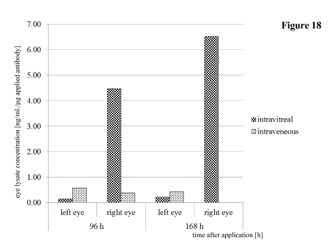
Figure 18: Comparison of eye lysate concentration after
intravitreal
and intravenous application of antibody IGF-1R 0033.
Figure 19: Comparison of eye lysate concentration after intravitreal
and intravenous application of antibody IGF-1R 0035.
Figure 20: Comparison of eye lysate concentration after
intravitreal
and intravenous application of antibody IGF-1R 0045.
CA 02931979 2016-05-27
WO 2015/107025
PCT/EP2015/050425
- 25 -
DETAILED DESCRIPTION OF EMBODIMENTS OF THE INVENTION
I. DEFINITIONS
The term "about" denotes a range of +/- 20 % of the thereafter following
numerical
value. In one embodiment the term about denotes a range of +/- 10 % of the
thereafter following numerical value. In one embodiment the term about denotes
a
range of +/- 5 % of the thereafter following numerical value.
An "acceptor human framework" for the purposes herein is a framework
comprising the amino acid sequence of a light chain variable domain (VL)
framework or a heavy chain variable domain (VH) framework derived from a
human immunoglobulin framework or a human consensus framework, as defined
below. An acceptor human framework "derived from" a human immunoglobulin
framework or a human consensus framework may comprise the same amino acid
sequence thereof, or it may contain amino acid sequence alterations. In some
embodiments, the number of amino acid alterations are 10 or less, 9 or less, 8
or
less, 7 or less, 6 or less, 5 or less, 4 or less, 3 or less, or 2 or less. In
some
embodiments, the VL acceptor human framework is identical in sequence to the
VL human immunoglobulin framework sequence or human consensus framework
sequence.
An "affinity matured" antibody refers to an antibody with one or more
alterations
in one or more hypervariable regions (HVRs), compared to a parent antibody
which does not possess such alterations, such alterations resulting in an
improvement in the affinity of the antibody for antigen.
The term "alteration" denotes the mutation (substitution), insertion
(addition), or
deletion of one or more amino acid residues in a parent antibody or fusion
polypeptide, e.g. a fusion polypeptide comprising at least an FcRn binding
portion
of an Fc-region, to obtain a modified antibody or fusion polypeptide. The term
õmutation" denotes that the specified amino acid residue is substituted for a
different amino acid residue. For example the mutation L234A denotes that the
amino acid residue lysine at position 234 in an antibody Fc-region
(polypeptide) is
substituted by the amino acid residue alanine (substitution of lysine with
alanine)
(numbering according to the Kabat EU index numbering system).
As used herein, the amino acid positions of all constant regions and domains
of the
heavy and light chain are numbered according to the Kabat numbering system
CA 02931979 2016-05-27
WO 2015/107025
PCT/EP2015/050425
- 26 -
described in Kabat, et al., Sequences of Proteins of Immunological Interest,
5th ed.,
Public Health Service, National Institutes of Health, Bethesda, MD (1991) and
is
referred to as "numbering according to Kabat" herein. Specifically the Kabat
numbering system (see pages 647-660) of Kabat, et al., Sequences of Proteins
of
Immunological Interest, 5th ed., Public Health Service, National Institutes of
Health, Bethesda, MD (1991) is used for the light chain constant domain CL of
kappa and lambda isotype and the Kabat EU index numbering system (see pages
661-723) is used for the constant heavy chain domains (CH1, Hinge, CH2 and
CH3).
A "naturally occurring amino acid residues" denotes an amino acid residue from
the group consisting of alanine (three letter code: Ala, one letter code: A),
arginine
(Arg, R), asparagine (Asn, N), aspartic acid (Asp, D), cysteine (Cys, C),
glutamine
(Gln, Q), glutamic acid (Glu, E), glycine (Gly, G), histidine (His, H),
isoleucine
(Ile, I), leucine (Leu, L), lysine (Lys, K), methionine (Met, M),
phenylalanine (Phe,
F), proline (Pro, P), serine (Ser, S), threonine (Thr, T), tryptophane (Tip,
W),
tyrosine (Tyr, Y), and valine (Val, V).
The term "amino acid mutation" denotes the substitution of at least one
existing
amino acid residue with another different amino acid residue (= replacing
amino
acid residue). The replacing amino acid residue may be a "naturally occurring
amino acid residues" and selected from the group consisting of alanine (three
letter
code: ala, one letter code: A), arginine (arg, R), asparagine (asn, N),
aspartic acid
(asp, D), cysteine (cys, C), glutamine (gln, Q), glutamic acid (glu, E),
glycine (gly,
G), histidine (his, H), isoleucine (ile, I), leucine (leu, L), lysine (lys,
K), methionine
(met, M), phenylalanine (phe, F), proline (pro, P), serine (ser, S), threonine
(thr, T),
tryptophan (tip, W), tyrosine (tyr, Y), and valine (val, V). The replacing
amino acid
residue may be a "non-naturally occurring amino acid residue". See e.g. US
6,586,207, WO 98/48032, WO 03/073238, US 2004/0214988, WO 2005/35727,
WO 2005/74524, Chin, J.W., et al., J. Am. Chem. Soc. 124 (2002) 9026-9027;
Chin, J.W. and Schultz, P.G., ChemBioChem 11(2002) 1135-1137; Chin, J.W., et
al., PICAS United States of America 99 (2002) 11020-11024; and, Wang, L. and
Schultz, P.G., Chem. (2002) 1-10 (all entirely incorporated by reference
herein).
The term "amino acid insertion" denotes the (additional) incorporation of at
least
one amino acid residue at a predetermined position in an amino acid sequence.
In
one embodiment the insertion will be the insertion of one or two amino acid
CA 02931979 2016-05-27
WO 2015/107025
PCT/EP2015/050425
-27 -
residues. The inserted amino acid residue(s) can be any naturally occurring or
non-
naturally occurring amino acid residue.
The term "amino acid deletion" denotes the removal of at least one amino acid
residue at a predetermined position in an amino acid sequence.
The term "ANG-2" as used herein refers to human angiopoietin-2 (ANG-2)
(alternatively abbreviated with ANGPT2 or ANG2) (SEQ ID NO: 31) which is
described e.g. in Maisonpierre, P.C., et al, Science 277 (1997) 55-60 and
Cheung,
A.H., et al., Genomics 48 (1998) 389-91. The angiopoietins-1 (SEQ ID NO: 32)
and -2 were discovered as ligands for the Ties, a family of tyrosine kinases
that is
selectively expressed within the vascular endothelium (Yancopoulos, G.D., et
al.,
Nature 407 (2000) 242-248). There are now four definitive members of the
angiopoietin family. Angiopoietin-3 and -4 (ANG-3 and ANG-4) may represent
widely diverged counterparts of the same gene locus in mouse and man (Kim, I.,
et
al., FEBS Let, 443 (1999) 353-356; Kim, I., et al., J. Biol. Chem. 274 (1999)
26523-26528). ANG-1 and ANG-2 were originally identified in tissue culture
experiments as agonist and antagonist, respectively (see for ANG-1: Davis, S.,
et
al., Cell 87 (1996) 1161-1169; and for ANG-2: Maisonpierre, P.C., et al.,
Science
277 (1997) 55-60). All of the known angiopoietins bind primarily to Tie2 (SEQ
ID
NO: 33), and both ANG-1 and -2 bind to Tie2 with an affinity of 3 nM (Kd)
(Maisonpierre, P.C., et al., Science 277 (1997) 55-60).
The term "antibody" herein is used in the broadest sense and encompasses
various
antibody structures, including but not limited to monoclonal antibodies,
multispecific antibodies (e.g. bispecific antibodies, trispecific antibodies),
and
antibody fragments so long as they exhibit the desired antigen-, and/or
protein A
and/or FcRn-binding activity.
The term "asymmetric Fc-region" denotes a pair of Fc-region polypeptides that
have different amino acid residues at corresponding positions according to the
Kabat EU index numbering system.
The term "asymmetric Fc-region with respect to FcRn binding" denotes an Fc-
region that consists of two polypeptide chains that have different amino acid
residues at corresponding positions, whereby the positions are determined
according to the Kabat EU index numbering system, whereby the different
positions affect the binding of the Fc-region to the human neonatal Fc-
receptor
(FcRn). For the purpose herein the differences between the two polypeptide
chains
CA 02931979 2016-05-27
WO 2015/107025
PCT/EP2015/050425
- 28 -
of the Fe-region in an "asymmetric Fe-region with respect to FcRn binding" do
not
include differences that have been introduced to facilitate the formation of
heterodimeric Fe-regions, e.g. for the production of bispecific antibodies.
These
differences can also be asymmetric, i.e. the two chains have differences at
non
corresponding amino acid residues according to the Kabat EU index numbering
system. These differences facilitate heterodimerization and reduce
homodimerization. Examples of such differences are the so-called "knobs into
holes" substitutions (see, e.g., US 7,695,936 and US 2003/0078385). The
following
knobs and holes substitutions in the individual polypeptide chains of an Fe-
region
of an IgG antibody of subclass IgG1 have been found to increase heterodimer
formation: 1) Y407T in one chain and T366Y in the other chain; 2) Y407A in one
chain and T366W in the other chain; 3) F405A in one chain and T394W in the
other chain; 4) F405W in one chain and T3945 in the other chain; 5) Y407T in
one
chain and T366Y in the other chain; 6) T366Y and F405A in one chain and T394W
and Y407T in the other chain; 7) T366W and F405W in one chain and T3945 and
Y407A in the other chain; 8) F405W and Y407A in one chain and T366W and
T3945 in the other chain; and 9) T366W in one chain and T3665, L368A, and
Y407V in the other chain, whereby the last listed is especially suited. In
addition,
changes creating new disulfide bridges between the two Fe-region polypeptide
chains facilitate heterodimer formation (see, e.g., US 2003/0078385). The
following substitutions resulting in appropriately spaced apart cysteine
residues for
the formation of new intra-chain disulfide bonds in the individual polypeptide
chains of an Fe-region of an IgG antibody of subclass IgG1 have been found to
increase heterodimer formation: Y349C in one chain and 5354C in the other;
Y349C in one chain and E356C in the other; Y349C in one chain and E357C in the
other; L351C in one chain and 5354C in the other; T394C in one chain and E397C
in the other; or D399C in one chain and K392C in the other. Further examples
of
heterodimerization facilitating amino acid changes are the so-called "charge
pair
substitutions" (see, e.g., WO 2009/089004). The following charge pair
substitutions
in the individual polypeptide chains of an Fe-region of an IgG antibody of
subclass
IgG1 have been found to increase heterodimer formation: 1) K409D or K409E in
one chain and D399K or D399R in the other chain; 2) K392D or K392E in one
chain and D399K or D399R in the other chain; 3) K439D or K439E in one chain
and E356K or E356R in the other chain; 4) K370D or K370E in one chain and
E357K or E357R in the other chain; 5) K409D and K360D in one chain plus
D399K and E356K in the other chain; 6) K409D and K370D in one chain plus
D399K and E357K in the other chain; 7) K409D and K392D in one chain plus
CA 02931979 2016-05-27
WO 2015/107025
PCT/EP2015/050425
- 29 -
D399K, E356K, and E357K in the other chain; 8) K409D and K392D in one chain
and D399K in the other chain; 9) K409D and K392D in one chain and D399K and
E356K in the other chain; 10) K409D and K392D in one chain and D399K and
D357K in the other chain; 11) K409D and K370D in one chain and D399K and
D357K in the other chain; 12) D399K in one chain and K409D and K360D in the
other chain; and 13) K409D and K439D in one chain and D399K and E356K on
the other.
The term "binding (to an antigen)" denotes the binding of an antibody to its
antigen
in an in vitro assay, in one embodiment in a binding assay in which the
antibody is
bound to a surface and binding of the antigen to the antibody is measured by
Surface Plasmon Resonance (SPR). Binding means a binding affinity (KD) of 10-8
M or less, in some embodiments of 10-13 to 10-8 M, in some embodiments of 10-
13
to 10-9 M.
Binding can be investigated by a BIAcore assay (GE Healthcare Biosensor AB,
Uppsala, Sweden). The affinity of the binding is defined by the terms ka (rate
constant for the association of the antibody from the antibody/antigen
complex), kd
(dissociation constant), and KAd/ka).
The term "chimeric" antibody refers to an antibody in which a portion of the
heavy
and/or light chain is derived from a particular source or species, while the
remainder of the heavy and/or light chain is derived from a different source
or
species.
The term "CH2-domain" denotes the part of an antibody heavy chain polypeptide
that extends approximately from EU position 231 to EU position 340 (EU
numbering system according to Kabat). In one embodiment a CH2 domain has the
amino acid sequence of SEQ ID NO: 09: APELLGG PSVFLFPPKP
KDTLMISRTP EVTCVWDVS HEDPEVKFNW YVDGVEVHNA KTKPREEQ
E STYRWSVLT VLHQDWLNGK EYKCKVSNKA LPAPIEKTIS KAK.
The term "CH3-domain" denotes the part of an antibody heavy chain polypeptide
that extends approximately from EU position 341 to EU position 446. In one
embodiment the CH3 domain has the amino acid sequence of SEQ ID NO: 10:
GQPREPQ VYTLPPSRDE LTKNQVSLTC LVKGFYPSDI AVEWESNGQP
ENNYKTTPPV LDSDGSFFLY SKLTVDKSRW QQGNVFSCSV
MHEALHNHYT QKSL SL S PG.
CA 02931979 2016-05-27
WO 2015/107025
PCT/EP2015/050425
- 30 -
The "class" of an antibody refers to the type of constant domain or constant
region
possessed by its heavy chain. There are five major classes of antibodies: IgA,
IgD,
IgE, IgG, and IgM, and several of these may be further divided into subclasses
(isotypes), e.g., IgGi, IgG2, IgG3, Igai, IgAi, and IgA2. The heavy chain
constant
domains that correspond to the different classes of immunoglobulins are called
a, 8,
e, 7, and , respectively.
The term "comparable length" denotes that two polypeptides comprise the
identical
number of amino acid residues or can be different in length by one or more and
up
to 10 amino acid residues at most. In one embodiment the (Fc-region)
polypeptides
comprise the identical number of amino acid residues or differ by a number of
from
1 to 10 amino acid residues. In one embodiment the (Fc-region) polypeptides
comprise the identical number of amino acid residues or differ by a number of
from
1 to 5 amino acid residues. In one embodiment the (Fc-region) polypeptides
comprise the identical number of amino acid residues or differ by a number of
from
1 to 3 amino acid residues.
"Effector functions" refer to those biological activities attributable to the
Fc-region
of an antibody, which vary with the antibody class. Examples of antibody
effector
functions include: C 1 q binding and complement dependent cytotoxicity (CDC);
Fc
receptor binding; antibody-dependent cell-mediated cytotoxicity (ADCC);
phagocytosis; down regulation of cell surface receptors (e.g. B cell
receptor); and
B-cell activation.
An "effective amount" of an agent, e.g., a pharmaceutical formulation, refers
to an
amount effective, at dosages and for periods of time necessary, to achieve the
desired therapeutic or prophylactic result.
The term "Fc-fusion polypeptide" denotes a fusion of a binding domain (e.g. an
antigen binding domain such as a single chain antibody, or a polypeptide such
as a
ligand of a receptor) with an antibody Fc-region that exhibits the desired
target-,
protein A- and FcRn-binding activity.
The term "Fc-region of human origin" denotes the C-terminal region of an
immunoglobulin heavy chain of human origin that contains at least a part of
the
hinge region, the CH2 domain and the CH3 domain. In one embodiment, a human
IgG heavy chain Fc-region extends from Cys226, or from Pro230, to the carboxyl-
terminus of the heavy chain. In one embodiment the Fc-region has the amino
acid
CA 02931979 2016-05-27
WO 2015/107025
PCT/EP2015/050425
-31 -
sequence of SEQ ID NO: 60. However, the C-terminal lysine (Lys447) of the Fe-
region may or may not be present.
The term "FcRn" denotes the human neonatal Fe-receptor. FcRn functions to
salvage IgG from the lysosomal degradation pathway, resulting in reduced
clearance and increased half-life. The FcRn is a heterodimeric protein
consisting of
two polypeptides: a 50 kDa class I major histocompatibility complex-like
protein
(a-FcRn) and a 15 kDa I32-microglobulin (I32m). FcRn binds with high affinity
to
the CH2-CH3 portion of the Fe-region of IgG. The interaction between IgG and
FcRn is strictly pH dependent and occurs in a 1:2 stoichiometry, with one IgG
binding to two FcRn molecules via its two heavy chains (Huber, A.H., et al.,
J. Mol.
Biol. 230 (1993) 1077-1083). FcRn binding occurs in the endosome at acidic pH
(pH < 6.5) and IgG is released at the neutral cell surface (pH of about 7.4).
The pH-
sensitive nature of the interaction facilitates the FcRn-mediated protection
of IgGs
pinocytosed into cells from intracellular degradation by binding to the
receptor
within the acidic environment of endosomes. FcRn then facilitates the
recycling of
IgG to the cell surface and subsequent release into the blood stream upon
exposure
of the FcRn-IgG complex to the neutral pH environment outside the cell.
The term "FcRn binding portion of an Fe-region" denotes the part of an
antibody
heavy chain polypeptide that extends approximately from EU position 243 to EU
position 261 and approximately from EU position 275 to EU position 293 and
approximately from EU position 302 to EU position 319 and approximately from
EU position 336 to EU position 348 and approximately from EU position 367 to
EU position 393 and EU position 408 and approximately from EU position 424 to
EU position 440. In one embodiment one or more of the following amino acid
residues according to the EU numbering of Kabat are altered F243, P244, P245
P,
K246, P247, K248, D249, T250, L251, M252, 1253, S254, R255, T256, P257,
E258, V259, T260, C261, F275, N276, W277, Y278, V279, D280, V282, E283,
V284, H285, N286, A287, K288, T289, K290, P291, R292, E293, V302, V303,
S304, V305, L306, T307, V308, L309, H310, Q311, D312, W313, L314, N315,
G316, K317, E318, Y319, 1336, S337, K338, A339, K340, G341, Q342, P343,
R344, E345, P346, Q347, V348, C367, V369, F372, Y373, P374, S375, D376,
1377, A378, V379, E380, W381, E382, S383, N384, G385, Q386, P387, E388,
N389, Y391, T393, S408, S424, C425, S426, V427, M428, H429, E430, A431,
L432, H433, N434, H435, Y436, T437, Q438, K439, and S440 (EU numbering).
CA 02931979 2016-05-27
WO 2015/107025
PCT/EP2015/050425
- 32 -
"Framework" or "FR" refers to variable domain residues other than
hypervariable
region (HVR) residues. The FR of a variable domain generally consists of four
FR
domains: FR1, FR2, FR3, and FR4. Accordingly, the HVR and FR sequences
generally appear in the following sequence in VH (or VL): FR1-H1(L1)-FR2-
H2(L2)-FR3-H3(L3)-FR4.
The term "full length antibody" denotes an antibody having a structure
substantially similar to a native antibody structure comprising four
polypeptides or
having heavy chains that contain an Fc-region as defined herein. A full length
antibody may comprise further domains, such as e.g. a scFy or a scFab
conjugated
to one or more of the chains of the full length antibody. These conjugates are
also
encompassed by the term full length antibody.
The term "dimeric polypeptide" denotes a complex comprising at least two
polypeptides that are associated covalently. The complex may comprise further
polypeptides that are also associated covalently or non-covalently with the
other
polypeptides. In one embodiment the dimeric polypeptide comprises two or four
polypeptides.
The terms "heterodimer" or "heterodimeric" denote a molecule that comprises
two
polypeptides (e.g. of comparable length), wherein the two polypeptides have an
amino acid sequence that have at least one different amino acid residue in a
corresponding position, whereby corresponding position is determined according
to
the Kabat EU index numbering system.
The terms "homodimer" and "homodimeric" denote a molecule that comprises two
polypeptides of comparable length, wherein the two polypeptides have an amino
acid sequence that is identical in corresponding positions, whereby
corresponding
positions are determined according to the Kabat EU index numbering system.
A dimeric polypeptide as reported herein can be homodimeric or heterodimeric
which is determined with respect to mutations or properties in focus. For
example,
with respect to FcRn and/or protein A binding (i.e. the focused on properties)
a
dimeric polypeptide is homodimeric (i.e. both polypeptides of the dimeric
polypeptide comprise these mutations) with respect to the mutations H310A,
H433A and Y436A (these mutations are in focus with respect to FcRn and/or
protein A binding property of the dimeric polypeptide) but at the same time
heterodimeric with respect to the mutations Y349C, T366S, L368A and Y407V
(these mutations are not in focus as these mutations are directed to the
CA 02931979 2016-05-27
WO 2015/107025
PCT/EP2015/050425
- 33 -
heterodimerization of the dimeric polypeptide and not to the FcRn/protein A
binding properties) as well as the mutations S354C and T366W, respectively
(the
first set is comprised only in the first polypeptide whereas the second set is
comprised only in the second polypeptide). Further for example, a dimeric
polypeptide as reported herein can be heterodimeric with respect to the
mutations
I253A, H310A, H433A, H435A and Y436A (i.e. these mutations are directed all to
the FcRn and/or protein A binding properties of the dimeric polypeptide), i.e.
one
polypeptide comprises the mutations I253A, H310A and H435A, whereas the other
polypeptide comprises the mutations H310A, H433A and Y436A.
The terms "host cell", "host cell line", and "host cell culture" are used
interchangeably and refer to cells into which exogenous nucleic acid has been
introduced, including the progeny of such cells. Host cells include
"transformants"
and "transformed cells," which include the primary transformed cell and
progeny
derived therefrom without regard to the number of passages. Progeny may not be
completely identical in nucleic acid content to a parent cell, but may contain
mutations. Mutant progeny that have the same function or biological activity
as
screened or selected for in the originally transformed cell are included
herein.
A "human antibody" is one which possesses an amino acid sequence which
corresponds to that of an antibody produced by a human or a human cell or
derived
from a non-human source that utilizes human antibody repertoires or other
human
antibody-encoding sequences. This definition of a human antibody specifically
excludes a humanized antibody comprising non-human antigen-binding residues.
A "human consensus framework" is a framework which represents the most
commonly occurring amino acid residues in a selection of human immunoglobulin
VL or VH framework sequences. Generally, the selection of human
immunoglobulin VL or VH sequences is from a subgroup of variable domain
sequences. Generally, the subgroup of sequences is a subgroup as in Kabat,
E.A. et
al., Sequences of Proteins of Immunological Interest, 5th ed., Bethesda MD
(1991),
NIH Publication 91-3242, Vols. 1-3. In one embodiment, for the VL, the
subgroup
is subgroup kappa I as in Kabat et al., supra. In one embodiment, for the VH,
the
subgroup is subgroup III as in Kabat et al., supra.
The term "derived from" denotes that an amino acid sequence is derived from a
parent amino acid sequence by introducing alterations at at least one
position. Thus
a derived amino acid sequence differs from the corresponding parent amino acid
CA 02931979 2016-05-27
WO 2015/107025
PCT/EP2015/050425
- 34 -
sequence at at least one corresponding position (numbering according to Kabat
EU
index for antibody Fc-regions). In one embodiment an amino acid sequence
derived
from a parent amino acid sequence differs by one to fifteen amino acid
residues at
corresponding positions. In one embodiment an amino acid sequence derived from
a parent amino acid sequence differs by one to ten amino acid residues at
corresponding positions. In one embodiment an amino acid sequence derived from
a parent amino acid sequence differs by one to six amino acid residues at
corresponding positions. Likewise a derived amino acid sequence has a high
amino
acid sequence identity to its parent amino acid sequence. In one embodiment an
amino acid sequence derived from a parent amino acid sequence has 80 % or more
amino acid sequence identity. In one embodiment an amino acid sequence derived
from a parent amino acid sequence has 90 % or more amino acid sequence
identity.
In one embodiment an amino acid sequence derived from a parent amino acid
sequence has 95 % or more amino acid sequence identity.
The term "human Fc-region polypeptide" denotes an amino acid sequence which is
identical to a "native" or "wild-type" human Fc-region polypeptide. The term
"variant (human) Fc-region polypeptide" denotes an amino acid sequence which
derived from a "native" or "wild-type" human Fc-region polypeptide by virtue
of at
least one "amino acid alteration". A "human Fc-region" is consisting of two
human
Fc-region polypeptides. A "variant (human) Fc-region" is consisting of two Fc-
region polypeptides, whereby both can be variant (human) Fc-region
polypeptides
or one is a human Fc-region polypeptide and the other is a variant (human) Fc-
region polypeptide.
In one embodiment the human Fc-region polypeptide has the amino acid sequence
of a human IgG1 Fc-region polypeptide of SEQ ID NO: 60, or of a human IgG2
Fc-region polypeptide of SEQ ID NO: 61, or of a human IgG4 Fc-region
polypeptide of SEQ ID NO: 63 with the mutations as reported herein. In one
embodiment the variant (human) Fc-region polypeptide is derived from an Fc-
region polypeptide of SEQ ID NO: 60, or 61, or 63 and has at least one amino
acid
mutation compared to the Fc-region polypeptide of SEQ ID NO: 60, or 61, or 63.
In one embodiment the variant (human) Fc-region polypeptide comprises/has from
about one to about ten amino acid mutations, and in one embodiment from about
one to about five amino acid mutations. In one embodiment the variant (human)
Fc-region polypeptide has at least about 80 % homology with a human Fc-region
polypeptide of SEQ ID NO: 60, or 61, or 63. In one embodiment the variant
(human) Fc-region polypeptide has least about 90 % homology with a human Fc-
CA 02931979 2016-05-27
WO 2015/107025
PCT/EP2015/050425
- 35 -
region polypeptide of SEQ ID NO: 60, or 61, or 63. In one embodiment the
variant
(human) Fe-region polypeptide has at least about 95 % homology with a human Fe-
region polypeptide of SEQ ID NO: 60, or 61, or 63.
The variant (human) Fe-region polypeptide derived from a human Fe-region
polypeptide of SEQ ID NO: 60, or 61, or 63 is defined by the amino acid
alterations that are contained. Thus, for example, the term P329G denotes a
variant
(human) Fe-region polypeptide derived human Fe-region polypeptide with the
mutation of proline to glycine at amino acid position 329 relative to the
human Fe-
region polypeptide of SEQ ID NO: 60, or 61, or 63.
For all positions discussed in the present invention, numbering is according
to the
Kabat EU index numbering system.
A human IgG1 Fe-region polypeptide has the following amino acid sequence:
DKTHTCPPCPAPELLGGPSVFLFPPKPKDTLMISRTPEVTCVVVDVSHEDPE
VKFNWYVDGVEVHNAKTKPREEQYNSTYRVVSVLTVLHQDWLNGKEYK
CKV SNKALPAPIEKTI SKAKG QPREP QVYTLPP SRDELTKNQV SLTC LVKGF
YPSDIAVEWESNGQPENNYKTTPPVLDSDGSFFLYSKLTVDKSRWQQGNV
FSCSVMHEALHNHYTQKSLSLSPGK (SEQ ID NO: 60).
A human IgG1 Fe-region derived Fe-region polypeptide with the mutations L234A,
L235A has the following amino acid sequence:
DKTHTCPPCPAPEAAGGPSVFLFPPKPKDTLMISRTPEVTCVVVDVSHEDPE
VKFNWYVDGVEVHNAKTKPREEQYNSTYRVVSVLTVLHQDWLNGKEYK
CKV SNKALPAPIEKTI SKAKG QPREP QVYTLPP SRDELTKNQV SLTC LVKGF
YPSDIAVEWESNGQPENNYKTTPPVLDSDGSFFLYSKLTVDKSRWQQGNV
FSCSVMHEALHNHYTQKSLSLSPGK (SEQ ID NO: 64).
A human IgG1 Fe-region derived Fe-region polypeptide with Y349C, T3665,
L368A and Y407V mutations has the following amino acid sequence:
DKTHTCPPCPAPELLGGPSVFLFPPKPKDTLMISRTPEVTCVVVDVSHEDPE
VKFNWYVDGVEVHNAKTKPREEQYNSTYRVVSVLTVLHQDWLNGKEYK
CKVSNKALPAPIEKTISKAKGQPREPQVCTLPPSRDELTKNQVSLSCAVKGF
YPSDIAVEWESNGQPENNYKTTPPVLDSDGSFFLVSKLTVDKSRWQQGNV
FSCSVMHEALHNHYTQKSLSLSPGK (SEQ ID NO: 65).
CA 02931979 2016-05-27
WO 2015/107025
PCT/EP2015/050425
- 36 -
A human IgG1 Fc-region derived Fc-region polypeptide with S354C, T366W
mutations has the following amino acid sequence:
DKTHTCPPCPAPELLGGPSVFLFPPKPKDTLMISRTPEVTCVVVDVSHEDPE
VKFNWYVDGVEVHNAKTKPREEQYNSTYRVVSVLTVLHQDWLNGKEYK
CKVSNKALPAPIEKTISKAKGQPREPQVYTLPPCRDELTKNQVSLWCLVKG
FYP SDIAVEWESNGQ PENNYKTTPPVLDSDGSFFLYSKLTVDKSRWQQ GN
VFSCSVMHEALHNHYTQKSLSLSPGK (SEQ ID NO: 66).
A human IgG1 Fc-region derived Fc-region polypeptide with L234A, L235A
mutations and Y349C, T3665, L368A, Y407V mutations has the following amino
acid sequence:
DKTHTCPPCPAPEAAGGPSVFLFPPKPKDTLMISRTPEVTCVVVDVSHEDPE
VKFNWYVDGVEVHNAKTKPREEQYNSTYRVVSVLTVLHQDWLNGKEYK
CKVSNKALPAPIEKTISKAKGQPREPQVCTLPPSRDELTKNQVSLSCAVKGF
YPSDIAVEWESNGQPENNYKTTPPVLDSDGSFFLVSKLTVDKSRWQQGNV
FSCSVMHEALHNHYTQKSLSLSPGK (SEQ ID NO: 67).
A human IgG1 Fc-region derived Fc-region polypeptide with a L234A, L235A and
5354C, T366W mutations has the following amino acid sequence:
DKTHTCPPCPAPEAAGGPSVFLFPPKPKDTLMISRTPEVTCVVVDVSHEDPE
VKFNWYVDGVEVHNAKTKPREEQYNSTYRVVSVLTVLHQDWLNGKEYK
CKVSNKALPAPIEKTISKAKGQPREPQVYTLPPCRDELTKNQVSLWCLVKG
FYP SDIAVEWESNGQ PENNYKTTPPVLDSDGSFFLYSKLTVDKSRWQQ GN
VFSCSVMHEALHNHYTQKSLSLSPGK (SEQ ID NO: 68).
A human IgG1 Fc-region derived Fc-region polypeptide with a P329G mutation
has the following amino acid sequence:
DKTHTCPPCPAPELLGGPSVFLFPPKPKDTLMISRTPEVTCVVVDVSHEDPE
VKFNWYVDGVEVHNAKTKPREEQYNSTYRVVSVLTVLHQDWLNGKEYK
CKVSNKALGAPIEKTISKAKGQPREPQVYTLPPSRDELTKNQVSLTCLVKG
FYP SDIAVEWESNGQ PENNYKTTPPVLDSDGSFFLYSKLTVDKSRWQQ GN
VFSCSVMHEALHNHYTQKSLSLSPGK (SEQ ID NO: 69).
A human IgG1 Fc-region derived Fc-region polypeptide with L234A, L235A
mutations and P329G mutation has the following amino acid sequence:
CA 02931979 2016-05-27
WO 2015/107025
PCT/EP2015/050425
-37 -
DKTHTCPPCPAPEAAGGPSVFLFPPKPKDTLMISRTPEVTCVVVDVSHEDPE
VKFNWYVDGVEVHNAKTKPREEQYNSTYRVVSVLTVLHQDWLNGKEYK
CKVSNKALGAPIEKTISKAKGQPREPQVYTLPPSRDELTKNQVSLTCLVKG
FYP SDIAVEWESNGQ PENNYKTTPPVLDSDGSFFLYSKLTVDKSRWQQ GN
VFSCSVMHEALHNHYTQKSLSLSPGK (SEQ ID NO: 70).
A human IgG1 Fc-region derived Fc-region polypeptide with a P239G mutation
and Y349C, T3665, L368A, Y407V mutations has the following amino acid
sequence:
DKTHTCPPCPAPELLGGPSVFLFPPKPKDTLMISRTPEVTCVVVDVSHEDPE
VKFNWYVDGVEVHNAKTKPREEQYNSTYRVVSVLTVLHQDWLNGKEYK
CKVSNKALGAPIEKTISKAKGQPREPQVCTLPPSRDELTKNQVSLSCAVKG
FYP SDIAVEWESNGQ PENNYKTTPPVLDSDGSFFLVSKLTVDKSRWQQ GN
VFSCSVMHEALHNHYTQKSLSLSPGK (SEQ ID NO: 71).
A human IgG1 Fc-region derived Fc-region polypeptide with a P329G mutation
and 5354C, T366W mutation has the following amino acid sequence:
DKTHTCPPCPAPELLGGPSVFLFPPKPKDTLMISRTPEVTCVVVDVSHEDPE
VKFNWYVDGVEVHNAKTKPREEQYNSTYRVVSVLTVLHQDWLNGKEYK
CKVSNKALGAPIEKTISKAKGQPREPQVYTLPPCRDELTKNQVSLWCLVKG
FYP SDIAVEWESNGQ PENNYKTTPPVLDSDGSFFLYSKLTVDKSRWQQ GN
VFSCSVMHEALHNHYTQKSLSLSPGK (SEQ ID NO: 72).
A human IgG1 Fc-region derived Fc-region polypeptide with L234A, L235A,
P329G and Y349C, T3665, L368A, Y407V mutations has the following amino
acid sequence:
DKTHTCPPCPAPEAAGGPSVFLFPPKPKDTLMISRTPEVTCVVVDVSHEDPE
VKFNWYVDGVEVHNAKTKPREEQYNSTYRVVSVLTVLHQDWLNGKEYK
CKVSNKALGAPIEKTISKAKGQPREPQVCTLPPSRDELTKNQVSLSCAVKG
FYP SDIAVEWESNGQ PENNYKTTPPVLDSDGSFFLVSKLTVDKSRWQQ GN
VFSCSVMHEALHNHYTQKSLSLSPGK (SEQ ID NO: 73).
A human IgG1 Fc-region derived Fc-region polypeptide with L234A, L235A,
P329G mutations and 5354C, T366W mutations has the following amino acid
sequence:
CA 02931979 2016-05-27
WO 2015/107025
PCT/EP2015/050425
- 38 -
DKTHTCPPCPAPEAAGGPSVFLFPPKPKDTLMISRTPEVTCVVVDVSHEDPE
VKFNWYVDGVEVHNAKTKPREEQYNSTYRVVSVLTVLHQDWLNGKEYK
CKVSNKALGAPIEKTISKAKGQPREPQVYTLPPCRDELTKNQVSLWCLVKG
FYP SDIAVEWESNGQ PENNYKTTPPVLDSDGSFFLYSKLTVDKSRWQQ GN
VFSCSVMHEALHNHYTQKSLSLSPGK (SEQ ID NO: 74).
A human IgG4 Fc-region polypeptide has the following amino acid sequence:
ESKYGPPCPSCPAPEFLGGPSVFLFPPKPKDTLMISRTPEVTCVVVDVSQED
PEVQFNWYVDGVEVHNAKTKPREEQFNSTYRVVSVLTVLHQDWLNGKEY
KCKVSNKGLP S SIEKTI SKAKGQPREPQVYTLPP S QEEMTKNQVSLTCLVK
GFYP SDIAVEWE SNGQPENNYKTTPPVLD SDGSFFLYSRLTVDKSRWQEGN
VFSCSVMHEALHNHYTQKSLSLSLGK (SEQ ID NO: 63).
A human IgG4 Fc-region derived Fc-region polypeptide with 5228P and L235E
mutations has the following amino acid sequence:
ESKYGPPCPPCPAPEFEGGPSVFLFPPKPKDTLMISRTPEVTCVVVDVSQED
PEVQFNWYVDGVEVHNAKTKPREEQFNSTYRVVSVLTVLHQDWLNGKEY
KCKVSNKGLP S SIEKTI SKAKGQPREPQVYTLPP S QEEMTKNQVSLTCLVK
GFYP SDIAVEWE SNGQPENNYKTTPPVLD SDGSFFLYSRLTVDKSRWQEGN
VFSCSVMHEALHNHYTQKSLSLSLGK (SEQ ID NO: 75).
A human IgG4 Fc-region derived Fc-region polypeptide with 5228P, L235E
mutations and P329G mutation has the following amino acid sequence:
ESKYGPPCPPCPAPEFEGGPSVFLFPPKPKDTLMISRTPEVTCVVVDVSQED
PEVQFNWYVDGVEVHNAKTKPREEQFNSTYRVVSVLTVLHQDWLNGKEY
KCKVSNKGLGS SIEKTISKAKGQPREPQVYTLPP S QEEMTKNQVSLTCLVK
GFYP SDIAVEWE SNGQPENNYKTTPPVLD SDGSFFLYSRLTVDKSRWQEGN
VFSCSVMHEALHNHYTQKSLSLSLGK (SEQ ID NO: 76).
A human IgG4 Fc-region derived Fc-region polypeptide with 5354C, T366W
mutations has the following amino acid sequence:
ESKYGPPCPSCPAPEFLGGPSVFLFPPKPKDTLMISRTPEVTCVVVDVSQED
PEVQFNWYVDGVEVHNAKTKPREEQFNSTYRVVSVLTVLHQDWLNGKEY
KCKVSNKGLP S SIEKTI SKAKGQPREPQVYTLPPCQEEMTKNQV SLWCLVK
GFYP SDIAVEWE SNGQPENNYKTTPPVLD SDGSFFLYSRLTVDKSRWQEGN
VFSCSVMHEALHNHYTQKSLSLSLGK (SEQ ID NO: 77).
CA 02931979 2016-05-27
WO 2015/107025
PCT/EP2015/050425
- 39 -
A human IgG4 Fc-region derived Fc-region polypeptide with Y349C, T366S,
L368A, Y407V mutations has the following amino acid sequence:
ESKYGPPCPSCPAPEFLGGPSVFLFPPKPKDTLMISRTPEVTCVVVDVSQED
PEVQFNWYVDGVEVHNAKTKPREEQFNSTYRVVSVLTVLHQDWLNGKEY
KCKVSNKGLP S SIEKTISKAKGQPREPQVCTLPP SQEEMTKNQVSLSCAVK
GFYPSDIAVEWESNGQPENNYKTTPPVLDSDGSFFLVSRLTVDKSRWQEGN
VFSCSVMHEALHNHYTQKSLSLSLGK (SEQ ID NO: 78).
A human IgG4 Fc-region derived Fc-region polypeptide with a 5228P, L235E and
5354C, T366W mutations has the following amino acid sequence:
ESKYGPPCPPCPAPEFEGGPSVFLFPPKPKDTLMISRTPEVTCVVVDVSQED
PEVQFNWYVDGVEVHNAKTKPREEQFNSTYRVVSVLTVLHQDWLNGKEY
KCKVSNKGLP S SIEKTI SKAKGQPREPQVYTLPPCQEEMTKNQV SLWCLVK
GFYPSDIAVEWESNGQPENNYKTTPPVLDSDGSFFLYSRLTVDKSRWQEGN
VFSCSVMHEALHNHYTQKSLSLSLGK (SEQ ID NO: 79).
A human IgG4 Fc-region derived Fc-region polypeptide with a 5228P, L235E and
Y349C, T3665, L368A, Y407V mutations has the following amino acid sequence:
ESKYGPPCPPCPAPEFEGGPSVFLFPPKPKDTLMISRTPEVTCVVVDVSQED
PEVQFNWYVDGVEVHNAKTKPREEQFNSTYRVVSVLTVLHQDWLNGKEY
KCKVSNKGLP S SIEKTISKAKGQPREPQVCTLPP SQEEMTKNQVSLSCAVK
GFYPSDIAVEWESNGQPENNYKTTPPVLDSDGSFFLVSRLTVDKSRWQEGN
VFSCSVMHEALHNHYTQKSLSLSLGK (SEQ ID NO: 80).
A human IgG4 Fc-region derived Fc-region polypeptide with a P329G mutation
has the following amino acid sequence:
ESKYGPPCPSCPAPEFLGGPSVFLFPPKPKDTLMISRTPEVTCVVVDVSQED
PEVQFNWYVDGVEVHNAKTKPREEQFNSTYRVVSVLTVLHQDWLNGKEY
KCKVSNKGLGS SIEKTISKAKGQPREPQVYTLPP SQEEMTKNQVSLTCLVK
GFYPSDIAVEWESNGQPENNYKTTPPVLDSDGSFFLYSRLTVDKSRWQEGN
VFSCSVMHEALHNHYTQKSLSLSLGK (SEQ ID NO: 81).
A human IgG4 Fc-region derived Fc-region polypeptide with a P239G and Y349C,
T3665, L368A, Y407V mutations has the following amino acid sequence:
CA 02931979 2016-05-27
WO 2015/107025
PCT/EP2015/050425
- 40 -
ESKYGPPCPSCPAPEFLGGPSVFLFPPKPKDTLMISRTPEVTCVVVDVSQED
PEVQFNWYVDGVEVHNAKTKPREEQFNSTYRVVSVLTVLHQDWLNGKEY
KCKVSNKGLGS SIEKTISKAKGQPREPQVCTLPPS QEEMTKNQVSL SCAVK
GFYPSDIAVEWESNGQPENNYKTTPPVLDSDGSFFLVSRLTVDKSRWQEGN
VFSCSVMHEALHNHYTQKSLSLSLGK (SEQ ID NO: 82).
A human IgG4 Fc-region derived Fc-region polypeptide with a P329G and 5354C,
T366W mutations has the following amino acid sequence:
ESKYGPPCPSCPAPEFLGGPSVFLFPPKPKDTLMISRTPEVTCVVVDVSQED
PEVQFNWYVDGVEVHNAKTKPREEQFNSTYRVVSVLTVLHQDWLNGKEY
KCKVSNKGLGS SIEKTISKAKGQPREPQVYTLPPCQEEMTKNQVSLWCLVK
GFYPSDIAVEWESNGQPENNYKTTPPVLDSDGSFFLYSRLTVDKSRWQEGN
VFSCSVMHEALHNHYTQKSLSLSLGK (SEQ ID NO: 83).
A human IgG4 Fc-region derived Fc-region polypeptide with a 5228P, L235E,
P329G and Y349C, T3665, L368A, Y407V mutations has the following amino
acid sequence:
ESKYGPPCPPCPAPEFEGGPSVFLFPPKPKDTLMISRTPEVTCVVVDVSQED
PEVQFNWYVDGVEVHNAKTKPREEQFNSTYRVVSVLTVLHQDWLNGKEY
KCKVSNKGLGS SIEKTISKAKGQPREPQVCTLPPS QEEMTKNQVSL SCAVK
GFYPSDIAVEWESNGQPENNYKTTPPVLDSDGSFFLVSRLTVDKSRWQEGN
VFSCSVMHEALHNHYTQKSLSLSLGK (SEQ ID NO: 84).
A human IgG4 Fc-region derived Fc-region polypeptide with a 5228P, L235E,
P329G and 5354C, T366W mutations has the following amino acid sequence:
ESKYGPPCPPCPAPEFEGGPSVFLFPPKPKDTLMISRTPEVTCVVVDVSQED
PEVQFNWYVDGVEVHNAKTKPREEQFNSTYRVVSVLTVLHQDWLNGKEY
KCKVSNKGLGS SIEKTISKAKGQPREPQVYTLPPCQEEMTKNQVSLWCLVK
GFYPSDIAVEWESNGQPENNYKTTPPVLDSDGSFFLYSRLTVDKSRWQEGN
VFSCSVMHEALHNHYTQKSLSLSLGK (SEQ ID NO: 85).
An alignment of the different human Fc-regions is shown below (Kabat EU index
numbering system):
CA 02931979 2016-05-27
WO 2015/107025
PCT/EP2015/050425
-41 -
2
1
6(IgG1,2,4)
IGG1 ............................................................. EPKSC
IGG2 ........................................................... ERKCC
IGG3 KTPLGDTTHT
CPRCPEPKSC DTPPPCPRCP EPKSCDTPPP CPRCPEPKSC
IGG4 ............................................................. ESKYG
-- HINGE ---------------------------------------------------------
2 2
3 5
0 0
IGG1 DKTHTCPPCP APELLGGPSV FLFPPKPKDT LMISRTPEVT CVVVDVSHED
IGG2 ...VECPPCP
APP.VAGPSV FLFPPKPKDT LMISRTPEVT CVVVDVSHED
IGG3 DTPPPCPRCP APELLGGPSV FLFPPKPKDT LMISRTPEVT CVVVDVSHED
IGG4 ...PPCPSCP
APEFLGGPSV FLFPPKPKDT LMISRTPEVT CVVVDVSQED
-- HINGE -I-- CH2 ------------------------------------------------
3
0
0
IGG1 PEVKFNWYVD GVEVHNAKTK PREEQYNSTY RVVSVLTVLH QDWLNGKEYK
IGG2 PEVQFNWYVD GVEVHNAKTK PREEQFNSTF RVVSVLTVVH QDWLNGKEYK
IGG3 PEVQFKWYVD GVEVHNAKTK PREEQYNSTF RVVSVLTVLH QDWLNGKEYK
IGG4 PEVQFNWYVD GVEVHNAKTK PREEQFNSTY RVVSVLTVLH QDWLNGKEYK
-- CH2 -----------------------------------------------------------
3
5
0
IGG1 CKVSNKALPA PIEKTISKAK GQPREPQVYT LPPSRDELTK NQVSLTCLVK
IGG2 CKVSNKGLPA PIEKTISKTK GQPREPQVYT LPPSREEMTK NQVSLTCLVK
IGG3 CKVSNKALPA PIEKTISKTK GQPREPQVYT LPPSREEMTK NQVSLTCLVK
IGG4 CKVSNKGLPS SIEKTISKAK GQPREPQVYT LPPSQEEMTK NQVSLTCLVK
-- CH2 ------ CH2 1 CH3 --------------------
4
0
0
IGG1 GFYPSDIAVE WESNGQPENN YKTTPPVLDS DGSFFLYSKL TVDKSRWQQG
IGG2 GFYPSDISVE WESNGQPENN YKTTPPMLDS DGSFFLYSKL TVDKSRWQQG
IGG3 GFYPSDIAVE WESSGQPENN YNTTPPMLDS DGSFFLYSKL TVDKSRWQQG
IGG4 GFYPSDIAVE WESNGQPENN YKTTPPVLDS DGSFFLYSRL TVDKSRWQEG
-- CH3 -----------------------------------------------------------
4
4
7
IGG1 NVFSCSVMHE ALHNHYTQKS LSLSPGK
IGG2 NVFSCSVMHE ALHNHYTQKS LSLSPGK
IGG3 NIFSCSVMHE ALHNRFTQKS LSLSPGK
IGG4 NVFSCSVMHE ALHNHYTQKS LSLSLGK
--CH3 1
A "humanized" antibody refers to a chimeric antibody comprising amino acid
residues from non-human HVRs and amino acid residues from human FRs. In
certain embodiments, a humanized antibody will comprise substantially all of
at
CA 02931979 2016-05-27
WO 2015/107025
PCT/EP2015/050425
- 42 -
least one, and typically two, variable domains, in which all or substantially
all of
the HVRs (e.g., the CDRs) correspond to those of a non-human antibody, and all
or
substantially all of the FRs correspond to those of a human antibody. A
humanized
antibody optionally may comprise at least a portion of an antibody constant
region
derived from a human antibody. A "humanized form" of an antibody, e.g., a non-
human antibody, refers to an antibody that has undergone humanization.
The term "hypervariable region" or "HVR", as used herein, refers to each of
the
regions of an antibody variable domain which are hypervariable in sequence
("complementarity determining regions" or "CDRs") and form structurally
defined
loops ("hypervariable loops"), and/or contain the antigen-contacting residues
("antigen contacts"). Generally, antibodies comprise six HVRs; three in the VH
(H1, H2, H3), and three in the VL (L1, L2, L3). HVRs as denoted herein include
(a) hypervariable loops occurring at amino acid residues 26-32 (L1), 50-52
(L2),
91-96 (L3), 26-32 (H1), 53-55 (H2), and 96-101 (H3) (Chothia, C. and Lesk,
A.M., J. Mol. Biol. 196 (1987) 901-917);
(b) CDRs occurring at amino acid residues 24-34 (Li), 50-56 (L2), 89-97 (L3),
31-35b (H1), 50-65 (H2), and 95-102 (H3) (Kabat, E.A. et al., Sequences of
Proteins of Immunological Interest, 5th ed. Public Health Service, National
Institutes of Health, Bethesda, MD (1991), NIH Publication 91-3242.);
(c) antigen contacts occurring at amino acid residues 27c-36 (L1), 46-55 (L2),
89-96 (L3), 30-35b (H1), 47-58 (H2), and 93-101 (H3) (MacCallum et al. J.
Mol. Biol. 262: 732-745 (1996)); and
(d) combinations of (a), (b), and/or (c), including HVR amino acid residues 46-
56 (L2), 47-56 (L2), 48-56 (L2), 49-56 (L2), 26-35 (H1), 26-35b (H1), 49-65
(H2), 93-102 (H3), and 94-102 (H3).
Unless otherwise indicated, HVR residues and other residues in the variable
domain (e.g., FR residues) are numbered herein according to the Kabat EU index
numbering system (Kabat et al., supra).
The term "IGF-1R" as used herein, refers to any native IGF-1R from any
vertebrate
source, including mammals such as primates (e.g. humans) and rodents (e.g.,
mice
and rats), unless otherwise indicated. The term encompasses "full-length",
unprocessed IGF-1R as well as any form of IGF-1R that results from processing
in
the cell. The term also encompasses naturally occurring variants of IGF-1R,
e.g.,
splice variants or allelic variants. The amino acid sequence of human IGF-1R
is
shown in SEQ ID NO: 11.
CA 02931979 2016-05-27
WO 2015/107025
PCT/EP2015/050425
- 43 -
An "individual" or "subject" is a mammal. Mammals include, but are not limited
to,
domesticated animals (e.g. cows, sheep, cats, dogs, and horses), primates
(e.g.,
humans and non-human primates such as monkeys), rabbits, and rodents (e.g.,
mice
and rats). In certain embodiments, the individual or subject is a human.
An "isolated" antibody is one which has been separated from a component of its
natural environment. In some embodiments, an antibody is purified to greater
than
95 % or 99 % purity as determined by, for example, electrophoretic (e.g., SDS-
PAGE, isoelectric focusing (IEF), capillary electrophoresis) or
chromatographic
(e.g., size exclusion chromatography, ion exchange or reverse phase HPLC). For
review of methods for assessment of antibody purity, see, e.g., Flatman, S. et
al., J.
Chrom. B 848 (2007) 79-87.
An "isolated" nucleic acid refers to a nucleic acid molecule that has been
separated
from a component of its natural environment. An isolated nucleic acid includes
a
nucleic acid molecule contained in cells that ordinarily contain the nucleic
acid
molecule, but the nucleic acid molecule is present extrachromosomally or at a
chromosomal location that is different from its natural chromosomal location.
"Isolated nucleic acid encoding an anti-IGF-1R antibody" refers to one or more
nucleic acid molecules encoding antibody heavy and light chains (or fragments
thereof), including such nucleic acid molecule(s) in a single vector or
separate
vectors, and such nucleic acid molecule(s) present at one or more locations in
a
host cell.
The term "monoclonal antibody" as used herein refers to an antibody obtained
from
a population of substantially homogeneous antibodies, i.e., the individual
antibodies comprising the population are identical and/or bind the same
epitope,
except for possible variant antibodies, e.g., containing naturally occurring
mutations or arising during production of a monoclonal antibody preparation,
such
variants generally being present in minor amounts. In contrast to polyclonal
antibody preparations, which typically include different antibodies directed
against
different determinants (epitopes), each monoclonal antibody of a monoclonal
antibody preparation is directed against a single determinant on an antigen.
Thus,
the modifier "monoclonal" indicates the character of the antibody as being
obtained
from a substantially homogeneous population of antibodies, and is not to be
construed as requiring production of the antibody by any particular method.
For
example, the monoclonal antibodies to be used in accordance with the present
CA 02931979 2016-05-27
WO 2015/107025
PCT/EP2015/050425
- 44 -
invention may be made by a variety of techniques, including but not limited to
the
hybridoma method, recombinant DNA methods, phage-display methods, and
methods utilizing transgenic animals containing all or part of the human
immunoglobulin loci, such methods and other exemplary methods for making
monoclonal antibodies being described herein.
"Native antibodies" refer to naturally occurring immunoglobulin molecules with
varying structures. For example, native IgG antibodies are heterotetrameric
glycoproteins of about 150,000 daltons, composed of two identical light chains
and
two identical heavy chains that are disulfide-bonded. From N- to C-terminus,
each
heavy chain has a variable region (VH), also called a variable heavy domain or
a
heavy chain variable domain, followed by three constant domains (CH1, CH2, and
CH3). Similarly, from N- to C-terminus, each light chain has a variable region
(VL), also called a variable light domain or a light chain variable domain,
followed
by a constant light (CL) domain. The light chain of an antibody may be
assigned to
one of two types, called kappa (x) and lambda (X), based on the amino acid
sequence of its constant domain.
The term "package insert" is used to refer to instructions customarily
included in
commercial packages of therapeutic products, that contain information about
the
indications, usage, dosage, administration, combination therapy,
contraindications
and/or warnings concerning the use of such therapeutic products.
"Percent (%) amino acid sequence identity" with respect to a reference
polypeptide
sequence is defined as the percentage of amino acid residues in a candidate
sequence that are identical with the amino acid residues in the reference
polypeptide sequence, after aligning the sequences and introducing gaps, if
necessary, to achieve the maximum percent sequence identity, and not
considering
any conservative substitutions as part of the sequence identity. Alignment for
purposes of determining percent amino acid sequence identity can be achieved
in
various ways that are within the skill in the art, for instance, using
publicly
available computer software such as BLAST, BLAST-2, ALIGN or Megalign
(DNASTAR) software. Those skilled in the art can determine appropriate
parameters for aligning sequences, including any algorithms needed to achieve
maximal alignment over the full length of the sequences being compared. For
purposes herein, however, % amino acid sequence identity values are generated
using the sequence comparison computer program ALIGN-2. The ALIGN-2
sequence comparison computer program was authored by Genentech, Inc., and the
CA 02931979 2016-05-27
WO 2015/107025
PCT/EP2015/050425
- 45 -
source code has been filed with user documentation in the U.S. Copyright
Office,
Washington D.C., 20559, where it is registered under U.S. Copyright
Registration
No. TXU510087. The ALIGN-2 program is publicly available from Genentech,
Inc., South San Francisco, California, or may be compiled from the source
code.
The ALIGN-2 program should be compiled for use on a UNIX operating system,
including digital UNIX V4.0D. All sequence comparison parameters are set by
the
ALIGN-2 program and do not vary.
In situations where ALIGN-2 is employed for amino acid sequence comparisons,
the % amino acid sequence identity of a given amino acid sequence A to, with,
or
against a given amino acid sequence B (which can alternatively be phrased as a
given amino acid sequence A that has or comprises a certain % amino acid
sequence identity to, with, or against a given amino acid sequence B) is
calculated
as follows:
100 times the fraction X/Y
where X is the number of amino acid residues scored as identical matches by
the
sequence alignment program ALIGN-2 in that program's alignment of A and B,
and where Y is the total number of amino acid residues in B. It will be
appreciated
that where the length of amino acid sequence A is not equal to the length of
amino
acid sequence B, the % amino acid sequence identity of A to B will not equal
the %
amino acid sequence identity of B to A. Unless specifically stated otherwise,
all %
amino acid sequence identity values used herein are obtained as described in
the
immediately preceding paragraph using the ALIGN-2 computer program.
The term "pharmaceutical formulation" refers to a preparation which is in such
form as to permit the biological activity of an active ingredient contained
therein to
be effective, and which contains no additional components which are
unacceptably
toxic to a subject to which the formulation would be administered.
A "pharmaceutically acceptable carrier" refers to an ingredient in a
pharmaceutical
formulation, other than an active ingredient, which is nontoxic to a subject.,
A
pharmaceutically acceptable carrier includes, but is not limited to, a buffer,
excipient, stabilizer, or preservative.
The term "peptidic linker" as used herein denotes a peptide with amino acid
sequences, which is in one embodiment of synthetic origin. The peptidic linker
is in
one embodiment a peptide with an amino acid sequence with a length of at least
30
CA 02931979 2016-05-27
WO 2015/107025
PCT/EP2015/050425
- 46 -
amino acids, in one embodiment with a length of 32 to 50 amino acids. In one
embodiment the peptidic linker is a peptide with an amino acid sequence with a
length of 32 to 40 amino acids. In one embodiment the peptidic linker is
(GxS)n
with G = glycine, S = serine, (x = 3, n = 8,9 or 10) or (x = 4 and n= 6,7 or
8), in
one embodiment with x = 4, n = 6 or 7, in one embodiment with x = 4, n = 7. In
one embodiment the peptidic linker is (G4S)6G2.
The term "recombinant antibody", as used herein, denotes all antibodies
(chimeric,
humanized and human) that are prepared, expressed, created or isolated by
recombinant means. This includes antibodies isolated from a host cell such as
a
NSO or CHO cell, or from an animal (e.g. a mouse) that is transgenic for human
immunoglobulin genes, or antibodies expressed using a recombinant expression
vector transfected into a host cell. Such recombinant antibodies have variable
and
constant regions in a rearranged form. The recombinant antibodies can be
subjected
to in vivo somatic hypermutation. Thus, the amino acid sequences of the VH and
VL regions of the recombinant antibodies are sequences that, while derived
from
and related to human germ line VH and VL sequences, may not naturally exist
within the human antibody germ line repertoire in vivo.
As used herein, "treatment" (and grammatical variations thereof such as
"treat" or
"treating") refers to clinical intervention in an attempt to alter the natural
course of
the individual being treated, and can be performed either for prophylaxis or
during
the course of clinical pathology. Desirable effects of treatment include, but
are not
limited to, preventing occurrence or recurrence of disease, alleviation of
symptoms,
diminishment of any direct or indirect pathological consequences of the
disease,
preventing metastasis, decreasing the rate of disease progression,
amelioration or
palliation of the disease state, and remission or improved prognosis. In some
embodiments, antibodies or Fc-region fusion polypeptides as reported herein
are
used to delay development of a disease or to slow the progression of a
disease.
The term "valent" as used within the current application denotes the presence
of a
specified number of binding sites in a (antibody) molecule. As such, the terms
"bivalent", "tetravalent", and "hexavalent" denote the presence of two binding
site,
four binding sites, and six binding sites, respectively, in a (antibody)
molecule. The
bispecific antibodies as reported herein are in one preferred embodiment
"bivalent".
The term "variable region" or "variable domain" refer to the domain of an
antibody
heavy or light chain that is involved in binding of the antibody to its
antigen. The
CA 02931979 2016-05-27
WO 2015/107025
PCT/EP2015/050425
-47 -
variable domains of the heavy chain and light chain (VH and VL, respectively)
of
an antibody generally have similar structures, with each domain comprising
four
framework regions (FRs) and three hypervariable regions (HVRs) (see, e.g.,
Kindt,
T.J. et al. Kuby Immunology, 6th ed., W.H. Freeman and Co., N.Y. (2007), page
91). A single VH or VL domain may be sufficient to confer antigen-binding
specificity. Furthermore, antibodies that bind a particular antigen may be
isolated
using a VH or VL domain from an antibody that binds the antigen to screen a
library of complementary VL or VH domains, respectively (see, e.g., Portolano,
S.
et al., J. Immunol. 150 (1993) 880-887; Clackson, T. et al., Nature 352 (1991)
624-
628).
The term "ocular vascular disease" includes, but is not limited to intraocular
neovascular syndromes such as diabetic retinopathy, diabetic macular edema,
retinopathy of prematurity, neovascular glaucoma, retinal vein occlusions,
central
retinal vein occlusions, macular degeneration, age-related macular
degeneration,
retinitis pigmentosa, retinal angiomatous proliferation, macular
telangectasia,
ischemic retinopathy, iris neovascularization, intraocular neovascularization,
corneal neovascularization, retinal neovascularization, choroidal
neovascularization,
and retinal degeneration (see e.g. Garner, A., Vascular diseases, In:
Pathobiology
of ocular disease, A dynamic approach, Garner, A., and Klintworth, G.K.,
(eds.),
2nd edition, Marcel Dekker, New York (1994), pp. 1625-1710).
The term "vector", as used herein, refers to a nucleic acid molecule capable
of
propagating another nucleic acid to which it is linked. The term includes the
vector
as a self-replicating nucleic acid structure as well as the vector
incorporated into
the genome of a host cell into which it has been introduced. Certain vectors
are
capable of directing the expression of nucleic acids to which they are
operatively
linked. Such vectors are referred to herein as "expression vectors".
The term "VEGF" as used herein refers to human vascular endothelial growth
factor (VEGFNEGF-A) the 165-amino acid human vascular endothelial cell
growth factor (amino acid 27-191 of precursor sequence of human VEGF165: SEQ
ID NO: 30; amino acids 1-26 represent the signal peptide), and related 121,
189,
and 206 vascular endothelial cell growth factor isoforms, as described by
Leung,
D.W., et al., Science 246 (1989) 1306-1309; Houck et al., Mol. Endocrin. 5
(1991)
1806-1814; Keck, P.J., et al., Science 246 (1989) 1309-1312 and Connolly,
D.T., et
al., J. Biol. Chem. 264 (1989) 20017-20024; together with the naturally
occurring
allelic and processed forms of those growth factors. VEGF is involved in the
CA 02931979 2016-05-27
WO 2015/107025
PCT/EP2015/050425
- 48 -
regulation of normal and abnormal angiogenesis and neovascularization
associated
with tumors and intraocular disorders (Ferrara, N., et al., Endocrin. Rev. 18
(1997)
4-25; Berkman, R.A., et al., J. Clin. Invest. 91 (1993) 153-159; Brown, L.F.,
et al.,
Human Pathol. 26 (1995) 86-91; Brown, L.F., et al., Cancer Res. 53 (1993) 4727-
4735; Mattern, J., et al., Brit. J. Cancer. 73 (1996) 931-934; and Dvorak,
H.F., et al.,
Am. J. Pathol. 146 (1995) 1029-1039). VEGF is a homodimeric glycoprotein that
has been isolated from several sources and includes several isoforms. VEGF
shows
highly specific mitogenic activity for endothelial cells.
The term "with (the) mutation IHH-AAA" as used herein refers to the
combination
of the mutations I253A (Ile253A1a), H310A (His310A1a), and H435A (His435A1a)
and the term "with (the) mutation HHY-AAA" as used herein refers to the
combination of the mutations H310A (His310A1a), H433A (His433A1a), and
Y436A (Tyr436A1a) and the term "with (the) mutation YTE" as used herein refers
to the combination of mutations M252Y (Met252Tyr), S254T (Ser254Thr), and
T256E (Thr256G1u) in the constant heavy chain region of IgG1 or IgG4 subclass,
wherein the numbering is according to the Kabat EU index numbering system.
The term "with (the) mutations P329G LALA" as used herein refers to the
combination of the mutations L234A (Leu235A1a), L235A (Leu234A1a) and
P329G (Pro329Gly) in the constant heavy chain region of IgG1 subclass, wherein
the numbering is according to the Kabat EU index numbering system. The term
"with (the) mutation SPLE" as used herein refers to the combination of the
mutations S228P (Ser228Pro) and L235E (Leu235G1u) in the constant heavy chain
region of IgG4 subclass, wherein the numbering is according to the Kabat EU
index numbering system. The term "with (the) mutation SPLE and P329G" as used
herein refers to the combination of the mutations S228P (Ser228Pro), L235E
(Leu235G1u) and P329G (Pro329Gly) in the constant heavy chain region of IgG4
subclass, wherein the numbering is according to the Kabat EU index numbering
system.
II. COMPOSITIONS AND METHODS
In one aspect, the invention is based, in part, on the finding that specific
mutations
or combination of mutations which influence the binding of an immunoglobulin
Fc-region to the neonatal Fc-receptor (FcRn), i.e. which reduce or even
eliminate
the binding of the Fc-region to FcRn, do not simultaneously eliminate the
binding
of the Fc-region to Staphylococcal protein A. This has a profound effect on
the
CA 02931979 2016-05-27
WO 2015/107025
PCT/EP2015/050425
- 49 -
purification process that can be employed as e.g. no specific and species
limited
affinity chromatography materials, such as e.g. KappaSelect which only binds
to
antibodies comprising a kappa light chain, are required. Thus, with the
combination
of mutations as reported herein it is possible at the same time to reduce or
even
eliminate the binding to FcRn while maintaining the binding to Staphylococcal
protein A.
In one aspect, the invention is based, in part, on the finding that by using
different
mutations in the Fc-regions of each heavy chain of a heterodimeric molecule,
such
as e.g. a bispecific antibody, can be provided that on the one hand has a
reduced or
even eliminated binding to FcRn but on the other hand maintains the ability to
bind
to Staphylococcal protein A. This binding to Staphylococcal protein A can be
used
to separate the heterodimeric molecule from homodimeric by-products. For
example by combining the mutations I253A, H310A and H435A in one heavy
chain Fc-region with the mutations H310A, H433A and Y436A in the other heavy
chain Fc-region using the knobs-into-hole approach a heterodimeric Fc-region
can
be obtained that on the one hand does not bind to FcRn (both sets of mutations
are
silent with respect to the human FcRn) but maintains binding to Staphylococcal
protein A (the heavy chain Fc-region with the mutations I253A, H310A and
H435A does not bind to FcRn and does not bind to Staphylococcal protein A,
whereas the heavy chain Fc-region with the mutations H310A, H433A and Y436A
does not bind to FcRn but does still bind to Staphylococcal protein A). Thus,
standard protein A affinity chromatography can be used to remove the
homodimeric hole-hole by-product as this no longer binds to Staphylococcal
protein A). Thus, by combining the knobs-into-holes approach with the
mutations
I253A, H310A and H435A in the hole chain and the mutations H310A, H433A and
Y436A in the knobs chain the purification/separation of the heterodimeric
knobs-
into-holes product from the homodimeric hole-hole by-product can be
facilitated.
In one aspect, the invention is based, in part, on the finding that antibodies
for
intravitreal application are beneficial that do not have FcRn-binding as these
antibodies can cross the blood-retinal-barrier, do not have substantially
prolonged
or shortened half-lives in the eye and are cleared fast from the blood
circulation
resulting in no or very limited systemic side effects outside the eye.
Antibodies of
the invention are useful, e.g., for the diagnosis or treatment of ocular
vascular
diseases.
CA 02931979 2016-05-27
WO 2015/107025 PCT/EP2015/050425
- 50 -
The invention is based, at least in part, on the finding that by using
different
mutations in each of the Fc-region polypeptides of an Fc-region a
heterodimeric
molecule, such as e.g. a bispecific antibody, can be provided that has tailor-
made
FcRn-binding and therewith antibodies can be provided that have a tailor-made
systemic half-life.
The combination of mutations I253A, H310A, H435A, or L251D, L314D, L432D,
or L251S, L314S, L432S result in a loss of the binding to protein A, whereas
the
combination of mutations I253A, H310A, H435A, or H310A, H433A, Y436A, or
L251D, L314D, L432D result in a loss of the binding to the human neonatal Fc
receptor.
The following table presents an exemplary overview of the amino acid residues
in
an Fc-region that are involved in interactions or have been changed to modify
interactions.
residue interaction with KiH protein A effect of
mutations on
protein A FcRn knob hole binding FcRn binding
Pro238 P238A increase
Thr250 T250Q/M428L
increase
Leu251 main-chain
contact
Met252 hydrophobic M252W increase;
packing M252Y increase;
M252Y/T256Q increase;
M252F/T256D increase;
M252Y/S254T/T256E
increase
11e253 main-chain interaction 1253A reduction
contact;
hydrogen
bonding;
significant
binding
reduction if
mutated to Ala
Ser254 polar S254A reduction;
interaction; M252Y/S254T/T256E
hydrogen increase
bonding
Arg255 salt-bridge R255A reduction
CA 02931979 2016-05-27
WO 2015/107025 PCT/EP2015/050425
- 5 1 -
residue interaction with KiH protein A effect of
mutations on
protein A FcRn knob hole binding FcRn binding
Thr256 T256A increase;
T256Q increase;
T256P increase;
M252Y/T256Q
reduction;
M252F/T256D
reduction;
M252Y/S254T/T256E
increase
Pro257 P257I/Q3111
increase;
P257I/N434H increase
G1u272 E272A increase
Asp280 D280K increase
His285 reduction
Lys288 K288A reduction;
K288A/N434A increase
Va1305 V305A increase
Thr307 T307A increase;
T307A/E380A/N434A
increase;
T307Q/N434A increase;
T307Q/N434S increase;
T307Q/E380A/N434A
increase
Va1308 V308P/N434A increase
Leu309 L309A reduction
His310 interaction H310A reduction;
H310Q/H433N
reduction
G1n311 polar or Q311A increase;
charged P257I/Q3111 increase
interaction
Asp312 D312A increase
Leu314 hydrophobic
interaction
Lys317 K317A increase
A1a339 A339T increase
Tyr349 Y349C
Ser354 S354C
Thr366 T366W T366S
Leu368 L368A
Asp376 D376A increase;
D376V/N434H increase
A1a378 A378Q increase
CA 02931979 2016-05-27
WO 2015/107025 PCT/EP2015/050425
- 52 -
residue interaction with KiH protein A effect of
mutations on
protein A FcRn knob hole binding FcRn binding
G1u380 salt-bridge E380A increase
E380A/N434A increase;
T307A/E380A/N434A
increase;
T307Q/E380A/N434A
increase
G1u382 E382A increase
G1y385 G385H increase;
G385A/Q386P/N389S
increase
G1n386 G385A/Q386P/N389S
increase
Asn389 G385A/Q386P/N389S
increase
Tyr407 Y407V
Ser415 S415A reduction
Ser424 S424A increase
Met428 M428L increase;
T250Q/M428L increase
Leu432 polar or
charged
interaction
His433 polar or interaction H433A reduction;
charged H310Q/H433N
interaction; reduction;
salt-bridge H433K/N434F/Y436Hin
crease;
H433R/N434Y/Y436Hin
crease;
H433K/N434F increase
Asn434 hydrogen interaction N434W/Y/F/A/H
bonding; increase;
significant K288A/N434A
increase;
binding E380A/N434A
increase;
reduction if T307A/E380A/N434A
replaced by increase;
Ala N434F/Y436H
increase;
H433K/N434F/Y436Hin
crease;
H433R/N434Y/Y436Hin
crease;
H433K/N434F increase;
P257I/N434H increase;
D376V/N434H increase;
T307Q/N434A increase;
T307Q/N434S increase;
V308P/N434A increase;
T307Q/E380A/N434A
increase
CA 02931979 2016-05-27
WO 2015/107025
PCT/EP2015/050425
- 53 -
residue interaction with KiH protein A effect of
mutations on
protein A FcRn knob hole binding FcRn binding
His435 hydrophobic interaction H435R/Y4 H435A reduction;
packing; 36F H435R reduction
significant eliminates
binding binding to
reduction if protein A
mutated to Ala
Tyr436 hydrophobic interaction H435R/Y4 Y436A reduction;
packing; 36F N434F/Y436H
increase;
significant eliminates
H433K/N434F/Y436Hin
binding binding to crease;
reduction if protein A
H433R/N434Y/Y436H
replaced by increase
Ala
The modifications as reported herein alter the binding specificity for one or
more
Fc receptors such as the human FcRn. At the same time some of the mutations
which alter the binding to human FcRn do not alter the binding to
Staphylococcal
protein A.
In one embodiment the combination of mutations as reported herein does alter
or
does substantially alter the serum half-life of the dimeric polypeptide as
compared
with a corresponding dimeric polypeptide that lacks this combination of
mutations.
In one embodiment the combination of mutations further does not alter or does
not
substantially alter the binding of the dimeric polypeptide to Staphylococcal
protein
A as compared with a corresponding dimeric polypeptide that lacks this
combination of mutations.
A. The neonatal Fe-receptor (FcRn)
The neonatal Fc-receptor (FcRn) is important for the metabolic fate of
antibodies of
the IgG class in vivo. The FcRn functions to salvage wild-type IgG from the
lysosomal degradation pathway, resulting in reduced clearance and increased
half-
life. It is a heterodimeric protein consisting of two polypeptides: a 50 kDa
class I
major histocompatibility complex-like protein (a-FcRn) and a 15 kDa 132-
microglobulin (I32m). FcRn binds with high affinity to the CH2-CH3 portion of
the
Fc-region of an antibody of the class IgG. The interaction between an antibody
of
the class IgG and the FcRn is pH dependent and occurs in a 1:2 stoichiometry,
i.e.
one IgG antibody molecule can interact with two FcRn molecules via its two
heavy
chain Fc-region polypeptides (see e.g. Huber, A.H., et al., J. Mol. Biol. 230
(1993)
1077-1083).
CA 02931979 2016-05-27
WO 2015/107025
PCT/EP2015/050425
- 54 -
Thus, an IgGs in vitro FcRn binding properties/characteristics are indicative
of its
in vivo pharmacokinetic properties in the blood circulation.
In the interaction between the FcRn and the Fc-region of an antibody of the
IgG
class different amino acid residues of the heavy chain CH2- and CH3-domain are
participating. The amino acid residues interacting with the FcRn are located
approximately between EU position 243 and EU position 261, approximately
between EU position 275 and EU position 293, approximately between EU
position 302 and EU position 319, approximately between EU position 336 and EU
position 348, approximately between EU position 367 and EU position 393, at EU
position 408, and approximately between EU position 424 and EU position 440.
More specifically the following amino acid residues according to the EU
numbering of Kabat are involved in the interaction between the Fc-region and
the
FcRn: F243, P244, P245 P, K246, P247, K248, D249, T250, L251, M252, 1253,
S254, R255, T256, P257, E258, V259, T260, C261, F275, N276, W277, Y278,
V279, D280, V282, E283, V284, H285, N286, A287, K288, T289, K290, P291,
R292, E293, V302, V303, S304, V305, L306, T307, V308, L309, H310, Q311,
D312, W313, L314, N315, G316, K317, E318, Y319, 1336, S337, K338, A339,
K340, G341, Q342, P343, R344, E345, P346, Q347, V348, C367, V369, F372,
Y373, P374, S375, D376, 1377, A378, V379, E380, W381, E382, S383, N384,
G385, Q386, P387, E388, N389, Y391, T393, S408, S424, C425, S426, V427,
M428, H429, E430, A431, L432, H433, N434, H435, Y436, T437, Q438, K439,
and S440.
Site-directed mutagenesis studies have proven that the critical binding sites
in the
Fc-region of IgGs for FcRn are Histidine 310, Histidine 435, and Isoleucine
253
and to a lesser extent Histidine 433 and Tyrosine 436 (see e.g. Kim, J.K., et
al., Eur.
J. Immunol. 29 (1999) 2819-2825; Raghavan, M., et al., Biochem. 34 (1995)
14649-14657; Medesan, C., et al., J Immunol. 158 (1997) 2211-2217).
Methods to increase IgG binding to FcRn have been performed by mutating IgG at
various amino acid residues: Threonine 250, Methionine 252, Serine 254,
Threonine 256, Threonine 307, Glutamic acid 380, Methionine 428, Histidine
433,
and Asparagine 434 (see Kuo, T.T., et al., J. Clin. Immunol. 30 (2010) 777-
789).
In some cases antibodies with reduced half-life in the blood circulation are
desired.
For example, drugs for intravitreal application should have a long half-live
in the
CA 02931979 2016-05-27
WO 2015/107025
PCT/EP2015/050425
- 55 -
eye and a short half-life in the blood circulation of the patient. Such
antibodies also
have the advantage of increased exposure to a disease site, e.g. in the eye.
Different mutations that influence the FcRn binding and therewith the half-
live in
the blood circulation are known. Fc-region residues critical to the mouse Fc-
region-
-mouse FcRn interaction have been identified by site-directed mutagenesis (see
e.g.
Dall'Acqua, W.F., et al. J. Immunol 169 (2002) 5171-5180). Residues 1253,
H310,
H433, N434, and H435 (EU numbering according to Kabat) are involved in the
interaction (Medesan, C., et al., Eur. J. Immunol. 26 (1996) 2533-2536; Firan,
M.,
et al., Int. Immunol. 13 (2001) 993-1002; Kim, J.K., et al., Eur. J. Immunol.
24
(1994) 542-548). Residues 1253, H310, and H435 were found to be critical for
the
interaction of human Fc with murine FcRn (Kim, J.K., et al., Eur. J. Immunol.
29
(1999) 2819-2855). Residues M252Y, 5254T, T256E have been described by
Dall'Acqua et al. to improve FcRn binding by protein-protein interaction
studies
(Dall'Acqua, W.F., et al. J. Biol. Chem. 281 (2006) 23514-23524). Studies of
the
human Fc-human FcRn complex have shown that residues 1253, S254, H435, and
Y436 are crucial for the interaction (Firan, M., et al., Int. Immunol. 13
(2001) 993-
1002; Shields, R.L., et al., J. Biol. Chem. 276 (2001) 6591-6604). In Yeung,
Y.A.,
et al. (J. Immunol. 182 (2009) 7667-7671) various mutants of residues 248 to
259
and 301 to 317 and 376 to 382 and 424 to 437 have been reported and examined.
Exemplary mutations and their effect on FcRn binding are listed in the
following
Table.
Table.
mutation effect on FcRn half-live in the reference
binding circulation
H285 reduced reduced Kim, J.K.,
H310Q/H433N (murine) (in mouse) Scand. J.
(murine IgG1) Immunol. 40
(1994) 457-
465
I253A reduced reduced Ghetie, V. and
H310A (murine) (in mouse) Ward, E.S.,
H435A Immunol.
H436A Today 18
(murine IgG1) (1997) 592-
598
CA 02931979 2016-05-27
WO 2015/107025
PCT/EP2015/050425
- 56 -
mutation effect on FcRn half-live in the reference
binding circulation
T252L/T254S/T256F increased increased Ghetie, V. and
T252A/T254S/T256A (murine) (in mouse) Ward, E.S.,
(murine IgG1) Immunol.
Today 18
(1997) 592-
598
I253A reduced reduced Medesan, C.,
H310A (murine) (in mouse) et al., J.
H435A Immunol. 158
H436A (1997) 2211-
H433A/N434Q 2217
(murine IgG1)
I253A reduced reduced Kim, J.K.,
H310A H310A: <0.1 (in mouse) Eur. J.
H435A rel. binding to Immunol. 29
H435R muFcRn (1999) 2819-
(human IgG1) (murine) 2825
H433A 1.1 rel. binding Kim, J.K.,
(human IgG1) to muFcRn, Eur. J.
0.4 rel. binding Immunol. 29
hu FcRn (1999) 2819
(murine) 2825
I253A reduced reduced Shields, R.L.,
S254A <0.1 relative et al., J. Biol.
H435A binding to Chem. 276
Y436A huFcRn (2001) 6591-
(human IgG1) 6604
R255A reduced reduced Shields, R.L.,
K288A (human) et al., J. Biol.
L309A Chem. 276
5415A (2001) 6591-
H433A 6604
(human IgG1)
CA 02931979 2016-05-27
WO 2015/107025
PCT/EP2015/050425
- 57 -
mutation effect on FcRn half-live in the reference
binding circulation
P238A increased increased Shields, R.L.,
T256A (human) et al., J. Biol.
E272A Chem. 276
V305A (2001) 6591-
T307A 6604
Q311A
D312A
K317A
D376A
A378Q
E380A
E382A
S424A
N434A
K288A/N434A
E380A/N434A
T307A/E380A/N434A
(human IgG1)
H435A reduced reduced Firan, M., et
(humanized IgG1) <0.1 rel. al., Int.
binding to Immunol. 13
huFcRn (2001) 993-
1002
I253A (no binding) increased reduced Dall'Acqua, J.
M252W (murine and (in mouse) Immunol. 169
M252Y human) (2002) 5171-
M252Y/T256Q 5180
M252F/T256D
N434F/Y436H
M252Y/5254T/T256E
G385A/Q386P/N3895
H433K/N434F/Y436H
H433R/N434Y/Y436H
G385R/Q386T/P387R/N389P
M252Y/5254T/T256E/H433K
/N434F/Y436H
M252Y/5254T/T256E/G385R
/Q386T/P387R/N389P
(human IgG1)
M428L increased increased Hinton, P.R.,
T250Q/M428L (human) (in monkey) et al., J. Biol.
(human IgG2) Chem. 279
(2004) 6213-
6216
CA 02931979 2016-05-27
WO 2015/107025 PCT/EP2015/050425
- 58 -
mutation effect on FcRn half-live in the reference
binding circulation
M252Y/S254T/T256E + increased increased Vaccaro, C., et
H433K/N434F (human) (in mouse) al., Nat.
(human IgG) Biotechnol. 23
(2005) 1283-
1288
T307A/E380A/N434A increased increased in Pop, L.M., et
(chimeric IgG1) transgenic mouse al., Int.
Immunopharm
acol. 5 (2005)
1279-1290
T250Q increased increased in Petkova, S.B.,
E380A (human) transgenic mouse et al., Int.
M428L Immunol 18
N434A (2006) 1759-
K288A/N434A 1769
E380A/N434A
T307A/E380A/N434A
(human IgG1)
I253A reduced reduced in Petkova, S.B.,
(human IgG1) (human) transgenic mouse et al., Int.
Immunol 18
(2006) 1759-
1769
S239D/A330L/1332E increased increased in Dall'Acqua,
M252Y/S254T/T256E (human and Cynomolgus W.F., et al., J.
(humanized) Cynomolgus) Biol. Chem.
281 (2006)
23514-23524
T250Q increased increased in Rhesus Hinton, P.R.,
M428L (human) apes et al., J.
T250Q/M428L Immunol. 176
(human IgG1) (2006) 346-
356
T250Q/M428L increased no change in Datta-
P257I/Q311I (mouse and Cynomolgus Mannan, A., et
(humanized IgG1) Cynomolgus) increased in mouse al., J. Biol.
Chem. 282
(2007) 1709-
1717
P257I/Q3111 increased reduced in mice Datta-
P2571/N434H at pH 6 P257I/N434H Mannan, A., et
D376V/N434H (human, reduced in al., Drug
(humanized IgG1) Cynomolgus, Cynomolgus Metab.
mouse) Dispos. 35
(2007) 86-94
CA 02931979 2016-05-27
WO 2015/107025 PCT/EP2015/050425
- 59 -
mutation effect on FcRn half-live in the reference
binding circulation
abrogate FcRn binding: increased and reducing the Ropeenian,
1253 reduced binding ability of D.C. and
H310 IgG for FcRn Akilesh, S.,
H433 reduces its serum Nat. Rev.
H435 persistence; a Immunol. 7
reduce FcRn binding: higher-affinity (2007) 715-
Y436 FcRn-IgG 725
increased FcRn binding: interaction prolongs
T250 the half-lives of IgG
N252 and Fc-coupled
S254 drugs in the serum
T256
T307
M428
N434
N434A increased increased in Yeung, Y.A.,
T307Q/N434A (Cynomolgus Cynomolgus et al., Cancer
T307Q/N434S monkey) monkey Res. 70 (2010)
V308P/N434A 3269-3277
T307Q/E380A/N434A
(human IgG1)
256P increased at W02011/
280K neutral pH 122011
339T
385H
428L
434W/Y/F/A/H
(human IgG)
It has been found that one mutation one-sided in one Fc-region polypeptide is
sufficient to weaken the binding significantly. The more mutations are
introduced
into the Fc-region the weaker the binding to the FcRn becomes. But one-sided
asymmetric mutations are not sufficient to completely inhibit FcRn binding.
Mutations on both sides are necessary to completely inhibit FcRn binding.
The results of a symmetric engineering of an IgG1 Fc-region to influence FcRn
binding is shown in the following table (alignment of mutations and retention
time
on an FcRn-affinity chromatography column).
CA 02931979 2016-05-27
WO 2015/107025
PCT/EP2015/050425
- 60 -
Table.
FcRn-
FcRn- FcRn- FcRn- affinity
effector function
binding binding binding column
influencing
influencing influencing influencing retention
mutations
mutation 1 mutation 2 mutation 3 time
[min]
L234A/L235A/P329G --- --- --- 45.3
L234A/L235A/P329G 1253A H310A H435A 2.3
L234A/L235A/P329G I253A --- --- 2.7
L234A/L235A/P329G --- H310A --- 2.4
L234A/L235A/P329G --- --- H435A 2.7
L234A/L235A/P329G I253A H310A --- 2.3
L234A/L235A/P329G I253A --- H435A 2.3
L234A/L235A/P329G --- H310A H435A 2.4
L234A/L235A/P329G --- H310A Y436A 2.3
L234A/L235A/P329G H310A H433A Y436A 2.4
L234A/L235A/P329G --- --- Y436A 41.3
Retention times below 3 minutes correspond to no binding as the substance is
in
the flow-through (void peak).
The single mutation H310A is the most silent symmetrical mutation to delete
any
FcRn-binding.
The symmetric single mutation I253A and H435A result in a relative shift of
retention time of 0.3 to 0.4 min. This can be generally regarded as a non-
detectable
binding.
The single mutation Y436A results in detectable interaction strength to the
FcRn
affinity column. Without being bound by this theory this mutation could have
an
effect on FcRn mediated half-life in vivo which can be differentiated from a
zero
interaction such as the combination of the I253A, H310A and H435A mutations
(IHH-AAA mutation).
The results obtained with a symmetrically modified anti-HER2 antibody are
presented in the following table (see WO 2006/031370 for reference).
CA 02931979 2016-05-27
WO 2015/107025
PCT/EP2015/050425
-61 -
Table.
mutation retention time
[min]
1253H no binding
M252D no binding
S254D no binding
R255D 41.4
M252H 43.6
K288E 45.2
L309H 45.5
E258H 45.6
T256H 46.0
K290H 46.2
D98E 46.2
wild-type 46.3
K317H 46.3
Q311H 46.3
E430H 46.4
T307H 47.0
N434H 52.0
The effect of the introduction of asymmetric FcRn-binding affecting mutations
in
the Fc-region has been exemplified with a bispecific antibody assembled using
the
knobs-into-holes technology (see e.g. US 7,695,936, US 2003/0078385; "hole
chain" mutations: 5354C/T366W, "knob chain" mutations:
Y349C/T3665/L368A/Y407V). The effect of the asymmetrically introduced
mutations on FcRn-binding can easily be determined using an FcRn affinity
chromatography method (see Figure 9 and the following Table). Antibodies that
have a later elution from the FcRn affinity column, i.e. that have a longer
retention
time on the FcRn affinity column, have a longer half-life in vivo, and vice
versa.
CA 02931979 2016-05-27
WO 2015/107025
PCT/EP2015/050425
- 62 -
Table.
FcRn affecting retention time on
mutation FcRn affinity column
one chain with
M252Y/S254T/T256E 56.2 min.
none 51.8 min.
one chain with
1253A or 48.8 min.
H435A
one chain with
H310A 48.4 min.
one chain with
1253A/H435A or
I253A/H310A or 48.0 min.
H310A/H435A
one chain with
H310A/H433A/Y436A 46.7 min.
one chain with
I253A/H310A/H435A 46.6 min.
one chain with
L251D/L314D/L432D 46.3 min.
first chain with
I253A/H310A/H435A
and second chain with
H310A or no binding
H435A or
I253A/H310A/H435A
The effect of the introduction of asymmetric FcRn-binding affecting mutations
in
the Fc-region has further been exemplified with a monospecific anti-IGF-1R
antibody assembled using the knobs-into-holes technology in order to allow the
introduction of asymmetric mutations (see e.g. US 7,695,936, US 2003/0078385;
"hole chain" mutations: 5354C/T366W, "knob chain" mutations:
Y349C/T3665/L368A/Y407V). The effect of the asymmetrically introduced
mutations on FcRn-binding can easily be determined using an FcRn affinity
chromatography method (see the following Table). Antibodies that have a later
elution from the FcRn affinity column, i.e. that have a longer retention time
on the
FcRn affinity column, have a longer half-life in vivo, and vice versa.
CA 02931979 2016-05-27
WO 2015/107025 PCT/EP2015/050425
- 63 -
Table.
FcRn affecting retention time on
mutation FcRn affinity column
one chain with
57.6 min.
M252Y/S254T/T256E
none 53.0 min.
one chain with
H310A/H433A/Y436A 42.4 min.
one chain with
42.0 min.
I253A/H310A/H435A
one chain with
40.9 min.
L251D/L314D/L432D
first chain with
I253A/H310A/H435A
and second chain with
H310A or no binding
H435A or
I253A/H310A/H435A
The asymmetric IHH-AAA and LLL-DDD mutations (LLL-DDD mutation =
combination of the mutations L251D, L314D and L432D) show weaker binding
than the corresponding parent or wild-type antibody.
The symmetric HHY-AAA mutation (= combination of the mutations H310A,
H433A and Y436A) results in an Fc-region that does no longer bind to the human
FcRn whereas the binding to protein A is maintained (see Figures 11, 12, 13
and
14).
The effect of the introduction of asymmetric FcRn-binding affecting mutations
in
the Fc-region has further been exemplified with a monospecific anti-IGF-1R
antibody (IGF-1R), a bispecific anti-VEGF/ANG2 antibody (VEGF/ANG2), and a
full length antibody with fusions to the C-terminus of both heavy chains
(fusion)
assembled using the knobs-into-holes technology in order to allow the
introduction
of asymmetric mutations (see e.g. US 7,695,936, US 2003/0078385; "hole chain"
mutations: 5354C/T366W, "knob chain" mutations:
Y349C/T3665/L368A/Y407V). The effect of the introduced mutations on FcRn-
binding and protein A binding can easily be determined using an FcRn affinity
chromatography method, a protein A affinity chromatography method and SPR-
based methods (see the following Table).
CA 02931979 2016-05-27
WO 2015/107025 PCT/EP2015/050425
- 64 -
antibody further further FcR FcRn FcRn protein A protein A
mutation mutation binding binding binding binding binding
in knob in hole affecting (SPR) (column) (SPR) (column)
chain chain mutations
VEGF/ none none L234A yes yes stable yes
ANG2 L235A binding
0096 P329G
VEGF/ none I253A L234A yes yes fast off- yes
ANG2 H310A L235A rate
0097 H435A P329G
VEGF/ none H310A L234A yes yes stable yes
ANG2 H433A L235A binding
0098 Y436A P329G
VEGF/ none L251D L234A reduced reduced fast off- yes
ANG2 L314D L235A rate
0099 L432D P329G
VEGF/ none M252Y L234A in- in- n.d. yes
ANG2 S254T L235A creased creased
0100 T256E P329G
VEGF/ I253A I253A L234A n.d. no n.d. no
ANG2 H310A H310A L235A
0016 H435A H435A P329G
VEGF/ H310A H310A L234A n.d. n.d. n.d. yes
ANG2 H433A H433A L235A
0121 Y436A Y436A P329G
IGF-1R none none none yes yes n.d. yes
0033
IGF-1R none I253A L234A n.d. yes n.d. yes
0034 H310A L235A
H435A P329G
IGF-1R none H310A none reduced reduced n.d. yes
0035 H433A
Y436A
IGF-1R none L251D L234A n.d. yes n.d. yes
0037 L314D L235A
L432D P329G
IGF-1R none M252Y L234A n.d. yes n.d. yes
0036 S254T L235A
T256E P329G
IGF-1R H310A H310A none n.d. n.d. n.d. yes
0045 H433A H433A
Y436A Y436A
fusion none none L234A n.d. yes n.d. n.d.
0008 L235A
P329G
fusion I253A I253A L234A n.d. no n.d. n.d.
0019 L235A
P329G
CA 02931979 2016-05-27
WO 2015/107025 PCT/EP2015/050425
- 65 -
antibody further further FcR FcRn FcRn protein A protein A
mutation mutation binding binding binding binding binding
in knob in hole affecting (SPR) (column) (SPR)
(column)
chain chain mutations
fusion H310A H310A L234A n.d. no n.d. n.d.
0020 L235A
P329G
fusion H435A H435A L234A n.d. no n.d. n.d.
0021 L235A
P329G
fusion Y436A Y436A L234A n.d. reduced n.d. n.d.
0038 L235A
P329G
fusion I253A I253A L234A n.d. no n.d. n.d.
0022 H310A H310A L235A
P329G
fusion I253A I253A L234A n.d. no n.d. n.d.
0023 H435A H435A L235A
P329G
fusion H310A H310A L234A n.d. no n.d. n.d.
0036 H435A H435A L235A
P329G
fusion H310A H310A L234A n.d. no n.d. n.d.
0037 Y436A Y436A L235A
P329G
fusion I253A I253A L234A n.d. no n.d. n.d.
0018 H310A H310A L235A
H435A H435A P329G
fusion H310A H310A L234A n.d. no n.d. n.d.
0019 H433A H433A L235A
Y436A Y436A P329G
One aspect as reported herein is an antibody or Fc-region fusion polypeptide
comprising the variant human IgG class Fc-region as reported herein.
The Fc-region (dimeric polypeptide) as reported herein when contained in an Fc-
region fusion polypeptide or a full length antibody confers the above
described
characteristics to the molecule. The fusion partner can be any molecules
having a
biological activity who's in vivo half-live shall be reduced or increased,
i.e. who's
in vivo half-live shall be clearly defined and tailor-made for its intended
application.
Fc-region fusion polypeptides may comprise e.g. a variant (human) IgG class Fc-
region as reported herein and a receptor protein that binds to a target
including a
ligand, such as, for example, TNFR-Fc-region fusion polypeptide (TNFR = human
tumor necrosis factor receptor), or IL-1R-Fc-region fusion polypeptide (IL-1R
=
CA 02931979 2016-05-27
WO 2015/107025
PCT/EP2015/050425
- 66 -
human interleukin-1 receptor), or VEGFR-Fc-region fusion polypeptides (VEGFR
= human vascular endothelial growth factor receptor), or ANG2R-Fc-region
fusion
polypeptides (ANG2R = human angiopoietin 2 receptor).
Fc-region fusion polypeptides may comprise e.g. a variant (human) IgG class Fc-
region as reported herein and an antibody fragment that binds to a target
including,
such as, for example, an antibody Fab fragment, scFvs (see e.g. Nat.
Biotechnol. 23
(2005) 1126-1136), or domain antibodies (dAbs) (see e.g. WO 2004/058821, WO
2003/002609).
Fc-region fusion polypeptides may comprise e.g. a variant (human) human IgG
class Fc-region as reported herein and a receptor ligand (either naturally
occurring
or artificial).
Antibodies, e.g. full length antibodies or CrossMabs, can comprise a variant
(human) human IgG class Fc-region as reported herein.
B. Ocular Vascular diseases
Ocular vascular diseases are any pathological condition characterized by
altered or
unregulated proliferation and invasion of new blood vessels into the
structures of
ocular tissues such as the retina or cornea.
In one embodiment the ocular vascular disease is selected from the group
consisting of wet age-related macular degeneration (wet AMD), dry age-related
macular degeneration (dry AMD), diabetic macular edema (DME), cystoid macular
edema (CME), non-proliferative diabetic retinopathy (NPDR), proliferative
diabetic retinopathy (PDR), cystoid macular edema, vasculitis (e.g. central
retinal
vein occlusion), papilloedema, retinitis, conjunctivitis, uveitis,
choroiditis,
multifocal choroiditis, ocular histoplasmosis, blepharitis, dry eye (Sjogren's
disease) and other ophthalmic diseases wherein the eye disease or disorder is
associated with ocular neovascularization, vascular leakage, and/or retinal
edema.
The antibody comprising the dimeric polypeptide as reported herein is useful
in the
prevention and treatment of wet AMD, dry AMD, CME, DME, NPDR, PDR,
blepharitis, dry eye and uveitis, in one preferred embodiment wet AMD, dry
AMD,
blepharitis, and dry eye, also in one preferred embodiment CME, DME, NPDR and
PDR, also in one preferred embodiment blepharitis, and dry eye, in particular
wet
AMD and dry AMD, and also particularly wet AMD.
CA 02931979 2016-05-27
WO 2015/107025
PCT/EP2015/050425
- 67 -
In some embodiments, the ocular vascular disease is selected from the group
consisting of wet age-related macular degeneration (wet AMD), macular edema,
retinal vein occlusions, retinopathy of prematurity, and diabetic retinopathy.
Other diseases associated with corneal neovascularization include, but are not
limited to, epidemic keratoconjunctivitis, Vitamin A deficiency, contact lens
overwear, atopic keratitis, superior limbic keratitis, pterygium keratitis
sicca,
Sjogren's disease, acne rosacea, phylectenulosis, syphilis, Mycobacteria
infections,
lipid degeneration, chemical burns, bacterial ulcers, fungal ulcers, Herpes
simplex
infections, Herpes zoster infections, protozoan infections, Kaposi sarcoma,
Mooren
ulcer, Terrien's marginal degeneration, mariginal keratolysis, rheumatoid
arthritis,
systemic lupus, polyarteritis, trauma, Wegener's sarcoidosis, Scleritis,
Steven's
Johnson disease, periphigoid radial keratotomy, and corneal graph rejection.
Diseases associated with retinal/choroidal neovascularization include, but are
not
limited to, diabetic retinopathy, macular degeneration, sickle cell anemia,
sarcoid,
syphilis, pseudoxanthoma elasticum, Paget's disease, vein occlusion, artery
occlusion, carotid obstructive disease, chronic uveitis/vitritis,
mycobacterial
infections, Lyme's disease, systemic lupus erythematosis, retinopathy of
prematurity, retinitis pigmentosa, retina edema (including macular edema),
Eale's
disease, Bechet's disease, infections causing a retinitis or choroiditis,
presumed
ocular histoplasmosis, Best's disease, myopia, optic pits, Stargart's disease,
pars
planitis, chronic retinal detachment, hyperviscosity syndromes, toxoplasmosis,
trauma and post-laser complications.
Other diseases include, but are not limited to, diseases associated with
rubeosis
(neovascularization of the angle) and diseases caused by the abnormal
proliferation
of fibrovascular or fibrous tissue including all forms of proliferative
vitreoretinopathy.
Retinopathy of prematurity (ROP) is a disease of the eye that affects
prematurely
born babies. It is thought to be caused by disorganized growth of retinal
blood
vessels which may result in scarring and retinal detachment. ROP can be mild
and
may resolve spontaneously, but may lead to blindness in serious cases. As
such, all
preterm babies are at risk for ROP, and very low birth weight is an additional
risk
factor. Both oxygen toxicity and relative hypoxia can contribute to the
development
of ROP.
CA 02931979 2016-05-27
WO 2015/107025
PCT/EP2015/050425
- 68 -
Macular degeneration is a medical condition predominantly found in elderly
adults
in which the center of the inner lining of the eye, known as the macula area
of the
retina, suffers thinning, atrophy, and in some cases, bleeding. This can
result in loss
of central vision, which entails inability to see fine details, to read, or to
recognize
faces. According to the American Academy of Ophthalmology, it is the leading
cause of central vision loss (blindness) in the United States today for those
over the
age of fifty years. Although some macular dystrophies that affect younger
individuals are sometimes referred to as macular degeneration, the term
generally
refers to age-related macular degeneration (AMD or ARMD).
Age-related macular degeneration begins with characteristic yellow deposits in
the
macula (central area of the retina which provides detailed central vision,
called
fovea) called drusen between the retinal pigment epithelium and the underlying
choroid. Most people with these early changes (referred to as age-related
maculopathy) have good vision. People with drusen can go on to develop
advanced
AMD. The risk is considerably higher when the drusen are large and numerous
and
associated with disturbance in the pigmented cell layer under the macula.
Large
and soft drusen are related to elevated cholesterol deposits and may respond
to
cholesterol lowering agents or the Rheo Procedure.
Advanced AMD, which is responsible for profound vision loss, has two forms:
dry
and wet. Central geographic atrophy, the dry form of advanced AMD, results
from
atrophy to the retinal pigment epithelial layer below the retina, which causes
vision
loss through loss of photoreceptors (rods and cones) in the central part of
the eye.
While no treatment is available for this condition, vitamin supplements with
high
doses of antioxidants, lutein and zeaxanthin, have been demonstrated by the
National Eye Institute and others to slow the progression of dry macular
degeneration and in some patients, improve visual acuity.
Retinitis pigmentosa (RP) is a group of genetic eye conditions. In the
progression
of symptoms for RP, night blindness generally precedes tunnel vision by years
or
even decades. Many people with RP do not become legally blind until their 40s
or
50s and retain some sight all their life. Others go completely blind from RP,
in
some cases as early as childhood. Progression of RP is different in each case.
RP is
a type of hereditary retinal dystrophy, a group of inherited disorders in
which
abnormalities of the photoreceptors (rods and cones) or the retinal pigment
epithelium (RPE) of the retina lead to progressive visual loss. Affected
individuals
first experience defective dark adaptation or nyctalopia (night blindness),
followed
CA 02931979 2016-05-27
WO 2015/107025
PCT/EP2015/050425
- 69 -
by reduction of the peripheral visual field (known as tunnel vision) and,
sometimes,
loss of central vision late in the course of the disease.
Macular edema occurs when fluid and protein deposits collect on or under the
macula of the eye, a yellow central area of the retina, causing it to thicken
and
swell. The swelling may distort a person's central vision, as the macula is
near the
center of the retina at the back of the eyeball. This area holds tightly
packed cones
that provide sharp, clear central vision to enable a person to see form,
color, and
detail that is directly in the line of sight. Cystoid macular edema is a type
of
macular edema that includes cyst formation.
C. Antibody purification with a Staphylococcus protein A affinity
chromatography column
In one aspect, a dimeric polypeptide comprising
a first polypeptide and a second polypeptide each comprising in N-terminal
to C-terminal direction at least a portion of an immunoglobulin hinge region,
which comprises one or more cysteine residues, an immunoglobulin CH2-
domain and an immunoglobulin CH3-domain,
wherein
i) the first and the second polypeptide each comprise the mutations
H310A, H433A and Y436A, or
ii) the first and the second polypeptide each comprise the mutations
L251D, L314D and L432D, or
iii) the first and the second polypeptide each comprise the mutations
L251S, L314S and L432S, or
iv) the first polypeptide comprises the mutations I253A, H310A and
H435A and the second polypeptide comprises the mutations H310A,
H433A and Y436A, or
v) the first polypeptide comprises the mutations I253A, H310A and
H435A and the second polypeptide comprises the mutations L251D,
L314D and L432D, or
CA 02931979 2016-05-27
WO 2015/107025
PCT/EP2015/050425
- 70 -
vi) the first polypeptide comprises the mutations I253A, H310A and
H435A and the second polypeptide comprises the mutations L251S,
L314S and L432S
is provided.
These dimeric polypeptides have due to the mutations the properties of not
binding
to human FcRn whereas the binding to Staphylococcal protein A is maintained.
Thus, these antibodies can be purified, i.e. separated from unwanted by-
products by
using conventional protein A affinity materials, such as MabSelectSure. It is
not
required to use highly sophisticated but species limited affinity materials,
such as
e.g. KappaSelect, which is only useable with antibodies comprising a light
chain of
the kappa subclass. Additionally it is not required to adopt the purification
method
if a modification/exchange of the light chain subclass is made (see Figure 11
and
12, respectively).
One aspect as reported herein is a method for producing a dimeric polypeptide
as
reported herein comprising the following steps:
a) cultivating a mammalian cell comprising one or more nucleic acids
encoding a dimeric polypeptide as reported herein,
b) recovering the dimeric polypeptide from the cultivation medium, and
c) purifying the dimeric polypeptide with a protein A affinity
chromatography and thereby producing the dimeric polypeptide.
One aspect as reported herein is the use of the mutations H310A, H433A and
Y436A for separating heterodimeric polypeptides from homodimeric polypeptides.
One aspect as reported herein is the use of the mutations L251D, L314D and
L432D for separating heterodimeric polypeptides from homodimeric polypeptides.
One aspect as reported herein is the use of the mutations L251S, L314S and
L432S
for separating heterodimeric polypeptides from homodimeric polypeptides.
One aspect as reported herein is the use of the mutations I253A, H310A and
H435A in a first Fc-region polypeptide in combination with the mutations
H310A,
H433A and Y436A in a second Fc-region polypeptide for separating heterodimeric
CA 02931979 2016-05-27
WO 2015/107025
PCT/EP2015/050425
- 71 -
Fe-regions comprising the first and the second Fc-region polypeptide from
homodimeric Fc-regions.
One aspect as reported herein is the use of the mutations I253A, H310A and
H435A in a first Fc-region polypeptide in combination with the mutations
L251D,
L314D and L432D in a second Fc-region polypeptide for separating heterodimeric
Fc-regions comprising the first and the second Fc-region polypeptide from
homodimeric Fc-regions.
One aspect as reported herein is the use of the mutations I253A, H310A and
H435A in a first Fe-region polypeptide in combination with the mutations L25
is,
L3145 and L4325 in a second Fe-region polypeptide for separating heterodimeric
Fe-regions comprising the first and the second Fe-region polypeptide from
homodimeric Fe-regions.
In one embodiment of the previous three aspects the first Fe-region
polypeptide
further comprises the mutations Y349C, T3665, L368A and Y407V and the second
Fe-region polypeptide further comprises the mutations 5354C and T366W.
In one embodiment of the previous three aspects the first Fe-region
polypeptide
further comprises the mutations 5354C, T3665, L368A and Y407V and the second
Fe-region polypeptide further comprises the mutations Y349C and T366W.
One aspect as reported herein is the use of the mutation Y436A for increasing
the
binding of a dimeric Fe-region polypeptide to protein A.
It has been found that by introducing the mutation Y436A the binding of an Fe-
region to Staphylococcal protein A (SPA) can be increased. This is
advantageous
e.g. if additional mutations are introduced that reduce the binding to SPA,
such as
e.g. I253A and H310A or H310A and H435A (see Figure 15).
One aspect as reported herein is a dimeric polypeptide comprising
a first polypeptide and a second polypeptide each comprising in N-terminal
to C-terminal direction at least a portion of an immunoglobulin hinge region,
which comprises one or more cysteine residues, an immunoglobulin CH2-
domain and an immunoglobulin CH3-domain,
CA 02931979 2016-05-27
WO 2015/107025
PCT/EP2015/050425
- 72 -
wherein the first, the second or the first and the second polypeptide comprise
the mutation Y436A (numbering according to the Kabat EU index numbering
system).
In one embodiment the first and the second polypeptide comprise the mutation
Y436A.
One aspect as reported herein is a bispecific antibody providing ease of
isolation/purification comprising immunoglobulin heavy chain Fc-regions that
are
differentially modified, wherein at least one of the modifications results in
i) a
differential affinity of the bispecific antibody for protein A and ii) a
differential
affinity of the bispecific antibody for the human FcRn, and the bispecific
antibody
is isolable from a disrupted cell, from medium, or from a mixture of
antibodies
based on its affinity for protein A.
In one embodiment the bispecific antibody elutes at a pH value above pH 4Ø
In one embodiment the bispecific antibody is isolated using a protein A
affinity
chromatography and a pH gradient or pH step, wherein the pH gradient or pH
step
includes the addition of a salt. In a specific embodiment, the salt is present
at a
concentration of about 0.5 molar to about 1 molar. In one embodiment, the salt
is
selected from the group consisting of lithium, sodium, and potassium salts of
acetate; sodium and potassium bicarbonates; lithium, sodium, and potassium
carbonates; lithium, sodium, potassium, and magnesium chlorides; sodium and
potassium fluorides; sodium, potassium, and calcium nitrates; sodium and
potassium phosphates; and calcium and magnesium sulfates. In one embodiment
the salt is a halide salt of an alkaline metal or alkaline earth metal. In one
preferred
embodiment the salt is sodium chloride.
In one aspect the dimeric polypeptide comprises a first polypeptide that is
modified
as reported herein and a second polypeptide that is not modified regarding
protein
A and FcRn binding, so as to form a heterodimeric polypeptide, wherein the
differential modification results in the dimeric polypeptide eluting from a
protein A
affinity material at 0.5, 0.6, 0.7, 0.8, 0.9, 1.0, 1.2, 1.3, or 1.4 pH unit(s)
higher than
a corresponding dimeric polypeptide that lacks the differential modification.
In one
embodiment, the differentially modified dimeric polypeptide elutes at a pH of
4 or
higher, whereas the unmodified dimeric polypeptide elutes at a pH of 3.5 or
lower.
In one embodiment, the differentially modified dimeric polypeptide elutes at a
pH
of about 4, whereas the unmodified dimeric polypeptide elutes at a pH of about
CA 02931979 2016-05-27
WO 2015/107025
PCT/EP2015/050425
-73 -
2.8-3.5, 2.8-3.2, or 2.8-3. In these embodiments, "unmodified" refers to lack
of the
modification H310A, H433A and Y436A (Kabat EU index numbering system) in
both of the polypeptides.
For chromatographic runs the addition of 0.5 molar to 1 molar salt (e.g. NaC1)
may
improve the separation of homodimeric polypeptide and heterodimeric
polypeptide,
especially if derived from the human IgG1 subclass. The addition of salt to
the
elution solution increasing the pH value can broaden the pH range for elution
such
that e.g. a pH step gradient could successfully separate the two species.
Accordingly, in one embodiment a method for separating a bispecific antibody
comprising a heterodimeric IgG Fc-region with one chain comprising mutations
as
reported herein, comprises a step of employing a pH gradient in the presence
of a
salt. In one embodiment, the salt is present at a concentration sufficient to
maximize the pH difference between elution from a protein A chromatography
material of an IgG Fc-region homodimer and an IgG Fc-region heterodimer. In
one
embodiment the salt is present at a concentration of about 0.5 molar to about
1
molar. In one embodiment the salt is a salt of an alkaline metal or an
alkaline earth
metal and a halogen. In one embodiment the salt is a chloride salt of an
alkaline
metal or an alkaline earth metal, such as e.g. NaC1, KC1, LiC1, CaC12, or
MgC12. In
one embodiment the pH gradient is from about pH 4 to about pH 5. In one
embodiment the gradient is a linear gradient. In one embodiment, the pH
gradient
is a step gradient. In one embodiment the method comprises applying to an
equilibrated protein A affinity column a solution of about pH 4. In one
embodiment
the bispecific antibody comprising the heterodimeric IgG Fc-region with
respect to
the modifications as reported herein elutes from the protein A affinity
chromatography material in one or more fractions substantially free of non-
heterodimeric bispecific antibody.
The dimeric polypeptide as reported herein is produced by recombinant means.
Thus, one aspect of the current invention is a nucleic acid encoding the
dimeric
polypeptide as reported herein and a further aspect is a cell comprising the
nucleic
acid encoding the dimeric polypeptide as reported herein. Methods for
recombinant
production are widely known in the state of the art and comprise protein
expression
in prokaryotic and eukaryotic cells with subsequent isolation of the dimeric
polypeptide and usually purification to a pharmaceutically acceptable purity.
For
the expression of the dimeric polypeptides as aforementioned in a host cell,
nucleic
acids encoding the respective first and second polypeptides are inserted into
CA 02931979 2016-05-27
WO 2015/107025
PCT/EP2015/050425
- 74 -
expression vectors by standard methods. Expression is performed in appropriate
prokaryotic or eukaryotic host cells like CHO cells, NSO cells, SP2/0 cells,
HEK293 cells, COS cells, PER.C6 cells, yeast, or E.coli cells, and the dimeric
polypeptide is recovered from the cells (cultivation supernatant or cells
after lysis).
General methods for recombinant production of antibodies are well-known in the
state of the art and described, for example, in the review articles of
Makrides, S.C.,
Protein Expr. Purif. 17 (1999) 183-202; Geisse, S., et al., Protein Expr.
Purif. 8
(1996) 271-282; Kaufman, R.J., Mol. Biotechnol. 16 (2000) 151-160; Werner,
R.G.,
Drug Res. 48 (1998) 870-880.
Accordingly one aspect as reported herein is a method for the production of a
dimeric polypeptide as reported herein, comprising the steps of
a) transforming a host cell with one or more vectors comprising nucleic
acid molecules encoding a dimeric polypeptide as reported herein,
b) culturing the host cell under conditions that allow synthesis of the
dimeric polypeptide, and
c) recovering the dimeric polypeptide from the culture and thereby
producing the dimeric polypeptide.
In one embodiment the recovering step under c) includes the use of an
immunoglobulin Fc-region specific capture reagent. In one embodiment this Fc-
region specific capture reagent is used in a bind-and-elute-mode. Examples of
such
Fc-region specific capture reagents are e.g. Staphylococcus protein A-based
affinity chromatography columns, which are based on a highly rigid agarose
base
matrix that allows high flow rates and low back pressure at large scale. They
feature a ligand that binds to the dimeric polypeptide, i.e. its Fc-region.
The ligands
are attached to the matrix via a long hydrophilic spacer arm to make it easily
available for binding to the target molecule.
The dimeric polypeptides as reported herein are suitably separated from the
culture
medium by conventional immunoglobulin purification procedures such as, for
example, protein A-S epharose, hydroxylapatite chromatography, gel
electrophoresis, dialysis, or affinity chromatography. B-cells or hybridoma
cells
can serve as a source of DNA and RNA encoding the dimeric polypeptide. DNA
and RNA encoding the monoclonal antibodies are readily isolated and sequenced
using conventional procedures. Once isolated, the DNA may be inserted into
expression vectors, which are then transfected into host cells such as HEK 293
CA 02931979 2016-05-27
WO 2015/107025
PCT/EP2015/050425
- 75 -
cells, CHO cells, or myeloma cells that do not otherwise produce dimeric
polypeptides, to obtain the synthesis of recombinant monoclonal dimeric
polypeptides in the host cells.
Purification of antibodies is performed in order to eliminate cellular
components or
other contaminants, e.g. other cellular nucleic acids or proteins, by standard
techniques, including alkaline/SDS treatment, CsC1 banding, column
chromatography, agarose gel electrophoresis, and others well known in the art
(see
Ausubel, F., et al., ed. Current Protocols in Molecular Biology, Greene
Publishing
and Wiley Interscience, New York (1987)). Different methods are well
established
and widespread used for protein purification, such as affinity chromatography
with
microbial proteins (e.g. protein A or protein G affinity chromatography), ion
exchange chromatography (e.g. cation exchange (carboxymethyl resins), anion
exchange (amino ethyl resins) and mixed-mode exchange), thiophilic adsorption
(e.g. with beta-mercaptoethanol and other SH ligands), hydrophobic interaction
or
aromatic adsorption chromatography (e.g. with phenyl-sepharose, aza-
arenophilic
resins, or m-aminophenylboronic acid), metal chelate affinity chromatography
(e.g.
with Ni(II)- and Cu(II)-affinity material), size exclusion chromatography, and
electrophoretical methods (such as gel electrophoresis, capillary
electrophoresis)
(Vijayalakshmi, M.A., Appl. Biochem. Biotech. 75 (1998) 93-102).
One aspect of the invention is a pharmaceutical formulation comprising a
dimeric
polypeptide or an antibody as reported herein. Another aspect of the invention
is
the use of a dimeric polypeptide or an antibody as reported herein for the
manufacture of a pharmaceutical formulation. A further aspect of the invention
is a
method for the manufacture of a pharmaceutical formulation comprising a
dimeric
polypeptide or an antibody as reported herein. In another aspect, the present
invention provides a formulation, e.g. a pharmaceutical formulation,
containing a
dimeric polypeptide or an antibody as reported herein, formulated together
with a
pharmaceutical carrier.
A formulation as reported herein can be administered by a variety of methods
known in the art. As will be appreciated by the skilled artisan, the route
and/or
mode of administration will vary depending upon the desired results. To
administer
a compound of the invention by certain routes of administration, it may be
necessary to coat the compound with, or co-administer the compound with, a
material to prevent its inactivation. For example, the compound may be
administered to a subject in an appropriate carrier, for example, liposomes,
or a
CA 02931979 2016-05-27
WO 2015/107025
PCT/EP2015/050425
- 76 -
diluent. Pharmaceutically acceptable diluents include saline and aqueous
buffer
solutions. Pharmaceutical carriers include sterile aqueous solutions or
dispersions
and sterile powders for the extemporaneous preparation of sterile injectable
solutions or dispersion. The use of such media and agents for pharmaceutically
active substances is known in the art.
Many possible modes of delivery can be used, including, but not limited to
intraocular application or topical application. In one embodiment the
application is
intraocular and includes, but it's not limited to subconjunctival injection,
intracanieral injection, injection into the anterior chamber via the termporai
limbus,
intrastromal injection, intracorneal injection, subretinal injection, aqueous
humor
injection, subtenon injection or sustained delivery device, intravitreal
injection (e.g.,
front, mid or back vitreal injection). In one embodiment the application is
topical
and includes, but it's not limited to eye drops to the cornea.
In one embodiment the dimeric polypeptide as reported herein or the
pharmaceutical formulation as reported herein is administered via intravitreal
application, e.g. via intravitreal injection. This can be performed in
accordance
with standard procedures known in the art (see, e.g., Ritter et al., J. Clin.
Invest.
116 (2006) 3266-3276; Russelakis-Carneiro et al., Neuropathol. Appl.
Neurobiol.
(1999) 196-206; and Wray et al., Arch. Neurol. 33 (1976) 183-185).
20 In some embodiments, therapeutic kits of the invention can contain one
or more
doses of a dimeric polypeptide as reported herein present in a pharmaceutical
formulation as described herein, a suitable device for intravitreal injection
of the
pharmaceutical formulation, and an instruction detailing suitable subjects and
protocols for carrying out the injection. In these embodiments, the
formulations are
25 typically administered to the subject in need of treatment via
intravitreal injection.
This can be performed in accordance with standard procedures known in the art.
See, e.g., Ritter et al., J. Clin. Invest. 116 (2006) 3266-3276; Russelakis-
Carneiro et
al., Neuropathol. Appl. Neurobiol. 25 (1999) 196-206; and Wray et al., Arch.
Neurol. 33 (1976) 183-185.
The formulation may also contain adjuvants such as preservatives, wetting
agents,
emulsifying agents and dispersing agents. Prevention of presence of
microorganisms may be ensured both by sterilization procedures, supra, and by
the
inclusion of various antibacterial and antifungal agents, for example,
paraben,
chlorobutanol, phenol, sorbic acid, and the like. It may also be desirable to
include
CA 02931979 2016-05-27
WO 2015/107025
PCT/EP2015/050425
- 77 -
isotonic agents, such as sugars, sodium chloride, and the like into the
formulations.
In addition, prolonged absorption of the injectable pharmaceutical form may be
brought about by the inclusion of agents which delay absorption such as
aluminum
monostearate and gelatin.
Regardless of the route of administration selected, the compounds as reported
herein, which may be used in a suitable hydrated form, and/or the
pharmaceutical
formulations as reported herein, are formulated into pharmaceutically
acceptable
dosage forms by conventional methods known to those of skill in the art.
Actual dosage levels of the active ingredients in the pharmaceutical
formulation as
reported herein may be varied so as to obtain an amount of the active
ingredient
which is effective to achieve the desired therapeutic response for a
particular
patient, composition, and mode of administration, without being toxic to the
patient.
The selected dosage level will depend upon a variety of pharmacokinetic
factors
including the activity of the particular compositions of the present invention
employed, the route of administration, the time of administration, the rate of
excretion of the particular compound being employed, the duration of the
treatment,
other drugs, compounds and/or materials used in combination with the
particular
compositions employed, the age, sex, weight, condition, general health and
prior
medical history of the patient being treated, and like factors well known in
the
medical arts.
The formulation must be sterile and fluid to the extent that the formulation
is
deliverable by syringe. In addition to water, the carrier in one preferred
embodiment is an isotonic buffered saline solution.
Proper fluidity can be maintained, for example, by use of coating such as
lecithin,
by maintenance of required particle size in the case of dispersion and by use
of
surfactants. In many cases, it is preferable to include isotonic agents, for
example,
sugars, polyalcohols such as mannitol or sorbitol, and sodium chloride in the
composition.
The formulation can comprise an ophthalmic depot formulation comprising an
active agent for subconjunctival administration. The ophthalmic depot
formulation
comprises microparticles of essentially pure active agent, e.g., a dimeric
polypeptide as reported herein. The microparticles comprising a dimeric
polypeptide as reported herein can be embedded in a biocompatible
pharmaceutically acceptable polymer or a lipid encapsulating agent. The depot
CA 02931979 2016-05-27
WO 2015/107025
PCT/EP2015/050425
- 78 -
formulations may be adapted to release all of substantially all the active
material
over an extended period of time. The polymer or lipid matrix, if present, may
be
adapted to degrade sufficiently to be transported from the site of
administration
after release of all or substantially all the active agent. The depot
formulation can
be liquid formulation, comprising a pharmaceutical acceptable polymer and a
dissolved or dispersed active agent. Upon injection, the polymer forms a depot
at
the injections site, e.g. by gelifying or precipitating.
Another aspect of the invention is a dimeric polypeptide or an antibody as
reported
herein for use in the treatment of ocular vascular diseases.
One embodiment of the invention is a dimeric polypeptide or an antibody as
reported herein for use in the treatment of ocular vascular diseases.
Another aspect of the invention is the pharmaceutical formulation for use in
the
treatment of ocular vascular diseases.
Another aspect of the invention is the use of a dimeric polypeptide or an
antibody
as reported herein for the manufacture of a medicament for the treatment of
ocular
vascular disease.
Another aspect of the invention is method of treatment of patient suffering
from
ocular vascular diseases by administering a dimeric polypeptide or an antibody
as
reported herein to a patient in the need of such treatment.
It is herewith expressly stated that the term "comprising" as used herein
comprises
the term "consisting of'. Thus, all aspects and embodiments that contain the
term
"comprising" are likewise disclosed with the term "consisting of'.
D. Modifications
In a further aspect, a dimeric polypeptide according to any of the above
embodiments may incorporate any of the features, singly or in combination, as
described in Sections 1-6 below:
1. Antibody Affinity
In one embodiment, Kd is measured using a BIACORE surface plasmon
resonance assay. For example, an assay using a BIACORE -2000 or a
BIACORE -3000 (GE Healthcare Inc., Piscataway, NJ) is performed at 25 C with
CA 02931979 2016-05-27
WO 2015/107025
PCT/EP2015/050425
- 79 -
immobilized binding partner CM5 chips at ¨10 response units (RU). In one
embodiment, carboxymethylated dextran biosensor chips (CM5, GE Healthcare
Inc.) are activated with N-ethyl-N'-(3-dimethylaminopropy1)-carbodiimide
hydrochloride (EDC) and N-hydroxysuccinimide (NHS) according to the supplier's
instructions. Binding partner is diluted with 10 mM sodium acetate, pH 4.8, to
5
ug/mL (¨ 0.2 M) before injection at a flow rate of 5 1/minute to achieve
approximately 10 response units (RU) of coupled binding partner. Following the
injection of the binding partner, 1 M ethanolamine is injected to block non-
reacted
groups. For kinetics measurements, two-fold serial dilutions of the dimeric
polypeptide containing fusion polypeptide or antibody (0.78 nM to 500 nM) are
injected in PBS with 0.05 % polysorbate 20 (TWEEN-20Tm) surfactant (PBST) at
25 C at a flow rate of approximately 25 L/min. Association rates (kon) and
dissociation rates (koff) are calculated using a simple one-to-one Langmuir
binding
model (BIACORE Evaluation Software version 3.2) by simultaneously fitting the
association and dissociation sensorgrams. The equilibrium dissociation
constant
(Kd) is calculated as the ratio koff/kon (see, e.g., Chen, Y. et al., J. Mol.
Biol. 293
(1999) 865-881). If the on-rate exceeds 106 M-1 5-1 by the surface plasmon
resonance assay above, then the on-rate can be determined by using a
fluorescent
quenching technique that measures the increase or decrease in fluorescence
emission intensity (excitation = 295 nm; emission = 340 nm, 16 nm band-pass)
at
C of a 20 nM anti-antigen antibody (Fab form) in PBS, pH 7.2, in the presence
of increasing concentrations of antigen as measured in a spectrometer, such as
a
stop-flow equipped spectrophotometer (Aviv Instruments) or a 8000-series SLM-
AMINCO TM spectrophotometer (ThermoSpectronic) with a stirred cuvette.
25 2. Chimeric and Humanized Antibodies
In certain embodiments, a dimeric polypeptide as reported herein is a chimeric
antibody. Certain chimeric antibodies are described, e.g., in US 4,816,567;
and
Morrison, S.L., et al., Proc. Natl. Acad. Sci. USA 81(1984) 6851-6855). In one
example, a chimeric antibody comprises a non-human variable region (e.g., a
variable region derived from a mouse, rat, hamster, rabbit, or non-human
primate,
such as a monkey) and a human constant region. In a further example, a
chimeric
antibody is a "class switched" antibody in which the class or subclass has
been
changed from that of the parent antibody. Chimeric antibodies include antigen-
binding fragments thereof.
CA 02931979 2016-05-27
WO 2015/107025
PCT/EP2015/050425
- 80 -
In certain embodiments, a chimeric antibody is a humanized antibody.
Typically, a
non-human antibody is humanized to reduce immunogenicity to humans, while
retaining the specificity and affinity of the parental non-human antibody.
Generally,
a humanized antibody comprises one or more variable domains in which HVRs,
e.g., CDRs, (or portions thereof) are derived from a non-human antibody, and
FRs
(or portions thereof) are derived from human antibody sequences. A humanized
antibody optionally will also comprise at least a portion of a human constant
region.
In some embodiments, some FR residues in a humanized antibody are substituted
with corresponding residues from a non-human antibody (e.g., the antibody from
which the HVR residues are derived), e.g., to restore or improve antibody
specificity or affinity.
Humanized antibodies and methods of making them are reviewed, e.g., in
Almagro,
J.C. and Fransson, J., Front. Biosci. 13 (2008) 1619-1633, and are further
described,
e.g., in Riechmann, I., et al., Nature 332 (1988) 323-329; Queen, C., et al.,
Proc.
Natl. Acad. Sci. USA 86 (1989) 10029-10033; US 5, 821,337, US 7,527,791,
US 6,982,321, and US 7,087,409; Kashmiri, S.V., et al., Methods 36 (2005) 25-
34
(describing specificity determining region (SDR) grafting); Padlan, E.A., Mol.
Immunol. 28 (1991) 489-498 (describing "resurfacing"); Dall'Acqua, W.F. et
al.,
Methods 36 (2005) 43-60 (describing "FR shuffling"); Osbourn, J. et al.,
Methods
36 (2005) 61-68; and Klimka, A. et al., Br. J. Cancer 83 (2000) 252-260
(describing the "guided selection" approach to FR shuffling).
Human framework regions that may be used for humanization include but are not
limited to: framework regions selected using the "best-fit" method(see, e.g.,
Sims,
M.J., et al., J. Immunol. 151 (1993) 2296-2308; framework regions derived from
the consensus sequence of human antibodies of a particular subgroup of light
or
heavy chain variable regions (see, e.g., Carter, P., et al., Proc. Natl. Acad.
Sci. USA
89 (1992) 4285-4289; and Presta, L.G., et al., J. Immunol. 151 (1993) 2623-
2632);
human mature (somatically mutated) framework regions or human germline
framework regions (see, e.g., Almagro, J.C. and Fransson, J., Front. Biosci.
13
(2008) 1619-1633); and framework regions derived from screening FR libraries
(see, e.g., Baca, M. et al., J. Biol. Chem. 272 (1997) 10678-10684 and Rosok,
M.J.
et al., J. Biol. Chem. 271 (19969 22611-22618).
CA 02931979 2016-05-27
WO 2015/107025
PCT/EP2015/050425
- 81 -
3. Human Antibodies
In certain embodiments, a dimeric polypeptide as reported herein is a human
antibody. Human antibodies can be produced using various techniques known in
the art. Human antibodies are described generally in van Dijk, M.A. and van de
Winkel, J.G., Curr. Opin. Pharmacol. 5 (2001) 368-374 and Lonberg, N., Curr.
Opin. Immunol. 20 (2008) 450-459.
Human antibodies maybe prepared by administering an immunogen to a transgenic
animal that has been modified to produce intact human antibodies or intact
antibodies with human variable regions in response to antigenic challenge.
Such
animals typically contain all or a portion of the human immunoglobulin loci,
which
replace the endogenous immunoglobulin loci, or which are present
extrachromosomally or integrated randomly into the animal's chromosomes. In
such transgenic mice, the endogenous immunoglobulin loci have generally been
inactivated. For review of methods for obtaining human antibodies from
transgenic
animals, see Lonberg, N., Nat. Biotech. 23 (2005) 1117-1125. See also, e.g.,
US 6,075,181 and US 6,150,584 describing XENOMOUSETm technology;
US 5,770,429 describing HuMABO technology; US 7,041,870 describing K-M
MOUSE technology, and US 2007/0061900, describing VELociMousE0
technology). Human variable regions from intact antibodies generated by such
animals may be further modified, e.g., by combining with a different human
constant region.
Human antibodies can also be made by hybridoma-based methods. Human
myeloma and mouse-human heteromyeloma cell lines for the production of human
monoclonal antibodies have been described. (See, e.g., Kozbor, D., J.
Immuno1.133
(1984) 3001-3005; Brodeur, B.R., et al., Monoclonal Antibody Production
Techniques and Applications, Marcel Dekker, Inc., New York (1987), pp. 51-63;
and Boerner, P., et al., J. Immunol. 147 (1991) 86-95). Human antibodies
generated
via human B-cell hybridoma technology are also described in Li, J., et al.,
Proc.
Natl. Acad. Sci. USA 103 (2006) 3557-3562. Additional methods include those
described, for example, in US 7,189,826 (describing production of monoclonal
human IgM antibodies from hybridoma cell lines) and Ni, J., Xiandai Mianyixue
26 (2006) 265-268 (describing human-human hybridomas). Human hybridoma
technology (Trioma technology) is also described in Vollmers, H.P. and
Brandlein,
S., Histology and Histopathology 20 (2005) 927-937 and Vollmers, H.P. and
CA 02931979 2016-05-27
WO 2015/107025
PCT/EP2015/050425
- 82 -
Brandlein, S., Methods and Findings in Experimental and Clinical Pharmacology
27 (2005) 185-191.
Human antibodies may also be generated by isolating Fv clone variable domain
sequences selected from human-derived phage display libraries. Such variable
domain sequences may then be combined with a desired human constant domain.
Techniques for selecting human antibodies from antibody libraries are
described
below.
4. Library-Derived Antibodies
In certain embodiments a dimeric polypeptide as reported herein is a library-
derived antibody. Library-derived antibodies may be isolated by screening
combinatorial libraries for antibodies with the desired activity or
activities. For
example, a variety of methods are known in the art for generating phage
display
libraries and screening such libraries for antibodies possessing the desired
binding
characteristics. Such methods are reviewed, e.g., in Hoogenboom, H.R. et al.,
Methods in Molecular Biology 178 (2001) 1-37 and further described, e.g., in
the
McCafferty, J. et al., Nature348 (1990) 552-554; Clackson, T. et al., Nature
352
(1991) 624-628; Marks, J.D. et al., J. Mol. Biol. 222 (1992) 581-597; Marks,
J.D.
and Bradbury, A., Methods in Molecular Biology 248 (2003) 161-175; Sidhu, S.S.
et al., J. Mol. Biol. 338 (2004) 299-310; Lee, C.V. et al., J. Mol. Biol. 340
(2004)
1073-1093; Fellouse, F.A., Proc. Natl. Acad. Sci. USA 101 (2004) 12467-12472;
and Lee, C.V. et al., J. Immunol. Methods 284 (2004) 119-132.
In certain phage display methods, repertoires of VH and VL genes are
separately
cloned by polymerase chain reaction (PCR) and recombined randomly in phage
libraries, which can then be screened for antigen-binding phage as described
in
Winter, G., et al., Ann. Rev. Immunol. 12 (1994) 433-455. Phage typically
display
antibody fragments, either as single-chain Fv (scFv) fragments or as Fab
fragments.
Libraries from immunized sources provide high-affinity antibodies to the
immunogen without the requirement of constructing hybridomas. Alternatively,
the
naive repertoire can be cloned (e.g., from human) to provide a single source
of
antibodies to a wide range of non-self and also self-antigens without any
immunization as described by Griffiths, A.D., et al., EMBO J. 12 (1993) 725-
734.
Finally, naive libraries can also be made synthetically by cloning non-
rearranged
V-gene segments from stem cells, and using PCR primers containing random
sequence to encode the highly variable CDR3 regions and to accomplish
CA 02931979 2016-05-27
WO 2015/107025
PCT/EP2015/050425
- 83 -
rearrangement in vitro, as described by Hoogenboom, H.R. and Winter, G., J.
Mol.
Biol. 227 (1992) 381-388. Patent publications describing human antibody phage
libraries include, for example: US 5,750,373, and US 2005/0079574,
US 2005/0119455, US 2005/0266000, US 2007/0117126, US 2007/0160598,
US 2007/0237764, US 2007/0292936, and US 2009/0002360.
Antibodies or antibody fragments isolated from human antibody libraries are
considered human antibodies or human antibody fragments herein.
5. Multispecific Antibodies
In certain embodiments, a dimeric polypeptide as reported herein is a
multispecific
antibody, e.g. a bispecific antibody. Multispecific antibodies are monoclonal
antibodies that have binding specificities for at least two different sites.
In certain
embodiments, one of the binding specificities is for a first antigen and the
other is
for a different second antigen. In certain embodiments, bispecific antibodies
may
bind to two different epitopes of the same antigen. Bispecific antibodies may
also
be used to localize cytotoxic agents to cells which express at least one of
the
antigens. Bispecific antibodies can be prepared as full length antibodies or
antibody
fragments.
Techniques for making multispecific antibodies include, but are not limited
to,
recombinant co-expression of two immunoglobulin heavy chain-light chain pairs
having different specificities (see Milstein, C. and Cuello, A.C., Nature 305
(1983)
537-540, WO 93/08829, and Traunecker, A., et al., EMBO J. 10 (1991) 3655-
3659), and "knob-in-hole" engineering (see, e.g., US 5,731,168). Multi-
specific
antibodies may also be made by engineering electrostatic steering effects for
making antibody Fc-heterodimeric molecules (WO 2009/089004); cross-linking
two or more antibodies or fragments (see, e.g., US 4,676,980, and Brennan, M.
et
al., Science229 (1985) 81-83); using leucine zippers to produce bi-specific
antibodies (see, e.g., Kostelny, S.A., et al., J. Immunol. 148 (1992) 1547-
1553;
using "diabody" technology for making bispecific antibody fragments (see,
e.g.,
Holliger, P. et al., Proc. Natl. Acad. Sci. USA 90 (1993) 6444-6448); and
using
single-chain FIT (scFv) dimers (see, e.g. Gruber, M et al., J. Immunol. 152
(1994)
5368-5374); and preparing trispecific antibodies as described, e.g., in Tutt,
A. et al.,
J. Immunol. 147 (1991) 60-69).
CA 02931979 2016-05-27
WO 2015/107025
PCT/EP2015/050425
- 84 -
Engineered antibodies with three or more functional antigen binding sites,
including "Octopus antibodies," are also included herein (see, e.g.
US 2006/0025576).
The antibody or fragment herein also includes a "Dual Acting Fab" or "DAF"
(see,
US 2008/0069820, for example).
The antibody or fragment herein also includes multispecific antibodies
described in
WO 2009/080251, WO 2009/080252, WO 2009/080253, WO 2009/080254,
W02010/112193, W02010/115589, W02010/136172, W02010/145792, and
WO 2010/145793.
6. Antibody Variants
In certain embodiments, a dimeric polypeptide as reported herein is an
antibody. In
further embodiment amino acid sequence variants of the antibodies provided
herein
are contemplated. For example, it may be desirable to improve the binding
affinity
and/or other biological properties of the antibody. Amino acid sequence
variants of
an antibody may be prepared by introducing appropriate modifications into the
nucleotide sequence encoding the antibody, or by peptide synthesis. Such
modifications include, for example, deletions from, and/or insertions into
and/or
substitutions of residues within the amino acid sequences of the antibody. Any
combination of deletion, insertion, and substitution can be made to arrive at
the
final construct, provided that the final construct possesses the desired
characteristics, e.g., antigen-binding.
a) Substitution, Insertion, and Deletion Variants
In certain embodiments, antibody variants having one or more amino acid
substitutions are provided. Sites of interest for substitutional mutagenesis
include
the HVRs and FRs. Conservative substitutions are shown in the Table below
under
the heading of "preferred substitutions". More substantial changes are
provided in
the following Table under the heading of "exemplary substitutions", and as
further
described below in reference to amino acid side chain classes. Amino acid
substitutions may be introduced into an antibody of interest and the products
screened for a desired activity, e.g., retained/improved antigen binding,
decreased
immunogenicity, or improved ADCC or CDC.
CA 02931979 2016-05-27
WO 2015/107025
PCT/EP2015/050425
- 85 -
TABLE.
Original Exemplary Preferred
Residue Substitutions Substitutions
Ala (A) Val; Leu; Ile Val
Arg (R) Lys; Gin; Asn Lys
Asn (N) Gin; His; Asp, Lys; Arg Gin
Asp (D) Glu; Asn Glu
Cys (C) Ser; Ala Ser
Gin (Q) Asn; Glu Asn
Glu (E) Asp; Gin Asp
Gly (G) Ala Ala
His (H) Asn; Gin; Lys; Arg Arg
Ile (I) Leu; Val; Met; Ala; Phe; Leu
Norleucine
Leu (L) Norleucine; Ile; Val; Met; Ile
Ala; Phe
Lys (K) Arg; Gin; Asn Arg
Met (M) Leu; Phe; Ile Leu
Phe (F) Trp; Leu; Val; Ile; Ala; Tyr Tyr
Pro (P) Ala Ala
Ser (S) Thr Thr
Thr (T) Val; Ser Ser
Trp (W) Tyr; Phe Tyr
Tyr (Y) Trp; Phe; Thr; Ser Phe
Val (V) Ile; Leu; Met; Phe; Ala; Leu
Norleucine
Amino acids may be grouped according to common side-chain properties:
(1) hydrophobic: Norleucine, Met, Ala, Val, Leu, Ile;
(2) neutral hydrophilic: Cys, Ser, Thr, Asn, Gln;
(3) acidic: Asp, Glu;
(4) basic: His, Lys, Arg;
(5) residues that influence chain orientation: Gly, Pro;
(6) aromatic: Trp, Tyr, Phe.
CA 02931979 2016-05-27
WO 2015/107025
PCT/EP2015/050425
- 86 -
Non-conservative substitutions will entail exchanging a member of one of these
classes for another class.
One type of substitutional variant involves substituting one or more
hypervariable
region residues of a parent antibody (e.g. a humanized or human antibody).
Generally, the resulting variant(s) selected for further study will have
modifications
(e.g., improvements) in certain biological properties (e.g., increased
affinity,
reduced immunogenicity) relative to the parent antibody and/or will have
substantially retained certain biological properties of the parent antibody.
An
exemplary substitutional variant is an affinity matured antibody, which may be
conveniently generated, e.g., using phage display-based affinity maturation
techniques such as those described herein. Briefly, one or more HVR residues
are
mutated and the variant antibodies displayed on phage and screened for a
particular
biological activity (e.g. binding affinity).
Alterations (e.g., substitutions) may be made in HVRs, e.g., to improve
antibody
affinity. Such alterations may be made in HVR "hotspots," i.e., residues
encoded
by codons that undergo mutation at high frequency during the somatic
maturation
process (see, e.g., Chowdhury, P.S., Methods Mol. Biol. 207 (2008) 179-196),
and/or residues that contact antigen, with the resulting variant VH or VL
being
tested for binding affinity. Affinity maturation by constructing and
reselecting from
secondary libraries has been described, e.g., in Hoogenboom, H.R. et al. in
Methods in Molecular Biology 178 (2002) 1-37. In some embodiments of affinity
maturation, diversity is introduced into the variable genes chosen for
maturation by
any of a variety of methods (e.g., error-prone PCR, chain shuffling, or
oligonucleotide-directed mutagenesis). A secondary library is then created.
The
library is then screened to identify any antibody variants with the desired
affinity.
Another method to introduce diversity involves HVR-directed approaches, in
which several HVR residues (e.g., 4-6 residues at a time) are randomized. HVR
residues involved in antigen binding may be specifically identified, e.g.,
using
alanine scanning mutagenesis or modeling. CDR-H3 and CDR-L3 in particular are
often targeted.
In certain embodiments, substitutions, insertions, or deletions may occur
within one
or more HVRs so long as such alterations do not substantially reduce the
ability of
the antibody to bind antigen. For example, conservative alterations (e.g.,
conservative substitutions as provided herein) that do not substantially
reduce
binding affinity may be made in HVRs. Such alterations may, for example, be
CA 02931979 2016-05-27
WO 2015/107025
PCT/EP2015/050425
- 87 -
outside of antigen contacting residues in the HVRs. In certain embodiments of
the
variant VH and VL sequences provided above, each HVR either is unaltered, or
contains no more than one, two or three amino acid substitutions.
A useful method for identification of residues or regions of an antibody that
may be
targeted for mutagenesis is called "alanine scanning mutagenesis" as described
by
Cunningham, B.C. and Wells, J.A., Science 244 (1989) 1081-1085. In this
method,
a residue or group of target residues (e.g., charged residues such as Arg,
Asp, His,
Lys, and Glu) are identified and replaced by a neutral or negatively charged
amino
acid (e.g., alanine or polyalanine) to determine whether the interaction of
the
antibody with antigen is affected. Further substitutions may be introduced at
the
amino acid locations demonstrating functional sensitivity to the initial
substitutions.
Alternatively, or additionally, a crystal structure of an antigen-antibody
complex to
identify contact points between the antibody and antigen can be used. Such
contact
residues and neighboring residues may be targeted or eliminated as candidates
for
substitution. Variants may be screened to determine whether they contain the
desired properties.
Amino acid sequence insertions include amino- and/or carboxyl-terminal fusions
ranging in length from one residue to polypeptides containing a hundred or
more
residues, as well as intrasequence insertions of single or multiple amino acid
residues. Examples of terminal insertions include an antibody with an N-
terminal
methionyl residue. Other insertional variants of the antibody molecule include
the
fusion to the N- or C-terminus of the antibody to an enzyme (e.g. for ADEPT)
or a
polypeptide which increases the serum half-life of the antibody.
b) Glycosylation variants
In certain embodiments, an antibody provided herein is altered to increase or
decrease the extent to which the antibody is glycosylated. Addition or
deletion of
glycosylation sites to an antibody may be conveniently accomplished by
altering
the amino acid sequence such that one or more glycosylation sites is created
or
removed.
Where the antibody comprises an Fc-region, the carbohydrate attached thereto
may
be altered. Native antibodies produced by mammalian cells typically comprise a
branched, biantennary oligosaccharide that is generally attached by an N-
linkage to
Asn297 of the CH2 domain of the Fc-region. See, e.g., Wright, A. and Morrison,
S.L., TIBTECH 15 (1997) 26-32. The oligosaccharide may include various
CA 02931979 2016-05-27
WO 2015/107025
PCT/EP2015/050425
- 88 -
carbohydrates, e.g., mannose, N-acetyl glucosamine (G1cNAc), galactose, and
sialic acid, as well as a fucose attached to a GlcNAc in the "stem" of the
biantennary oligosaccharide structure. In some embodiments, modifications of
the
oligosaccharide in an antibody of the invention may be made in order to create
antibody variants with certain improved properties.
In one embodiment, antibody variants are provided having a carbohydrate
structure
that lacks fucose attached (directly or indirectly) to an Fc-region. For
example, the
amount of fucose in such antibody may be from 1% to 80%, from 1% to 65%, from
5 % to 65 % or from 20 % to 40 %. The amount of fucose is determined by
calculating the average amount of fucose within the sugar chain at Asn297,
relative
to the sum of all glycostructures attached to Asn 297 (e. g. complex, hybrid
and
high mannose structures) as measured by MALDI-TOF mass spectrometry, as
described in WO 2008/077546, for example. Asn297 refers to the asparagine
residue located at about position 297 in the Fc-region (EU numbering of Fc-
region
residues); however, Asn297 may also be located about 3 amino acids upstream
or
downstream of position 297, i.e., between positions 294 and 300, due to minor
sequence variations in antibodies. Such fucosylation variants may have
improved
ADCC function. See, e.g., US 2003/0157108; US 2004/0093621. Examples of
publications related to "defucosylated" or "fucose-deficient" antibody
variants
include: US 2003/0157108; WO 2000/61739; WO 2001/29246; US 2003/0115614;
US 2002/0164328; US 2004/0093621; US 2004/0132140; US 2004/0110704;
US 2004/0110282; US 2004/0109865; WO 2003/085119; WO 2003/084570;
WO 2005/035586; WO 2005/035778; WO 2005/053742; WO 2002/031140;
Okazaki, A. et al., J. Mol. Biol. 336 (2004) 1239-1249; Yamane-Ohnuki, N. et
al.,
Biotech. Bioeng. 87 (2004) 614-622. Examples of cell lines capable of
producing
defucosylated antibodies include Lec13 CHO cells deficient in protein
fucosylation
(Ripka, J., et al., Arch. Biochem. Biophys. 249 (1986) 533-545; US
2003/0157108;
and WO 2004/056312, especially at Example 11), and knockout cell lines, such
as
alpha-1,6-fucosyltransferase gene, FUT8, knockout CHO cells (see, e.g., Yamane-
Ohnuki, N., et al., Biotech. Bioeng. 87 (2004) 614-622; Kanda, Y., et al.,
Biotechnol. Bioeng. 94 (2006) 680-688; and WO 2003/085107).
Antibodies variants are further provided with bisected oligosaccharides, e.g.,
in
which a biantennary oligosaccharide attached to the Fc-region of the antibody
is
bisected by GlcNAc. Such antibody variants may have reduced fucosylation
and/or
improved ADCC function. Examples of such antibody variants are described,
e.g.,
in WO 2003/011878; US 6,602,684; and US 2005/0123546. Antibody variants with
CA 02931979 2016-05-27
WO 2015/107025
PCT/EP2015/050425
- 89 -
at least one galactose residue in the oligosaccharide attached to the Fc-
region are
also provided. Such antibody variants may have improved CDC function. Such
antibody variants are described, e.g., in WO 1997/30087; WO 1998/58964; and
WO 1999/22764.
c) Fc-region variants
In certain embodiments, one or more further amino acid modifications may be
introduced into a dimeric polypeptide as reported herein, thereby generating
an Fc-
region variant. The Fc-region variant may comprise a human Fc-region sequence
(e.g., a human IgG1 , IgG2, IgG3 or IgG4 Fc-region) comprising an amino acid
modification (e.g. a substitution/mutation) at one or more amino acid
positions.
In certain embodiments, the invention contemplates a dimeric polypeptide that
possesses some but not all effector functions, which make it a desirable
candidate
for applications in which the half-life of the dimeric polypeptide in vivo is
important yet certain effector functions (such as CDC and ADCC) are
unnecessary
or deleterious. In vitro and/or in vivo cytotoxicity assays can be conducted
to
confirm the reduction/depletion of CDC and/or ADCC activities. For example, Fc
receptor (FcR) binding assays can be conducted to ensure that the dimeric
polypeptide antibody lacks FcyR binding (hence likely lacking ADCC activity),
but
retains FcRn binding ability. The primary cells for mediating ADCC, NK cells,
express FcyRIII only, whereas monocytes express FcyRI, FcyRII and FcyRIII. FcR
expression on hematopoietic cells is summarized in Table 3 on page 464 of
Ravetch, J.V. and Kinet, J.P., Annu. Rev. Immunol. 9 (1991) 457-492. Non-
limiting examples of in vitro assays to assess ADCC activity of a molecule of
interest are described in US 5,500,362 (see, e.g. Hellstrom, I. et al., Proc.
Natl.
Acad. Sci. USA 83 (1986) 7059-7063; and Hellstrom, I. et al., Proc. Natl.
Acad.
Sci. USA 82 (1985) 1499-1502); US 5,821,337 (see Bruggemann, M. et al., J.
Exp.
Med. 166 (1987) 1351-1361). Alternatively, non-radioactive assays methods may
be employed (see, for example, ACTITm non-radioactive cytotoxicity assay for
flow cytometry (CellTechnology, Inc. Mountain View, CA; and CytoTox 96 non-
radioactive cytotoxicity assay (Promega, Madison, WI). Useful effector cells
for
such assays include peripheral blood mononuclear cells (PBMC) and Natural
Killer
(NK) cells. Alternatively, or additionally, ADCC activity of the molecule of
interest may be assessed in vivo, e.g., in an animal model such as that
disclosed in
Clynes, R. et al., Proc. Natl. Acad. Sci. USA 95 (1998) 652-656. C 1 q binding
assays may also be carried out to confirm that the dimeric polypeptide is
unable to
CA 02931979 2016-05-27
WO 2015/107025
PCT/EP2015/050425
- 90 -
bind Clq and hence lacks CDC activity. See, e.g., Clq and C3c binding ELISA in
WO 2006/029879 and WO 2005/100402. To assess complement activation, a CDC
assay may be performed (see, for example, Gazzano-Santoro, H. et al., J.
Immunol.
Methods 202 (1996) 163-171; Cragg, M.S. et al., Blood 101 (2003) 1045-1052;
and
Cragg, M.S. and M.J. Glennie, Blood 103 (2004) 2738-2743). FcRn binding and in
vivo clearance/half-life determinations can also be performed using methods
known
in the art (see, e.g., Petkova, S.B. et al., Int. Immunol. 18 (2006) 1759-
1769).
Dimeric polypeptides with reduced effector function include those with
substitution
of one or more of Fc-region residues 238, 265, 269, 270, 297, 327 and 329
(US 6,737,056). Such Fc-region variants include Fc-regions with substitutions
at
two or more of amino acid positions 265, 269, 270, 297 and 327, including the
so-
called "DANA" Fc-region mutant with substitution of residues 265 and 297 to
alanine (US 7,332,581).
Certain antibody variants with improved or diminished binding to FcRs are
described. (See, e.g., US 6,737,056; WO 2004/056312, and Shields, R.L. et al.,
J.
Biol. Chem. 276 (2001) 6591-6604)
In certain embodiments, a dimeric polypeptide variant comprises an Fc-region
with
one or more amino acid substitutions which improve ADCC, e.g., substitutions
at
positions 298, 333, and/or 334 of the Fc-region (EU numbering of residues).
In some embodiments, alterations are made in the Fc-region that result in
altered
(i.e., either improved or diminished) Clq binding and/or Complement Dependent
Cytotoxicity (CDC), e.g., as described in US 6,194,551, WO 99/51642, and
Idusogie, E.E. et al., J. Immunol. 164 (2000) 4178-4184.
Antibodies with increased half-lives and improved binding to the neonatal Fc
receptor (FcRn), which is responsible for the transfer of maternal IgGs to the
fetus
(Guyer, R.L. et al., J. Immunol. 117 (1976) 587-593, and Kim, J.K. et al., J.
Immunol. 24 (1994) 2429-2434), are described in US 2005/0014934. Those
antibodies comprise an Fc-region with one or more substitutions therein which
improve binding of the Fc-region to FcRn. Such Fc-region variants include
those
with substitutions at one or more of Fc-region residues: 238, 256, 265, 272,
286,
303, 305, 307, 311, 312, 317, 340, 356, 360, 362, 376, 378, 380, 382, 413, 424
or
434, e.g., substitution of Fc-region residue 434 (US 7,371,826).
CA 02931979 2016-05-27
WO 2015/107025
PCT/EP2015/050425
- 91 -
See also Duncan, A.R. and Winter, G., Nature 322 (1988) 738-740; US 5,648,260;
US 5,624,821; and WO 94/29351 concerning other examples of Fc-region variants.
d) Cysteine engineered antibody variants
In certain embodiments, it may be desirable to create cysteine engineered
dimeric
polypeptides, e.g., in analogy to "thioMAbs," in which one or more residues of
an
antibody are substituted with cysteine residues. In particular embodiments,
the
substituted residues occur at accessible sites of the dimeric polypeptide. By
substituting those residues with cysteine, reactive thiol groups are thereby
positioned at accessible sites of the dimeric polypeptide and may be used to
conjugate the dimeric polypeptide to other moieties, such as drug moieties or
linker-drug moieties, to create an immunoconjugate, as described further
herein. In
certain embodiments, any one or more of the following residues may be
substituted
with cysteine: V205 (Kabat numbering) of the light chain; A118 (EU numbering)
of the heavy chain; and S400 (EU numbering) of the heavy chain Fc-region.
Cysteine engineered dimeric polypeptides may be generated as described, e.g.,
in
US 7,521,541.
e) Derivatives
In certain embodiments, a dimeric polypeptide as reported herein may be
further
modified to contain additional non-proteinaceous moieties that are known in
the art
and readily available. The moieties suitable for derivatization of the dimeric
polypeptide include but are not limited to water soluble polymers. Non-
limiting
examples of water soluble polymers include, but are not limited to,
polyethylene
glycol (PEG), copolymers of ethylene glycol/propylene glycol,
carboxymethylcellulose, dextran, polyvinyl alcohol, polyvinyl pyrrolidone,
poly-
1,3-dioxolane, poly-1,3,6-trioxane, ethylene/maleic anhydride copolymer,
polyaminoacids (either homopolymers or random copolymers), and dextran or
poly(n-vinyl pyrrolidone)polyethylene glycol, propropylene glycol
homopolymers,
prolypropylene oxide/ethylene oxide co-polymers, polyoxyethylated polyols
(e.g.,
glycerol), polyvinyl alcohol, and mixtures thereof Polyethylene glycol
propionaldehyde may have advantages in manufacturing due to its stability in
water.
The polymer may be of any molecular weight, and may be branched or non-
branched. The number of polymers attached to the dimeric polypeptide may vary,
and if more than one polymer is attached, they can be the same or different
molecules. In general, the number and/or type of polymers used for
derivatization
CA 02931979 2016-05-27
WO 2015/107025
PCT/EP2015/050425
- 92 -
can be determined based on considerations including, but not limited to, the
particular properties or functions of the dimeric polypeptide to be improved,
whether the dimeric polypeptide derivative will be used in a therapy under
defined
conditions, etc.
In another embodiment, conjugates of a dimeric polypeptide as reported herein
and
non-proteinaceous moiety that may be selectively heated by exposure to
radiation
are provided. In one embodiment, the non-proteinaceous moiety is a carbon
nanotube (Kam, N.W. et al., Proc. Natl. Acad. Sci. USA 102 (2005) 11600-
11605).
The radiation may be of any wavelength, and includes, but is not limited to,
wavelengths that do not harm ordinary cells, but which heat the non-
proteinaceous
moiety to a temperature at which cells proximal to the dimeric polypeptide-non-
proteinaceous moiety are killed.
U Heterodimerization
There exist several approaches for CH3-modifications to enforce the
heterodimerization, which are well described e.g. in WO 96/27011, WO
98/050431,
EP 1870459, WO 2007/110205, WO 2007/147901, WO
2009/089004,
WO 2010/129304, WO 2011/90754, WO 2011/143545, WO 2012058768,
WO 2013157954, WO 2013096291. Typically in all such approaches the first CH3
domain and the second CH3 domains are both engineered in a complementary
manner so that each CH3 domain (or the heavy chain comprising it) cannot
longer
homodimerize with itself but is forced to heterodimerize with the
complementary
engineered other CH3 domain ( so that the first and second CH3 domain
heterodimerize and no homodimers between the two first or the two second CH3
domains are formed).These different approaches for improved heavy chain
heterodimerization are contemplated as different alternatives in combination
with
the heavy ¨light chain modifications (VH and VL exchange/replacement in one
binding arm and the introduction of substitutions of charged amino acids with
opposite charges in the CH1/CL interface) in the multispecific antibodies
according
to the invention which reduce light chain mispairing an Bence-Jones type side
products.
In one preferred embodiment of the invention (in case the multispecific
antibody
comprises CH3 domains in the heavy chains) the CH3 domains of said
multispecific antibody according to the invention can be altered by the "knob-
into-
holes" technology which is described in detail with several examples in e.g.
CA 02931979 2016-05-27
WO 2015/107025
PCT/EP2015/050425
- 93 -
WO 96/027011, Ridgway, J.B., et al., Protein Eng. 9 (1996) 617-621; and
Merchant, A.M., et al., Nat. Biotechnol. 16 (1998) 677-681; WO 98/ 050431. In
this method the interaction surfaces of the two CH3 domains are altered to
increase
the heterodimerization of both heavy chains containing these two CH3 domains.
Each of the two CH3 domains (of the two heavy chains) can be the "knob", while
the other is the "hole". The introduction of a disulfide bridge further
stabilizes the
heterodimers (Merchant, A.M., et al., Nature Biotech. 16 (1998) 677-681;
Atwell,
S., et al., J. Mol. Biol. 270 (1997) 26-35) and increases the yield.
Thus in one embodiment of the invention said multispecific antibody (comprises
a
CH3 domain in each heavy chain and) is further characterized in that
the first CH3 domain of the first heavy chain of the antibody under a)
and the second CH3 domain of the second heavy chain of the antibody
under b) each meet at an interface which comprises an original
interface between the antibody CH3 domains.
wherein said interface is altered to promote the formation of the
multispecific antibody, wherein the alteration is characterized in
that:
i) the CH3 domain of one heavy chain is altered,
so that within the original interface of the CH3 domain of one
heavy chain that meets the original interface of the CH3 domain
of the other heavy chain within the multispecific antibody,
an amino acid residue is replaced with an amino acid residue
having a larger side chain volume, thereby generating a
protuberance within the interface of the CH3 domain of one
heavy chain which is positionable in a cavity within the interface
of the CH3 domain of the other heavy chain
and
ii) the CH3 domain of the other heavy chain is altered,
so that within the original interface of the second CH3 domain
that meets the original interface of the first CH3 domain within
the multispecific antibody
an amino acid residue is replaced with an amino acid residue
having a smaller side chain volume, thereby generating a cavity
within the interface of the second CH3 domain within which a
protuberance within the interface of the first CH3 domain is
po sitionab le .
CA 02931979 2016-05-27
WO 2015/107025
PCT/EP2015/050425
- 94 -
Preferably said amino acid residue having a larger side chain volume is
selected
from the group consisting of arginine (R), phenylalanine (F), tyrosine (Y),
tryptophan (W).
Preferably said amino acid residue having a smaller side chain volume is
selected
from the group consisting of alanine (A), serine (S), threonine (T), valine
(V).
In one aspect of the invention both CH3 domains are further altered by the
introduction of cysteine (C) as amino acid in the corresponding positions of
each
CH3 domain such that a disulfide bridge between both CH3 domains can be
formed.
In one preferred embodiment, said multispecific antibody comprises a amino
acid
T366W mutation in the first CH3 domain of the "knobs chain" and amino acid
T366S, L368A, Y407V mutations in the second CH3 domain of the "hole chain".
An additional interchain disulfide bridge between the CH3 domains can also be
used (Merchant, A.M., et al., Nature Biotech. 16 (1998) 677-681) e.g. by
introducing an amino acid Y349C mutation into the CH3 domain of the "hole
chain" and an amino acid E356C mutation or an amino acid S354C mutation into
the CH3 domain of the "knobs chain".
In one preferred embodiment, said multispecific antibody (which comprises a
CH3
domain in each heavy chain) comprises amino acid S354C, T366W mutations in
one of the two CH3 domains and amino acid Y349C, T366S, L368A, Y407V
mutations in the other of the two CH3 domains (the additional amino acid S354C
mutation in one CH3 domain and the additional amino acid Y349C mutation in the
other CH3 domain forming an interchain disulfide bridge) (numbering according
to
Kab at).
Other techniques for CH3-modifications to enforcing the heterodimerization are
contemplated as alternatives of the invention and described e.g. in WO
96/27011,
WO 98/050431, EP 1870459, WO 2007/110205, WO
2007/147901,
WO 2009/089004, WO 2010/129304, WO 2011/90754, WO 2011/143545,
WO 2012/058768, WO 2013/157954, WO 2013/096291.
In one embodiment the heterodimerization approach described in EP 1 870 459A1,
can be used alternatively. This approach is based on the by the introduction
of
substitutions/mutations of charged amino acids with the opposite charge at
specific
CA 02931979 2016-05-27
WO 2015/107025
PCT/EP2015/050425
- 95 -
amino acid positions of the in the CH3/ CH3 domain interface between both
heavy
chains. One preferred embodiment for said multispecific antibody are amino
acid
R409D; K370E mutations in the first CH3 domain of the (of the multispecific
antibody) and amino acid D399K; E357K mutations in the seconds CH3 domain of
the multispecific antibody (numbering according to Kabat).
In another embodiment said multispecific antibody comprises a amino acid T366W
mutation in the CH3 domain of the "knobs chain" and amino acid T366S, L368A,
Y407V mutations in the CH3 domain of the "hole chain" and additionally amino
acid R409D; K370E mutations in the CH3 domain of the "knobs chain" and amino
acid D399K; E357K mutations in the CH3 domain of the "hole chain".
In another embodiment said multispecific antibody comprises amino acid S354C,
T366W mutations in one of the two CH3 domains and amino acid Y349C, T366S,
L368A, Y407V mutations in the other of the two CH3 domains or said
multispecific antibody comprises amino acid Y349C, T366W mutations in one of
the two CH3 domains and amino acid S354C, T366S, L368A, Y407V mutations in
the other of the two CH3 domains and additionally amino acid R409D; K370E
mutations in the CH3 domain of the "knobs chain" and amino acid D399K; E357K
mutations in the CH3 domain of the "hole chain".
In one embodiment the heterodimerization approach described in W02013/157953
can be used alternatively. In one embodiment a first CH3 domain comprises
amino
acid T366K mutation and a second CH3 domain polypeptide comprises amino acid
L351D mutation. In a further embodiment the first CH3 domain comprises further
amino acid L351K mutation. In a further embodiment the second CH3 domain
comprises further amino acid mutation selected from Y349E, Y349D and L368E
(preferably L368E).
In one embodiment the heterodimerization approach described in W02012/058768
can be used alternatively. In one embodiment a first CH3 domain comprises
amino
acid L351Y, Y407A mutations and a second CH3 domain comprises amino acid
T366A, K409F mutations. In a further embodiment the second CH3 domain
comprises a further amino acid mutation at position T411, D399, S400, F405,
N390, or K392 e.g. selected from a) T411 N, T411 R, T411Q, T411 K, T411D,
T411E or T411W, b) D399R, D399W, D399Y or D399K, c S400E, S400D,
S400R, or S400K F4051, F405M, F405T, F405S, F405V or F405W N390R,
N390K or N390D K392V, K392M, K392R, K392L, K392F or K392E. In a further
CA 02931979 2016-05-27
WO 2015/107025
PCT/EP2015/050425
- 96 -
embodiment a first CH3 domain comprises amino acid L351Y, Y407A mutations
and a second CH3 domain comprises amino acid T366V, K409F mutations. In a
further embodiment a first CH3 domain comprises amino acid Y407A mutations
and a second CH3 domain comprises amino acid T366A, K409F mutations. In a
further embodiment the second CH3 domain comprises a further amino acid
K392E, T411E, D399R and S400R mutations.
In one embodiment the heterodimerization approach described in W02011/143545
can be used alternatively e.g. with the amino acid modification at a position
selected from the group consisting of 368 and 409.
In one embodiment the heterodimerization approach described in W02011/090762
which also uses the knobs-into-holes technology described above can be used
alternatively,. In one embodiment a first CH3 domain comprises amino acid
T366W mutations and a second CH3 domain comprises amino acid Y407A
mutations. In one embodiment a first CH3 domain comprises amino acid T366Y
mutations and a second CH3 domain comprises amino acid Y407T mutations.
In one embodiment the multispecific antibody is of IgG2 isotype and the
heterodimerization approach described in W02010/129304 can be used
alternatively.
In one embodiment the heterodimerization approach described in W02009/089004
can be used alternatively. In one embodiment a first CH3 domain comprises
amino
acid substitution of K392 or N392 with a negative-charged amino acid (e.g.
glutamic acid (E), or aspartic acid (D), preferably K392D or N392D) and a
second
CH3 domain comprises amino acid substitution of D399, E356, D356, or E357
with a positive-charged amino acid (e.g. Lysine (K) or arginine (R),
preferably
D399K, E356K, D356K, or E357K and more preferably D399K and E356K. In a
further embodiment the first CH3 domain further comprises amino acid
substitution of K409 or R409 with a negative-charged amino acid (e.g. glutamic
acid (E), or aspartic acid (D), preferably K409D or R409D). In a further
embodiment the first CH3 domain further or alternatively comprises amino acid
substitution of K439 and/or K370 with a negative-charged amino acid (e.g.
glutamic acid (E), or aspartic acid (D)).
In one embodiment the heterodimerization approach described in W02007/147901
can be used alternatively. In one embodiment a first CH3 domain comprises
amino
CA 02931979 2016-05-27
WO 2015/107025
PCT/EP2015/050425
- 97 -
acid K253E, D282K, and K322D mutations and a second CH3 domain comprises
amino acid D239K, E240K, and K292D mutations.
In one embodiment the heterodimerization approach described in W02007/110205
can be used alternatively.
E. Recombinant Methods and Compositions
Antibodies may be produced using recombinant methods and compositions, e.g.,
as
described in US 4,816,567. In one embodiment, isolated nucleic acid(s)
encoding a
dimeric polypeptide as reported herein is(are) provided. Such nucleic acid may
encode an amino acid sequence comprising the first polypeptide and/or an amino
acid sequence comprising the second polypeptide of the dimeric polypeptide. In
a
further embodiment, one or more vectors (e.g., expression vectors) comprising
such nucleic acid are provided. In a further embodiment, a host cell
comprising
such nucleic acid is provided. In one such embodiment, a host cell comprises
(e.g.,
has been transformed with): (1) a vector comprising a nucleic acid that
encodes an
amino acid sequence comprising the first polypeptide of the dimeric
polypeptide
and an amino acid sequence comprising the second polypeptide of the dimeric
polypeptide, or (2) a first vector comprising a nucleic acid that encodes an
amino
acid sequence comprising the first polypeptide of the dimeric polypeptide and
a
second vector comprising a nucleic acid that encodes an amino acid sequence
comprising the second polypeptide of the dimeric polypeptide. In one
embodiment,
the host cell is eukaryotic, e.g. a Chinese Hamster Ovary (CHO) cell or
lymphoid
cell (e.g., YO, NSO, Sp20 cell). In one embodiment, a method of making a
dimeric
polypeptide as reported herein is provided, wherein the method comprises
culturing
a host cell comprising a nucleic acid encoding the dimeric polypeptide, as
provided
above, under conditions suitable for expression of the dimeric polypeptide,
and
optionally recovering the antibody from the host cell (or host cell culture
medium).
For recombinant production of a dimeric polypeptide as reported herein,
nucleic
acid encoding a dimeric polypeptide, e.g., as described above, is isolated and
inserted into one or more vectors for further cloning and/or expression in a
host cell.
Such nucleic acid may be readily isolated and sequenced using conventional
procedures (e.g., by using oligonucleotide probes that are capable of binding
specifically to genes encoding the variant Fc-region polypeptide(s) and the
heavy
and light chains of the antibody).
CA 02931979 2016-05-27
WO 2015/107025
PCT/EP2015/050425
- 98 -
Suitable host cells for cloning or expression of dimeric polypeptide-encoding
vectors include prokaryotic or eukaryotic cells described herein. For example,
dimeric polypeptides may be produced in bacteria, in particular when
glycosylation
and Fc effector function are not needed. For expression of antibody fragments
and
polypeptides in bacteria, see, e.g., US 5,648,237, US 5,789,199, and US
5,840,523.
(See also Charlton, K.A., In: Methods in Molecular Biology, Vol. 248, Lo,
B.K.C.
(ed.), Humana Press, Totowa, NJ (2003), pp. 245-254, describing expression of
antibody fragments in E. coli.). After expression, the dimeric polypeptide may
be
isolated from the bacterial cell paste in a soluble fraction and can be
further
purified.
In addition to prokaryotes, eukaryotic microbes such as filamentous fungi or
yeast
are suitable cloning or expression hosts for dimeric polypeptide-encoding
vectors,
including fungi and yeast strains whose glycosylation pathways have been
"humanized" resulting in the production of a dimeric polypeptide with a
partially or
fully human glycosylation pattern. See Gerngross, T.U., Nat. Biotech. 22
(2004)
1409-1414; and Li, H. et al., Nat. Biotech. 24 (2006) 210-215.
Suitable host cells for the expression of glycosylated a dimeric polypeptide
are also
derived from multicellular organisms (invertebrates and vertebrates). Examples
of
invertebrate cells include plant and insect cells. Numerous baculoviral
strains have
been identified which may be used in conjunction with insect cells,
particularly for
transfection of Spodoptera frugiperda cells.
Plant cell cultures can also be utilized as hosts. See, e.g., US 5,959,177,
US 6,040,498, US 6,420,548, US 7,125,978, and US 6,417,429 (describing
PLANTIBODIES TM technology for producing antibodies in transgenic plants).
Vertebrate cells may also be used as hosts. For example, mammalian cell lines
that
are adapted to grow in suspension may be useful. Other examples of useful
mammalian host cell lines are monkey kidney CV1 line transformed by 5V40
(COS-7); human embryonic kidney line (HEK293 or 293 cells as described, e.g.,
in
Graham, F.L., et al., J. Gen Virol. 36 (1977) 59-74); baby hamster kidney
cells
(BHK); mouse sertoli cells (TM4 cells as described, e.g., in Mather, J.P.,
Biol.
Reprod. 23 (1980) 243-252); monkey kidney cells (CV1); African green monkey
kidney cells (VERO-76); human cervical carcinoma cells (HELA); canine kidney
cells (MDCK); buffalo rat liver cells (BRL 3A); human lung cells (W138); human
liver cells (Hep G2); mouse mammary tumor (MMT 060562); TRI cells, as
CA 02931979 2016-05-27
WO 2015/107025
PCT/EP2015/050425
- 99 -
described, e.g., in Mather, J.P., et al., Annals N.Y. Acad. Sci. 383 (1982) 44-
68;
MRC 5 cells; and FS4 cells. Other useful mammalian host cell lines include
Chinese hamster ovary (CHO) cells, including DHFR- CHO cells (Urlaub, G., et
al.,
Proc. Natl. Acad. Sci. USA 77 (1980) 4216-4220); and myeloma cell lines such
as
YO, NSO and 5p2/0. For a review of certain mammalian host cell lines suitable
for
antibody production, see, e.g., Yazaki, P. and Wu, A.M., Methods in Molecular
Biology, Vol. 248, Lo, B.K.C. (ed.), Humana Press, Totowa, NJ (2004), pp. 255-
268.
F. Combination treatment
In certain embodiments the dimeric polypeptide as reported herein or
pharmaceutical formulation as reported herein is administered alone (without
an
additional therapeutic agent) for the treatment of one or more ocular vascular
diseases described herein.
In other embodiments the dimeric polypeptide antibody or pharmaceutical
formulation as reported herein is administered in combination with one or more
additional therapeutic agents or methods for the treatment of one or more
vascular
eye diseases described herein.
In other embodiments, the dimeric polypeptide or pharmaceutical formulation as
reported herein is formulated in combination with one or more additional
therapeutic agents and administered for the treatment of one or more vascular
eye
diseases described herein.
In certain embodiments, the combination treatments provided herein include
that
the dimeric polypeptide or pharmaceutical formulation as reported herein is
administered sequentially with one or more additional therapeutic agents for
the
treatment of one or more ocular vascular diseases described herein.
The additional therapeutic agents include, but are not limited to,
Tryptophanyl-
tRNA synthetase (TrpRS), Eye001 (anti-VEGF PEGylated aptamer), squalamine,
RETAANE(TM) (anecortave acetate for depot suspension; Alcon, Inc.),
Combretastatin A4 Prodrug (CA4P), MACUGEN(TM), MIFEPREX(TM)
(mifepristone-ru486), subtenon triamcinolone acetonide, intravitreal
crystalline
triamcinolone acetonide, Prinomastat (AG3340- synthetic matrix
metalloproteinase
inhibitor, Pfizer), fluocinolone acetonide (including fluocinolone intraocular
implant, Bausch & Lomb/Control Delivery Systems), VEGFR inhibitors (Sugen),
CA 02931979 2016-05-27
WO 2015/107025
PCT/EP2015/050425
- 100 -
VEGF-Trap (Regeneron/Aventis), VEGF receptor tyrosine kinase inhibitors such
as 4-
(4-bromo-2-fluoroanilino)-6-methoxy-7-(1-methylpiperidin-4-
ylmethoxy)quinazoline (ZD6474), 4-(4-fluoro-2-methylindo1-5-yloxy)-6-methoxy-
7-(3-pyrrolidin-1 -ylpropoxy)quinazo line (AZD2171), vatalanib (PTK787) and
SU1
1248 (sunitinib), linomide, and inhibitors of integrin v.beta.3 function and
angiostatin.
Other pharmaceutical therapies that can be used in combination with the
dimeric
polypeptide or pharmaceutical formulation as reported herein, including, but
are
not limited to, VISUDYNE(TM) with use of a non-thermal laser, PKC 412,
Endovion (NeuroSearch A/S), neurotrophic factors, including by way of example
Glial Derived Neurotrophic Factor and Ciliary Neurotrophic Factor, diatazem,
dorzolamide, Phototrop, 9-cis-retinal, eye medication (including Echo Therapy)
including phospholine iodide or echothiophate or carbonic anhydrase
inhibitors,
AE-941 (AEterna Laboratories, Inc.), Sirna-027 (Sima Therapeutics, Inc.),
pegaptanib (NeXstar Pharmaceuticals/Gilead Sciences), neurotrophins
(including,
by way of example only, NT-4/5, Genentech), Cand5 (Acuity Pharmaceuticals),
INS-37217 (Inspire Pharmaceuticals), integrin antagonists (including those
from
Jerini AG and Abbott Laboratories), EG-3306 (Ark Therapeutics Ltd.), BDM-E
(BioDiem Ltd.), thalidomide (as used, for example, by EntreMed, Inc.),
cardiotrophin-1 (Genentech), 2-methoxyestradiol (Allergan/Oculex), DL-8234
(Toray Industries), NTC-200 (Neurotech), tetrathiomolybdate (University of
Michigan), LYN-002 (Lynkeus Biotech), microalgal compound
(Aquasearch/Albany, Mera Pharmaceuticals), D-9120 (Celltech Group plc.), ATX-
S10 (Hamamatsu Photonics), TGF-beta 2 (Genzyme/Celtrix), tyrosine kinase
inhibitors (Allergan, SUGEN, Pfizer), NX-278-L (NeXstar Pharmaceuticals/Gilead
Sciences), Opt-24 (OPTIS France SA), retinal cell ganglion neuroprotectants
(Cogent Neurosciences), N-nitropyrazole derivatives (Texas A&M University
System), KP-102 (Krenitsky Pharmaceuticals), cyclosporin A, Timited retinal
translocation, photodynamic therapy, (including, by way of example only,
receptor-
targeted PDT, Bristol-Myers Squibb, Co.; porfimer sodium for injection with
PDT;
verteporfin, QLT Inc.; rostaporfin with PDT, Miravent Medical Technologies;
talaporfin sodium with PDT, Nippon Petroleum; motexafin lutetium,
Pharmacyclics, Inc.), antisense oligonucleotides (including, by way of
example,
products tested by Novagali Pharma SA and ISIS-13650, Isis Pharmaceuticals),
laser photocoagulation, drusen lasering, macular hole surgery, macular
translocation surgery, implantable miniature telescopes, Phi-Motion
Angiography
CA 02931979 2016-05-27
WO 2015/107025
PCT/EP2015/050425
- 101 -
(also known as Micro-Laser Therapy and Feeder Vessel Treatment), Proton Beam
Therapy, microstimulation therapy, Retinal Detachment and Vitreous Surgery,
Scleral Buckle, Submacular Surgery, Transpupillary Thermotherapy, Photosystem
I therapy, use of RNA interference (RNAi), extracorporeal rheopheresis (also
known as membrane differential filtration and Rheotherapy), microchip
implantation, stem cell therapy, gene replacement therapy, ribozyme gene
therapy
(including gene therapy for hypoxia response element, Oxford Biomedica;
Lentipak, Genetix; PDEF gene therapy, GenVec), photoreceptor/retinal cells
transplantation (including transplantable retinal epithelial cells, Diacrin,
Inc.;
retinal cell transplant, Cell Genesys, Inc.), and acupuncture.
Any anti-angiogenic agent can be used in combination with the dimeric
polypeptide or pharmaceutical formulation as reported herein, including, but
not
limited to, those listed by Carmeliet and Jain (Nature 407 (2000) 249-257). In
certain embodiments, the anti-angiogenic agent is another VEGF antagonist or a
VEGF receptor antagonist such as VEGF variants, soluble VEGF receptor
fragments, aptamers capable of blocking VEGF or VEGFR, neutralizing anti-
VEGFR antibodies, low molecule weight inhibitors of VEGFR tyrosine kinases
and any combinations thereof and these include anti-VEGF aptamers (e.g.
Pegaptanib), soluble recombinant decoy receptors (e.g. VEGF Trap). In certain
embodiments, the anti-angiogenic agent is include corticosteroids, angiostatic
steroids, anecortave acetate, angiostatin, endostatin, small interfering RNA's
decreasing expression of VEGFR or VEGF ligand, post-VEGFR blockade with
tyrosine kinase inhibitors, MMP inhibitors, IGFBP3, SDF-1 blockers, PEDF,
gamma-secretase, Delta-like ligand 4, integrin antagonists, HIF-1 alpha
blockade,
protein kinase CK2 blockade, and inhibition of stem cell (i.e. endothelial
progenitor cell) homing to the site of neovascularization using vascular
endothelial
cadherin (CD-144) and stromal derived factor (SDF)-I antibodies. Small
molecule
RTK inhibitors targeting VEGF receptors including PTK787 can also be used.
Agents that have activity against neovascularization that are not necessarily
anti-
VEGF compounds can also be used and include anti-inflammatory drugs, m-Tor
inhibitors, rapamycin, everolismus, temsirolismus, cyclospohne, anti-TNF
agents,
anti-complement agents, and non-steroidal anti-inflammatory agents. Agents
that
are neuroprotective and can potentially reduce the progression of dry macular
degeneration can also be used, such as the class of drugs called the
õneurosteroids".
These include drugs such as dehydroepiandrosterone (DHEA) (Brand names:
Prastera(R) and Fidelin(R)), dehydroepiandrosterone sulfate, and pregnenolone
CA 02931979 2016-05-27
WO 2015/107025
PCT/EP2015/050425
- 102 -
sulfate. Any AMD (age-related macular degeneration) therapeutic agent can be
used in combination with the dimeric polypeptide or pharmaceutical formulation
as
reported herein, including but not limited to verteporfin in combination with
PDT,
pegaptanib sodium, zinc, or an antioxidant(s), alone or in any combination.
G. Pharmaceutical Formulations
Pharmaceutical formulations of a dimeric polypeptide as reported herein are
prepared by mixing such dimeric polypeptide having the desired degree of
purity
with one or more optional pharmaceutically acceptable carriers (Remington's
Pharmaceutical Sciences, 16th edition, Osol, A. (ed.) (1980)), in the form of
lyophilized formulations or aqueous solutions. Pharmaceutically acceptable
carriers
are generally nontoxic to recipients at the dosages and concentrations
employed,
and include, but are not limited to: buffers such as phosphate, citrate, and
other
organic acids; antioxidants including ascorbic acid and methionine;
preservatives
(such as octadecyl dimethylbenzyl ammonium chloride; hexamethonium chloride;
benzalkonium chloride; benzethonium chloride; phenol, butyl or benzyl alcohol;
alkyl parabens such as methyl or propyl paraben; catechol; resorcinol;
cyclohexanol; 3-pentanol; and m-cresol); low molecular weight (less than about
10
residues) polypeptides; proteins, such as serum albumin, gelatin, or
immunoglobulins; hydrophilic polymers such as poly(vinylpyrrolidone); amino
acids such as glycine, glutamine, asparagine, histidine, arginine, or lysine;
monosaccharides, disaccharides, and other carbohydrates including glucose,
mannose, or dextrins; chelating agents such as EDTA; sugars such as sucrose,
mannitol, trehalose or sorbitol; salt-forming counter-ions such as sodium;
metal
complexes (e.g. Zn-protein complexes); and/or non-ionic surfactants such as
polyethylene glycol (PEG). Exemplary pharmaceutically acceptable carriers
herein
further include interstitial drug dispersion agents such as soluble neutral-
active
hyaluronidase glycoproteins (sHASEGP), for example, human soluble PH-20
hyaluronidase glycoproteins, such as rhuPH20 (HYLENEX , Baxter International,
Inc.). Certain exemplary sHASEGPs and methods of use, including rhuPH20, are
described in US 2005/0260186 and US 2006/0104968. In one aspect, a sHASEGP
is combined with one or more additional glycosaminoglycanases such as
chondroitinases.
Exemplary lyophilized antibody formulations are described in US 6,267,958.
Aqueous antibody formulations include those described in US 6,171,586 and
WO 2006/044908, the latter formulations including a histidine-acetate buffer.
CA 02931979 2016-05-27
WO 2015/107025
PCT/EP2015/050425
- 103 -
The formulation herein may also contain more than one active ingredients as
necessary for the particular indication being treated, preferably those with
complementary activities that do not adversely affect each other. Such active
ingredients are suitably present in combination in amounts that are effective
for the
purpose intended.
Active ingredients may be entrapped in microcapsules prepared, for example, by
coacervation techniques or by interfacial polymerization, for example,
hydroxymethylcellulose or gelatin-microcapsules and poly-(methyl methacrylate)
microcapsules, respectively, in colloidal drug delivery systems (for example,
liposomes, albumin microspheres, micro emulsions, nanoparticles and
nanocapsules) or in macroemulsions. Such techniques are disclosed in
Remington's
Pharmaceutical Sciences, 16th edition, Osol, A. (ed.) (1980).
Sustained-release preparations may be prepared. Suitable examples of sustained-
release preparations include semi-permeable matrices of solid hydrophobic
polymers containing the antibody, which matrices are in the form of shaped
articles,
e.g. films, or microcapsules.
The formulations to be used for in vivo administration are generally sterile.
Sterility
may be readily accomplished, e.g., by filtration through sterile filtration
membranes.
H. Therapeutic Methods and Compositions
Any of the dimeric polypeptides as reported herein may be used in therapeutic
methods.
In one aspect, a dimeric polypeptide as reported herein for use as a
medicament is
provided. In further aspects, a dimeric polypeptide for use in treating ocular
vascular diseases is provided. In certain embodiments, a dimeric polypeptide
for
use in a method of treatment is provided. In certain embodiments, the
invention
provides a dimeric polypeptide for use in a method of treating an individual
having
an ocular vascular disease comprising administering to the individual an
effective
amount of the dimeric polypeptide as reported herein. In one such embodiment,
the
method further comprises administering to the individual an effective amount
of at
least one additional therapeutic agent, e.g., as described above in section D.
In
further embodiments, the invention provides a dimeric polypeptide for use in
inhibiting angiogenesis in the eye. In certain embodiments, the invention
provides a
dimeric polypeptide for use in a method of inhibiting angiogenesis in an
individual
CA 02931979 2016-05-27
WO 2015/107025
PCT/EP2015/050425
- 104 -
comprising administering to the individual an effective of the dimeric
polypeptide
to inhibit angiogenesis. An "individual" according to any of the above
embodiments is in one preferred embodiment a human.
In a further aspect, the invention provides for the use of a dimeric
polypeptide in
the manufacture or preparation of a medicament. In one embodiment, the
medicament is for treatment of an ocular vascular disease. In a further
embodiment,
the medicament is for use in a method of treating an ocular vascular disease
comprising administering to an individual having an ocular vascular disease an
effective amount of the medicament. In one such embodiment, the method further
comprises administering to the individual an effective amount of at least one
additional therapeutic agent, e.g., as described above. In a further
embodiment, the
medicament is for inhibiting angiogenesis. In a further embodiment, the
medicament is for use in a method of inhibiting angiogenesis in an individual
comprising administering to the individual an amount effective of the
medicament
to inhibit angiogenesis. An "individual" according to any of the above
embodiments may be a human.
In a further aspect, the invention provides a method for treating a vascular
eye
disease. In one embodiment, the method comprises administering to an
individual
having such a vascular eye disease an effective amount of a dimeric
polypeptide as
reported herein. In one such embodiment, the method further comprises
administering to the individual an effective amount of at least one additional
therapeutic agent, as described below. An "individual" according to any of the
above embodiments may be a human.
In a further aspect, the invention provides a method for inhibiting
angiogenesis in
the eye in an individual. In one embodiment, the method comprises
administering
to the individual an effective amount of a dimeric polypeptide as reported
herein to
inhibit angiogenesis. In one embodiment, an "individual" is a human.
In a further aspect, the invention provides pharmaceutical formulations
comprising
any of the dimeric polypeptides as reported herein, e.g., for use in any of
the above
therapeutic methods. In one embodiment, a pharmaceutical formulation comprises
any of the dimeric polypeptides as reported herein and a pharmaceutically
acceptable carrier. In another embodiment, a pharmaceutical formulation
comprises
any of the dimeric polypeptides as reported herein and at least one additional
therapeutic agent, e.g., as described below.
CA 02931979 2016-05-27
WO 2015/107025
PCT/EP2015/050425
- 105 -
Dimeric polypeptide as reported herein can be used either alone or in
combination
with other agents in a therapy. For instance, a dimeric polypeptide as
reported
herein may be co-administered with at least one additional therapeutic agent
A dimeric polypeptide as reported herein (and any additional therapeutic
agent) can
be administered by any suitable means, including parenteral, intrapulmonary,
and
intranasal, and, if desired for local treatment, intralesional administration.
Parenteral infusions include intramuscular, intravenous, intraarterial,
intraperitoneal, or subcutaneous administration. Dosing can be by any suitable
route, e.g. by injections, such as intravenous or subcutaneous injections,
depending
in part on whether the administration is brief or chronic. Various dosing
schedules
including but not limited to single or multiple administrations over various
time-
points, bolus administration, and pulse infusion are contemplated herein.
Dimeric polypeptides as reported herein would be formulated, dosed, and
administered in a fashion consistent with good medical practice. Factors for
consideration in this context include the particular disorder being treated,
the
particular mammal being treated, the clinical condition of the individual
patient, the
cause of the disorder, the site of delivery of the agent, the method of
administration,
the scheduling of administration, and other factors known to medical
practitioners.
The dimeric polypeptide need not be, but is optionally formulated with one or
more
agents currently used to prevent or treat the disorder in question. The
effective
amount of such other agents depends on the amount of dimeric polypeptide
present
in the formulation, the type of disorder or treatment, and other factors
discussed
above. These are generally used in the same dosages and with administration
routes
as described herein, or about from 1 to 99 % of the dosages described herein,
or in
any dosage and by any route that is empirically/clinically determined to be
appropriate.
For the prevention or treatment of disease, the appropriate dosage of a
dimeric
polypeptide as reported herein (when used alone or in combination with one or
more other additional therapeutic agents) will depend on the type of disease
to be
treated, the type of dimeric polypeptide, the severity and course of the
disease,
whether the dimeric polypeptide is administered for preventive or therapeutic
purposes, previous therapy, the patient's clinical history and response to the
dimeric
polypeptide, and the discretion of the attending physician. The dimeric
polypeptide
is suitably administered to the patient at one time or over a series of
treatments.
Depending on the type and severity of the disease, about 1 ug/kg to 15 mg/kg
(e.g.
CA 02931979 2016-05-27
WO 2015/107025
PCT/EP2015/050425
- 106 -
0.5 mg/kg - 10 mg/kg) of dimeric polypeptide can be an initial candidate
dosage for
administration to the patient, whether, for example, by one or more separate
administrations, or by continuous infusion. One typical daily dosage might
range
from about 1 ig/kg to 100 mg/kg or more, depending on the factors mentioned
above. For repeated administrations over several days or longer, depending on
the
condition, the treatment would generally be sustained until a desired
suppression of
disease symptoms occurs. One exemplary dosage of the dimeric polypeptide would
be in the range from about 0.05 mg/kg to about 10 mg/kg. Thus, one or more
doses
of about 0.5 mg/kg, 2.0 mg/kg, 4.0 mg/kg or 10 mg/kg (or any combination
thereof) may be administered to the patient. Such doses may be administered
intermittently, e.g. every week or every three weeks (e.g. such that the
patient
receives from about two to about twenty, or e.g. about six doses of the
dimeric
polypeptide). An initial higher loading dose, followed by one or more lower
doses
may be administered. The progress of this therapy is easily monitored by
conventional techniques and assays.
III. Articles of Manufacture
In another aspect of the invention, an article of manufacture containing
materials
useful for the treatment, prevention and/or diagnosis of the disorders
described
above is provided. The article of manufacture comprises a container and a
label or
package insert on or associated with the container. Suitable containers
include, for
example, bottles, vials, syringes, IV solution bags, etc. The containers may
be
formed from a variety of materials such as glass or plastic. The container
holds
a composition which is by itself or combined with another composition
effective
for treating, preventing and/or diagnosing the condition and may have a
sterile
access port (for example the container may be an intravenous solution bag or a
vial
having a stopper pierceable by a hypodermic injection needle). At least one
active
agent in the composition is a dimeric polypeptide as reported herein. The
label or
package insert indicates that the composition is used for treating the
condition of
choice. Moreover, the article of manufacture may comprise (a) a first
container
with a composition contained therein, wherein the composition comprises a
dimeric
polypeptide as reported herein; and (b) a second container with a composition
contained therein, wherein the composition comprises a further cytotoxic or
otherwise therapeutic agent. The article of manufacture in this embodiment of
the
invention may further comprise a package insert indicating that the
compositions
can be used to treat a particular condition. Alternatively, or additionally,
the article
of manufacture may further comprise a second (or third) container comprising a
CA 02931979 2016-05-27
WO 2015/107025
PCT/EP2015/050425
- 107 -
pharmaceutically-acceptable buffer, such as bacteriostatic water for injection
(BWFI), phosphate-buffered saline, Ringer's solution and dextrose solution. It
may
further include other materials desirable from a commercial and user
standpoint,
including other buffers, diluents, filters, needles, and syringes.
It is understood that any of the above articles of manufacture may include an
immunoconjugate as reported herein in place of or in addition to a dimeric
polypeptide as reported herein.
IV. SPECIFIC EMBODIMENTS
1. A dimeric polypeptide comprising
a first polypeptide and a second polypeptide each comprising in N-terminal
to C-terminal direction at least a portion of an immunoglobulin hinge region,
which comprises one or more cysteine residues, an immunoglobulin CH2-
domain and an immunoglobulin CH3-domain,
wherein
i) the first and the second polypeptide comprise the mutations H310A,
H433A and Y436A, or
ii) the first and the second polypeptide comprise the mutations L251D,
L314D and L432D, or
iii) the first and the second polypeptide comprise the mutations L25 is,
L3145 and L4325, or
iv) the first polypeptide comprises the mutations I253A, H310A and
H435A and the second polypeptide comprises the mutations H310A,
H433A and Y436A, or
v) the first polypeptide comprises the mutations I253A, H310A and
H435A and the second polypeptide comprises the mutations L251D,
L314D and L432D, or
vi) the first polypeptide comprises the mutations I253A, H310A and
H435A and the second polypeptide comprises the mutations L2515,
L3145 and L4325.
CA 02931979 2016-05-27
WO 2015/107025
PCT/EP2015/050425
- 108 -
2. The dimeric polypeptide according to item 1, characterized in that the
dimeric polypeptide does not specifically bind to the human FcRn and does
specifically bind to Staphylococcal protein A.
3. The dimeric polypeptide according to any one of items 1 to 2,
characterized
in that the dimeric polypeptide is a homodimeric polypeptide.
4. The dimeric polypeptide according to any one of items 1 to 2,
characterized
in that the dimeric polypeptide is a heterodimeric polypeptide.
5. The dimeric polypeptide according to any one of items 1 to 4,
characterized
in that i) the first polypeptide further comprises the mutations Y349C, T366S,
L368A and Y407V and the second polypeptide comprises the mutations
S354C and T366W, or ii) the first polypeptide further comprises the
mutations S354C, T366S, L368A and Y407V and the second polypeptide
comprises the mutations Y349C and T366W.
6. The dimeric polypeptide according to any one of items 1 to 5,
characterized
in that the immunoglobulin hinge region, the immunoglobulin CH2-domain
and the immunoglobulin CH3-domain are of the human IgG1 subclass.
7. The dimeric polypeptide according to any one of items 1 to 6,
characterized
in that the first polypeptide and the second polypeptide further comprise the
mutations L234A and L235A.
8. The dimeric polypeptide according to any one of items 1 to 5,
characterized
in that the immunoglobulin hinge region, the immunoglobulin CH2-domain
and the immunoglobulin CH3-domain are of the human IgG2 subclass
optionally with the mutations V234A, G237A, P238S, H268A, V309L,
A330S and P331S.
9. The dimeric polypeptide according to any one of items 1 to 5,
characterized
in that the immunoglobulin hinge region, the immunoglobulin CH2-domain
and the immunoglobulin CH3-domain are of the human IgG4 subclass.
10. The dimeric polypeptide according to any one of items 1 to 5 and 9,
characterized in that the first polypeptide and the second polypeptide further
comprise the mutations S228P and L235E.
CA 02931979 2016-05-27
WO 2015/107025
PCT/EP2015/050425
- 109 -
11. The dimeric polypeptide according to any one of items 1 to 10,
characterized
in that the first polypeptide and the second polypeptide further comprise the
mutation P329G.
12. The dimeric polypeptide according to any one of items 1 to 11,
characterized
in that the dimeric polypeptide is an Fc-region fusion polypeptide.
13. The dimeric polypeptide according to any one of items 1 to 11,
characterized
in that the dimeric polypeptide is an (full length) antibody.
14. The dimeric polypeptide according to any one of items 1 to 11 and 13,
characterized in that the (full length) antibody is a monospecific antibody.
15. The dimeric polypeptide according to any one of items 1 to 11 and 13 to
14,
characterized in that the monospecific antibody is a monovalent
monospecific antibody.
16. The dimeric polypeptide according to any one of items 1 to 11 and 13 to
15,
characterized in that the monospecific antibody is a bivalent monospecific
antibody.
17. The dimeric polypeptide according to any one of items 1 to 11 and 13,
characterized in that the (full length) antibody is a bispecific antibody.
18. The dimeric polypeptide according to any one of items 1 to 11 and 13
and 17,
characterized in that the bispecific antibody is a bivalent bispecific
antibody.
19. The dimeric polypeptide according to any one of items 1 to 11 and 13 and
17
to 18, characterized in that the bispecific antibody is a tetravalent
bispecific
antibody.
20. The dimeric
polypeptide according to any one of items 1 to 11 and 13,
characterized in that the (full length) antibody is a trispecific antibody.
21. The dimeric polypeptide according to any one of items 1 to 11 and 13 and
20,
characterized in that the trispecific antibody is a trivalent trispecific
antibody.
22. The dimeric
polypeptide according to any one of items 1 to 11 and 13 and 20
to 21, characterized in that the trispecific antibody is a tetravalent
trispecific
antibody.
CA 02931979 2016-05-27
WO 2015/107025
PCT/EP2015/050425
-110-
23. A dimeric polypeptide comprising
a first polypeptide and a second polypeptide each comprising in N-terminal
to C-terminal direction at least a portion of an immunoglobulin hinge region,
which comprises one or more cysteine residues, an immunoglobulin CH2-
domain and an immunoglobulin CH3-domain,
wherein the first, the second or the first and the second polypeptide comprise
the mutation Y436A (numbering according to the Kabat EU index numbering
system).
24. The dimeric polypeptide according to item 23, characterized in that the
first
and the second polypeptide comprise the mutation Y436A.
25. The dimeric polypeptide according to any one of items 23 to 24,
characterized in that the dimeric polypeptide does not specifically bind to
the
human FcRn and does specifically bind to Staphylococcal protein A.
26. The dimeric polypeptide according to any one of items 23 to 25,
characterized in that the dimeric polypeptide is a homodimeric polypeptide.
27. The dimeric polypeptide according to any one of items 23 to 25,
characterized in that the dimeric polypeptide is a heterodimeric polypeptide.
28. The dimeric polypeptide according to any one of items 23 to 27,
characterized in that
a) the first polypeptide further comprises the mutations Y349C, T366S,
L368A and Y407V and the second polypeptide comprises the
mutations S354C and T366W,
Or
the first polypeptide further comprises the mutations S354C, T366S,
L368A and Y407V and the second polypeptide comprises the
mutations Y349C and T366W, and/or
b) i) the first and the second polypeptide comprise the mutations
H310A,
H433A and Y436A, or
ii) the first and the second polypeptide comprise the mutations L251D,
L314D and L432D, or
CA 02931979 2016-05-27
WO 2015/107025
PCT/EP2015/050425
- 1 1 1 -
iii) the first and the second polypeptide comprise the mutations L25 is,
L314S and L4325, or
iv) the first polypeptide comprises the mutations I253A, H310A and
H435A and the second polypeptide comprises the mutations H310A,
H433A and Y436A, or
v) the first polypeptide comprises the mutations I253A, H310A and
H435A and the second polypeptide comprises the mutations L251D,
L314D and L432D, or
vi) the first polypeptide comprises the mutations I253A, H310A and
H435A and the second polypeptide comprises the mutations L2515,
L3145 and L4325.
29. The dimeric polypeptide according to any one of items 23 to 28,
characterized in that the immunoglobulin hinge region, the immunoglobulin
CH2-domain and the immunoglobulin CH3-domain are of the human IgG1
subclass.
30. The dimeric polypeptide according to any one of items 23 to 29,
characterized in that the first polypeptide and the second polypeptide further
comprise the mutations L234A and L235A.
31. The dimeric polypeptide according to any one of items 23 to 28,
characterized in that the immunoglobulin hinge region, the immunoglobulin
CH2-domain and the immunoglobulin CH3-domain are of the human IgG2
subclass optionally with the mutations V234A, G237A, P2385, H268A,
V309L, A3305 and P33 1S.
32. The dimeric polypeptide according to any one of items 23 to 28,
characterized in that the immunoglobulin hinge region, the immunoglobulin
CH2-domain and the immunoglobulin CH3-domain are of the human IgG4
subclass.
33. The dimeric polypeptide according to any one of items 23 to 28 and 32,
characterized in that the first polypeptide and the second polypeptide further
comprise the mutations 5228P and L235E.
CA 02931979 2016-05-27
WO 2015/107025
PCT/EP2015/050425
-112-
34. The dimeric polypeptide according to any one of items 23 to 33,
characterized in that the first polypeptide and the second polypeptide further
comprise the mutation P329G.
35. The dimeric polypeptide according to any one of items 23 to 34,
characterized in that the dimeric polypeptide is an Fc-region fusion
polypeptide.
36. The dimeric polypeptide according to any one of items 23 to 34,
characterized in that the dimeric polypeptide is an (full length) antibody.
37. The dimeric polypeptide according to any one of items 23 to 34 and 36,
characterized in that the (full length) antibody is a monospecific antibody.
38. The dimeric polypeptide according to any one of items 23 to 34 and 36
to 37,
characterized in that the monospecific antibody is a monovalent
monospecific antibody.
39. The dimeric polypeptide according to any one of items 23 to 34 and 36
to 38,
characterized in that the monospecific antibody is a bivalent monospecific
antibody.
40. The dimeric polypeptide according to any one of items 23 to 34 and 36,
characterized in that the (full length) antibody is a bispecific antibody.
41. The dimeric polypeptide according to any one of items 23 to 34 and 36
and
40, characterized in that the bispecific antibody is a bivalent bispecific
antibody.
42. The dimeric polypeptide according to any one of items 23 to 34 and 36
and
40 to 41, characterized in that the bispecific antibody is a tetravalent
bispecific antibody.
43. The dimeric polypeptide according to any one of items 23 to 34 and 36,
characterized in that the (full length) antibody is a trispecific antibody.
44. The
dimeric polypeptide according to any one of items 23 to 34 and 36 and
43, characterized in that the trispecific antibody is a trivalent trispecific
antibody.
CA 02931979 2016-05-27
WO 2015/107025
PCT/EP2015/050425
-113-
45. The dimeric polypeptide according to any one of items 23 to 34 and 36
and
43 to 44, characterized in that the trispecific antibody is a tetravalent
trispecific antibody.
46. A dimeric polypeptide comprising
a first polypeptide comprising in N-terminal to C-terminal direction a first
heavy chain variable domain, an immunoglobulin CH1-domain of the
subclass IgGl, an immunoglobulin hinge region of the subclass IgGl, an
immunoglobulin CH2-domain of the subclass IgG1 and an immunoglobulin
CH3-domain of the subclass IgGl,
a second polypeptide comprising in N-terminal to C-terminal direction a
second heavy chain variable domain, an immunoglobulin CH1-domain of the
subclass IgGl, an immunoglobulin hinge region of the subclass IgGl, an
immunoglobulin CH2-domain of the subclass IgG1 and an immunoglobulin
CH3-domain of the subclass IgGl,
a third polypeptide comprising in N-terminal to C-terminal direction a first
light chain variable domain and a light chain constant domain,
a fourth polypeptide comprising in N-terminal to C-terminal direction a
second light chain variable domain and a light chain constant domain,
wherein the first heavy chain variable domain and the first light chain
variable domain form a first binding site that specifically binds to a first
antigen,
wherein the second heavy chain variable domain and the second light chain
variable domain form a second binding site that specifically binds to a second
antigen,
wherein i) the first polypeptide comprises the mutations Y349C, T366S,
L368A and Y407V and the second polypeptide comprises the mutations
S354C and T366W, or ii) the first polypeptide further comprises the
mutations S354C, T366S, L368A and Y407V and the second polypeptide
comprises the mutations Y349C and T366W,
wherein the first and the second polypeptide further comprise the mutations
L234A, L235A and P329G, and
CA 02931979 2016-05-27
WO 2015/107025
PCT/EP2015/050425
- 114 -
wherein
i) the first and the second polypeptide comprise the mutations H310A,
H433A and Y436A, or
ii) the first and the second polypeptide comprise the mutations L251D,
L314D and L432D, or
iii) the first and the second polypeptide comprise the mutations L25 is,
L314S and L4325, or
iv) the first polypeptide comprises the mutations I253A, H310A and
H435A and the second polypeptide comprises the mutations H310A,
H433A and Y436A, or
v) the first polypeptide comprises the mutations I253A, H310A and
H435A and the second polypeptide comprises the mutations L251D,
L314D and L432D, or
vi) the first polypeptide comprises the mutations I253A, H310A and
H435A and the second polypeptide comprises the mutations L2515,
L3145 and L4325.
47. A dimeric polypeptide comprising
a first polypeptide comprising in N-terminal to C-terminal direction a first
heavy chain variable domain, an immunoglobulin light chain constant
domain, an immunoglobulin hinge region of the subclass IgG1 , an
immunoglobulin CH2-domain of the subclass IgG1 and an immunoglobulin
CH3-domain of the subclass IgGl,
a second polypeptide comprising in N-terminal to C-terminal direction a
second heavy chain variable domain, an immunoglobulin CH 1-domain of the
subclass IgGl, an immunoglobulin hinge region of the subclass IgGl, an
immunoglobulin CH2-domain of the subclass IgG1 and an immunoglobulin
CH3-domain of the subclass IgGl,
a third polypeptide comprising in N-terminal to C-terminal direction a first
light chain variable domain and an immunoglobulin CH1-domain of the
subclass IgGl,
CA 02931979 2016-05-27
WO 2015/107025
PCT/EP2015/050425
- 115 -
a fourth polypeptide comprising in N-terminal to C-terminal direction a
second light chain variable domain and a light chain constant domain,
wherein the first heavy chain variable domain and the first light chain
variable domain form a first binding site that specifically binds to a first
antigen,
wherein the second heavy chain variable domain and the second light chain
variable domain form a second binding site that specifically binds to a second
antigen,
wherein i) the first polypeptide comprises the mutations Y349C, T366S,
L368A and Y407V and the second polypeptide comprises the mutations
S354C and T366W, or ii) the first polypeptide comprises the mutations
S354C, T366S, L368A and Y407V and the second polypeptide comprises the
mutations Y349C and T366W,
wherein the first and the second polypeptide further comprise the mutations
L234A, L235A and P329G, and
wherein
i) the first and the second polypeptide comprise the mutations H310A,
H433A and Y436A, or
ii) the first and the second polypeptide comprise the mutations L251D,
L314D and L432D, or
iii) the first and the second polypeptide comprise the mutations L25 is,
L314S and L4325, or
iv) the first polypeptide comprises the mutations 1253A, H310A and
H435A and the second polypeptide comprises the mutations H310A,
H433A and Y436A, or
v) the first polypeptide comprises the mutations 1253A, H310A and
H435A and the second polypeptide comprises the mutations L251D,
L314D and L432D, or
CA 02931979 2016-05-27
WO 2015/107025
PCT/EP2015/050425
- 116 -
vi) the first polypeptide comprises the mutations I253A, H310A and
H435A and the second polypeptide comprises the mutations L251S,
L314S and L432S.
48. A dimeric polypeptide comprising
a first polypeptide comprising in N-terminal to C-terminal direction a first
heavy chain variable domain, an immunoglobulin CH1-domain of the
subclass IgG4, an immunoglobulin hinge region of the subclass IgG4, an
immunoglobulin CH2-domain of the subclass IgG4 and an immunoglobulin
CH3-domain of the subclass IgG4,
a second polypeptide comprising in N-terminal to C-terminal direction a
second heavy chain variable domain, an immunoglobulin CH1-domain of the
subclass IgG4, an immunoglobulin hinge region of the subclass IgG4, an
immunoglobulin CH2-domain of the subclass IgG4 and an immunoglobulin
CH3-domain of the subclass IgG4,
a third polypeptide comprising in N-terminal to C-terminal direction a first
light chain variable domain and a light chain constant domain,
a fourth polypeptide comprising in N-terminal to C-terminal direction a
second light chain variable domain and a light chain constant domain,
wherein the first heavy chain variable domain and the first light chain
variable domain form a first binding site that specifically binds to a first
antigen,
wherein the second heavy chain variable domain and the second light chain
variable domain form a second binding site that specifically binds to a second
antigen,
wherein i) the first polypeptide comprises the mutations Y349C, T366S,
L368A and Y407V and the second polypeptide comprises the mutations
S354C and T366W, or ii) the first polypeptide comprises the mutations
S354C, T366S, L368A and Y407V and the second polypeptide comprises the
mutations Y349C and T366W,
wherein the first and the second polypeptide further comprise the mutations
S228P, L235E and P329G, and
CA 02931979 2016-05-27
WO 2015/107025
PCT/EP2015/050425
- 117 -
wherein
i) the first and the second polypeptide comprise the mutations H310A,
H433A and Y436A, or
ii) the first and the second polypeptide comprise the mutations L251D,
L314D and L432D, or
iii) the first and the second polypeptide comprise the mutations L25 is,
L314S and L4325, or
iv) the first polypeptide comprises the mutations I253A, H310A and
H435A and the second polypeptide comprises the mutations H310A,
H433A and Y436A, or
v) the first polypeptide comprises the mutations I253A, H310A and
H435A and the second polypeptide comprises the mutations L251D,
L314D and L432D, or
vi) the first polypeptide comprises the mutations I253A, H310A and
H435A and the second polypeptide comprises the mutations L2515,
L3145 and L4325.
49. A dimeric polypeptide comprising
a first polypeptide comprising in N-terminal to C-terminal direction a first
heavy chain variable domain, an immunoglobulin light chain constant
domain, an immunoglobulin hinge region of the subclass IgG4, an
immunoglobulin CH2-domain of the subclass IgG4 and an immunoglobulin
CH3-domain of the subclass IgG4,
a second polypeptide comprising in N-terminal to C-terminal direction a
second heavy chain variable domain, an immunoglobulin CH 1-domain of the
subclass IgG4, an immunoglobulin hinge region of the subclass IgG4, an
immunoglobulin CH2-domain of the subclass IgG4 and an immunoglobulin
CH3-domain of the subclass IgG4,
a third polypeptide comprising in N-terminal to C-terminal direction a first
light chain variable domain and an immunoglobulin CH1-domain of the
subclass IgG4,
CA 02931979 2016-05-27
WO 2015/107025
PCT/EP2015/050425
- 118 -
a fourth polypeptide comprising in N-terminal to C-terminal direction a
second light chain variable domain and a light chain constant domain,
wherein the first heavy chain variable domain and the first light chain
variable domain form a first binding site that specifically binds to a first
antigen,
wherein the second heavy chain variable domain and the second light chain
variable domain form a second binding site that specifically binds to a second
antigen,
wherein i) the first polypeptide comprises the mutations Y349C, T366S,
L368A and Y407V and the second polypeptide comprises the mutations
S354C and T366W, or ii) the first polypeptide comprises the mutations
S354C, T366S, L368A and Y407V and the second polypeptide comprises the
mutations Y349C and T366W,
wherein the first and the second polypeptide further comprise the mutations
S228P, L235E and P329G, and
wherein
i) the first and the second polypeptide comprise the mutations H310A,
H433A and Y436A, or
ii) the first and the second polypeptide comprise the mutations L251D,
L314D and L432D, or
iii) the first and the second polypeptide comprise the mutations L25 is,
L314S and L4325, or
iv) the first polypeptide comprises the mutations 1253A, H310A and
H435A and the second polypeptide comprises the mutations H310A,
H433A and Y436A, or
v) the first polypeptide comprises the mutations 1253A, H310A and
H435A and the second polypeptide comprises the mutations L251D,
L314D and L432D, or
CA 02931979 2016-05-27
WO 2015/107025
PCT/EP2015/050425
- 119 -
vi) the first polypeptide comprises the mutations I253A, H310A and
H435A and the second polypeptide comprises the mutations L251S,
L314S and L432S.
50. A dimeric polypeptide comprising
a first polypeptide comprising in N-terminal to C-terminal direction a first
heavy chain variable domain, an immunoglobulin CH1-domain of the
subclass IgGl, an immunoglobulin hinge region of the subclass IgGl, an
immunoglobulin CH2-domain of the subclass IgG1,. an immunoglobulin
CH3-domain of the subclass IgGl, a peptidic linker and a first scFv,
a second polypeptide comprising in N-terminal to C-terminal direction a
second heavy chain variable domain, an immunoglobulin CH1-domain of the
subclass IgGl, an immunoglobulin hinge region of the subclass IgGl, an
immunoglobulin CH2-domain of the subclass IgGl, an immunoglobulin
CH3-domain of the subclass IgGl, a peptidic linker and a second scFv,
a third polypeptide comprising in N-terminal to C-terminal direction a first
light chain variable domain and a light chain constant domain,
a fourth polypeptide comprising in N-terminal to C-terminal direction a
second light chain variable domain and a light chain constant domain,
wherein the first heavy chain variable domain and the first light chain
variable domain form a first binding site that specifically binds to a first
antigen, the second heavy chain variable domain and the second light chain
variable domain form a second binding site that specifically binds to a first
antigen, the first and the second scFv specifically bind to a second antigen,
wherein i) the first polypeptide comprises the mutations Y349C, T366S,
L368A and Y407V and the second polypeptide comprises the mutations
S354C and T366W, or ii) the first polypeptide comprises the mutations
S354C, T366S, L368A and Y407V and the second polypeptide comprises the
mutations Y349C and T366W,
wherein the first and the second polypeptide further comprise the mutations
L234A, L235A and P329G, and
wherein
CA 02931979 2016-05-27
WO 2015/107025
PCT/EP2015/050425
- 120 -
i) the first and the second polypeptide comprise the mutations H310A,
H433A and Y436A, or
ii) the first and the second polypeptide comprise the mutations L251D,
L314D and L432D, or
iii) the first and the second polypeptide comprise the mutations L25 is,
L314S and L4325, or
iv) the first polypeptide comprises the mutations I253A, H310A and
H435A and the second polypeptide comprises the mutations H310A,
H433A and Y436A, or
v) the first polypeptide comprises the mutations I253A, H310A and
H435A and the second polypeptide comprises the mutations L251D,
L314D and L432D, or
vi) the first polypeptide comprises the mutations I253A, H310A and
H435A and the second polypeptide comprises the mutations L2515,
L3145 and L432S.
51. A dimeric polypeptide comprising
a first polypeptide comprising in N-terminal to C-terminal direction a first
heavy chain variable domain, an immunoglobulin light chain constant
domain, an immunoglobulin hinge region of the subclass IgGl, an
immunoglobulin CH2-domain of the subclass IgGl, an immunoglobulin
CH3-domain of the subclass IgGl, a peptidic linker and a first scFv,
a second polypeptide comprising in N-terminal to C-terminal direction a
second heavy chain variable domain, an immunoglobulin CH 1-domain of the
subclass IgGl, an immunoglobulin hinge region of the subclass IgGl, an
immunoglobulin CH2-domain of the subclass IgGl, an immunoglobulin
CH3-domain of the subclass IgGl, a peptidic linker and a second scFv,
a third polypeptide comprising in N-terminal to C-terminal direction a first
light chain variable domain and an immunoglobulin CH1-domain of the
subclass IgGl,
CA 02931979 2016-05-27
WO 2015/107025
PCT/EP2015/050425
- 121 -
a fourth polypeptide comprising in N-terminal to C-terminal direction a
second light chain variable domain and a light chain constant domain,
wherein the first heavy chain variable domain and the first light chain
variable domain form a first binding site that specifically binds to a first
antigen, the second heavy chain variable domain and the second light chain
variable domain form a second binding site that specifically binds to a first
antigen, and the first and the second scFv specifically bind to a second
antigen,
wherein i) the first polypeptide comprises the mutations Y349C, T366S,
L368A and Y407V and the second polypeptide comprises the mutations
S354C and T366W, or ii) the first polypeptide comprises the mutations
S354C, T366S, L368A and Y407V and the second polypeptide comprises the
mutations Y349C and T366W,
wherein the first and the second polypeptide further comprise the mutations
L234A, L235A and P329G, and
wherein
i) the first and the second polypeptide comprise the mutations H310A,
H433A and Y436A, or
ii) the first and the second polypeptide comprise the mutations L251D,
L314D and L432D, or
iii) the first and the second polypeptide comprise the mutations L25 is,
L314S and L4325, or
iv) the first polypeptide comprises the mutations 1253A, H310A and
H435A and the second polypeptide comprises the mutations H310A,
H433A and Y436A, or
v) the first polypeptide comprises the mutations 1253A, H310A and
H435A and the second polypeptide comprises the mutations L251D,
L314D and L432D, or
CA 02931979 2016-05-27
WO 2015/107025
PCT/EP2015/050425
- 122 -
vi) the first polypeptide comprises the mutations I253A, H310A and
H435A and the second polypeptide comprises the mutations L251S,
L314S and L432S.
52. A method for producing a dimeric polypeptide according to any one
of items
1 to 51 comprising the following steps:
a) cultivating a mammalian cell comprising one or more nucleic acids
encoding the dimeric polypeptide according to any one of items 1 to 51,
b) recovering the dimeric polypeptide from the cultivation medium, and
c) purifying the dimeric polypeptide with a protein A affinity
chromatography.
53. Use of the mutation Y436A for increasing the binding of a dimeric
polypeptide to protein A.
54. Use of the mutations H310A, H433A and Y436A for separating
heterodimeric polypeptides from homodimeric polypeptides.
55. Use of the mutations L251D, L314D, L432D, or the mutations L251S, L314S,
L432S for separating heterodimeric polypeptides from homodimeric
polypeptides.
56. Use of the mutations I253A, H310A and H435A in a first polypeptide in
combination with the mutations H310A, H433A and Y436A in a second
polypeptide for separating heterodimeric polypeptides comprising the first
and the second polypeptide from homodimeric polypeptides.
57. Use of the mutations I253A, H310A and H435A in a first polypeptide in
combination with the mutations L251D, L314D, L432D or the mutations
L251S, L314S, L4325 in a second polypeptide for separating heterodimeric
polypeptides comprising the first and the second polypeptide from
homodimeric polypeptides.
58. The use according to any one of items 53 to 57, characterized in that
the first
polypeptide further comprises the mutations Y349C, T3665, L368A and
Y407V and the second polypeptide further comprises the mutations 5354C
and T366W.
CA 02931979 2016-05-27
WO 2015/107025
PCT/EP2015/050425
- 123 -
59. A method of treatment of a patient suffering from ocular vascular
diseases by
administering a dimeric polypeptide according to any one of items 1 to 51 to
a patient in the need of such treatment.
60. A dimeric polypeptide according to any one of items 1 to 51 for
intravitreal
application.
61. A dimeric polypeptide according to any one of items 1 to 51 for the
treatment
of vascular eye diseases.
62. A pharmaceutical formulation comprising a dimeric polypeptide according
to
any one of items 1 to 51 and optionally a pharmaceutically acceptable carrier.
63. Use of a dimeric polypeptide according to any one of items 1 to 51 for the
transport of a soluble receptor ligand from the eye over the blood-ocular-
barrier into the blood circulation.
64. Use of a
dimeric polypeptide according to any one of items 1 to 51 for the
removal of one or more soluble receptor ligands from the eye.
65. Use of a dimeric polypeptide according to any one of items 1 to 51 for the
treatment of eye diseases, especially of ocular vascular diseases.
66. Use of a
dimeric polypeptide according to any one of items 1 to 51 for the
transport of one or more soluble receptor ligands from the intravitreal space
to the blood circulation.
67. A dimeric polypeptide according to any one of items 1 to 51 for use in
treating an eye disease.
68. A dimeric
polypeptide according to any one of items 1 to 51 for use in the
transport of a soluble receptor ligand from the eye over the blood-ocular-
barrier into the blood circulation.
69. A dimeric polypeptide according to any one of items 1 to 51 for use in the
removal of one or more soluble receptor ligands from the eye.
70. A dimeric
polypeptide according to any one of items 1 to 51 for use in
treating eye diseases, especially ocular vascular diseases.
CA 02931979 2016-05-27
WO 2015/107025
PCT/EP2015/050425
- 124 -
71. A dimeric polypeptide according to any one of items 1 to 51 for use in
the
transport of one or more soluble receptor ligands from the intravitreal space
to the blood circulation.
72. A method of treating an individual having an ocular vascular disease
comprising administering to the individual an effective amount of a dimeric
polypeptide according to any one of items 1 to 51.
73. A method for transporting a soluble receptor ligand from the eye over
the
blood-ocular-barrier into the blood circulation in an individual comprising
administering to the individual an effective amount of a dimeric polypeptide
according to any one of items 1 to 51 to transport a soluble receptor ligand
from the eye over the blood-ocular-barrier into the blood circulation.
74. A method the removal of one or more soluble receptor ligands from the
eye
in an individual comprising administering to the individual an effective
amount of a dimeric polypeptide according to any one of items 1 to 51 to
remove one or more soluble receptor ligands from the eye.
75. A method for the transport of one or more soluble receptor ligands from
the
intravitreal space to the blood circulation in an individual comprising
administering to the individual an effective amount of a dimeric polypeptide
according to any one of items 1 to 51 to transport of one or more soluble
receptor ligands from the intravitreal space to the blood circulation.
76. A method for transporting a soluble receptor ligand from the intravitreal
space or the eye over the blood-ocular-barrier into the blood circulation in
an
individual comprising administering to the individual an effective amount of
a dimeric polypeptide according to any one of items 1 to 51 to transport a
soluble receptor ligand from the eye over the blood-ocular-barrier into the
blood circulation.
V. EXAMPLES
The following are examples of methods and compositions of the invention. It is
understood that various other embodiments may be practiced, given the general
description provided above.
Although the foregoing invention has been described in some detail by way of
illustration and example for purposes of clarity of understanding, the
descriptions
CA 02931979 2016-05-27
WO 2015/107025
PCT/EP2015/050425
- 125 -
and examples should not be construed as limiting the scope of the invention.
The
disclosures of all patent and scientific literature cited herein are expressly
incorporated in their entirety by reference.
Methods
Electrospray ionization mass spectrometry (ESI-MS)
Protein aliquots (50 [tg) were deglycosylated by adding 0.5 1AL N-Glycanase
plus
(Roche) and sodium phosphate buffer (0.1 M, pH 7.1) to obtain a final sample
volume of 115 [iL. The mixture was incubated at 37 C for 18 h. Afterwards for
reduction and denaturing 60 1AL 0.5 M TCEP (Pierce) in 4 M guanidine * HC1
(Pierce) and 501AL 8 M guanidine * HC1 were added. The mixture was incubated
at
37 C for 30 min. Samples were desalted by size exclusion chromatography
(Sepharose G-25, isocratic, 40 % acetonitrile with 2 % formic acid). ESI mass
spectra (+ve) were recorded on a Q-TOF instrument (maXis, Bruker) equipped
with a nano ESI source (TriVersa NanoMate, Advion). MS parameter settings were
as follows: Transfer: Funnel RF, 400 Vpp; ISCID Energy, 0 eV; Multipole RF,
400
Vpp; Quadrupole: Ion Energy, 4.0 eV; Low Mass, 600 m/z; Source: Dry Gas, 8
L/min; Dry Gas Temperature, 160 C; Collision Cell: Collision Energy, 10 eV;
Collision RF: 2000 Vpp; Ion Cooler: Ion Cooler RF, 300 Vpp; Transfer Time: 120
las; Pre Puls Storage, 10 las; scan range m/z 600 to 2000. For data evaluation
in-
house developed software (MassAnalyzer) was used.
FcRn surface plasmon resonance (SPR) analysis
The binding properties of wild-type antibody and the mutants to FcRn were
analyzed by surface plasmon resonance (SPR) technology using a BIAcore T100
instrument (BIAcore AB, Uppsala, Sweden). This system is well established for
the
study of molecular interactions. It allows a continuous real-time monitoring
of
ligand/analyte bindings and thus the determination of kinetic parameters in
various
assay settings. SPR-technology is based on the measurement of the refractive
index
close to the surface of a gold coated biosensor chip. Changes in the
refractive index
indicate mass changes on the surface caused by the interaction of immobilized
ligand with analyte injected in solution. If molecules bind to an immobilized
ligand
on the surface the mass increases, in case of dissociation the mass decreases.
In the
current assay, the FcRn receptor was immobilized onto a BIAcore CM5-biosensor
chip (GE Healthcare Bioscience, Uppsala, Sweden) via amine coupling to a level
of
400 Response units (RU). The assay was carried out at room temperature with
PBS,
CA 02931979 2016-05-27
WO 2015/107025
PCT/EP2015/050425
- 126 -
0.05 % Tween20 pH 6.0 (GE Healthcare Bioscience) as running and dilution
buffer.
200 nM of samples were injected at a flow rate of 50 L/min at room
temperature.
Association time was 180 sec., dissociation phase took 360 sec. Regeneration
of
the chip surface was reached by a short injection of HBS-P, pH 8Ø Evaluation
of
SPR-data was performed by comparison of the biological response signal height
at
180 sec. after injection and at 300 sec. after injection. The corresponding
parameters are the RU max level (180 sec. after injection) and late stability
(300
sec. after end of injection).
Protein A surface plasmon resonance (SPR) analysis
The assay is based on surface plasmon resonance spectroscopy. Protein A is
immobilized onto the surface of a SPR biosensor. By injecting the sample into
the
flow cells of the SPR spectrometer it forms a complex with the immobilized
protein A resulting in an increasing mass on the sensor chip surface, and
therefore
to a higher response (as 1 RU is defined as 1 pg/mm2). Afterwards the sensor
chip
is regenerated by dissolving the sample-protein A-complex. The gained
responses
are then evaluated for the signal high in response units (RU) and the
dissociation
behavior
Around 3500 response units (RU) of protein A (20 iug/mL) were coupled onto a
CM5 chip (GE Healthcare) at pH 4.0 by using the amine coupling kit of GE
Healthcare.
The sample and system buffer was HBS-P+ (0.01 M HEPES, 0.15 M NaC1,
0.005 % Surfactant P20 Sterile-filtered, pH 7.4). Flow cell temperature was
set to
C and sample compartment temperature to 12 C. The system was primed with
running buffer. Then, a 5 nM solutions of the sample constructs were injected
for
25 120 seconds with a flow rate of 30 4/min, followed by a 300 seconds
dissociation
phase. Then the sensor chip surface was regenerated by two 30 seconds long
injections of Glycine-HC1 pH 1.5 at a flow rate of 30 L/min. Each sample was
measured as a triplicate.
Bispecific antibodies and their respective sequences
Description Sequences
anti-VEGF/ANG2 SEQ ID NO: 34, SEQ ID
CrossMab IgG1 with NO: 35, SEQ ID NO: 36,
IHH-AAA mutations SEQ ID NO: 37
CA 02931979 2016-05-27
WO 2015/107025
PCT/EP2015/050425
- 127 -
anti-VEGF/ANG2 SEQ ID NO: 52, SEQ ID
CrossMab IgG1 wild type NO: 53, SEQ ID NO: 54,
(without IHH-AAA SEQ ID NO: 55
mutations)
anti-VEGF/ANG2 SEQ ID NO: 38, SEQ ID
CrossMab IgG1 with NO: 39, SEQ ID NO: 40,
IHH-AAA mutations and SEQ ID NO: 41
P329G LALA mutations
anti-VEGF/ANG2 SEQ ID NO: 56, SEQ ID
CrossMab IgG1 with NO: 57, SEQ ID NO: 58,
P329G LALA mutations SEQ ID NO: 59
only (without IHH-AAA
mutations)
anti-VEGF/ANG2 SEQ ID NO: 42, SEQ ID
CrossMab IgG4 with NO: 43, SEQ ID NO: 44,
IHH-AAA mutations and SEQ ID NO: 45
with SPLE mutations
anti-VEGF/ANG2 SEQ ID NO: 46, SEQ ID
0AscFab IgG1 with IHH- NO: 47, SEQ ID NO: 48
AAA mutations
<VEGF-ANG-2> SEQ ID NO: 49, SEQ ID
0AscFab IgG4 with IHH- NO: 50, SEQ ID NO: 51
AAA mutations and with
SPLE mutations
anti-VEGF/ANG2 SEQ ID NO: 102, SEQ ID
CrossMab IgG1 with NO: 103, SEQ ID NO: 36,
HHY-AAA mutations SEQ ID NO: 37
anti-VEGF/ANG2 SEQ ID NO: 104, SEQ ID
CrossMab IgG1 with NO: 105, SEQ ID NO: 36,
HHY-AAA mutations and SEQ ID NO: 37
P329G LALA mutations
anti-VEGF/ANG2 SEQ ID NO: 106, SEQ ID
CrossMab IgG4 with NO: 107, SEQ ID NO: 58,
HHY-AAA mutations and SEQ ID NO: 59
with SPLE mutations
<VEGF-ANG-2> SEQ ID NO: 108, SEQ ID
0AscFab IgG1 with NO: 109, SEQ ID NO: 48
HHY-AAA mutations
<VEGF-ANG-2> SEQ ID NO: 110, SEQ ID
0AscFab IgG4 with NO: 111, SEQ ID NO: 51
HHY-AAA mutations
and with SPLE mutations
CA 02931979 2016-05-27
WO 2015/107025
PCT/EP2015/050425
- 128 -
The term "with (the) mutation IHH-AAA" as used herein refers the combination
of
the mutations I253A (Ile253A1a), H310A (His310A1a), and H435A (His435A1a) in
a constant heavy chain region of IgG1 or IgG4 subclass (numbering according to
the Kabat EU index numbering system), the term "with (the) mutation HHY-AAA"
as used herein refers the combination of the mutations H310A (His310A1a),
H433A (His433A1a) and Y436A (Tyr436A1a) in a constant heavy chain region of
IgG1 or IgG4 subclass (numbering according to the Kabat EU index numbering
system), the term "with (the) mutation P329G LALA" as used herein refers to
the
combination of the mutations L234A (Leu234A1a), L235A (Leu235A1a) and
P329G (Pro329Gly) in a constant heavy chain region of IgG1 subclass (numbering
according to the Kabat EU index numbering system), and the term "with (the)
mutation SPLE" as used herein refers to the combination of the mutations S228P
(Ser228Pro) and L235E (Leu235G1u) in a constant heavy chain region of IgG4
subclass (numbering according to the Kabat EU index numbering system).
Description Sequences
<IGF-1R> IgG1 wt SEQ ID NO: 88
SEQ ID NO: 89
<IGF-1R> IgG1 with SEQ ID NO: 88
I253A, H310A, H435A SEQ ID NO: 90
<IGF-1R> IgG1 with SEQ ID NO: 88
M252Y, 5254T, T256E SEQ ID NO: 91
<IgF-1R> IgG1 wt, KiH SEQ ID NO: 88
SEQ ID NO: 92
SEQ ID NO: 93
<IgF-1R> IgG1 knob wt, SEQ ID NO: 88
hole I253A, H310A, SEQ ID NO: 94
H435A SEQ ID NO: 95
<IGF-1R> IgG1 knob wt, SEQ ID NO: 88
hole H310A, H433A, SEQ ID NO: 96
Y436A SEQ ID NO: 97
<IGF-1R> IgG1 knob wt, SEQ ID NO: 88
hole M252Y, 5254T, SEQ ID NO: 98
T256E SEQ ID NO: 99
<IGF-1R> IgG1 knob wt, SEQ ID NO: 88
hole L251D, L314D, SEQ ID NO: 100
L432D SEQ ID NO: 101
<IGF-1R> IgG1 with SEQ ID NO: 88
H310A, H433A, Y436A SEQ ID NO: 112
CA 02931979 2016-05-27
WO 2015/107025
PCT/EP2015/050425
- 129 -
General
General information regarding the nucleotide sequences of human immunoglobulin
light and heavy chains is given in: Kabat, E.A., et al., Sequences of Proteins
of
Immunological Interest, 5th ed., Public Health Service, National Institutes of
Health, Bethesda, MD (1991). Amino acid residues of antibody chains are
numbered and referred to according to EU numbering (Edelman, G.M., et al.,
Proc.
Natl. Acad. Sci. USA 63 (1969) 78-85; Kabat, E.A., et al., Sequences of
Proteins of
Immunological Interest, 5th ed., Public Health Service, National Institutes of
Health, Bethesda, MD (1991)).
Recombinant DNA techniques
Standard methods were used to manipulate DNA as described in Sambrook, J. et
al.,
Molecular Cloning: A laboratory manual; Cold Spring Harbor Laboratory Press,
Cold Spring Harbor, New York (1989). The molecular biological reagents were
used according to the manufacturer's instructions.
Gene synthesis
Desired gene segments were ordered according to given specifications at
Geneart
(Regensburg, Germany).
DNA sequence determination
DNA sequences were determined by double strand sequencing performed at
MediGenomix GmbH (Martinsried, Germany) or SequiServe GmbH (Vaterstetten,
Germany).
DNA and protein sequence analysis and sequence data management
The GCG's (Genetics Computer Group, Madison, Wisconsin) software package
version 10.2 and Infomax's Vector NT1 Advance suite version 8.0 was used for
sequence creation, mapping, analysis, annotation and illustration.
Expression vectors
For the expression of the described antibodies expression vectors for
transient
expression (e.g. in HEK293-F cells) based either on a cDNA organization with
or
without a CMV-Intron A promoter or on a genomic organization with a CMV
promoter were used.
CA 02931979 2016-05-27
WO 2015/107025
PCT/EP2015/050425
- 130 -
Beside the antibody expression cassette the vectors contained:
- an origin of replication which allows replication of this vector in E.
coli,
- a B-lactamase gene which confers ampicillin resistance in E. coli., and
- the dihydrofolate reductase gene from Mus muscu/us as a selectable
marker in eukaryotic cells.
The transcription unit of the antibody gene was composed of the following
elements:
- unique restriction site(s) at the 5' end,
- the immediate early enhancer and promoter from the human
cytomegalovirus,
- in the case of the cDNA organization followed by the Intron A sequence,
- a 5'-untranslated region of a human immunoglobulin gene,
- a nucleic acid encoding an immunoglobulin heavy chain signal
sequence,
- a nucleic acid encoding the human antibody chain (wild-type or with
domain exchange) either as cDNA or in genomic organization with the
immunoglobulin exon-intron organization,
- a 3' non-translated region with a polyadenylation signal sequence, and
- unique restriction site(s) at the 3' end.
The nucleic acids encoding the antibody chains were generated by PCR and/or
gene synthesis and assembled by known recombinant methods and techniques by
connection of the according nucleic acid segments e.g. using unique
restriction
sites in the respective vectors. The subcloned nucleic acid sequences were
verified
by DNA sequencing. For transient transfections larger quantities of the
vectors
were prepared by vector preparation from transformed E. coli cultures
(Nucleobond
AX, Macherey-Nagel).
Cell culture techniques
Standard cell culture techniques were used as described in Current Protocols
in
Cell Biology (2000), Bonifacino, J.S., Dasso, M., Harford, J.B., Lippincott-
Schwartz, J. and Yamada, K.M. (eds.), John Wiley & Sons, Inc.
The bispecific antibodies were expressed by transient co-transfection of the
respective expression vectors in HEK29-F cells growing in suspension as
described
below.
CA 02931979 2016-05-27
WO 2015/107025
PCT/EP2015/050425
- 131 -
Example 1
Expression and Purification
Transient transfections in HEK293-F system
The monospecific and bispecific antibodies were generated by transient
transfection with the respective vectors (e.g. encoding the heavy and modified
heavy chain, as well as the corresponding light and modified light chain)
using the
HEK293-F system (Invitrogen) according to the manufacturer's instruction.
Briefly,
HEK293-F cells (Invitrogen) growing in suspension either in a shake flask or
in a
stirred fermenter in serum-free FreeStyleTM 293 expression medium (Invitrogen)
were transfected with a mix of the respective expression vectors and
293fectinTm or
fectin (Invitrogen). For 2 L shake flask (Corning) HEK293-F cells were seeded
at a
density of 1*106 cells/mL in 600 mL and incubated at 120 rpm, 8 % CO2. The day
after the cells were transfected at a cell density of approx. 1.5*106 cells/mL
with
approx. 42 mL mix of A) 20 mL Opti-MEM (Invitrogen) with 600 iLig total vector
DNA (1 g/mL) encoding the heavy or modified heavy chain, respectively and the
corresponding light chain in an equimolar ratio and B) 20 ml Opti-MEM with 1.2
mL 293 fectin or fectin (2 L/mL). According to the glucose consumption
glucose
solution was added during the course of the fermentation. The supernatant
containing the secreted antibody was harvested after 5-10 days and antibodies
were
either directly purified from the supernatant or the supernatant was frozen
and
stored.
Purification
Bispecific antibodies were purified from cell culture supernatants by affinity
chromatography using MabSelectSure-SepharoseTM (for non-IHH-AAA mutants)
(GE Healthcare, Sweden) or KappaSelect-Agarose (for IHH-AAA mutants) (GE
Healthcare, Sweden), hydrophobic interaction chromatography using butyl-
Sepharose (GE Healthcare, Sweden) and Superdex 200 size exclusion (GE
Healthcare, Sweden) chromatography.
Briefly, sterile filtered cell culture supernatants were captured on a
MabSelectSuRe
resin equilibrated (non-IHH-AAA mutations and wild-type antibodies) with PBS
buffer (10 mM Na2HPO4, 1 mM KH2PO4, 137 mM NaC1 and 2.7 mM KC1, pH 7.4),
washed with equilibration buffer and eluted with 25 mM sodium citrate at pH
3Ø
The IHH-AAA mutants were captured on a KappaSelect resin equilibrated with 25
mM Tris, 50 mM NaC1, pH 7.2, washed with equilibration buffer and eluted with
CA 02931979 2016-05-27
WO 2015/107025 PCT/EP2015/050425
- 132 -
25 mM sodium citrate pH 2.9. The eluted antibody fractions were pooled and
neutralized with 2 M Tris, pH 9Ø The antibody pools were prepared for
hydrophobic interaction chromatography by adding 1.6 M ammonium sulfate
solution to a final concentration of 0.8 M ammonium sulfate and the pH
adjusted to
pH 5.0 using acetic acid. After equilibration of the butyl-Sepharose resin
with
35 mM sodium acetate, 0.8 M ammonium sulfate, pH 5.0, the antibodies were
applied to the resin, washed with equilibration buffer and eluted with a
linear
gradient to 35 mM sodium acetate pH 5Ø The (monospecific or bispecific)
antibody containing fractions were pooled and further purified by size
exclusion
chromatography using a Superdex 200 26/60 GL (GE Healthcare, Sweden) column
equilibrated with 20 mM histidine, 140 mM NaC1, pH 6Ø The (monospecific or
bispecific) antibody containing fractions were pooled, concentrated to the
required
concentration using Vivaspin ultrafiltration devices (Sartorius Stedim Biotech
S.A.,
France) and stored at -80 C.
Table: Yields of bispecific <VEGF-ANG-2> antibodies
VEGF/ANG2-0015 VEGF/ANG2-0016 VEGF/ANG2-
(without IHH-AAA (with IHH-AAA 0121 (with HHY-
mutation ) mutation) AAA mutation)
titer supernatant 64 iug/mL, (2 L = n.a. (2 L scale) 60.8 iug/mL
(2L =
128 mg) 121.60 mg)
protein A 118 mg (¨ 70% n.a. 100.5 mg (pooh l +
(MabSelectSure) monomer) pool2)
Kappa Select n.a. 117 mg (¨ 83 % n.a.
monomer)
Butyl Sepharose 60 mg 57 mg 49 mg
SEC 35 mg (>95 % 38 mg (> 95 % 32.4 mg (>95 %
monomer) monomer) monomer)
Purity and antibody integrity were analyzed after each purification step by CE-
SDS
using microfluidic Labchip technology (Caliper Life Science, USA). Five iut of
protein solution was prepared for CE-SDS analysis using the HT Protein Express
Reagent Kit according manufacturer's instructions and analyzed on Labchip GXII
system using a HT Protein Express Chip. Data were analyzed using Labchip GX
Software.
CA 02931979 2016-05-27
WO 2015/107025
PCT/EP2015/050425
- 133 -
Table: Removal of typical side products by different sequential purification
steps determined by CE-SDS.
purific VEGF/ANG2-0015 VEGF/ANG2-
0016
ation
step
% peak area* * analysis: CE-SDS (Caliper Labchip GXII)
mA % (HC)2 1/2 (LC)2 LC mA % (HC)2 1/2 (LC) LC
b Ab Ab b Ab Ab 2
MAb 55.7 19 10.6 9.8 3.5 0.9
Select
Sure
Kappa 63 13.4 3.5 6.1 5.8 7.4
Select
Butyl- 81.4 1.9 2.3 8.2 3.6 1.8 76.2 1.3 0.7 8.3 7.7 5.8
Sepha-
rose
Super- 92.4 1.8 2.6 1.4 0.5 0.5 99 1.1 n.d. n.d. n.d. n.d.
dex
200
SEC
The aggregate content of antibody samples was analyzed by high-performance SEC
using a Superdex 200 analytical size-exclusion column (GE Healthcare, Sweden)
in
2xPBS (20 mM Na2HPO4, 2 mM KH2PO4, 274 mM NaC1 and 5.4 mM KC1, pH 7.4)
running buffer at 25 C. 25 iug protein were injected on the column at a flow
rate of
0.75 mL/min and eluted isocratic over 50 minutes.
Analogously the anti-VEGF/ANG2 antibodies VEGF/ANG2-0012 and
VEGF/ANG2-0201 were prepared and purified with the following yields:
VEGF/ANG2-0012 VEGF/ANG2-0201 (without
(with IHH-AAA mutation) IHH-AAA mutation)
titer //amount - 36 iug/mL / 72 mg
scale 2.1L 2L
protein A 66 mg (-95 % monomer)
(MabSelectSure)
KappaSelect 43 mg (- 65 % monomer) -
Butyl Sepharose 45 mg
SEC 14 mg 21 mg (> 98 % monomer)
yield hydroxylapatite 8.5 mg (> 98 % monomer)
total yield (recovery) 8.5 mg (20 %) 21 mg (30 %)
Also the anti-VEGF/ANG2 bispecific antibodies anti-VEGF/ANG2 CrossMAb
IgG4 with IHH-AAA mutation and with SPLE mutation (SEQ ID NO: 42, SEQ ID
CA 02931979 2016-05-27
WO 2015/107025
PCT/EP2015/050425
- 134 -
NO: 43, SEQ ID NO: 44, SEQ ID NO: 45), anti-VEGF/ANG2 0AscFab IgG1 with
IHH-AAA mutation (SEQ ID NO: 46, SEQ ID NO: 47, SEQ ID NO: 48), anti-
VEGF/ANG2 0AscFab IgG4 with IHH-AAA mutation and with SPLE mutation
(SEQ ID NO: 49, SEQ ID NO: 50, SEQ ID NO: 51), anti-VEGF/ANG2 CrossMab
IgG1 with HHY-AAA mutation and P329G LALA mutation (SEQ ID NO: 90,
SEQ ID NO: 91, SEQ ID NO: 40, SEQ ID NO: 41), anti-VEGF/ANG2 CrossMab
IgG4 with HHY-AAA mutation and SPLE mutation (SEQ ID NO: 92, SEQ ID NO:
93, SEQ ID NO: 44, SEQ ID NO: 45), anti-VEGF/ANG2 0AscFab IgG1 with
HHY-AAA mutation (SEQ ID NO: 94, SEQ ID NO: 95, SEQ ID NO: 48), and
anti-VEGF/ANG2 0AscFab IgG4 with HHY-AAA mutation and SPLE mutation
(SEQ ID NO: 96, SEQ ID NO: 97, SEQ ID NO: 51) and also the anti-IGF-1R
monospecific antibodies anti-IGF-1R wild-type (SEQ ID NO: 88, SEQ ID NO: 89),
anti-IGF-1R IgG1 with IHH-AAA mutation (SEQ ID NO: 88, SEQ ID NO: 90),
anti-IGF-1R IgG1 with YTE mutation (SEQ ID NO: 88, SEQ ID NO: 91), anti-
IGF-1R IgG1 wild-type with KiH mutation (SEQ ID NO: 88, SEQ ID NO: 92,
SEQ ID NO: 93), anti-IGF-1R IgG1 with KiH mutation and the IHH-AAA
mutation in the hole chain (SEQ ID NO: 88, SEQ ID NO: 94, SEQ ID NO: 95),
anti-IGF-1R IgG1 with KiH mutation and the HHY-AAA mutation in the hole
chain (SEQ ID NO: 88, SEQ ID NO: 96, SEQ ID NO: 97), anti-IGF-1R IgG1 with
KiH mutation and the YTE mutation (SEQ ID NO: 88, SEQ ID NO: 98, SEQ ID
NO: 99), anti-IGF-1R IgG1 with KiH mutation and the DDD mutation (SEQ ID
NO: 88, SEQ ID NO: 100, SEQ ID NO: 101), and anti-IGF-1R IgG1 with HHY-
AAA mutation (SEQ ID NO: 88, SEQ ID NO: 112) can be prepared and purified
analogously.
Example 2
Analytics & Developability
Small-scale DLS-based viscosity measurement.
Viscosity measurement was essentially performed as described in (He, F. et
al.,
Analytical Biochemistry 399 (2009) 141-143). Briefly, samples are concentrated
to
various protein concentrations in 200 mM arginine succinate, pH 5.5, before
polystyrene latex beads (300 nm diameter) and Polysorbate 20 (0.02 % v/v) are
added. Samples are transferred into an optical 384-well plate by
centrifugation
through a 0.4 gm filter plate and covered with paraffin oil. The apparent
diameter
of the latex beads is determined by dynamic light scattering at 25 C. The
viscosity
of the solution can be calculated as 11 = 110(rh/rh,0) (fl: viscosity; 110:
viscosity of
CA 02931979 2016-05-27
WO 2015/107025
PCT/EP2015/050425
- 135 -
water; rh: apparent hydrodynamic radius of the latex beads; rh,0: hydrodynamic
radius of the latex beads in water).
To allow comparison of various samples at the same concentration, viscosity-
concentration data were fitted with the Mooney equation (Equation 1) (Mooney,
M.,
Colloid. Sci., 6 (1951) 162-170; Monkos, K., Biochem. Biophys. Acta 304 (1997)
1339) and data interpolated accordingly.
17 =170 exp r SO
1¨ KO i Equation 1
(S: hydrodynamic interaction parameter of the protein; K: self-crowding
factor; (I):
volume fraction of the dissolved protein)
Results are shown in Figure 2: VEGF/ANG2-0016 with IHH-AAA mutation in the
Fc-region shows a lower viscosity at all measured temperatures compared to
VEGF/ANG2-0015 without the IHH-AAA mutation in the Fc-region.
DLS aggregation onset temperature
Samples are prepared at a concentration of 1 mg/mL in 20 mM
histidine/histidine
hydrochloride, 140 mM NaC1, pH 6.0, transferred into an optical 384-well plate
by
centrifugation through a 0.4 gm filter plate and covered with paraffin oil.
The
hydrodynamic radius is measured repeatedly by dynamic light scattering while
the
samples are heated with a rate of 0.05 C/min from 25 C to 80 C. The
aggregation onset temperature is defined as the temperature at which the
hydrodynamic radius starts to increase. Results are shown in Figure 3. In
Figure 3
the aggregation of VEGF/ANG2-0015 without the IHH-AAA mutation versus
VEGF/ANG2-0016 with IHH-AAA mutation in the Fc-region is shown.
VEGF/ANG2-0016 showed an aggregation onset temperature of 61 C whereas
VEGF/ANG2-0015 without the IHH-AAA mutation showed an onset temperature
of 60 C.
DLS time-course
Samples are prepared at a concentration of 1 mg/mL in 20 mM
histidine/histidine
hydrochloride, 140 mM NaC1, pH 6.0, transferred into an optical 384-well plate
by
centrifugation through a 0.4 gm filter plate and covered with paraffin oil.
The
hydrodynamic radius is measured repeatedly by dynamic light scattering while
the
CA 02931979 2016-05-27
WO 2015/107025
PCT/EP2015/050425
- 136 -
samples are kept at a constant temperature of 50 C for up to 145 hours. In
this
experiment, aggregation tendencies of the native, unfolded protein at elevated
temperature would lead to an increase of the average particle diameter over
time.
This DLS-based method is very sensitive for aggregates because these
contribute
over-proportionally to the scattered light intensity. Even after 145 hours at
50 C (a
temperature close to the aggregation-onset temperature, see above), an average
particle diameter increase of only less than 0.5 nm was found for both
VEGF/ANG2-0015 and VEGF/ANG2-0016.
Seven day storage at 40 C at 100 mg/mL
Samples are concentrated to a final concentration of 100 mg/mL in 200 mM
arginine succinate, pH 5.5, sterile filtered and quiescently stored at 40 C
for 7 days.
Before and after storage, the content of high and low molecular weight species
(HMWs and LMWs, respectively) is determined by size-exclusion chromatography.
The difference in HMW and LMW content between the stored sample and a
sample measured immediately after preparation is reported as "HMW increase"
and
"LMW increase", respectively. Results are shown in the Table below and Figure
4,
which show that VEGF/ANG2-0015 (without IHH-AAA mutation) shows a higher
reduction of the main peak and a higher HMW increase compared to
VEGF/ANG2-0016 (with IHH-AAA mutation). Surprisingly VEGF/ANG2-0016
(with IHH-AAA mutation) showed a lower aggregation tendency compared to
VEGF/ANG2-0015 (without IHH-AAA mutation).
Table: Delta Main-, HMW and LMW peaks after 7d at 40 C
delta_area%(40 C-(-80 C))
main Peak HMW LMW
VEGF/ANG2-0015
(without IHH-AAA mutation) -3.56 2.89 0.67
VEGF/ANG2-0016
(with IHH-AAA mutation) -1.74 1.49 0.25
The functional analysis of anti-VEGF/ANG2 bispecific antibodies was assessed
by
Surface Plasmon Resonance (SPR) using a BIAcore0 T100 or T200 instrument
(GE Healthcare) at 25 C. The BIAcore0 system is well established for the
study
of molecule interactions. SPR-technology is based on the measurement of the
refractive index close to the surface of a gold coated biosensor chip. Changes
in the
refractive index indicate mass changes on the surface caused by the
interaction of
immobilized ligand with analyte injected in solution. The mass increases if
CA 02931979 2016-05-27
WO 2015/107025
PCT/EP2015/050425
- 137 -
molecules bind immobilized ligands on the surface, and vice versa, the mass
decreases in case of dissociation of the analyte from the immobilized ligand
(reflecting complex dissociation). SPR allows a continuous real-time
monitoring of
ligand/analyte binding and thus the determination of the association rate
constant
(ka), the dissociation rate constant (kd), and of the equilibrium constant
(KD).
Example 3
Binding to VEGF, ANG2, FcgammaR and FcRn
VEGF isoforms kinetic affinity including assessment of species-cross-
reactivity
Around 12,000 resonance units (RU) of the capturing system (10 iug/mL goat
anti
human F(ab)' 2 ; Order Code: 28958325; GE Healthcare Bio-Sciences AB, Sweden)
were coupled on a CM5 chip (GE Healthcare BR-1005-30) at pH 5.0 by using an
amine coupling kit supplied by GE Healthcare. The sample and system buffer was
PBS-T (10 mM phosphate buffered saline including 0.05 % Tween20) pH 7.4. The
flow cell was set to 25 C - and the sample block set to 12 C - and primed
with
running buffer twice. The bispecific antibody was captured by injecting a 50
nM
solution for 30 seconds at a flow of 5 L/min. Association was measured by
injection of human hVEGF121, mouse mVEGF120 or rat rVEGF164 in various
concentrations in solution for 300 seconds at a flow of 30 L/min starting
with
300 nM in 1:3 dilutions. The dissociation phase was monitored for up to 1200
seconds and triggered by switching from the sample solution to running buffer.
The
surface was regenerated by 60 seconds washing with a Glycine pH 2.1 solution
at a
flow rate of 30 L/min. Bulk refractive index differences were corrected by
subtracting the response obtained from a goat anti human F(ab')2 surface.
Blank
injections are also subtracted (= double referencing). For calculation of
apparent
KD and other kinetic parameters the Langmuir 1:1 model was used. Results are
shown below.
ANG2 solution affinity including assessment of species-cross-reactivity
Solution affinity measures the affinity of an interaction by determining the
concentration of free interaction partners in an equilibrium mixture. The
solution
affinity assay involves the mixing of an anti-VEGF/ANG2 antibody, kept at a
constant concentration, with a ligand (= ANG2) at varying concentrations.
Maximum possible resonance units (e.g. 17,000 resonance units (RU)) of an
antibody was immobilized on the CM5 chip (GE Healthcare BR-1005-30) surface
at pH 5.0 using an amine coupling kit supplied by GE Healthcare. The sample
and
CA 02931979 2016-05-27
WO 2015/107025
PCT/EP2015/050425
- 138 -
system buffer was HBS-P pH 7.4. Flow cell was set to 25 C and sample block to
12 C and primed with running buffer twice. To generate a calibration curve
increasing concentrations of ANG2 were injected into a BIAcore flow-cell
containing the immobilized anti-VEGF/ANG2 antibody. The amount of bound
ANG2 was determined as resonance units (RU) and plotted against the
concentration. Solutions of each ligand (11 concentrations from 0 to 200 nM
for
the anti-VEGF/ANG2 antibody) were incubated with 10 nM ANG2 and allowed to
reach equilibrium at room temperature. Free ANG2 concentrations were
determined from calibration curve generated before and after measuring the
response of solutions with known amounts of ANG2. A 4-parameter fit was set
with XLfit4 (IDBS Software) using Model 201 using free ANG2 concentration as
y-axis and used concentration of antibody for inhibition as x-axis. The
affinity was
calculated by determining the inflection point of this curve. The surface was
regenerated by one time 30 seconds washing with a 0.85 % H3PO4 solution at a
flow rate of 30 L/min. Bulk refractive index differences were corrected by
subtracting the response obtained from a blank-coupled surface. Results are
shown
in below.
FcRn steady state affinity
For FcRn measurement a steady state affinity was used to compare bispecific
antibodies against each other. Human FcRn was diluted into coupling buffer
(10 iug/mL, Na-Acetate, pH 5.0) and immobilized on a Cl-Chip (GE Healthcare
BR-1005-35) by targeted immobilization procedure using a BIAcore wizard to a
final response of 200 RU. Flow cell was set to 25 C and sample block to 12 C
and primed with running buffer twice. The sample and system buffer was PBS-T
(10 mM phosphate buffered saline including 0.05 % Tween20) pH 6Ø To assess
different IgG concentrations for each antibody, a concentration of 62.5 nM,
125 nM, 250 nM, and 500 nM was prepared. Flow rate was set to 30 L/min and
the different samples were injected consecutively onto the chip surface
choosing
180 seconds association time. The surface was regenerated by injected PBS-T pH
8
for 60 seconds at a flow rate of 30 L/min. Bulk refractive index differences
were
corrected by subtracting the response obtained from a blank surface. Buffer
injections are also subtracted (= double referencing). For calculation of
steady state
affinity the method from the BIA-Evaluation software was used. Briefly, the RU
values were plotted against the analyzed concentrations, yielding a dose-
response
curve. Based on a 2-parametric fit, the upper asymptote is calculated,
allowing the
determination of the half-maximal RU value and hence the affinity. Results are
CA 02931979 2016-05-27
WO 2015/107025
PCT/EP2015/050425
- 139 -
shown in Figure 5 and the Table below. Analogously the affinity to Cynomolgus,
mouse and rabbit FcRn can be determined.
FcgammaRIIIa measurement
For FcgammaRIIIa measurement a direct binding assay was used. Around 3,000
resonance units (RU) of the capturing system (1 ,g/mL Penta-His; Qiagen) were
coupled on a CM5 chip (GE Healthcare BR-1005-30) at pH 5.0 by using an amine
coupling kit supplied by GE Healthcare. The sample and system buffer was HBS-
P+ pH 7.4. The flow cell was set to 25 C - and sample block to 12 C - and
primed
with running buffer twice. The FcgammaRIIIa-His-receptor was captured by
injecting a 100 nM solution for 60 seconds at a flow of 5 L/min. Binding was
measured by injection of 100 nM of bispecific antibody or monospecific control
antibodies (anti-digoxygenin antibody for IgG1 subclass and an IgG4 subclass
antibody) for 180 seconds at a flow of 30 L/min. The surface was regenerated
by
120 seconds washing with Glycine pH 2.5 solution at a flow rate of 30 L/min.
Because FcgammaRIIIa binding differs from the Langmuir 1:1 model, only
binding/no binding was determined with this assay. In a similar manner
FcgammaRIa and FcgammaRIIa binding can be determined. Results are shown in
Figure 6, where it follows that by introduction of the mutations P329G LALA no
more binding to FcgammaRIlla could be detected.
Assessment of independent VEGF- and ANG2-binding to the anti-VEGF/ANG2
antibodies
Around 3,500 resonance units (RU) of the capturing system (10 ,g/mL goat anti-
human IgG; GE Healthcare Bio-Sciences AB, Sweden) were coupled on a CM4
chip (GE Healthcare BR-1005-34) at pH 5.0 by using an amine coupling kit
supplied by GE Healthcare. The sample and system buffer was PBS-T (10 mM
phosphate buffered saline including 0.05 % Tween20) pH 7.4. The temperature of
the flow cell was set to 25 C and of the sample block to 12 C. Before
capturing,
the flow cell was primed with running buffer twice.
The bispecific antibody was captured by injecting a 10 nM solution for 60
seconds
at a flow of 5 L/min. Independent binding of each ligand to the bispecific
antibody was analyzed by determining the active binding capacity for each
ligand,
either added sequentially or simultaneously (flow of 30 L/min):
CA 02931979 2016-05-27
WO 2015/107025
PCT/EP2015/050425
- 140 -
1. Injection of human VEGF with a concentration of 200 nM for 180 seconds
(identifies the single binding of the antigen).
2. Injection of human ANG2 with a concentration of 100 nM for 180 seconds
(identifies single binding of the antigen).
3. Injection of human VEGF with a concentration of 200 nM for 180 seconds
followed by an additional injection of human ANG2 with a concentration of
100 nM for 180 seconds (identifies binding of ANG2 in the presence of
VEGF).
4. Injection of human ANG2 with a concentration of 100 nM for 180 seconds
followed by an additional injection of human VEGF with a concentration of
200 nM (identifies binding of VEGF in the presence of ANG2).
5. Co-injection of human VEGF with a concentration of 200 nM and of human
ANG2 with a concentration of 100 nM for 180 seconds (identifies the
binding of VEGF and of ANG2 at the same time).
The surface was regenerated by 60 seconds washing with a 3 M MgC12 solution at
a flow rate of 30 4/min. Bulk refractive index differences were corrected by
subtracting the response obtained from a goat anti-human IgG surface.
The bispecific antibody is able to bind both antigens mutual independently if
the
resulting final signal of the approaches 3, 4 & 5 equals or is similar to the
sum of
the individual final signals of the approaches 1 and 2. Results are shown in
the
Table below, where both antibodies VEGF/ANG2-0016, VEGF/ANG2-0012 are
shown to be able to bind mutual independently to VEGF and ANG2.
Assessment of simultaneous VEGF- and ANG2-binding to the anti-VEGF/ANG2
antibodies
First, around 1,600 resonance units (RU) of VEGF (20 iug/mL) were coupled on a
CM4 chip (GE Healthcare BR-1005-34) at pH 5.0 by using an amine coupling kit
supplied by GE Healthcare. The sample and system buffer was PBS-T (10 mM
phosphate buffered saline including 0.05 % Tween20) pH 7.4. Flow cell was set
to
25 C and sample block to 12 C and primed with running buffer twice. Second,
50 nM solution of the bispecific antibody was injected for 180 seconds at a
flow of
30 4/min. Third, hANG2 was injected for 180 seconds at a flow of 30 4/min.
The binding response of hANG2 depends from the amount of the bispecific
antibody bound to VEGF and shows simultaneous binding. The surface was
regenerated by 60 seconds washing with a 0.85 % H3PO4 solution at a flow rate
of
CA 02931979 2016-05-27
WO 2015/107025
PCT/EP2015/050425
- 141 -
30 4/min. Simultaneous binding is shown by an additional specific binding
signal
of hANG2 to the previous VEGF bound anti-VEGF/ANG2 antibodies. For both
bispecific antibodies VEGF/ANG2-0015 and VEGF/ANG2-0016 simultaneous
VEGF- and ANG2-binding to the anti-VEGF/ANG2 antibodies could be detected
(data not shown).
Table: Results: Kinetic affinities to VEGF isoforms from different species
VEGF/ANG2- VEGF/ANG2- VEGF/ANG2- VEGF/ANG2-
0015- 0016- 0012- 0201 -
apparent apparent apparent apparent
affinity affinity affinity affinity
human 1 pM (out of 1 pM (out of 1 pM (out of 1 pM (out
of
VEGF 121 BIAcore BIAcore BIAcore BIAcore
specification) specification) specification) specification)
mouse no binding no binding no binding no binding
VEGF 120
rat VEGF 13 nM 14 nM 24 nM 35 nM
164
Table: Results: Solution affinities to ANG2
VEGF/ANG2- VEGF/ANG2- VEGF/ANG2- VEGF/ANG2-
0015 0016 0012 0201
KD [nM] KD [nM] KD [nM] KD [nM]
human ANG2 8 20 20 n.d.
cyno ANG2 5 13 10 n.d.
mouse ANG2 8 13 8 n.d.
rabbit ANG2 4 11 8 n.d.
Table: Results: Affinity to FcRn of anti-VEGF/ANG2 antibodies
VEGF/ANG2- VEGF/ANG2- VEGF/ANG2- VEGF/ANG2-
0015 [affinity] 0016 [affinity] 0012 [affinity] 0201 [affinity]
human 0.8 ILIM no binding no binding 0.8 ILIM
FcRn
cynomolgus 0.9 ILIM no binding no binding 1.0 ILIM
FcRn
mouse 0.2 ILIM no binding no binding 0.2 ILIM
FcRn
CA 02931979 2016-05-27
WO 2015/107025 PCT/EP2015/050425
- 142 -
Table: Results Binding to FcgammaRI ¨ Ma
VEGF/ANG2- VEGF/ANG2- VEGF/ANG2- VEGF/ANG2
0015 0016 0012 -0201
Fc'yRIa no binding no binding binding binding
FcyRIIa no binding no binding no binding binding
FcyRIIIa no binding no binding no binding binding
Table: Results: Independent binding of VEGF- and ANG2 to anti-
VEGF/ANG2 antibodies
ANG2 VEGF first first Co-
injection
[RUmax] [RUmax] VEGF ANG2 ANG2+VEGF
then then [RUmax]
ANG2 VEGF
[RUmax] [RUmax]
VEGF/ANG2-
174 50 211 211 211
0016
VEGF/ANG2-
143 43 178 177 178
0012
Example 4
Mass spectrometry
This section describes the characterization of anti-VEGF/ANG2 antibodies with
emphasis on the correct assembly. The expected primary structures were
confirmed
by electrospray ionization mass spectrometry (ESI-MS) of the deglycosylated,
and
intact or IdeS-digested (IgG-degrading enzyme of S. pyogenes) anti-VEGF/ANG2
antibodies. The IdeS-digestion was performed with 100 iug purified antibody
incubated with 2 iug IdeS protease (Fabricator) in 100 mmol/L NaH2PO4 /
Na2HPO4, pH 7.1 at 37 C for 5 h. Subsequently, the antibodies were
deglycosylated with N-Glycosidase F, Neuraminidase and 0-glycosidase (Roche)
in 100 mmol/L NaH2PO4 / Na2HPO4, pH 7.1 at 37 C for up to 16 hours at a
protein concentration of 1 mg/mL and subsequently desalted via HPLC on a
Sephadex G25 column (GE Healthcare). The total mass was determined via ESI-
MS on a maXis 4G UHR-QTOF MS system (Bruker Daltonik) equipped with a
TriVersa NanoMate source (Advion).
The masses obtained for the IdeS-digested, deglycosylated (Table below), or
intact,
deglycosylated (Table below) molecules correspond to the predicted masses
deduced from the amino acid sequences for the anti-VEGF/ANG2 antibodies
CA 02931979 2016-05-27
WO 2015/107025 PCT/EP2015/050425
- 143 -
consisting of two different light chains LCANG2 and LCuicentis, and two
different
heavy chains HCANG2 and HCLucentis=
Table: Masses of the deglycosylated and IdeS-digested bispecific anti-
VEGF/ANG2 antibodies VEGF/ANG2-0201 (without IHH-AAA mutation)
and VEGF/ANG2-0012 (with IHH-AAA mutation)
deglycosylated Fe-
F(ab')2 of the anti-
region of the anti-
VEGF/ANG2 antibody
VEGF/ANG2 antibody
sample
predicted observed predicted observed
average average average average
mass [Da] mass [Da] mass [Da] mass [Da]
VEGF/ANG2-
99360.8 99360.7 47439.2 47430.1
0201
VEGF/ANG2-
99360.8 99361.1 47087.7 47082.0
0012
Table: Masses of the deglycosylated anti-VEGF/ANG2 antibodies
VEGF/ANG2-0016 (with IHH-AAA mutation) and VEGF/ANG2-0015
(without IHH-AAA mutation)
deglycosylated anti-VEGF/ANG2 antibody
predicted average observed average
mass [Da] mass [Da]
VEGF/ANG2-
146156.9 146161.2
0016
VEGF/ANG2-
146505.3 146509.4
0015
Example 5
FcRn Chromatography
Coupling to streptavidin sepharose:
One gram streptavidin sepharose (GE Healthcare) was added to the biotinylated
and dialyzed receptor and incubated for two hours with shaking. The receptor
derivatized sepharose was filled in a 1 mL XK column (GE Healthcare).
Chromatography using the FcRn affinity column:
Conditions:
column dimensions: 50 mm x 5 mm
bed height: 5 cm
CA 02931979 2016-05-27
WO 2015/107025
PCT/EP2015/050425
- 144 -
loading: 50 iug sample
equilibration buffer: 20 mM MES, with 150 mM NaC1, adjusted to pH 5.5
elution buffer: 20 mM Tris/HC1, with 150 mM NaC1, adjusted to pH 8.8
elution: 7.5 CV equilibration buffer, in 30 CV to 100 %
elution
buffer, 10 CV elution buffer
Human FcRn affinity column chromatography
In the following Table retention times of anti-VEGF/ANG2 antibodies on
affinity
columns comprising human FcRn are given. Data were obtained using the
conditions above.
Table: Results: retention times of anti-VEGF/ANG2 antibodies
antibody retention time [min]
VEGF/ANG2-0015 (without 78.5
IHH-AAA mutation)
VEGF/ANG2-0201 (without 78.9
IHH-AAA mutation)
VEGF/ANG2-0012 (with IHH- 2.7 (void-peak)
AAA mutation)
VEGF/ANG2-0016 (with IHH- 2.7 (void-peak)
AAA mutation)
Example 6
Pharmacokinetic (PK) properties of antibodies with IHH-AAA mutation
PK data with FcRn mice transgenic for human FcRn
In life phase:
The study included female C57BL/6J mice (background); mouse FcRn deficient,
but hemizygous transgenic for human FcRn (huFcRn, line 276 -/tg)
Part 1:
All mice were injected once intravitreally into the right eye with 2 4/animal
of the
appropriate solution (i.e. 21 iug compound/animal (VEGF/ANG2-0015 (without
IHH-AAA mutation)) or 23.6 iug compound/animal (VEGF/ANG2-0016 (with
IHH-AAA mutation)).
CA 02931979 2016-05-27
WO 2015/107025
PCT/EP2015/050425
- 145 -
Mice were allocated to 2 groups with 6 animals each. Blood samples are taken
from group 1 at 2, 24 and 96 hours and from group 2 at 7, 48 and 168 hours
after
dosing.
Injection into the vitreous of the right mouse eye was performed by using the
NanoFil Microsyringe system for nanoliter injection from World Precision
Instruments, Inc., Berlin, Germany. Mice were anesthetized with 2.5 %
Isoflurane
and for visualization of the mouse eye a Leica MZFL 3 microscope with a 40
fold
magnification and a ring-light with a Leica KL 2500 LCD lightning was used.
Subsequently, 2 4 of the compound were injected using a 35-gauge needle.
Blood was collected via the retrobulbar venous plexus of the contralateral eye
from
each animal for the determination of the compound levels in serum.
Serum samples of at least 50 4 were obtained from blood after 1 hour at RT by
centrifugation (9,300 x g) at 4 C for 3 min. Serum samples were frozen
directly
after centrifugation and stored frozen at -80 C until analysis. Treated eyes
of the
animals of group 1 were isolated 96 hours after treatment and of the animals
of
group 2 168 hours after treatment. Samples were stored frozen at -80 C until
analysis.
Part 2:
All mice were injected once intravenously via the tail vein with 200 4/animal
of
the appropriate solution (i.e. 21 iLig compound/animal (VEGF/ANG2-0015
(without
IHH-AAA mutation)) or 23.6 iLig compound/animal (VEGF/ANG2-0016 (with
IHH-AAA mutation)).
Mice were allocated to 2 groups with 5 animals each. Blood samples are taken
from group 1 at 1, 24 and 96 hours and from group 2 at 7, 48 and 168 hours
after
dosing. Blood was collected via the retrobulbar venous plexus from each animal
for
the determination of the compound levels in serum.
Serum samples of at least 50 4 were obtained from blood after 1 hour at RT by
centrifugation (9,300 x g) at 4 C for 3 min. Serum samples were frozen
directly
after centrifugation and stored frozen at -80 C until analysis.
CA 02931979 2016-05-27
WO 2015/107025
PCT/EP2015/050425
- 146 -
Preparation of whole eye lysates (mice)
The eye lysates were gained by physico-chemical disintegration of the whole
eye
from laboratory animals. For mechanical disruption, each eye was transferred
into a
1.5 mL micro vial with conical bottom. After freeze and thawing, the eyes were
washed with 1 mL cell washing buffer once (Bio-Rad, Bio-Plex Cell Lysis Kit,
Cat.
No. 171-304011). In the following step, 500 lut of freshly prepared cell lysis
buffer
were added and the eyes were grinded using a 1.5 mL tissue grinding pestle
(Kimble Chase, 1.5 mL pestle, Art. No. 749521-1500). The mixture was then
frozen and thawed five times and grinded again. To separate lysate from
remaining
tissue the samples were centrifuged for 4 min. at 4,500 g. After centrifuging
the
supernatant was collected and stored at -20 C until further analysis in the
quantification ELISA.
Analysis
The concentrations of the anti-VEGF/ANG2 antibodies in mice serum and eye
lysates were determined with an enzyme linked immunosorbent assay (ELISA)
For quantification of anti-VEGF/ANG2 antibodies in mouse serum samples and
eye lysates, a standard solid-phase serial sandwich immunoassay with
biotinylated
and digoxigenylated monoclonal antibodies used as capture and detection
antibodies was performed. To verify the integrity of the bispecificity of the
analyte
the biotinylated capture antibody recognizes the VEGF-binding site whereas the
digoxigenylated detection antibody will bind to the ANG2 binding site of the
analyte. The bound immune complex of capture antibody, analyte and detection
antibody on the solid phase of the streptavidin coated micro titer plate (SA-
MTP) is
then detected with a horseradish-peroxidase coupled to an anti-digoxigenin
antibody. After washing unbound material from the SA-MTP and addition of
ABTS-substrate, the gained signal is proportional to the amount of analyte
bound
on the solid phase of the SA-MTP. Quantification is then done by converting
the
measured signals of the samples into concentrations referring to calibrators
analyzed in parallel.
In a first step the SA-MTP was coated with 100 L/well of biotinylated capture
antibody solution (mAb<Id<VEGF>>M-2.45.51-IgG-Bi(DDS), anti-idiotypic
antibody) with a concentration of 1 ug/mL for one hour at 500 rpm on a MTP-
shaker. Meanwhile calibrators, QC-samples and samples were prepared.
CA 02931979 2016-05-27
WO 2015/107025
PCT/EP2015/050425
- 147 -
Calibrators and QC-samples are diluted to 2 % serum matrix; samples were
diluted
until the signals were within the linear range of the calibrators.
After coating the SA-MTP with capture antibody, the plate was washed three
times
with washing buffer and 300 L/well. Subsequently 100 L/well of the
calibrators,
QC-samples and samples were pipetted on the SA-MTP and incubated again for
one hour at 500 rpm. The analyte was now bound with its VEGF binding site via
the capture antibody to the solid phase of the SA-MTP. After incubation and
removal of unbound analyte by washing the plate 100 L/well of the first
detection
antibody (mAb<Id-<ANG2>>M-2.6.81-IgG-Dig(X0Su), anti-idiotypic antibody)
with a concentration of 250 ng/mL was added to the SA-MTP. Again, the plate
was
incubated for one hour at 500 rpm on a shaker. After washing, 100 L/well of
the
second detection antibody (pAb<Digoxigenin>S-Fab-POD (poly)) at a
concentration of 50 mU/mL was added to the wells of the SA-MTP and the plate
was incubated again for one hour at 500 rpm. After a final washing step to
remove
excess of detection antibody, 100 L/well substrate (ABTS) is added. The
antibody-enzyme conjugate catalyzes the color reaction of the ABTSO substrate.
The signal was then measured by an ELISA reader at 405 nm wavelength
(reference wavelength: 490 nm ([405/490] nm)).
Pharmacokinetic Evaluation
The pharmacokinetic parameters were calculated by non-compartmental analysis,
using the pharmacokinetic evaluation program WinNonlinTM (Pharsight), version
5.2.1.
Results:
A) Serum concentrations
Results for serum concentrations are shown in the following Tables and Figures
7B
to 7C.
CA 02931979 2016-05-27
WO 2015/107025
PCT/EP2015/050425
- 148 -
Table: VEGF/ANG2-0015 (without IHH-AAA mutation): Comparison
of serum concentrations after intravitreal and intravenous
application
serum concentration serum concentration
after intravitreal after intravenous
application application
ID average conc. [iLig/mL] average conc. [iLig/mL]
1 h 17.7
2h 9.8
7h 10.4 12.1
24h 6.4 8.3
48 h 6.5 6.9
96h 3.4 4.1
168 h 2.9 2.7
Table: VEGF/ANG2-0016 (with IHH-AAA mutation): Comparison of
serum concentrations after intravitreal and intravenous
application
serum concentration serum concentration
after intravitreal after intravenous
application application
ID average conc. [iLig/mL] average conc. [iLig/mL]
1 h 18.4
2h 7.0
7h 8.7 10.0
24h 2.2 3.3
48h 1.0 1.0
96h 0.1 0.1
168 h 0.0 0.0
CA 02931979 2016-05-27
WO 2015/107025
PCT/EP2015/050425
- 149 -
Table: VEGF/ANG2-0015 (without IHH-AAA mutation) and
VEGF/ANG2-0016 (with IHH-AAA mutation): Comparison of
serum concentrations after intravitreal application)
VEGF/ANG2-0015 VEGF/ANG2-0016
(without IHH-AAA (with IHH-AAA
mutation) mutation)
ID average conc. [ag/mL] average
conc. [ug/mL]
2h 9.8 7.0
7h 10.4 8.7
24h 6.4 2.2
48h 6.5 1.0
96h 3.4 0.1
168 h 2.9 0.0
Table: VEGF/ANG2-0015 (without IHH-AAA mutation) and
VEGF/ANG2-0016 (with IHH-AAA mutation): Comparison of
serum concentrations after intravenous application
VEGF/ANG2-0015 VEGF/ANG2-0016
(without IHH-AAA (with IHH-AAA
mutation) mutation)
ID average conc. [ug/mL] average
conc. [ug/mL]
1 h 17.7 18.4
7h 12.1 10.0
24h 8.3 3.3
48h 6.9 1.0
96h 4.1 0.1
168 h 2.7 0.0
Results:
B) Concentrations in eye-lysates of left and right eyes
Results for concentrations in eye lysates are shown in the following Tables
and
Figures 7D to 7E.
CA 02931979 2016-05-27
WO 2015/107025
PCT/EP2015/050425
- 150 -
Table: Concentrations of VEGF/ANG2-0015 (without IHH-AAA
mutation) in eye lysates after intra vitreal application into right
eye
mean conc. values from n=6 mice
ID mean conc. [ng/mL]
96h left eye 8.7
right eye 46.1
168h left eye 4.3
right eye 12.9
Table: Concentrations of VEGF/ANG2-0015 (without IHH-AAA
mutation) in eye lysates after intravenous application
mean conc. values from n=5 mice
ID mean conc. [ng/mL]
96h left eye 4.2
right eye 7.5
168h left eye 3.4
right eye 6.1
Table: Concentrations of VEGF/ANG2-0016 (with IHH-AAA mutation)
in eye lysates after intra vitreal application into right eye
mean conc. values from n=5 mice
ID mean conc. [ng/mL]
96h left eye 0.3
right eye 34.5
168h left eye 0.1
right eye 9.0
CA 02931979 2016-05-27
WO 2015/107025
PCT/EP2015/050425
- 151 -
Table:
Concentrations of VEGF/ANG2-0016 (with IHH-AAA mutation)
in eye lysates after intravenous application
mean conc. values from n=5 mice
ID mean conc. [ng/mL]
96h left eye 0.0
right eye 0.1
168h left eye 0.0
right eye 0.1
Summary of Results:
After intravitreal application the bispecific anti-VEGF/ANG2antibody as
reported
herein VEGF/ANG2-0016 (with IHH-AAA mutation) shows similar concentrations
(after 96 and 168 hours) in the eye lysates as compared to the bispecific anti-
VEGF/ANG2 antibody without IHH-AAA mutation VEGF/ANG2-0015.
Also after intravitreal application the bispecific anti-VEGF/ANG2 antibody as
reported herein VEGF/ANG2-0016 (with IHH-AAA mutation) shows in addition a
faster clearance and shorter half-life in the serum as compared to the
bispecific
anti-VEGF/ANG2 antibody without IHH-AAA mutation VEGF/ANG2-0015.
Example 7
Mouse cornea micropocket angiogenesis assay
To test the anti-angiogenic effect bispecific anti-VEGF/ANG2antibody with the
respective VEGF binding VH and VL of SEQ ID NO: 20 and 21 and the ANG2
binding VH and VL of SEQ ID NO: 28 and 29 on VEGF-induced angiogenesis in
vivo, a mouse corneal angiogenesis assay was performed. In this assay a VEGF
soaked Nylaflo disc is implanted into a pocket of the avascular cornea at a
fixed
distance to the limbal vessels. Vessels immediately grow into the cornea
towards
the developing VEGF gradient. 8 to 10 weeks old female Balb/c mice were
purchased from Charles River, Sulzfeld, Germany. The protocol is modified
according to the method described by Rogers, M.S., et al., Nat. Protoc. 2
(2007)
2545-2550. Briefly, micropockets with a width of about 500 gm are prepared
under
a microscope at approximately 1 mm from the limbus to the top of the cornea
using
a surgical blade and sharp tweezers in the anesthetized mouse. The disc
(NylafloO,
Pall Corporation, Michigan) with a diameter of 0.6 mm is implanted and the
surface of the implantation area was smoothened. Discs are incubated in
corresponding growth factor or in vehicle for at least 30 min. After 3, 5 and
7 days
CA 02931979 2016-05-27
WO 2015/107025
PCT/EP2015/050425
- 152 -
(or alternatively only after 3, 5 or 7 days) eyes are photographed and
vascular
response is measured. The assay is quantified by calculating the percentage of
the
area of new vessels per total area of the cornea.
The discs are loaded with 300 ng VEGF or with PBS as a control and implanted
for
7 days. The outgrowth of vessels from the limbus to the disc is monitored over
time
on day 3, 5 and/or 7. One day prior to disc implantation the antibodies are
administered intravenously at a dose of 10 mg/kg (due to the intravenous
application the serum-stable VEGF/ANG2-0015 (without IHH-AAA mutation)
which only differs from VEGF/ANG2-0016 by the IHH-AAA mutation and has the
same VEGF and ANG2 binding VHs and VLs to mediate efficacy, is used as
surrogate) for testing the anti-angiogenic effect on VEGF-induced angiogenesis
in
vivo. Animals in the control group receive vehicle. The application volume is
10 mL/kg.
Example 8
Pharmacokinetic (PK) properties of antibodies with HHY-AAA mutation
PK data with FcRn mice transgenic for human FcRn
In life phase:
The study included female C57BL/6J mice (background); mouse FcRn deficient,
but hemizygous transgenic for human FcRn (huFcRn, line 276 -/tg)
Part 1:
All mice were injected once intravitreally into the right eye with the
appropriate
solution of IGF-1R 0033, IGF-1R 0035, IGF-1R 0045 (i.e. 22.2 lug
compound/animal of IGF-1R 0033, 24.4 iug compound/animal IGF-1R 0035, 32.0
iug compound/animal IGF-1R and 32.0 iug compound/animal of IGF-1R 0045).
Thirteen mice were allocated to 2 groups with 6 and 7, respectively, animals
each.
Blood samples are taken from group 1 at 2, 24 and 96 hours and from group 2 at
7,
48 and 168 hours after dosing.
Injection into the vitreous of the right mouse eye was performed by using the
NanoFil Microsyringe system for nanoliter injection from World Precision
Instruments, Inc., Berlin, Germany. Mice were anesthetized with 2.5 %
Isoflurane
and for visualization of the mouse eye a Leica MZFL 3 microscope with a 40
fold
CA 02931979 2016-05-27
WO 2015/107025
PCT/EP2015/050425
- 153 -
magnification and a ring-light with a Leica KL 2500 LCD lightning was used.
Subsequently, 2 iut of the compound were injected using a 35-gauge needle.
Blood was collected via the retrobulbar venous plexus of the contralateral eye
from
each animal for the determination of the compound levels in serum.
Serum samples of at least 50 iut were obtained from blood after 1 hour at RT
by
centrifugation (9,300 x g) at 4 C for 3 min. Serum samples were frozen
directly
after centrifugation and stored frozen at -80 C until analysis. Treated eyes
of the
animals of group 1 were isolated 96 hours after treatment and of the animals
of
group 2 168 hours after treatment. Samples were stored frozen at -80 C until
analysis.
Part 2:
All mice were injected once intravenously via the tail vein with the
appropriate
solution of IGF-1R 0033, IGF-1R 0035, IGF-1R 0045 (i.e. 22.2 iLig
compound/animal of IGF-1R 0033, 24.4 iLig compound/animal IGF-1R 0035, 32.0
iLig compound/animal IGF-1R and 32.0 iLig compound/animal of IGF-1R 0045).
Twelve mice were allocated to 2 groups with 6 animals each. Blood samples are
taken from group 1 at 1, 24 and 96 hours and from group 2 at 7, 48 and 168
hours
after dosing. Blood was collected via the retrobulbar venous plexus from each
animal for the determination of the compound levels in serum.
Serum samples of at least 50 iut were obtained from blood after 1 hour at RT
by
centrifugation (9,300 x g) at 4 C for 3 min. Serum samples were frozen
directly
after centrifugation and stored frozen at -80 C until analysis.
Preparation of cell lysis buffer
Carefully mix 100 iut factor 1, 50 iut factor 2 and 24.73 mL Cell Lysis buffer
(all
from Bio-Rad, Bio-Plex Cell Lysis Kit, Cat. No. 171-304011) and add 125 iut
PMSF- solution (174.4 mg phenylmethylsulfonylfluoride diluted in 2.0 mL
DMSO).
Preparation of whole eye lysates (mice)
The eye lysates were gained by physico-chemical disintegration of the whole
eye
from laboratory animals. For mechanical disruption each eye was transferred
into a
CA 02931979 2016-05-27
WO 2015/107025
PCT/EP2015/050425
- 154 -
1.5 mL micro vial with conical bottom. After thawing, the eyes were washed
with
1 mL cell washing buffer once (Bio-Rad, Bio-Plex Cell Lysis Kit, Cat. No. 171-
304011). In the following step 500 iut of freshly prepared cell lysis buffer
were
added and the eyes were grinded using a 1.5 mL tissue grinding pestle (VWR
Int.,
Art. No. 431-0098). The mixture was then frozen and thawed five times and
grinded again. To separate lysate from remaining tissue the samples were
centrifuged for 4 min. at 4500 x g. After centrifuging the supernatant was
collected
and stored at -20 C until further analysis in the quantification ELISA
Analysis (serum)
For quantification of antibodies in mouse serum sample, a standard solid-phase
serial sandwich immunoassay with biotinylated and digoxigenated monoclonal
antibodies used as capture and detection antibodies is performed. Serum
accounts
for about 50 % of the full blood sample volume.
More detailed, concentrations of the antibodies in mouse serum samples were
determined by a human-IgG (Fab) specific enzyme linked immunosorbent assay.
Streptavidin coated microtiter plates were incubated with the biotinylated
anti-
human Fab(kappa) monoclonal antibody M-1.7.10-IgG as capture antibody diluted
in assay buffer for one hour at room temperature with agitation. After washing
three times with phosphate-buffered saline-polysorbate 20 (Tween20), serum
samples at various dilutions were added followed by second incubation for one
hour at room temperature. After three repeated washings bound antibody was
detected by subsequent incubation with the anti-human Fab(CH1) monoclonal
antibody M-1.19.31-IgG conjugated to digoxigenin, followed by an anti-
digoxigenin antibody conjugated to horseradish peroxidase (HRP). ABTS (2,2'-
azino-bis(3-ethylbenzothiazoline-6-sulphonic acid); Roche Diagnostics GmbH,
Mannheim, Germany) was used as HRP substrate to form a colored reaction
product. Absorbance of the resulting reaction product was read at 405 nm
(ABTS;
reference wavelength: 490 nm).
All samples, positive and negative control samples were analyzed in replicates
and
calibrated against an antibody standard provided.
Analysis (eye lysate)
The concentrations of the analytes in mouse eye lysate samples were determined
using a qualified electro-chemiluminescence immunoassay (ECLIA) method based
CA 02931979 2016-05-27
WO 2015/107025
PCT/EP2015/050425
- 155 -
on the ELECSYSO instrument platform (Roche Diagnostics GmbH, Mannheim,
Germany) under non-GLP conditions.
The undiluted supernatant (eye lysates) was incubated with capture and
detection
molecules for 9 min. at 37 C. Biotinylated anti-human-Fab(kappa) monoclonal
antibody M-1.7.10-IgG was used as capture molecule and a
ruthenium(II)tris(bispyridy1)32 labeled anti-human-Fab(CH1) monoclonal
antibody
M-1.19.31-IgG was used for detection. Streptavidin-coated magnetic
microparticles
were added and incubated for additional 9 min. at 37 C to allow binding of
preformed immune complexes due to biotin-streptavidin interactions. The
microparticles were magnetically captured on an electrode and a
chemiluminescent
signal generated using the co-reactant tripropyl amine (TPA). The gained
signal
was measured by a photomultiplier detector.
Table: Standard chart IGF-1R 0033
standard
deviation serum-
concentration signal mean signal conc. recovery
[ng/mL] counts counts [ng/mL]
[%]
standard sample 9 0 1038 46
standard sample 8 0.686 2682 105 0.675 98
standard sample 7 2.06 6275 791 2.06 100
standard sample 6 6.17 15907 316 6.23 101
standard sample 5 18.5 45455 1238 18.8 102
standard sample 4 55.6 133940 949 55.7 100
standard sample 3 167 388069 2929 165 99
standard sample 2 500 1129804 16777 503 101
standard sample 1 1500 2956965 60287 1499 100
CA 02931979 2016-05-27
WO 2015/107025 PCT/EP2015/050425
- 156 -
Table: Standard chart IGF-1R 0035
standard
deviation serum-
concentration signal mean signal conc. recovery
[ng/mL] counts counts [ng/mL]
[%]
standard sample 9 0 1024 63 -
standard sample 8 0.686 2817 38 0.681 99
standard sample 7 2.06 6451 39 2.08 101
standard sample 6 6.17 17100 319 6.13 99
standard sample 5 18.5 49693 713 18.6 100
standard sample 4 55.6 146746 2575 56.1 101
standard sample 3 167 423597 5068 165 99
standard sample 2 500 1224244 11655 502 100
standard sample 1 1500 3144901 44536 1499 100
Table: Standard chart IGF-1R 0045
standard
deviation serum-
concentration signal mean signal conc. recovery
[ng/mL] counts counts [ng/mL]
[%]
standard sample 9 0 1339 545 -
standard sample 8 0.686 3108 61 0.622 91
standard sample 7 2.06 7032 189 1.93 94
standard sample 6 6.17 19175 750 6.10 99
standard sample 5 18.5 55526 823 18.7 101
standard sample 4 55.6 158591 5412 55.7 100
standard sample 3 167 456316 28759 167 100
standard sample 2 500 1274801 47532 499 100
standard sample 1 1500 3280452 239523 1501 100
Results:
A) Serum concentrations
Results for serum concentrations are shown in the following Tables and Figure
17.
CA 02931979 2016-05-27
WO 2015/107025
PCT/EP2015/050425
- 157 -
Table: IGF-1R 0033 (without HHY-AAA mutation): Comparison of
serum concentrations after intravitreal and intravenous
application (n.d. = not determined)
serum concentration serum concentration
after intravitreal after intravenous
application application
ID average conc. [iLig/mL] average conc. [iLig/mL]
1 h n.d. 34.7
2h 5.9 n.d.
7h 11.1 24.7
24h 4.4 13.6
48 h 7.8 12.6
96h 2.1 8.9
168 h 2.9 6.2
Table: IGF-1R 0035 (with HHY-AAA mutation in one Fc-region
polypeptide): Comparison of serum concentrations after
intravitreal and intravenous application
serum concentration serum concentration
after intravitreal after intravenous
application application
ID average conc. [iLig/mL] average conc. [iLig/mL]
1 h n.d. 24.5
2h 7.3 n.d.
7h 7.9 16.1
24h 2.3 5.7
48h 1.7 2.9
96h 0.3 0.6
168h 0.1 0.2
CA 02931979 2016-05-27
WO 2015/107025 PCT/EP2015/050425
- 158 -
Table: IGF-1R 0045 (with HHY-AAA mutation in both Fe-region
polypeptides): Comparison of serum concentrations after
intravitreal and intravenous application (BLQ = below limit of
quantitation)
serum concentration serum concentration
after intravitreal after intravenous
application application
ID average conc. [iLig/mL] average conc. [iLig/mL]
1 h n.d. 40.5
2h 13.2 n.d.
7h 9.6 21.7
24h 2.2 5.1
48 h 0.9 0.7
96 h 0.05 0.03
168h 0.01 BLQ
Table: Comparison of serum concentrations after intravenous application
of antibodies IGF-1R 0033, 0035 and 0045 normalized to 1 iLig
applied antibody
IGF-1R 0033 IGF-1R 0035 IGF-1R 0045
ID average conc. [ng/mL/ g applied antibody]
1 h 1564 1006 1266
7h 1114 659 679
24h 613 234 160
48h 569 118 21
96h 399 26 1
168h 280 7 0
Results:
B) Concentrations in eye-lysates of left and right eyes
Results for concentrations in eye lysates are shown in the following Tables
and
Figures 18 to 20.
CA 02931979 2016-05-27
WO 2015/107025
PCT/EP2015/050425
- 159 -
Table: Concentrations of IGF-1R 0033 (without HHY-AAA mutation)
in eye lysates after intravitreal application into the right eye
mean conc. values from n=7 (96 h) and n=6 (196 h) mice
ID mean conc. [ng/mL]
96h left eye 3.3
right eye 99.5
168h left eye 5.2
right eye 144.9
Table: Concentrations of IGF-1R 0033 (without HHY-AAA mutation)
in eye lysates after intravenous application (BLQ = below limit of
quantitation)
mean conc. values from n=5 (96 h) and n=6 (196 h) mice
ID mean conc. [ng/mL]
96h left eye 12.7
right eye 8.5
168h left eye 9.7
right eye BLQ
Table: Concentrations of IGF-1R 0035 (with the HHY-AAA mutation
in
one Fc-region polypeptide) in eye lysates after intravitreal
application into the right eye
mean conc. values from n=6 mice
ID mean conc. [ng/mL]
96h left eye 1.1
right eye 169.2
168h left eye 0.3
right eye 114.7
Table: Concentrations of IGF-1R 0035 (with the HHY-AAA mutation
in
one Fc-region polypeptide) in eye lysates after intravenous
application (BLQ = below limit of quantitation)
mean conc. values from n=6 mice
ID mean conc. [ng/mL]
96h left eye 3.7
right eye 1.7
168h left eye 1.4
right eye 0.3
CA 02931979 2016-05-27
WO 2015/107025 PCT/EP2015/050425
- 160 -
Table: Concentrations of IGF-1R 0045 (with the HHY-AAA mutation
in
both Fc-region polypeptides) in eye lysates after intravitreal
application into the right eye
mean conc. values from n=6 mice
ID mean conc. [ng/mL]
96h left eye 1.4
right eye 322.6
168h left eye 1.4
right eye 156.8
Table: Concentrations of IGF-1R 0045 (with the HHY-AAA mutation
in
both Fc-region polypeptides) in eye lysates after intravenous
application (BLQ = below limit of quantitation)
mean conc. values from n=6 (96 h) and n=5 (196 h) mice
ID mean conc. [ng/mL]
96h left eye 3.6
right eye 1.3
168h left eye 0.8
right eye 0.4
Table: Concentrations of IGF-1R 0033, 0035 and 0045 in eye
lysates
after intravitreal application into the right eye normalized to 1 iug
applied antibody
IGF-1R 0033 IGF-1R 0035 IGF-1R 0045
ID mean conc. [ng/mL]
96h left eye 0.15 0.05 0.04
right eye 4.48 6.93 10.08
168h left eye 0.24 0.01 0.04
right eye 6.53 4.70 4.90
Summary of Results:
After intravitreal application the anti-IGF-1R antibodies 0035 and 0045 as
reported
herein (with one sided or both sided HHY-AAA mutation) shows similar
concentrations (after 96 and 168 hours) in the eye lysates as compared to the
anti-
IGF-1R antibody without HHY-AAA mutation (IGF-1R 0033).
Also after intravitreal application the anti-IGF-1R antibodies 0035 and 0045
as
reported herein (with one sided or both sided HHY-AAA mutation) shows in
CA 02931979 2016-05-27
WO 2015/107025
PCT/EP2015/050425
- 161 -
addition a faster clearance and shorter half-life in the serum as compared to
the
anti-IGF-1R antibody without HHY-AAA mutation (IGF-1R 0033).
Although the foregoing invention has been described in some detail by way of
illustration and example for purposes of clarity of understanding, the
descriptions
and examples should not be construed as limiting the scope of the invention.
The
disclosures of all patent and scientific literature cited herein are expressly
incorporated in their entirety by reference.