Note: Descriptions are shown in the official language in which they were submitted.
CA 02932013 2016-05-27
WO 2015/094727
PCT/US2014/068912
Forward Scanning-Optical Probes, Circular Scan Patterns, Offset Fibers
10 BACKGROUND
TECHNICAL FIELD
Embodiments disclosed herein are related to forward scanning optical probes
with fiber
actuator systems. Embodiments can be used in devices such as Optical Coherence
Tomography
(OCT) probes, laser coagulation and laser ablation devices.
RELATED ART
The importance of, and need for, high performance optical probes keeps growing
in
several fields. They can be used as imaging probes of Optical Coherence
Tomography (OCT)
systems, in laser coagulation systems and in laser ablation systems.
These probes typically include a handle and a cannula, where the cannula is
inserted into
a target tissue, such as a human eye. The probes typically also have an
optical fiber that carries
the light from a light source through the cannula to a distal end of the probe
where the light is
emitted to a target region of the target tissue. In most existing devices the
fiber is affixed to the
cannula and thus can image or ablate the spot of the target region to which
the cannula is directed
to.
Recently, the functionality of some probes has been enhanced by making the
fiber
capable of moving relative to the cannula. This enhancement can impart a
scanning functionality
on the probe. For example, such enhanced, or scanning, probes can image or
ablate the target
region not only at a spot, but along a scanning line. Some scanning probes
achieve this scanning
functionality by moving an offset moving fiber through a sequence of offset
positions. Existing
scanning probes are known with the following features.
(1) In some scanning probes, the ultimate distal optical element is fixed to
the cannula
and the offset fiber is scanning back and forth relative to this optical
element along a straight
scanning line.
(2) In some scanning probes, the fiber is glued to the ultimate distal optical
element, so
the fiber and the optical element scan together. Therefore, the ultimate
distal optical element is
moving relative to the surrounding ophthalmic tissue and the cannula.
(3) In some scanning probes, the actuator that moves the offset fiber is in
the disposable
1
CA 02932013 2016-05-27
WO 2015/094727
PCT/US2014/068912
portion of the probe.
(4) In some scanning probes, a substantial portion of the actuator is in fact
in the cannula
itself. This makes a diameter of the cannula larger. Typically, the diameter
of these cannulas is
larger than 20 gauge.
SUMMARY
Consistent with some embodiments, an optical light scanning probe can comprise
a
handle, shaped for grasping by a user; a cannula, protruding from a distal
portion of the handle
with an outer diameter smaller than 20 gauge; an optical fiber with a distal
fiber-portion off a
probe-axis, configured to receive a light from a light-source at a proximal
fiber-portion, and
configured to emit the received light at the distal fiber-portion; a fixed
beam forming unit,
disposed at a distal portion of the cannula, configured to receive the light
from the distal fiber-
portion, and to deflect the received light toward a target region; and a fiber
actuator, housed at
least partially in the handle, configured to move the distal fiber- portion to
scan the deflected
light along a scanning curve in the target region, wherein the probe-axis is
one of a cannula-axis
and a beam forming unit-axis.
Consistent with some embodiments, an optical imaging system can comprise an
Optical
Coherence Tomography engine, comprising an imaging light source, and an OCT
image
detector-processor; and an imaging probe, comprising a handle, and a cannula,
protruding from a
distal portion of the handle with an outer diameter smaller than 20 gauge; and
an optical fiber
with a distal fiber-portion off a probe-axis, and configured to guide a
light from the imaging light-source; a fixed beam forming unit, disposed at a
distal portion of the
cannula, configured to deflect the guided light toward a target; and a fiber
actuator, housed at
least partially in the handle, configured to move the distal fiber-portion to
scan the deflected light
along a scanning curve in a target region, wherein the probe-axis is one of a
cannula-axis and a
beam forming unit-axis.
Consistent with some embodiments, a method of imaging with an imaging probe
that
comprises a handle; a cannula, protruding from the handle with an outer
diameter smaller than 20
gauge; an optical fiber with a distal fiber-portion off a probe-axis; and a
fixed beam forming unit
at a distal portion of the cannula; can comprise receiving a light by the
fiber from an imaging
light-source at a proximal fiber-portion; emitting the received light by the
fiber at the distal
fiber-portion towards the fixed beam forming unit; deflecting the emitted
light by the fixed beam
forming unit; and moving the distal fiber-portion by a fiber- actuator, housed
at least partially in
the handle of the imaging probe to scan the deflected light along a scanning
curve in a target
region, wherein the probe-axis is one of a cannula- axis and a beam forming
unit-axis.
2
CA 02932013 2016-05-27
WO 2015/094727
PCT/US2014/068912
BRIEF DESCRIPTION OF THE DRAWINGS
FIG. 1 illustrates an imaging probe.
FIG. 2 illustrates an imaging probe with a torque cable.
FIG. 3 illustrates an imaging probe with an eccentric pusher.
FIG. 4 illustrates an imaging probe with an eccentric hole.
FIGS. 5A-B illustrate an OCT imaging system.
FIG. 6 illustrates a method of imaging with an imaging probe.
In the drawings, elements having the same designation have the same or similar
functions.
DETAILED DESCRIPTION
In the following description specific details are set forth describing certain
embodiments.
It will be apparent, however, to one skilled in the art that the disclosed
embodiments may be
practiced without some or all of these specific details. The specific
embodiments presented are
meant to be illustrative, but not limiting. One skilled in the art may realize
other material that,
although not specifically described herein, is within the scope and spirit of
this disclosure.
Problems with the above-described features of existing scanning probes include
the
following.
(1) In scanning probes with the offset fiber scanning back and forth along a
straight
scanning line, the scanning is not available along curved lines, loops, or
circles. Scanning along
a circle could allow imaging spherically shaped ophthalmic targets more
efficiently. For
example, the preparation for a capsulotomy can benefit from imaging the lens
capsule along a
circle.
(2) In scanning probes with the ultimate distal optical element moving
relative to the
surrounding ophthalmic tissue, the moving distal optical element can catch
pieces of the target
tissue which, in tum, can clog the probe and reduce a functionality of the
scanning probe itself.
Further, the rotation and movement of the distal optical element may cause
iatrogenic defects.
This is an undesired surgical effect.
(3) The scanning probes with the actuator in the disposable portion of the
probe are more
expensive as the moving and/or energized actuator, a pricey component, is
disposed after each
procedure.
(4) The scanning probes with a substantial portion of the actuator in the
cannula, are
typically forced to have a diameter larger than 20 gauge, likely causing more
extensive scar
3
CA 02932013 2016-05-27
WO 2015/094727
PCT/US2014/068912
tissue. Also, a larger diameter sclerotomy typically requires suturing, that
prolongs healing time
and reduces patient comfort.
Embodiments in this patent document offer improvements for at least the above-
described problems by applying at least the following designs. (1) Some
embodiments may be
configured to scan the light beam along a non-linear scanning curve. (2) Some
embodiments
may have a fixed ultimate distal optical element in the cannula and thus avoid
catching pieces of
a target tissue. (3) Some embodiments may include a fiber actuator that is
largely positioned in
the non-disposable portion of the probe or even outside the probe. (4) Some
embodiments may
include a fiber actuator that is largely positioned outside the cannula,
allowing the diameter of
the cannula to be smaller than 20 gauge. Some embodiments may contain
combinations of the
above described designs.
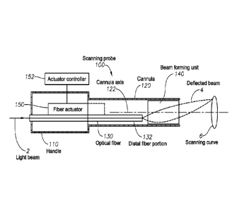
FIG. 1 schematically illustrates an optical light scanning probe 100
consistent with some
embodiments. The scanning probe 100 can include a handle 110, shaped for
grasping by a user,
a cannula 120, protruding from a distal portion of the handle 110, the cannula
120 having an
outer diameter smaller than 20 gauge. The outer diameter of the cannula being
less than 20
gauge may address the above outlined problem (4) by reducing the size and
amount of scar
tissue. The scanning probe 100 can also include an optical fiber 130 with a
distal fiber-portion
132 off a cannula-axis 122, wherein the fiber 130 can be configured to receive
a light or light
beam 2 from a light-source at a proximal fiber-portion, and to emit the
received light at the distal
fiber-portion 132. In some embodiments, the fiber 130 can be positioned
partially in the handle
110, with the distal fiber-portion 132 in the cannula 120.
The scanning probe 100 can also include a fixed beam forming unit 140,
disposed at a
distal portion of the cannula 120, configured to receive the light beam from
the distal fiber-
portion 132, and to deflect the received light beam as a deflected light 4 or
deflected beam 4
toward a target region. The fixed nature of the beam forming unit 140 may
address the above
outlined problem (2) by avoiding catching portions of the target tissue in the
moving ultimate
distal optical element.
In FIG. 1 the distal fiber-portion 132 is shown to be off a cannula-axis 122.
In other
embodiments, it can be off an axis of the fixed beam forming unit 140. These
axes can coincide
when the fixed beam forming unit 140 is placed co-axially with the cannula
120. However, these
axes can also differ when the beam forming unit 140 is not co-axial with the
cannula 120. The
cannula-axis, the beam forming unit-axis and other analogous axes of the
system will be
commonly referred to as examples of a probe-axis.
The scanning probe 100 can also include a fiber actuator 150, housed at least
partially in
the handle 110. The fiber actuator 150 can be configured to move the distal
fiber- portion 132 to
4
CA 02932013 2016-05-27
WO 2015/094727
PCT/US2014/068912
scan the deflected beam 4 along a scanning curve 6 in the target region. In
some embodiments, a
substantial portion of the fiber actuator 150 can be housed in the handle 110,
or even outside the
handle 110. In either of these embodiments, the fiber actuator 150 can be
housed separate from
a disposable portion of the scanning probe 100. In some embodiments, the fiber
actuator 150
can include a small portion positioned in the cannula 120, shown by the dashed
actuator portion.
The actuator 150 being able to scan the deflected beam 4 along a scanning
curve may address the
above problem (1) by providing a curved scanning functionality. The
positioning of the actuator
150 may address the above outlined problem (3) by a substantial portion of the
actuator 150
being positioned outside the cannula 120, away from the disposable portion of
the probe 100.
In some embodiments, the fiber actuator 150 can be configured to be controlled
by an
actuator controller 152, positioned at least partially outside the probe 100.
In various embodiments, the scanning curve 6 can be an open curve, an arc, a
closed
loop, a circle, a cycloid, and an ellipse. In FIG. 1 the scanning curve 6 is a
loop. In some
embodiments the beam forming unit 140 can include a GRIN lens, a lens, a lens
system, or a
focusing element to form the deflected beam 4 by focusing the received light.
FIG. 2 illustrates an embodiment of the probe 100 that can have several
elements
analogous to the embodiment of FIG. 1. In the embodiment of FIG. 2, a portion
of the fiber
actuator 150 can be positioned in the cannula 120: a rotation tube 210,
rotatably positioned in the
cannula 120 and having an eccentric hole 212 off the cannula-axis 122.
Further, the fiber
actuator 150 can include a hollow torque cable 220, with a distal portion
inside the cannula 120,
configured to be rotatable by a motor 230, and configured to rotate the
rotation tube 210 when
rotated by the motor 230. The hollow torque cable 220 can house a portion of
the fiber 130 that
extends into the cannula 120 to guide the fiber 130 to the eccentric hole 212.
The fiber actuator
150 can be configured to circularly move the distal fiber-portion 132 by
rotating the hollow
torque cable 220 that rotates the rotation tube 210 with the eccentric hole
212 that is coupled to
the distal fiber-portion 132. When the fiber actuator 150 circularly moves the
distal fiber-portion
132, the light beam 2 that enters the probe 100 and is output as deflected
beam 4 through the
beam forming unit 140 can be scanned along a scanning curve 6, such as a
scanning loop.
In some embodiments of the scanning probe 100 the fiber 130 can be rotatably
housed
inside the hollow torque cable 220 so that the fiber actuator 150 can rotate
the hollow torque
cable 220 without twisting the fiber 130. Such embodiments allow the motor 230
to rotate the
torque cable 220 while avoiding the twisting of the fiber 130.
In some embodiments, the fiber 130 can be attached to the hollow torque cable
220 in a
non-rotatable manner. Such embodiments can prevent the twisting of the optical
fiber 130 by
5
CA 02932013 2016-05-27
WO 2015/094727
PCT/US2014/068912
coupling the fiber 130 to a light guide 250 through an optical rotary
connector 240. In other
embodiments, the motor 230 can scan the distal fiber-portion 132 along a
scanning curve 6 in a
back-and-forth manner.
Concerning the design of the distal portion of the scanning probe 100,
different
embodiments can be realized. In some designs, the distal fiber-portion 132 can
be disposed
distal to a distal end of the torque cable 220. In others, proximal to the
distal end of the torque
cable 220. In some designs, a distal end of the torque cable 220 can be
disposed distal to a distal
end of the rotation tube 210, or proximal to the distal end of the rotation
tube 210.
In some designs, the motor 230 can be housed outside the handle 110, or in a
console,
separate from the handle 110. The handle 110 can have a non-disposable portion
and a
disposable portion, and the motor 230 can be housed in the non-disposable
portion to address the
above problem (3) by positioning an expensive actuator component non-
disposably. In some
cases, the motor 230 can be housed in the disposable portion. Finally, in
embodiments, the
actuator controller 152 can control an operation of the motor 230.
FIG. 3 illustrates an embodiment of the scanning probe 100 that again can
include
several elements analogous to those in FIGS. 1 and 2. In the embodiment of
FIG. 3, a portion
of the fiber actuator 150 can again be positioned in the cannula 120: a
rotatable hollow drive tube
310, coupled to the motor 230 that can be positioned at least partially inside
the handle 110. The
drive tube 310 can be rotatable in the cannula 120 by the motor 230. An
operation of the motor
230 can be again controlled by the actuator controller 152, coupled to the
motor 230 by an
electric, mechanic, electro-mechanic or pneumatic coupling. The drive tube 310
can house a
portion of the fiber 130 that extends into the cannula 120. The drive tube 310
can also include an
eccentric pusher 312 in a distal region of the drive tube 310 to keep the
distal fiber-portion 132
off the cannula-axis 122.
In some embodiments of the scanning probe 100, the distal fiber-portion 132
can be
attached to the eccentric pusher 312. In such embodiments of the probe 100,
the fiber 130 gets
twisted to some degree as the drive tube 310 and the eccentric pusher 312 are
rotated by the
motor 230. Such embodiments can include a service loop 334 in the fiber 130 to
accommodate
a twisting of the fiber 130 when the motor 230 rotates the drive tube 310. To
limit the twisting
of the fiber 130, the fiber actuator 150 can be configured to rotate the drive
tube 310 and thus the
distal fiber-portion 132 reciprocally, that is, back-and-forth along a
scanning arc, sometimes
called in a reciprocal manner. For example, the scanning arc can extend from
minus 180 degree
to plus 180 degree. In other embodiments, the scanning arc can extend from
minus 90 degree to
plus 90 degree. In yet other embodiments, the scanning arc can extend in a
range between these
two examples.
6
CA 02932013 2016-05-27
WO 2015/094727
PCT/US2014/068912
FIG. 4 illustrates another embodiment of the scanning probe 100. The
embodiment of
FIG. 4 can include numerous elements that are analogous to those in FIGS. 1-3.
In the scanning
probe 100 of FIG. 4, the fiber actuator 150 can include the motor 230,
positioned at least
partially inside the cannula 120, the rotatable hollow drive tube 310,
positioned at least partially
inside the cannula 120, coupled to the motor 230 to be rotated in
the cannula 120. The drive tube 310 can house a portion of the fiber 130 that
extends into the
cannula 120, and include an eccentric hole 412 in a distal region of the drive
tube 310 to keep the
distal fiber-portion 132 off the cannula-axis 122.
In contrast to the embodiment of FIG. 3, in the fiber actuator 150 of FIG. 4
the distal
fiber-portion 132 can be rotatably positioned in the eccentric hole 412, so
that the fiber 130 is not
twisted when the motor 230 rotates the drive tube 310. Such embodiments of the
scanning probe
100 can perform not only reciprocal, back-and-forth type scanning, but also
circular scanning as
well, as indicated.
In some embodiments, the beam forming unit 140 can include a glass element, a
no-core
fiber, or a glass rod. These elements can be attached to a GRIN lens. These,
as well as other
optical elements can shape or deflect the beam emitted from the distal fiber-
portion 132.
In some embodiments, the fiber actuator may not extend into the cannula 120.
Instead, in
these embodiments the distal end of the fiber 130 with the distal fiber-
portion 132 can be
positioned proximal to the cannula 120, i.e. inside the handle 110. The beam
emitted by the
distal end of the fiber 130 can be forwarded to a relay lens inside the
cannula 120, sometimes
positioned near the fixed beam forming unit 140.
As discussed before, systems where the fiber actuator is positioned in a
disposable
handle can be quite costly since when the handle is disposed after a surgical
procedure, it takes
with it the pricey actuator as well. To reduce this cost, in embodiments of
the scanning probe
100 a valuable portion of the fiber actuator 150, such as the motor 230, can
be positioned in a
non-disposable handle 110, or in a non-disposable portion of the handle 110.
For example, in
some embodiments, the entire handle 110 may be non- disposable, and only the
cannula 120 can
be disposed after each procedure. In other embodiments, the handle 110 can
have a proximal
non-disposable portion and a distal, disposable portion. In all of these
embodiments, a valuable
portion of the fiber actuator 150, such as the motor 230, can be in the non-
disposable handle
110, or in the proximal, non- disposable portion of the handle 110.
Of course, in some probes 100 a portion of the fiber actuator 150 can be
positioned in a
disposable portion of the handle 110.
[0045] As mentioned before, embodiments of the optical light scanning probe
100, described in
7
CA 02932013 2016-05-27
WO 2015/094727
PCT/US2014/068912
relation to FIGS. 1-4, can be used for several different functions. These
include imaging,
photocoagulation and ablation.
FIGS. 5A-B illustrate that embodiments of the scanning probe 100 can be part
of an
Optical Coherence Tomography (OCT) imaging system 500. The scanning probe 100
can be
coupled, for example, to an Optical Coherence Tomography (OCT) engine 510. The
OCT engine
510 can include an OCT imaging light source 512 that emits the light to the
optical fiber 130.
The OCT engine 510 can also be configured to detect the imaging beam, returned
from the target
by the scanning probe 100 and to generate an OCT image from an interference of
the returned
imaging beam and a reference beam by an OCT image detector-processor 514. Many
OCT
imaging systems are known and can all be used with the probe 100.
FIG. 5A illustrates that in some embodiments, a portion of the fiber actuator
150, such
as the motor 230 that rotates the torque cable 220 of FIG. 2, can be
positioned in a console of
the OCT engine 510. In some embodiments, the fiber actuator controller 152 can
also be part of
the OCT engine 510, as shown.
FIG. 5B illustrates that in other embodiments, the actuator controller 152 can
be
included in the console of the OCT engine 510, while at least portions of the
fiber actuator 150,
such as its motor 230 can be positioned in the handle 110 as in FIGS. 3-4.
FIG. 6 illustrates a method 600 of operating embodiments of the scanning probe
100.
The scanning probe 100 can be any of the embodiments described in relation to
FIGS. 1-5. The
method 600 can include:
receiving a light (610) by a fiber from an imaging light-source at a proximal
fiber-
portion;
emitting the received light (620) by the fiber at the distal fiber-portion
towards a fixed
beam forming unit;
deflecting the emitted light (630) by the fixed beam forming unit; and
moving the distal fiber-portion (640) by a fiber-actuator, housed at least
partially in a
handle of an imaging probe, so that the light beam deflected by the fixed beam
forming unit
scans along a scanning curve in a target region.
The examples provided above are exemplary only and are not intended to be
limiting.
One skilled in the art may readily devise other systems consistent with the
disclosed
embodiments which are intended to be within the scope of this disclosure. As
such, the
application is limited only by the following claims.
8