Note: Descriptions are shown in the official language in which they were submitted.
CA 02932298 2016-05-31
WO 2015/100134
PCT/US2014/071188
Forward Scanning Optical Probes and
Associated Devices, Systems, and Methods
Lingfeng Yu and Kambiz Parto
Technical Field
Embodiments disclosed herein are related to devices, systems, and methods for
scanning tissue with a forward scanning optical probe, and more particularly,
to devices,
systems, and methods that utilize an optical coherence tomography (OCT) probe,
having
a fiber bundle for ophthalmic imaging.
Background
Optical Coherence Tomography (OCT) systems are used to capture and generate
images of patient tissue layers. These systems often include OCT probes that
can
invasively penetrate tissue to obtain visualization of tissue within a
patient. In
ophthalmology, OCT probes are used to obtain detailed images of tissue about
the eye or
tissue forming a part of the eye, such as the retina.
The OCT probes often include a projecting cannula that can invasively
penetrate
patient tissue. The imaging probe scans tissue by refracting the optical light
beam
through a lens disposed at an end of the cannula. A scanner can steer an
imaging light to
scan the target tissues. The scanner can be placed at a distal end of the
cannula of an
OCT probe. Nevertheless, placing the scanner at the distal end of the cannula
may cause
the size of the distal end of the cannula to be bulky and have complex
structure, which is
not suitable for insertion into an eye.
As an alternative, the scanner can be placed outside the eye directly above
the eye
to be able to directly project the imaging light into the eye. Nevertheless,
placing the
scanner directly above the eye can interfere with an optical pathway of a
surgical
microscope and can take up essential operating space between the surgical
microscope
and the eye.
Accordingly, it is beneficial to position the scanner away from the optical
pathway
of the surgical microscope. For example, the scanner can be placed either in a
handpiece
- 1 -
CA 02932298 2016-05-31
WO 2015/100134
PCT/US2014/071188
of an OCT probe or in a separate scanning unit. If the scanner is placed away
from the
optical pathway, an optical guide is provided to guide the imaging light back
to the
cannula that is inserted inside the eye. The optical guide can be a fiber
bundle formed by
a bundle of optical fibers in order to have certain flexibility. Nevertheless,
in a fiber
bundle, there are finite gaps formed between the optical fibers. Thus, when an
imaging
light is scanned across a proximal surface of the fiber bundle, the scanning
may be
intermittently be interrupted when the imaging light passes through the gaps
between the
optical fibers. As a result, the output scanning beam can become jumpy, which
can cause
the scanning image to be grainy and noisy with insufficient resolution.
Accordingly, there is a need for devices, systems, and methods utilizing an
OCT
imaging system with a scanner scanning a fiber bundle that improve scanning
resolution
to be finer than a fiber-to-fiber separation of the fiber bundle and that
address one or more
of the needs discussed above.
SUMMARY
Embodiments disclosed herein are related to devices, systems, and methods that
utilize an actuator that can adjust a position of optical fibers in a fiber
bundle in
cooperation with a scanner scanning an imaging light across a proximal surface
of the
fiber bundle to improve imaging resolution.
Consistent with some embodiments, an optical imaging apparatus is provided.
The optical imaging apparatus can include a fiber bundle having a set of
optical fibers
configured to guide an imaging light, a beam forming unit configured to
receive the
imaging light from the fiber bundle and to redirect the imaging light to a
target region,
and a bundle actuator configured to adjust a position of the set of optical
fibers of the
fiber bundle. The optical imaging apparatus can include a scanning unit
configured to
scan the imaging light over a proximal surface of the fiber bundle to cause
the redirected
imaging light to scan along a scanning pattern in the target region.
The optical imaging apparatus also can include an imaging light source
configured
to generate the imaging light and a scanning unit configured to scan the
imaging light
over a proximal surface of the fiber bundle.
Consistent with some embodiments, a method of ophthalmic imaging is provided.
The method can include scanning an imaging beam across a proximal end of a
fiber
- 2 -
CA 02932298 2016-05-31
WO 2015/100134
PCT/US2014/071188
bundle with a scanning unit; adjusting a configuration of a distal portion of
the fiber
bundle with a bundle actuator; and directing the imaging light by a beam
forming unit to a
target region.
Additional aspects, features, and advantages of the present disclosure will
become
apparent from the following detailed description.
BRIEF DESCRIPTION OF THE DRAWINGS
FIG. 1 illustrates an exemplary OCT imaging system.
FIG. 2a illustrates a cross-sectional view of a fiber bundle.
FIG. 2b illustrates a cross-sectional view of a fiber bundle.
FIG. 2c illustrates a cross-sectional view of a fiber bundle.
FIG. 2d illustrates a cross-sectional view of a fiber bundle.
FIG. 3 illustrates an exemplary OCT imaging system.
FIG. 4 illustrates a cross-sectional view of an imaging probe.
FIG. 5 illustrates a cross-sectional view of an imaging probe and an OCT
engine.
FIGS. 6a-b illustrate an OCT imaging system.
FIG. 7 illustrates an eye under treatment and an exemplary OCT imaging system.
DETAILED DESCRIPTION
In the following description specific details are set forth describing certain
embodiments. It will be apparent, however, to one skilled in the art that the
disclosed
embodiments may be practiced without some or all of these specific details.
The specific
embodiments presented are meant to be illustrative, but not limiting. One
skilled in the
art may realize other material that, although not specifically described
herein, is within
the scope and spirit of this disclosure. Any alterations and further
modifications to the
described devices, systems, and methods, and any further application of the
principles of
the present disclosure are fully contemplated and included within the present
disclosure as
would normally occur to one skilled in the art to which the disclosure
relates. In
particular, it is fully contemplated that the features, components, and/or
steps described
with respect to one embodiment may be combined with the features, components,
and/or
- 3 -
CA 02932298 2016-05-31
WO 2015/100134
PCT/US2014/071188
steps described with respect to other embodiments of the present disclosure.
For the sake
of brevity, however, the numerous iterations of these combinations will not be
described
separately.
The present disclosure relates generally to OCT probes, OCT systems, and
methods that scan an imaging light across a target tissue to generate an OCT
image. The
imaging probe can include a housing, or handle, and a cannula, protruding from
the
housing. The cannula can be configured to invasively penetrate patient tissue,
such as the
globe of an eye. The cannula can house a lens and a fiber bundle. The fiber
bundle
include a set of optical fibers each configured to direct an imaging light
through the lens
and capture reflected imaging light that passes back through the lens. A
scanner can scan
the imaging light across a proximal surface of the fiber bundle to obtain an
image.
Because there are inherent gaps between the individual optical fibers in the
fiber bundle,
the scanned image may become grainy or noisy.
Exemplary aspects described herein utilize a technique of changing a position
of
the set of optical fibers of the fiber bundle in cooperation with the scanning
of the
imaging light across a proximal surface of the fiber bundle to improve the
resolution of
the scanned image. In particular, a bundle actuator can be provided to change
a position,
or configuration, of the set of optical fibers of the fiber bundle in
cooperation with a
scanning of the imaging light across the proximal surface of the fiber bundle
to cover the
areas of the gaps between the optical fibers and to increase a resolution of
the scanned
image. Changing the position of the set of optical fibers can overcome one or
more of the
problems or limitations of previous approaches. As a result, embodiments of
the present
disclosure can (1) eliminate or reduce imaging artifacts associated with the
spacing or
separation between the individual optical fibers of a fiber bundle; (2)
improve image
clarity and/or resolution; and (3) increase the image sampling density.
FIG. 1 is a diagrammatic schematic view of an exemplary OCT imaging
apparatus 100. In particular, the OCT imaging apparatus 100 can include a
fiber bundle
102 and a beam forming unit 104. The fiber bundle 102 can include a set of
optical fibers
configured to guide an imaging light. The number of optical fibers in the
fiber bundle
102 can vary in a wide range, including between 2 fibers to 1,000,000 fibers,
2 fibers to
100,000 fibers and 2 fibers to 10,000 fibers. Each optical fiber can have a
size or
diameter between 1 micron and 100 microns, between 2 microns and 50 microns,
or
- 4 -
CA 02932298 2016-05-31
WO 2015/100134
PCT/US2014/071188
between 5 microns and 20 microns. The individual fibers of the bundle can be
single
mode fibers, multi-mode fibers, single-mode waveguides, multi-mode waveguides,
and
hollow tubes.
The beam forming unit 104 can be configured to receive the imaging light from
the fiber bundle 102 and to direct, or redirect, the imaging light to a target
region. The
beam forming unit 104 can focus the imaging light onto the target region. For
example,
the beam forming unit 104 can include a Gradient Index (GRIN) lens, a ball
lens, a
diffractive element, an aspherical lens, or an objective.
The OCT imaging apparatus 100 also can include a scanning unit 106 configured
to scan the imaging light over/across a proximal surface 114 of the fiber
bundle 102 to
cause the redirected imaging light to scan along a scanning pattern in the
target region.
The scanning unit 106 can include a coupling lens 108, a scanner 110, and a
collimating
lens 112. An optical fiber 115 can guide the imaging light generated by an
imaging light
source to the scanning unit 106. The imaging light can be received by the
collimating
lens 112 from the optical fiber 115. The scanner 110 can receive the
collimated imaging
light from the collimating lens 112 and direct the imaging light to the
coupling lens 108.
The coupling lens 108 can couple the imaging light into a single or a few
optical fibers of
the fiber bundle 102.
The scanner 110 can include optical elements configured to scan the collimated
beam of the imaging light. For example, the scanner 110 can include one or
more of a
rotatable mirror, a galvanometer, a resonant scanner, a polygon scanner, and a
MEMS
scanner. Thus, the scanner 110 can steer the direction of the imaging light to
scan the
imaging light across the proximal surface 114 of the fiber bundle 102. The
scanning
imaging light can be guided by the fiber bundle 102 toward the beam forming
unit 104
and be directed or output by the beam forming unit 104 to scan the target
region along a
scanning pattern.
The OCT imaging apparatus 100 can include a bundle actuator 116 configured to
actuate all or a portion of the fiber bundle 102. The bundle actuator 116 can
be
positioned adjacent to a proximal portion, a central portion, and/or a distal
portion of the
fiber bundle 102. In some implementations, the bundle actuator 116 can adjust
a position,
or configuration, of the set of optical fibers at the distal end of the fiber
bundle 102. For
example, the bundle actuator 116 can rotate, twist, laterally translate,
and/or
- 5 -
CA 02932298 2016-05-31
WO 2015/100134
PCT/US2014/071188
longitudinally translate the distal portion of the set of optical fibers of
the fiber bundle
102.
The bundle actuator 116 can also be a portion of a manual- or auto-focus sub-
system configured to longitudinally adjust the distal portion of the set of
optical fibers of
the fiber bundle 102 or the beam forming unit 104 to adjust a focal distance
between the
beam forming unit 104 and the target tissue. For example, the bundle actuator
116 can
move the distal portion of the fiber bundle 102 toward or away from the beam
forming
unit 104 to adjust the focus of imaging beam. The bundle actuator 116 can
include any
number of components configured to facilitate rotating, twisting, laterally
translating,
and/or longitudinally translating the fiber bundle 102 or portion thereof.
These
components may include without limitation electric motor(s), bias element(s)
(e.g., coil
springs, leaf springs, etc.), mechanical interface(s) and/or connector(s)
(e.g., pulleys,
ramps, clamps, bolts, nuts, screws, nails, etc.), electromagnetic element(s)
(e.g.,
permanent magnets, electromagnets, coils, etc.) pneumatic drivers, piezo-based
drivers
and/or combinations thereof.
FIGS. 2a-2d illustrate a cross-sectional view of the fiber bundle 102. FIG. 2a
illustrates an embodiment in which the fiber bundle 102 has four optical
fibers: core 1,
core 2, core 3, and core 4. However, it is understood that the concepts
described below
are equally applicable to fiber bundles having any number of optical fibers,
such as a
number between 2 fibers and 1,000,000 fibers, 2 fibers and 100,000 fibers, and
2 fibers
and 10,000 fibers.
In the four fibers, or four cores embodiment, the scanning unit 106 can scan
the
imaging light across the proximal surface 114, across the four cores 1-4.
However, as
described above, the imaging light emitted at the distal end of the fiber
bundle 102
sequentially by the fibers 1-4 will hit four target or scanning spots that are
separated by a
distance set by D, the separation of the centers of neighboring fibers. This
distance D is a
factor limiting the resolution of the imaging.
Some embodiments reduce the separation of scanning spots relative to the fixed-
fiber systems by the bundle actuator 116 adjusting the configuration of the
fiber bundle
102. In some embodiments, the bundle actuator 116 can adjust the configuration
of the
fiber bundle 102 after a first scan by the scanning unit 106. In some
embodiments, the
bundle actuator 116 can shift or rotate at least a portion of the fiber bundle
102 to a
- 6 -
CA 02932298 2016-05-31
WO 2015/100134
PCT/US2014/071188
second position or configuration, as shown in FIG. 2b. For example, the bundle
actuator
116 can rotate the fiber bundle 102 clockwise by an angle to the second
rotated position.
The fiber bundle 102 can be rotated by a small angle and by a corresponding
small
distance less than a distance or separation between the cores, which can be
between 0.1
micron and a few hundred microns, For example, the fiber bundle 102 can be
rotated by a
small angle between 0 degree and 90 degrees in FIG. 2B, in order to direct the
imaging
beam to scanning spots between the spots reached before the rotation. The
scanning unit
106 then can scan the imaging beam across the proximal face 114 of the fiber
bundle 102
for the second time. After the second scan, the fiber bundle 102 can be
rotated clockwise
again to a third position, as shown in FIG. 2c. The scanning unit 106 can then
scan the
imaging beam across the proximal ends of the four cores for the third time. In
this
embodiment, the cores of the fiber bundle 102 can be rotated around a central
longitudinal axis of the fiber bundle 102. In an embodiment, a core positioned
at a center
of the fiber bundle 102 can be rotated without changing positions relative to
the central
longitudinal axis of the fiber bundle 102.
Accordingly, FIG. 2d shows that by choosing an ever smaller rotation angle,
the
fibers can be rotated into a sequence of positions to cover the gaps between
the optical
fibers in small steps. When the scanning unit 106 re-scans the proximal
surface 114 of
the fiber bundle 102 after each of these small angle rotations, a density of
the scanning
spots can be increased considerably, enabling a higher resolution imaging.
In some embodiments, an actuator controller 117 can control the bundle
actuator
116, and thereby the actuation of the fiber bundle 102. The actuator
controller 117 can
execute the actuation in coordination or synchronization with the scanning
operation of
the scanning unit 106. For example, the actuation of the fiber bundle 102 can
begin after
a scanning operation and can finish before the start of a subsequent scanning
operation.
The actuator controller 117 can be in communication with the scanning unit 106
to coordinate the actuation and scanning operation. The actuator controller
117 can be a
part of the actuator 116, or it can be disposed near the scanning unit 106, or
it can be
disposed in a separate console, in communication with a controller of the
scanning unit
106. The actuator controller 117 can be synchronized with the scanning unit
106 via an
electrical or mechanical, or electro-mechanical coupling.
- 7 -
CA 02932298 2016-05-31
WO 2015/100134
PCT/US2014/071188
In some embodiments, a proximal portion of the fiber bundle 102 can move or
rotate together with a distal portion of the fiber bundle 102. In some
embodiments, the
entire fiber bundle 102 can rotate together. The bundle actuator 116 can be
configured to
cause both the proximal and distal portions of the fiber bundle 102 to rotate,
laterally
translate, and/or longitudinally translate together. Thus, the fiber bundle
102 can be
rotated or moved without being twisted, if desired.
FIG. 3 illustrates an OCT imaging apparatus 300. The OCT imaging apparatus
300 is similar in many respects to the OCT imaging apparatus 100 described
above. For
example, the scanning unit 106 can scan the imaging light across the proximal
surface
114 of the fiber bundle 102. Further, the bundle actuator 116 can actuate the
fiber bundle
102 to improve imaging resolution.
In addition, a movable stage 118 can be provided to perform the scanning
action
of the scanner 110. The movable stage 118 can move the scanning unit 106 to
scan the
imaging light across the proximal surface 114 of the fiber bundle 102. For
example, the
movable stage 118 can support the collimating lens 112 and the coupling lens
108. When
the movable stage 118 moves, the beam of the imaging light can move to scan
the
proximal surface 114 of the fiber bundle 102. The scanning unit 106 can
include multiple
stages or actuators to move the scanning unit 106 in various directions.
FIG. 4 illustrates a cross-sectional view of an imaging probe 402. In some
designs, embodiments of the imaging probe 402 can accommodate or house the OCT
imaging apparatus 100/300, or at least parts of it. The imaging probe 402 can
include a
handle 122, configured to be operated by a surgeon during operation, and a
cannula 120, a
distal end of which is configured to be inserted into a tissue, e.g., an eye.
The scanning
unit 106 can be positioned in the handle 122. The cannula 120 can be coupled
to the
handle 122, or protrude from the handle 122. The fiber bundle 102 can be
positioned in
the cannula 120, or at least parts of it in the cannula 120. The beam forming
unit 104 can
be positioned at a distal end of the cannula 120. The scanning unit 106 can
scan the
imaging light across the proximal surface 114 of the fiber bundle 102. The
scanned
imaging light, or beam, can be guided by the fiber bundle 102 to the beam
forming unit
104. The beam forming unit 104 can direct, or redirect, the scanned imaging
beam, and
output the redirected scanned imaging beam toward the target tissue. The fiber
bundle
102 can be actuated, as discussed above, to improve the imaging resolution.
- 8 -
CA 02932298 2016-05-31
WO 2015/100134
PCT/US2014/071188
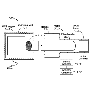
FIG. 5 illustrates an OCT imaging apparatus 500. In a cross-sectional view,
the
OCT imaging apparatus 500 can include an imaging probe 502 and an OCT engine
504.
The imaging probe 502 can be similar in many respects to the imaging probe 402
described above. For example, the imaging probe 502 can include a handle 122
configured to be operated by a surgeon during operation, and a cannula 120, a
distal end
of which is configured to be inserted into a tissue, e.g., an eye. Further,
the cannula 120
can be coupled to the handle 122. The fiber bundle 102 can be positioned in
the cannula
120. The beam forming unit 104 can be positioned at a distal end of the
cannula 120.
In contrast to the imaging probe 402, in the presently shown design, the
scanning
unit 106 can be positioned in a separate OCT engine 504 spaced from the handle
122,
such as in a separate console. The fiber bundle 102 can extend between the OCT
engine
504 and the imaging probe 502. The OCT engine 504 can be configured to
generate an
OCT image from a returned scanned imaging light, returned from the target
tissue.
For example, the OCT engine 504 can control the scanning unit 106 to scan the
imaging light across the proximal surface 114 of the fiber bundle 102. The
imaging light
can be guided by the fiber bundle 102 from the OCT engine 504 to the imaging
probe
502. Within the imaging probe 502, the fiber bundle 102 can guide the imaging
light to
the beam forming unit 104 to be output to the target tissue. The imaging light
then can be
reflected by the target tissue. The reflected imaging light can be captured
back into the
fiber bundle 102 via the beam forming unit 104. The reflected imaging light
can be
guided back to the OCT engine 504. The OCT engine 504 can analyze the
reflected
imaging light using OCT methods to generate an OCT image, including forming an
interference with a reference beam. The generated OCT image can be displayed
to the
user, e.g., the surgeon, on a user interface display in communication with the
OCT engine
504.
FIG. 6a illustrates that in some embodiments, the OCT imaging apparatus
100/300/500 can further include a movable beam steering unit 510 positioned at
a distal
end of the fiber bundle 102, but proximal to the beam forming unit 104, and
configured to
move to increase a density of scanning spots in a target tissue. FIG. 6b
illustrates that in
some embodiments, the OCT imaging apparatus 100/300/500 can further include a
movable beam steering unit 510 positioned at a distal end of the beam forming
unit 104
and configured to move to increase a density of scanning spots in a target
tissue.
- 9 -
CA 02932298 2016-05-31
WO 2015/100134
PCT/US2014/071188
FIG. 7 illustrates an eye under treatment by an OCT imaging apparatus
100/300/500. In particular, an eye 600 under treatment is shown. The eye 600
includes a
sclera 602, a cornea 604, an anterior chamber 606, and a posterior chamber
608. A
capsular bag 610 is illustrated in the posterior chamber 608. The eye 600
further includes
a retina 612.
The OCT imaging apparatus 500 is also illustrated in FIG. 7. As discussed
above,
the OCT imaging apparatus 500 can be configured to image portions of the eye
600, such
as the retina 612. The OCT imaging apparatus 500 can include an imaging light
source
622, the optical coherence tomography (OCT) engine 504, a controller 626, a
user
interface 628, and the imaging probe 402/502. The light source 622 can be
configured to
provide imaging light that will be directed onto the target biological tissue
by the imaging
probe 502. The light source 622 can include super-luminescent diodes, ultra-
short pulsed
lasers, wavelength sweeping sources or supercontinuum lasers that provide
relatively
broad bandwidth light, such as between 700 nm and 1400 nm, between 900 nm and
1200nm, or between 1000 nm and 1100 nm. Imaging light reflected from the
target
biological tissue and captured by the imaging probe 502 is utilized to
generate images of
the target biological tissue.
The OCT engine 504 is configured to split the imaging light received from the
light source 622 into the imaging beam that is directed toward the target
biological tissue
by the imaging probe 502, and a reference beam that can be directed onto a
reference
mirror. The OCT engine 504 can be a spectral domain, swept-source, or a time
domain
system. The OCT engine 504 can be further configured to receive the imaging
light
reflected from the target biological tissue and captured by the imaging probe
502. The
OCT engine 504 then can interfere the returned imaging beam and the reference
beam,
returned from the reference mirror to form an interference pattern. The
interference
pattern between the reflected imaging light and the reference beam can be
utilized to
generate an image of the target biological tissue. Accordingly, the OCT engine
504 can
include a detector configured to detect the interference pattern. The detector
can include
photodiode detector, balanced detectors, Charge-Coupled Detectors (CCDs),
pixels, or an
array of any other type of sensor(s) that generate an electric signal based on
detected
light. Further, the detector can include a two-dimensional sensor array and a
detector
camera.
- 10 -
CA 02932298 2016-05-31
WO 2015/100134
PCT/US2014/071188
The controller 626 can include a processor and memory, which may include one
or more executable programs for controlling operations of the light source
622, the user
interface 628, the actuator controller 117 of the bundle actuator 116, and/or
the imaging
probe 502, and for executing and performing functions and processes to carry
out an OCT
imaging procedure. For example, the controller 626 can be configured to
control the
bundle actuator 116 in the imaging probe 502 to actuate a distal end of the
fiber bundle
102 in sync with a scanning operation of the OCT imaging apparatus 500.
One or more of the light source 622, the OCT engine 504, the controller 626,
and
the user interface 628 can be implemented in a separate console
communicatively coupled
to one another, or within a common console. In some designs, parts of the OCT
engine,
such as its scanning unit 106 can be housed in the probe 402, as in FIG. 4. In
other
designs, the scanning unit can be house separately from the probe 502, such as
in FIG. 5.
For example, in some implementations the light source 622, the OCT engine 504,
and the controller 626 can be positioned within a console that is
communicatively
coupled to the user interface 628. The user interface 628 can be carried on or
form part of
the console. Further, the user interface 628, or at least part(s) thereof, can
be separate
from the console. The user interface 628 can include a display configured to
present
images to a user or a patient, and display tissue scanned by the imaging probe
502 during
an OCT imaging procedure. The user interface 628 can also include input
devices or
systems, including by way of non-limiting example, a keyboard, a mouse, a
joystick, a
touchscreen, dials, and buttons, among other input devices.
In some designs, the imaging probe 402/502 can be in optical communication
with
the OCT engine 504. In that regard, the imaging probe 402/502 is configured to
present
light from the light source 622 that passes through OCT engine 504 onto the
target
biological tissue for the purpose of imaging the tissue. Further, the imaging
probe
402/502 can be in electrical communication with the controller 626. In that
regard, the
controller 626 can control the bundle actuator 116 of the imaging probe
402/502 via
electrical signals sent to the imaging probe 402/502 in order to cause the
actuation system
to scan the imaging beam across the target biological tissue. An optical cable
632 can
connect the imaging probe 402/502 to the OCT engine 504 and/or the controller
626. In
that regard, the cable 632 can include the fiber bundle 102, a fiber 115,
electrical
conductor(s), insulator(s), shield(s), and/or other features configured to
facilitate optical
- 11 -
CA 02932298 2016-05-31
WO 2015/100134
PCT/US2014/071188
and/or electrical communication between the imaging probe 402/502 and the OCT
engine
504 and/or the controller 626. Further, it is understood that cable 632 can
include
multiple, separate cables. For example, in some instances an optical cable can
connect
the imaging probe 402/502 to the OCT engine 504, and a separate electrical
cable can
connect the imaging probe 402/502 to controller 626.
In the illustrated embodiment, the cable 632 can terminate in a connector 634
that
is configured to facilitate removable coupling of the imaging probe 402/502 to
the cable
632. The connector 634 can be configured to selectively engage with a
connector 636
associated with the imaging probe 402/502 to facilitate mechanical, optical,
and/or
electrical coupling of the imaging probe 402/502 to the cable 632. For
example, the fiber
bundle 102 extending along the length of the imaging probe 402/502 can be
optically
coupled to the OCT engine 504 via the coupling of the connectors 634 and 636.
In the
illustrated embodiment, the connector 636 can be configured to threadingly
engage with
the connector 634. However, it is understood that any type of selective
engagement
feature(s) or connectors can be utilized to couple the imaging probe 402/502
to the cable
632, including without limitation press fit, luer lock, threads and
combinations thereof.
The selective engagement of the connector 636 with the connector 634 allows
the entire
probe 402/502 to be a disposable component configured for use in a single
procedure,
while the connector 634 and cable 632 can be reusable components that can be
sterilized
(e.g., using autoclave procedures) and used in multiple procedures. In the
embodiments
of FIG. 5, the cable 632 can be part of the imaging probe 402/502, and the
connector
coupling the imaging probe 402/502 and the cable 632 to the OCT engine 504 can
be
positioned in, next to, or close to the OCT engine 504.
The scanning unit 106 can be positioned in the disposable portion of the
handle
122, or in a non-disposable and reusable portion of the handle 122, or in a
separate
portion of the OCT engine 504, again making it reusable.
The handle 122, sometimes also called the housing 122, can be sized and shaped
for grasping by a hand of the user, such as the surgeon. To this end, the
handle 122 can
include a textured surface 648 (e.g., roughened, knurled, or include
projections/recesses,
tapers, other surface features, and/or combinations thereof) to enhance the
user's grip on
the handle 122. In operation, the user can control the position of the cannula
120, distally
- 12 -
CA 02932298 2016-05-31
WO 2015/100134
PCT/US2014/071188
coupled to the housing/handle 122, by maneuvering the handle 122 such that the
imaging
light beam is directed towards the target biological tissue.
The cannula 120 can be sized and shaped for insertion into the eye 600 through
the sclera 602 of the eye 600 to facilitate imaging of the retina 612. The
cannula 120 can
be integrally formed with the handle 122. Alternatively, the cannula 120 and
the handle
122 can be separate components fixedly secured to one another. In that regard,
the probe
402/502 can include one or more connectors to facilitate mechanical, optical,
and/or
electrical coupling of the cannula 120 and the handle 122. As a result, the
cannula 120, or
the cannula 120 and a portion of handle 122 can be a disposable component
configured
for use in a single procedure, while the handle 122 or remaining portions of
the handle
122 are reusable components that can be sterilized (e.g., using autoclave
procedures) and
used in multiple procedures. In yet other embodiments, the entire handle 122
can be
disposable. Finally, in some designs the entire probe 402/502 can be
disposable. The
beam forming unit 104, such as a lens, can be secured within the distal end of
the cannula
120. The beam forming unit 104 can be configured to focus the imaging light
onto the
target biological tissue, such as the retina 612. The beam forming unit 104
can be a
gradient index (GRIN) lens. Depending upon the embodiment, the gradient index
may be
spherical, axial, or radial. The beam forming unit 104 can also be a spherical
lens. Other
lens shapes may be used.
The examples provided above are exemplary only and are not intended to be
limiting. One skilled in the art may readily devise other systems consistent
with the
disclosed embodiments which are intended to be within the scope of this
disclosure. As
such, the application is limited only by the following claims.
- 13 -