Note: Descriptions are shown in the official language in which they were submitted.
VIRAL VECTOR NANOCAPSULE FOR
TARGETING GENE THERAPY AND ITS PREPARATION
STATEMENT AS TO RIGHTS TO INVENTIONS MADE UNDER
FEDERALLY SPONSORED RESEARCH AND DEVELOPMENT
This invention was made with Government support under Grant No.
AI069350, awarded by the National Institutes of Health, and Grant No. HDTRA1-
09-
1-0001, awarded by the U.S. Depai ______________________________________ anent
of Defense, Defense Threat Reduction
Agency. The U.S. Government may have certain rights in this invention.
BACKGROUND OF THE INVENTION
Targeted gene transduction to specific tissues and organs is a desirable
method
of gene delivery. There have been many attempts to develop targeted gene
transduction systems based upon various viral vectors. Adenovirus and adeno-
associated virus vectors have been used in targeted gene delivery strategies
because of
their simple binding and entry mechanisms (see, e.g. Nicklin, et al. Curr.
Gene Ther. 2,
273-293, 2002). Although these vectors have been used successfully in vitro
for
targeting to specific cells, their usefulness in vivo has been limited by
their natural
tropism (see, e.g. Muller, et al. Nat. Biotechnol. 21, 1040-1046, 2003),
especially to
liver cells (see, e.g. Martin, et al. Mol. Ther. 8, 485-494, 2003).
The application of specific targeting with retroviral vectors has also been
problematic and the few studies of retroviral vector targeting in living
animals are not
efficient (see, e.g. Martin, et al. Mol. Ther. 5, 269-274, 2002; Jiang, et al.
Gene Ther.
6, 1982-1987, 1999). Inserting ligands, peptides or single-chain antibodies
into the
retroviral receptor binding envelope subunit has been the most common approach
used to alter or restrict the host range of retroviral vectors (see, e.g.
Martin, et al. Mol.
Ther. 5, 269-274, 2002; Jiang, et al. Gene Ther. 6, 1982-1987, 1999; Han, et
al. Proc.
Natl. Acad. Sci. USA 92, 9747-9751, 1995; Mann, et al. J. Virol. 70, 2957-
2962,
1
Date Recue/Date Received 2021-04-26
1996; Nilson, et al. Gene Ther. 3, 280-286, 1996; Somia, et al. Proc. Natl.
Acad. Sci.
USA 92, 7570-7574, 1995; Valsesia-Wittman, et al. J. Virol. 68,4609-4619,
1994).
Another approach is bridging virus vector and cells by antibodies or ligands
(see, e.g.
Boerger, et al. Proc. Natl. Acad. Sci. USA 96, 9867-9872, 1999; Roux, et al.
Proc.
Natl. Acad. Sci. USA 86, 9079-9083, 1989). In general, most strategies have
suffered
from inconsistent specificity and low viral titers as a result of modification
of the
retroviral envelope (see, e.g. Han, et al. Proc. Natl. Acad. Sci. USA 92, 9747-
9751,
1995; Mann, et al. J. Virol. 70, 2957-2962, 1996; Nilson, et al. Gene Then 3,
280-
286, 1996; Somia, et al. Proc. Natl. Acad. Sci. USA 92, 7570-7574, 1995;
Valsesia-
Wittman, et al. J. Virol. 68,4609-4619, 1994; Kasahara, et al. Science 266,
1373-
1376, 1994).
Chemical modification of the Adenovirus vector with synthetic polymers such
as polyethylene glycol (PEG) significantly reduce innate immune responses to
the
Adenovirus vector, evading pre-existing anti-Ad antibodies (see, e.g. Kreppel,
et al.
The American Style of Gene Ther. 16, 16-29, 2008). However in vivo targeting
efficiency using PEGlated Adenovirus vector is still not sufficient and
background
infectivity still exists in liver cells (see, e.g. Lanciotti, et al. Mol.
Ther. 8, 99-107,
2003). Although PEGlated VSV-G pseudotyped lentiviral vector was reported to
be
prevented from serum inactivation (see, e.g. Croyle, et al. J.V. 78, 912-921,
2004),
targeting lentiviral vector by chemical modification has never been reported
before.
The use of viral vectors having controllable targeting abilities has important
implications for the use of such vectors in the clinic. For this reason, new
methods
and materials that can increase or modulate such targeting of cells or tissues
are
highly desirable.
SUMMARY OF THE INVENTION
The ability to introduce transgenes with precise specificity to target cells
or
tissues is key to a more facile application of genetic therapy. The invention
disclosed
herein provides novel methods, materials and systems that utilize
nanotechnological
2
CA 2932542 2020-03-18
techniques to generate viral vectors with altered recognition of host cell
receptor
specificity. In the working embodiments of the invention that are disclosed
herein,
the infectivity of the VSV-G envelope glycoprotein pseudotyped lentiviral
vectors
was shielded by a thin polymer shell synthesized in situ onto the viral
envelope and
new binding ability was conferred to the shielded virus by conjugating cyclic
RGD
(cRGD) peptide onto the polymer shell. These polymer encapsulated viral
vectors are
further shown to have a number of additional characteristics that are highly
desirable
including a higher stability at room temperature, resistance to serum
inactivation in
vivo, and an ability to infect dividing and non-dividing cells with high
efficiencies.
Embodiments of the invention include, for example, a composition of matter
comprising a viral vector encapsulated by a cross-linked degradable polymer
shell to
which one or more targeting agents is coupled. The targeting agents are
typically
polypeptides such as antibodies, receptors, ligands and peptides. For example,
the
targeting agent can comprise chimeric inununoglobulins, immunoglobulin chains
or
fragments thereof (such as Fv, Fab, Fab', F(ab')2 or other binding
subsequences of
antibodies). Targeting agents are typically selected to have a specific
desirable
affinity for a selected tissue, cell lineage, tumor cell or the like. In
illustrative
embodiments of the invention, the viral vector is a Vesicular stomatitis
Indiana virus
G protein (VSV-G) pseudotyped lentiviral vector, the cross-linked degradable
polymer shell comprises at least one of N-acryloxysuccinimide (NAS),
acrylamide or
glycidyl methacrylate (GMA), and the targeting agent is a cyclic arginine-
glycine-
aspartic acid (cRGD), said cRGB having an affinity to an avf33 integrin on a
tumor
cell.
In one aspect of the present invention, chemical modification and in-situ
polymerization is used to fabricate a cross-linked degradable polymer shell on
the
surface of a viral vector. This polymer shell functions to temporarily shield
the native
binding ability of the viral vector. Targeting agents are coupled to the
surface of the
polymer complex so as to direct the polymer encapsulated viral vector to
specific cells
3
CA 2932542 2020-03-18
such as tumor cells, neurons, and human mobilized PBMCs (see FIG. I). These
targeting agents then modulate vector targeting of specific cells or tissues
Embodiments of the invention also include methods of preparing an
encapsulated viral vector by reacting a polymerizable molecular anchor with a
viral
vector to generate a polymerizable group; reacting the polymerizable group to
a
plurality of monomers to form a polymer shell that encapsulates the viral
vector;
coupling the polymer shell with a degradable cross-linker; and further
attaching a
targeting agent to this complex. In some embodiments of the invention, the
polymerizable molecular anchor is conjugated to a lysine of a protein
expressed by
the vital vector. In certain embodiment of the invention, the shell is formed
from
constituents (e.g. the degradable crosslinker) that degrade under certain
condition,
(e.g. an acidic environment), thereby releasing the viral vector from the
polymer shell
under those conditions. Optionally, for example the viral protein is a
Vesicular
stomatitis Indiana virus G protein (VSV-G) and the degradable cross-linker is
glycidyl methacrylate (GMA). In some embodiments of the invention, the
crosslinlcing agent is selected to comprise a peptide having an amino acid
sequence
that is cleaved by a protease. In certain embodiments, the targeting agent is
a cyclic
arginine-glycine-aspartic acid (cRGD), said cRGB having an affinity to an
av133
integrin on a tumor cell.
Other embodiments of the invention include methods for modulating the
cellular specificity of viral vectors. Typically such methods comprise
selecting a viral
vector having a first specificity for a target tissue or cellular lineage and
then
encapsulating the viral vector in a shell formed from a plurality of polymers,
polymers
that are cross-linked by a degradable agent so as to form a polymer shell that
can
degrade in an in vivo environment. In such methods a targeting agent is
further
attached to the polymeric shell complex. This targeting agent is selected to
have a
second specificity for a target tissue or cellular lineage (e.g. a specificity
for a cell
lineage or tumor specific antigen). In this way, the cellular specificity of
the viral
vector can be modulated.
4
CA 2932542 2020-03-18
Other objects, features and advantages of the present invention will become
apparent to those skilled in the art from the following detailed description.
It is to be
understood, however, that the detailed description and specific examples,
while
indicating some embodiments of the present invention are given by way of
illustration
and not limitation. Many changes and modifications within the scope of the
present
invention may be made without departing from the spirit thereof, and the
invention
includes all such modifications.
BRIEF DESCRIPTION OF THE DRAWINGS
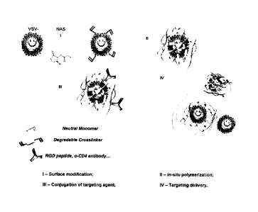
Figure 1: Fabrication of viral vector nanocapsule.
Figure 2: HeLa cell transduced by RGD conjugated VSVg-HIV lentiviral
nanocapsules (Left: After; Right: Before).
Figure 3: VSVg-HIV lentivinis Nanocapsules using ROD as targeting agent.
Different thickness and densities of the polymer result in different shielding
of the
viral infectivity. Best combination is NAS : virus=2x10⁴ with Monomers : virus
125.
Figure 4: TEM pictures of VSVg-HIV lentiviral Nanocapsules (Left: VSVg-
HIV; Middle : Nanocapsule (monomer to viral vector = 125); Right: Nanocapsule
(monomer to viral vector = 250).
Figure 5: Schematic illustration of the synthesis and delivery of VSV-G
pseudotyped lentivims nanocapsules: 1) Surface modification of the lysine
group of
the envelope protein by NAS; II) In-situ polymerization of monomer and
crosslinker
at the surface of modified virus; III) Conjugation of the targeting agents
onto the
surface of the polymer shell; IV) Targeting delivery of the nanovinis to
cells.
Figure 6: Shielding of virus infectivity by polymer shell. 1x10⁵ Hela cells
were transduced with equal amount of VSVG, cRGD-nVSVG, and nVSVG (p24=10
ng). EGFP expression was monitored by flow cytometry.
Figure 7: Optimization of transduction efficiency of the targeting nanovirus.
VSV-G pseudotyped lentivirus (p24=60ng) were reacted with different amounts of
NAS and monomers with or without cRGD. P24=1Ong virus were then use to infect
CA 2932542 2020-03-18
1 x10⁵ Hela cells for 4h. Virus transduction was monitored by EGFP expression
3
days after infection (Top panel). Sizes of the virus nanocapsules were
measured by
DLS (bottom panel). Left panel is virus with cRGD and right panel is virus
without
cRGD. At a combination ratio of NAS: virus (m/nr--2x104) and monomer: virus
(w/w=125), we obtained the best transduction efficiency by cRGD-nVSVG (35%)
while transduction efficiency by nVSVG is 0%. Transduction efficiency of VSV-G
pseudotypes is 42%.
Figure 8: Targeted transduction of Hela cells by the nanovirus is cRGD
dependent. 1 x10⁵ Hela cells were transduced with equal amount of VSVG, cRGD-
nVSVG, and cRAD-nVSVG (p24=10 ng) using the best ratio of NAS and monomer
obtained from Figure 7. EGFP expression was monitored by flow cytometry.
Figure 9: Targeted transduction of Hela cells by the nanovirus is cRGD and
integrin dependent. 1 x10⁵ Hela cells were pre-incubated with cRGD (1mg/m1),
cRAD( 1 mg/ml), anti-integrins antibodies (2Oug/m1), and isotype control
(4Oug/m1) for
half and hour then transduced with equal amount of VSVG and cRGD-nVSVG
(p24=10 ng).
Figure 10: Entry kinetics of the targeting nanovirus by 13-lactamase assay.
Both VSV-G pseudotypes and targeting nanovirus incorporating the BlaM-Vpr
fusion
protein (p24=130 ng) were used to transduce 1x10⁵ Hela cells for 5, 15, 30,
45, 60,
90, and 120 minutes. The percentage of cells with fl-lactamase activity was
measured
by flow cytometry.
Figure 11: Fusion and reverse transcription of the targeting nanovirus. A)
1 x10⁵ Hela cells were treated or not treated with vacuolar-type H+-ATPase
inhibitor
bafilomycin-A for half an hour then transduced with VSV-G or cRGD-nVSVG
(p24=10 ng) with or without bafilomycin-A for 2 hour. B) Transduction by the
nanovirus is inhibited by reverse transcriptase AZT. 1x10⁵ Hela cells were
treated or
not treated with AZT for half an hour then transduced with VSV-G or cRGD-nVSVG
(p24=10 ng) for 2 hour with or without AZT.
6
CA 2932542 2020-03-18
Figure 12: Enhanced stability of the targeting nanovirus in the presence of
human serum. VSVG or cRGD-nVSVG were pre-incubated with PBS, heat-
inactivacted (HI) human serum (HS) or HS (v:v=1:1) for 1h. The pre-treated
virus
(p24-10ng) were then used to transduce 1x10⁵ Hela cells.
DETAILED DESCRIPTION OF THE INVENTION
Unless otherwise defined, all terms of art, notations and other scientific
terms
or terminology used herein are intended to have the meanings commonly
understood
by those of skill in the art to which this invention pertains. Many of the
techniques
and procedures described or referenced herein are well understood and commonly
employed using conventional methodology by those skilled in the art.
Publications
cited herein are cited for their disclosure prior to the filing date of the
present
application. Nothing here is to be construed as an admission that the
inventors are not
entitled to antedate the publications by virtue of an earlier priority date or
prior date of
invention. Further the actual publication dates may be different from those
shown and
require independent verification. In the description of the preferred
embodiment,
reference is made to the accompanying drawings which form a part hereof, and
in
which is shown by way of illustration a specific embodiment in which the
invention
may be practiced. It is to be understood that other embodiments may be
utilized and
structural changes may be made without departing from the scope of the present
invention.
Generally in this field of technology, chemical modification of viral vectors
such as adenovirus vectors and VSV-G pseudotyped lentiviral vectors with
synthetic
polymers, such as polyethylene glycol (PEG), has used a "grafting-onto"
strategy.
This strategy includes two steps, activating linear polymers and conjugating
polymers
to the surface of the viral vector. However, the "grafting-onto" strategy can
only
conjugate linear polymers onto the viral surface, and thus the shielding of
the viral
infectivity is not complete. The invention disclosed herein provides methods,
7
CA 2932542 2020-03-18
materials and systems that address such limitations in the art by utilizing
polymer
chemistry techniques to generate viral vectors having altered host cell
specificities.
In one aspect of the present invention, a "growing-onto" process is provided
to
grow in-situ polymer networks on the surface of viral vectors with a
controllable
thickness and density, which provides better shielding of the native viral
infectivity.
Additionally, in one or more embodiments, the polymer shell is designed to be
pH-
sensitive and therefore can be removed in the endosome after endocytosis. This
unshielding of the polymer from the viral vector within the endosome results
in better
infection activity. Furthermore, targeting efficiency is elevated by
introducing as
much targeting agents as possible to the surface of the virus nanocapsule.
In another aspect of the present invention, a method is provided for creating
a
virus nanocapsule with a highly controllable polymer shell for targeting gene
therapy.
This encapsulation approach provides the inner virus vector with a diversified
targeting ability and extra stabilization upon serum inactivation.
Different monomers and cross-linkers may be used to encapsulate the viral
vectors by conjugation and in-situ polymerization. Targeting agents, such as
antibodies, peptides, or growth factors, are covalently conjugated with the
polymer to
allow for targeted gene transduction to specific tissues and organs through
intravenous injection.
The invention provided herein combines the advantages of both viral vectors
and polymer nanocapsules. The polymer encapsulated viral vectors provided have
high stability at room temperature, can be prevented from serum inactivation
in vivo,
and can infect dividing and non-dividing cells with high efficiency. Chemical
modification provides advantages over genetic modification (which is
traditionally
used in retargeting lentiviral and retroviral vectors) since the viral
envelope is not
disrupted by the mutation of amino acids and therefore the viral infectivity
can be
maintained after polymer encapsulation. Furthermore, chemical modification of
the
polymer to confer different properties (e.g. specificities, charges,
stabilities to
environment) is technically much easier than the genetic modification of
virions. The
8
CA 2932542 2020-03-18
diversified and controllable targeting ability provided to viral vectors by
the
nanocapsules may be used in clinical applications using viral vectors for
targeting
gene and protein delivery.
The invention disclosed herein has a number of embodiments. One
embodiment of the invention is a composition of matter comprising a viral
vector a
degradable polymer shell encapsulating the viral vector. The viral vectors
useful in
the compositions and methods of the invention include retroviral vectors,
adenoviral
vectors, adeno-associated viral vectors, lentiviral vectors, herpes simplex
viral
vectors, vaccinia, pox viral vectors, and Sindbis viral vectors. As is known
in the art,
these retroviral vectors can exhibit variable tropism for different tissues
(tissue
tropism refers to the cells and tissues of a host which can be infected by,
and typically
support the growth of, a particular virus). In such compositions, a targeting
agent can
be selected and coupled to the encapsulated viral vector in order to direct
the vector to
certain cells or tissues (e.g. so that the cell or tissues targeted by the
targeting agent
are not those targeted by viral vector tropism). In typical compositions, the
degradable polymer shell is crosslinked (e.g. with an agent such as glycerol
dimethacrylate) and a targeting agent is coupled to the degradable polymer
shell. In
illustrative embodiments of the invention, the viral vector is a Vesicular
stomatitis
Indiana virus G protein (VSV-G) pseudotyped lentiviral vector.
In certain embodiments, the polymers that form the polymer shell are
crosslinked by one or more degradable crosslinking compounds. Optionally, the
crosslinker is a degradable crosslinker comprising a glycerol dimethacrylate,
a N,N-
methylenebis(acrylamide), a 1,4-bis(acryloyl)piperazine, an ethylene glycol
diacrylate, a N,N1-(1,2-dihydroxy-ethylene)bisacrylamide, or a poly(ethylene
glycol)diacrylate (see, e.g. WO 2013/006762). In certain embodiments of the
invention, the crosslinked polymeric network can be designed to exhibit a
specific
material profile, for example a surface charge of between 3 and 5 millivolts
at a
physiological pH.
9
CA 2932542 2020-03-18
In embodiments of the invention, the structure of the polymeric shell is
designed in a manner that allows it to release the viral vector into selected
environments. For example, in some embodiments of the invention, polymer
components of the shell can be interconnected by disulfide-containing
crosslinked
moieties, linkages which maintain the integrity of the polymer shell under
certain
environmental conditions such as those typically found outside of cells (see,
e.g. WO
2012/142410). Such linkages can be selected for an ability to degrade under
other
environmental conditions such as those that occur within the cellular cytosol.
This
degradation compromises the integrity of the polypeptide shell and results in
the viral
vector being released from this shell. As disclosed herein, by utilizing, for
example,
the redox potential differences that occur in different environments, a
variety of viral
vector delivery systems can be made. Embodiments of the invention include
forming
compositions of the invention by combining together a mixture comprising a
viral
vector, a plurality of polymerizable monomers; and a crosslinking agent
selected for
its ability to form disulfide bonds that are reduced in the cytosol of a
mammalian cell.
Illustrative embodiments of the invention include methods for using
compositions of
the invention for the intracellular delivery of viral vectors to cells or
tissues not
naturally infected by the virus.
In yet other embodiments of the invention, the crosslinlcing agent is selected
to
comprise a peptide having an amino acid sequence that is cleaved by a protease
so
that the polymer shell degrades in those in vivo environments where the
protease is
active (see, e.g. Biwas et al., ACS Nano. 2011 Feb 22;5(2):1385-94).
A number of targeting agents can be coupled to the polymer shells disclosed
herein and used in the compositions and methods of the invention. For example,
antibodies are known to be versatile tumor-targeting agents that can be used
in
embodiments of the invention (see, e.g. Lin et al., Clin Cancer Res (2005) 11;
129).
In addition, a wide variety of ligands are useful as targeting agents can be
adapted for
use with embodiments of the invention (see .e.g. Brumlik et al., Expert Opin
Drug
Deliv. 2008 Jan;5(1):87-103; Vaitilingam et al., J Nucl Med. 2012
Jul;53(7):1127-34
CA 2932542 2020-03-18
and Das et al., Expert Opin Drug Deily. 2009 Mar;6(3):285-304). For example,
ligands to P-selectin, endothelial selectin (E-selectin) and ICAM-1 have been
found to
adhere to inflamed endothelium (see, e.g. Barthel et al., Expert Opin Ther
Targets.
2007 Nov;11(11):1473-9). Certain embodiments of the invention can use cyclic
arginine-glycine-aspartic acid (cRGD) molecules that are known to have an
affinity to
an avi33 integrin on tumor cells (see, e.g. Anderson et al., J Nucl Med 2010;
51:1S-
15S). In typical embodiments of the invention, the targeting agent binds a
tumor cell,
a neuronal cell or a peripheral blood mononuclear cell. For example, in
illustrative
embodiments of the invention, the targeting agent is a cyclic arginine-glycine-
aspartic
acid (cRGD), said cRGB having an affinity to an av133 integrin on a tumor
cell.
Methods of the invention include forming a mixture comprising a viral vector,
a plurality of polymerizable monomers; and a crosslinking agent selected for
its
ability to degrade in certain in vivo environments (e.g. ability to form
disulfide bonds
that are reduced in certain in vivo environments). Optionally, the polymer
shell
degrades in an acidic environment (e.g. below about pH 6), thereby releasing
the viral
vector from the polymer shell. Alternatively, the polymer shell is designed to
degrade
in a basic environment (e.g. above about pH 8), thereby releasing the viral
vector
from the polymer shell. In typical embodiments of the invention the
crosslinked
polymer shell is designed to degrade in an acidic environment, thereby
releasing the
viral vector from the polymer shell. In illustrative embodiments of the
invention, the
crosslinked polymer shell can be adapted to remain stable at a pH of 7 and
above (or a
pH of 6 and above), yet degrade at a pH below 7 (or a pH of below 6, or a pH
of
below 5).
Optionally, the cross-linked degradable polymer shell comprises at least one
of N-acryloxysuccinimide (NAS), acrylamide or glycidyl methacrylate (GMA). In
such methods the mixture is exposed to conditions that first allow the
plurality of
polymerizable monomers and the crosslinking agent to adsorb to surfaces of the
viral
vector. Polymerization of the plurality of polymerizable monomers and the
crosslinking agent at interfaces between the monomers and the viral vector is
then
11
CA 2932542 2020-03-18
initiated so that the modifiable polymeric nanocapsule is formed, one that
surrounds
and protects the viral vector. In certain embodiments of the invention, the
plurality of
polymerizable monomers comprises an acrylamide, the crosslinlcing agent
comprises
a cystamine moiety, and polymerization is initiated by adding a free radical
initiator to
the mixture.
Related embodiments of the invention include methods of preparing a viral
vector encapsulated by a protective polymer shell. These methods can comprise
reacting a polymerizable molecular anchor with a viral vector so as to
generate a
polymerizable group; and then reacting the polymerizable group to a plurality
of
monomers to form a polymer shell that encapsulates the viral vector. These
methods
can further comprise crosslinking the polymer shell with a degradable cross-
linking
agent. These methods can also comprise coupling a targeting agent (e.g. an
antibody,
a ligand or a growth factor) to the polymer shell. In common embodiments of
the
invention, the virus is selected to exhibit a specified tissue tropism, and
for example,
to bind a tumor cell, a neuronal cell or a peripheral blood mononuclear cell.
In certain
methods of the invention, the targeting agent is selected so that the cell or
tissues
targeted by the targeting agent are different those associated with the viral
vector
tropism. Optionally, the targeting agent is a cyclic arginine-glycine-aspartic
acid
(cRGD), said cRGB having an affinity to an avI33 integriri on a tumor cell.
Optionally in the methods, the polymerizable molecular anchor is coupled
(e.g. covalently bonded to) to a lysine of a vector protein or a chemical
group found
on the polymer network. Commonly, one can react the polymerizable group to a
plurality of monomers to form a polymer shell over a surface of the viral
vector
occurs in-situ. In illustrative embodiments of the invention, at least one
viral protein
expressed by the viral vector is a Vesicular stomatitis Indiana virus G
protein (VSV-
G), the polymerizable molecular anchor is N-acryloxysuccinimide (NAS), the
monomer is acrylamide and/or the degradable cross-linker is glycidyl
methacrylate
(GMA). In typical embodiments of the invention, the m/m ratio of NAS to viral
12
CA 2932542 2020-03-18
vector is between 0.5x10⁴ and 5x10⁴ (and preferably is 2x10⁴) and the w/w
ratio of
monomer to virus is between 50 and 500 (and preferably is 125).
Other embodiments of the invention include methods of modulating the
cellular specificity of a viral vector by temporarily masking the molecules
that control
viral tropism. Typically in these methods, one selects a viral vector having a
first
specificity or tropism for a target tissue or cellular lineage. In these
methods, the viral
vector is then encapsulated in a polymeric shell. This polymeric shell then
temporarily masks the molecules found on the viral vector that control its
tropism. In
these methods, the shell then comprises a plurality of polymers that cross-
linked by a
crosslinking agent that is selected for its ability to degrade in one or more
selected in
vivo environments, so as to form a polymer shell that degrades in vivo.
Additionally
in these methods, a targeting agent can be attached to the cross-linked
degradable
polymer shell in a manner that allows the cell to target selected tissues
and/or cells.
In typical methods, the targeting agent is selected to have a specificity for
a target
tissue or cellular lineage that is different than that of the viral vector so
that the
cellular specificity of the viral vector is modulated.
EXAMPLES
A number of examples are provided as follows to illustrate the versatility and
scope of embodiments of the instant invention.
Example 1: Viral vector nanocapsules for targeting gene therapy and its
preparation
We use chemical modification and in-situ polymerization to fabricate
crosslinked degradable polymer shell on the surface of single viral vector
with
designed thickness and properties. This polymer shell shields the native
binding
ability of the viral vectors. Targeting agents, such as antibodies, peptides,
or growth
factors, are covalently conjugated on the surface of the polymer and direct
targeting of
the polymer encapsulated viral vectors to specific cells including tumor
cells, neurons,
and human mobilized PBMCs (FIG. 1).
13
CA 2932542 2020-03-18
The transduction of Hela cells with RGD conjugated VSVg-HIV lentiviral
nanocapsules is shown in FIG. 2. Transduction is indicated by the expression
of
EGFP in the cells. To test the effect of different thicknesses and densities
of the
polymer on viral transduction, the VSV-G pseudotyped lentiviral vectors were
encapsulated with different concentrations of NAS and monomers. As shown in
FIG.
3, an increased concentration of NAS and monomers results in better shielding
of the
native viral infectivity, however, overshielding of the viral vector could
also result in
decreased transduction efficiency of the RGD conjugated viral nanocapsules.
Therefore, a balance of the shielding and targeting transduction efficiency is
required
in the design of nanocapsules.
In our test, we found that a combination of NAS : virus (2x10⁴) and Monomer
: virus (125) can completely shield the native viral infectivity without
affecting the
transduction efficiency by RGD conjugated viral nanocapsules (FIG. 3). We also
studied the nano-structure of the viral nanocapsules via Transmission Electron
Microscopy (TEM). Pictures of the native VSV-G pseudotyped lentiviral virus
and
VSVg-HIV lentiviral nanocapsules with different thicknesses are shown in FIG.
4.
The thicker the polymer the bigger the nanocapsule.
Example 2: Retargeting VSV-G pseudotyped lentiviral vectors with enhanced
stability by in situ synthesized polymer shell
The ability to introduce transgenes with precise specificity to the desired
target
cells or tissues is key to a more facile application of genetic therapy. Here,
we
describe a method using nanotechnology to generate lentiviral vectors with
altered
recognition of host cell receptor specificity. Briefly, the infectivity of the
VSV-G
pseudotyped lentiviral vectors was shielded by a thin polymer shell
synthesized in situ
onto the viral envelope and a new binding ability was conferred to the
shielded virus
by conjugating cyclic RGD (cRGD) peptide onto the polymer shell. We termed the
resulting virus "targeting nanovirus". The targeting nanovirus has similar
titer with
VSV-G pesudotypes and specifically transduced Hela cells with high
transduction
14
CA 2932542 2020-03-18
efficiency. In addition, the encapsulation of the VSV-G pseudotyped lentivirus
by the
polymer shell did not change the pathway that VSV-G pseudotypes enter and fuse
with cells as well as later events such as reverse transcription and gene
expression.
Furthermore, the targeting nanovirus possessed enhanced stability in the
presence of
human serum, indicating protection of the virus by the polymer shell from
human
serum complement inactivation. This novel use of nanotechnology demonstrates
an
approach which can be more generally applied for redirecting viral vectors for
laboratory and clinical purposes.
Introduction
Stably integrating retroviral and lentiviral vectors are commonly utilized for
gene delivery (see, e.g. Aiuti et al. (2009) The New England Journal of
Medicine 360,
447-458; Aiuti et al. (2002) Science 296, 2410-2413; Cartier et al. (2009)
Science
326, 818-23; and Cavazzana-Calvo et al. (2000) Science 288, 669-672). Because
the
current vectors have broad host range, typically due to pseudotyping with VSV-
G
envelope (see, e.g. Marsh and Helenius (1989) Adv Virus Res 36, 107-51), the
vectors
are limited in their use to applications where the desired target cells and
tissues can be
purified and/or physically isolated for transduction. The creation of
retroviral vectors
which can target specific cells within mixed populations allows a more general
application of genetic therapy. The primary obstacles have been modification
of
vector envelopes to specifically target while at the same time maintaining
virion
stability and titer (Han et al. (1995) Proc Natl. Acad Sci U.S.A. 92, 9747-
9751;
Kasahara etal. (1994) Science 266, 1373-1376; Mann et al. (1996) J. Virol. 70,
2957-
2962; Nilson et al. (1996) Gene Therapy 3, 280-286; Somia et al. (1995) Proc
Natl.
Acad Sci U.S.A. 92, 7570-7574; Valsesia-Wittrnann et al. (1994) J. Virol. 68,
4609-
4619; Yu and Schaffer (2005) Adv Biochem Eng Biotechnol 99, 147-67). In
addition,
viral envelopes encode a variety of receptor binding moieties that are non-
target cell
specific, such as binding to heparin sulfate, laminin, integrin.s,
carbohydrates, lipids,
etc. (Haywood (1994) J Virol 68, 1-5).
CA 2932542 2020-03-18
Several retroviral systems have been reported to redirect vectors to specific
cells; yet, few accomplish targeting while maintaining high titers of stable
transduction (Han et al. (1995) Proc Natl. Acad Sci U.S.A. 92, 9747-9751;
Kasahara
et al. (1994) Science 266, 1373-1376; Valsesia-Wittmann et al. (1994) J.
Virol. 68,
4609-4619). Modification of lentiviml vectors to achieve specific targeting
requires
two approaches. First, modifications to vectors must be made so that they can
utilize
unique cell surface molecules as new receptors to redirect vector binding to
the
desired target cells. We have successfully accomplished targeted transduction
in vitro
and in vivo using a modified Sindbis virus envelope pseudotype (Liang et al.
(2009)
Journal of Gene Medicine 11, 185-96; Morizono et al. (2001) Journal of
Virology 75,
8016-8020; Morizono and Chen (2005) Cell Cycle 4, 854-6; Morizono et al.
(2010)
Journal of Virology 84, 6923-34; Morizono et al. (2009) Journal of Gene
Medicine
11, 549-58; Morizono et al. (2006) Virology 10, 71-81; Morizono et al. (2005)
Cell
Cycle 4, 854-6; Pariente et al. (2008) Journal of Gene Medicine 10, 242-8;
Pariente et
al. (2007) Mol Ther. 15, 1973-1981). Our initial construct consisted of a
Sindbis
virus envelope pseudotype modified by conjugation with affinity reagents such
as
antibodies directed to cell surface molecules or genetically engineered for
covalent
incorporation of ligands that bind specific cell surface molecules. We
demonstrated
that our vectors could be utilized in murine models to target tumors (Morizono
et at.
(2005) Cell Cycle 4, 854-6; Pariente et al. (2007) Mol Ther. 15, 1973-1981).
The
second complementary approach is to reduce off-target binding. We made several
specific mutations which ablate native receptor binding of the Sindbis
envelope.
However, a residual low-level, non-specific binding complicated the targeted
transduction. We recently identified one source of non-specific binding
mediated
through virion phosphatidylserine binding to molecules which bridge to
receptors on
the cell surface (Morizono et al. (2011) Cell Host Microbe 9, 286-98).
In addition to genetic and metabolic modifications of the virus envelope for
targeting, chemical modifications of viral vectors were also reported for
adenovirus
and VSV-G pseudotyped lentivirus. Until now, chemical modification of
adenovirus
16
CA 2932542 2020-03-18
vectors and VSV-G pseudotyped lentiviral vectors with synthetic polymers such
as
polyethylene glycol (PEG) uses a "grafting-onto" strategy. This strategy
includes two
steps, activating linear polymers and conjugating polymers to the surface of
the viral
vector. "Grafting-onto" strategy can only conjugate linear polymers onto the
viral
surface therefore the shielding of the viral infectivity is not complete. For
example,
modification of Adenovirus vector with PEG significantly reduces innate immune
responses to Adenovirus vector, evades pre-existing anti-Ad antibodies
(Giordano et
al. (2011) Human Gene Therapy 22, 697-710; Lee et al. (2005) Biotechnol Bioeng
92,
24-34; Muller-Sieburg et al. (2004) Blood 103, 4111-8; Muller-Sieburg et al.
(2012)
Blood 119, 3900-7). However in vivo tageting efficiency using PEGlated
Adenovirus
vector is still not sufficient and background infectivity still exists in
liver cells
(Kreppel and Kochanek 2008). VSV-G envelope protein confers unobtainable
robust
physical stability on the virus-like particles which prevents it from being
disrupted by
shear forces encountered during concentration by ultracentrifugation and
multiple
freeze-thaw cycles. However, use of VSV-G pseudotyped vectors in vivo
continues to
be hampered by an innate immune response directed against the virus particles
(DePolo et al. (2000) Mol Ther 2, 218-22). This effect is largely mediated
through
the classical complement pathway (Beebe and Cooper (1981) J Inununol 126, 1562-
8). Although PEGlated VSV-G pseudotyped lentiviral vector was reported to be
prevented from human serum complement inactivation (Croyle et al. (2004) J
Virol
78, 912-21), chemical modification to redirect VSV-G pseudotyped lentiviral
vectors
to new receptors has not been previously reported.
We previously synthesized a family of small nanocapsules in which single
protein molecules were encapsulated into an organic polymer nanocapsule with a
thin
crosslinked network shell (Yan et al. (2010) Nature Nanotechnology 5, 48-53;
Yan et
al. (2006) Journal of the American Chemical Society 128, 11008-9) . Different
from
the "grafting-onto" strategy, the crosslinked network shell were synthesized
on the
protein surface by a two-step "growing-onto" process. First, A polymerizable
molecular anchor, N-acryloxysuccinimide (NAS) was used to react with the
lysine of
17
CA 2932542 2020-03-18
the protein to generate polymerizable groups; II) These polymerizable groups
then
react with the vinyl groups of the monomers, such as acrylamide, to form
polymers on
the viral surface. Crosslinkers, such as Glycerol dimethacrylate (GMA), were
included in the reaction to stabilize the polymer structure. These
nanocapsules
presented uniform size (-20 nm), high protein activity retention, and
outstanding
protein stability. Such nanocapsules exhibited two orders of magnitude higher
efficiency of intracellular delivery compared with protein transduction
through TAT
peptide conjugation; moreover the polymer shell protects the proteins from
protease
attack and thermal inactivation, greatly increasing the half-life of the
protein payload.
The in vitro toxicity of nanocapsules was lower than those using TAT peptide
conjugation. Recent studies also show success in delivery and low toxicity in
vivo in
mouse models. We also directed targeting delivery of EGFP nanocapsules to
cells
expressing CD4 by conjugating anti-CD4 antibodies onto the nanocapsules.
In this study, we applied this in situ polymerization method to encapsulate
VSV-G pseudotyped lentivirus with crosslinked polymer shell and generated a
targeting nanovirus with enhanced targeting ability, infectivity, and
stability for gene
therapy.
Materials and Methods
Virus production and titer
All lentivirus vectors were produced by ealciumphosphate-mediated transient
transfection of 293T cells, as previously described (Morizono et al. (2001)
Journal of
Virology 75, 8016-8020; Morizono and Chen (2005) Cell Cycle 4, 854-6).
Briefly,
293T cells (1.8 x 10⁷ cells) were transfected with 12.5 gg of pCMVR8.2_VPR,
12.5
ps of SIN18-RhMLV-E with central polypurine tract (termed cppt2e) and 5 j.tg
VSV-
G expressing plasmids. 6 ug Beta lactamase-Vpr fused protein-coding plasmid
was
included for generating VSV-G psuedotyped virus packaging with beta lactamase.
The viral vectors were harvested in AIM VS Medium (Invitrogen, Carlsbad, CA,
USA) with antibiotics. Lentiviral vectors were concentrated by
ultracentrifugation at
18
CA 2932542 2020-03-18
28,000 RPM for 90 min at 4 'V by SW32 rotor (Beckman, Palo Alto, CA, USA). The
pellets were resuspended in a 100-fold lower volume of PBS. The viral titer
was
measured by anti-p24 Gag enzyme-linked immunosorbent assay (ELISA). Reporter
gene expression was monitored by flow cytometry. Data were collected on a
Cytomics FC500 (Beckman Coulter, Fullerton, CA, USA) and analysed using FCS
express (De Novo Software, Los Angeles, CA, USA).
Synthesis of Actyl-cRGD
Cyclic [Arg-Gly-Asp-d-Phe-Lys(PEG-PEG)], which is marked as cRGD, was
order from Peptides International, Inc. The preparation of aciyl-cRGD was
achieved
by reacting cRGD with acrylic acid, hydroxysuccinimicie ester (NAS). Briefly,
cRGD
(5 mg) was dissolved in 1 mL pH=8 50mM HEPES buffer and NAS (1.2 mg) were
dissolved in 100uL DMSO. The NAS solution was then added into the cRGD
solution gradually at room temperature. After overnight reaction, the mixture
was
diluted to 0.02% with lx PBS buffer as stock.
Synthesis and size characterization of nanovirus
100x concentrated VSV-G pseudotyped lentivirus were dialyzed in lx PBS
buffer at 4 C overnight. The viral titer was measured by anti-p24 Gag ELISA.
5 mg
N-acryloxysuccinimide (NAS) (SiginaTm #A8060) was dissolved in 0.1 mL of DMSO
and diluted to 0.002% with ice-cold lx PBS buffer. NAS solution was added to
the
virus (p24=60ng) at different molar ratios (m/m) of lx104, 2x10⁴, 5x10⁴, and 1
x105.
The reaction was carried out for 1 h at 4 C. 1 ILL of 100mM pH=7 TRIS buffer
was
added to the microtube to stop the surface modification by NAS. A cocktail
solution
containing 2% (w/v) acrylamide (Sigma #A3553), 2% (w/v) GMA (Sigma #436895),
0.5% (w/v) APS (Sigma #A3678) and
0.1% (w/v) N,N,M,Nt-
tetramethylethylenediamine (Sigma #T9281) were added to the NAS modified virus
at different weight ratios (w/w) of 125, 250, 500, 750 with or without
acryloxilated
cRGD (0.02%) at a molar ratio of 2x10⁴ to initiate the radical polymerization
at the
19
CA 2932542 2020-03-18
surface. The reaction was allowed to proceed at 4 C for another 60 min. Size
distribution of the virus and nanovims (p24---60ng) were measured by dynamic
light
scatter (DLS) with a Malvern particle sizer Nano-ZS in lx PBS buffer.
Virus Transduction
Both VSV-G pseudotypes and targeting nanovirus (p24=10 ng) were used to
transduce 1x10⁵ Hela cells for 4 hours, then cells were washed twice with 1 x
PBS,
cultured in 500 I DMEM/10%FBS/1%GPS for 2 days. Reporter gene expression
(EGFP) was monitored by flow cytometry. For blocking assay, cRGD (1mg/m1),
cRAD(Img/m1), a mixture of anti-integrin antibodies (20 g/m1 of both anti-
integrin
aVi33 and ca/05 antibody) (Chemicon), or isotype control antibody (40 rg/m1)
were
incubated with the virus 30 minute prior to and during the infection. For
fusion assay,
bafilomycin-A (Sigma, B1793) was incubated with the virus at a final
concentration
of 125nM 30 minutes prior to and during the transduction. For reverse
transcription
assay, AZT (Sigma, A2169) was incubated with the virus at a final
concentration of 2
5 M during virus infection. Cells were washed with 1 x PBS and continued
cultured
in the presence of AZT for 2 days before flow cytomet,ry analysis.
Entry assay
Both VSV-G pseudotypes and targeting nanovirus incorporating the Vpr-13-
lactamase fusion protein (p24-130 ng) were used to transduce 1x10⁵ Hela cells
for 5,
15, 30, 45, 60, 90, and 120 minutes. Cells were washed twice with 1 x PBS.
Beta
lactamase substrate CCF2-AM (Invitrogen114, K1039) was incubated with cells
for 2h
at room temperature in dark following company protocol. Fluorescence was
monitored by flow cytometry.
Results
Synthesis of targeting nanovirus
CA 2932542 2020-03-18
The virus envelope is comprised of proteins, lipids and carbohydrates. Since
proteins are major components of the envelope, we considered our previous
method
of synthesis protein nanocapsules could be applied to synthesize virus
nanocapsules.
We hypothesize that the crosslinked polymer shell synthesized around the
virion will
ablate native infectivity of the virus and new target binding ability can be
conferred
through ligands conjugated on the surface of the polymer shell. To synthesize
targeting virus nanocapsule, we used a three-step procedure to modify the VSV-
G
pseudotyped lentiviral vectors expressing EGFP (FIG. 5), I) A polymerizable
molecular anchor, N-acryloxysuccinimide (NAS) was used to react with the
lysine of
the VSV-G envelope protein to generate polymerizable groups; II) These
polymerizable groups then react with the vinyl groups of the monomers
(acrylamide)
to form polymers on the viral surface. Crosslinkers (GMA) were included in the
reaction to stabilize the polymer structure. The crosslinkers are degradable
at pH<6
which allows release of the virion from the polymer shell at an acidic
environment
such as endosome; III) New binding activity is then conferred by targeting
molecules
chemically-conjugated on the polymer shell via reaction with the excessive
vinyl
groups on polymers (Michael-addition reactions). To target Hela cells, we
generated
a targeting nanovirus conjugating with cyclic arginine-glycine-aspartic acid
(cRGD),
which displays a strong affinity and selectivity to the 0433 integrin and is
abundantly
expressed on tumor endothelial and tumor cells.
In the synthesis of both protein and virus nanocapsules, the polymerization
process starts with reaction of polymerizable molecular anchors (NAS) with
lysine of
the protein or envelope protein of the virus. Although the mechanism of
polymerization is similar, there are several differences to encapsulate a
virus
compared to a single protein. First, the size of virus (-100 run) is bigger
than a single
protein (-10 nm). To fully encapsulate a virion, the amounts of NAS and
monomers
as well as the reaction time need to be adjusted. Second, virus is stable at 4
C. To
maintain virus stability thus their infectivity, we optimized the reaction
temperature to
be 4 C instead of 25 C previously used for protein. Third, efficient
polymerization
21
CA 2932542 2020-03-18
requires high concentration of substrate. We concentrated the virus to achieve
a high
concentration of 100 g/mL to accelerate the polymerization process.
Optimization of transduction of cRGD conjugated targeting nanovirus in Hela
cells
Composition, size, and degradability of the polymer shell of the targeting
nanovirus are the essential parameters affecting their delivery efficiency and
potency.
The amount of NAS determines the amount of surface anchor for subsequent
polymerization thus the gap distance between the polymers. The amount of
monomers controls the length of the polymer thus the size of the polymer
shell.
Increased concentration of NAS and monomers resulted in a better shielding of
the
native viral infectivity, however, overshielding of the viral vectors might
also led to
diminish transduction efficiency of the targeting nanovirus. Therefore, a
balance of
shielding and targeting transduction efficiency is required in the design of
targeting
nanovirus. We first tested a ratio of NAS : virion (m:m=1x10⁴) with a ratio of
monomer : virion (w:vv=250) and achieved transduction efficiency of 26% with
cRGD conjugation and 11% without cRGD conjugation compared to the 58% of
VSV-G pseudotypes (FIG. 6), indicating a partial ablation of the virus
infectivity by
the polymer shell. To achieve optimal targeting transduction efficiency and
minimal
background infectivity, we further tested a series of combinations of
different
amounts of NAS and monomers (FIG. 7) with or without cRGD. We examined the
transduction efficiency of the targeting nanovirus in HeLa cells. As shown in
FIG. 7,
transduction of nanovirus without cRGD is always lower than transduction of
nanovirus with cRGD, indicating the polymer shell shielded the native
infectivity of
the VSV-G pseudotyped lentivirus and cRGD conferred new binding ability to the
virus. In our test, we found that a combination of NAS: virus (m/m=2x104) and
monomer: virus (w/w=125) could completely shield the native viral infectivity
and
resulted in a transduction efficiency of 35% with cRGD, which is similar to
the VSV-
G pseudotypes (42%) with the same amount of p24 (FIG. 7), indicating a high
infectivity of targeting nanovirus can be achieved by adjusting the gap
distance and
22
CA 2932542 2020-03-18
size of the polymer shell. Transduction efficiency of nanovirus without cRGD
was
0% at this combinational ratio. We also determined the sizes of the nanovirus
by
dynamic light scattering (DLS). Size of the nanovirus ranged from ¨100 nm to
¨150
nm (FIG. 7). As predicted, the size of the nanovirus increased with increased
amounts of monomer. The size of nanovirus with or without cRGD was similar
(FIG.
7). When testing the transduction efficiency of the targeting nanovirus with
the
optimal ratio of NAS and monomer to virus in another RGD-expressing cell,
human
umbilical vein endothelial cells (HUVEC), a transduction efficiency of 8.1%
was
observed with a transduction efficiency of 11.7% by the VSVG pseudotypes and a
transduction efficiency of 0.2% by the nanovirus without cRGD. This result
suggests
the possibility of using the targeting nanovirus in a variety of RGD-
expressing cells.
Stability of the targeting nanovirus at 4 C and under freeze-thaw cycles
VSVG pseudotyped lentivirus are stable during short time storage at 4 C or
when
frozen at -70 C and thawed. To examine whether the targeting nanovirus possess
similar stability as VSVG pseudotypes, we tested the transduction by the
targeting
nanovirus after keeping at 4 C for 4h, 8h, and 24h as well as under freeze-
thaw
cycles. Experimental data showed that there were no loss of the infectivity of
both
VSVG pseudotypes and the targeting nanovirus at different time points,
indicating the
targeting nanovirus are stable at 4 C at least for 24 hours as the VSVG
pseudotypes.
When under freeze-thaw cycles, the infectivity of VSVG pseudotypes and the
targeting nanovirus were stable after two cycles of freeze-thaw but reduced
significantly at the third cycle of freeze-thaw, indicating the targeting
nanovirus can
be frozen and thawed at least twice without significantly loss of infectivity
as the
VSVG pseudotypes.
Transduction by targeting nanovirus to Hela cells is speccally mediated by RGD
and integrin interaction
23
CA 2932542 2020-03-18
To confirm the specificity of the targeting transduction, we tested
transduction
of targeting nanovirus conjugated with a nonspecific peptide, cyclic RAD
(cRAD), in
Hela cells and observed no transduction, indicating the transduction of the
nanovirus
in Hela cells was conferred by cRGD not cRAD (FIG. 8). We also blocked HeLa
cells by soluble cRGD or cRAD then transduced the cells with either VSV-G
pseudotyped lentivirus or the cRGD conjugated targeting nanovirus (cRGD-
nVSVG).
Blocking by either cRGD or cRAD had no effect on the transduction efficiency
by
VSV-G pseudotyped lentivirus (FIG. 9). Blocking by cRGD but not cRAD inhibited
the transduction by cRGD-nVSVG to 40% of the transduction in the absence of
blocking molecules (FIG. 9). These results further support that transduction
by
cRGD-nVSVG in Hela cells is cRGD specific.
Integrin is the receptor for RGD peptide on cell surface. Therefore, we
further
tested whether the transduction by cRGD-nVSVG is integrin-dependent We blocked
the Hela cells by anti -integrin antibodies followed by transduction of VSV-G
pseudoetypes or cRGD-nVSVG. Anti-integrin antibodies suppressed transduction
by
cRGD-nVSVG but not VSV-G pseudotypes (FIG. 9), indicating binding of cRGD on
the cell surface is integrin-dependent. Isotype antibodies had no effect on
either the
VSV-G pseudotypes or the cRGD-nVSVG. The incomplete blocking by soluble
cRGD peptide or anti-integrin antibody is probably due to the competition with
the
multivalent binding of the targeting nanovirus to the cell surface integrins.
Entry kinetics of targeting nanovirus
We examined the virological properties of the targeting nanovirus compared
to the classical standard VSV-G pesudotypes. First, we accessed the entry
kinetics of
the virus by p-lactamase (BlaM) assay, which has been used previously as a
measure
of viral entry into cells. BlaM-Vpr fusion protein was incorporated into both
VSV-G
pseudotypes and targeting nanovirus. Cytosolic BlaM activity was subsequently
detected by loading cells with CCF2-AM, which is converted to be a BlaM
substrate
by endogenous cytoplasmic esterases and retains in the cytosol. CCF2-AM
exhibits a
24
CA 2932542 2020-03-18
shift from men to blue fluorescence upon BlaM cleavage. BlaM activity was
examined at 5, 15, 30, 45, 60, 90, and 120 minute after incubating virus with
cells at
37 C. As shown in FIG. 10, at 5, 15, and 30 minute, BlaM activity was slightly
higher in VSV-G pseudotypes transduced cells compared to the targeting
nanovirus
transduced cells. After 30 minute incubation, no significant difference was
observed
for BlaM activity in both virus transduced cells (FIG. 10). These results
indicated that
the VSV-G pseudotypes entered cells and released BlaM slightly faster than the
targeting nanovirus within the first 30 minutes. The delayed release of BlaM
from the
targeting nanovirus may be due to the degradation of the crosslinker thus the
polymer
shell before fusion of the viral membrane occurs.
Fusion of targeting nanovirus
Native VSV-G virus enters cells via endocytosis and fuses at the endosome.
Bafilomycin-A, an inhibitor of the vacuolar-type H+-ATPase, has been used to
neutralize the pH in endosome thus inhibiting pH-dependent virus fusion and
the
following transduction. The crosslinker of the polymer shell of the targeting
nanovirus degrades at low-pH which allows release of the virion. Therefore, we
hypothesized that the targeting nanovirus might also enter cells via
endocytosis and
the crosslinker degrades at the endosome to expose VSV-G protein for fusion.
To
confirm whether the endosome is the fusion site of the targeting nanovirus,
VSV-G
pseudotypes and targeting nanovirus were used to transduce HeLa cells with or
without bafilomycin-A. In the presence of bafilomycin A, both transduction of
VSV-
G pseudotypes and targeting nanovirus were inhibited (FIG. 11A), indicating
both
virus entered and fused through the endosome and the shielding by polymer
shell only
ablated the binding but not fusion of the virus.
Transduction of targeting nanovirus can be inhibited by reverse transcription
inhibitor
CA 2932542 2020-03-18
To confirm that reverse transcription occurs during the transduction of
targeting nanovirus, AZT, a reverse transcription inhibitor was used to block
transduction. VSV-G pseudotypes and targeting nanovirus were used to transduce
HeLa cells with or without AZT. In the absence of AZT, both virus transduced
HeLa
cells with transduction efficiency of 43% and 39% respectively (FIG. 11B). In
the
presence of AZT, both transductions were blocked (FIG. 11B), demonstrating
reverse
transcription is required for the infectivity by the targeting nanovirus.
Stability of the targeting nanovirus in the presence of human serum
It has been reported that VSV-G pseudotyped HIV vectors produced in human
cells can be inactivated by human serum complement, suggesting higher
stability of
the envelope is required for therapeutic vector in clinical applications. We
further
examined whether the polymer shell of the targeting nanovirus can provide a
protection to the virus from the inactivation by human serum complement. VSV-G
pseudotypes or targeting nanovirus were incubated with PBS, heat-inactivated
human
serum, or non-inactivated human serum at a 1:1 ratio for 30 minutes prior to
the
infection and throughout the infection. As shown in FIG. 12, after human serum
treatment, transduction efficiency of VSV-G pseudotypes was reduced, which is
consistent with the published data. In the presence of human serum,
transduction by
the targeting nanovirus was 5 fold higher compared to VSV-G pseudotypes (FIG.
12).
The transduction efficiency of VSV-G pseudotypes and targeting nanovirus were
similar with PBS or heat-inactivated human serum treatment. These data
demonstrated that VSV-G pseudotypes can be inactivated by human serum
complement and the polymer shell can protect the targeting nanovirus from
inactivation by human serum.
Discussion
Successful application of gene therapy for treatment of human disease requires
the efficient and safe delivery of therapeutic genes to the desired sites of
expression.
26
CA 2932542 2020-03-18
The most effective approach would be to develop vectors that home to and
transduce
specific cells and tissues. Numerous previous efforts have been made to
develop
retroviral vectors that can target specific cells and tissues. Typically, this
involved
modification of the native envelope and/or pseudotyping with other viral
envelopes.
These approaches have not been generally applicable because modifications in
native
envelope lead to large reductions in viral titer and pseudotypes with other
viral
envelopes are not generally applicable to orgeting of many different types of
cells
and tissues.
We previously developed and described a targeting lentiviral vector
pseudotyped with a modified version of the Sindbis virus envelope proteins
that can
target human leukocyte antigen (HLA) class I, CD4, CD19, CD20, CD45, CD146,
CD34, P-glycoprotein of melanoma cells, and prostate stem cell antigen either
in vitro
or in vivo. The distinguishing properties of this vector relative to past
retroviral
targeting vectors were that it could be produced in high titers and home to
specific
cells and tissues after systemic administration via the bloodstream. However,
despite
our extensive analysis of the Sindbis virus envelope and genetic ablation of
envelope
domains that confer native binding, we still observed residual off-target
infectivity in
some cell types, most notably endothelial cells. We discovered that this
infectivity is
conferred by bovine protein S in fetal calf serum, or Gas6, its human homolog.
Gas6
enhances native infectivity of pseudotypes of multiple viral envelope
proteins. Gas6
mediates binding of the virus to target cells, bridging virion envelope
phosphatidylserine to Axl, a TAM receptor tyrosine kinase on target cells. The
interactions between native virion envelope proteins as well as between novel
interactions such as those through virion phosphatidylserine let us to
consider other
means to ablate binding between virions and cells and to confer novel
specificities.
We designed a novel nanotechnology which encapsulates the virion by a
polymer shell to reduce non-specific targeting by preventing interactions
between
virion components and the target cells. Indeed, our results showed that the
infectivity
of VSV-G pseudotypes could be ablated completely by the polymer shell. For
27
CA 2932542 2020-03-18
targeting delivery, we further conjugated the cRGD peptide to the polymer
shell to
direct targeted transduction to Hela cells. RGD specifically target avll3
integrin and
has been considered as a ligand to deliver anti-cancer drugs to inhibit tumor
angiogenesis and tumor growth. We selected the cRGD peptide, which confer
greater
stability and selectivity over the linear RGD, to target Hela cells as a proof
of concept
of the targeting delivery of the targeting nanovirus. Unlike genetic
modification for
desired retargeting properties, a 3-step chemical approach was used to
transform a
lentiviral vector into a novel targeting nanovirus; 1) anchor molecules are
conjugated
to the specific amino acid (lysine) of envelope proteins; 2) a thin degradable
polymer
network grows through in situ polymerization from those anchors; 3) the
ligands
(cRGD peptides) are conjugated with the polymer shell of the nanovirus to
redirect
binding to the desired receptor. Once internalized via endocytosis, the acid-
degradable linkages of the polymer react, releasing the virion and allowing
fusion and
entry of the virion into the cytoplasm. This nano-engineering approach has
several
advantages. First, the polymer shell shields the virion envelope from
interaction with
cells, preventing both specific and off-target binding due to interactions
between
envelope proteins as well as N-glycans, and lipids, which we reported. In
addition,
we expect that there are as yet uncharacterized envelope-cell interactions
through
other carbohydrates, proteins and lipids that would contribute to off-target
transduction. We optimized the polymerization of the nanovirus shell to
prevent all
such interactions without affecting subsequent steps involved in entry,
fusion, and
reverse transcription.
In addition to confer properties of targeting and reduced off-target
infectivity,
the targeting nanovirus also presents distinctive properties such as high
titer and
enhanced stability in human serum. Although VSV-G envelope possesses robust
physical stability which allows it to be concentrated and achieve high titer,
there are
no successful attempts to transform it for targeting. Our targeting nanovirus
successfully combines the advantages of both VSV-G envelope and a polymer
shell to
achieve targeting with high transduction efficiency and enhanced stability.
28
CA 2932542 2020-03-18
This example demonstrates the encapsulation of VSV-G pseudotyped
lentivirus for efficient targeting delivery by polymer nanoteclmology. This
technology also allows for targeted delivery using other ligand.s.
CONCLUSION
This concludes the description of the preferred embodiment of the present
invention. The foregoing description of one or more embodiments of the
invention
has been presented for the purposes of illustration and description. It is not
intended to
be exhaustive or to limit the invention to the precise form disclosed. Many
modifications and variations are possible in light of the above teaching.
29
CA 2932542 2020-03-18