Note: Descriptions are shown in the official language in which they were submitted.
EXCIMER FORMING COMPOUNDS
[0001]
FIELD
[0002] The present application is directed to excimer forming sensor
compounds. In
particular, the application is directed to excimer forming sensor compounds
for the
detection of proximally phosphorylated sites including those found on
proteins,
pyrophosphate and RNA.
INTRODUCTION
[0003] Protein phosphorylation is a ubiquitous post-translation
modification, which
serves, amongst other roles, as a switch to control proteins' activation
state. 1 Significantly,
perturbed protein phosphorylation levels and/or the overexpression of
phosphorylated
proteins in signaling pathways are the hallmark of many human disease.2 Thus,
development of molecular methods for the detection and quantification of
phosphorylated
proteins is of utmost interest and importance.
[0004] Pro-Q Diamond, a commercially available fluorescent phospho-
protein stain,
has been applied for studying the phospho-proteome3 and for identification of
kinase/phosphatase targets.4 Although highly efficient at determining the
total
phosphorylation levels (staining all tyrosine (pY), serine (pS) and threonine
(pT) residues),5
it offers no information about the spatial arrangement of these phosphorylated
sites.
[0005] Di-phosphorylation on proximal residues is required for the
activation of a
subset of proteins including, Jak26 and ERK27 kinases, resulting in pYpY and
pTXpY
motifs, respectively (X = any amino acid). Importantly, many of these
activated kinases are
overexpressed in a variety of diseases, notably human cancers.2 Therefore, a
sensor
capable of detecting proximal phosphorylated residues will provide valuable
information
about specific protein activation status and serve as a molecularly targeted
diagnostic tool
for disease detection.
-1-
6669122
Date Recue/Date Received 2021-07-09
CA 02932844 2016-06-06
WO 2015/089639 PCT/CA2014/000901
SUMMARY
[0006] The present application describes a turn-on dual emission
fluorescent sensor
which selectively detects proximally phosphorylated sites including those
found on proteins,
pyrophosphate and RNA, for example in aqueous solutions and, polyacrylamide
gels,
blotting membranes, solid-support assays and in cell culture samples.
[0007] In one embodiment, the turn-on dual emission fluorescent sensor
is an
excimer forming compound, in which the sensor is comprised of an excimer
forming
fluorophore. When two or more of the excimer forming fluorophores overlap or
otherwise
associate, a bathochronnic shift in emission occurs, thereby increasing
fluorescence
intensity of the excimer-state fluorophore, indicating the presence of at
least two proximally
phosphorylated sites.
[0008] In one embodiment, the present disclosure includes an excimer
forming
compound of the Formula I
w¨v¨+Y
n (I)
wherein,
W is an excimer forming fluorophore;
V is a linker moiety;
Y is a metal ion coordinating moiety; and
n is 1,2 or 3.
[0009] In one embodiment, the present disclosure includes an excimer
forming
compound of the Formula la
v\i¨v¨EY I n (Ia)
wherein,
W is an excimer forming fluorophore;
V is a linker moiety;
Y is a metal ion coordinating moiety containing a metal ion, for example a
transition,
post-transition or a lanthanide metal ion;
n is 1,2 or 3
- 2 -
CA 02932844 2016-06-06
WO 2015/089639 PCT/CA2014/000901
[0010] In one embodiment, the present disclosure also includes a
composition
comprising a compound of the Formula (I) and a suitable metal ion.
[0011] In a further embodiment, the present disclosure also includes
an aqueous
composition comprising a compound of the Formula (I) and a suitable metal ion.
[0012] In one embodiment, the present disclosure includes a binding
solution,
comprising:
(a) an excimer-forming Compound of the Formula I, and
(b) a suitable metal ion, and
optionally, other additives such as salts, buffers or other organic
components.
[0013] In another embodiment, the present disclosure includes a binding
solution la,
comprising:
(a) an excimer-forming compound of the Formula la, and
optionally, other additives such as salts, buffers or other organic
components.
[0014] In another embodiment, the disclosure includes a method of
detecting
proximal phosphorylation of a polypeptide comprising:
(a) contacting a polypeptide sample with a binding solution of the disclosure
(wherein the binding solution comprises a compound of the Formula I and a
suitable metal ion);
(b) detecting a fluorescence signal at a wavelength specific for the excimer
forming fluorophore of the compound present in the binding solution of the
disclosure;
wherein detection of a signal having a fluorescence intensity greater than a
signal of a
sample containing distal phosphorylation, mono-phosphorylation or no
phosphorylation
indicates that the polypeptide contains phosphorylation of at least two sites
proximal to
each other.
[0015] In a further embodiment, the disclosure includes a method of
detecting
proximal phosphorylation of a polypeptide comprising:
- 3 -
CA 02932844 2016-06-06
WO 2015/089639 PCT/CA2014/000901
(a) contacting a polypeptide sample with a binding solution of the disclosure
(wherein the binding solution comprises a compound of the Formula I and a
suitable metal ion);
(b) detecting a fluorescence signal at a wavelength specific for the excimer
forming fluorophore of the compound present in the binding solution of the
disclosure;
(c) comparing the fluorescence signal of (b) with the fluorescence intensity
of
a distally phosphorylated, monophosphorylated or unphosphorylated control;
wherein detection of a signal having a fluorescence intensity greater than the
control
indicates that the polypeptide contains phosphorylation of at least two sites
proximal to
each other.
[0016]
In yet another embodiment, there is provided a method of quantifying
proximal phosphorylation comprising:
(a) contacting a sample with a binding solution of the disclosure (wherein the
binding solution comprises a compound of the Formula I and a suitable metal
ion);
(b) detecting a fluorescence signal at a wavelength specific for the excimer
forming fluorophore of the compound present in the binding solution of the
disclosure;
(c) comparing the fluorescence signal of (b) with the fluorescence intensity
of
control samples of known quantities of proximal phosphorylation;
wherein detection of a signal having a fluorescence intensity similar to one
of the control
samples indicates the amount of proximal phosphorylation in the sample.
[0017]
In one embodiment, the amino acids that are proximally phosphorylated are
within 1-10 amino acid residues of each other, optionally within 1-4 amino
acid residues of
each other, or are otherwise found proximal through space as a result of
secondary and
tertiary folding.
[0018]
The polypeptide sample may be a protein extract from a cell line, such as a
prokaryotic cell line (for example a bacterial cell line), a yeast cell line,
a eukaryotic cell line,
or the polypeptide sample may be obtained from a subject, such as a human,
suffering
- 4 -
CA 02932844 2016-06-06
WO 2015/089639 PCT/CA2014/000901
from a disease associated with increased proximal phosphorylation or
pyrophosphate of
proteins. In another embodiment, the polypeptide sample is a sample
synthesized using a
peptide synthesizer or is a sample from a genetically modified protein
expressed on a
vector.
[0019] In yet a further embodiment, there is provided a method of assessing
the
activation status of a protein that is activated by proximal phosphorylation
comprising:
(a) contacting a sample of the protein with a binding solution of the
disclosure
(wherein the binding solution comprises a compound of the Formula I and a
suitable metal ion);
(b) detecting a fluorescence signal at a wavelength specific for the excimer
forming fluorophore of the compound present in the binding solution of the
disclosure;
(c) comparing the fluorescence signal of (b) with the fluorescence intensity
of
an unactivated protein sample;
wherein detection of a signal having a fluorescence intensity greater than the
unactivated
protein sample indicates that the protein sample is activated. In one
embodiment, the
protein that is activated by proximal phosphorylation is an enzyme or a
kinase, such as
Jak2 or Erk2.
[0020] In another embodiment, the disclosure provides a method of
detecting
pyrophosphates comprising:
(a) contacting a sample with a binding solution of the disclosure (wherein the
binding solution comprises a compound of the Formula I and a suitable metal
ion);
(b) detecting a fluorescence signal at a wavelength specific for the excimer
forming fluorophore of the compound present in the binding solution of the
disclosure;
(c) comparing the fluorescence signal of (b) with the fluorescence intensity
of
a control sample;
wherein detection of a signal having a fluorescence intensity greater than the
control
sample indicates that the protein sample contains pyrophosphates.
- 5 -
CA 02932844 2016-06-06
WO 2015/089639 PCT/CA2014/000901
[0021] Also provided is a method of quantifying pyrophosphates
comprising:
(a) contacting a sample with a binding solution of the disclosure (wherein the
binding solution comprises a compound of the Formula I and a suitable metal
ion);
(b) detecting a fluorescence signal at a wavelength specific for the excimer
forming fluorophore of the compound present in a binding solution of the
disclosure;
(c) comparing the fluorescence signal of (b) with the fluorescence intensity
of
control samples of known quantities of pyrophosphates;
wherein detection of a signal having a fluorescence intensity similar to one
of the control
samples indicates the amount of pyrophosphates in the sample.
[0022] In an embodiment, the sample for pyrophosphate detection or
quantification is
a bodily sample, such as urine, synovial fluid or blood. In one embodiment,
the sample is
used in a assay for the detection and/or quantification of the release or
consumption of PH,
such as an assay measuring ATP consumption, which is used to monitor enzyme
activity or
a PCR reaction to monitor the progress of the reaction by release of PH.
[0023] In some embodiments, the methods disclosed herein are performed
in
solution. In other embodiment, the methods disclosed herein are performed in a
gel or a
membrane, other solid support assay, or in fixed or live cells.
[0024] Other features and advantages of the present disclosure will
become
apparent from the following detailed description. It should be understood,
however, that the
detailed description and the specific examples while indicating preferred
embodiments of
the disclosure are given by way of illustration only, since various changes
and modifications
within the spirit and scope of the disclosure will become apparent to those
skilled in the art
from this detailed description.
DRAWINGS
[0025] The present disclosure will now be described in greater
detail with
reference to the following drawings in which:
- 6 -
CA 02932844 2016-06-06
WO 2015/089639 PCT/CA2014/000901
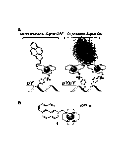
[0026] Figure 1 is (A) a schematic representation of a compound of the
Formula la
demonstrating binding to one or two phosphorylation sites; and (B) is the
chemical structure
of the compound referred to in Figure 1A;
[0027] Figure 2 is (A) an emission spectrum resulting from contacting
a binding
solution in one embodiment of the disclosure with proximally phosphorylated
sites; (B) a
graph showing the fluorescence emission factor versus log peptide
concentration
demonstrating detection of proximally phosphorylated sites; and (C) a
fluorescence image
of a binding solution in one embodiment of the disclosure detecting proximally
phosphorylated sites;
[0028] Figure 3 is (A) a graph demonstrating the fluorescent response of a
binding
solution in one embodiment of the disclosure to the target proximally
phosphorylated and
off-target distally phosphorylated and non-phosphorylated proteins in HEPES
buffer; and
(B) stained polyacrylamide gels;
[0029] Figure 4 is emission spectra of a binding solution in one
embodiment of the
disclosure for (A) a pY peptide and (B) buffer;
[0030] Figure 5 is emission spectra of a binding solution in one
embodiment of the
disclosure containing 220 JIM of compound of the Formula la titrated with 0.2
to 440 pM (A)
pYpY peptide, (B) buffer and (C) pY peptide;
[0031] Figure 6 is pYpY titration data points fit in Origin software
using Hill equation;
[0032] Figure 7 shows gels stained with (A) Coomassie Blue, (B) Pro-Q
Diamond
and (C) a binding solution in one embodiment of the disclosure. Lanes 1-5
correspond to
BSA, a casein-D (D = dephosphorylated), p casein, a casein and Stat5,
respectively;
[0033] Figure 8 shows titration of a binding solution in one
embodiment of the
disclosure with protein from 60 nM to 51.IM;
[0034] Figure 9 shows a-casein stained with a binding solution in one
embodiment of
the disclosure on a polyacrylamide gel. Amount of protein loaded is labeled in
p,g;
[0035] Figure 10 shows titration of a binding solution in one
embodiment of the
disclosure with pY and pYpY peptides from 60 nM to 40 p.M;
- 7 -
CA 02932844 2016-06-06
WO 2015/089639 PCT/CA2014/000901
[0036] Figure 11 shows titration of a binding solution in one
embodiment of the
disclosure with varying concentrations of PPi;
[0037] Figure 12 shows titration of a binding solution in one
embodiment of the
disclosure with varying concentrations of small phosphorylated molecules (30
nM to 80
M);
[0038] Figure 13 shows titration of a binding solution I (right) and
la (left) in one
embodiment of the disclosure with varying concentrations of pY and pYpY
peptides of 40
11M;
[0039] Figure 14 shows titration of a binding solution in one
embodiment of the
disclosure with varying concentrations of pY and pYpY peptides in aqueous
buffers of
variable composition;
[0040] Figure 15 shows titration of a binding solution in one
embodiment of the
disclosure with varying concentrations of proximally phosphorylated target
peptides and
mono-phosphorylated or non-phosphorylated off-target peptides in aqueous
buffers of
variable composition;
[0041] Figure 16 shows titration of a binding solution in one
embodiment of the
disclosure with varying concentrations of proximally phosphorylated target
peptides and
mono-phosphorylated off-target peptides;
[0042] Figure 17 shows titration of a binding solution in one
embodiment of the
disclosure with varying concentrations of proximally phosphorylated target
peptides and
mono-phosphorylated off-target peptides;
[0043] Figure 18 shows titration of a binding solution in one
embodiment of the
disclosure with varying concentrations of pYpY and pY peptides;
[0044] Figure 19 shows (top panel) fluorescence emission spectra of a
binding
solution in one embodiment of the disclosure without peptides or in the
presence of pY and
pYpY peptides and (bottom panel) same spectra represented as ratios;
[0045] Figure 20 shows titration of a binding solution in one
embodiment of the
disclosure with varying concentrations of pYpY and pY peptides; fluorescence
emission
- 8 -
CA 02932844 2016-06-06
WO 2015/089639 PCT/CA2014/000901
was acquired using a fluorescence scan with 2 nm steps from 410-430 nm or by
measuring
fluorescence intensity at 420 nm using 20 nm bandwidth;
[0046] Figure 21 shows titration of a binding solution in one
embodiment of the
disclosure with varying concentrations of pYpY and pY peptides using variable
time-
resolved settings;
[0047] Figure 22 shows titration of a binding solution in one
embodiment of the
disclosure with varying concentrations of pYpY and pY peptides using (left
panel) variable
integration time and (right panel) variable bandwidth for acquisition of
fluorescence
emission;
[0048] Figure 23 shows fluorescence emission spectra of the monomer and
excimer
regions of a binding solution in one embodiment of the disclosure with varying
concentrations of pYpY and pY peptides;
[0049] Figure 24 shows titration of a binding solution in one
embodiment of the
disclosure with varying concentrations of pYpY and pY peptides;
[0050] Figure 25 shows titration of a binding solution in one embodiment of
the
disclosure with varying concentrations of small phospho-anions, calculated
using (left)
fluorescence enhancement factor and (right) A fluorescence intensity formula;
[0051] Figure 26 shows titration of a binding solution in one
embodiment of the
disclosure with varying concentrations of small phospho-anions, calculated
using (left)
fluorescence enhancement factor and (right) A fluorescence intensity formula;
[0052] Figure 27 shows titration of a binding solution in one
embodiment of the
disclosure with varying concentrations of the proximally phosphorylated
positive control
proteins (alpha casein, beta casein, D-alpha-casein), and negative control
proteins (distally
phosphorylated ovalbumin, and non-phosphorylated lysozyme and BSA);
[0053] Figure 28 shows titration of a binding solution in one embodiment of
the
disclosure with varying concentrations of the proximally phosphorylated
positive control
proteins (alpha casein, beta casein, D-alpha-casein), and negative control
proteins (distally
phosphorylated ovalbumin, and non-phosphorylated lysozyme and BSA);
- 9 -
CA 02932844 2016-06-06
WO 2015/089639 PCT/CA2014/000901
[0054] Figure 29 shows titration of a binding solution in one
embodiment of the
disclosure with varying concentrations of the proximally phosphorylated
positive control
proteins (alpha casein, beta casein, D-alpha-casein), and negative control
proteins (distally
phosphorylated ovalbumin, and non-phosphorylated lysozyme and BSA);
[0055] Figure 30 shows titration of a binding solution in one embodiment of
the
disclosure with varying concentrations of the proximally phosphorylated
positive control
proteins (alpha casein, beta casein, D-alpha-casein), and negative control
proteins (distally
phosphorylated ovalbumin, and non-phosphorylated lysozyme and BSA);
[0056] Figure 31 shows a bar graph of fluorescence intensity of the
excimer region of
a binding solution in one embodiment of the disclosure with 10 M of the
proximally
phosphorylated positive control proteins (alpha casein, beta casein, D-alpha-
casein), and
negative control proteins (distally phosphorylated ovalbumin, and non-
phosphorylated
lysozyme and BSA) with and without treatment with phosphatase;
[0057] Figure 32 shows fluorescent image of a polyacrylamide gel
stained with a
binding solution in one embodiment of the disclosure, and the corresponding
lane analysis.
Lanes 1, 2, 3, and 4 contain 1.0, 0.5, 0.25 and 0.125 4 of each of the four
proteins.
Proteins included in each lane are (top to bottom) BSA, ovalbumin, 13-casein
and lysozyme.
Proximally phosphorylated protein I3-casein results in the strongest signal;
[0058] Figure 33 shows fluorescent image of a single polyacrylamide
gel sequentially
stained with a binding solution in one embodiment of the disclosure, Pro-Q
Diamond and
SYPRO Ruby stains. Each protein was loaded in the amount of 14;
[0059] Figure 34 shows fluorescent image of a single polyacrylamide
gel sequentially
stained with a binding solution in one embodiment of the disclosure, Pro-0
Diamond and
SYPRO Ruby stains. The amount of each protein loaded per lane is labeled in
lig;
[0060] Figure 35 shows fluorescent image of a single polyacrylamide gel
sequentially
stained with a binding solution in one embodiment of the disclosure, Pro-Q
Diamond and
SYPRO Ruby stains. The amount of each protein loaded per lane is 0.5 4 of
protein. Each
stain is color-coded and merged lane provides a color-map of the
phosphorylation status of
- 10-
CA 02932844 2016-06-06
WO 2015/089639 PCT/CA2014/000901
a protein (i.e. proximally phosphorylated, distally phosphorylated and non-
phosphorylated
appear in different colors);
[0061] Figure 36 shows fluorescent image of a single polyacrylamide
gel sequentially
stained with a binding solution in one embodiment of the disclosure, Pro-Q
Diamond and
SYPRO Ruby stains, and their lane analyses. The amount of each protein loaded
per lane
is 1, 0.5, 0.25 and 0.12514 (left to right). Proteins included in each lane
are (top to bottom)
BSA, ovalbumin, 13-casein and lysozyme;
[0062] Figure 37 shows fluorescent image of polyacrylamide gels
stained with a
binding solution in one embodiment of the disclosure and de-stained for
variable time
intervals. The amount of each protein loaded per lane is 1, 0.5, 0.25 and
0.125 g (left to
right). Proteins included in each lane are (top to bottom) BSA, ovalbumin, p-
casein and
lysozyme;
[0063] Figure 38 shows lane analysis of the fluorescent image of
polyacrylamide
gels stained with a binding solution in one embodiment of the disclosure and
de-stained for
variable time intervals. The analysis was performed on the lane containing 1
vig of protein;
[0064] Figure 39 shows lane analysis of the fluorescent image of
polyacrylamide
gels sequentially stained with a binding solution in one embodiment of the
disclosure and
SYPRO Ruby stain. The analysis was performed on the lane containing 1 g of
protein;
[0065] Figure 40 shows fluorescent image of polyacrylamide gels
stained with a
binding solution in one embodiment of the disclosure and de-stained for
variable time
intervals. The amount of each protein loaded per lane is 1, 0.5, 0.25 and
0.125 jig (left to
right). Proteins included in each lane are (top to bottom) BSA, ovalbumin, I3-
casein and
lysozyme;
[0066] Figure 41 shows lane analysis of the fluorescent image of
polyacrylamide
gels stained with a binding solution in one embodiment of the disclosure and
de-stained for
variable time intervals. The analysis was performed on the lane containing 1
jig of protein;
[0067] Figure 42 shows fluorescent image of polyacrylamide gels
stained with a
binding solution in one embodiment of the disclosure and de-stained for
variable time
intervals. The amount of each protein loaded per lane is 1, 0.5, 0.25 and
0.125 g (left to
-11 -
CA 02932844 2016-06-06
WO 2015/089639 PCT/CA2014/000901
right). Proteins included in each lane are (top to bottom) BSA, ovalbumin, (3-
casein and
lysozyme;
[0068] Figure 43 shows lane analysis of the fluorescent image of
polyacrylamide
gels stained with a binding solution in one embodiment of the disclosure and
de-stained for
variable time intervals. The analysis was performed on the lane containing 1
g of protein;
[0069] Figure 44 shows lane analysis of the fluorescent image of
polyacrylamide
gels sequentially stained with a binding solution in one embodiment of the
disclosure and
SYPRO Ruby stain. The analysis was performed on the lane containing 1 pg of
protein;
[0070] Figure 45 shows fluorescent images of polyacrylamide gels
stained with a
binding solution in one embodiment of the disclosure and de-stained for
variable time
intervals. The amount of each protein loaded per lane is 1, 0.5, 0.25 and
0.125 lig (left to
right). Proteins included in each lane are (top to bottom) BSA, ovalbumin, fl-
casein and
lysozyme;
[0071] Figure 46 shows lane analysis of the fluorescent image of
polyacrylamide
gels stained with a binding solution in one embodiment of the disclosure, and
de-stained for
variable time intervals. The analysis was performed on the lane containing 1
pig of protein;
[0072] Figure 47 shows titration of a compound of the Formula I with
variable
concentration of a metal ion salt at different pH;
[0073] Figure 48 shows titration of a compound of the Formula I with
variable
concentration of a metal ion salt at different pH;
[0074] Figure 49 shows fluorescent images of the same polyacrylamide
gel
sequentially stained with a binding solution in one embodiment of the
disclosure, Pro-Q
Diamond and SYPRO Ruby stains. Each of the 7 lanes contains 40 [tg of cell
lysate
obtained from different cell lines;
[0075] Figure 50 shows fluorescent images of a PVDF membrane stained with a
binding solution in one embodiment of the disclosure. The amount of proteins
loaded per
lane is shown in ng;
- 12-
CA 02932844 2016-06-06
WO 2015/089639 PCT/CA2014/000901
[0076] Figure 51 shows fluorescent images of a single PVDF membrane
sequentially
stained with a binding solution in one embodiment of the disclosure, Pro-Q
Diamond and
SYPRO Ruby. The amount of proteins loaded per lane is 1, 0.5, 0.25, 0.125 and
0.063 4;
[0077] Figure 52 shows fluorescent image of a single PVDF membrane
sequentially
stained with a binding solution in one embodiment of the disclosure, Pro-Q
Diamond and
SYPRO Ruby stains. Each stain is color-coded and merged lane provides a color-
map of
the phosphorylation status of a protein (i.e. proximally phosphorylated,
distally
phosphorylated and non-phosphorylated appear in different colors);
[0078] Figure 53 shows fluorescent images of the same PVDF membrane
sequentially stained with a binding solution in one embodiment of the
disclosure, Pro-Q
Diamond and SYPRO Ruby stains. Each of the 7 lanes contains 40 4 of cell
lysate
obtained from different cell lines;
[0079] Figure 54 shows fluorescent images of the western blot analysis
for 13-actin
protein on the PVDF membrane which was first sequentially stained with a
binding solution
in one embodiment of the disclosure, Pro-Q Diamond and SYPRO Ruby stains. Each
of the
7 lanes contains 40 4 of cell lysate obtained from different cell lines;
[0080] Figure 55 shows fluorescent images acquired using fluorescence
microscopy
of fixed cells which were either treated or not treated with a binding
solution in one
embodiment of the disclosure;
[0081] Figure 56 shows fluorescent images acquired using fluorescence
microscopy
of fixed cells which were treated with a binding solution in one embodiment of
the
disclosure, it's non-metallated derivative and a derivative lacking metal-
coordinating moiety;
[0082] Figure 57 shows fluorescent images acquired using fluorescence
microscopy
of fixed cells which were treated with a binding solution in one embodiment of
the
disclosure, with or without prior permeabilization;
[0083] Figure 58 shows fluorescent images acquired using fluorescence
microscopy
of fixed cells which were treated with a binding solution in one embodiment of
the
disclosure, with or without pre-treatment with RNase enzyme;
-13-
CA 02932844 2016-06-06
WO 2015/089639 PCT/CA2014/000901
[0084] Figure 59 shows fluorescent images acquired using fluorescence
microscopy
of fixed cells which were co-stained with a binding solution in one embodiment
of the
disclosure and a nuclear stain propidium iodide;
[0085] Figure 60 shows fluorescent images acquired using fluorescence
microscopy
of fixed (a) MRC-9 and (b) A549 cells which were treated with a binding
solution in one
embodiment of the disclosure, with or without pre-treatment with Interleukin-
6;
[0086] Figure 61 shows a bar graph which demonstrates quantification
of the change
in fluorescence intensity of fixed cells which were treated with a binding
solution in one
embodiment of the disclosure, with or without pre-treatment with Interleukin-
6;
[0087] Figure 62 shows fluorescent images acquired using fluorescence
microscopy
of fixed cells which were treated with a binding solution in one embodiment of
the
disclosure, with or without pre-treatment with staurosporine;
[0088] Figure 63 shows time-dependent cytotoxicity of a binding
solution in one
embodiment of the disclosure;
[0089] Figure 64 shows fluorescent images acquired using fluorescence
microscopy
of live cells, which were treated with a binding solution in one embodiment of
the disclosure
for variable time intervals;
[0090] Figure 65 shows images of live cells acquired using bright
field microscopy
before and after excitation with laser, which were treated with a binding
solution in one
embodiment of the disclosure; and
[0091] Figure 66 is a graph demonstrating selective detection of
proximally
phosphorylated proteins using a binding solution of the disclosure in a solid
support assay.
DESCRIPTION OF VARIOUS EMBODIMENTS
(I) DEFINITIONS
[0092] The term "excimer forming fluorophore" as used herein refers to a
moiety
which upon interacting, overlapping or otherwise associating with a second
excimer forming
fluorophore results in an increase in fluorescence emission at a longer
wavelength and a
decrease of monomer emission at a shorter wavelength as compared to the
unbound
fluorophore.
- 14-
CA 02932844 2016-06-06
WO 2015/089639 PCT/CA2014/000901
[0093] The term "Calkyl" as used herein means straight or branched
chain,
saturated alkyl groups containing from one to n carbon atoms and includes
(depending on
the identity of n) methyl, ethyl, propyl, isopropyl, n-butyl, s-butyl,
isobutyl, t-butyl, 2,2-
dimethylbutyl, n-pentyl, 2-methylpentyl, 3-methylpentyl, 4-methylpentyl, n-
hexyl and the
like, where the variable n is an integer representing the largest number of
carbon atoms in
the alkyl radical.
[0094] The term "C2,alkenyl" as used herein means straight or branched
chain,
unsaturated alkyl groups containing from two to n carbon atoms and one to
three double
bonds, and includes (depending on the identity of n) vinyl, allyl, 2-
methylprop-1-enyl, but-1-
enyl, but-2-enyl, but-3-enyl, 2-methylbut-1-enyl, 2-methylpent-1-enyl, 4-
methylpent-1-enyl,
4-methylpent-2-enyl, 2-methylpent-2-enyl, 4-methylpenta-1,3-dienyl, hexen-1-y1
and the
like, where the variable n is an integer representing the largest number of
carbon atoms in
the alkenyl radical.
[0095] The term "C2_nalkynyl" as used herein means straight or
branched chain,
unsaturated alkyl groups containing from two to n carbon atoms and one to
three triple
bonds, and includes (depending on the identity of n) ethynyl, propynyl, 2-
methylprop-1-ynyl,
but-1-ynyl, but-2-ynyl, but-3-ynyl, 3-methylbut-1-ynyl, 2-methylpent-1-ynyl, 4-
methylpent-1-
ynyl, 4-methylpent-2-ynyl, 4-methylpent-2-ynyl, penta-1,3-diynyl, hexyn-1-y1
and the like,
where the variable n is an integer representing the largest number of carbon
atoms in the
.. alkynyl radical.
[0096] The term "cycloalkyl" as used herein refers to an aliphatic
ring system having
3 to "n" carbon atoms including (depending on the identity of n), but not
limited to,
cyclopropyl, cyclopentyl, cyclohexyl, and the like, where the variable n is an
integer
representing the largest number of carbon atoms in the cycloalkyl radical.
[0097] The term "bicyclic or polycyclic aryl moiety" as used herein refers
to a bicyclic
or polycyclic conjugated substituted or unsubstituted carbocyclic ring system
having two or
more rings including, but not limited to, naphthyl, tetrahydronaphthyl,
phenanthrenyl,
biphenylenyl, indanyl, indenyl, pyrenyl, peryleneyl, tetraceneyl and the like.
Non-
conjugated or unsaturated rings may also be fused to the conjugated ring
system.
-15-
CA 02932844 2016-06-06
WO 2015/089639 PCT/CA2014/000901
[0098] The term "bicyclic or polycyclic heteroaryl moiety" as used
herein refers to a
bicyclic or polycyclic conjugated substituted or unsubstituted carbocyclic
ring system having
two or more rings containing, of which one or more, for example 1-8, suitably
1-6, more
suitably 1-5, and more suitably 1-4, of the atoms are a heteromoiety selected
from 0, S,
NH, NC1_6alkyl, and C(=0), with the remaining atoms being C or CH, said ring
system.
Examples of heteroaryl moieties, include, but are not limited to substituted
carbazoles (9-
phenyl-9H-carbazole), 1H-benzo[de]isoquinoline-1,3(2H)-dione, anthra[2,1,9-
def:6,5,10-
d'eT]diisoquinoline-1,3,8,10(2H,9H)-tetraone and the like. Non-conjugated or
unsaturated
rings may also be fused to the conjugated ring system.
[0099] The term "C6..naryl" as used herein means a monocyclic, bicyclic or
tricyclic
carbocyclic ring system containing from 6 to n carbon atoms and at least one
aromatic ring
and includes, depending on the identity of n, phenyl, naphthyl, anthracenyl,
1,2-
dihydronaphthyl, 1,2,3,4-tetrahydronaphthyl, fluorenyl, indanyl, indenyl and
the like, where
the variable n is an integer representing the largest number of carbon atoms
in the aryl
radical.
[00100] The term "heteroaryl" as used herein means a monocyclic,
bicyclic or tricyclic
ring system containing from 5 to 14 atoms of which one or more, for example 1-
8, suitably,
1-6, more suitably 1-5, and more suitably 1-4, of the atoms are a heteromoiety
selected
from 0, S, NH and NC1_6alkyl, with the remaining atoms being C or CH, said
ring system
containing at least one aromatic ring. Examples of heteroaryl groups, include,
but are not
limited to thienyl, imidazolyl, pyridyl, oxazolyl, indolyl, furanyl,
benzothienyl, benzofuranyl
and the like.
[00101] The suffix "ene" added on to any of the above groups means that
the group is
divalent, i.e. inserted between two other groups. When the group is a ring
system, the two
other groups may be located at any location on the ring system, including at
adjacent and
non-adjacent nodes.
[00102] The term "oxo-substituted" as used herein refers to a carbonyl
group (0=0)
generally replacing a CH2 moiety.
[00103] The term "halo" as used herein means halogen and includes
chlorine,
bromine, iodine and fluorine.
-16-
CA 02932844 2016-06-06
WO 2015/089639 PCT/CA2014/000901
[00104] The term "linker moiety" as used herein refers to a carbon-
based moiety
which connects the excimer forming fluorophore with the metal ion coordinating
moiety.
The linker moiety may be straight-chained, branched, or cyclic, or a
combination of all
three, and connects one or more metal ion coordinating moieties with the
excimer forming
fluorphore. The linker moiety optionally contains carbonyl, nitrogen and/or
other
heteroatom functionalities.
[00105] The term "metal ion coordinating moiety" as used herein refers
to a moiety
which coordinates with a metal ion, for example, a transition metal ion, a
lanthanide metal
ion or a post-transition metal ion and comprises one or more cyclic or acyclic
organic
ligands which can coordinate to a metal ion center, for example, amino, amido,
carboxyl or
hydroxyl groups.
[00106] The term "metal ion" as used herein refers to the positively
charged forms or
cations of metals.
[00107] The term "post-transition metal ion" as used herein refers to
metal ions in
Groups IIIB, IVB, VB, and VIB in the periodic table of the elements, and
includes, but is not
limited to, aluminum, gallium, germanium, indium, tin, antimony etc.
[00108] The term "lanthanide metal ion" as used herein refers to the
metal ions with
the atomic number from 57 to 71 in the periodic table of the elements, and
includes, but is
not limited to, terbium, europium, ytterbium etc.
[00109] The term "binding solution" as used herein refers to an aqueous
solution
containing a compound of the Formula (I) and a suitable metal ion, which
optionally forms
compounds of the Formula la in solution.
[00110] The term carboxyl as used herein refers to a group of the
formula COOH or
COO-.
[00111] The term "hydroxyl" as used herein refers to a group of the formula
OH.
[00112] The term "amino" as used herein refers to an unsubstituted
amino radical or a
primary, secondary or tertiary amino moiety substituted by alkyl or aryl
groups. The term
amino also includes unsaturated amino groups such as imines, or aromatic amine
such as
pyridine.
-17-
CA 02932844 2016-06-06
WO 2015/089639 PCT/CA2014/000901
[00113] The term
"polypeptide" as used herein means a polymer of amino acids and
does not refer to any particular length of polymer. Such term also includes
genetically
expressed peptides, post-translationally modified polypeptides or proteins
(e.g.,
glycosylated, acetylated, phosphorylated, etc.), synthetic peptides, such as
short synthetic
peptides, crude synthetic peptides, purified synthetic peptides. The term
"polypeptide" also
encompasses the term "protein".
[00114] The term
"phosphorylation" as used herein refers to the addition of a
phosphate group to a biological or organic molecule including macromolecules,
including,
but not limited to proteins, polypeptides, including all amino acids, DNA,
RNA, sugars etc.
[00115] The term
"proximal" as used herein refers to the spacing between
phosphorylation sites on polypeptides, nucleic acids or small phospho-anions
such as
pyrophosphate, such that the sites are sufficiently close to allow a bound
excimer
containing fluorophore compound at one site to interact, overlap or otherwise
associate
with a bound excimer-forming fluorophore on the other site. In an embodiment,
the term
refers to a specific number of amino acids, such as between 1-10 amino acids,
optionally 1-
4 amino acids between the two sites. The term also refers to the spatial
proximity of
phosphorylation sites after three-dimensional folding of the polypeptide or
other
biomolecule. For example, phosphorylation sites that are significantly distant
from each
other along a polypeptide chain become spatially proximal to each other upon
three-
dimensional folding and may be between 2 and 100 Angstroms, optionally 3 and
50
Angstroms or suitably 5 and 30 Angstroms.
[00116] The term
"control" as used herein refers to a sample that has a particular
level of proximal phosphorylation. An unphosphorylated, distally
phosphorylated or
monophosphorylated control contains no proximal phosphorylation and would be a
negative control. Alternatively, a control may contain a known amount of
proximal
phosphorylation and would be a positive control. The control can also be a
predetermined
standard.
[00117] The term
"subject" as used herein refers to any member of the animal
kingdom. In one embodiment, the subject is a mammal, such as a human.
-18-
CA 02932844 2016-06-06
WO 2015/089639 PCT/CA2014/000901
(II) EXCIMER FORMING COMPOUNDS
[00118] The present application describes a turn-on dual emission
fluorescent sensor
which selectively detects proximally phosphorylated sites including those
found on proteins,
pyrophosphates and RNA, in aqueous solutions, polyacrylamide gels, PVDF
membranes,
immobilized on solid supports (e.g. polymers, antibody), fixed cells and live
cells.
[00119] In one embodiment, the turn-on fluorescent sensor is an excimer
forming
compound, in which the sensor is comprised of an excimer forming fluorophore.
When two
or more of the excimer forming fluorophores overlap, the fluorescence
intensity of the
fluorophores decreases at a shorter wavelength and fluorescence intensity
increases at a
longer wavelength, indicating the presence of at least two spatially proximal
sites of
phosphorylation.
[00120] Accordingly, in one embodiment, the disclosure provides a
compound of the
Formula I
w __________________________________ V¨k 1
n (I)
wherein,
W is an excimer forming fluorophore;
V is a linker moiety;
Y is a metal ion chelate moiety; and
n is 1,2 or 3.
[00121] In another embodiment, the disclosure provides an excimer forming
compound of the Formula la
\N¨v¨EY I
n (la)
wherein,
W is an excimer forming fluorophore;
V is a linker moiety;
Y is a metal ion chelate moiety containing a metal ion; and
n is 1,2 or 3.
=
-19-
CA 02932844 2016-06-06
WO 2015/089639 PCT/CA2014/000901
[00122] In one embodiment, the present disclosure also includes a
composition
comprising a compound of the Formula (I) and a suitable metal ion.
[00123] In a further embodiment, the present disclosure also includes
an aqueous
composition comprising a compound of the Formula (I) and a suitable metal ion.
[00124] In one embodiment, the present disclosure includes a binding
solution,
comprising:
(a) an excimer-forming Compound of the Formula I, and
(b) a suitable metal ion, and
optionally, other additives such as salts, buffers or other organic
components.
[00125] In another embodiment, the present disclosure includes a binding
solution la,
comprising:
(a) an excimer-forming compound of the Formula la, and
optionally, other additives such as salts, buffers or other organic
components.
[00126] In one embodiment, the excimer forming fluorophore is an
optionally
.. substituted bicyclic or polycyclic aryl or heteroaryl moiety, wherein the
optional substituents
are selected from halo, C1_20-alkyl, 02..20-alkenyl, C2_20-alkynyl, C6_14-
aryl, and C5_14-
heteroaryl.
[00127] In another embodiment, the excimer forming fluorophore is
optionally
substituted C10_40-aryl or optionally substituted C9_40-heteroaryl, wherein
the optional
substituents are selected from halo, C1_20-alkyl, C2_20-alkenyl, C2_20-
alkynyl, C6_14-aryl, and
C5_14-heteroaryl. In another embodiment, the excimer forming fluorophore is
optionally
substituted C10-20-aryl or optionally substituted C9.20-heteroaryl, wherein
the optional
substituents are selected from halo, 01_20-alkyl, C2_20-alkenyl, C2_20-
alkynyl, 06_14-aryl, and
C5_14-heteroaryl
[00128] In one embodiment of the disclosure, the excimer forming
fluorophore is an
optionally substituted moiety shown below with any suitable point of
attachment
- 20 -
CA 02932844 2016-06-06
WO 2015/089639 PCT/CA2014/000901
ccii
0*
H
0 N 0
O.
N 0101
0 N 0
H
H
0 N 0
4101
SO" 11100110
$710010 40 0 0
al
, 0 N
H 0 el , or
1
I
wherein the optional substituents are selected from
halo, carboxy, hydroxyl, 01_20-alkyl, C2_20-alkenyl, C2_20-alkynyl,
C3_20cycloalkyl, C1_20alkoxy, -
-21-
CA 02932844 2016-06-06
WO 2015/089639 PCT/CA2014/000901
NR'R" C6_14-aryl, and C5_14-heteroaryl, wherein R' and R" are simultaneously
or
independently H or C1_6alkyl.
[00129] In another embodiment, the excimer forming fluorophore is
optionally
substituted or unsubstituted
, or
[00130] In another embodiment, the excimer forming fluorophore is
optionally
substituted or unsubstituted
[00131] In another embodiment of the disclosure, the linker moiety is
i) C1_40-alkylene, C2_40-alkenylene, C2_40-alkynylene, or C3_20-cycloalkyl,
each of which
is optionally oxo-substituted (=0) 1-6 times, optionally 1-3 times, and in
which 1-3
carbon atoms are optionally replaced with a heteroatom selected from N, 0, S
or Si;
ii) C6_10-aryl, or C5-10-heteroaryl, each of which is optionally substituted
with 1-4 R
groups, wherein
R is simultaneously or independently C1_20-alkylene, C2_20-alkenylene or C2-20-
alkynylene, each of which is optionally oxo-substituted (=0) between 1-3
times, and in which 1-3 carbon atoms are optionally replaced with a
heteroatom selected from N, 0, S or Si.
- 22 -
CA 02932844 2016-06-06
WO 2015/089639 PCT/CA2014/000901
[00132] In one embodiment, the linker moiety has the following
structure with any
suitable point of attachment
¨Rm
or
wherein,
R is as defined above;
m is 1, 2,3 or 4,
and wherein 1-4 of the carbon atoms in the phenyl or naphthyl rings are
optionally replaced
with nitrogen atoms.
[00133] In a further embodiment, the linker moiety is C1_20-alkylene,
C2_20-alkenylene,
C2_20-alkynylene, or C3_10-cyclo each of which is optionally oxo-substituted
(=0) 1-3 times,
and in which 1-3 carbon atoms are optionally replaced with a heteroatom
selected from N,
0, S or Si.
[00134] In one embodiment, linker moiety is C1_10-alkylene, which is
optionally oxo-
substituted (=0) 1-3 times, and in which 1-3 carbon atoms are optionally
replaced with a
heteroatom selected from N, 0, S or Si. In another embodiment, the linker
moiety is C1-6-
alkylene, which is optionally oxo-substituted (=0) 1-3 times, and in which 1-3
carbon atoms
are optionally replaced with a heteroatom selected from N, 0, S or Si.
[00135] In another embodiment, the linker moiety is methylene,
butylene,
401 )ssõ, N
0 0 0 0
)11.1Lrrif )()Lcss! '4)Ltsss%
0
,
H
-23-
CA 02932844 2016-06-06
WO 2015/089639 PCT/CA2014/000901
0 0 0
N.
N2t1 '12-
H Nor H .
[00136] In one embodiment, the linker moiety is methylene.
[00137] In another embodiment, the linker moiety is
j)P
sj)p
=P')p
P
I
) )p )
P P
1 , )
\ p ( \
r
)p
, , ,
- 24 -
CA 02932844 2016-06-06
WO 2015/089639 PCT/CA2014/000901
( \
)
1 -X
X<
,' p
=Prx .
j'S ________________________________________________________________________
0
( )p < 1
) X
I
,
ssl* ) 0
P
j&H)2L
P 5s IC(X-k.1-i'Xrett'
0 0
12(H'N X
P X '('*)',55
, or 0 P
, wherein X is a
heteroatom selected from 0, S, Si, or NH and p is an integer from 1-20,
wherein the
alkylene groups are further optionally oxo-substituted (=0) 1-3 times, and in
which 1-3
carbon atoms are further optionally replaced with a heteroatom selected from
N, 0, S or Si,
and wherein 1-4 of the carbon atoms in the phenyl or naphthyl rings are
optionally replaced
with nitrogen atoms, and where .2L indicates the attachment to W or Y.
[00138] In another
embodiment of the disclosure, the metal ion coordinating moiety is
a multi-dentate moiety comprising amino, carboxyl, hydroxyl, amide, or ether
groups, or
other heteroatom containing moieties, wherein the heteroatom is 0, S, or N.
[00139] In one
embodiment, the metal ion coordinating moiety is a tri- or tetra-dentate
amino group. In one embodiment, the tetra-dentate amino group is optionally
substituted
with any suitable point of attachment
- 25 -
CA 02932844 2016-06-06
WO 2015/089639 PCT/CA2014/000901
/\ /\ /\
NH HN.. ______________ NH
HN ( \N NH ______ FIN __ ) N __
HN
)
HN../
N N
\ _________ / , '' \ ____ / \ __ /
$
0 oOH 0
HO> K \/ \N./ HO
) \ /
N N
___________ N N __ )
\N ./
N ,IleN\ /\
\ ________________ / \
OOH OOH
,
N
H
N
HN HN ca2-N
_____________ /
[00140] In another embodiment, the metal ion coordinating moiety is
o
0 OH
'OH
N--,
OH !
-26-
CA 02932844 2016-06-06
WO 2015/089639 PCT/CA2014/000901
OH
o ( OH
Xx_x
0
OH
HO 0 (wherein X is 0 or N),
NH
CNF1Nh
HN
N/ 11NH HN
HN
\NH
or H .
[00141] In another embodiment, the metal ion coordinating moiety is
OH 0
0 (OH
0
\-0
0
OH
_______________________ = HO 0
-27-
CA 02932844 2016-06-06
WO 2015/089639 PCT/CA2014/000901
[00142] In one embodiment of the disclosure, the excimer forming compound
of the
Formula I is
HO 0 compound 2
OH
C) 0TN ,-..1i3OH
N 0 0
compound 1 ISI el
HO .'0
*IL 7H
itiPwqr () HN,i
H
HO
compound 3
0 compound 4
rIV'MN
OH HN--N)
r) HN,)
\N or
N'NN)
H
(31.) O.
OH
,...._ compound 5 compound 6
HN A compound 7
() HN.) HN-Th
r.) HN.) H
1-11\1N)
N.../N
H N.,...,N) L.i HN
H 1\1,7i
41101
Ille
- 28 -
CA 02932844 2016-06-06
WO 2015/089639 PCT/CA2014/000901
compound 8
HO .O
OH %-,,,
04') 0-Th
N r 0 0 0 0
HO ''..0 compound 9
0 NH
WOO H
01.1 N'' NI)
.0 LI HN
HN.)
,SO
,
compound 10
eel 0 compound 11
001 N
r.) HN1 0 H
N
HNNN--j / FiN...)NH
Nõ....N
H
compound 12
compound 13
( NH HN)
4100 (N HN...1
.10 LN HN
NH MI)
C) el* C.)
compound 14
yi- N compound 15
00 1 N N
410411) L N
, N I
I
- 29 -
CA 02932844 2016-06-06
WO 2015/089639 PCT/CA2014/000901
compound 16
HN
JN
H 1101
1.1401 0
04111
NH r)
NH
,or
(,NH compound 17
CNN
1410 N
HN'Th
011:1 HN
[00143] In another embodiment, the excimer forming compounds of the
Formula I
further comprise a metal ion coordinated to the metal ion coordinating moiety
to optionally
5 form compounds of the Formula la in solution. In one embodiment, the
metal ion is any
suitable metal ion which coordinates, or otherwise interacts (i.e. through
hydrogen bonding,
ionic bonding, dipole interactions, metal-ligand interactions etc.) with the
metal ion
coordinating moiety, and which simultaneously binds to a phosphorylated moiety
(on a
protein, peptide, enzyme, nucleic acid etc.) or pyrophosphate. In one
embodiment, the
10 metal ion is a transition metal ion, a lanthanide metal ion or post-
transition metal ion. In
one embodiment, the transition metal ion is Zn(II), Cu(ll), Mn(II), Ni(II),
Fe(ll), Cd(II) AI(III),
Fe(III). In one embodiment the post-transition metal ion is AI(III) or
Ga(III). In one
embodiment, the lanthanide metal ion is Tb(III), Eu(III), Y6(111). In one
embodiment, the
suitable metal ions are in the form of salts, such as any metal ion salt, for
example, zinc(II)
15 trifluoromethanesulfonate, gallium(III) chloride or terbium(III)
chloride.
- 30 -
CA 02932844 2016-06-06
WO 2015/089639 PCT/CA2014/000901
[00144] In one embodiment, the compounds of the Formula la are formed
in situ, for
example by preparing a binding solution of a compound of the Formula I with a
suitable
metal ion to form in solution compounds of the Formula la. In one embodiment,
the binding
solution comprises (i) a compound of the Formula I; and (ii) a suitable metal
ion, for
example in the form of a salt. In one embodiment, the components of the
binding solution
are kept separate until ready for use.
[00145] The present disclosure also includes a kit, comprising:
(i) a compound of the Formula (I);
(ii) a suitable metal ion, for example in the form of a salt; and
(iii) instructions for use.
(III) METHODS
[00146] The excimer forming compounds of the Formula I and la of the
present
disclosure and binding solutions and compositions of the disclosure are useful
for detecting
proximally phosphorylated polypeptide residues, and other proximally
phosphorylated
molecules. Such detection is useful for a variety of applications, including
without limitation,
detecting and quantifying proximally phosphorylated proteins in protein
expression and
purification; assessing activation status of proteins that are activated by
proximal
phosphorylation; monitoring de-phosphorylation rate and progress of proximally
phosphorylated protein sites; comparing the abundance of proximally
phosphorylated
proteins in protein extracts of various cells lines and samples; detecting
diseases in which
abundance of proximal phosphorylation is increased; and detecting
pyrophosphates.
[00147] In one embodiment, excimer formation is accompanied by a
decrease in
monomer-region fluorescence and the extent of excimer formation can be
detected and
quantified by measuring the decrease in monomer fluorescence. Likewise,
excimer
formation is accompanied by an increase in fluorescence at the excimer-forming
region of a
fluorophore and the extent of excimer formation can be detected and quantified
by
measuring the increase in fluorescence. In one embodiment, ratios of the
decrease in
monomer-region fluorescence and the increase in fluorescence at the excimer-
forming
region can be calculated to detect and quantify changes at both regions.
- 31 -
CA 02932844 2016-06-06
WO 2015/089639 PCT/CA2014/000901
[00148] In one embodiment, the methods of the disclosure are performed
by
measuring fluorescence intensity in the excimer and/or monomer regions. In
another
embodiment, analysis of the fluorescence is performed using fluorescence
polarization, as
the tertiary complex between a proximally phosphorylated target and two
excimer units
limits the tumbling rate of the excimer fluorophore and increase fluorescence
polarization
and anisotropy values.
[00149] Accordingly, in one embodiment, the present disclosure provides
a method of
detecting proximal phosphorylation of a molecule, such as a polypeptide (or
protein),
comprising:
(a) contacting a sample of the molecule, such as a polypeptide sample, with a
binding
solution of the disclosure;
(b) detecting a fluorescence signal at a wavelength specific for the excimer
forming
fluorophore of a binding solution of the disclosure;
wherein detection of a signal having a fluorescence intensity greater than a
signal of a
sample containing a distal phosphorylation, monophosphorylation or no
phosphorylation
indicates that the molecule, such as the polypeptide, contains phosphorylation
of at least
two sites proximal to each other.
[00150] In an embodiment, the signal from a sample comprising only a
monophosphorylated site is undetectable or similar to background levels or to
the level of a
sample containing no phosphorylation.
[00151] In one embodiment, the method is performed on a gel as a gel-
based assay
in which the molecule, such as a polypeptide, is separated on polyacrylamide
gels and
detected directly on the gel without need for excising the band(s).
[00152] In another embodiment, the present disclosure provides a method
of
detecting proximal phosphorylation of a polypeptide comprising:
(a) contacting a polypeptide sample with a binding solution of the disclosure;
(b) detecting a fluorescence signal at a wavelength specific for the excimer
forming
fluorophore of a binding solution of the disclosure;
(c) comparing the fluorescence signal of (b) with the fluorescence intensity
of a distally
phosphorylated, monophosphorylated or unphosphorylated control;
- 32 -
CA 02932844 2016-06-06
WO 2015/089639 PCT/CA2014/000901
wherein detection of a signal having a fluorescence intensity greater than the
control
indicates that the polypeptide contains phosphorylation of at least two sites
proximal to
each other. It will be understood that the increase in fluorescence intensity
depends on the
protein concentration in the sample, and the concentration of the excimer
forming
compound in a binding solution of the disclosure, and the number of proximally
phosphorylated sites. For quantification, a calibration curve is generated
based on the
protein of known concentration and compositions which is used to compare the
fluorescence signal from the sample under investigation.
[00153] In one embodiment, the method is performed on a gel as a gel-
based assay
in which polypeptide is separated on polyacrylamide gels and detected directly
on the gel
without need for excising the band(s). In another embodiment, quantification
can also be
performed on the gel by comparison of the signal intensity of the bands.
[00154] In one embodiment, the amino acids that are proximally
phosphorylated are
sufficiently close in a native or denatured state, for example within 1-6,
suitably 1-4, amino
acid residues. In another embodiment, the amino acids that are proximally
phosphorylated
are sufficiently close when the protein is in its 3D conformation.
[00155] In a further embodiment, the present disclosure provides a
method of
quantifying proximal phosphorylation comprising:
(a) contacting a sample with a binding solution of the disclosure;
(b) detecting a fluorescence signal at a wavelength specific for the excimer
forming
fluorophore of a binding solution of the disclosure;
(c) comparing the fluorescence signal of (b) with the fluorescence intensity
of control
samples of known quantities of proximal phosphorylation;
wherein detection of a signal having a fluorescence intensity similar to one
of the control
samples indicates the amount of proximal phosphorylation in the sample. In one
embodiment, the relative number of proximally phosphorylated sites can be
monitored over
time to determine, for example, whether in the process of a reaction (e.g.
enzymatic or
chemical phosphorylation or dephosphorylation of a peptide) the proximally
phosphorylated
sites increase or decrease over time.
- 33 -
CA 02932844 2016-06-06
WO 2015/089639 PCT/CA2014/000901
[00156] In an embodiment, the polypeptide sample is a protein extract
from a
bacterial, yeast, insect or mammalian cell line including human cell lines.
[00157] In another embodiment, the polypeptide sample is from a
subject suffering
from a disease associated with increased proximal phosphorylation of proteins,
such as
cancer, Alzheimer's etc. In another embodiment, the polypeptide sample is a
sample
synthesized using a peptide synthesizer or is a sample from a genetically
modified protein
expressed on a vector.
[00158] In another embodiment, the present disclosure provides a
method of
assessing the activation status of a protein that is activated by proximal
phosphorylation
comprising:
(a) contacting a sample of the protein with a binding solution of the
disclosure;
(b) detecting a fluorescence signal at a wavelength specific for the excimer
forming
fluorophore of a binding solution of the disclosure;
(c) comparing the fluorescence signal of (b) with the fluorescence intensity
of an
unactivated protein sample;
wherein detection of a signal having a fluorescence intensity greater than the
unactivated
protein sample indicates that the protein sample is activated. In an
embodiment, the protein
that is activated by proximal phosphorylation is an enzyme or kinase,
including but not
limited to Jak2 or Erk2.
[00159] In another embodiment, the present disclosure provides a method for
identification of phosphatase or kinase substrates, comprising:
(a) contacting a sample of a protein (or peptide) with a binding solution of
the disclosure
and a phosphatase or kinase;
(b) detecting a fluorescence signal at a wavelength specific for the excimer
forming
fluorophore of a binding solution of the disclosure;
(c) comparing the fluorescence signal of (b) with the fluorescence intensity
of a second
sample which does not contain a phosphatase or kinase;
wherein detection of a decrease for the phosphatase or an increase for the
kinase in the
intensity of the fluorescence signal from (b) compared to the signal from (c)
indicates that
the protein is a substrate of the phosphatase or kinase. In one embodiment,
this method
- 34 -
CA 02932844 2016-06-06
WO 2015/089639 PCT/CA2014/000901
can be used to determine the kinetic parameters of a kinase/phosphatase by
measuring
fluorescence over time.
[00160] In one embodiment, the identification and quantification of
phosphatase or
kinase substrates can be conducted on gels in a gel-based assay by pre-
treating a
peptide/protein sample with a kinase/phosphatase and then separating the
sample on the
gel. The gel is then treated with a binding solution of the disclosure and
bands displaying
signals different from the untreated control are potential substrates of the
phosphatase or
kinase. In one embodiment, the kinase and/or phosphatase are capable of
phosphorylating
or de-phosphorylating, respectively, proximal sites.
[00161] In yet another embodiment, the present disclosure provides a method
of
detecting pyrophosphates comprising:
(a) contacting a sample with a binding solution of the disclosure;
(b) detecting a fluorescence signal at a wavelength specific for the excimer
forming
fluorophore of a binding solution of the disclosure;
(c) comparing the fluorescence signal of (b) with the fluorescence intensity
of a control
sample;
wherein detection of a signal having a fluorescence intensity greater than the
control
sample indicates that the protein sample contains pyrophosphates. In one
embodiment,
pyrophosphates are selectively detected over ATP, ADP, AMP and Pi, wherein a
ratiometric data analysis is performed, wherein a ratio of monomer excimer
emission is
calculated (fluorescence enhancement factor).
[00162] In a further embodiment, the present disclosure provides a
method of
quantifying pyrophosphates comprising:
(a) contacting a sample with a binding solution of the disclosure;
(b) detecting a fluorescence signal at a wavelength specific for the excimer
forming
fluorophore of a binding solution of the disclosure;
(c) comparing the fluorescence signal of (b) with the fluorescence intensity
of control
samples of known quantities of pyrophosphates;
wherein detection of a signal having a fluorescence intensity similar to one
of the control
samples indicates the amount of pyrophosphates in the sample. In one
embodiment, the
- 35 -
CA 02932844 2016-06-06
WO 2015/089639 PCT/CA2014/000901
relative amount of pyrophosphate can be monitored over time to determine, for
example,
whether in the process of a reaction (e.g. enzymatic or chemical driven
consumption or
liberation of pyrophosphate), pyrophosphate increases or decreases over time.
[00163] In an embodiment, the sample is a bodily sample, such as urine,
synovial fluid
or blood, or any sample which releases or consumes PPi. In one embodiment, the
sample
is used in an assay for the detection and/or quantification of the release or
consumption of
PPi, such as an assay measuring ATP consumption, which is used to monitor
enzyme
activity or a PCR reaction to monitor the progress of the reaction by release
of PPi.
[00164] In an embodiment, the methods disclosed herein are performed in
solution,
such as an aqueous buffer.
[00165] In another embodiment, the methods disclosed herein are
performed in a gel,
for example, a 1-D or 2-D gel. In one embodiment, the gel is run first, and
then incubated
in a binding solution of the disclosure. In one embodiment, the fluorescence
is detected on
a membrane, such as a PVDF (polyvinylidene fluoride) membrane.
[00166] In one embodiment, due to the non-covalent nature of the excimer
forming
compounds of the disclosure, binding solutions of the disclosure do not alter
the post-
translational modifications or the primary sequence of the proteins/peptides.
In one
embodiment therefore, on gel-based assays, a binding solution of the
disclosure can be
first used to stain the gel to visualize the proximally phosphorylated sites
and then a binding
solution of the disclosure can be washed away and the gel can be subject to
many other
commercially available stains or other manipulations known to a person skilled
in the art.
For example, in one embodiment, following staining with a binding solution of
the disclosure
(for the detection of proximally phosphorylated sites), the gel can be further
analyzed for
the total phosphorylation or any other post-translational modification.
Additionally, following
staining with a binding solution of the disclosure (for the detection of
proximally
phosphorylated sites), the gel can be also additionally analyzed for the total
protein content.
In one embodiment, by applying a binding solution of the disclosure in
conjunction with a
total phospho-protein stain on the same gel, the number of proximally
phosphorylated sites
as compared to total phosphorylation level per protein/peptide band can be
ratiometrically
assessed. In another embodiment, bands isolated from gel-based assays can be
analyzed
- 36 -
CA 02932844 2016-06-06
WO 2015/089639 PCT/CA2014/000901
by mass spectroscopy, or by performing protein digestion (e.g. trypsin
digestion) and
analyzing the peptides by LC-MS/MS (liquid chromatography tandem mass
spectrometry).
[00167] In another embodiment, gels and membranes stained with a
binding solution
of the disclosure can be visualized using trans-UV light.
[00168] In another embodiment, the methods of the disclosure can also be
performed
on blotting membranes, which can also include a Western blot analysis
following staining of
the membranes with a binding solution of the disclosuredisclosure. In one
embodiment,
polyvinylidene fluoride (PVDF) low fluorescence blotting membranes are
compatible with a
binding solution of the disclosure. In one embodiment, the gels are separated
using
polyacrylamide gel electrophoresis (PAGE), electro-blotted to a blotting
membrane, and
stained with a binding solution of the disclosure for the detection of
proximally
phosphorylated sites. Alternatively, a sample of interest can be DOT-blotted
onto the
blotting membrane using standard protocols.
[00169] In another embodiment, a binding solution of the disclosure can
also be used
for the detection of proximally phosphorylated sites, which are attached or
immobilized by
any biological or synthetic means (e.g. antibody, polymer).
[00170] In another embodiment, a binding solution of the disclosure can
be used for
the detection of proximally phosphorylated sites in fixed cells or live cells.
For example in
one embodiment, levels of intracellular RNA can be monitored due to
association of the
compound of a binding solution of the disclosure with the phosphate backbone.
In one
embodiment, a binding solution of the disclosure can be used to monitor the
changes in the
amount of proximally phosphorylated sites including those on proteins in
response to
changing cellular environment, for example assessment of effect of drugs,
hormones,
pollutants, or any other biological or synthetic agent (e.g. efficiency of
agonists or
antagonist of kinase or phosphatase pathways can be assessed).
[00171] In one embodiment, RNA and DNA can be visualized by applying a
binding
solution of the disclosure and detecting proximally phosphorylated sites of
the phosphate
backbone of these nucleic acids on agarose gels.
[00172] In another embodiment, a binding solution of the disclosure may
be useful as
photosensitizers of cells, for example, by inducing selective cytotoxicity.
- 37 -
CA 02932844 2016-06-06
WO 2015/089639 PCT/CA2014/000901
[00173] It will be understood that the above methods can be conducted
with a binding
solution or kit, in which a compound of the Formula I and a suitable metal ion
are contacted
in situ to optionally form the compound of the Formula la or the binding
solution. In one
embodiment, the binding solution is formed before contact with a sample; for
example, a
binding solution comprising a compound of the Formula I and a suitable metal
ion are
combined in an aqueous solution to form the binding solution which is then
combined with a
sample to detect proximal phosphorylation. In another embodiment, the binding
solution is
formed after contact with a sample; for example, an aqueous solution of a
sample of a
compound of the Formula I is first prepared, followed by addition of a
suitable metal ion to
form the binding solution.
[00174] The following non-limiting examples are illustrative of the
disclosure:
EXAMPLES:
[00175] Materials and Methods
[00176] All reagents and solvents were purchased from Sigma¨Aldrich.
Silica gel
chromatography was performed with Silica Gel 60 (particle size 40-63 pm)
obtained from
EMD. Thin layer chromatrography (TLC) plates were obtained from EMD. Peptides
were
purchased from CanPeptide at 95 % purity. Stat5 protein was purchased from
SignalChenn
at 95 % purity. Bovine serum albumin (BSA), a-casein, 3-casein and
dephosphorylated a-
casein (a-casein-D) were purchased from Sigma Aldrich as lyophilized powders.
Pro-Q
Diamond stain was purchased from Invitrogen/Molecular Probes. Criterion TGX
precast
10% polyacrylamide gels were purchased from BIORAD.
[00177] All peptides were purchased from CanPeptide at 95% purity as
lyophilized
powder. Following abbreviations were used for peptides: YpY or pY - Ac-AYpYAA-
NH2, YY
- Ac-AYYAA-NH2, pYpY ¨ Ac-ApYpYAA-NH2, pSpS - Ac-ApSpSAA-NH2, SpS or pS- Ac-
ASpSAA-NH2, pSApS - Ac-ApSApSAA-NH2, pTAY or pT - Ac-ApTAYAA-NH2, pTApY -
Ac-ApTApYAA-NH2, pYAApY - Ac-ApYAApYA-NH2, pYAAApY - Ac-pYAAApY-NH2,
pYAAAApY - Ac-pYAAAApY-N H2, pYAAAAApY - Ac-pYAAAAApY-NH2.
[00178] Example 1: Synthesis and Characterization
- 38 -
CA 02932844 2016-06-06
WO 2015/089639 PCT/CA2014/000901
[00179] Care was taken to minimize exposure of compounds to light
during synthesis,
storage and testing. Molecular sieves were activated by heating to 125 C
under vacuum
overnight. NMR spectra were recorded on a Bruker Avance Ill spectrometer at 23
C,
operating at 400 MHz for 1H NMR and 100 MHz for 13C NMR spectroscopy in either
CDCI3
or CD3CN. Chemical shifts (6) are reported in parts per million (ppm)
referenced to residual
isotopic solvent. Coupling constants (J) are reported in Hertz (Hz). High
Resolution Mass
Spectrometry (HRMS) was performed on an AB/Sciex QStar mass spectrometer with
an
ESI source, MS/MS and accurate mass capabilities, associated with an Agilent
1100
capillary LC system. Low Resolution Mass Spectrometry (LRMS) was performed on
a
Waters Micromass ZQ model MM1. UV-vis spectra were collected using a Hewlett
Packard
8452A diode array spectrophotometer with 200 pL quartz cuvettes. Purifications
by prep-
HPLC were performed using Atlantis Prep 13 10 pm 018 (2) 250 x 19 mm column
run at 20
mL/min (preparative) using gradient mixtures of water with 0.1% TFA and 10:1
acetonitrile/water with 0.1% TFA. The crude mixture was injected as a solution
4:1 0.1%
.. TFA in water! acetonitrile. Analysis by rpHPLC was performed using a
Phenomex Luna 5
pm C18 (2) 150 x 4.60 mm column run at 1.2 mL/min (analytical) using gradient
mixtures of
0.1% TFA in water and acetonitrile. Condition (A) started with 0.1% TFA water
with a
gradient going to 100% acetonitrile over 30 min, followed by 5 min at 100%
acetonitrile.
Condition (B) started with 0.1% TFA in water with a gradient going to 100%
acetonitrile
.. over 50 min, followed by 5 min at 100% acetonitrile. All final compounds,
except
compound 15, were lyophilized from water/acetonitrile after purification by
chromatography
prior to testing. Titanic solvent was made using 92% DCM, 7% methanol and 1%
ammonium hydroxide..
[00180] Example 1.1a: Tri-tert-butyl
10-(pyren-1-ylmethyl)-1,4,7,10-
.. tetraazacyclododecane-1,4,7-tricarboxylate (1)
- 39 -
CA 02932844 2016-06-06
WO 2015/089639 PCT/CA2014/000901
compound la
compound 18
100
)<#0 ,<
1J NO
/N
H
di& 0
N 0 Na(BH(OAc)3)
Teri cr\I DCE
0 )
y N
0 0 4 A Sieves
compound 1
T wipFA 4010õ
HN
[00181] To a stirred solution of 1-pyrene aldehyde (200 mg, 0.87 mmol)
in 9 mL 1,2-
dichloroethane was added Boc3Cyclen (compound 18) (410 mg, 0.87 mmol), sodium
triacetoxyborohydride (552 mg, 2.60 mmol) and 5-10 4A molecular sieves. This
reaction
mixture was allowed to stir at ambient temperature over 24 h under N2
atmosphere.
Subsequently, the reaction mixture was extracted with 40 mL of DCM and washed
3 times
with 40 mL aliquots of saturated NaHCO3(aq). The extract was concentrated down
in vacuo.
Flash column chromatography was performed (20% Et0Ac in toluene) to afford a
white
solid (484 mg, 81%): mp 87-91 C; 1H NMR (400 MHz, CD3CN) 6 8.47-8.40 (d, J =
9.4 Hz,
1H), 8.20-8.15 (d, J= 7.8 Hz, 2H), 8.12-8.05 (m, 2H), 8.04-7.96 (m, 4H), 4.30
(s, 2H), 3.60-
3.49 (br, 4H), 3.42-2.92 (m, 8H), 2.71-2.51 (br, 4H), 1.43 (s, 9H), 1.39-0.98
(br, 18H); 130
NMR (100 MHz, CD3CN) 6 155.5, 155.1, 132.3, 131.1, 130.6, 130.4, 129.7, 129.1,
127.2,
127.0, 126.9, 125.9, 124.9, 124.8, 124.42, 124.35, 123.8, 78.6, 55.6, 55.1,
49.0, 47.6, 47.3,
27.8, 27.4; UV-Vis (Me0H) Amax 224, 258, 264, 326 nm; LRMS (ESI+) m/z calc'd
for
C40H55N406 [M + H]+ 687.41, found 687.42.
[00182] Example 1.1 b: 1-(Pyren-1-methyl)-1,4,7,10-
tetraazacyclododecane
[00183] To a solution of la (200 mg, 0.29 mmol) in 5 mL of DCM was
added 5 mL of
TFA with stirring. After 2 hours the reaction mixture was concentrated down in
vacuo and
the TFA was azeotroped off in vacuo with Me0H. The crude product was taken up
in
- 40 -
CA 02932844 2016-06-06
WO 2015/089639
PCT/CA2014/000901
Me0H and passed through a column packed with Amberlite IRN-78, followed by
evaporation of the solvent in vacuo. The crude product was then purified by
preparative
HPLC. The product was again passed through a column packed with Amberlite IRN-
78 to
afford an off white solid (108 mg, 96%): mp 65-68 C; 1H NMR (400 MHz, CDCI3)
6 8.52-
8.47 (d, J = 9.3 Hz, 1H), 8.18-8.14 (dd, J = 7.5 Hz, 3.1 Hz, 2H), 8.13-8.10
(d, J = 8.5 Hz,
2H), 8.05-7.95 (m, 4H), 4.30 (s, 2H), 2.78-2.64 (m, 14H), 2.52-2.45 (m, 5H);
13C NMR (100
MHz, CDCI3) 6 132.3, 131.2, 130.8, 130.7, 129.7, 128.4, 127.3, 127.2, 127.0,
125.6, 125.0,
124.9, 124.80, 124.76, 124.4, 123.6, 58.4, 52.2, 47.0, 45.9, 45.2; UV-Vis
(Me0H) Amax 210,
246, 324, 338 nm; LRMS (ESI+) m/z calc'd for C25H31 N4 [M + H]F 387.25, found
387.17;
HRMS (ESI+) m/z calc'd for C25H31N4 [M + H]F 387.25487, found 387.25551;
rpHPLC tR:
condition (A) 11.474 min., condition (B) 16.637 min., purity 99.5% and 98.0%
respectively.
[00184] Example 1.1c: 1-(Pyren-1-methyl)-1,4,7,10-tetraazacyclododecane-
zinc(II)
trifluoromethanesulfonate
2
0 -S ¨CF3
_
1
1.110A,1 compound 1 0
WWI r3 Zn2+ 0 -V ¨CF3
r,
N
HN
Zn2*
HN
2 HN
[00185] To a solution of 1 (50 mg, 0.13 mmol) in 2 mL acetonitrile was
added zinc(II)
trifluoromethanesulfonate (47 mg, 0.13 mmol) and allowed to stir for 0.5 h at
ambient
temperature. The acetonitrile was then removed in vacua to yield the final
product as a
white solid (97 mg, quantitative): mp 132-137 C (decomposed); 1H NMR (400
MHz,
CD3CN) 6 8.56-8.49 (d, J = 9.2 Hz, 1H), 8.36-8.26 (m, 4H), 8.24-8.16 (m, 2H),
8.14-8.05
(m, 2H), 4.77 (s, 2H), 3.75-3.60 (br, 2H), 3.38-3.20 (m, 3H), 3.07-2.92 (m,
6H), 2.79-2.65
(m, 6H); UV-vis (Me0H) Amõ 240, 266, 314, 326 nm; LRMS (ESI+) m/z calc'd for
C26H30F3N403SZn [M - OTf] 599.13, found 599.15, m/2z calc'd for C25H301\14Zn
[M - 20Tf]2+
225.09, found 225.13.
[00186] Example 1.2: Synthesis of compound 2
- 41 -
CA 02932844 2016-06-06
WO 2015/089639 PCT/CA2014/000901
compound 2a
compound 19 04') 0-Th
0 0
.,,O,r0 0 OH .0 40 40
0
TBTU 0 0 NH NThr `. DIPEA
N 0 0 DMF
,C 40
0
NH2
HO ,e0 compound 2
OH
O) 0 LN Thr,OH
0 0
Li0H.H 20 40
HO 'O
0 NH
[00187] Example 1.2a: Synthesis of compound 2a
[00188] To a solution of 1-pyrenebutyric acid (Sigma Aldrich, cat.
257354, 52 mg,
0.18 mmol) in 3 mL DMF, TBTU (70 mg, 0.22 mmol) and DIPEA (41 pL, 0.23 mmol),
compound 19 (100 mg, 0.18 mmol) was added. The reaction was stirred at room
temperature for 12 h under N2 atmosphere. The mixture was extracted with ethyl
acetate.
The extract was washed with sodium bicarbonate. This was purified by flash
chromatography with ethyl acetate/hexanes to give dimethyl 2,2'-((2-(2-(2-
(bis(2-methoxy-
2-oxoethyl)amino)-5-(4-(pyren-1-
yl)butanamido)phenoxy)ethoxy)phenyl)azanediy1)diacetate
(compound 2a) as a white solid (102 mg, 73%); 1H NMR (400 MHz, CDCI3) 6 8.25
(d, J =
9.3 Hz, 1H), 8.13 (d, J = 7.6 Hz, 2H), 8.08 ¨ 8.02 (m, 2H), 8.01 ¨ 7.92 (m,
3H), 7.81 (d, J =
7.8 Hz, 1H), 7.22 ¨ 7.15 (m, 1H), 7.00 (d, J= 8.6 Hz, 1H), 6.93 ¨ 6.78 (m,
4H), 6.75(d, J =
8.6 Hz, 1H), 4.17(s, 4H), 4.11 (s, 4H), 4.07(s, 4H), 3.52 (s, 12H), 3.41 ¨3.31
(m, 2H), 2.41
¨ 2.30 (m, 2H), 2.29¨ 2.20 (m, 2H); 13C NMR (100 MHz, CDCI3) 6 171.9, 171.7,
170.8,
- 42 -
CA 02932844 2016-06-06
WO 2015/089639 PCT/CA2014/000901
150.4, 150.2, 138.9, 135.6, 135.3, 133.3, 131.2, 130.7, 129.8, 128.6, 127.3,
127.24,
127.19, 126.5, 125.7, 124.9, 124.79, 124.76, 124.6, 123.2, 122.3, 121.3,
119.2, 119.0,
112.9, 112.4, 106.0, 67.2, 66.8, 53.3, 53.2, 51.5, 51.4, 36.5, 32.4, 27Ø
[00189] Example 1.2b: Synthesis of compound 2
[00190] Compound 2a (100 mg, 0.12 mmol) was dissolved in 10 mL of a 1:1
mixutre
of water and THF. Li0H.H20 (26 mg, 0.62 mmol) was added and the reaction
mixture was
allowed to stir for 2 hours. 50 mL of 1M NaOH was added the reaction mixture
was washed
twice with 50mL of Et0Ac. The aqueous layer was then acidified with HCI and
extracted 3
times with 50 mL portions of Et0Ac. The solvent was removed in vacuo to give
2,2'-((2-(2-
(2-(bis(carboxymethyl)amino)-5-(4-(pyren-1-
yl)butanamido)phenoxy)ethoxy)phenyl)azanediy1)diacetic acid as an off white
solid (90 mg,
97%); 1H NMR (400 MHz, CD30D) 6 8.33(d, J = 9.0 Hz, 1H), 8.14(d, J- 7.6 Hz,
2H), 8.12
-8.06 (m, 3H), 8.01 -7.93 (m, 4H), 7.89 (d, J = 6.7 Hz, 1H), 6.98 - 6.82 (m,
5H), 4.42 -
3.98 (m, 10H), 3.48 - 3.35 (m, 2H), 2.55 - 2.44 (m, 2H), 2.30 - 2.19 (m, 2H);
LRMS (ESI+)
m/z calc'd for C42H39N3011 [M + H]' 761.26, found 762.10, [M + Na]- found
784.13; HRMS,
(ES1+) m/z calc'd for C42H40N3011 [M + Hr 762.2657, found 762.2663;rpHPLC tR:
condition
(A) 20.326 min., condition (B) 30.477 min., purity 84.5% and 86.6%
respectively.
[00191] Example 1.3: Synthesis of compound 4
compound 18
0 >L0I compound 4a
0 H
0 41N1 1,!-c
DCE
N 0 HN *0ieves
BH(OAc3 ) NaNa) N
CIO
0 0
>1\ >c
compound 4
HN
DCM
HN
TFA
`=N)
- 43 -
CA 02932844 2016-06-06
WO 2015/089639 PCT/CA2014/000901
[00192] Example 1.3a: Synthesis of compound 4a
[00193] To a solution of compound 18 (100 mg, 0.21 mmol) in 2mL DCE, 1-
Napthaldehyde (Sigma Aldrich, cat. N109, 29 pL, 0.31 mmol) was added. To this
reaction
mixture, 4 A molecular sieves were added. The reaction was left to stir for 2
h, after which
sodium triacetoxyborohydride was added (66 mg, 0.31 mmol) and the reaction was
allowed
to stir for 24 hours. Subsequently, the mixture was purified by flash
chromatography with
30% ethyl acetate/hexanes to give the tri-tert-butyl 10-(naphthalen-1-
ylmethyl)-1,4,7,10-
tetraazacyclododecane-1,4,7-tricarboxylate as a white solid (109 mg, 85%); 1H
NMR (400
MHz, CDCI3) 68.19 (d, J = 6.8 Hz, 1H), 7.82 (d, J = 8.3 Hz, 1H), 7.75 (d, J =
8.2 Hz, 1H),
7.51 - 7.42 (m, 3H), 7.39 (t, J= 7.5 Hz, 1H), 4.12 (s, 2H), 3.57 - 2.60 (m,
16H), 1.49 - 1.29
(m, 27H); LRMS (ESI+) m/z calc'd for 034H52N406 [M + H]+ 612.39, found 613.39;
[M + Na]
found 635.55.
[00194] Example 1.3b: Synthesis of compound 4
[00195] To a solution of compound 4a (105 mg, 0.17 mmol) in 10 mL DCM,
5 mL TFA
was added. The reaction mixture was stirred at it. The progress of the
reaction was
monitored using MS. The reaction mixture was concentrated down in vacuo and
the TFA
was azeotroped off in vacuo with Me0H. The crude product was taken up in Me0H
and
passed through a column packed with Amberlite IRN-78. The solvent was
evaporated in
vacuo. The mixture was then purified by preparative HPLC. The product was
again passed
through a column packed with Amberlite IRN-78 to give 1-(naphthalen-1-
ylmethyl)-1,4,7,10-
tetraazacyclododecane as an oil (45mg, 89%); 1H NMR (400 MHz, CD30D) 6 7.92
(d, J =
8.5 Hz, 1H), 7.90 - 7.86 (m, 2H), 7.85 (s, 1H), 7.54 - 7.48 (m, 3H), 4.02 (s,
2H), 3.29 -
3.12 (m, 8H), 3.04 - 2.87 (m, 8H); 13C NMR (100 MHz, CD30D) 6 134.3, 132.0,
131.6,
129.0, 128.9, 128.6, 126.6, 125.8, 125.3, 122.5, 54.7, 48.8, 44.2, 42.1, 41.8;
LRMS (ESI+)
m/z calc'd for C19H28N4 [M + H1+ 312.23, found 313.23; HRMS, (ESI+) m/z calc'd
for
C19H29N4 [M + Hr 313.2387, found 313.2380; rpHPLC tR: condition (A) 8.675
min.,
condition (B) 11.866 min.
[00196] Example 1.4: Synthesis of compound 5
- 44 -
CA 02932844 2016-06-06
WO 2015/089639 PCT/CA2014/000901
compound 5a
compound 18 >LO
>LO
0 N"
0 N N 0
0
N 0
H
HN
oo
0 0
compound 5
HN
HN
N
Lie
[00197] Example 1.4a: Synthesis of compound 5a
[00198] To a solution of compound 18 (300 mg, 0.64 mmol) in 5 mL DCE, 2-
Napthaldehyde (Sigma Aldrich, cat. N206, 112 mg, 0.72 mmol) was added. To this
reaction
5 mixture, 4 A molecular sieves were added. The reaction was left to stir
for 2 h, after which
sodium triacetoxyborohydride was added (270 mg, 1.28 mmol). Subsequently, the
mixture
was purified by flash chromatography with 30% ethyl acetate/hexanes to give
the tri-tert-
butyl 10-(naphthalen-2-ylmethyl)-1,4,7,10-tetraazacyclododecane-1,4,7-
tricarboxylate as an
oil (314 mg, 80%); 1H NMR (400 MHz, CDCI3) 6 7.76¨ 7.68 (m, 3H), 7.62 (s, 1H),
7.42 ¨
10 7.33(m, 3H), 3.82 (s, 2H), 3.68-3.11 (m, 16H), 2.65 (s, 3H), 1.44¨ 1.32
(m, 27H).
[00199] Example 1.4b: Synthesis of compound 5
[00200] To a solution of compound 5a (101 mg, 0.16 mmol) in 10 mL DCM,
5 mL TFA
was added. The reaction mixture was stirred at rt. The progress of the
reaction was
monitored using MS. The reaction mixture was concentrated down in vacuo and
the TFA
15 was azeotroped off in vacuo with Me0H. The crude product was taken up in
Me0H and
passed through a column packed with Amberlite IRN-78. The solvent was
evaporated in
vacuo. The mixture was then purified by preparative HPLC. The product was
again passed
- 45 -
CA 02932844 2016-06-06
WO 2015/089639 PCT/CA2014/000901
through a column packed with Amberlite IRN-78 to give 1-(naphthalen-2-
ylmethyl)-1,4,7,10-
tetraazacyclododecane as an oil (73 mg, 75%); 1H NMR (400 MHz, CD30D) 6 7.92
(d, J =
8.5 Hz, 1H), 7.90 ¨ 7.86 (m, 2H), 7.86 - 784 (br, 1H), 7.54 ¨ 7.49 (m, 3H),
4.02 (s, 2H), 3.27
¨3.10 (m, 8H), 3.05¨ 2.86 (m, 8H); 13C NMR (100 MHz, CD30D) 6 133.4, 133.1,
132.4,
128.9, 128.4, 127.5, 127.3, 126.9, 126.2, 126.1, 57.0, 47.8, 44.4, 42.0 41.8;
LRMS (ESI+)
m/z calc'd for C19H28N4 [M + Hr 312.23, found 313.27; HRMS, (ESI+) m/z calc'd
for
019H29N4 [M + Hr 313.2387, found 313.2395; rpHPLC tR: condition (A) 9.530
min.,
condition (B) 13.277 min., purity 99.5% and 98.6% respectively.
[00201] Example 1.5: Synthesis of compound 7
compound 7a
compound 18
0 N 0 H 0
N1,70 N 0
HN
0 0 0
'\
compound 7
HN
HN
[00202] Example 1.5a: Synthesis of compound 7a
[00203] To a solution of compound 18 (150 mg, 0.32 mmol) in 3.2 mL DCE,
9-
Anthracenecarboxaldehyde (Sigma Aldrich, cat. 278688, 204 mg, 0.99 mmol) was
added.
To the reaction mixture, 4 A molecular sieves were added. The mixture was
stirred at it for
.. 3 h, after which sodium triacetoxyborohydride (271 mg, 1.3 mmol) was added.
The reaction
was left to stir at it overnight. Upon reaction completion, the crude mixture
was filtered
through a course porosity sintered glass funnel and the filtrate quenched with
water. The
- 46 -
CA 02932844 2016-06-06
WO 2015/089639 PCT/CA2014/000901
aqueous phase was extracted thrice with DCM and the combined organic phase
washed
with brine. The crude material was purified via flash chromatography employing
a 5%-40%
gradient of ethyl acetate in hexanes to give 160 mg of the tri-tert-butyl 10-
(anthracen-9-
ylmethyl)-1,4,7,10-tetraazacyclododecane-1,4,7-tricarboxylate (compound 7a) as
a yellow
powder; 1H NMR (400 MHz, CDCI3) 6 8.47 (d, J = 8.8Hz, 2H), 8.38 (s, 1H), 7.96
(d, J = 8.3
Hz, 2H), 7.51 - 7.40 (m, 4H), 4.67 (s, 2H), 3.43 - 2.80 (m, 16H), 1.45 - 1.28
(m, 27H); 130
NMR (100 MHz, CDCI3) 6 155.7, 155.3, 131.23, 131.20, 128.9, 127.7, 125.7,
125.0, 124.8,
79.1, 60.0, 52.2, 49.1, 47.8, 28.5, 28.3; LRMS (ESI+) m/z calc'd for
038H54N406 [M + H]+
662.40, found [M + Hr 663.40, found [M + Na] 685.47.
[00204] Example 1.5b: Synthesis of compound 7
[00205] To a solution of compound 7a (120 mg, 0.19 mmol) in 3 mL DCM
was added
1 mL TFA. The reaction mixture was stirred at -10 C and the progress of the
reaction was
monitored using HPLC. Upon completion, the crude mixture was concentrated down
in
vacuo using Me0H to azeotrope off TFA. The crude mixture was then purified by
preparative HPLC to afford 1-(anthracen-9-ylmethyl)-1,4,7,10-
tetraazacyclododecane
(compound 7) as a slightly brown oil that solidified upon standing; 1H NMR
(400 MHz,
CD3CN) 6 7.23 (s, 1H), 6.98 (d, J = 9.1 Hz, 2H), 6.75 (d, 8.6 Hz, 2H), 6.30
(t, J = 7.3 Hz,
2H), 6.17 (t, J= 7.6 Hz, 2H), 3.47 (s, 2H), 1.77 - 1.71 (m, 4H), 1.68- 1.60
(m, 8H), 1.59 -
1.50 (m, 4H); 13C NMR (100 MHz, CD3CN) 6 130.1, 129.4, 128.1, 127.5, 125.6,
125.0,
123.5, 121.5, 48.5, 47.7, 42.4, 40.4, 40.2; LRMS (ESI+) m/z calc'd for
023H30N4 [M +
362.25, found 363.34; rpHPLC tR: condition (A) 10.287 min., condition (B)
14.659 min.,
purity 99.1% and 99.0% respectively.
[00206] Example 1.6: Synthesis of compound 8
- 47 -
CA 02932844 2016-06-06
WO 2015/089639 PCT/CA2014/000901
compound 8a
00
compound 19
O OTh V `=
0 ral
0 OH 0
d's) 0 --`1
^y--
N 0 0 0 W
0 NH
*
0
NH2
HO
compound 8
*0
OH
4N.) L 0NThOH
eigh 0 0
___________ DP-
HO 0
0 NH
ii
[00207] Example 1.6a: Synthesis of compound 8a
[00208] A solution of 1-Pyrene carboxylic acid (42 mg, 0.17 mmol) in 4
mL DMF was
incubated with TBTU (108 mg, 0.34 mmol) and DIPEA (88 pL, 0.51 mmol) for 20
min.
Following incubation, compound 19 (92 mg, 0.17 mmol) was added. The reaction
mixture
was stirred at rt for 24 h under N2 atmosphere. The reaction mixture was
extracted with
Et0Ac washing 3 times with saturated NaHCO3(ac) and purified by silica gel
column
chromatography using 1:19 MeOH:DCM eluent to yield dimethyl 2,2'-((2-(2-(2-
(bis(2-
methoxy-2-oxoethyl)amino)-5-(pyrene-1-
carboxamido)phenoxy)ethoxy)phenyl)azanediy1)diacetate as a white solid (85mg,
65%). 1H
NMR (400 MHz, CDCI3) 6 8.55 (s, 1H), 8.45 (d, J = 9.2 Hz, 1H), 8.13 (t, J =
7.7 Hz, 2H),
8.01 ¨ 7.94 (m, 4H), 7.88 (d, J = 9.0 Hz, 1H), 7.50 (s, 1H), 7.32 (d, J = 8.5
Hz, 1H), 6.93 ¨
6.72 (m, 5H), 4.27 (s, 2H), 4.19 (s, 2H), 4.16 ¨ 4.06 (m, 9H), 3.55 (s, 6H),
3.47 (s, 6H); 130
NMR (100 MHz, CDCI3) 6 171.8, 171.7, 150.5, 150.3, 138.9, 135.8, 133.6, 132.3,
130.9,
130.8, 130.4, 128.5, 128.45, 128.36, 127.6, 126.8, 126.1, 125.7, 125.6,
124.45, 124.36,
124.0, 123.9, 122.3, 121.2, 119.2, 119.1, 113.7, 112.9, 112.8, 106.5, 67.4,
66.8, 53.3, 53.1,
51.5, 51.4.
- 48 -
CA 02932844 2016-06-06
WO 2015/089639 PCT/CA2014/000901
[00209] Example 1.6b: Synthesis of compound 8
[00210] Compound 8a (100 mg, 0.12 mmol) was dissolved in 10 mL of a 1:1
mixutre
of water and THF. Li0H.H20 (26 mg, 0.62 mmol) was added and the reaction
mixture was
allowed to stir for 2 hours. 50 mL of 1M NaOH was added the reaction mixture
was washed
twice with 50mL of Et0Ac. The aqueous layer was then acidified with HCI and
extracted 3
times with 50 mL portions of Et0Ac. The solvent was removed in vacuo to yield
a white
solid. The product was lyophilised to give 2,2'4(2-(2-(2-
(bis(carboxymethyl)amino)-5-
(pyrene-1-carboxamido)phenoxy)ethoxy)phenyl)azanediy1)diacetic acid (compound
8) as a
white solid (92mg, quant.); 1H NMR (400 MHz, CD30D) 6 8.48 (d, J = 9.3 Hz,
1H), 8.25 (t, J
= 7.0 Hz, 3H), 8.17 (t, J = 9.8 Hz, 3H), 8.13 ¨ 8.02 (m, 2H), 7.61 (s, 1H),
7.29 (d, J = 8.8
Hz, 1H), 7.05¨ 6.82 (m, 5H), 4.37 (d, J = 7.2 Hz, 4H), 4.18 ¨4.05 (m, 8H);
HRMS, (ESI+)
m/z calc'd for C39H34N3011 [M + H] 720.2188, found 720.2193; rpHPLC tR:
condition (A)
18.478 min., condition (B) 27.506 min.
[00211] Example 1.7: Synthesis of compound 9
compound 9a
compound 18
compound 20
>L0 0 0
iff
N
0 W") 0
N
N,0 0
HN ) y 0
0 0
compound 9
DON 001
TFA
00
HN
HN
[00212] Example 1.7a: Synthesis of compound 9a
[00213] To a solution of compound 20 (96 mg, 0.35 mmol) in 10mL 1,2-
dichloroethane (DCE), compound 18 (167mg, 0.35 mmol) was added and stirred
together
- 49 -
CA 02932844 2016-06-06
WO 2015/089639 PCT/CA2014/000901
with 4A molecular sieves for 2 h under N2 atmosphere. To this solution sodium
triacetoxyborohydride (90 mg, 0.42 mmol) was added and the reaction mixture
was allowed
to stir at ambient temperature over 24 h under N2 atmosphere. Subsequently,
the reaction
mixture was extracted with ethyl acetate (Et0Ac) and washed three times with
sodium
bicarbonate. The extract was purified by flash chromatography with ethyl
acetate/hexanes
(1:1) to give the tri-tert-butyl 10-(4-(pyren-1-yl)buty1)-1,4,7,10-
tetraazacyclododecane-1,4,7-
tricarboxylate (compound 9a) as a white solid (212 mg, 83%); 1H NMR (400 MHz,
CDCI3) 6
8.25 (d, J = 9.2 Hz, 1H), 8.18 - 8.13 (m, 2H), 8.12 - 8.07 (m, 2H), 8.05 -
7.95 (m, 3H), 7.84
(d, J = 7.8 Hz, 1H), 3.37 - 3.08 (m, 14H), 2.56 (s, 2H), 2.43-2.36 (m, 3H),
1.87- 1.73 (m,
4H), 1.68 - 1.54 (m, 4H), 1.51 -1.37 (m, 27H); 13C NMR (100 MHz, CDCI3) 6
155.5, 155.4,
136.7, 131.3, 130.8, 129.6, 128.4, 127.4, 127.09, 127.05, 126.4, 125.7,
124.95, 124.89,
124.72, 124.66, 124.5, 123.2, 79.2, 55.2, 51.3, 46.9, 45.5, 33.4, 29.7, 28.4,
28.3, 26.6.
[00214] Example 1.7b: Synthesis of compound 9
[00215] To a solution of compound 9a(100 mg, 0.14 mmol) in 15 mL DCM, 1
mL TFA
was added. The reaction mixture was stirred at rt. The progress of the
reaction was
monitored using MS. The reaction mixture was concentrated down in vacua and
the TFA
was azeotroped off in vacuo with Me0H. The crude product was taken up in Me0H
and
passed through a column packed with Amberlite IRN-78. The solvent was
evaporated in
vacuo. The mixture was then purified by preparative HPLC. The product was
again passed
through a column packed with Amberlite IRN-78 to give 1-(4-(pyren-1-yl)butyI)-
1,4,7,10-
tetraazacyclododecane (compound 9) as an oil (54 mg, 90%); 1H NMR (400 MHz,
CDCI3) 6
8.27 (d, J= 9.3 Hz, 1H), 8.16-8.11 (m, 2H), 8.10-8.06 (m, 2H), 8.03-7.94 (m,
3H), 7.85 (d, J
= 7.8 Hz, 1H), 3.33 (t, J = 7.8 Hz, 2H), 2.69-2.64 (m, 4H), 2.61-2.56 (m, 4H),
2.53-2.44 (m,
10H), 1.87(quint, J- 7.8 Hz, 2H), 1.66 (quint, J= 7.5 Hz, 2H); 130 NMR (100
MHz, CDCI3)
6 136.9, 131.3, 130.8, 129.6, 128.4, 127.4, 127.1, 126.9, 126.3, 125.6,
124.91, 124.88,
124.65, 124.63, 124.5, 123.4, 54.4, 51.5, 47.0, 45.8, 45.2, 33.4, 29.7, 27.5;
LRMS (ESI+)
m/z calc'd for C28F136N4 [M + H]+ 428.29, found 429.17; HRMS, (ESI+) m/z
calc'd for
C28H37N4 [M + Hr 429.3013, found 429.3021; rpHPLC tR: condition (A) 12.949
min.,
condition (B) 20.751 min., purity 99.6% and 99.2% respectively.
[00216] Example 1.8: Synthesis of compound 10
- 50 -
CA 02932844 2016-06-06
WO 2015/089639 PCT/CA2014/000901
compound 18
L
compound 10a
(;>L
0 N k
o 0y0
HN1) N Y- OS 0 N
OH
1.10
N 0
y 0
compound 10
es
*0 HN
HN
[00217] Example 1.8a: Synthesis of compound 10a
[00218] To a solution of 1-Pyreneacetic acid (Sigma Aldrich, cat.
392189, 100 mg,
0.38 mmol) in 3.8mL DMF was added compound 18 (179 mg, 0.38 mmol) and TBTU
(297
mg, 0.77 mmol) and the reaction mixture was stirred for 20 min. DIPEA (196 pL,
1.14
mmol) was then added to this reaction mixture and stirred at rt for 16 h.
Subsequently, this
was extracted using sodium bicarbonate. The extract was purified by flash
chromatography
with 30-40% ethyl acetate/hexanes (1:1) to give the tri-tert-butyl 10-(2-
(pyren-1-ypacety1)-
1,4,7,10-tetraazacyclododecane-1,4,7-tricarboxylate (compound 10a) (217 mg,
80%); 1H
NMR (400 MHz, CDCI3) 6 8.25 (d, J = 8.9 Hz, 1H), 8.19 ¨ 8.11 (m, 4H), 8.03 (s,
2H), 7.99
(d, J = 7.6 Hz, 1H), 7.91 (d, J = 7.8 Hz, 1H), 4.43 (s, 2H), 3.87 ¨ 3.23 (m,
16H), 1.56 -1.43
(m, 27H).
[00219] Example 1.8a: Synthesis of compound 10
[00220] To a solution of compound 10a (106 mg, 0.15 mmol) in 10 mL DCM,
5 mL
TFA was added. The reaction mixture was stirred at rt. The progress of the
reaction was
monitored using MS. The reaction mixture was concentrated down in vacuo and
the TFA
was azeotroped off in vacuo with Me0H. The crude product was taken up in Me0H
and
passed through a column packed with Amberlite IRN-78. The solvent was
evaporated in
vacuo. The mixture was then purified by preparative HPLC. The product was
again passed
- 51 -
CA 02932844 2016-06-06
WO 2015/089639 PCT/CA2014/000901
through a column packed with Amberlite IRN-78. This was lyophilized with
water/acetonitrile to give 1-(1,4,7,10-tetraazacyclododecan-1-y1)-2-(pyren-1-
yl)ethan-1-one
(compound 10) as an off white powder (45 mg, 72%); mp 75-79 C; 1H NMR (400
MHz,
CDCI3) 68.23 (d, J= 9.2 Hz, 1H), 8.18-8.06 (m, 4H), 8.04-7.94 (m, 3H), 7.84
(d, J= 7.8 Hz,
1H), 4.47 (s, 2H), 3.60-3.54 (br, 4H), 3.39 (s, 1H), 2.88 (s, 1H), 2.84 (s,
1H), 2.79-2.72 (m,
4H), 2.72-2.65 (m, 2H), 2.62-2.52 (m, 6H); LRMS (ESI+) m/z calc'd for
C26H31N40 [M +
415.25, found 416.27; HRMS (ESI+) m/z calc'd for 026H31N40 [M + Hr 415.2498,
found
415.2502; rpHPLC tR: condition (A) 12.241 min., condition (B) 17.924 min.,
purity 100.0%
and 97.887% respectively.
[00221] Example 1.9: Compound 3
o
(c) compound 3a
N
CH HN N-1
NH Hi g CH3CN
0
(0 compound 3a compound 20
NaBH(OAc)3 TFA
/N N
NH
H
1,2 DCE
DCM
c,N ¨1 0
0
HO
compound 3
N Thr.OH
01.1
0
OH
- 52 -
CA 02932844 2016-06-06
WO 2015/089639 PCT/CA2014/000901
[00222] To a solution of compound 3a (90 mg, 0.1749 mmol) in 1.3 mL 1,2-
DOE,
compound 20 (47.63 mg, 0.1749 mmol) was added. To this mixture, 4 A molecular
sieves
were added. The reaction mixture was stirred for 4 h. To this reaction
mixture, sodium
triacetoxyborohydride (44.51 mg, 0.2099 mmol) was added and the reaction was
left to stir
at it overnight under N2 atmosphere.
[00223] The solvent was removed in vacuo and the obtained residue was
re-dissolved
in 2.5 mL of DCM, to which 1.5 mL of TFA was added. The reaction mixture was
stirred at
it. The progress of the reaction was monitored using MS. The reaction mixture
was
concentrated down in vacuo and the TFA was azeotroped off in vacuo with Me0H.
The
mixture was then purified by preparative HPLC to give 2,2',2"-(10-(4-(pyren-1-
yl)buty1)-
1,4,7,10-tetraazacyclododecane-1,4,7-triyptriacetic acid (compound 3) (24 mg,
22 %). 1H
NMR (400 MHz, CD30D) 6 8.34 (d, J = 9.2 Hz, 1H), 8.22 ¨ 8.11 (m, 4H), 8.05 (s,
2H), 8.00
(t, J = 7.6 Hz, 1H), 7.92 (d, J = 7.9 Hz, 1H), 4.14 (s, 2H), 3.71 ¨ 3.32 (m,
14H), 3.16 ¨ 2.82
(m, 8H), 2.14 (s, 2H), 2.07 ¨ 1.90 (m, 4H); 130 NMR (100 MHz, CD30D) 6 173.2,
167.5,
135.5, 131.3, 130.7, 129.9, 128.3, 127.0, 126.9, 126.3, 125.5, 124.7, 124.54,
124.48,
124.3, 122.7, 118.0, 115.1, 54.7, 54.3, 52.0, 51.4, 49.6, 48.3, 48.0, 32.2,
28.1, 23.0; LRMS
(ESI+) m/z calc'd for C34H44N406 [M + H]+ 604.30, found 604.30, [M + Na] found
625.27;
HRMS, (ESI+) m/z calc'd for 034H43N406 [M + Hr 603.3177, found 603.3188;
rpHPLC tR:
condition (A) 14.205 min., condition (B) 21.115 min., purity 98.5% and 92.0%
respectively.
[00224] Example 1.10: Compound 6
- 53 -
CA 02932844 2016-06-06
WO 2015/089639 PCT/CA2014/000901
compound 6a
compound 18
(.?L'
0 H C)L
0 N N
HN N
0 0 0 0
>1\ >I\
compound 6
HN
1,c,) HN
crl
[00225] To a solution of compound 6a (100 mg, 0.15 mmol) in 10 mL DCM,
10 mL
TEA was added. The reaction mixture was stirred at rt. The progress of the
reaction was
monitored using MS. The reaction mixture was concentrated down in vacuo and
the TFA
was azeotroped off in vacuo with Me0H. The crude product was taken up in Me0H
and
passed through a column packed with Amberlite IRN-78. The solvent was
evaporated in
vacuo. The mixture was then purified by preparative HPLC. The product was
again passed
through a column packed with Amberlite IRN- 1-(phenanthren-9-ylmethyl)-
1,4,7,10-
tetraazacyclododecane (compound 6) (39 mg, 71%) as an oil; 1H NMR (400 MHz,
CD3CN)
68.81 (d, J= 8.5 Hz, 1H), 8.72 (d, J= 8.2 Hz, 1H), 8.07 (d, J= 8.7 Hz, 1H),
7.95 (d, J = 7.9
Hz, 1H), 7.83 (s, 1H), 7.77 ¨ 7.64 (m, 4H), 4.31 (s, 2H), 3.21 ¨ 2.85 (m,
16H); 13C NMR
(100 MHz, CD3CN) 6 131.0, 130.8, 130.3, 130.1, 129.1, 128.7, 127.4, 127.3,
127.02,
126.96, 123.7, 123.4, 122.5, 114.4, 55.9, 49.3, 44.1, 42.4, 41.9; LRMS (ESI+)
m/z calc'd for
023H301\14 [M + Hr 362.25, found 362.42; HRMS, (ESI+) m/z calc'd for 023H31N4
[M + Hi
363.2543, found 363.2547; rpHPLC tR: condition (A) 10.693 min., condition (B)
15.298 min.,
purity 100% and 100% respectively.
- 54 -
CA 02932844 2016-06-06
WO 2015/089639 PCT/CA2014/000901
Example 1.11: Compound 11
compound 11a
compound 18
>1'0
t:) N 0
OH N N
HN
N'j 0
0 0 0 N
compound 11
0 N
N
.
[00226] To a solution of compound 11a (102 mg, 0.14 mmol) in 10 mL DCM,
5 mL
TFA was added. The reaction mixture was stirred at rt. The progress of the
reaction was
monitored using MS. The reaction mixture was concentrated down in vacuo and
the TFA
was azeotroped off in vacuo with Me0H. The crude product was taken up in Me0H
and
passed through a column packed with Amberlite IRN-78. The solvent was
evaporated in
vacuo. The mixture was then purified by preparative HPLC. The product was
again passed
through a column packed with Amberlite IRN-78 to give 1-(1,4,7,10-
tetraazacyclododecan-
1-y1)-4-(pyren-1-yl)butan-1-one (compound 11) (49 mg, 79%); mp 83-86 C; 1H
NMR (400
MHz, CD30D) 6 8.31 (d, J = 9.3 Hz, 1H), 8.15-8.10 (m, 2H), 8.09-8.04 (m, 2H),
8.01-7.91
(m, 3H), 7.84(d, J= 7.8 Hz, 1H), 3.60-3.53 (m, 4H), 3.36-3.29 (m, 2H), 3.10
(br, 10H), 3.00
(br, 2H), 2.53 (t, J = 7.3 Hz, 2H), 2.08 (quint, J = 7.7 Hz, 2H); LRMS (ESI+)
m/z calc'd for
C28H35N40 [M +1-1]+ 443.28, found 444.22; HRMS (ESI+) m/z calc'd for C28H35N40
[M + H]+
443.2811, found 443.2815; rpHPLC tR: condition (A) 13.382 min., condition (B)
19.843 min.,
purity 100.0% and 99.8% respectively.
- 55 -
CA 02932844 2016-06-06
WO 2015/089639
PCT/CA2014/000901
Example 1.12: Compound
12
compound 12a
compound 21
k
r
N N
NH N 0
fa& 0
00
I 0 0 11170 H
I 0 0
compound 12
100 r)
es cN HN
NH HN
L)
[00227]
To a solution of compound 12a (108 mg, 0.15 mmol) in 10 mL DCM, 5 mL
TFA was added. The reaction mixture was stirred at rt. The progress of the
reaction was
monitored using MS. The reaction mixture was concentrated down in vacuo and
the TFA
was azeotroped off in vacuo with Me0H. The crude product was taken up in Me0H
and
passed through a column packed with Amberlite IRN-78. The solvent was
evaporated in
vacuo. The mixture was then purified by preparative HPLC. The product was
again passed
through a column packed with Amberlite IRN-78 to give 1-(pyren-1-ylmethyl)-
1,4,8,11-
tetraazacyclotetradecane (compound 12) (45 mg, 72%); mp 101-104 C; 1H NMR
(400
MHz, CDCI3) 6 8.47 (d, J = 9.3 Hz, 1H), 8.18-8.05 (m, 5H), 8.02 (s, 2H), 7.97
(t, J = 7.6 Hz,
1H), 4.19 (s, 2H), 2.90-2.79 (m, 6H), 2.78-2.74 (m, 2H), 2.70-2.64 (m, 6H),
2.62-2.57 (m,
2H), 2.56-2.48 (m, 4H), 1.87 (quint, J = 5.3 Hz, 2H), 1.60 (quint, J = 5.3 Hz,
2H); 13C NMR
(100 MHz, CD013) 6 132.8, 131.2, 130.6, 130.4, 129.5, 128.0, 127.3, 127.0,
126.9, 125.7,
124.9, 124.8, 124.74, 124.72, 124.4, 123.5, 56.8, 54.8, 54.1, 50.3, 48.9,
48.6, 48.3, 47.5,
47.4, 27.9, 26.4; LRMS (ESI+) m/z calc'd for C27H35N4 [M
H]' 415.29, found 415.20;
HRMS (ESI+) m/z calc'd for C27H35N4 [M
H]' 415.2862, found 415.2854; rpHPLC tR:
condition (A) 12.499 min., condition (B) 19.601 min., purity 99.8% and 96.1%
respectively.
- 56 -
CA 02932844 2016-06-06
WO 2015/089639 PCT/CA2014/000901
Example 1.13: Compound
13
compound 13a
compound 21 compound 20
0k>1'0 IDLN N 04's
NH N 100 IWO C
>r
C elei = olcurot, NI Nil ,..e0
compound 13
NH HN
S.
*elNHN
[00228]
To a solution compound 13a (79 mg, 0.1 mmol) in 15 mL DCM, 1mL TFA was
added. The reaction mixture was stirred at it. The progress of the reaction
was monitored
using MS. The reaction mixture was concentrated down in vacuo and the TFA was
azeotroped off in vacuo with Me0H. The crude product was taken up in Me0H and
passed
through a column packed with Amberlite IRN-78. The solvent was evaporated in
vacuo.
The mixture was then purified by preparative HPLC. The product was again
passed
through a column packed with Amberlite IRN-78 to give 1-(4-(pyren-1-yl)butyI)-
1,4,8,11-
tetraazacyclotetradecane (compound 13); 1H NMR (400 MHz, CDCI3) 6 8.30 (d, J =
9.3 Hz,
1H), 8.17-8.08 (m, 4H), 8.04-7.95 (m, 3H), 7.88 (d, J= 7.8 Hz, 1H), 3.35 (t, J
= 7.5 Hz, 2H),
2.62 (t, J = 5.3 Hz, 2H), 2.59-2.54 (m, 4H), 2.51-2.47 (m, 2H), 2.46-2.41 (m,
4H), 2.40-2.33
(m, 4H), 2.24-2.20 (m, 2H), 1.85 (quint, J = 8.1Hz, 2H), 1.73-1.66 (m, 2H),
1.65-1.59 (m,
.. 2H), 1.59-1.51 (m, 2H); 130 NMR (100 MHz, CDCI3) 6 137.1, 131.3, 130.8,
129.6, 128.4,
127.3, 127.2, 127.1, 126.4, 125.6, 124.9, 124.67, 124.65, 124.5, 123.4, 54.6,
54.2, 52.6,
51.2, 49.8, 49.3, 48.5, 47.7, 47.6, 33.4, 30.0, 28.6, 26.4, 26.1; LRMS (ESI+)
m/z calc'd for
0301-140N4 [M + Hr 456.33, found 457.32.
- 57 -
CA 02932844 2016-06-06
WO 2015/089639 PCT/CA2014/000901
Example 1.14: Compound 14
compound 14
N
11"00 H HN
L.,C; Nccl
[00229]
To a solution of 1-Pyrenealdehyde (200 mg, 0.87 mmol) in 4.35 mL DCE, Di-
(2-picolyl)amine (DPA) (137.4 pL, 0.87 mmol) was added along with sodium
triacetoxyborohydride (553.2 mg, 2.61 mmol). To this reaction mixture, 4 A
molecular
sieves were added. The mixture was left to stir at rt overnight. This was then
passed
through a column packed with Amberlite IRN-78. This was purified by flash
chromatography with Titanic/DCM (1:1) to give 1-(pyren-1-yI)-N,N-bis(pyridin-2-
ylmethyl)methanamine (compound 14) (291 mg, 81%); mp 108-112 C; 1H NMR (400
MHz,
CDCI3) 6 8.53 (d, J = 4.7 Hz, 2H), 8.40 (d, J = 9.3 Hz, 1H), 8.20-8.06 (m,
5H), 8.05-7.97 (m,
3H), 7.61 (t, J = 7.6 Hz, 2H), 7.49 (d, J = 7.8 Hz, 2H), 7.12 (t, J = 6.3 Hz,
2H), 4.47 (s, 2H),
3.99 (s, 4H); 13C NMR (100 MHz, CD0I3) 6 148.5, 136.5, 131.1, 130.73, 130.67,
129.8,
128.3, 127.3, 127.1, 127.0, 125.7, 124.90, 124.86, 124.6, 124.4, 123.9, 123.4,
122.0, 77.1,
60.1, 57.0; LRMS (ESI+) m/z calc'd for 0291-124N3 [M H]+
414.20, found 414.24; HRMS
(ESI+) nrilz calc'd for 029H24N3 [M
H]+ 414.1970, found 414.1981; rpHPLC tR: condition
(A) 13.531 min., condition (B) 19.731 min., purity 98.8% and 97.5%
respectively.
Example 1.15: Compound
15
compound 20
compound 15
=NI,õ N CLN
EIJ(H HN N
0
.
[00230]
To a solution of compound 20 (143 mg, 0.526 mmol) in 2.6 mL DCE, DPA (95
pL, 0.526 mmol) was added along with sodium triacetoxyborohydride (334 mg,
1.578
mmol). To this reaction mixture, 4 A molecular sieves were added. The mixture
was left to
stir at rt overnight. The mixture was extracted with DCM/sodium bicarbonate.
The extract
- 58 -
CA 02932844 2016-06-06
WO 2015/089639 PCT/CA2014/000901
was purified by flash chromatography with Titanic/DCM (1:1) to give 4-(pyren-1-
yI)-N,N-
bis(pyridin-2-ylmethyl)butan-1-amine (compound 15) (189 mg, 79%); mp 61-62 C;
1H NMR
(400 MHz, CDCI3) 6 8.52 (d, J = 4.8 Hz, 2H), 8.19 (d, J = 9.5 Hz, 1H), 8.15-
8.10 (m, 2H),
8.07-8.02(m, 2H), 8.01-7.94(m, 3H), 7.76 (d, J= 7.8 Hz, 1H), 7.53 (td, J= 7.8
Hz, 1.3 Hz,
2H), 7.46 (d, J = 7.8 Hz, 2H), 7.06 (t, J = 6.2 Hz, 2H), 3.83 (s, 4H), 3.24
(t, J = 7.4 Hz, 2H),
2.65 (t, J = 7.1 Hz, 2H), 1.84 (quint, J = 7.5 Hz, 2H), 1.71 (quint, J = 7.5
Hz, 2H); 130 NMR
(100 MHz, CDC13) 6 159.9, 148.8, 136.7, 136.2, 131.3, 130.8, 129.6, 128.4,
127.4, 127.1,
127.0, 126.4, 125.6, 124.94, 124.91, 124.7, 124.6, 124.5, 123.3, 122.8, 121.7,
60.5, 54.2,
33.1, 29.3, 27.0; LRMS (ESI+) m/z calc'd for 032H301\13 [M + H]. 456.24, found
456.31;
HRMS (ESI+) m/z calc'd for 032H30N3 [M + H]- 456.2440, found 456.2445; rpHPLC
tR:
condition (A) 17.561 min., condition (B) 26.221 min., purity 98.8% and 98.9%
respectively
Example 1.16a: Compound 16
compound 22
0 ,r0 0,t,0
compound 16a
*0 OH ...,01c,N .0
Br
C eN_ N 0
N LNO
r4/
H compound 16
NININ
H *
0 cN NH
NH(LNH
[00231] To a solution of compound 16a (102 mg, 0.077 mmol) in 5mL DCM, 5mL
TFA
was added. The reaction mixture was stirred at it. The progress of the
reaction was
monitored using MS. The reaction mixture was concentrated down in vacuo and
the TFA
was azeotroped off in vacuo with Me0H. The crude product was taken up in Me0H
and
passed through a column packed with Amberlite IRN-78. The solvent was
evaporated in
- 59 -
CA 02932844 2016-06-06
WO 2015/089639
PCT/CA2014/000901
vacuo. The mixture was then purified by preparative HPLC. The product was
again passed
through a column packed with Amberlite IRN-78 to give 1,1'4(54(4-(pyren-1-
yl)butoxy)methyl)-1,3-phenylene)bis(methylene))bis(1,4,7,10-
tetraazacyclododecane)
(compound 16); mp 55-60 C; 1H NMR (400 MHz, CD3CN) 68.31 (d, J = 9.3 Hz, 1H),
8.21
(d, J = 7.7 Hz, 2H), 8.16 (d, J = 7.8 Hz, 1H), 8.11 (d, J = 9.2 Hz, 1H), 8.07
(d, J = 2.1 Hz,
2H), 8.03 (t, J = 7.6 Hz, 1H) 7.91 (d, J = 7.8 Hz, 1H), 7.17 (s, 2H), 7.10 (s,
1H), 4.43 (s,
2H), 3.63 (s, 4H), 3.3.52 (t, J = 6.3 Hz, 2H), 3.33 (t, J = 7.8 Hz, 2H), 3.20-
2.50 (m, 32H),
1.91-1.81 (m, 2H), 1.80-1.70 (m, 2H); 13C NMR (100 MHz, CD3CN) 6 140.4, 137.4,
136.5,
131.2, 130.7, 130.0, 129.5, 128.34, 128.28, 127.4, 127.0, 126.4, 126.1, 124.9,
124.8,
124.7, 124.53,124.45, 123.5, 120.5, 117.6, 114.7, 111.8, 71.5, 69.8, 56.5,
47.8, 44.2, 41.8,
41.7, 32.6, 29.4, 28.5; LRMS (ESI+) m/z calc'd for C45H63N80 [M + Hr 733.53,
found
733.47; HRMS (ESI+) m/z calc'd for 0451-165N801 [M + Hr 733.5281, found
733.5265;
rpHPLC tR: condition (A) 12.470 min., condition (B) 18.539 min., purity 99.8%
and 99.4%
respectively.
[00232] Example 1.17a: Compound 17
compound 23
0 ii J
NLI compound
17a
0 j< o ,N
Br 0
NH2 HCI
N 0 N
0 -.40 arrhh
N )
(NH
NH L. compound 17
( N
111
1,4 N HN
(VW ahh 111N
1110
- 60 -
CA 02932844 2016-06-06
WO 2015/089639 PCT/CA2014/000901
[00233] To a solution of compound 17a in 15 mL DCM, 1 ml TFA was added.
The
reaction mixture was stirred at rt. The progress of the reaction was monitored
using MS.
The reaction mixture was concentrated down in vacuo and the TFA was azeotroped
off in
vacuo with Me0H. The crude product was taken up in Me0H and passed through a
column
packed with Amberlite IRN-78. The solvent was evaporated in vacuo. The mixture
was then
purified by preparative HPLC. The product was again passed through a column
packed
with Amberlite I RN-78 to
give N,N-bis(3-((1,4,7,10-tetraazacyclododecan-1-
yl)methyl)benzy1)-1-(pyren-1-yl)methanamine (compound 17); 1H NMR (400 MHz,
CDCI3) 5
8.35 (d, J = 9.1 Hz, 1H), 8.20-8.07 (m, 4H), 8.06-7.94 (m, 4H), 7.30-7.22 (m,
6H), 7.20-7.14
(br, 2H), 4.21 (s, 2H), 3.68-3.57 (br, 8H), 2.80-2.40 (m, 38H); 13C NMR (100
MHz, CDCI3) 5
139.1, 138.4, 133.1, 130.8, 130.4, 129.9, 129.7, 128.1, 127.8, 127.6, 127.3,
126.8, 126.7,
125.6, 124.8, 124.73, 124.68, 124.6, 124.3, 59.0, 58.4, 56.6, 51.0, 47.0,
46.2, 45.0; LRMS
(ESI+) m/z calc'd for 049H65N9 [M + H]+ 779.54, found 780.55, [M + Na] found
802.55.
[00234] Example 2: Fluorescence measurements and imaging experiments
[00235] All experiments were performed in triplicate. Tecan Infinite M1000
plate
reader was used for all solution fluorescence intensity measurements at 400 Hz
in black
384 well, flat bottom plates. All solution experiments were performed in 5%
DMSO / 50 mM
HEPES buffer, pH 7.2, unless otherwise noted. All gels were run in 25 mM Tris
/192 mM
glycine /0.1% SDS buffer, pH 8.3. All fluorescence imaging was performed using
BIORAD
ChemiDoc MP imaging system.
[00236] Fluorescence enhancement factors (Fe) were calculated using the
following
formula:
excimer emission with analyte /
¨ / monomer emission with analyte
Fe excimer emission without analyte/
/monomer emission wihout analyte
where monomer emission is defined as the integrated area from 366-386 nm (or
point
fluorescence measurement at 376 nm with bandwidth of 10-20 nm) and excimer
emission
is defined as the integrated area from 466-486 nm (or point fluorescence
measurement at
476 nm with bandwidth of 10-20 nm), for pyrene derivatives only. Wavelengths
for other
fluorophores vary depending on the nature of the fluorophore.
A Fluorescence Intensity values were calculated using the following formula:
- 61 -
CA 02932844 2016-06-06
WO 2015/089639 PCT/CA2014/000901
excimer emission analyte
A Fluorescence Intensity=
excimer emission buffer
where excimer emission is defined as the integrated area from 466-486 nm (or
point
fluorescence measurement at 476 nm with bandwidth of 10-20 nm), for pyrene
derivatives
only. Wavelengths for other fluorophores vary depending on the nature of the
fluorophore.
[00237] Example 3:
Peptide Studies: Serial dilution fluorescence intensity
measurements
[00238] For
initial testing, compound 1 complexed with zinc(II) (triflate salt) (Figure
1B) was evaluated against peptide sequences containing mono- and di-
phosphorylated
sites (AYpYAA and ApYpYAA) as models of differentially phosphorylated
proteins. All
experiments were performed in aqueous solutions under physiological conditions
(HEPES).
As predicted, upon excitation at 350 nm, compound 1 produced a pronounced
shift in
emission from the 380 to 480 nm region in response to pYpY-containing peptides
at
concentrations as low as 10-6 M (Figure 2A). This shift, attributed to excimer
formation, was
at least 5-fold lower in response to mono-phosphorylated pY peptides at all
concentrations
tested (3-250 pM of peptide), suggesting that strong excimer signal emission
is specific to a
di-phosphorylated motif. As pY peptides induced minor excimer formation, it
was
hypothesized that while not favorable, pY association with two molecules of
the compound
1 can occur.
[00239] Compound
1-Zn2+ is a pyrene-mediated excimer emission compound which
acts as a turn-on fluorescent reporter component. Briefly, when two pyrene
molecules
associate, there is an observed increase in excimer emission in the 480 nm
region." The
binding component of compound 1-Zn2+ is a Lewis Zn(II)-coordination complex
that non-
specifically binds all phosphorylated sites. Since only proximal pyrene
molecules produce
an excimer signal, a pyrene coupled to Zn(II)-cyclen macrocycle,
preferentially forms a 1:1
complex with a pX-containing peptide/protein site and therefore does not
produce an
excimer signal (Fig 1A). However, for proximally di-phosphorylated peptides,
each pX
residue coordinates a Zn(II)-cyclen unit, facilitating pyrene interaction and
resultant excimer
emission (Fig 1A).
- 62 -
CA 02932844 2016-06-06
WO 2015/089639 PCT/CA2014/000901
[00240] Compound 1-Zn2+ and peptides (2:1 constant ratio) were serially
diluted from
250 to 3 jiM peptide concentration. The fluorescence intensity was measured
upon
excitation at 350 nm at 2 nm steps. The resulting emission spectra are shown
in Figure 4.
Emission spectra for pYpY are presented in Figure 2A.
[00241] The maximum excimer signal resulted from a 2:1 excimer
compound:peptide
complexation stoichiometry (Figure 2B). Consistently, upon addition of excess
peptide,
reduction in signal was observed, corresponding to a shift in the equilibrium
toward a 1:1
complex. Association of the excimer forming fluorophore with the pYpY peptide
was also
found to be highly cooperative (nH,Il = 3.2 0.2; log K,pp.= 4.3 0.1 M-1). It
was also possible
to observe this selective response by standard fluorescence imaging, further
broadening
the utility of the sensor (Figure 2C).
[00242] Titration experiments were performed at a constant
concentration of
compound (220 [iM) and concentration of peptides was varied from 0.2 to 440
M.
Fluorescence emission spectra are presented in Figure 5.
[00243] Fluorescence enhancement values for pYpY peptides were plotted
against
peptide concentration and fit using Hill equation in Origin software (Figure
6, from which the
hill coefficient and apparent dissociation constants were derived.
[00244] Example 3: Protein Studies: Serial dilution fluorescence
intensity
measurements
[00245] 30 p,M compound-Zn2+ and 10 [LM proteins were incubated for 30 min
and
serially diluted (2:1) to 1 jiM protein concentration. At all concentrations
fluorescence
intensity was measured upon excitation at 350 nm and fluorescence enhancement
factors
were calculated. The plot is presented in Figure 3A.
[00246] While initial experiments demonstrated the efficacy of the
compound in a
model system, it was next sought to probe whether it could retain selectively
for the target
di-phosphorylated motifs within full-length proteins. Thus, a selection of
variably
phosphorylated proteins (Table 1) were incubated with the compound 1-Zn2+ at a
range of
concentrations (1-10 ti,M of protein) in aqueous solution and assessed for
excimer
fluorescence enhancement. The results of this experiment are illustrated in
Figure 3A.
- 63 -
CA 02932844 2016-06-06
WO 2015/089639 PCT/CA2014/000901
Table 1. Phosphorylation motifs of proteins selected for treatment with
compound 1.
Protein Phosphorylation motif
BSA No phosphorylation
a-casein-
2 pS residues; motif unknown
13-casein pSLpSpSpS
a-casein pSEpS, pSIpSpSpS
Stat5 3 distal pY residues
[00247] For non-phosphorylated BSA protein, no significant excimer
formation was
observed, indicating limited non-specific binding to non-phosphorylated
protein surfaces
(Figure 3A). Excimer formation was also not induced upon incubation with Stat5
protein
(containing three distal phosphorylated residues). These results correlate
with the
hypothesis and the initial peptide studies. Importantly, for a- and [3-casein,
which both
contain di-phosphorylated motifs, there was observed an almost 30-fold
fluorescence
enhancement, suggesting that the sensor solution (a binding solution of the
disclosure) is
selective for proximal di-phosphorylated sites. Dephosphorylated a-casein (a-
casein-D)
was also tested which, while partially dephosphorylated, is known to retain
two
phosphorylated residues,5 but their relative spatial arrangement is unknown.
As can be
seen, the data suggests that these two phosphate esters might be relatively
close in
proximity, as indicated by a 5-fold increase in excimer formation.
[00248] Example 4: Protein Studies: Gel Experiments
[00249] 1.2 [ig of each protein was loaded in each well in triplicate.
Gels were run at
100 V/75 mA for 50 min. One part of the gel was stained with Coomassie Blue
for 1 h and
destained for 1 h according to the general protocols. The second part of the
gel was
stained with the Pro-Q Diamond according to the supplier's protocol. The last
part of the gel
was fixed for 40 min and stained in 300 1.1M solution of the sensor (a binding
solution of the
disclosure) for 40 min. The gel was then rinsed with 40% acetonitrile / sodium
acetate
buffer (pH 4.2) and imaged under UV light. Representative imaged gels are
shown in
Figure 7.
[00250] Since proteins are, in the prevailing majority of studies,
detected by staining
following separation on polyacrylamide gels, the utility of the sensor was
next examined in
- 64 -
CA 02932844 2016-06-06
WO 2015/089639 PCT/CA2014/000901
this medium. Briefly, on the same polyacrylamide gel, approximately equal
amounts of
proteins (1.2 1.1g,
50 pnnols) were run and stained with the universal Coomassie Blue
protein stain (Figure 3B, top row). To determine the relative levels of total
protein
phosphorylation, the gel was stained with the Pro-Q Diamond stain. As
expected, staining
of non-phosphorylated BSA protein with Pro-Q Diamond was negligible. While
phosphorylated Stat5, a-casein and a-casein-D exhibited comparable
phosphorylation
levels, 8-casein was stained to a greater extent. Gels were then treated with
a solution of
compound 1-Zn2+ as follows. The gel was fixed for 40 min in a solution of 50%
methano1/10% acetic acid in water and then incubated in a solution of 300 1AM
compound
for 40 min and rinsed with acidic sodium acetate-acetonitrile buffer. Gels
were then imaged
with a BIORAD ChemiDoc MP fluorescent imaging system.
[00251]
Proteins possessing proximally phosphorylated residues were selectively
detected over the non-phosphorylated BSA at concentrations as low as 300 nM.
The
difference between the number of proximally phosphorylated sites (1 in
dephosphorylated
a-casein, multiple in a-casein and 8-casein) was detected at concentrations as
low as 600
nM (Figure 8).
[00252]
The lowest detection limit of proximally phosphorylated site using a
compound 1-Zn2+ is 0.6 l_tg of protein (Figure 9).
[00253]
Proximally phosphorylated peptide can be detected over mono-
phosphorylated ones at concentrations as low as 600 nM (Figure 10).
[00254]
As expected, the BSA band was negligibly stained by both Pro-Q Diamond
and compound 1-Zn2 . Differential staining of STAT5 (bearing distal pY motifs)
by Pro-Q
Diamond and compound 1-Zn2+ was observed: compound 1-Zn2+ did not stain the
Stat5
band, which was detected by Pro-Q Diamond. This data strongly suggested that
compound
1-Zn2+ does not form excimers with mono-phosphorylated protein motifs. In
addition,
compound 1-Zn2+ more intensely stained a-casein over 8-casein, despite the
higher total
phosphorylation of the latter (as determined by Pro-Q Diamond). This
observation further
supported the hypothesis, since a-casein has an additional di-phosphorylated
site (Table
1), and would therefore facilitate increased excimer formation per protein
molecule. As can
be seen, the distinct pattern of staining by compound 1-Zn2+ relative to that
of Pro-Q
- 65 -
CA 02932844 2016-06-06
WO 2015/089639 PCT/CA2014/000901
Diamond, strongly suggest that compound 1-Zn2+ is selective for di-
phosphorylated protein
motifs. Owing to its unique excimer turn-on mechanism, stained gels have
essentially no
background fluorescence and therefore do not require de-staining, making it
possible to
complete an entire protocol in under 1.5 hours.
[00255] Solution containing compound 1-Zn2+, a binding solution of the
disclosure,
also detects pyrophosphate (PPi) at concentrations as low as 1 p,M (Figure
11).
Pyrophosphate can be selectively detected over other phosphorylated
nucleotides and
orthophosphate at concentrations above 1012M (Figure 12).
[00256] In conclusion, the present inventors have demonstrated a turn-
on dual
emission fluorescent sensor specific to phosphorylated protein sites (such as
di-
phosphorylated sites) with demonstrated utility for both solution and gel-
based fluorescent
detection techniques.
[00257] Example 5: In-situ vs pre-metallation
[00258] Cyclen-metal complexes were generated as described in Example
1.1c and
stored at -20 C as lyophilized powder, which can be dissolved in the buffer
of choice prior
to experiment. Alternatively, metallation was performed in situ, by combining
equimolar
amounts of the pre-metallated precursor (e.g. compound 1) and metal ion salt.
[00259] In one example 40 p,M of compound 1 pre-metallated with
zinc(II) triflate
(referred to as "Zn2+ pre-metallation" in Figure 13; procedure described in
example) was
titrated with 80-0.04 p,M of pY and pYpY peptides in 50 mM HEPES pH 7.5, 5%
DMSO. In
another example, 40 M of compound 1 was dissolved in 50 mM HEPES pH 7.5, 5%
DMSO, which contained 40 M of zinc(II) triflate (referred to as "in situ Zn2+
metallation" in
Figure 13) and was titrated with 80-0.04 .1\il of pY and pYpY peptides.
Fluorescence
measurements were performed as described in Example 2. As can be seen from
Figure 13,
no significant difference in the titration curves was observed between the two
different
metallation procedures.
[00260] Example 6: pY and pYpY Conditions for Detection of Proximally
Phosphorylated Peptides
- 66 -
CA 02932844 2016-06-06
WO 2015/089639 PCT/CA2014/000901
[00261] Various conditions for the detection of proximally
phosphorylated peptides
and proteins in aqueous solution are shown in Table 1. For the screens,
analytes (peptides)
were dissolved in a specific buffer at various concentrations (normally 100 M
to 40 nM),
and combined with 404M of compound 1 in complex with Zn2+ (triflate salt)
dissolved in the
same buffer on a 384 well flat bottom black plate. The mixture was incubated
for 20
minutes and fluorescence emission intensity at 476 nm (10-20 nm bandwidth) was
measured using a Tecan M1000 microplate reader, upon excitation at 350 nm (5
nm
bandwidth) at 400 Hz. The different conditions tested were assessed based on
the ratios
of signal intensity of positive to negative control analytes and signal
intensity in general.
Figure 14 shows conditions for the detection of proximally phosphorylated
peptides in
aqueous solutions, in which single digit p.M detection limits for a single
proximally
phosphorylated site were achieved (7.5, 50 mM HEPES, 25 mM NaCI, 10 %
propylene
glycol and pH 7.5, 50 mM HEPES, 10 % DMSO).
[00262] Example 7: Detection of Proximally Phosphorylated Peptides
using
Compound 1-Zn2+
[00263] The detection of proximally phosphorylated peptides using
compound 1 in
complex with Zn2+ (triflate salt) was comparable under the two tested
conditions: pH 7.5, 50
mM HEPES, 25 mM NaCI, 10 % propylene glycol and pH 7.5, 50 mM HEPES, 10 % DMSO
(as shown in Figure 15).
[00264] Peptides containing two phosphotyrosine residues, spaced by 2-5
alanine
residues, were titrated into 40 p.IM solution of compound 1 in complex with
Zn2+ (triflate salt)
(pH 7.5 50 mM HEPES, 10% propylene, 25 mM NaCI) from 8011M to 40 nM (Figure
16). Y-
axis is A Fluorescence Intensity as defined by the formula in example 2.
[00265] This example demonstrates that compound 1 in complex with a
metal ion (in
this example Zn2"*" as the triflate salt) is able to recognize all most
commonly proximally-
phosphorylated sites including those on serine, tyrosine and threonine
residues (see Figure
15). Mono-phosphorylated or non-phosphorylated peptides did not produce a
significant
signal. Error bars represent +/- s.d. Fluorescence measurements were performed
as
described in example 6. Full peptide sequences can be found in Materials and
Methods.
- 67 -
CA 02932844 2016-06-06
WO 2015/089639 PCT/CA2014/000901
[00266] Example 8: Detection of Proximally Phosphorylated Peptides
using
Compounds 1, 9, 14 and /5
[00267] Using the same methods as described in Example 6, Zn2+
(triflate salt)
complexes of compounds 1, compound 9, compound 15, compound 14 were tested in
pH
7.5, 50mM HEPES, 25mM NaCl, 10% propylene glycol. The results of the
experiment are
shown in Figure 17.
Discussion
[00268] The difference between compounds 1 and 9 is the length of the
linker
between the fluorophore and the metal-coordinating moiety. The linker in the
compound 9
is three carbons longer, and therefore, sensitivity of compound 9 is increased
for the
peptides in which the two phosphorylated tyrosine residues are more spaced
out. These
results also demonstrate that dipicolylamine and cyclen groups are suitable
for the
detection of proximally phosphorylated sites.
[00269] Example 9: Detection of Proximally Phosphorylated Peptides pY
and pYpY
using Compounds 1 and 12
[00270] Using the same methods as described in Example 6, Zn2+
(triflate salt)
complexes of compound 1 and compound 12 were tested in pH 7.5, 50mM HEPES, 5%
DMSO. The results of the experiment are shown in Figure 18.
[00271] Figure 18 demonstrates that compound 12, bearing a cyclam metal-
chelating
moiety, in complex with Zn2+ (triflate salt) is potent at detecting proximally
phosphorylated
peptides.
[00272] Example 10: Detection of Proximally Phosphorylated Peptides pY
and pYpY
using Compounds 1 and 12
[00273] Non-pyrene excimer forming derivatives (compounds 4-7) were
synthesized
and assessed for their ability to sense proximally phosphorylated peptides.
All experiments
for these derivatives were performed in pH 7.5 50 mM HEPES, 10% DMSO. 125 p.M
pY
and pYpY peptides were combined with 250 1.1M of compounds 4, 5 and 6 (Zn2+
triflate
complexes), or buffer as background control, and the solution was irradiated
at 290 nm and
fluorescence emission scan was recorded. The resultant fluorescence emission
spectra are
- 68 -
CA 02932844 2016-06-06
WO 2015/089639 PCT/CA2014/000901
shown in Figure 19 (top panel). No distinct peak corresponding to excimer
emission in
response to pYpY peptide was observed. However, when pYpY spectra were divided
by
those of pY or buffer control (Figure 19 bottom panel), a distinct peak in the
420 nm region
was observed for both naphthyl derivatives (compounds 4 and 5). These peaks
indicate
that the spike in response to the addition of pYpY corresponds to excimer
formation, since
420 nm region corresponds to a roughly 100 nm shift from the monomer emission
maximum (approximately at 330 nm).
[00274] pY and pYpY peptides were titrated into 250 NI of compound 4
in complex
with Zn2+ (triflate salt) in pH 7.5 50 mM HEPES, 10% DMSO. The resulting
titration curve is
displayed in Figure 20. As can be seen, the pYpY peptide is selectively
detected over the
pY peptide with the detection limit of single-digit M (this titration curve
was generated
using fluorescence point emission and not integration; 250 p,M sensor was
used). Time-
resolved experiments were also conducted, where the delay time prior to
acquisition of
fluorescence was varied (0, 5, 10, and 15 s; Figure 21). In this case, the
concentration of
compound 4 in complex with Zn2+ (triflate salt) was constant at 40 M. It can
be seen that
even with 15 tis delay time, the signal selectivity towards pYpY was retained,
indicating that
the fluorescent species is long-lived, which is consistent with the excimer
mechanism.
Consistent with the long-lived fluorescent species resultant of association
with pYpY,
increased integration time of the fluorescence signal yields slight
enhancements in [pYpY:
buffer] ratios (Figure 22 left panel). pYpY-selective signal was slightly
improved upon
narrowing the detection of fluorescence at 420nm to 10 or 5 nm bandwidth as
compared to
the original 20 nm.
[00275] pY and pYpY peptides were also titrated into 250 ;AM of
compound 5 in
complex with Zn2+ (triflate salt) in pH 7.5, 50 mM HEPES, 10% DMSO. The
fluorescence
emission spectra (Figure 23) indicate a pYpY-selective enhancement in the
excimer region
of the fluorophore. The titration curve is displayed in Figure 24. As can be
seen, the pYpY
peptide is selectively detected over the pY peptide with the detection limit
of single-digit M.
[00276] Example 11: In-Solution Detection of Small Phospho -Anions
[00277] 40 M of compound 1 in complex with Zn2+ (triflate salt) was
incubated with
various concentrations of ATP, ADP, AMP, PPi and Pi (20-0.04 M) in pH 7.5, 50
mM
- 69 -
CA 02932844 2016-06-06
WO 2015/089639 PCT/CA2014/000901
HEPES, 5 % DMSO. The complex was incubated for 20 min and fluorescence
intensity
was measured in two regions: 366-386 and 466-486 nm, corresponding to the
monomer
and excimer regions of compound 1-Zn2+, respectively. Fluorescence enhancement
factor
and A fluorescence intensity were calculated and the results are presented in
Figure 25. As
can be seen, PPi can be selectively detected when the signal is analyzed using
the
Fluorescence enhancement formula but not the A fluorescence intensity. Similar
results
were observed with compound 12-Zn2+, in which the experiment was performed
under
identical conditions as described for compound 1-Zn2+, the results of which
are shown in
Figure 26.
[00278] Example 12: In-Solution Detection of Proteins using Compound 1
[00279] Non-phosphorylated (BSA and lysozyme) and distally
phosphorylated
negative control proteins (ovalbumin) were used in detection methods for in-
solution, on gel
and on blot applications. Dephosphorylated a-casein (D-a-casein), I3-casein
and a-casein
served as proximally phosphorylated positive controls (see Table 2). The
phosphosites
presented in Table 2 were obtained from the PhosphoSitePlus database or a
UniProt
database.
[00280] Various conditions for the detection of proximally
phosphorylated proteins and
proteins in aqueous solution are shown in Table 3. Proteins were dissolved in
a buffer at
various concentrations (normally 100 tM to 40 nM), and combined with 40 M of
compound 1 in complex with Zn2+ (triflate salt) dissolved in the same buffer
on a 384 well
flat bottom black plate. The mixture was incubated for 20 minutes and
fluorescence
emission intensity at 476 nm (10-20 nm bandwidth) was measured using a Tecan
M1000
microplate reader, upon excitation at 350 nm (5 nm bandwidth) at 400 Hz. The
different
conditions tested were assessed based on the ratios of signal intensity of
positive to
negative control analytes and signal intensity in general. Among the
parameters assessed
in this study and using these specific control proteins, the following
conditions were
selected for proteins studies: pH 5.5 50 mM Na0Ac, 50 mM NaCI, 5% DMSO (see
Figure
27). Using these conditions, detection limits of low nM to single-digit 1AM
were achieved,
dependent on the number of proximally phosphorylated sites. Depending on the
number of
- 70 -
CA 02932844 2016-06-06
WO 2015/089639 PCT/CA2014/000901
proximally di-phosphorylated sites, lower detection limits could be achieved
by using lower
concentration of a sensor.
[00281] Example 13: In-Solution Detection of Proteins using Compounds
1, 9, 14 and
5 [00282] Using same methods as described in Example 1.1c, Zn2+
(triflate salt)
complexes of compounds 1 (top left), compound 9 (top right), compound 15
(middle left),
and compound 14 (middle right) were tested in pH 7.5, 50mM HEPES, 75mM NaCl,
20%
DMSO (see Figure 28). Compounds 1, 9, 14 and 15 all demonstrated comparable
efficiency at detecting the proximally phosphorylated proteins (a-casein, I3-
casein and D-a-
10 casein).
[00283] Example 14: In-Solution Detection of Proteins using Compounds 1
and 12
[00284] Using same methods as described in Example 14, 40 and 20 p.M of
Zn2+
(triflate salt) complexes of compound 1 (top) and compound 12 (bottom) (Figure
29), were
tested in pH 7.5, 50mM HEPES, 5% DMSO.
15 [00285] Using same methods as described above, 60, 40, 20 and 10 M
of Zn2+
(triflate salt) complexed with compound 12 (Figure 30), were titrated with 80-
0.04 M of
proteins in pH 7.5, 50mM HEPES, 5% DMSO. The resulting titration curves are
demonstrated in Figure 31. As can be seen, by lowering the concentration of
the sensor,
the detection limit of the system was improved.
[00286] Example 15: In-Solution Detection of Phosphatase Substrates using
Compounds 1 and 12
[00287] 1 1.1 of alkaline phosphatase (Sigma Aldrich, cat: P6774) was
dissolved in 25
L. of 10 mM Tris pH 8.0, 1 mM MgCl2. 2 1AL of diluted phosphatase was combined
with 100
tit of 11.25 M analyte protein and incubated at 37 C for 30 min. 30 L of
the each of the
treated proteins were combined with 30 L of the compound 1 in complex with
Zn2+ (triflate
salt) in pH 7.5 50 mM HEPES, 5 % DMSO, to provide a final sensor concentration
of 40
M. As can be seen from Figure 31, following treatment with phosphatase, signal
of the
proximally phosphorylated protein has been decreased significantly, which
indicates that
this sensory system is phosphate-dependent. Additionally, this experiment
demonstrates
- 71 -
CA 02932844 2016-06-06
WO 2015/089639 PCT/CA2014/000901
that this sensor can be used for the identification of phosphatases, which are
capable of
de-phosphorylation on proximal residues. Therefore, this sensor can also be
used for the
identification of substrates for kinases, which are capable of phosphorylation
on
neighboring residues.
[00288] Example 16: On-Gel Detection of Proximally Phosphorylated Protein
[00289] Gel electrophoresis: Control proteins
[00290] 15 well 15 pl Mini-PROTEAN Precast Gel (Bio-Rad, cat. 456
1086) or 26 well
pl Criterion TGX Precast gel (Bio-Rad, cat. 567-1085) were used for control
protein
studies
10 [00291] BSA (Sigma Aldrich, cat. A7030-10G), ovalbumin (Sigma
Aldrich, cat. A5503-
1G), 13-casein (Sigma Aldrich, cat. C6905-250MG) and lysozyme (Sigma Aldrich,
cat.
L6876-1G) were used as control proteins. For loading onto the gel, proteins
were dissolved
in 1X PBS and combined 2:1 with Native Sample Buffer (Bio-Rad, cat. 1610738).
Each lane
contained equal amounts of each of the four proteins. For the determination of
detection
15 limits protein amounts tested were 1, 0.5, 0.25 and 0.125, 0.063 and
0.031 pg per protein
per lane. Each gel also contained BLUeye Prestained Protein Ladder (GeneDirex,
cat.
PM007-0500). The gel was run in 1X Tris/Glycine/SDS Buffer (Bio-Rad, cat. 161-
0732) at
110-150 V until the bromophenol blue band ran off the gel.
[00292] Following separation of the mixture of the four proteins, gel
was fixed in 25
mL of 50% methanol (Me0H), 10% acetic acid (AcOH)) 2 X 30 min. Gel was washed
in 25
mL of MilliQ water 3 X 10 min and then stained with 25 mL of 100 pM of
compound of this
disclosure in pH 5.5 50 mM sodium acetate (Na0Ac), 5% dimethyl sulfoxide
(DMSO), 25
mM sodium chloride (NaCI) for 1 h protected from light. Gel was washed in 25
mL of MilliQ
water 3 X 5 min and imaged on a Bio-Rad ChemiDoc MP using UV Trans
illumination and
530/28 emission filter. Alternatively, gels can be visualized under trans-UV
illumination.
[00293] Following acquisition of image after staining of a gel with
compounds of the
disclosure, gel was de-stained in 25 mL of 2X PBS, 20% DMSO 3 X 20 min and
then
washed with 25 mL of MilliQ water 3 X 5 min to remove a binding solution of
the disclosure
. The gel was then stained with Pro-Q Diamond Gel Stain (Life Technologies,
cat. P33300),
and imaged according to the manufacturer's protocol.
- 72 -
CA 02932844 2016-06-06
WO 2015/089639 PCT/CA2014/000901
[00294] Subsequent staining by SYPRO Ruby Gel Stain Solution (Life
Technologies,
cat. 512000) and imaging was performed according to the manufacturer's
protocol.
[00295] Compound 1
[00296] Compound 1 in complex with Zn2+ (triflate salt) was used to
identify conditions
for detecting proximally phosphorylated proteins directly on polyacrylamide
gels. The final
conditions used are described above. These conditions afforded a detection
limit of at least
250 ng for 13-casein protein (see Figure 32), over BSA, ovalbumin and lysozyme
proteins.
Due to the turn-on mechanism of compound 1 in complex with Zn2+ (triflate
salt), essentially
no de-staining steps are required, facilitating shorter protocol times and
milder conditions
(2.5 h including fixation of the gel). Lane profile analysis was performed on
the gel, which is
also shown in Figure 32. According to the analysis, at 0.5 pg of each protein
the signal for
proximally phosphorylated 3-casein is around 5.5 fold stronger as compared to
the distally
phosphorylated ovalbumin or non-phosphorylated BSA.
[00297] Example 17: Detection of Proximally Phosphorylated Protein ¨ MS
Compatability
[00298] For additional analysis of the gels stained with compound 1 in
complex with
Zn2+ (triflate salt) (Figure 32), additional studies were developed which
would allow more
extensive characterization of the sample under investigation. The non-covalent
nature of
compound 1 in complex with Zn2+ (triflate salt) and mild protocol conditions
permitted
destaining of the gel from compound 1 in complex with Zn2+ (triflate salt),
and subjecting it
to analysis for total phosphorylation and total protein content by
commercially available
stains, Pro-Q Diamond and SYPRO ruby, respectively (see Figure 33; methods are
described in example 16 and in following paragraphs). As a result of this
three-way
multiplex analysis, which can be completed in under 15 h, one can distinguish
among
proximally-, distally- and non-phosphorylated proteins as exemplified in
(Figure 33).
[00299] 1 !..tg of each of the BSA, ovalbumin, a-casein,
dephosphorylated a-casein,
and (3-casein were separated on a polyacrylamide gel and stained with compound
1 in
complex with Zn2+ (triflate salt) as described in example 16. Staining with
compound 1 in
complex with Zn2+ (triflate salt) demonstrates that both p -casein and a-
casein are
proximally phosphorylated, which is consistent with literature (see Table 2).
- 73 -
CA 02932844 2016-06-06
WO 2015/089639 PCT/CA2014/000901
Dephosphorylated a-casein, which is known to retain two residues12 was also
detected,
indicating its proximal phosphorylation. Compound 1 in complex with Zn2+
(triflate salt) was
then removed or destained from the gel by incubating the gel in the solution
of pH 4.5, 50
mM Na0Ac buffer, 5 % DMSO overnight, then rinsed with water 3 X 10 min and the
gel
was stained with the commercially available Pro-Q Diamond and SYPRO Ruby
stains as
per the manufacturer's instructions. The results in Figure 33 demonstrate that
the distally
phosphorylated ovalbumin protein was detected by the total phosphoprotein
stain Pro-Q
diamond, but not by compound 1 in complex with Zn2+ (triflate salt). Thus, it
demonstrates
that distally phosphorylated proteins are not detected by compound 1.
Additionally, this
suggests that staining of the gel with compound 1 in complex with Zn2+
(triflate salt) prior to
Pro-Q Diamond stain does not interfere with the performance of the latter.
Lastly, staining
by SYPRO Ruby reveals all five proteins, including the band corresponding to
the non-
phosphorylated protein BSA, which was not detected by Pro-Q Diamond or
compound 1 in
complex with Zn2+ (triflate salt), suggesting that compound 1 in complex with
Zn2+ (triflate
salt) does not interfere with the SYPRO Ruby stain either.
[00300] Mass spectrometry analysis:
[00301] Following multiplex protocol bands corresponding to a-casein
was excised
out of the gel under UV lamp and dehydrated with acetonitrile (ACN) at 25 C
for 10 min.
ACN was fully removed and the bands were incubated in 300 pl of 10 mM
dithiothreitol
(DTT, Sigma-Aldrich) in a 50 mM solution of ammonium bicarbonate (NH4HCO3) for
30 min
at 60 C and cooled to room temperature for 10 min. Following the removal of
the DTT
solution and another ACN dehydration, the gel bands were incubated in 300 pl
of a 100 mM
iodoacetamide solution in 50 mM NH4HCO3 for 45 min at 37 C in the dark. The
gel bands
were then dehydrated with ACN and rehydrated by addition of 300 pl 50 mM
NH4HCO3,
repeating these steps 3 times. Following the last ACN dehydration step, the
gel bands were
incubated in 100 pl of 50 mM NH4HCO3 solution containing 1 pg of sequencing
grade
modified trypsin (Promega) overnight at 37 C. The digestion solution
containing the tryptic
peptides was removed and dried by Speed Vac (ThermoFisher Scientific) to
completion for
1 h. The dried peptide mixture was re-suspended in 50 pl of 1 M glycolic acid
in 80% ACN
solution containing 5% trifluoroacetic acid. Phosphopeptides form this mixture
were
enriched using titanium dioxide (TiO2) Mag Sepharose (GE Healthcare) following
the
- 74 -
CA 02932844 2016-06-06
WO 2015/089639 PCT/CA2014/000901
manufacturer's protocol. Eluted phosphopeptides were dried to completion by
Speed Vac
and resuspended in 40 pl of H20 with 1% formic acid for use in subsequent
MS/MS
analysis.
[00302]
Enriched phosphopeptides were sprayed directly into an LTQ-Orbitrap Velos
mass spectrometer (ThermoFisher Scientific) with a CID fragmentation method
using a
nanospray ion source (Proxeon). Fifteen MS/MS data-dependent scans in centroid
mode
were acquired simultaneously for each full scan profile mode mass spectrum.
The full scan
was performed in 60 000 resolution, with MS2 scans performed with 35%
collision energy,
isolation width of 1 m/z, and 10 ms activation time over scan range from 300
to 1600 m/z.
Parent masses with a charge stat of +1 were rejected for MS2. The resulting
RAW files
were searched with MaxQuant (version 1.5Ø0) under default settings using the
ipi.BOVIN.v3.54.fasta protein database. Search parameters were set to allow
for two
missed cleavage sites. The settings allowed for variable oxidations of
methionine residues,
N-terminal acetylation, and phosphorylation of STY residues.
Cysteine by
carbamidomethylation was set as a fixed modification.
[00303]
Table 4 shows the most prominent fragments identified by MS. Collectively
this data confirms that the protein that was analyzed was a-casein. Thus, it
has been
demonstrated that compound 1 in complex with Zn2+ (triflate salt) and the
complete
multiplex protocols do not interfere with the mass-spectrometry-based
identification of
protein bands of interest, including analysis of phosphorylation sites as
shown in Table 4.
[00304]
Example 18: On-Gel Detection of Proximally Phosphorylated Protein using
Compound 1
[00305]
Four proteins BSA, ovalbumin, 13-casein and Lysozyme were separated on a
polyacrylamide gel, which was then sequentially stained with ProxyPhos, Pro-Q
Diamond
and SYPRO Ruby (see example 16 and 17 for details). Each well contained equal
amounts
of the four proteins. The amount of each protein loaded in wells was 1.0, 0.5,
0.25 and
0.125 g (left to right) (see Figure 34). The arrow shows the detection limit
of compound 1
in complex with Zn2+ (triflate salt) on a polyacrylamide gel at 250 ng of the
proximally
phosphorylated I3-casein protein. Subsequent Pro-Q Diamond staining confirmed
that
- 75 -
CA 02932844 2016-06-06
WO 2015/089639 PCT/CA2014/000901
ovalbumin is a phosphorylated protein, but not BSA or lysozyme. SYPRO Ruby
stain
detected all proteins regardless of their phosphorylation status.
[00306] Figure 35 shows over-laid fluorescent images of lane 2
(containing 0.5 lig of
each protein) which were acquired following staining with compound 1 in
complex with
Zn2+ (triflate salt), Pro-Q Diamond and SYPRO Ruby. When bands from the three
channels are assigned different colors and overlaid, this results in a color-
coded 3-way
analysis of the lane. Thus if compound 1 is assigned green color, red for Pro-
Q Diamond
and blue for SYPRO Ruby, following merging one can visualize proximally
phosphorylated
proteins in yellow (I3-casein), distally-phosphorylated proteins in purple
(ovalbumin) and
non-phosphorylated proteins in blue (BSA and Lysozyme).
[00307] Analysis of the lane containing 500 ng of each of the four
proteins was
performed. As can be seen all four proteins appear to be stained to similar
intensities by
SYPRO Ruby, indicating that loading was relatively consistent and all proteins
are present
on gels at comparable amounts (Figure 36). Pro-Q diamond staining reveals I3-
casein as
the most intensively stained band (5 phosphates, relative intensity = 1),
followed by
ovalbumin (2 phosphates, relative intensity = 0.45), consistent with the
literature. Staining
with compound 1 in complex with Zn2+ (triflate salt) reveals 0-casein as the
most prominent
band indicating that it is the only proximally-phosphorylated protein.
[00308] Example 19: On-Gel Detection of Proximally Phosphorylated
Protein using
Compound 8
[00309] Compound 8 was dissolved in 50mM Na0Ac buffer, pH 4.5, 50 mM
NaCI,
10% DMSO and combined with 80, 160 or and 240 tM GaCI3 solutions. Each gel
contained
4 lanes with equal amounts of BSA, ovalbumin (Ova), 13-casein (0-cas) and
lysozyme
(Lyso) in each lane (left to right: 1, 0.5, 0.25 and 0.125 iug per protein per
lane) (see Figure
37). Gels were stained for 1 h with the sensor solution. Gels were first de-
stained with 50
mM Na0Ac buffer pH 4.5 for 2 h and then with 50 mM Na0Ac buffer pH 4.0 20%
acetonitrile for 40 h. Gels that were stained with more than 1:1 equivalent of
compound
8:GaCI3, did not result in good selectivity for the 13-casein protein.
Staining of gel with 80 IAM
of compound 8 in complex with GaCI3 (1:1 equivalent of compound 8:GaCI3) for 1
hr,
revealed higher selectivity for the target I3-casein, which was improved upon
destaining the
- 76 -
CA 02932844 2016-06-06
WO 2015/089639 PCT/CA2014/000901
gel for 2 h (Figure 38, 1.0 pig per protein), and further improved following a
40 h destaining
step, resulting in 4.5-fold selectivity for 3-casein over a distally
phosphorylated protein
ovalbumin
[00310] Example 20: On-Gel Multiplex Detection of Proximally
Phosphorylated Protein
using Compound 8
[00311] Following staining of the gel with 80 piM of compound 8 in
complex with GaCI3
(1:1 equivalent of compound 8:GaCI3) and 40 h destain in 50 mM Na0Ac buffer pH
4.0
20% acetonitrile, and acquisition of the image, the gel was further de-stained
from
compound 8 in 2X PBS 20% DMSO for 1 h and stained with a SYPRO Ruby according
to
the manufacturer's protocol. As can be seen from Figure 39, all four proteins
were present
in comparable amounts. Additionally, this experiment demonstrated that
staining of gels
with compound 8 in complex with GaCI3 (1:1 equivalent of compound 8:GaCI3)
prior to the
total protein analysis with SYPRO Ruby, does not interfere with the latter.
[00312] Example 20: On-Gel Detection of Proximally Phosphorylated
Protein using
Compound 8
[00313] Compound 8 was dissolved in 100 mM Na0Ac buffer, pH 4.5, 150
mM NaCI,
20% propylene glycol and combined with 10 and 15 piM GaCI3 solutions. Each gel
contained 4 lanes with equal amounts of BSA, ovalbumin (Ova), p-casein (p-cas)
and
lysozyme (Lyso) in each lane (Figure 40) left to right: 1, 0.5, 0.25 and 0.125
pig per protein
per lane). Gels were stained for 1.5 h with the sensor solution. Destaining
was performed
for variable time (specified at the top) with 50 mM Na0Ac, pH 4.0, 20%
acetonitrile. Gels
were rinsed with MiliQ water for 5 min prior to imaging. Images were acquired
using
ChemiDoc MP, using a 530 nm emission filter and UV trans excitation.
[00314] Gels that were stained with both 1:1 and 1:1.5 equivalent of
compound
8:GaCI3 (no destaining), did not result in selective staining for the 3-casein
protein and the
most intensively stained protein was lysozyme. However, following 16 h destain
in 50 mM
Na0Ac, pH 4.0, 20% acetonitrile, the trend in staining has reversed and 13-
casein was the
most prominently stained band (Figure 41, 0.25 pig per perotein lane).
- 77 -
CA 02932844 2016-06-06
WO 2015/089639 PCT/CA2014/000901
[00315] Example 21: On-Gel Detection of Proximally Phosphorylated
Protein using
Compound 2
[00316] Compound 2 was dissolved in 50mM Na0Ac buffer, pH 4.5, 50 mM
NaCI,
10% DMSO and combined with 80, 160 or and 240 fiM GaCI3 solutions. Each gel
contained
4 lanes with equal amounts of BSA, ovalbumin (Ova), 13-casein (p-cas) and
lysozyme
(Lyso) in each lane (left to right: 1, 0.5, 0.25 and 0.1251.tg per protein per
lane) (Figure 42).
Gels were stained for 1 h with the sensor solution. Gels were first de-stained
with 50 mM
Na0Ac buffer pH 4.5 for 2 h and then with 50 mM Na0Ac buffer pH 4.0 20%
acetonitrile for
40 h. Gels that were stained with more than 1:1 equivalent of compound
2:GaCI3, did not
result in good selectivity for the 3-casein protein. Staining of gel with 80
vi,M of compound 2
in complex with GaCI3 (1:1 equivalent of compound 8:GaCI3) for 1 hr, following
a 2 h
destaining step, did not result in the selective staining of I3-casein (Figure
42 and 43).
However with the increased destaining time the intensity of 13-casein band
relative to other
proteins was increasing, resulting in a 6.7-fold higher intensity over the
distally
phosphorylated protein ovalbumin. Lane containing 1 fig of each protein was
analyzed
(Figure 43).
[00317] Example 22: On-Gel Multiplex Detection of Proximally
Phosphorylated Protein
using Compound 2
[00318] Following staining of the gel with 80 fiM of compound 2 in
complex with
GaCI3 (1:1 equivalent of compound 8:GaCI3) and 40 h destain in 50 mM Na0Ac
buffer pH
4.0 20% acetonitrile, and acquisition of the image, the gel was further de-
stained from
compound 2 and stained with the total protein stain SYPRO Ruby according to
the
manufacturer's protocol. As can be seen from Figure 44, all four proteins were
present in
comparable amounts. Additionally, this experiment demonstrated that staining
of gels with
compound 2 in complex with GaCI3 (1:1 equivalent of compound 8:GaCI3) prior to
the total
protein analysis with SYPRO Ruby, does not interfere with the latter.
[00319] Example 23: On-Gel Detection of Proximally Phosphorylated
Protein using
Compound 2
- 78 -
CA 02932844 2016-06-06
WO 2015/089639 PCT/CA2014/000901
[00320] Compound 2 was dissolved in 100 mM Na0Ac buffer, pH 4.5, 150
mM NaCI,
20% propylene glycol and combined with 10 and 15 ptl\A GaCI3 solutions. Each
gel
contained 4 lanes with equal amounts of BSA, ovalbumin (Ova), 13-casein (13-
cas) and
lysozyme (Lyso) in each lane (Figure 45) left to right: 1, 0.5, 0.25 and 0.125
p,g per protein
per lane). Gels were stained for 1.5 h with the sensor solution. Destaining
was performed
for variable time (specified at the top) with 50 mM Na0Ac, pH 4.0, 20%
acetonitrile (see
Figure 45 and 46). Gels were rinsed with MiliQ water for 5 min prior to
imaging. Images
were acquired using ChemiDoc MP, using a 530 nm emission filter and UV trans
excitation.
Gels that were stained with both 1:1 and 1:1.5 equivalent of compound 8:GaCI3
(no
destaining), did not result in selective staining for the 3-casein protein.
However, following
16 h destain in 50 mM Na0Ac, pH 4.0, 20% acetonitrile, the trend in staining
has changed
significantly and 13-casein was the most prominently stained band with 10-fold
higher signal
than a distally phosphorylated protein ovalbumin (Figure 46).
[00321] Example 24: On-Gel Detection of Proximally Phosphorylated
Protein using
Compound 2¨ pH Variability
[00322] GaCI3 was titrated into 40 tM (constant) of compound 2 from
500 to 0.25 I.LM
at pH 4.5 (50 mM Na0Ac) and 7.5 (HEPES). Fluorescence intensity was measured
at 376
and 476 nm at 350 nm excitation. As can be seen from the resulting titration
curve at 376
nm (Figure 47), at pH 7.5 at higher concentration of GaCI3, saturation was not
observed. At
pH 4.5 saturation is observed at 101AM of GaCI3.
[00323] Example 25: On-Gel Detection of Proximally Phosphorylated
Protein using
Compound 3¨ pH Variability
[00324] 40 1..iM of compound 3 was titrated with GaCI3 (250-0.12 p.M)
and the change
in emission at 376 nm was recorded upon excitation at 350 nm as shown in
Figure 48. Use
of buffers at lower pH values (5.5 and 4.5, 50 mM Na0Ac), [Ga3+-compound 3]
complex
formation was observed at single-digit M concentrations.
[00325] Example 26: On-Gel Detection of Proximally Phosphorylated
Protein in Cell
Lysates using Compound 1
- 79 -
CA 02932844 2016-06-06
WO 2015/089639 PCT/CA2014/000901
[00326] Using the developed multiplex method above, protein extracts
obtained from
different cancer lineages were analyzed for their profiles in proximal
phosphorylation.
[00327] MDA-MB-231 (human breast adenocarcinoma), MRC-9 (human lung
normal),
MV-4-11 (human peripheral blood myelomonocytic leukaemia), MDA-MB-468 (human
breast adenocarcinoma), K-562 (human bone marrow chronic myelogenous
leukaemia),
A549 (human lung carcinoma) and MDA-MB-435 (human breast ductal carcinoma)
were
used in this study.
[00328] MRC-9, MV-4-11 and K-562 were obtained from ATCC (cat. ATCC
CCL-212,
ATCC CRL-9591 and ATCC CCL-243, respectively). MDA-MB-231, MDA-MB-468, A549
and MDA-MB-435 were a generous gift from Dr. Leda Raptis, Queen's University.
[00329] MV-4-11 and K-562 cells were cultured in lscove's modified
Dulbecco's
Medium (Gibco) supplemented with 10% FBS (Sigma Aldrich). MDA-MB-231, MRC-9,
MDA-MB-468, A549 and MDA-MB-435 were grown in Dulbecco's Modified Eagles
Medium
(DMEM) supplemented with 10% FBS.
[00330] Cells were washed twice with ice cold 1X Dulbecco's Phosphate
Buffered
Saline (PBS) (Sigma Aldrich, cat. D1408) and cells were lysed using RIPA
buffer containing
protease and phosphatase inhibitor cocktail (Roche, cat. 11836153001 and
04906845001). Protein concentration in each cell lysate was quantified using
the Thermo
Scientific Pierce BCA Protein Assay Kit using the Microplate Procedure as
according to the
manufacturer's protocol (Thermo Scientific cat. 23225 or 23227). 10 well 30 pl
Mini-
PROTEAN TGX gel (Bio-Rad, cat. 456-1083) were used.
[00331] 40 pg of protein lysate prepared from MDA-MB-231, MRC-9, MV-4-
11, MDA-
MB-468, K-562, A549 and MDA-MB-435 were combined 2:1 (v:v) with Native Sample
Buffer for loading onto gel. BSA, ovalbumin, I3-casein and Lysozyme were
loaded as
control proteins at 500 and 250 ng per protein. BLUeye Prestained Protein
Ladder was
included on gels. Gel was run at 110-150 V until bromophenol blue band ran off
the gel.
[00332] Following separation, gel was fixed in 25 mL of 50% methanol
(Me0H), 10%
acetic acid (AcOH)) 2 X 30 min. Gel was washed in 25 mL of MilliQ water 3 X 10
min and
then stained with 25 mL of 100 pM of compound 1 in complex with Zn2+ (triflate
salt) in pH
5.5 50 mM sodium acetate (Na0Ac), 5% dimethyl sulfoxide (DMSO), 25 mM sodium
- 80 -
CA 02932844 2016-06-06
WO 2015/089639 PCT/CA2014/000901
chloride (NaCl) for 1 h protected from light. Gel was washed in 25 mL of
MilliQ water 3 X 5
min and imaged on a Bio-Rad ChemiDoc MP using UV Trans illumination and 530/28
emission filter.
[00333] Following acquisition of image resulting from staining with
compound 1 in
complex with Zn2+ (triflate salt), gel was de-stained in 25 mL of 2X PBS, 20%
DMSO 3 X 20
min and then washed with 25 mL of MilliQ water 3 X 5 min to remove compound 1.
The gel
was then stained with Pro-Q Diamond Gel Stain (Life Technologies, cat.
P33300), and
imaged according to the manufacturer's protocol.
[00334] Subsequent staining by SYPRO Ruby Gel Stain Solution (Life
Technologies,
cat. S12000) and imaging was performed according to the manufacturer's
protocol.
[00335] As seen from Figure 49, significant differences in staining by
compound 1 in
complex with Zn2+ are observed among cell lines derived from breast, lung,
blood and skin
cancers. Moreover, compound 1 in complex with Zn2+ is also capable of
detecting
differences between cancer cells derived from the same organ (e.g. breast).
Compound 1
in complex with Zn2+ reveals a fingerprint unique to each cell line, which is
unlike total
phosphorylation (Pro-Q in Figure 49) and total protein profiles (SYPRO in
Figure 49). Thus,
compound 1 in complex with Zn2+ is used for detection of subtle differences in
the levels of
proximal phosphorylation between cell lines.
[00336] Example 27: On-Membrane Detection of Proximally Phosphorylated
Protein
using Compound 1
[00337] 15 well 15 pl Mini-PROTEAN Precast Gel (Bio-Rad, cat. 456
1086) or 26 well
15 pl Criterion TGX Precast gel (Bio-Rad, cat. 567-1085) were used for control
protein
studies.
[00338] BSA (Sigma Aldrich, cat. A7030-10G), ovalbumin (Sigma Aldrich,
cat. A5503-
1G), 13-casein (Sigma Aldrich, cat. C6905-250MG) and lysozyme (Sigma Aldrich,
cat.
L6876-1G) were used as control proteins. For loading onto the gel, proteins
were dissolved
in 1X PBS and combined 2:1 with Native Sample Buffer (Bio-Rad, cat. 1610738).
Each lane
contained equal amounts of each of the four proteins. For the determination of
detection
limits protein amounts tested were 1, 0.5, 0.25 and 0.125, 0.063 and 0.031 pg
per protein
per lane. Each gel also contained BLUeye Prestained Protein Ladder (GeneDirex,
cat.
- 81 -
CA 02932844 2016-06-06
WO 2015/089639 PCT/CA2014/000901
PM007-0500). The gel was run in 1X Tris/Glycine/SDS Buffer (Bio-Rad, cat. 161-
0732) at
110-150 V until the bromophenol blue band ran off the gel.
[00339] The proteins were transferred to a Midi-size LF PVDF membrane
available
from Bio-Rad using the Bio-Rad Trans-Blot Turbo. The membrane was dipped in
methanol
.. and the proteins were fixed face down in 25 mL of 7% AcOH, 10% Me0H for 10
min. The
membrane was washed face up in 25 mL of MilliQ water 4 X 5 min. The membrane
was
then stained in 25 mL of 20 pM of compound 1 in complex with Zn2+ (triflate
salt) in pH 5.5
50 mM Na0Ac, 5% DMSO, 25 mM NaCI for 15 min. It was then de-stained in 25 mL
of pH
5.5 50 mM Na0Ac, 15% DMSO, 25 mM NaCI for 5 min followed by 2 X 5 min de-stain
in 25
mL of pH 7.5 50 mM HEPES, 20% DMSO, 75 mM NaCI. The membrane was imaged while
wet on a Bio-Rad ChemiDoc MP using UV Trans Illumination and a standard
emission
filter. Alternatively, membranes can be visualized under trans-UV
illumination. As can be
seen from Figure 50, (3-casein band was exclusively detected using this
method, with the
detection limit of approximately 30 ng.
[00340] Example 28: On-Membrane Multiplex Detection of Proximally
Phosphorylated
Protein using Compound 1
[00341] Staining with compound 1 in complex with Zn2+ (triflate salt)
was performed as
described in Example 27. Following acquisition of the image after staining
with compound 1
in complex with Zn2+ (triflate salt), without letting membrane to dry, it was
de-stained of
.. compound 1 by 3 X 10 min washes with 25 mL 2X PBS, 20% DMSO, followed by 3
X 5 min
washes with 25 mL of MilliQ water. Alternatively, de-staining can be performed
in pH 4.0
50 mM Na0Ac, 20% ACN 3 X 10 min followed by 3 X 5 min washes with 25 mL of
MilliQ
water. Following that, Pro-Q Diamond blot stain protocol was carried out
according to
manufacturer's instructions (Life Technologies cat. P33356) starting from step
3.3. The
membrane was imaged while wet using Green Epi illumination and a 605/50
emission filter.
Following Pro-Q imaging, staining for total protein was carried out as
described in the
SYPRO Ruby blot stain manual (Life technologies, cat. S11791) skipping re-
fixation step,
as the membrane was still wet. The membrane was imaged using UV Trans
illumination
and a 605/50 emission filter.
- 82 -
CA 02932844 2016-06-06
WO 2015/089639 PCT/CA2014/000901
[00342] Figure 51 presents the resulting 3 images of the same gel
following staining
with compound 1 in complex with Zn2+ (triflate salt), Pro-Q Diamond and SYPRO
Ruby.
Lanes 1, 2, 3, 4, 5 contain 1.0, 0.5, 0.25, 0.125 and 0.06 tig of protein,
respectively. The
analysis of the lane containing 500 ng of protein is also provided. Figure 51
demonstrates
that compound 1 in complex with Zn2+, selectively detects proximally
phosphorylated
proteins over distally phosphorylated ones and non-phosphorylated proteins.
[00343] Figure 52 shows over-laid fluorescent images of lane 2
(containing 0.5 g of
each protein) which were acquired following staining with compound 1 in
complex with Zn2+
(triflate salt), Pro-Q Diamond and SYPRO Ruby. When bands from the three
channels are
.. assigned different colors and overlaid, this results in a color-coded 3-way
analysis of the
lane. Thus, if compound 1 is assigned green color, red for Pro-Q Diamond and
blue for
SYPRO Ruby, following merging one can visualize proximally phosphorylated
proteins in
yellow (13-casein), distally-phosphorylated proteins in purple (ovalbumin) and
non-
phosphorylated proteins in blue (BSA and Lysozyme).
[00344] Example 29: On-Membrane Multiplex Detection of Proximally
Phosphorylated
Protein from Cell Lysate using Compound .1 ¨ Western Blot
[00345] Using the multiplex protocol for PVDF membranes in Example 28,
same cell
lines as used in example 26 were analyzed in gels. Methods for electrophoresis
and
electro-blotting can be found in examples 26 and 28. Methods for staining with
compound
1 and following multiplex analysis with Pro-Q Diamond and SYPRO Ruby can be
found in
example 28.
[00346] Consistent with gel results, staining with compound 1 in
complex with Zn2+
(triflate salt) revealed unique staining profiles for each cell line (Figure
53). As a post-
multiplex analysis, similar to gels, PVDF bands can be identified by MS. 13'
14
[00347] Additionally, a PVDF membrane can be analyzed by Western blotting,
a
technique incompatible with gels. Thus, in order to demonstrate that the
performed
multiplex protocol does not interfere with the subsequent western blot
analysis, w an
antibody-based detection of I3-actin was performed. Following acquisition of a
SYPRO
Ruby image, wet membrane was washed with 1X PBS 3 x 15 min. The blot was
blocked in
5% milk in PBST for 1 h. After washing the blot with 1X PBST for 5 min, it was
incubated
- 83 -
CA 02932844 2016-06-06
WO 2015/089639 PCT/CA2014/000901
o/n with 1:2500 I3-actin mouse (Cell Signaling, cat. 3700S) in SuperBlock
Blocking Buffer
(Thermo Scientific, cat. 37515). The blot was washed in 1X PBST 3 X 5 min and
stained
with 1:10,000 Anti-mouse Alexa 647 (Cell Signalling, cat. 4410S) in 1X PBST
for 1 h. The
blot was then washed in 1X PBST 3 X 5 min and imaged while wet with a Bio-Rad
ChemiDoc MP using Red Epi Illumination and a 695/55 emission filter.
[00348] As can be seen in Figure 54, following multiplex analysis, a 13-
actin protein
was successfully detected by antibody, demonstrating the three-stain multiplex
protocol
performed prior to antibody-staining did not interfere with the latter.
[00349] Example 30: Fixed Cell Imaging Using Compound 1
[00350] MRC-9 cells were plated on an 8-chamber tissue culture treated
glass slide
(BD Falcon; 40,000 cells/chamber) and cultured for two days at 37 C, 5% CO2.
Media was
removed and cells were incubated with 200 1_ of 3.4% formaldehyde solution in
1X PBS
for 10 min. Cells were washed 3 X 3min with PBS and stored in PBS at 4 C
until staining.
Cells were washed 3X with 50 mM HEPES and incubated in 40 IN solution of
compound 1
in complex with Zn2+ (triflate salt) dissolved in 50 mM HEPES, 75 mM NaCI, 10%
DMSO for
1 h. Cells were rinsed 2 X with 50 mM HEPES and mounted with a mounting
solution and a
cover slip.
[00351] As can be seen in Figure 55, morphology of cells treated with
compound 1-
Zn2+ complex, looks comparable to an untreated control under bright field (BF)
microscope.
Upon irradiation of cell with 370 nm light, bright blue intracellular
fluorescence is observed
in cells treated with compound 1-Zn2+ complex but not in an untreated sample.
[00352] To be certain that the signal is specific to phosphorylation,
cells were stained
with compound 1 not complexed with a metal ion, as well as a pyrene
derivative, which did
not contain a metal-chelating moiety. As can be seen in Figure 56, only
compound 1-Zn2+
complex, is capable of inducing intracellular fluorescence, which indicates
that compounds
of the disclosure target proximally phosphorylated proteins.
[00353] In order to assess if permeabilization of cells affects
staining, MDA-MB-231
cells were incubated at it in 0.2% Tween 20 (BIOSHOP) in 1X PBS for 4 minutes
and
washed four times with 1X PBS prior to staining with compound 1-Zn2+ complex.
As can be
- 84 -
CA 02932844 2016-06-06
WO 2015/089639 PCT/CA2014/000901
seen from Figure 57, no difference in cellular staining was observed in
permeabilized cells.
Thus, staining could be performed on both permeabilized and intact cells,
which allows
great flexibility in the protocol design. Compounds of the disclosure,
including compound 1-
Zn2+ complex resulted in similar response in other cell lines including those
derived from
normal and cancerous human lung tissues, MRC-9 and A549, respectively.
[00354] Example 31: Selective Imaging of RNA and Target Proteins in
Fixed Cell
Imaging Using Compound 1
[00355] In order to determine if the signal was a result of association
with RNA,
following permeabilization, MRC-9 cells were equilibrated in 2X SSC buffer
(Ambion, cat.
AM9763) and 150 iAL of 100 ptg/ L DNase-free RNase (Thermo Scientific, A/T1
mix) was
added and incubated at 37 C, 5% CO2 for 20 min. Cells were then rinsed twice
with 2X
SSC. Staining with compound 1-Zn2+ complex was performed as described in
example 32.
[00356] As can be seen from Figure 58, the signal has been decreased
upon
treatment with RNase, which demonstrates that compound 1-Zn2+ complex is
partially
detecting the proximally phosphorylated sites on the phosphate backbone of
RNA.
However, the signal has not been completely abolished, indicating that in
RNase treated
samples the signal is most likely resulting from the association with the
proximally
phosphorylated proteins. Thus, compounds of the disclosure, including compound
1-Zn2+
complex, can be used for imaging RNA by avoiding treatment of samples with the
RNase
enzyme, while proximally phosphorylated proteins can be selectively imaged by
pre-
treatment of cells with RNAse.
[00357] Example 32: Fixed Cell Imaging Using Compound 1 ¨ Co-Staining
[00358] A binding solution of the disclosure was demonstrated to be
compatible with
other dyes, as exemplified by successful co-staining with the nuclear stain,
propidium
iodide (PI) (Figure 59). MDA-MB-231 cells were permeabilized, treated with
RNase and
stained with compound 1-Zn2+ complex (as described in example 32). Cells were
then
equilibrated in 2X SSC and incubated with 200 I_ of 15 M propidium iodide
(Molecular
Probes) for 5 min at rt. Cells were rinsed twice with 2X SSC. Cytoseal 280
(Thermo
Scientific) mounting media was added following all staining and the slide was
sealed with a
micro cover glass (MR)
- 85 -
CA 02932844 2016-06-06
WO 2015/089639 PCT/CA2014/000901
[00359] Thus, it has been demonstrated that compound 1-Zn2+ complex is
compatible
with other stains, including those which are imaged using red channel.
[00360] Example 33: Fixed Cell Imaging Using Compound 1 ¨ Monitoring
Increase in
Proximal Phosphorylation
[00361] To demonstrate that ProxyPhos was sensitive to increases in
proximal
phosphorylation, we treated MRC-9 and A549 cell lines with a JAK2 pathway-
inducing
agent IL6,15' 16. The fluorescent signal from compound 1-Zn2+ was
significantly increased in
cells pre-treated with IL-6 (Figure 60). Moreover, the signal could be
quantified by
demonstrating that A549 cell line was more susceptible towards treatment with
IL-6 (Figure
61).
[00362] Example 34: Fixed Cell Imaging Using Compound 1 ¨ Monitoring
Decrease in
Proximal Phosphorylation
[00363] Conversely, pre-treatment of cells with the pan-kinase
inhibitor,
staurosporine,17 prior to cell fixation, led to a time-dependent decrease in
the fluorescence
intensity of compound 1-Zn2+ complex as compared to the untreated control
(Figure 62).
(3h prior to fixation, media was removed and 22 nM of Staurosporine (BioShop,
cat.
STA001) solution in fresh media was added and incubated at 37 C, 5% CO2 for 3
h).
Thus, it has been demonstrated that compounds of the disclosure, including
compound 1-
Zn2+ complex, can be used to monitor the fluctuations in the amount of
proximally di-
phosphoyrlated sites in response to exogenous agents, including but not
limited to drugs,
inhibitors and pollutants.
[00364] Example 35: Live Cell Treatment using Compound 1 - Cytotoxicity
[00365] MRC-9 cells were cultured on 96-well tissue culture treated
plates (10,000
cells per well) in 50 L of regular media. Media was removed and replaced with
50 I of
compound 1-Zn2+ complex (100 M to 100 nM) in regular media. Following
incubation for
2.5 h, 5.5 h, 24 h and 4 days, cell viability was estimated using CellTiter-
Blue Reagent
(Promega, cat. G808A) according to the manufacturer's protocol.
[00366] Compound 1-Zn2+ complex was not significantly cytotoxic to
normal human
lung cells after 24 h of incubation at 100 M (Figure 63). Cytotoxicity was
only observed
- 86 -
CA 02932844 2016-06-06
WO 2015/089639 PCT/CA2014/000901
after 4 days of incubation. Additionally, compound 1-Zn2+ complex was readily
cell-
permeable to live cells with significant intracellular uptake within 1 h of
treatment, which
was further increased upon 19 h incubation (Figure 64). Thus, it has been
demonstrated
that compounds of the disclosure can be used for monitoring intra-cellular
differences in
proximal phosphorylation using a robust method, which allows for great
flexibility in the
experimental design.
[00367] Example 36: Live Cell Imaging using Compound 1
[00368] MRC-9 cells were cultured on 96 well tissue culture treated
plates in regular
media. Media was removed and 100 tiL of 25 M of compound 1-Zn2+ complex in
clear
media (Gibco, cat. 21063-029) was added to each well. Cells with compound 1-
Zn2+
complex were incubated under regular cell culturing conditions for variable
time (Figure 64)
until imaging. Cells were imaged without removing the compound 1-Zn2+ complex
solution.
[00369] As can be seen from Figure 64, rapid uptake of compound 1-Zn2+
complex is
observed within 1 h, which is then significantly increased at 19 h of
incubation. This
demonstrates that compounds of the disclosure, including compound 1 1-Zn2+
complex, are
readily cell-permeable and live cell imaging experiments can be conducted over
a broad
time-frame.
[00370] Example 37: Photosensitivity of Compound 1
Compound 1 in complex with Zn2+ (triflate salt) was shown to induce
photosensitivity in
cells upon exposure to light in the trans-UV region. Incubation of live MRC-9
cells with 25
p,M of compound 1 complexed with Zn2+ (triflate salt) in regular culturing
media, following
exposure to Trans-UV / violet light, significant changes in morphology were
observed
(Figure 65). This effect is not observed under the same conditions when cells
are not
exposed to trans-UVNiolet light, or upon excitation with blue light. This
effect is also not
observed upon exposure to trans-UV/short wave violet light in the absence of
compound 1
complexed with Zn2+ (triflate salt). Thus, it has been demonstrated that
compounds of the
disclosure, including compound 1 complexed with Zn2+ (triflate salt), can
serve as efficient
photo-sensitizers.
- 87 -
[00371] Example 38: Detection of Proximally Phosphorylated Protein
Immobilized on
Solid Support using Compound 1
[00372] Proximally phosphorylated a-casein and I3-casein proteins,
distally
phosphorylated ovalbumin and non-phosphorylated BSA and lysozyme were
dissolved in
PBS at a concentration of 1 mg/mL and 12 1:1 serial dilutions were made. 90
1_ of each of
the serially diluted protein were transferred to a high-binding COSTAR 96-well
black plate
and incubated overnight at 4 C overnight to allow for the adherence of
proteins to the
surface of the plate. 100 A of 200 I\A of compound 1-Zn2+ in pH 7.5 50 mM
HEPES, 75
mM NaCI, 10% DMSO was added to all wells and incubated for 20 min. The binding
solution was removed and replaced with 100 pt of 50 mM HEPES pH 7.5 and
incubated for
min. Fluorescence emission was recorded using 350 nm excitation and 476 nm
emission (20 nm bandwidth). As expected, a-casein possessing the most
proximally
phosphorylated sites induced the highest signal, followed by I3-casein. This
was observed
across all concentrations tested. Using these conditions, as low as 4.1 pmol
of a proximally
phosphorylated protein could be detected (Figure 66). This experiment
demonstrates that a
binding solution of this disclosure can be used for the detection of
proximally
phosphorylated targets immobilized on a solid support.
[00373] While the present disclosure has been described with reference to
what are
presently considered to be the preferred examples, it is to be understood that
the
disclosure is not limited to the disclosed examples. To the contrary, the
disclosure is
intended to cover various modifications and equivalent arrangements included
within the
spirit and scope of the appended claims.
- 88 -
6669122
Date Recue/Date Received 2021-07-09
CA 02932844 2016-06-06
WO 2015/089639 PCT/CA2014/000901
Table 1: Select optimization conditions tested for in-solution detection of
proximally
phosphorylated proteins and peptides
Parameter Variable Conditions tested
7.5 6.5 5.5 4.5 3.5
pH HEPES Sodium acetate
Ionic strength /
[NaCI] mM 0 25 50 75 100 120
polarity
Propylene
0 5 10 15 20 25
Hydrophobicity glycol
DMSO 0 5 10 15 20 25
- 89 -
CA 02932844 2016-06-06
WO 2015/089639 PCT/CA2014/000901
Table 2: Phosphorylation profiles of proteins used for In-Solution Detection
of Proximal
Phosphorylation
Uniprot
Protein Organism Phosphosites
reference
BSA Bos taurus P02769
Gallus
Lysozyme P00698 N/A
gallus
Gallus
Ovalbumin P01012 pS69; pS345
gallus
13-casein Bos taurus P02666 pS30, pS32, pS33, pS34, pS50
a-casein pS56, pS61, pS63, pS79, pS81, pS82, pS83,
pS90,
Bos taurus P02662
Si pS130
a-casein Bos taurus P02663 pS23, pS24, pS25, pS28, pS46, pS71, pS72,
pS73,
S2 pS76, pS158
- 90 -
CA 02932844 2016-06-06
WO 2015/089639 PCT/CA2014/000901
Table 3. Conditions tested for in-solution detection of proximally
phosphorylated proteins and
peptides
Parameter Variable Conditions tested
7.5 6.5 5.5 4.5 3.5
pH
HEPES Sodium acetate
Ionic strength /
[NaCl] mM 0 25 50 75 100 120
polarity
Propylene
0 5 10 15 20 25
Hydrophobicity glycol
DMSO 0 5 10 15 20 25
- 91 -
CA 02932844 2016-06-06
WO 2015/089639 PCT/CA2014/000901
Table 4: Mass spectrometry identification of proteins following in-gel
multiplex analysis
Chara Phosphoryla
Modified sequence Intensity Protein
tion site*
DIGS(ph)ES(ph)TEDQAM(o
2 29,357,000 a-casein sl 61, 63
x)EDIK
_EQLS(ph)TS(ph)EENSKK_ 2 27,806,000 a-casein s2
144, 146
TVDM(ox)ES(ph)TEVFTK 2 9,055,400 a-casein s2 158
_EQLSTS(ph)EENSKK 2 8,353,600 a-casein s2 146
_EQLS(ph)TS(ph)EENSK 2 4,451,800 a-casein s2 144, 146
_NM(ox)AINPS(ph)KENLCS 3
2,255,400 a-casein s2 46
TFCK_
* assignment was performed using PhosphositePlus or Uniprot databases. Only
peptides with
intensity over 2,000,000 are shown.
- 92 -
REFERENCES CITED HEREIN
1. Hunter, T. Protein kinases and phosphatases: the yin and yang of protein
phosphorylation and signaling. Cell 80, 225-236 (1995).
2. Cohen, P. Protein kinases ¨ the major drug targets of the twenty-first
century? Nat
Rev Drug Discov 1, 309-315 (2002).
3. Su, H.-C., Hutchison, C. A. & Giddings, M. C. Mapping phosphoproteins in
Mycoplasma genitalium and Mycoplasma pneumoniae. BMC Microbiol. 7, 63 (2007).
4. Orsatti, L. et al. 2-D Difference in gel electrophoresis combined with
Pro-Q Diamond
staining: A successful approach for the identification of kinase/phosphatase
targets.
ELECTROPHORESIS 30, 2469-2476 (2009).
5. Steinberg, T. H. et al. Global quantitative phosphoprotein analysis
using Multiplexed
Proteomics technology. Proteomics 3, 1128-1144 (2003).
6. Lucet, I. S. et al. The structural basis of Janus kinase 2 inhibition by
a potent and
specific pan-Janus kinase inhibitor. Blood 107, 176-183 (2006).
7. Dephoure, N. et al. A quantitative atlas of mitotic phosphorylation.
Proc. Natl. Acad.
Sci. U.S.A. 105, 10762-10767 (2008).
8. Schmidt, F., Stadlbauer, S. & K6nig, B. Zinc-cyclen coordination to UTP,
TTP or
pyrophosphate induces pyrene excimer emission. Dalton Trans. 39, 7250-7261
(2010).
9. Cho, H. K., Lee, D. H. & Hong, J.-I. A fluorescent pyrophosphate sensor
via excimer
formation in water. Chem. Commun. (Camb.) 1690-1692 (2005).
doi:10.1039/b417845a
- 93 -
6669122
Date Recue/Date Received 2021-07-09