Note: Descriptions are shown in the official language in which they were submitted.
CA 02933043 2016-06-07
WO 2015/094608
PCT/US2014/067314
TISSUE-SENSING VITRECTOMY SURGICAL SYSTEMS AND METHODS
BACKGROUND
The present invention pertains to vitrectomy probes, systems, and methods.
More particularly, but not by way of limitation, the present invention
pertains to the
monitoring of vitrectomy probes and their operating environments.
Microsurgical procedures frequently require precision cutting and/or removing
various body tissues. For example, certain ophthalmic surgical procedures
require
cutting and removing portions of the vitreous humor, a transparent jelly-like
material
that fills the posterior segment of the eye. The vitreous humor, or vitreous,
is
composed of numerous microscopic fibrils that are often attached to the
retina.
Therefore, cutting and removing the vitreous must be done with great care to
avoid
traction on the retina, the separation of the retina from the choroid, a
retinal tear, or, in
the worst case, cutting and removal of the retina itself. In particular,
delicate
operations such as mobile tissue management (e.g. cutting and removal of
vitreous
near a detached portion of the retina or a retinal tear), vitreous base
dissection, and
cutting and removal of membranes are particularly difficult.
The use of microsurgical cutting probes in posterior segment ophthalmic
surgery is well known. These cutting probes typically include a hollow outer
cutting
member, a hollow inner cutting member arranged coaxially with and movably
disposed within the hollow outer cutting member, and a port extending radially
through the outer cutting member near the distal end thereof. Vitreous humor
and/or
membranes are aspirated into the open port, and the inner member is actuated,
closing
the port. Upon the closing of the port, cutting surfaces on both the inner and
outer
cutting members cooperate to cut the vitreous and/or membranes, and the cut
tissue is
then aspirated away through the inner cutting member.
Many complications can arise during procedures requiring the use of these
microsurgical cutting probes. Some of these complications may arise because of
the
nature of the procedures. For example, during removal of vitreous humor, the
surgeon
may inadvertently aspirate and cut typically non-target ocular tissues, such
as the
retina.
The present disclosure is directed to addressing one or more of the
deficiencies
the prior art.
SUMMARY
Certain exemplary embodiments can provide a device for removing a tissue
from an eye of a patient during a medical procedure, the device comprising: a
housing;
a cutter extending from a distal end of the housing along a longitudinal axis,
the cutter
comprising: an outer cutting tube coupled to the housing, the outer cutting
tube
including an outer surface and an inner surface; an outer port formed in the
outer
cutting tube, the outer port comprising an aperture extending from the outer
surface to
.. the inner surface of the outer cutting tube, the outer port sized to
receive the tissue; an
inner cutting tube disposed within the outer cutting tube, the inner cutting
tube having
a distal tube end slidable along the longitudinal axis between a retracted
position
proximal to the outer port and an extended position distal to the outer port;
and a tissue
sensor positioned on the cutter and configured to measure a characteristic of
the tissue
received within the outer port to identify when the tissue entering the outer
port is non-
target tissue and when the tissue entering the outer port is target tissue; an
actuator
positioned within the housing and configured to reciprocate the inner cutting
tube to
slide the distal tube end between the retracted position and the extended
position to
open and close the outer port and cut the tissue; and an arresting mechanism
.. comprising a component configured to selectively halt or allow motion of
the inner
cutting tube relative to the outer cutting tube depending on whether the non-
target
tissue is detected by the tissue sensor; wherein the arresting mechanism
comprises a
first fastening element shaped and configured to interact with a second
fastening
element disposed on a proximal portion of the cutter within the housing to
halt motion
of the inner cutting tube in the retracted position.
2
CA 2933043 2017-12-04
In another exemplary aspect, the present disclosure is directed to a device
for
removing a tissue from an eye of a patient during a medical procedure, the
device
comprising a housing, a cutter extending from a distal end of the housing
along a
longitudinal axis, a tissue sensor positioned on the cutter, and an actuator.
In one
aspect, the cutter comprises an inner cutting tube that is disposed within an
outer
cutting tube coupled to the housing. In one aspect, there is an outer port
formed in the
outer cutting tube that comprises an aperture extending from an outer surface
to an
inner surface of the outer cutting tube. The outer port is sized to receive
the tissue. The
inner cutting tube has a distal tube end slidable along the longitudinal axis
between a
retracted position proximal to the outer port and an extended position distal
to the
outer port. The tissue sensor is configured to measure a characteristic of the
tissue
received within the outer port to identify when nontarget tissue enters the
outer port.
The actuator is configured to reciprocate the inner cutting member to slide
the distal
tube end between the retracted position and the extended position to open and
close
the outer port and cut the tissue. In one aspect, the actuator positioned
within the
housing.
In another aspect, the device further includes an arresting mechanism disposed
within the vitrectomy probe and coupled to the cutter, the arresting mechanism
configured to halt the motion of the inner cutting tube.
In an additional exemplary aspect, the present disclosure is directed to a
vitrectomy surgical system including a vitrectomy probe, an actuator, at least
one
tissue sensor coupled to the vitrectomy probe, and a processor. In one aspect,
the
vitrectomy probe includes a cutter comprising an outer cutting tube, an outer
port
disposed on the outer cutting tube, and an inner cutting tube disposed within
the outer
cutting tube, the inner cutting tube being movable relative to the outer
cutting tube to
cut tissue during a vitrectomy procedure. In one aspect, the actuator is
configured to
move the inner cutting tube relative to the outer cutting tube to open and
close the
outer port to cut tissue aspirated through the outer port into the outer
cutting tube. In
2a
CA 2933043 2017-12-04
CA 02933043 2016-06-07
WO 2015/094608
PCT/US2014/067314
one aspect, the at least one tissue sensor is coupled to the vitrectomy probe
adjacent
the outer port, and is configured to measure a characteristic of the tissue
aspirated
through the outer port; and a processor communicatively coupled to the at
least one
tissue sensor and operable to control the movement of the inner cutting tube.
In another exemplary aspect, the present disclosure is directed to a method of
treating an ophthalmic condition. The method comprises inserting a probe
through a
sclera into a vitreous chamber of a patient, the probe including a cutter
comprising an
inner cutting tube slidably disposed within an outer cutting tube, an outer
port in the
outer cutting tube, and at least one tissue sensor positioned near the outer
port. In one
aspect, the method further comprises measuring a tissue characteristic of
tissue
aspirated into the cutter with the at least one tissue sensor, and
communicating the
tissue characteristic to a processor operable to control the motion of the
inner cutting
tube relative to the outer cutting tube. In one aspect, the method comprises
evaluating
the measured tissue characteristic with a logic algorithm of the processor.
The
method comprises suspending the motion of the inner cutting tube based upon
the
measured tissue characteristic.
It is to be understood that both the foregoing general description and the
following drawings and detailed description are exemplary and explanatory in
nature
and are intended to provide an understanding of the present disclosure without
limiting the scope of the present disclosure. In that regard, additional
aspects, features,
and advantages of the present disclosure will be apparent to one skilled in
the art from
the following.
3
CA 02933043 2016-06-07
WO 2015/094608
PCT/US2014/067314
BRIEF DESCRIPTION OF THE DRAWINGS
The accompanying drawings illustrate embodiments of the devices and
methods disclosed herein and together with the description, serve to explain
the
principles of the present disclosure.
Fig. 1 is an illustration of a surgical system according to exemplary aspects
of
the present disclosure.
Fig. 2 is a cross-sectional illustration of a vitrectomy probe according to
exemplary aspects of the present disclosure.
Fig. 3 is a close-up cross-sectional illustration of an exemplary distal
portion
of the cutter of the vitrectomy probe shown in Fig. 2 according to aspects of
the
present disclosure.
Fig. 4 is a close-up cross-sectional illustration of an exemplary distal
portion
of a cutter of a vitrectomy probe according to aspects of the present
disclosure.
Fig. 5 is a close-up cross-sectional illustration of the exemplary distal
portion
of the cutter shown in Fig. 3 according to aspects of the present disclosure.
Fig. 6 is an illustration of a vitrectomy probe and an infusion line in situ
in an
eye according to exemplary aspects of the present disclosure.
Fig. 7 is a flowchart showing a method of treating an ophthalmic condition
according to exemplary aspects of the present disclosure.
These figures will be better understood by reference to the following Detailed
Description.
DETAILED DESCRIPTION
For the purposes of promoting an understanding of the principles of the
present disclosure, reference will now be made to the embodiments illustrated
in the
drawings and specific language will be used to describe them. It will
nevertheless be
understood that no limitation of the scope of the disclosure is intended. Any
4
CA 02933043 2016-06-07
WO 2015/094608
PCT/US2014/067314
alterations and further modifications to the described devices, instruments,
methods,
and any further application of the principles of the present disclosure are
fully
contemplated as would normally occur to one skilled in the art to which the
disclosure
relates. In particular, it is fully contemplated that the features,
components, and/or
steps described with respect to one embodiment may be combined with the
features,
components, and/or steps described with respect to other embodiments of the
present
disclosure. For simplicity, in some instances the same reference numbers are
used
throughout the drawings to refer to the same or like parts.
The present disclosure relates generally to systems and methods for sensing
and characterizing tissue to prevent the inadvertent aspiration of various
tissues
during ophthalmic procedures, particularly procedures involving the removal of
vitreous humor from a patient's eye. The inadvertent aspiration and cutting of
necessary ocular tissue (e.g., retina) can adversely affect the outcome of
such
procedures and introduce unfortunate complications (such as, by way of non-
limiting
example, retinal tears or retinal detachment. In some aspects described
herein, a
vitrectomy probe includes tissue sensors to sense a characteristic of the
aspirated
tissue that enables the vitrectomy system to characterize the tissue as
vitreous humor
or another type of tissue. In some of the systems and methods described
herein, the
vitrectomy system includes an arresting mechanism that halts the cutting
mechanism
when the system, based on the sensed data, concludes that the aspirated tissue
is not
vitreous tissue. In some embodiments, the vitrectomy system includes processor
logic
that prevents initiation of the cutting mechanism when the system, based on
the
sensed data, concludes that the aspirated tissue is not vitreous tissue. In
some aspects,
the system includes an over-ride mechanism that allows the surgeon to
temporarily
disable the arresting mechanism and/or the processor logic, thereby enabling
the
vitrectomy probe to aspirate and cut non-vitreous tissue. The systems and
methods
disclosed herein may enable a surgeon to more effectively avoid inadvertent
tissue
aspirations that arise during vitrectomy procedures. By enabling the
vitrectomy
system to prevent or minimize the inadvertent aspiration and cutting of non-
target
(e.g., non-vitreous) tissues during a vitrectomy procedure, outcomes for
patients may
be improved. In one embodiment, the systems and methods described herein
minimize the risk of inadvertently aspirating and cutting retinal tissue
during removal
of the vitreous.
5
CA 02933043 2016-06-07
WO 2015/094608
PCT/US2014/067314
Fig. 1 illustrates a vitrectomy surgical system 100 according to an exemplary
embodiment. The surgical system 100 includes a console 102 that has mobile
base
housing 103 and an associated display screen 104 showing data relating to
system
operation and performance during a vitrectomy surgical procedure. The surgical
system 100 includes a vitrectomy probe system 110 that will be described in
greater
detail below. The console 102 of the surgical system 100 includes features
that may
allow for control of the vitrectomy probe system 110. For example, pneumatic
and/or
electrical supply lines 112 may couple the probe system 110 to the console
102. in
some embodiments, the supply lines 112 may facilitate control and monitoring
to the
probe system 110 by also transmitting data between the probe system 110 and
the
console 102. In other embodiments, data may be transferred wirelessly between
the
probe system 110 and the console 102.
The console 102 further includes one or more processors 114 in
communication with a memory 116. The processor 114 may have computer-
instructions to control the probe system 110, display information on the
screen 104,
and receive and process input commands and data. In some embodiments, the
surgical system 100 includes a data transmission module 118. In some
embodiments,
the surgical system 100 may include a network interface 120 for communication
with
a network. In the pictured embodiment, the surgical system 100 includes a user
interface 122 that enables the user to input data and/or command signals. For
example, in one embodiment, the user interface 122 may include an over-ride
element
124 that allows the user to over-ride one or more logic functions of the
processor 114.
In some embodiments, the over-ride element comprises a button that may be
manually
depressed to activate the over-ride function. However, the over-ride element
124 may
comprise any of a variety of ON/OFF switches, buttons, toggles, wheels,
digital
controls, touchscreen controls, or other user input devices. In some
embodiments, the
over-ride element 124 and/or another over-ride element 126 may be additionally
or
alternatively disposed on the probe system 110. In some embodiments, the over-
ride
element 124 and/or another over-ride element 126 may be additionally or
alternatively
disposed on an accessory control device, such as, by way of non-limiting
example, a
surgical footswitch, a remote control device, a touchscreen control device,
and/or
another computing device. These features facilitate control and monitoring of
the
probe system 110 during operation. Additionally, these features may facilitate
the
6
CA 02933043 2016-06-07
WO 2015/094608
PCT/US2014/067314
monitoring, data processing, and control for one or more tissue sensors 150
disposed
on the probe system 110.
The processor 114 is typically an integrated circuit with power, input, and
output pins capable of performing logic functions. For example, the processor
114
may perform logic functions based on inputs from the tissue sensor 150 to
characterize the tissue type (e.g., determine whether the tissue is vitreous
humor or
another type of tissue) of the tissue drawn into the probe system 110. In some
embodiments, the processor 114 controls the supply of power from a power
source to
the probe system 110 and/or signal commands to the probe system 110. In
various
embodiments, the processor 114 may be a targeted device controller or a
microprocessor configured to control more than one component of the probe
system
110 or a combination thereof. The processor 114 may include one or more
programmable processor units running programmable code instructions for
implementing the tissue characterization and vitrectomy control methods
described
herein, among other functions. For example, in some embodiments, the processor
114
controls the cutting mechanism of the probe system 110 by initiating,
signaling,
and/or triggering the movement of the cutting mechanism within the probe
system
110 (e.g., the inner cutting tube 214 shown in Fig. 2).
The processor 114 may be wirelessly coupled to a computer and/or other types
of processor-based devices suitable for a variety of ocular applications. In
various
embodiments, the processor 114 can receive input data from a user, the tissue
sensor
150, the probe system 110, and/or various accessory devices via wireless or
wired
mechanisms. The processor 114 may use such input data to generate control
signals
to control or direct the operation of the probe system 110. In some
embodiments, the
processor 114 is in direct wireless communication with the probe system 110,
and can
receive data from and send commands to the probe system 110.
The memory 116, which is typically a semiconductor memory such as RAM,
FRAM, or flash memory, interfaces with the processor 114. As such, the
processor
114 can write to and read from the memory 116, and perform other common
functions
associated with managing semiconductor memory. For example, a series of tissue
characterizations and/or command sequences can be stored in the memory 116.
7
CA 02933043 2016-06-07
WO 2015/094608
PCT/US2014/067314
The processor 114 and/or the memory 116 may also include software
containing one or more algorithms defining one or more functions or
relationships
between command signals and input data (received from the user, the tissue
sensor
150, and/or accessory devices). The algorithm may dictate activation or
deactivation
command protocols/signals (e.g., to the cutting mechanism of the probe system
110)
depending on the received input data or mathematical derivatives thereof. In
some
embodiments, the algorithm may dictate activation or deactivation control
signals
affecting the cutting functionality of the probe system 110 when the input
data from
the sensor 150 indicates that the aspirated tissue in the probe system 110 is
non-target
tissue (e.g., retina) or target tissue (vitreous humor). For example, in some
embodiments, the processor 114 includes logic algorithms that use input data
from the
tissue sensor 150 to determine whether the sensed tissue is target tissue that
should be
cut or is non-target tissue that should not be cut. If the processor, using
the logic
algorithm, determines that the tissue is non-target tissue, the processor 114
may not
initiate, trigger, and/or signal the movement of the cutting mechanism of the
probe
system 110. if the processor 114 does not initiate the cutting mechanism of
the probe
system, the probe system 110 cannot cut tissue.
Thus, the processor 114 may be operable to selectively implement one or more
control or logic algorithms to enable vitrectomy control, and, in particular,
control of
the cutting functionality of the probe system 110. In some embodiments, the
processor 114 may be re-programmed to selectively implement one or more
particular
control algorithms. For example, in embodiments that include an over-ride
element
(e.g., the over-ride element 124 or the over-ride element 126), the processor
114 may
be redirected to deactivate or temporarily ignore one or more control
algorithms while
the over-ride element is in an activated condition (e.g., while the over-ride
element is
in an ON position). In some embodiments, the over-ride element 124 (and/or
126)
need not be continuously depressed or contacted to be in an ON position, but
rather
remains in an ON condition after one user input (e.g., after a single user
input on a
touchscreen button or a mechanical switch) to disable the control algorithms
until the
.. user actively turns OFF the over-ride element. In some embodiments, the
over-ride
element 124 (or 126) may be repeatedly depressed or contacted to temporarily
disable
the control algorithms only while the user is manually putting the over-ride
element
into the ON position. In some embodiments, the over-ride element 124 (and/or
126)
8
CA 02933043 2016-06-07
WO 2015/094608
PCT/US2014/067314
is configured for both continuous deactivation and temporary disablement of
the
relevant control algorithms.
As mentioned above, in various embodiments, the probe system 110 may be
operatively coupled to the console 102 (and, in particular, the processor 114)
by way
of wired or wireless communication mechanisms. Contemplated wireless
communication methods include, by way of nonlimiting example, cooperating
transmitters and receivers positioned on various components of the probe
system 110
to allow remote communication with various components of the vitrectomy system
100. Thus, the data transmission module 118 may employ any of a number of
different types of data transmission. In some embodiments, the data
transmission
module 118 may be activated to communicate the sensed data from the sensor 150
within the probe system 110 to the processor 114 and/or the memory 116. In
some
embodiments, control signals or program algorithms may be transmitted to the
data
transmission module 118 from the user interface 122 and/or an external device
to
adjust the treatment settings/algorithms.
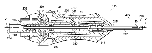
Fig. 2 shows a cross-sectional view of the vitrectomy probe system 110
previously shown in Fig. 1. In this example, the probe system 110 is a
pneumatically
driven system that operates by receiving pneumatic pressure alternating
through first
and second ports 202 and 204 over the supply lines 112 illustrated in Fig. 1.
The
probe system 110 includes as its basic components a cutter 210 and a probe
actuator
220 shown here as a reciprocating air driven diaphragm 220, all partially
encased by a
probe housing 230. The probe housing 230 includes an end piece 232 at the
probe
proximal end with the first and second air supply ports 202, 204 and one
suction port
234. The cutter 210 comprises an outer cutting tube 212 and an inner cutting
tube
214. As can be seen, the cutter 210 extends distally from a distal end 215 of
the
housing 230 and includes a distal portion 216. The outer cutting tube 212 is
coupled
to the housing 230, and the inner cutting tube 214 is slidable within the
outer cutting
tube 212 along a longitudinal axis LA of the probe 110.
Fig. 3 is a cross-sectional view that provides additional detail regarding the
distal portion 216 of the cutter 210 as seen in Fig. 2 and discussed above.
The distal
portion 216 includes an outer port 302 in the outer cutting tube 212 that
receives
tissue, such as ophthalmic tissue, during use. The outer port 302 is
proximally offset
9
CA 02933043 2016-06-07
WO 2015/094608
PCT/US2014/067314
from a closed end 304 of the distal portion 216. The inner cutting tube 214 is
located
within an inner channel 306 of the outer cutting tube 212. The outer port 302
is in
fluid communication with the inner channel 306 of the outer cutting tube 212.
The
inner cutting tube 214 has an inner bore 308, an open distal end 310, and a
cutting
surface 312.
The inner bore 308 is in fluid communication with an aspiration line (not
shown in Figs. 2 and 3) coupled to the suction port 234 of Fig. 2. The
aspiration line
may be part of the supply lines 112 of Fig. 1. 'The suction port 234 connects
the
aspiration line to a vacuum (that provides an aspiration pressure), which may
be
provided by console 102 or another device, and is used to pull tissue into the
outer
port 302 when the inner cutting surface 312 is located proximal to and away
from the
port 302. During operation of the vitrectomy probe 110, the inner cutting tube
214
moves in a reciprocal fashion (i.e., back-and-forth along the longitudinal
axis LA of
the probe 110) within the inner channel 306 of the outer cutting tube 212 to
cut tissue
.. that is pulled into the outer port 302 by the aspiration line.
The processor 114 initiates or triggers the movement of the inner cutting tube
214 to cut tissue that is aspirated or drawn into the outer port 302. When
used to cut
tissue, upon actuation or signaling from the processor 114, the distal end 310
of inner
cutting tube 214 is initially moved proximally away from the outer port 302
into a
retracted position and the vacuum pressure pulls tissue into the outer port
302 and the
inner channel 306. The distal end 310 of the inner cutting tube 214 then moves
distally toward the outer port 302 into an extended position and severs the
tissue
within the inner channel 306 with the cutting surface 312. The severed tissue
is
pulled through the inner bore 308 of the inner cutting tube 214 by the
aspiration
system. The inner cutting tube 214 then moves proximally away from the outer
port
302 into the retracted position (as shown in Fig. 5), and the cutting process
is
repeated. In some embodiments, without initiation or triggering (e.g., via
signals or
commands) from the processor 114, the motion inner cutting tube 214 would be
arrested.
With reference now to both Figs. 2 and 3, the inner cutting tube 214 is driven
by air pressure directed on opposing sides of the diaphragm 220 (e.g., in
response to
control signals from the processor 114). In one example of operation, if air
pressure
CA 02933043 2016-06-07
WO 2015/094608
PCT/US2014/067314
is increased at the first port 202, the diaphragm 220 will move distally,
displacing the
inner cutting tube 214 relative to the outer cutting tube 212, thereby closing
the tissue-
receiving outer port 302 of the outer cutting tube 212. This cuts any material
which
may have been aspirated into the tissue-receiving outer port 302. Venting the
pressure at the first port 202 and increasing the pressure at the second port
204 will
move the diaphragm 220 proximally, opening the tissue-receiving outer port 302
so
that it can draw in new material to be cut.
It's worth noting that other embodiments include alternative probe actuators.
For example, some actuator embodiments include a piston motor in place of a
.. diaphragm. In this type of embodiment, the cutter 210 is arranged so that
movement
of the piston also moves the inner cutting tube 214 of the cutter 210. Yet
other
actuator embodiments include other types of pneumatic or electric motors that
drive
the inner cutting tube 214.
Generally, for example in most vitrectomy procedures, the target ophthalmic
tissues for aspiration and cutting are substantially transparent tissues such
as, by way
of non-limiting example, vitreous humor and transparent membranes. Non-target
tissues are generally less transparent and more opaque than target tissues.
However,
because the cutter 210 operates extremely rapidly, with the inner cutting tube
214
moving within the outer cutting tube 212 at a very high rate, a surgeon cannot
easily
halt the operation of the cutter immediately upon aspiration of non-target
tissue.
Thus, non-target tissue (e.g., retinal tissue) may be aspirated into the outer
port 302
and inadvertently cut by the cutter 210 during a vitrectomy procedure, which
may
cause unnecessary injury to the retina and/or other ocular structures.
By nature, the retina is very flexible and conformal, and therefore retinal
tissue
may be drawn by the vacuum source into the outer port 302, occluding the inner
bore
308, preventing or limiting aspiration of the target tissue, and/or injuring
the retina. In
a healthy human eye, the retina is physically attached to the choroid in a
generally
circumferential manner. The vitreous humor, a transparent jelly-like material
that fills
the posterior segment of the eye, helps to cause the remainder of the retina
to lie
against, but not physically attach, to the choroid. A helpful analogy is to
imagine the
choroid as the walls of a fishbowl filled with vitreous humor. The retina is
like a sheet
of thin material that is pressed against the walls of the bowl by the vitreous
humor,
11
CA 02933043 2016-06-07
WO 2015/094608
PCT/US2014/067314
but is only physically attached to the walls at the rim of the bowl. If the
vitrectomy
probe 110 inadvertently cuts a portion of the retina, at least that portion of
the retina
may become detached from the choroid, which can cause vision loss and other
adverse effects. Sometimes a portion of the retina will tear, allowing aqueous
humor,
and sometimes vitreous humor, to flow between the retina and the choroid,
which also
may result in a loss of vision. The tissue sensor 150 can assist the surgeon
in avoiding
the inadvertent cutting of non-target tissues such as the retina.
As mentioned above in relation to Fig. 1, the vitrectomy probe system 110
includes at least one tissue sensor 150. As shown in Fig. 2, the tissue sensor
150 is
positioned within the cutter 210. In particular, the tissue sensor 150 is
positioned in
close proximity to the outer port 302 to enable the sensor 150 to measure
(e.g., by
detecting) a characteristic of the tissue aspirated into the outer port 302.
The tissue
sensor 150 comprises any type of sensor configured to sense a characteristic
of the
aspirated tissue that would enable the vitrectomy system 100 (e.g., the
processor 114)
to determine whether the tissue is a target tissue or a non-target tissue. For
example,
in one embodiment, the tissue sensor 150 comprises a fiberoptic sensor that
can
measure the degree of transparency (e.g., by detecting the amount of light
that passes
through the aspirated tissue) of the aspirated tissue. Other tissue
characteristics that
may be sensed by the tissue sensor 150 include, by way of non-limiting
example, the
amount of reflectivity, the electrical impedance, and/or indicators of the
structural
composition (e.g., layered or anamorphic) of the tissue.
In the embodiment pictured in Figs. 2 and 3, the tissue sensor 150 is embedded
in the outer cutting tube 212 and is configured to sense and measure (e.g., by
detecting) a characteristic (e.g., the degree of transparency) of the tissue
aspirated into
the outer port 302. As depicted, the tissue sensor 150 is a fiber optic tissue
sensor 150
coupled to electronics in the probe housing 230 as seen in Fig. 2 and/or the
console
102 as seen in Fig. 1 by a sensor line 232. The sensor line 232 is configured
to
transfer the sensed data from the sensor 150 to electronics in the probe
housing 230
and/or the processor 114 shown in Fig. 1. The sensor line 232 may be an
electrical or
a fiber optic line depending on the type of tissue sensor 150. In the pictured
embodiment in Figs. 2 and 3, both the tissue sensor 150 and the sensor line
232 are
positioned within recesses formed in the outer cutting tube 212 so that the
outer
12
CA 02933043 2016-06-07
WO 2015/094608
PCT/US2014/067314
surface 235 of the outer cutting tube 212 remains smooth and uninterrupted. In
some
embodiments this recess is formed on the outside of the cutting tube 212,
while in
others it is formed on the inside, with an opening provided for the sensor 150
to
access the aspirated tissue.
In the illustrated embodiment, the tissue sensor 150 is disposed distal to the
system housing 230 and adjacent to the outer port 302. In the pictured
embodiment,
the tissue sensor 150 is positioned at the outer surface 235 of the outer
cutting tube
212 in order to measure the tissue immediately as it enters the outer cutting
tube 212.
In other embodiments, the tissue sensor 150 can be positioned on an inner
surface 240
of the outer cutting tube 212. In other embodiments, the tissue sensor 150 can
be
embedded entirely within the outer cutting tube 212 between the outer surface
235
and the inner surface 240. In each of these embodiments, the tissue sensor 150
is
positioned with access to or exposure to an inner surface 237 of the outer
bore 302
and insulation from the outer surface 235. Thus, the sensor 150 can measure by
detecting characteristics of the tissue aspirated into the outer bore 302
while
remaining shielded from the tissue immediately outside the outer cutting tube
212.
In other embodiments, as shown in Fig. 4, a tissue sensor 150' may be
positioned on an outer surface 240 of an inner cutting tube 214'. Fig. 4
illustrates
another exemplary distal portion 216' of a cutter 210'. The cutter 210' is
substantially similar to the cutter 210 except for the differences described
herein. In
the pictured embodiment in Fig. 4, both the tissue sensor 150' and the sensor
line 232'
are positioned within recesses formed in the outer surface 240 of the inner
cutting
tube 214' so that the sensor 150 lies flush with the outer surface 240 and the
outer
surface 240 remains smooth and uninterrupted. In this embodiment, the tissue
sensor
150' is positioned with access to or exposure to the outer surface 240 of the
inner
cutting tube 214'. Thus, the sensor 150' can measure characteristics of the
tissue
aspirated into the outer bore 302'. A sensor line 232' couples the tissue
sensor 150'
to electronics as discussed above in connection with the tissue sensor 150.
In some embodiments, the sensor recess is less than a thickness of the sensor
150 or more than the thickness of the sensor 150. The recess may be square
shaped or
any other shape suitable for receiving and housing the tissue sensor 150.
Elongated
recesses are provided for the electrical and/or optical supply lines.
13
CA 02933043 2016-06-07
WO 2015/094608
PCT/US2014/067314
Returning to Fig. 2, in the pictured embodiment, the vitrectomy probe 110
includes an arresting mechanism 320. The arresting mechanism 320 is configured
to
halt the cutting mechanism of the cutter 210 when the vitrectomy surgical
system 100,
based on the sensed data from the tissue sensor 150, concludes that the tissue
(e.g.,
.. tissue 322 shown in Fig. 5) within the outer port 302 is not target tissue.
In the
pictured embodiment, the arresting mechanism 320 is disposed within the probe
110
adjacent to a proximal portion 325 of the cutter 210. As the inner cutting
tube 214 is
moved reciprocally within the outer cutting tube 212, the proximal portion 325
of the
cutter 210 moves in unison with the inner cutting tube 214. The arresting
mechanism
may comprise any type of suitable element shaped and configured relative to
the
cutter 210 to arrest the motion of the proximal portion 325, and thereby halt
the
motion of the inner cutting tube 214 proximal to the outer port 302, as shown
in Fig.
5. In one embodiment, the arresting mechanism 320 comprises a damper element
configured to grasp the proximal portion 325 and halt the motion of the inner
cutting
.. tube 214. In another embodiment, the arresting mechanism 320 comprises a
first
fastening element shaped and configured to interact with (e.g., hook or snag)
a
corresponding second fastening element (not shown) on the proximal portion 325
to
halt the motion of the inner cutting tube 214.
In some embodiments, the proximal portion 325 may comprise an aspiration
.. line that is separable from the remainder of the cutter 210. In other
embodiments, the
proximal portion 325 comprises an integral part of the cutter 210. The
arresting
mechanism 325 may be disposed anywhere along the length of the cutter 210
(e.g.,
the proximal portion 325) that enables the arresting mechanism to halt the
motion of
the cutter 210. For example, in other embodiments, the proximal portion 325
may
comprise a more proximal or a more distal portion of the cutter 210 than shown
in Fig
2.
The arresting mechanism 320 may be connected in a wired or wireless fashion
to the console 102 and/or the processor 114. In the pictured embodiment, the
arresting mechanism 320 is connected to the console 102 and/or the processor
114 via
a communication cable 340. The communication cable 340 may extend from the
console 102 into the vitrectomy probe 110 to the arresting mechanism 320. In
some
14
CA 02933043 2016-06-07
WO 2015/094608
PCT/US2014/067314
embodiments, the communication cable 340 is coupled to or forms part of the
supply
lines 102 shown in Fig. 1.
As described above, the tissue sensor 150 senses and measures (e.g., by
detecting) a characteristic of the tissue aspirated into the outer port 302
and conveys
that data to the processor 114. For example, in one embodiment, as tissue is
drawn
into the outer port 302, the tissue sensor 150 measures degree of transparency
of the
tissue and communicates that data to the processor 114 in the console 102
shown in
Fig. 1. 'Me processor 114 includes logic algorithms that use input data from
the tissue
sensor 150 to determine whether the sensed tissue is target tissue that should
be cut or
is non-target tissue that should not be cut. The processor 114 is operable to
control
the movement of the inner cutting member 214 based on the characteristic
measured
by the tissue sensor.
If the processor 114 determines, based on the input data, that the sensed
tissue
is non-target tissue, the processor 114 disables the cutting mechanism of the
probe
system 110 by either halting the motion of the inner cutting tube 214 (e.g.,
with the
arresting mechanism 320) or preventing the actuation of the inner cutting tube
214
(e.g., by not initiating, triggering, or signaling the movement of the inner
cutting tube
214). For example, in embodiments including the arresting mechanism 320, the
processor 114 may signal, command, or activate the arresting mechanism 320 to
halt
the movement of the inner cutting tube 214 of the probe system 110.
In alternative embodiments, the vitrectomy probe 110 lacks the arresting
mechanism 320. In such embodiments, if the processor 114, using the logic
algorithm
and the input data from the tissue sensor 150, determines that the tissue is
non-target
tissue that should not be cut, the processor 114 does not initiate, trigger,
actuate,
and/or signal the movement of the cutting mechanism of the probe system 110
(e.g.,
the triggering function of the processor 114). If the processor 114 does not
initiate the
cutting mechanism of the probe system, the probe system 110 cannot cut tissue.
In the pictured embodiment, the vitrectomy probe 110 includes an over-ride
element 345, which may be the same as the over-ride element 126 shown in Fig.
1.
The described features of the over-ride element 345 may also apply to the over-
ride
element 124 and/or 126. The over-ride element 345 comprises any of a variety
of
CA 02933043 2016-06-07
WO 2015/094608
PCT/US2014/067314
user input structures having ON/OFF functionality such as, by way of non-
limiting
example, a button, a dial, a switch, and a toggle. When activated or switched
to an
ON position, the over-ride element 345 enables the surgeon to over-ride the
arresting
mechanism of the vitrectomy surgical system 100 and/or to over-ride the
triggering
function of the processor 114. In particular, in embodiments including an
arresting
mechanism 320, when the over-ride element 345 is switched to an ON position,
regardless of what type of tissue is aspirated into the outer port 302, the
motion of the
inner cutting tube 214 will not be halted by the arresting mechanism 320. For
example, in one embodiment, when the over-ride element 345 is switched to an
ON
position, the arresting mechanism 320 is temporarily and reversibly disabled.
In
embodiments lacking an arresting mechanism 320, when the over-ride element 345
is
switched to an ON position, regardless of what type of tissue is aspirated
into the
outer port 302, the motion of the inner cutting tube 214 will continue to be
triggered
by the processor 114. In other embodiments, when the over-ride clement 345 is
switched to an ON position, the tissue sensor 150 is temporarily and
reversibly
disabled.
The over-ride element 345 may be connected in a wired or wireless fashion to
the console 102, the processor 114, and/or the arresting mechanism 320. In the
pictured embodiment, the over-ride element 345 is connected to the console 102
and/or the processor 114 via a communication cable 350. The communication
cable
350 may extend from the console 102 into the vitrectomy probe 110 to the over-
ride
element 345. In some embodiments, the communication cable 340 is coupled to or
forms part of the supply lines 102 shown in Fig. 1. In some embodiments, the
over-
ride element 345 is additionally or alternatively connected to the arresting
mechanism
320 via a communication cable 355.
Fig. 6 illustrates a partially cross-sectional view of an eye 400 undergoing a
procedure involving the vitrectomy surgical system 100 and an infusion line or
infusion cannula 420. During a vitrectomy procedure, a surgeon typically
inserts the
vitrectomy probe 110 into the posterior segment of the eye via an incision
through the
sclera in the pars plana. Such an incision is called a sclerotomy. The surgeon
typically also inserts a fiber optic light source and the infusion cannula 420
into the
eye via similar incisions, and may sometimes substitute an aspiration probe
for the
16
CA 02933043 2016-06-07
WO 2015/094608
PCT/US2014/067314
vitrectomy probe 110. While viewing the posterior segment under a microscope
and
with the aid of the fiber optic light source, the surgeon cuts and aspirates
away
vitreous using the vitrectomy probe 110 to gain access to the area of interest
(e.g., the
site of a retinal detachment or tear). The surgeon may also use the vitrectomy
probe
110 to remove any membrane that has contributed to the retinal detachment or
tear.
During this portion of the surgery, a saline solution is typically infused
into the eye
via the infusion cannula 420 to maintain the appropriate intraocular pressure.
Both the vitrectomy probe 110 and the infusion line 420 may be coupled to a
console, such as the console 102 shown in Fie. 1. In Fig. 6, the vitrectomy
probe 110
and the infusion line 420 are inserted through the sclera 402 and into the
vitreous
chamber 404 of the eye 400. The infusion line 420 is a specialized type of
probe used
to deliver replacement fluid or irrigation fluid into the vitreous chamber 404
during
vitrectomy procedures. A pressure level of the irrigation fluid may be
increased or
decreased by a surgical system.
In the illustrated embodiment, the vitrectomy probe 110 includes the tissue
sensor 150 adjacent the outer port 302. As depicted, the tissue sensor 150 is
positioned on the cutter 210 to measure a characteristic such as, by way of
non-
limiting example, the degree of transparency, of the tissue aspirated into the
outer port
302. The data sensed by the tissue sensor 150 may be communicated to the
console
102 and/or the processor 114 shown in Fig. 1 either wirelessly or via the
supply lines
112. The tissue characteristics or data that may be sensed by the sensor 150
of the
vitrectomy probe 110 facilitate improved control by the vitrectomy surgical
system
100 of Fig. 1 by providing additional information that can be processed by the
surgical system 100 (and/or the processor 114) and used for automated control
of the
cutter 210. For example, in one embodiment, by measuring and determining the
transparency of the aspirated tissue, the vitrectomy surgical system 100 may
be able
to avoid the inadvertent cutting and removal of non-target tissue during a
vitrectomy
procedure by halting the motion of the inner cutting tube 214 before it cuts
the tissue
aspirated into the outer port 302 (as shown in Fig. 5).
The processor 114 shown in Fig. 1 may have user-settable or pre-defined
limits for acceptable tissue characteristics or measurements that reflect
target tissue
characteristics. For example, in one embodiment where the tissue sensor 150 is
17
CA 02933043 2016-06-07
WO 2015/094608
PCT/US2014/067314
configured to measure a degree of transparency of the tissue, the processor
114 may
contain pre-defined or user-settable ranges defining the range of transparency
associated with target tissues such as, by way of non-limiting example,
vitreous and
membranes (e.g., transparent tissues). The acceptable range of values
corresponding
to a desired target tissue may be modified before, during, or after a
procedure. In
embodiments including an arresting mechanism 320, when the tissue data sensed
by
the tissue sensor 150 exceeds the predetermined level of acceptable
transparency (or
other tissue characteristic) as a result of the aspiration of non-target
tissue (e.g., retinal
tissue) into the outer port 302, the processor 114 can signal the arresting
mechanism
320 shown in Fig. 2 to halt the motion of the inner cutting tube 214 before it
cuts the
tissue 322 as shown in Fig. 5. In embodiments that lack the arresting
mechanism 320,
when the tissue data sensed by the tissue sensor 150 exceeds the predetermined
level
of acceptable transparency (or other tissue characteristic) as a result of the
aspiration
of non-target tissue (e.g., retinal tissue) into the outer port 302, the
processor 114 does
not initiate, signal, or trigger the movement of the inner cutting tube 214
and thereby
prevents it from cutting the tissue 322 as shown in Fig. 5. For example, in
some
embodiments, the processor 114 suspends the motion of the inner cutting tube
214
(based upon the measured tissue characteristic) by halting the transmission of
driving
power to the inner cutting tube 214.
However, if the surgeon desires to cut typically non-target (e.g., less
transparent and more opaque tissue), the surgeon may adjust the over-ride
element
345 shown in Fig. 2 (and/or the over-ride element 125 as shown in Fig. 1) to
an ON
position to allow the vitrectomy surgical system 100 to at least temporarily
cut the
typically non-target tissue. For example, in some instances, the surgeon may
want the
vitrectomy probe 110 to cut, aspirate, and remove certain tissues or
materials,
including without limitation, coagulated blood, debris, retinal tissue, and
retinal
pigment epithelium
Fig. 7 is a flowchart of an exemplary method 700 of operating the vitrectomy
surgical system 100 in treating an ophthalmic condition according to one
embodiment
of the present disclosure. As illustrated, the method 700 includes a number of
enumerated steps, but embodiments of the method 700 may include additional
steps
before, after, and in between the enumerated steps. The illustrated embodiment
18
CA 02933043 2016-06-07
WO 2015/094608
PCT/US2014/067314
begins at step 702 in which a surgeon inserts a probe (e.g., the vitrectomy
probe 110)
that includes at least one tissue sensor 150 and the arresting mechanism 320
through a
sclera into a vitreous chamber of a patient. At step 704, the surgeon may
aspirate
tissue into the outer port 302 of the vitrectomy probe 110. At step 606, the
tissue
sensor 150 can measure a tissue characteristic (e.g., transparency) of the
aspirated
tissue by detecting the characteristic of the tissue and communicate data or
signals
representing the tissue characteristic to the console 102 (e.g., the processor
114).
At step 708, the processor 114 can evaluate whether the measured tissue data
lies within the predetermined acceptable range of values for typical target
tissue (e.g.,
vitreous humor and membranes). If the processor 114 determines that the tissue
data
lies within the predetermined range, then the system 100 continues to aspirate
and cut
the tissue at step 710. If, however, the processor 114 determines that the
tissue data
does not lie within the predetermined range, then, at step 712, the processor
114
queries whether the over-ride element 124 or 126 is in an ON position. If the
over-
ride element 124, 126 is in an ON position, then the system 100 continues to
aspirate
and cut the tissue at step 710. Thus, when the over-ride element 124, 126 is
in the ON
position, it prevents the processor 114 from controlling the movement of the
inner
cutting tube 214 based on the measured characteristic. If the over-ride
element 124,
126 is in an OFF position, then, at step 714, the system 100 can prevent the
cutting of
the aspirated tissue within the outer port 302 at step 714 by either: (1) the
arresting
mechanism 320 halting the movement of the inner cutting tube 214, or (2) the
processor 114 not initiating or triggering movement of the inner cutting tube
214..
In some embodiments, the processor 114 queries the ON/OFF status of the
over-ride element 124 or 126 before step 706, and disables the tissue sensor
150 if the
over-ride element is in the ON position (thereby avoiding steps 706 and 708
and
continuing directly to step 710). In other embodiments, the processor 114
queries the
ON/OFF status of the over-ride element 124 or 126 immediately after step 706,
and
disables the arresting mechanism 320 (or continues to trigger movement of the
inner
cutting tube 214) if the over-ride element is in the ON position (thereby
avoiding step
708 and continuing directly to step 710). After the the movement of the inner
cutting
tube 214 has been prevented at step 714, the surgeon may reposition the probe
110 to
aspirate a different portion of tissue at steps 716 and 710. At any time
before, during,
19
CA 02933043 2016-06-07
WO 2015/094608
PCT/US2014/067314
or after the procedure, the surgeon may readjust the over-ride element 124,
126 to turn
it ON or OFF.
The systems and methods disclosed herein may be used to enable better
performance of vitrectomy surgical systems by enabling focal tissue
measurements
during a vitrectomy procedure that help the system determine in real-time
whether or
not aspirated tissue within the vitrectomy probe should be cut, aspirated, and
removed. This additional level of control may enable a surgeon to avoid
inadvertently
cutting and removing typically non-target tissues (e.g., retinal tissue).
'Ibis may result
in more effective vitrectomy procedures and reduced risk of ocular injury,
thereby
.. improving the overall clinical result.
Persons of ordinary skill in the art will appreciate that the embodiments
encompassed by the present disclosure are not limited to the particular
exemplary
embodiments described above. In that regard, although illustrative embodiments
have
been shown and described, a wide range of modification, change, combination,
and
substitution is contemplated in the foregoing disclosure. It is understood
that such
variations may be made to the foregoing without departing from the scope of
the
present disclosure. Accordingly, it is appropriate that the appended claims be
construed broadly and in a manner consistent with the present disclosure.