Note: Descriptions are shown in the official language in which they were submitted.
CA 02933305 2016-06-09
WO 2015/097170 PCT/EP2014/079028
1
CD44 binding peptides
FIELD OF THE INVENTION
The present invention relates to a protein which binds to the domain encoded
by exon 9 of
human 0D44 (CD44ex9), to fusion proteins and conjugates of said protein and
especially to
nanoparticles conjugated to said protein. The invention further relates to a
method of
production for the protein and the respective conjugated nanoparticles, to
CD44v5 derived
peptides and the use of the protein and the peptides of the invention for
treatment and
diagnosis of various diseases, and especially for cancer.
BACKGROUND OF THE INVENTION
Extensive studies of cancer transcriptional patterns have led to the discovery
of molecular
targets to distinguish the malignant from the benign and the most aggressive
cancers from
those that are less aggressive. Cancers often overexpress a number of
proteins, including
certain cell surface antigens, e.g. cell surface receptors. Antibodies that
bind such
overexpressed cell surface antigens can facilitate detection and treatment of
such cancers. A
number of approaches have been utilized to generate antibodies to cancer cell
surface
receptors which can be used as potential diagnostics or therapeutics.
Identification of
overexpressed cell surface receptors and antibodies which bind them provide a
route to the
development of cancer therapies, especially for those cancer subtypes with
poor prognosis
and resistance to traditional therapies. CD44, CA19-9 and CEA are such
overexpressed cell
surface receptors, which are known to be relevant for tumor progression and
malignancy.
CD44, a major adhesion molecule for the extracellular matrix, has been
implicated in a wide
variety of physiological processes, including leukocyte homing and activation,
wound healing,
and cell migration, as well as in tumor cell invasion and metastasis (Gunthert
et al., 1991;
Nagano and Saya, 2004; Ponta et al., 2003).
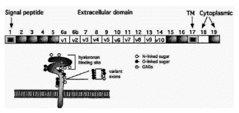
CD44 is a single chain molecule comprising a conserved N-terminal
extracellular domain, a
non-conserved membrane proximal region, a variable region expressing various
combinations of variant exons, a conserved transmembrane spanning domain and a
conserved cytoplasmic tail. The genomic map of CD44 includes 5 constant exons
at the 5'
terminus and 5 constant exons in the 3' end. The mouse CD44 gene includes also
10 variant
exons in the middle of the molecule designated V1-V10 resulting in a total of
20 exons. The
human C044 gene comprises only 9 of these 10 variant exons (V2-V10) thus
comprising a
total of 19 exons. Differential alternative splicing generates many isoforms
of CD44 that
express various combinations of variant exons (designated Exon Vx, x = 1 to
10), which are
inserted in the membrane proximal domain and constitute the variable region of
the
CA 02933305 2016-06-09
WO 2015/097170 PCT/EP2014/079028
2
molecule. These molecules are designated 0044 variants (CD44v). To date, 20
isoforms of
CD44 are known.
Whereas the standard 0D44 isoform (CD44s) is expressed predominantly in
hematopoietic
cells and normal epithelial cell subsets, the CD 44 variants with insertions
in the membrane-
proximal extracellular region were found to be abundant in a variety of human
tumors,
including colon, mammary, gastric, bladder, prostate carcinomas, and various
hematopoietic
neoplasms. Additional reports suggested a close correlation between expression
of the
variant 0044 and tumor progression and in particular the lymphatic spread of
neoplastic
cells.
Taken together, these observations point to an important function of 0D44
variants in tumor
initiation and the maintenance of cancer cells in addition to its more
established functions in
cell adhesion and migration.
Beside the role of CD44 in cancer, 0044 has also been suggested as a potential
target in
autoimmune diseases. It has been reported (Hale et al., 1992) that
administration of a C044
protein or peptide or derivative can be used for treating various autoimmune
diseases.
Based on the established role of CD44 in cancer and autoimmune diseases,
monoclonal
antibodies directed against various variant regions of 0044 have also been
generated as
potential agents for diagnosis or therapy of 0044-related disorders.
Seiter et al. describe mAbs directed against metastasis-specific variants of
0044 surface
protein of a rat pancreatic adenocarcinoma (Seiter et al., 1993). These
antibodies are
proposed for producing immunosuppression for therapeutic treatment of
immunoregulatory
disorders including, for example, diseases of the rheumatic type. Monoclonal
antibodies
reactive with 0044 which inhibit T-cell proliferation were also provided for
treatment of
various autoimmune diseases. Monoclonal antibodies binding to forms of CD44
containing
the exon v6 peptide were also reported as being useful for diagnosing
inflammatory diseases
(Jalkanen et al., 1986).
The WO 91/17248 relates to the use of anti-CD44v antibodies for the therapy
and diagnosis
of tumors. WO 95/00851 relates to the use of antibodies directed against the
variable exons
of 0044 for diagnosis of tumors. The WO 95/04547 discloses the use of anti-
0044
antibodies especially directed against epitopes within the sequence of exon v5
for
immunotherapeutic and immunoscintigraphic purposes. An anti-0044 antibody
directed
against exon v6 is disclosed by WO 95/33771. Furthermore, the EP 0 538 754
teaches the
use of antibodies directed against C044 variants for immunosuppression.
The anti-0044 antibodies of the prior art show several disadvantages. They
represent large
and complex molecules which have to be generated by recombinant production
methods.
Hence, the production process is elaborate, costly and has to fulfill strict
regulatory
CA 02933305 2016-06-09
WO 2015/097170 PCT/EP2014/079028
3
requirements. Furthermore, the necessity for a reduced immunogenic potential
requires the
generation of human or humanized antibodies.
Hence, there is still the need for 0D44 variant-binding molecules. The
objective of the
present invention thus is to provide a 0D44 variant binding substance which
overcomes at
least one of the above mentioned disadvantages.
This problem is solved by provision of a CD44-variant binding protein
according to claim 1
and a use of said binding protein for diagnosis and therapy, especially by
using nanoparticles
coated with said protein. Specific embodiments of the invention are subject
matter of further
independent or dependent claims.
SUMMARY OF THE INVENTION
In a first aspect the invention provides a protein binding to a polypeptide
encoded by exon 9
of human CD44 (CD44ex9), said protein comprises or consists of
a) an amino acid sequence according SEQ ID No. 1 to 5; or
b) an amino acid sequence with at least 80% identity, preferably 85%, more
preferably
90% and most preferably 95% identity to the amino acid sequence given in SEQ
ID
No. 1 to 5; or
c) an amino acid sequence with at least 90% identity, preferable at least 95%
or 100%
identity to the 0D44-binding motif defined by the sequence:
"PYYGKXLX3YLQP5FAVQVX25XQX10-14AIE" as depicted in SEQ ID Nos. 6 to 10,
wherein X denotes an arbitrary amino acid; and
wherein said protein has a length of 100 amino acids or less.
By performing a yeast-two hybrid-screening the inventors were able to identify
five different
peptides with a specific binding to the polypeptide encoded by exon 9 of CD44.
The amino
acid sequences are presented in Figure 3 and in the following table and
designated as SEQ
ID No. 1 to 5.
SEQ ID Amino acid sequence
No.
1 PGVPGVQLTA NIQSLVLMSA FDIATEVTFT SS
2 PGLQISFAVQ VPVSVQESSP SVQEGIQIQV ATE
3 PGGDHERAPA SCYNGERLSS QLSFEVQWET SETLKILVKP LVFVCREVYD
PPC
4 PGPYYGKKLH VGYLQPLAAV QVSFAPNNTG KEVTVECKID GSANLKSQDD
RDKFLGRVMF KITARA
5 YYPYYGKLLQ PKYLQPLLAV QFTNLTMDTE IRIECKAYGE NIGYSEKDRF
QGRFDVKIEV KS
CA 02933305 2016-06-09
WO 2015/097170 PCT/EP2014/079028
4
A further analysis revealed that a consensus sequence can be derived from
these
sequences exhibiting the following amino acid sequence:
PYYGKXLX3YLQPSFAVQVX2SXQX10-14AIE.
These consensus sequences are designated as SEQ ID No. 6 to 10 as depicted in
the
following table whereby these five sequences differ in the length of the
arbitrary sequence
X10_14. Please note that "X" is defined as an arbitrary amino acid.
SEQ Amino acid sequence
ID No.
6 PYYGKXLXXX YLQPSFAVQV XXSXQXXXXX XXXXXAIE
7 PYYGKXLXXX YLQPSFAVQV XXSXQXXXXX XXXXXXAIE
8 PYYGKXLXXX YLQPSFAVQV XXSXQXXXXX XXXXXXXAIE
9 PYYGKXLXXX YLQPSFAVQV XXSXQXXXXX XXXXXXXXAI E
PYYGKXLXXX YLQPSFAVQV XXSXQXXXXX XXXXXXXXXA IE
The ability to derive a unique consensus sequence which encompasses all
independent
isolated 0044-interacting peptides further validates the results of the two-
hybrid screening
method.
In subsequent two independent affinity assays the peptides of SEQ ID No. 1 to
5 were
analysed for binding towards the polypeptide encoded by exon 9 of 0044 and
allowed an
affinity ranking of the peptides, whereby the peptides were named as follows:
Peptide of SEQ ID No. 1: Peptide A
Peptide of SEQ ID No. 2: Peptide C
Peptide of SEQ ID No. 3: Peptide B
Peptide of SEQ ID No. 4: Peptide D
Peptide of SEQ ID No. 5: Peptide E
Both affinity assays, the pseudohitpicking assay and the Fluorescein di-beta-D-
galacto-
pyranoside (FDG)-based assay revealed the same ranking order:
B> A,C,D,E
C> A,D,E
D> A,E
E> A
A>
In sum, peptide B (= SEQ ID No. 3) possesses the highest affinity towards the
polypeptide
encoded by exon 9 of 0D44 and has to be regarded as preferred peptide in the
context of
the invention. For this peptide an epitope mapping was performed, whereby a
deletion
analysis and an alanine scan was performed.
CA 02933305 2016-06-09
WO 2015/097170 PCT/EP2014/079028
Furthermore, also the peptides A, C, D and E were analysed in a deletion
assay, confirming
the validity of the consensus sequence.
Based on these analyses, the invention provides in a further aspect a protein
binding to a
5 polypeptide encoded by exon 9 of human CD44 (CD44ex9), said protein
comprises or
consists of
a) an amino acid sequence according SEQ ID No. 39 to 52; or
b) an amino acid sequence with at least 80% identity, preferably 85%, more
preferably
90% and most preferably 95% identity to the amino acid sequence given in SEQ
ID
No. 39 to 52; or
c) an amino acid sequence with at least 90% identity, preferable at least 95%
or 100%
identity to the CD44-binding motif defined by the sequence:
"PG LQ PS FAVQVX2s/AXQX10-i4A I E" as depicted in SEQ ID Nos. 53 to 62,
wherein X
denotes an arbitrary amino acid; and
wherein said protein has a length of 100 amino acids or less.
SEQ ID Amino acid sequence
No.
39 PGQLSFEVQW ETSETLKILV KPLVFVCREV YDPPC
40 PGALSFEVQW ETSETLKILV KPLVFVCREV YDPPC
41 PGQASFEVQW ETSETLKILV KPLVFVCREV YDPPC
42 PGQLAFEVQW ETSETLKILV KPLVFVCREV YDPPC
43 PGQLSAEVQW ETSETLKILV KPLVFVCREV YDPPC
44 PGQLSFAVQW ETSETLKILV KPLVFVCREV YDPPC
45 PGQLSFEAQW ETSETLKILV KPLVFVCREV YDPPC
46 PGQLSFEVAW ETSETLKILV KPLVFVCREV YDPPC
47 PGQLSFEVQA ETSETLKILV KPLVFVCREV YDPPC
48 PGQLSFEVQW ATSETLKILV KPLVFVCREV YDPPC
49 PGQLSFEVQW EASETLKILV KPLVFVCREV YDPPC
50 PGQLSFEVQW ETAETLKILV KPLVFVCREV YDPPC
51 PGQPLAAVQV SFAPNNTGKE VTVECKIDGS ANLKSQDDRD KFLGRVMFKI
TARA
52 PGLQPLLAVQ FTNLTMDTEI RIECKAYGEN IGYSEKDRFQ GRFDVKIEVK
Based on this epitope mapping of the peptides the above disclosed consensus
sequence in
general was confirmed. However, a more preferred consensus sequence can be
derived
from these sequences exhibiting the following amino acid sequence:
PGLQPSFAVQVX2s/AXQX10-14AIE.
CA 02933305 2016-06-09
WO 2015/097170 PCT/EP2014/079028
6
These consensus sequences are designated as SEQ ID No. 53 to 62 as depicted in
the
following table whereby these sequences differ in an amino acid exchange at
position 14
(Ser-Ala) and the length of the arbitrary sequence X10-14. Please note that
"X" is defined as an
arbitrary amino acid.
_________________________________________________________________
SEQ Amino acid sequence
ID No.
53 PGLQPSFAVQ VXXSXQXXXX XXXXXXAIE
54 PGLQPSFAVQ VXXSXQXXXX XXXXXXXAIE
55 PGLQPSFAVQ VXXSXQXXXX XXXXXXXXAI E
56 PGLQPSFAVQ VXXSXQXXXX XXXXXXXXXA IE
57 PGLQPSFAVQ VXXSXQXXXX XXXXXXXXXX AIE
58 PGLQPSFAVQ VXXAXQXXXX XXXXXXAIE
59 PGLQPSFAVQ VXXAXQXXXX XXXXXXXAIE
60 PGLQPSFAVQ VXXAXQXXXX XXXXXXXXAI E
61 PGLQPSFAVQ VXXAXQXXXX XXXXXXXXXA IE
62 PGLQPSFAVQ VXXAXQXXXX XXXXXXXXXX AIE
It has to be emphasized that the peptides of the invention show for the first
time that not only
large antibodies can be generated against CD44 epitopes but also rather small
peptides with
a length between 32 and 66 amino acids which bind specifically and selectively
to a clinically
relevant 0044 domain.
In contrast to the (monoclonal) CD44v5 antibodies of the prior art, these
peptides exhibit
several advantages.
Since they represent short peptides without the necessity to form complexes or
intramolecular disulfide bridges they are much easier to produce and to
purify.
As peptides with a length of between 32 and 66 amino acids it is even possible
to synthesize
them by solid phase synthesis (according Merrifield). This ex vivo-protein
synthesis is
especially of advantage given the strict regulatory requirements associated
with recombinant
protein expression.
Furthermore, as small peptides they can be advantageously conjugated or fused
to a
different protein, specific compounds or even nanoparticles in order to be
used for therapy or
diagnosis.
As small peptides they are able to cross the blood brain barrier and cellular
membranes to
deliver compounds to compartments which could not be addressed by antibodies.
Due to their small size the proteins of the inventions the risk to elicit an
immune response is
strongly reduced. It has to be remarked that the risk of immunogenicity which
is inherent for
antibodies markedly impairs their clinical use.
7
It has to be emphasized that the CD44ex9-binding protein of the invention by
interfering only
with the variant domain v5 leaves the normal CD44 function untouched and
therefore allows
a specific intervention.
In sum the CD44ex9-binding proteins of the invention open the door to novel
strategies for
therapy and diagnosis and cancer with a reduced risk of side effects.
DETAILED DESCRIPTION OF THE INVENTION
According to an aspect of the invention, there is provided use of a protein
for binding to a
polypeptide encoded by exon 9 of human CD44 (CD44ex9), said CD44ex9-binding
protein
comprises of (i) an amino acid sequence according to any one of SEQ ID No. 1
to 5 and 39
to 52; or (ii) an amino acid sequence with at least 85% identity to the full
length of the amino
acid sequence as set forth in any one of SEQ ID No. 1 to 5, 39 to 50 and 52;
or (iii) an amino
acid sequence with at least 95% identity to the full length of the amino acid
sequence as set
forth in SEQ ID No. 51; wherein said protein has a length of 100 amino acids
or less.
According to another aspect of the invention, there is provided use of a
protein capable of
binding to a polypeptide encoded by exon 9 of human CD44 (CD44ex9), said
CD44ex9-
binding protein consists of (i) an amino acid sequence according to any one of
SEQ ID No. 1
to 5 and 39 to 52; or (ii) an amino acid sequence with at least 85% identity
to the full length of
the amino acid sequence as set forth in any one of SEQ ID No. 1 to 5, 39 to 50
and 52; or (iii)
an amino acid sequence with at least 95% identity to the full length of the
amino acid
sequence as set forth in SEQ ID No. 51; wherein said protein has a length of
100 amino
acids or less.
According to a further aspect of the invention, there is provided a conjugate
comprising: a)
the CD44ex9 binding protein as described herein; b) a compound selected from
the group
consisting of a carbohydrate, a dye molecule, a radioactive isotope, a toxin,
a cytostatic
agent, a cytokine, and an immunomodulatory agent, wherein said compound is
linked directly
or via a linker molecule to the CD44ex9 binding protein.
CA 2933305 2018-08-27
7a
According to a still further aspect of the invention, there is provided an
isolated nucleic acid
molecule encoding the CD44ex9 binding protein as described herein.
According to yet another aspect of the invention, there is provided an
expression vector
containing the nucleotide sequence as described above.
According to another aspect of the invention, there is provided a host cell
containing the
expression vector as described above.
According to another aspect of the invention, there is provided a method of
producing the
CD44ex9-binding protein as described herein, the method comprising: a)
transforming a host
cell with an expression construct comprising a nucleic acid molecule encoding
the CD44ex9
binding protein as described herein; and b) culturing the host cell under
conditions suitable
for producing the CD44ex9 binding protein or the respective fusion protein.
According to another aspect of the invention, there is provided a nanoparticle
conjugated to
the CD44ex9 binding protein as described herein.
According to another aspect of the invention, there is provided use of the
CD44ex9 binding
protein as described herein or the nanoparticle as described herein in the
diagnosis or
treatment of a disease selected from the group consisting of autoimmune
diseases, multiple
sclerosis, SjOgren's syndrome or systemic lupus erythematosus (SLE); skin
diseases;
chronic inflammatory diseases; tissue injury; allergic diseases and cancer
disease.
According to another aspect of the invention, there is provided a
pharmaceutical composition
comprising the CD44ex9 binding protein as described herein or the nanoparticle
as described
herein and a pharmaceutically acceptable excipient.
CA 2933305 2018-08-27
7b
According to another aspect of the invention, there is provided a medicament
and dosimeter
combination package comprising: a) a medicament to be individually dosed, and
b) a
diagnostic indicator system for a patient-specific property that is relevant
for the action, side
effect, interaction, metabolism, absorption, distribution, metabolism, and
elimination of the
medicament to be administered to a patient, wherein the patient-specific
property is selected
from the group consisting of an endogenous substance, a regulation mechanism,
a gene or
an indication system, and wherein the medicament or the diagnostic indicator
system
comprises the CD44ex9 binding protein as described herein or the nanoparticle
as described
herein.
In one embodiment of the invention the invention provides a protein binding to
a polypeptide
encoded by exon 9 of human CD44 (CD44ex9), said protein comprises or consists
of an
amino acid sequence with at least 80% identity, preferably at least 85%, more
preferably
least 90%, most preferably at least 95% identity and specifically at least
97.5% identity to the
amino acid sequence given in SEQ ID No. 1 to 5 and 39 to 52.
In a further embodiment the invention provides a protein binding to a
polypeptide encoded by
exon 9 of human CD44 (CD44ex9), said protein comprises or consists of an amino
acid
sequence with at least 90% identity, preferably at least 95%, more preferably
at least 97.5%
and even more preferably at least 100% identity to the CD44-binding motif
defined by the
sequence: "PYYGKXLX3YLQPSFAVQVX2SXQX10-14AIE" as depicted in SEQ ID Nos. 6 to
10,
and "PGLQPSFAVQVX2s/A XQX10-14AIE" as depicted in SEQ ID Nos. 53 to 62,
wherein X
denotes an arbitrary amino acid.
The proteins as claimed herein have a length of 300 amino acids or less,
preferably of 200
amino acids or less, more preferably a length of 100 amino acids or less and
even more
preferably a length of 90, 85, 80, 75, 70 or 66 amino acids or less.
In a further embodiment of the invention the CD44ex9-binding protein has a
high affinity to
the polypeptide encoded by exon9 with a k; value of less than 10 pM,
preferably of less than
1 pM, more preferably of less than 100nM and most preferably of less than 10
nM.
CA 2933305 2018-08-27
7c
In a further embodiment of the invention the CD44ex9-binding protein binds
selectively to the
polypeptide encoded by exon9, which is given when it binds to other arbitrary
proteins with a
k, value which is preferably at least 10 times less and more preferably 100
times less of the kJ
value for CD44ex9-binding.
In one embodiment the CD44ex9-binding protein of the invention is capable to
bind to every
CD44 isoform that contains the domain encoded by exon 9, which is also
designated as
"variant 5" (v5).
In a preferred embodiment the CD44ex9-binding protein of the invention binds
to a CD44
isoform selected from the list consisting of CD44 vs, CD44 v5 -v6, CD44 v3 -
v6, 0D44 v3 -
CA 2933305 2018-08-27
CA 02933305 2016-06-09
WO 2015/097170 PCT/EP2014/079028
8
v6, CD44 v2 ¨ v10, CD44 v3 ¨ v10, CD44 V4 ¨ v7 and CD44 v4 ¨ v10.
Notably, the CD44ex9-binding protein of the invention binds also to proteins
that comprise
beside the domain v5 also other variant domains such as v1, v2, v3, v4, v6,
v7, v8, v9 and/or
v10.
In a further embodiment of the invention the CD44ex9-binding protein of the
invention is
conjugated or fused to a heterologous protein or polypeptide. This
conjugation/fusion might
change the pharmacodynamic or pharmacokinetic properties of the binding
protein or it might
serve to reduce potential side effects.
It might further be used as a reporter protein enabling the detection of the
binding protein or
as a peptide or protein which enables protein purification, and which can be
further
engineered with cleavage sites for proteases or chemical agents which enable
the liberation
of the two separate proteins.
This technique is often used for identification and purification of proteins,
by fusing a GST
protein, FLAG peptide, or a hexa-His peptide (6 x His-tag) which can be
isolated using affinity
chromatography with nickel or cobalt resins.
The binding protein can be preferably linked or fused to a protein selected
from the group
consisting of antibodies, toxins, immunomodulatory peptides and cytokines.
In a preferred embodiment the conjugated/fused protein is an enzyme which
catalyses the
generation of a cytotoxic or cytostatic agent from a precursor molecule. A non-
exhaustive
lists of suitable enzymes contains the aldehyde-oxidase, amino acid oxidase,
cytochrome
P450 oxidase, NAD(P)H:quinone oxidoreductase, tyrosinase, thymidilate
synthase,
thymidine phosphorylase, glutathione-S transferase, deoxycytidine kinase,
carboxylesterase,
alkaline phosphatase, beta-glucuronidase, cysteine conjugate-beta lyase, and
nitroreductase.
The binding protein of the invention might also be conjugated or fused to a
protein that
inhibits, impairs or kills the cancer cell or makes it sensitive to a further
cytotoxic compound.
Non-limiting examples for said proteins are cytosine deaminase, soluble Fms-
like tyrosine
kinase ligand, herpes simplex virus-1 thymidine kinase (HSV1-TK), cytochrome
P450 261,
retinoblastoma related proteins, p16/cdkn2 and MMAC1/PTEN. MMAC1/PTEN acts as
a
negative regulator of the phosphor-inositide 3-kinase. MMAC1 stands for
Mutated in Multiple
Advanced Cancers 1.
In a further aspect the invention provides a conjugate comprising the CD44ex9-
binding
protein of the invention which is linked directly or indirectly by a linker to
a compound
selected from the group consisting of a carbohydrate, a dye molecule, a
radioactive isotope,
a toxin, a cytostatic agent, a cytokine, a immunomodulatory agent, or a
prodrug thereof.
CA 02933305 2016-06-09
WO 2015/097170 PCT/EP2014/079028
9
The conjugate according to invention can in general be linked to any type of
drug which
impairs, inhibits or even kills cancer cells. Hence, a cytotoxic agent is
particularly preferred.
Non-limiting examples for cytotoxic agents include emozolomide, carmustine,
lomustine,
procarbazine, streptozocin, irinotecan or any combination of two or more of
these agents.
Further examples for suitable cancer inhibitors are (-)-Ci-Cdp1, (-)-Ci-Cdp2,
(-)-
epigallocatechin gallate, (+)-Cbi-Cdpi2, (+)-Ci-Cdp2, 10-Deacetylbaccatin Ill,
4-demethoxy
daunorubicin, 5-azacytidine/5-aza-2'-deoxycytidine, 5-fluorouracil, 5-
iminodoxorubicin
hydrochloride, 6-mercaptopurine, aclarubicin, acodazole, actinomycin D,
adenine phosphate,
adenosine, aderbasib, adozelesin; U-73, 975, afeletecan, alemtuzumab,
alitreninoin,
alosetron HCI, alphitolic acid, altretamine, alvespimycin, ambazone,
ametantrone,
amifostine, aminoglutethimide, amsacrine HCI, amsilarotene, amygdalin,
anagrelide,
anastrozole, anaxirone, ancitabine, annomontacin, annomuricin A, (C19/C20-
Erythro),
annomuricin B, (C 1 0/C I I, C19/C20-Erythro), annomuricin C, (All Threo)
annomuricin E,
annonacin, annonacin-10-One, annonacin-A-One, annonidin B, annonin VI,
annosquamosin
A, annosquamosin B, antramycin, apaziquone, argimesna, aristoforin, arsenic
trioxide,
artemisinin, ascomycin, asparaginase, atosiban, atrimustine, axitinib,
azasetron HCI,
azatepa, azathioprine, azotomycin, bafetinib, balamapimod, banoxantrone,
batabulin,
batimastat, Bbr-34384, becatecarin, belotecan, benaxibine, bendamustine,
benzodepa,
berubicin, betulin, betulinic acid, betulinic aldehyde, bevacizumab,
bexarotene, bicalutamide,
bietaserpine, biricodar, bisantrene, bistramid A; bistratene A, bizelesin,
bleomycin, bleomycin
A2 [Sulfate], bleomycin A5, bleomycin Sulfate, bortezomib, bosentan,
bosutinib, brequinar
sodium, brequinar, bropirimine, brostallicin, budotitane, bullatacin,
buserelin, busulfan,
cabazitaxel, calcium folinate, calcium levofolinate, calusterone,
camptothecin, canertinib,
canfosfamide, cantharidin, capecitabine, caracemide, carbetimer, carboplatin,
carboprost
(carboprost tromethamine), carboquone, carfilzomib, carglumic acid, carmofur,
carzelesin,
cedefingol, cemadotin, cetuximab, cevipabulin,
chlorambucil, chlormethine
(mechlorethamine), chlorotamoxifen, chlorotrianisene, cioteronel, cisplatin,
cladribine,
clanfenur, clofarabine, clofazimine, clomifene citrate, cordycepin, corosolic
acid, crisnatol,
curcumin, cyclocytidine, cyclophosphamide, cytarabine, cytidine, D-
aminolevulinic acid,
dacarbazine, damsin, daniquidone, danusertib, daporinad, darinaparsin,
dasatinib,
daunoblastin, daunorubicin/ daunomycin, decitabine, deferasirox, deforolimus,
demecolcine,
denibulin, detorubicin, dexniguldipine, dexormaplatin, dezaguanine,
dianhydrodulcitolum,
dibrospidium chloride, dienogest, diflomotecan, dinalin, disermolide,
docetaxel, dofequidar,
dolasetron mesylate, dovitinib, doxifluridine, doxorubicin, dromostanolone,
duazomycin,
duocarmycin, dynemicin, ecomustine, edatrexate, edotecarin, edotreotide,
eflornithine,
elacridar, eacytarabine, elesclomol, elinafide, elomotecan, elsamitrucin,
emitefur, enloplatin,
enocitabine, enpromate, entecavir, entinostat, entricitabine, enzastaurin,
epirubicin,
eptaloprost, eribulin, erlotinib, Esorubicin, estramustine, etalocib,
etanidazole, etoglucid,
etoposide, exatecan, exemestane, exisulind, fadrozole, fazarabine,
fiacitabine, floxuridine,
fludarabine, fluoxymesterone, fluorocitabine, flutamide, formestane,
forodesine, fosfluridine
tidoxil, fosquidone, fostriecin, fotemustine, fotretamine, fulvestrant,
fumagillin, galarubicin,
galocitabine, gefitinib, gemcitabine, gemtuzumab ozogamicin, geroquinol,
gigantetronenin,
gigantetroneninone, gimatecan, gimeracil, gloxazone, glufosfamide,
goniothalamicin,
CA 02933305 2016-06-09
WO 2015/097170 PCT/EP2014/079028
goniothalamicinone, goserelin, granisetron HCI, gusperimus, hexarelin,
homoharrIngonine,
hydrocamptothecine, hydroxy carbamide, hydroxyurea, hypericin, ibandronate
sodium,
ibandronic acid, idarubicin HCI, idronoxil, ifosfamide, ilmofosine, imatinib,
imatinib mesylate,
imexon, improsulfan, incadronate, indibulin, indisulam, inolitazone,
inproquone, intiquinatine,
5 intoplicine, iobenguane, irofulven, irsogladine, ispinesib, ixabepilone,
ketotrexate, L-
alanosine, laniquidar, lapatinib ditosylate, laromustine, larotaxel,
ledoxantrone, lenalidomide,
lentinan, lestaurtinib, letrozole, leuprolide acetate, leuprorelin,
lexacalcitol, liarozole,
lobaplatin, lonafamib, lonidamine, losoxantrone, Ly-83583, lysipressin,
mafosfamide,
mannomustine, mannosulfan, marimastat, marinomycin A, masitinib, maslinic
acid,
10 masoprocol, mechlorethamine, medorubicin, megestrol, mepitiostane,
mercaptopurine,
mesna, methotrexate, methyl aminolevulinate, metomidate, metoprine,
meturedepa,
miboplatin, midostaurin, mifamurtide, milataxel, miproxifene, miriplatin,
misonidazole,
mitindomide, mitoflaxone, mitoguazone, mitomycin, mitonafide, mitoquidone,
mitotane,
mitoxantrone, mitozolomide, mivobulin, mizoribine, mofarotene, mopidamol,
motesanib,
motexafin, mubritinib, muricapentocin, muricatacin, mustine HCI, mycophenolate
mofetil,
mycophenolic acid, nedaplatin, nelzarabine, nemorubicin, neocuproine,
neptamustine,
neratinib, nigericin, nilotinib, nilutamide, nimustine, ninopterin,
nitracrine, nogalamycin,
nolatrexed, norcantharidine, nor-dihydroguaiaretic acid, nortopixantrone,
novembichin,
obatoclax, octreotide, olaparib, oleanolic aldehyde, omacetaxine
mepesuccinate, ombrabulin,
omtripolide, ondansetron HCI, ortataxel, oteracil, oteracil potassium,
oxaliplatin, oxisuran,
oxophenarsine, paclitaxel ceribate, palifosfamide, palonosetron, pamidronate
disodium,
pamidronic acid, panitumumab, panobinostat, patubilone, pazelliptine,
pazopanib,
pegaspargase, peldesine, pelitinib, pelitrexol, pemetrexed disodium,
pentostatin, peplomycin,
peretinoin, perfosfamide, perifosine, pibrozelesin hydrobromide, picoplatin,
pinafide,
piposulfan, pirarubicin, pirfenidone, piritrexim, piroxantrone, pixantrone,
plevitrexed,
plicamycin, plitidepsin, plomestane, podophyllotoxin, pomalidomide, porfimer
sodium,
pralatrexate, prinomastat, procarbazine HCI, propamidine, prospidium chloride,
pumitepa,
puromycin, pyrazofurin, ouarfloxin, raltegravir, raltitrexed, ramosetron HC1,
ranimustine,
retaspimycin, retelliptine, riboprine, ritrosulfan, rituximab, roflumilast,
romidepsin,
ropidoxuridine, roquinimex, rosabulin, rubitecan, sabarubicin, safingol,
salirasib,
sapacitabine, saracatinib, sardomozide, satraplatin, sebriplatin, seliciclib,
semaxanib; SU-
5416, semustine, sermorelin, simotaxel, simtrazene, sitagliptin, sizofuran,
soblitodin,
sobuzoxane, sodium phenylbutyrate, sorafenib, sparfosic acid, sparsomycin,
spiroplatin,
squalamine, squamocin, streptonigrin, streptovarycin, sufosfamide, sulofenur,
sunitinib,
swainsonine, tacedinaline, tafluposide, talabostat, talisomycin, tallimustine,
talotrexin,
taltobulin, tamoxifen citrate, tandutinib, tanespimycin, tariquidar,
tasidotin, tasisulam,
tauromustine, tegafur, tegafur-uracil, telantinib, teloxantrone, temozolomide,
teniposide,
tenuazonic acid, terameprocol, teriparatide, tesetaxel, testolactone,
tezacitabine, thiamiprine,
thioguanine, thiotepa, thymopoietin, tiazofurine, tilomisole, tilorone,
timcodar, timonacic,
tioguanine, tirapazamine, tocladesine, tomudex, topotecan hydrochloride,
toremifene citrate,
tosedostat, tositumomab, toxipantrone, trastuzumab, trenimon, tretinoin,
triciribine, trilostane,
trimetrexate, triplatin tetranitrate, triptolide, triptorelin, trofosfamide,
tropisetron HC1,
tubulozole, tylophorin, U-67786, U-68415, U-71184, U-76074, U-78057, ubenimex,
uramustine, uredepa, urethane, uridine, ursolic acid, ursolic aldehyde,
vadimezan, valrubicin,
CA 02933305 2016-06-09
WO 2015/097170 PCT/EP2014/079028
11
valspodar, vandetanib, vapreotide, vatalanib; PTK-787, verteporfin,
vildagliptin, vinblastine
sulfate, vincristine, vindesine, vinepidine, vinflunine, vinformide,
vinfosiltine, vinleucinol,
vinleurosine, vinorelbine [Base], vinorelbine tartrate, vintriptol,
vinzolidine, voriconazole,
vorinostat, vorozole, wilforlide A, xanthomycin A, zalcitabine, zeniplatin,
zilascorb, zinostatin,
zoledronic acid, zorubicin, zosuquidar, or the like, or a combination
comprising at least one of
the foregoing cancer inhibitors.
Furthermore, also an anti-angiogenic drug could be used for the conjugate of
the invention.
Non-limiting examples are bevacizumab, aflibercept, cediranib, sorafenib,
sunitinib,
vandetanib, pazopanib, vatalanib, imatinib mesylate, cilengitide, angiostatin,
endostatin,
platelet factor-4. Particularly, said anti-angiogenic drug could be used in
any combination of
two or more of the listed examples.
The radiation sensitizing agents represents a further drug class for the
conjugate comprising
the CD44ex9-binding protein according the invention. Non-limiting examples are
carboplatin,
cilengitide, 0G841251, staurosporine derivatives such as MK-1775, 4-
phenylbutyrate, a
gadolinium containing compound such as motexafin-gadolinium, a taxane
derivative such as
paclitaxel or docetaxel or a combination thereof.
In another aspect the invention provides an isolated nucleic acid molecule
encoding the
CD44ex9-binding protein or the CD44v5 peptide according to the invention.
In a further aspect the invention is related to an expression vector
containing the nucleotide
sequence of the CD44ex9-binding protein, the CD44v5 peptide or a fusion
protein thereof.
In a still further aspect the invention provides a host cell containing the
expression vector
containing the nucleotide sequence of the 0D44 ex9 protein, the CD44v5 peptide
or a fusion
protein thereof.
In a further embodiment the invention provides a method of producing the
CD44ex9-binding
protein or the CD44v5 peptide, wherein said method comprises the following
steps:
1.) Transforming a host cell with an expression construct comprising a
nucleic acid
molecule encoding the protein of the invention or a respective fusion protein;
and
2.) Culturing the host cell under conditions suitable for producing the
CD44ex9-binding
protein or the CD44v5 peptide.
In one embodiment of the invention the CD44ex9-binding protein or the CD44v5
peptide can
be used for diagnosis or treatment of a disease selected from the group
consisting of
autoimmune diseases such as insulin-dependent diabetes, multiple sclerosis,
Sjogren's
syndrome or systemic lupus erythematosus (SLE); skin diseases such as
psoriasis or atopic
dermatitis; chronic inflammatory diseases such as rheumatoid arthritis or
inflammatory bowel
disease; tissue injury; allergic diseases and cancer disease.
The use of the CD44ex9-binding protein or the CD44v5 peptide of the invention
for the
CA 02933305 2016-06-09
WO 2015/097170 PCT/EP2014/079028
12
diagnosis or treatment of inflammation is based on the fact that CD44 plays a
role in directing
inflammatory cells to the site of infection or tissue destruction. Hereby the
CD44 on activated
T-lymphocytes, monocytes or activated endothelial cells binds the
extracellular matrix
component hyaluronan, induces chemokines such as CCR2 which then stimulates
the
migration of inflammatory cells to the inflammatory site (Johnson & Ruffell,
2009). Blockage
of CD44 by the CD44ex9-binding protein or blockade of the hyaluronan by CD44v5
peptide
would prevent this process.
For example CD44 expression has been extensively studied in patients with
rheumatoid
arthritis whereby the level of 0044 expression on monocytic cells in the
synovial fluid from
patients with RA has been shown to be elevated. This increase in 0D44
expression was
positively correlated with the degree of synovial inflammation in RA.
Furthermore, 0D44
deficient mice have lower grades of arthritis and anti-0D44 treatment was
shown to
effectively reduce the arthritis score in animal models. Hence, the CD44-ex9-
binding protein
or the CD44v5 peptide of the invention can be used for diagnosis or therapy of
RA.
In a further embodiment of the invention the CD44ex9-binding protein or the
CD44v5 peptide
can be used for producing immunosuppression, e.g. for prevention of transplant
rejection by
inhibiting T-cell proliferation.
In a preferred embodiment of the invention the CD44ex9-binding protein or the
CD44v5
peptide can be used for diagnosis or treatment of cancer disease. This
preferred use is
based on the fact that 0044 variants comprising the v5 domain have been
described in
cancer cells.
Due to the specific binding of the CD44ex9-binding protein towards the v5
domain of the
0044 protein, said binding protein inhibits the interaction of 0044 with
various extracellular
matrix proteins and thus inhibits the attachment, migration and metastasis of
tumor cells and
may also impair the proliferation rate of tumor cells.
In a further preferred embodiment the CD44ex9-binding protein or the CD44v5
peptide of the
invention can be used for the diagnosis or treatment of cancer stem cells
(CSC). An
emerging concept indicates that CSC ultimately determine the success of cancer
treatment
since the cells represent a long term surviving population which later on
leads to relapses
and metastasis (Deonarain et al., 2009). 0044 was shown to be expressed on
CSCs and
was made responsible for said long term survival and metastatic potential of
the respective
tumors. As an example the CSC of breast cancer are characterized by the
expression of
CD44+/CD24bw, which argues for CD44 as a promising anti CLC-target.
In another embodiment of the invention the 0044ex9-binding protein or the
CD44v5 peptide
of the invention can be used for the diagnosis or treatment of chronic
lymphatic leukemia
(CLL). Zhang et al (2013) could show that CLL cells show a high expression of
0044 and
furthermore that anti CD44 antibodies were effective in killing CLL-cells in
vitro and in vivo,
whereby in a xenograft model the tumor completely disappeared from the
organism.
CA 02933305 2016-06-09
WO 2015/097170 PCT/EP2014/079028
13
The diagnosis or treatment of breast cancer by the CD44ex9 binding protein or
the CD44v5
peptide of the invention is supported by the fact that CD44v5 represents the
most abundantly
expressed 0044 variant in breast cancer (56% for CD44v5 vs. 24% for CD44v6 and
15% for
CD44v8). Furthermore the expression of the CD44v5 variant is associated with a
shorter
survival time of breast cancer: The five year survival time is 71% in CD44v5
cancer patient
versus 86% in CD44v5 negative patients (Tempfer et al. 1996).
In a preferred embodiment the CD44ex9-binding protein or the CD44v5 peptide of
the
invention can be used for the diagnosis or treatment of colorectal cancer, as
it was
demonstrated that CD44v5 (and 0044v6) are associated with a poor prognosis in
colorectal
cancer. Hereby, patients with higher 0044v5 or CD44v6 content in tumor samples
had a
considerably shorter relapse-free survival (Vizoso et al. 2004).
In another preferred embodiment the CD44ex9-binding protein or the CD44v5
peptide of the
invention can be used for the diagnosis or treatment of head and neck squamous
cell
(HNSCC) tumors, as these tumors exhibit a high expression of CD44 and the
subpopulation
with high 0D44 expression are less sensitive to radiation and chemotherapy (La
Fleur et al,
2012).
In a further embodiment the CD44ex9-binding protein and especially the CD44v5
peptide of
the invention can be used for the diagnosis or treatment of melanomas, as a
0044-derived
peptide showed in vitro and in vivo efficacy in a melanoma tumor model
(Piotrowicz et al,.
2011).
In a preferred embodiment of the invention the 0044ex9-binding protein is used
for
preparing a contrast agent for medical use.
In a further preferred embodiment the contrast agent is capable to identify
cancer cells or
carcinoma in situ cells.
In a specific embodiment the binding protein or any derivatives, conjugates,
fusion proteins
or as protein bound to a nanoparticle is capable to identify cancer cells
which are selected
from the list consisting of adenocarcinoma cells, thymic epithelial tumor
cells, cervical
carcinoma cells, non-Hodgkin lymphoma cells, lung cell carcinoma cells,
pancreas
carcinoma cells and cancer stem cells.
The CD44ex9-binding proteins of the invention may be bound to nanoparticles of
any kind.
Several classes of nanoparticles are known in prior art and the skilled person
can select the
appropriate type of nanoparticle according to the specific therapeutic or
diagnostic
requirements.
Examples for nanoparticle to be coupled with the CD44ex9-binding protein are
quantum
dots, Noble metal clusters, superparamagnetic iron oxide nanoparticles
(IONPs), block-
copolymer micelles, nanocells, dendrimers, nanotubes, polymersomes, XPclad
CA 02933305 2016-06-09
WO 2015/097170 PCT/EP2014/079028
14
nanoparticles, and nanoparticles consisting of amorphous silica surrounded by
a crystalline
luminescent calcium phosphate layer (e.g. ORMOBEAD ).
The ORMOBEAD particles can be suitably modified on the surface with
polyethylene imine
or TRIAMO yielding amine groups or 6-amino hexanoic acid (AHA) or with adipic
acid
yielding carboxyl groups. These groups can be used for the coupling to the
proteins of the
invention. The ORMOBEAD technology is disclosed by Dembski et al. (2013) and
reference
is made to the entire contents thereof for purposes of disclosure of preparing
and using said
nanoparticles.
In one embodiment of the invention Si02/Zn2SiO4:Mn2+ and
Si02/Ca1o(PO4)60H:Eu3+ core-
shell nanoparticles with diameters below 100 nm are used as nanoparticles for
coupling with
the proteins of the invention. These particles are disclosed by Dembski et al.
(2011a, 2011b)
and reference is made to the entire contents thereof for purposes of
disclosure of preparing
and using said nanoparticles.
In a further embodiment luminescent dye-labeled hybrid nanoparticles can be
used. These
nanoparticles consist of a SiO2-based particle matrix with covalently attached
organic
fluorophores. They combine the optical properties of organic dye molecules and
the
inorganic particle matrix properties. As a result they show an increased
resistance to
photobleaching and a decreased dye leakage. Respective nanoparticles are
disclosed by
Probst et al. (2012) and reference is made to the entire contents thereof for
purposes of
disclosure of preparing and using said nanoparticles.
In another embodiment, cadmium-free quantum dots can be used. These
nanoparticles show
bright emission in the visible and near infra-red region of the spectrum.
Respective
nanoparticles are developed by Nanoco Technologies Ltd. (Manchester, UK) and
are
disclosed in W007/020416, W008/100276, W010/52455, W010/15824, W010/10329 and
W013/93631 and reference is made to the entire contents of these patent
applications for
purposes of disclosure of preparing and using said nanoparticles.
In one embodiment of the invention (group II-alloyed) group
semiconductor quantum
dots, group III-V quantum dots or micronized semiconductor nanocrystal
complexes as
developed by Evident Technologies (Troy, NY, USA) can be used. These
nanoparticles are
disclosed in W007/118118, W008/94292, W006/17125 or W005/110916 respectively
and
reference is made to the entire contents of these patent applications for
purposes of
disclosure of preparing and using said nanoparticles.
In another embodiment superparamagnetic iron oxide nanoparticles (IONPs),
block-
copolymer micelles, nanocells, dendrimers, nanotubes, polymersomes and XPclad
nanoparticles can be used. Respective nanoparticles are disclosed by Singh and
Lillard
(2009) and Xie et al. (2010) and reference is made to the entire contents
thereof for
purposes of disclosure of preparing and using said nanoparticles.
15
In a further embodiment of the invention non-Cd-nanoparticles can be used,
comprising a
core area being covered by a shell area which represents an antireflective
coating of the
core area. Respective nanoparticles are disclosed by US 2008/0286826 Al
(Philips
Intellectual Property & Standards) and reference is made to the entire
contents thereof for
purposes of disclosure of preparing and using said nanoparticles.
In another embodiment of the invention, magnetic particles can be used, which
are
especially suited for targeted drug delivery. In a preferred embodiment said
magnetic
particles consist of superparamagnetic metal oxides and/or metals and are
coated with the
peptides of the invention and optionally with one or more additional drugs.
Respective
magnetic particles are disclosed by EP 1 267 843 B1 (EUCRO) and reference is
made to the
entire contents thereof for purposes of disclosure of preparing and using said
magnetic
particles.
The modified nanoparticles may preferably be employed as in vivo contrast
agents for
detecting CRC cells: WO 2007/057182 A3 discloses advantageous nanoparticles,
and
reference is made to the entire contents thereof for purposes of disclosure of
preparing and
using said nanoparticles. Said nanoparticles are in particular those whose
hydrodynamic
diameter does not exceed 15 nm and which are non-inert in biological systems.
The nanoparticles of the invention can be further conjugated to at least one
tumor antigen-
binding substance and/or cytotoxic agent.
In a preferred embodiment of the invention the nanoparticle is further
conjugated to a tumor
antigen-binding substance which is selected from the list consisting of an
antibody against
CEA, an antibody against CA-19-9, and an adhesin or any combination thereof.
In a preferred embodiment the adhesin conjugated to nanoparticle is modified
in its amino
acid sequence, and more preferably modified according WO 2009/106102 Al.
CA 2933305 2017-09-22
15a
The preferred nanoparticles of the invention comprise at least three
structures, namely an
inorganic core which is coated by a layer comprising an imidazole component
containing
layer (which in the following is also referred to as a "passivation layer")
which then in turn
carries specific ligands, wherein said specific ligands may also be part of
the layer. Said
ligands result in the nanoparticle binding specifically to the target of the
biological system.
In preferred nanoparticles, the inorganic core including the passivation layer
surrounding it
has a hydrodynamic diameter of no more than 15, if possible, preferably no
more than 10
nm. Particular preference is given to hydrodynamic diameters of no more than 8
nm or no
more than 5 nm. This applies especially to spherical nanoparticles.
Nanoparticles of this size
can be illuminated via the kidneys and therefore do not accumulate, or at most
accumulate
in tolerable quantities, in the body. This makes in vivo application possible.
This applies in
particular to nanoparticles having a hydrodynamic diameter of no more than 5
nm.
CA 2933305 2017-09-22
CA 02933305 2016-06-09
WO 2015/097170 PCT/EP2014/079028
16
In an alternative embodiment, the nanoparticles may also be rod-like. In this
embodiment, it
is advantageous if the diameter of the rod does not exceed the abovementioned
limit of
15 nm. Here too, preference is given to diameters in the range of 5, 8 or 10
nm to facilitate
elimination from the body. Thus, for example, the nanoparticles employable
according to the
invention may have length/breadth dimensions of 8 x 15 nm.
The nanoparticles employable according to the invention preferably have
maximum emission
at a wavelength between 600 and 700 nm, for example between 620 and 660,
particularly
preferably at about 625 nm or 655 nm. Said emission is readily visible to the
human eye, and
such nanoparticles can therefore be used directly as contrast agents for
medical
interventions. Consequently, auxiliary optical instruments may in some
circumstances be
dispensed with.
In an alternative embodiment, nanoparticles exceeding the abovementioned
hydrodynamic
diameters may be employed according to the invention, as long as the particles
are
guaranteed to be non-inert in vivo. The latter is the precondition for said
particles to be
biodegradable and, as a result, the metals (e.g. Cd) which are bound therein
as particulates
initially, to be converted into the ionic form. The degradation products can
be illuminated via
the kidneys.
Inorganic nanoparticles having a passivation layer containing an imidazole
component are
indeed non-inert in vivo, as has been demonstrated previously, but they are
degraded under
these conditions. Said nanoparticles therefore satisfy the criterion of
biodegradability and of
renal passage of the degradation products, which is particularly relevant for
in vivo
application. This was a surprise finding because the passivation layer serves
especially also
to increase the chemical and/or physical stability of the nanoparticles (in
this context, see
also the additional comments below). Thus, the relationship between on the one
hand the
stability of the nanoparticles which is required for good diagnostics, and on
the other hand
biodegradability which is required for renal passage of "large" particles is
suitable for use as
in vivo contrast agent.
The main task of the passivation layer is to increase fluorescence intensity
and chemical and
physical stability of the inorganic core. The inorganic cores coated by the
passivation layer
are characterized by a quantum yield of at least 10%, advantageously at least
30, 50 or even
70%. Quantum yield here means the ratio of the amount of the light emitted by
a sample to
the amount of light absorbed by the sample. Advantageously, the passivation
layer has a
thickness of no more than 1 nm. In this case, the diameter of the passivated
core increased
by no more than 2 nm.
Advantageously, the nanoparticles are in each case also provided with
modifiers, in
particular for improving compatibility with the biological environment.
Preferably, the increase
in the hydrodynamic radius due to the use of modifiers does not exceed 2 nm.
In particular
CA 02933305 2016-06-09
WO 2015/097170 PCT/EP2014/079028
17
cases, the thickness of the passivation layer and the modifiers also depends
on the
relationships of the two structures among each other and in relation to the
inorganic core.
The preferably used nanoparticles of the invention, if restricted in size as
mentioned above,
are particularly suitable for the use as diagnostic agent in a living patient.
Thus, the size
reduction increases the rate of diffusion and depth of penetration into the
tissue. This allows
the nanoparticles to spread evenly and rapidly in the biological environment
and also
penetration as far as possible of a tissue (for example a tumor) after local
administration. The
nanoparticles of the invention likewise allow systemic administration which
may also be
carried out by way of injection. However, local administration, for example
topical application
or intra- or peritumoral administration for the treatment of tumors is also
possible.
Particularly advantageous embodiments of the invention comprising the
nanoparticles
coupled to the proteins of the invention have a hydrodynamic diameter of no
more than 8,
particularly preferably of no more than 4 nm. Nanoparticles of this order of
magnitude may
already be illuminated via the kidneys and therefore do not accumulate, or
accumulate to a
distinctly lesser extent, in the body. As a result, the nanoparticles of the
invention reduce
considerably the problem of long-term toxicity probably associated with the
known quantum
dots.
The nanoparticles advantageously emit a fluorescent spectrum between 600 and
700 nm,
particularly preferably with maximum emission between 600 and 660 nm,
particularly
preferably between 620 and 660 nm. Said emission spectrum has the advantage of
very high
tissue transmission owing to only low absorption by hemoglobin and other light-
absorbing
substances in a living system (including water). Light of these wavelengths
can still be
sensed by the human eye and therefore enables the physician in charge of the
treatment to
identify the labeled tissue without any further complicated technical
detection aids (e.g. CCD
cameras). This is particularly advantageous when using the nanoparticles of
the invention as
contrast agents during surgical intervention for identifying 0D44, CEA- and/or
CA19-9-
expressing cells, in particular for discriminating carcinogenic and healthy
tissues.
In one embodiment, the preferably employable nanoparticles are known
nanoparticles having
a core of, for example, CdSe, CdS or CdTe, as described, for example, in US
2004/0247861
with reference to scientific publications. This printed publication also makes
reference to
documents regarding the preparation of the core materials, for example to US
6,179,912.
Reference is made to the entire contents of these documents regarding the
disclosure of the
properties of these known nanoparticles and the preparation thereof. A method
of preparing
nanoparticles is furthermore also disclosed in US 7,147,712 B2 to which
reference is also
made for purposes of disclosure.
Particularly advantageously, the inorganic core of the nanoparticles
essentially consists of
semiconductors. These cores emit light of various colors, depending on their
individual size
and/or composition, but all of them absorb over a broad band within the same
range of the
light spectrum (UV to VIS range). Due to the high Stokes shift, excitation and
emission
CA 02933305 2016-06-09
WO 2015/097170 PCT/EP2014/079028
18
spectra are far apart, enabling simple and simultaneous excitation of various
nanoparticles.
They have narrow and symmetric emission spectra which overlap only slightly or
not at all.
Other beneficial properties which are of great importance particularly for
improved depth of
filtration and in vivo labeling are the high quantum yield of up to 80% and
high photostability.
Preferred nanoparticles have been disclosed, for example, in WO 2005/001889.
Accordingly,
they comprise an inorganic core made of an alloy of at least two
semiconductors which either
are distributed homogeneously or for which there is in each case a
concentration gradient
within the alloy. In respect of the disclosure of the nature and preparation
of said
nanoparticles, reference is made to WO 2005/001889 cited above. The cores may
deviate in
their size by in each case 5%.
Accordingly, the inorganic core of the nanoparticles may comprise an alloy of
at least two
semiconductors, wherein the core has a homogeneous composition and is
characterized by
a "band-gap energy" which is nonlinear to the molar ratio of the two
semiconductors.
Alternatively, the core may be non-homogeneous, with the concentration of the
first
semiconductor gradually increasing, starting from the center of the core to
the surface of the
core, and the concentration of the second semiconductor gradually decreasing
from the
center of the core to its surface.
For both cores, at least one of the semiconductors is a group II-group VI
semiconductor or a
group III-group V semiconductor (the definition of groups corresponds to the
groups of the
Periodic Table of the Elements). For example, the alloy may be selected from
the group of
the following alloys: CdSeTe, CdSSe, CdSTe, ZnSeTe, ZnCdTe, CdHgS, CdHgTe,
InGaAs,
InGaP, GaAlAs, InGaN. These cores may moreover carry a coating of inorganic
material
such as, for example, semiconductors (e.g. ZnS). This additional layer is
known to the skilled
worker as "capping" or "shell".
Group II-group VI and group III-group V semiconductors are generally known and
include, for
example, CdSi,Sex, CdSi_xTex, CdSei_xTex, ZnSei_xTex, Zni_xCdx-fe, Cd 15HgS,
Cdi_xHgxTe,
Ini_xGaxAs, Gai_xAlxAs and Ini_xGaxP. Preference is given to using the
semiconductors CdSei_
xTex, CdSi_xTex, ZnSei_xTex, Zni_xCdxTe, Cdi_xHgxS, Cdi_xHgxTe, Ini_xGaxAs,
Ini_xGaxP, where
x is a fraction from 0 to 1.
The molar ratio of the semiconductors may be any molar ratio. However, if the
alloy
comprises CdSSe, preference is given to an alloy having the molecular formula
CdSi_xSex. If
the alloy comprises CdSTe, preference is given to an alloy having the
molecular formula
CdSi_xTex. If the alloy comprises ZnSeTe, preference is given to an alloy
having the
molecular formula ZnSei_xTex. If the alloy comprises ZnCdTe, preference is
given to an alloy
having the molecular formula of CdTe alone. In each of these cases, x is a
fraction between
0 and 1.
CA 02933305 2016-06-09
WO 2015/097170 PCT/EP2014/079028
19
These preferred inorganic cores of the nanoparticles may be prepared using the
following
steps: (i) preparation of a first solution under conditions which enable
nanocrystals to form,
(ii) preparation of a second solution which comprises a precursor of the
semiconductors with
a molar ratio under a condition which does not enable nanocrystals to form,
(iii) addition of
the second solution to the first solution which enables nanoparticles to form,
and (iv)
alteration of the conditions, which stops growth and formation of the
nanocrystals. The
method of preparing the cores is illustrated in WO 2005/001889 to which
reference is made
in respect of the disclosure of the preparation of this preferred embodiment
of the inorganic
core of the nanoparticles of the invention.
In an alternative embodiment, the inorganic core may essentially consist of a
noble metal
cluster which preferably comprises 2 and 27 noble metal atoms. In a preferred
embodiment,
the noble metal was selected from a group consisting of gold, silver, copper,
platinum,
palladium, osmium, iridium, ruthenium and rhodium. The cluster may have
varying charges.
These cores have the advantage that they can be detected readily as individual
"nanodots",
using a weak mercury lamp excitation, owing to their strong absorbance and
emission. The
nanoparticles of the invention containing these cores can advantageously be
used as
fluorescent individual molecule label and mass label.
The term "noble metal" to a group of elements selected from a group consisting
of gold, silver
and copper, and the platinum group metals (PGM), platinum, palladium, osmium,
iridium, ru-
thenium and rhodium. In preferred embodiments of the present invention, the
noble metals
are selected from the group consisting of gold, silver and copper. In a
particularly preferred
embodiment, the noble metal is silver or gold.
The term "cluster" relates to a compound of 2-27 atoms of a metal. Clusters
are known
inter alia from the fields of chemical catalysis, ceramics, semiconductor
technology and
material sciences. A person skilled in the art is therefore familiar with
their preparation.
WO 2004/003558 describes inter alia the preparation of noble metal clusters
and in addition
contains extensive further references on this subject. More specifically, it
discloses the
preparation of noble metal nanoclusters associated with organic molecules. The
term
association here means any form of binding, independently of the chemical or
physical
nature of the binding (thus, for example, covalent, non-covalent,
electrostatic or van der
Waals binding). Reference is made to WO 2004/003558 in respect of preparation
of the
nanoclusters as core of the nanoparticles of the invention.
The nanoparticles preferably employable according to the invention have a
passivation layer
which increases fluorescence intensity and improves the chemical and physical
stability of
the inorganic core. As a result, the nanoparticles emit light preferably with
a quantum yield of
more than 10%, preferably of more than 50%.
Said nanoparticles preferably have a storage stability of at least 12 months
in an aqueous
environment at 4 C and are, if possible, stable across a pH range from pH 5 to
pH 10,
CA 02933305 2016-06-09
WO 2015/097170 PCT/EP2014/079028
preferably from pH 7 to pH 10, i.e. they exhibit deviations of less than 50%
in respect of their
specific spectral characteristics such as quantum yield, position of maximum
emission, half-
width of the emission spectrum. Preferred particles exhibit deviations of less
than 10% in
respect of these specific spectral characteristics.
5
The nanoparticles employable according to another embodiment of the invention
exhibit
essentially a constancy/stability of the properties of the core (including the
passivation layer
surrounding it) also under biological (i.e. physiological) conditions or in
vivo over a period of
at least three days. Preferred particles exhibit a constancy/stability of this
kind for a period of
10 from 7 to 14 days, wherein by way of stability retaining at least 50% of
the one constancy of
the properties. This information refers especially to the stability of the
nanoparticles in the
actual target organ. It is noteworthy that the stability of the nanoparticles
in organs which
have primarily catabolic function may be distinctly less stable (for example
in the liver). This
may even be expressly desirable.
Although the nanoparticles are stable in the above sense, they are
nevertheless
fundamentally degradable in vivo and consequently are non-inert. In this
sense, "non-inert"
means that at least 50% of the nanoparticles have already been degraded after
12 weeks or
more post-administration. Preference is given to at least 50% degradation
being detectable
already after 8, 6 or 4 weeks. Detection of the particles remaining in the
body includes
detection in body organs and in the plasma for this purpose. Accordingly,
"inert" means that
more than 50%, even up to nearly 100%, of the particles are still detectable
in the body of the
patient after 4 weeks post-administration.
Degradability of the nanoparticles can be detected by assays which are known
to the skilled
worker, namely, for example, by inductively coupled plasma mass spectrometry
(ICP-MS),
which assays may also be supplemented by fluorescence spectrometry
measurements, if the
samples are suitable.
The passivation layer contains at least one imidazole component. Such a
compound is
capable of coordinating metal atoms or metal ions, for example zinc ions,
mercury ions or
cadmium ions. In a preferred embodiment, the imidazole group is in a terminal
position
based on the structure of the molecule. The passivation layer may furthermore
have a
crosslinker, or the cyclic or linear imidazole component may also act as a
crosslinker. The
crosslinker may be alkaline.
The coordination compounds containing metal atoms or metal ions may
functionally bind to
fluorescent inorganic cores by means of chelation, coordination or electron
donor properties
of Lewis bases and have correspondingly conjugated moieties/groups. Said
molecules may
moreover contain moieties which impart solubility or wettability to the cores
coated with them
in aqueous solutions.
The imidazole component is suitably crosslinked by a phosphine compound, being
preferably
an alkylphosphine compound.
CA 02933305 2016-06-09
WO 2015/097170 PCT/EP2014/079028
21
The term "imidazole component" means for the purposes of the present
description a
heterocyclic or heteroaromatic molecule which contains at least one imidazole
group
(including imidazole derivatives) and which is available for binding of the
inorganic core or
the passivation layer having a metal such as cadmium, zinc, gallium, or a
metal cation or a
substrate containing such a cation. In this connection, preferably at least
one imidazole
group should be at a terminal position based on the structure of the molecule.
The imidazole
component in its functional form binds via the ring which contains delocalized
molecular
orbitals to the fluorescent nanocrystal. Usually, the nitrogen atoms of the
imidazole ring serve
as coordination ligands to functionally bind a metal ion such as cadmium or
zinc.
In one embodiment, the imidazole component comprises reactive functional
groups such as
one or two amino acid(s), for example histidine, carnosine, anserine, baleine,
homocarnosine, histidylphenylalanine, cyclo-histidylphenylalanine, 5-amino-4-
imidazole-
carboxamide, histidylleucine, 2-mercaptoimidazole, boc-histidine, hydrazide,
histinol,
1-methylhistidine, 3-methylhistidine, imidazolysine, imidazole-containing
ornithine (e.g.
5-methylimidazole), imidazole-containing alanine (e.g. (beta)-(2-imidazolyI)-L-
(alpha)
alanine), carzinine, histamine. These histidine-based molecules or imidazole-
containing
amino acids may be synthesized by generally known methods.
The term "phosphine" means for the purpose of the invention a molecule which
has at least
one phosphine group (including their derivatives) for binding or chelating a
nonmetal such as
Se, S or other nonmetals or substrates containing such atoms, and which
provides at least
one functional group (for example hydroxyl-, amino-, thiol-, carboxyl-,
carboxamide- etc.) for
reaction with neighboring molecules.
In a preferred embodiment of the invention the imidazole component is a
peptide comprising
at least one histidyl residue, and preferably a dipeptide containing one or
two His-residues.
In a further preferred embodiment the imidazole component is a mixture of
different histidyl-
containing dipeptides.
Preferably, at least one phosphine group should be located at a terminal
position based on
the structure of the molecule. The phosphine moieties serve as coordination
ligands to bind
in its functional form with a fluorescent core or a compound from the
shielding layer a
nonmetal or ion such as Se or S.
In a preferred embodiment, the phosphine-containing compound includes one, two
or more
phosphine groups coupled to one another (e.g. in polymeric form) which may
include
hydroxymethylphosphine compounds or the like but without being limited
thereto. Phosphine-
containing compounds may be synthesized by generally known methods.
Furthermore,
alkylphosphine-containing compounds are known to possibly also have one or
more
additional functional groups (e.g. hydroxyl-, amino-, thiol-, carboxyl-,
carboxamide-, etc.).
Examples of derivatives are hydroxymethylphosphine derivatives, amides or
esters, as long
CA 02933305 2016-06-09
WO 2015/097170 PCT/EP2014/079028
22
as said derivatization is compatible with the functions described herein of
phosphine as
coating.
Particular preference is given to tris(hydroxymethyl)phosphine and [3-
[tris(hydroxy-
methyl)phosphino]propanoic acid for coating the fluorescent inorganic cores of
the
nanoparticles of the invention. Crosslinked phosphine-containing compounds are
well known
to additionally be able to functionally bind to metal atoms and/or ions such
as Zn or Cd.
Isocyanates or alkylcyanoacrylates functionalized in this respect may
furthermore be useful
as crosslinkers for ligands and the formation of adducts with fluorescent
cores. Said
crosslinkers may also be basic.
The passivating effect of the passivation layer present according to the
invention is based on
the shielding of surface cadmium or zinc atoms or the like by complex
formation with the
imidazole component, and on the shielding of the counteratoms (Se or S or the
like) via
complex formation with the phosphine-containing compounds.
The passivation layer of the nanoparticles of the invention has been disclosed
in
US 2004/0247861 Al. This laid-open application describes the preparation of
inorganic cores
coated with the passivation layer, for example of quantum dots. Reference is
therefore made
to US 2004/0247861 for purposes of disclosure of the preparation of the
passivation layer
employed according to the invention and of the inorganic cores coated
therewith.
The molecules of the passivation layer may furthermore have or carry chemical
groups in
order to bind and crosslink target molecules and cells (specific ligands). In
the presence of
appropriately suitable reagents such as ZnSO4 and Na2S, said molecules or
compounds may
form a passivation layer with the molecules on the fluorescent core ("capping"
or "shell").
These reagents may also functionally bind to atoms or ions on the surface of
the fluorescent
nanocrystal and, as a result, this additional passivation layer may also be
formed directly on
the surface of the core.
In an advantageous embodiment, the nanoparticles of the invention may
additionally have
modifiers which may consist of organic and/or inorganic moieties. They are
used for
improving compatibility, efficacy and/or solubility of the nanoparticles in a
liquid or a
suspension medium, in particular in the physiological environment. This
surface modification
is especially advantageous for achieving very low unspecific adsorption and
increased
compatibility in biological systems, in particular in the human body.
One possibility is to modify the surface with polyethylene glycol (PEG) which
has already
been approved for particular medical applications, in particular in low
molecular weight forms
for the nanoparticle to maintain a small overall size. Thereby both
biocompatibility and blood
circulation time of the nanoparticles and also the efficiency of uptake into
cells may be
increased. Combining a low molecular weight PEG layer with other substances
such as
vitamins, for example folic acid, may achieve a lower uptake of said
nanoparticles into
CA 02933305 2016-06-09
WO 2015/097170 PCT/EP2014/079028
23
macrophages because protein adsorption to the nanoparticles, which is reduced
thereby,
makes recognition of said nanoparticles by the immune system more difficult.
Another possible advantageous surface modification by using modifiers is the
coating with
monosaccharides, di- or trisaccharides at up to low molecular weight
polysaccharides
composed of one type of monosaccharide or different monosaccharides. One
possible type
of development is a modification with polyglucose, for example, in which
dextran can be used
which has been proved medically as blood substitute. It exhibits good
biocompatibility/
tolerance. Another embodiment is the use of stereoisomeric forms (D-/L-) of
saccharides in
order to counteract possible degradation.
Another embodiment is the use of biologically compatible hydrophilic vitamins
as modifiers,
for example thiamine, riboflavin, niacin, pyridoxine, cobalamin, panthothenic
acid, ascorbic
acid and folic acid. Thus, for example, folic acid can lead to a preferred
binding of
nanoparticles to cancer cells. This vitamin exhibits only low immunogenicity
and therefore
high biocompatibility. Internalization of the nanoparticles is facilitated by
binding to the
membrane-bound folic acid receptor.
Surface modifications are also possible with lipophilic vitamins such as
retinol,
cholecalciferol, tocopherol and phylloquinone. Thus, for example, vitamin E
can increase the
cellular uptake of nanoparticles.
Fatty acids such as, for example, 1-octadecene or 18-methyleicosanoic acid and
their
derivatives, may increase solubility and stability of the colloids and have a
terminal functional
carboxyl group which may be utilized for subsequent binding of specific
ligands. It is
therefore useful to include fatty acids as modifiers.
Another embodiment of surface modification is a coating with polyalcohols such
as, for
example, diethylene glycol (DEG), which are particularly good at reducing
unspecific protein
adsorption. The same applies to polytetrafluoroethylene (PTFE, Teflon), in
particular in its
low molecular weight forms, which can achieve reduced protein adsorption.
Polytetrafluoroethylene is frequently used in cardiosurgical applications.
Surface modifications can likewise be carried out using one or more naturally
occurring
amino acids which include both proteinogenic and non-proteinogenic amino
acids, and
synthetic amino acids. Both stereoisomers (D- and L-forms) may be used here.
Di-, tri-, tetra-
up to small polypeptides of the abovementioned amino acids hardly stimulate
the immune
system and are therefore likewise suitable for a thin compatibility layer.
They may be artificial
amino acid sequences as well as sequences from biological proteins. Peptide
derivatives of
natural proteins such as, for example, phytochelatin, may likewise be used for
surface
modification. Surface modification with Tat peptide and Tat peptide-containing
peptides is
another possibility of making nanoparticles available for the use in
biomedical applications.
The Tat peptide is an effective molecule, for example, for delivering gold
nanoparticles
through the cell membrane all the way into the nucleus.
CA 02933305 2016-06-09
WO 2015/097170 PCT/EP2014/079028
24
Another embodiment of possible modifiers is the formation of a
phosphorylcholine coating.
Phosphorylcholine reduces a possible unspecific protein adsorption, for
example on contact
lenses. Owing to its non-thrombogenic properties, a phosphorylcholine
modification can
readily be employed in biological systems and is distinguished by high storage
stability.
Since polylactate is biocompatible, this substance is used in a variety of
medical applications.
More specifically, low molecular weight forms of polylactate constitute
another possible
surface modification of the nanoparticles of the invention. Both stereoisomers
(D-IL-forms)
may be employed here in order to reduce possible biodegradation.
Apart from the surface modifications mentioned, proteolytically cleavable
binding of
unspecific proteins to the nanoparticles is also possible. This may increase
biocompatibility/compatibility. At the target location, the large protein may
be removed with
the small nanoparticles being released in the tissue. Said removal may also
take place after
an appropriate dwell time. Suitable for this are preferably commonly used
proteins such as,
for example, transferrin, lactoferrin, ceruloplasmin, elastin and albumin in
addition to other
proteins which reduce unspecific adsorption. Thus, for example, surface
coating composed
of combinations of polypeptides with elastin may prevent undesired clot
formation and
therefore increase the biocompatibility of the nanoparticles.
The major serum protein albumin may reduce non-specific interactions with
plasma
membranes. Furthermore, the appropriately modified nanoparticle retains the
ability to
develop specific interactions with target cells by a specific ligand
simultaneously binding to
the particle surface. A coating with serum albumin may result in a
substantially longer blood
circulation time by preventing a rapid uptake by microphages after intravenous
administration, than is the case with uncoated nanoparticles.
The combination of reduced hydrodynamic diameter which results in the higher
rates of
diffusion and perfusion mentioned, together with the properties and
improvements described
above and the high fluorescence intensity, especially in the visible red light
range, renders
the nanoparticles of the invention a simple diagnostic agent which can be
employed in many
different ways for selective and accurate discrimination of tissue forms in
vivo. These
possibilities, in combination with antigen-specific biomarkers, are used
especially for
identifying CD44v5, CEA- and/or CA-19-9-expressing cells, in particular for
distinguishing
abnormal, (pre-)carcinogenic tissue from normal tissue, assisting in the
visual assessment
during surgical intervention for a more precise tumor resection.
The inventive nanoparticles employable herein therefore serve as contrast
agents. This
applies in particular to the use in cancer diagnosis and surgery.
The nanoparticles carrying the CD44ex9-binding protein according to the
invention may be
employed either as in vitro, as ex vivo or as in vivo diagnostic agent,
theranostic agent and/or
therapeutic agent. For this purpose, they may be administered locally (e.g.
intratumorally,
CA 02933305 2016-06-09
WO 2015/097170 PCT/EP2014/079028
intramuscularly or into surgically accessible tissues/organs) or else also
systemically (e.g.
intravenously). Local/topical administration may be provided for by way of a
liquid, spraying
solution, foam, cream or active patch. This may be preferred in particular for
the
treatment/diagnosis of hollow organs such as in the case of bowel cancer. Oral
intake is also
5 possible, for example as syrup or in the form of tablets or capsules.
Inhalation is equally
possible (e.g. spray). Anal administration by suppository is envisaged. In one
variant, the
nanoparticles may be implanted in depot form.
In a preferred embodiment of the invention the CD44ex9-binding protein may be
used for
10 targeted drug delivery. In the context of the present invention, targeted
drug delivery is
defined as a measure to concentrate the medication in the tissues of interest
while reducing
the relative concentration of the medication in the remaining tissues. For
this purpose, the
CD44ex9-binding protein alone or as a fusion protein or conjugate (preferably
conjugated to
a nanoparticle) is bound to a drug delivery vehicle. There are numerous types
of drug
15 delivery vehicles which are known to the skilled person and which he can
select according
the specific purpose, such as, e.g. polymeric micelles, liposomes, lipoprotein-
based drug
carriers, nano-particle drug carriers, dendrimers etc. An ideal drug delivery
vehicle must be
non-toxic, biocompatible, non-immunogenic, biodegradable and avoids
recognition by the
host's defense mechanisms.
In a preferred embodiment of the invention liposomes comprising lipid-
derivatized
bisphosphonic acids can be used for targeted drug delivery, especially for a
delivery to bone
structures. Respective lipid-derivatized bisphosphonic acids are disclosed by
WO
2005/070952 A2 (MCS Micro Carrier Systems GmbH) and reference is made to the
entire
contents thereof for purposes of disclosure of preparing and using said
bisphosphonic acids.
In a preferred embodiment of the invention the CD44ex9-binding protein of the
invention may
be used as ex vivo or in vitro diagnostic agent. The in vitro or ex vivo
diagnostic use relates
to the detection of the CD44v5 protein in a sample derived from a patient. The
sample to be
analysed can be of any type including tissues, organs and biopsies therefrom
or body fluids
such as blood, serum, urine, saliva or cerebral liquor.
For the diagnostic use, the CD44ex9-binding protein is labeled for detection.
Appropriate
labels are known in prior art and include dyes such as luminescent or
fluorescent dyes, a
radioactive isotope, an enzyme, a protein tag such as a FLAG tag, a GST-tag,
an MBP-TAG
or a His-tag, or a nanoparticle.
For the incorporation of a label several prior art methods are known which
rely on the
random modification of sulfhydryl, amine, or carboxyl groups. Alternatively,
genetic
engineering can be used to introduce or remove additional reactive groups,
such as a
cysteine residue.
As an alternative method a short peptide tag can be attached to the protein
that functions as
a label acceptor site. Here the label is attached either through an intrinsic
activity of the tag
CA 02933305 2016-06-09
WO 2015/097170 PCT/EP2014/079028
26
or the activity of an enzyme supplied in trans. Examples for said activity-
associated labeling
include site-specific biotinylation, as mediated by the biotin holoenzyme
synthetase BirA
which allows tagged proteins to be combined with a broad range of
avidin/streptavidin
reagents. Proteins fused to the LUMIO-tag (a hexapeptide containing a
tetracysteine motif)
can be stained directly with dyes containing two arsenic atoms.
In a preferred embodiment of the invention, the engineered version of the
human DNA-repair
enzyme 0(6)-alkylguanine DNA alkyl transferase (AGT; also known as SNAP-Tag)
is fused
to the CD44ex-binding protein. Substrates containing 0(6)-benzylguanine are
covalently
bound to the fusion proteins via a stable thioether bond in a rapid and highly
specific self-
labeling reaction. This label technique and its use for diagnostic purposes is
disclosed by
Kampmeier et al. (2009) and Xie et al. (2010) and reference is made to the
entire contents
thereof for purposes of disclosure of preparing and using said labels.
In a preferred embodiment, the CD44ex9-binding protein is bound to a
nanoparticle. Various
types of nanoparticles are known in prior art and also disclosed in the
present application.
In a further preferred embodiment, the labeled CD44ex9-binding protein, which
preferably is
bound to a nanoparticle, is contained within an aqueous immersion bath. By
dipping the
sample into the immersion bath, the labeled CD44ex9-binding protein will
specifically label
the CD44v5 proteins of the sample which can be directly assessed visually by
the
investigator. This procedure is especially suited for surgery, whereby the
withdrawn tissue
can be analysed during surgery for the presence of tumor markers. As a result
the surgeon
has an immediate feedback regarding the precision of tumor resection.
The term "diagnostic agent" is used in the context of the present invention as
a synonym for
"contrast agent", i.e. it serves for the discriminating visualization of
morphological or
functional structures in biological systems, especially in living people, to
assist a medical
intervention.
The nanoparticles may be employed as diagnostic agent especially in surgical
interventions.
They can likewise be used in minimally invasive methods (e.g. endoscopy,
laparoscopy).
Combination with imaging methods such as PET, MRT, CT, etc. is worthwhile.
As already stated above, the use according to the invention in the form of
local administration
is particularly advantageous. The amount of Cd employed on local
administration in this
connection advantageously does not exceed one tenth of the total exposure
which normally
accumulate anyway during the course of life in the liver and kidney of a
person of advanced
age and usual lifestyle. The total exposure of these organs is about 18 mg.
Accordingly, it is
advantageous on local administration for the amount of nanoparticles to be
limited so that the
amount of Cd supplied at least does not substantially exceed 2 mg. In a
particularly preferred
embodiment, tumor visualization is possible even with an amount of contrast
agent which
does not exceed a total amount of 0.6 mg, particularly preferably 0.2 mg, of
cadmium.
CA 02933305 2016-06-09
WO 2015/097170 PCT/EP2014/079028
27
"Local administration" means for the purpose of the invention any
administration, on which an
increased amount or dose of the contrast agent can be expected in distinct
regions of the
body depending on the manner of administration. Accordingly a vascular
administration of
the contrast agent is also a local administration, if accompanying measures by
the
administering personnel, such as, for example, applying a ligature to afferent
or efferent
vessels, prevent the contrast agent from spreading across the blood vascular
system in the
body in an essentially unimpeded manner.
The particular advantage of this embodiment is that the use of the
nanoparticles in medical
application on a living person is thereby possible because otherwise ¨ i.e. as
systemic
administration ¨ this is precluded because of the toxicity associated
therewith. This is
because local administration reduces the dose of nanoparticles necessary for
adequate
visualization.
It has emerged that the Cd-containing contrast agent is advantageously
employed according
to the invention for visualizing a tumor in vivo in a dose corresponding to an
amount of from
0.002 to 0.02 mg of Cd per cm3 of tumor tissue. Dosages of the contrast agent
of from 0.002
to 0.015 mg of Cd/cm3 of tumor tissue are particularly advantageous, in
particular those
between 0.002 and 0.010 mg of Cd/cm3. It is possible with this advantageous
dosage to
visualize tumors with a volume of up to about 150 cm3 in vivo without thereby
exceeding the
normally acceptable upper limit of exposure for humans. The visualization of
tumors with a
volume of up to 50 cm3 is particularly favorable.
The investigations may relate to all accessible tissues/organs of the patient,
especially in the
skin, hollow organs (e.g. in the gastrointestinal, urogenital, respiratory
tract) or else externally
accessible regions of the sensory organs and also the cardiovascular system.
Use as an in vitro diagnostic agent is also possible, for example
immunohistochemistry or
FACS, and ELISA. A combination of in vivo and in vitro diagnosis (e.g. biopsy
material) is
particularly advantageous.
The CD44ex9-binding proteins according to the invention may remain bound to
the
nanoparticles or may be removable or detectable or releasable.
In a further aspect the invention provides CD44 v5-peptides as listed in the
following table
and designated as SEQ ID No. 11 to 38. These peptides are 12-mers with
overlapping
sequences covering the complete v5 domain of human CD44.
CD44v5- Amino acid SEQ ID
Peptide No. sequence No.
1 DVDRNGTTAYEG 11
2 VDRNGTTAYEGN 12
3 DRNGTTAYEGNW 13
4 RNGTTAYEGNWN 14
5 NGTTAYEGNWNP 15
CA 02933305 2016-06-09
WO 2015/097170 PCT/EP2014/079028
28
6 GTTAYEGNWNPE 16
7 TTAYEGNWNPEA 17
8 TAYEGNWNPEAH 18
9 AYEGNWNPEAHP 19
YEGNWNPEAHPP 20
11 EGNWNPEAHPPL 21
12 GNWNPEAHPPLI 22
13 NWNPEAHPPLIH 23
14 WNPEAHPPLIHH 24
NPEAHPPLIHHE 25
16 PEAHPPLIHHEH 26
17 EAHPPLIHHEHH 27
18 AHPPLIHHEHHE 28
19 HPPLIHHEHHEE 29
PPLIHHEHHEEE 30
21 PLIHHEHHEEEE 31
22 LIHHEHHEEEET 32
23 IHHEHHEEEETP 33
24 HHEHHEEEETPH 34
HEHHEEEETPHS 35
26 EHHEEEETPHST 36
27 HHEEEETPHSTS 37
28 HEEEETPHST ST 38
In one embodiment, the invention relates also to a peptide as designated as
SEQ ID No. 11
to 38 having an N-terminal and/or C-terminal deletion of one, two or three
amino acids.
5
In another embodiment, the invention relates also to a peptide as designated
as SEQ ID No.
11 to 38 having an N-terminal and/or C-terminal extension of one, two or three
amino acids,
whereby these additional amino acids are those of the respective CD44v5 amino
acid
sequence.
In a preferred embodiment, said peptide can be used for blocking the CD44ex9-
binding
protein of the invention. Due to this antagonistic activity, these peptides
could be used to
verify the specificity of an in vitro, ex vivo or in vivo CD44 labelling.
Furthermore, said peptide could be used as antagonist for the therapeutic use
of the
CD44ex9-binding protein. In this context the peptide could be used to
regulate, diminish or
cancel the effect of the CD44ex9-associated therapy.
In a therapeutic setting the CD44v5 peptides of the invention could directly
bind to the
endogenous ligands of the CD44 receptor. This could lead to neutralization of
these ligands
(making them unavailable for CD44 receptors) and could also lead to a rapid
clearance by
macrophages.
CA 02933305 2016-06-09
WO 2015/097170 PCT/EP2014/079028
29
In a further preferred embodiment of the invention, one or more of these
peptides according
SEQ ID No. 11 to 38 could be used as a vaccine in order to stimulate the
immune system of
the individual to develop adaptive immunity against the CD44 variants
expressing the v5-
domain. As a vaccine said peptides could be used for prevention of cancer or
for an
immunotherapy of cancer.
Due to successful outcome of the two hybrid screening leading to the
identification of the
CD44ex9-binding protein, the respective peptides have been validated as
immunologically
relevant epitopes. Hence, they are suitable for the use as vaccines.
It is also an aspect of the invention to provide a vaccine composition
comprising an isolated
protein according SEQ ID No. 11 to 38 or a peptide fragment hereof or a
nucleic acid
encoding said protein or said peptide fragment for use as a medicament in the
prevention or
treatment of a cancer.
In a further aspect the invention relates to a pharmaceutical composition
comprising the
CD44ex9-binding protein or a CD44v5 peptide of the invention or a labelled
CD44ex9-
binding protein or CD44v5 peptide, which preferably is bound to a
nanoparticle, and a
pharmaceutically acceptable carrier.
As the peptides of the invention are relatively small molecules it may be
required in such
compositions to combine the peptides with various materials such as adjuvants,
to produce
vaccines, immunogenic compositions, etc. Adjuvants, broadly defined, are
substances which
promote immune responses. Frequently, the adjuvant of choice is Freund's
complete or
incomplete adjuvant, or killed B. pertussis organisms, used e.g. in
combination with an alum-
precipitated antigen. A general discussion of adjuvants is provided in Goding,
Monoclonal
Antibodies: Principles & Practice (2nd edition, 1986) at pages 61-63.
According to Goding, a
coupling to an immunogenic carrier is recommended when the antigen of interest
is of low
molecular weight or is poorly immunogenic. Examples of such carrier molecules
include
keyhole limpet haemocyanin, bovine serum albumin, ovalbumin and fowl
immunoglobulin.
Various saponin extracts have also been suggested to be useful as adjuvants in
immunogenic compositions. Recently, it has been proposed to use granulocyte-
macrophage
colony stimulating factor (GM-CSF), a well-known cytokine, as an adjuvant (WO
97/28816).
The vaccine compositions according to the invention preferably comprise an
adjuvant and/or
a carrier. Examples of useful adjuvants and carriers are given herein below.
Thus the CD44v5-peptide or peptide fragment thereof present in the composition
can be
associated with a carrier such as e.g. a protein or an antigen presenting cell
such as e.g. a
dendritic cell (DC) capable of presenting the CD44v5-peptide or fragment
thereof to a T-cell.
Adjuvants are any substance whose admixture into the vaccine composition
increases or
otherwise modifies the immune response to the CD44v5 protein of the invention.
Carriers are
scaffold structures, for example a polypeptide or a polysaccharide, to which
the CD44v5
CA 02933305 2016-06-09
WO 2015/097170 PCT/EP2014/079028
peptide or fragment thereof is capable of being associated.
Adjuvants could for example be selected from the group consisting of:
AIK(SO4)2, AINa(SO4)2
AINH4(SO4), silica, alum, Al(OH)3, Ca3(PO4)2, kaolin, carbon, aluminium
hydroxide, muramyl
5 dipeptides, N-acetyl-muramyl-L-threonyl-D-isoglutamine (thr-DMP), N-acetyl-
nornuramyl-L-
alanyl-D-isoglutamine (CGP 11687, also referred to as nor-MOP),N-
acetylmuramyul-L-
alanyl-D-isoglutaminyl-L-alanine-2-(1'2'-d ipalmitoyl-sn-glycero-3-
hydroxphosphoryloxy)-ethyl-
amine (CGP 19835A, also referred to as MTP-PE), RIBI (MPL+TDM+CWS) in a 2%
squalene/Tween-80-RTM-emulsion, lipopolysaccharides and its various
derivatives, including
10 lipid A, Freund's Complete Adjuvant (FCA), Freund's Incomplete Adjuvants,
Merck Adjuvant
65, polynucleotides (for example, poly IC and poly AU acids), wax 0 from
Mycobacterium,
tuberculosis, substances found in Corynebacterium parvum, Bordetella
pertussis, and
members of the genus BruceIla, liposomes or other lipid emulsions, Titermax,
ISCOMS, Quil
A, ALUN (see US 58767 and 5,554,372), Lipid A derivatives, choleratoxin
derivatives, HSP
15 derivatives, LPS derivatives, synthetic peptide matrixes or GMDP,
Interleukin 1, Interleukin 2,
Montanide ISA-51 and QS-21. Preferred adjuvants to be used with the invention
include
oil/surfactant based adjuvants such as Montanide adjuvants (available from
Seppic,
Belgium), preferably Montanide ISA-51. Other preferred adjuvants are bacterial
DNA based
adjuvants, such as adjuvants including CpG oligonucleotide sequences. Yet
other preferred
20 adjuvants are viral dsRNA based adjuvants, such as poly I:C
imidazochinilines are yet
another example of preferred adjuvants. In addition, preferred adjuvants are
liposomes. The
most preferred adjuvants are adjuvants suitable for human use.
Montanide adjuvants (all available from Seppic, Belgium), may be selected from
the group
25 consisting of Montanide ISA-51, Montanide ISA-50, Montanide ISA-70,
Montanide ISA-206,
Montanide ISA-25, Montanide ISA-720, Montanide ISA-708, Montanide ISA-763A,
Montanide ISA-207, Montanide ISA-264 , Montanide ISA-27, Montanide ISA-35,
Montanide
ISA 51 F, Montanide ISA 0160 and Montanide IMS, preferably from the group
consisting of
Montanide ISA-51, Montanide IMS and Montanide ISA-720, more preferably from
the group
30 consisting of Montanide ISA-51. Montanide ISA-51 (Seppic, Inc.) is
oil/surfactant based
adjuvants in which different surfactants are combined with a non-metabolizable
mineral oil, a
metabolizable oil, or a mixture of the two. They are prepared for use as an
emulsion with an
aqueous solution comprising the CD44v5-peptide or a fragment thereof. The
surfactant is
mannide oleate. OS-21 (Antigenics; Aquila Biopharmaceuticals, Framingham, MA)
is a highly
purified, water-soluble saponin that handles as an aqueous solution. OS-21 and
Montanide
ISA-51 adjuvants can be provided in sterile, single-use vials.
A vaccine composition according to the present invention may comprise more
than one
different adjuvant. Furthermore, the invention encompasses a therapeutic
composition
further comprising any adjuvant substance including any of the above or
combinations
thereof. It is also contemplated that the CD44v5 peptide or fragments thereof,
and the
adjuvant can be administered separately in any appropriate sequence.
A carrier may be present independently of an adjuvant. The function of a
carrier can for
CA 02933305 2016-06-09
WO 2015/097170 PCT/EP2014/079028
31
example be to increase the molecular weight of the peptide or the fragment
thereof in order
to increase their activity or immunogenicity, to confer stability, to increase
the biological
activity, or to increase serum half-life. Furthermore, a carrier may aid
presenting the CD44v5
peptide or fragments thereof to T-cells. The carrier may be any suitable
carrier known to the
person skilled in the art, for example a protein or an antigen presenting
cell. A carrier protein
could be but is not limited to keyhole limpet haemocyanin, serum proteins such
as
transferrin, bovine serum albumin, human serum albumin, thyroglobulin or
ovalbumin,
immunoglobulins, or hormones, such as insulin or palmitic acid. For
immunization of humans,
the carrier must be a physiologically acceptable carrier acceptable to humans
and safe.
However, tetanus toxoid and/or diphtheria toxoid are suitable carriers in one
embodiment of
the invention. Alternatively, the carrier may be a dextran for example
sepharose.
Accordingly, the invention encompasses a therapeutic composition further
comprising an
adjuvant substance including any of the above or combinations thereof. It is
also
contemplated that the antigen, i.e. the peptide of the invention and the
adjuvant can be
administered simultaneously or separately in any appropriate sequence.
The pharmaceutical compositions prepared according to the invention comprise
the
CD44ex9-binding protein or CD44v5 peptide together with a pharmaceutically
acceptable
carrier. The term "pharmaceutically acceptable" includes any carrier which
does not interfere
with the effectiveness of the biological activity of the CD44ex9-binding
protein or CD44v5
peptide and which is not toxic to the host to which it is administered.
As one embodiment of the invention, the compositions are prepared using
CD44ex9-binding
protein or CD44v5 peptide stabilized with human serum albumin. For this
purpose, a
preparation of CD44ex9-binding protein or CD44v5 peptide is lyophilised with
human serum
albumin in vials.
In certain embodiments, the pharmaceutical composition further comprises one
or more
other drugs that act as toxin, cytokine, cytostatic agent, or immunomodulatory
agent.
As used herein, the language "pharmaceutically acceptable carrier" is intended
to include
any and all solvents, solubilizers, fillers, stabilizers, binders, absorbents,
bases, buffering
agents, lubricants, controlled release vehicles, diluents, emulsifying agents,
humectants,
lubricants, dispersion media, coatings, antibacterial or antifungal agents,
isotonic and
absorption delaying agents, and the like, compatible with pharmaceutical
administration. The
use of such media and agents for pharmaceutically active substances is well-
known in the
art. Except insofar as any conventional media or agent is incompatible with
the active
compound, use thereof in the compositions is contemplated. Supplementary
agents can also
be incorporated into the compositions.
A pharmaceutical composition of the invention is formulated to be compatible
with its
intended route of administration. Examples of routes of administration include
parenteral,
e.g., intravenous, intradermal, subcutaneous, oral (e.g., inhalation),
transdermal (topical),
CA 02933305 2016-06-09
WO 2015/097170 PCT/EP2014/079028
32
transmucosal, and rectal administration. Solutions or suspensions used for
parenteral,
intradermal, or subcutaneous application can include the following components:
a sterile
diluent such as water for injection, saline solution, fixed oils or other
synthetic solvents;
antibacterial agents such as benzyl alcohol or methyl paraben; antioxidants
such as ascorbic
acid or sodium bisulfate; chelating agents such as EDTA; buffers such as
acetates, citrates
or phosphates and agents for the adjustment of tonicity such as sodium
chloride or dextrose.
The pH value can be adjusted with acids or bases, such as hydrochloric acid or
sodium
hydroxide. The parenteral preparation can be enclosed in ampoules, disposable
syringes or
multiple dose vials made of glass or plastic.
Pharmaceutical compositions suitable for injectable use include sterile
aqueous solutions or
dispersions and sterile powders for the extemporaneous preparation of sterile
injectable
solutions or dispersion. For intravenous administration, suitable carriers
include physiological
saline, bacteriostatic water, Cremophor ELTM (BASF, Ludwigshafen, BRD) or
phosphate
buffered saline (PBS). In all cases, the injectable composition should be
sterile and should
be fluid to the extent that easy syringability exists. It must be stable under
the conditions of
manufacture and storage and must be preserved against the contaminating action
of
microorganisms such as bacteria and fungi. The carrier can be a solvent or
dispersion
medium containing, for example, water, ethanol, and the like), and suitable
mixtures thereof.
The proper fluidity can be maintained, for example, by the use of a coating
such as lecithin,
by the maintenance of the required particle size in the case of dispersion and
by the use of
surfactants.
Prevention of the action of microorganisms can be achieved by various
antibacterial and
antifungal agents, for example, parabens, chlorobutanol, phenol, ascorbic
acid, thiomersal,
and the like. In many cases, it will be preferable to include isotonic agents,
for example,
sugars, polyalcohols such as mannitol, sorbitol, or sodium chloride in the
composition.
Prolonged absorption of the injectable compositions can be brought about by
including in the
composition an agent which delays absorption, for example, aluminium
monostearate.
Sterile injectable solutions can be prepared by incorporating the active
compound (e.g., the
CD44ex9-binding protein or CD44v5 peptide) in the required amount in an
appropriate
solvent with one or a combination of ingredients enumerated above, as
required, followed by
filtered sterilization. Generally, dispersions are prepared by incorporating
the active
compound into a sterile vehicle which contains a basic dispersion medium and
the required
other ingredients from those enumerated above. In the case of sterile powders
for the
preparation of sterile injectable solutions, the preferred methods of
preparation are vacuum
drying and freeze-drying which yields a powder of the active ingredient plus
any additional
desired ingredient from a previously sterile-filtered solution thereof.
Oral compositions generally include an inert diluent or an edible carrier.
They can be
enclosed in gelatine capsules or compressed into tablets. For the purpose of
oral therapeutic
administration, the active compound can be incorporated with excipients and
used in the
form of tablets, troches, or capsules. Oral compositions can also be prepared
using a fluid
CA 02933305 2016-06-09
WO 2015/097170 PCT/EP2014/079028
33
carrier for use as a mouthwash, wherein the compound in the fluid carrier is
applied orally
and swished and expectorated or swallowed. Pharmaceutically compatible binding
agents,
and/or adjuvant materials can be included as part of the composition. The
tablets, pills,
capsules, troches and the like can contain any of the following ingredients,
or compounds of
a similar nature: a binder such as microcrystalline cellulose; an excipient
such as starch or
lactose; a disintegrating agent such as alginic acid, sodium starch glycollate
(e.g. Primogel
CI), or corn starch; a lubricant such as magnesium stearate; a glidant such as
colloidal silicon
dioxide; a sweetening agent such as sucrose or saccharin; or a flavouring
agent such as
peppermint, methyl salicylate, or orange flavouring.
For administration by inhalation, the compounds are delivered in the form of
an aerosol spray
from a pressured container or dispenser which contains a suitable propellant,
e.g., a gas
such as carbon dioxide, or a nebulizer.
Systemic administration can also be by transmucosal or transdermal means. For
transmucosal or transdermal administration, penetrants appropriate to the
barrier to be
permeated are used in the formulation. Such penetrants are generally known in
the art, and
include, for example, for transmucosal administration, detergents, bile salts,
and fusidic acid
derivatives. Transmucosal administration can be accomplished through the use
of nasal
sprays or suppositories. For transdermal administration, the bioactive
compounds are
formulated into ointments, salves, or creams as generally known in the art.
The composition can also be prepared in the form of suppositories (e.g., with
conventional
suppository bases such as cocoa butter and other glycerides) or retention
enemas for rectal
delivery.
In one embodiment, the therapeutic moieties, which may contain the CD44ex9-
binding
protein or CD44v5 peptide, are prepared with carriers that will protect the
compound against
rapid elimination from the body, such as a controlled release formulation,
including implants
and microencapsulated delivery systems. Methods for preparation of such
formulations will
be apparent to those skilled in the art. The materials can also be obtained
commercially from
e.g. Alza Corporation and Nova Pharmaceuticals Incorporation. Liposomal
suspensions
(including liposomes targeted to cancer cells with antibodies to tumor
antigens) can also be
used as pharmaceutically acceptable carriers. These can be prepared according
to methods
known to those skilled in the art.
It is especially advantageous to formulate oral or parenteral compositions in
dosage unit form
for ease of administration and uniformity of dosage. Dosage unit form, as used
herein,
includes physically discrete units suited as unitary dosages for the subject
to be treated;
each unit contains a predetermined quantity of active protein calculated to
produce the
desired therapeutic effect in association with the required pharmaceutical
carrier. The
specification for the dosage unit forms of the invention are dictated by and
directly dependent
on the unique characteristics of the active protein and the particular
therapeutic effect to be
achieved, and the limitations inherent in the art of compounding such an
active protein for the
CA 02933305 2016-06-09
WO 2015/097170 PCT/EP2014/079028
34
treatment of individuals.
Toxicity and therapeutic efficacy of the protein of the invention can be
determined by
standard pharmaceutical procedures in cell cultures or experimental animals,
e.g., for
determining the LD50 (the dose lethal to 50% of the population) and the ED50
(the dose
therapeutically effective in 50% of the population). The dose ratio between
toxic and
therapeutic effects is the therapeutic index and it can be expressed as the
ratio LD50/E050.
Proteins which exhibit large therapeutic indices are preferred. While proteins
that exhibit toxic
side effects may be used, care should be taken to design a delivery system
that targets such
proteins to the site of affected tissue in order to minimize potential damage
to non-cancer
cells and, thereby, reduce side effects.
The data obtained from the cell culture assays and animal studies can be used
in formulating
a range of dosage for use in humans. The dosage of such compounds lies
preferably within
a range of circulating concentrations that includes the ED50 with little or no
toxicity. The
dosage may vary within this range depending upon the dosage form employed and
the route
of administration utilized. For any compound used in the method of the
invention, the
therapeutically effective dose can be estimated initially from cell culture
assays. A dose may
be formulated in animal models to achieve a circulating plasma concentration
range that
includes the 1050 (f.e., the concentration of the test compound which achieves
a half maximal
inhibition of symptoms) as determined in cell culture. Such information can be
used to more
accurately determine useful doses in humans. Levels in plasma may be measured,
for
example, by high performance liquid chromatography.
The pharmaceutical compositions can be included in a container, pack, or
dispenser together
with instructions for administration.
In another aspect, the invention provides a medicament/dosimeter combination
package
comprising: a) a medicament to be individually dosed, and b) a diagnostic
indicator system
for a patient-specific property that is relevant for the action, side effect,
interaction,
metabolism, absorption, distribution, metabolism, and elimination of the
medicament to be
administered to a patient, wherein the patient-specific property is selected
from the group
consisting of an endogenous substance, a regulation mechanism, a gene or an
indication
system. In the above described combination package, the medicament or the
diagnostic
indicator system can be the isolated CD44ex9-binding protein or any fusion
protein or
conjugate therewith. For the above described purpose, also the CD44v5 peptides
of the
invention can be used as a medicament or diagnostic indicator system.
A suitable combination package is disclosed by EP 1 542 644 B1 (MCS Micro
Carrier
Systems GmbH) and reference is made to the entire contents thereof for
purposes of
disclosure of preparing and using said medicament/dosimeter combination.
Another aspect of invention includes methods for preparing pharmaceutical
compositions for
CA 02933305 2016-06-09
WO 2015/097170 PCT/EP2014/079028
modulating the expression or activity of the protein of the present invention.
Such methods
comprise formulating a pharmaceutically acceptable carrier with an agent which
modulates
expression or activity of the protein of the present invention. Such
compositions can further
include additional active agents. Thus, the invention further includes methods
for preparing a
5 pharmaceutical composition by formulating a pharmaceutically acceptable
carrier with an
agent which modulates expression or activity of the protein of the present
invention and one
or more additional bioactive agents.
10 Definitions
In the context of the present invention, the term "CD44ex9-binding protein" is
used for a
protein with a length of 300 amino acids or less that binds to the polypeptide
encoded by
exon 9 of the human CD44 gene. In the description it is also referred to as
"binding protein"
or "active compound".
A protein in the context of the invention is defined as a molecule that is
composed of amino
acids. The term protein therefore includes dipeptides, tripeptides,
tetrapeptides
pentapeptides, hexapeptides, heptapeptides, octapeptides, longer oligopeptides
and amino
acid chains. The amino acid chain can be in a linear or a branched form.
Amino acids in the context of the present invention are organic compounds that
contain at
least one amino and at least one carboxyl group which in the protein form an
amide bond.
The amino acids that are part of the protein can be taken from the 20 standard
alpha-amino
acids or non-standard amino acids, like selenocysteine, pyrrolysine,
lanthionine,
hydroxyproline, carboxygluatamate, 2-aminoisobutyric acid, dehydroalanine.
Furthermore,
other amino acids like beta-amino acids (f.e. beta alanine), gamma amino acids
or even
higher amino acids could be part of the protein.
For certain amino acids different enantiomeric or stereoisomeric form could be
chosen, like
e.g. for the alpha amino acids that exist as two enantiomers, the D form and
the L-form.
The protein may be manufactured synthetically (by means of organic synthesis
in a cell-free
system that makes use of a ribosome system that may or may not be artificial)
or it may be
manufactured naturally by cells that may or may not be genetically
manipulated, or by a
single cell organism (e.g. by a bacterium or a yeast cell) or a multicellular
organism.
The protein could be modified in many ways. Typical modifications are post-
translational
modifications that include lipids, acetate groups, phosphate groups, or
carbohydrates. The
protein could also chemically modified with e.g. biotin groups.
The protein can be a multimer consisting of identical subunits (homomer) or
different proteins
(heteromer) which are hold together by covalent bonds or non-covalent
interactions.
CA 02933305 2016-06-09
WO 2015/097170 PCT/EP2014/079028
36
The term "binding affinity" used herein refers to the strength of the affinity
between the
CD44ex9 binding protein and CD44v5 domain and is described by the dissociation
constant
KD. The KD value for the binding affinity between the CD44ex9 binding protein
and CD44v5
domain may be determined by equilibrium methods, (e.g. enzyme-linked
immunoabsorbent
assay (ELISA) or radioimmunoassay (RIA)) or kinetics (e.g. BIACORETM
analysis), for
example.
"KID" refers to the relative binding affinity between the CD44ex9 binding
protein and CD44v5
domain. High KD values represent low binding affinity.
In the context of the invention the term "acute treatment" refers to a short-
term medical
treatment, usually in a hospital, for patients having an acute illness or
injury or recovering
from surgery. The term "post-acute treatment" refers to a mid-term medical
treatment, usually
in a hospital, for patients having an acute illness or injury or recovering
from surgery.
The term "treating" or "treatment" refers to any medical measure for
preventing, reducing,
mitigating or curing physiological disorders within a mammal, in particular a
human, in the
need thereof. According the invention the treatment also encompasses a
targeted drug
delivery.
A "therapeutically effect amount" is defined as the amount of active
ingredient that will reduce
the symptoms associated with a neurological or neurodegenerative disease, such
as stroke.
"Therapeutically effective" also refers to any improvement in disorder
severity or the
frequency of incidences compared to no treatment. The term "treatment"
encompasses either
curing or healing as well as mitigation, remission or prevention.
The term "expression vector" refers to vectors that have the ability to
incorporate and
express heterologous DNA fragments in a foreign cell. Many prokaryotic and
eukaryotic
expression vectors are known and/or commercially available. Selection of
appropriate
expression vectors is within the knowledge of the skilled person.
In the context of the invention the term "nanoparticle" refers to a particle
with a size below 1
pm and preferably between 1 and 100 nm. Thus, the term "nanoparticle" includes
nanocrystals made from semiconductor materials (so called quantum dots), as
well as
nanoparticles consisting of a Noble metal cluster, rare-earth based inorganic
luminescent
nanoparticles, superparamagnetic iron oxide nanoparticles (IONPs), block-
copolymer
micelles, nanocells, dendrimers, nanotubes, polymersomes, XPclad
nanoparticles, and
nanoparticles consisting of amorphous silica surrounded by a crystalline
luminescent calcium
phosphate layer (e.g. ORMOBEAD 0).
Literature
Dembski S, Probst J, Kockenbring T and Barth S, News Analytik, 18.1.2013; p. 1
-3.
CA 02933305 2016-06-09
WO 2015/097170 PCT/EP2014/079028
37
Dembski S, Rupp S, Milde M, Gellermann C, Dyrba M, Schweizer S, Batentschuk M,
Osvet
A, Winnacker A, Optical Materials, 2011, p.1106 -1110. (2011a)
Dembski S, Gellermann C, Probst J, Klockenbring T, Barth S, GIT-Labor-
Fachzeitschrift
01/2011: 48-49. (2011b)
Deonarain MP, Kousparou CA, Epenetos AA, MAbs, 2009, 1(1): 12-25.
Goding JW, Monoclonal Antibodies: Principles & Practice (Academic Press,
London, 2nd
edition, 1986), p. 61-63.
Gunthert U, Hofmann M, Rudy W, Reber S, Zoller M, L Haubmann L, Manzku S,
Wenzel A,
Ponta A, Herrlich P: "A new variant of glycoprotein CD44 confers metastatic
potential to
rat carcinoma cells", Cell 1991, 65: 13-24.
Hale LP and Haynes BF, J lmmunol. 1992, 149(12): 3809-3816.
Jalkanen ST, Bargatze RF, Herron LR, Butcher EC. A lymphoid cell surface
glycoprotein
involved in endothelial cell recognition and lymphocyte homing in man. Eur J
lmmunol.
1986 Oct;16(10):1195-1202.
Johnson P & Ruffell B, Inflamm Allergy Drug Targets, 2009, 8(3): 208 ¨ 220.
Kampmeier F, Ribbert M, Nachreiner T, Dembski S, Beaufils F, Brecht A and
Brecht F,
Bioconjugate Chem 2009, 20(5): 1010¨ 1015.
La Fleur L, Johansson A-C, Roberg K, PLoS ONE 2012, 7(9): e44071
Nagano 0 and Saya H, Cancer Sci 2004, 95(12): 930-935.
Piotrowicz RS, Damaj BB, Hachicha M, lncardona F, Howell SB and Finlayson M,
Mol
Cancer Ther 2011, 10(11): 2072 ¨2082.
Ponta H, Sherman L, Herrlich PA, Nat Rev Mol Cell Biol. 2003, 4(1): 33-45.
Probst J, Dembski S, Milde M, Rupp S, Expert Review of Molecular Diagnostics
2012, 12(1):
49-64.
Seiter BS, Arch R, Reber S, Komitowski D, Hofmann M, Ponta H, Herrlich P,
Matzku S, and
Zoller M. Prevention of tumor metastasis formation by anti-variant CD44. J Exp
Med,
177: 443-455, 1993.
Saturag et al 2000; British Journal of Nutrition; 2000, (84), 791-802
Singh R and Lillard Jr. JW, Exp Mol Pathol, 2009, 86(3): 215-223.
CA 02933305 2016-06-09
WO 2015/097170 PCT/EP2014/079028
38
Tempfer C, Losch A, Heinz! H, Hausler G, Hanzal E, Kolb! H, Breitenecker G,
Kainz C, Eur J
Cancer, 1996, 32A: 2023-2025.
Vizoso FJ, Fernandez JC, Corte MD, Bongera M, Gaya R, Allende MT, Garcia-Muniz
JL,
Garcia-Moran M, J Cancer Res Clin Oncol, 2004, 130: 679-686.
Xie S, Lee S, Chen X, Adv Drug Deliv Rev 2010, 62(11): 1064-1079.
Zhang S, Wu CC, Fecteau JF, Cui B, Chen L, Zhang L, Wu R, Rassenti L, Lao F,
Weigand
S, Kipps TJ, Proc Natl Acad Sci USA, 2013, 110: 6127-6132.
Figure legends
Fig. 1: Genomic structure and protein domains of CD44.
Fig. 2: (A) Schematic outline of the strategy of the yeast-two-hybrid
screening. (B) Vector
used for the Y2H-screen.
Fig. 3: Amino acid sequences of the positive CD44-interacting clones together
with the
consensus sequence derived from it.
Fig. 4: Labeling of HT29 cells with the nanoparticle conjugate Q_CA19-9-3.
Fig. 5: Negative control for labeling of HT29 cells with the nanoparticle
conjugate Q CA19-
9-5.
Fig. 6: Quantification of HT29 cell-labeling with the nanoparticle conjugate
Q_CA19-9-3 or
the negative control MBP-nanoparticle.
Fig. 7: Competition of HT29 cell-labeling with the nanoparticle conjugate
Q_CA19-9-3 by
addition of increasing amounts of the free target CA19-9.
Fig. 8: Labeling of Colo29 cells with the nanoparticle conjugate Q_CA19-9-3.
Fig. 9: Negative control for labeling of Colo29 cells with the nanoparticle
conjugate
0_CA19-9-5.
Fig. 10: Labeling of SW116 cells with the nanoparticle conjugate Q_CA19-9-3.
Fig. 11: Negative control for labeling of SW116 cells with the nanoparticle
conjugate
0_CA19-9-5.
CA 02933305 2016-06-09
WO 2015/097170 PCT/EP2014/079028
39
Fig. 12: Labelling of mucinous adenocarcinoma tissue (sample No. 35) with a
bispecific
nanoparticle Q CEA/CA19-9 12 conjugated to an anti-CEA and an anti-CA-19-9
antibody. In the four quadrants, the following conditions were analysed:
Upper left: Labeling with Q_CEA/CA19-9_12
Upper right: Labeling with CEA/CA19-9_12 by addition of the free target MBP-
CEA
(25 pM)
Lower left: Labeling with Q_CEA/CA19-9_12 by addition of the free target
Sialyl
Lewis. (250 pM)
Lower right: Labeling with Q CEA/CA19-9 12 by addition of the free targets MBP-
CEA (25 pM) and Sialyl Lewis a (250 pM)
Fig. 13: Affinity ranking experiments for peptides A to E: Results of the
Pseudohitpicking
assay I
Fig. 14: Affinity ranking experiments for peptides A to E: Results of the FDG
assay I
Fig. 15: Affinity ranking experiments for peptides A to E: Results of the
Pseudohitpicking
assay II
Fig. 16: Affinity ranking experiments for peptides A to E: Results of the FDG
assay II
Fig. 17: Overall summary of the Affinity ranking experiments for peptides A to
E
Fig. 18: Diagram showing the deletion mutants for the peptides A, C, D and E
Fig. 19: Diagram showing the deletion mutants for the peptide B (left side)
and the results of
their affinity analysis in the FDG assay (right side)
Fig. 20: Table showing the alanine scanning mutants for the peptide B and the
results of
their affinity analysis in PHP and FDG assays, both performed in twice.
Fig. 21: Diagram showing the alternative 0D44 constructs that were generated
for the
binding analyses.
Fig. 22: Diagram giving an overview on the in vitro cytochemical experiments
performed
with CD44v5-binding peptides A, B or C coupled to fluorescent nanoparticles.
EXAMPLES
Example 1: Isolation of CD44ex9-binding proteins
CA 02933305 2016-06-09
WO 2015/097170 PCT/EP2014/079028
For the isolation of proteins or protein fragments binding to the v5 domain of
CD44 a
competitive two hybrid screening was performed according to the method
disclosed in EP 1
721 974 Al. As bait, the v5-fragment was fused the GAL4 binding domain. The
respective
ORF was cloned into a plasmid vector and designated as pGBKT7 (see Figure 2).
A human
5 cDNA library with fragments fused to the GAL4-activation domain was used as
a prey (vector
pGADT7-RecAB; see Figure 2).
The screening was performed under stringent selection conditions and resulted
in the
identification of 39 positive clones. After selective co-transformation of the
bait and the pray
10 plasmid, 21 of the 39 clones remained positive. The 21 clones were
sequenced and
analysed by BLAST search. As result the 21 sequences could be reduced to 12
different
sequences since four sequences were identified twofold and one sequence was
present in
four clones.
15 Furthermore, a sequence comparison revealed that these five sequence
families show
extensive sequence homologies allowing the formation of a consensus sequence
(see Figure
3).
20 Example 2: In vitro detection of antigen-expressing tumor cells
2.1 Background and Objective
In order to demonstrate the ability of the protein-quantum dots conjugates to
specifically
detect antigen-expressing tumor cells, a tumor specimen or in vitro cultivated
tumor cells
25 were analysed by fluorescence and in parallel by immunohistochemistry for
expression of the
respective antigen. As a first antigen, the carcinoembryonic antigen (CEA) was
analysed.
CEA is a glycoprotein involved in cell adhesion. As a detection-ligand, the
anti CEA antibody
was conjugated to the quantum dot QDBP-655 (charge #773780). Furthermore, the
cancer
antigen 19-9 (CA19-9) was analysed. CA19-9 is a sialylated Lewis (a) antigen
and
30 represents a tumor marker that is used primarily in the management of
pancreatic cancer.
2.2. Samples
2.2.1 Cell culture samples
35 In a first set of experiments, the in vitro cultivated human cancer cell
lines HT29, Colo25 and
SW116 were used. H129 is an adherent colorectal adenocarcinoma cell line,
Colo25 a
human colon carcinoma cell line and SW116 a colorectal cancer cell line.
2.2.2. Tumor sample
40 In a separate set of experiments, a sample taken from a human tumor was
used. The tumor
sample No. 35 was resected in 2011 from coecum and was diagnosed as a mucinous
adenocarcinoma. It was classified according the TNM classification as
pT4No(0/18)G3Ro.
This sample which was not otherwise pretreated was prepared and fixed as
follows.
CA 02933305 2016-06-09
WO 2015/097170 PCT/EP2014/079028
41
Preparation and fixation of tissue sections
The resected tumor sample, stored at -70 C was used to prepare cryosections
with a
thickness of 5 pm using a HM560 MV cryostate (Thermo Scientific, Walldorf,
Germany). The
sections were dried for 60 to 120 min at room temperature and incubated with
cold acetone
(-20 C) for 10 minutes. After a further drying step for 10 min at room
temperature, the
sections were stored over night at -70 C.
The sections were defrosted in a closed box for 30 min at room temperature.
Fixation was
performed by incubation in a solution containing 20 mg/ml dimethyl
suberimidate (DMS) and
20 mM CaCl2 in 250 mM Tris-HCI buffer pH 8Ø Afterwards the sections were
washed for 5
minutes in D-PBS-T buffer containing 130 mM NaCI, 7 mM Na2HPO4, 3 mM KH2PO4
and
0.1% Tween 20 by a quenching step for 20 minutes in a buffer containing 130 mM
NaCI, 7
mM Na2HPO4, 3 mM KH2PO4 and 200 mM glycerine. In a final step the sections
were
washed for 5 minutes in D-PBS-T buffer.
Counterstaining of the tissue sections.
Counterstaining was performed using the Dako Autostainer Plus (DAKO
Deutschland GmbH,
Hamburg, Germany) according the following protocol:
- Rinsing with D-PBS-T buffer
- Blocking for 60 min with 1% BSA / 5% goat serum in D-PBS buffer
- Blowing off
- Incubation with 2 x 100 pl conjugate (20nM in 1% BSA / 5% goat serum in D-
PBS
buffer) for 60 min
- Rinsing for three times with D-PBS-T
- Blowing off
- Incubation with 2 x 100 pl primary antibody (20nM in 1% BSA / 5% goat
serum in D-
PBS buffer) for 60 min
- Rinsing for three times with D-PBS-T
- Blowing off
- Incubation with 2 x 100 pl secondary antibody (20nM in 1% BSA / 5% goat
serum in
D-PBS buffer) for 60 min
- Rinsing for three times with D-PBS-T
- Blowing off
- Incubation with 3 x 200 pl Hoechst 33342 (2pg/m1 in D-PBS buffer) for 10
min
- Rinsing for three times with D-PBS-T
- Embedding in Mowiol/Triethylenediamine (DABCO)
Microscopic Evaluation
The sections as prepared above were evaluated with a reverse microscope
Axiovert 200 with
Axiocam HrM and a Axiocam Hrc CCD camera system, HXP 120 C illuminating
device. The
pictures were analysed using the Axiovision software (version 4.8; Carl Zeiss,
Oberkochen,
Germany).
2.3 Protein-quantum dot conjugates
CA 02933305 2016-06-09
WO 2015/097170
PCT/EP2014/079028
42
For antigen detection the quantum dots as listed in the following table were
prepared:
Conjugate Type of Ligand
QDBP/Ligand
QDBP-655 ratio
quantum dot
Q561 #773780 S16166 N/A
0_CA19-9-20 #773780 MBP-NS-19-9-scFv-His6 1:50
+ SI6166
Q_CEA/CA19-9_9 Q561 MBP-NS-19-9-scFv-His6 1:10
+ SI6166
Q_CEA/CA19-9_10 0561 MBP-NS-19-9-scFv-His6 1:20
+ SI6166
Q_CEA/CA19-9_11 Q561 MBP-NS-19-9-scFv-His6 1:30
+ SI6166
Q CEA/CA19-9 12 Q561 MBP-NS-19-9-scFv-His6 1:40
+ SI6166
Q_CEA/CA19-9_13 Q561 MBP-NS-19-9-scFv-His6 1:50
+ SI6166
Q_CEA/CA19-9_1 Q561 MB P-NS-19-9-scFv-His6 1:50
+ SI6166
The conjugate 0561 is a conjugate between the quantum dot QDBP-655 (batch
#773780)
and the ligand SI6166. QDBP-655 has a hydrodynamic diameter of 6 nm and
possesses a
Ni-NTA ligand. The ligand S16166 is an CEA-binding protein having a
hydrodynamic
diameter of 2.1 nm, and the final conjugate exhibits a hydrodynamic diameter
of 10.1 nm.
The anti CEA protein is coupled to the quantum dots.
The quantum dots Q_CA19-9-x are conjugates between the quantum dot QDBP-655
(batch
#773780) and the anti-CA 19-9 antibody "MBP-NS-19-9-scFv-His6". This anti-CA19-
9
antibody which is a recombinant antibody mimetic consisting of the variable
region of the
heavy chain (VH) and the light chain (VL) and can be described as a "single
chain fragment
of variable regions", scFv. This scFv fragment is fused to a His-tag which is
used for the
coupling to the quantum dot. The anti-CA 19-9 antibody "MBP-NS-19-9-scFv-His6"
is also
denominated as SI6950.
The quantum dots Q_CEA/CA19-9_x are bispecific conjugates, whereby the quantum
dot
0561 harboring the anti CEA-antibody SI6166 is further conjugated with the
anti-CA 19-9
antibody SI6950.
Different batches of the conjugate Q CEA/CA19-9 x were prepared differing by
the ratio of
the quantum dot to the ligand (see last column of the table).
3. Results of CA19-9 conjugated nanoparticles
CA 02933305 2016-06-09
WO 2015/097170 PCT/EP2014/079028
43
3.1 HT29 cells
As shown in upper left panel of Fig. 4, the conjugate Q CA19-9-3 labels the
cell membrane
of the H129 cells. A staining of the cells with the anti-CA19-9 antibody
detected by Alexa
Fluor 488 fluorescence (upper right panel) shows a perfect co-localization of
the signals
showing the specificity of the conjugate binding. In the lower right panel the
cells are shown
by differential interference contrast (DIC) microscopy.
In the negative control a quantum dot conjugated to MBP (MBP-His/QDP655) was
used. As
shown in the upper left panel of Fig. 5, no labeling could be detected. The
presence of the
CA19-9 antigen was verified by staining of the cells with the anti-CA19-9
antibody detected
by Alexa Fluor 488 fluorescence (upper right panel).
The specific labeling of the CA19-9 antigen was also shown by a titration
experiment. A
shown in Fig. 6, an increasing amount of the conjugate Q_CA19-9_3 leads to an
increased
labeling of the HT29 cells as demonstrated by increasing relative
fluorescence. In contrast,
the negative control MBP-His/0DP655 leads only to a sparse fluorescent
labeling which
show no major increase at higher conjugate concentrations.
The specificity of the CA19-9 labeling was further verified by a competition
experiment as
shown in Figure 7: An increasing amount of free CA19-9 antigen added to the
labeling
mixture leads to a decreased CA19-9 labeling.
3.2 Colo29 cells
As shown in upper left panel of Fig. 8, the conjugate Q CA19-9-3 labels the
cell membrane
of the Colo29 cells. A staining of the cells with the anti-CA19-9 antibody
detected by Alexa
Fluor 488 fluorescence (upper right panel) shows a perfect colocalization of
the signals
showing the specificity of the conjugate binding. In the lower right panel the
cells are shown
by differential interference contrast (DIC) microscopy.
In the negative control a quantum dot conjugated to MBP (MBP-His/QDP655) was
used. As
shown in the upper left panel of Fig. 9, no labeling could be detected. The
presence of the
CA19-9 antigen was verified by staining of the cells with the anti-CA19-9
antibody detected
by Alexa Fluor 488 fluorescence (upper right panel).
3.3 SW116 cells
As shown in upper left panel of Fig. 10, the conjugate Q_CA19-9-3 labels the
cell membrane
of the Colo29 cells. A staining of the cells with the anti-CA19-9 antibody
detected by Alexa
Fluor 488 fluorescence (upper right panel) shows a perfect colocalization of
the signals
showing the specificity of the conjugate binding. In the lower right panel the
cells are shown
by differential interference contrast (DIC) microscopy.
In the negative control a quantum dot conjugated to MBP (MBP-His/0DP655) was
used. As
shown in the upper left panel of Fig.11, no labeling could be detected. The
presence of the
CA 02933305 2016-06-09
WO 2015/097170 PCT/EP2014/079028
44
CA19-9 antigen was verified by staining of the cells with the anti-CA19-9
antibody detected
by Alexa Fluor 488 fluorescence (upper right panel).
4. Results of bispecific conjugated nanoparticles
This experiment was performed in order to test the ability of the bispecific
conjugated
nanoparticle to interact with both antigens in a given target. For this
purpose the nanoparticle
conjugated with both the anti-CA19-9 antibody and the anti CEA antibody was
used to detect
the respective antigens on the tumor cells of above described sample No. 35.
The specificity
was demonstrated by inhibition with the free targets MBP-CEA and 19-9.
As shown in the upper left part of Figure 12, the bispecific nanoparticles
labeled cell
membranes of the tumor tissue. A staining of the tumor tissue with an anti-
CA19-9 antibody
or an anti CEA-antibody showed in each case a partial colocolization with
combines to the
complete labeling profile of the nanoparticle,
When the bispecific nanoparticle is used in combination with the free target
MBP-CEA, a
labeling can be observed that shows a perfect colocalization with the staining
of the anti-CA-
19-9 antibody (see Figure 12, upper right quadrant). Hence, the free target
inhibits the
interaction with the cellular-bound CEA but still allows a labeling of the
CA19-9.
As shown in the lower left quadrant of Figure 12, the addition of the free
target Sialyl-Lewisa
inhibits the interaction with the cellular CA19-9 antigen but retains the CEA-
labeling.
In a final experiment the bispecific nanoparticles were combined with both
free targets, the
MBP-CEA and Sialyl-Lewisa (see lower right quadrant of Figure 12). As a result
the labeling
was completely inhibited (upper left corner of the quadrant). The colocalized
expression of
both antigens was verified by staining with the respective anti-CEA and CA19-9
antibodies.
Example 3: Characterization of CD44ex9-binding proteins by affinity ranking
Introduction:
The CD44ex9-binding proteins as identified in the competitive two hybrid
screening of
Example 1 were further analysed by two different affinity assays to establish
an affinity
ranking.
Methods:
As a first assay a pseudohitpicking assay was used, which was performed as
follows: In
order to compare the affinity of two different peptides, colonies expressing
the first peptide or
the second peptide, respectively, were plated on the first or second half of
an agar plate. The
threshold value for defining a colony as a "hit" was then determined, that a
percentage of
colonies of the first peptide, which is defined in advance, in the reference
region of the plate
are picked as "hits". In the same experiment, the colonies of the second
peptide are scanned
and picked. The number of the colonies of the second peptide are then compared
to the
CA 02933305 2016-06-09
WO 2015/097170 PCT/EP2014/079028
number of colonies of the first peptide and expressed as a percentage. Based
on this
percentage the relative interaction strength (i.e. the affinity to the CD44
peptide) can be
derived.
5 The FDG assay is based on the reporter system of the two hybrid scanning
system. Hereby
the GAL4-binding domain with the CD44v5 peptide interacts with the respective
peptides
fused to the GAL4-actvation domain. After complex formation two independent
reporter
genes are activated, the first one encodes a fluorescent protein, the second
reporter gene
encodes the enzyme beta-galactosidase, whose enzymatic activity can be
determined by
10 using the fluorogenic substrate Fluorescein di-beta-D-galactopyranoside
(FDG).
In the pseudohitpicking assay both reporter systems will be considered: At
first the
fluorescence of endogen reporter gene and after incubation with a fluorescence
substrate the
activity of the second enzyme-encoding reporter gene. Therewith, the results
of two different
15 reporter genes can be determined within one experiment.
As shown in Figures 13 to 16, ten different combinations of two of the five
peptides of SEQ
ID No. 1 to 5 were compared in the pseudohitpicking assay and in the FDG
assay, performed
in parallel. Hereby, two independent experiments were conducted as shown in
Figures 13 to
20 16.
Results:
Both experiments showed that peptide B (SEQ ID No. 3) has the highest affinity
towards the
CD44v5 peptide followed by peptide C and peptide D. The overall summary of the
affinity
25 ranking analysis is given in Figure 17.
Example 4: Characterization of CD44ex9-binding proteins by deletion
30 Introduction:
For the five peptides a deletion-based epitope mapping was performed in order
to identify
and characterize the binding sites of these peptides. The results represent an
important
information which can aid in the development of CD44v5-associated therapeutics
and
diagnostics.
Methods:
For all five peptides A to E, deletion mutants were generated and tested in
the PHP and in
the FDG assay. For the peptides A, C, D and E an N- or C-terminal deleted
fragment was
generated and denominated as peptide Al, Cl, D1 and El. The sequences of the
deletion
mutants for peptides A, C; D and E are depicted in Figure 18.
For the peptide B, altogether four deletion mutants, namely the peptides B1 to
B4 were
generated as shown in Figure 19.
CA 02933305 2016-06-09
WO 2015/097170 PCT/EP2014/079028
46
The peptides were comparatively tested in the PHP and the FDG assay.
Results:
For the peptide B, the N-terminal deletion of 18 amino acids (peptide B1)
resulted in a
peptide that slightly decreased activity (see Figure 19, right side). Hence,
this N-terminal
region is not crucial for the interaction to the CD44v5 peptide and therefore
peptide B1
represents a core sequence for the interaction with CD44v5.
As shown in Figure 19, peptides with N- and C-terminal deletion (peptide B4),
with C-terminal
deletion only (peptide B2) and with deletion of the core sequence
"QLSFEVQWETS" (peptide
B3) do completely loss to ability to bind to CD44v5. This result is in perfect
agreement with
the profile of peptide B1 showing that at least the core sequence together
with the C-terminal
part are required for CD44v5 binding.
The peptides Al and Cl differ from their parent peptides A and C by the lack
of the C-
terminal 3 or 8 amino acid long sequence including the motif "AlE" of the
consensus
sequence. In both cases the binding to CD44v5 is almost completely abolished
showing the
relevance of this part of the consensus sequence.
The peptides D1 and El differ from their parent peptides D and E by the lack
of a N-terminal
sequence including the motif "PYYGKXLXX" of the consensus sequence. In both
cases the
binding to CD44v5 is not diminished. Whereas peptide D1 is of similar
affinity, the peptide El
show even a stronger binding towards CD44v5 compared to the parent peptide E.
These results are in perfect accordance with the results for the peptide B as
described
above. The N-terminal region is not necessary for CD44v5 binding and a
shortened peptide
and its respective consensus sequence as a core sequence represents the
preferred peptide
motif for CD44v5-associated diagnosis and therapy.
Example 5: Characterization of CD44ex9-binding proteins by Alanine scanning
Introduction:
For the most affine peptide B an alanine scan of the core sequence was
performed in order
to identify those amino acids which are crucial for CD44v5 binding. As shown
by the mutated
peptide B3, the presence of the core sequence "QLSFEVQWETS" is mandatory for
CD44v5
binding.
Methods:
The peptide B was used to generate altogether 11 mutants, whereby every amino
acid of the
core sequence "QLSFEVQWETS" was independently substituted by the amino acid
alanine
(see Figure 20). These mutants were tested in comparison with the peptide B in
the PHP and
in the FDG assay. For all five peptides A to D, deletion mutants were
generated and tested in
the PHP and in the FDG assay.
CA 02933305 2016-06-09
WO 2015/097170 PCT/EP2014/079028
47
Results:
As shown in Figure 20, the substitution of the first 10 amino acids of the
core sequence does
not led to a significant change in affinity towards CD44v5. However, the
exchange of the 11th
amino acid serine of the underlined core sequence by alanine resulted in a
peptide with
higher affinity.
Hence, there is no single amino acid within the core sequence, that is crucial
for CD44v5
binding but it has to be assumed that these amino acids bind in a coordinative
manner to the
target.
However, based on the results an even improved peptide B and an improved
consensus
sequence with a Ser-Ala exchange in the 11th amino acid were identified (see
consensus
sequences of SEQ ID No. 58 to 62).
Example 5: Binding analysis of CD44ex9-binding proteins towards different CD44
variants.
As shown in Figure 21, altogether four different 0D44 variants were generated
that contain
the v5 domain in combination with different domains. As revealed by PHP and
FDG assays,
the CD44ex9-binding proteins of the invention were able to bind also these
0D44 variants.
As a negative control the CD44 variant pGKT7_CD44 was used encoding only the
domains 1
to 5 and 15, 16 (not shown here). As expected, the CD44ex9-binding proteins do
not show
any binding to this 0D44 protein.
Example 5: Binding analysis of nanoparticles coupled to CD44ex9-binding
proteins towards
in vitro cultivated CD44v5-expressing tumor cells
In order to underscore the diagnostic and therapeutic potency of the CD44ex9-
binding
proteins, the peptides A, B and C were expressed as MBP-fusion proteins
containing a His-
tag, coupled to quantum dots and used for immunocytochemical analysis of CD44-
expressing tumour cells.
Methods:
The peptides were expressed as MBP-(YTH_v5_A/B/C)-His6 fusion proteins in
E.coli, purified
using Ni-NTA affinity chromatography and coupled via the His6_Tag to the QDBP-
655
quantum dots. These quantum dots possess a CdSe core, a ZnS shell and a
passivation
layer comprising an imidazole compound.
The characteristics of the QDBP-655 nanoparticles as herein can be summarized
as follows:
CA 02933305 2016-06-09
WO 2015/097170 PCT/EP2014/079028
48
The nanoparticles have a cadmium selenide [CdSe] core and zinc sulfide [ZnS]
shell. The
shell is surrounded by a dipeptide coating consisting of glycine-histidine,
histidine-leucine,
carnosine, and amino-PEG (polyethylene glycol) cross-linked via
aminobenzophenone and
3-[Tris(hydroxymethyl)phosphonio]propionate (THPP). The dipeptide coating is
coordinatively bound via its imidazole rings to the zinc ions of the shell
structure. Free
primary amino groups are available in the dipeptide coating for coupling
reactions. Additional
information concerning the dipeptide coating is disclosed in the US patent
application US
2003/0059635 Al.
QDBP-655 is sold by Life Technologies Corporation (Eugene, Oregon, USA). It is
delivered
as a reddish colloidal suspension in buffer. The nanoparticles exhibit the
following product
characteristics:
Appearance: Reddish clear liquid
Hydrodynamic radius: 6.5 nm (determined by size exclusion chromatography)
Molecular Weight: 1000 kDa
Emission Maximum: 655 nm 4 nm
Coupling:
The expressed and affinity-purified peptides A, B or C are dialysed for 2
hours against 50
mM sodium borate pH =8.3 (Slide-A-Lyzer Mini Dialysis Unit ¨ 10 KDa; (Thermo
Fisher,
Rockford, IL, USA). Afterwards the protein concentration is determined and the
solution
diluted to the required concentration. The QDBP-655 quantum dot and the
protein are mixed
together to an end concentration of 0.3pM (QDBP-655) in QDBP-655/ligand ratios
of 1:12.5,
1:25, 1:50, 1:75 and 1:100 in sterile borosilicate vials as summarized in the
following table:
Q_CD44v5 QDBP/I- nQDBP-655 CQDBP-655 VQDBP-655 VLIgand nLigand CLigand
X1 1:- 78 pmol 3.9 pmol 20 pl 240 pl -
X2 1:12.5 78 pmol 3.9 pmol 20 pl 240 pl 975 pmol
4.06 pmol/pl
X3 1:25 78 pmol 3.9 pmol 20 pl 240 pl 1950 pmol
8.125 pmol/pl
X4 1:50 78 pmol 3.9 pmol 20 pl 240 pl 3990 pmol
16.25 pmol/pl
X5 1:75 78 pmol 3.9 pmol 20 pl 240 pl 5850 pmol
24.4 pmol/pl
X6 1:100 78 pmol 3.9 pmol 20 pl 240 pl 7800 pmol
32.5 pmol/pl
The successful coupling is shown by SDS-agarose gel-electrophoresis, whereby a
ratio of
1:50 or more leads to a dramatic increase in molecular weight showing the
successful
coupling reaction.
Immunocytochemical analysis
The immuncytochemical analysis of the tumor cells was performed as follows:
= Each 5 x 10⁵ tumor cells on coating glasses
= Cultivation for 48 hours at 37 C, 5% CO2
= Washing of the cells for 3 x 1 minute with 1 mL D-PBS
CA 02933305 2016-06-09
WO 2015/097170 PCT/EP2014/079028
49
= Fixation for 20 minutes with 1 ml dimethylsuberimidate (DMS) solution (20
mg/ml DMS in
250 ml Tris-HCI, 20 mM CaCl2, pH 8.0)
= Washing of the cells for 1 x 1 minute with 1 mL D-PBS
= Quenching by incubation for 20 min in 0.2% glycine in 0-PBS
= Blocking by incubation for 60 min in 1% BSA / 5% goat serum in 0-PBS
= Incubation with 50p1 of the preincubated Qdot-peptide conjugates (100 nM)
at RT
= Washing of the cells for 3 x 5 minute with 1 mL 0-PBS
= Incubation with 50p1 primary antibody (anti-CD44v5: VFF-7, 1:25)
= Washing of the cells for 3 x 5 minute with 1 mL 0-PBS
= Incubation with 50p1 secondary antibody (GAM-A488, 1:100)
= Washing of the cells for 3 x 5 minute with 1 mL 0-PBS
= Conterstaining with 300 pl Hoechst 33342 (200ng/m1 in D-PBS) for 10
minutes
= Washing of the cells for 3 x 5 minute with 1 mL 0-PBS
= Embedding in Mowiol 4-88 (Sigma Aldrich, St. Louis, MI, USA)
Results:
The nanoparticles coupled to peptides A, B or C were tested with the human
colon
carcinoma cell line HCT-116 (see Figure 22). When using the peptide conjugated
nanoparticles a labelling of the tumour cells could be observed which
colocalizes with the
labeling as generated by the anti-0044v5 antibody as positive control.