Note: Descriptions are shown in the official language in which they were submitted.
CA 02933620 2016-06-10
WO 2015/089396
PCT/US2014/070010
METHODS FOR REPAIRING TISSUE DAMAGE USING PROTEASE-RESISTANT MUTANTS OF
STROMAL CELL DERIVED FACTOR-1
Background of the Invention
In general, the invention relates to methods of repairing tissue damage using
SDF-1 or protease-
resistant mutants of stromal cell derived factor-1 (SDF-1).
SDF-1 (also known as CXCL12) is a 68 amino acid member of the chemokine family
that is a
chemoattractant for resting T-lymphocytes, monocytes, and CD34+ stem cells.
SDF-1 is produced in multiple
forms: SDF-la (CXCL12a), SDF-18 (CXCL12b), and SDF-1y, which are the result of
differential mRNA
splicing. The sequences of SDF-la and SDF-1r3 are essentially the same, except
that SDF-113 is extended
by four amino acids (Arg-Phe-Lys-Met) at the C-terminus. The first three exons
of SDF-1y are identical to
those of SDF-la and SDF-18. The fourth exon of SDF-1y is located 3200 base-
pairs downstream from the
third exon on the SDF-1 locus and lies between the third exon and the fourth
exon of SDF-18. SDF-1 is
initially made with a signal peptide (21 amino acids in length) that is
cleaved to make the active peptide.
SDF-1 plays a key role in the homing of hematopoietic stem cells to bone
marrow during embryonic
development and after stem cell transplantation. In addition to its role in
stem cell homing, SDF-1 is also
important in cardiogenesis and vasculogenesis. SDF-1-deficient mice die
perinatally and have defects in
cardiac ventricular septal formation, bone marrow hematopoiesis, and organ-
specific vasculogenesis. It has
also been reported that abnormally low levels of SDF-1 are at least partially
responsible for impaired wound
healing associated with diabetic patients and that impairment can be reversed
by the administration of SDF-1
at the site of tissue damage.
In the normal adult heart, SDF-1 is expressed constitutively, but expression
is upregulated within
days after myocardial infarction. It has been shown previously that SDF-1
expression increased eight weeks
after myocardial infarction by intramyocardial transplantation of stably
transfected cardiac fibroblasts
overexpressing SDF-1, in combination with G-CSF therapy. This procedure was
associated with higher
numbers of bone marrow stem cells (c-Kit or CD34+) and endothelial cells in
the heart and resulted in an
increase of vascular density and an improvement of left ventricular function.
These studies suggest that the
insufficiency of the naturally-occurring myocardial repair process may be, in
part, due to inadequate SDF-1
availability. Hence, the delivery of SDF-1 in a controlled manner after
myocardial infarction may attract more
progenitor cells and thereby promote tissue repair.
There exists a need in the art for improved methods of promoting wound healing
and tissue repair.
Summary of the Invention
SDF-1 is involved in the homing of hematopoietic stem cells and in
cardiogenesis and
vasculogenesis. In order to promote its stem cell recruitment and wound
healing effects, a local gradient
of SDF-1 is believed to be required to attract progenitors and to promote
revascularization and repair.
We have discovered that systemic delivery, and specifically intravenous ("IV")
delivery, of SDF-1 and
protease resistant SDF-1 mutants is very effective for the treatment of tissue
damage, a surprising result
given the requirement for a local gradient of SDF-1. IV delivery has many
clinical advantages compared
to other routes of administration, including but not limited to ease of
delivery. In addition, we have
1
CA 02933620 2016-06-10
WO 2015/089396
PCT/US2014/070010
discovered that a delay in dosing of anywhere from several minutes post tissue
damage event (e.g.,
myocardial infarction) up to several hours, several days, several weeks, or
several months after the onset
of tissue damage (e.g. cardiac tissue damage, vascular tissue damage, or
tissue damage from wounds,
injury, or disease) of the intravenous administration of the SDF-1 or mutant
SDF-1 peptides is also
effective for promoting revascularization and repair. Here again, our
discovery of the efficacy of the
compositions after a period of delay is an unexpected finding given the acute
nature of the tissue damage
in some conditions and diseases.
Accordingly, the present invention features the intravenous administration of
compositions that
include SDF-1 and mutant SDF-1 peptides that have been mutated in a manner
that preserves their ability to
function as chemoattractants, but renders them resistant to inactivation by
proteases, particularly matrix
metalloproteinase-2 (MMP-2), matrix metalloproteinase-9 (MMP-9), dipeptidyl
peptidase IV (DPPIV/CD26),
leukocyte elastase, cathepsin G, carboxypeptidase M, and carboxypeptidase N.
The methods of the
present invention may be useful in the treatment of, for example, peripheral
vascular disease (PVD; also
known as peripheral artery disease (PAD) or peripheral artery occlusive
disease (PAOD)); ulcers in the
gastrointestinal tract or elsewhere; wounds resulting from accident, surgery,
or disease; chronic heart
failure; tissue damage; or cardiac tissue damaged as a result of myocardial
infarction or other
cardiovascular event. The methods of the present invention may also be useful
in treating or reducing the
likelihood of tissue damage caused by wounds, ulcers, or lesions in diabetic
patients. Further, the
methods of the invention may be useful for regeneration or repair of organs
(such as kidney or liver, for
example, resulting from disease or injury), repair of CNS injury, and repair
of injury resulting from
inflammatory disease (for example, rheumatoid arthritis, Crohn's disease, or
graft-versus-host disease).
In one aspect, the invention features a method of treating or ameliorating
tissue damage (e.g.,
tissue damage resulting from a disease or condition) in a subject in need
thereof by intravenously
administering a stem cell expressing, or a composition that includes, an
isolated SDF-1 or mutant form of
SDF-1 peptide with the formula of: a mutant SDF-1 (mSDF-1), mSDF-1-Yz, Xp-mSDF-
1, or Xp-mSDF-1-
Yz. SDF-1 is a peptide with the amino acid sequence of at least amino acids 1-
8 of SEQ ID NO:53 and
which may be optionally extended at the C-terminus by all or any portion of
the remaining sequence of
SEQ ID NO:53, and SEQ ID NO:53 includes the amino acid sequence:
K P X3 X4 X5 X6YRCPCRFFESHVARANVKHLKILNTPNCALQIVARLKN
NNRQVCIDPKLKWIQEYLEKALNK(SEQIDNO:53),wherein X3, X4, X5, and X6
are any amino acid, and
a) Xp is a proteinogenic amino acid(s) or a protease protective organic group
and p is any integer
from 1 to 4;
b) Yz is a proteinogenic amino acid(s) or protease protective organic group
and z is any integer
from 1 to 4;
c) mSDF-1 or mSDF-1-Yz maintains chemoattractant activity for T cells and is
inactivated by
matrix metalloproteinase-2 (MMP-2), matrix metalloproteinase-9 (MMP-9),
leukocyte elastase, and/or
cathepsin G at a rate that is at least 50% less than the rate of inactivation
of native SDF-1; and
d) Xp-mSDF-1 or Xp-mSDF-1-Yz maintains chemoattractant activity for T cells,
is inactivated by
dipeptidyl peptidase IV (DPP IV) at a rate that is at least 50% less than the
rate at which native SDF-1 is
inactivated, and is inactivated by MMP-2, MMP-9, leukocyte elastase, and/or
cathepsin G at a rate that is
at least 50% less than the rate of inactivation of native SDF-1;
2
CA 02933620 2016-06-10
WO 2015/089396
PCT/US2014/070010
wherein isolated mutant form of SDF-1 is administered intravenously in an
amount sufficient to
treat or ameliorate tissue damage in a subject.
In one particular embodiment, the isolated mutant form of SDF-1 peptide does
not include the
amino acid sequence of at least amino acids 1-8 of SEQ ID NO:52.
In one embodiment, X3 is valine, histidine, or cysteine. In another
embodiment, X4 is serine or
valine. In another embodiment, X5 is leucine, proline, threonine, or valine.
In another embodiment, X6 is
serine, cysteine, or glycine.
In certain embodiments of the methods of the present invention, the peptide is
an Xp-mSDF-1
peptide or Xp-mSDF-1-Y, peptide, wherein X is a serine and p is 1. In other
embodiments, the peptide is
an mSDF-1-Yz peptide or Xp-mSDF-1-Yz peptide, wherein Y is a serine and z is
1.
In certain embodiments, C-terminal modifications, including the addition of an
Fc peptide may be
made to any of the SDF-1 peptides described herein including, but not limited
to, wild-type SDF-1.
In certain embodiments, the mutant form of SDF-1 includes the sequence set
forth in SEQ ID
NOs: 63, 67, or 69.
The methods of the present invention may also feature a mutant form of SDF-1,
wherein SDF-1
is a fusion protein with the formula A-(L)-Fc, wherein: A is the isolated
mutant form of SDF-1; n is an
integer from 0-3 (e.g., 1); L is a linker sequence of 3-9 amino acids; and Fc
is an Fc peptide from an Fc
region of an immunoglobulin. In certain embodiments, L is GGGGS (SEQ ID
NO:66). In certain
embodiments, the fusion protein may form a biologically compatible peptide
membrane.
In certain embodiments, the mutant form of SDF-1 is expressed by a stem cell,
for example, an
adult stem cell, a mesenchymal stem cell, or a mesenchymal precursor cell.
In other embodiments, the stem cells expressing the mutant SDF-1 peptide or
the composition
including the isolated mutant SDF-1 peptide is co-administered with exogenous
stem cells, for example,
adult stem cells, mesenchymal stem cells, or mesenchymal precursor cells. The
exogenous stem cells
may be administered before, after, or concurrently with the administration of
the SDF-1-expressing stem
cells or SDF-1 peptide composition.
In any embodiment of the present invention, the disease or condition being
treated may be
stroke, limb ischemia, tissue damage due to trauma, myocardial infarction,
peripheral vascular disease,
chronic heart failure, diabetes, CNS damage due to injury or disease, or
damage due to inflammatory
conditions (for example, rheumatoid arthritis, Crohn's disease, or graft-
versus-host disease).
Alternatively, the methods of the invention may be used for organ regeneration
or repair (for example,
kidney or liver regeneration or repair).
In any embodiment of the present invention, the damaged tissue is a cardiac
tissue or a vascular
tissue.
In any embodiment of the present invention, the SDF-1 or mutant SDF-1 protein
composition or
expressing stem cell composition is administered to any vein in the body of a
mammal, including but not
limited to a peripheral vein (e.g., a vein on the arm, a vein in the leg, the
back of the hand, or the median
cubital vein) or a central vein, for example, via a central intravenous line
to a large vein (e.g., the superior
vena cava or inferior vena cava or within the right atrium of the heart).
In any embodiment of the present invention, the SDF-1 or mutant SDF-1 protein
composition or
expressing stem cell composition is administered within minutes, or within 1
hour, 2 hours, 3 hours, 4
hours, 6 hours, 12 hours, 24 hours, at least 48 hours, at least 3 days, 4
days, 5 days, 6 days, 7 days, 10
3
CA 02933620 2016-06-10
WO 2015/089396
PCT/US2014/070010
days, 2 weeks, one month, two months, three months, six months, one year, two
years, or more after
initial occurrence of the tissue damage or after onset, recognition, or
diagnosis of the disease or
condition.
In additional embodiments of the present invention, the SDF-1 or mutant SDF-1
protein
composition or expressing stem cell composition is administered in combination
with a second form of
delivery (for example, intra-arterial or intracoronary delivery or
intramuscular or intramyocardial delivery)
of SDF-1 or a mutant SDF-1 peptide or stem cells. The intravenous
administration can be before or after
the second, for example, intra-arterial, administration. In one example, an
SDF-1 or mutant SDF-1
protein composition is administered first intra-arterially and then, after a
period of time ranging from
several minutes to 1 hour to several hours, to 1 day to 1 week to 1 month to 1
year, the SDF-1 or mutant
SDF-1 protein composition is administered intravenously. The intra-arterial
administration may be
repeated during the period of time prior to the intravenous administration or
after the intravenous
administration.
The SDF-1 or mutant SDF-1 protein composition or expressing stem cell
composition may be
administered one or more times to ameliorate one or more symptoms of the
disease or condition. The
SDF-1 or mutant SDF-1 composition or expressing stem cell composition may be
administered one or
more times until the tissue damage is reduced, the tissue is repaired, or new
blood vessel formation
occurs.
In various embodiments, the disease or condition is tissue damage due to
trauma, myocardial
infarction, or peripheral vascular disease. In additional embodiments, the
disease or condition is a
cardiovascular disease.
In any embodiment of the present invention, the damaged tissue is a cardiac
tissue or a vascular
tissue.
By "an amount sufficient" is meant the amount of a therapeutic agent (e.g., an
mSDF-1 peptide),
alone or in combination with another therapeutic regimen, required to treat or
ameliorate a disorder or
condition in a clinically relevant manner. In one example, a sufficient amount
of an SDF-1 or mutant
SDF-1 peptide of the invention is an amount that promotes wound healing or
tissue repair or new blood
vessel formation (e.g., vasculogenesis). A sufficient amount of a therapeutic
agent used to practice the
present invention for therapeutic treatment of, e.g., tissue damage varies
depending upon the manner of
administration, age, and general health of the subject. Ultimately, the
medical practitioner prescribing
such treatment will decide the appropriate amount and dosage regimen.
By "fragment" is meant a portion of a nucleic acid or polypeptide that
contains at least, e.g., 10%,
20%, 30%, 40%, 50%, 60%, 70%, 80%, 90%, 95%, or more of the entire length of
the nucleic acid or
polypeptide. A nucleic acid fragment may contain, e.g., 10, 20, 30, 40, 50,
60, 70, 80, 90, 100, or 200 or
more nucleotides, up to the full length of the nucleic acid. A polypeptide
fragment may contain, e.g., 10,
20, 30, 40, 50, or 60 or more amino acids, up to the full length of the
polypeptide. Fragments can be
modified as described herein and as known in the art.
By "intravenous administration," "intravenous therapy," "IV administration,"
or "IV therapy" is
meant the administration of a substance into a vein (e.g., peripheral or
central). Intravenous
administration may include direct injection into a vein via a needle connected
directly to a syringe or
connected to a length of tubing and a container (e.g., a sterile container
housing the pharmaceutical
composition to be administered).
4
CA 02933620 2016-06-10
WO 2015/089396
PCT/US2014/070010
By "intra-arterial administration" is meant the administration of a substance
into an artery (e.g., a
coronary artery (e.g., intra-coronary administration)). lntra-arterial
administration may include intra-
arterial injection or infusion, or administration via an intra-arterial
catheter.
By "intramuscular administration" is meant the administration of a substance
into a muscle.
By "intramyocardial administration" is meant the administration of a substance
into the
myocardium, or heart muscle.
By "pharmaceutically acceptable carrier" is meant a carrier that is
physiologically acceptable to
the treated subject while retaining the therapeutic properties of the
composition with which it is
administered. One exemplary pharmaceutically acceptable carrier substance is
physiological saline.
Other physiologically acceptable carriers and their formulations are known to
one skilled in the art and are
described, for example, in Remington's Pharmaceutical Sciences (20th edition,
ed. A. Gennaro, 2000,
Lippincott, Williams & Wilkins, Philadelphia, PA).
By "promoting wound healing" or "promoting tissue repair" is meant augmenting,
improving,
increasing, or inducing closure, healing, or repair of a wound or damaged
tissue. The wound or tissue
damage may be the result of any disorder or condition (e.g., disease, injury,
or surgery) and may be
found in any location in the subject (e.g., an internal or external wound).
For example, the wound or
tissue damage may be the result of a cardiovascular condition such as, e.g.,
myocardial infarction, and
the damaged tissue may be cardiac tissue. Alternatively, the wound or tissue
damage may be the result
of peripheral vascular disease or diabetes.
By "protein," "polypeptide," "polypeptide fragment," or "peptide" is meant any
chain of two or more
amino acid residues, regardless of posttranslational modification (e.g.,
glycosylation or phosphorylation),
constituting all or part of a naturally occurring polypeptide or peptide or
constituting a non-naturally
occurring polypeptide or peptide. A polypeptide or peptide may be said to be
"isolated" or "substantially
pure" when physical, mechanical, or chemical methods have been employed to
remove the polypeptide
from cellular constituents. An "isolated peptide," "substantially pure
polypeptide," or "substantially pure
and isolated polypeptide" is typically considered removed from cellular
constituents and substantially pure
when it is at least 60% by weight free from the proteins and naturally
occurring organic molecules with
which it is naturally associated. The polypeptide may be at least 75%, 80%,
85%, 90%, 95%, or 99% by
weight pure. A substantially pure polypeptide may be obtained by standard
techniques, for example, by
extraction from a natural source (e.g., cell lines or biological fluids), by
expression of a recombinant
nucleic acid encoding the polypeptide, or by chemically synthesizing the
polypeptide. Purity can be
measured by any appropriate method, e.g., by column chromatography,
polyacrylamide gel
electrophoresis, or high pressure liquid chromatography (HPLC) analysis.
Alternatively, a polypeptide is
considered isolated if it has been altered by human intervention, placed in a
location that is not its natural
site, or if it is introduced into one or more cells.
The peptides and polypeptides of the invention, as defined above, include all
"mimetic" and
"peptidomimetic" forms. The terms "mimetic" and "peptidomimetic" refer to a
synthetic chemical
compound that has substantially the same structural and/or functional
characteristics of the peptides or
polypeptides of the invention. The mimetic can be either entirely composed of
synthetic, non-natural
analogs of amino acids or may be a chimeric molecule of natural amino acids
and non-natural analogs of
amino acids. The mimetic can also incorporate any amount of conservative
substitutions, as long as such
substitutions do not substantially alter the mimetic's structure or activity.
5
CA 02933620 2016-06-10
WO 2015/089396
PCT/US2014/070010
By "preventing" or "reducing the likelihood of" is meant reducing the
severity, the frequency,
and/or the duration of a disease or disorder (e.g., myocardial infarction or
peripheral vascular disease) or
the symptoms thereof.
By "protease protective organic group" is meant an organic group, other than a
proteinogenic
amino acid, that, when added to the N-terminal amino acid of SDF-1 or a
mutated form of SDF-1 (mSDF-
1), results in a modified peptide that maintains at least, for example, 10,
15, 20, 25, 30, 40, 50, 60, 70, 80,
90, 95, 99, or 100 /0 of the chemoattractant activity of unmodified SDF-1 (as
determined by, e.g., assays
of Jurkat T cell migration or other assays known in the art to measure
chemotaxis) and that is inactivated
by an enzyme (e.g., DPPIV) at a rate of less than, for example, 50, 45, 40,
35, 30, 25, 20, 15, 10, 5, or
1% of the rate of inactivation of unmodified SDF-1.
By "protease resistant" is meant a peptide or polypeptide that contains one or
more modifications
in its primary sequence of amino acids compared to a native or wild-type
peptide or polypeptide (e.g.,
native or wild-type SDF-1) and exhibits increased resistance to proteolysis
compared to the native or wild-
type peptide or polypeptide without the one or more amino acid modifications.
By "increased protease
resistance" is meant an increase as assessed by in vitro or in vivo assays, as
compared to the peptide or
polypeptide absent the amino acid sequence changes. Increased resistance to
proteases can be
assessed by testing for activity or expression following exposure to
particular proteases (e.g., MMP-2,
MMP-9, DPPIV, leukocyte elastase, cathepsin G, carboxypeptidase M, or
carboxypeptidase N) using
assays such as, for example, Jurkat T-lymphocyte migration assays, CXCR-4-cAMP
receptor activation
assays, and CXCR4- or CXCR7-6-arrestin assays. Typically, the increase in
protease resistance is at
least about 1%, 2%, 3%, 4%, 5%, 6%, 7%, 8%, 9%, 10%, 20%, 30%, 40%, 50%, 60%,
70%, 80%, 90%,
91%, 92%, 93%, 94%, 95%, 96%, 97%, 98%, 99%, 100%, 200%, 300%, 400%, 500%, or
more compared
to the same peptide or polypeptide, absent the changes in amino acid sequence
that confer the
resistance.
By "proteinogenic" is meant that the amino acids of a polypeptide or peptide
are the L-isomers of:
alanine (A); arginine (R); asparagine (N); aspartic acid (D); cysteine (C);
glutamic acid (E); glutamine (Q);
glycine (G); histidine (H); isoleucine (I); leucine (L); lysine (K);
methionine (M); phenylalanine (F); proline
(P); serine (S); threonine (T); tryptophan (W); tyrosine (Y); or valine (V).
By "SDF" or "SDF-1" is meant a stromal cell derived factor peptide which can
include the
sequence of SEQ ID NO:52 or any of the multiple forms of SDF (e.g., SDF-la
(CXCL12a), SDF-16
(CXCL12b), and SDF-y, produced by alternate splicing of the same gene). SDF-16
includes an additional
four amino acid residues at the C-terminus of SDF-la, Arg-Phe-Lys-Met. The
first three exons of SDF-1y
are identical to those of SDF-la and SDF-16. The fourth exon of SDF-1y is
located 3200 base-pairs
downstream from the third exon on the SDF-1 locus and lies between the third
exon and the fourth exon of
SDF-16. Although SEQ ID NO:52 shows the sequence of SDF-la, this sequence may
be extended at the
C-terminus to include additional amino acid residues. The invention includes
mutations of SDF-la, SDF-
16, and SDF-y. For the purposes of the present invention, the term "SDF" or
"SDF-1" refers to the active
form of the peptide, i.e., after cleavage of the signal peptide.
By "mSDF-1," "mSDF," or "SDF(NqN')" (where N is the one letter designation of
the amino acid
originally present, q is its position from the N-terminus of the peptide, and
N' is the amino acid that has
replaced N) is meant a mutant SDF-1 peptide. Peptides that have been mutated
by the addition of amino
acids (e.g., one or more amino acids) at the N-terminus are abbreviated "X-R,"
where X is a proteinogenic
6
CA 02933620 2016-06-10
WO 2015/089396
PCT/US2014/070010
amino acid or protease protective organic group, p is an integer, and R is the
peptide prior to extension (e.g.,
SDF-1 or mSDF-1). For example, "S-SDF-1" or "S-mSDF-1" is an SDF-1 or mSDF-1
molecule, respectively,
with a serine residue added at the N-terminus. Peptides that have been mutated
by the addition of amino
acids (e.g., one or more amino acids) at the C-terminus are abbreviated "R-Y,"
where Y is a proteinogenic
amino acid or protease protective organic group, z is an integer, and R is the
peptide prior to extension (e.g.,
SDF-1, mSDF-1, or Xp-mSDF-1). Unless otherwise indicated, all pharmaceutically
acceptable forms of
peptides may be used, including all pharmaceutically acceptable salts.
By "SDF-1 or mutant SDF-1 peptide of the invention" is meant any wild-type SDF-
1 (including
isoforms) or mutant SDF-1 peptides described herein. Also included in the term
are compositions (e.g.,
pharmaceutical compositions) that include the wild-type SDF-1 or mutant SDF-1
peptides described
herein.
By "stem cell" is meant an undifferentiated biological cell that is
pluripotent and can differentiate
into a variety of specialized cells, and further can divide to produce more
stem cells. This term is meant
to include embryonic stem cells, adult stem cells, mesenchymal stem cells, and
mesenchymal precursor
cells. By "mesenchymal stem cells" is meant stem cells that are multipotent
stromal cells; "mesenchymal
precursor cells" are precursor cells of mesenchymal lineage characterized by
the presence of the cell
surface marker, STRO-1 ("STRO-1+").
By "subject" is meant a mammal, including, but not limited to, a human or non-
human mammal,
such as a bovine, equine, canine, ovine, or feline.
By "sustained release" or "controlled release" is meant that the
therapeutically active component
is released from the formulation at a controlled rate such that
therapeutically beneficial levels (but below
toxic levels) of the component are maintained over an extended period of time
ranging from, e.g., about
12 hours to about 4 weeks (e.g., 12 hours, 24 hours, 48 hours, 1 week, 2
weeks, 3 weeks, or 4 weeks),
thus providing, for example, a 12-hour to a 4-week dosage form.
By "treating" or "ameliorating" is meant administering a pharmaceutical
composition for
therapeutic purposes or administering treatment to a subject already suffering
from a disorder to improve
the subject's condition. By "treating a disorder" or "ameliorating a disorder"
is meant that the disorder and
the symptoms associated with the disorder are, e.g., alleviated, reduced,
cured, or placed in a state of
remission. As compared with an equivalent untreated control, such amelioration
or degree of treatment is
at least 5%, 10%, 20%, 30%, 40%, 50%, 60%, 70%, 80%, 90%, 95%, 99%, or 100%,
as measured by
any standard technique.
Other features and advantages of the invention will be apparent from the
detailed description and
from the claims.
Brief Description of the Drawings
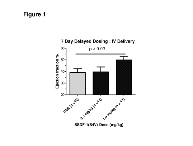
Figure 1 is a bar graph showing that SSDF-1(54V) delivered intravenously and 7
days post-
ischemia reperfusion injury improves Ejection Fraction (EF) by 10 percentage
points compared to the
PBS control.
Figure 2 is a graph showing that intracoronary administration of SSDF-1(54V)
immediately post-
infarct, followed by an intravenous administration of SSDF-1(54V) 4 weeks post-
infarct, in a micro
Yucatan pig model of ischemia reperfusion injury improves EF by 2.7 percentage
points compared to the
PBS control, even at 12 weeks post-infarct. 1-sided T-test performed; p <
0.05; n = 5 pigs/group.
7
CA 02933620 2016-06-10
WO 2015/089396
PCT/US2014/070010
Detailed Description
The present invention is based upon the discovery that the recovery of damaged
tissue, e.g.,
damaged cardiac tissue, is promoted by intravenous administration of wild-type
SDF-1 or SDF-1 that has
been mutated to increase resistance to enzymatic cleavage (e.g., cleavage by
one or more of MMP-2,
MMP-9, DPPIV, leukocyte elastase, cathepsin G, carboxypeptidase M, or
carboxypeptidase N). Such
peptides may be administered as isolated peptides, with or without a
pharmaceutically acceptable carrier.
In addition, we have surprisingly discovered that delayed administration from
within minutes after initial
occurrence of the tissue damage or after onset, recognition, or diagnosis of
the disease or condition, to
within 1 hour, 2 hours, 3 hours, 4 hours, 6 hours, 12 hours, 24 hours, at
least 48 hours, at least 3 days, 4
days, 5 days, 6 days, 7 days, 10 days, 2 weeks, one month, two months, three
months, six months, one
year, two years, or more after initial occurrence of the tissue damage or
after onset, recognition, or
diagnosis of the disease or condition is also useful in promoting the recovery
of damaged tissue. This
approach may be used to treat damaged tissue resulting from any type of injury
or disease.
8
CA 02933620 2016-06-10
WO 2015/089396
PCT/US2014/070010
Intravenous Administration
SDF-1 or mutant SDF-1 peptide-containing compositions or expressing stem cell
compositions
used in the methods of the present invention are administered intravenously,
for example, by intravenous
(IV) injection or using an implantable device (e.g., a catheter). Intravenous
administration generally
involves injections into any accessible vein in the body of a mammal,
including but not limited to a
peripheral vein (e.g., a vein on the arm, a vein in the leg, the back of the
hand, or the median cubital vein)
or via a central line to a large vein (e.g., the superior vena cava or
inferior vena cava or within the right
atrium of the heart). Intravenous administration can also include
administration by peripherally inserted
central catheter, central venous lines, or implantable ports.
A peripheral IV line consists of a short catheter (a few centimeters long)
inserted through the skin
into a peripheral vein (e.g., any vein that is not inside the chest or
abdomen) using, for example, a
cannula-over-needle device, in which a flexible plastic cannula comes mounted
on a metal trocar. The
part of the catheter that remains outside the skin is called the connecting
hub; it can be connected to a
syringe or an intravenous infusion line. Ported cannulae have an injection
port on the top that may be
used to administer the SDF-1 mutant SDF-1 peptides of the invention.
Peripherally inserted central catheter (P ICC) lines are used when IV access
is required over a
prolonged period of time or when the material to be infused would cause quick
damage and early failure
of a peripheral IV and when a conventional central line may be too dangerous
to attempt.
Also included in IV delivery methods of the invention are central venous lines
in which, for
example, a catheter is inserted into a subclavian internal jugular or a
femoral vein and advanced toward
the heart until it reaches the superior vena cava or right atrium.
Another central IV delivery method is through the use of a central IV line
which flows through a
catheter with its tip within a large vein, usually the superior vena cava or
inferior vena cava or within the
right atrium of the heart.
Another type of central line useful in the IV delivery methods of the
invention is a Hickman line or
Broviac catheter, which is inserted into the target vein and then "tunneled"
under the skin to emerge a
short distance away.
Implantable ports are also used for IV delivery of the SDF-1 and mutant SDF-1
peptide
compounds or stem cells of the invention. An implantable port is a central
venous line that does not have
an external connector; instead, it has a small reservoir that is covered with
silicone rubber and is
implanted under the skin. The peptide compounds are administered
intermittently by placing a small
needle through the skin, piercing the silicone, into the reservoir. A port can
be left in a subject's body for
weeks, months, even years. Intermittent infusion is another type of
intravenous administration that can
be used when a subject requires administration of the SDF-1 and mSDF-1 peptide
compounds or stem
cells of the invention only at certain times.
An SDF-1 or mSDF-1 peptide-containing composition or expressing stem cell
composition may
be administered into one vein or several veins. The SDF-1 or mSDF-1 peptide-
containing composition or
expressing stem cell composition can be intravenously administered for a
period of about 1 minute, 1 to 5
minutes, 10 to 20 minutes, 20 to 30 minutes, or for a sufficient time as
determined by the clinician into, for
example, one or more veins. The administration can be repeated intermittently
to achieve or sustain the
predicted benefit. The timing for repeat administration is based on the
subject's response, for example,
by monitoring symptoms associated with tissue damage. A therapeutically
effective dose or amount of
9
CA 02933620 2016-06-10
WO 2015/089396
PCT/US2014/070010
an SDF-1 or mSDF-1 peptide-containing composition or expressing stem cell
composition that is to be
given can be divided into two or more doses, and a dose may be administered
into two or more veins
with a single puncture or multiple punctures.
SDF-1 and Protease-Resistant Mutants
SDF-1 is a small cytokine belonging to the chemokine family that is officially
designated
chemokine (C-X-C motif) ligand 12 (CXCL12). SDF-1 is produced in multiple
forms, SDF-la (CXCL12a),
SDF-18 (CXCL12b), and SDF-1y, by alternate splicing of the same gene.
Unmutated SDF-la has the following sequence:
KPVSLSYRCPCRFFESHVARANVKHLKILNTPNCALQIVARLKNNN
RQVCIDPKLKWIQEYLEKALNK(SEQIDNO:52)
The SDF-1 peptides described herein include SDF-1 peptides with mutations to
render the peptides
resistant to, for example, matrix metalloproteinase-2 (MMP-2), matrix
metalloproteinase-9 (MMP-9),
dipeptidyl peptidase IV (DPPIV), leukocyte elastase, cathepsin G,
carboxypeptidase M, or carboxypeptidase
N. In the methods of the present invention, unmutated SDF-1 may also be
administered by intravenous
delivery for treatment or amelioration of tissue damage.
The methods of the invention feature mutant forms of SDF-1 (mSDF-1), which are
characterized
by a change in the third, fourth, fifth, and/or sixth amino acid residue from
the N-terminus of unmutated
SDF-1. mSDF-1 peptides of the invention have at least amino acids 1-8 of SEQ
ID NO:53 and may be
extended at the C-terminus by all or any portion of the remaining sequence of
SEQ ID NO:53, which has
the following sequence:
KPX3X4X5X6YRCPCRFFESHVARANVKHLKILNTPNCALQIVARLKN
KNRQVCIDPKLKWIQEYLEKALNK(SEQIDNO:53),wherein X3, X4, X5, and X6
are any amino acid residue.
In certain embodiments, X3 is valine, histidine, or cysteine.
In certain embodiments, X4 is serine or valine.
In certain embodiments, X5 is leucine, proline, threonine, or valine.
In certain embodiments, X6 is serine, cysteine, or glycine.
For example, the mSDF-1 peptide may include a mutation at the fourth (e.g.,
Ser¨Val) and/or
fifth (e.g., Leu¨>Pro) amino acid position.
KPVVLSYRCPCRFFESHVARANVKHLKILNTPNCALQIVARLKNNN
RQVCIDPKLKWIQEYLEKALNK(SEQIDNO:63)
KPVSPSYRCPCRFFESHVARANVKHLKILNTPNCALQIVARLKNNN
RQVCIDPKLKWIQEYLEKALNK(SEQIDNO:64)
KPVVPSYRCPCRFFESHVARANVKHLKILNTPNCALQIVARLKNNN
RQVCIDPKLKWIQEYLEKALNK(SEQIDNO:65)
In another example, the mSDF-1 peptide may include a Val¨His (SEQ ID NO:54) or
Val¨>Cys
(SEQ ID NO:55) mutation at the third amino acid position.
KPHSLSYRCPCRFFESHVARANVKHLKILNTPNCALQIVARLKNNN
RQVCIDPKLKWIQEYLEKALNK(SEQIDNO:54)
KPCSLSYRCPCRFFESHVARANVKHLKILNTPNCALQIVARLKNNN
RQVCIDPKLKWIQEYLEKALNK(SEQIDNO:55)
CA 02933620 2016-06-10
WO 2015/089396
PCT/US2014/070010
In other embodiments, the mSDF-1 peptide may include a Leu¨>Thr (SEQ ID NO:56)
or
Leu¨Val (SEQ ID NO:60) mutation at the fifth amino acid position.
KPVSTSYRCPCRFFESHVARANVKHLKILNTPNCALQIVARLKNNN
RQVCIDPKLKWIQEYLEKALNK(SEQIDNO:56)
KPVSVSYRCPCRFFESHVARANVKHLKILNTPNCALQIVARLKNNN
RQVCIDPKLKWIQEYLEKALNK(SEQIDNO:60)
In other embodiments, the mSDF-1 peptide may include a Ser¨>Cys (SEQ ID NO:61)
or
Ser¨>Gly (SEQ ID NO:62) mutation at the sixth amino acid position.
KPVSLCYRCPCRFFESHVARANVKHLKILNTPNCALQIVARLKNNN
RQVCIDPKLKWIQEYLEKALNK(SEQIDNO:61)
KPVSLGYRCPCRFFESHVARANVKHLKILNTPNCALQIVARLKNNN
RQVCIDPKLKWIQEYLEKALNK(SEQIDNO:62)
The methods of the invention may also include peptides that encompass any
combination of the
mutations described herein. For example, the mSDF-1 peptides may include a
Val¨>Cys mutation at the
third amino acid position of SEQ ID NO:53 and a Ser¨>Cys mutation at the sixth
amino acid position of
SEQ ID NO:53.
Mutations made to the SDF-1 peptides to confer protease resistance may also
include, for
example, the addition of a moiety (e.g., a proteinogenic amino acid or
protease protective organic group)
to the N-terminus of, e.g., the mSDF-1 peptides (described above), yielding Xp-
mSDF-1. For example, X
may be: R1-(CH2)d-, where d is an integer from 0-3, and R1 is selected from:
hydrogen (with the caveat
that when R1 is hydrogen, d must be at least 1); a branched or straight C1-C3
alkyl; a straight or branched
C2-C3 alkenyl; a halogen, CF3; -CONR5R4; -COOR5; -COR5; -(CH2)pNR5R4; -
(CH2)pSOR5; -(CH2)pS02R5,
-(CH2)pS02NR5R4; and 0R5, where R4 and R5 are each independently hydrogen or a
straight or branched
C1-C3 alkyl. In instances where an organic group is used for X, p should be 1.
X may also represent a
proteinogenic amino acid, wherein, for example, 1-10 (e.g., 1-9, 1-8, 1-7, 1-
6, 1-5, 1-4, 1-3, 1-2, or 1)
amino acid(s) is/are added to the N-terminus of SDF-1 (e.g., mSDF-1), and one
or more of these added
amino acids may be substituted with a protease protective organic group. For
example, a proteinogenic
amino acid (e.g., serine) or protease protective organic group may be added to
the N-terminus of SDF-1
(e.g., mSDF-1) to confer, for example, resistance to DPPIV cleavage without
substantially changing the
chemoattractant activity or resistance to other proteases (e.g., MMP-2). The
sequences below represent
exemplary SDF-1 mutants with a serine amino acid added to the N-terminus.
SKPX3X4X5X6YRCPCRFFESHVARANVKHLKILNTPNCALQIVARLK
NNNRQVCIDPKLKWIQEYLEKALNK(SEQIDNO:68),wherein X3, X4, X5, and X6
are any amino acid residue.
In certain embodiments, X3 is valine, histidine, or cysteine.
In certain embodiments, X4 is serine or valine.
In certain embodiments, X5 is leucine, proline, threonine, or valine.
In certain embodiments, X6 is serine, cysteine, or glycine.
Specific examples of sequences include:
SKPVVLSYRCPCRFFESHVARANVKHLKILNTPNCALQIVARLKNN
NRQVCIDPKLKWIQEYLEKALNK(SEQIDNO:69)
11
CA 02933620 2016-06-10
WO 2015/089396
PCT/US2014/070010
SKPVSPSYRCPCRFFESHVARANVKHLKILNTPNCALQIVARLKNN
NRQVCIDPKLKWIQEYLEKALNK(SEQIDNO:70)
SKPVVPSYRCPCRFFESHVARANVKHLKILNTPNCALQIVARLKNN
NRQVCIDPKLKWIQEYLEKALNK(SEQIDNO:71)
SKPHSLSYRCPCRFFESHVARANVKHLKILNTPNCALQIVARLKNN
NRQVCIDPKLKWIQEYLEKALNK(SEQIDNO:72)
SKPCSLSYRCPCRFFESHVARANVKHLKILNTPNCALQIVARLKNN
NRQVCIDPKLKWIQEYLEKALNK(SEQIDNO:73)
SKPVSTSYRCPCRFFESHVARANVKHLKILNTPNCALQIVARLKNN
NRQVCIDPKLKWIQEYLEKALNK(SEQIDNO:74)
SKPVSVSYRCPCRFFESHVARANVKHLKILNTPNCALQIVARLKNN
NRQVCIDPKLKWIQEYLEKALNK(SEQIDNO:75)
SKPVSLCYRCPCRFFESHVARANVKHLKILNTPNCALQIVARLKNN
NRQVCIDPKLKWIQEYLEKALNK(SEQIDNO:76)
SKPVSLGYRCPCRFFESHVARANVKHLKILNTPNCALQIVARLKNN
NRQVCIDPKLKWIQEYLEKALNK(SEQIDNO:77)
Mutations made to the SDF-1 peptides to confer protease resistance may also
include, for
example, the addition of a moiety (e.g., a proteinogenic amino acid) to the C-
terminus of, e.g., the mSDF-
1 peptides (described above), yielding mSDF-1-Y, or Xp-mSDF-1-Yz. Y may
represent a proteinogenic
amino acid, wherein, for example, 1-10 (e.g., 1-9, 1-8, 1-7, 1-6, 1-5, 1-4, 1-
3, 1-2, or 1) amino acid(s)
is/are added to the C-terminus of SDF-1 (e.g., mSDF-1 or Xp-mSDF-1). For
example, a proteinogenic
amino acid (e.g., serine) may be added to the C-terminus of SDF-1, mSDF-1, or
Xp-mSDF-1 to confer, for
example, resistance to carboxypeptidase M or carboxypeptidase N cleavage
without substantially
changing the chemoattractant activity or resistance to other proteases (e.g.,
MMP-2). In one
embodiment, the invention features an isolated mSDF-1-Yz or Xp-mSDF-1-Yz
peptide, wherein SDF-1
includes the amino acid sequence of SEQ ID NO:53. However, C-terminal
modifications may be made to
SDF-1 and any of the SDF-1 peptides described herein. The mutated SDF-1
peptides described herein
retain their ability to act as chemoattractants, but are resistant to
enzymatic (e.g., proteolytic) digestion.
The mSDF-1 peptides maintain chemoattractant activity with a sensitivity (as
determined by, e.g., the
effective concentration needed to obtain 50% of maximal response in the assays
of, e.g., Jurkat T cell
migration or any other chemotaxis assay known in the art) of at least, for
example, 10, 15, 20, 25, 30, 40,
50, 60, 70, 80, 90, 95, 99, or 100% of the sensitivity of unmutated SDF-1.
Loss of chemoattractant
activity may be due to cleavage by, for example, MMP-2, MMP-9, leukocyte
elastase, DPPIV, cathepsin
G, carboxypeptidase M, or carboxypeptidase N. The rate of inactivation of mSDF-
1 may be less than, for
example, 50, 45, 40, 35, 30, 25, 20, 15, 10, 5, or 1% of the rate of
inactivation of SDF-1.
The mutated SDF-1 peptides may be resistant to cleavage by, for example, MMP-
2, MMP-9,
DPPIV, leukocyte elastase, cathepsin G, carboxypeptidase M, or
carboxypeptidase N. Thus, they are
ideally suited for use at sites such as, e.g., damaged tissue (e.g., damaged
cardiac tissue), where
proteolytic enzymes are present at high concentrations, or delivery to the
site via the blood or plasma.
Accordingly, mutated SDF-1 peptides are suitable for intravenous
administration due to the improved
stability of such peptides.
12
CA 02933620 2016-06-10
WO 2015/089396
PCT/US2014/070010
Protease-resistant SDF-1 peptides described herein may include amino acids or
sequences
modified either by natural processes, such as posttranslational processing, or
by chemical modification
using techniques known in the art. Modifications may occur anywhere in a
polypeptide, including the
polypeptide backbone, the amino acid side-chains, and the amino- or carboxy-
terminus. The same type
of modification may be present in the same or varying degrees at several sites
in a given polypeptide, and
a polypeptide may contain more than one type of modification. Modifications
include, e.g., PEGylation,
acetylation, acylation, addition of acetomidomethyl (Acm) group, ADP-
ribosylation, alkylation, amidation,
biotinylation, carbamoylation, carboxyethylation, esterification, covalent
attachment to fiavin, covalent
attachment to a heme moiety, covalent attachment of a nucleotide or nucleotide
derivative, covalent
attachment of drug, covalent attachment of a marker (e.g., a fluorescent or
radioactive marker), covalent
attachment of a lipid or lipid derivative, covalent attachment of
phosphatidylinositol, cross-linking,
cyclization, disulfide bond formation, demethylation, formation of covalent
crosslinks, formation of cystine,
formation of pyroglutamate, formylation, gamma-carboxylation, glycosylation,
GPI anchor formation,
hydroxylation, iodination, methylation, myristoylation, oxidation, proteolytic
processing, phosphorylation,
prenylation, racemization, selenoylation, sulfation, transfer-RNA mediated
addition of amino acids to
proteins (e.g., arginylation), and ubiquitination. Posttranslational
modifications also include the addition of
polymers to stabilize the peptide or to improve pharmacokinetics or
pharmacodynamics. Exemplary
polymers include, e.g., poly(2-hydroxy ethyl methacrylate), poly(methyl
methacrylate), poly(acrylic acid),
poly(ethylene-co-vinyl acetate), poly(methacrylic acid), polyglycolides (PLG),
polyanhydrides, poly(N-vinyl
pyrrolidone), poly(vinyl alcohol), polyacrylamide, poly(ethylene glycol),
polylactides (PLA), poly(lactide-co-
glycolides) (PLGA), polyglutamic acid (PGA), and polyorthoesters.
Fusion Proteins
The methods of the invention may also utilize fusion proteins in which any of
the SDF-1, mSDF-1,
Xp-mSDF-1, mSDF-1-Yz, or Xp-mSDF-1-Yz peptide sequences described herein are
linked to the Fc
region of IgG (e.g., human IgG1). Alternatively, the Fc region may be derived
from IgA, IgM, IgE, or IgD
of humans or other animals, including swine, mice, rabbits, hamsters, goats,
rats, and guinea pigs. The
Fc region of IgG includes the CH2 and CH3 domains of the IgG heavy chain and
the hinge region. The
hinge serves as a flexible spacer between the two parts of the Fc fusion
protein, allowing each part of the
molecule to function independently. The Fc region used in the present
invention can be prepared in, for
example, monomeric and dimeric form.
An exemplary Fc fusion peptide is S-SDF-1(54V)-Fc with the following amino
acid sequence.
The GGGGS linker (SEQ ID NO:66) is indicated in bold and the Fc peptide is
underlined.
SKPVVLSYRCPCRFFESHVARANVKHLKILNTPNCALQIVARLKNN
NRQVCIDPKLKWIQEYLEKALNKGGGGSVDKTHTCPPCPAPELLG
GPSVFLFPPKPKDTLMetISRTPEVTCVVVDVSHEDPEVKFNWYVD
GVEVHNAKTKPREEQYNSTYRVVSVLTVLHQDWLNGKEYKCKVSN
KALPAPIEKTISKAKGQPREPQVYTLPPSRDELTKNQVSLTCLVKG
FYPSDIAVEWESNGQPENNYKTTPPVLDSDGSFFLYSKLTVDKSR
WQQGNVFSCSVMetHEALHNHYTQKSLSLSPGK(SEQIDNO:67)
Other non-limiting examples of Fc fusion peptides include, e.g., SDF-1(54V)-
Fc, SDF-1(L5P)-Fc,
SDF-1(56C)-Fc, SDF-1(V3H)-Fc, SDF-1-Fc, S-SDF-1-Fc, and SDF-1-Fc.
13
CA 02933620 2016-06-10
WO 2015/089396
PCT/US2014/070010
All of the above proteins are included in the terms "SDF-1 and mSDF-1 proteins
of the invention"
or "peptides of the invention."
Peptide Synthesis
The SDF-1 or protease-resistant mutant SDF-1 peptides used in the methods of
the present
invention can be made by solid-phase peptide synthesis using, for example,
standard N-tert-
butyoxycarbonyl (t-Boc) chemistry and cycles using n-methylpyrolidone
chemistry. Exemplary methods
for synthesizing peptides are found, for example, in U.S. Patent Nos.
4,192,798; 4,507,230; 4,749,742;
4,879,371; 4,965,343; 5,175,254; 5,373,053; 5,763,284; and 5,849,954, hereby
incorporated by
reference. These peptides may also be made using recombinant DNA techniques.
Once peptides have been synthesized, they can be purified using procedures
such as, for
example, HPLC on reverse-phase columns. Purity may also be assessed by H PLC,
and the presence of
a correct composition can be determined by amino acid analysis. A purification
procedure suitable for
mSDF-1 peptides is described, for example, in U.S. Patent Application
Publication No. 2008/0095758,
hereby incorporated by reference.
Fusion proteins may either be chemically synthesized or made using recombinant
DNA
techniques. Other non-limiting examples of Fc fusion peptides include, e.g.,
SDF-1(54V)-Fc, SDF-
1(L5P)-Fc, SDF-1(56C)-Fc, SDF-1(V3H)-Fc, SDF-1-Fc, S-SDF-1-Fc, and SDF-1-Fc.
14
CA 02933620 2016-06-10
WO 2015/089396
PCT/US2014/070010
Stem Cells Expressing SDF-1 Peptides
The invention provides stem cells and/or progeny cells thereof that are
genetically modified, for
example, to express and/or secrete a peptide of the invention (e.g., SDF-1 or
protease-resistant mutant
SDF-1 peptides). Any suitable stem cell may be genetically modified to express
and/or secrete a peptide
of the invention, including, for example, adult stem cells, mesenchymal
precursor cells (MPCs), and
mesenchymal stem cells (MSCs). In some embodiments, the stem cell may
naturally express a basal
level of a wild-type SDF-1, and the genetic modification may cause the stem
cell to express an increased
level of wild-type SDF-1 and/or to express a protease-resistant mutant SDF1-
peptide.
Methods for genetically modifying a cell, for example a stem cell, will be
apparent to the skilled
artisan. For example, a nucleic acid that is to be expressed in a cell is
operably-linked to a promoter for
inducing expression in the cell. For example, the nucleic acid is linked to a
promoter operable in a variety
of cells of a subject, such as, for example, a viral promoter, e.g., a CMV
promoter (e.g., a CMV-IE
promoter) or a SV-40 promoter. In other instances, the promoter may be
operable specifically in a
particular type of stem cell. Additional suitable promoters are known in the
art and shall be taken to apply
mutatis mutandis to the present example of the disclosure.
In one example, the nucleic acid is provided in the form of an expression
construct. As used
herein, the term "expression construct" refers to a nucleic acid that has the
ability to confer expression on
a nucleic acid (e.g. a reporter gene and/or a counter-selectable reporter
gene) to which it is operably
connected, in a cell. Within the context of the present disclosure, it is to
be understood that an
expression construct may comprise or be a plasmid, bacteriophage, phagem id,
cosm id, virus sub-
genomic or genomic fragment, or other nucleic acid capable of maintaining
and/or replicating
heterologous DNA in an expressible format.
Methods for the construction of a suitable expression construct for
performance of the disclosure
will be apparent to the skilled artisan and are described, for example, in
Ausubel et al. (In: Current
Protocols in Molecular Biology. Wiley lnterscience, ISBN 047 150338, 1987) or
Sambrook et al. (In:
Molecular Cloning: Molecular Cloning: A Laboratory Manual, Cold Spring Harbor
Laboratories, New York,
Third Edition 2001). For example, each of the components of the expression
construct is amplified from a
suitable template nucleic acid using, for example, polymerase chain reaction
(PCR) and subsequently
cloned into a suitable expression construct, such as for example, a plasmid or
a phagemid.
Vectors suitable for such an expression construct are known in the art and/or
described herein.
For example, an expression vector suitable for methods of the present
disclosure in a mammalian cell is,
for example, a vector of the pcDNA vector suite supplied by lnvitrogen, a
vector of the pCI vector suite
(Promega), a vector of the pCMV vector suite (Clontech), a pM vector
(Clontech), a pSI vector (Promega),
a VP 16 vector (Clontech) or a vector of the pcDNA vector suite (lnvitrogen).
The skilled artisan will be aware of additional vectors and sources of such
vectors, such as, for
example, Life Technologies Corporation, Clontech or Promega.
Methods for introducing the isolated nucleic acid molecule or a gene construct
comprising same
into a cell for expression are known to those skilled in the art. The
technique used for a given organism
depends on the known successful techniques. Methods for introducing
recombinant DNA into cells
include microinjection, transfection mediated by DEAE-dextran, transfection
mediated by liposomes such
as by using lipofectamine (Gibco, Md., USA) and/or cellfectin (Gibco, Md.,
USA), PEG-mediated DNA
CA 02933620 2016-06-10
WO 2015/089396
PCT/US2014/070010
uptake, electroporation and microparticle bombardment such as by using DNA-
coated tungsten or gold
particles (Agracetus Inc., WI, USA) amongst others.
Alternatively, an expression construct of the disclosure is a viral vector.
Suitable viral vectors are
known in the art and commercially available. Conventional viral-based systems
for the delivery of a
nucleic acid and integration of that nucleic acid into a host cell genome
include, for example, a retroviral
vector, a lentiviral vector or an adeno-associated viral vector.
Alternatively, an adenoviral vector is useful
for introducing a nucleic acid that remains episomal into a host cell. Viral
vectors are an efficient and
versatile method of gene transfer in target cells and tissues. Additionally,
high transduction efficiencies
have been observed in many different cell types and target tissues.
For example, a retroviral vector generally comprises cis-acting long terminal
repeats (LTRs) with
packaging capacity for up to 6-10 kb of foreign sequence. The minimum cis-
acting LTRs are sufficient for
replication and packaging of a vector, which is then used to integrate the
expression construct into the
target cell to provide long term expression. Widely used retroviral vectors
include those based upon
murine leukemia virus (MuLV), gibbon ape leukemia virus (GaLV), simian
immunodeficiency virus (SrV),
human immunodeficiency virus (HIV), and combinations thereof (see, e.g.,
Buchscher et al. J. Virol.
56:2731-2739 (1992); Johann et al. J. Virol 65:1635-1640 (1992); Sommerfelt et
al. Virol 76:58-59 (1990);
Wilson et al. J. Virol 63:274-2318 (1989); Miller et al. J. Virol 65:2220-2224
(1991); PCT/U594/05700;
Miller et al. BioTechniques 7:980-990, 1989; Miller, Human Gene Therapy 7:5-
14, 1990; Scarpa et al.
Virology 75:849-852, 1991; and Burns et al. Proc. Natl Acad. Sci USA 90:8033-
8037, 1993).
Various adeno-associated virus (AAV) vector systems have also been developed
for nucleic acid
delivery. AAV vectors can be readily constructed using techniques known in the
art. See, for example,
U.S. Pat. Nos. 5,173,414 and 5,139,941; International Publication Nos. WO
92/01070 and WO 93/03769;
Lebkowski et al. Molec. Cell Biol 5:3988-3996, 1988; Vincent et al. (1990)
Vaccines 90 (Cold Spring
Harbor Laboratory Press); Carter, Current Opinion in Biotechnology 5:533-539,
1992; Muzyczka, Current
Topics in Microbiol, and lmmunol. 755:97-129, 1992; Kotin, Human Gene Therapy
5:793-801, 1994;
Shelling et al. Gene Therapy 7:165-169, 1994; and Zhou et al. J Exp. Med.
779:1867-1875, 1994.
Additional viral vectors useful for delivering an expression construct of the
disclosure include, for
example, those derived from the pox family of viruses, such as vaccinia virus
and avian poxvirus or an
alphavirus or a conjugate virus vector (e.g. that described in Fisher-Hoch et
al. Proc. Natl Acad. Sci. USA
56:317-321, 1989).
Co-administration with Exogenous Stem Cells
Any of the peptides or stem cells (e.g., stem cells expressing a SDF-1 or
protease-resistant
mutant SDF-1 peptide) employed in the methods of the present invention may be
administered with
exogenous stem cells. Cells that may be administered in conjunction with the
peptides or genetically
modified stem cells of the invention include, but are not limited to,
multipotent or pluripotent stem cells, or
bone marrow cells. Examples of suitable exogenous stem cells include adult
stem cells, mesenchymal
precursor cells (for example, cells that express the Mesenchymal Precursor
Cell Marker STRO-1, e.g.,
STRO-1 bright cells, as described in US Publ. No. 2014/0271567), and
mesenchymal stem cells. In some
embodiments, an exogenous stem cell may be allogeneic to the subject. In other
embodiments, an
exogenous stem cell may be autologous to the subject.
16
CA 02933620 2016-06-10
WO 2015/089396
PCT/US2014/070010
The exogenous stem cells may be admixed with a composition of the invention
immediately or
shortly prior to administration, or they may be co-cultured together for a
period of time prior to
administration. In other instances, the exogenous stem cells may be
administered separately from the
peptide and/or stem cell (e.g., stem cell expressing a SDF-1 or protease-
resistant SDF-1 peptide) of the
invention. The exogenous stem cell may be administered before, after, or
concurrently with the peptide
or expressing stem cell.
In one example, a composition administered to a subject may include an
effective amount or a
therapeutically or prophylactically effective amount of stem cells. An
exemplary range of stem cells to be
administered is about 1x10³ cells/kg to about 1x10⁹ cells/kg (e.g., 1x10³
cells/kg, 1x10⁴ cells/kg, 1x10⁵
cells/kg, 1 x1 06 cells/kg, 1 x1 07 cells/kg, 1 x1 08 cells/kg, 1 x1 09
cells/kg). For instance, the composition may
comprise about 1x10⁵ STRO-1+ cells/kg to about 1x10⁷STRO-1+ cells/kg, or about
1x10⁶to about 5x10⁶
STRO-1+ cells/kg. The exact amount of cells to be administered is dependent
upon a variety of factors,
including the age, weight, and sex of the patient, and the extent and severity
of tissue damage in the
subject.
In one example, the cells are administered as a total cell number dose
irrespective of the
subject's weight. For example, in some instances, the stem cells are
administered at a dose of between
about 50 million to 500 million cells (e.g., 50 million, 100 million, 150
million, 200 million, 250 million, 300
million, 350 million, 400 million, 450 million, or 500 million cells)
irrespective of the weight of the subject.
In some instances, the stem cells are contained within a chamber that does not
permit the cells to
exit into a subject's circulation, however that permits factors secreted by
the cells to enter the circulation.
In this manner soluble factors may be administered to a subject by permitting
the cells to secrete factors
into the subject's circulation. Such a chamber may be implanted at a site in a
subject to increase local
levels of the soluble factors, e.g., implanted near a site of tissue damage in
a subject.
In some examples of the invention, it may not be necessary or desirable to
immunosuppress a
subject prior to initiation of therapy with compositions that include
exogenous stem cells. For example,
transplantation with allogeneic, or even xenogeneic, STRO1+ cells or progeny
thereof may be tolerated in
some instances.
However, in other examples it may be desirable or appropriate to
pharmacologically
immunosuppress a patient prior to initiating cell therapy and/or reduce an
immune response of a subject
against a composition that includes exogenous stem cells. This may be
accomplished through the use of
systemic or local immunosuppressive agents, of which a wide variety are known
in the art, or it may be
accomplished by delivering the cells in an encapsulated device, as described
above. The cells may be
encapsulated in a capsule that is permeable to nutrients and oxygen required
by the cell and to
therapeutic factor(s) that the cell is secreting yet impermeable to immune
humoral factors and cells. For
example, the encapsulant is hypoallergenic, is easily and stably situated in a
target tissue, and provides
added protection to the implanted structure. These and other means for
reducing or eliminating an
immune response to the transplanted cells are known in the art. As an
alternative, the exogenous stem
cells may be genetically modified to reduce their immunogenicity.
Pharmaceutical Compositions and Dosages
Any of the peptides or stem cells employed in the methods of the present
invention may be
contained in any appropriate amount in any suitable carrier substance, and the
protease-resistant
17
CA 02933620 2016-06-10
WO 2015/089396
PCT/US2014/070010
peptides or fusion proteins are generally present in an amount of 1-95% by
weight of the total weight of
the composition, e.g., 5%, 10%, 20%, or 50%. The protease-resistant SDF-1
peptides or fusion proteins
described herein may be incorporated into a pharmaceutical composition
containing a carrier such as,
e.g., saline, water, Ringer's solution, and other agents or excipients. The
composition is designed for
intravenous delivery (e.g., by injection or implantable port). Thus, the
composition may be in the form of,
e.g., suspensions, emulsions, solutions, or injectables. All compositions may
be prepared using methods
that are standard in the art (see, e.g., Remington's Pharmaceutical Sciences,
16th ed., A. Oslo. ed.,
Easton, PA (1980)).
The peptides of the invention can be delivered in a controlled-release or
sustained-release
system. For example, polymeric materials can be used to achieve controlled or
sustained release of the
peptides (see, e.g., Medical Applications of Controlled Release, Langer and
Wise (eds.), CRC Pres.,
Boca Raton, Fla. (1974); Controlled Drug Bioavailability, Drug Product Design
and Performance, Smolen
and Ball (eds.), Wiley, N.Y. (1984); U.S. Patent Nos. 5,679,377; 5,916,597;
5,912,015; 5,989,463; and
5,128,326; PCT Publication Nos. WO 99/15154 and WO 99/20253, hereby
incorporated by reference).
Examples of polymers used in sustained-release formulations include, e.g.,
poly(2-hydroxy ethyl
methacrylate), poly(methyl methacrylate), poly(acrylic acid), poly(ethylene-co-
vinyl acetate),
poly(methacrylic acid), polyglycolides (PLG), polyanhydrides, poly(N-vinyl
pyrrolidone), poly(vinyl alcohol),
polyacrylamide, poly(ethylene glycol), polylactides (PLA), poly(lactide-co-
glycolides) (PLGA), polyglutamic
acid (PGA), and polyorthoesters.
It is expected that the skilled practitioner can adjust dosages of the peptide
on a case by case
basis using methods well established in clinical medicine. The optimal dosage
may be determined by
methods known in the art and may be influenced by factors such as the age of
the subject being treated,
disease state, and other clinically relevant factors. Generally, when
administered to a human, the dosage
of any of the therapeutic agents (e.g., SDF-1 or protease-resistant mutant SDF-
1 peptides) described
herein will depend on the nature of the agent and can readily be determined by
one skilled in the art.
Typically, such a dosage is normally about 0.001 pg to 2000 mg per day,
desirably about 1 mg to 1000
mg per day, and more desirably about 5 mg to 500 mg per day. In one embodiment
the dosage is 0.01
mg/kg to 100 mg/kg, or desirably 1 mg/kg to 10 mg/kg per day (e.g., 1 mg/kg, 2
mg/kg, 3 mg/kg, 4 mg/kg,
5 mg/kg, 6 mg/kg, 7 mg/kg, 8 mg/kg, 9 mg/kg, and 10 mg/kg per day).
The peptides or stem cells of the invention may be administered intravenously
once, twice, three
times, four times, or five times each day; once per week, twice per week,
three times per week, four times
per week, five times per week, or six times per week; once per month, once
every two months, once
every three months, or once every six months; or once per year. Alternatively,
the peptides or stem cells
of the invention may be administered one or two times and repeated
administration may not be needed.
Administration of the peptides or stem cells described herein can continue
until tissue damage (e.g.,
tissue damage resulting from myocardial infarction or peripheral vascular
disease) has healed or has
been ameliorated. The duration of therapy can be, e.g., one day to one week,
one week to one month,
one week to one year, or one week to more than one year; alternatively, the
peptides or stem cells of the
invention can be administered for a shorter or a longer duration. Continuous
daily dosing with the
peptides or stem cells may not be required. A therapeutic regimen may require
cycles, during which time
a composition is not administered, or therapy may be provided on an as-needed
basis.
18
CA 02933620 2016-06-10
WO 2015/089396
PCT/US2014/070010
The SDF-1 or mutant SDF-1 peptides or stem cells of the invention may be
delivered immediately
at the time of tissue damage or within minutes after initial occurrence of the
tissue damage or after onset,
recognition, or diagnosis of the disease or condition (e.g., post myocardial
infarction or acute organ
damage, such as acute kidney or liver damage). The SDF-1 or mutant SDF-1
peptides of the invention
can also be delivered after a short or long delay following the initial tissue
damage. For example, the
SDF-1 or mutant SDF-1 peptides or stem cells of the invention can be delivered
at any period after the
initial damage occurs ranging from several minutes to within 1 hour, 2 hours,
3 hours, 4 hours, 6 hours,
12 hours, 24 hours, at least 48 hours, at least 3 days, 4 days, 5 days, 6
days, 7 days, 10 days, 2 weeks,
one month, two months, three months, six months, one year, two years, or more
after initial occurrence of
the tissue damage or after onset, recognition, or diagnosis of the disease or
condition. For tissue
damage that is more chronic in nature and occurs over time, including but not
limited to PVD, diabetic
wounds, chronic organ damage (for example, chronic kidney or liver damage),
and damage resulting from
inflammatory conditions (for example, rheumatoid arthritis or Crohn's
disease), the SDF-1 or mutant SDF-
1 peptides or stem cells of the invention may be delivered immediately after
the onset of the damage or
immediately after the diagnosis or initial or subsequent indications of the
damage (e.g., PVD or diabetic
wounds). In such cases, the delivery of the SDF-1 or mutant SDF-1 peptides or
stem cells of the
invention may be three days, seven days, one week, two weeks, three weeks, a
month, two months,
three months, four months, five months, six months, or even a year or more
after the tissue damage has
occurred or after onset, recognition, or diagnosis of the tissue damage or
disease or condition.
For any type of tissue damage, disease, or disorder described herein, initial
IV administration of
the SDF-1 or mutant SDF-1 peptides or stem cells of the invention may be at a
time ranging from minutes
to two years after the initial occurrence, recognition or diagnosis of tissue
damage, or one hour to two
years after the initial occurrence, recognition or diagnosis of tissue damage,
one day to one year after the
initial occurrence, recognition or diagnosis of tissue damage, one day to six
months after the initial
occurrence, recognition or diagnosis of tissue damage, one month to six months
after the initial
occurrence, recognition or diagnosis of tissue damage, one day to one month
after the initial occurrence,
recognition or diagnosis of tissue damage, one week to one month after the
initial occurrence, recognition
or diagnosis of tissue damage, one week to two weeks after the initial
occurrence, recognition or
diagnosis of tissue damage, one hour to one week after the initial occurrence,
recognition or diagnosis of
tissue damage, one hour to three days after the initial occurrence,
recognition or diagnosis of tissue
damage, or several minutes to one hour after the initial occurrence,
recognition or diagnosis of tissue
damage.
The SDF-1 or mutant SDF-1 peptides or stem cells of the invention may be
delivered once over
the duration of therapy or multiple times over the duration of therapy.
Depending on the severity of the
tissue damage, the SDF-1 or mutant SDF-1 peptides or stem cells of the
invention may be delivered
repeatedly over time to ensure repair or recovery of the damaged tissue.
In addition, the intravenous delivery of the SDF-1 or mutant SDF-1 peptides or
stem cells of the
invention may be combined with additional forms of delivery of the SDF-1 or
mutant SDF-1 peptides or
stem cells of the invention. In one example, such as after a myocardial
infarction, SDF-1 or mutant SDF-
1 peptides or stem cells of the invention may be delivered initially via intra-
coronary or intra-arterial
methods and then followed by subsequent delivery of either SDF-1 or mutant SDF-
1 peptides or stem
cells via intravenous methods. In another example, SDF-1 or mutant SDF-1
peptides or stem cells may
19
CA 02933620 2016-06-10
WO 2015/089396
PCT/US2014/070010
be delivered initially via intramuscular or intramyocardial methods and then
followed by subsequent
delivery of either therapy via intravenous methods. In any of these multiple
delivery methods, intravenous
administration would begin 1 day, 2 days, 3 days, 4 days, 5 days, 6 days, 7
days, 2 weeks, 1 month, 2
month, 3 months, 4 months, 5 months, 6 months, one year, or more after the
initial delivery. Here again,
depending on the severity of the tissue damage, the SDF-1 or mutant SDF-1
peptides or stem cells of the
invention may be delivered repeatedly over time to ensure repair or recovery
of the damaged tissue.
Appropriate dosages of the peptides or stem cells used in the methods of the
invention depend
on several factors, including the administration method, the severity of the
disorder, and the age, weight,
and health of the subject to be treated. Additionally, pharmacogenomic
information (e.g., the effect of
genotype on the pharmacokinetic, pharmacodynamic, or efficacy profile of a
therapeutic) about a
particular subject may affect the dosage used.
Diagnosis and Treatment
The methods of the present invention are useful for treating any subject that
has been diagnosed
with or has suffered from tissue damage (e.g., damage to cardiac tissue due to
myocardial infarction or
tissue damage resulting from peripheral vascular disease) or wounds (e.g.,
diabetic wounds). Tissue
damage may be the result of, for example, a cardiovascular condition (e.g.,
myocardial infarction);
peripheral vascular disease (PVD); peripheral artery disease (PAD); ulcers
(e.g.,skin wound ulcers);
surgery; or diabetes. Tissue damage may also result form CNS disorders or
injury or inflammatory
conditions (such as rheumatoid arthritis, Crohn's disease, or graft-versus-
host disease). The methods of
the invention may also be used for repair or regeneration of organ damage (for
example, kidney or liver
damage) resulting from disease or injury. The methods of the present invention
may be used to promote
wound healing or tissue repair. One skilled in the art will understand that
subjects of the invention may
have been subjected to standard tests or may have been identified, without
examination, as one at high
risk due to the presence of one or more risk factors. Diagnosis of these
disorders may be performed
using any standard method known in the art.
The methods described herein may also be used to treat any disease or
condition characterized
by a high concentration of protease (e.g., MMP-2, MMP-9, DPPIV, leukocyte
elastase, cathepsin G,
carboxypeptidase M, and/or carboxypeptidase N), where the attraction of stem
cells upon the
administration of a protease-resistant SDF-1 peptide may induce regeneration
or healing. Exemplary
disorders to be treated by compositions of the present invention include
inflammatory and ischemic
diseases (e.g., myocardial infarction, stroke or limb ischemia), wound
healing, and diabetic ulcers.
The efficacy of treatment can be monitored using methods known to one of skill
in the art
including, e.g., assessing symptoms of the disease or disorder, physical
examination, histopathological
examination, blood chemistry analysis, computed tomography, cytological
examination, and magnetic
resonance imaging. In certain embodiments, hemodynamic data is collected to
determine the efficacy of
treatment. Hemodynamic tests may include, e.g., determining an ejection
fraction (e.g., fraction of blood
pumped out of ventricles with each heart beat), determining end diastolic
pressure, and determining end
systolic elastance (e.g., volume of blood present in the left ventricle). In
one example, hemodynamic
tests may be used to monitor cardiac function in a subject that has suffered
tissue damage resulting from
myocardial infarction or other form of cardiac ischemia.
CA 02933620 2016-06-10
WO 2015/089396
PCT/US2014/070010
The methods of the present invention may be used in combination with
additional therapies to
promote wound healing or tissue repair. Treatment therapies that can be used
in combination with the
methods of the invention include, but are not limited to, heparin, 6-blockers
(e.g., atenolol, metoprolol,
nadolol, oxprenolol, pindolol, propranolol, or timolol), angiotensin-
converting enzyme (ACE) inhibitors
(e.g., captopril, enalapril, fosinopril, lisinopril, perindopril, quinapril,
ramipril, trandolapril, or benazepril),
angiotensin II receptor blockers (e.g., candesartan, eprosartan, irbesartan,
losartan, olmesartan,
telmisartan, or valsartan), diuretics, aspirin, cholesterol-lowering drugs
(e.g., HMG-CoA reductase
inhibitors (e.g., atorvastatin, cerivastatin, fluvastatin, lovastatin,
mevastatin, pitavastatin, pravastatin,
rosuvastatin, or simvastatin)), cell therapy, anti-platelet drugs (e.g.,
clopidogrel, prasugrel, ticlopidine,
cilostazol, abciximab, eptifibatide, tirofiban, or dipyridamole), anti-
hypertensive drugs, anti-arrhythmic
drugs (e.g., quinidine, procainamide, disopyramide, lidocaine, mexiletine,
tocainide, phenytoin, moricizine,
flecainide, sotalol, ibutilide, amiodarone, bretylium, dofetilide, diltiazem,
or verapamil), angiogenic drugs,
wound dressings, PDGF, and/or negative pressure devices and therapies.
Examples
The present invention is illustrated by the following example, which is in no
way intended to be
limiting of the invention.
Example 1. Delayed and IV dosing of protease resistant SDF-1 variants improve
cardiac function
in a rodent ischemia reperfusion model
In the following example, we describe experiments demonstrating that
intravenous delivery and
long term delayed dosing of an mSDF-1 peptide-containing composition improves
cardiac function in an
ischemia reperfusion model.
Rats were anesthetized with 0.05 mg/kg of buprenorphine and 2-3% of
isoflurane. After
intubation, the chest was opened between ribs 4 and 5, and the left anterior
descending (LAD) coronary
artery was ligated for 90 minutes. After 90 minutes, the suture was removed
from the LAD to initiate
reperfusion in the infarct zone. The chest and skin of the rats were then
closed. mSDF-1 peptide was
administered by intravenous injection 7 days post infarction (>15 rats per
group). For intravenous
injection, 100 I of S-SDF-1 (54V) (at doses of 0, 0.1, and 1.0 mg/kg) in PBS
were injected into the tail
veins of rats.
In each of the experiments described above, hemodynamic function in the rats
was analyzed in a
randomized and blinded study four weeks after intravenous dosing (five weeks
post the ischemia
reperfusion injury). Rats were anesthetized with 0.05 mg/kg of buprenorphine
and 2-3% of isoflurane. A
16G endotracheal tube was inserted into the rats and mechanical ventilation
was started. The left jugular
vein was cannulated with PE 10 to deliver hyperosmotic saline (50 I of a 25%
NaCI solution in water).
Hyperosmotic saline was used to measure parallel conductance of the volume
measurements.
To determine the ejection fraction (EF) and intra-ventricular pressure, the
right carotid artery was
cannulated. A pressure-volume catheter was inserted and passed into the left
ventricle. A baseline
pressure-volume measurement was obtained. A hyperosmotic saline solution
(described above) was
injected into the jugular vein, and a pressure-volume measurement was then
obtained.
21
CA 02933620 2016-06-10
WO 2015/089396
PCT/US2014/070010
Our results showed that intravenous injection of S-SDF-1(S4V) delivered 7 days
post ischemia
reperfusion injury resulted in a 10% improvement in the measured ejection
fraction in rats compared to
the PBS control (Figure 1).
Example 2. Delayed and IV dosing of protease resistant SDF-1 variants improve
cardiac function
in a pig ischemia reperfusion model
We also assessed the effects of intravenous delivery and delayed dosing of
mSDF-1 peptide-
containing compositions on cardiac function in a micro Yucatan pig infarct
model.
In these experiments, pigs were anesthetized and their left anterior
descending (LAD) coronary
artery was occluded by balloon catheter. After 90 minutes, the balloon
catheter was removed from the
LAD to initiate reperfusion in the infarct zone. The chest and skin of the
pigs were then closed.
Randomized and blinded studies were performed in which pigs were first dosed
with either mSDF-1
peptide (at 1 mg/kg or 3 mg/kg) or a PBS control via intracoronary
administration immediately post-
ischemia (n = 5 pigs for each of the three groups). At 4 weeks (one month)
post-infarct, a second dose of
mSDF-1 peptide (at 1 mg/kg or 3 mg/kg) or a PBS control was administered
intravenously.
In each of the experiments described above, the ejection fraction (EF) was
determined at 4
weeks post-infarct, 8 weeks post-infarct, and 12 weeks post-infarct. Our
results demonstrated significant
improvements in the EF at 12 weeks post-infarct in pigs dosed at 3 mg/kg with
mSDF-1 peptide (Figure
2). In particular, we observed an absolute EF improvement of 2.7% at 12 weeks
post-infarct in pigs
administered with 3 mg/kg mSDF-1 peptide compared to the control group (1-
sided T-test, p < 0.05). The
lower dose group (1 mg/kg mSDF-1 peptide) also showed a clear trend toward EF
improvement, which
was sustained from 4-12 weeks (Figure 2).
Other Embodiments
From the foregoing description, it is apparent that variations and
modifications may be made to
the invention described herein to adopt it to various usages and conditions.
Such embodiments are also
within the scope of the following claims.
All publications, patent applications, and patents, mentioned in this
specification are herein
incorporated by reference to the same extent as if each independent
publication or patent application was
specifically and individually indicated to be incorporated by reference.
22