Note: Descriptions are shown in the official language in which they were submitted.
CA 02933707 2016-06-13
WO 2015/095895
PCT/US2014/072007
TAGGED CHIMERIC EFFECTOR MOLECULES AND RECEPTORS THEREOF
CROSS REFERENCE TO RELATED APPLICATION
This application claims the benefit under 35 U.S.C. 119(e) to U.S.
Provisional Application No. 61/919,201 filed on December 20, 2013, which
application
is incorporated by reference herein in its entirety.
STATEMENT OF GOVERNMENT INTEREST
This invention was made with government support under Grant/Contract
No. CA136551 awarded by the National Institutes of Health. The government has
certain rights in this invention.
STATEMENT REGARDING SEQUENCE LISTING
The Sequence Listing associated with this application is provided in text
format
in lieu of a paper copy, and is hereby incorporated by reference into the
specification.
The name of the text file containing the Sequence Listing is
360056 426W0 SEQUENCE LISTING.txt. The text file is 32.3 KB, was created on
December 22, 2014, and is being submitted electronically via EFS-Web.
BACKGROUND
Technical Field
The present disclosure relates to fusion proteins containing a tag cassette
and,
more particularly, to tagged chimeric effector molecules (Key-ChEMs) and
tagged
chimeric antigen receptor molecules (T-ChARMs), and recombinant host cells
producing such fusion proteins, wherein the recombinant host cells can be
identified,
isolated, sorted, induced to proliferate, tracked, eliminated, and/or used as
a therapeutic
(e.g., in adoptive immunotherapy).
Description of the Related Art
T cell-based immunotherapies began to be developed when tumor-reactive T
cells were found among a population of tumor-infiltrating lymphocytes (TILs)
(Clark et
at., Cancer Res. 29:705, 1969). One strategy, known as adoptive T cell
transfer,
involves the isolation of tumor infiltrating lymphocytes pre-selected for
tumor-
reactivity, clonal expansion of the tumor-reactive T cells induced by anti-CD3
and anti-
1
CA 02933707 2016-06-13
WO 2015/095895 PCT/US2014/072007
CD28 antibodies in the presence of IL-2, and finally infusing the expanded
cell
population back to the tumor-bearing patient (together with chemotherapy and
repetitive administration of IL-2) (Dudley et at., Science 298:850, 2002).
This form of
adoptive T cell therapy with tumor infiltrating lymphocytes is technically
cumbersome
and leads to complete remission in only a minor fraction of patients with
melanoma and
is rarely effective in other cancers (Besser et at., Clin. Cancer Res.
/6:2646, 2010).
Isolation of tumor-reactive T cell clones led to the development of another
immunotherapeutic approach ¨ the generation of recombinant T cell receptors
(TCRs)
specific for particular antigens, which are introduced into T cells using a
vector delivery
system to confer specificity for a tumor-associated peptide presented by an
MHC
molecule expressed on a tumor cell. A similar approach introduces a synthetic
receptor,
termed a chimeric antigen receptor (CAR), which contains an antigen-binding
domain,
which, e.g., in the context of anti-tumor therapy can bind to a tumor-specific
or
associated antigen, linked to one or more intracellular component comprising
an
effector domains, such as a TCR and/or costimulatory signaling domains. Unlike
TILs,
the basic procedure for TCR or CAR T cell immunotherapy is to genetically
modify
human T cells with a transgene encoding a tumor targeting moiety, ex vivo
expansion of
the recombinant T cells, and transfusing the expanded recombinant T cells back
into
patients. In the case of adoptive therapy with CART cells, the composition of
the
synthetic CAR structure, as well as the quality and purity of the genetically
engineered
T cells, will determine therapeutic efficacy against tumors in vivo. But,
there are
challenges to expanding and selecting the recombinant cell populations, as
well as
making sure the cells are effective and specific enough in vivo to avoid
serious
autoimmune side effects.
Currently, there remains a need in the immunotherapy field for compositions
and methods for identifying, efficiently isolating/sorting, selectively
expanding, in vivo
tracking and controlling or eliminating engineered cells, such as engineered
immune
cells (e.g., T cells).
BRIEF SUMMARY
In certain aspects, the present disclosure is directed to a single chain
fusion
protein, comprising an extracellular component and an intracellular component
connected by a hydrophobic portion, wherein the extracellular component
comprises a
binding domain that specifically binds a target, a tag cassette, and a
connector region
comprising a hinge, and wherein the intracellular component comprises an
effector
domain.
2
CA 02933707 2016-06-13
WO 2015/095895
PCT/US2014/072007
In some aspects, the present disclosure is directed to a chimeric antigen
receptor
molecule, comprising a fusion protein having one or more extracellular tag
cassettes (a)
located at the amino-terminus of an extracellular binding domain, (b) imbedded
within
an extracellular binding domain, or (c) disposed between and connecting an
extracellular binding domain and an intracellular component comprising an
effector
domain.
In further aspects, the present disclosure is directed to a single chain
fusion
protein, comprising a hydrophobic portion disposed between and connecting an
extracellular component and an intracellular component, wherein the
extracellular
component comprises a tag cassette and a connector region comprising a hinge,
and
wherein the intracellular component comprises an effector domain.
In still further aspects, the present disclosure is directed to a method for
activating a cell, such as a T cell (e.g., a non-natural T cell), comprising
contacting a
cell with a binding domain specific for a tag cassette, wherein the cell
comprises a
nucleic acid molecule encoding a fusion protein according to this disclosure
and the
binding domain specific for the tag cassette is attached to a solid surface.
In yet further aspects, the present disclosure is directed to a method for
promoting cell proliferation, such as T cell proliferation, comprising
contacting a cell
(e.g., a non-natural T cell) with a binding domain specific for a tag cassette
and a
growth factor cytokine for a time sufficient to allow cell growth, wherein the
cell
comprises a nucleic acid molecule encoding a fusion protein according to this
disclosure and the binding domain specific for the tag cassette is attached to
a solid
surface.
In certain other aspects, the present disclosure is directed to a method for
identifying cell, such as a T cell, comprising contacting a sample comprising
a cell,
such as a T cell (e.g., a non-natural T cell) with a binding domain specific
for a tag
cassette, wherein the cell comprises a nucleic acid molecule encoding a fusion
protein
according to this disclosure and the binding domain specific for the tag
cassette
comprises a detectable moiety, and detecting the presence of the cell
expressing a
fusion protein in the sample.
In certain further aspects, the present disclosure is directed to a method for
sorting a T cell, comprising contacting a sample comprising a non-natural T
cell with a
binding domain specific for a tag cassette, wherein the non-natural T cell
comprises a
nucleic acid molecule encoding a fusion protein according to this disclosure
and the
binding domain specific for the tag cassette comprises a detectable moiety,
and sorting
3
CA 02933707 2016-06-13
WO 2015/095895 PCT/US2014/072007
the non-natural T cell expressing a fusion protein from other cells not
expressing a
fusion protein in the sample.
In certain aspects, the present disclosure is directed to a method for
enriching or
isolating a T cell, comprising contacting a sample comprising a non-natural T
cell with
a binding domain specific for a tag cassette, wherein the non-natural T cell
comprises a
nucleic acid molecule encoding a fusion protein according to this disclosure
and the
binding domain specific for the tag cassette comprises a detectable moiety,
and
enriching for or isolating the non-natural T cell expressing a fusion protein
away from
other cells not expressing a fusion protein in the sample.
In further aspects, the present disclosure is directed to a method for
depleting
certain T cells, comprising contacting a non-natural T cell with a binding
domain
specific for a tag cassette, wherein the non-natural T cell comprises a
nucleic acid
molecule encoding a fusion protein according to this disclosure and wherein
binding of
the binding domain specific for the tag cassette leads to cell death of the T
cells
expressing a fusion protein.
These and other aspects of the present invention will become apparent upon
reference to the following detailed description and attached drawings. All
references
disclosed herein are hereby incorporated by reference in their entirety as if
each was
incorporated individually.
BRIEF DESCRIPTION OF THE DRAWINGS
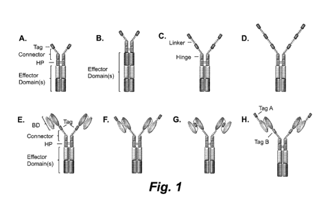
Figures lA ¨ 1H show illustrations of various single chain chimeric effector
molecules containing one or more affinity tag cassettes (A-D, referred to
herein as a
Key-ChEMs), and optionally containing one or more specific binding domains (E-
G,
referred to herein as a T-ChARMs). The single chain ChEMs and ChARMs contain
an
intracellular domain. The tag cassettes may be any type of affinity tag, such
as Strep
tag II (SEQ ID NO. :1), Myc tag (SEQ ID NO. :7), V5 tag (SEQ ID NO. :8), Flag
tag
(SEQ ID NO. :3), His tag, or other peptides or molecules, which are recognized
by a
non-endogenous cognate binding partner (e.g., receptor, protein, antibody). As
shown,
a Key-ChEM may contain (A, B) one tag cassette, (C) two tag cassettes (Key-
ChEM2),
(D) three tag cassettes (Key-ChEM3), or more. In addition, the chimeric
molecules may
have multiple effector domains (e.g., the molecules of A and C-G have two,
while the
molecule shown in B has three effector domains), and the tag cassettes may be
placed in
various different areas of a Key-ChEM or T-ChARM molecule. In these particular
examples, T-ChARMs have one tag cassette located between the specific binding
domain and the effector domain (E), at the distal end (e.g., amino-terminus)
of the
4
CA 02933707 2016-06-13
WO 2015/095895 PCT/US2014/072007
specific binding domain (F), integrated within the specific binding domain (G)
(e.g.,
located within the flexible linker between the VH and VL chains of an scFv),
and
having two different tags - one C-terminal of the binding domain and one N-
terminal of
the binding domain (H). The T-ChARMs may also have two, three or more tag
cassettes as shown for the Key-ChEMs. As is evident in these illustrations, a
tag
cassette may be connected to another Key-ChEM or T-ChARM component or another
tag via a linker module (e.g., a flexible (GlyxSer)õ linker module). The
linker length
may be tailored to be longer or shorter to achieve the best interaction of a
specific
binding domain with a target ligand or antigen, and to achieve the best
interaction
between the cell expressing the ChEM or T-ChARM and the target cell.
Figures 2A ¨ 2D show the cytolytic activity of human effector T cells
expressing various kinds of anti-CD19 T-ChARMs and conventional anti-C19 CARs
(lacking a tag cassette and with short, intermediate, and long spacer domains)
against
K562 leukemia cells transfected to express CD19 or ROR1 (control), CD19/ROR1 '
Raji lymphoma cells, and EBV transformed B cells that express a membrane bound
anti-CD3 mAb single chain antibody (OKT3 scFv) to activate all effector T
cells.
Figures 3A ¨ 3F show the results of a multiplex cytokine assay (Luminex0) of
supernatants obtained 24 hours after T cells expressing various anti-CD19 T-
ChARMs
(A-C) and conventional anti-C19 CARs (D-F) were co-cultured with K562 cells
expressing either CD19 (A and D) or ROR1 (negative control; B and E), and with
PMA/ionomycin (positive control; C and F).
Figures 4A and 4B show the results of a multiplex cytokine assay (Luminex0)
of supernatants obtained 24 hours after T cells expressing various anti-CD19 T-
ChARMs (A) and CD19 CARs (B) after co-culture with CD19+ Raji cells.
Figure 5 shows results of a T cell proliferation assay, wherein
carboxyfluoroscein dye dilution indicates that anti-CD19 CD8 ' T cells
expressing T-
ChARMs (containing one, two or three tag cassettes) or a conventional CAR
(CD19
(Long)) were proliferating in response to tumor cells expressing CD19 (blue),
while not
proliferating in the presence of tumor cells expressing ROR1 (red).
Figures 6A ¨ 6E show that anti-CD19 human T cells expressing either a
T-ChARM (containing one, two or three tag cassettes) or conventional CARs
(containing short or intermediate connector regions) can eradicate established
Raji
tumors in NSG mice. In these experiments, the Raji cells are transfected to
express the
firefly luciferase gene, and tumor growth is measured by injecting the mice
with
luciferin and bioluminescence imaging.
5
CA 02933707 2016-06-13
WO 2015/095895
PCT/US2014/072007
Figure 7 shows that anti-CD19 CAR and T-ChARM expressing human T cells
can persist in the blood following adoptive transfer into NSG mice that were
inoculated
with Raji lymphoma. Human T cells are distinguished by staining with
monoclonal
antibodies specific for the human CD8 and CD45 cell surface molecules.
Figures 8A ¨ 8D show that T-ChARM expressing T cells can be identified by
flow cytometry using a tag specific binding agent. In the examples, purified T-
ChARM
T cells are detected by the expression marker tEGFR (A), detected by anti-
strep tag II
(STII) (B), or with StrepTactin APC (C, D).
Figure 9 shows that T-ChARM expressing T cells can be sorted by flow
cytometry from low purity (15% in the example) to high purity (99% in the
example)
with a tag-specific binding agent linked to a fluorochrome. In the example,
the tag is
StrepTag II and the tag-specific binding agent is anti STII mAb linked to a
fluorochrome.
Figure 10 shows direct enrichment of T-ChARM expressing T cells (containing
three Strep-tag tag cassettes) by using Strep-Tactin0 beads of various sizes.
The panels
on the left show staining of the enriched fraction and the panels on the right
show the
effluent (un-enriched fraction).
Figure 11 shows light photomicrographs of T-ChARM (containing one, two or
three tag cassettes) or conventional anti-CD19 CAR expressing T cells (CD19
Long)
that have been co-cultured with beads linked to binding ligand (Strep-Tactin0)
for the
tag sequence. The photomicrographs demonstrate selective clustering and
proliferation
of T-ChARM T cells.
Figure 12 shows the growth curve of T-ChARM expressing T cells (containing
one, two or three tag cassettes) over 10 days of culture with Strep-Tactin0
microbeads.
Figures 13A and 13B show activation of T-ChARM expressing T cells as
determined by upregulation of CD25 and CD69 after binding of the tag cassette
by
either Streptactin microbeads, nanobeads or anti-StrepTag II mAb alone or in
combination with anti-CD28 mAb. Data is shown after (A) 24 hours and (B) 48
hours
of stimulation.
Figures 14A and 14B show the selective expansion of T-ChARM expressing T
cells. Unsorted T-ChARM1/4-1BB and T-ChARM1/CD28 transduced T cells (CD8+
and CD4+) cultured with anti-Strep tag/anti-CD28-MB for 9 days. The percentage
of
T-ChARM cells was assessed by (A) flow detection of Strep tag expression on T
cells
before and after culture. Culture cells treated with anti-CD3/anti-CD28-MB
alone were
used as control. (B) FACS sorted EGFR+ anti-CD19 ChARM T cells after CD19+
6
CA 02933707 2016-06-13
WO 2015/095895 PCT/US2014/072007
immortalized B cell line (TM-LCL) expansion. Stained with anti-EGFR (upper
row)
and anti-Streptag II (lower row) antibodies, respectively
Figure 15 shows proliferation of anti-CD19 T-ChARM expressing T cells
(containing one, two or three tag cassettes) as measured by the level of Ki-67
protein 7
days after stimulation with varying amounts of Strep-Tactin0 beads. In the
bottom
panels, the expression of Ki-67 in T-ChARM expressing T cells after
stimulation
through the anti-CD19 binding component of the T-ChARM with CD19 EBV-LCL
(TM-LCL) is shown.
Figure 16 shows the growth curve of T-ChARM expressing T cells cultured on
different kinds of Streptactin, anti-Streptag II or antiCD3/anti-CD28
conjugated beads.
Figures 17A and 17B show the selective expansion of anti-CD19 T-ChARM
expressing T cells on Strep-Tactin beads (A). The anti-CD19 T-ChARM expressing
T
cells can subsequently be expanded by stimulation through the anti-CD19
chimeric
receptor with CD19' LCL (B).
Figures 18A ¨ 18D show that T cells can be transduced with two types of
T-ChARM (effector domain of 4-1BB/CD3c (A and B), or CD28/CD3c (C and D))
after culture in the presence of IL-7 and IL-15 without prior activation with
anti-
CD3/anti-CD28 beads. The transduced T-ChARM expressing T cells can be
selectively
expanded and enriched by adding anti-Strep tag II beads to the culture (B and
D) (even
in the absence anti-CD3/anti-CD28 bead stimulation), but are not expanded when
anti-
Strep tag II beads are not added to the culture (A and C).
Figures 19A ¨ 19D show that anti-CD19 T-ChARM1 T cells that were
expanded by stimulation with Strep-Tactin0 microbeads retain a comparable or
superior ability to produce cytokines (GM-CSF, interferon-y, IL-2, and TNF-a)
upon
re-stimulation with CD19 positive tumor cells (A. K562/CD19; B- Raji) as
control T
cells that express the anti-CD19 CAR(short) (CD19-S). K562 cells (C) and PMA-
ionomycin (D) served as negative and positive controls, respectively.
Figure 20 shows that T-ChARM expressing T cells can be induced to form
clusters and to proliferate with anti-Strep tag beads alone or with beads
containing
anti-Strep tag and anti-CD27 antibodies or containing anti-Strep tag and anti-
CD28
antibodies.
Figure 21 shows flow cytometry analysis (MFI) of FACS sorted EGFR+
anti-CD19 ChARM T cells after CD19+ immortalized B cell line (TM-LCL)
expansion.
Stained with anti-EGFR (upper row) and anti-Streptag II (lower row)
antibodies,
respectively.
7
CA 02933707 2016-06-13
WO 2015/095895 PCT/US2014/072007
Figure 22 shows chromium release assay results for examining the cytolytic
effect of various anti-CD19 ChARM transduced T cells (effectors) against the
K562
cells transduced with CD19 (K562/CD19), or ROR1 (K562/ROR1) or CD19+ Raji
tumor cells (targets). E/T = Effector/target ratio.
Figures 23A and 23B show the cytolytic activity of T cells expressing (A)
anti-CD19 short, T-ChARM1, T-ChARM2, T-ChARM3 with a CD28/CD3c effector
domain, and (B) having an anti-ROR1 R12 short and T-ChARM' with a 41BB/CD3c
effector domain. The cells were tested for cytolytic activity against K562
cells
transduced with CD19 (K562/CD19), or ROR1 (K562/ROR1) or CD19+ Raji tumor
cells (targets). E/T = Effector/target ratio.
Figure 24 shows IL2/IFN-y production of various anti-CD19 T-ChARM
transduced T cells (Effector) against K562 cells transduced with CD19
(K562/CD19),
or ROR1 (K562/ROR1) or CD19+ Raji tumor cells (Target).
Figures 25A-25C show luminex multiplex cytokine analysis of triplicate co-
culture supernatants of ChARM transduced T cells with CD19+ Raji cells (1:4
ratio)
after 24h. The data is derived from three independent experiments using T
cells from
different donors, and all data are expressed as means SD. Student's t test
was
performed. * P<0.01. (A) Comparison of cytokine production by CD8+ T cells
expressing the anti-CD19 CAR with long (CH3-CH2-hinge), intermediate (CH3-
hinge),
and short (hinge only) spacers. Multiplex cytokine data from 3 independent
experiments were normalized (cytokine release by CD19-CAR 'long/41BB' = 1);
(B)
comparison of cytokine production by CD8+ T cells expressing anti-CD19 T-
ChARM1
(1ST), T-ChARM2 (25T), T-ChARM3 (35T) with a 4-1BB/CD3c effector domain as
compared to anti-CD19 CAR-Short with 4-1BB/CD3c effector domain. Multiplex
cytokine data from 3 independent experiments were normalized (cytokine release
by
CD19-CAR-Short: Hi/4-1BB = 1); and (C) comparison of cytokine production by
CD8+ T cells expressing anti-CD19 T-ChARM' (1ST), T-ChARM2 (25T), T-ChARM3
(35T) with a CD28/CD3c effector domain as compared to anti-CD19 CAR-Short with
CD28/CD3c effector domain. Multiplex cytokine data from 3 independent
experiments
were normalized (cytokine release by CD19-CAR-Short: Hi/CD28 = 1).
Figure 26 shows CFSE dye dilution used to measure proliferation of anti-CD19
4-1BB or CD28 ChARM expressing T cells 5 days after stimulation with CD19+
Raji
tumor cells (solid grey) or medium only (grey lines) without addition of
exogenous
cytokines.
Figures 27A-27D show FACS sorted EGFR+ anti-CD19 ChARM (A) CD8+
T cells (CD19-Hi/4-1BB, ST-CD19/4-1BB, CD19(VH-ST-VL)/4-1BB; CD19-1ST/4-
8
CA 02933707 2016-06-13
WO 2015/095895 PCT/US2014/072007
1BB, CD19-2ST/4-1BB, CD19-3ST/4-1BB CAR); (B) CD4+T cells (CD19-Hi/4-1BB,
ST-CD19/4-1BB, CD19(VH-ST-VL)/4-1BB; CD19-1ST/4-1BB, CD19-2ST/4-1BB,
CD19-35T/4-1BB CAR); (C) anti-CD19 ChARM CD8+ T cells (CD19-Hi/CD28,
CD19-1ST/CD28, CD19-25T/CD28, CD19-35T/CD28 CAR); and (D) anti-ROR1 R12
ChARM T cells (R12-Hi/4-1BB, R12-1ST/4-1BB), which were stimulated with
StrepTactin coated microbeads (StrepTactin-MB), anti-Streptag antibody or
anti-Streptag/anti-CD28 antibody coated microbeads (aStrep tag-MB and aStrep
tag/CD28-MB) in the culture with IL2. After 48 hours of stimulation, the cells
were
harvested and T cell activation marker CD25 was assessed by flow cytometry.
Untreated cells (medium) were used as controls.
Figure 28 shows representative microscopic images of FACS sorted EGFR+
anti-CD19 4-1BB ChARM T cells (CD8+) that were stimulated with StrepTactin-MB,
aStrep tag-MB and aStrep tag/CD28-MB in presence of IL2. Untreated cells
(medium)
were used as control. Microscopic images were taken after 48h of stimulation.
Figures 29A and 29B show growth curves of ChARM T cells. FACS sorted
EGFR+ anti-CD19 ChARM (A) CD8+ and (B) CD4+ T cells were cultured in CTL
medium with StrepTactin-MB, aStrep tag-MB and aStrep tag/CD28-MB in presence
of
IL2.
Figures 30A-30F show anti-CD3/anti-CD28 microbead-stimulated CD8+ T
cells transduced with anti-CD19-1ST/4-1BB or CD19-1ST/CD28 CAR; after EGFR
staining and sorting, pure CAR T cells were expanded with TM-LCL or aStrep tag-
MB
or aStrep tag/CD28-MB for 8 days. In vitro functionality tests were carried
out to
evaluate CAR T cell function before (aCD3/CD28-MB) or after expansion (TM-LCL
or
aStrep tag-MB or aStrep tag/CD28-MB). (A) chromium release assays were carried
out to examine cytolytic effect of ChARM T cells against target cells
(K562/CD19) or
control cells (K562/ROR1). E/T: Effector/target ratio; (B) cytokine production
was
measured by ELISA to evaluate IFN-y and IL2 in supernatants obtained after 24
hours
from co-cultures of 5 x 10⁴ anti-CD19 ChARM T cells with target cells
(K562/CD19),
or control cells (K562/ROR1). PMA/Ionomycin stimulated T cells were used as
positive control. (n=3; * P<0.05); (C) CFSE proliferation assay of ChARM T
cells 5
days after stimulation with target cells (K562/CD19) (solid grey), or control
cells
(K562/ROR1) (grey lines) without addition of exogenous cytokines. For
analysis,
triplicate wells were pooled and the proliferation of live (PI-), EGFR-
positive CAR T
cells was analyzed.; (D) flow detection of CD45RO, CD62L, CD28 and CD27
expression on the ChARM T cells before (aCD3/CD28-MB) or after expansion (TM-
LCL or aStrep tag-MB or aStrep tag/CD28-MB); (E) cohorts of mice were
inoculated
9
CA 02933707 2016-06-13
WO 2015/095895 PCT/US2014/072007
with Raji-ffluc via tail vein injection at day 1, and then 5 x 10⁶ CD8+ ChARM
T cells
(CD19-Hi/4-1BB and CD19-1ST/4-1BB), which were expanded on either CD19+ B
LCL or aStrep tag/CD28-MB were administered 7 days after tumor engraftment.
Tumor progression and distribution was evaluated by serial bioluminescence
imaging
after injection of luciferin substrate; and (F) persistence of anti-CD19 ChARM
T cells
following adoptive transfer into NSG/Raji mice. Flow cytometric analysis of
ChARM
T cells in the peripheral blood (eye bleeds) of the cohort of mice treated
with various
ChARM transduced T cells at different time points after T cell infusion. The
frequency
of CD8+ tEGFR+ and ChARM+ T cells was used as the percentage of live
peripheral
blood cells.
Figure 31 shows CFSE dye dilution used to measure proliferation of anti-CD19
CAR-Short, T-ChARM', T-ChARM3, and Myc-ChARM with 4-1BB T cells 5 days
after stimulation with CD19 (K562/CD19), ROR1 (K562/ROR1), medium alone, or
CD19+ Raji tumor cells without addition of exogenous cytokines.
Figure 32 shows chromium release assays carried out to examine cytolytic
effect of anti-CD19 CAR-Short, T-ChARM', T-ChARM3, and Myc-ChARM with
4-1BB T cells against target cells (K562/CD19) or control cells (K562/ROR1).
E/T:
Effector/target ratio
DETAILED DESCRIPTION
The instant disclosure provides compositions and methods for generating
various fusion proteins containing one or more affinity tag cassettes, which
are chimeric
effector molecules (ChEMs) that function like a "key" to access and manipulate
(i.e.,
turn on or off or modulate) any of a variety of biological pathways. These
chimeric
effector molecules are referred to herein as a Key-ChEMs. Nucleic acid
molecules
encoding such fusion proteins can be used to generate modified host cells in
which
specific cellular responses, such as proliferation or killing, are elicited,
controlled, or
both. For example, certain types of progenitor cells may be obtained from a
subject,
modified to express a fusion protein comprising a tag cassette, induced to
proliferate,
and then infused back into the subject for a particular therapeutic effect
(e.g.,
reconstitute a subject's depleted immune system). Alternatively, such fusion
proteins
containing a tag may further have a binding domain specific for a particular
target (e.g.,
a tumor antigen). In such examples, these fusion proteins are tagged chimeric
antigen
receptor molecules (T-ChARMs) that can be introduced into a particular cell
and then
used to identify, sort, activate, or expand that modified cell. In certain
embodiments,
CA 02933707 2016-06-13
WO 2015/095895 PCT/US2014/072007
such tagged chimeric molecules are transduced into and expressed in cells,
such as
immune cells (e.g.,T cells).
In certain aspects, the present disclosure further provides methods for
selectively activating, promoting proliferation, identifying, sorting,
enriching, isolating,
.. tracking, or depleting cells (e.g., T cells) comprising a nucleic acid
molecule encoding a
fusion protein having one or more tag cassettes (Key-ChEMs or T-ChARMs).
Additionally, this disclosure provides Key-ChEMs or T-ChARMs, as well as
cells,
compositions and methods for using the Key-ChEMs or T-ChARMs of this
disclosure
in various therapeutic applications, including the treatment of a disease in
subject (e.g.,
.. cancer, infectious disease, inflammatory disease, immune disease, aging-
associated
disease).
Prior to setting forth this disclosure in more detail, it may be helpful to an
understanding thereof to provide definitions of certain terms to be used
herein.
Additional definitions are set forth throughout this disclosure.
In the present description, any concentration range, percentage range, ratio
range, or integer range is to be understood to include the value of any
integer within the
recited range and, when appropriate, fractions thereof (such as one tenth and
one
hundredth of an integer), unless otherwise indicated. Also, any number range
recited
herein relating to any physical feature, such as polymer subunits, size or
thickness, are
.. to be understood to include any integer within the recited range, unless
otherwise
indicated. As used herein, the term "about" means 20% of the indicated
range, value,
or structure, unless otherwise indicated. It should be understood that the
terms "a" and
"an" as used herein refer to "one or more" of the enumerated components. The
use of
the alternative (e.g., "or") should be understood to mean either one, both, or
any
.. combination thereof of the alternatives. As used herein, the terms
"include," "have" and
"comprise" are used synonymously, which terms and variants thereof are
intended to be
construed as non-limiting.
In addition, it should be understood that the individual compounds, or groups
of
compounds, derived from the various combinations of the structures and
substituents
.. described herein, are disclosed by the present application to the same
extent as if each
compound or group of compounds was set forth individually. Thus, selection of
particular structures or particular substituents is within the scope of the
present
disclosure.
The term "consisting essentially of' limits the scope of a claim to the
specified
.. materials or steps, or to those that do not materially affect the basic
characteristics of a
claimed invention. For example, a protein domain, region, module or cassette
(e.g., a
11
CA 02933707 2016-06-13
WO 2015/095895 PCT/US2014/072007
binding domain, hinge region, linker module, tag cassette) or a protein (which
may have
one or more domains, regions, modules or cassettes) "consists essentially of'
a
particular amino acid sequence when the amino acid sequence of a domain,
region,
module, cassette or protein includes extensions, deletions, mutations, or a
combination
thereof (e.g., amino acids at the amino- or carboxy-terminus or between
domains) that,
in combination, contribute to at most 20% (e.g., at most 15%, 10%, 8%, 6%, 5%,
4%,
3%, 2% or 1%) of the length of a domain, region, module, cassette or protein
and do not
substantially affect (i.e., do not reduce the activity by more than 50%, such
as no more
than 40%, 30%, 25%, 20%, 15%, 10%, 5%, or 1%) the activity of the domain(s),
region(s), module(s), cassette(s) or protein (e.g., the target binding
affinity of a binding
protein or tag cassette).
A "binding domain" (also referred to as a "binding region" or "binding
moiety"),
as used herein, refers to a molecule, such as a peptide, oligopeptide,
polypeptide, or
protein that possesses the ability to specifically and non-covalently
associate, unite, or
combine with a target molecule (e.g., CD19, CD20, CD22, ROR1, mesothelin, PD-
L1,
PD-L2, PSMA). A binding domain includes any naturally occurring, synthetic,
semi-
synthetic, or recombinantly produced binding partner for a biological molecule
or other
target of interest. In some embodiments, the binding domain is an antigen-
binding
domain, such as an antibody or T cell receptor (TCR) or functional binding
domain or
antigen-binding fragment thereof. Exemplary binding domains include single
chain
antibody variable regions (e.g., domain antibodies, sFv, scFv, Fab), receptor
ectodomains (e.g., TNF-a), ligands (e.g., cytokines, chemokines), antigen-
binding
regions of T cell receptors (TCRs), such as single chain TCRs (scTCRs), or
synthetic
polypeptides selected for the specific ability to bind to a biological
molecule.
As used herein, "specifically binds" refers to an association or union of a
binding domain, or a fusion protein thereof, to a target molecule with an
affinity or K.
(i.e., an equilibrium association constant of a particular binding interaction
with units of
1/M) equal to or greater than 105 M-1, while not significantly associating or
uniting with
any other molecules or components in a sample. Binding domains (or fusion
proteins
thereof) may be classified as "high affinity" binding domains (or fusion
proteins
thereof) or "low affinity" binding domains (or fusion proteins thereof). "High
affinity"
binding domains refer to those binding domains with a Ka of at least 107 M-1,
at least
108 M-1, at least 109 M-1, at least 1010 M-1, at least 1011 M-1, at least 1012
M-1, or at least
1013 M-1. "Low affinity" binding domains refer to those binding domains with a
Ka of
up to 107 M-1, up to 106 M-1, up to 105 M-1. Alternatively, affinity may be
defined as an
equilibrium dissociation constant (KO of a particular binding interaction with
units of
12
CA 02933707 2016-06-13
WO 2015/095895
PCT/US2014/072007
M (e.g., 10 M to 10-13 M). In certain embodiments, a binding domain may have
"enhanced affinity," which refers to a selected or engineered binding domain
with
stronger binding to a target antigen than a wild type (or parent) binding
domain. For
example, enhanced affinity may be due to a Ka (equilibrium association
constant) for
the target antigen that is higher than the wild type binding domain, or due to
a Kd
(dissociation constant) for the target antigen that is less than that of the
wild type
binding domain, or due to an off-rate (Koff) for the target antigen that is
less than that of
the wild type binding domain. A variety of assays are known for identifying
binding
domains of the present disclosure that specifically bind a particular target,
as well as
determining binding domain or fusion protein affinities, such as Western blot,
ELISA,
and Biacore0 analysis (see also, e.g., Scatchard et at., Ann. N.Y. Acad. Sci.
5/:660,
1949; and U.S. Patent Nos. 5,283,173, 5,468,614, or the equivalent).
As used herein, "heterologous" or "non-endogenous" or "exogenous" refers to
any gene, protein, compound, molecule or activity that is not native to a host
cell or a
subject, or is any gene, protein, compound, molecule or activity native to a
host or host
cell but has been altered or mutated such that the structure, activity or both
is different
as between the native and mutated molecules. In certain embodiments,
heterologous,
non-endogenous or exogenous molecules (e.g., receptors, ligands) may not be
endogenous to a host cell or subject, but instead nucleic acids encoding such
molecules
may have been added to a host cell by conjugation, transformation,
transfection,
electroporation, or the like, wherein the added nucleic acid molecule may
integrate into
a host cell genome or can exist as extra-chromosomal genetic material (e.g.,
as a
plasmid or other self-replicating vector). The term "homologous" or "homolog"
refers
to a molecule or activity found in or derived from a host cell, species or
strain. For
example, a heterologous or exogenous molecule or gene encoding the molecule
may be
homologous to a native host or host cell molecule or gene that encodes the
molecule,
respectively, but may have an altered structure, sequence, expression level or
combinations thereof. A non-endogenous molecule may be from the same species,
a
different species or a combination thereof
As used herein, the term "endogenous" or "native" refers to a gene, protein,
compound, molecule or activity that is normally present in a host or host
cell.
As used herein, "tag cassette" refers to a unique peptide sequence affixed to,
fused to, or that is part of a protein of interest, to which a heterologous or
non-
endogenous cognate binding molecule (e.g., receptor, ligand, antibody, or
other binding
partner) is capable of specifically binding where the binding property can be
used to
detect, identify, isolate or purify, track, enrich for, or target a tagged
protein or cells
13
CA 02933707 2016-06-13
WO 2015/095895 PCT/US2014/072007
expressing a tagged protein, particularly when a tagged protein is part of a
heterogeneous population of proteins or other material, or when cells
expressing a
tagged protein are part of a heterogeneous population of cells (e.g., a
biological sample
like peripheral blood). In certain embodiments, a cell expressing a tagged
protein can
be contacted with a heterologous or non-endogenous cognate binding molecule
and
induce a biological response, such as promote cell activation, cell
proliferation or cell
death. In the provided fusion proteins, the ability of the tag cassette(s) to
be specifically
bound by the cognate binding molecule(s) is distinct from or in addition to
the ability of
the binding domain(s) to specifically bind to the target molecule(s). The tag
cassette
generally is not an antigen-binding molecule, for example, is not an antibody
or TCR or
an antigen-binding portion thereof
As used herein, a "hinge region" or a "hinge" refers to (a) an immunoglobulin
hinge sequence (made up of, for example, upper and core regions) or a
functional
fragment or variant thereof, (b) a type II C-lectin interdomain (stalk) region
or a
functional fragment or variant thereof, or (c) a cluster of differentiation
(CD) molecule
stalk region or a functional variant thereof. As used herein, a "wild type
immunoglobulin hinge region" refers to a naturally occurring upper and middle
hinge
amino acid sequences interposed between and connecting the CH1 and CH2 domains
(for IgG, IgA, and IgD) or interposed between and connecting the CH1 and CH3
domains (for IgE and IgM) found in the heavy chain of an antibody. In certain
embodiments, a hinge region is human, and in particular embodiments, comprises
a
human IgG hinge region.
As used herein, a "connector region" refers to one or more proteins,
polypeptides, oligopeptides, peptides, domains, regions, modules, cassettes,
motifs or
any combination thereof that join two or more proteins, polypeptides,
oligopeptides,
peptides, domains, regions, modules, cassettes, motifs or any combination
thereof in a
fusion protein. For example, a connector region may provide a spacer function
to
facilitate the interaction of two single chain fusion proteins, or positioning
of one or
more binding domains, so that the resulting polypeptide structure maintains a
specific
binding affinity to a target molecule or maintains signaling activity (e.g.,
effector
domain activity) or both. In certain embodiments, a connector region may
comprise a
"linker module" that is an amino acid sequence having from about to two up to
about
500 amino acids, which can provide flexibility and room for conformational
movement
between two regions, domains, motifs, cassettes or modules connected by a
linker.
Exemplary linker modules include those having from one to about ten repeats of
GlyxSery, wherein x and y are independently an integer from 0 to 10 provided
that x and
14
CA 02933707 2016-06-13
WO 2015/095895 PCT/US2014/072007
y are not both 0 (e.g., (Gly4Ser)2(SEQ ID NO: 67), (Gly3Ser)2(SEQ ID NO: 68),
Gly2Ser, or a combination thereof such as (Gly3Ser)2Gly2Ser)(SEQ ID NO: 69).
In
certain other embodiments, a connector region may have a linker module that
comprises
one or more immunoglobulin heavy chain constant regions, such as a CH3 alone
or a
CH2CH3. In further embodiments, a connector region may comprise a hinge region
or
a tag cassette. Each such connector component is not mutually exclusive. For
example,
a connector region may comprise a hinge and one or more linker modules, or a
connector region may comprise a hinge, one or more linker modules, and one or
more
tag cassettes. Exemplary connector regions can vary in length, for instance,
from about
five to about 500 amino acids, or from about ten to about 350 amino acids, or
from
about 15 to about 100 amino acids, or from about 20 to about 75 amino acids,
or from
about 25 to about 35 amino acids.
A "hydrophobic portion," as used herein, means any amino acid sequence
having a three-dimensional structure that is thermodynamically stable in a
cell
membrane, and generally ranges in length from about 15 amino acids to about 30
amino
acids. The structure of a hydrophobic domain may comprise an alpha helix, a
beta
barrel, a beta sheet, a beta helix, or any combination thereof
As used herein, an "effector domain" is an intracellular portion of a fusion
protein or receptor that can directly or indirectly promote a biological or
physiological
response in a cell when receiving the appropriate signal. In certain
embodiments, an
effector domain is part of a protein or protein complex that receives a signal
when
bound, or it binds directly to a target molecule, which triggers a signal from
the effector
domain. An effector domain may directly promote a cellular response when it
contains
one or more signaling domains or motifs, such as an immunoreceptor tyrosine-
based
activation motif (ITAM). In other embodiments, an effector domain will
indirectly
promote a cellular response by associating with one or more other proteins
that directly
promote a cellular response.
A "variable region linker" specifically refers to a five to about 35 amino
acid
sequence that connects a heavy chain immunoglobulin variable region to a light
chain
immunoglobulin variable region or connects T cell receptor V,03 and Ccup
chains (e.g.,
V-C, Vp-C, Vc,-V) or connects each V-C, Vp-C, Vc,-Vi3 pair to a hinge or
hydrophobic domain, which provides a spacer function and flexibility
sufficient for
interaction of the two sub-binding domains so that the resulting single chain
polypeptide retains a specific binding affinity to the same target molecule as
an
antibody or T cell receptor. In certain embodiments, a variable region linker
comprises
from about ten to about 30 amino acids or from about 15 to about 25 amino
acids. In
CA 02933707 2016-06-13
WO 2015/095895 PCT/US2014/072007
particular embodiments, a variable region linker peptide comprises from one to
ten
repeats of GlyxSery, wherein x and y are independently an integer from 0 to 10
provided
that x and y are not both 0 (e.g., Gly4Ser (SEQ ID NO: 10), Gly3Ser (SEQ ID
NO: 71),
Gly2Ser, or (Gly3Ser)õ(Gly4Ser)i (SEQ ID NO: 72), (Gly3Ser)õ(Gly2Ser)õ, (SEQ
ID NO:
73) (Gly3Ser)õ(Gly4Ser)õ (SEQ ID NO: 72), or (Gly4Ser)õ (SEQ ID NO: 10),
wherein n
is an integer of 1, 2, 3, 4, 5, 6, 7, 8, 9 or 10) and wherein linked variable
regions form a
functional immunoglobulin-like binding domain (e.g., scFv, scTCR). Exemplary
variable region linkers include those amino acid sequences set forth in SEQ
IDNOS.:44,
65-69, and 71-73, and (Gly4Ser)õ (SEQ ID NO: 10), wherein n is 3, as found in
T-
ChARM having the amino acid sequence set forth in SEQ ID NO.:57.
"Junction amino acids" or "junction amino acid residues" refer to one or more
(e.g., about 2-20) amino acid residues between two adjacent motifs, regions or
domains
of a polypeptide, such as between a binding domain and an adjacent linker
region or
between a hydrophobic domain and an adjacent effector domain or on one or both
ends
of a linker region that links two motifs, regions or domains (e.g., between a
linker and
an adjacent binding domain and/or between a linker and an adjacent hinge).
Junction
amino acids may result from the construct design of a fusion protein (e.g.,
amino acid
residues resulting from the use of a restriction enzyme site during the
construction of a
nucleic acid molecule encoding a fusion protein). For example, a single
junction amino
acid, asparagine, is encoded by the AAT codon found between the nucleic acid
sequence encoding the secretory signal sequence (SEQ ID NO. :63) and the
sequence
encoding the tag cassette (SEQ ID NO. :38) in the T-ChARM encoded by the
nucleic
acid sequence set forth in SEQ ID NO. :58. Similarly, an asparagine (N)
junction amino
acid is found between the flexible linker amino acid sequence of GGSGSG (SEQ
ID
NO. :65) and the amino acid tag sequence WSHPQFEK (SEQ ID NO.:1) found in the
T-
ChARM having the amino acid sequence set forth in SEQ ID NO. :54.
Terms understood by those in the art of antibody technology are each given the
meaning acquired in the art, unless expressly defined differently herein. The
term
"antibody" refers to an intact antibody comprising at least two heavy (H)
chains and
two light (L) chains inter-connected by disulfide bonds, as well as an antigen-
binding
portion of an intact antibody that has or retains the capacity to bind a
target molecule.
A monoclonal antibody or antigen-binding portion thereof may be non-human,
chimeric, humanized, or human, preferably humanized or human. Immunoglobulin
structure and function are reviewed, for example, in Harlow et at., Eds.,
Antibodies: A
Laboratory Manual, Chapter 14 (Cold Spring Harbor Laboratory, Cold Spring
Harbor,
1988).
16
CA 02933707 2016-06-13
WO 2015/095895 PCT/US2014/072007
For example, the terms "VL" and "VH" refer to the variable binding region from
an antibody light and heavy chain, respectively. The variable binding regions
are made
up of discrete, well-defined sub-regions known as "complementarity determining
regions" (CDRs) and "framework regions" (FRs). The term "CL" refers to an
"immunoglobulin light chain constant region" or a "light chain constant
region," i.e., a
constant region from an antibody light chain. The term "CH" refers to an
"immunoglobulin heavy chain constant region" or a "heavy chain constant
region,"
which is further divisible, depending on the antibody isotype into CH1, CH2,
and CH3
(IgA, IgD, IgG), or CH1, CH2, CH3, and CH4 domains (IgE, IgM). A "Fab"
(fragment
antigen binding) is the part of an antibody that binds to antigens and
includes the
variable region and CH1 of the heavy chain linked to the light chain via an
inter-chain
disulfide bond.
As used herein, "Fc region portion" refers to the heavy chain constant region
segment of the Fc fragment (the "fragment crystallizable" region or Fc region)
from an
antibody, which can in include one or more constant domains, such as CH2, CH3,
CH4,
or any combination thereof In certain embodiments, an Fc region portion
includes the
CH2 and CH3 domains of an IgG, IgA, or IgD antibody or any combination
thereof, or
the CH3 and CH4 domains of an IgM or IgE antibody and any combination thereof
In
other embodiments, a CH2CH3 or a CH3CH4 structure has sub-region domains from
the same antibody isotype and are human, such as human IgGl, IgG2, IgG3, IgG4,
IgAl, IgA2, IgD, IgE, or IgM (e.g., CH2CH3 from human IgG1). By way of
background, an Fc region is responsible for the effector functions of an
immunoglobulin, such as ADCC (antibody-dependent cell-mediated cytotoxicity),
CDC
(complement-dependent cytotoxicity) and complement fixation, binding to Fc
receptors
(e.g., CD16, CD32, FcRn), greater half-life in vivo relative to a polypeptide
lacking an
Fc region, protein A binding, and perhaps even placental transfer (see Capon
et at.,
Nature 337:525, 1989). In certain embodiments, an Fc region portion found in
fusion
proteins of the present disclosure will be capable of mediating one or more of
these
effector functions, or will lack one or more or all of these activities by way
of, for
example, one or more mutations known in the art.
In addition, antibodies have a hinge sequence that is typically situated
between
the Fab and Fc region (but a lower section of the hinge may include an amino-
terminal
portion of the Fc region). By way of background, an immunoglobulin hinge acts
as a
flexible spacer to allow the Fab portion to move freely in space. In contrast
to the
constant regions, hinges are structurally diverse, varying in both sequence
and length
between immunoglobulin classes and even among subclasses. For example, a human
17
CA 02933707 2016-06-13
WO 2015/095895 PCT/US2014/072007
IgG1 hinge region is freely flexible, which allows the Fab fragments to rotate
about
their axes of symmetry and move within a sphere centered at the first of two
inter-heavy
chain disulfide bridges. By comparison, a human IgG2 hinge is relatively short
and
contains a rigid poly-proline double helix stabilized by four inter-heavy
chain disulfide
bridges, which restricts the flexibility. A human IgG3 hinge differs from the
other
subclasses by its unique extended hinge region (about four times as long as
the IgG1
hinge), containing 62 amino acids (including 21 prolines and 11 cysteines),
forming an
inflexible poly-proline double helix and providing greater flexibility because
the Fab
fragments are relatively far away from the Fc fragment. A human IgG4 hinge is
shorter
than IgG1 but has the same length as IgG2, and its flexibility is intermediate
between
that of IgG1 and IgG2.
"T cell receptor" (TCR) refers to a molecule found on the surface of T cells
(or
T lymphocytes) that, in association with CD3, is generally responsible for
recognizing
antigens bound to major histocompatibility complex (MHC) molecules. The TCR
has a
disulfide-linked heterodimer of the highly variable a and 0 chains (also known
as
TCRa and TCR13, respectively) in most T cells. In a small subset of T cells,
the TCR is
made up of a heterodimer of variable 7 and 8 chains (also known as TCRy and
TCR8,
respectively). Each chain of the TCR is a member of the immunoglobulin
superfamily
and possesses one N-terminal immunoglobulin variable domain, one
immunoglobulin
constant domain, a transmembrane region, and a short cytoplasmic tail at the C-
terminal
end (see Janeway et at., Immunobiology: The Immune System in Health and
Disease, 3rd
Ed., Current Biology Publications, p. 4:33, 1997). TCR, as used in the present
disclosure, may be from various animal species, including human, mouse, rat,
cat, dog,
goat, horse, or other mammals. TCRs may be cell-bound (i.e., have a
transmembrane
region or domain) or in soluble form.
"Major histocompatibility complex molecules" (MHC molecules) refer to
glycoproteins that deliver peptide antigens to a cell surface. MHC class I
molecules are
heterodimers consisting of a membrane spanning a chain (with three a domains)
and a
non-covalently associated 132 microglobulin. MHC class II molecules are
composed of
two transmembrane glycoproteins, a and 13, both of which span the membrane.
Each
chain has two domains. MHC class I molecules deliver peptides originating in
the
cytosol to the cell surface, where peptide:MHC complex is recognized by CD8 '
T cells.
MHC class II molecules deliver peptides originating in the vesicular system to
the cell
surface, where they are recognized by CD4 ' T cells. An MHC molecule may be
from
various animal species, including human, mouse, rat, or other mammals.
18
CA 02933707 2016-06-13
WO 2015/095895 PCT/US2014/072007
A "vector" is a nucleic acid molecule that is capable of transporting another
nucleic acid. Vectors may be, for example, plasmids, cosmids, viruses, or
phage. An
"expression vector" is a vector that is capable of directing the expression of
a protein
encoded by one or more genes carried by the vector when it is present in the
appropriate
environment.
"Retroviruses" are viruses having an RNA genome. "Gammaretrovirus" refers
to a genus of the retroviridae family. Exemplary gammaretroviruses include
mouse
stem cell virus, murine leukemia virus, feline leukemia virus, feline sarcoma
virus, and
avian reticuloendotheliosis viruses.
"Lentivirus" refers to a genus of retroviruses that are capable of infecting
dividing and non-dividing cells. Several examples of lentiviruses include HIV
(human
immunodeficiency virus: including HIV type 1, and HIV type 2); equine
infectious
anemia virus; feline immunodeficiency virus (FIV); bovine immune deficiency
virus
(BIV); and simian immunodeficiency virus (SIV).
A "hematopoietic progenitor cell" is a cell derived from hematopoietic stem
cells or fetal tissue that is capable of further differentiation into mature
cells types (e.g.,
cells of the T cell lineage). In certain embodiments, CD241 Lin- CD117
hematopoietic
progenitor cells are useful. As defined herein, hematopoietic progenitor cells
may
include embryonic stem cells, which are capable of further differentiation to
cells of the
T cell lineage. Hematopoietic progenitor cells may be from various animal
species,
including human, mouse, rat, or other mammals. A "thymocyte progenitor cell"
or
"thymocyte" is a hematopoietic progenitor cell present in the thymus.
"Hematopoietic stem cells" refer to undifferentiated hematopoietic cells that
are
capable of self-renewal either in vivo, essentially unlimited propagation in
vitro, and
capable of differentiation to other cell types including cells of the T cell
lineage.
Hematopoietic stem cells may be isolated, for example, but not limited to,
from fetal
liver, bone marrow, cord blood.
"Embryonic stem cells" or "ES cells" or "ESCs" refer to undifferentiated
embryonic stem cells that have the ability to integrate into and become part
of the germ
line of a developing embryo. Embryonic stem cells are capable of
differentiating into
hematopoietic progenitor cells, and any tissue or organ. Embryonic stem cells
that are
suitable for use herein include cells from the J1 ES cell line, 129J ES cell
line, murine
stem cell line D3 (American Type Culture Collection), the R1 or E14K cell
lines
derived from 129/Sv mice, cell lines derived from Balb/c and C57B1/6 mice, and
human
embryonic stem cells (e.g. from WiCell Research Institute, WI; or ES cell
International,
Melbourne, Australia).
19
CA 02933707 2016-06-13
WO 2015/095895 PCT/US2014/072007
"Cells of T cell lineage" refer to cells that show at least one phenotypic
characteristic of a T cell or a precursor or progenitor thereof that
distinguishes the cells
from other lymphoid cells, and cells of the erythroid or myeloid lineages.
Such
phenotypic characteristics can include expression of one or more proteins
specific for T
cells (e.g. , CD3 ', CD4, CD8 '), or a physiological, morphological,
functional, or
immunological feature specific for a T cell. For example, cells of the T cell
lineage
may be progenitor or precursor cells committed to the T cell lineage; CD25 '
immature
and inactivated T cells; cells that have undergone CD4 or CD8 linage
commitment;
thymocyte progenitor cells that are CD4 'CD8 ' double positive; single
positive CD4 ' or
CD8 '; TCRc43 or TCR yo; or mature and functional or activated T cells.
"Nucleic acid molecule", or polynucleotides, may be in the form of RNA or
DNA, which includes cDNA, genomic DNA, and synthetic DNA. A nucleic acid
molecule may be double stranded or single stranded, and if single stranded,
may be the
coding strand or non-coding (anti-sense strand). A coding molecule may have a
coding
sequence identical to a coding sequence known in the art or may have a
different coding
sequence, which, as the result of the redundancy or degeneracy of the genetic
code, or
by splicing, can encode the same polypeptide.
"Treat" or "treatment" or "ameliorate" refers to medical management of a
disease, disorder, or condition of a subject (e.g., a human or non-human
mammal, such
as a primate, horse, dog, mouse, rat). In general, an appropriate dose or
treatment
regimen comprising a host cell expressing a Key-ChEM or T-ChARM of this
disclosure, and optionally an adjuvant, is administered in an amount
sufficient to elicit a
therapeutic or prophylactic benefit. Therapeutic or prophylactic/preventive
benefit
includes improved clinical outcome; lessening or alleviation of symptoms
associated
with a disease; decreased occurrence of symptoms; improved quality of life;
longer
disease-free status; diminishment of extent of disease, stabilization of
disease state;
delay of disease progression; remission; survival; prolonged survival; or any
combination thereof
A "therapeutically effective amount" or "effective amount" of a fusion protein
or
cell expressing a fusion protein of this disclosure (e.g., Key-ChEM, T-ChARM)
refers
to that amount of compound or cells sufficient to result in amelioration of
one or more
symptoms of the disease being treated in a statistically significant manner.
When
referring to an individual active ingredient or a cell expressing a single
active
ingredient, administered alone, a therapeutically effective dose refers to the
effects of
that ingredient or cell expressing that ingredient alone. When referring to a
combination, a therapeutically effective dose refers to the combined amounts
of active
CA 02933707 2016-06-13
WO 2015/095895 PCT/US2014/072007
ingredients or combined adjunctive active ingredient with a cell expressing an
active
ingredient that results in a therapeutic effect, whether administered serially
or
simultaneously. Another combination may be a cell expressing more than one
active
ingredient, such as two different T-ChARMs, a T-ChARM and a TCR, a T-ChARM
and a CAR, or combinations thereof.
Additional definitions are provided throughout the present disclosure.
Key-ChEMs and T-ChARMs
In certain aspects, the present disclosure provides a single chain fusion
protein,
referred to as a Key-ChEM, which comprises an extracellular component and an
intracellular component connected by a hydrophobic portion, wherein the
extracellular
component comprises a tag cassette and a connector region comprising a hinge,
and
wherein the intracellular component comprises an effector domain. In certain
embodiments, a connector region further comprises a linker module, or one or
more tag
cassettes are located within the connector region. In certain other
embodiments, one or
more tag cassettes are linked to the connector region by a linker module.
In further Key-ChEM embodiments, the fusion protein comprises from amino-
terminus to carboxy-terminus: a tag cassette, a connector region comprising a
hinge, a
hydrophobic portion, and an intracellular component comprising an effector
domain
(see, e.g., Figures lA and 1B). In still further Key-ChEM embodiments, the
fusion
protein comprises from amino-terminus to carboxy-terminus: a first connector
region, a
tag cassette, a second connector region comprising a hinge, a hydrophobic
portion, and
an intracellular component comprising an effector domain. In yet further Key-
ChEM
embodiments, the fusion protein comprises from amino-terminus to carboxy-
terminus: a
first tag cassette, a first connector region, a second tag cassette, a second
connector
region comprising a hinge, a hydrophobic portion, and an intracellular
component
comprising an effector domain (see, e.g., Figure 1C). In even further Key-ChEM
embodiments, the fusion protein comprises from amino-terminus to carboxy-
terminus: a
first tag cassette, a first connector region, a second tag cassette, a second
connector
region, a third tag cassette, a third connector region comprising a hinge, a
hydrophobic
portion, and an intracellular component comprising an effector domain (see,
e.g., Figure
1D).
In certain other Key-ChEM embodiments, the fusion protein further comprises a
non-covalently associated binding domain, such as a binding domain associated
with
the tag cassette (i.e., a multichain T-ChARM). In still other Key-ChEM
embodiments,
the non-covalently associated binding domain is bi-specific, wherein the first
binding
end is specific for the tag cassette and the second binding end is specific
for a target
21
CA 02933707 2016-06-13
WO 2015/095895
PCT/US2014/072007
other than the tag cassette, or the first and second binding ends are both
specific for the
tag cassette. In yet other Key-ChEM embodiments, the non-covalently associated
binding domain is multispecific, wherein a first end binds to a tag cassette
and a second
end is specific for one or more targets other than the tag cassette. In such
embodiments,
a Key-ChEM comprises a multimer protein. In some embodiments, such Key-ChEMs
comprising one or more non-covalently associated binding domains comprise
heteromultimers.
In other aspects, the present disclosure provides a single chain fusion
protein,
referred to as a T-ChARM, which comprises an extracellular component and an
intracellular component connected by a hydrophobic portion, wherein the
extracellular
component comprises a binding domain that specifically binds a target, a tag
cassette,
and a connector region comprising a hinge, and wherein the intracellular
component
comprises an effector domain. In certain embodiments, a T-ChARM binding domain
is
a scFv, scTCR, receptor ectodomain, or ligand.
In further T-ChARM embodiments, the fusion protein comprises from amino-
terminus to carboxy-terminus: an extracellular binding domain, a tag cassette,
a
connector region comprising a hinge, a hydrophobic portion, and an
intracellular
component comprising an effector domain (see, e.g., Figure 1E). In still
further T-
ChARM embodiments, the fusion protein comprises from amino-terminus to carboxy-
terminus: an extracellular binding domain, a first connector region, a tag
cassette, a
second connector region comprising a hinge, a hydrophobic portion, and an
intracellular
component comprising an effector domain. In yet further T-ChARM embodiments,
the
fusion protein comprises from amino-terminus to carboxy-terminus: an
extracellular
binding domain, a first tag cassette, a first connector region, a second tag
cassette, a
second connector region comprising a hinge, a hydrophobic portion, and an
intracellular
component comprising an effector domain. In even further T-ChARM embodiments,
the fusion protein comprises from amino-terminus to carboxy-terminus: an
extracellular
binding domain, a first tag cassette, a first connector region, a second tag
cassette, a
second connector region, a third tag cassette, a third connector region
comprising a
hinge, a hydrophobic portion, and an intracellular component comprising an
effector
domain.
In certain other T-ChARM embodiments, the fusion protein comprises from
amino-terminus to carboxy-terminus: a tag cassette, an extracellular binding
domain, a
connector region comprising a hinge, a hydrophobic portion, and an
intracellular
component comprising an effector domain (see, e.g., Figure 1F). In still other
T-
ChARM embodiments, the fusion protein comprises from amino-terminus to carboxy-
22
CA 02933707 2016-06-13
WO 2015/095895 PCT/US2014/072007
terminus: an extracellular scFv or scTCR binding domain comprising a variable
region
linker containing a tag cassette disposed between the variable regions (e.g.,
at or closer
to the N-terminal end of the variable region linker, at or closer to the C-
terminal end of
the variable region linker, or imbedded closer to the middle of the variable
region
linker), a connector region comprising a hinge, a hydrophobic portion, and an
intracellular component comprising an effector domain. An exemplary tag
cassette
imbedded in a variable region linker comprises GGSGSG(X),IWSHPQFEKGSGSG
(SEQ ID NO.:45), wherein Xis optional, may be any amino acid and n is 0, 1, 2,
3, 4 or
5. In SEQ ID NO. :54, such a variable region linker having an imbedded tag is
present,
wherein n is 1 and X is asparagine (N).
A Key-ChEM or T-ChARM may be cell-bound (e.g., expressed on a cell
surface) or in soluble form. In certain embodiments, nucleic acid molecules
encoding
Key-ChEM or T-ChARM fusion proteins may be codon optimized to enhance or
maximize expression in certain types of cells, such as T cells (Scholten et
at., Clin.
Immunol. 119:135, 2006).
In other embodiments, Key-ChEM or T-ChARM may further comprise a
cytotoxic component (e.g., chemotherapeutic drugs such as anti-mitotics (e.g.,
vindesine), antifolates, alkylating agents (e.g., temozolomide), bacterial
toxins, ricin,
anti-virals, radioisotopes, radiometals), which is useful for specific killing
or disabling a
cancer cell, infected cell or other diseased cell. In further embodiments, Key-
ChEM or
T-ChARM may further comprise a detectable component (e.g., biotin, fluorescent
moiety, radionuclide), which is useful for tracking or imaging cancer cells,
infected
cells, or other tissues (e.g., tissue under autoimmune attack). In still
further
embodiments, Key-ChEM or T-ChARM may further comprise a functional component
(e.g., an immunostimulatory moiety, cytokine, immune modulator, immunoglobulin
protein, or the like).
Component parts of the fusion proteins of the present disclosure are further
described in detail herein.
Tag Cassette
A tag cassette contained in a single chain fusion protein according to the
present
disclosure (e.g., Key-ChEM or T-ChARM) will be an extracellular component that
can
specifically bind to a cognate receptor or binding partner (e.g., antibody)
with high
affinity or avidity, wherein the cognate receptor or binding partner is
heterologous or
non-endogenous to a host or a cell expressing a Key-ChEM or T-ChARM. Within a
single chain fusion protein structure, a tag cassette may be located (a)
immediately
amino-terminal to a connector region, (b) interposed between and connecting
linker
23
CA 02933707 2016-06-13
WO 2015/095895 PCT/US2014/072007
modules, (c) immediately carboxy-terminal to a binding domain, (d) interposed
between
and connecting a binding domain (e.g., scFv) to an effector domain, (e)
interposed
between and connecting subunits of a binding domain, or (f) at the amino-
terminus of a
single chain fusion protein of this disclosure. In certain embodiments, one or
more
junction amino acids may be disposed between and connecting a tag cassette
with a
hydrophobic portion, or disposed between and connecting a tag cassette with a
connector region, or disposed between and connecting a tag cassette with a
linker
module, or disposed between and connecting a tag cassette with a binding
domain.
Exemplary tag cassettes include Strep tag (which refers the original Strep
tag,
Strep tag II, or any variant thereof see, e.g., U.S. Patent No. 7,981,632,
which Strep
tags are incorporated herein by reference), His tag, Flag tag (SEQ ID NO. :3),
Xpress
tag (SEQ ID NO.:4), Avi tag (SEQ ID NO.:5), Calmodulin tag (SEQ ID NO.:19),
Polyglutamate tag, HA tag (SEQ ID NO. :6), Myc tag (SEQ ID NO. :7), Nus tag, S
tag,
SBP tag, Softag 1 (SEQ ID NO. :9), Softag 3 (SEQ ID NO. :32), V5 tag (SEQ ID
NO. :8), CREB-binding protein (CBP), glutathione S-transferase (GST), maltose
binding protein (MBP), green fluorescent protein (GFP), Thioredoxin tag, or
any
combination thereof In certain embodiments, a tag cassette is a Strep tag
having an
amino acid sequence of Trp-Ser-His-Pro-Gln-Phe-Glu-Lys (SEQ ID NO. :1) or Trp-
Arg-His-Pro-Gln-Phe-Gly-Gly (SEQ ID NO. :2). In other embodiments, a tag
cassette
may be a genetically engineered affinity site, such as a minimal chelation
site (e.g.,
HGGHHG, SEQ ID NO. :33)
Tag cassettes may be present in multiple copies in fusion proteins of this
disclosure. For example, a fusion protein of this disclosure can have one,
two, three,
four or five tag cassettes (e.g., Strep tag). In certain embodiments, a
connector region
of a Key-ChEM or T-ChARM includes one tag cassette, two tag cassettes, three
tag
cassettes, four tag cassettes, or five tag cassettes. Each of the plurality of
tag cassettes
may be the same or different. Exemplary embodiments include a Key-ChEM or T-
ChARM having a Strep tag and a Strep tag cassette, or a His tag and a Strep
tag
cassette, or a HA tag and a Strep tag cassette, or a Myc tag and a Strep tag
cassette.
Alternatively, a Key-ChEM or T-ChARM will have multiple tag cassettes of the
same
type or same amino acid sequence, such as two, three, four or five Strep tag
cassettes
(e.g., Strep tag II).
For example, a Key-ChEM or T-ChARM may have at least two different tag
cassettes. In some embodiments, a first tag cassette can provide a stimulation
signal
and a distinct second tag cassette might be used to associate with a detection
reagent or
associate with an antibody-toxin conjugate or with an antibody-imaging agent
24
CA 02933707 2016-06-13
WO 2015/095895 PCT/US2014/072007
conjugate. In further embodiments, the two or more first tag cassettes may be
located
in different areas of a Key-ChEM or T-ChARM. In certain embodiments, a first
tag
cassette is located in the connector region and a second tag cassette is
located at the
amino-terminus or carboxy terminus or both of a Key-ChEM or T-ChARM (see,
e.g.,
Figure 1H).
In certain embodiments, a tag cassette comprises from about five to about 500
amino acids, or from about six to about 100 amino acids, or from about seven
to about
50 amino acids, or from about eight to about 20 amino acids. In some
embodiments, a
tag cassette has seven to ten amino acids. Preferably, a tag cassette is
non-immunogenic or minimally immunogenic. Essentially, a tag cassette can
function
as a handle or beacon to allow for the identification, enrichment, isolation,
promotion of
proliferation, activation, tracking, or elimination of cells expressing a Key-
ChEM or
T-ChARM.
In certain embodiments, a tag cassette is located within a connector region of
a
fusion protein of this disclosure. For example, a connector region may further
comprise
a linker module adjacent to a tag cassette, wherein the linker module with the
tag
cassette has an amino acid sequence of (Gly-Gly-Gly-Gly-Ser)2-Trp-Ser-His-Pro-
Gln-
Phe-Glu-Lys (SEQ ID NO. :20), Trp-Ser-His-Pro-Gln-Phe-Glu-Lys-(Gly-Gly-Gly-Gly-
Ser)2 (SEQ ID NO. :21), (Gly-Gly-Gly-Gly-Ser)2-Trp-Ser-His-Pro-Gln-Phe-Glu-Lys-
(Gly-Gly-Gly-Ser)2¨Gly-Gly-Ser-Trp-Ser-His-Pro-Gln-Phe-Glu-Lys (SEQ ID NO.
:22),
Trp-Ser-His-Pro-Gln-Phe-Glu-Lys-(Gly-Gly-Gly-Ser)2¨Gly-Gly-Ser-Trp-Ser-His-Pro-
Gln-Phe-Glu-Lys-(Gly-Gly-Gly-Gly-Ser)2 (SEQ ID NO. :23), (Gly-Gly-Gly-Gly-
Ser)2-
Trp-Ser-His-Pro-Gln-Phe-Glu-Lys-(Gly-Gly-Gly-Ser)2¨Gly-Gly-Ser-Trp-Ser-His-Pro-
Gln-Phe-Glu-Lys-(Gly-Gly-Gly-Gly-Ser)2-Trp-Ser-His-Pro-Gln-Phe-Glu-Lys (SEQ ID
NO. :24), or Trp-Ser-His-Pro-Gln-Phe-Glu-Lys-(Gly-Gly-Gly-Gly-Ser)2¨Trp-Ser-
His-
Pro-Gln-Phe-Glu-Lys-(Gly-Gly-Gly-Ser)2¨Gly-Gly-Ser-Trp-Ser-His-Pro-Gln-Phe-Glu-
Lys-(Gly-Gly-Gly-Gly-Ser)2 (SEQ ID NO. :25).
A single chain fusion protein comprising one or more tag cassettes as
described
herein will be capable of associating with a cognate binding partner, wherein
the
cognate binding partner is heterologous to the host or cell expressing a
fusion protein
comprising a tag cassette as described herein. In certain embodiments, a tag
cassette
present in a single chain Key-ChEM or T-ChARM of this disclosure is a Strep
tag,
which has streptavidin, streptactin or both as a cognate binding partner, or
is recognized
by antibodies specific for a Strep tag. In certain embodiments, the cognate
binding
partner (e.g., receptor, protein, antibody) may be soluble, part of a matrix
composition,
CA 02933707 2016-06-13
WO 2015/095895
PCT/US2014/072007
or conjugated to a solid surface (e.g., plate, bead). Exemplary solid surfaces
include
beads and particles (e.g., micro and nano), such as magnetic beads and
particles.
In single chain T-ChARM fusion protein embodiments, a protein complex can
form between a fusion protein and a cognate tag cassette binding partner,
which is a
result of binding between the tag cassette and the binding partner. In certain
embodiments, a T-ChARM comprises a scFv or scTCR binding domain where the tag
cassette is located within the variable region linker (between the binding
domain
subunits). In other embodiments, a T-ChARM has a tag cassette located at the
amino-
terminus of the binding domain. In such protein complexes or fusion protein
structures,
a T-ChARM binding domain will retain its target specificity or its specific
target
binding affinity.
Connector Region and Hinge
A connector region comprising a hinge in a single chain fusion protein
according to the present disclosure may be located (a) immediately amino-
terminal to a
hydrophobic portion, (b) interposed between and connecting a tag cassette
(e.g., Strep
tag) and an effector domain, (c) immediately carboxy-terminal to a binding
domain, or
(d) interposed between and connecting a linker module and an effector domain.
A
single chain fusion protein comprising a connector region with a hinge as
described
herein will be capable of associating with another single chain fusion protein
to form a
dimer (e.g., homodimer or heterodimer), wherein a Key-ChEM or T-ChARM dimer
will contain one or more tag cassettes capable of binding a cognate binding
partner, and
a T-ChARM dimer will further comprise a binding domain that retains its target
specificity or its specific target binding affinity.
A connector region can be comprised of a hinge only, linker modules only, a
hinge and linker modules, or a hinge, one or more linker modules and one or
more tag
cassettes. In certain embodiments, linker modules include from about two to
about 20
amino acids that form a flexible structure. Exemplary linker modules include
an
immunoglobulin CH2CH3, an immunoglobulin CH3, or one or more GlyxSery, wherein
x and y are independently an integer from 0 to 10 provided that x and y are
not both 0
(e.g., (Gly4Ser)2(SEQ ID NO: 67), (Gly3Ser)2(SEQ ID NO: 68), Gly2Ser, or a
combination thereof such as (Gly3Ser)2Gly2Ser) (SEQ ID NO: 69). In further
embodiments, a connector region comprises a tag cassette. For example, a
connector
region contains from one to five tag cassettes, wherein each tag cassette is
connected to
one or two linker modules comprising a (GlyxSery)õ, wherein n is an integer
from 1 to
10, and x and y are independently an integer from 0 to 10 provided that x and
y are not
both 0. Exemplary linker modules have an amino acid sequence of Gly-Gly-Gly-
Gly-
26
CA 02933707 2016-06-13
WO 2015/095895 PCT/US2014/072007
Ser (SEQ ID NO.:10), (Gly-Gly-Gly-Gly-Ser)2 (SEQ ID NO.:11), (Gly-Gly-Gly-
Ser)2-
Gly-Gly-Ser (SEQ ID NO.:12), which may be present in any combination within a
connector region.
In certain embodiments, a hinge present in a single chain Key-ChEM or T-
ChARM of this disclosure may be an immunoglobulin hinge region, such as a wild
type
immunoglobulin hinge region or an altered immunoglobulin hinge region thereof
In
certain embodiments, a hinge is a wild type human immunoglobulin hinge region.
In
certain other embodiments, one or more amino acid residues may be added at the
amino- or carboxy-terminus of a wild type immunoglobulin hinge region as part
of a
fusion protein construct design. For example, one, two or three additional
junction
amino acid residues may be present at the hinge amino-terminus or carboxy-
terminus,
or a hinge may contain a terminal or internal deletion and have added back
one, two or
three additional junction amino acid residues.
In certain embodiments, a hinge is an altered immunoglobulin hinge in which
one or more cysteine residues in a wild type immunoglobulin hinge region is
substituted
with one or more other amino acid residues. Exemplary altered immunoglobulin
hinges
include an immunoglobulin human IgGl, IgG2 or IgG4 hinge region having one,
two or
three cysteine residues found in a wild type human IgGl, IgG2or IgG4 hinge
substituted by one, two or three different amino acid residues (e.g., serine
or alanine). In
certain embodiments, a hinge polypeptide comprises or is a sequence that is at
least
80%, at least 81%, at least 82%, at least 83%, at least 84%, at least 85%, at
least 86%,
at least 87%, at least 88%, at least 89%, at least 90%, at least 91%, at least
92%, at least
93%, at least 94%, at least 95%, at least 96%, at least 97%, at least 98%, at
least 99%
identical to a wild type immunoglobulin hinge region, such as a wild type
human IgG1
hinge, a wild type human IgG2 hinge, or a wild type human IgG4 hinge.
In further embodiments, a hinge present in a single chain Key-ChEM or T-
ChARM of this disclosure may be a hinge that is not based on or derived from
an
immunoglobulin hinge (i.e., not a wild type immunoglobulin hinge or an altered
immunoglobulin hinge). Examples of such hinges include peptides of about five
to
about 150 amino acids of the stalk region of type II C-lectins or CD
molecules,
including peptides of about eight to about 25 amino acids or peptides of about
seven to
about 18 amino acids, or variants thereof
A "stalk region" of a type II C-lectin or CD molecule refers to the portion of
the
extracellular domain of the type II C-lectin or CD molecule that is located
between the
C-type lectin-like domain (CTLD; e.g., similar to CTLD of natural killer cell
receptors)
and the hydrophobic portion (transmembrane domain). For example, the
extracellular
27
CA 02933707 2016-06-13
WO 2015/095895
PCT/US2014/072007
domain of human CD94 (GenBank Accession No. AAC50291.1) corresponds to amino
acid residues 34-179, but the CTLD corresponds to amino acid residues 61-176,
so the
stalk region of the human CD94 molecule comprises amino acid residues 34-60,
which
are located between the hydrophobic portion (transmembrane domain) and CTLD
(see
Boyington et at., Immunity 10:75, 1999; for descriptions of other stalk
regions, see also
Beavil et at., Proc. Nat'l. Acad. Sci. USA 89:753, 1992; and Figdor et at.,
Nat. Rev.
Immunol. 2:77, 2002). These type II C-lectin or CD molecules may also have
junction
amino acids between the stalk region and the transmembrane region or the CTLD.
In
another example, the 233 amino acid human NKG2A protein (GenBank Accession No.
P26715.1) has a hydrophobic portion (transmembrane domain) ranging from amino
acids 71-93 and an extracellular domain ranging from amino acids 94-233. The
CTLD
comprises amino acids 119-231, and the stalk region comprises amino acids 99-
116,
which may be flanked by additional junction amino acids. Other type II C-
lectin or CD
molecules, as well as their extracellular ligand-binding domains, stalk
regions, and
CTLDs are known in the art (see, e.g., GenBank Accession Nos. NP 001993.2;
AAH07037.1; NP 001773.1; AAL65234.1; CAA04925.1; for the sequences of human
CD23, CD69, CD72, NKG2A and NKG2D and their descriptions, respectively).
A "derivative" of a stalk region hinge, or fragment thereof, of a type II C-
lectin
or CD molecule includes about an eight to about 150 amino acid sequence in
which
one, two, or three amino acids of the stalk region of a wild type type II C-
lectin or CD
molecule have a deletion, insertion, substitution, or any combination thereof
For
instance, a derivative can comprise one or more amino acid substitutions
and/or an
amino acid deletion. In certain embodiments, a derivative of a stalk region is
more
resistant to proteolytic cleavage as compared to the wild-type stalk region
sequence,
such as those derived from about eight to about 20 amino acids of NKG2A,
NKG2D,
CD23, CD64, CD72, or CD94.
In certain embodiments, stalk region hinges may comprise from about seven to
about 18 amino acids and can form an a-helical coiled coil structure. In
certain
embodiments, stalk region hinges contain 0, 1, 2, 3, or 4 cysteines. Exemplary
stalk
region hinges include fragments of the stalk regions, such as those portions
comprising
from about ten to about 150 amino acids from the stalk regions of CD69, CD72,
CD94,
NKG2A and NKG2D.
Alternative hinges that can be used in single chain Key-ChEMs or T-ChARMs
of this disclosure are from portions of cell surface receptors (interdomain
regions) that
connect immunoglobulin V-like or immunoglobulin C-like domains. Regions
between
Ig V-like domains where the cell surface receptor contains multiple Ig V-like
domains
28
CA 02933707 2016-06-13
WO 2015/095895 PCT/US2014/072007
in tandem and between Ig C-like domains where the cell surface receptor
contains
multiple tandem Ig C-like regions are also contemplated as hinges useful in
single chain
Key-ChEMs or T-ChARMs of this disclosure. In certain embodiments, hinge
sequences comprised of cell surface receptor interdomain regions may further
contain a
naturally occurring or added motif, such as an IgG core hinge sequence to
provide one
or more disulfide bonds to stabilize the Key-ChEM or T-ChARM dimer formation.
Examples of hinges include interdomain regions between the Ig V-like and Ig C-
like
regions of CD2, CD4, CD22, CD33, CD48, CD58, CD66, CD80, CD86, CD150,
CD166, or CD244.
In certain embodiments, hinge sequences have from about 5 to about 150 amino
acids, about 5 to about 10 amino acids, about 10 to about 20 amino acids,
about 20 to
about 30 amino acids, about 30 to about 40 amino acids, about 40 to about 50
amino
acids, about 50 to about 60 amino acids, about 5 to about 60 amino acids,
about 5 to
about 40 amino acids, for instance, about 8 to about 20 amino acids or about
10 to about
15 amino acids. The hinges may be primarily flexible, but may also provide
more rigid
characteristics or may contain primarily a-helical structure with minimal 13-
sheet
structure.
In certain embodiments, a hinge sequence is stable in plasma and serum, and is
resistant to proteolytic cleavage. For example, the first lysine in an IgG1
upper hinge
region may be mutated or deleted to minimize proteolytic cleavage, and hinges
may
include junction amino acids. In some embodiments, a hinge sequence may
contain a
naturally occurring or added motif, such as an immunoglobulin hinge core
structure
CPPCP (SEQ ID NO. :26) that confers the capacity to form a disulfide bond or
multiple
disulfide bonds to stabilize dimer formation.
Hydrophobic Portion
A hydrophobic portion contained in a single chain fusion protein of the
present
disclosure (e.g., Key-ChEM or T-ChARM) will allow a fusion protein of this
disclosure
to associate with a cellular membrane such that a portion of the fusion
protein will be
located extracellularly (e.g., tag cassette, connector domain, binding domain)
and a
portion will be located intracellularly (e.g., effector domain). A hydrophobic
portion
will generally be disposed within the cellular membrane phospholipid bilayer.
In
certain embodiments, one or more junction amino acids may be disposed between
and
connecting a hydrophobic portion with an effector domain, or disposed between
and
connecting a hydrophobic portion with a connector region, or disposed between
and
connecting a hydrophobic portion with a tag cassette.
29
CA 02933707 2016-06-13
WO 2015/095895
PCT/US2014/072007
In certain embodiments, a hydrophobic domain is a transmembrane domain,
such as one derived from an integral membrane protein (e.g., receptor, cluster
of
differentiation (CD) molecule, enzyme, transporter, cell adhesion molecule, or
the like).
In particular embodiments, a hydrophobic portion is a transmembrane domain
from
CD4, CD8, CD27, or CD28. In certain embodiments, a transmembrane domain is a
CD28 transmembrane domain having an amino acid as set forth in SEQ ID NO. :16.
Effector Domain
An effector domain contained in a single chain fusion protein of the present
disclosure (e.g., Key-ChEM or T-ChARM) will be an intracellular component and
capable of transmitting functional signals to a cell. In certain embodiments,
a single
chain Key-ChEM or T-ChARM will dimerize with a second single chain Key-ChEM or
T-ChARM, respectively, wherein the dimerization allows the intracellular
component
comprising an effector domains to be in close proximity and promote signal
transduction when exposed to the proper signal. In addition to forming such
dimer
protein complexes, the effector domains may further associate with other
signaling
factors, such as costimulatory factors, to form multiprotein complexes that
produce an
intracellular signal. In certain embodiments, an effector domain will
indirectly promote
a cellular response by associating with one or more other proteins that
directly promote
a cellular response. An effector domain may include one, two, three or more
receptor
signaling domains, costimulatory domains, or combinations thereof Any
intracellular
component comprising an effector domain, costimulatory domain or both from any
of a
variety of signaling molecules (e.g., signal transduction receptors) may be
used in the
fusion proteins of this disclosure.
An effector domain useful in the fusion proteins of this disclosure may be
from
a protein of a Wnt signaling pathway (e.g., LRP, Ryk, ROR2), NOTCH signaling
pathway (e.g., NOTCH1, NTOCH2, NOTCH3, NOTCH4), Hedgehog signaling
pathway (e.g., PTCH, SMO), receptor tyrosine kinases (RTKs) (e.g., epidermal
growth
factor (EGF) receptor family, fibroblast growth factor (FGF) receptor family,
hepatocyte growth factor (HGF) receptor family, Insulin receptor (IR) family,
platelet-
derived growth factor (PDGF) receptor family, vascular endothelial growth
factor
(VEGF) receptor family, tropomycin receptor kinase (Trk) receptor family,
ephrin
(Eph) receptor family, AXL receptor family, leukocyte tyrosine kinase (LTK)
receptor
family, tyrosine kinase with immunoglobulin-like and EGF-like domains 1 (TIE)
receptor family, receptor tyrosine kinase-like orphan (ROR) receptor family,
discoidin
domain (DDR) receptor family, rearranged during transfection (RET) receptor
family,
tyrosine-protein kinase-like (PTK7) receptor family, related to receptor
tyrosine kinase
CA 02933707 2016-06-13
WO 2015/095895 PCT/US2014/072007
(RYK) receptor family, muscle specific kinase (MuSK) receptor family); G-
protein-
coupled receptors, GPCRs (Frizzled, Smoothened); serine/threonine kinase
receptors
(BMPR, TGFR); or cytokine receptors (IL1R, IL2R, IL7R, IL15R).
In certain embodiments, an effector domain comprises a lymphocyte receptor
signaling domain or comprises an amino acid sequences having one or a
plurality of
immunoreceptor tyrosine-based activation motifs (ITAMs). In still further
embodiments, an effector domain comprises a cytoplasmic portion that
associates with
a cytoplasmic signaling protein, wherein the cytoplasmic signaling protein is
a
lymphocyte receptor or signaling domain thereof, a protein comprising a
plurality of
ITAMs, a costimulatory factor, or any combination thereof
Exemplary effector domains include those from 4-1BB (e.g., SEQ ID NO.:17),
CD38, CD36, CD3C (e.g., SEQ ID NO.:18), CD27, CD28 (e.g., SEQ ID NO.:35),
CD79A, CD79B, CARD11, DAP10, FcRa, FcRI3, FcRy, Fyn, HVEM, ICOS, Lck,
LAG3, LAT, LRP, NOTCH1, Wnt, NKG2D, 0X40, ROR2, Ryk, SLAMF1, Slp76,
pTa, TCRa, TCRI3, TRIM, Zap70, PTCH2, or any combination thereof
In particular embodiments, an effector domain of a Key-ChEM or T-ChARM of
the instant disclosure is CD3C and CD28, is CD3C and 4-1BB, or is CD3C, CD28
and 4-
1BB.
Binding Domain
As described herein, a T-ChARM single chain fusion protein of the present
disclosure comprises a binding domain that specifically binds a target.
Binding of a
target by the binding domain may block the interaction between the target
(e.g., a
receptor or a ligand) and another molecule and, for example, interfere, reduce
or
eliminate certain functions of the target (e.g., signal transduction), or the
binding of a
target may induce certain biological pathways or identify the target for
elimination.
A binding domain may be any peptide that specifically binds a target of
interest.
Sources of binding domains include antibody variable regions from various
species
(which can be in the form of antibodies, sFvs, scFvs, Fabs, scFv-based
grababody, or
soluble VH domain or domain antibodies), including human, rodent, avian, or
ovine.
Additional sources of binding domains include variable regions of antibodies
from
other species, such as camelid (from camels, dromedaries, or llamas; Ghahroudi
et at.,
FEBS Lett. 414:521, 1997; Vincke et at., J. Biol. Chem. 284:3273, 2009; Hamers-
Casterman et at., Nature 363:446, 1993 and Nguyen et at., J. Mot. Biol.
275:413, 1998),
nurse sharks (Roux et at., Proc. Nat'l. Acad. Sci. (USA) 95:11804, 1998),
spotted ratfish
(Nguyen et at., Immunogen. 54:39, 2002), or lamprey (Herrin et at., Proc.
Nat'l. Acad.
Sci. (USA) 105:2040, 2008 and Alder et at. Nat. Immunol. 9:319, 2008). These
31
CA 02933707 2016-06-13
WO 2015/095895
PCT/US2014/072007
antibodies can form antigen-binding regions using only a heavy chain variable
region,
i.e., these functional antibodies are homodimers of heavy chains only
(referred to as
"heavy chain antibodies") (Jespers et at., Nat. Biotechnol. 22:1161, 2004;
Cortez-
Retamozo et at., Cancer Res. 64:2853, 2004; Baral et at., Nature Med. /2:580,
2006;
and Barthelemy et at., J. Biol. Chem. 283:3639, 2008).
An alternative source of binding domains of this disclosure includes sequences
that encode random peptide libraries or sequences that encode an engineered
diversity
of amino acids in loop regions of alternative non-antibody scaffolds, such as
scTCR
(see, e.g., Lake et at., Int. Immunol.11:745, 1999; Maynard et at., J.
Immunol. Methods
306:51, 2005; U.S. Patent No. 8,361,794), fibrinogen domains (see, e.g.,
Weisel et al.,
Science 230:1388, 1985), Kunitz domains (see, e.g., US Patent No. 6,423,498),
designed ankyrin repeat proteins (DARPins) (Binz et at., J. Mot. Biol.
332:489, 2003
and Binz et at., Nat. Biotechnol. 22:575, 2004), fibronectin binding domains
(adnectins
or monobodies) (Richards et at., J. Mot. Biol. 326:1475, 2003; Parker et at.,
Protein
Eng. Des. Selec. /8:435, 2005 and Hackel et at. (2008) J. Mot. Biol. 381:1238-
1252),
cysteine-knot miniproteins (Vita et at. (1995) Proc. Nat'l. Acad. Sci. (USA)
92:6404-
6408; Martin et at. (2002) Nat. Biotechnol. 21:71, 2002 and Huang et at.
(2005)
Structure 13:755, 2005), tetratricopeptide repeat domains (Main et at.,
Structure
//:497, 2003 and Cortajarena et at., ACS Chem. Biol. 3:161, 2008), leucine-
rich repeat
domains (Stumpp et at., J. Mot. Biol. 332:471, 2003), lipocalin domains (see,
e.g., WO
2006/095164, Beste et al., Proc. Nat'l. Acad. Sci. (USA) 96:1898, 1999 and
Schonfeld
et at., Proc. Nat'l. Acad. Sci. (USA) 106:8198, 2009), V-like domains (see,
e.g., US
Patent Application Publication No. 2007/0065431), C-type lectin domains
(Zelensky
and Gready, FEBS J. 272:6179, 2005; Beavil et at., Proc. Nat'l. Acad. Sci.
(USA)
89:753, 1992 and Sato et at., Proc. Nat'l. Acad. Sci. (USA) 100:7779, 2003),
mAb2 or
FcabTM (see, e.g., PCT Patent Application Publication Nos. WO 2007/098934; WO
2006/072620), armadillo repeat proteins (see, e.g., Madhurantakam et at.,
Protein Sci.
21: 1015, 2012; PCT Patent Application Publication No. WO 2009/040338),
affilin
(Ebersbach et at., J. Mot. Biol. 372: 172, 2007), affibody, avimers, knottins,
fynomers,
atrimers, cytotoxic T-lymphocyte associated protein-4 (Weidle et at., Cancer
Gen.
Proteo. 10:155, 2013) or the like (Nord et al., Protein Eng. 8:601, 1995; Nord
et al.,
Nat. Biotechnol. /5:772, 1997; Nord et at., Euro. J. Biochem. 268:4269, 2001;
Binz et
at., Nat. BiotechnoL 23:1257, 2005; Boersma and Pliickthun, Curr. Opin.
Biotechnol.
22:849, 2011).
Binding domains of this disclosure can be generated as described herein or by
a
variety of methods known in the art (see, e.g.,U U.S. Patent Nos. 6,291,161
and
32
CA 02933707 2016-06-13
WO 2015/095895 PCT/US2014/072007
6,291,158). For example, binding domains of this disclosure may be identified
by
screening a Fab phage library for Fab fragments that specifically bind to a
target of
interest (see Hoet et at., Nat. BiotechnoL 23:344, 2005). Additionally,
traditional
strategies for hybridoma development using a target of interest as an
immunogen in
convenient systems (e.g., mice, HuMAb mouse , TC mouseTM, KM-mouse , llamas,
chicken, rats, hamsters, rabbits, etc.) can be used to develop binding domains
of this
disclosure.
In some embodiments, a binding domain is a single chain Fv fragment (scFv)
that comprises VH and VL regions specific for a target of interest. In certain
embodiments, the VH and VL regions are human. Exemplary VH and VL regions
include
the segments of anti-CD19 specific monoclonal antibody FMC63 (see, e.g., SEQ
ID
NOS.:51 and 52, respectively).
In certain embodiments, a binding domain comprises or is a sequence that is at
least 90%, at least 91%, at least 92%, at least 93%, at least 94%, at least
95%, at least
96%, at least 97%, at least 98%, at least 99%, at least 99.5%, or 100%
identical to an
amino acid sequence of a light chain variable region (VL) (e.g., from FMC63,
SEQ ID
NO.:52; from R12, SEQ ID NO.:56) or to a heavy chain variable region (VH)
(e.g., from
FMC63, SEQ ID NO.:51; from R12, SEQ ID NO.:55), or both, wherein each CDR
comprises zero changes or at most one, two, or three changes, from a
monoclonal
antibody or fragment or derivative thereof that specifically binds to target
of interest
(e.g., CD19, ROR1).
In certain embodiments, a binding domain VH region of the present disclosure
can be derived from or based on a VH of a known monoclonal antibody and
contains
one or more (e.g., 2, 3, 4, 5, 6, 7, 8, 9, 10) insertions, one or more (e.g.,
2, 3, 4, 5, 6, 7,
8, 9, 10) deletions, one or more (e.g., 2, 3, 4, 5, 6, 7, 8, 9, 10) amino acid
substitutions
(e.g., conservative amino acid substitutions or non-conservative amino acid
substitutions), or a combination of the above-noted changes, when compared
with the
VH of a known monoclonal antibody. An insertion, deletion or substitution may
be
anywhere in the VH region, including at the amino- or carboxy-terminus or both
ends of
this region, provided that each CDR comprises zero changes or at most one,
two, or
three changes and provided a binding domain containing the modified VH region
can
still specifically bind its target with an affinity similar to the wild type
binding domain.
In further embodiments, a VL region in a binding domain of the present
disclosure is derived from or based on a VL of a known monoclonal antibody and
contains one or more (e.g., 2, 3, 4, 5, 6, 7, 8, 9, 10) insertions, one or
more (e.g., 2, 3, 4,
5, 6, 7, 8, 9, 10) deletions, one or more (e.g., 2, 3, 4, 5, 6, 7, 8, 9, 10)
amino acid
33
CA 02933707 2016-06-13
WO 2015/095895 PCT/US2014/072007
substitutions (e.g., conservative amino acid substitutions), or a combination
of the
above-noted changes, when compared with the VL of the known monoclonal
antibody.
An insertion, deletion or substitution may be anywhere in the VL region,
including at
the amino- or carboxy-terminus or both ends of this region, provided that each
CDR
comprises zero changes or at most one, two, or three changes and provided a
binding
domain containing the modified VL region can still specifically bind its
target with an
affinity similar to the wild type binding domain.
The VH and VL domains may be arranged in either orientation (i.e., from amino-
terminus to carboxyl terminus, VH-VL or VL-VH) and may be joined by an amino
acid
sequence (e.g., having a length of about five to about 35 amino acids) capable
of
providing a spacer function such that the two sub-binding domains can interact
to form
a functional binding domain. In certain embodiments, a variable region linker
that joins
the VH and VL domains includes those belonging to the (GlyõSer) family, such
as
(Gly3Ser)õ(Gly4Ser)i (SEQ ID NO: 72), (Gly3Ser)i(Gly4Ser)õ (SEQ ID NO: 72),
(Gly3Ser)õ(Gly4Ser)õ (SEQ ID NO: 72), or (Gly4Ser)õ (SEQ ID NO: 10), wherein n
is
an integer of 1 to 5. In certain embodiments, the linker is (Gly-Gly-Gly-Gly-
Ser)3
(SEQ ID NO.:13) or Gly-Gly-Gly-Ser)4 (SEQ ID NO.:14). In certain embodiments,
these (GlyõSer)-based linkers are used to link the VH and VL domains in a
binding
domain, and these linkers may also be used to link the binding domain to a
connector
region or to a tag cassette, or to link a tag cassette to an effector domain.
In certain
other embodiments, a tag cassette is a part of or is located within a
(GlyõSer)-based
linker used to link the VH and VL domains of a binding domain. In still
further
embodiments, a (GlyõSer)-based linker may be used to connect one or more tag
cassettes to the N-terminal end of a T-ChARM binding domain.
In some embodiments, a binding domain is a single chain T cell receptor
(scTCR) comprising Vco and Cco chains (e.g., V c,-C,õ V p-Cp, Vc,-V) or
comprising
V-C, Vp-C, Vc,-Vp pair specific for a target of interest (e.g., peptide-MHC
complex).
In certain embodiments, a binding domain comprises or is a sequence that is at
least 90%, at least 91%, at least 92%, at least 93%, at least 94%, at least
95%, at least
96%, at least 97%, at least 98%, at least 99%, at least 99.5%, or 100%
identical to an
amino acid sequence of a TCR V,õ Vp, C,õ or Cp, wherein each CDR comprises
zero
changes or at most one, two, or three changes, from a TCR or fragment or
derivative
thereof that specifically binds to a target of interest.
In certain embodiments, a binding domain V, Vi3, C, or Cpregion of the
present disclosure can be derived from or based on a V,õ Vp, C,õ or Ci3 of a
known TCR
(e.g., a high-affinity TCR) and contains one or more (e.g., 2, 3, 4, 5, 6, 7,
8, 9, 10)
34
CA 02933707 2016-06-13
WO 2015/095895 PCT/US2014/072007
insertions, one or more (e.g., 2, 3, 4, 5, 6, 7, 8, 9, 10) deletions, one or
more (e.g., 2, 3,
4, 5, 6, 7, 8, 9, 10) amino acid substitutions (e.g., conservative amino acid
substitutions
or non-conservative amino acid substitutions), or a combination of the above-
noted
changes, when compared with the V,õ Vp, C,õ or Cp of a known TCR. An
insertion,
deletion or substitution may be anywhere in a V,õ Vp, C,õ or Cp region,
including at the
amino- or carboxy-terminus or both ends of these regions, provided that each
CDR
comprises zero changes or at most one, two, or three changes and provided a
binding
domain containing a modified V,õ Vp, C,õ or Cp region can still specifically
bind its
target with an affinity similar to wild type.
A target molecule, which is specifically bound by a binding domain contained
in
a T-ChARM single chain fusion protein of the present disclosure, may be found
on or in
association with a cell of interest ("target cell"). Exemplary target cells
include a cancer
cell, a cell associated with an autoimmune disease or disorder or with an
inflammatory
disease or disorder, and an infectious organism or cell (e.g., bacteria,
virus, virus-
infected cell). A cell of an infectious organism, such as a mammalian
parasite, is also
contemplated as a target cell.
In certain embodiments, binding domains of a T-ChARM single chain fusion
protein of the present disclosure recognize a target selected from a tumor
antigen, a B-
cell target, a TNF receptor superfamily member, a Hedgehog family member, a
receptor
tyrosine kinase, a proteoglycan-related molecule, a TGF-I3 superfamily member,
a
Wnt-related molecule, a T-cell target, a dendritic cell target, an NK cell
target, a
monocyte/macrophage cell target, or an angiogenesis target. In further
embodiments,
the binding domains of a T-ChARM single chain fusion protein of the present
disclosure bind a receptor protein, such as peripheral membrane receptor
proteins or
transmembrane receptor proteins.
In certain embodiments, a T-ChARM single chain fusion protein of the present
disclosure specifically binds a target, such as CD3, CEACAM6, c-Met, EGFR,
EGFRvIII, ErbB2, ErbB3, ErbB4, EphA2, IGF1R, GD2, 0-acetyl GD2, 0-acetyl GD3,
GHRHR, GHR, FLT1, KDR, FLT4, CD44v6, CD151, CA125, CEA, CTLA-4, GITR,
BTLA, TGFBR2, TGFBR1, IL6R, gp130, Lewis A, Lewis Y, TNFR1, TNFR2, PD1,
PD-L1, PD-L2, HVEM, MAGE-A, mesothelin, NY-ESO-1, PSMA, RANK, ROR1,
TNFRSF4, CD40, CD137, TWEAK-R, HLA, tumor or pathogen derived peptides
bound to HLA (such as from hTERT, tyrosinase, or WT-1), LTI3R, LIFRI3, LRP5,
MUC1, OSMRI3, TCRa, TCRI3, CD19, CD20, CD22, CD25, CD28, CD30, CD33,
CD52, CD56, CD80, CD81, CD86, CD123, CD171, CD276, B7H4, TLR7, TLR9,
PTCH1, PTCH1, Robol, a-fetoprotein (AFP), Frizzled, 0X40 (also referred to as
CA 02933707 2016-06-13
WO 2015/095895 PCT/US2014/072007
CD134), or CD79b. In certain embodiments, a T-ChARM single chain fusion
protein
of the present disclosure specifically binds a pathogen specific molecule
expressed on
infected cells, such as molecules from an adenovirus, bunyavirus, herpesvirus
(e.g.,
Epstein Barr Virus, cytomegalocvirus), papovavirus, papillomavirus (e.g.,
human
papilloma virus, HPV), paramyxovirus, picornavirus, rhabdovirus (e.g.,
Rabies),
orthomyxovirus (e.g., influenza), poxvirus (e.g., Vaccinia), reovirus,
retrovirus,
lentivirus (e.g., human immunodeficiency virus, HIV), flavivirus (e.g.,
Hepatitis C
virus, HCV; Hepatitis B virus, HBV).
Host Cells and Nucleic Acids
In certain aspects, the present disclosure provides nucleic acid molecules
that
encode any one or more of the Key-ChEM or T-ChARM described herein. Such
nucleic acid molecules can be inserted into an appropriate vector (e.g., viral
vector or
non-viral plasmid vector) for introduction in a host cell of interest (e.g.,
hematopoietic
progenitor cell, T cell).
As used herein, the term "recombinant" or "non-natural" refers to an organism,
microorganism, cell, nucleic acid molecule, or vector that includes at least
one genetic
alteration or has been modified by introduction of an exogenous nucleic acid
molecule,
wherein such alterations or modifications are introduced by genetic
engineering.
Genetic alterations include, for example, modifications introducing
expressible nucleic
acid molecules encoding proteins, fusion proteins or enzymes, or other nucleic
acid
molecule additions, deletions, substitutions or other functional disruption of
a cell's
genetic material. Additional modifications include, for example, non-coding
regulatory
regions in which the modifications alter expression of a gene or operon. In
certain
embodiments, a cell, such as a T cell, obtained from a subject may be
converted into a
non-natural or recombinant cell (e.g., a non-natural or recombinant T cell) by
introducing a nucleic acid that encodes a Key-ChEM or T-ChARM as described
herein
and whereby the cell expresses a cell surface located Key-ChEM or T-ChARM.
A vector that encodes a core virus is referred to herein as a "viral vector."
There
are a large number of available viral vectors suitable for use with the
compositions of
the instant disclosure, including those identified for human gene therapy
applications
(see Pfeifer and Verma, Ann. Rev. Genomics Hum. Genet. 2:177, 2001). Suitable
viral
vectors include vectors based on RNA viruses, such as retrovirus-derived
vectors, e.g.,
Moloney murine leukemia virus (MLV)-derived vectors, and include more complex
retrovirus-derived vectors, e.g., lentivirus-derived vectors. HIV-1-derived
vectors
belong to this category. Other examples include lentivirus vectors derived
from HIV-2,
FIV, equine infectious anemia virus, SIV, and Maedi-Visna virus (ovine
lentivirus).
36
CA 02933707 2016-06-13
WO 2015/095895 PCT/US2014/072007
Methods of using retroviral and lentiviral viral vectors and packaging cells
for
transducing mammalian host cells with viral particles containing chimeric
antigen
receptor transgenes are known in the art and have been previous described, for
example,
in U.S. Patent 8,119,772; Walchli et at., PLoS One 6:327930, 2011; Zhao et
at., J.
Immunol. /74:4415, 2005; Engels et at., Hum. Gene Ther. 14:1155, 2003; Frecha
et at.,
Mot. Ther. 18:1748, 2010; Verhoeyen et at., Methods Mot. Biol. 506:97, 2009.
Retroviral and lentiviral vector constructs and expression systems are also
commercially available.
In certain embodiments, a viral vector is used to introduce a non-endogenous
nucleic acid sequence encoding a Key-ChEM or a non-endogenous nucleic acid
sequence encoding a T-ChARM specific for a target. A viral vector may be a
retroviral
vector or a lentiviral vector. A viral vector may also include nucleic acid
sequences
encoding a marker for transduction. Transduction markers for viral vectors are
known
in the art and include selection markers, which may confer drug resistance, or
detectable markers, such as fluorescent markers or cell surface proteins that
can be
detected by methods such as flow cytometry. In particular embodiments, a viral
vector
further comprises a gene marker for transduction comprising green fluorescent
protein,
an extracellular domain of human CD2, or a truncated human EGFR (huEGFRt; see
Wang et at., Blood 118:1255, 2011). When a viral vector genome comprises a
plurality
of nucleic acid sequences to be expressed in a host cell as separate
transcripts, the viral
vector may also comprise additional sequences between the two (or more)
transcripts
allowing bicistronic or multicistronic expression. Examples of such sequences
used in
viral vectors include internal ribosome entry sites (IRES), furin cleavage
sites, viral 2A
peptide, or any combination thereof
Other vectors also can be used for polynucleotide delivery including DNA viral
vectors, including, for example adenovirus-based vectors and adeno-associated
virus
(AAV)-based vectors; vectors derived from herpes simplex viruses (HSVs),
including
amplicon vectors, replication-defective HSV and attenuated HSV (Krisky et at.,
Gene
Ther. 5: 1517, 1998).
Other vectors recently developed for gene therapy uses can also be used with
the
compositions and methods of this disclosure. Such vectors include those
derived from
baculoviruses and a-viruses. (Jolly, D J. 1999. Emerging Viral Vectors. pp 209-
40 in
Friedmann T. ed. The Development of Human Gene Therapy. New York: Cold Spring
Harbor Lab), or plasmid vectors (such as sleeping beauty or other transposon
vectors).
In some embodiments, a viral or plasmid vector further comprises a gene marker
for
transduction (e.g. green fluorescent protein, huEGFRt).
37
CA 02933707 2016-06-13
WO 2015/095895 PCT/US2014/072007
In certain embodiments, hematopoietic progenitor cells or embryonic stem cells
are modified to comprise a non-endogenous nucleic acid molecule that encodes a
Key-ChEM or T-ChARM of this disclosure. Hematopoietic progenitor cells may
comprise thymocyte progenitor cells or induced pluripotent stem cells, which
may be
derived or originate from fetal liver tissue, bone marrow, cord blood, or
peripheral
blood. The hematopoietic progenitor cells may be from human, mouse, rat, or
other
mammals. In particular embodiments, CD241 Lin- CD117 thymocyte progenitor
cells
are used.
In certain embodiments, culture conditions entail culturing hematopoietic
progenitor cells expressing fusion proteins of this disclosure for a
sufficient time to
induce proliferation or differentiation. The cells are maintained in culture
generally for
about 3 days to about 5 days, or about 4 to about 10 days, or about 5 to about
20 days.
It will be appreciated that the cells may be maintained for an appropriate
amount of
time required to achieve a desired result, i.e., a desired cellular
composition or level of
proliferation. For example, to generate a cellular composition comprising
primarily
immature and inactivated T cells, cells may be maintained in culture for about
5 to
about 20 days. Cells may be maintained in culture for about 20 to about 30
days to
generate a cellular composition comprising primarily mature T cells. Non-
adherent
cells may also be collected from culture at various time points, such as from
about
several days to about 25 days. In certain embodiments, hematopoietic stem
cells are co-
cultured on stromal cells lines (U.S. Patent No. 7,575,925; Schmitt et at.,
Nat. Immunol.
5:410, 2004; Schmitt et al., Immunity /7:749, 2002).
One or more cytokines that promote commitment or differentiation of
hematopoietic progenitor cells may be added to the culture. The cytokines may
be
human or non-human. Representative examples of cytokines that may be used
include
all members of the FGF family, including FGF-4 and FGF-2; Flt-3-ligand, stem
cell
factor (SCF), thrombopoietin (TPO), and IL-7. Cytokines may be used in
combination
with a glycosaminoglycan, such as heparin sulfate.
In some embodiments, cells capable of expressing a fusion protein of this
disclosure on the cell surface are T cells, including primary cells or cell
lines derived
from human, mouse, rat, or other mammals. If obtained from a mammal, a T cell
can
be obtained from numerous sources, including blood, bone marrow, lymph node,
thymus, or other tissues or fluids. A T cell may be enriched or purified. T
cell lines are
well known in the art, some of which are described in Sandberg et at.,
Leukemia
2/:230, 2000. In certain embodiments, T cells that lack endogenous expression
of
TCRa and 0 chains are used. Such T cells may naturally lack endogenous
expression of
38
CA 02933707 2016-06-13
WO 2015/095895
PCT/US2014/072007
TCRa and f3 chains or may have been modified to block expression (e.g., T
cells from a
transgenic mouse that does not express TCR a and 0 chains or cells that have
been
manipulated to inhibit expression of TCR a and 0 chains) or to knockout TCRa
chain,
TCRI3 chain, or both genes. In certain embodiments, cells capable of
expressing a
fusion protein of this disclosure on the cell surface are not T cells or cells
of a T cell
lineage, but cells that are progenitor cells, stem cells or cells that have
been modified to
express cell surface anti-CD3.
In certain embodiments, the host T cell transfected to express a Key-ChEM or
T-ChARM of this disclosure is a functional T cell, such as a virus-specific T
cell, a
tumor antigen specific cytotoxic T cell, a naïve T cell, a memory stem T cell,
a central
or effector memory T cell, or a CD4+ CD25+ regulatory T cell.
One or more growth factor cytokines that promote proliferation of T cells
expressing a Key-ChEM or T-ChARM of this disclosure may be added to the
culture.
The cytokines may be human or non-human. Exemplary growth factor cytokines
that
may be used promote T cell proliferation include IL2, IL15, or the like.
Uses
Diseases that may be treated with cells expressing Key-ChEM or T-ChARM as
described in the present disclosure include cancer, infectious diseases
(viral, bacterial,
protozoan infections), immune diseases (e.g., autoimmune), or aging-related
diseases
(e.g., senescence). Adoptive immune and gene therapy are promising treatments
for
various types of cancer (Morgan et at., Science 314:126, 2006; Schmitt et at.,
Hum.
Gene Ther. 20:1240, 2009; June, J. Clin. Invest. 117:1466, 2007) and
infectious disease
(Kitchen et at., PLoS One 4:38208, 2009; Rossi et at., Nat. BiotechnoL
25:1444, 2007;
Zhang et al., PLoS Pathog. 6:e1001018, 2010; Luo et al., J. Mot. Med. 89:903,
2011).
A wide variety of cancers, including solid tumors and leukemias are amenable
to the compositions and methods disclosed herein. Exemplary types of cancer
that may
be treated include adenocarcinoma of the breast, prostate, and colon; all
forms of
bronchogenic carcinoma of the lung; myeloid leukemia; melanoma; hepatoma;
neuroblastoma; papilloma; apudoma; choristoma; branchioma; malignant carcinoid
syndrome; carcinoid heart disease; and carcinoma (e.g., Walker, basal cell,
basosquamous, Brown-Pearce, ductal, Ehrlich tumor, Krebs 2, Merkel cell,
mucinous,
non-small cell lung, oat cell, papillary, scirrhous, bronchiolar,
bronchogenic, squamous
cell, and transitional cell). Additional types of cancers that may be treated
include
histiocytic disorders; malignant histiocytosis; leukemia; Hodgkin's disease;
immunoproliferative small; non-Hodgkin's lymphoma; plasmacytoma;
reticuloendotheliosis; melanoma; chondroblastoma; chondroma; chondrosarcoma;
39
CA 02933707 2016-06-13
WO 2015/095895 PCT/US2014/072007
fibroma; fibrosarcoma; giant cell tumors; histiocytoma; lipoma; liposarcoma;
mesothelioma; myxoma; myxosarcoma; osteoma; osteosarcoma; chordoma;
craniopharyngioma; dysgerminoma; hamartoma; mesenchymoma; mesonephroma;
myosarcoma; ameloblastoma; cementoma; odontoma; teratoma; thymoma;
trophoblastic tumor. Further, the following types of cancers are also
contemplated as
amenable to treatment: adenoma; cholangioma; cholesteatoma; cyclindroma;
cystadenocarcinoma; cystadenoma; granulosa cell tumor; gynandroblastoma;
hepatoma;
hidradenoma; islet cell tumor; Leydig cell tumor; papilloma; sertoli cell
tumor; theca
cell tumor; leimyoma; leiomyosarcoma; myoblastoma; myomma; myosarcoma;
rhabdomyoma; rhabdomyosarcoma; ependymoma; ganglioneuroma; glioma;
medulloblastoma; meningioma; neurilemmoma; neuroblastoma; neuroepithelioma;
neurofibroma; neuroma; paraganglioma; paraganglioma nonchromaffin. The types
of
cancers that may be treated also include angiokeratoma; angiolymphoid
hyperplasia
with eosinophilia; angioma sclerosing; angiomatosis; glomangioma;
hemangioendothelioma; hemangioma; hemangiopericytoma; hemangiosarcoma;
lymphangioma; lymphangiomyoma; lymphangiosarcoma; pinealoma; carcinosarcoma;
chondrosarcoma; cystosarcoma phyllodes; fibrosarcoma; hemangiosarcoma;
leiomyosarcoma; leukosarcoma; liposarcoma; lymphangiosarcoma; myosarcoma;
myxosarcoma; ovarian carcinoma; rhabdomyosarcoma; sarcoma; neoplasms;
nerofibromatosis; and cervical dysplasia.
Exemplifying the variety of hyperproliferative disorders amenable to Key-
ChEM or T-ChARM therapy are B-cell cancers, including B-cell lymphomas (such
as
various forms of Hodgkin's disease, non-Hodgkins lymphoma (NHL) or central
nervous
system lymphomas), leukemias (such as acute lymphoblastic leukemia (ALL),
chronic
lymphocytic leukemia (CLL), Hairy cell leukemia, B cell blast transformation
of
chronic myeloid leukemia) and myelomas (such as multiple myeloma). Additional
B
cell cancers include small lymphocytic lymphoma, B-cell prolymphocytic
leukemia,
lymphoplasmacytic lymphoma, splenic marginal zone lymphoma, plasma cell
myeloma, solitary plasmacytoma of bone, extraosseous plasmacytoma, extra-nodal
marginal zone B-cell lymphoma of mucosa-associated (MALT) lymphoid tissue,
nodal
marginal zone B-cell lymphoma, follicular lymphoma, mantle cell lymphoma,
diffuse
large B-cell lymphoma, mediastinal (thymic) large B-cell lymphoma,
intravascular
large B-cell lymphoma, primary effusion lymphoma, Burkitt's lymphoma/leukemia,
B-
cell proliferations of uncertain malignant potential, lymphomatoid
granulomatosis, and
post-transplant lymphoproliferative disorder.
CA 02933707 2016-06-13
WO 2015/095895 PCT/US2014/072007
Inflammatory and autoimmune diseases include arthritis, rheumatoid arthritis,
juvenile rheumatoid arthritis, osteoarthritis, polychondritis, psoriatic
arthritis, psoriasis,
dermatitis, polymyositis/dermatomyositis, inclusion body myositis,
inflammatory
myositis, toxic epidermal necrolysis, systemic scleroderma and sclerosis,
CREST
syndrome, inflammatory bowel disease, Crohn's disease, ulcerative colitis,
respiratory
distress syndrome, adult respiratory distress syndrome (ARDS), meningitis,
encephalitis, uveitis, colitis, glomerulonephritis, allergic conditions,
eczema, asthma,
conditions involving infiltration of T cells and chronic inflammatory
responses,
atherosclerosis, autoimmune myocarditis, leukocyte adhesion deficiency,
systemic
lupus erythematosus (SLE), subacute cutaneous lupus erythematosus, discoid
lupus,
lupus myelitis, lupus cerebritis, juvenile onset diabetes, multiple sclerosis,
allergic
encephalomyelitis, neuromyelitis optica, rheumatic fever, Sydenham's chorea,
immune
responses associated with acute and delayed hypersensitivity mediated by
cytokines and
T-lymphocytes, tuberculosis, sarcoidosis, granulomatosis including Wegener's
granulomatosis and Churg-Strauss disease, agranulocytosis, vasculitis
(including
hypersensitivity vasculitis/angiitis, ANCA and rheumatoid vasculitis),
aplastic anemia,
Diamond Blackfan anemia, immune hemolytic anemia including autoimmune
hemolytic anemia (AIHA), pernicious anemia, pure red cell aplasia (PRCA),
Factor
VIII deficiency, hemophilia A, autoimmune neutropenia, pancytopenia,
leukopenia,
diseases involving leukocyte diapedesis, central nervous system (CNS)
inflammatory
disorders, multiple organ injury syndrome, myasthenia gravis, antigen-antibody
complex mediated diseases, anti-glomerular basement membrane disease, anti-
phospholipid antibody syndrome, allergic neuritis, Behcet disease, Castleman's
syndrome, Goodpasture's syndrome, Lambert-Eaton Myasthenic Syndrome, Reynaud's
syndrome, Sjorgen's syndrome, Stevens-Johnson syndrome, solid organ transplant
rejection, graft versus host disease (GVHD), bullous pemphigoid, pemphigus,
autoimmune polyendocrinopathies, seronegative spondyloarthropathies, Reiter's
disease, stiff-man syndrome, giant cell arteritis, immune complex nephritis,
IgA
nephropathy, IgM polyneuropathies or IgM mediated neuropathy, idiopathic
thrombocytopenic purpura (ITP), thrombotic throbocytopenic purpura (TTP),
Henoch-
Schonlein purpura, autoimmune thrombocytopenia, autoimmune disease of the
testis
and ovary including autoimmune orchitis and oophoritis, primary
hypothyroidism;
autoimmune endocrine diseases including autoimmune thyroiditis, chronic
thyroiditis
(Hashimoto's Thyroiditis), subacute thyroiditis, idiopathic hypothyroidism,
Addison's
disease, Grave's disease, autoimmune polyglandular syndromes (or polyglandular
endocrinopathy syndromes), Type I diabetes also referred to as insulin-
dependent
41
CA 02933707 2016-06-13
WO 2015/095895
PCT/US2014/072007
diabetes mellitus (IDDM) and Sheehan's syndrome; autoimmune hepatitis,
lymphoid
interstitial pneumonitis (HIV), bronchiolitis obliterans (non-transplant), non-
specific
interstitial pneumonia (NSIP), Guillain-BarreSyndrome, large vessel vasculitis
(including polymyalgia rheumatica and giant cell (Takayasu's) arteritis),
medium vessel
vasculitis (including Kawasaki's disease and polyarteritis nodosa),
polyarteritis nodosa
(PAN) ankylosing spondylitis, Berger's disease (IgA nephropathy), rapidly
progressive
glomerulonephritis, primary biliary cirrhosis, Celiac sprue (gluten
enteropathy),
cryoglobulinemia, cryoglobulinemia associated with hepatitis, amyotrophic
lateral
sclerosis (ALS), coronary artery disease, familial Mediterranean fever,
microscopic
polyangiitis, Cogan's syndrome, Whiskott-Aldrich syndrome and thromboangiitis
obliterans.
In particular embodiments, a method of treating a subject with the Key-ChEM
or T-ChARM as disclosed herein include acute myelocytic leukemia, acute
lymphocytic
leukemia, and chronic myelocytic leukemia.
Infectious diseases include those associated with infectious agents and
include
any of a variety of bacteria (e.g., pathogenic E. coli, S. typhimurium, P.
aeruginosa, B.
anthracis, C. botulinum, C. difficile, C. perfringens, H. pylori, V. cholerae,
Listeria
spp., Rickettsia spp., Chlamydia spp., and the like), mycobacteria, and
parasites
(including any known parasitic member of the Protozoa). Infectious viruses
include
eukaryotic viruses, such as adenovirus, bunyavirus, herpesvirus, papovavirus,
papillomavirus (e.g., HPV), paramyxovirus, picornavirus, rhabdovirus (e.g.,
Rabies),
orthomyxovirus (e.g., influenza), poxvirus (e.g., Vaccinia), reovirus,
retrovirus,
lentivirus (e.g., HIV), flavivirus (e.g., HCV, HBV) or the like. In certain
embodiments,
infection with cytosolic pathogens whose antigens are processed and displayed
with
MHC Class I molecules, are treated with Key-ChEM or T-ChARM of this
disclosure.
A Key-ChEM or T-ChARM of this disclosure may be administered to a subject
in cell-bound form (e.g., gene therapy of target cell population (mature T
cells (e.g.,
CD8 or CD4 ' T cells) or other cells of T cell lineage)). In a particular
embodiment,
cells of T cell lineage expressing Key-ChEM or T-ChARM administered to a
subject
are syngeneic, allogeneic, or autologous cells. In other embodiments, Key-ChEM
or T-
ChARM may be administered to a subject in soluble form. Soluble TCRs are known
in
the art (see, e.g., Molloy et at., Curr. Opin. Pharmacol. 5:438, 2005; U.S.
Patent No.
6,759,243).
Pharmaceutical compositions including Key-ChEM or T-ChARM of this
disclosure may be administered in a manner appropriate to the disease or
condition to
be treated (or prevented) as determined by persons skilled in the medical art.
An
42
CA 02933707 2016-06-13
WO 2015/095895 PCT/US2014/072007
appropriate dose, suitable duration, and frequency of administration of the
compositions
will be determined by such factors as the condition of the patient, size, type
and severity
of the disease, particular form of the active ingredient, and the method of
administration. The present disclosure provides pharmaceutical compositions
comprising cells expressing a Key-ChEM or T-ChARM as disclosed herein and a
pharmaceutically acceptable carrier, diluents, or excipient. Suitable
excipients include
water, saline, dextrose, glycerol, or the like and combinations thereof.
An advantage of the instant disclosure is that Key-ChEM or T-ChARM
expressing cells administered to a patient can be depleted using the cognate
binding
partner to a tag cassette. In certain embodiments, the present disclosure
provides a
method for depleting a T cell expressing a Key-ChEM or T-ChARM by using an
antibody specific for the tag cassette, using a cognate binding partner
specific for the
tag cassette, or by using a second T cell expressing a CAR and having
specificity for
the tag cassette. In certain embodiments, a tag cassette allows for
immunodepletion of
a T cell expressing a Key-ChEM or T-ChARM of this disclosure. Elimination of
engineered T cells may be accomplished using depletion agents specific for a
tag
cassette. For example, if a Strep tag is used, then an anti-Strep tag
antibody, anti-Strep
tag scFv, or Streptactin each fused to or conjugated to a cell-toxic reagent
(such as a
toxin, radiometal) may be used, or an anti-Strep tag /anti-CD3 bispecific
scFv, or an
anti-Strep tag CAR T cell may be used.
In certain other embodiments, cells expressing a Key-ChEM or T-ChARM of
this disclosure can be identified, sorted, enriched or isolated by binding to
antibodies
having specificity to a tag cassette (e.g., anti-tag antibodies), or by other
proteins that
specifically bind a tag cassette (e.g., Streptactin binding to the Strep tag),
which are
conjugated to beads, a cell culture plate, agarose, or any other solid surface
matrix. In
certain embodiments, such cells are sorted, enriched or isolated by using an
affinity
column.
In certain embodiments, the present disclosure provides a method for
selectively
activating a T cell by contacting a non-natural or recombinant T cell
expressing a
Key-ChEM or T-ChARM with a binding domain specific for a tag cassette and
attached
to a solid surface or as part of a biocompatible matrix (e.g., alginate,
basement
membrane matrix (Matrige10), biopolymer). The recombinant T cell comprises an
exogenous nucleic acid molecule encoding a Key-ChEM or T-ChARM fusion protein
of this disclosure. For example, a T cell expressing a Key-ChEM or T-ChARM may
be
activated with beads coated or conjugated with a cognate binding partner
(e.g.,
antibody) specific for the tag cassette. For example, if the tag cassette is a
Strep tag,
43
CA 02933707 2016-06-13
WO 2015/095895 PCT/US2014/072007
then StrepTactin coated beads or anti-Strep tag antibody conjugated beads can
be used
to induce T cell activation. In certain embodiments, the method comprises
activating ex
vivo recombinant T cells expressing a Key-ChEM or T-ChARM of this disclosure
and
is optionally further expressing a chimeric antigen receptor (CAR). Such
activated T
cells are useful in the disease treatment methods described herein.
In another aspect, the present disclosure provides a method for selectively
promoting proliferation of a recombinant T cell expressing a Key-ChEM or T-
ChARM
of this disclosure. In certain embodiments, the method comprises selective ex
vivo
proliferation of T cells expressing a Key-ChEM or T-ChARM using a tag binding
partner, such as an antibody. In further embodiments, the method comprises
expanding
functional T cells (e.g., virus-specific, TAA (tumor-associated antigen)
specific CTL, or
specific T cell subsets, such as naïve T cells, memory stem T cells, central
or effector
memory T cells, CD4+ CD25+ regulatory T cells) with a tag binding partner,
such as an
antibody, which may optionally be done in the presence of a costimulatory
molecule
binding partner (such as an anti-CD27 or antiCD28 antibody). In certain
embodiments,
anti-tag binding partners may be used to activate a Key-ChEM (e.g., a Wnt or
Notch
Key-ChEM) transduced hematopoietic stem cell, embryonic stem cell, or tissue
stem
cell (e.g., neural stem cell) to self-renew, proliferate or differentiate into
one or more
desired phenotype for therapeutic use.
In still further embodiments, a Key-ChEM or T-ChARM allows for selective
promotion of T cell proliferation in vivo when expressing a Key-ChEM or T-
ChARM
of this disclosure. In certain embodiments, a T cell expressing a CAR
comprising a tag
cassette allows for expansion of the CAR T cells in vivo when contacting cells
expressing a ligand (e.g., including T cell suppressor cell ligands PD-L1, PD-
L2). Such
expanded T cells are useful in the disease treatment methods described herein.
In
certain embodiments, proliferation or expansion of cells expressing Key-ChEM
or T-
ChARM as disclosed herein is induced in vivo, which may be induced with a tag
cassette binding partner (such as an anti-tag antibody) and optionally a
costimulatory
molecule binding partner (such as an anti-CD27 or antiCD28 antibody).
In certain further embodiments, cells expressing Key-ChEM or T-ChARM as
disclosed herein are activated in vivo, such as at the site of a tumor. For
example, a
composition (e.g., alginate, basement membrane matrix (Matrige10), biopolymer,
or
other matrix) or a carrier (e.g., microbead, nanoparticle, or other solid
surface)
comprising a tag cassette binding partner (such as an anti-tag antibody) and a
costimulatory molecule binding partner (such as an anti-CD27 or antiCD28
antibody)
44
CA 02933707 2016-06-13
WO 2015/095895 PCT/US2014/072007
may be used to locally activate at the site of a tumor (e.g., a solid tumor) a
T cell
expressing a Key-ChEM or T-ChARM as disclosed herein.
In certain embodiments, recombinant cells expressing a Key-ChEM or T-
ChARM may be detected or tracked in vivo by using antibodies that bind with
specificity to a tag cassette (e.g., anti-Tag antibodies), or by other cognate
binding
proteins that specifically bind the tag cassette sequence (e.g., Streptactin
binding to
Strep tag), which binding partners for the tag cassette are conjugated to a
fluorescent
dye, radio-tracer, iron-oxide nanoparticle or other imaging agent known in the
art for
detection by X-ray, CT-scan, MRI-scan, PET-scan, ultrasound, flow-cytometry,
near
infrared imaging systems, or other imaging modalities (see, e.g., Yu et at.,
Theranostics
2:3,2012).
In further embodiments, cells expressing Key-ChEM or T-ChARM of the
instant disclosure may be used in diagnostic methods or imaging methods,
including
methods used in relation to the indications or conditions identified herein.
EXAMPLES
EXAMPLE 1
KEY-CHIMERIC EFFECTOR MOLECULES (KEY-CHEMS) AND TAGGED CHIMERIC
ANTIGEN RECEPTOR MOLECULES (T-CHARMS), AND DERIVATIVES THEREOF
Exemplary chimeric fusion proteins containing one or more affinity tag
cassettes
are illustrated in Figure 1. The tag cassettes are generally small (i.e.,
minimally
immunogenic or non-immunogenic) and do not associate with or bind to any
molecules
endogenous to a host or host cell. The tags do specifically bind to a
heterologous
cognate receptor (e.g., ligand, antibody, or other binding partner), which
binding can be
used in the context of these chimeric effector molecules (ChEMs) as a "key" to
access
and manipulate (i.e., turn on or off or modulate) any of a variety of cellular
pathways
(referred to herein as a Key-ChEMs). These tagged chimeric fusion proteins may
further comprise a binding domain specific for a particular target (e.g., a
tumor
antigen). For example, the tagged chimeric fusion proteins include chimeric
antigen
receptor molecules (referred to herein as T-ChARMs).
An exemplary nucleic acid molecule encoding a Key-ChEM (Figure 1A)
comprises the following elements (5' to 3'): Strep tag II (SEQ ID NO. :38
encoding
peptide Trp-Ser-His-Pro-Gln-Phe-Glu-Lys as set forth in SEQ ID NO.:1), a
connector
portion including a linker module (SEQ ID NO. :42 encoding peptide (Gly-Gly-
Gly-
CA 02933707 2016-06-13
WO 2015/095895 PCT/US2014/072007
Gly-Ser)2 as set forth in SEQ ID NO.:11) and a modified IgG4 hinge (SEQ ID NO.
:27
encoding peptide Glu-Ser-Lys-Tyr-Gly-Pro-Pro-Cys-Pro-Pro-Cys-Pro as set forth
in
SEQ ID NO. :15), a CD28 transmembrane domain (SEQ ID NO. :27 encoding a
peptide
as set forth in SEQ ID NO.:16), and an intracellular component comprising an
effector
domain comprising a 4-1BB portion (SEQ ID NO. :29 encoding a peptide as set
forth in
SEQ ID NO.:17) and a CD3C portion (SEQ ID NO. :30 encoding a peptide as set
forth in
SEQ ID NO.:18; Kowolik et at., Cancer Res. 66:10995, 2006). This Key-ChEM
(single
tag) encoding nucleic acid molecule was cloned into an epHIV7 lentiviral
vector, as
described by Yam et at. (Mot. Ther. 5:479, 2002) and Wang et at. (Blood
118:1255,
2011).
The epHIV7 lentiviral vector was derived from the pHIV7 vector by replacing
the cytomegalovirus promoter of pHIV7 with an EF-1 promoter (Wang et at.,
2011;
Yam et at., 2002). The lentiviral vector also encodes a truncated human EGFR
polypeptide (huEGFRt) that is devoid of extracellular N-terminal ligand
binding
domains and intracellular receptor tyrosine kinase activity but retains the
native amino
acid sequence, type I transmembrane cell surface localization, and a
conformationally
intact binding epitope for anti-EGFR monoclonal antibody, cetuximab (Wang et
at.,
2011). The lentiviral vectors coordinately express a Key-ChEM and huEGFRt
separated by a self-cleaving T2A sequence (Szymczak et at., Nat. Biotechnol.
22:589,
2004), wherein the huEGFRt serves as an alternative selection epitope for Key-
ChEM
positive cells by using biotinylated cetuximab in conjunction with anti-biotin
immunomagnetic microbeads.
An exemplary nucleic acid molecule encoding a T-ChARM (Figure 1E)
comprises the following elements: a scFv containing VH and VL gene segments of
the
CD19-specific FMC63 monoclonal antibody (SEQ ID NO. :36; Wang et at., 2011), a
Strep tag II (SEQ ID NO. :38, encoding peptide Trp-Ser-His-Pro-Gln-Phe-Glu-
Lys as
set forth in SEQ ID NO.:1), a connector portion including a linker module (SEQ
ID
NO.:39, 40, or 41, encoding peptide (Gly-Gly-Gly-Gly-Ser)2 as set forth in SEQ
ID
NO.:11) and an IgG4 hinge (SEQ ID NO. :27), a CD28 transmembrane domain (SEQ
ID
NO. :28), and an intracellular component comprising an effector domain
comprising a 4-
1BB portion (SEQ ID NO. :29) and a CD3C portion (SEQ ID NO. :30). An exemplary
T-ChARM comprising two tags (T-ChARM2) differs from the single tag T-ChARM
(T-ChARM1) by including a second linker module (encoding peptide (Gly-Gly-Gly-
Ser)2-Gly-Gly-Ser as set forth in SEQ ID NO. :12) between first and second
Strep tags.
An exemplary T-ChARM comprising three tags (T-ChARM3) differs from the double
tag T-ChARM by including a third linker module (encoding peptide (Gly-Gly-Gly-
Gly-
46
CA 02933707 2016-06-13
WO 2015/095895 PCT/US2014/072007
Ser)2 as set forth in SEQ ID NO. :11) between second and third Strep tags. In
certain
embodiments, an scFv includes VH and VL regions of the ROR1-specific R12
monoclonal antibody (Yang et at., PLoS One 6:e21018, 2011) as set forth in SEQ
ID
NO. :57) and a variable domain linker as set forth in SEQ ID NO.:13. In
addition, both
anti-CD19 and anti-ROR1 T-ChARMs were alternatively constructed with an
intracellular component comprising an effector domain comprising a CD28
portion
(SEQ ID NO.:35) in place of a 4-1BB portion.
In certain embodiments, any of the fusion proteins described herein comprise
from amino-terminus to carboxy-terminus: an extracellular scFv or scTCR
binding
domain, a tag cassette, a connector region comprising an IgG hinge, a
transmembrane
domain, and an intracellular component comprising an effector domain. In some
embodiments, an effector domain comprises a pairing of 4-1BB and CD3C, CD27
and
CD3C, CD28 and CD3C, 0X40 and CD3C, CD28, 4-1BB and CD3C, 0X40, 4-1BB and
CD3C, or CD28, OX40 and CD3C. As defined herein, an effector domain for any of
these molecules may the entire intracellular portion or may include only a
effector
portion of the selected molecule.
An exemplary nucleic acid molecule encoding a T-ChARM (NiChARM; Figure
1F; SEQ ID NO. :58) having an N-terminal tag comprises the following elements:
a
secretory signal sequence (SEQ ID NO. :63, encoding peptide
MLLLVTSLLLCELPHPAFLLIP as set forth in SEQ ID NO. :47, which is cleaved from
the mature protein), an asparagine junction amino acid, a Strep tag II (SEQ
ID
NO. :38, encoding peptide Trp-Ser-His-Pro-Gln-Phe-Glu-Lys as set forth in SEQ
ID
NO.:1), a linker module (SEQ ID NO. :42, encoding peptide (Gly-Gly-Gly-Gly-
Ser)2 as
set forth in SEQ ID NO.:11), scFv of the VH and VL gene segments of the CD19-
specific FMC63 monoclonal antibody (SEQ ID NO. :36; Wang et at., 2011), an
IgG4
hinge (SEQ ID NO. :27), a CD28 transmembrane domain (SEQ ID NO. :28), and an
intracellular component comprising an effector domain comprising a 4-1BB
portion
(SEQ ID NO. :29) and a CD3C portion (SEQ ID NO. :30).
An exemplary nucleic acid molecule encoding a T-ChARM (ChlARM; Figure
1G; SEQ ID NO.:59) having a tag imbedded in the variable region linker
comprises the
following elements: a secretory signal sequence (SEQ ID NO. :63, encoding
peptide
MLLLVTSLLLCELPHPAFLLIP as set forth in SEQ ID NO. :47, which is cleaved from
the mature protein), the VH gene segment of CD19-specific FMC63 monoclonal
antibody (encoding the amino acid sequence as set forth in SEQ ID NO. :51), a
first
linker module (encoding peptide Gly-Gly-Ser-Gly-Ser-Gly as set forth in SEQ ID
NO. :65), an asparagine junction amino acid, a Strep tag II (SEQ ID NO. :38,
encoding
47
CA 02933707 2016-06-13
WO 2015/095895
PCT/US2014/072007
peptide Trp-Ser-His-Pro-Gln-Phe-Glu-Lys as set forth in SEQ ID NO.:1), a
second
linker module (encoding peptide Gly-Ser-Gly-Ser-Gly as set forth in SEQ ID NO.
:66),
the VL gene segment of CD19-specific FMC63 monoclonal antibody (encoding the
amino acid sequence as set forth in SEQ ID NO. :52), an IgG4 hinge (SEQ ID NO.
:27),
a CD28 transmembrane domain (SEQ ID NO. :28), and an intracellular component
comprising an effector domain comprising a 4-1BB portion (SEQ ID NO. :29) and
a
CD3C portion (SEQ ID NO. :30).
Nucleic acid molecules encoding each of these exemplary T-ChARM (e.g.,
single, double or triple tagged, N-terminal tagged, imbedded tag, indicated
scFvs) were
individually cloned into an epHIV7 lentiviral vector, as described by Yam et
at. (Mol.
Ther. 5:479, 2002), and used to transduce T cells as described in the examples
herein.
In certain embodiments, the nucleic acid molecules encoding Key-ChARMs of the
instant disclosure were codon optimized before cloning into the epHIV7
lentiviral
vector. The T-ChARM-encoding lentivirus supernatants were produced in 293T
cells
co-transfected with each of the lentiviral vector plasmids and the packaging
vectors
pCHGP-2, pCMV-Rev2 and pCMV-G using Calphos transfection reagent (Clontech,
Mountain View, CA). Medium was changed 16 hours post transfection, and
lentivirus
collected after 24, 48 and 72 hours.
EXAMPLE 2
PRODUCTION OF RECOMBINANT T CELLS AND EXPRESSION OF T-CHARMs
CD8+ and CD4+ were isolated from PBMC of normal donors using
CD8+/CD4+ T Cell Isolation Kit (Miltenyi Biotec), activated with anti-CD3/CD28
beads (Life Technologies) according to the manufacturer's instructions, and
transduced
with a lentiviral supernatant (as indicated in each Example) (MOI = 3)
supplemented
with 0.8 [tg/mL polybrene (Millipore, Bedford, MA) on day 3 after activation
by
centrifugation at 2,100 rpm for 45 min at 32 C. T cells were expanded in RPMI,
10%
human serum, 2 mM L-glutamine and 1% penicillin-streptomycin (CTL medium),
supplemented with recombinant human (rh) IL-2 to a final concentration of 50
U/mL
every 48 hours. After expansion, an aliquot of each transduced T cell line was
stained
with biotin-conjugated anti-EGFR antibody and streptavidin-PE (Miltenyi,
Auburn,
CA). The tEGFR+ T cells were isolated by sorting on a FACS-Aria cell sorter
(Becton
Dickinson). The tEGFR+ T cell subset was then stimulated with irradiated
(8,000 rad)
CD19+ B-LCL at a T cell:LCL ratio of 1:7, and expanded for 8 days in CTL
medium
48
CA 02933707 2016-06-13
WO 2015/095895 PCT/US2014/072007
with addition of 50 U/mL rh IL-2 every 48 hours or using a rapid expansion
protocol
for R12 T-ChARMs (Riddell and Greenberg, J. Immunol. Methods 128:189, 1990).
The following conjugated antibodies were used for flow cytometric phenotyping
and analysis: CD4, CD8, CD25, CD137, CD45, Annexin V, CD62L, CD27, CD28 (BD
Biosciences), anti-Streptag II antibody (Genscript), EGFR antibody (ImClone
Systems
Incorporated, Branchburg, NJ); strepTavidin-PE (BD Biosciences, San Jose, CA).
Staining with propidium iodide (PI, BD Biosciences) was performed for
live/dead cell
discrimination as directed by the manufacturer. Flow analyses were done on a
FACS
Canto II, sort-purifications on a FACS AriaII (Becton Dickinson, Franklin
Lakes, NJ)
and data analyzed using FlowJo software (Treestar, Ashland, OR).
To examine cell surface expression of T-ChARMs, transduced T cells were
sorted for EGFRt expression and evaluated by staining with fluorochrome
labeled anti-
Streptag mAb. The mean fluorescence intensity (MFI) of EGFR staining was
similar
on T cells transduced with each of the T-ChARMs and the CD19-Short CAR, which
indicates that introducing a tag into a CAR to produce a ChARM did not
interfere with
transgene expression (Figure 21). An anti-Streptag mAb specifically stained T
cells
transduced with the various T-ChARMs, independent of the position or number of
tag
sequences in each ChARM. The MFI of anti-Streptag staining was higher for T
cells
transduced with T-ChARM2 and T-ChARM3 as compared to T-ChARM1, presumably
due to more sites on each T-ChARM2 and T-ChARM3 for binding the antibody-
fluorochrome conjugate (Figure 21).
EXAMPLE 3
CYTOLYTIC ACTIVITY OF T CELLS EXPRESSING T-CHARMs
The in vitro effector function of CD8+ bulk T cells engineered to express anti-
CD19 (scFv) T-ChARM1, T-ChARM2, or T-ChARM3 were compared to the effector
function of T cells engineered to express anti-CD19 CARs containing connector
regions
of different lengths ¨ an IgG4 hinge only (short), an IgG4 CH3 and hinge
(intermediate), and an IgG4 CH2CH3 and hinge (long), respectively ¨ in a
chromium
release assay. Briefly, target cells were labeled with 51Cr (PerkinElmer,
Norwalk, CT)
overnight, washed and incubated in triplicate at 1-2 x 10³ cells/well with
effector T cells
at various effector to target (E:T) ratios. Supernatants were harvested for y-
counting
after incubating for 4 hours and specific lysis calculated using a standard
formula. The
target cells used were Raji/ROR1 (naturally CD19+, transduced to express
unrelated
antigen ROR1) and K562/CD19 (transduced to express CD19), with K562/ROR1
49
CA 02933707 2016-06-13
WO 2015/095895 PCT/US2014/072007
(naturally CD19-, transduced to express unrelated antigen ROR1) used as a
negative
control and LCL-OKT3 cells (transduced to express cell surface anti-CD3) used
as a
positive control. Lymphoblastoid cell line (LCL) cells engineered to express a
membrane bound anti-CD3 scFv (LCL-OKT3) was used as a reference standard for
the
maximal activation potential of a T cell line since these OKT3 expressing
cells activate
T cells by binding the CD3 complex.
T cells expressing each of the different anti-CD19 T-ChARM and CAR
constructs were not cytotoxic for K562/ROR1 cells (Figure 2C), but were
activated to
be cytolytic in the presence of the anti-CD3 expressing LCL/OKT3 cells (Figure
2D).
Moreover, the T-ChARM or CAR expressing T cells conferred specific cytolytic
activity against CD19+ cells, Raji cells (Figure 2B) and K562/CD19 (Figure
2A).
Similar results were obtained when the tag was located at the amino-terminus
of the
T-ChARM (NiChARM) or imbedded in the scFv (VH-tag-VL; ChlARM) (see Figure
22). In addition, the efficiency of lysis was not affected by effector domain
(CD28
instead of 4-1BB; see Figure 23A), binding domain (anti-ROR1 instead of anti-
CD19;
see Figure 23B), or the tag used (Figure 32 shows the cytolytic effect of a
Myc tagged
ChARM). The T-ChARM expressing cells killed tumor cells as efficiently as the
CARs
containing the short, intermediate and long IgG4 Fc spacers.
EXAMPLE 4
CYTOKINE RELEASE BY T CELLS EXPRESSING T-CHARMs
CO-CULTURED WITH K562 CELLS
For analysis of cytokine secretion, effector (E) cells (T cells expressing
anti-
CD19 T-ChARMs and CARs) and target (T) cells (K562/CD19 and K562/ROR1,
negative control) were co-cultured in triplicate at an E:T ratio of 4:1,
incubated 24
hours, and then the supernatants were measured for GM-CSF,IFN-y, IL-2, and TNF-
a
levels using a multiplex cytokine immunoassay (Luminex0).
The results (Figure 3D) show that cells expressing anti-CD19 CARs with a short
connector region produce larger amounts of cytokine after engaging target
cells than T
cells expressing anti-CD19 CARs with intermediate or long connector regions. A
similar pattern was observed with anti-CD19 T-ChARM expressing cells, wherein
(Figure 3A) T-ChARM1 cells having a shorter linker and a single tag produce
greater
amounts of cytokine after engaging target cells than T-ChARM2 or T-ChARM3
cells
having two tags and three tags, respectively. The levels of cytokines produced
were
similar for the anti-CD19 T-ChARM and anti-CD19 CAR cells, although the
CA 02933707 2016-06-13
WO 2015/095895 PCT/US2014/072007
T-ChARM expressing cells induced a significantly higher level of IFN-y
production
than did the CAR expressing cells. Figures 3B and 3E show that cytokine
production
was not induced in K562 cells that do not express CD19. Figures 3C and 3F show
the
results from the positive control, which is stimulation with PMA / Ionomycin.
Similar
results were observed when examining NiChARM and ChlARM constructs (see Figure
24). In addition, the hierarchy of cytokine production and proliferation of T
cells
transduced with the anti-CD19 ChARM was independent of the co-stimulatory
domain
(4-1BB or CD28) used in the ChARM (Figure 25).
EXAMPLE 5
CYTOKINE RELEASE BY T CELLS EXPRESSING T-CHARM MOLECULES
CO-CULTURED WITH RAJI B CELL LYMPHOMA CELLS
T cells expressing various anti-CD19 T-ChARMs or CARs were co-cultured
with CD19+ Raji cells for 24 hours and the supernatants were examined in a
multiplex
cytokine assay (Luminex0). For analysis of cytokine secretion, effector (E)
cells (T
cells expressing anti-CD19 T-ChARMs and CARs) and target (T) cells (Raji) were
co-
cultured in triplicate at an E:T ratio of 2:1, incubated 24 hours, and then
the
supernatants were measured for GM-CSF,IFN-y, IL-2, and TNF-a levels using a
multiplex cytokine immunoassay (Luminex0).
The results indicate that T cells expressing anti-CD19 T-ChARMs with one, two
or three tag cassettes were able to produce much higher levels of IFN-y and GM-
CSF
when co-cultured with Raji cells (Figure 4A) as compared to T cells expressing
any of
the conventional anti-CD19 CARs (Figure 4B).
EXAMPLE 6
PROLIFERATION OF T CELLS EXPRESSING T-CHARM MOLECULES
For analysis of cell proliferation, T cells expressing anti-CD19 T-ChARMs or
CARs were labeled with 0.2 [iM carboxyfluorescein succinimidyl ester (CFSE,
Invitrogen), which binds to intracellular proteins and makes the cells visible
by flow
cytometry in the FITC channel. After labeling, the cells were washed and
plated in
triplicate with stimulator cells at a ratio of 4:1 (K562/CD19 or K562/ROR1,
negative
control) in CTL medium without exogenous cytokines. After incubating 72 hours,
cells
were labeled with PI to exclude dead cells from the analysis. Samples were
analyzed
by flow cytometry and cell division of live CD3+ T cells was assessed by the
degree of
51
CA 02933707 2016-06-13
WO 2015/095895
PCT/US2014/072007
CFSE dilution (i.e., dye dilution is an indicator of proliferation since the
strength of
label is diluted by half with each cell division).
For analysis, triplicate wells were pooled and proliferation of live CD8+ T
cells
was measured. The left-most column is a forward scatter/side scatter plot of
the total
number of cells, the middle column is a plot gated on CD8+ T cells, and the
right-most
column is a histogram showing CFSE dilution in the CD8+ T cell subset
(increased
dilution to the left). The red peak in the right-most column indicates no cell
division,
and the blue peaks represent indicate >3, 2, or 1 cell division and the three
numbers in
each of the histograms indicate the percent of cells that have diluted CFSE
and
undergone more than 3, 2, or 1 cell division, respectively. The histogram
shows that
T-ChARM and CAR expressing T cells proliferated vigorously during the 72 hours
after co-culture stimulation with K562/CD19 cells (blue), but not with the
negative
control cells K562/ROR1 (red) (Figure 5). The average number of cell divisions
was
higher in T-ChARM1 and T-ChARM2 expressing T cells as compared to either
T-ChARM3 or CAR(long) expressing T cells. Similarly, the level of
proliferation was
independent of the co-stimulatory domain (4-1BB or CD28) used in the ChARM
(Figure 26) and independent of the tag used (Figure 31 shows equal
proliferation when
a Myc tag is used).
EXAMPLE 7
IN VIVO ADOPTIVE TRANSFER OF T CELLS EXPRESSING T-CHARM MOLECULES
Six- to eight-week old female NOD.CB17-Prkdcscid/J (NOD/SCID) or
NOD.Cg-Prkdc1dIl2relwJi/SzJ (NSG) mice were obtained from Jackson Laboratory
or bred in-house. Mice were injected intravenously (i.v.) with 0.5x10⁶ Raji
lymphoma
tumor cells transfected with firefly luciferase (Raji-ffluc) via the tail vein
and tumor
engraftment was allowed to occur for 6 days. On day 7, mice received a single
intra-
venous (i.v.) injection of 5 x 10⁶ of T cells transduced with one of anti-CD19
(scFv)
T-ChARM1, T-ChARM2, T-ChARM3, CAR (short), CAR (medium), and CAR (long)
human T cells. To verify tumor engraftment, bioluminescence imaging was
performed
on day 6 after Raji-ffluc inoculation (Figure 6A). To monitor anti-tumor
activity of the
adoptive T cell therapy, bioluminescence imaging was performed on day 7
(Figure 6B),
day 11 (Figure 6C), day 18 (Figure 6D), and day 26 (Figure 6E) after T cell
administration.
For bioluminescence imaging of tumor cells, mice received intraperitoneal
(i.p.)
injections of luciferin substrate (CaliperLife Sciences, Hopkinton, MA)
resuspended in
52
CA 02933707 2016-06-13
WO 2015/095895
PCT/US2014/072007
PBS (15 i.tg/g body weight). Mice were anesthetized with isoflurane in an
induction
chamber and imaged using an Xenogen IVIS In Vivo Imaging System (Caliper Life
Sciences) 10, 12 and 14 minutes after the injection of luciferin in small
binning mode at
an acquisition time of 1 second -1 minute to obtain unsaturated images.
Luciferase
activity was analyzed using Living Image Software (Caliper Life Sciences) and
photon
flux was analyzed within regions of interest that encompassed the entire body
of each
individual mouse.
The bioluminescence images show that T cells expressing anti-CD19
T-ChARM1, T-ChARM2, or T-ChARM3 eradicated tumor as efficiently as T cells
expressing anti-CD19 CAR (short) or CAR (intermediate), while the T cells
expressing
CAR (long) were not very effective for this particular construct and/or target
(Figure 6).
EXAMPLE 8
IN VIVO PERSISTENCE OF T CELLS EXPRESSING T-CHARM MOLECULES
A cohort of NSG mice bearing Raji tumors were treated with 5 x 10⁶ anti-CD19
CAR/huEGFRt or T-ChARM/huEGFRt expressing human T cells, and 3 weeks later
peripheral blood (eye bleeds) was analyzed by flow cytometry using anti-
huEGFR,
anti-human CD8, and anti-human CD45 monoclonal antibodies. The frequency of
CD8+ huEGFRt+ (Wang et at., 2011) T cells is shown as a percentage of live
peripheral blood cells in Figure 7. The level of detectable huEGFRt correlates
to the
level of T-ChARM expressing T cells.
Although anti-CD19 CAR (long) expressing T cells were not consistently
prominent after 3 weeks, all other anti-CD19 CAR and T-ChARM expressing T
cells
were easily detected in the peripheral blood of NSG mice for at least 3 weeks
after
adoptive transfer and tumor eradication. These results indicate that anti-CD19
CAR
and T-ChARM expressing T cells can persist for an extended period of time in
vivo and
mediate antitumor activity.
EXAMPLE 9
IDENTIFICATION OF T CELLS EXPRESSING T-CHARM MOLECULES
Anti-CD19 T-ChARM/huEFRt expressing T cells were stained with EGFR Ab-
biotin/StrepTavidin-PE, anti-Strep tag II-FITC, Strep-TactinO-APC
(allophycocyanin),
and then analyzed by flow cytometry. Anti-CD19 CAR(short) transduced T cells
were
used as a control. All of the transduced T-ChARM and anti-CD19 CAR (short) T
cells
53
CA 02933707 2016-06-13
WO 2015/095895
PCT/US2014/072007
stained positively with the anti-EGFR mAb indicating they were transduced and
expressed the huEGFRT (Figure 8A).
The results show that T-ChARM2 and T-ChARM3 transduced T cells could be
easily distinguished from non-transduced cells with reagents that stained the
tag
sequence expressed in the T-ChARM cells (Figure 8B,C). The T-ChARM1,
T-ChARM2 and T-ChARM3 transduced T cells, but not the anti-CD19 CAR (short),
stained positive with the anti-Strep tag II-FITC antibody (Figure 8B). Those
with more
copies of the tag sequence had an increased staining signal. The T-ChARM cells
also
stained with Streptactin APC (Figure 8C), demonstrating that in the case of
Strep tag,
more than one staining reagent can be used to detect the T cells.
EXAMPLE 10
SORTING T CELLS EXPRESSING T-CHARM MOLECULES
T-ChARM2 transduced T cells were stained with anti-Strep tag-FITC labeled
antibody and then sorted using a benchtop FACS cell sorter (BD FACSAria II
cell
sorter, BD Biosciences, San Jose, CA). Figure 9 shows the cell populations
before
sorting (top row) and after sorting (bottom row). The furthest right panel
(after sorting)
shows that T-ChARM2 expressing T cells were enriched from a cell population of
15.8% to a cell population that is greater than 99% T-ChARM2 T cells.
EXAMPLE 11
ENRICHMENT OF T CELLS EXPRESSING T-CHARM MOLECULES
USING IMMUNOMAGNETIC SELECTION
Cells were incubated with Streptactin-microbeads or Nanobeads (IBA,
Goettingen, Germany), then loaded onto a MACS column (Miltenyi Biotec) in a
Magnetic separator. The column was washed 3 times with 3 ml MACS buffer. The
column was then removed from the separator and the Streptactin0 magnetic beads
with
the attached T cells expressing the strep tag were flushed out by firmly
pushing a
plunger into the column. T-ChARM3 transduced T cells mixed with control T
cells
were labeled with one of the following types of beads: Strep-Tactin Microbeads
1#
(generally used for protein purification, size of about 0.5 to 1.5 [tm); Strep-
Tactin
Microbeads 2# (generally used for cell isolation with Fab Streptamers0 [Strep-
tagged
Fab fragment], size of about 0.5 [tm); Strep-Tactin Nanobeads 3# (generally
used for
cell isolation with MHC I Streptamers [Strep-tagged MHCI monomer], size of
about
54
CA 02933707 2016-06-13
WO 2015/095895 PCT/US2014/072007
100 nm); loaded onto a MACS column (Miltenyi) and inserted into a magnetic
separator. The direct effluent and retained fractions were individually
stained with an
anti-Strep tag-FITC labeled antibody and analyzed by flow cytometry.
The first row of Figure 10 shows cell populations before being applied to a
Strep-Tactin bead column, while the second, third and fourth rows of Figure 10
show
the cell populations from each sample after passage through bead column 1#,
2#, and
3#, respectively. The second row shows there was some cell loss, which may be
due to
the size of Strep-Tactin Microbeads 1# not allowing some cells to pass through
the
column. Overall, the data show that any type of Strep-Tactin bead tested was
useful for
directly enriching T-ChARM expressing T cells.
EXAMPLE 12
ACTIVATION OF T CELLS EXPRESSING CELL SURFACE T-CHARMs
WITH TAG BINDING REAGENTS
T cell activation and proliferation requires two signals mediated through
engagement of the T cell antigen-specific receptor (TCR) and a costimulatory
signal,
most typically binding of CD28 by CD80 and CD86 (Ledbetter et at., Blood 7
5:1531,
1990). Accordingly, anti-CD3/CD28 mAb coated microbeads have been developed to
provide both requisite signals, and non-specifically activate and expand T
cells for
clinical applications (Riddell and Greenberg, 1990). Anti CD3/CD28 stimulation
of T
cells also facilitates transduction with retroviral or lentiviral vectors that
encode CARs,
but does not selectively expand transduced T cells.
T cells transduced with anti-CD19 T-ChARM3 were cultured for 48h in CTL
medium with either no treatment (negative control) or with one of the
following
treatments: (a) Strep-Tactin Microbeads 1#; (b) Strep-Tactin Microbeads 2#;
(c)
Strep-Tactin Nanobeads 3#; (d) anti-Strep tag antibody conjugated to protein G
beads
(size of about 2 [tm); (e) anti-Strep tag antibody /anti-CD28 antibody dual
conjugated
protein G beads, or (f) co-cultured with irradiated TM-LCL cells plus 50 U/ml
IL2
(positive control). To determine whether the cells were being activated after
culturing
for 24h and 48h, cells were examined for the presence of CD25/CD69 using
immunofluorescence staining and flow cytometry. T cells express de novo
activation
molecules, including CD69 and CD 25, after activation through the T cell
surface
receptor or by signaling through a CAR that expresses CD3c. CD69 is one of the
earliest cell surface activation markers and may be involved with the ongoing
activation
CA 02933707 2016-06-13
WO 2015/095895 PCT/US2014/072007
process. CD25 synthesis (the IL2 receptor a chain), along with IL2 itself, is
induced by
T cell activation when initially encountering an antigen.
The data unexpectedly show that Strep tag binding of T-ChARM expressing T
cells through either Strep-Tactin or anti-Strep tag antibody coated beads
significantly
activated these T cells, and further show that bead size may also have an
effect on the
level of T cell activation (Figure 13).
In further experiments with additional constructs, Strep-Tactin microbeads
induced CD25 upregulation on CD8+ (Figure 27A) and CD4+ (Figure 27B) T cells
that
expressed ChARM2 and ChARM3, but not T cells that expressed ChARM' or CARs
that lacked a tag, indicating that ChARM' affinity for binding Strep-Tactin
microbeads
is suboptimal in ChARM-based T cell activation. But, anti-Strep tag antibody-
coated
microbeads, which have a binding affinity to Strep tag (KD = ¨10nm) 100 fold
higher
than Strep-Tactin (KD = ¨luM), activated various ChARM T cells, independent of
the
copy number or location of the tag in the ChARM (Figures 27A and B). Notably,
Strep-tag binding-mediated activation could be found in both 4-1BB and CD28
ChARM T cells (Figure 27C) and non-CD19 targeting ChARM T cells (Figure 27D,
ROR1-targeting R12 ChARM').
EXAMPLE 13
PROLIFERATION OF T CELLS EXPRESSING CELL SURFACE T-CHARMs
WITH TAG BINDING REAGENTS
Anti-CD19 T-ChARM', T-ChARM2, T-ChARM3 and CAR (long) (negative
control) transduced T cells that were individually cultured in CTL medium with
Strep-
Tactin microbeads and 50 U/ml IL2. Microscopy imaging on day 5 reveals that
T-ChARM2 and T-ChARM3 expressing T cells surprisingly developed large clusters
around the beads, indicative of cell proliferation on the Strep-Tactin beads,
which was
not evident with the anti-CD19 CAR (long) expressing T cells (Figure 11). The
T-ChARM' expressing T cells showed less expansive cell clusters, but there was
clearly
cell expansion since there were more cells visible on the plate as compared to
the
negative control. In further experiments, various different ChARM expressing T
cells
(including NiChARM and ChlARM) had proliferation clusters appear within just
48
hours after stimulation with either StrepTactin microbeads or anti-Streptag
antibody
microbeads (Figure 28). The conventional short spacer CAR T cells (CD19-Hi)
were
used as negative control.
56
CA 02933707 2016-06-13
WO 2015/095895 PCT/US2014/072007
The growth curve of T-ChARM expressing T cells cultured on Strep-Tactin
microbeads was determined (see Figures 12 and 29). A total of about 1 x 10⁶
anti-CD19 T-ChARM1, T-ChARM2, and T-ChARM3 transduced T cells were
individually plated in CTL medium with Strep-Tactin microbeads, anti-Strep tag
mAb,
or anti- Strep tag/anti-CD28 mAb coated microbeads (Figure 28) in the presence
of
50 U/ml IL2 and 5ng/m1IL15, and cultured for 10 days. Cell numbers for each
well
was counted at day 3, 6 and 9. The data show that T-ChARM3 transduced T cells
had
the highest growth rate over 9 days when stimulated by Strep-Tactin beads.
With
Strep-Tactin bead stimulation, CD8 or CD4 ' anti-CD19 ChARM-T cells expanded
about 20 to about 100-fold, and the greatest expansion was observed in ChARM3
T
cells (Figures 29A and B). Anti-Strep tag and anti-Strep tag /anti-CD28 mAb
coated
beads induced even greater expansion (100 to 250-fold) in total ChARM T cell
numbers, and unlike StrepTactin bead stimulation, CD8' and CD4 ' T cells
expressing a
ChARM1 exhibited a trend towards greater expansion than T cells expressing
ChARM2
or ChARM3. T cells that expressed a CD28 ChARM or anti-ROR1 ChARM were also
effectively expanded with anti-Strep tag/anti-CD28 beads, demonstrating the
applicability of this approach for expanding ChARM T cells with different co-
stimulatory domains and specificity for different tumor targets (data not
shown).
In another growth curve assay, a total of about 5 x 10⁵ anti-CD19 T-ChARM3
transduced T cells were plated in CTL medium with 50 U/ml IL2; one of the
following
beads: (a) Strep-Tactin Microbeads 1#, (b) Strep-Tactin Microbeads 2#, (c)
Strep-
Tactin Nanobeads 3#, (d) anti-Strep tag antibody conjugated to protein G
beads, (e)
anti-Strep tag antibody / anti-CD28 antibody dual conjugated protein G beads,
or (f)
anti-CD3 / anti-CD28 dual antibody beads (positive control); and cultured for
7 days.
Cell numbers for each well was counted at day 3, day 5 and day 7. The data
show that
anti-Strep tag antibody/anti-CD28 antibody dual conjugated protein G beads
promoted
maximal T-ChARM3 expressing T cell proliferation by day 5, which was
significantly
better that the anti-CD3/anti-CD28 positive control (Figure 16). The Strep tag
engaging
reagents, other than Strep-Tactin Microbeads 2#, promoted proliferation of T-
ChARM
expressing T cells to about the same level as the anti-CD3/anti-CD28 positive
control.
To further verify proliferation of T-ChARM expressing T cells, the level of Ki-
67 protein was measured as a surrogate measure of proliferation. Ki-67 is a
nuclear
protein associated with and possibly required for cellular proliferation. T
cells
transduced with anti-CD19 T-ChARM3 were cultured for 5 days in CTL medium in
the
presence of one of the following treatments: (a) Strep-Tactin Microbeads 1#;
(b)
Strep-Tactin Microbeads 2#; (c) Strep-Tactin Nanobeads 3#; (d) anti-Strep tag
antibody
57
CA 02933707 2016-06-13
WO 2015/095895 PCT/US2014/072007
conjugated to protein G beads; (e) anti-Strep tag antibody /anti-CD28 antibody
dual
conjugated protein G beads, or (f) anti-CD3/anti-CD28 dual antibody beads
(positive
control). After culturing for 5 days, the cells were fixed, permeabilized,
stained with
anti-Ki-67-FITC conjugated antibody, and analyzed by flow cytometry.
These data show that Strep-Tactin beads or anti-Strep tag beads can promote
selective cell proliferation and the proliferation as measured by Ki-67
staining was
better than that observed with the anti-CD3/anti-CD28 positive control (Figure
14).
A further level of Ki-67 protein as was performed on T cells transduced with
anti-CD19 T-ChARM1, T-ChARM2, or T-ChARM3 and cultured for 7 days in CTL
medium in the presence of: (a) no treatment; (b) Strep-Tactin Microbeads 1#
at a dose
of 15pg, 50pg, or 150pg per 1 x 10⁶ cells; or (c) co-cultured with irradiated
TM-LCL
cells plus 50 U/ml IL2 (positive control). After culturing for 7 days, the
cells were
fixed, permeabilized, stained with anti-Ki-67-FITC conjugated antibody, and
analyzed
by flow cytometry. These results also show that Strep-Tactin beads can promote
proliferation in T-ChARM expressing T cells regardless of the amount of the
beads
used, particularly for the T-ChARM2 and T-ChARM3 cells (Figure 15). Moreover,
each of the T-ChARM expressing T cells proliferated in the presence of Strep-
Tactin
beads as well as or better than the TM-LCL positive control stimulation.
EXAMPLE 14
SELECTIVE EXPANSION OF T CELLS EXPRESSING T-CHARM MOLECULES
A total of about 5 x 10⁵ human CD8+ T cells were stimulated with
anti-CD3/anti-CD28 beads. On day 2, the treated cells were transduced with a
lentivirus containing a nucleic acid molecule encoding an anti-CD19
T-ChARM1/huEGFRt. On day 5, the anti-CD3/anti-CD28 beads were removed. At
this point, the treated cells were split into two groups, one group was not
treated any
further and the other group was treated with Strep-Tactin microbeads (about
0.5 pm to
about 1.5 [tm). On day 10, the cells from each group were harvested, stained
with
immunofluorescent anti-Strep tag antibody and analyzed by flow cytometry. The
growth curve shows that, after the removal of the anti-CD3/anti-CD28 beads,
the
addition of Strep-Tactin microbeads continued to promote significant T cell
proliferation (Figure 17A). The flow cytometry analysis shows that the cells
that were
proliferating were in fact T-ChARM expressing T cells since there was a
significantly
higher percentage of T-ChARM expressing T cells (as measured by huEGFRt
staining)
in the Strep-Tactin microbead treated group (bottom panel) as compared to the
control
58
CA 02933707 2016-06-13
WO 2015/095895 PCT/US2014/072007
group (top panel) (Figure 17B). The cells from each group were then further
sorted
using the huEGFRt marker, then 5.0 x 10⁵ cells were expanded by stimulation
with
CD19+ TM-LCL. The cells previously treated with the StrepTactin microbeads
underwent significant and quick proliferation to a level of about 8.0 x 10⁷
cells in 7
days as compared to only 4.0 x 10⁶ cells in the control group. This
demonstrates that
after Strep-Tactin microbead stimulation through the tag sequence of the T-
ChARM,
subsequent re-stimulation through the anti-CD19 scFv component of the T-ChARM
is
highly effective.
To determine whether anti-CD3/anti-CD28 bead stimulation was needed at all to
expand T-ChARM expressing T cells, we examined whether T cells could be
transduced to express the T-ChARM with cytokine stimulation alone and then
selectively expanded by treatment with anti-Strep tag beads only. A total of
about
5 x 10⁵ human CD8+ T cells were cultured with 5ng/mL IL-7 and 10 ng/mL IL-15
for
24 h and then transduced with the same titer of virus encoding two types of
anti-CD19
T-ChARM3 (41BB or CD28 effector domains). The transduced cells were treated
with
anti-Strep tag antibody conjugated to protein G beads on day 2, and then on
day 7 were
harvested, stained with immunofluorescent anti-Strep tag II antibody, and
analyzed by
flow cytometry.
The data show that anti-Strep tag antibody conjugated to protein G beads
promoted proliferation of T-ChARM expressing T cells to greater than 60% of
the cells
in the culture (Figures 18B and 18D) in the absence of anti-CD3/anti-CD28 bead
stimulation. When transduced cells were not exposed to anti-Strep tag antibody
beads,
then less than 1% of the transduced cells would proliferate (Figures 18A and
18C).
The functionality of ChARM T cells after expansion on anti-Strep tag alone or
anti-Strep tag/anti-CD28 mAb coated microbeads was tested to ensure that
stimulation
through the ChARM would not have detrimental effects on tumor recognition in
vitro
or in vivo. Independent of the co-stimulatory domain in the design, ChARM T
cells
expanded on anti-Strep tag microbeads displayed potent cytolytic activity,
efficiently
release cytokines and retained extensive proliferation capacity among antigen
stimulation compared the cells before expansion or after antigen-driven
expansion (TM-
LCL) (Figures 30A-30C). After selective expansion, the ChARM T cells had high
viability (>90%), a large proportion retained expression of co-stimulatory
receptors
(CD27/CD28) and central memory T cell markers CD45R0 and CD62L (Figure 30D),
were able to eliminated Raji tumors in NSG mice (Figure 30E), and were able to
persist
as well as CAR T cells expanded by stimulation with CD19 B cells (Figure 30F).
59
CA 02933707 2016-06-13
WO 2015/095895
PCT/US2014/072007
EXAMPLE 15
EFFECT OF ENGAGEMENT OF TAG BINDING REAGENTS ON
CYTOKINE RELEASE BY T CELLS EXPRESSING T-CHARMs
Anti-CD19 CAR (short) (expanded by TM-LCL stimulation) or T-ChARM1
expressing T cells (expanded by TM-LCL or StrepTactin microbead stimulation)
were
co-cultured for 24 hours with Raji cells (Figure 19C) or K562 cells expressing
either
CD19 (Figure 19A) or ROR1 (negative control) (Figure 19B). PMA / Ionomycin
were
used as the positive control (Figure 19D). Supernatants were harvested and
analyzed
using a multiplex cytokine assay (Luminex0). The level of cytokine release by
the
anti-CD19 T-ChARM1 expressing T cells cultured on Strep-Tactin microbeads was
higher (except for IFN-y) than observed for T-ChARM1 expressing T cells
stimulated
with TM-LCL cells (Figure 19A, C, and D). Regardless of conditions, the
K562/ROR1
cell co-culture group (negative control) did not produce any detectable
cytokines
(Figure 19B). Interestingly, there was a significantly higher level of IL2
production in
the Strep-Tactin bead induced cultures (more than a 10-fold increase) as
compared to
the TM-LCL stimulated group.
EXAMPLE 16
PROLIFERATION ENHANCED WITH ANTI-STREP TAG ANTIBODY COMBINED WITH
ANTI-CD27 OR ANTI-CD28 ANTIBODIES
Purified anti-CD19 T-ChARM3 expressing T cells (5 x 10⁵) were placed in CTL
medium plus 50U/m1IL2 at day 0, and then 2 [tg G protein Magnetic Beads (NEB),
anti-Strep tag II (0.5m)/ anti-CD27 antibody (0.5m) conjugated G protein
beads, or
anti-Strep tag II (0.5m)/ anti-CD28 antibody (0.5m) conjugated G protein
beads, were
added to the cell culture. The cells in culture medium only were used as a
negative
control. At day 5, the cells were examined under a microscope.
Figure 20 shows that anti- Strep tag II antibody conjugated protein G beads
promoted expansion of T-ChARM expressing T cells, and that combining anti-
Strep
tag II with either anti-CD28 or anti-CD27 antibodies would promote T-ChARM
expressing T cell expansion even more efficiently.
The various embodiments described above can be combined to provide further
embodiments. All of the U.S. patents, U.S. patent application publications,
U.S. patent
applications, foreign patents, foreign patent applications and non-patent
publications
CA 02933707 2016-06-13
WO 2015/095895
PCT/US2014/072007
referred to in this specification and/or listed in the Application Data Sheet
are
incorporated herein by reference, in their entirety. Aspects of the
embodiments can be
modified, if necessary to employ concepts of the various patents, applications
and
publications to provide yet further embodiments.
These and other changes can be made to the embodiments in light of the above-
detailed description. In general, in the following claims, the terms used
should not be
construed to limit the claims to the specific embodiments disclosed in the
specification
and the claims, but should be construed to include all possible embodiments
along with
the full scope of equivalents to which such claims are entitled. Accordingly,
the claims
are not limited by the disclosure.
61