Note: Descriptions are shown in the official language in which they were submitted.
CA 02933881 2016-06-14
WO 2015/095410 PCT/US2014/070983
METHODS OF TREATING CANCER USING PD-1 AXIS BINDING ANTAGONISTS AND
AN ANTI-CD20 ANTIBODY
CROSS REFERENCE TO RELATED APPLICATIONS
[0001] This application claims the priority benefit of U.S. Provisional
Application No.
61/917,264, filed December 17, 2013, and U.S. Provisional Application No.
62/034,766, filed
August 7, 2014, each of which is hereby incorporated by reference in its
entirety.
SUBMISSION OF SEQUENCE LISTING ON ASCII TEXT FILE
[0002] The content of the following submission on ASCII text file is
incorporated herein by
reference in its entirety: a computer readable form (CRF) of the Sequence
Listing (file name:
1463920279405eqList.txt, date recorded: December 16, 2014, size: 57 KB).
BACKGROUND
[0003] The provision of two distinct signals to T-cells is a widely accepted
model for
lymphocyte activation of resting T lymphocytes by antigen-presenting cells
(APCs). Lafferty et
al, Aust. J. Exp. Biol. Med. Sci. 53: 27-42 (1975). This model further
provides for the
discrimination of self from non-self and immune tolerance. Bretscher et al,
Science 169: 1042-
1049 (1970); Bretscher, P.A., P.N.A.S. USA 96: 185-190 (1999); Jenkins et al,
J. Exp. Med.
165: 302-319 (1987). The primary signal, or antigen specific signal, is
transduced through the
T- cell receptor (TCR) following recognition of foreign antigen peptide
presented in the context
of the major histocompatibility-complex (MHC). The second or co-stimulatory
signal is
delivered to T-cells by co-stimulatory molecules expressed on antigen-
presenting cells (APCs),
and induce T-cells to promote clonal expansion, cytokine secretion and
effector function.
Lenschow et al., Ann. Rev. Immunol. 14:233 (1996). In the absence of co-
stimulation, T-cells
can become refractory to antigen stimulation, do not mount an effective immune
response, and
further may result in exhaustion or tolerance to foreign antigens.
[0004] In the two-signal model T-cells receive both positive and negative
secondary co-
stimulatory signals. The regulation of such positive and negative signals is
critical to maximize
the host's protective immune responses, while maintaining immune tolerance and
preventing
autoimmunity. Negative secondary signals seem necessary for induction of T-
cell tolerance,
while positive signals promote T-cell activation. While the simple two-signal
model still
-1-
CA 02933881 2016-06-14
WO 2015/095410 PCT/US2014/070983
provides a valid explanation for naive lymphocytes, a host's immune response
is a dynamic
process, and co- stimulatory signals can also be provided to antigen-exposed T-
cells. The
mechanism of co-stimulation is of therapeutic interest because the
manipulation of co-
stimulatory signals has shown to provide a means to either enhance or
terminate cell-based
immune response. Recently, it has been discovered that T cell dysfunction or
anergy occurs
concurrently with an induced and sustained expression of the inhibitory
receptor, programmed
death 1 polypeptide (PD-1). As a result, therapeutic targeting of PD-1 and
other molecules
which signal through interactions with PD-1, such as programmed death ligand 1
(PD-L1) and
programmed death ligand 2 (PD-L2) are an area of intense interest.
[0005] PD-Li is overexpressed in many cancers and is often associated with
poor prognosis
(Okazaki T et al., Intern. Immun. 2007 19(7):813) (Thompson RH et al., Cancer
Res 2006,
66(7):3381). Interestingly, the majority of tumor infiltrating T lymphocytes
predominantly
express PD-1, in contrast to T lymphocytes in normal tissues and peripheral
blood T
lymphocytes indicating that up-regulation of PD-1 on tumor-reactive T cells
can contribute to
impaired antitumor immune responses (Blood 2009 114(8):1537). This may be due
to
exploitation of PD-Li signaling mediated by PD-Li expressing tumor cells
interacting with PD-
1 expressing T cells to result in attenuation of T cell activation and evasion
of immune
surveillance (Sharpe et al., Nat Rev 2002) (Keir ME et al., 2008 Annu. Rev.
Immunol. 26:677).
Therefore, inhibition of the PD-Ll/PD-1 interaction may enhance CD8+ T cell-
mediated killing
of tumors.
[0006] The inhibition of PD-1 axis signaling through its direct ligands (e.g.,
PD-L1, PD-L2)
has been proposed as a means to enhance T cell immunity for the treatment of
cancer (e.g.,
tumor immunity). Moreover, similar enhancements to T cell immunity have been
observed by
inhibiting the binding of PD-Li to the binding partner B7-1. Furthermore,
combining inhibition
of PD-1 signaling with other signaling pathways (e.g. MAPK pathway, "MEK")
that are
deregulated in tumor cells may further enhance treatment efficacy. However, an
optimal
therapeutic treatment would combine blockade of PD-1 receptor/ligand
interaction with an agent
that directly inhibited tumor growth, optionally further including unique
immune enhancing
properties not provided by PD-1 blockade alone. There remains a need for such
an optimal
therapy for treating, stabilizing, preventing, and/or delaying development of
various cancers.
[0007] All references, publications, and patent applications disclosed herein
are hereby
incorporated by reference in their entirety.
-2-
CA 02933881 2016-06-14
WO 2015/095410 PCT/US2014/070983
BRIEF SUMMARY
[0008] In one aspect, provided herein are methods for treating or delaying
progression of
cancer in an individual comprising administering to the individual an
effective amount of a PD-1
axis binding antagonist and an anti-CD20 antibody.
[0009] In another aspect, provided herein are methods of enhancing immune
function in an
individual having cancer comprising administering an effective amount of a
combination of a
PD-1 axis binding antagonist and an anti-CD20 antibody. In some embodiments,
CD8 T cells in
the individual have enhanced priming, activation, proliferation and/or
cytolytic activity relative
to prior to the administration of the combination. In some embodiments, the
CD8 T cell
activation is characterized by an elevated frequency of y-IFN CD8 T cells
and/or enhanced
cytolytic activity relative to prior to administration of the combination. In
some embodiments,
the number of CD8 T cells is elevated relative to prior to administration of
the combination. In
some embodiments, the CD8 T cell is an antigen-specific CD8 T cell.
[0010] In another aspect, provided herein is use of a human PD-1 axis binding
antagonist in
the manufacture of a medicament for treating or delaying progression of cancer
in an individual,
wherein the medicament comprises the human PD-1 axis binding antagonist and an
optional
pharmaceutically acceptable carrier, and wherein the treatment comprises
administration of the
medicament in combination with a composition comprising an anti-CD20 antibody
and an
optional pharmaceutically acceptable carrier.
[0011] In another aspect, provided herein is use of an anti-CD20 antibody in
the manufacture
of a medicament for treating or delaying progression of cancer in an
individual, wherein the
medicament comprises the anti-CD20 antibody and an optional pharmaceutically
acceptable
carrier, and wherein the treatment comprises administration of the medicament
in combination
with a composition comprising a human PD-1 axis binding antagonist and an
optional
pharmaceutically acceptable carrier.
[0012] In another aspect, provided herein is a composition comprising a human
PD-1 axis
binding antagonist and an optional pharmaceutically acceptable carrier for use
in treating or
delaying progression of cancer in an individual, wherein the treatment
comprises administration
of said composition in combination with a second composition, wherein the
second composition
comprises an anti-CD20 antibody and an optional pharmaceutically acceptable
carrier.
[0013] In another aspect, provided herein is a composition comprising an anti-
CD20 antibody
and an optional pharmaceutically acceptable carrier for use in treating or
delaying progression of
-3-
CA 02933881 2016-06-14
WO 2015/095410 PCT/US2014/070983
cancer in an individual, wherein the treatment comprises administration of
said composition in
combination with a second composition, wherein the second composition
comprises a human
PD-1 axis binding antagonist and an optional pharmaceutically acceptable
carrier.
[0014] In another aspect, provided herein is use of a human PD-1 axis binding
antagonist in
the manufacture of a medicament for enhancing immune function in an individual
having cancer,
wherein the medicament comprises the human PD-1 axis binding antagonist and an
optional
pharmaceutically acceptable carrier, and wherein the treatment comprises
administration of the
medicament in combination with a composition comprising an anti-CD20 antibody
and an
optional pharmaceutically acceptable carrier. In some embodiments, CD8 T cells
in the
individual have enhanced priming, activation, proliferation and/or cytolytic
activity relative to
prior to the administration of the combination. In some embodiments, the CD8 T
cell activation
is characterized by an elevated frequency of y-IFN CD8 T cells and/or
enhanced cytolytic
activity relative to prior to administration of the combination. In some
embodiments, the
number of CD8 T cells is elevated relative to prior to administration of the
combination. In
some embodiments, the CD8 T cell is an antigen-specific CD8 T cell.
[0015] In another aspect, provided herein is use of an anti-CD20 antibody in
the manufacture
of a medicament for enhancing immune function in an individual having cancer,
wherein the
medicament comprises the anti-CD20 antibody and an optional pharmaceutically
acceptable
carrier, and wherein the treatment comprises administration of the medicament
in combination
with a composition comprising a human PD-1 axis binding antagonist and an
optional
pharmaceutically acceptable carrier. In some embodiments, CD8 T cells in the
individual have
enhanced priming, activation, proliferation and/or cytolytic activity relative
to prior to the
administration of the combination. In some embodiments, the CD8 T cell
activation is
characterized by an elevated frequency of y-IFN CD8 T cells and/or enhanced
cytolytic activity
relative to prior to administration of the combination. In some embodiments,
the number of
CD8 T cells is elevated relative to prior to administration of the
combination. In some
embodiments, the CD8 T cell is an antigen-specific CD8 T cell.
[0016] In another aspect, provided herein is a composition comprising a human
PD-1 axis
binding antagonist and an optional pharmaceutically acceptable carrier for use
in enhancing
immune function in an individual having cancer, wherein the treatment
comprises administration
of said composition in combination with a second composition, wherein the
second composition
comprises an anti-CD20 antibody and an optional pharmaceutically acceptable
carrier. In some
-4-
CA 02933881 2016-06-14
WO 2015/095410 PCT/US2014/070983
embodiments, CD8 T cells in the individual have enhanced priming, activation,
proliferation
and/or cytolytic activity relative to prior to the administration of the
combination. In some
embodiments, the CD8 T cell activation is characterized by an elevated
frequency of y-IFN
CD8 T cells and/or enhanced cytolytic activity relative to prior to
administration of the
combination. In some embodiments, the number of CD8 T cells is elevated
relative to prior to
administration of the combination. In some embodiments, the CD8 T cell is an
antigen-specific
CD8 T cell.
[0017] In another aspect, provided herein is a composition comprising an anti-
CD20 antibody
and an optional pharmaceutically acceptable carrier for use in enhancing
immune function in an
individual having cancer, wherein the treatment comprises administration of
said composition in
combination with a second composition, wherein the second composition
comprises a human
PD-1 axis binding antagonist and an optional pharmaceutically acceptable
carrier. In some
embodiments, CD8 T cells in the individual have enhanced priming, activation,
proliferation
and/or cytolytic activity relative to prior to the administration of the
combination. In some
embodiments, the CD8 T cell activation is characterized by an elevated
frequency of y-IFN
CD8 T cells and/or enhanced cytolytic activity relative to prior to
administration of the
combination. In some embodiments, the number of CD8 T cells is elevated
relative to prior to
administration of the combination. In some embodiments, the CD8 T cell is an
antigen-specific
CD8 T cell.
[0018] In some embodiments of the methods, uses, compositions, and kits
described above
and herein, the cancer is a non-solid tumor. In some embodiments, the cancer
is a lymphoma or
a leukemia. In some embodiments, the leukemia is chronic lymphocytic leukemia
(CLL) or
acute myeloid leukemia (AML). In some embodiments, the lymphoma is follicular
lymphoma
(FL), diffuse large B-cell lymphoma (DLBCL), or Non-Hodgkin's lymphoma (NHL).
[0019] In some embodiments of the methods, uses, compositions, and kits
described above
and herein, the PD-1 axis binding antagonist is selected from the group
consisting of a PD-1
binding antagonist, a PD-Li binding antagonist and a PD-L2 binding antagonist.
In some
embodiments, the PD-1 axis binding antagonist is a PD-1 binding antagonist. In
some
embodiments, the PD-1 binding antagonist inhibits the binding of PD-1 to its
ligand binding
partners. In some embodiments, the PD-1 binding antagonist inhibits the
binding of PD-1 to
PD-L1, PD-1 to PD-L2, or PD-1 to both PD-Li and PD-L2. In some embodiments,
the PD-1
binding antagonist is an antibody. In some embodiments, the PD-1 binding
antagonist is MDX-
-5-
CA 02933881 2016-06-14
WO 2015/095410 PCT/US2014/070983
1106, Merck 3745, CT-011, or AMP-224. In some embodiments, the PD-1 axis
binding
antagonist is a PD-Li binding antagonist. In some embodiments, the PD-Li
binding antagonist
inhibits the binding of PD-Li to PD-1, PD-Li to B7-1, or PD-Li to both PD-1
and B7-1. In
some embodiments, the PD-Li binding antagonist is an anti-PD-Li antibody. In
some
embodiments, the anti-PD-Li antibody is a monoclonal antibody. In some
embodiments, the
anti-PD-Li antibody is an antibody fragment selected from the group consisting
of Fab, Fab'-SH,
Fv, scFv, and (Fab')2 fragments. In some embodiments, the anti-PD-Li antibody
is a humanized
antibody or a human antibody. In some embodiments, the PD-Li binding
antagonist is selected
from the group consisting of: YW243.55.S70, MPDL3280A, MDX-1105, and MEDI4736.
In
some embodiments, the antibody comprises a heavy chain comprising HVR-Hl
sequence of
SEQ ID NO:15, HVR-H2 sequence of SEQ ID NO:16, and HVR-H3 sequence of SEQ ID
NO:3;
and a light chain comprising HVR-Li sequence of SEQ ID NO:17, HVR-L2 sequence
of SEQ
ID NO:18, and HVR-L3 sequence of SEQ ID NO:19. In some embodiments, the
antibody
comprises a heavy chain variable region comprising the amino acid sequence of
SEQ ID NO:24
or 28 and a light chain variable region comprising the amino acid sequence of
SEQ ID NO:21.
In some embodiments, the anti-PD-Li antibody comprises a heavy chain
comprising the amino
acid sequence set forth in SEQ ID NO:26 and a light chain comprising the amino
acid sequence
set forth in SEQ ID NO:27. In some embodiments, the PD-1 axis binding
antagonist is a PD-L2
binding antagonist. In some embodiments, the PD-L2 binding antagonist is an
antibody. In
some embodiments, the PD-L2 binding antagonist is an immunoadhesin. In some
embodiments,
the PD-1 axis binding antagonist is an antibody (e.g., anti-PD1 antibody, anti-
PDL1 antibody, or
anti-PDL2 antibody) comprising one or more aglycosylation site mutation (e.g.,
a substitution).
In some embodiments, the substitution mutation includes one or more
substitutions at amino
acid position N297, L234, L235, and D265 (EU numbering). In some embodiments,
the
substitution mutation is selected from the group consisting of N297G, N297A,
L234A, L235A,
and D265A (EU numbering). In some embodiments, the antibody is a human IgGl.
In some
embodiments, the antibody (e.g., anti-PD1 antibody, anti-PDL1 antibody, or
anti-PDL2 antibody)
is a human IgG1 having Asn to Ala substitution at position 297 according to EU
numbering.
[0020] In some embodiments the methods, uses, compositions, and kits described
above and
herein, the anti-CD20 antibody is rituximab described herein. In some
embodiments, the anti-
CD20 antibody is a humanized B-Lyl antibody described herein. In some
embodiments, the
anti-CD20 antibody is a GA101 antibody described herein. In some embodiments,
the GA101 is
-6-
CA 02933881 2016-06-14
WO 2015/095410 PCT/US2014/070983
an anti-human CD20 antibody comprising an HVR-H1 comprising the amino acid
sequence of
SEQ ID NO:50, an HVR-H2 comprising the amino acid sequence of SEQ ID NO:51, an
HVR-
H3 comprising the amino acid sequence of SEQ ID NO:52, an HVR-L1 comprising
the amino
acid sequence of SEQ ID NO:53, an HVR-L2 comprising the amino acid sequence of
SEQ ID
NO:54, and an HVR-L3 comprising the amino acid sequence of SEQ ID NO:55. In
some
embodiments, the GA101 antibody comprises a VH domain comprising the amino
acid sequence
of SEQ ID NO:56 and a VL domain comprising the amino acid sequence of SEQ ID
NO:57. In
some embodiments, the GA101 antibody comprises an amino acid sequence of SEQ
ID NO:58
and an amino acid sequence of SEQ ID NO:59. In some embodiments, the GA101
antibody is
known as obinutuzumab. In some embodiments, the GA101 antibody described above
is not
obinutuzumab. In some embodiments, the GA101 antibody comprises an amino acid
sequence
that has at least 95% sequence identity with amino acid sequence of SEQ ID
NO:58 and that
comprises an amino acid sequence that has at least 95%sequence identity with
an amino acid
sequence of SEQ ID NO:59. In some embodiments, the anti-CD20 antibody is not
rituximab or
obinutuzumab.
[0021] In some embodiments the methods, uses, compositions, and kits described
above and
herein, the anti-CD20 antibody is a multispecific antibody. In some
embodiments, the anti-
CD20 antibody is a bispecific antibody.
[0022] In some embodiments of the methods, uses, compositions and kits
described above and
herein, the anti-CD20 antibody or the PD-1 axis binding antagonist is
administered continuously.
In some embodiments, the anti-CD20 antibody or the PD-1 axis binding
antagonist is
administered intermittently. In some embodiments, the anti-CD20 antibody is
administered
before the PD-1 axis binding antagonist. In some embodiments, the anti-CD20
antibody is
administered simultaneous with the PD-1 axis binding antagonist. In some
embodiments, the
anti-CD20 antibody is administered after the PD-1 axis binding antagonist. In
some
embodiments, the PD-1 axis binding antagonist and/or the anti-CD20 antibody is
administered
intravenously, intramuscularly, subcutaneously, topically, orally,
transdermally, intraperitoneally,
intraorbitally, by implantation, by inhalation, intrathecally,
intraventricularly, or intranasally. In
some embodiments, the anti-PD-Li antibody is administered to the individual
intravenously at a
dose of 1200 mg once every three weeks. In some embodiments, the anti-CD20
antibody is
administered to the individual intravenously at a dose of 1000 mg once on days
1, 8, and 15 of
cycle 1 and on day 1 of cycles 2 to 8. In some embodiments, the individual is
a human.
-7-
CA 02933881 2016-06-14
WO 2015/095410 PCT/US2014/070983
[0023] In another aspect, provided herein are kits comprising a PD-1 axis
binding antagonist
and a package insert comprising instructions for using the PD-1 axis binding
antagonist in
combination with an anti-CD20 antibody to treat or delay progression of cancer
in an individual.
In another aspect, provided herein are kits comprising a PD-1 axis binding
antagonist and an
anti-CD20 antibody. In some embodiments, the kits further comprise a package
insert
comprising instructions for using the PD-1 axis binding antagonist and the
anti-CD20 antibody
to treat or delay progression of cancer in an individual. In another aspect,
provided herein are
kits comprising an anti-CD20 antibody and a package insert comprising
instructions for using
the anti-CD20 antibody in combination with a PD-1 axis binding antagonist to
treat or delay
progression of cancer in an individual. In another aspect, provided herein are
kits comprising a
PD-1 axis binding antagonist and a package insert comprising instructions for
using the PD-1
axis binding antagonist in combination with an anti-CD20 antibody to enhance
immune function
in an individual having cancer. In another aspect, provided herein are kits
comprising a PD-1
axis binding antagonist and an anti-CD20 antibody, and a package insert
comprising instructions
for using the PD-1 axis binding antagonist and the anti-CD20 antibody to
enhance immune
function in an individual having cancer. In another aspect, provided herein
are kits comprising
an anti-CD20 antibody and a package insert comprising instructions for using
the anti-CD20
antibody in combination with a PD-1 axis binding antagonist to enhance immune
function in an
individual having cancer.
[0024] In some embodiments the methods, uses, compositions and kits described
above and
herein, the individual is a human. In some embodiments, the individual has
cancer or has been
diagnosed with cancer. In some embodiments, the individual is suffering from
replaced or
refractory cancer (e.g., a non-solid tumor). In some embodiments, the
individual is suffering
from leukemia (e.g., CLL, AML) or lymphoma (e.g., NHL). In some embodiments,
the
individual is suffering from relapsed or refractory or previously untreated
CLL. In some
embodiments, the individual is suffering from refractory or relapsed
follicular lymphoma or
diffuse large B-cell lymphoma (DLBCL).
[0025] It is to be understood that one, some, or all of the properties of the
various
embodiments described herein may be combined to form other embodiments of the
present
invention. These and other aspects of the invention will become apparent to
one of skill in the
art. These and other embodiments of the invention are further described by the
detailed
description that follows.
-8-
CA 02933881 2016-06-14
WO 2015/095410 PCT/US2014/070983
BRIEF DESCRIPTION OF THE DRAWINGS
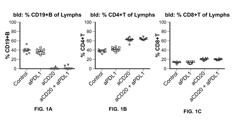
[0026] FIG. 1A-1C show the results of experiments performed to determine
the effect of the
administration of an anti-PD-Li antibody in combination with an anti-CD20
antibody on B cell
depletion. FIG. 1A depicts the percent (%) of CD19+ B lymphocytes. FIG. 1B
depicts the
percent (%) of CD4+ T lymphocytes. FIG. 1C depicts the percent (%) of CD8+ T
lymphocytes.
[0027] FIG. 2 shows the results of experiments performed to determine the
effect of the
administration of an anti-PD-Li antibody in combination with an anti-CD20
antibody on tumor
growth in a mouse model using A20 cells. Treatment groups 1-4 are described in
Example 2 in
detail. The graphs show individual plots (Trellis plots) and represent a
"cubic spline fit" of the
tumor volumes of each treatment over time. This is a mathematical algorithm
that chooses the
best smooth curve that fits all the data per treatment group.
[0028] FIG. 3 shows the results of experiments performed to determine the
effect of the
administration of an anti-PD-Li antibody in combination with an anti-CD20
antibody on tumor
growth in a mouse model using A20pRK-CD2O-GFP cells. Treatment groups 1-6 are
described
in Example 2 in detail. The graphs show individual plots (Trellis plots) and
represent a "cubic
spline fit" of the tumor volumes of each treatment over time. This is a
mathematical algorithm
that chooses the best smooth curve that fits all the data per treatment group.
DETAILED DESCRIPTION
I. General techniques
[0029] The techniques and procedures described or referenced herein are
generally well
understood and commonly employed using conventional methodology by those
skilled in the art,
such as, for example, the widely utilized methodologies described in Sambrook
et al., Molecular
Cloning: A Laboratory Manual 3d edition (2001) Cold Spring Harbor Laboratory
Press, Cold
Spring Harbor, N.Y.; Current Protocols in Molecular Biology (F.M. Ausubel, et
al. eds.,
(2003)); the series Methods in Enzymology (Academic Press, Inc.): PCR 2: A
Practical
Approach (M.J. MacPherson, B.D. Hames and G.R. Taylor eds. (1995)), Harlow and
Lane, eds.
(1988) Antibodies, A Laboratory Manual, and Animal Cell Culture (R.I.
Freshney, ed. (1987));
Oligonucleotide Synthesis (M.J. Gait, ed., 1984); Methods in Molecular
Biology, Humana Press;
Cell Biology: A Laboratory Notebook (J.E. Cellis, ed., 1998) Academic Press;
Animal Cell
-9-
CA 02933881 2016-06-14
WO 2015/095410 PCT/US2014/070983
Culture (R.I. Freshney), ed., 1987); Introduction to Cell and Tissue Culture
(J.P. Mather and
P.E. Roberts, 1998) Plenum Press; Cell and Tissue Culture: Laboratory
Procedures (A. Doyle,
J.B. Griffiths, and D.G. Newell, eds., 1993-8) J. Wiley and Sons; Handbook of
Experimental
Immunology (D.M. Weir and C.C. Blackwell, eds.); Gene Transfer Vectors for
Mammalian
Cells (J.M. Miller and M.P. Cabs, eds., 1987); PCR: The Polymerase Chain
Reaction, (Mullis
et al., eds., 1994); Current Protocols in Immunology (J.E. Coligan et al.,
eds., 1991); Short
Protocols in Molecular Biology (Wiley and Sons, 1999); Immunobiology (C.A.
Janeway and P.
Travers, 1997); Antibodies (P. Finch, 1997); Antibodies: A Practical Approach
(D. Catty., ed.,
IRL Press, 1988-1989); Monoclonal Antibodies: A Practical Approach (P.
Shepherd and C.
Dean, eds., Oxford University Press, 2000); Using Antibodies: A Laboratory
Manual (E. Harlow
and D. Lane (Cold Spring Harbor Laboratory Press, 1999); The Antibodies (M.
Zanetti and J. D.
Capra, eds., Harwood Academic Publishers, 1995); and Cancer: Principles and
Practice of
Oncology (V.T. DeVita et al., eds., J.B. Lippincott Company, 1993).
II. Definitions
[0030] The term "antagonist" is used in the broadest sense, and includes any
molecule that
partially or fully blocks, inhibits, or neutralizes a biological activity of a
native polypeptide
disclosed herein. In a similar manner, the term "agonist" is used in the
broadest sense and
includes any molecule that mimics a biological activity of a native
polypeptide disclosed herein.
Suitable agonist or antagonist molecules specifically include agonist or
antagonist antibodies or
antibody fragments, fragments or amino acid sequence variants of native
polypeptides, peptides,
antisense oligonucleotides, small organic molecules, etc. Methods for
identifying agonists or
antagonists of a polypeptide may comprise contacting a polypeptide with a
candidate agonist or
antagonist molecule and measuring a detectable change in one or more
biological activities
normally associated with the polypeptide.
[0031] The term "aptamer" refers to a nucleic acid molecule that is capable of
binding to a
target molecule, such as a polypeptide. For example, an aptamer of the
invention can
specifically bind to a B-raf polypeptide, or to a molecule in a signaling
pathway that modulates
the expression or activity of B-raf. The generation and therapeutic use of
aptamers are well
established in the art. See, e.g., U.S. Pat. No. 5,475,096, and the
therapeutic efficacy of
Macugen (Eyetech, New York) for treating age-related macular degeneration.
-10-
CA 02933881 2016-06-14
WO 2015/095410 PCT/US2014/070983
[0032] The term "PD-1 axis binding antagonist" is a molecule that inhibits the
interaction of a
PD-1 axis binding partner with either one or more of its binding partner, so
as to remove T-cell
dysfunction resulting from signaling on the PD-1 signaling axis ¨ with a
result being to restore
or enhance T-cell function (e.g., proliferation, cytokine production, target
cell killing). As used
herein, a PD-1 axis binding antagonist includes a PD-1 binding antagonist, a
PD-Li binding
antagonist and a PD-L2 binding antagonist.
[0033] The term "PD-1 binding antagonists" is a molecule that decreases,
blocks, inhibits,
abrogates or interferes with signal transduction resulting from the
interaction of PD-1 with one
or more of its binding partners, such as PD-L1, PD-L2. In some embodiments,
the PD-1 binding
antagonist is a molecule that inhibits the binding of PD-1 to its binding
partners. In a specific
aspect, the PD-1 binding antagonist inhibits the binding of PD-1 to PD-Li
and/or PD-L2. For
example, PD-1 binding antagonists include anti-PD-1 antibodies, antigen
binding fragments
thereof, immunoadhesins, fusion proteins, oligopeptides and other molecules
that decrease,
block, inhibit, abrogate or interfere with signal transduction resulting from
the interaction of PD-
1 with PD-Li and/or PD-L2. In one embodiment, a PD-1 binding antagonist
reduces the
negative co-stimulatory signal mediated by or through cell surface proteins
expressed on T
lymphocytes mediated signaling through PD-1 so as render a dysfunctional T-
cell less
dysfunctional (e.g., enhancing effector responses to antigen recognition). In
some embodiments,
the PD-1 binding antagonist is an anti-PD-1 antibody. In a specific aspect, a
PD-1 binding
antagonist is MDX-1106 described herein. In another specific aspect, a PD-1
binding antagonist
is Merck 3745 described herein. In another specific aspect, a PD-1 binding
antagonist is CT-011
described herein.
[0034] The term "PD-Li binding antagonists" is a molecule that decreases,
blocks, inhibits,
abrogates or interferes with signal transduction resulting from the
interaction of PD-Li with
either one or more of its binding partners, such as PD-1, B7-1. In some
embodiments, a PD-Li
binding antagonist is a molecule that inhibits the binding of PD-Li to its
binding partners. In a
specific aspect, the PD-Li binding antagonist inhibits binding of PD-Li to PD-
1 and/or B7-1. In
some embodiments, the PD-Li binding antagonists include anti-PD-Li antibodies,
antigen
binding fragments thereof, immunoadhesins, fusion proteins, oligopeptides and
other molecules
that decrease, block, inhibit, abrogate or interfere with signal transduction
resulting from the
interaction of PD-Li with one or more of its binding partners, such as PD-1,
B7-1. In one
embodiment, a PD-Li binding antagonist reduces the negative co-stimulatory
signal mediated
-11-
CA 02933881 2016-06-14
WO 2015/095410 PCT/US2014/070983
by or through cell surface proteins expressed on T lymphocytes mediated
signaling through PD-
Li so as to render a dysfunctional T-cell less dysfunctional (e.g., enhancing
effector responses to
antigen recognition). In some embodiments, a PD-Li binding antagonist is an
anti-PD-Li
antibody. In a specific aspect, an anti-PD-Li antibody is YW243.55.S70
described herein. In
another specific aspect, an anti-PD-Li antibody is MDX-1105 described herein.
In still another
specific aspect, an anti-PD-Li antibody is MPDL3280A described herein.
[0035] The term "PD-L2 binding antagonists" is a molecule that decreases,
blocks, inhibits,
abrogates or interferes with signal transduction resulting from the
interaction of PD-L2 with
either one or more of its binding partners, such as PD-1. In some embodiments,
a PD-L2
binding antagonist is a molecule that inhibits the binding of PD-L2 to its
binding partners. In a
specific aspect, the PD-L2 binding antagonist inhibits binding of PD-L2 to PD-
1. In some
embodiments, the PD-L2 antagonists include anti-PD-L2 antibodies, antigen
binding fragments
thereof, immunoadhesins, fusion proteins, oligopeptides and other molecules
that decrease,
block, inhibit, abrogate or interfere with signal transduction resulting from
the interaction of PD-
L2 with either one or more of its binding partners, such as PD-1. In one
embodiment, a PD-L2
binding antagonist reduces the negative co-stimulatory signal mediated by or
through cell
surface proteins expressed on T lymphocytes mediated signaling through PD-L2
so as render a
dysfunctional T-cell less dysfunctional (e.g., enhancing effector responses to
antigen
recognition). In some embodiments, a PD-L2 binding antagonist is an
immunoadhesin.
[0036] The term "dysfunction" in the context of immune dysfunction, refers to
a state of
reduced immune responsiveness to antigenic stimulation. The term includes the
common
elements of both exhaustion and/or anergy in which antigen recognition may
occur, but the
ensuing immune response is ineffective to control infection or tumor growth.
[0037] The term "dysfunctional", as used herein, also includes refractory or
unresponsive to
antigen recognition, specifically, impaired capacity to translate antigen
recognition into down-
stream T-cell effector functions, such as proliferation, cytokine production
(e.g., IL-2) and/or
target cell killing.
[0038] The term "anergy" refers to the state of unresponsiveness to antigen
stimulation
resulting from incomplete or insufficient signals delivered through the T-cell
receptor (e.g.
increase in intracellular Ca+2 in the absence of ras-activation). T cell
anergy can also result upon
stimulation with antigen in the absence of co-stimulation, resulting in the
cell becoming
refractory to subsequent activation by the antigen even in the context of
costimulation. The
-12-
CA 02933881 2016-06-14
WO 2015/095410 PCT/US2014/070983
unresponsive state can often be overriden by the presence of Interleukin-2.
Anergic T-cells do
not undergo clonal expansion and/or acquire effector functions.
[0039] The term "exhaustion" refers to T cell exhaustion as a state of T cell
dysfunction that
arises from sustained TCR signaling that occurs during many chronic infections
and cancer. It is
distinguished from anergy in that it arises not through incomplete or
deficient signaling, but
from sustained signaling. It is defined by poor effector function, sustained
expression of
inhibitory receptors and a transcriptional state distinct from that of
functional effector or
memory T cells. Exhaustion prevents optimal control of infection and tumors.
Exhaustion can
result from both extrinsic negative regulatory pathways (e.g.,
immunoregulatory cytokines) as
well as cell intrinsic negative regulatory (costimulatory) pathways (PD-1, B7-
H3, B7-H4, etc.).
[0040] "Enhancing T-cell function" means to induce, cause or stimulate a T-
cell to have a
sustained or amplified biological function, or renew or reactivate exhausted
or inactive T-cells.
Examples of enhancing T-cell function include: increased secretion of 7-
interferon from CD8+
T-cells, increased proliferation, increased antigen responsiveness (e.g.,
viral, pathogen, or tumor
clearance) relative to such levels before the intervention. In one embodiment,
the level of
enhancement is as least 50%, alternatively 60%, 70%, 80%, 90%, 100%, 120%,
150%, or 200%.
The manner of measuring this enhancement is known to one of ordinary skill in
the art.
[0041] A "T cell dysfunctional disorder" is a disorder or condition of T-cells
characterized by
decreased responsiveness to antigenic stimulation. In a particular embodiment,
a T-cell
dysfunctional disorder is a disorder that is specifically associated with
inappropriate increased
signaling through PD-1. In another embodiment, a T-cell dysfunctional disorder
is one in which
T-cells are anergic or have decreased ability to secrete cytokines,
proliferate, or execute cytolytic
activity. In a specific aspect, the decreased responsiveness results in
ineffective control of a
pathogen or tumor expressing an immunogen. Examples of T cell dysfunctional
disorders
characterized by T-cell dysfunction include unresolved acute infection,
chronic infection and
tumor immunity.
[0042] "Tumor immunity" refers to the process in which tumors evade immune
recognition
and clearance. Thus, as a therapeutic concept, tumor immunity is "treated"
when such evasion is
attenuated, and the tumors are recognized and attacked by the immune system.
Examples of
tumor recognition include tumor binding, tumor shrinkage and tumor clearance.
[0043] "Immunogenicity" refers to the ability of a particular substance to
provoke an immune
response. Tumors are immunogenic and enhancing tumor immunogenicity aids in
the clearance
-13-
CA 02933881 2016-06-14
WO 2015/095410 PCT/US2014/070983
of the tumor cells by the immune response. Examples of enhancing tumor
immunogenicity
include treatment with anti-PDL antibodies and an anti-CD20 antibody.
[0044] "Sustained response" refers to the sustained effect on reducing tumor
growth after
cessation of a treatment. For example, the tumor size may remain to be the
same or smaller as
compared to the size at the beginning of the administration phase. In some
embodiments, the
sustained response has a duration at least the same as the treatment duration,
at least 1.5X, 2.0X,
2.5X, or 3.0X length of the treatment duration.
[0045] As used herein, "cancer" and "cancerous" refer to or describe the
physiological
condition in mammals that is typically characterized by unregulated cell
growth. Included in
this definition are benign and malignant cancers as well as dormant tumors or
micrometastases.
Examples of cancer include but are not limited to, carcinoma, lymphoma,
blastoma, sarcoma,
and leukemia. More particular examples of such cancers include but are not
limited to
squamous cell cancer, lung cancer (including small-cell lung cancer, non-small
cell lung cancer,
adenocarcinoma of the lung, and squamous carcinoma of the lung), cancer of the
peritoneum,
hepatocellular cancer, gastric or stomach cancer (including gastrointestinal
cancer), pancreatic
cancer, glioblastoma, cervical cancer, ovarian cancer, liver cancer, bladder
cancer, hepatoma,
breast cancer, colon cancer, colorectal cancer, endometrial or uterine
carcinoma, salivary gland
carcinoma, kidney or renal cancer, liver cancer, prostate cancer, vulval
cancer, thyroid cancer,
hepatic carcinoma and various types of head and neck cancer, as well as B-cell
lymphoma
(including low grade/follicular non-Hodgkin's lymphoma (NHL); small
lymphocytic (SL) NHL;
intermediate grade/follicular NHL; intermediate grade diffuse NHL; high grade
immunoblastic
NHL; high grade lymphoblastic NHL; high grade small non-cleaved cell NHL;
bulky disease
NHL; mantle cell lymphoma; AIDS-related lymphoma; and Waldenstrom's
Macroglobulinemia);
chronic lymphocytic leukemia (CLL); acute lymphoblastic leukemia (ALL); Hairy
cell leukemia;
chronic myeloblastic leukemia; and post-transplant lymphoproliferative
disorder (PTLD), as
well as abnormal vascular proliferation associated with phakomatoses, edema
(such as that
associated with brain tumors), and Meigs' syndrome. Examples of cancer may
include primary
tumors of any of the above types of cancer or metastatic tumors at a second
site derived from
any of the above types of cancer.
[0046] The term "antibody" includes monoclonal antibodies (including full
length antibodies
which have an immunoglobulin Fc region), antibody compositions with
polyepitopic specificity,
multispecific antibodies (e.g., bispecific antibodies, diabodies, and single-
chain molecules, as
-14-
CA 02933881 2016-06-14
WO 2015/095410 PCT/US2014/070983
well as antibody fragments (e.g., Fab, F(abt)2, and Fv). The term
"immunoglobulin" (Ig) is used
interchangeably with "antibody" herein.
[0047] The basic 4-chain antibody unit is a heterotetrameric glycoprotein
composed of two
identical light (L) chains and two identical heavy (H) chains. An IgM antibody
consists of 5 of
the basic heterotetramer units along with an additional polypeptide called a J
chain, and contains
antigen binding sites, while IgA antibodies comprise from 2-5 of the basic 4-
chain units
which can polymerize to form polyvalent assemblages in combination with the J
chain. In the
case of IgGs, the 4-chain unit is generally about 150,000 daltons. Each L
chain is linked to an H
chain by one covalent disulfide bond, while the two H chains are linked to
each other by one or
more disulfide bonds depending on the H chain isotype. Each H and L chain also
has regularly
spaced intrachain disulfide bridges. Each H chain has at the N-terminus, a
variable domain (VH)
followed by three constant domains (CH) for each of the cc and 7 chains and
four CH domains for
1.1 and c isotypes. Each L chain has at the N-terminus, a variable domain (VL)
followed by a
constant domain at its other end. The VL is aligned with the VH and the CL is
aligned with the
first constant domain of the heavy chain (CH1). Particular amino acid residues
are believed to
form an interface between the light chain and heavy chain variable domains.
The pairing of a
VH and VL together forms a single antigen-binding site. For the structure and
properties of the
different classes of antibodies, see e.g., Basic and Clinical Immunology, 8th
Edition, Daniel P.
Sties, Abba I. Terr and Tristram G. Parsolw (eds), Appleton & Lange, Norwalk,
CT, 1994, page
71 and Chapter 6. The L chain from any vertebrate species can be assigned to
one of two clearly
distinct types, called kappa and lambda, based on the amino acid sequences of
their constant
domains. Depending on the amino acid sequence of the constant domain of their
heavy chains
(CH), immunoglobulins can be assigned to different classes or isotypes. There
are five classes
of immunoglobulins: IgA, IgD, IgE, IgG and IgM, having heavy chains designated
cc, 8, c, 7 and
1..t, respectively. The 7 and cc classes are further divided into subclasses
on the basis of relatively
minor differences in the CH sequence and function, e.g., humans express the
following
subclasses: IgG 1, IgG2A, IgG2B, IgG3, IgG4, IgAl and IgA2.
[0048] The "variable region" or "variable domain" of an antibody refers to the
amino-
terminal domains of the heavy or light chain of the antibody. The variable
domains of the heavy
chain and light chain may be referred to as "VH" and "VL", respectively. These
domains are
generally the most variable parts of the antibody (relative to other
antibodies of the same class)
and contain the antigen binding sites.
-15-
CA 02933881 2016-06-14
WO 2015/095410 PCT/US2014/070983
[0049] The term "variable" refers to the fact that certain segments of the
variable domains
differ extensively in sequence among antibodies. The V domain mediates antigen
binding and
defines the specificity of a particular antibody for its particular antigen.
However, the variability
is not evenly distributed across the entire span of the variable domains.
Instead, it is
concentrated in three segments called hypervariable regions (HVRs) both in the
light-chain and
the heavy chain variable domains. The more highly conserved portions of
variable domains are
called the framework regions (FR). The variable domains of native heavy and
light chains each
comprise four FR regions, largely adopting a beta-sheet configuration,
connected by three
HVRs, which form loops connecting, and in some cases forming part of, the beta-
sheet structure.
The HVRs in each chain are held together in close proximity by the FR regions
and, with the
HVRs from the other chain, contribute to the formation of the antigen binding
site of antibodies
(see Kabat et al., Sequences of Immunological Interest, Fifth Edition,
National Institute of
Health, Bethesda, MD (1991)). The constant domains are not involved directly
in the binding of
antibody to an antigen, but exhibit various effector functions, such as
participation of the
antibody in antibody-dependent cellular toxicity.
[0050] The term "monoclonal antibody" as used herein refers to an antibody
obtained from a
population of substantially homogeneous antibodies, i.e., the individual
antibodies comprising
the population are identical except for possible naturally occurring mutations
and/or post-
translation modifications (e.g., isomerizations, amidations) that may be
present in minor
amounts. Monoclonal antibodies are highly specific, being directed against a
single antigenic
site. In contrast to polyclonal antibody preparations which typically include
different antibodies
directed against different determinants (epitopes), each monoclonal antibody
is directed against
a single determinant on the antigen. In addition to their specificity, the
monoclonal antibodies
are advantageous in that they are synthesized by the hybridoma culture,
uncontaminated by other
immunoglobulins. The modifier "monoclonal" indicates the character of the
antibody as being
obtained from a substantially homogeneous population of antibodies, and is not
to be construed
as requiring production of the antibody by any particular method. For example,
the monoclonal
antibodies to be used in accordance with the present invention may be made by
a variety of
techniques, including, for example, the hybridoma method (e.g., Kohler and
Milstein., Nature,
256:495-97 (1975); Hongo et al., Hybridoma, 14 (3): 253-260 (1995), Harlow et
al., Antibodies:
A Laboratory Manual, (Cold Spring Harbor Laboratory Press, 2nd ed. 1988);
Hammerling et al.,
in: Monoclonal Antibodies and T-Cell Hybridomas 563-681 (Elsevier, N.Y.,
1981)),
-16-
CA 02933881 2016-06-14
WO 2015/095410 PCT/US2014/070983
recombinant DNA methods (see, e.g., U.S. Patent No. 4,816,567), phage-display
technologies
(see, e.g., Clackson et al., Nature, 352: 624-628 (1991); Marks et al., J.
Mol. Biol. 222: 581-597
(1992); Sidhu et al., J. Mol. Biol. 338(2): 299-310 (2004); Lee et al., J.
Mol. Biol. 340(5): 1073-
1093 (2004); Fellouse, Proc. Natl. Acad. Sci. USA 101(34): 12467-12472 (2004);
and Lee et al.,
J. Immunol. Methods 284(1-2): 119-132 (2004), and technologies for producing
human or
human-like antibodies in animals that have parts or all of the human
immunoglobulin loci or
genes encoding human immunoglobulin sequences (see, e.g., WO 1998/24893; WO
1996/34096; WO 1996/33735; WO 1991/10741; Jakobovits et al., Proc. Natl. Acad.
Sci. USA
90: 2551 (1993); Jakobovits et al., Nature 362: 255-258 (1993); Bruggemann et
al., Year in
Immunol. 7:33 (1993); U.S. Patent Nos. 5,545,807; 5,545,806; 5,569,825;
5,625,126; 5,633,425;
and 5,661,016; Marks et al., Bio/Technology 10: 779-783 (1992); Lonberg et
al., Nature 368:
856-859 (1994); Morrison, Nature 368: 812-813 (1994); Fishwild et al., Nature
Biotechnol. 14:
845-851 (1996); Neuberger, Nature Biotechnol. 14: 826 (1996); and Lonberg and
Huszar,
Intern. Rev. Immunol. 13: 65-93 (1995).
[0051] The term "naked antibody" refers to an antibody that is not conjugated
to a cytotoxic
moiety or radiolabel.
[0052] The terms 'full-length antibody," "intact antibody" or "whole antibody"
are used
interchangeably to refer to an antibody in its substantially intact form, as
opposed to an antibody
fragment. Specifically whole antibodies include those with heavy and light
chains including an
Fc region. The constant domains may be native sequence constant domains (e.g.,
human native
sequence constant domains) or amino acid sequence variants thereof. In some
cases, the intact
antibody may have one or more effector functions.
[0053] An "antibody fragment" comprises a portion of an intact antibody,
preferably the
antigen binding and/or the variable region of the intact antibody. Examples of
antibody
fragments include Fab, Fab', F(abt)2 and Fv fragments; diabodies; linear
antibodies (see U.S.
Patent 5,641,870, Example 2; Zapata et al., Protein Eng. 8(10): 1057-1062
[1995]); single-chain
antibody molecules and multispecific antibodies formed from antibody
fragments. Papain
digestion of antibodies produced two identical antigen-binding fragments,
called "Fab"
fragments, and a residual "Fc" fragment, a designation reflecting the ability
to crystallize readily.
The Fab fragment consists of an entire L chain along with the variable region
domain of the H
chain (VH), and the first constant domain of one heavy chain (CH1). Each Fab
fragment is
monovalent with respect to antigen binding, i.e., it has a single antigen-
binding site. Pepsin
-17-
CA 02933881 2016-06-14
WO 2015/095410 PCT/US2014/070983
treatment of an antibody yields a single large F(abt)2 fragment which roughly
corresponds to two
disulfide linked Fab fragments having different antigen-binding activity and
is still capable of
cross-linking antigen. Fab' fragments differ from Fab fragments by having a
few additional
residues at the carboxy terminus of the CH1 domain including one or more
cysteines from the
antibody hinge region. Fab'-SH is the designation herein for Fab' in which the
cysteine
residue(s) of the constant domains bear a free thiol group. F(abt)2 antibody
fragments originally
were produced as pairs of Fab' fragments which have hinge cysteines between
them. Other
chemical couplings of antibody fragments are also known.
[0054] The Fc fragment comprises the carboxy-terminal portions of both H
chains held
together by disulfides. The effector functions of antibodies are determined by
sequences in the
Fc region, the region which is also recognized by Fc receptors (FcR) found on
certain types of
cells.
[0055] "Fv" is the minimum antibody fragment which contains a complete antigen-
recognition
and -binding site. This fragment consists of a dimer of one heavy- and one
light-chain variable
region domain in tight, non-covalent association. From the folding of these
two domains
emanate six hypervariable loops (3 loops each from the H and L chain) that
contribute the amino
acid residues for antigen binding and confer antigen binding specificity to
the antibody.
However, even a single variable domain (or half of an Fv comprising only three
HVRs specific
for an antigen) has the ability to recognize and bind antigen, although at a
lower affinity than the
entire binding site.
[0056] "Single-chain Fv" also abbreviated as "sFv" or "scFv" are antibody
fragments that
comprise the VH and VL antibody domains connected into a single polypeptide
chain.
Preferably, the sFv polypeptide further comprises a polypeptide linker between
the VH and VL
domains which enables the sFv to form the desired structure for antigen
binding. For a review
of the sFv, see Pluckthun in The Pharmacology of Monoclonal Antibodies, vol.
113, Rosenburg
and Moore eds., Springer-Verlag, New York, pp. 269-315 (1994).
[0057] "Functional fragments" of the antibodies of the invention comprise a
portion of an
intact antibody, generally including the antigen binding or variable region of
the intact antibody
or the Fc region of an antibody which retains or has modified FcR binding
capability. Examples
of antibody fragments include linear antibody, single-chain antibody molecules
and
multispecific antibodies formed from antibody fragments.
-18-
CA 02933881 2016-06-14
WO 2015/095410 PCT/US2014/070983
[0058] The term "diabodies" refers to small antibody fragments prepared by
constructing sFy
fragments (see preceding paragraph) with short linkers (about 5-10) residues)
between the VH
and VL domains such that inter-chain but not intra-chain pairing of the V
domains is achieved,
thereby resulting in a bivalent fragment, i.e., a fragment having two antigen-
binding sites.
Bispecific diabodies are heterodimers of two "crossover" sFy fragments in
which the VH and VL
domains of the two antibodies are present on different polypeptide chains.
Diabodies are
described in greater detail in, for example, EP 404,097; WO 93/11161;
Hollinger et al., Proc.
Natl. Acad. Sci. USA 90: 6444-6448 (1993).
[0059] The monoclonal antibodies herein specifically include "chimeric"
antibodies
(immunoglobulins) in which a portion of the heavy and/or light chain is
identical with or
homologous to corresponding sequences in antibodies derived from a particular
species or
belonging to a particular antibody class or subclass, while the remainder of
the chain(s) is(are)
identical with or homologous to corresponding sequences in antibodies derived
from another
species or belonging to another antibody class or subclass, as well as
fragments of such
antibodies, so long as they exhibit the desired biological activity (U.S.
Patent No. 4,816,567;
Morrison et al., Proc. Natl. Acad. Sci. USA, 81:6851-6855 (1984)). Chimeric
antibodies of
interest herein include PRIMATIZED antibodies wherein the antigen-binding
region of the
antibody is derived from an antibody produced by, e.g., immunizing macaque
monkeys with an
antigen of interest. As used herein, "humanized antibody" is used a subset of
"chimeric
antibodies."
[0060] "Humanized" forms of non-human (e.g., murine) antibodies are chimeric
antibodies
that contain minimal sequence derived from non-human immunoglobulin. In one
embodiment, a
humanized antibody is a human immunoglobulin (recipient antibody) in which
residues from an
HVR (hereinafter defined) of the recipient are replaced by residues from an
HVR of a non-
human species (donor antibody) such as mouse, rat, rabbit or non-human primate
having the
desired specificity, affinity, and/or capacity. In some instances, framework
("FR") residues of
the human immunoglobulin are replaced by corresponding non-human residues.
Furthermore,
humanized antibodies may comprise residues that are not found in the recipient
antibody or in
the donor antibody. These modifications may be made to further refine antibody
performance,
such as binding affinity. In general, a humanized antibody will comprise
substantially all of at
least one, and typically two, variable domains, in which all or substantially
all of the
hypervariable loops correspond to those of a non-human immunoglobulin
sequence, and all or
-19-
CA 02933881 2016-06-14
WO 2015/095410 PCT/US2014/070983
substantially all of the FR regions are those of a human immunoglobulin
sequence, although the
FR regions may include one or more individual FR residue substitutions that
improve antibody
performance, such as binding affinity, isomerization, immunogenicity, etc. The
number of these
amino acid substitutions in the FR are typically no more than 6 in the H
chain, and in the L
chain, no more than 3. The humanized antibody optionally will also comprise at
least a portion
of an immunoglobulin constant region (Fc), typically that of a human
immunoglobulin. For
further details, see, e.g., Jones et al., Nature 321:522-525 (1986); Riechmann
et al., Nature
332:323-329 (1988); and Presta, Curr. Op. Struct. Biol. 2:593-596 (1992). See
also, for
example, Vaswani and Hamilton, Ann. Allergy, Asthma & Immunol. 1:105-115
(1998); Harris,
Biochem. Soc. Transactions 23:1035-1038 (1995); Hurle and Gross, Curr. Op.
Biotech. 5:428-
433 (1994); and U.S. Pat. Nos. 6,982,321 and 7,087,409.
[0061] A "human antibody" is an antibody that possesses an amino-acid sequence
corresponding to that of an antibody produced by a human and/or has been made
using any of
the techniques for making human antibodies as disclosed herein. This
definition of a human
antibody specifically excludes a humanized antibody comprising non-human
antigen-binding
residues. Human antibodies can be produced using various techniques known in
the art,
including phage-display libraries. Hoogenboom and Winter, J. Mol. Biol.,
227:381 (1991);
Marks et al., J. Mol. Biol., 222:581 (1991). Also available for the
preparation of human
monoclonal antibodies are methods described in Cole et al., Monoclonal
Antibodies and Cancer
Therapy, Alan R. Liss, p. 77 (1985); Boerner et al., J. Immunol., 147(1):86-95
(1991). See also
van Dijk and van de Winkel, Curr. Opin. Pharmacol., 5: 368-74 (2001). Human
antibodies can
be prepared by administering the antigen to a transgenic animal that has been
modified to
produce such antibodies in response to antigenic challenge, but whose
endogenous loci have
been disabled, e.g., immunized xenomice (see, e.g., U.S. Pat. Nos. 6,075,181
and 6,150,584
regarding XENOMOUSETm technology). See also, for example, Li et al., Proc.
Natl. Acad. Sci.
USA, 103:3557-3562 (2006) regarding human antibodies generated via a human B-
cell
hybridoma technology.
[0062] The term "hypervariable region," "HVR," or "HV," when used herein
refers to the
regions of an antibody variable domain which are hypervariable in sequence
and/or form
structurally defined loops. Generally, antibodies comprise six HVRs; three in
the VH (H1, H2,
H3), and three in the VL (L1, L2, L3). In native antibodies, H3 and L3 display
the most
diversity of the six HVRs, and H3 in particular is believed to play a unique
role in conferring
-20-
CA 02933881 2016-06-14
WO 2015/095410 PCT/US2014/070983
fine specificity to antibodies. See, e.g., Xu et al., Immunity 13:37-45
(2000); Johnson and Wu,
in Methods in Molecular Biology 248:1-25 (Lo, ed., Human Press, Totowa, NJ,
2003). Indeed,
naturally occurring camelid antibodies consisting of a heavy chain only are
functional and stable
in the absence of light chain. See, e.g., Hamers-Casterman et al., Nature
363:446-448 (1993);
Sheriff et al., Nature Struct. Biol. 3:733-736 (1996).
[0063] A number of HVR delineations are in use and are encompassed herein. The
Kabat
Complementarity Determining Regions (CDRs) are based on sequence variability
and are the
most commonly used (Kabat et al., Sequences of Proteins of Immunological
Interest, 5th Ed.
Public Health Service, National Institutes of Health, Bethesda, MD. (1991)).
Chothia refers
instead to the location of the structural loops (Chothia and Lesk, J. Mol.
Biol. 196:901-917
(1987)). The AbM HVRs represent a compromise between the Kabat HVRs and
Chothia
structural loops, and are used by Oxford Molecular's AbM antibody modeling
software. The
"contact" HVRs are based on an analysis of the available complex crystal
structures. The
residues from each of these HVRs are noted below.
Loop Kabat AbM Chothia Contact
Li L24-L34 L24-L34 L26-L32 L30-L36
L2 L50-L56 L50-L56 L50-L52 L46-L55
L3 L89-L97 L89-L97 L91-L96 L89-L96
H1 H31-H35B H26-H35B H26-H32 H30-H35B (Kabat numbering)
H1 H31-H35 H26-H35 H26-H32 H30-H35 (Chothia numbering)
H2 H50-H65 H50-H58 H53-H55 H47-H58
H3 H95-H102 H95-H102 H96-H101 H93-H101
[0064] HVRs may comprise "extended HVRs" as follows: 24-36 or 24-34 (L1), 46-
56 or 50-
56 (L2) and 89-97 or 89-96 (L3) in the VL and 26-35 (H1), 50-65 or 49-65 (H2)
and 93-102, 94-
102, or 95-102 (H3) in the VH. The variable domain residues are numbered
according to Kabat
et al., supra, for each of these definitions.
[0065] The expression "variable-domain residue-numbering as in Kabat" or
"amino-acid-
position numbering as in Kabat," and variations thereof, refers to the
numbering system used for
heavy-chain variable domains or light-chain variable domains of the
compilation of antibodies in
Kabat et al., supra. Using this numbering system, the actual linear amino acid
sequence may
contain fewer or additional amino acids corresponding to a shortening of, or
insertion into, a FR
or HVR of the variable domain. For example, a heavy-chain variable domain may
include a
single amino acid insert (residue 52a according to Kabat) after residue 52 of
H2 and inserted
residues (e.g. residues 82a, 82b, and 82c, etc. according to Kabat) after
heavy-chain FR residue
-21-
CA 02933881 2016-06-14
WO 2015/095410 PCT/US2014/070983
82. The Kabat numbering of residues may be determined for a given antibody by
alignment at
regions of homology of the sequence of the antibody with a "standard" Kabat
numbered
sequence.
[0066] "Framework" or "FR" residues are those variable-domain residues other
than the HVR
residues as herein defined.
[0067] A "human consensus framework" or "acceptor human framework" is a
framework that
represents the most commonly occurring amino acid residues in a selection of
human
immunoglobulin VL or VH framework sequences. Generally, the selection of human
immunoglobulin VL or VH sequences is from a subgroup of variable domain
sequences.
Generally, the subgroup of sequences is a subgroup as in Kabat et al.,
Sequences of Proteins of
Immunological Interest, 5" Ed. Public Health Service, National Institutes of
Health, Bethesda,
MD (1991). Examples include for the VL, the subgroup may be subgroup kappa I,
kappa II,
kappa III or kappa IV as in Kabat et al., supra. Additionally, for the VH, the
subgroup may be
subgroup I, subgroup II, or subgroup III as in Kabat et al., supra.
Alternatively, a human
consensus framework can be derived from the above in which particular
residues, such as when
a human framework residue is selected based on its homology to the donor
framework by
aligning the donor framework sequence with a collection of various human
framework
sequences. An acceptor human framework "derived from" a human immunoglobulin
framework
or a human consensus framework may comprise the same amino acid sequence
thereof, or it
may contain pre-existing amino acid sequence changes. In some embodiments, the
number of
pre-existing amino acid changes are 10 or less, 9 or less, 8 or less, 7 or
less, 6 or less, 5 or less, 4
or less, 3 or less, or 2 or less.
[0068] A "VH subgroup III consensus framework" comprises the consensus
sequence obtained
from the amino acid sequences in variable heavy subgroup III of Kabat et al.,
supra. In one
embodiment, the VH subgroup III consensus framework amino acid sequence
comprises at least
a portion or all of each of the following sequences: EVQLVESGGGLVQPGGSLRLSCAAS
(HC-FR1)(SEQ ID NO:4), WVRQAPGKGLEWV (HC-FR2), (SEQ ID NO:5),
RFTISADTSKNTAYLQMNSLRAEDTAVYYCAR (HC-FR3, SEQ ID NO:6),
WGQGTLVTVSA (HC-FR4), (SEQ ID NO:7).
[0069] A "VL kappa I consensus framework" comprises the consensus sequence
obtained from
the amino acid sequences in variable light kappa subgroup I of Kabat et al.,
supra. In one
embodiment, the VH subgroup I consensus framework amino acid sequence
comprises at least a
-22-
CA 02933881 2016-06-14
WO 2015/095410 PCT/US2014/070983
portion or all of each of the following sequences: DIQMTQSPSSLSASVGDRVTITC (LC-
FR1)
(SEQ ID NO:11), WYQQKPGKAPKLLIY (LC-FR2) (SEQ ID NO:12),
GVPSRFSGSGSGTDFTLTISSLQPEDFATYYC (LC-FR3)(SEQ ID NO:13), FGQGTKVEIKR
(LC-FR4)(SEQ ID NO:14).
[0070] An "amino-acid modification" at a specified position, e.g. of the Fc
region, refers to the
substitution or deletion of the specified residue, or the insertion of at
least one amino acid
residue adjacent the specified residue. Insertion "adjacent" to a specified
residue means
insertion within one to two residues thereof. The insertion may be N-terminal
or C-terminal to
the specified residue. The preferred amino acid modification herein is a
substitution.
[0071] An "affinity-matured" antibody is one with one or more alterations in
one or more
HVRs thereof that result in an improvement in the affinity of the antibody for
antigen, compared
to a parent antibody that does not possess those alteration(s). In one
embodiment, an affinity-
matured antibody has nanomolar or even picomolar affinities for the target
antigen. Affinity-
matured antibodies are produced by procedures known in the art. For example,
Marks et al.,
Bio/Technology 10:779-783 (1992) describes affinity maturation by VH- and VL-
domain
shuffling. Random mutagenesis of HVR and/or framework residues is described
by, for
example: Barbas et al. Proc Nat. Acad. Sci. USA 91:3809-3813 (1994); Schier et
al. Gene
169:147-155 (1995); Yelton et al. J. Immunol. 155:1994-2004 (1995); Jackson et
al., J.
Immunol. 154(7):3310-9 (1995); and Hawkins et al, J. Mol. Biol. 226:889-896
(1992).
[0072] As use herein, the term "specifically binds to" or is "specific for"
refers to measurable
and reproducible interactions such as binding between a target and an
antibody, which is
determinative of the presence of the target in the presence of a heterogeneous
population of
molecules including biological molecules. For example, an antibody that
specifically binds to a
target (which can be an epitope) is an antibody that binds this target with
greater affinity,
avidity, more readily, and/or with greater duration than it binds to other
targets. In one
embodiment, the extent of binding of an antibody to an unrelated target is
less than about 10% of
the binding of the antibody to the target as measured, e.g., by a
radioimmunoassay (RIA). In
certain embodiments, an antibody that specifically binds to a target has a
dissociation constant
(Kd) of < li.tM, < 100 nM, < 10 nM, < 1 nM, or < 0.1 nM. In certain
embodiments, an antibody
specifically binds to an epitope on a protein that is conserved among the
protein from different
species. In another embodiment, specific binding can include, but does not
require exclusive
binding.
-23-
CA 02933881 2016-06-14
WO 2015/095410 PCT/US2014/070983
[0073] As used herein, the term "immunoadhesin" designates antibody-like
molecules which
combine the binding specificity of a heterologous protein (an "adhesin") with
the effector
functions of immunoglobulin constant domains. Structurally, the immunoadhesins
comprise a
fusion of an amino acid sequence with the desired binding specificity which is
other than the
antigen recognition and binding site of an antibody (i.e., is "heterologous"),
and an
immunoglobulin constant domain sequence. The adhesin part of an immunoadhesin
molecule
typically is a contiguous amino acid sequence comprising at least the binding
site of a receptor
or a ligand. The immunoglobulin constant domain sequence in the immunoadhesin
may be
obtained from any immunoglobulin, such as IgG-1, IgG-2 (including IgG2A and
IgG2B), IgG-3,
or IgG-4 subtypes, IgA (including IgA-1 and IgA-2), IgE, IgD or IgM. The Ig
fusions
preferably include the substitution of a domain of a polypeptide or antibody
described herein in
the place of at least one variable region within an Ig molecule. In a
particularly preferred
embodiment, the immunoglobulin fusion includes the hinge, CH2 and CH3, or the
hinge, CH1,
CH2 and CH3 regions of an IgG1 molecule. For the production of immunoglobulin
fusions see
also US Patent No. 5,428,130 issued June 27, 1995. For example, useful
immunoadhesins as
second medicaments useful for combination therapy herein include polypeptides
that comprise
the extracellular or PD-1 binding portions of PD-Li or PD-L2 or the
extracellular or PD-Li or
PD-L2 binding portions of PD-1, fused to a constant domain of an
immunoglobulin sequence,
such as a PD-Li ECD ¨ Fc, a PD-L2 ECD ¨ Fc, and a PD-1 ECD - Fc, respectively.
Immunoadhesin combinations of Ig Fc and ECD of cell surface receptors are
sometimes termed
soluble receptors.
[0074] A 'fusion protein" and a 'fusion polypeptide" refer to a polypeptide
having two
portions covalently linked together, where each of the portions is a
polypeptide having a
different property. The property may be a biological property, such as
activity in vitro or in vivo.
The property may also be simple chemical or physical property, such as binding
to a target
molecule, catalysis of a reaction, etc. The two portions may be linked
directly by a single
peptide bond or through a peptide linker but are in reading frame with each
other.
[0075] A "PD-1 oligopeptide," "PD-Li oligopeptide," or "PD-L2 oligopeptide" is
an
oligopeptide that binds, preferably specifically, to a PD-1, PD-Li or PD-L2
negative
costimulatory polypeptide, respectively, including a receptor, ligand or
signaling component,
respectively, as described herein. Such oligopeptides may be chemically
synthesized using
known oligopeptide synthesis methodology or may be prepared and purified using
recombinant
-24-
CA 02933881 2016-06-14
WO 2015/095410 PCT/US2014/070983
technology. Such oligopeptides are usually at least about 5 amino acids in
length, alternatively
at least about 6, 7, 8, 9, 10, 11, 12, 13, 14, 15, 16, 17, 18, 19, 20, 21, 22,
23, 24, 25, 26, 27, 28,
29, 30, 31, 32, 33, 34, 35, 36, 37, 38, 39, 40, 41, 42, 43, 44, 45, 46, 47,
48, 49, 50, 51, 52, 53, 54,
55, 56, 57, 58, 59, 60, 61, 62, 63, 64, 65, 66, 67, 68, 69, 70, 71, 72, 73,
74, 75, 76, 77, 78, 79, 80,
81, 82, 83, 84, 85, 86, 87, 88, 89, 90, 91, 92, 93, 94, 95, 96, 97, 98, 99, or
100 amino acids in
length or more. Such oligopeptides may be identified using well known
techniques. In this
regard, it is noted that techniques for screening oligopeptide libraries for
oligopeptides that are
capable of specifically binding to a polypeptide target are well known in the
art (see, e.g., U.S.
Patent Nos. 5,556,762, 5,750,373, 4,708,871, 4,833,092, 5,223,409, 5,403,484,
5,571,689,
5,663,143; PCT Publication Nos. WO 84/03506 and W084/03564; Geysen et al.,
Proc. Natl.
Acad. Sci. U.S.A., 81:3998-4002 (1984); Geysen et al., Proc. Natl. Acad. Sci.
U.S.A., 82:178-182
(1985); Geysen et al., in Synthetic Peptides as Antigens, 130-149 (1986);
Geysen et al., J. ImmunoL
Meth., 102:259-274 (1987); Schoofs et al., J. ImmunoL, 140:611-616 (1988),
Cwirla, S. E. et al.
Proc. Natl. Acad. Sci. USA, 87:6378 (1990); Lowman, H.B. et al. Biochemistry,
30:10832 (1991);
Clackson, T. et al. Nature, 352: 624 (1991); Marks, J. D. et al., J. Mol.
Biol., 222:581 (1991); Kang,
A.S. et al. Proc. Natl. Acad. Sci. USA, 88:8363 (1991), and Smith, G. P.,
Current Opin. BiotechnoL,
2:668 (1991).
[0076] A "blocking" antibody or an "antagonist" antibody is one that inhibits
or reduces a
biological activity of the antigen it binds. In some embodiments, blocking
antibodies or
antagonist antibodies substantially or completely inhibit the biological
activity of the antigen.
The anti-PD-Li antibodies of the invention block the signaling through PD-1 so
as to restore a
functional response by T-cells (e.g., proliferation, cytokine production,
target cell killing) from a
dysfunctional state to antigen stimulation.
[0077] An "agonist" or activating antibody is one that enhances or initiates
signaling by the
antigen to which it binds. In some embodiments, agonist antibodies cause or
activate signaling
without the presence of the natural ligand.
[0078] The term "Fc region" herein is used to define a C-terminal region of an
immunoglobulin heavy chain, including native-sequence Fc regions and variant
Fc regions.
Although the boundaries of the Fc region of an immunoglobulin heavy chain
might vary, the
human IgG heavy-chain Fc region is usually defined to stretch from an amino
acid residue at
position Cys226, or from Pro230, to the carboxyl-terminus thereof. The C-
terminal lysine
(residue 447 according to the EU numbering system) of the Fc region may be
removed, for
-25-
CA 02933881 2016-06-14
WO 2015/095410 PCT/US2014/070983
example, during production or purification of the antibody, or by
recombinantly engineering the
nucleic acid encoding a heavy chain of the antibody. Accordingly, a
composition of intact
antibodies may comprise antibody populations with all K447 residues removed,
antibody
populations with no K447 residues removed, and antibody populations having a
mixture of
antibodies with and without the K447 residue. Suitable native-sequence Fc
regions for use in
the antibodies of the invention include human IgGl, IgG2 (IgG2A, IgG2B), IgG3
and IgG4.
[0079] "Fc receptor" or "FcR" describes a receptor that binds to the Fc region
of an antibody.
The preferred FcR is a native sequence human FcR. Moreover, a preferred FcR is
one which
binds an IgG antibody (a gamma receptor) and includes receptors of the Fc7RI,
Fc7RII, and
Fc7RIII subclasses, including allelic variants and alternatively spliced forms
of these receptors,
Fc7RII receptors include Fc7RIIA (an "activating receptor") and Fc7RIIB (an
"inhibiting
receptor"), which have similar amino acid sequences that differ primarily in
the cytoplasmic
domains thereof. Activating receptor Fc7RIIA contains an immunoreceptor
tyrosine-based
activation motif (ITAM) in its cytoplasmic domain. Inhibiting receptor Fc7RIIB
contains an
immunoreceptor tyrosine-based inhibition motif (ITIIVI) in its cytoplasmic
domain. (see M.
Daeron, Annu. Rev. Immunol. 15:203-234 (1997). FcRs are reviewed in Ravetch
and Kinet,
Annu. Rev. Immunol. 9: 457-92 (1991); Capel et al., Immunomethods 4: 25-34
(1994); and de
Haas et al., J. Lab. Clin. Med. 126: 330-41 (1995). Other FcRs, including
those to be identified
in the future, are encompassed by the term "FcR" herein.
[0080] The term "Fc receptor" or "FcR" also includes the neonatal receptor,
FcRn, which is
responsible for the transfer of maternal IgGs to the fetus. Guyer et al., J.
Immunol. 117: 587
(1976) and Kim et al., J. Immunol. 24: 249 (1994). Methods of measuring
binding to FcRn are
known (see, e.g., Ghetie and Ward, Immunol. Today 18: (12): 592-8 (1997);
Ghetie et al., Nature
Biotechnology 15 (7): 637-40 (1997); Hinton et al., J. Biol. Chem. 279 (8):
6213-6 (2004); WO
2004/92219 (Hinton et al.). Binding to FcRn in vivo and serum half-life of
human FcRn high-
affinity binding polypeptides can be assayed, e.g., in transgenic mice or
transfected human cell
lines expressing human FcRn, or in primates to which the polypeptides having a
variant Fc
region are administered. WO 2004/42072 (Presta) describes antibody variants
which improved
or diminished binding to FcRs. See also, e.g., Shields et al., J. Biol. Chem.
9(2): 6591-6604
(2001).
[0081] The phrase "substantially reduced," or "substantially different," as
used herein, denotes
a sufficiently high degree of difference between two numeric values (generally
one associated
-26-
CA 02933881 2016-06-14
WO 2015/095410 PCT/US2014/070983
with a molecule and the other associated with a reference/comparator molecule)
such that one of
skill in the art would consider the difference between the two values to be of
statistical
significance within the context of the biological characteristic measured by
said values (e.g., Kd
values). The difference between said two values is, for example, greater than
about 10%, greater
than about 20%, greater than about 30%, greater than about 40%, and/or greater
than about 50%
as a function of the value for the reference/comparator molecule.
[0082] The term "substantially similar" or "substantially the same," as used
herein, denotes a
sufficiently high degree of similarity between two numeric values (for
example, one associated
with an antibody of the invention and the other associated with a
reference/comparator
antibody), such that one of skill in the art would consider the difference
between the two values
to be of little or no biological and/or statistical significance within the
context of the biological
characteristic measured by said values (e.g., Kd values). The difference
between said two values
is, for example, less than about 50%, less than about 40%, less than about
30%, less than about
20%, and/or less than about 10% as a function of the reference/comparator
value.
[0083] "Carriers" as used herein include pharmaceutically acceptable carriers,
excipients, or
stabilizers that are nontoxic to the cell or mammal being exposed thereto at
the dosages and
concentrations employed. Often the physiologically acceptable carrier is an
aqueous pH
buffered solution. Examples of physiologically acceptable carriers include
buffers such as
phosphate, citrate, and other organic acids; antioxidants including ascorbic
acid; low molecular
weight (less than about 10 residues) polypeptide; proteins, such as serum
albumin, gelatin, or
immunoglobulins; hydrophilic polymers such as polyvinylpyrrolidone; amino
acids such as
glycine, glutamine, asparagine, arginine or lysine; monosaccharides,
disaccharides, and other
carbohydrates including glucose, mannose, or dextrins; chelating agents such
as EDTA; sugar
alcohols such as mannitol or sorbitol; salt-forming counterions such as
sodium; and/or nonionic
surfactants such as TWEENTm, polyethylene glycol (PEG), and PLURONICSTM.
[0084] A "package insert" refers to instructions customarily included in
commercial packages
of medicaments that contain information about the indications customarily
included in
commercial packages of medicaments that contain information about the
indications, usage,
dosage, administration, contraindications, other medicaments to be combined
with the packaged
product, and/or warnings concerning the use of such medicaments, etc.
[0085] As used herein, the term "treatment" refers to clinical intervention
designed to alter the
natural course of the individual or cell being treated during the course of
clinical pathology.
-27-
CA 02933881 2016-06-14
WO 2015/095410 PCT/US2014/070983
Desirable effects of treatment include decreasing the rate of disease
progression, ameliorating or
palliating the disease state, and remission or improved prognosis. For
example, an individual is
successfully "treated" if one or more symptoms associated with cancer are
mitigated or
eliminated, including, but are not limited to, reducing the proliferation of
(or destroying)
cancerous cells, decreasing symptoms resulting from the disease, increasing
the quality of life
of those suffering from the disease, decreasing the dose of other medications
required to treat the
disease, delaying the progression of the disease, and/or prolonging survival
of individuals.
[0086] As used herein, "delaying progression of a disease" means to defer,
hinder, slow,
retard, stabilize, and/or postpone development of the disease (such as
cancer). This delay can be
of varying lengths of time, depending on the history of the disease and/or
individual being
treated. As is evident to one skilled in the art, a sufficient or significant
delay can, in effect,
encompass prevention, in that the individual does not develop the disease. For
example, a late
stage cancer, such as development of metastasis, may be delayed.
[0087] As used herein, "reducing or inhibiting cancer relapse" means to reduce
or inhibit
tumor or cancer relapse or tumor or cancer progression. As disclosed herein,
cancer relapse
and/or cancer progression include, without limitation, cancer metastasis.
[0088] An "effective amount" is at least the minimum concentration required to
effect a
measurable improvement or prevention of a particular disorder. An effective
amount herein may
vary according to factors such as the disease state, age, sex, and weight of
the patient, and the
ability of the antibody to elicit a desired response in the individual. An
effective amount is also
one in which any toxic or detrimental effects of the treatment are outweighed
by the
therapeutically beneficial effects. For prophylactic use, beneficial or
desired results include
results such as eliminating or reducing the risk, lessening the severity, or
delaying the onset of
the disease, including biochemical, histological and/or behavioral symptoms of
the disease, its
complications and intermediate pathological phenotypes presenting during
development of the
disease. For therapeutic use, beneficial or desired results include clinical
results such as
decreasing one or more symptoms resulting from the disease, increasing the
quality of life of
those suffering from the disease, decreasing the dose of other medications
required to treat the
disease, enhancing effect of another medication such as via targeting,
delaying the progression
of the disease, and/or prolonging survival. In the case of cancer or tumor, an
effective amount of
the drug may have the effect in reducing the number of cancer cells; reducing
the tumor size;
inhibiting (i.e., slow to some extent or desirably stop) cancer cell
infiltration into peripheral
-28-
CA 02933881 2016-06-14
WO 2015/095410 PCT/US2014/070983
organs; inhibit (i.e., slow to some extent and desirably stop) tumor
metastasis; inhibiting to some
extent tumor growth; and/or relieving to some extent one or more of the
symptoms associated
with the disorder. An effective amount can be administered in one or more
administrations. For
purposes of this invention, an effective amount of drug, compound, or
pharmaceutical
composition is an amount sufficient to accomplish prophylactic or therapeutic
treatment either
directly or indirectly. As is understood in the clinical context, an effective
amount of a drug,
compound, or pharmaceutical composition may or may not be achieved in
conjunction with
another drug, compound, or pharmaceutical composition. Thus, an "effective
amount" may be
considered in the context of administering one or more therapeutic agents, and
a single agent
may be considered to be given in an effective amount if, in conjunction with
one or more other
agents, a desirable result may be or is achieved.
[0089] As used herein, "in conjunction with" refers to administration of one
treatment
modality in addition to another treatment modality. As such, "in conjunction
with" refers to
administration of one treatment modality before, during, or after
administration of the other
treatment modality to the individual.
[0090] As used herein, "complete response" or "CR" refers to disappearance of
all target
lesions; "partial response" or "PR" refers to at least a 30% decrease in the
sum of the longest
diameters (SLD) of target lesions, taking as reference the baseline SLD; and
"stable disease" or
"SD" refers to neither sufficient shrinkage of target lesions to qualify for
PR, nor sufficient
increase to qualify for PD, taking as reference the smallest SLD since the
treatment started.
[0091] As used herein, "progressive disease" or "PD" refers to at least a 20%
increase in the
SLD of target lesions, taking as reference the smallest SLD recorded since the
treatment started
or the presence of one or more new lesions.
[0092] As used herein, "progression free survival" (PFS) refers to the length
of time during
and after treatment during which the disease being treated (e.g., cancer) does
not get worse.
Progression-free survival may include the amount of time patients have
experienced a complete
response or a partial response, as well as the amount of time patients have
experienced stable
disease.
[0093] As used herein, "overall response rate" (ORR) refers to the sum of
complete response
(CR) rate and partial response (PR) rate.
[0094] As used herein, "overall survival" refers to the percentage of
individuals in a group
who are likely to be alive after a particular duration of time.
-29-
CA 02933881 2016-06-14
WO 2015/095410 PCT/US2014/070983
[0095] A "chemotherapeutic agent" is a chemical compound useful in the
treatment of cancer.
Examples of chemotherapeutic agents include alkylating agents such as thiotepa
and
cyclophosphamide (CYTOXANI0); alkyl sulfonates such as busulfan, improsulfan,
and
piposulfan; aziridines such as benzodopa, carboquone, meturedopa, and uredopa;
ethylenimines
and methylamelamines including altretamine, triethylenemelamine,
trietylenephosphoramide,
triethiylenethiophosphoramide and trimethylolomelamine; acetogenins
(especially bullatacin and
bullatacinone); delta-9-tetrahydrocannabinol (dronabinol, MARINOUD); beta-
lapachone;
lapachol; colchicines; betulinic acid; a camptothecin (including the synthetic
analogue topotecan
(HYCAMTINIO), CPT-11 (irinotecan, CAMPTOSAR0), acetylcamptothecin,
scopolectin, and
9-aminocamptothecin); bryostatin; pemetrexed; callystatin; CC-1065 (including
its adozelesin,
carzelesin and bizelesin synthetic analogues); podophyllotoxin; podophyllinic
acid; teniposide;
cryptophycins (particularly cryptophycin 1 and cryptophycin 8); dolastatin;
duocarmycin
(including the synthetic analogues, KW-2189 and CB1-TM1); eleutherobin;
pancratistatin;
TLK-286; CDP323, an oral alpha-4 integrin inhibitor; a sarcodictyin;
spongistatin; nitrogen
mustards such as chlorambucil, chlornaphazine, cholophosphamide, estramustine,
ifosfamide,
mechlorethamine, mechlorethamine oxide hydrochloride, melphalan, novembichin,
phenesterine, prednimustine, trofosfamide, uracil mustard; nitrosureas such as
carmustine,
chlorozotocin, fotemustine, lomustine, nimustine, and ranimnustine;
antibiotics such as the
enediyne antibiotics (e.g., calicheamicin, especially calicheamicin gamma 11
and calicheamicin
omegaIl (see, e.g., Nicolaou et al., Angew. Chem Intl. Ed. Engl., 33: 183-186
(1994));
dynemicin, including dynemicin A; an esperamicin; as well as neocarzinostatin
chromophore
and related chromoprotein enediyne antibiotic chromophores), aclacinomysins,
actinomycin,
authramycin, azaserine, bleomycins, cactinomycin, carabicin, carminomycin,
carzinophilin,
chromomycinis, dactinomycin, daunorubicin, detorubicin, 6-diazo-5-oxo-L-
norleucine,
doxorubicin (including ADRIAMYCIN , morpholino-doxorubicin, cyanomorpholino-
doxorubicin, 2-pyrrolino-doxorubicin, doxorubicin HC1 liposome injection
(DOXIUD) and
deoxydoxorubicin), epirubicin, esorubicin, idarubicin, marcellomycin,
mitomycins such as
mitomycin C, mycophenolic acid, nogalamycin, olivomycins, peplomycin,
potfiromycin,
puromycin, quelamycin, rodorubicin, streptonigrin, streptozocin, tubercidin,
ubenimex,
zinostatin, zorubicin; anti-metabolites such as methotrexate, gemcitabine
(GEMZAR0), tegafur
(UFTORAUD), capecitabine (XELODA10), an epothilone, and 5-fluorouracil (5-FU);
folic acid
analogues such as denopterin, methotrexate, pteropterin, trimetrexate; purine
analogs such as
-30-
CA 02933881 2016-06-14
WO 2015/095410 PCT/US2014/070983
fludarabine, 6-mercaptopurine, thiamiprine, thioguanine; pyrimidine analogs
such as ancitabine,
azacitidine, 6-azauridine, carmofur, cytarabine, dideoxyuridine,
doxifluridine, enocitabine,
floxuridine, and imatinib (a 2-phenylaminopyrimidine derivative), as well as
other c-Kit
inhibitors; anti-adrenals such as aminoglutethimide, mitotane, trilostane;
folic acid replenisher
such as frolinic acid; aceglatone; aldophosphamide glycoside; aminolevulinic
acid; eniluracil;
amsacrine; bestrabucil; bisantrene; edatraxate; defofamine; demecolcine;
diaziquone;
elfornithine; elliptinium acetate; etoglucid; gallium nitrate; hydroxyurea;
lentinan; lonidainine;
maytansinoids such as maytansine and ansamitocins; mitoguazone; mitoxantrone;
mopidanmol;
nitraerine; pentostatin; phenamet; pirarubicin; losoxantrone; 2-
ethylhydrazide; procarbazine;
PSK polysaccharide complex (JHS Natural Products, Eugene, OR); razoxane;
rhizoxin;
sizofiran; spirogermanium; tenuazonic acid; triaziquone; 2,2',2"-
trichlorotriethylamine;
trichothecenes (especially T-2 toxin, verracurin A, roridin A and anguidine);
urethan; vindesine
(ELDISINE , FILDESINI0); dacarbazine; mannomustine; mitobronitol; mitolactol;
pipobroman; gacytosine; arabinoside ("Ara-C"); thiotepa; taxoids, e.g.,
paclitaxel (TAXOUD),
albumin-engineered nanoparticle formulation of paclitaxel (ABRAXANETm), and
doxetaxel
(TAXOTERRO); chloranbucil; 6-thioguanine; mercaptopurine; methotrexate;
platinum analogs
such as cisplatin and carboplatin; vinblastine (VELBANI0); platinum; etoposide
(VP-16);
ifosfamide; mitoxantrone; vincristine (ONCOVINI0); oxaliplatin; leucovovin;
vinorelbine
(NAVELBINE10); novantrone; edatrexate; daunomycin; aminopterin; ibandronate;
topoisomerase inhibitor RFS 2000; difluorometlhylornithine (DMF0); retinoids
such as retinoic
acid; pharmaceutically acceptable salts, acids or derivatives of any of the
above; as well as
combinations of two or more of the above such as CHOP, an abbreviation for a
combined
therapy of cyclophosphamide, doxorubicin, vincristine, and prednisolone, and
FOLFOX, an
abbreviation for a treatment regimen with oxaliplatin (ELOXATINTm) combined
with 5-FU and
leucovovin.
[0096] Additional examples of chemotherapeutic agents include anti-hormonal
agents that act
to regulate, reduce, block, or inhibit the effects of hormones that can
promote the growth of
cancer, and are often in the form of systemic, or whole-body treatment. They
may be hormones
themselves. Examples include anti-estrogens and selective estrogen receptor
modulators
(SERMs), including, for example, tamoxifen (including NOLVADEX tamoxifen),
raloxifene
(EVISTA10), droloxifene, 4-hydroxytamoxifen, trioxifene, keoxifene, LY117018,
onapristone,
and toremifene (FARESTONC1); anti-progesterones; estrogen receptor down-
regulators (ERDs);
-31-
CA 02933881 2016-06-14
WO 2015/095410 PCT/US2014/070983
estrogen receptor antagonists such as fulvestrant (FASLODEX ); agents that
function to
suppress or shut down the ovaries, for example, leutinizing hormone-releasing
hormone (LHRH)
agonists such as leuprolide acetate (LUPRON and ELIGARD0), goserelin acetate,
buserelin
acetate and tripterelin; anti-androgens such as flutamide, nilutamide and
bicalutamide; and
aromatase inhibitors that inhibit the enzyme aromatase, which regulates
estrogen production in
the adrenal glands, such as, for example, 4(5)-imidazoles, aminoglutethimide,
megestrol acetate
(MEGASE10), exemestane (AROMASINIO), formestanie, fadrozole, vorozole
(RIVISOR0),
letrozole (FEMARA10), and anastrozole (ARIMIDEVD). In addition, such
definition of
chemotherapeutic agents includes bisphosphonates such as clodronate (for
example,
BONEFOS or OSTACC,), etidronate (DIDROCAUD), NE-58095, zoledronic
acid/zoledronate
(ZOMETA10), alendronate (FOSAMAX0), pamidronate (AREDIA10), tiludronate
(SKELIDO),
or risedronate (ACTONEUD); as well as troxacitabine (a 1,3-dioxolane
nucleoside cytosine
analog); anti-sense oligonucleotides, particularly those that inhibit
expression of genes in
signaling pathways implicated in abherant cell proliferation, such as, for
example, PKC-alpha,
Raf, H-Ras, and epidermal growth factor receptor (EGF-R); vaccines such as
THERATOPE
vaccine and gene therapy vaccines, for example, ALLOVECTIN vaccine, LEUVECTIN
vaccine, and VAXID vaccine; topoisomerase 1 inhibitor (e.g., LURTOTECANC));
an anti-
estrogen such as fulvestrant; a Kit inhibitor such as imatinib or EXEL-0862 (a
tyrosine kinase
inhibitor); EGFR inhibitor such as erlotinib or cetuximab; an anti-VEGF
inhibitor such as
bevacizumab; arinotecan; rmRH (e.g., ABARELIX ); lapatinib and lapatinib
ditosylate (an
ErbB-2 and EGFR dual tyrosine kinase small-molecule inhibitor also known as
GW572016);
17AAG (geldanamycin derivative that is a heat shock protein (Hsp) 90 poison),
and
pharmaceutically acceptable salts, acids or derivatives of any of the above.
[0097] As used herein, the term "cytokine" refers generically to proteins
released by one cell
population that act on another cell as intercellular mediators or have an
autocrine effect on the
cells producing the proteins. Examples of such cytokines include lymphokines,
monokines;
interleukins ("ILs") such as IL-1, IL-la, IL-2, IL-3, IL-4, IL-5, IL-6, IL-7,
IL-8, IL-9, IL10, IL-
11, IL-12, IL-13, IL-15, IL-17A-F, IL-18 to IL-29 (such as IL-23), IL-31,
including
PROLEUKIN rIL-2; a tumor-necrosis factor such as TNF-a or TNF-I3, TGF-I31-3;
and other
polypeptide factors including leukemia inhibitory factor ("LIF"), ciliary
neurotrophic factor
("CNTF"), CNTF-like cytokine ("CLC"), cardiotrophin ("CT"), and kit ligand
("KL").
-32-
CA 02933881 2016-06-14
WO 2015/095410 PCT/US2014/070983
[0098] As used herein, the term "chemokine" refers to soluble factors (e.g.,
cytokines) that
have the ability to selectively induce chemotaxis and activation of
leukocytes. They also trigger
processes of angiogenesis, inflammation, wound healing, and tumorigenesis.
Example
chemokines include IL-8, a human homolog of murine keratinocyte
chemoattractant (KC).
[0099] "CD20" as used herein refers to the human B-lymphocyte antigen CD20
(also known
as CD20, B-lymphocyte surface antigen Bl, Leu-16, Bp35, BM5, and LF5; the
sequence is
characterized by the SwissProt database entry P11836) is a hydrophobic
transmembrane protein
with a molecular weight of approximately 35 kD located on pre-B and mature B
lymphocytes.
(Valentine, M.A., et al., J. Biol. Chem. 264(19) (1989 11282-11287; Tedder,
T.F., et al, Proc.
Natl. Acad. Sci. U.S.A. 85 (1988) 208-12; Stamenkovic, I., et al., J. Exp.
Med. 167 (1988) 1975-
80; Einfeld, D.A., et al., EMBO J. 7 (1988) 711-7; Tedder, T.F., et al., J.
Immunol. 142 (1989)
2560-8). The corresponding human gene is Membrane-spanning 4-domains,
subfamily A,
member 1, also known as MS4A1. This gene encodes a member of the membrane-
spanning 4A
gene family. Members of this nascent protein family are characterized by
common structural
features and similar intron/exon splice boundaries and display unique
expression patterns among
hematopoietic cells and nonlymphoid tissues. This gene encodes the B-
lymphocyte surface
molecule which plays a role in the development and differentiation of B-cells
into plasma cells.
This family member is localized to 1 1q12, among a cluster of family members.
Alternative
splicing of this gene results in two transcript variants which encode the same
protein.
[0100] The terms "CD20" and "CD20 antigen" are used interchangeably herein,
and include
any variants, isoforms and species homologs of human CD20 which are naturally
expressed by
cells or are expressed on cells transfected with the CD20 gene. Binding of an
antibody of the
invention to the CD20 antigen mediate the killing of cells expressing CD20
(e.g., a tumor cell)
by inactivating CD20. The killing of the cells expressing CD20 may occur by
one or more of the
following mechanisms: Cell death/apoptosis induction, ADCC and CDC.
[0101] Synonyms of CD20, as recognized in the art, include B-lymphocyte
antigen CD20, B-
lymphocyte surface antigen Bl, Leu-16, Bp35, BM5, and LF5.
[0102] The term "anti-CD20 antibody" according to the invention is an antibody
that binds
specifically to CD20 antigen. Depending on binding properties and biological
activities of anti-
CD20 antibodies to the CD20 antigen, two types of anti-CD20 antibodies (type I
and type II
anti-CD20 antibodies) can be distinguished according to Cragg, M.S., et al.,
Blood 103 (2004)
2738-2743; and Cragg, M.S., et al., Blood 101 (2003) 1045-1052, see Table 1.
-33-
CA 02933881 2016-06-14
WO 2015/095410 PCT/US2014/070983
Table 1: Properties of type I and type II anti-CD20 antibodies
Type I anti-CD20 antibodies type II anti-CD20 antibodies
type I CD20 epitope type II CD20 epitope
Localize CD20 to lipid rafts Do not localize CD20 to lipid rafts
Increased CDC (if IgG1 isotype) Decreased CDC (if IgG1 isotype)
ADCC activity (if IgG1 isotype) ADCC activity (if IgG1 isotype)
Full binding capacity Reduced binding capacity
Homotypic aggregation Stronger homotypic aggregation
. Strong cell death induction without
Apoptosis induction upon cross-linking
cross-linking
[0103] Examples of type II anti-CD20 antibodies include e.g. humanized B-Lyl
antibody
IgG1 (a chimeric humanized IgG1 antibody as disclosed in WO 2005/044859), 11B8
IgG1 (as
disclosed in WO 2004/035607), and AT80 IgGl. Typically type II anti-CD20
antibodies of the
IgG1 isotype show characteristic CDC properties. Type II anti-CD20 antibodies
have a
decreased CDC (if IgG1 isotype) compared to type I antibodies of the IgG1
isotype.
[0104] Examples of type I anti-CD20 antibodies include e.g. rituximab, HI47
IgG3 (ECACC,
hybridoma), 2C6 IgG1 (as disclosed in WO 2005/103081), 2F2 IgG1 (as disclosed
and WO
2004/035607 and WO 2005/103081) and 2H7 IgG1 (as disclosed in WO 2004/056312).
[0105] The afucosylated anti-CD20 antibodies according to the invention is
preferably a type
II anti-CD20 antibodies, more preferably an afucosylated humanized B-Ly 1
antibody as
described in WO 2005/044859 and WO 2007/031875.
[0106] The "rituximab" antibody (reference antibody; example of a type I anti-
CD20
antibody) is a genetically engineered chimeric human gamma 1 murine constant
domain
containing monoclonal antibody directed against the human CD20 antigen.
However this
antibody is not glycoengineered and not afocusylates and thus has an amount of
fucose of at
least 85 %. This chimeric antibody contains human gamma 1 constant domains and
is identified
by the name "C2B8" in US 5,736,137 (Andersen, et. al.) issued on April 17,
1998, assigned to
IDEC Pharmaceuticals Corporation. Rituximab is approved for the treatment of
patients with
relapsed or refracting low-grade or follicular, CD20 positive, B cell non-
Hodgkin's lymphoma.
In vitro mechanism of action studies have shown that rituximab exhibits human
complement-
dependent cytotoxicity (CDC) (Reff, M.E., et. al, Blood 83(2) (1994) 435-445).
Additionally, it
exhibits activity in assays that measure antibody-dependent cellular
cytotoxicity (ADCC).
[0107] The term "GA101 antibody" as used herein refers to any one of the
following
antibodies that bind human CD20: (1) an antibody comprising an HVR-H1
comprising the
-34-
CA 02933881 2016-06-14
WO 2015/095410 PCT/US2014/070983
amino acid sequence of SEQ ID NO:50, an HVR-H2 comprising the amino acid
sequence of
SEQ ID NO:51, an HVR-H3 comprising the amino acid sequence of SEQ ID NO:52, an
HVR-
Ll comprising the amino acid sequence of SEQ ID NO:53, an HVR-L2 comprising
the amino
acid sequence of SEQ ID NO:54, and an HVR-L3 comprising the amino acid
sequence of SEQ
ID NO:55; (2) an antibody comprising a VH domain comprising the amino acid
sequence of
SEQ ID NO:56 and a VL domain comprising the amino acid sequence of SEQ ID
NO:57, (3) an
antibody comprising an amino acid sequence of SEQ ID NO:58 and an amino acid
sequence of
SEQ ID NO: 59; (4) an antibody known as obinutuzumab, or (5) an antibody that
comprises an
amino acid sequence that has at least 95%, 96%, 97%, 98% or 99% sequence
identity with
amino acid sequence of SEQ ID NO:58 and that comprises an amino acid sequence
that has at
least 95%, 96%, 97%, 98% or 99% sequence identity with an amino acid sequence
of SEQ ID
NO: 59. In one embodiment, the GA101 antibody is an IgG1 isotype antibody. In
some
embodiments, the anti-CD20 antibody is a humanized B-Lyl antibody.
[0108] The term "humanized B-Lyl antibody" refers to humanized B-Lyl antibody
as
disclosed in WO 2005/044859 and WO 2007/031875, which were obtained from the
murine
monoclonal anti-CD20 antibody B-Lyl (variable region of the murine heavy chain
(VH): SEQ
ID NO: 30; variable region of the murine light chain (VL): SEQ ID NO: 31- see
Poppema, S.
and Visser, L., Biotest Bulletin 3 (1987) 131-139) by chimerization with a
human constant
domain from IgG1 and following humanization (see WO 2005/044859 and WO
2007/031875).
These "humanized B-Lyl antibodies" are disclosed in detail in WO 2005/ 044859
and WO
2007/031875.
[0109] In one embodiment, the "humanized B-Lyl antibody" has variable region
of the heavy
chain (VH) selected from group of SEQ ID No.32 to SEQ ID No.48 (corresponding
to B-HH2 to
B-HH9 and B-HL8 to B-HL17 of WO 2005/044859 and WO 2007/031875). In one
specific
embodiment, such variable domain is selected from the group consisting of SEQ
ID No. 32, 33,
36, 38, 40, 42 and 44 (corresponding to B-HH2, BHH-3, B-HH6, B-HH8, B-HL8, B-
HL11 and
B-HL13 of WO 2005/044859 and WO 2007/031875). In one specific embodiment, the
"humanized B-Lyl antibody" has variable region of the light chain (VL) of SEQ
ID No. 49
(corresponding to B-KV1 of WO 2005/044859 and WO 2007/031875). In one specific
embodiment, the "humanized B-Lyl antibody" has a variable region of the heavy
chain (VH) of
SEQ ID No.36 (corresponding to B-HH6 of WO 2005/044859 and WO 2007/031875) and
a
variable region of the light chain (VL) of SEQ ID No. 49 (corresponding to B-
KV1 of
-35-
CA 02933881 2016-06-14
WO 2015/095410 PCT/US2014/070983
WO 2005/044859 and WO 2007/031875). Furthermore in one embodiment, the
humanized B-
Ly1 antibody is an IgG1 antibody. According to the invention such afocusylated
humanized B-
Ly1 antibodies are glycoengineered (GE) in the Fc region according to the
procedures described
in WO 2005/044859, WO 2004/065540, WO 2007/031875, Umana, P. et al., Nature
Biotechnol.
17 (1999) 176-180 and WO 99/154342. In one embodiment, the afucosylated glyco-
engineered
humanized B-Lyl is B-HH6-B-KV1 GE. In one embodiment, the anti-CD20 antibody
is
obinutuzumab (recommended INN, WHO Drug Information, Vol. 26, No. 4, 2012, p.
453). As
used herein, obinutuzumab is synonymous for GA101 or R05072759. This replaces
all previous
versions (e.g. Vol. 25, No. 1, 2011, p.75-'76), and is formerly known as
afutuzumab
(recommended INN, WHO Drug Information, Vol. 23, No. 2, 2009, p. 176;Vol. 22,
No. 2, 2008,
p. 124). In some embodiments, the humanized B-Lyl antibody is an antibody
comprising a
heavy chain comprising the amino acid sequence of SEQ ID NO:60 and a light
chain comprising
the amino acid sequence of SEQ ID NO:61 or an antigen-binding fragment
thereof. In some
embodiments, the humanized B-Lyl antibody comprises a heavy chain variable
region
comprising the three heavy chain CDRs of SEQ ID NO:60 and a light chain
variable region
comprising the three light chain CDRs of SEQ ID NO:61.
Heavy chain (SEQ ID NO:60)
QVQLVQSGAE VKKPGSSVKV SCKASGYAFS YSWINWVRQA PGQGLEWMGR 50
IFPGDGDTDY NGKFKGRVTI TADKSTSTAY MELSSLRSED TAVYYCARNV 100
FDGYWLVYWG QGTLVTVSSA STKGPSVFPL APSSKSTSGG TAALGCLVKD 150
YFPEPVTVSW NSGALTSGVH TFPAVLQSSG LYSLSSVVTV PSSSLGTQTY 200
ICNVNHKPSN TKVDKKVEPK SCDKTHTCPP CPAPELLGGP SVFLFPPKPK 250
DTLMISRTPE VTCVVVDVSH EDPEVKFNWY VDGVEVHNAK TKPREEQYNS 300
TYRVVSVLTV LHQDWLNGKE YKCKVSNKAL PAPIEKTISK AKGQPREPQV 350
YTLPPSRDEL TKNQVSLTCL VKGFYPSDIA VEWESNGQPE NNYKTTPPVL 400
DSDGSFFLYS KLTVDKSRWQ QGNVFSCSVM HEALHNHYTQ KSLSLSPG 449
Light chain (SEQ ID NO:61)
DIVMTQTPLS LPVTPGEPAS ISCRSSKSLL HSNGITYLYW YLQKPGQSPQ 50
LLIYQMSNLV SGVPDRFSGS GSGTDFTLKI SRVEAEDVGV YYCAQNLELP 100
YTFGGGTKVE IKRTVAAPSV FIFPPSDEQL KSGTASVVCL LNNFYPREAK 150
VQWKVDNALQ SGNSQESVTE QDSKDSTYSL SSTLTLSKAD YEKHKVYACE 200
VTHQGLSSPV TKSFNRGEC 219
[0110] In some embodiments, the humanized B-Lyl antibody is an afucosylated
glyco-
engineered humanized B-Lyl. Such glycoengineered humanized B-Lyl antibodies
have an
altered pattern of glycosylation in the Fc region, preferably having a reduced
level of fucose
residues. Preferably the amount of fucose is 60 % or less of the total amount
of oligosaccharides
-36-
CA 02933881 2016-06-14
WO 2015/095410 PCT/US2014/070983
at Asn297 (in one embodiment the amount of fucose is between 40 % and 60 %, in
another
embodiment the amount of fucose is 50 % or less, and in still another
embodiment the amount of
fucose is 30 % or less). Furthermore the oligosaccharides of the Fe region are
preferably
bisected. These glycoengineered humanized B-Lyl antibodies have an increased
ADCC.
[0111] The "ratio of the binding capacities to CD20 on Raji cells (ATCC-No.
CCL-86) of an
anti-CD20 antibodies compared to rituximab" is determined by direct
immunofluorescence
measurement (the mean fluorescence intensities (MFI) is measured) using said
anti-CD20
antibody conjugated with Cy5 and rituximab conjugated with Cy5 in a FACSArray
(Becton
Dickinson) with Raji cells (ATCC-No. CCL-86), as described in Example No. 2,
and calculated
as follows:
Ratio of the binding capacities to CD20 on Raji cells (ATCC-No. CCL-86) =
MFI(Cy5- anti- CD20 antibody) Cy5 - labeling ratio (Cy5- rituximab)
x
MFI(Cy5- rituximab) Cy5- labeling ratio (Cy5- anti- CD20
antibody)
[0112] MFI is the mean fluorescent intensity. The "Cy5-labeling ratio" as used
herein means
the number of Cy5-label molecules per molecule antibody.
[0113] Typically said type II anti-CD20 antibody has a ratio of the binding
capacities to CD20
on Raji cells (ATCC-No. CCL-86) of said second anti-CD20 antibody compared to
rituximab of
0.3 to 0.6, and in one embodiment, 0.35 to 0.55, and in yet another
embodiment, 0.4 to 0.5.
[0114] In one embodiment said type II anti-CD20 antibody, e.g., a
GA101antibody, has
increased antibody dependent cellular cytotoxicity (ADCC).
[0115] By "antibody having increased antibody dependent cellular cytotoxicity
(ADCC)", it is
meant an antibody, as that term is defined herein, having increased ADCC as
determined by any
suitable method known to those of ordinary skill in the art. One accepted in
vitro ADCC assay is
as follows:
1) the assay uses target cells that are known to express the target antigen
recognized by the antigen-binding region of the antibody;
2) the assay uses human peripheral blood mononuclear cells (PBMCs),
isolated from blood of a randomly chosen healthy donor, as effector cells;
3) the assay is carried out according to following protocol:
i) the PBMCs are isolated using standard density centrifugation
procedures
and are suspended at 5 x 10⁶ cells/ml in RPMI cell culture medium;
-37-
CA 02933881 2016-06-14
WO 2015/095410 PCT/US2014/070983
ii) the target cells are grown by standard tissue culture methods,
harvested
from the exponential growth phase with a viability higher than 90%, washed in
RPMI cell
culture medium, labeled with 100 micro-Curies of 51Cr, washed twice with cell
culture medium,
and resuspended in cell culture medium at a density of 105 cells/ml;
iii) 100 microliters of the final target cell suspension above are
transferred to
each well of a 96-well microtiter plate;
iv) the antibody is serially-diluted from 4000 ng/ml to 0.04 ng/ml in cell
culture medium and 50 microliters of the resulting antibody solutions are
added to the target
cells in the 96-well microtiter plate, testing in triplicate various antibody
concentrations covering
the whole concentration range above;
v) for the maximum release (MR) controls, 3 additional wells in the plate
containing the labeled target cells, receive 50 microliters of a 2% (VN)
aqueous solution of non-
ionic detergent (Nonidet, Sigma, St. Louis), instead of the antibody solution
(point iv above);
vi) for the spontaneous release (SR) controls, 3 additional wells in the
plate
containing the labeled target cells, receive 50 microliters of RPMI cell
culture medium instead of
the antibody solution (point iv above);
vii) the 96-well microtiter plate is then centrifuged at 50 x g for 1
minute and
incubated for 1 hour at 4 C;
viii) 50 microliters of the PBMC suspension (point i above) are added to
each
well to yield an effector:target cell ratio of 25:1 and the plates are placed
in an incubator under
5% CO2 atmosphere at 37 C for 4 hours;
ix) the cell-free supernatant from each well is harvested and the
experimentally released radioactivity (ER) is quantified using a gamma
counter;
x) the percentage of specific lysis is calculated for each antibody
concentration according to the formula (ER-MR)/(MR-SR) x 100, where ER is the
average
radioactivity quantified (see point ix above) for that antibody concentration,
MR is the average
radioactivity quantified (see point ix above) for the MR controls (see point V
above), and SR is
the average radioactivity quantified (see point ix above) for the SR controls
(see point vi above);
4) "increased ADCC" is defined as either an increase in the
maximum
percentage of specific lysis observed within the antibody concentration range
tested above,
and/or a reduction in the concentration of antibody required to achieve one
half of the maximum
percentage of specific lysis observed within the antibody concentration range
tested above. In
-38-
CA 02933881 2016-06-14
WO 2015/095410 PCT/US2014/070983
one embodiment, the increase in ADCC is relative to the ADCC, measured with
the above assay,
mediated by the same antibody, produced by the same type of host cells, using
the same standard
production, purification, formulation and storage methods, which are known to
those skilled in
the art, except that the comparator antibody (lacking increased ADCC) has not
been produced by
host cells engineered to overexpress GnTIII and/or engineered to have reduced
expression from
the fucosyltransferase 8 (FUT8) gene (e.g., including, engineered for FUT8
knock out).
[0116] Said "increased ADCC" can be obtained by, for example, mutating and/or
glycoengineering of said antibodies. In one embodiment, the antibody is
glycoengineered to
have a biantennary oligosaccharide attached to the Fc region of the antibody
that is bisected by
GlcNAc, e.g., in WO 2003/011878 (Jean-Mairet et al.); US Patent No. 6,602,684
(Umana et al.);
US 2005/0123546 (Umana et al.), Umana, P., et al., Nature Biotechnol. 17
(1999) 176-180). In
another embodiment, the antibody is glycoengineered to lack fucose on the
carbohydrate
attached to the Fc region by expressing the antibody in a host cell that is
deficient in protein
fucosylation (e.g., Lec13 CHO cells or cells having an alpha-1,6-
fucosyltransferase gene (FUT8)
deleted or the FUT gene expression knocked down (see, e.g., Yamane-Ohnuki et
al. Biotech.
Bioeng. 87: 614 (2004); Kanda, Y. et al., Biotechnol. Bioeng., 94(4):680-688
(2006); and
W02003/085107). In yet another embodiment, the antibody sequence has been
engineered in its
Fc region to enhance ADCC (e.g., in one embodiment, such engineered antibody
variant
comprises an Fc region with one or more amino acid substitutions at positions
298, 333, and/or
334 of the Fc region (EU numbering of residues)).
[0117] The term "complement-dependent cytotoxicity (CDC)" refers to lysis of
human tumor
target cells by the antibody according to the invention in the presence of
complement. CDC can
be measured by the treatment of a preparation of CD20 expressing cells with an
anti-CD20
antibody according to the invention in the presence of complement. CDC is
found if the
antibody induces at a concentration of 100 nM the lysis (cell death) of 20% or
more of the tumor
cells after 4 hours. In one embodiment, the assay is performed with 51Cr or Eu
labeled tumor
cells and measurement of released 51Cr or Eu. Controls include the incubation
of the tumor
target cells with complement but without the antibody.
[0118] The term "expression of the CD20" antigen is intended to indicate an
significant level
of expression of the CD20 antigen in a cell, e.g., a T- or B- Cell. In one
embodiment, patients to
be treated according to the methods of this invention express significant
levels of CD20 on a B-
cell tumor or cancer.. Patients having a "CD20 expressing cancer" can be
determined by
-39-
CA 02933881 2016-06-14
WO 2015/095410 PCT/US2014/070983
standard assays known in the art. e.g., CD20 antigen expression is measured
using
immunohistochemical (IHC) detection, FACS or via PCR-based detection of the
corresponding
mRNA.
[0119] The term "CD20 expressing cancer" as used herein refers to all cancers
in which the
cancer cells show an expression of the CD20 antigen. Such CD20 expressing
cancer may be, for
example, lymphomas, lymphocytic leukemias, lung cancer, non small cell lung
(NSCL) cancer,
bronchioloalviolar cell lung cancer, bone cancer, pancreatic cancer, skin
cancer, cancer of the
head or neck, cutaneous or intraocular melanoma, uterine cancer, ovarian
cancer, rectal cancer,
cancer of the anal region, stomach cancer, gastric cancer, colon cancer,
breast cancer, uterine
cancer, carcinoma of the fallopian tubes, carcinoma of the endometrium,
carcinoma of the
cervix, carcinoma of the vagina, carcinoma of the vulva, Hodgkin's Disease,
cancer of the
esophagus, cancer of the small intestine, cancer of the endocrine system,
cancer of the thyroid
gland, cancer of the parathyroid gland, cancer of the adrenal gland, sarcoma
of soft tissue, cancer
of the urethra, cancer of the penis, prostate cancer, cancer of the bladder,
cancer of the kidney or
ureter, renal cell carcinoma, carcinoma of the renal pelvis, mesothelioma,
hepatocellular cancer,
biliary cancer, neoplasms of the central nervous system (CNS), spinal axis
tumors, brain stem
glioma, glioblastoma multiforme, astrocytomas, schwanomas, ependymonas,
medulloblastomas,
meningiomas, squamous cell carcinomas, pituitary adenoma, including refractory
versions of
any of the above cancers, or a combination of one or more of the above
cancers.
[0120] In one embodiment, CD20 expressing cancer as used herein refers to
lymphomas (e.g.,
B-Cell Non-Hodgkin's lymphomas (NHL)) and lymphocytic leukemias. Such
lymphomas and
lymphocytic leukemias include e.g. a) follicular lymphomas, b) Small Non-
Cleaved Cell
Lymphomas/ Burkitt's lymphoma (including endemic Burkitt's lymphoma, sporadic
Burkitt's
lymphoma and Non-Burkitt's lymphoma) c) marginal zone lymphomas (including
extranodal
marginal zone B cell lymphoma (Mucosa-associated lymphatic tissue lymphomas,
MALT),
nodal marginal zone B cell lymphoma and splenic marginal zone lymphoma), d)
Mantle cell
lymphoma (MCL), e) Large Cell Lymphoma (including B-cell diffuse large cell
lymphoma
(DLCL), Diffuse Mixed Cell Lymphoma, Immunoblastic Lymphoma, Primary
Mediastinal B-
Cell Lymphoma, Angiocentric Lymphoma-Pulmonary B-Cell Lymphoma) f) hairy cell
leukemia, g ) lymphocytic lymphoma, waldenstrom's macroglobulinemia, h) acute
lymphocytic
leukemia (ALL), chronic lymphocytic leukemia (CLL)/ small lymphocytic lymphoma
(SLL),
-40-
CA 02933881 2016-06-14
WO 2015/095410 PCT/US2014/070983
B-cell prolymphocytic leukemia, i) plasma cell neoplasms, plasma cell myeloma,
multiple
myeloma, plasmacytoma j) Hodgkin's disease.
[0121] In one embodiment, the CD20 expressing cancer is a B-Cell Non-Hodgkin's
lymphomas (NHL). In another embodiment, the CD20 expressing cancer is a Mantle
cell
lymphoma (MCL), acute lymphocytic leukemia (ALL), chronic lymphocytic leukemia
(CLL),
B-cell diffuse large cell lymphoma (DLCL), Burkitt's lymphoma, hairy cell
leukemia, follicular
lymphoma, multiple myeloma, marginal zone lymphoma, post transplant
lymphoproliferative
disorder (PTLD), HIV associated lymphoma, waldenstrom's macroglobulinemia, or
primary
CNS lymphoma.
[0122] "Relapsed or Refractory" CLL as used herein includes CLL patients who
have received
at least 1 prior chemotherapy containing treatment regimen. Relapsed patients
generally have
developed progressive disease following a response to the prior chemotherapy-
containing
treatment regimen. Refractory patients have generally failed to respond or
relapsed within 6
months to the last prior chemotherapy-containing regimen.
[0123] "Previously untreated" CLL as used herein includes patients diagnosed
with CLL, but
who have, in general, received no prior chemotherapy or immunotherapy.
Patients with a
history of emergency, loco-regional radiotherapy (e.g., for relief of
compressive signs or
symptoms) or corticosteroids can still be considered previously untreated.
[0124] As used herein and in the appended claims, the singular forms "a,"
"or," and "the"
include plural referents unless the context clearly dictates otherwise.
[0125] Reference to "about" a value or parameter herein includes (and
describes) variations
that are directed to that value or parameter per se. For example, description
referring to "about
X" includes description of "X".
[0126] It is understood that aspects and variations of the invention described
herein include
"consisting of' and/or "consisting essentially of' aspects and variations.
III. Methods
[0127] In one aspect, provided herein is a method for treating or delaying
progression of
cancer in an individual comprising administering to the individual an
effective amount of a PD-1
axis binding antagonist and anti-CD20 antibody.
[0128] The methods of this invention may find use in treating conditions where
enhanced
immunogenicity is desired such as increasing tumor immunogenicity for the
treatment of cancer.
A variety of cancers may be treated, or their progression may be delayed,
including but are not
-41-
CA 02933881 2016-06-14
WO 2015/095410 PCT/US2014/070983
limited to a cancer that is a non-solid tumor. In some embodiments, the cancer
is a lymphoma or
a leukemia. In some embodiments, the leukemia is chronic lymphocytic leukemia
(CLL) or
acute myeloid leukemia (AML). In some embodiments, the lymphoma is follicular
lymphoma
(FL), diffuse large B-cell lymphoma (DLBCL), or Non-Hodgkin's lymphoma (NHL).
[0129] The cancers described above can be treated with an anti-CD20 antibody
and a PD-1
axis binding antagonist includes the treatment of CD20 expressing cancer. In
some
embodiments, the individual treated is suffering from a CD20 expressing
cancer.
[0130] In one embodiment, the anti-CD20 antibody has a ratio of the binding
capacities to
CD20 on Raji cells (ATCC-No. CCL-86) of said type II anti-CD20 antibody
compared to
rituximab of 0.3 to 0.6, and in one embodiment, 0.35 to 0.55, and in another
embodiment, 0.4 to
0.5.
[0131] In one embodiment, said type II anti-CD20 antibody is a GA101antibody.
[0132] In one embodiment, said type II anti-CD20 antibody has increased
antibody dependent
cellular cytotoxicity (ADCC).
[0133] In certain embodiments of the methods of treatment of a cancer in a
patient provided
herein, the cancer is a non-solid tumor. In one embodiment, the non-solid
tumor is a CD20
expressing non-solid tumor. Exemplary non-solid tumors that can be treated in
the methods
provided herein, include, for instance, a leukemia or a lymphoma. In one
embodiment, the non-
solid tumor is a B cell lymphoma.
[0134] In one embodiment, the CD20 expressing cancer is a B-Cell Non-Hodgkin's
lymphoma (NHL).
[0135] In some embodiments, the individual has cancer or is at risk of
developing cancer. In
some embodiments, the treatment results in a sustained response in the
individual after cessation
of the treatment. In some embodiments, the individual has cancer that may be
at early stage or
late stage. In some embodiments, the cancer is metastatic. In some
embodiments, the individual
is a human.
[0136] In some embodiments, the individual is a mammal, such as domesticated
animals (e.g.,
cows, sheep, cats, dogs, and horses), primates (e.g., humans and non-human
primates such as
monkeys), rabbits, and rodents (e.g., mice and rats). In some embodiments, the
individual treated
is a human.
-42-
CA 02933881 2016-06-14
WO 2015/095410 PCT/US2014/070983
[0137] In another aspect, provided herein is a method of enhancing immune
function in an
individual having cancer comprising administering an effective amount of a PD-
1 axis binding
antagonist and an anti-CD20 antibody.
[0138] In some embodiments, the CD8 T cells in the individual have enhanced
priming,
activation, proliferation and/or cytolytic activity relative to prior to the
administration of the PD-
1 pathway antagonist and the anti-CD20 antibody. In some embodiments, the CD8
T cell
priming is characterized by elevated CD44 expression and/or enhanced cytolytic
activity in CD8
T cells. In some embodiments, the CD8 T cell activation is characterized by an
elevated
frequency of y-IFN CD8 T cells. In some embodiments, the CD8 T cell is an
antigen-specific
T-cell. In some embodiments, the immune evasion by signaling through PD-Li
surface
expression is inhibited.
[0139] In some embodiments, the cancer cells in the individual have elevated
expression of
MHC class I antigen expression relative to prior to the administration of the
PD-1 pathway
antagonist and the anti-CD20 antibody.
[0140] In some embodiments, the antigen presenting cells in the individual
have enhanced
maturation and activation relative prior to the administration of the PD-1
pathway antagonist and
the anti-CD20 antibody. In some embodiments, wherein the antigen presenting
cells are
dendritic cells. In some embodiments, the maturation of the antigen presenting
cells is
characterized by increased frequency of CD83+ dendritic cells. In some
embodiments, the
activation of the antigen presenting cells is characterized by elevated
expression of CD80 and
CD86 on dendritic cells.
[0141] In some embodiments, the serum levels of cytokine IL-10 and/or
chemokine IL-8, a
human homolog of murine KC, in the individual are reduced relative prior to
the administration
of the anti-PD-Li antibody and the anti-CD20 antibody.
[0142] In some embodiments, the cancer has elevated levels of T-cell
infiltration.
[0143] In some embodiments, the combination therapy of the invention comprises
administration of a PD-1 axis binding antagonist and an anti-CD20 antibody.
The PD-1 axis
binding antagonist and the anti-CD20 antibody may be administered in any
suitable manner
known in the art. For example, The PD-1 axis binding antagonist and the anti-
CD20 antibody
may be administered sequentially (at different times) or concurrently (at the
same time).
[0144] In some embodiments, the PD-1 axis binding antagonist or anti-CD20
antibody is
administered continuously. In some embodiments, the PD-1 axis binding
antagonist or anti-
-43-
CA 02933881 2016-06-14
WO 2015/095410 PCT/US2014/070983
CD20 antibody is administered intermittently. In some embodiments, the anti-
CD20 antibody is
administered before administration of the PD-1 axis binding antagonist. In
some embodiments,
the anti-CD20 antibody is administered simultaneously with administration of
the PD-1 axis
binding antagonist. In some embodiments, the anti-CD20 antibody is
administered after
administration of the PD-1 axis binding antagonist.
[0145] In some embodiments, provided is a method for treating or delaying
progression of
cancer in an individual comprising administering to the individual an
effective amount of a PD-1
axis binding antagonist and am anti-CD20 antibody, further comprising
administering an
additional therapy. The additional therapy may be radiation therapy, surgery
(e.g., lumpectomy
and a mastectomy), chemotherapy, gene therapy, DNA therapy, viral therapy, RNA
therapy,
immunotherapy, bone marrow transplantation, nanotherapy, monoclonal antibody
therapy, or a
combination of the foregoing. The additional therapy may be in the form of
adjuvant or
neoadjuvant therapy. In some embodiments, the additional therapy is the
administration of small
molecule enzymatic inhibitor or anti-metastatic agent. In some embodiments,
the additional
therapy is the administration of side-effect limiting agents (e.g., agents
intended to lessen the
occurrence and/or severity of side effects of treatment, such as anti-nausea
agents, etc.). In some
embodiments, the additional therapy is radiation therapy. In some embodiments,
the additional
therapy is surgery. In some embodiments, the additional therapy is a
combination of radiation
therapy and surgery. In some embodiments, the additional therapy is gamma
irradiation. In
some embodiments, the additional therapy is therapy targeting PI3K/AKT/mTOR
pathway,
HSP90 inhibitor, tubulin inhibitor, apoptosis inhibitor, and/or
chemopreventative agent. The
additional therapy may be one or more of the chemotherapeutic agents described
hereabove.
[0146] The PD-1 axis binding antagonist and the anti-CD20 antibody may be
administered by
the same route of administration or by different routes of administration. In
some embodiments,
the PD-1 axis binding antagonist is administered intravenously,
intramuscularly, subcutaneously,
topically, orally, transdermally, intraperitoneally, intraorbitally, by
implantation, by inhalation,
intrathecally, intraventricularly, or intranasally. In some embodiments, the
anti-CD20 antibody
is administered intravenously, intramuscularly, subcutaneously, topically,
orally, transdermally,
intraperitoneally, intraorbitally, by implantation, by inhalation,
intrathecally, intraventricularly,
or intranasally. An effective amount of the PD-1 axis binding antagonist and
the anti-CD20
antibody may be administered for prevention or treatment of disease. The
appropriate dosage of
the PD-1 axis binding antagonist and/or the anti-CD20 antibody may be
deterimined based on
-44-
CA 02933881 2016-06-14
WO 2015/095410 PCT/US2014/070983
the type of disease to be treated, the type of the PD-1 axis binding
antagonist and the anti-CD20
antibody, the severity and course of the disease, the clinical condition of
the individual, the
individual's clinical history and response to the treatment, and the
discretion of the attending
physician.
[0147] In some embodiments, a method of treating cancer will be performed even
with a low
likelihood of success, but which, given the medical history and estimated
survival expectancy of
a patient, is nevertheless deemed to induce an overall beneficial course of
action. In some
embodiments, the anti-CD20 antibody and the PD-1 axis binding antagonist is co-
administered,
e.g., the administration of said anti-CD20 antibody and the PD-1 axis binding
antagonist as two
separate formulations. The co-administration can be simultaneous or sequential
in either order.
In one further embodiment, there is a time period while both (or all) active
agents
simultaneously exert their biological activities. Said anti-CD20 antibody and
said PD-1 axis
binding antagonist are co-administered either simultaneously or sequentially
(e.g. via an
intravenous (i.v.) through a continuous infusion. When both therapeutic agents
are co-
administered sequentially the agents are administered in two separate
administrations that are
separated by a "specific period of time". The term specific period of time is
meant anywhere
from 1 hour to 15 days. For example, one of the agents can be administered
within about 15, 14,
13, 12, 11, 10, 9, 8, 7, 6, 5, 4, 3, 2, or 1 day, or 24, 23, 22, 21, 20, 19,
18, 17, 16, is, 14, 13, 12,
11, 10, 9, 8, 7, 6, 5, 4, 3, 2 or 1 hour from the administration of the other
agent, and, in one
embodiment, the specific period time is 10, 9, 8, 7, 6, 5, 4, 3, 2, or 1 day,
or 24, 23, 22, 21, 20,
19, 18,17,16,15,14,13, 12, 11, 10, 9, 8,7, 6, 5,4, 3,2 or 1 hour.
[0148] In some embodiments, simultaneous administration means at the same time
or within a
short period of time, usually less than 1 hour.
[0149] A dosing period as used herein is meant a period of time, during which
each
therapeutic agent has been administered at least once. A dosing cycle is
usually about 1, 2, 3, 4,
5, 6,7, 8, 9, 10, 11, 12, 13, 14, 15, 16, 17, 18, 19, 20, 21, 22, 23, 24, 25,
26, 27, 28, 29, or 30
days, and, in one embodiment, 6,7, 8, 9, 10, 11, 12, 13, or 14 days, for
example, 7 or 14 days.
[0150] In some embodiments, the PD-1 axis binding antagonist is an anti-PD-Li
antibody. In
some embodiments, the anti-PD-Li antibody is administered to the individual
intravenously at a
dose of 1200 mg once every three weeks. In some embodiments, the anti-PD-Li
antibody is
administered with an anti-CD20 antibody. In some embodiments, the anti-CD20
antibody is
-45-
CA 02933881 2016-06-14
WO 2015/095410 PCT/US2014/070983
administered to the individual intravenously at a dose of 1000 mg once on days
1, 8, and 15 of
cycle 1 and on day 1 of cycles 2 to 8.
[0151] Any of the PD-1 axis binding antagonists and the anti-CD20 antibodies
known in the
art or described below may be used in the methods.
PD-1 axis binding antagonists
[0152] Provided herein is a method for treating or delaying progression of
cancer in an
individual comprising administering to the individual an effective amount of a
PD-1 axis binding
antagonist and an anti-CD20 antibody. For example, a PD-1 axis binding
antagonist includes a
PD-1 binding antagonist, a PD-Li binding antagonist and a PD-L2 binding
antagonist.
Alternative names for "PD-1" include CD279 and SLEB2. Alternative names for
"PD-Li"
include B7-H1, B7-4, CD274, and B7-H. Alternative names for "PD-L2" include B7-
DC, Btdc,
and CD273. In some embodiments, PD-1, PD-L1, and PD-L2 are human PD-1, PD-Li
and PD-
L2.
[0153] In some embodiments, the PD-1 binding antagonist is a molecule that
inhibits the
binding of PD-1 to its ligand binding partners. In a specific aspect the PD-1
ligand binding
partners are PD-Li and/or PD-L2. In another embodiment, a PD-Li binding
antagonist is a
molecule that inhibits the binding of PD-Li to its binding partners. In a
specific aspect, PD-Li
binding partners are PD-1 and/or B7-1. In another embodiment, the PD-L2
binding antagonist is
a molecule that inhibits the binding of PD-L2 to its binding partners. In a
specific aspect, a PD-
L2 binding partner is PD-1. The antagonist may be an antibody, an antigen
binding fragment
thereof, an immunoadhesin, a fusion protein, or oligopeptide.
[0154] In some embodiment, the PD-1 binding antagonist is an anti-PD-1
antibody (e.g., a
human antibody, a humanized antibody, or a chimeric antibody). In some
embodiments, the
anti-PD-1 antibody is selected from the group consisting of MDX-1106 (also
known as
nivolumab, MDX-1106-04, ONO-4538, BMS-936558, and OPDIV00), Merck 3475 (also
known as pembrolizumab, MK-3475, lambrolizumab, KEYTRUDA , and SCH-900475),
and
CT-011 (also known as pidilizumab, hBAT, and hBAT-1). In some embodiments, the
PD-1
binding antagonist is an immunoadhesin (e.g., an immunoadhesin comprising an
extracellular or
PD-1 binding portion of PD-Li or PD-L2 fused to a constant region (e.g., an Fc
region of an
immunoglobulin sequence). In some embodiments, the PD-1 binding antagonist is
AMP-224
(also known as B7-DCIg). In some embodiments, the PD-Li binding antagonist is
anti-PD-Li
antibody. In some embodiments, the anti-PD-Li binding antagonist is selected
from the group
-46-
CA 02933881 2016-06-14
WO 2015/095410 PCT/US2014/070983
consisting of YW243.55.S70, MPDL3280A, MDX-1105, and MEDI4736. MDX-1105, also
known as BMS-936559, is an anti-PD-Li antibody described in W02007/005874.
Antibody
YW243.55.S70 (heavy and light chain variable region sequences shown in SEQ ID
Nos. 20 and
21, respectively) is an anti-PD-Li described in WO 2010/077634 Al. MEDI4736 is
an anti-PD-
Li antibody described in W02011/066389 and U52013/034559. MDX-1106, also known
as
MDX-1106-04, ONO-4538 or BMS-936558, is an anti-PD-1 antibody described in
W02006/121168. Merck 3745, also known as MK-3475 or SCH-900475, is an anti-PD-
1
antibody described in W02009/114335. CT-011, also known as hBAT or hBAT-1, is
an anti-
PD-1 antibody described in W02009/101611. AMP-224, also known as B7-DCIg, is a
PD-L2-
Fc fusion soluble receptor described in W02010/027827 and W02011/066342.
[0155] In some embodiments, the anti-PD-1 antibody is MDX-1106. Alternative
names for
"MDX-1106" include MDX-1106-04, ONO-4538, BMS-936558 or Nivolumab. In some
embodiments, the anti-PD-1 antibody is Nivolumab (CAS Registry Number: 946414-
94-4). In a
still further embodiment, provided is an isolated anti-PD-1 antibody
comprising a heavy chain
variable region comprising the heavy chain variable region amino acid sequence
from SEQ ID
NO:22 and/or a light chain variable region comprising the light chain variable
region amino acid
sequence from SEQ ID NO:23. In a still further embodiment, provided is an
isolated anti-PD-1
antibody comprising a heavy chain and/or a light chain sequence, wherein:
(a) the heavy chain sequence has at least 85%, at least 90%, at least 91%,
at least 92%, at
least 93%, at least 94%, at least 95%, at least 96%, at least 97%, at least
98%, at least 99% or
100% sequence identity to the heavy chain sequence:
QVQLVESGGGVVQPGRSLRLDCKASGITFSNSGMHWVRQAPGKGLEWVAVIVVY
DGSKRYYADSVKGRFTISRDNSKNTLFLQMNSLRAEDTAVYYCATNDDYWGQGTLVT
VSSASTKGPSVFPLAPCSRSTSESTAALGCLVKDYFPEPVTVSWNSGALTSGVHTFPAVL
QSSGLYSLSSVVTVPSSSLGTKTYTCNVDHKPSNTKVDKRVESKYGPPCPPCPAPEFLGG
PSVFLFPPKPKDTLMISRTPEVTCVVVDVSQEDPEVQFNWYVDGVEVHNAKTKPREEQF
NSTYRVVSVLTVLHQDWLNGKEYKCKVSNKGLPSSIEKTISKAKGQPREPQVYTLPPSQ
EEMTKNQVSLTCLVKGFYPSDIAVEWESNGQPENNYKTTPPVLDSDGSFFLYSRLTVDK
SRWQEGNVFSCSVMHEALHNHYTQKSLSLSLGK (SEQ ID NO:22), or
[0156] (b) the light chain sequences has at least 85%, at least 90%,
at least 91%, at
least 92%, at least 93%, at least 94%, at least 95%, at least 96%, at least
97%, at least 98%, at
least 99% or 100% sequence identity to the light chain sequence:
-47-
CA 02933881 2016-06-14
WO 2015/095410
PCT/US2014/070983
EIVLTQSPATLSLSPGERATLSCRASQSVSSYLAWYQQKPGQAPRLLIYDASNRAT
GIPARFSGSGSGTDFTLTISSLEPEDFAVYYCQQSSNWPRTFGQGTKVEIKRTVAAPSVFI
FPPSDEQLKSGTASVVCLLNNFYPREAKVQWKVDNALQSGNSQESVTEQDSKDSTYSL
SSTLTLSKADYEKHKVYACEVTHQGLSSPVTKSFNRGEC (SEQ ID NO:23).
[0157] Examples of anti-PD-Li antibodies useful for the methods of this
invention, and
methods for making thereof are described in PCT patent application WO
2010/077634 Al,
which is incorporated herein by reference.
[0158] In some embodiments, the PD-1 axis binding antagonist is an anti-PD-Li
antibody. In
some embodiments, the anti-PD-Li antibody is capable of inhibiting binding
between PD-Li
and PD-1 and/or between PD-Li and B7-1. In some embodiments, the anti-PD-Li
antibody is a
monoclonal antibody. In some embodiments, the anti-PD-Li antibody is an
antibody fragment
selected from the group consisting of Fab, Fab'-SH, Fv, scFv, and (Fab')2
fragments. In some
embodiments, the anti-PD-Li antibody is a humanized antibody. In some
embodiments, the
anti-PD-Li antibody is a human antibody.
[0159] The anti-PD-Li antibodies useful in this invention, including
compositions containing
such antibodies, such as those described in WO 2010/077634 Al and US
8,217,149, may be
used in combination with an anti-CD20 antibody to treat cancer. In some
embodiments, the anti-
PD-Li antibody comprises a heavy chain variable region comprising the amino
acid sequence of
SEQ ID NO:20 and a light chain variable region comprising the amino acid
sequence of SEQ ID
NO:21.
[0160] In one embodiment, the anti-PD-Li antibody contains a heavy chain
variable region
polypeptide comprising an HVR-H1, HVR-H2 and HVR-H3 sequence, wherein:
(a) the
HVR-Hl sequence is GFTFSX1SWIH (SEQ ID NO:1);
(b) the HVR-H2 sequence is AWIX2PYGGSX3YYADSVKG (SEQ ID NO:2);
(c) the
HVR-H3 sequence is RHWPGGFDY (SEQ ID NO:3);
further wherein: Xi is D or G; X2 is S or L; X3 is T or S.
[0161] In one specific aspect, X1 is D; X2 is S and X3 is T. In another
aspect, the polypeptide
further comprises variable region heavy chain framework sequences juxtaposed
between the
HVRs according to the formula: (HC-FR1)-(HVR-H1)-(HC-FR2)-(HVR-H2)-(HC-FR3)-
(HVR-
H3)-(HC-FR4). In yet another aspect, the framework sequences are derived from
human
consensus framework sequences. In a further aspect, the framework sequences
are VH subgroup
-48-
CA 02933881 2016-06-14
WO 2015/095410
PCT/US2014/070983
III consensus framework. In a still further aspect, at least one of the
framework sequences is the
following:
HC-FR1 is EVQLVESGGGLVQPGGSLRLSCAAS (SEQ ID NO:4)
HC-FR2 is WVRQAPGKGLEWV (SEQ ID NO:5)
HC-FR3 is RFTISADTSKNTAYLQMNSLRAEDTAVYYCAR (SEQ ID NO:6)
HC-FR4 is WGQGTLVTVSA (SEQ ID NO:7).
[0162] In a still further aspect, the heavy chain polypeptide is further
combined with a variable
region light chain comprising an HVR-L1, HVR-L2 and HVR-L3, wherein:
(a) the HVR-L1 sequence is RASQX4X5X6TX7X8A (SEQ
ID NO:8);
(b) the HVR-L2 sequence is SASX9LX10S, (SEQ ID NO:9);
(c) the HVR-L3 sequence is QQX1iXi2X13X14PX15T (SEQ ID NO:10);
further wherein: X4 is D or V; X5 is V or I; X6 is S or N; X7 is A or F; X8 is
V or L; X9 is
F or T; Xio is Y or A; XII is Y, G, F, or S; X12 is L, Y, F or W; X13 is Y, N,
A, T, G, F or
I; X14 is H, V, P, T or I; X15 is A, W, R, P or T.
[0163] In a still further aspect, X4 is D; X5 is V; X6 is 5; X7 is A; X8 is V;
X9 is F; X10 is Y; Xii
is Y; X12 is L; X13 is Y; X14 is H; X15 is A. In a still further aspect, the
light chain further
comprises variable region light chain framework sequences juxtaposed between
the HVRs
according to the formula: (LC-FR1)-(HVR-L1)-(LC-FR2)-(HVR-L2)-(LC-FR3)-(HVR-
L3)-
(LC-FR4). In a still further aspect, the framework sequences are derived from
human consensus
framework sequences. In a still further aspect, the framework sequences are VL
kappa I
consensus framework. In a still further aspect, at least one of the framework
sequence is the
following:
LC-FR1 is DIQMTQSPSSLSASVGDRVTITC (SEQ ID NO:11)
LC-FR2 is WYQQKPGKAPKLLIY (SEQ ID NO:12)
LC-FR3 is GVPSRFSGSGSGTDFTLTISSLQPEDFATYYC (SEQ ID NO:13)
LC-FR4 is FGQGTKVEIKR (SEQ ID NO:14).
[0164] In another embodiment, provided is an isolated anti-PD-Li antibody or
antigen binding
fragment comprising a heavy chain and a light chain variable region sequence,
wherein:
(a) the heavy chain comprises and HVR-H1, HVR-H2 and HVR-H3, wherein further:
(i) the HVR-Hl sequence is GFTFSX1SWIH;
(SEQ ID NO:1)
(ii) the HVR-H2 sequence is AWIX2PYGGSX3YYADSVKG (SEQ ID NO:2)
-49-
CA 02933881 2016-06-14
WO 2015/095410 PCT/US2014/070983
(iii) the HVR-H3 sequence is RHWPGGFDY, and (SEQ ID NO:3)
(b) the light chain comprises and HVR-L1, HVR-L2 and HVR-L3, wherein further:
(i) the HVR-L1 sequence is RASQX4X5X6TX7X8A (SEQ ID NO:8)
(ii) the HVR-L2 sequence is SASX9LX10S; and (SEQ ID NO:9)
(iii) the HVR-L3 sequence is QQX11X12X13X14PX15T; (SEQ ID NO:10)
Further wherein: Xi is D or G; X2 iS S or L; X3 is T or S; X4 is D or V; X5 iS
V or I; X6 iS
S or N; X7 is A or F; X8 is V or L; X9 is F or T; X10 is Y or A; Xii is Y, G,
F, or S; X12 is
L, Y, F or W; X13 is Y, N, A, T, G, F or I; X14 is H, V, P, T or I; X15 is A,
W, R, P or T.
[0165] In a specific aspect, X1 is D; X2 is S and X3 is T. In another aspect,
X4 is D; X5 is V; X6
is 5; X7 is A; X8 is V; X9 is F; X10 is Y; X11 is Y; X12 is L; X13 is Y; X14
is H; X15 is A. In yet
another aspect, Xi is D; X2 is S and X3 is T, X4 is D; X5 is V; X6 is 5; X7 is
A; X8 is V; X9 is F;
X10 is Y; Xii is Y; X12 is L; X13 is Y; X14 is H and X15 is A.
[0166] In a further aspect, the heavy chain variable region comprises one or
more framework
sequences juxtaposed between the HVRs as: (HC-FR1)-(HVR-H1)-(HC-FR2)-(HVR-H2)-
(HC-
FR3)-(HVR-H3)-(HC-FR4), and the light chain variable regions comprises one or
more
framework sequences juxtaposed between the HVRs as: (LC-FR1)-(HVR-L1)-(LC-FR2)-
(HVR-
L2)-(LC-FR3)-(HVR-L3)-(LC-FR4). In a still further aspect, the framework
sequences are
derived from human consensus framework sequences. In a still further aspect,
the heavy chain
framework sequences are derived from a Kabat subgroup I, II, or III sequence.
In a still further
aspect, the heavy chain framework sequence is a VH subgroup III consensus
framework. In a
still further aspect, one or more of the heavy chain framework sequences is
the following:
HC-FR1 EVQLVESGGGLVQPGGSLRLSCAAS (SEQ ID NO:4)
HC-FR2 WVRQAPGKGLEWV (SEQ ID NO:5)
HC-FR3 RFTISADTSKNTAYLQMNSLRAEDTAVYYCAR (SEQ ID NO:6)
HC-FR4 WGQGTLVTVSA (SEQ ID NO:7).
[0167] In a still further aspect, the light chain framework sequences are
derived from a Kabat
kappa I, II, II or IV subgroup sequence. In a still further aspect, the light
chain framework
sequences are VL kappa I consensus framework. In a still further aspect, one
or more of the
light chain framework sequences is the following:
-50-
CA 02933881 2016-06-14
WO 2015/095410 PCT/US2014/070983
LC-FR1 DIQMTQSPSSLSASVGDRVTITC (SEQ ID NO:11)
LC-FR2 WYQQKPGKAPKLLIY (SEQ ID NO:12)
LC-FR3 GVPSRFSGSGSGTDFTLTISSLQPEDFATYYC (SEQ ID NO:13)
LC-FR4 FGQGTKVEIKR (SEQ ID NO:14).
[0168] In a still further specific aspect, the antibody further comprises a
human or murine
constant region. In a still further aspect, the human constant region is
selected from the group
consisting of IgGl, IgG2, IgG2, IgG3, IgG4. In a still further specific
aspect, the human
constant region is IgGl. In a still further aspect, the murine constant region
is selected from the
group consisting of IgGl, IgG2A, IgG2B, IgG3. In a still further aspect, the
murine constant
region if IgG2A. In a still further specific aspect, the antibody has reduced
or minimal effector
function. In a still further specific aspect the minimal effector function
results from an "effector-
less Fc mutation" or aglycosylation. In still a further embodiment, the
effector-less Fc mutation
is an N297A or D265A/N297A substitution in the constant region.
[0169] In yet another embodiment, provided is an anti-PD-Li antibody
comprising a heavy
chain and a light chain variable region sequence, wherein:
(a) the heavy chain further comprises and HVR-H1, HVR-H2 and an HVR-
H3 sequence having at least 85% sequence identity to GFTFSDSWIH (SEQ ID
NO:15), AWISPYGGSTYYADSVKG (SEQ ID NO:16) and RHWPGGFDY
(SEQ ID NO:3), respectively, or
(b) the light chain further comprises an HVR-L1, HVR-L2 and an HVR-L3
sequence having at least 85% sequence identity to RASQDVSTAVA (SEQ ID
NO:17), SASFLYS (SEQ ID NO:18) and QQYLYHPAT (SEQ ID NO:19),
respectively.
[0170] In a specific aspect, the sequence identity is 86%, 87%, 88%, 89%, 90%,
91%, 92%,
93%, 94%, 95%, 96%, 97%, 98%, 99% or 100%. In another aspect, the heavy chain
variable
region comprises one or more framework sequences juxtaposed between the HVRs
as: (HC-
FR1)-(HVR-H1)-(HC-FR2)-(HVR-H2)-(HC-FR3)-(HVR-H3)-(HC-FR4), and the light
chain
variable regions comprises one or more framework sequences juxtaposed between
the HVRs as:
(LC-FR1)-(HVR-L1)-(LC-FR2)-(HVR-L2)-(LC-FR3)-(HVR-L3)-(LC-FR4). In yet another
aspect, the framework sequences are derived from human consensus framework
sequences. In a
-51-
CA 02933881 2016-06-14
WO 2015/095410 PCT/US2014/070983
still further aspect, the heavy chain framework sequences are derived from a
Kabat subgroup I,
II, or III sequence. In a still further aspect, the heavy chain framework
sequence is a VH
subgroup III consensus framework. In a still further aspect, one or more of
the heavy chain
framework sequences is the following:
HC-FR1 EVQLVESGGGLVQPGGSLRLSCAAS (SEQ ID NO:4)
HC-FR2 WVRQAPGKGLEWV (SEQ ID NO:5)
HC-FR3 RFTISADTSKNTAYLQMNSLRAEDTAVYYCAR (SEQ ID NO:6)
HC-FR4 WGQGTLVTVSA (SEQ ID NO:7).
[0171] In a still further aspect, the light chain framework sequences are
derived from a Kabat
kappa I, II, II or IV subgroup sequence. In a still further aspect, the light
chain framework
sequences are VL kappa I consensus framework. In a still further aspect, one
or more of the
light chain framework sequences is the following:
LC-FR1 DIQMTQSPSSLSASVGDRVTITC (SEQ ID NO:11)
LC-FR2 WYQQKPGKAPKLLIY (SEQ ID NO:12)
LC-FR3 GVPSRFSGSGSGTDFTLTISSLQPEDFATYYC (SEQ ID NO:13)
LC-FR4 FGQGTKVEIKR (SEQ ID NO:14).
[0172] In a still further specific aspect, the antibody further comprises a
human or murine
constant region. In a still further aspect, the human constant region is
selected from the group
consisting of IgG 1, IgG2, IgG2, IgG3, IgG4. In a still further specific
aspect, the human
constant region is IgGl. In a still further aspect, the murine constant region
is selected from the
group consisting of IgG 1, IgG2A, IgG2B, IgG3. In a still further aspect, the
murine constant
region if IgG2A. In a still further specific aspect, the antibody has reduced
or minimal effector
function. In a still further specific aspect the minimal effector function
results from an "effector-
less Fc mutation" or aglycosylation. In still a further embodiment, the
effector-less Fc mutation
is an N297A or D265A/N297A substitution in the constant region.
[0173] In a still further embodiment, provided is an isolated anti-PD-Li
antibody comprising a
heavy chain and a light chain variable region sequence, wherein:
-52-
CA 02933881 2016-06-14
WO 2015/095410 PCT/US2014/070983
(a) the heavy chain sequence has at least 85% sequence identity to the
heavy chain sequence:
EVQLVESGGGLVQPGGSLRLSCAASGFTFSDSWIHWVRQAPGKGLEWVAWIS
PYGGSTYYADSVKGRFTISADTSKNTAYLQMNSLRAEDTAVYYCARRHWPGGFDYWG
QGTLVTVSA (SEQ ID NO:20), or
(b) the light chain sequences has at least 85% sequence identity to the
light chain sequence:
DIQMTQSPSSLSASVGDRVTITCRASQDVSTAVAWYQQKPGKAPKLLIY SASF
LYSGVPSRFSGSGSGTDFTLTISSLQPEDFATYYCQQYLYHPATFGQGTKVEIKR (SEQ
ID NO:21).
[0174] In a specific aspect, the sequence identity is 86%, 87%, 88%, 89%, 90%,
91%, 92%,
93%, 94%, 95%, 96%, 97%, 98%, 99% or 100%. In another aspect, the heavy chain
variable
region comprises one or more framework sequences juxtaposed between the HVRs
as: (HC-
FR1)-(HVR-H1)-(HC-FR2)-(HVR-H2)-(HC-FR3)-(HVR-H3)-(HC-FR4), and the light
chain
variable regions comprises one or more framework sequences juxtaposed between
the HVRs as:
(LC-FR1)-(HVR-L1)-(LC-FR2)-(HVR-L2)-(LC-FR3)-(HVR-L3)-(LC-FR4). In yet another
aspect, the framework sequences are derived from human consensus framework
sequences. In a
further aspect, the heavy chain framework sequences are derived from a Kabat
subgroup I, II, or
III sequence. In a still further aspect, the heavy chain framework sequence is
a VH subgroup III
consensus framework. In a still further aspect, one or more of the heavy chain
framework
sequences is the following:
HC-FR1 EVQLVESGGGLVQPGGSLRLSCAAS (SEQ ID NO:4)
HC-FR2 WVRQAPGKGLEWV (SEQ ID NO:5)
HC-FR3 RFTISADTSKNTAYLQMNSLRAEDTAVYYCAR (SEQ ID NO:6)
HC-FR4 WGQGTLVTVSA (SEQ ID NO:7).
[0175] In a still further aspect, the light chain framework sequences are
derived from a Kabat
kappa I, II, II or IV subgroup sequence. In a still further aspect, the light
chain framework
sequences are VL kappa I consensus framework. In a still further aspect, one
or more of the
light chain framework sequences is the following:
LC-FR1 DIQMTQSPSSLSASVGDRVTITC (SEQ ID NO:11)
LC-FR2 WYQQKPGKAPKLLIY (SEQ ID NO:12)
-53-
CA 02933881 2016-06-14
WO 2015/095410 PCT/US2014/070983
LC-FR3 GVPSRFSGSGSGTDFTLTISSLQPEDFATYYC (SEQ ID NO:13)
LC-FR4 FGQGTKVEIKR (SEQ ID NO:14).
[0176] In a still further specific aspect, the antibody further comprises a
human or murine
constant region. In a still further aspect, the human constant region is
selected from the group
consisting of IgG 1, IgG2, IgG2, IgG3, IgG4. In a still further specific
aspect, the human
constant region is IgGl. In a still further aspect, the murine constant region
is selected from the
group consisting of IgG 1, IgG2A, IgG2B, IgG3. In a still further aspect, the
murine constant
region if IgG2A. In a still further specific aspect, the antibody has reduced
or minimal effector
function. In a still further specific aspect, the minimal effector function
results from production
in prokaryotic cells. In a still further specific aspect the minimal effector
function results from
an "effector-less Fc mutation" or aglycosylation. In still a further
embodiment, the effector-less
Fc mutation is an N297A or D265A/N297A substitution in the constant region.
[0177] In another further embodiment, provided is an isolated anti-PD-Li
antibody
comprising a heavy chain and a light chain variable region sequence, wherein:
(a) the heavy chain sequence has at least 85% sequence identity to the
heavy chain
sequence:EVQLVESGGGLVQPGGSLRLSCAASGFTFSDSWIHWVRQAPGKGLEWVAWIS
PYGGSTYYADSVKGRFTISADTSKNTAYLQMNSLRAEDTAVYYCARRHWPGGFDYWG
QGTLVTVSS (SEQ ID NO:24), or
(b) the light chain sequences has at least 85% sequence identity to the
light chain sequence:
DIQMTQSPSSLSASVGDRVTITCRASQDVSTAVAWYQQKPGKAPKLLIY SASF
LYSGVPSRFSGSGSGTDFTLTISSLQPEDFATYYCQQYLYHPATFGQGTKVEIKR (SEQ
ID NO:21).
[0178] In a still further embodiment, provided is an isolated anti-PDL1
antibody comprising a
heavy chain and a light chain variable region sequence, wherein:
(a) the heavy chain sequence has at least 85% sequence identity to the
heavy chain sequence:
EVQLVESGGGLVQPGGSLRLSCAASGFTFSDSWIHWVRQAPGKGLEWVAWI
SPYGGSTYYADSVKGRFTIS ADTSKNTAYLQMNSLRAEDTAVYYCARRHWPGGFDYW
GQGTLVTVSSASTK (SEQ ID NO:28), or
(b) the light chain sequences has at least 85% sequence identity to the
light chain sequence:
DIQMTQSPSSLSASVGDRVTITCRASQDVSTAVAWYQQKPGKAPKLLIYSASF
-54-
CA 02933881 2016-06-14
WO 2015/095410 PCT/US2014/070983
LYSGVPSRFSGSGSGTDFTLTISSLQPEDFATYYCQQYLYHPATFGQGTKVEIKR (SEQ
ID NO:29).
[0179] In a specific aspect, the sequence identity is 86%, 87%, 88%, 89%, 90%,
91%, 92%,
93%, 94%, 95%, 96%, 97%, 98%, 99% or 100%. In another aspect, the heavy chain
variable
region comprises one or more framework sequences juxtaposed between the HVRs
as: (HC-
FR1)-(HVR-H1)-(HC-FR2)-(HVR-H2)-(HC-FR3)-(HVR-H3)-(HC-FR4), and the light
chain
variable regions comprises one or more framework sequences juxtaposed between
the HVRs as:
(LC-FR1)-(HVR-L1)-(LC-FR2)-(HVR-L2)-(LC-FR3)-(HVR-L3)-(LC-FR4). In yet another
aspect, the framework sequences are derived from human consensus framework
sequences. In a
further aspect, the heavy chain framework sequences are derived from a Kabat
subgroup I, II, or
III sequence. In a still further aspect, the heavy chain framework sequence is
a VH subgroup III
consensus framework. In a still further aspect, one or more of the heavy chain
framework
sequences is the following:
HC-FR1 EVQLVESGGGLVQPGGSLRLSCAAS (SEQ ID NO:4)
HC-FR2 WVRQAPGKGLEWV (SEQ ID NO:5)
HC-FR3 RFTISADTSKNTAYLQMNSLRAEDTAVYYCAR (SEQ ID NO:6)
HC-FR4 WGQGTLVTVSS (SEQ ID NO:25).
[0180] In a still further aspect, the light chain framework sequences are
derived from a Kabat
kappa I, II, II or IV subgroup sequence. In a still further aspect, the light
chain framework
sequences are VL kappa I consensus framework. In a still further aspect, one
or more of the
light chain framework sequences is the following:
LC-FR1 DIQMTQSPSSLSASVGDRVTITC (SEQ ID NO:11)
LC-FR2 WYQQKPGKAPKLLIY (SEQ ID NO:12)
LC-FR3 GVPSRFSGSGSGTDFTLTISSLQPEDFATYYC (SEQ ID NO:13)
LC-FR4 FGQGTKVEIKR (SEQ ID NO:14).
[0181] In a still further specific aspect, the antibody further comprises a
human or murine
constant region. In a still further aspect, the human constant region is
selected from the group
consisting of IgG 1, IgG2, IgG2, IgG3, IgG4. In a still further specific
aspect, the human
-55-
CA 02933881 2016-06-14
WO 2015/095410 PCT/US2014/070983
constant region is IgGl. In a still further aspect, the murine constant region
is selected from the
group consisting of IgG 1, IgG2A, IgG2B, IgG3. In a still further aspect, the
murine constant
region if IgG2A. In a still further specific aspect, the antibody has reduced
or minimal effector
function. In a still further specific aspect, the minimal effector function
results from production
in prokaryotic cells. In a still further specific aspect the minimal effector
function results from
an "effector-less Fc mutation" or aglycosylation. In still a further
embodiment, the effector-less
Fc mutation is an N297A or D265A/N297A substitution in the constant region.
[0182] In yet another embodiment, the anti-PD-1 antibody is MPDL3280A. In a
still further
embodiment, provided is an isolated anti-PD-1 antibody comprising a heavy
chain variable
region comprising the heavy chain variable region amino acid sequence from SEQ
ID NO:24
and/or a light chain variable region comprising the light chain variable
region amino acid
sequence from SEQ ID NO:25. In a still further embodiment, provided is an
isolated anti-PDL-1
antibody comprising a heavy chain and/or a light chain sequence, wherein:
(a) the heavy chain sequence has at least 85%, at least 90%, at least 91%,
at least 92%, at
least 93%, at least 94%, at least 95%, at least 96%, at least 97%, at least
98%, at least 99% or
100% sequence identity to the heavy chain sequence:
EVQLVESGGGLVQPGGSLRLSCAASGFTFSDSWIHWVRQAPGKGLEWVAWISPYGGST
YYADSVKGRFTISADTSKNTAYLQMNSLRAEDTAVYYCARRHWPGGFDYWGQGTLVT
VSSASTKGPSVFPLAPSSKSTSGGTAALGCLVKDYFPEPVTVSWNSGALTSGVHTFPAVL
QSSGLYSLSSVVTVPSSSLGTQTYICNVNHKPSNTKVDKKVEPKSCDKTHTCPPCPAPEL
LGGPSVFLFPPKPKDTLMISRTPEVTCVVVDVSHEDPEVKFNWYVDGVEVHNAKTKPR
EEQYASTYRVVSVLTVLHQDWLNGKEYKCKVSNKALPAPIEKTISKAKGQPREPQVYT
LPPSREEMTKNQVSLTCLVKGFYPSDIAVEWESNGQPENNYKTTPPVLDSDGSFFLYSK
LTVDKSRWQQGNVFSCSVMHEALHNHYTQKSLSLSPG (SEQ ID NO:26), or
[0183] (b) the light chain sequences has at least 85%, at least 90%,
at least 91%, at
least 92%, at least 93%, at least 94%, at least 95%, at least 96%, at least
97%, at least 98%, at
least 99% or 100% sequence identity to the light chain sequence:
DIQMTQSPSSLSASVGDRVTITCRASQDVSTAVAWYQQKPGKAPKLLIYSASFLYSGVPS
RFSGSGSGTDFTLTISSLQPEDFATYYCQQYLYHPATFGQGTKVEIKRTVAAPSVFIFPPS
DEQLKSGTASVVCLLNNFYPREAKVQWKVDNALQSGNSQESVTEQDSKDSTYSLSSTL
TLSKADYEKHKVYACEVTHQGLSSPVTKSFNRGEC (SEQ ID NO:27).
-56-
CA 02933881 2016-06-14
WO 2015/095410 PCT/US2014/070983
[0184] In a still further embodiment, the invention provides for compositions
comprising any
of the above described anti-PD-Li antibodies in combination with at least one
pharmaceutically-
acceptable carrier.
[0185] In a still further embodiment, provided is an isolated nucleic acid
encoding a light
chain or a heavy chain variable region sequence of an anti-PD-Li antibody,
wherein:
(a) the heavy chain further comprises and HVR-H1, HVR-H2 and an HVR-
H3 sequence having at least 85% sequence identity to GFTFSDSWIH (SEQ ID
NO:15), AWISPYGGSTYYADSVKG (SEQ ID NO:16) and RHWPGGFDY
(SEQ ID NO:3), respectively, and
(b) the light chain further comprises an HVR-L1, HVR-L2 and an HVR-L3
sequence having at least 85% sequence identity to RASQDVSTAVA (SEQ ID
NO:17), SASFLYS (SEQ ID NO:18) and QQYLYHPAT (SEQ ID NO:19),
respectively.
[0186] In a specific aspect, the sequence identity is 86%, 87%, 88%, 89%, 90%,
91%, 92%,
93%, 94%, 95%, 96%, 97%, 98%, 99% or 100%. In aspect, the heavy chain variable
region
comprises one or more framework sequences juxtaposed between the HVRs as: (HC-
FR1)-
(HVR-H1)-(HC-FR2)-(HVR-H2)-(HC-FR3)-(HVR-H3)-(HC-FR4), and the light chain
variable
regions comprises one or more framework sequences juxtaposed between the HVRs
as: (LC-
FR1)-(HVR-L1)-(LC-FR2)-(HVR-L2)-(LC-FR3)-(HVR-L3)-(LC-FR4). In yet another
aspect,
the framework sequences are derived from human consensus framework sequences.
In a further
aspect, the heavy chain framework sequences are derived from a Kabat subgroup
I, II, or III
sequence. In a still further aspect, the heavy chain framework sequence is a
VH subgroup III
consensus framework. In a still further aspect, one or more of the heavy chain
framework
sequences is the following:
HC-FR1 EVQLVESGGGLVQPGGSLRLSCAAS (SEQ ID NO:4)
HC-FR2 WVRQAPGKGLEWV (SEQ ID NO:5)
HC-FR3 RFTISADTSKNTAYLQMNSLRAEDTAVYYCAR (SEQ ID NO:6)
HC-FR4 WGQGTLVTVSA (SEQ ID NO:7).
[0187] In a still further aspect, the light chain framework sequences are
derived from a Kabat
kappa I, II, II or IV subgroup sequence. In a still further aspect, the light
chain framework
-57-
CA 02933881 2016-06-14
WO 2015/095410 PCT/US2014/070983
sequences are VL kappa I consensus framework. In a still further aspect, one
or more of the
light chain framework sequences is the following:
LC-FR1 DIQMTQSPSSLSASVGDRVTITC (SEQ ID NO:11)
LC-FR2 WYQQKPGKAPKLLIY (SEQ ID NO:12)
LC-FR3 GVPSRFSGSGSGTDFTLTISSLQPEDFATYYC (SEQ ID NO:13)
LC-FR4 FGQGTKVEIKR (SEQ ID NO:14).
[0188] In a still further specific aspect, the antibody described herein (such
as an anti-PD-1
antibody, an anti-PD-Li antibody, or an anti-PD-L2 antibody) further comprises
a human or
murine constant region. In a still further aspect, the human constant region
is selected from the
group consisting of IgGl, IgG2, IgG2, IgG3, IgG4. In a still further specific
aspect, the human
constant region is IgGl. In a still further aspect, the murine constant region
is selected from the
group consisting of IgGl, IgG2A, IgG2B, IgG3. In a still further aspect, the
murine constant
region if IgG2A. In a still further specific aspect, the antibody has reduced
or minimal effector
function. In a still further specific aspect, the minimal effector function
results from production
in prokaryotic cells. In a still further specific aspect the minimal effector
function results from
an "effector-less Fc mutation" or aglycosylation. In still a further aspect,
the effector-less Fc
mutation is an N297A or D265A/N297A substitution in the constant region.
[0189] In a still further aspect, provided herein are nucleic acids encoding
any of the
antibodies described herein. In some embodiments, the nucleic acid further
comprises a vector
suitable for expression of the nucleic acid encoding any of the previously
described anti-PD-L1,
anti-PD-1, or anti-PD-L2 antibodies. In a still further specific aspect, the
vector further
comprises a host cell suitable for expression of the nucleic acid. In a still
further specific aspect,
the host cell is a eukaryotic cell or a prokaryotic cell. In a still further
specific aspect, the
eukaryotic cell is a mammalian cell, such as Chinese Hamster Ovary (CHO).
[0190] The antibody or antigen binding fragment thereof, may be made using
methods known
in the art, for example, by a process comprising culturing a host cell
containing nucleic acid
encoding any of the previously described anti-PD-L1, anti-PD-1, or anti-PD-L2
antibodies or
antigen-binding fragment in a form suitable for expression, under conditions
suitable to produce
such antibody or fragment, and recovering the antibody or fragment.
-58-
CA 02933881 2016-06-14
WO 2015/095410 PCT/US2014/070983
[0191] In a still further embodiment, the invention provides for a composition
comprising an
anti-PD-L1, an anti-PD-1, or an anti-PD-L2 antibody or antigen binding
fragment thereof as
provided herein and at least one pharmaceutically acceptable carrier. In some
embodiments, the
anti-PD-L1, anti-PD-1, or anti-PD-L2 antibody or antigen binding fragment
thereof administered
to the individual is a composition comprising one or more pharmaceutically
acceptable carrier.
Any of the pharmaceutically acceptable carrier described herein or known in
the art may be
used.
[0192] In some embodiments, the anti-PD-Li antibody described herein is in a
formulation
comprising the antibody at an amount of about 60 mg/mL, histidine acetate in a
concentration of
about 20 mM, sucrose in a concentration of about 120 mM, and polysorbate
(e.g., polysorbate
20) in a concentration of 0.04% (w/v), and the formulation has a pH of about
5.8. In some
embodiments, the anti-PD-Li antibody described herein is in a formulation
comprising the
antibody in an amount of about 125 mg/mL, histidine acetate in a concentration
of about 20 mM,
sucrose is in a concentration of about 240 mM, and polysorbate (e.g.,
polysorbate 20) in a
concentration of 0.02% (w/v), and the formulation has a pH of about 5.5.
Anti-CD20 Antibodies
[0193] Provided herein is a method for treating or delaying progression of
cancer in an
individual comprising administering to the individual an effective amount of a
PD-1 axis binding
antagonist and an anti-CD20 antibody. Any CD20 antibodies known in the art and
described
herein may be used in the methods. In some embodiments, the anti-CD20 antibody
binds to
human CD20. In some embodiments, the anti-CD20 antibody is a type I antibody
or a type II
antibody. In some embodiments, the anti-CD20 antibody is afucosylated.
[0194] Examples of type II anti-CD20 antibodies include e.g. humanized B-Lyl
antibody
IgG1 (a chimeric humanized IgG1 antibody as disclosed in WO 2005/044859), 11B8
IgG1 (as
disclosed in WO 2004/035607), and AT80 IgGl. Typically type II anti-CD20
antibodies of the
IgG1 isotype show characteristic CDC properties. Type II anti-CD20 antibodies
have a
decreased CDC (if IgG1 isotype) compared to type I antibodies of the IgG1
isotype.
[0195] Examples of type I anti-CD20 antibodies include e.g. rituximab, HI47
IgG3 (ECACC,
hybridoma), 2C6 IgG1 (as disclosed in WO 2005/103081), 2F2 IgG1 (as disclosed
and WO
2004/035607 and WO 2005/103081) and 2H7 IgG1 (as disclosed in WO 2004/056312).
[0196] In some embodiments, the anti-CD20 antibody is a GA101 antibody
described herein.
In some embodiments, the anti-CD20 is any one of the following antibodies that
bind human
-59-
CA 02933881 2016-06-14
WO 2015/095410 PCT/US2014/070983
CD20: (1) an antibody comprising an HVR-H1 comprising the amino acid sequence
of
GYAFSY (SEQ ID NO:50), an HVR-H2 comprising the amino acid sequence of
FPGDGDTD
(SEQ ID NO:51), an HVR-H3 comprising the amino acid sequence of NVFDGYWLVY
(SEQ
ID NO:52), an HVR-L1 comprising the amino acid sequence of RSSKSLLHSNGITYLY
(SEQ
ID NO:53), an HVR-L2 comprising the amino acid sequence of QMSNLVS (SEQ ID
NO:54),
and an HVR-L3 comprising the amino acid sequence of AQNLELPYT (SEQ ID NO:55);
(2) an
antibody comprising a VH domain comprising the amino acid sequence of SEQ ID
NO:56 and a
VL domain comprising the amino acid sequence of SEQ ID NO:57, (3) an antibody
comprising
an amino acid sequence of SEQ ID NO:58 and an amino acid sequence of SEQ ID
NO: 59; (4)
an antibody known as obinutuzumab, or (5) an antibody that comprises an amino
acid sequence
that has at least 95%, 96%, 97%, 98% or 99% sequence identity with amino acid
sequence of
SEQ ID NO:58 and that comprises an amino acid sequence that has at least 95%,
96%, 97%,
98% or 99% sequence identity with an amino acid sequence of SEQ ID NO: 59. In
one
embodiment, the GA101 antibody is an IgG1 isotype antibody.
[0197] In some embodiments, the anti-CD20 antibody comprises a heavy chain
variable
region (VH) comprising the amino acid sequence of SEQ ID NO:56, and a light
chain variable
region (VL) comprising the amino acid sequence of SEQ ID NO:57.
[0198] QVQLVQSGAEVKKPGS SVKVSCKASGYAFSYSWINWVRQAPGQGLEWMGRIFPGDGDT
DYNGKFKGRVT I TADKS T S TAYMEL S SLRSEDTAVYYCARNVEDGYWLVYWGQGTLVTVS S
_
(SEQ ID NO:56)
[0199] DIVMTQTPL SLPVTPGEPAS I SCRSSKSLLHSNGITYLYWYLQKPGQSPQLL I YQMSN
LVSGVPDRF S GS GS GTDF TLK I SRVEAEDVGVYYCAQNLELPYTFGGGTKVE IKRTV (SEQ ID
_
NO: 57)
[0200] In some embodiments, the anti-CD20 antibody comprises a heavy chain
comprising the
amino acid sequence of SEQ ID NO:58, and a light chain comprising the amino
acid sequence of
SEQ ID NO:59.
QVQLVQSGAEVKKPGS SVKVSCKAS GYAFSYSW I NWVRQAPGQGLEWMGR I FPGDGDTDYNGKF
KGRVT I TADKS T S TAYMEL S SLRSEDTAVYYCARNVFDGYWLVYWGQGTLVTVS SASTKGPSVF
PLAPSSKSTSGGTAALGCLVKDYFPEPVTVSWNSGALTSGVHTFPAVLQSSGLYSLSSVVTVPS
SSLGTQTYICNVNHKPSNTKVDKKVEPKSCDKTHTCPPCPAPELLGGPSVFLFPPKPKDTLMIS
RTPEVTCVVVDVSHEDPEVKFNWYVDGVEVHNAKTKPREEQYNSTYRVVSVLTVLHQDWLNGKE
YKCKVSNKALPAPIEKTISKAKGQPREPQVYTLPPSRDELTKNQVSLTCLVKGFYPSDIAVEWE
SNGQPENNYKTTPPVLDSDGSFFLYSKLTVDKSRWQQGNVFSCSVMHEALHNHYTQKSLSLSPG
(SEQ ID NO: 58)
-60-
CA 02933881 2016-06-14
WO 2015/095410 PCT/US2014/070983
D I VMT Q TPL SLPVTPGEPAS I SCRSSKSLLHSNGITYLYWYLQKPGQSPQLL TY QMSNLVSGVP
DRF SGSGSGTDF TLK I SRVEAEDVGVYYCAQNLELPYTFGGGTKVE IKRTVAAPSVF IFPPSDE
QLKSGTASVVCLLNNFYPREAKVQWKVDNALQSGNSQESVTEQDSKDS TYSL S S TLTL SKADYE
KHKVYACEVTHQGL S SPVTKSFNRGEC (SEQ ID NO: 59)
[0201] In some embodiments, the anti-CD20 antibody is a humanized B-Lyl
antibody. In
some embodiments, the humanized B-Lyl antibody comprises a heavy chain
variable region
comprising the three heavy chain CDRs of SEQ ID NO:60 and a light chain
variable region
comprising the three light chain CDRs of SEQ ID NO:61. In some embodiments,
the humanized
B-Lyl antibody comprises a heavy chain comprising the sequence of SEQ ID NO:60
and a light
chain comprising the sequence of SEQ ID NO:61.
Heavy chain (SEQ ID NO:60)
QVQLVQSGAE VKKPGSSVKV SCKASGYAFS YSWINWVRQA PGQGLEWMGR 50
IFPGDGDTDY NGKFKGRVTI TADKSTSTAY MELSSLRSED TAVYYCARNV 100
FDGYWLVYWG QGTLVTVSSA STKGPSVFPL APSSKSTSGG TAALGCLVKD 150
YFPEPVTVSW NSGALTSGVH TFPAVLQSSG LYSLSSVVTV PSSSLGTQTY 200
ICNVNHKPSN TKVDKKVEPK SCDKTHTCPP CPAPELLGGP SVFLFPPKPK 250
DTLMISRTPE VTCVVVDVSH EDPEVKFNWY VDGVEVHNAK TKPREEQYNS 300
TYRVVSVLTV LHQDWLNGKE YKCKVSNKAL PAPIEKTISK AKGQPREPQV 350
YTLPPSRDEL TKNQVSLTCL VKGFYPSDIA VEWESNGQPE NNYKTTPPVL 400
DSDGSFFLYS KLTVDKSRWQ QGNVFSCSVM HEALHNHYTQ KSLSLSPG 449
Light chain (SEQ ID NO:61)
DIVMTQTPLS LPVTPGEPAS ISCRSSKSLL HSNGITYLYW YLQKPGQSPQ 50
LLIYQMSNLV SGVPDRFSGS GSGTDFTLKI SRVEAEDVGV YYCAQNLELP 100
YTFGGGTKVE IKRTVAAPSV FIFPPSDEQL KSGTASVVCL LNNFYPREAK 150
VQWKVDNALQ SGNSQESVTE QDSKDSTYSL SSTLTLSKAD YEKHKVYACE 200
VTHQGLSSPV TKSFNRGEC 219
[0202] In some embodiments, the anti-CD20 antibody is an afucosylated glyco-
engineered
antibody. Such glycoengineered antibodies have an altered pattern of
glycosylation in the Fc
region, preferably having a reduced level of fucose residues. Preferably the
amount of fucose is
60 % or less of the total amount of oligosaccharides at Asn297 (in one
embodiment the amount
of fucose is between 40 % and 60 %, in another embodiment the amount of fucose
is 50 % or
less, and in still another embodiment the amount of fucose is 30 % or less).
Furthermore the
oligosaccharides of the Fc region are preferably bisected. These
glycoengineered humanized
anti-CD20 (e.g., B-Lyl) antibodies have an increased ADCC.
[0203] The oligosaccharide component can significantly affect properties
relevant to the
efficacy of a therapeutic glycoprotein, including physical stability,
resistance to protease attack,
-61-
CA 02933881 2016-06-14
WO 2015/095410 PCT/US2014/070983
interactions with the immune system, pharmacokinetics, and specific biological
activity. Such
properties may depend not only on the presence or absence, but also on the
specific structures, of
oligosaccharides. Some generalizations between oligosaccharide structure and
glycoprotein
function can be made. For example, certain oligosaccharide structures mediate
rapid clearance of
the glycoprotein from the bloodstream through interactions with specific
carbohydrate binding
proteins, while others can be bound by antibodies and trigger undesired immune
reactions.
(Jenkins, N., et al., Nature Biotechnol. 14 (1996) 975-81).
[0204] Mammalian cells are the preferred hosts for production of therapeutic
glycoproteins,
due to their capability to glycosylate proteins in the most compatible form
for human
application. (Cumming, D.A., et al., Glycobiology 1(1991) 115-30; Jenkins, N.,
et al., Nature
Biotechnol. 14 (1996) 975-81). Bacteria very rarely glycosylate proteins, and
like other types of
common hosts, such as yeasts, filamentous fungi, insect and plant cells, yield
glycosylation
patterns associated with rapid clearance from the blood stream, undesirable
immune interactions,
and in some specific cases, reduced biological activity. Among mammalian
cells, Chinese
hamster ovary (CHO) cells have been most commonly used during the last two
decades. In
addition to giving suitable glycosylation patterns, these cells allow
consistent generation of
genetically stable, highly productive clonal cell lines. They can be cultured
to high densities in
simple bioreactors using serum free media, and permit the development of safe
and reproducible
bioprocesses. Other commonly used animal cells include baby hamster kidney
(BHK) cells,
NSO- and 5P2/0-mouse myeloma cells. More recently, production from transgenic
animals has
also been tested. (Jenkins, N., et al., Nature Biotechnol. 14 (1996) 975-981).
[0205] All antibodies contain carbohydrate structures at conserved positions
in the heavy
chain constant regions, with each isotype possessing a distinct array of N-
linked carbohydrate
structures, which variably affect protein assembly, secretion or functional
activity. (Wright, A.,
and Morrison, S.L., Trends Biotech. 15 (1997) 26-32). The structure of the
attached N-linked
carbohydrate varies considerably, depending on the degree of processing, and
can include high-
mannose, multiply-branched as well as biantennary complex oligosaccharides.
(Wright, A., and
Morrison, S.L., Trends Biotech. 15 (1997) 26-32). Typically, there is
heterogeneous processing
of the core oligosaccharide structures attached at a particular glycosylation
site such that even
monoclonal antibodies exist as multiple glycoforms. Likewise, it has been
shown that major
differences in antibody glycosylation occur between cell lines, and even minor
differences are
-62-
CA 02933881 2016-06-14
WO 2015/095410 PCT/US2014/070983
seen for a given cell line grown under different culture conditions. (Lifely,
M.R., et al.,
Glycobiology 5(8) (1995) 813-22).
[0206] One way to obtain large increases in potency, while maintaining a
simple production
process and potentially avoiding significant, undesirable side effects, is to
enhance the natural,
cell-mediated effector functions of monoclonal antibodies by engineering their
oligosaccharide
component as described in Umana, P., et al., Nature Biotechnol. 17 (1999) 176-
180 and US
6,602,684. IgG1 type antibodies, the most commonly used antibodies in cancer
immunotherapy,
are glycoproteins that have a conserved N-linked glycosylation site at Asn297
in each CH2
domain. The two complex biantennary oligosaccharides attached to Asn297 are
buried between
the CH2 domains, forming extensive contacts with the polypeptide backbone, and
their presence
is essential for the antibody to mediate effector functions such as antibody
dependent cellular
cytotoxicity (ADCC) (Lifely, M.R., et al., Glycobiology 5 (1995) 813-822;
Jefferis, R., et al.,
Immunol. Rev. 163 (1998) 59-76; Wright, A., and Morrison, S.L., Trends
Biotechnol. 15 (1997)
26-32).
[0207] It was previously shown that overexpression in Chinese hamster ovary
(CHO) cells of
B(1,4)-N-acetylglucosaminyltransferase 111 ("GnTII17y), a glycosyltransferase
catalyzing the
formation of bisected oligosaccharides, significantly increases the in vitro
ADCC activity of an
antineuroblastoma chimeric monoclonal antibody (chCE7) produced by the
engineered CHO
cells. (See Umana, P., et al., Nature Biotechnol. 17 (1999) 176-180; and WO
99/154342, the
entire contents of which are hereby incorporated by reference). The antibody
chCE7 belongs to a
large class of unconjugated monoclonal antibodies which have high tumor
affinity and
specificity, but have too little potency to be clinically useful when produced
in standard
industrial cell lines lacking the GnTIII enzyme (Umana, P., et al., Nature
Biotechnol. 17 (1999)
176-180). That study was the first to show that large increases of ADCC
activity could be
obtained by engineering the antibody producing cells to express GnTIII, which
also led to an
increase in the proportion of constant region (Fc)-associated, bisected
oligosaccharides,
including bisected, non-fucosylated oligosaccharides, above the levels found
in naturally-
occurring antibodies.
[0208] In some embodiments, the anti-CD20 antibody is a multispecific antibody
or a
bispecific antibody.
-63-
CA 02933881 2016-06-14
WO 2015/095410 PCT/US2014/070983
IV. Antibody Preparation
[0209] The antibody described herein is prepared using techniques available in
the art for
generating antibodies, exemplary methods of which are described in more detail
in the following
sections.
[0210] The antibody is directed against an antigen of interest (i.e., PD-Li
(such as a human
PD-L1) or CD20 (such as human CD20)). Preferably, the antigen is a
biologically important
polypeptide and administration of the antibody to a mammal suffering from a
disorder can result
in a therapeutic benefit in that mammal.
[0211] In certain embodiments, an antibody provided herein has a dissociation
constant (Kd)
of < li.tM, < 150 nM, < 100 nM, < 50 nM, < 10 nM, < 1 nM, < 0.1 nM, < 0.01 nM,
or < 0.001
nM (e.g. 10-8 M or less, e.g. from 10-8 M to 10-13 M, e.g., from 10-9 M to 10-
13 M) to a specific
antigen of interest.
[0212] In one embodiment, Kd is measured by a radiolabeled antigen binding
assay (RIA)
performed with the Fab version of an antibody of interest and its antigen as
described by the
following assay. Solution binding affinity of Fabs for antigen is measured by
equilibrating Fab
with a minimal concentration of (125I)-labeled antigen in the presence of a
titration series of
unlabeled antigen, then capturing bound antigen with an anti-Fab antibody-
coated plate (see,
e.g., Chen et al., J. Mol. Biol. 293:865-881(1999)). To establish conditions
for the assay,
MICROTITER multi-well plates (Thermo Scientific) are coated overnight with 5
tg/m1 of a
capturing anti-Fab antibody (Cappel Labs) in 50 mM sodium carbonate (pH 9.6),
and
subsequently blocked with 2% (w/v) bovine serum albumin in PBS for two to five
hours at room
temperature (approximately 23 C). In a non-adsorbent plate (Nunc #269620), 100
pM or 26 pM
[125Mantigen are mixed with serial dilutions of a Fab of interest. The Fab of
interest is then
incubated overnight; however, the incubation may continue for a longer period
(e.g., about 65
hours) to ensure that equilibrium is reached. Thereafter, the mixtures are
transferred to the
capture plate for incubation at room temperature (e.g., for one hour). The
solution is then
removed and the plate washed eight times with 0.1% polysorbate 20 (TWEEN-20 )
in PBS.
When the plates have dried, 150 p1/well of scintillant (MICROSCINT-20 TM;
Packard) is added,
and the plates are counted on a TOPCOUNT TM gamma counter (Packard) for ten
minutes.
Concentrations of each Fab that give less than or equal to 20% of maximal
binding are chosen
for use in competitive binding assays.
-64-
CA 02933881 2016-06-14
WO 2015/095410 PCT/US2014/070983
[0213] According to another embodiment, Kd is measured using surface plasmon
resonance
assays using a BIACORE -2000 or a BIACORE -3000 (BIAcore, Inc., Piscataway,
NJ) at 25 C
with immobilized antigen CMS chips at ¨10 response units (RU). Briefly,
carboxymethylated
dextran biosensor chips (CMS, BIACORE, Inc.) are activated with N-ethyl-N'- (3-
dimethylaminopropy1)-carbodiimide hydrochloride (EDC) and N-hydroxysuccinimide
(NHS)
according to the supplier's instructions. Antigen is diluted with 10 mM sodium
acetate, pH 4.8,
to 5 tg/m1 (-0.21AM) before injection at a flow rate of 5 i.iliminute to
achieve approximately 10
response units (RU) of coupled protein. Following the injection of antigen, 1
M ethanolamine is
injected to block unreacted groups. For kinetics measurements, two-fold serial
dilutions of Fab
(0.78 nM to 500 nM) are injected in PBS with 0.05% polysorbate 20 (TWEEN-
2017\4) surfactant
(PBST) at 25 C at a flow rate of approximately 25 i.ilimin. Association rates
(kon) and
dissociation rates (kat-) are calculated using a simple one-to-one Langmuir
binding model
(BIACORE Evaluation Software version 3.2) by simultaneously fitting the
association and
dissociation sensorgrams. The equilibrium dissociation constant (Kd) is
calculated as the ratio
koff/kon. See, e.g., Chen et al., J. Mol. Biol. 293:865-881 (1999). If the on-
rate exceeds 106 M-
1 5-1 by the surface plasmon resonance assay above, then the on-rate can be
determined by using
a fluorescent quenching technique that measures the increase or decrease in
fluorescence
emission intensity (excitation = 295 nm; emission = 340 nm, 16 nm band-pass)
at 250C of a 20
nM anti-antigen antibody (Fab form) in PBS, pH 7.2, in the presence of
increasing
concentrations of antigen as measured in a spectrometer, such as a stop-flow
equipped
spectrophometer (Aviv Instruments) or a 8000-series SLM-AMINCO TM
spectrophotometer
(ThermoSpectronic) with a stirred cuvette.
(i) Antigen Preparation
[0214] Soluble antigens or fragments thereof, optionally conjugated to other
molecules, can be
used as immunogens for generating antibodies. For transmembrane molecules,
such as receptors,
fragments of these (e.g. the extracellular domain of a receptor) can be used
as the immunogen.
Alternatively, cells expressing the transmembrane molecule can be used as the
immunogen.
Such cells can be derived from a natural source (e.g. cancer cell lines) or
may be cells which
have been transformed by recombinant techniques to express the transmembrane
molecule.
Other antigens and forms thereof useful for preparing antibodies will be
apparent to those in the
art.
-65-
CA 02933881 2016-06-14
WO 2015/095410 PCT/US2014/070983
(ii) Certain Antibody-Based Methods
[0215] Polyclonal antibodies are preferably raised in animals by multiple
subcutaneous (sc) or
intraperitoneal (ip) injections of the relevant antigen and an adjuvant. It
may be useful to
conjugate the relevant antigen to a protein that is immunogenic in the species
to be immunized,
e.g., keyhole limpet hemocyanin, serum albumin, bovine thyroglobulin, or
soybean trypsin
inhibitor using a bifunctional or derivatizing agent, for example,
maleimidobenzoyl
sulfosuccinimide ester (conjugation through cysteine residues), N-
hydroxysuccinimide (through
lysine residues), glutaraldehyde, succinic anhydride, SOC12, or RiN,C=NR,
where R and R1 are
different alkyl groups.
[0216] Animals are immunized against the antigen, immunogenic conjugates, or
derivatives
by combining, e.g., 100 lug or 5 lug of the protein or conjugate (for rabbits
or mice, respectively)
with 3 volumes of Freund's complete adjuvant and injecting the solution
intradermally at
multiple sites. One month later the animals are boosted with 1/5 to 1/10 the
original amount of
peptide or conjugate in Freund's complete adjuvant by subcutaneous injection
at multiple sites.
Seven to 14 days later the animals are bled and the serum is assayed for
antibody titer. Animals
are boosted until the titer plateaus. Preferably, the animal is boosted with
the conjugate of the
same antigen, but conjugated to a different protein and/or through a different
cross-linking
reagent. Conjugates also can be made in recombinant cell culture as protein
fusions. Also,
aggregating agents such as alum are suitably used to enhance the immune
response.
[0217] Monoclonal antibodies of the invention can be made using the hybridoma
method first
described by Kohler et al., Nature, 256:495 (1975), and further described,
e.g., in Hongo et al.,
Hybridoma, 14 (3): 253-260 (1995), Harlow et al., Antibodies: A Laboratory
Manual, (Cold
Spring Harbor Laboratory Press, 2nd ed. 1988); Hammerling et al., in:
Monoclonal Antibodies
and T-Cell Hybridomas 563-681 (Elsevier, N.Y., 1981), and Ni, Xiandai
Mianyixue, 26(4):265-
268 (2006) regarding human-human hybridomas. Additional methods include those
described,
for example, in U.S. Pat. No. 7,189,826 regarding production of monoclonal
human natural IgM
antibodies from hybridoma cell lines. Human hybridoma technology (Trioma
technology) is
described in Vollmers and Brandlein, Histology and Histopathology, 20(3):927-
937 (2005) and
Vollmers and Brandlein, Methods and Findings in Experimental and Clinical
Pharmacology,
27(3):185-91 (2005).
[0218] For various other hybridoma techniques, see, e.g., US 2006/258841; US
2006/183887
(fully human antibodies), US 2006/059575; US 2005/287149; US 2005/100546; US
-66-
CA 02933881 2016-06-14
WO 2015/095410 PCT/US2014/070983
2005/026229; and U.S. Pat. Nos. 7,078,492 and 7,153,507. An exemplary protocol
for producing
monoclonal antibodies using the hybridoma method is described as follows. In
one embodiment,
a mouse or other appropriate host animal, such as a hamster, is immunized to
elicit lymphocytes
that produce or are capable of producing antibodies that will specifically
bind to the protein used
for immunization. Antibodies are raised in animals by multiple subcutaneous
(sc) or
intraperitoneal (ip) injections of a polypeptide of the invention or a
fragment thereof, and an
adjuvant, such as monophosphoryl lipid A (MPL)/trehalose dicrynomycolate (TDM)
(Ribi
Immunochem. Research, Inc., Hamilton, Mont.). A polypeptide of the invention
(e.g., antigen)
or a fragment thereof may be prepared using methods well known in the art,
such as recombinant
methods, some of which are further described herein. Serum from immunized
animals is assayed
for anti-antigen antibodies, and booster immunizations are optionally
administered.
Lymphocytes from animals producing anti-antigen antibodies are isolated.
Alternatively,
lymphocytes may be immunized in vitro.
[0219] Lymphocytes are then fused with myeloma cells using a suitable fusing
agent, such as
polyethylene glycol, to form a hybridoma cell. See, e.g., Goding, Monoclonal
Antibodies:
Principles and Practice, pp. 59-103 (Academic Press, 1986). Myeloma cells may
be used that
fuse efficiently, support stable high-level production of antibody by the
selected antibody-
producing cells, and are sensitive to a medium such as HAT medium. Exemplary
myeloma cells
include, but are not limited to, murine myeloma lines, such as those derived
from MOPC-21 and
MPC-11 mouse tumors available from the Salk Institute Cell Distribution
Center, San Diego,
Calif. USA, and SP-2 or X63-Ag8-653 cells available from the American Type
Culture
Collection, Rockville, Md. USA. Human myeloma and mouse-human heteromyeloma
cell lines
also have been described for the production of human monoclonal antibodies
(Kozbor, J.
Immunol., 133:3001(1984); Brodeur et al., Monoclonal Antibody Production
Techniques and
Applications, pp. 51-63 (Marcel Dekker, Inc., New York, 1987)).
[0220] The hybridoma cells thus prepared are seeded and grown in a suitable
culture medium,
e.g., a medium that contains one or more substances that inhibit the growth or
survival of the
unfused, parental myeloma cells. For example, if the parental myeloma cells
lack the enzyme
hypoxanthine guanine phosphoribosyl transferase (HGPRT or HPRT), the culture
medium for
the hybridomas typically will include hypoxanthine, aminopterin, and thymidine
(HAT
medium), which substances prevent the growth of HGPRT-deficient cells.
Preferably, serum-
free hybridoma cell culture methods are used to reduce use of animal-derived
serum such as fetal
-67-
CA 02933881 2016-06-14
WO 2015/095410 PCT/US2014/070983
bovine serum, as described, for example, in Even et al., Trends in
Biotechnology, 24(3), 105-108
(2006).
[0221] Oligopeptides as tools for improving productivity of hybridoma cell
cultures are
described in Franek, Trends in Monoclonal Antibody Research, 111-122 (2005).
Specifically,
standard culture media are enriched with certain amino acids (alanine, serine,
asparagine,
proline), or with protein hydrolyzate fractions, and apoptosis may be
significantly suppressed by
synthetic oligopeptides, constituted of three to six amino acid residues. The
peptides are present
at millimolar or higher concentrations.
[0222] Culture medium in which hybridoma cells are growing may be assayed for
production
of monoclonal antibodies that bind to an antibody of the invention. The
binding specificity of
monoclonal antibodies produced by hybridoma cells may be determined by
immunoprecipitation
or by an in vitro binding assay, such as radioimmunoassay (RIA) or enzyme-
linked
immunoadsorbent assay (ELISA). The binding affinity of the monoclonal antibody
can be
determined, for example, by Scatchard analysis. See, e.g., Munson et al.,
Anal. Biochem.,
107:220 (1980).
[0223] After hybridoma cells are identified that produce antibodies of the
desired specificity,
affinity, and/or activity, the clones may be subcloned by limiting dilution
procedures and grown
by standard methods. See, e.g., Goding, supra. Suitable culture media for this
purpose include,
for example, D-MEM or RPMI-1640 medium. In addition, hybridoma cells may be
grown in
vivo as ascites tumors in an animal. Monoclonal antibodies secreted by the
subclones are
suitably separated from the culture medium, ascites fluid, or serum by
conventional
immunoglobulin purification procedures such as, for example, protein A-
Sepharose,
hydroxylapatite chromatography, gel electrophoresis, dialysis, or affinity
chromatography. One
procedure for isolation of proteins from hybridoma cells is described in US
2005/176122 and
U.S. Pat. No. 6,919,436. The method includes using minimal salts, such as
lyotropic salts, in the
binding process and preferably also using small amounts of organic solvents in
the elution
process.
(iii) Library-Derived Antibodies
[0224] Antibodies of the invention may be isolated by screening combinatorial
libraries for
antibodies with the desired activity or activities. For example, a variety of
methods are known
in the art for generating phage display libraries and screening such libraries
for antibodies
possessing the desired binding characteristics such as the methods described
in Example 3.
-68-
CA 02933881 2016-06-14
WO 2015/095410 PCT/US2014/070983
Additional methods are reviewed, e.g., in Hoogenboom et al. in Methods in
Molecular Biology
178:1-37 (O'Brien et al., ed., Human Press, Totowa, NJ, 2001) and further
described, e.g., in the
McCafferty et al., Nature 348:552-554; Clackson et al., Nature 352: 624-628
(1991); Marks et
al., J. Mol. Biol. 222: 581-597 (1992); Marks and Bradbury, in Methods in
Molecular Biology
248:161-175 (Lo, ed., Human Press, Totowa, NJ, 2003); Sidhu et al., J. Mol.
Biol. 338(2): 299-
310 (2004); Lee et al., J. Mol. Biol. 340(5): 1073-1093 (2004); Fellouse,
Proc. Natl. Acad. Sci.
USA 101(34): 12467-12472 (2004); and Lee et al., J. Immunol. Methods 284(1-2):
119-
132(2004).
[0225] In certain phage display methods, repertoires of VH and VL genes are
separately
cloned by polymerase chain reaction (PCR) and recombined randomly in phage
libraries, which
can then be screened for antigen-binding phage as described in Winter et al.,
Ann. Rev.
Immunol., 12: 433-455 (1994). Phage typically display antibody fragments,
either as single-
chain Fv (scFv) fragments or as Fab fragments. Libraries from immunized
sources provide
high-affinity antibodies to the immunogen without the requirement of
constructing hybridomas.
Alternatively, the naive repertoire can be cloned (e.g., from human) to
provide a single source of
antibodies to a wide range of non-self and also self-antigens without any
immunization as
described by Griffiths et al., EMBO J, 12: 725-734 (1993). Finally, naive
libraries can also be
made synthetically by cloning unrearranged V-gene segments from stem cells,
and using PCR
primers containing random sequence to encode the highly variable CDR3 regions
and to
accomplish rearrangement in vitro, as described by Hoogenboom and Winter, J.
Mol. Biol., 227:
381-388 (1992). Patent publications describing human antibody phage libraries
include, for
example: US Patent No. 5,750,373, and US Patent Publication Nos. 2005/0079574,
2005/0119455, 2005/0266000, 2007/0117126, 2007/0160598, 2007/0237764,
2007/0292936,
and 2009/0002360.
[0226] Antibodies or antibody fragments isolated from human antibody libraries
are
considered human antibodies or human antibody fragments herein.
(iv) Chimeric, Humanized and Human Antibodies
[0227] In certain embodiments, an antibody provided herein is a chimeric
antibody. Certain
chimeric antibodies are described, e.g., in U.S. Patent No. 4,816,567; and
Morrison et al., Proc.
Natl. Acad. Sci. USA, 81:6851-6855 (1984)). In one example, a chimeric
antibody comprises a
non-human variable region (e.g., a variable region derived from a mouse, rat,
hamster, rabbit, or
non-human primate, such as a monkey) and a human constant region. In a further
example, a
-69-
CA 02933881 2016-06-14
WO 2015/095410 PCT/US2014/070983
chimeric antibody is a "class switched" antibody in which the class or
subclass has been changed
from that of the parent antibody. Chimeric antibodies include antigen-binding
fragments
thereof.
[0228] In certain embodiments, a chimeric antibody is a humanized antibody.
Typically, a
non-human antibody is humanized to reduce immunogenicity to humans, while
retaining the
specificity and affinity of the parental non-human antibody. Generally, a
humanized antibody
comprises one or more variable domains in which HVRs, e.g., CDRs, (or portions
thereof) are
derived from a non-human antibody, and FRs (or portions thereof) are derived
from human
antibody sequences. A humanized antibody optionally will also comprise at
least a portion of a
human constant region. In some embodiments, some FR residues in a humanized
antibody are
substituted with corresponding residues from a non-human antibody (e.g., the
antibody from
which the HVR residues are derived), e.g., to restore or improve antibody
specificity or affinity.
[0229] Humanized antibodies and methods of making them are reviewed, e.g., in
Almagro and
Fransson, Front. Biosci. 13:1619-1633 (2008), and are further described, e.g.,
in Riechmann et
al., Nature 332:323-329 (1988); Queen et al., Proc. Nat'l Acad. Sci. USA
86:10029-10033
(1989); US Patent Nos. 5, 821,337, 7,527,791, 6,982,321, and 7,087,409;
Kashmiri et al.,
Methods 36:25-34 (2005) (describing SDR (a-CDR) grafting); Padlan, Mol.
Immunol. 28:489-
498 (1991) (describing "resurfacing"); Dall'Acqua et al., Methods 36:43-60
(2005) (describing
"FR shuffling"); and Osbourn et al., Methods 36:61-68 (2005) and Klimka et
al., Br. J. Cancer,
83:252-260 (2000) (describing the "guided selection" approach to FR
shuffling).
[0230] Human framework regions that may be used for humanization include but
are not
limited to: framework regions selected using the "best-fit" method (see, e.g.,
Sims et al. J.
Immunol. 151:2296 (1993)); framework regions derived from the consensus
sequence of human
antibodies of a particular subgroup of light or heavy chain variable regions
(see, e.g., Carter et
al. Proc. Natl. Acad. Sci. USA, 89:4285 (1992); and Presta et al. J. Immunol.,
151:2623 (1993));
human mature (somatically mutated) framework regions or human germline
framework regions
(see, e.g., Almagro and Fransson, Front. Biosci. 13:1619-1633 (2008)); and
framework regions
derived from screening FR libraries (see, e.g., Baca et al., J. Biol. Chem.
272:10678-10684
(1997) and Rosok et al., J. Biol. Chem. 271:22611-22618 (1996)).
[0231] In certain embodiments, an antibody provided herein is a human
antibody. Human
antibodies can be produced using various techniques known in the art. Human
antibodies are
-70-
CA 02933881 2016-06-14
WO 2015/095410 PCT/US2014/070983
described generally in van Dijk and van de Winkel, Curr. Opin. Pharmacol. 5:
368-74 (2001)
and Lonberg, Curr. Opin. Immunol. 20:450-459 (2008).
[0232] Human antibodies may be prepared by administering an immunogen to a
transgenic
animal that has been modified to produce intact human antibodies or intact
antibodies with
human variable regions in response to antigenic challenge. Such animals
typically contain all or
a portion of the human immunoglobulin loci, which replace the endogenous
immunoglobulin
loci, or which are present extrachromosomally or integrated randomly into the
animal's
chromosomes. In such transgenic mice, the endogenous immunoglobulin loci have
generally
been inactivated. For review of methods for obtaining human antibodies from
transgenic
animals, see Lonberg, Nat. Biotech. 23:1117-1125 (2005). See also, e.g., U.S.
Patent Nos.
6,075,181 and 6,150,584 describing XENOMOUSETm technology; U.S. Patent No.
5,770,429
describing HuMAB technology; U.S. Patent No. 7,041,870 describing K-M MOUSE
technology, and U.S. Patent Application Publication No. US 2007/0061900,
describing
VELociMousE technology). Human variable regions from intact antibodies
generated by such
animals may be further modified, e.g., by combining with a different human
constant region.
[0233] Human antibodies can also be made by hybridoma-based methods. Human
myeloma
and mouse-human heteromyeloma cell lines for the production of human
monoclonal antibodies
have been described. (See, e.g., Kozbor J. Immunol., 133: 3001 (1984); Brodeur
et al.,
Monoclonal Antibody Production Techniques and Applications, pp. 51-63 (Marcel
Dekker, Inc.,
New York, 1987); and Boerner et al., J. Immunol., 147: 86 (1991).) Human
antibodies
generated via human B-cell hybridoma technology are also described in Li et
al., Proc. Natl.
Acad. Sci. USA, 103:3557-3562 (2006). Additional methods include those
described, for
example, in U.S. Patent No. 7,189,826 (describing production of monoclonal
human IgM
antibodies from hybridoma cell lines) and Ni, Xiandai Mianyixue, 26(4):265-268
(2006)
(describing human-human hybridomas). Human hybridoma technology (Trioma
technology) is
also described in Vollmers and Brandlein, Histology and Histopathology,
20(3):927-937 (2005)
and Vollmers and Brandlein, Methods and Findings in Experimental and Clinical
Pharmacology, 27(3):185-91 (2005).
[0234] Human antibodies may also be generated by isolating Fv clone variable
domain
sequences selected from human-derived phage display libraries. Such variable
domain
sequences may then be combined with a desired human constant domain.
Techniques for
selecting human antibodies from antibody libraries are described below.
-71-
CA 02933881 2016-06-14
WO 2015/095410 PCT/US2014/070983
(v) Antibody Fragments
[0235] Antibody fragments may be generated by traditional means, such as
enzymatic
digestion, or by recombinant techniques. In certain circumstances there are
advantages of using
antibody fragments, rather than whole antibodies. The smaller size of the
fragments allows for
rapid clearance, and may lead to improved access to solid tumors. For a review
of certain
antibody fragments, see Hudson et al. (2003) Nat. Med. 9:129-134.
[0236] Various techniques have been developed for the production of antibody
fragments.
Traditionally, these fragments were derived via proteolytic digestion of
intact antibodies (see,
e.g., Morimoto et al., Journal of Biochemical and Biophysical Methods 24:107-
117 (1992); and
Brennan et al., Science, 229:81 (1985)). However, these fragments can now be
produced directly
by recombinant host cells. Fab, Fv and ScFv antibody fragments can all be
expressed in and
secreted from E. coli, thus allowing the facile production of large amounts of
these fragments.
Antibody fragments can be isolated from the antibody phage libraries discussed
above.
Alternatively, Fab'-SH fragments can be directly recovered from E. coli and
chemically coupled
to form F(abt)2 fragments (Carter et al., Bio/Technology 10:163-167 (1992)).
According to
another approach, F(ab') 2 fragments can be isolated directly from recombinant
host cell culture.
Fab and F(ab') 2 fragment with increased in vivo half-life comprising salvage
receptor binding
epitope residues are described in U.S. Pat. No. 5,869,046. Other techniques
for the production of
antibody fragments will be apparent to the skilled practitioner. In certain
embodiments, an
antibody is a single chain Fv fragment (scFv). See WO 93/16185; U.S. Pat. Nos.
5,571,894; and
5,587,458. Fv and scFv are the only species with intact combining sites that
are devoid of
constant regions; thus, they may be suitable for reduced nonspecific binding
during in vivo use.
scFv fusion proteins may be constructed to yield fusion of an effector protein
at either the amino
or the carboxy terminus of an scFv. See Antibody Engineering, ed. Borrebaeck,
supra. The
antibody fragment may also be a "linear antibody", e.g., as described in U.S.
Pat. No. 5,641,870,
for example. Such linear antibodies may be monospecific or bispecific.
(vi) Multispecific Antibodies
[0237] Multispecific antibodies have binding specificities for at least two
different epitopes,
where the epitopes are usually from different antigens. While such molecules
normally will only
bind two different epitopes (i.e. bispecific antibodies, BsAbs), antibodies
with additional
specificities such as trispecific antibodies are encompassed by this
expression when used herein.
-72-
CA 02933881 2016-06-14
WO 2015/095410 PCT/US2014/070983
Bispecific antibodies can be prepared as full length antibodies or antibody
fragments (e.g. F(abt)2
bispecific antibodies).
[0238] Methods for making bispecific antibodies are known in the art.
Traditional production
of full length bispecific antibodies is based on the coexpression of two
immunoglobulin heavy
chain-light chain pairs, where the two chains have different specificities
(Millstein et al., Nature,
305:537-539 (1983)). Because of the random assortment of immunoglobulin heavy
and light
chains, these hybridomas (quadromas) produce a potential mixture of 10
different antibody
molecules, of which only one has the correct bispecific structure.
Purification of the correct
molecule, which is usually done by affinity chromatography steps, is rather
cumbersome, and
the product yields are low. Similar procedures are disclosed in WO 93/08829,
and in Traunecker
et al., EMBO J., 10:3655-3659 (1991).
[0239] One approach known in the art for making bispecific antibodies is the
"knobs-into-
holes" or "protuberance-into-cavity" approach (see, e.g., US Pat. No.
5,731,168). In this
approach, two immunoglobulin polypeptides (e.g., heavy chain polypeptides)
each comprise an
interface. An interface of one immunoglobulin polypeptide interacts with a
corresponding
interface on the other immunoglobulin polypeptide, thereby allowing the two
immunoglobulin
polypeptides to associate. These interfaces may be engineered such that a
"knob" or
"protuberance" (these terms may be used interchangeably herein) located in the
interface of one
immunoglobulin polypeptide corresponds with a "hole" or "cavity" (these terms
may be used
interchangeably herein) located in the interface of the other immunoglobulin
polypeptide. In
some embodiments, the hole is of identical or similar size to the knob and
suitably positioned
such that when the two interfaces interact, the knob of one interface is
positionable in the
corresponding hole of the other interface. Without wishing to be bound to
theory, this is thought
to stabilize the heteromultimer and favor formation of the heteromultimer over
other species, for
example homomultimers. In some embodiments, this approach may be used to
promote the
heteromultimerization of two different immunoglobulin polypeptides, creating a
bispecific
antibody comprising two immunoglobulin polypeptides with binding specificities
for different
epitopes.
[0240] In some embodiments, a knob may be constructed by replacing a small
amino acid side
chain with a larger side chain. In some embodiments, a hole may be constructed
by replacing a
large amino acid side chain with a smaller side chain. Knobs or holes may
exist in the original
interface, or they may be introduced synthetically. For example, knobs or
holes may be
-73-
CA 02933881 2016-06-14
WO 2015/095410 PCT/US2014/070983
introduced synthetically by altering the nucleic acid sequence encoding the
interface to replace
at least one "original" amino acid residue with at least one "import" amino
acid residue.
Methods for altering nucleic acid sequences may include standard molecular
biology techniques
well known in the art. The side chain volumes of various amino acid residues
are shown in the
following table. In some embodiments, original residues have a small side
chain volume (e.g.,
alanine, asparagine, aspartic acid, glycine, serine, threonine, or valine),
and import residues for
forming a knob are naturally occurring amino acids and may include arginine,
phenylalanine,
tyrosine, and tryptophan. In some embodiments, original residues have a large
side chain
volume (e.g., arginine, phenylalanine, tyrosine, and tryptophan), and import
residues for forming
a hole are naturally occurring amino acids and may include alanine, serine,
threonine, and
valine.
Table 2: Properties of amino acid residues
Amino acid One-letter Massa Volumeb Accessible surface
abbreviation (daltons) (A3) area' (A2)
Alanine (Ala) A 71.08 88.6 115
Arginine (Arg) R 156.20 173.4 225
Asparagine (Asn) N 114.11 117.7 160
Aspartic Acid (Asp) D 115.09 111.1 150
Cysteine (Cys) C 103.14 108.5 135
Glutamine (Gin) Q 128.14 143.9 180
Glutamic Acid (Glu) E 129.12 138.4 190
Glycine (Gly) G 57.06 60.1 75
Histidine (His) H 137.15 153.2 195
Isoleucine (Ile) I 113.17 166.7 175
Leucine (Leu) L 113.17 166.7 170
Lysine (Lys) K 128.18 168.6 200
Methionine (Met) M 131.21 162.9 185
Phenylalanine (Phe) F 147.18 189.9 210
Proline (Pro) P 97.12 122.7 145
Serine (Ser) S 87.08 89.0 115
Threonine (Thr) T 101.11 116.1 140
Tryptophan (Trp) W 186.21 227.8 255
Tyrosine (Tyr) Y 163.18 193.6 230
Valine (Val) V 99.14 140.0 155
aMolecular weight of amino acid minus that of water. Values from Handbook of
Chemistry
and Physics, 43rd ed. Cleveland, Chemical Rubber Publishing Co., 1961.
bValues from A.A. Zamyatnin, Prog. Biophys. Mol. Biol. 24:107-123, 1972.
'Values from C. Chothia, J. Mol. Biol. 105:1-14, 1975. The accessible surface
area is defined
in Figures 6-20 of this reference.
-74-
CA 02933881 2016-06-14
WO 2015/095410 PCT/US2014/070983
[0241] In some embodiments, original residues for forming a knob or hole are
identified based
on the three-dimensional structure of the heteromultimer. Techniques known in
the art for
obtaining a three-dimensional structure may include X-ray crystallography and
NMR. In some
embodiments, the interface is the CH3 domain of an immunoglobulin constant
domain. In these
embodiments, the CH3/CH3 interface of human IgGi involves sixteen residues on
each domain
located on four anti-parallel I3-strands. Without wishing to be bound to
theory, mutated residues
are preferably located on the two central anti-parallel I3-strands to minimize
the risk that knobs
can be accommodated by the surrounding solvent, rather than the compensatory
holes in the
partner CH3 domain. In some embodiments, the mutations forming corresponding
knobs and
holes in two immunoglobulin polypeptides correspond to one or more pairs
provided in the
following table.
Table 3: Exemplary sets of corresponding knob-and hole-forming mutations
CH3 of first immunoglobulin CH3 of second immunoglobulin
T366Y Y407T
T366W Y407A
F405A T394W
Y407T T366Y
T366Y:F405A T394W:Y407T
T366W:F405W T394S:Y407A
F405W:Y407A T366W:T394S
F405W T394S
Mutations are denoted by the original residue, followed by the position using
the Kabat
numbering system, and then the import residue (all residues are given in
single-letter amino acid
code). Multiple mutations are separated by a colon.
[0242] In some embodiments, an immunoglobulin polypeptide comprises a CH3
domain
comprising one or more amino acid substitutions listed in Table 3 above. In
some embodiments,
a bispecific antibody comprises a first immunoglobulin polypeptide comprising
a CH3 domain
comprising one or more amino acid substitutions listed in the left column of
Table 3, and a
second immunoglobulin polypeptide comprising a CH3 domain comprising one or
more
corresponding amino acid substitutions listed in the right column of Table 3.
[0243] Following mutation of the DNA as discussed above, polynucleotides
encoding
modified immunoglobulin polypeptides with one or more corresponding knob- or
hole-forming
mutations may be expressed and purified using standard recombinant techniques
and cell
-75-
CA 02933881 2016-06-14
WO 2015/095410 PCT/US2014/070983
systems known in the art. See, e.g., U.S. Pat. Nos. 5,731,168; 5,807,706;
5,821,333; 7,642,228;
7,695,936; 8,216,805; U.S. Pub. No. 2013/0089553; and Spiess et al., Nature
Biotechnology 31:
753-758, 2013. Modified immunoglobulin polypeptides may be produced using
prokaryotic
host cells, such as E. coli, or eukaryotic host cells, such as CHO cells.
Corresponding knob- and
hole-bearing immunoglobulin polypeptides may be expressed in host cells in co-
culture and
purified together as a heteromultimer, or they may be expressed in single
cultures, separately
purified, and assembled in vitro. In some embodiments, two strains of
bacterial host cells (one
expressing an immunoglobulin polypeptide with a knob, and the other expressing
an
immunoglobulin polypeptide with a hole) are co-cultured using standard
bacterial culturing
techniques known in the art. In some embodiments, the two strains may be mixed
in a specific
ratio, e.g., so as to achieve equal expression levels in culture. In some
embodiments, the two
strains may be mixed in a 50:50, 60:40, or 70:30 ratio. After polypeptide
expression, the cells
may be lysed together, and protein may be extracted. Standard techniques known
in the art that
allow for measuring the abundance of homo-multimeric vs. hetero-multimeric
species may
include size exclusion chromatography. In some embodiments, each modified
immunoglobulin
polypeptide is expressed separately using standard recombinant techniques, and
they may be
assembled together in vitro. Assembly may be achieved, for example, by
purifying each
modified immunoglobulin polypeptide, mixing and incubating them together in
equal mass,
reducing disulfides (e.g., by treating with dithiothreitol), concentrating,
and reoxidizing the
polypeptides. Formed bispecific antibodies may be purified using standard
techniques including
cation-exchange chromatography and measured using standard techniques
including size
exclusion chromatography. For a more detailed description of these methods,
see Speiss et al.,
Nat Biotechnol 31:753-8, 2013. In some embodiments, modified immunoglobulin
polypeptides
may be expressed separately in CHO cells and assembled in vitro using the
methods described
above.
[0244] According to a different approach, antibody variable domains with the
desired binding
specificities (antibody-antigen combining sites) are fused to immunoglobulin
constant domain
sequences. The fusion preferably is with an immunoglobulin heavy chain
constant domain,
comprising at least part of the hinge, CH2, and CH3 regions. It is typical to
have the first heavy-
chain constant region (CH1) containing the site necessary for light chain
binding, present in at
least one of the fusions. DNAs encoding the immunoglobulin heavy chain fusions
and, if
desired, the immunoglobulin light chain, are inserted into separate expression
vectors, and are
-76-
CA 02933881 2016-06-14
WO 2015/095410 PCT/US2014/070983
co-transfected into a suitable host organism. This provides for great
flexibility in adjusting the
mutual proportions of the three polypeptide fragments in embodiments when
unequal ratios of
the three polypeptide chains used in the construction provide the optimum
yields. It is, however,
possible to insert the coding sequences for two or all three polypeptide
chains in one expression
vector when the expression of at least two polypeptide chains in equal ratios
results in high
yields or when the ratios are of no particular significance.
[0245] In one embodiment of this approach, the bispecific antibodies are
composed of a
hybrid immunoglobulin heavy chain with a first binding specificity in one arm,
and a hybrid
immunoglobulin heavy chain-light chain pair (providing a second binding
specificity) in the
other arm. It was found that this asymmetric structure facilitates the
separation of the desired
bispecific compound from unwanted immunoglobulin chain combinations, as the
presence of an
immunoglobulin light chain in only one half of the bispecific molecule
provides for a facile way
of separation. This approach is disclosed in WO 94/04690. For further details
of generating
bispecific antibodies see, for example, Suresh et al., Methods in Enzymology,
121:210 (1986).
[0246] According to another approach described in W096/27011, the interface
between a pair
of antibody molecules can be engineered to maximize the percentage of
heterodimers which are
recovered from recombinant cell culture. One interface comprises at least a
part of the CH 3
domain of an antibody constant domain. In this method, one or more small amino
acid side
chains from the interface of the first antibody molecule are replaced with
larger side chains (e.g.
tyrosine or tryptophan). Compensatory "cavities" of identical or similar size
to the large side
chain(s) are created on the interface of the second antibody molecule by
replacing large amino
acid side chains with smaller ones (e.g. alanine or threonine). This provides
a mechanism for
increasing the yield of the heterodimer over other unwanted end-products such
as homodimers.
[0247] Bispecific antibodies include cross-linked or "heteroconjugate"
antibodies. For
example, one of the antibodies in the heteroconjugate can be coupled to
avidin, the other to
biotin. Such antibodies have, for example, been proposed to target immune
system cells to
unwanted cells (U.S. Pat. No. 4,676,980), and for treatment of HIV infection
(WO 91/00360,
WO 92/200373, and EP 03089). Heteroconjugate antibodies may be made using any
convenient
cross-linking methods. Suitable cross-linking agents are well known in the
art, and are disclosed
in U.S. Pat. No. 4,676,980, along with a number of cross-linking techniques.
[0248] Techniques for generating bispecific antibodies from antibody fragments
have also
been described in the literature. For example, bispecific antibodies can be
prepared using
-77-
CA 02933881 2016-06-14
WO 2015/095410 PCT/US2014/070983
chemical linkage. Brennan et al., Science, 229: 81 (1985) describe a procedure
wherein intact
antibodies are proteolytically cleaved to generate F(aN)2 fragments. These
fragments are reduced
in the presence of the dithiol complexing agent sodium arsenite to stabilize
vicinal dithiols and
prevent intermolecular disulfide formation. The Fab' fragments generated are
then converted to
thionitrobenzoate (TNB) derivatives. One of the Fab'-TNB derivatives is then
reconverted to the
Fab'-thiol by reduction with mercaptoethylamine and is mixed with an equimolar
amount of the
other Fab'-TNB derivative to form the bispecific antibody. The bispecific
antibodies produced
can be used as agents for the selective immobilization of enzymes.
[0249] Recent progress has facilitated the direct recovery of Fab'-SH
fragments from E. coli,
which can be chemically coupled to form bispecific antibodies. Shalaby et al.,
J. Exp. Med., 175:
217-225 (1992) describe the production of a fully humanized bispecific
antibody F(aN)2
molecule. Each Fab' fragment was separately secreted from E. coli and
subjected to directed
chemical coupling in vitro to form the bispecific antibody.
[0250] Various techniques for making and isolating bispecific antibody
fragments directly
from recombinant cell culture have also been described. For example,
bispecific antibodies have
been produced using leucine zippers. Kostelny et al., J. Immunol., 148(5):1547-
1553 (1992). The
leucine zipper peptides from the Fos and Jun proteins were linked to the Fab'
portions of two
different antibodies by gene fusion. The antibody homodimers were reduced at
the hinge region
to form monomers and then re-oxidized to form the antibody heterodimers. This
method can also
be utilized for the production of antibody homodimers. The "diabody"
technology described by
Hollinger et al., Proc. Nall. Acad. Sci. USA, 90:6444-6448 (1993) has provided
an alternative
mechanism for making bispecific antibody fragments. The fragments comprise a
heavy-chain
variable domain (VH) connected to a light-chain variable domain (VL) by a
linker which is too
short to allow pairing between the two domains on the same chain. Accordingly,
the VH and VL
domains of one fragment are forced to pair with the complementary VL and VH
domains of
another fragment, thereby forming two antigen-binding sites. Another strategy
for making
bispecific antibody fragments by the use of single-chain Fv (sFv) dimers has
also been reported.
See Gruber et al, J. Immunol, 152:5368 (1994).
[0251] Another technique for making bispecific antibody fragments is the
"bispecific T cell
engager" or BiTE approach (see, e.g., W02004/106381, W02005/061547,
W02007/042261,
and W02008/119567). This approach utilizes two antibody variable domains
arranged on a
single polypeptide. For example, a single polypeptide chain includes two
single chain Fv (scFv)
-78-
CA 02933881 2016-06-14
WO 2015/095410 PCT/US2014/070983
fragments, each having a variable heavy chain (VH) and a variable light chain
(VL) domain
separated by a polypeptide linker of a length sufficient to allow
intramolecular association
between the two domains. This single polypeptide further includes a
polypeptide spacer
sequence between the two scFv fragments. Each scFv recognizes a different
epitope, and these
epitopes may be specific for different cell types, such that cells of two
different cell types are
brought into close proximity or tethered when each scFv is engaged with its
cognate epitope.
One particular embodiment of this approach includes a scFv recognizing a cell-
surface antigen
expressed by an immune cell, e.g., a CD3 polypeptide on a T cell, linked to
another scFv that
recognizes a cell-surface antigen expressed by a target cell, such as a
malignant or tumor cell.
[0252] As it is a single polypeptide, the bispecific T cell engager may be
expressed using any
prokaryotic or eukaryotic cell expression system known in the art, e.g., a CHO
cell line.
However, specific purification techniques (see, e.g., EP1691833) may be
necessary to separate
monomeric bispecific T cell engagers from other multimeric species, which may
have biological
activities other than the intended activity of the monomer. In one exemplary
purification
scheme, a solution containing secreted polypeptides is first subjected to a
metal affinity
chromatography, and polypeptides are eluted with a gradient of imidazole
concentrations. This
eluate is further purified using anion exchange chromatography, and
polypeptides are eluted
using with a gradient of sodium chloride concentrations. Finally, this eluate
is subjected to size
exclusion chromatography to separate monomers from multimeric species.
[0253] Antibodies with more than two valencies are contemplated. For example,
trispecific
antibodies can be prepared. Tuft et al. J. Immunol. 147: 60 (1991).
(vii) Single-Domain Antibodies
[0254] In some embodiments, an antibody of the invention is a single-domain
antibody. A
single-domain antibody is a single polypeptide chain comprising all or a
portion of the heavy
chain variable domain or all or a portion of the light chain variable domain
of an antibody. In
certain embodiments, a single-domain antibody is a human single-domain
antibody (Domantis,
Inc., Waltham, Mass.; see, e.g., U.S. Pat. No. 6,248,516 B1). In one
embodiment, a single-
domain antibody consists of all or a portion of the heavy chain variable
domain of an antibody.
(viii) Antibody Variants
[0255] In some embodiments, amino acid sequence modification(s) of the
antibodies described
herein are contemplated. For example, it may be desirable to improve the
binding affinity and/or
other biological properties of the antibody. Amino acid sequence variants of
the antibody may be
-79-
CA 02933881 2016-06-14
WO 2015/095410 PCT/US2014/070983
prepared by introducing appropriate changes into the nucleotide sequence
encoding the
antibody, or by peptide synthesis. Such modifications include, for example,
deletions from,
and/or insertions into and/or substitutions of, residues within the amino acid
sequences of the
antibody. Any combination of deletion, insertion, and substitution can be made
to arrive at the
final construct, provided that the final construct possesses the desired
characteristics. The amino
acid alterations may be introduced in the subject antibody amino acid sequence
at the time that
sequence is made.
(ix) Substitution, Insertion, and Deletion Variants
[0256] In certain embodiments, antibody variants having one or more amino acid
substitutions
are provided. Sites of interest for substitutional mutagenesis include the
HVRs and FRs.
Conservative substitutions are shown in Table 1 under the heading of
"conservative
substitutions." More substantial changes are provided in Table 1 under the
heading of
"exemplary substitutions," and as further described below in reference to
amino acid side chain
classes. Amino acid substitutions may be introduced into an antibody of
interest and the
products screened for a desired activity, e.g., retained/improved antigen
binding, decreased
immunogenicity, or improved ADCC or CDC.
Table 4: Exemplary Substitutions.
Original Residue Exemplary Substitutions Preferred
Substitutions
Ala (A) Val; Leu; Ile Val
Arg (R) Lys; Gln; Asn Lys
Asn (N) Gln; His; Asp, Lys; Arg Gln
Asp (D) Glu; Asn Glu
Cys (C) Ser; Ala Ser
Gln (Q) Asn; Glu Asn
Glu (E) Asp; Gln Asp
Gly (G) Ala Ala
His (H) Asn; Gln; Lys; Arg Arg
Ile (I) Leu; Val; Met; Ala; Phe; Norleucine Leu
Leu (L) Norleucine; Ile; Val; Met; Ala; Phe Ile
Lys (K) Arg; Gln; Asn Arg
Met (M) Leu; Phe; Ile Leu
Phe (F) Trp; Leu; Val; Ile; Ala; Tyr Tyr
Pro (P) Ala Ala
Ser (S) Thr Thr
Thr (T) Val; Ser Ser
Trp (W) Tyr; Phe Tyr
Tyr (Y) Trp; Phe; Thr; Ser Phe
-80-
CA 02933881 2016-06-14
WO 2015/095410 PCT/US2014/070983
Original Residue Exemplary Substitutions Preferred
Substitutions
Val (V) Ile; Leu; Met; Phe; Ala; Norleucine Leu
[0257] Amino acids may be grouped according to common side-chain properties:
a. hydrophobic: Norleucine, Met, Ala, Val, Leu, Ile;
b. neutral hydrophilic: Cys, Ser, Thr, Asn, Gln;
c. acidic: Asp, Glu;
d. basic: His, Lys, Arg;
e. residues that influence chain orientation: Gly, Pro;
f. aromatic: Trp, Tyr, Phe.
[0258] Non-conservative substitutions will entail exchanging a member of one
of these classes
for another class.
[0259] One type of substitutional variant involves substituting one or more
hypervariable
region residues of a parent antibody (e.g. a humanized or human antibody).
Generally, the
resulting variant(s) selected for further study will have modifications (e.g.,
improvements) in
certain biological properties (e.g., increased affinity, reduced
immunogenicity) relative to the
parent antibody and/or will have substantially retained certain biological
properties of the parent
antibody. An exemplary substitutional variant is an affinity matured antibody,
which may be
conveniently generated, e.g., using phage display-based affinity maturation
techniques such as
those described herein. Briefly, one or more HVR residues are mutated and the
variant
antibodies displayed on phage and screened for a particular biological
activity (e.g. binding
affinity).
[0260] Alterations (e.g., substitutions) may be made in HVRs, e.g., to improve
antibody
affinity. Such alterations may be made in HVR "hotspots," i.e., residues
encoded by codons that
undergo mutation at high frequency during the somatic maturation process (see,
e.g.,
Chowdhury, Methods Mol. Biol. 207:179-196 (2008)), and/or SDRs (a-CDRs), with
the resulting
variant VH or VL being tested for binding affinity. Affinity maturation by
constructing and
reselecting from secondary libraries has been described, e.g., in Hoogenboom
et al. in Methods
in Molecular Biology 178:1-37 (O'Brien et al., ed., Human Press, Totowa, NJ,
(2001).) In some
embodiments of affinity maturation, diversity is introduced into the variable
genes chosen for
maturation by any of a variety of methods (e.g., error-prone PCR, chain
shuffling, or
oligonucleotide-directed mutagenesis). A secondary library is then created.
The library is then
screened to identify any antibody variants with the desired affinity. Another
method to
-81-
CA 02933881 2016-06-14
WO 2015/095410 PCT/US2014/070983
introduce diversity involves HVR-directed approaches, in which several HVR
residues (e.g., 4-6
residues at a time) are randomized. HVR residues involved in antigen binding
may be
specifically identified, e.g., using alanine scanning mutagenesis or modeling.
CDR-H3 and
CDR-L3 in particular are often targeted.
[0261] In certain embodiments, substitutions, insertions, or deletions may
occur within one or
more HVRs so long as such alterations do not substantially reduce the ability
of the antibody to
bind antigen. For example, conservative alterations (e.g., conservative
substitutions as provided
herein) that do not substantially reduce binding affinity may be made in HVRs.
Such alterations
may be outside of HVR "hotspots" or SDRs. In certain embodiments of the
variant VH and VL
sequences provided above, each HVR either is unaltered, or contains no more
than one, two or
three amino acid substitutions.
[0262] A useful method for identification of residues or regions of an
antibody that may be
targeted for mutagenesis is called "alanine scanning mutagenesis" as described
by Cunningham
and Wells (1989) Science, 244:1081-1085. In this method, a residue or group of
target residues
(e.g., charged residues such as arg, asp, his, lys, and glu) are identified
and replaced by a neutral
or negatively charged amino acid (e.g., alanine or polyalanine) to determine
whether the
interaction of the antibody with antigen is affected. Further substitutions
may be introduced at
the amino acid locations demonstrating functional sensitivity to the initial
substitutions.
Alternatively, or additionally, a crystal structure of an antigen-antibody
complex to identify
contact points between the antibody and antigen. Such contact residues and
neighboring
residues may be targeted or eliminated as candidates for substitution.
Variants may be screened
to determine whether they contain the desired properties.
[0263] Amino acid sequence insertions include amino- and/or carboxyl-terminal
fusions
ranging in length from one residue to polypeptides containing a hundred or
more residues, as
well as intrasequence insertions of single or multiple amino acid residues.
Examples of terminal
insertions include an antibody with an N-terminal methionyl residue. Other
insertional variants
of the antibody molecule include the fusion to the N- or C-terminus of the
antibody to an
enzyme (e.g., for ADEPT) or a polypeptide which increases the serum half-life
of the antibody.
(x) Glycosylation variants
[0264] In certain embodiments, an antibody provided herein is altered to
increase or decrease
the extent to which the antibody is glycosylated. Addition or deletion of
glycosylation sites to
-82-
CA 02933881 2016-06-14
WO 2015/095410 PCT/US2014/070983
an antibody may be conveniently accomplished by altering the amino acid
sequence such that
one or more glycosylation sites is created or removed.
[0265] Where the antibody comprises an Fc region, the carbohydrate attached
thereto may be
altered. Native antibodies produced by mammalian cells typically comprise a
branched,
biantennary oligosaccharide that is generally attached by an N-linkage to
Asn297 of the CH2
domain of the Fc region. See, e.g., Wright et al. TIB TECH 15:26-32 (1997).
The
oligosaccharide may include various carbohydrates, e.g., mannose, N-acetyl
glucosamine
(G1cNAc), galactose, and sialic acid, as well as a fucose attached to a GlcNAc
in the "stem" of
the biantennary oligosaccharide structure. In some embodiments, modifications
of the
oligosaccharide in an antibody of the invention may be made in order to create
antibody variants
with certain improved properties.
[0266] In one embodiment, antibody variants are provided comprising an Fc
region wherein a
carbohydrate structure attached to the Fc region has reduced fucose or lacks
fucose, which may
improve ADCC function. Specifically, antibodies are contemplated herein that
have reduced
fucose relative to the amount of fucose on the same antibody produced in a
wild-type CHO cell.
That is, they are characterized by having a lower amount of fucose than they
would otherwise
have if produced by native CHO cells (e.g., a CHO cell that produce a native
glycosylation
pattern, such as, a CHO cell containing a native FUT8 gene). In certain
embodiments, the
antibody is one wherein less than about 50%, 40%, 30%, 20%, 10%, or 5% of the
N-linked
glycans thereon comprise fucose. For example, the amount of fucose in such an
antibody may
be from 1% to 80%, from 1% to 65%, from 5% to 65% or from 20% to 40%. In
certain
embodiments, the antibody is one wherein none of the N-linked glycans thereon
comprise
fucose, i.e., wherein the antibody is completely without fucose, or has no
fucose or is
afucosylated. The amount of fucose is determined by calculating the average
amount of fucose
within the sugar chain at Asn297, relative to the sum of all glycostructures
attached to Asn 297
(e. g. complex, hybrid and high mannose structures) as measured by MALDI-TOF
mass
spectrometry, as described in WO 2008/077546, for example. Asn297 refers to
the asparagine
residue located at about position 297 in the Fc region (Eu numbering of Fc
region residues);
however, Asn297 may also be located about 3 amino acids upstream or
downstream of
position 297, i.e., between positions 294 and 300, due to minor sequence
variations in
antibodies. Such fucosylation variants may have improved ADCC function. See,
e.g., US Patent
Publication Nos. US 2003/0157108 (Presta, L.); US 2004/0093621 (Kyowa Hakko
Kogyo Co.,
-83-
CA 02933881 2016-06-14
WO 2015/095410 PCT/US2014/070983
Ltd). Examples of publications related to "defucosylated" or "fucose-
deficient" antibody
variants include: US 2003/0157108; WO 2000/61739; WO 2001/29246; US
2003/0115614; US
2002/0164328; US 2004/0093621; US 2004/0132140; US 2004/0110704; US
2004/0110282;
US 2004/0109865; WO 2003/085119; WO 2003/084570; WO 2005/035586; WO
2005/035778;
W02005/053742; W02002/031140; Okazaki et al. J. Mol. Biol. 336:1239-1249
(2004);
Yamane-Ohnuki et al. Biotech. Bioeng. 87: 614 (2004). Examples of cell lines
capable of
producing defucosylated antibodies include Lec13 CHO cells deficient in
protein fucosylation
(Ripka et al. Arch. Biochem. Biophys. 249:533-545 (1986); US Pat Appl No US
2003/0157108
Al, Presta, L; and WO 2004/056312 Al, Adams et al., especially at Example 11),
and knockout
cell lines, such as alpha-1,6-fucosyltransferase gene, FUT8, knockout CHO
cells (see, e.g.,
Yamane-Ohnuki et al. Biotech. Bioeng. 87: 614 (2004); Kanda, Y. et al.,
Biotechnol. Bioeng.,
94(4):680-688 (2006); and W02003/085107).
[0267] Antibody variants are further provided with bisected oligosaccharides,
e.g., in which a
biantennary oligosaccharide attached to the Fc region of the antibody is
bisected by GlcNAc.
Such antibody variants may have reduced fucosylation and/or improved ADCC
function.
Examples of such antibody variants are described, e.g., in WO 2003/011878
(Jean-Mairet et al.);
US Patent No. 6,602,684 (Umana et al.); US 2005/0123546 (Umana et al.), and
Ferrara et al.,
Biotechnology and Bioengineering, 93(5): 851-861 (2006). Antibody variants
with at least one
galactose residue in the oligosaccharide attached to the Fc region are also
provided. Such
antibody variants may have improved CDC function. Such antibody variants are
described, e.g.,
in WO 1997/30087 (Patel et al.); WO 1998/58964 (Raju, S.); and WO 1999/22764
(Raju, S.).
[0268] In certain embodiments, the antibody variants comprising an Fc region
described
herein are capable of binding to an Fc7RIII. In certain embodiments, the
antibody variants
comprising an Fc region described herein have ADCC activity in the presence of
human effector
cells or have increased ADCC activity in the presence of human effector cells
compared to the
otherwise same antibody comprising a human wild-type IgGlFc region.
(xi) Fc region variants
[0269] In certain embodiments, one or more amino acid modifications may be
introduced into
the Fc region of an antibody provided herein, thereby generating an Fc region
variant. The Fc
region variant may comprise a human Fc region sequence (e.g., a human IgGl,
IgG2, IgG3 or
IgG4 Fc region) comprising an amino acid modification (e.g. a substitution) at
one or more
amino acid positions.
-84-
CA 02933881 2016-06-14
WO 2015/095410 PCT/US2014/070983
[0270] In certain embodiments, the invention contemplates an antibody variant
that possesses
some but not all effector functions, which make it a desirable candidate for
applications in which
the half-life of the antibody in vivo is important yet certain effector
functions (such as
complement and ADCC) are unnecessary or deleterious. In vitro and/or in vivo
cytotoxicity
assays can be conducted to confirm the reduction/depletion of CDC and/or ADCC
activities.
For example, Fc receptor (FcR) binding assays can be conducted to ensure that
the antibody
lacks Fc7R binding (hence likely lacking ADCC activity), but retains FcRn
binding ability. The
primary cells for mediating ADCC, NK cells, express Fc(RIII only, whereas
monocytes express
Fc(RI, Fc(RII and Fc(RIII. FcR expression on hematopoietic cells is summarized
in Table 3 on
page 464 of Ravetch and Kinet, Annu. Rev. Immunol. 9:457-492 (1991). Non-
limiting examples
of in vitro assays to assess ADCC activity of a molecule of interest is
described in U.S. Patent
No. 5,500,362 (see, e.g. Hellstrom, I. et al. Proc. Nat'l Acad. Sci. USA
83:7059-7063 (1986))
and Hellstrom, Jet al., Proc. Nat'l Acad. Sci. USA 82:1499-1502 (1985);
5,821,337 (see
Bruggemann, M. et al., J. Exp. Med. 166:1351-1361 (1987)). Alternatively, non-
radioactive
assays methods may be employed (see, for example, ACTITm non-radioactive
cytotoxicity assay
for flow cytometry (CellTechnology, Inc. Mountain View, CA; and CytoTox 96
non-
radioactive cytotoxicity assay (Promega, Madison, WI). Useful effector cells
for such assays
include peripheral blood mononuclear cells (PBMC) and Natural Killer (NK)
cells.
Alternatively, or additionally, ADCC activity of the molecule of interest may
be assessed in
vivo, e.g., in an animal model such as that disclosed in Clynes et al. Proc.
Nat'l Acad. Sci. USA
95:652-656 (1998). Clq binding assays may also be carried out to confirm that
the antibody is
unable to bind Clq and hence lacks CDC activity. See, e.g., Clq and C3c
binding ELISA in
WO 2006/029879 and WO 2005/100402. To assess complement activation, a CDC
assay may be
performed (see, for example, Gazzano-Santoro et al., J. Immunol. Methods
202:163 (1996);
Cragg, M.S. et al., Blood 101:1045-1052 (2003); and Cragg, M.S. and M.J.
Glennie, Blood
103:2738-2743 (2004)). FcRn binding and in vivo clearance/half-life
determinations can also be
performed using methods known in the art (see, e.g., Petkova, S.B. et al.,
Int'l. Immunol.
18(12):1759-1769 (2006)).
[0271] Antibodies with reduced effector function include those with
substitution of one or
more of Fc region residues 238, 265, 269, 270, 297, 327 and 329 (U.S. Patent
No. 6,737,056).
Such Fc mutants include Fc mutants with substitutions at two or more of amino
acid positions
-85-
CA 02933881 2016-06-14
WO 2015/095410 PCT/US2014/070983
265, 269, 270, 297 and 327, including the so-called "DANA" Fc mutant with
substitution of
residues 265 and 297 to alanine (US Patent No. 7,332,581).
[0272] Certain antibody variants with improved or diminished binding to FcRs
are described.
(See, e.g., U.S. Patent No. 6,737,056; WO 2004/056312, and Shields et al., J.
Biol. Chem. 9(2):
6591-6604 (2001).)
[0273] In certain embodiments, an antibody variant comprises an Fc region with
one or more
amino acid substitutions which improve ADCC, e.g., substitutions at positions
298, 333, and/or
334 of the Fc region (EU numbering of residues). In an exemplary embodiment,
the antibody
comprising the following amino acid substitutions in its Fc region: S298A,
E333.k and K.334A,
[0274] In some embodiments, alterations are made in the Fc region that result
in altered (i.e.,
either improved or diminished) Clq binding and/or Complement Dependent
Cytotoxicity
(CDC), e.g., as described in US Patent No. 6,194,551, WO 99/51642, and
Idusogie et al. J.
Immunol. 164: 4178-4184 (2000).
[0275] Antibodies with increased half-lives and improved binding to the
neonatal Fc receptor
(FcRn), which is responsible for the transfer of maternal IgGs to the fetus
(Guyer et al., J.
Immunol. 117:587 (1976) and Kim et al., J. Immunol. 24:249 (1994)), are
described in
U52005/0014934A1 (Hinton et al.)). Those antibodies comprise an Fc region with
one or more
substitutions therein which improve binding of the Fc region to FcRn. Such Fc
variants include
those with substitutions at one or more of Fc region residues: 238, 256, 265,
272, 286, 303, 305,
307, 311, 312, 317, 340, 356, 360, 362, 376, 378, 380, 382, 413, 424 or 434,
e.g., substitution of
Fc region residue 434 (US Patent No. 7,371,826). See also Duncan & Winter,
Nature 322:738-
40 (1988); U.S. Patent No. 5,648,260; U.S. Patent No. 5,624,821; and WO
94/29351 concerning
other examples of Fc region variants.
(xii) Antibody Derivatives
[0276] The antibodies of the invention can be further modified to contain
additional
nonproteinaceous moieties that are known in the art and readily available. In
certain
embodiments, the moieties suitable for derivatization of the antibody are
water soluble
polymers. Non-limiting examples of water soluble polymers include, but are not
limited to,
polyethylene glycol (PEG), copolymers of ethylene glycol/propylene glycol,
carboxymethylcellulose, dextran, polyvinyl alcohol, polyvinyl pyrrolidone,
poly-1,3-dioxolane,
poly-1,3,6-trioxane, ethylene/maleic anhydride copolymer, polyaminoacids
(either
homopolymers or random copolymers), and dextran or poly(n-vinyl
pyrrolidone)polyethylene
-86-
CA 02933881 2016-06-14
WO 2015/095410 PCT/US2014/070983
glycol, propropylene glycol homopolymers, prolypropylene oxide/ethylene oxide
co-polymers,
polyoxyethylated polyols (e.g., glycerol), polyvinyl alcohol, and mixtures
thereof. Polyethylene
glycol propionaldehyde may have advantages in manufacturing due to its
stability in water. The
polymer may be of any molecular weight, and may be branched or unbranched. The
number of
polymers attached to the antibody may vary, and if more than one polymer are
attached, they can
be the same or different molecules. In general, the number and/or type of
polymers used for
derivatization can be determined based on considerations including, but not
limited to, the
particular properties or functions of the antibody to be improved, whether the
antibody
derivative will be used in a therapy under defined conditions, etc.
(xiii) Vectors, Host Cells, and Recombinant Methods
[0277] Antibodies may also be produced using recombinant methods. For
recombinant
production of an anti-antigen antibody, nucleic acid encoding the antibody is
isolated and
inserted into a replicable vector for further cloning (amplification of the
DNA) or for expression.
DNA encoding the antibody may be readily isolated and sequenced using
conventional
procedures (e.g., by using oligonucleotide probes that are capable of binding
specifically to
genes encoding the heavy and light chains of the antibody). Many vectors are
available. The
vector components generally include, but are not limited to, one or more of
the following: a
signal sequence, an origin of replication, one or more marker genes, an
enhancer element, a
promoter, and a transcription termination sequence.
(a) Signal Sequence Component
[0278] An antibody of the invention may be produced recombinantly not only
directly, but
also as a fusion polypeptide with a heterologous polypeptide, which is
preferably a signal
sequence or other polypeptide having a specific cleavage site at the N-
terminus of the mature
protein or polypeptide. The heterologous signal sequence selected preferably
is one that is
recognized and processed (e.g., cleaved by a signal peptidase) by the host
cell. For prokaryotic
host cells that do not recognize and process a native antibody signal
sequence, the signal
sequence is substituted by a prokaryotic signal sequence selected, for
example, from the group
of the alkaline phosphatase, penicillinase, lpp, or heat-stable enterotoxin II
leaders. For yeast
secretion the native signal sequence may be substituted by, e.g., the yeast
invertase leader, a
factor leader (including Saccharomyces and Kluyveromyces a-factor leaders), or
acid
phosphatase leader, the C. albi cans glucoamylase leader, or the signal
described in WO
-87-
CA 02933881 2016-06-14
WO 2015/095410 PCT/US2014/070983
90/13646. In mammalian cell expression, mammalian signal sequences as well as
viral secretory
leaders, for example, the herpes simplex gD signal, are available.
(b) Origin of Replication
[0279] Both expression and cloning vectors contain a nucleic acid sequence
that enables the
vector to replicate in one or more selected host cells. Generally, in cloning
vectors this sequence
is one that enables the vector to replicate independently of the host
chromosomal DNA, and
includes origins of replication or autonomously replicating sequences. Such
sequences are well
known for a variety of bacteria, yeast, and viruses. The origin of replication
from the plasmid
pBR322 is suitable for most Gram-negative bacteria, the 2[1., plasmid origin
is suitable for yeast,
and various viral origins (5V40, polyoma, adenovirus, VSV or BPV) are useful
for cloning
vectors in mammalian cells. Generally, the origin of replication component is
not needed for
mammalian expression vectors (the 5V40 origin may typically be used only
because it contains
the early promoter.
(c) Selection Gene Component
[0280] Expression and cloning vectors may contain a selection gene, also
termed a selectable
marker. Typical selection genes encode proteins that (a) confer resistance to
antibiotics or other
toxins, e.g., ampicillin, neomycin, methotrexate, or tetracycline, (b)
complement auxotrophic
deficiencies, or (c) supply critical nutrients not available from complex
media, e.g., the gene
encoding D-alanine racemase for Bacilli.
[0281] One example of a selection scheme utilizes a drug to arrest growth of a
host cell. Those
cells that are successfully transformed with a heterologous gene produce a
protein conferring
drug resistance and thus survive the selection regimen. Examples of such
dominant selection use
the drugs neomycin, mycophenolic acid and hygromycin.
[0282] Another example of suitable selectable markers for mammalian cells are
those that
enable the identification of cells competent to take up antibody-encoding
nucleic acid, such as
DHFR, glutamine synthetase (GS), thymidine kinase, metallothionein-I and -II,
preferably
primate metallothionein genes, adenosine deaminase, ornithine decarboxylase,
etc.
[0283] For example, cells transformed with the DHFR gene are identified by
culturing the
transformants in a culture medium containing methotrexate (Mtx), a competitive
antagonist of
DHFR. Under these conditions, the DHFR gene is amplified along with any other
co-
transformed nucleic acid. A Chinese hamster ovary (CHO) cell line deficient in
endogenous
DHFR activity (e.g., ATCC CRL-9096) may be used.
-88-
CA 02933881 2016-06-14
WO 2015/095410 PCT/US2014/070983
[0284] Alternatively, cells transformed with the GS gene are identified by
culturing the
transformants in a culture medium containing L-methionine sulfoximine (Msx),
an inhibitor of
GS. Under these conditions, the GS gene is amplified along with any other co-
transformed
nucleic acid. The GS selection/amplification system may be used in combination
with the DHFR
selection/amplification system described above.
[0285] Alternatively, host cells (particularly wild-type hosts that contain
endogenous DHFR)
transformed or co-transformed with DNA sequences encoding an antibody of
interest, wild-type
DHFR gene, and another selectable marker such as aminoglycoside 3'-
phosphotransferase
(APH) can be selected by cell growth in medium containing a selection agent
for the selectable
marker such as an aminoglycosidic antibiotic, e.g., kanamycin, neomycin, or
G418. See U.S.
Pat. No. 4,965,199.
[0286] A suitable selection gene for use in yeast is the trpl gene present in
the yeast plasmid
YRp7 (Stinchcomb et al., Nature, 282:39 (1979)). The trpl gene provides a
selection marker for
a mutant strain of yeast lacking the ability to grow in tryptophan, for
example, ATCC No. 44076
or PEP4-1. Jones, Genetics, 85:12 (1977). The presence of the trpl lesion in
the yeast host cell
genome then provides an effective environment for detecting transformation by
growth in the
absence of tryptophan. Similarly, Leu2-deficient yeast strains (ATCC 20,622 or
38,626) are
complemented by known plasmids bearing the Leu2 gene.
[0287] In addition, vectors derived from the 1.6 lam circular plasmid pKD1 can
be used for
transformation of Kluyveromyces yeasts. Alternatively, an expression system
for large-scale
production of recombinant calf chymosin was reported for K lactis. Van den
Berg,
Bio/Technology, 8:135 (1990). Stable multi-copy expression vectors for
secretion of mature
recombinant human serum albumin by industrial strains of Kluyveromyces have
also been
disclosed. Fleer et al., Bio/Technology, 9:968-975 (1991).
(d) Promoter Component
[0288] Expression and cloning vectors generally contain a promoter that is
recognized by the
host organism and is operably linked to nucleic acid encoding an antibody.
Promoters suitable
for use with prokaryotic hosts include the phoA promoter, 13-lactamase and
lactose promoter
systems, alkaline phosphatase promoter, a tryptophan (trp) promoter system,
and hybrid
promoters such as the tac promoter. However, other known bacterial promoters
are suitable.
Promoters for use in bacterial systems also will contain a Shine-Dalgarno
(S.D.) sequence
operably linked to the DNA encoding an antibody.
-89-
CA 02933881 2016-06-14
WO 2015/095410 PCT/US2014/070983
[0289] Promoter sequences are known for eukaryotes. Virtually all eukaryotic
genes have an
AT-rich region located approximately 25 to 30 bases upstream from the site
where transcription
is initiated. Another sequence found 70 to 80 bases upstream from the start of
transcription of
many genes is a CNCAAT region where N may be any nucleotide. At the 3' end of
most
eukaryotic genes is an AATAAA sequence that may be the signal for addition of
the poly A tail
to the 3' end of the coding sequence. All of these sequences are suitably
inserted into eukaryotic
expression vectors.
[0290] Examples of suitable promoter sequences for use with yeast hosts
include the
promoters for 3-phosphoglycerate kinase or other glycolytic enzymes, such as
enolase,
glyceraldehyde-3-phosphate dehydrogenase, hexokinase, pyruvate decarboxylase,
phosphofructokinase, glucose-6-phosphate isomerase, 3-phosphoglycerate mutase,
pyruvate
kinase, triosephosphate isomerase, phosphoglucose isomerase, and glucokinase.
[0291] Other yeast promoters, which are inducible promoters having the
additional advantage
of transcription controlled by growth conditions, are the promoter regions for
alcohol
dehydrogenase 2, isocytochrome C, acid phosphatase, degradative enzymes
associated with
nitrogen metabolism, metallothionein, glyceraldehyde-3-phosphate
dehydrogenase, and enzymes
responsible for maltose and galactose utilization. Suitable vectors and
promoters for use in yeast
expression are further described in EP 73,657. Yeast enhancers also are
advantageously used
with yeast promoters.
[0292] Antibody transcription from vectors in mammalian host cells can be
controlled, for
example, by promoters obtained from the genomes of viruses such as polyoma
virus, fowlpox
virus, adenovirus (such as Adenovirus 2), bovine papilloma virus, avian
sarcoma virus,
cytomegalovirus, a retrovirus, hepatitis-B virus, Simian Virus 40 (5V40), or
from heterologous
mammalian promoters, e.g., the actin promoter or an immunoglobulin promoter,
from heat-
shock promoters, provided such promoters are compatible with the host cell
systems.
[0293] The early and late promoters of the 5V40 virus are conveniently
obtained as an 5V40
restriction fragment that also contains the 5V40 viral origin of replication.
The immediate early
promoter of the human cytomegalovirus is conveniently obtained as a HindIII E
restriction
fragment. A system for expressing DNA in mammalian hosts using the bovine
papilloma virus
as a vector is disclosed in U.S. Pat. No. 4,419,446. A modification of this
system is described in
U.S. Pat. No. 4,601,978. See also Reyes et al., Nature 297:598-601 (1982) on
expression of
human 13-interferon cDNA in mouse cells under the control of a thymidine
kinase promoter from
-90-
CA 02933881 2016-06-14
WO 2015/095410 PCT/US2014/070983
herpes simplex virus. Alternatively, the Rous Sarcoma Virus long terminal
repeat can be used as
the promoter.
(e) Enhancer Element Component
[0294] Transcription of a DNA encoding an antibody of this invention by higher
eukaryotes is
often increased by inserting an enhancer sequence into the vector. Many
enhancer sequences are
now known from mammalian genes (globin, elastase, albumin, a-fetoprotein, and
insulin).
Typically, however, one will use an enhancer from a eukaryotic cell virus.
Examples include the
5V40 enhancer on the late side of the replication origin (bp 100-270), the
cytomegalovirus early
promoter enhancer, the polyoma enhancer on the late side of the replication
origin, and
adenovirus enhancers. See also Yaniv, Nature 297:17-18 (1982) on enhancing
elements for
activation of eukaryotic promoters. The enhancer may be spliced into the
vector at a position 5'
or 3' to the antibody-encoding sequence, but is preferably located at a site
5' from the promoter.
(f) Transcription Termination Component
[0295] Expression vectors used in eukaryotic host cells (yeast, fungi, insect,
plant, animal,
human, or nucleated cells from other multicellular organisms) will also
contain sequences
necessary for the termination of transcription and for stabilizing the mRNA.
Such sequences are
commonly available from the 5' and, occasionally 3', untranslated regions of
eukaryotic or viral
DNAs or cDNAs. These regions contain nucleotide segments transcribed as
polyadenylated
fragments in the untranslated portion of the mRNA encoding antibody. One
useful transcription
termination component is the bovine growth hormone polyadenylation region. See
W094/11026
and the expression vector disclosed therein.
(g) Selection and Transformation of Host Cells
[0296] Suitable host cells for cloning or expressing the DNA in the vectors
herein are the
prokaryote, yeast, or higher eukaryote cells described above. Suitable
prokaryotes for this
purpose include eubacteria, such as Gram-negative or Gram-positive organisms,
for example,
Enterobacteriaceae such as Escherichia, e.g., E. coli, Enterobacter, Erwinia,
Klebsiella, Proteus,
Salmonella, e.g., Salmonella typhimurium, Serratia, e.g., Serratia marcescans,
and Shigella, as
well as Bacilli such as B. subtilis and B. lichenifonnis (e.g., B.
lichenifonnis 41P disclosed in
DD 266,710 published 12 Apr. 1989), Pseudomonas such as P. aeruginosa, and
Streptomyces.
One preferred E. coli cloning host is E. coli 294 (ATCC 31,446), although
other strains such as
E. coli B, E. coli X1776 (ATCC 31,537), and E. coli W3110 (ATCC 27,325) are
suitable. These
examples are illustrative rather than limiting.
-91-
CA 02933881 2016-06-14
WO 2015/095410 PCT/US2014/070983
[0297] Full length antibody, antibody fusion proteins, and antibody fragments
can be produced
in bacteria, in particular when glycosylation and Fc effector function are not
needed, such as
when the therapeutic antibody is conjugated to a cytotoxic agent (e.g., a
toxin) that by itself
shows effectiveness in tumor cell destruction. Full length antibodies have
greater half-life in
circulation. Production in E. coli is faster and more cost efficient. For
expression of antibody
fragments and polypeptides in bacteria, see, e.g., U.S. Pat. No. 5,648,237
(Carter et. al.), U.S.
Pat. No. 5,789,199 (Joly et al.), U.S. Pat. No. 5,840,523 (Simmons et al.),
which describes
translation initiation region (TIR) and signal sequences for optimizing
expression and secretion.
See also Charlton, Methods in Molecular Biology, Vol. 248 (B. K. C. Lo, ed.,
Humana Press,
Totowa, N.J., 2003), pp. 245-254, describing expression of antibody fragments
in E. coli. After
expression, the antibody may be isolated from the E. coli cell paste in a
soluble fraction and can
be purified through, e.g., a protein A or G column depending on the isotype.
Final purification
can be carried out similar to the process for purifying antibody expressed
e.g., in CHO cells.
[0298] In addition to prokaryotes, eukaryotic microbes such as filamentous
fungi or yeast are
suitable cloning or expression hosts for antibody-encoding vectors.
Saccharomyces cerevisiae,
or common baker's yeast, is the most commonly used among lower eukaryotic host
microorganisms. However, a number of other genera, species, and strains are
commonly
available and useful herein, such as Schizosaccharomyces pombe; Kluyveromyces
hosts such as,
e.g., K lactis, K. fragilis (ATCC 12,424), K bulgaricus (ATCC 16,045), K
wickeramii (ATCC
24,178), K waltii (ATCC 56,500), K drosophilarum (ATCC 36,906), K
thermotolerans, and K
marxianus; yarrowia (EP 402,226); Pichia pastoris (EP 183,070); Candida;
Trichoderma reesia
(EP 244,234); Neurospora crassa; Schwanniomyces such as Schwanniomyces
occidentalis; and
filamentous fungi such as, e.g., Neurospora, Penicillium, Tolypocladium, and
Aspergillus hosts
such as A. nidulans and A. niger. For a review discussing the use of yeasts
and filamentous fungi
for the production of therapeutic proteins, see, e.g., Gerngross, Nat.
Biotech. 22:1409-1414
(2004).
[0299] Certain fungi and yeast strains may be selected in which glycosylation
pathways have
been "humanized," resulting in the production of an antibody with a partially
or fully human
glycosylation pattern. See, e.g., Li et al., Nat. Biotech. 24:210-215 (2006)
(describing
humanization of the glycosylation pathway in Pichia pastoris); and Gerngross
et al., supra.
[0300] Suitable host cells for the expression of glycosylated antibody are
also derived from
multicellular organisms (invertebrates and vertebrates). Examples of
invertebrate cells include
-92-
CA 02933881 2016-06-14
WO 2015/095410 PCT/US2014/070983
plant and insect cells. Numerous baculoviral strains and variants and
corresponding permissive
insect host cells from hosts such as Spodoptera frugiperda (caterpillar),
Aedes aegypti
(mosquito), Aedes albopictus (mosquito), Drosophila melanogaster (fruitfly),
and Bombyx mori
have been identified. A variety of viral strains for transfection are publicly
available, e.g., the L-
1 variant of Auto grapha californica NPV and the Bm-5 strain of Bombyx mori
NPV, and such
viruses may be used as the virus herein according to the invention,
particularly for transfection
of Spodoptera frugiperda cells.
[0301] Plant cell cultures of cotton, corn, potato, soybean, petunia, tomato,
duckweed
(Leninaceae), alfalfa (M. truncatula), and tobacco can also be utilized as
hosts. See, e.g., U.S.
Pat. Nos. 5,959,177, 6,040,498, 6,420,548, 7,125,978, and 6,417,429
(describing
PLANTIBODIESTm technology for producing antibodies in transgenic plants).
[0302] Vertebrate cells may be used as hosts, and propagation of vertebrate
cells in culture
(tissue culture) has become a routine procedure. Examples of useful mammalian
host cell lines
are monkey kidney CV1 line transformed by SV40 (COS-7, ATCC CRL 1651); human
embryonic kidney line (293 or 293 cells subcloned for growth in suspension
culture, Graham et
al., J. Gen Virol. 36:59 (1977)); baby hamster kidney cells (BHK, ATCC CCL
10); mouse
sertoli cells (TM4, Mather, Biol. Reprod. 23:243-251 (1980)); monkey kidney
cells (CV1 ATCC
CCL 70); African green monkey kidney cells (VERO-76, ATCC CRL-1587); human
cervical
carcinoma cells (HELA, ATCC CCL 2); canine kidney cells (MDCK, ATCC CCL 34);
buffalo
rat liver cells (BRL 3A, ATCC CRL 1442); human lung cells (W138, ATCC CCL 75);
human
liver cells (Hep G2, HB 8065); mouse mammary tumor (MMT 060562, ATCC CCL51);
TRI
cells (Mather et al., Annals N.Y. Acad. Sci. 383:44-68 (1982)); MRC 5 cells;
FS4 cells; and a
human hepatoma line (Hep G2). Other useful mammalian host cell lines include
Chinese
hamster ovary (CHO) cells, including DHFR- CHO cells (Urlaub et al., Proc.
Natl. Acad. Sci.
USA 77:4216 (1980)); and myeloma cell lines such as NSO and Sp2/0. For a
review of certain
mammalian host cell lines suitable for antibody production, see, e.g., Yazaki
and Wu, Methods
in Molecular Biology, Vol. 248 (B. K. C. Lo, ed., Humana Press, Totowa, N.J.,
2003), pp. 255-
268.
[0303] Host cells are transformed with the above-described expression or
cloning vectors for
antibody production and cultured in conventional nutrient media modified as
appropriate for
inducing promoters, selecting transformants, or amplifying the genes encoding
the desired
sequences.
-93-
CA 02933881 2016-06-14
WO 2015/095410 PCT/US2014/070983
(h) Culturing the Host Cells
[0304] The host cells used to produce an antibody of this invention may be
cultured in a
variety of media. Commercially available media such as Ham's F10 (Sigma),
Minimal Essential
Medium ((MEM), (Sigma), RPMI-1640 (Sigma), and Dulbecco's Modified Eagle's
Medium
((DMEM), Sigma) are suitable for culturing the host cells. In addition, any of
the media
described in Ham et al., Meth. Enz. 58:44 (1979), Barnes et al., Anal.
Biochem. 102:255 (1980),
U.S. Pat. Nos. 4,767,704; 4,657,866; 4,927,762; 4,560,655; or 5,122,469; WO
90/03430; WO
87/00195; or U.S. Pat. Re. 30,985 may be used as culture media for the host
cells. Any of these
media may be supplemented as necessary with hormones and/or other growth
factors (such as
insulin, transferrin, or epidermal growth factor), salts (such as sodium
chloride, calcium,
magnesium, and phosphate), buffers (such as HEPES), nucleotides (such as
adenosine and
thymidine), antibiotics (such as GENTAMYCINTm drug), trace elements (defined
as inorganic
compounds usually present at final concentrations in the micromolar range),
and glucose or an
equivalent energy source. Any other necessary supplements may also be included
at appropriate
concentrations that would be known to those skilled in the art. The culture
conditions, such as
temperature, pH, and the like, are those previously used with the host cell
selected for
expression, and will be apparent to the ordinarily skilled artisan.
(xiv) Purification of Antibody
[0305] When using recombinant techniques, the antibody can be produced
intracellularly, in
the periplasmic space, or directly secreted into the medium. If the antibody
is produced
intracellularly, as a first step, the particulate debris, either host cells or
lysed fragments, are
removed, for example, by centrifugation or ultrafiltration. Carter et al.,
Bio/Technology 10:163-
167 (1992) describe a procedure for isolating antibodies which are secreted to
the periplasmic
space of E. coli. Briefly, cell paste is thawed in the presence of sodium
acetate (pH 3.5), EDTA,
and phenylmethylsulfonylfluoride (PMSF) over about 30 min. Cell debris can be
removed by
centrifugation. Where the antibody is secreted into the medium, supernatants
from such
expression systems are generally first concentrated using a commercially
available protein
concentration filter, for example, an Amicon or Millipore Pellicon
ultrafiltration unit. A protease
inhibitor such as PMSF may be included in any of the foregoing steps to
inhibit proteolysis and
antibiotics may be included to prevent the growth of adventitious
contaminants.
[0306] The antibody composition prepared from the cells can be purified using,
for example,
hydroxylapatite chromatography, hydrophobic interaction chromatography, gel
electrophoresis,
-94-
CA 02933881 2016-06-14
WO 2015/095410 PCT/US2014/070983
dialysis, and affinity chromatography, with affinity chromatography being
among one of the
typically preferred purification steps. The suitability of protein A as an
affinity ligand depends
on the species and isotype of any immunoglobulin Fc domain that is present in
the antibody.
Protein A can be used to purify antibodies that are based on human yl, y2, or
y4 heavy chains
(Lindmark et al., J. Immunol. Meth. 62:1-13 (1983)). Protein G is recommended
for all mouse
isotypes and for human y3 (Guss et al., EMBO J. 5:15671575 (1986)). The matrix
to which the
affinity ligand is attached is most often agarose, but other matrices are
available. Mechanically
stable matrices such as controlled pore glass or poly(styrenedivinyl)benzene
allow for faster
flow rates and shorter processing times than can be achieved with agarose.
Where the antibody
comprises a CH3 domain, the Bakerbond ABXTm resin (J. T. Baker, Phillipsburg,
N.J.) is useful
for purification. Other techniques for protein purification such as
fractionation on an ion-
exchange column, ethanol precipitation, Reverse Phase HPLC, chromatography on
silica,
chromatography on heparin SEPHAROSETh4 chromatography on an anion or cation
exchange
resin (such as a polyaspartic acid column), chromatofocusing, SDS-PAGE, and
ammonium
sulfate precipitation are also available depending on the antibody to be
recovered.
[0307] In general, various methodologies for preparing antibodies for use in
research, testing,
and clinical are well-established in the art, consistent with the above-
described methodologies
and/or as deemed appropriate by one skilled in the art for a particular
antibody of interest.
Selecting Biologically Active Antibodies
[0308] Antibodies produced as described above may be subjected to one or more
"biological
activity" assays to select an antibody with beneficial properties from a
therapeutic perspective or
selecting formulations and conditions that retain biological activity of the
antibody. The
antibody may be tested for its ability to bind the antigen against which it
was raised. For
example, methods known in the art (such as ELISA, Western Blot, etc.) may be
used.
[0309] For example, for an anti-PDL1 antibody, the antigen binding properties
of the antibody
can be evaluated in an assay that detects the ability to bind to PDLl. In some
embodiments, the
binding of the antibody may be determined by saturation binding; ELISA; and/or
competition
assays (e.g. RIA's), for example. Also, the antibody may be subjected to other
biological
activity assays, e.g., in order to evaluate its effectiveness as a
therapeutic. Such assays are
known in the art and depend on the target antigen and intended use for the
antibody. For
example, the biological effects of PD-Li blockade by the antibody can be
assessed in CD8+T
-95-
CA 02933881 2016-06-14
WO 2015/095410 PCT/US2014/070983
cells, a lymphocytic choriomeningitis virus (LCMV) mouse model and/or a
syngeneic tumor
model e.g., as described in US Patent 8,217,149.
[0310] To screen for antibodies which bind to a particular epitope on the
antigen of interest
(e.g., those which block binding of the anti-PDL1 antibody of the example to
PD-L1), a routine
cross-blocking assay such as that described in Antibodies, A Laboratory
Manual, Cold Spring
Harbor Laboratory, Ed Harlow and David Lane (1988), can be performed.
Alternatively, epitope
mapping, e.g. as described in Champe et al., J. Biol. Chem. 270:1388-1394
(1995), can be
performed to determine whether the antibody binds an epitope of interest.
Pharmaceutical Compositions and Formulations
[0311] Also provided herein are pharmaceutical compositions and formulations
comprising a
PD-1 axis binding antagonist and/or an antibody described herein (such as an
anti-PD-Li
antibody or an anti-CD20 antibody) and a pharmaceutically acceptable carrier.
[0312] Pharmaceutical compositions and formulations as described herein can be
prepared by
mixing the active ingredients (such as an antibody or a polypeptide) and/or an
anti-HER2
antibody having the desired degree of purity with one or more optional
pharmaceutically
acceptable carriers (Remington's Pharmaceutical Sciences 16th edition, Osol,
A. Ed. (1980)), in
the form of lyophilized formulations or aqueous solutions. Pharmaceutically
acceptable carriers
are generally nontoxic to recipients at the dosages and concentrations
employed, and include, but
are not limited to: buffers such as phosphate, citrate, and other organic
acids; antioxidants
including ascorbic acid and methionine; preservatives (such as
octadecyldimethylbenzyl
ammonium chloride; hexamethonium chloride; benzalkonium chloride; benzethonium
chloride;
phenol, butyl or benzyl alcohol; alkyl parabens such as methyl or propyl
paraben; catechol;
resorcinol; cyclohexanol; 3-pentanol; and m-cresol); low molecular weight
(less than about 10
residues) polypeptides; proteins, such as serum albumin, gelatin, or
immunoglobulins;
hydrophilic polymers such as polyvinylpyrrolidone; amino acids such as
glycine, glutamine,
asparagine, histidine, arginine, or lysine; monosaccharides, disaccharides,
and other
carbohydrates including glucose, mannose, or dextrins; chelating agents such
as EDTA; sugars
such as sucrose, mannitol, trehalose or sorbitol; salt-forming counter-ions
such as sodium; metal
complexes (e.g. Zn-protein complexes); and/or non-ionic surfactants such as
polyethylene glycol
(PEG). Exemplary pharmaceutically acceptable carriers herein further include
insterstitial drug
dispersion agents such as soluble neutral-active hyaluronidase glycoproteins
(sHASEGP), for
-96-
CA 02933881 2016-06-14
WO 2015/095410 PCT/US2014/070983
example, human soluble PH-20 hyaluronidase glycoproteins, such as rHuPH20
(HYLENEX ,
Baxter International, Inc.). Certain exemplary sHASEGPs and methods of use,
including
rHuPH20, are described in US Patent Publication Nos. 2005/0260186 and
2006/0104968. In
one aspect, a sHASEGP is combined with one or more additional
glycosaminoglycanases such
as chondroitinases.
[0313] Exemplary lyophilized antibody formulations are described in US Patent
No.
6,267,958. Aqueous antibody formulations include those described in US Patent
No. 6,171,586
and W02006/044908, the latter formulations including a histidine-acetate
buffer.
[0314] The composition and formulation herein may also contain more than one
active
ingredients as necessary for the particular indication being treated,
preferably those with
complementary activities that do not adversely affect each other. Such active
ingredients are
suitably present in combination in amounts that are effective for the purpose
intended.
[0315] Active ingredients may be entrapped in microcapsules prepared, for
example, by
coacervation techniques or by interfacial polymerization, for example,
hydroxymethylcellulose
or gelatin-microcapsules and poly-(methylmethacylate) microcapsules,
respectively, in colloidal
drug delivery systems (for example, liposomes, albumin microspheres,
microemulsions, nano-
particles and nanocapsules) or in macroemulsions. Such techniques are
disclosed in
Remington's Pharmaceutical Sciences 16th edition, Osol, A. Ed. (1980).
[0316] Sustained-release preparations may be prepared. Suitable examples of
sustained-
release preparations include semipermeable matrices of solid hydrophobic
polymers containing
the antibody, which matrices are in the form of shaped articles, e.g. films,
or microcapsules.
The formulations to be used for in vivo administration are generally sterile.
Sterility may be
readily accomplished, e.g., by filtration through sterile filtration
membranes.
V. Kits
[0317] In another aspect, provided is a kit comprising a PD-Li axis binding
antagonist and/or
an anti-CD20 antibody for treating or delaying progression of a cancer in an
individual or for
enhancing immune function of an individual having cancer. In some embodiments,
the kit
comprises a PD-1 axis binding antagonist and a package insert comprising
instructions for using
the PD-1 axis binding antagonist in combination with an anti-CD20 antibody to
treat or delay
progression of cancer in an individual or to enhance immune function of an
individual having
cancer. In some embodiments, the kit comprises an anti-CD20 antibody and a
package insert
comprising instructions for using the anti-CD20 antibody in combination with a
PD-1 axis
-97-
CA 02933881 2016-06-14
WO 2015/095410 PCT/US2014/070983
binding antagonist to treat or delay progression of cancer in an individual or
to enhance immune
function of an individual having cancer. In some embodiments, the kit
comprises a PD-laxis
binding antagonist and an anti-CD20 antibody, and a package insert comprising
instructions for
using the PD-1 axis binding antagonist and the anti-CD20 antibody to treat or
delay progression
of cancer in an individual or to enhance immune function of an individual
having cancer. Any
of the PD-1 axis binding antagonists and/or anti-CD20 antibodies described
herein may be
included in the kits.
[0318] In some embodiments, the kit comprises a container containing one or
more of the PD-
1 axis binding antagonists and anti-CD20 antibodies described herein. Suitable
containers
include, for example, bottles, vials (e.g., dual chamber vials), syringes
(such as single or dual
chamber syringes) and test tubes. The container may be formed from a variety
of materials such
as glass or plastic. In some embodiments, the kit may comprise a label (e.g.,
on or associated
with the container) or a package insert. The label or the package insert may
indicate that the
compound contained therein may be useful or intended for treating or delaying
progression of
cancer in an individual or for enhancing immune function of an individual
having cancer. The
kit may further comprise other materials desirable from a commercial and user
standpoint,
including other buffers, diluents, filters, needles, and syringes.
Anti-CD20 Antibody Sequences
<210> 30
<211> 112
<212> PRT
<213> Mus sp.
<220>
<221> MISC FEATURE
<223> amino acid sequence of variable region of the heavy chain (VH) of
murine monoclonal anti-CD20 antibody B-Ly1
<400> 30
Gly Pro Glu Leu Val Lys Pro Gly Ala Ser Val Lys Ile Ser Cys Lys
1 5 10 15
Ala Ser Gly Tyr Ala Phe Ser Tyr Ser Trp Met Asn Trp Val Lys Leu
20 25 30
Arg Pro Gly Gln Gly Leu Glu Trp Ile Gly Arg Ile Phe Pro Gly Asp
35 40 45
Gly Asp Thr Asp Tyr Asn Gly Lys Phe Lys Gly Lys Ala Thr Leu Thr
50 55 60
Ala Asp Lys Ser Ser Asn Thr Ala Tyr Met Gln Leu Thr Ser Leu Thr
65 70 75 80
Ser Val Asp Ser Ala Val Tyr Leu Cys Ala Arg Asn Val Phe Asp Gly
85 90 95
-98-
CA 02933881 2016-06-14
WO 2015/095410
PCT/US2014/070983
Tyr Trp Leu Val Tyr Trp Gly Gin Gly Thr Leu Val Thr Val Ser Ala
100 105 110
<210> 31
<211> 103
<212> PRT
<213> Mus sp.
<220>
<221> MISC FEATURE
<223> amino acid sequence of variable region of the light chain (VL) of
murine monoclonal anti-CD20 antibody B-Ly1
<400> 31
Asn Pro Val Thr Leu Gly Thr Ser Ala Ser Ile Ser Cys Arg Ser Ser
1 5 10 15
Lys Ser Leu Leu His Ser Asn Gly Ile Thr Tyr Leu Tyr Trp Tyr Leu
20 25 30
Gin Lys Pro Gly Gin Ser Pro Gin Leu Leu Ile Tyr Gin Met Ser Asn
35 40 45
Leu Val Ser Gly Val Pro Asp Arg Phe Ser Ser Ser Gly Ser Gly Thr
50 55 60
Asp Phe Thr Leu Arg Ile Ser Arg Val Glu Ala Glu Asp Val Gly Val
65 70 75 80
Tyr Tyr Cys Ala Gin Asn Leu Glu Leu Pro Tyr Thr Phe Gly Gly Gly
85 90 95
Thr Lys Leu Glu Ile Lys Arg
100
<210> 32
<211> 119
<212> PRT
<213> Artificial
<220>
<223> amino acid sequences of variable region of the heavy chain (VH)
of humanized B-Ly1 antibody (B-HH2)
<400> 32
Gin Val Gin Leu Val Gin Ser Gly Ala Glu Val Lys Lys Pro Gly Ser
1 5 10 15
Ser Val Lys Val Ser Cys Lys Ala Ser Gly Tyr Ala Phe Ser Tyr Ser
20 25 30
Trp Met Asn Trp Val Arg Gin Ala Pro Gly Gin Gly Leu Glu Trp Met
35 40 45
Gly Arg Ile Phe Pro Gly Asp Gly Asp Thr Asp Tyr Asn Gly Lys Phe
50 55 60
Lys Gly Arg Val Thr Ile Thr Ala Asp Lys Ser Thr Ser Thr Ala Tyr
65 70 75 80
Met Glu Leu Ser Ser Leu Arg Ser Glu Asp Thr Ala Val Tyr Tyr Cys
85 90 95
Ala Arg Asn Val Phe Asp Gly Tyr Trp Leu Val Tyr Trp Gly Gin Gly
100 105 110
Thr Leu Val Thr Val Ser Ser
115
-99-
CA 02933881 2016-06-14
WO 2015/095410
PCT/US2014/070983
<210> 33
<211> 119
<212> PRT
<213> Artificial
<220>
<223> amino acid sequences of variable region of the heavy chain (VH)
of humanized B-Ly1 antibody (B-HH3)
<400> 33
Gln Val Gln Leu Val Gln Ser Gly Ala Glu Val Lys Lys Pro Gly Ser
1 5 10 15
Ser Val Lys Val Ser Cys Lys Ala Ser Gly Tyr Ala Phe Ser Tyr Ser
20 25 30
Trp Met Asn Trp Val Arg Gln Ala Pro Gly Gln Gly Leu Glu Trp Met
35 40 45
Gly Arg Ile Phe Pro Gly Asp Gly Asp Thr Asp Tyr Asn Gly Lys Phe
50 55 60
Lys Gly Arg Val Thr Ile Thr Ala Asp Lys Ser Thr Ser Thr Ala Tyr
65 70 75 80
Met Glu Leu Ser Ser Leu Arg Ser Glu Asp Thr Ala Val Tyr Leu Cys
85 90 95
Ala Arg Asn Val Phe Asp Gly Tyr Trp Leu Val Tyr Trp Gly Gln Gly
100 105 110
Thr Leu Val Thr Val Ser Ser
115
<210> 34
<211> 119
<212> PRT
<213> Artificial
<220>
<223> amino acid sequences of variable region of the heavy chain (VH)
of humanized B-Ly1 antibody (B-HH4)
<400> 34
Gln Val Gln Leu Val Gln Ser Gly Ala Glu Val Lys Lys Pro Gly Ala
1 5 10 15
Ser Val Lys Val Ser Cys Lys Val Ser Gly Tyr Ala Phe Ser Tyr Ser
20 25 30
Trp Met Asn Trp Val Arg Gln Ala Pro Gly Gln Gly Leu Glu Trp Met
35 40 45
Gly Arg Ile Phe Pro Gly Asp Gly Asp Thr Asp Tyr Asn Gly Lys Phe
50 55 60
Lys Gly Arg Val Thr Ile Thr Ala Asp Lys Ser Thr Ser Thr Ala Tyr
65 70 75 80
Met Glu Leu Ser Ser Leu Arg Ser Glu Asp Thr Ala Val Tyr Tyr Cys
85 90 95
Ala Arg Asn Val Phe Asp Gly Tyr Trp Leu Val Tyr Trp Gly Gln Gly
100 105 110
Thr Leu Val Thr Val Ser Ser
115
<210> 35
<211> 119
-100-
CA 02933881 2016-06-14
WO 2015/095410
PCT/US2014/070983
<212> PRT
<213> Artificial
<220>
<223> amino acid sequences of variable region of the heavy chain (VH)
of humanized B-Ly1 antibody (B-HH5)
<400> 35
Gln Val Gln Leu Val Gln Ser Gly Ala Glu Val Lys Lys Pro Gly Ser
1 5 10 15
Ser Val Lys Val Ser Cys Lys Ala Ser Gly Tyr Ala Phe Ser Tyr Ser
20 25 30
Trp Met Ser Trp Val Arg Gln Ala Pro Gly Gln Gly Leu Glu Trp Met
35 40 45
Gly Arg Ile Phe Pro Gly Asp Gly Asp Thr Asp Tyr Asn Gly Lys Phe
50 55 60
Lys Gly Arg Val Thr Ile Thr Ala Asp Lys Ser Thr Ser Thr Ala Tyr
65 70 75 80
Met Glu Leu Ser Ser Leu Arg Ser Glu Asp Thr Ala Val Tyr Tyr Cys
85 90 95
Ala Arg Asn Val Phe Asp Gly Tyr Trp Leu Val Tyr Trp Gly Gln Gly
100 105 110
Thr Leu Val Thr Val Ser Ser
115
<210> 36
<211> 119
<212> PRT
<213> Artificial
<220>
<223> amino acid sequences of variable region of the heavy chain (VH)
of humanized B-Ly1 antibody (B-HH6)
<400> 36
Gln Val Gln Leu Val Gln Ser Gly Ala Glu Val Lys Lys Pro Gly Ser
1 5 10 15
Ser Val Lys Val Ser Cys Lys Ala Ser Gly Tyr Ala Phe Ser Tyr Ser
20 25 30
Trp Ile Asn Trp Val Arg Gln Ala Pro Gly Gln Gly Leu Glu Trp Met
35 40 45
Gly Arg Ile Phe Pro Gly Asp Gly Asp Thr Asp Tyr Asn Gly Lys Phe
50 55 60
Lys Gly Arg Val Thr Ile Thr Ala Asp Lys Ser Thr Ser Thr Ala Tyr
65 70 75 80
Met Glu Leu Ser Ser Leu Arg Ser Glu Asp Thr Ala Val Tyr Tyr Cys
85 90 95
Ala Arg Asn Val Phe Asp Gly Tyr Trp Leu Val Tyr Trp Gly Gln Gly
100 105 110
Thr Leu Val Thr Val Ser Ser
115
<210> 37
<211> 119
<212> PRT
<213> Artificial
-101-
CA 02933881 2016-06-14
WO 2015/095410
PCT/US2014/070983
<220>
<223> amino acid sequences of variable region of the heavy chain (VH)
of humanized B-Ly1 antibody (B-HH7)
<400> 37
Gln Val Gln Leu Val Gln Ser Gly Ala Glu Val Lys Lys Pro Gly Ser
1 5 10 15
Ser Val Lys Val Ser Cys Lys Ala Ser Gly Tyr Ala Phe Ser Tyr Ser
20 25 30
Trp Ile Ser Trp Val Arg Gln Ala Pro Gly Gln Gly Leu Glu Trp Met
35 40 45
Gly Arg Ile Phe Pro Gly Asp Gly Asp Thr Asp Tyr Asn Gly Lys Phe
50 55 60
Lys Gly Arg Val Thr Ile Thr Ala Asp Lys Ser Thr Ser Thr Ala Tyr
65 70 75 80
Met Glu Leu Ser Ser Leu Arg Ser Glu Asp Thr Ala Val Tyr Tyr Cys
85 90 95
Ala Arg Asn Val Phe Asp Gly Tyr Trp Leu Val Tyr Trp Gly Gln Gly
100 105 110
Thr Leu Val Thr Val Ser Ser
115
<210> 38
<211> 119
<212> PRT
<213> Artificial
<220>
<223> amino acid sequences of variable region of the heavy chain (VH)
of humanized B-Ly1 antibody (B-HH8)
<400> 38
Gln Val Gln Leu Val Gln Ser Gly Ala Glu Val Lys Lys Pro Gly Ala
1 5 10 15
Ser Val Lys Val Ser Cys Lys Ala Ser Gly Tyr Thr Phe Thr Tyr Ser
20 25 30
Trp Met Asn Trp Val Arg Gln Ala Pro Gly Gln Gly Leu Glu Trp Met
35 40 45
Gly Arg Ile Phe Pro Gly Asp Gly Asp Thr Asp Tyr Asn Gly Lys Phe
50 55 60
Lys Gly Arg Val Thr Ile Thr Ala Asp Lys Ser Thr Ser Thr Ala Tyr
65 70 75 80
Met Glu Leu Ser Ser Leu Arg Ser Glu Asp Thr Ala Val Tyr Tyr Cys
85 90 95
Ala Arg Asn Val Phe Asp Gly Tyr Trp Leu Val Tyr Trp Gly Gln Gly
100 105 110
Thr Leu Val Thr Val Ser Ser
115
<210> 39
<211> 119
<212> PRT
<213> Artificial
<220>
-102-
CA 02933881 2016-06-14
WO 2015/095410
PCT/US2014/070983
<223> amino acid sequences of variable region of the heavy chain (VH)
of humanized B-Ly1 antibody (B-HH9)
<400> 39
Gln Val Gln Leu Val Gln Ser Gly Ala Glu Val Lys Lys Pro Gly Ala
1 5 10 15
Ser Val Lys Val Ser Cys Lys Ala Ser Gly Tyr Thr Phe Ser Tyr Ser
20 25 30
Trp Met Asn Trp Val Arg Gln Ala Pro Gly Gln Gly Leu Glu Trp Met
35 40 45
Gly Arg Ile Phe Pro Gly Asp Gly Asp Thr Asp Tyr Asn Gly Lys Phe
50 55 60
Lys Gly Arg Val Thr Ile Thr Ala Asp Lys Ser Thr Ser Thr Ala Tyr
65 70 75 80
Met Glu Leu Ser Ser Leu Arg Ser Glu Asp Thr Ala Val Tyr Tyr Cys
85 90 95
Ala Arg Asn Val Phe Asp Gly Tyr Trp Leu Val Tyr Trp Gly Gln Gly
100 105 110
Thr Leu Val Thr Val Ser Ser
115
<210> 40
<211> 119
<212> PRT
<213> Artificial
<220>
<223> amino acid sequences of variable region of the heavy chain (VH)
of humanized B-Ly1 antibody (B-HL8)
<400> 40
Glu Val Gln Leu Val Glu Ser Gly Gly Gly Leu Val Lys Pro Gly Gly
1 5 10 15
Ser Leu Arg Leu Ser Cys Ala Ala Ser Gly Phe Thr Phe Ser Tyr Ser
20 25 30
Trp Met Asn Trp Val Arg Gln Ala Pro Gly Lys Gly Leu Glu Trp Val
35 40 45
Gly Arg Ile Phe Pro Gly Asp Gly Asp Thr Asp Tyr Asn Gly Lys Phe
50 55 60
Lys Gly Arg Val Thr Ile Thr Ala Asp Lys Ser Thr Ser Thr Ala Tyr
65 70 75 80
Met Glu Leu Ser Ser Leu Arg Ser Glu Asp Thr Ala Val Tyr Tyr Cys
85 90 95
Ala Arg Asn Val Phe Asp Gly Tyr Trp Leu Val Tyr Trp Gly Gln Gly
100 105 110
Thr Leu Val Thr Val Ser Ser
115
<210> 41
<211> 119
<212> PRT
<213> Artificial
<220>
<223> amino acid sequences of variable region of the heavy chain (VH)
of humanized B-Ly1 antibody (B-HL10)
-103-
CA 02933881 2016-06-14
WO 2015/095410
PCT/US2014/070983
<400> 41
Glu Val Gin Leu Val Glu Ser Gly Gly Gly Leu Val Lys Pro Gly Gly
1 5 10 15
Ser Leu Arg Leu Ser Cys Ala Ala Ser Gly Phe Ala Phe Ser Tyr Ser
20 25 30
Trp Met Asn Trp Val Arg Gin Ala Pro Gly Lys Gly Leu Glu Trp Val
35 40 45
Gly Arg Ile Phe Pro Gly Asp Gly Asp Thr Asp Tyr Asn Gly Lys Phe
50 55 60
Lys Gly Arg Val Thr Ile Thr Ala Asp Lys Ser Thr Ser Thr Ala Tyr
65 70 75 80
Met Glu Leu Ser Ser Leu Arg Ser Glu Asp Thr Ala Val Tyr Tyr Cys
85 90 95
Ala Arg Asn Val Phe Asp Gly Tyr Trp Leu Val Tyr Trp Gly Gin Gly
100 105 110
Thr Leu Val Thr Val Ser Ser
115
<210> 42
<211> 119
<212> PRT
<213> Artificial
<220>
<223> amino acid sequences of variable region of the heavy chain (VH)
of humanized B-Ly1 antibody (B-HL11)
<400> 42
Gin Val Gin Leu Val Glu Ser Gly Gly Gly Leu Val Lys Pro Gly Gly
1 5 10 15
Ser Leu Arg Leu Ser Cys Ala Ala Ser Gly Phe Thr Phe Ser Tyr Ser
20 25 30
Trp Met Asn Trp Val Arg Gin Ala Pro Gly Lys Gly Leu Glu Trp Val
35 40 45
Gly Arg Ile Phe Pro Gly Asp Gly Asp Thr Asp Tyr Asn Gly Lys Phe
50 55 60
Lys Gly Arg Val Thr Ile Thr Ala Asp Lys Ser Thr Ser Thr Ala Tyr
65 70 75 80
Met Glu Leu Ser Ser Leu Arg Ser Glu Asp Thr Ala Val Tyr Tyr Cys
85 90 95
Ala Arg Asn Val Phe Asp Gly Tyr Trp Leu Val Tyr Trp Gly Gin Gly
100 105 110
Thr Leu Val Thr Val Ser Ser
115
<210> 43
<211> 119
<212> PRT
<213> Artificial
<220>
<223> amino acid sequences of variable region of the heavy chain (VH)
of humanized B-Ly1 antibody (B-HL12)
<400> 43
-104-
CA 02933881 2016-06-14
WO 2015/095410
PCT/US2014/070983
Glu Val Gin Leu Val Glu Ser Gly Ala Gly Leu Val Lys Pro Gly Gly
1 5 10 15
Ser Leu Arg Leu Ser Cys Ala Ala Ser Gly Phe Thr Phe Ser Tyr Ser
20 25 30
Trp Met Asn Trp Val Arg Gin Ala Pro Gly Lys Gly Leu Glu Trp Met
35 40 45
Gly Arg Ile Phe Pro Gly Asp Gly Asp Thr Asp Tyr Asn Gly Lys Phe
50 55 60
Lys Gly Arg Val Thr Ile Thr Ala Asp Lys Ser Thr Ser Thr Ala Tyr
65 70 75 80
Met Glu Leu Ser Ser Leu Arg Ser Glu Asp Thr Ala Val Tyr Tyr Cys
85 90 95
Ala Arg Asn Val Phe Asp Gly Tyr Trp Leu Val Tyr Trp Gly Gin Gly
100 105 110
Thr Leu Val Thr Val Ser Ser
115
<210> 44
<211> 119
<212> PRT
<213> Artificial
<220>
<223> amino acid sequences of variable region of the heavy chain (VH)
of humanized B-Ly1 antibody (B-HL13)
<400> 44
Glu Val Gin Leu Val Glu Ser Gly Gly Gly Val Val Lys Pro Gly Gly
1 5 10 15
Ser Leu Arg Leu Ser Cys Ala Ala Ser Gly Phe Thr Phe Ser Tyr Ser
20 25 30
Trp Met Asn Trp Val Arg Gin Ala Pro Gly Lys Gly Leu Glu Trp Met
35 40 45
Gly Arg Ile Phe Pro Gly Asp Gly Asp Thr Asp Tyr Asn Gly Lys Phe
50 55 60
Lys Gly Arg Val Thr Ile Thr Ala Asp Lys Ser Thr Ser Thr Ala Tyr
65 70 75 80
Met Glu Leu Ser Ser Leu Arg Ser Glu Asp Thr Ala Val Tyr Tyr Cys
85 90 95
Ala Arg Asn Val Phe Asp Gly Tyr Trp Leu Val Tyr Trp Gly Gin Gly
100 105 110
Thr Leu Val Thr Val Ser Ser
115
<210> 45
<211> 119
<212> PRT
<213> Artificial
<220>
<223> amino acid sequences of variable region of the heavy chain (VH)
of humanized B-Ly1 antibody (B-HL14)
<400> 45
Glu Val Gin Leu Val Glu Ser Gly Gly Gly Leu Lys Lys Pro Gly Gly
-105-
CA 02933881 2016-06-14
WO 2015/095410
PCT/US2014/070983
1 5 10 15
Ser Leu Arg Leu Ser Cys Ala Ala Ser Gly Phe Thr Phe Ser Tyr Ser
20 25 30
Trp Met Asn Trp Val Arg Gln Ala Pro Gly Lys Gly Leu Glu Trp Met
35 40 45
Gly Arg Ile Phe Pro Gly Asp Gly Asp Thr Asp Tyr Asn Gly Lys Phe
50 55 60
Lys Gly Arg Val Thr Ile Thr Ala Asp Lys Ser Thr Ser Thr Ala Tyr
65 70 75 80
Met Glu Leu Ser Ser Leu Arg Ser Glu Asp Thr Ala Val Tyr Tyr Cys
85 90 95
Ala Arg Asn Val Phe Asp Gly Tyr Trp Leu Val Tyr Trp Gly Gln Gly
100 105 110
Thr Leu Val Thr Val Ser Ser
115
<210> 46
<211> 119
<212> PRT
<213> Artificial
<220>
<223> amino acid sequences of variable region of the heavy chain (VH)
of humanized B-Ly1 antibody (B-HL15)
<400> 46
Glu Val Gln Leu Val Glu Ser Gly Gly Gly Leu Val Lys Pro Gly Ser
1 5 10 15
Ser Leu Arg Leu Ser Cys Ala Ala Ser Gly Phe Thr Phe Ser Tyr Ser
20 25 30
Trp Met Asn Trp Val Arg Gln Ala Pro Gly Lys Gly Leu Glu Trp Met
35 40 45
Gly Arg Ile Phe Pro Gly Asp Gly Asp Thr Asp Tyr Asn Gly Lys Phe
50 55 60
Lys Gly Arg Val Thr Ile Thr Ala Asp Lys Ser Thr Ser Thr Ala Tyr
65 70 75 80
Met Glu Leu Ser Ser Leu Arg Ser Glu Asp Thr Ala Val Tyr Tyr Cys
85 90 95
Ala Arg Asn Val Phe Asp Gly Tyr Trp Leu Val Tyr Trp Gly Gln Gly
100 105 110
Thr Leu Val Thr Val Ser Ser
115
<210> 47
<211> 119
<212> PRT
<213> Artificial
<220>
<223> amino acid sequences of variable region of the heavy chain (VH)
of humanized B-Ly1 antibody (B-HL16)
<400> 47
Glu Val Gln Leu Val Glu Ser Gly Gly Gly Leu Val Lys Pro Gly Gly
1 5 10 15
Ser Leu Arg Val Ser Cys Ala Ala Ser Gly Phe Thr Phe Ser Tyr Ser
-106-
CA 02933881 2016-06-14
WO 2015/095410
PCT/US2014/070983
20 25 30
Trp Met Asn Trp Val Arg Gin Ala Pro Gly Lys Gly Leu Glu Trp Met
35 40 45
Gly Arg Ile Phe Pro Gly Asp Gly Asp Thr Asp Tyr Asn Gly Lys Phe
50 55 60
Lys Gly Arg Val Thr Ile Thr Ala Asp Lys Ser Thr Ser Thr Ala Tyr
65 70 75 80
Met Glu Leu Ser Ser Leu Arg Ser Glu Asp Thr Ala Val Tyr Tyr Cys
85 90 95
Ala Arg Asn Val Phe Asp Gly Tyr Trp Leu Val Tyr Trp Gly Gin Gly
100 105 110
Thr Leu Val Thr Val Ser Ser
115
<210> 48
<211> 119
<212> PRT
<213> Artificial
<220>
<223> amino acid sequences of variable region of the heavy chain (VH)
of humanized B-Ly1 antibody (B-HL17)
<400> 48
Glu Val Gin Leu Val Glu Ser Gly Gly Gly Leu Val Lys Pro Gly Gly
1 5 10 15
Ser Leu Arg Leu Ser Cys Ala Ala Ser Gly Phe Thr Phe Ser Tyr Ser
20 25 30
Trp Met Asn Trp Val Arg Gin Ala Pro Gly Lys Gly Leu Glu Trp Met
35 40 45
Gly Arg Ile Phe Pro Gly Asp Gly Asp Thr Asp Tyr Asn Gly Lys Phe
50 55 60
Lys Gly Arg Val Thr Ile Thr Ala Asp Lys Ser Thr Ser Thr Ala Tyr
65 70 75 80
Met Glu Leu Ser Ser Leu Arg Ser Glu Asp Thr Ala Val Tyr Tyr Cys
85 90 95
Ala Arg Asn Val Phe Asp Gly Tyr Trp Leu Val Tyr Trp Gly Gin Gly
100 105 110
Thr Leu Val Thr Val Ser Ser
115
<210> 49
<211> 115
<212> PRT
<213> Artificial
<220>
<223> amino acid sequences of variable region of the light chain (VL)
of humanized B-Ly1 antibody B-KV1
<400> 49
Asp Ile Val Met Thr Gin Thr Pro Leu Ser Leu Pro Val Thr Pro Gly
1 5 10 15
Glu Pro Ala Ser Ile Ser Cys Arg Ser Ser Lys Ser Leu Leu His Ser
20 25 30
Asn Gly Ile Thr Tyr Leu Tyr Trp Tyr Leu Gin Lys Pro Gly Gin Ser
-107-
CA 02933881 2016-06-14
WO 2015/095410 PCT/US2014/070983
35 40 45
Pro Gin Leu Leu Ile Tyr Gin Met Ser Asn Leu Val Ser Gly Val Pro
50 55 60
Asp Arg Phe Ser Gly Ser Gly Ser Gly Thr Asp Phe Thr Leu Lys Ile
65 70 75 80
Ser Arg Val Glu Ala Glu Asp Val Gly Val Tyr Tyr Cys Ala Gin Asn
85 90 95
Leu Glu Leu Pro Tyr Thr Phe Gly Gly Gly Thr Lys Val Glu Ile Lys
100 105 110
Arg Thr Val
115
<210> 50
<211> 6
<212> PRT
<213> Artificial
<220>
<223> Sequence of HVR-H1 of GA101 Antibody
<400> 50
Gly Tyr Ala Phe Ser Tyr
1 5
<210> 51
<211> 8
<212> PRT
<213> Artificial
<220>
<223> Sequence of HVR-H2 of GA101 Antibody
<400> 51
Phe Pro Gly Asp Gly Asp Thr Asp
1 5
<210> 52
<211> 10
<212> PRT
<213> Artificial
<220>
<223> Sequence of HVR-H3 of GA101 Antibody
<400> 52
Asn Val Phe Asp Gly Tyr Trp Leu Val Tyr
1 5 10
<210> 53
<211> 16
<212> PRT
<213> Artificial
-108-
CA 02933881 2016-06-14
WO 2015/095410
PCT/US2014/070983
<220>
<223> Sequence of HVR-L1 of GA101 Antibody
<400> 53
Arg Ser Ser Lys Ser Leu Leu His Ser Asn Gly Ile Thr Tyr Leu Tyr
1 5 10 15
<210> 54
<211> 7
<212> PRT
<213> Artificial
<220>
<223> Sequence of HVR-L2 of GA101 Antibody
<400> 54
Gin Met Ser Asn Leu Val Ser
1 5
<210> 55
<211> 9
<212> PRT
<213> Artificial
<220>
<223> Sequence of HVR-L3 of GA101 Antibody
<400> 55
Ala Gin Asn Leu Glu Leu Pro Tyr Thr
1 5
<210> 56
<211> 119
<212> PRT
<213> Artificial
<220>
<223> Sequence of VH of GA101 Antibody
<400> 56
Gin Val Gin Leu Val Gin Ser Gly Ala Glu Val Lys Lys Pro Gly Ser
1 5 10 15
Ser Val Lys Val Ser Cys Lys Ala Ser Gly Tyr Ala Phe Ser Tyr Ser
20 25 30
Trp Ile Asn Trp Val Arg Gin Ala Pro Gly Gin Gly Leu Glu Trp Met
35 40 45
Gly Arg Ile Phe Pro Gly Asp Gly Asp Thr Asp Tyr Asn Gly Lys Phe
50 55 60
Lys Gly Arg Val Thr Ile Thr Ala Asp Lys Ser Thr Ser Thr Ala Tyr
65 70 75 80
Met Glu Leu Ser Ser Leu Arg Ser Glu Asp Thr Ala Val Tyr Tyr Cys
-109-
CA 02933881 2016-06-14
WO 2015/095410
PCT/US2014/070983
85 90 95
Ala Arg Asn Val Phe Asp Gly Tyr Trp Leu Val Tyr Trp Gly Gln Gly
100 105 110
Thr Leu Val Thr Val Ser Ser
115
<210> 57
<211> 115
<212> PRT
<213> Artificial
<220>
<223> Sequence of VL of GA101 Antibody
<400> 57
Asp Ile Val Met Thr Gln Thr Pro Leu Ser Leu Pro Val Thr Pro Gly
1 5 10 15
Glu Pro Ala Ser Ile Ser Cys Arg Ser Ser Lys Ser Leu Leu His Ser
20 25 30
Asn Gly Ile Thr Tyr Leu Tyr Trp Tyr Leu Gln Lys Pro Gly Gln Ser
35 40 45
Pro Gln Leu Leu Ile Tyr Gln Met Ser Asn Leu Val Ser Gly Val Pro
50 55 60
Asp Arg Phe Ser Gly Ser Gly Ser Gly Thr Asp Phe Thr Leu Lys Ile
65 70 75 80
Ser Arg Val Glu Ala Glu Asp Val Gly Val Tyr Tyr Cys Ala Gln Asn
85 90 95
Leu Glu Leu Pro Tyr Thr Phe Gly Gly Gly Thr Lys Val Glu Ile Lys
100 105 110
Arg Thr Val
115
<210> 58
<211> 448
<212> PRT
<213> Artificial
<220>
<223> Sequence of Heavy Chain Full Sequence of GA101 Antibody
<400> 58
Gln Val Gln Leu Val Gln Ser Gly Ala Glu Val Lys Lys Pro Gly Ser
1 5 10 15
Ser Val Lys Val Ser Cys Lys Ala Ser Gly Tyr Ala Phe Ser Tyr Ser
20 25 30
Trp Ile Asn Trp Val Arg Gln Ala Pro Gly Gln Gly Leu Glu Trp Met
35 40 45
Gly Arg Ile Phe Pro Gly Asp Gly Asp Thr Asp Tyr Asn Gly Lys Phe
50 55 60
Lys Gly Arg Val Thr Ile Thr Ala Asp Lys Ser Thr Ser Thr Ala Tyr
65 70 75 80
Met Glu Leu Ser Ser Leu Arg Ser Glu Asp Thr Ala Val Tyr Tyr Cys
85 90 95
Ala Arg Asn Val Phe Asp Gly Tyr Trp Leu Val Tyr Trp Gly Gln Gly
100 105 110
-110-
CA 02933881 2016-06-14
WO 2015/095410
PCT/US2014/070983
Thr Leu Val Thr Val Ser Ser Ala Ser Thr Lys Gly Pro Ser Val Phe
115 120 125
Pro Leu Ala Pro Ser Ser Lys Ser Thr Ser Gly Gly Thr Ala Ala Leu
130 135 140
Gly Cys Leu Val Lys Asp Tyr Phe Pro Glu Pro Val Thr Val Ser Trp
145 150 155 160
Asn Ser Gly Ala Leu Thr Ser Gly Val His Thr Phe Pro Ala Val Leu
165 170 175
Gln Ser Ser Gly Leu Tyr Ser Leu Ser Ser Val Val Thr Val Pro Ser
180 185 190
Ser Ser Leu Gly Thr Gln Thr Tyr Ile Cys Asn Val Asn His Lys Pro
195 200 205
Ser Asn Thr Lys Val Asp Lys Lys Val Glu Pro Lys Ser Cys Asp Lys
210 215 220
Thr His Thr Cys Pro Pro Cys Pro Ala Pro Glu Leu Leu Gly Gly Pro
225 230 235 240
Ser Val Phe Leu Phe Pro Pro Lys Pro Lys Asp Thr Leu Met Ile Ser
245 250 255
Arg Thr Pro Glu Val Thr Cys Val Val Val Asp Val Ser His Glu Asp
260 265 270
Pro Glu Val Lys Phe Asn Trp Tyr Val Asp Gly Val Glu Val His Asn
275 280 285
Ala Lys Thr Lys Pro Arg Glu Glu Gln Tyr Asn Ser Thr Tyr Arg Val
290 295 300
Val Ser Val Leu Thr Val Leu His Gln Asp Trp Leu Asn Gly Lys Glu
305 310 315 320
Tyr Lys Cys Lys Val Ser Asn Lys Ala Leu Pro Ala Pro Ile Glu Lys
325 330 335
Thr Ile Ser Lys Ala Lys Gly Gln Pro Arg Glu Pro Gln Val Tyr Thr
340 345 350
Leu Pro Pro Ser Arg Asp Glu Leu Thr Lys Asn Gln Val Ser Leu Thr
355 360 365
Cys Leu Val Lys Gly Phe Tyr Pro Ser Asp Ile Ala Val Glu Trp Glu
370 375 380
Ser Asn Gly Gln Pro Glu Asn Asn Tyr Lys Thr Thr Pro Pro Val Leu
385 390 395 400
Asp Ser Asp Gly Ser Phe Phe Leu Tyr Ser Lys Leu Thr Val Asp Lys
405 410 415
Ser Arg Trp Gln Gln Gly Asn Val Phe Ser Cys Ser Val Met His Glu
420 425 430
Ala Leu His Asn His Tyr Thr Gln Lys Ser Leu Ser Leu Ser Pro Gly
435 440 445
<210> 59
<211> 219
<212> PRT
<213> Artificial
<220>
<223> Sequence of Light Chain Full Sequence of GA101 Antibody
<400> 59
Asp Ile Val Met Thr Gln Thr Pro Leu Ser Leu Pro Val Thr Pro Gly
1 5 10 15
Glu Pro Ala Ser Ile Ser Cys Arg Ser Ser Lys Ser Leu Leu His Ser
20 25 30
Asn Gly Ile Thr Tyr Leu Tyr Trp Tyr Leu Gln Lys Pro Gly Gln Ser
-111-
CA 02933881 2016-06-14
WO 2015/095410 PCT/US2014/070983
35 40 45
Pro Gin Leu Leu Ile Tyr Gin Met Ser Asn Leu Val Ser Gly Val Pro
50 55 60
Asp Arg Phe Ser Gly Ser Gly Ser Gly Thr Asp Phe Thr Leu Lys Ile
65 70 75 80
Ser Arg Val Glu Ala Glu Asp Val Gly Val Tyr Tyr Cys Ala Gin Asn
85 90 95
Leu Glu Leu Pro Tyr Thr Phe Gly Gly Gly Thr Lys Val Glu Ile Lys
100 105 110
Arg Thr Val Ala Ala Pro Ser Val Phe Ile Phe Pro Pro Ser Asp Glu
115 120 125
Gin Leu Lys Ser Gly Thr Ala Ser Val Val Cys Leu Leu Asn Asn Phe
130 135 140
Tyr Pro Arg Glu Ala Lys Val Gin Trp Lys Val Asp Asn Ala Leu Gin
145 150 155 160
Ser Gly Asn Ser Gin Glu Ser Val Thr Glu Gin Asp Ser Lys Asp Ser
165 170 175
Thr Tyr Ser Leu Ser Ser Thr Leu Thr Leu Ser Lys Ala Asp Tyr Glu
180 185 190
Lys His Lys Val Tyr Ala Cys Glu Val Thr His Gin Gly Leu Ser Ser
195 200 205
Pro Val Thr Lys Ser Phe Asn Arg Gly Glu Cys
210 215
EXAMPLES
[0319] The invention can be further understood by reference to the following
examples, which
are provided by way of illustration and are not meant to be limiting.
Example I: A Safety and Pharmacology Study of MPDL3280A Administered With
Obinutuzumab in Patients With Relapsed/Refractory Follicular Lymphoma and
Diffuse Large
B-cell Lymphoma
[0320] This Phase 1 interventional open-label, multicenter, global study is
designed to assess
the safety, tolerability, and pharmacokinetics of intravenous MPDL3280A (i.e.,
an anti-PD-Li
antibody) and obinutuzumab (i.e., an anti-CD20 antibody) administered in
combination to
patients with refractory or relapsed follicular lymphoma (FL) or diffuse large
B-cell lymphoma
(DLBCL). The anticipated duration of this study is of approximately 44 months.
The study
design is a treatment, single group assignment, open label, non-randomized
safety study.
[0321] The Stage 1 primary outcome measures are (a) incidence of dose-limiting
toxicites
(DLTs) within a time frame of up to 21 days and (b) the nature of the DLTs
observed within the
time frame of up to 21 days.
[0322] The secondary outcome measures are: (a) incidence of adverse events
(AEs), graded
according to the National Cancer Institute Common Terminology Criteria for
Adverse Events
(NCI CTCAE) v4.0 in a time frame of up to 44 months, (b) incidence of anti-
therapeutic
antibody response in a time frame of up to 44 months; (c) MPDL3280A maximum
serum
-112-
CA 02933881 2016-06-14
WO 2015/095410 PCT/US2014/070983
concentration (Cmax) at Day 1 of Cycle 2; (d) MPDL3280A minimum serum
concentration
(Cmin) at Day 1 of Cycles 1, 3, 4, and 9, and at study termination; and (e)
obinutuzumab pre-
dose and end of infusion serum concentrations (Cmax, Cmin) at Day 1 of Cycles
1-4 and at Day
8 of Cycle 1.
[0323] The estimated enrollment of this study is 52 individuals. There are two
arms in the
study. The first arm is the experimental safety evaluation stage (Stage 1).
The assigned
interventions in the first arm are (a) MPDL3280A: following a 21-day
obinutuzumab run-in
period, 1200 mg MPDL3280A W administered every 3 weeks, and (b) obinutuzumab:
1000 mg
obinutuzumab IV administered on Days 1 (the first dose is split and
administered over 2 days), 8,
and 15 of Cycle 1, and on Day 1 of Cycles 2 to 8. The second arm is the
expansion stage (Stage
2). The assigned interventions in the second arm are (a) MPDL3280A: following
a 21-day
obinutuzumab run-in period, 1200 mg MPDL3280A IV administered every 3 weeks,
and (b)
obinutuzumab: 1000 mg obinutuzumab IV administered on Days 1 (the first dose
is split and
administered over 2 days), 8, and 15 of Cycle 1, and on Day 1 of Cycles 2 to
8.
[0324] Individuals of both genders who are 18 years old and older are eligible
for this study.
The inclusion criteria are: (a) histologically documented, CD20-positive,
relapsed or refractory
(defined as having relapsed within 6 months to the previous treatment)
follicular lymphoma (FL)
or diffuse large B-cell lymphoma (DLBC), including primary mediastinal large B-
cell
lymphoma (PMLBCL); (b) bone marrow biopsy at screening (unless it was
performed within 3
months prior to screening); (c) Eastern Cooperative Oncology Group (ECOG)
performance
status of 0 or 1, (d) life expectancy > 12 weeks; (e) at least one bi-
dimensionally measurable
lesion > 2 cm in its largest dimension by computed tomography (CT) scan or
MRI, as defined by
Revised Response Criteria for Malignant Lymphoma; (f) adequate hematologic and
end-organ
function; (g) for female patients of childbearing potential and male patients
with partners of
childbearing potential, agreement (by patient and/or partner) to use highly
effective form(s) of
contraception; and (h) archival tumor tissue.
[0325] Exclusion criteria are: (a) central nervous system lymphoma,
leptomeningeal
lymphoma, or histologic evidence of transformation to a high-grade or DLBCL;
(b) grade 3b FL,
small lymphocytic lymphoma (SLL), or Waldenstrom's macroglobulinemia (WM); (c)
uncontrolled pleural effusion, pericardial effusion, or ascites requiring
recurrent drainage
procedures (once monthly or more frequently)*; (d) uncontrolled hypercalcemia
or symptomatic
hypercalcemia requiring continued use of bisphosphonate therapy or denosumab;
(e) history of
-113-
CA 02933881 2016-06-14
WO 2015/095410 PCT/US2014/070983
severe allergic or anaphylactic reactions to monoclonal antibody therapy; (f)
regular treatment
with corticosteroids within the 4 weeks prior to the start of Cycle 1, unless
administered for
indications other than non-Hodgkin's lymphoma at a dose equivalent to <30
mg/day
prednisone/prednisolone; (g) pregnant and lactating women; (h) history of
autoimmune disease;
(i) patients with history of confirmed progressive multifocal
leukoencephalopathy (PML); (j)
patients with prior allogeneic bone marrow transplantation or prior solid
organ transplantation;
(k) history of idiopathic pulmonary fibrosis, organizing pneumonia (e.g.,
bronchiolitis
obliterans), drug-induced pneumonitis, idiopathic pneumonitis, or evidence of
active
pneumonitis per chest CT scan at screening**; (1) positive test for HIV; (m)
history of chronic
hepatitis B infection or positive test results for active or chronic hepatitis
B or hepatitis C; (m)
significant cardiovascular disease, such as cardiac disease (New York Heart
Association Class II
or greater), myocardial infarction within the previous 3 months, unstable
arrhythmias, or
unstable angina; (n) hypersensitivity or prior treatment with obinutuzumab;
(o) fludarabine or
Campath within 12 months prior to study entry; (p) prior treatment with CD137
agonists or
immune checkpoint blockade therapies, including anti-CTLA4, anti-PD-1, and
anti-PD-Li
therapeutic antibodies; (q) treatment with systemic immunostimulatory agents
(including but not
limited to interferon, interleukin-2) within 6 weeks or 5 half-lives of the
drug, whichever is
shorter, prior to Cycle 1, Day 1; and (r) treatment with systemic
immunosuppressive medications,
including, but not limited to prednisone, cyclophosphamide, azathioprine,
methotrexate,
thalidomide, and anti-tumor necrosis factor (anti-TNF) agents within 2 weeks
prior to Cycle 1,
Day 1***.
*Patients with indwelling catheters are eligible.
**History of radiation pneumonitis in the radiation field (fibrosis) is
allowed.
***
Inhaled corticosteroids and mineralocorticoids are allowed.
Example 2: Effects of Anti-CD20 Antibody in Combination with Anti-PD-Li
Antibody on
Tumor Volume and Lymphocyte Populations in Mice
[0326] Mice were inoculated subcutaneously into the right unilateral-thoracic
area with 2.5
million A20 cells in HBSS + Matrigel in a volume of between 100u1 and 200u1.
The mice were
allowed to grow tumors. When the tumors achieved a mean tumor volume of
approximately 80-
150 mm3 (Day 0, approximately 6 days after inoculation), the mice were
recruited into treatment
groups outlined below. Treatment was initiated on Day 0. (Mice not recruited
into the treatment
groups (i.e., due to dissimilar tumor volume) were euthanized.
-114-
CA 02933881 2016-06-14
WO 2015/095410 PCT/US2014/070983
Treatment groups:
1. Anti-Ragweed (mIgG2a) 10mg/kg dose on Day 0, Day 3, 5mg/kg IP, on Day 10
and Day 17 + Mu IgG1 anti-gp120 9338, 10 mg/kg IP, TIVVx3 n=10
2. Anti-Ragweed (mIgG2a) 10mg/kg dose on Day 0, Day 3, 5mg/kg IP on Day 10 and
Day 17 + Mu IgG1 anti-PD-Li 6E11.1.9, 10mg/kg, IP, TIVVx3 n=10
3. Mu IgG2a anti-CD20 Ragweed/5D2 10mg/kg dose on Day 0, Day 3, 5mg/kg on
Day 10 and Day 17 + Mu IgG1 gp120 9338, 10 mg/kg, IP, TIVVx3 n=10
4. Mu IgG2a anti-CD20 Ragweed/5D2 10mg/kg dose on Day 0, Day 3, 5mg/kg on
Day 10 and Day 17 + Mu IgG1 anti-PD-Li 6E11.1.9, 10mg/kg, IP, TIVVx3 n=10
[0327] Mu IgG1 anti-gp120 , Mu IgG2a anti-PD-Li were administered on Days 3,
5, 7, 10, 12,
14, 17, 19, and 21. The antibodies in combination groups were dosed one after
another. The
combined dose volume did exceed 300 !IL per mouse. Anti-PD-Li antibodies were
diluted in
PBS or 20 mM histidine acetate, 240 mM sucrose, 0.02% Polysorbate 20 (Tween-
20), pH=5.5.
[0328] All mice were bled on day 4 or day 5 to determine effectiveness of B
cell depletion.
Blood was collected by orbital bleed (collection volume did exceed 200u1),
under isofluorane-
induced anesthesia (inhalation to effect). Orbits were alternated. Day 4 blood
FACS analyses to
determine the % CD19+ B lymphocytes, % CD4+ T lymphocytes, and % CD8+ T
lymphocytes
for each treatment group are shown in FIG. 1A, FIG. 1B, and FIG. 1C,
respectively.
[0329] Measurements and weights were collected at least twice per week. Mice
exhibiting
weight loss of >15% were weighed daily and euthanized if they lost >20% body
weight.
Throughout the entire study, clinical observations of all mice were performed
twice per week.
Mice showing adverse clinical issues were observed more frequently, for
example up to daily,
depending on severity. Mice were euthanized if moribund. Mice were euthanized
if tumor
volumes exceeded 3,000 mm3, or after 3 months if tumors did not form. Previous
studies have
shown that after 8 weeks, remaining tumors have a reduced growth rate and are
significantly less
aggressive. These remaining tumors were measured and weighed once a week. For
any large or
aggressively growing tumors present after 8 weeks, measurements and weights
for these specific
mice were collected twice per week. Plots of tumor volume vs. time (between
Day 0 and Day
30) for each treatment group are shown in FIG. 2. A mixed modeling approach
was used to
analyze the repeated measurement of tumor volumes from the same animals over
time. Pinheiro
et al., Stat Med. 2014 May 10;33(10):1646-61 (Epub 2013 Dec 3). This approach
addresses both
repeated measurements and modest dropouts before the end of the study. Cubic
regression
-115-
CA 02933881 2016-06-14
WO 2015/095410 PCT/US2014/070983
splines were used to fit a nonlinear profile to the time courses of log2
(tumor volume) at the
different treatments. Fitting was done via a linear mixed effects model within
R, version 2.15.2,
using the nlme package, version 3.1 108 (R Foundation for Statistical
Computing; Vienna,
Austria). Treatment with the anti-PD-Li antibody in combination with the anti-
CD20 antibody
was more effective in inhibiting tumor growth and delaying tumor growth than
the treatment
with either single agent.
[0330] The experiments described above were repeated in mice with A20pRK-CD20-
GFP.
100 Mice were inoculated with 2.5 million A20pRK-CD20-GFP cells as described
above, and
the mice were allowed to grow tumors. When tumors achieved a mean tumor volume
of
approximately 100-200 mm3 (Day 0, approximately 7 days after inoculation),
animals were
recruited into treatment groups outlined below. Treatment was initiated on Day
1. (Mice not
recruited into below treatment groups, for example due to dissimilar tumor
volume, were be
euthanized.)
Treatment Groups:
1. Anti-Ragweed (mIgG2a) 10mg/kg dose on Day -2, Day 1, 5mg/kg IP, on Day 8
and Day 15 + Mu IgG1 anti-gp120 9338, 10 mg/kg IP, tiwx3 n=10
2. Anti-Ragweed (mIgG2a) 10mg/kg dose on Day -2, Day 1, 5mg/kg IP on Day 8 and
Day 15 + Mu IgG2a anti-PDL1 25A1 DANA ,10 mg/kg, IP, tiwx3 n=10
3. Mu IgG2a anti-CD20 Ragweed/5D2 10mg/kg dose on Day -2, Day 1, 5mg/kg on
Day 8 and Day 15 + Mu IgG1 gp120 9338, 10 mg/kg, IP, tiwx3 n=10
4. Mu IgG2a anti-CD20 Ragweed/5D2 10mg/kg dose on Day -2, Day 1, 5mg/kg on
Day 8 and Day 15 + Mu IgG2a anti-PDL1 25A1 DANA ,10 mg/kg, IP, tiwx3 n=10
5. Mu IgG2a anti-hCD20 2H7-mIgG2a/5D2 10mg/kg on Dayl and 5mg/kg on Day 8
and Day 15 + Mu IgG1 gp120 9338, 10 mg/kg, IP, tiwx3 n=10
6. Mu IgG2a anti-hCD20 2H7-mIgG2a/5D2 10mg/kg on Dayl and 5mg/kg on Day 8
and Day 15 + Mu IgG2a anti-PDL1 25A1 DANA,10 mg/kg, IP, tiwx3 n=10
[0331] Mu IgG1 anti-gp120 , Mu IgG2a anti-PD-Li were administered on Days 3,
5, 7, 10, 12,
14, 17, 19, and 21. The antibodies in combination groups were dosed one after
another. The
combined dose volume did exceed 300 !IL per mouse. Anti-PD-Li antibodies were
diluted in
PBS or 20 mM histidine acetate, 240 mM sucrose, 0.02% Polysorbate 20 (Tween-
20), pH=5.5.
-116-
CA 02933881 2016-06-14
WO 2015/095410 PCT/US2014/070983
[0332] All animals were bled on day 2 or day 3 to determine effectiveness of B
cell depletion.
Blood was collected by orbital bleed (collection volume did not exceed 200u1),
under
isofluorane-induced anesthesia (inhalation to effect). Orbits were alternated.
[0333] Measurements and weights were collected at least twice per week. Mice
exhibiting
weight loss of >15% were weighed daily and euthanized if they lost >20% body
weight.
Throughout the entire study, clinical observations of all mice were performed
twice per week.
Mice showing adverse clinical issues were observed more frequently, for
example up to daily,
depending on severity. Mice were euthanized if moribund. Mice were euthanized
if tumor
volumes exceeded 3,000 mm3, or after 3 months if tumors did not form. Previous
studies have
shown that after 8 weeks, remaining tumors have a reduced growth rate and are
significantly less
aggressive. These remaining tumors were measured and weighed once a week. For
any large or
aggressively growing tumors present after 8 weeks, measurements and weights
for these specific
mice were collected twice per week. Plots of tumor volume vs. time (between
Day 0 and Day
30) for each treatment group are shown in FIG. 3. The same mixed modeling
approach utilized
for FIG. 2 was used to analyze the repeated measurement of tumor volumes from
the same
animals over time. Treatment with the anti-PD-Li antibody in combination with
the anti-CD20
antibody was more effective in inhibiting tumor growth and delaying tumor
growth than the
treatment with either single agent.
[0334] All patents, patent applications, documents, and articles cited herein
are herein
incorporated by reference in their entireties.
-117-