Note: Descriptions are shown in the official language in which they were submitted.
CA 02934313 2016-06-16
WO 2015/095329
PCT/US2014/070862
COMPOSITIONS AND METHODS FOR TREATING SARCOMA
SEQUENCE LISTING
The instant application contains a Sequence Listing which has been submitted
electronically in ASCII format and is hereby incorporated by reference in its
entirety. Said
ASCII copy, created on December 16, 2014, is named IGF-110W01_SL.txt and is
9,921 bytes in
size.
BACKGROUND OF THE INVENTION
Sarcomas are neoplasias from transformed cells of mesenchymal origin,
including
osteosarcoma and soft tissue sarcoma. Soft tissue sarcomas are the fifth most
common solid
tumour in children under 20 years old, with rhabdomyosarcoma being the most
common type.
Osteosarcomas are the third most common cancer in adolescence, with the two
most common
types being osteosarcoma and Ewing's sarcoma. Sarcomas also affect adults but
at lower
frequency.
Sarcomas exhibit a wide variety of histologic types and can occur anywhere in
the body.
At present, treatment options are surgery, with adjuvant radiation used
selectively for high-grade,
incompletely resected lesions. Chemotherapy has been shown to be of limited
benefit, delaying
time to recurrence but not affecting overall survival.
Advances in the combined use of chemotherapy, surgery, and radiation have
improved
the survival of rhabdomyosarcoma patients with localized disease. Between 1975
and 2002, the
5-year survival rate has increased from 53% to 65% for children younger than
15 years and from
30% to 47% for adolescents aged 15 to 19 years. However, in rhabdomyosarcoma
patients
metastatic disease remains a major predictor of poor outcome, and has not been
significantly
impacted by combination therapy.
For osteosarcoma patients, present treatment options include surgery and
chemotherapy
for micrometastatic disease, which is present but not detectable in most
patients at diagnosis.
Although radiotherapy is an important treatment for soft tissue sarcoma,
osteosarcomas are
uniformly resistant to radiation. While cure rates for localized osteosarcoma
using combination
1
CA 02934313 2016-06-16
WO 2015/095329
PCT/US2014/070862
therapies are in the range of 60-70%, patients who present with metastases or
multifocal disease
have a poor prognosis. With long-term survival rates of less than 25%,
osteosarcoma has one of
the lowest survival rates for pediatric cancer.
Therefore, compositions and methods for reducing the proliferation and
survival of
sarcoma cells, and for treating sarcoma are urgently required.
SUMMARY OF THE INVENTION
As described below, the present invention features compositions and methods
for the
treatment of sarcoma, particularly proliferating tumor cells (e.g., induced by
IGF-1/-2) within the
sarcoma. The compositions comprise an mTOR inhibitor and an antibody that
specifically binds
to at least one of IGF-1 and IGF-2.
In an embodiment, the invention refers to a pharmaceutical composition for the
treatment
of sarcoma comprising an effective amount of an mTOR inhibitor and an
effective amount of an
antibody that specifically binds to at least one of insulin-like growth factor
1 (IGF-1) and insulin-
like growth factor 2 (IGF-2). In some embodiments the antibody in the
pharmaceutical
composition neutralizes a least one of IGF-1 and IGF-2.
In particular embodiments of the invention, the antibody in the pharmaceutical
composition comprises a heavy chain complementarity determining region 1
(CDR1) comprising
the amino acid sequence set forth in SEQ ID NO: 1 (Ser Tyr Asp Ile Asn); a
heavy chain
complementarity determining region 2 (CDR2) comprising the amino acid sequence
set forth in
SEQ ID NO: 2 (Trp Met Asn Pro Asn Ser Gly Asn Thr Gly Tyr Ala Gln Lys Phe Gln
Gly); a
heavy chain complementarity determining region 3 (CDR3) comprising the amino
acid sequence
set forth in SEQ ID NO: 3 (Asp Pro Tyr Tyr Tyr Tyr Tyr Gly Met Asp Val); a
light chain
complementarity determining region 1 (CDR1) comprising the amino acid sequence
set forth in
SEQ ID NO: 4 (Ser Gly Ser Ser Ser Asn Ile Glu Asn Asn His Val Ser); a light
chain
complementarity determining region 2 (CDR2) comprising the amino acid sequence
set forth in
SEQ ID NO: 5 (Asp Asn Asn Lys Arg Pro Ser); and a light chain complementarity
determining
region 3 (CDR3) comprising the amino acid sequence set forth in SEQ ID NO: 6
(Glu Thr Trp
Asp Thr Ser Leu Ser Ala Gly Arg Val).
2
CA 02934313 2016-06-16
WO 2015/095329
PCT/US2014/070862
In some embodiments, the antibody in the pharmaceutical composition of the
invention
comprises one or more variable regions comprising an amino acid sequence
selected from the
amino acid sequences set forth in SEQ ID NO: 7 and SEQ ID NO: 8. In particular
embodiments,
the antibody in the pharmaceutical composition of the invention has the amino
acid sequence of
the antibody produced by hybridoma cell line 7.159.2 (ATCC Accession Number
PTA-7424).
In some embodiments, the pharmaceutical composition of the invention comprises
an
mTOR inhibitor selected from the group consisting of AZD2014, INK128, AZD8055,
NVP-
BEZ235, BGT226, SF1126, PKI-587, rapamycin, temsirolimus, everolimus,
ridaforolimus, and
combinations thereof. In particular embodiments, the mTOR inhibitor in the
pharmaceutical
composition of the invention comprises rapamycin. In particular embodiments,
the mTOR
inhibitor in the pharmaceutical composition of the invention comprises
AZD2014.
In some embodiments, the pharmaceutical composition of the invention is used
to treat a
sarcoma selected from the group consisting of Ewing's sarcoma, Osteosarcoma,
Rhabdomyosarcoma, Askin's tumor, Sarcoma botryoides, Chondrosarcoma, Malignant
Hemangioendothelioma, Malignant Schwannoma, soft tissue sarcoma, Alveolar soft
part
sarcoma, Angiosarcoma, Cystosarcoma Phyllodes, Dermatofibrosarcoma
protuberans, Desmoid
Tumor, Desmoplastic small round cell tumor, Epithelioid Sarcoma, Extraskeletal
chondrosarcoma, Extraskeletal osteosarcoma, Fibrosarcoma, Hemangiopericytoma,
Hemangiosarcoma, Kaposi's sarcoma, Leiomyosarcoma, Liposarcoma,
Lymphangiosarcoma,
Lymphosarcoma, Malignant peripheral nerve sheath tumor, Neurofibrosarcoma,
Synovial
sarcoma, and Undifferentiated pleomorphic sarcoma.
In an embodiment, the invention refers to a method for reducing the survival
or
proliferation of a sarcoma cell. The method comprises contacting at least one
sarcoma cell with
a pharmaceutical composition comprising an mTOR inhibitor and an antibody that
specifically
binds at least one of IGF-1 and IGF-2; measuring the survival or proliferation
of the sarcoma
cell contacted with the pharmaceutical composition and the survival or
proliferation of a sarcoma
cell not contacted with the pharmaceutical composition; comparing the survival
or proliferation
of the sarcoma cell contacted with the pharmaceutical composition with the
survival or
proliferation of the sarcoma cell not contacted with the pharmaceutical
composition; wherein the
survival or proliferation of the sarcoma cell treated with the pharmaceutical
composition is
3
CA 02934313 2016-06-16
WO 2015/095329
PCT/US2014/070862
reduced as compared with the survival or proliferation of the sarcoma cell not
treated with the
pharmaceutical composition.
In an embodiment, the invention relates to a method for treating sarcoma in a
subject
comprising administering to the subject a pharmaceutical composition
comprising an mTOR
inhibitor and an antibody that specifically binds at least one of IGF-1 and
IGF-2. In particular
embodiments of the invention, the antibody that specifically binds at least
one of IGF-1 and IGF-
2 neutralizes at least one of IGF-1 and IGF-2.
In particular embodiments, the antibody used in the method for treating
sarcoma
comprises a heavy chain complementarity determining region 1 (CDR1) comprising
the amino
acid sequence set forth in SEQ ID NO: 1 (Ser Tyr Asp Be Asn); a heavy chain
complementarity
determining region 2 (CDR2) comprising the amino acid sequence set forth in
SEQ ID NO: 2
(Trp Met Asn Pro Asn Ser Gly Asn Thr Gly Tyr Ala Gln Lys Phe Gln Gly); a heavy
chain
complementarity determining region 3 (CDR3) comprising the amino acid sequence
set forth in
SEQ ID NO: 3 (Asp Pro Tyr Tyr Tyr Tyr Tyr Gly Met Asp Val); a light chain
complementarity
determining region 1 (CDR1) comprising the amino acid sequence set forth in
SEQ ID NO: 4
(Ser Gly Ser Ser Ser Asn Ile Glu Asn Asn His Val Ser); a light chain
complementarity
determining region 2 (CDR2) comprising the amino acid sequence set forth in
SEQ ID NO: 5
(Asp Asn Asn Lys Arg Pro Ser); and a light chain complementarity determining
region 3
(CDR3) comprising the amino acid sequence set forth in SEQ ID NO: 6 (Glu Thr
Trp Asp Thr
Ser Leu Ser Ala Gly Arg Val). In particular embodiments of the invention, the
antibody that
specifically binds at least one of IGF-1 and IGF-2 comprises one or more
variable regions
comprising the amino acid sequence selected from the amino acid sequences set
forth in SEQ ID
NO: 7 and SEQ ID NO: 8.
In particular embodiments, the mTOR inhibitor used in the method for treating
sarcoma
is at least one of AZD2014, INK128, AZD8055, NVP-BEZ235, BGT226, SF1126, PKI-
587,
rapamycin, temsirolimus, everolimus, and ridaforolimus.
In particular embodiments, the sarcoma treated by the methods of the invention
is one of
more of Ewing's sarcoma, Osteosarcoma, Rhabdomyosarcoma, Askin's tumor,
Sarcoma
botryoides, Chondrosarcoma, Malignant Hemangioendothelioma, Malignant
Schwannoma, soft
tissue sarcoma, Alveolar soft part sarcoma, Angiosarcoma, Cystosarcoma
Phyllodes,
4
CA 02934313 2016-06-16
WO 2015/095329
PCT/US2014/070862
Dermatofibrosarcoma protuberans, Desmoid Tumor, Desmoplastic small round cell
tumor,
Epithelioid Sarcoma, Extraskeletal chondrosarcoma, Extraskeletal osteosarcoma,
Fibrosarcoma,
Hemangiopericytoma, Hemangiosarcoma, Kaposi's sarcoma, Leiomyosarcoma,
Liposarcoma,
Lymphangiosarcoma, Lymphosarcoma, Malignant peripheral nerve sheath tumor,
Neurofibrosarcoma, Synovial sarcoma, and Undifferentiated pleomorphic sarcoma.
In particular embodiments of the invention, the pharmaceutical composition is
administered at 10 mg/kg, 30 mg/kg, or 60 mg/kg. In some embodiments, the
method of treating
sarcoma of the invention inhibits tumor growth in the subject by at least
about 10%, 25%, 50%,
75% or more relative to a reference. In particular embodiments, the method of
treating sarcoma
of the invention inhibits sarcoma cell proliferation.
In particular embodiments, the pharmaceutical compositions of the invention
are
administerd by intravenous injection or oral administration. In particular
embodiments, in the
methods of treatment of the invention, the antibody and the mTOR inhibitor are
administered
concurrently, within about 1 hour to about 24 hours, or within about 1 day to
about 3 days.
In an embodiment, the invention refers to a method for treating a subject
having Ewing's
sarcoma, osteosarcoma, or rhabdomyosarcoma. In a particular embodiment, the
method
comprises administering to the subject an effective amount of MEDI-573 and
rapamycin. In a
particular embodiment, the method comprises administering to the subject an
effective amount of
MEDI-573 and AZD2014.
In an embodiment, the invention relates to a kit for treating sarcoma. The kit
comprises
an effective amount of an mTOR inhibitor and an antibody that specifically
binds IGF-1 and/or
IGF-2, and instructions for using the kit to treat sarcoma. In a particular
embodiment of the
invention, the kit comprises MEDI-573 antibody and rapamycin. In a particular
embodiment of
the invention, the kit comprises MEDI-573 antibody and AZD2014.
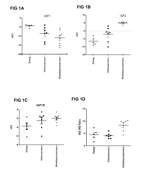
BRIEF DESCRIPTION OF THE DRAWINGS
FIG lA to FIG ID ¨ Depict the calculated ACt for IGF-1, IGF-2, IGF-1R, and the
IRA:IRB ratio calculated using the mRNA levels detected by quantitative
reverse transcription
polymerase chain reaction (qRT-PCR) in primary tumor xenografts from pediatric
sarcomas.
5
CA 02934313 2016-06-16
WO 2015/095329
PCT/US2014/070862
FIG lA depicts the calculated ACt for IGF-1; FIG 1B depicts the calculated ACt
for IGF-2; FIG
1C depicts the calculated ACt for IGF-1R; FIG 1D depicts the calculated ACt IR-
A:IR-B ratio.
FIG 2A and FIG 2B ¨ Depict the calculated ACt for IGF-1, IGF-2, IGF-1R, and
the
IRA:IRB ratio calculated using the mRNA levels detected by qRT-PCR in sarcoma
cell lines.
FIG2A depicts the calculated ACt for IGF-1, IGF-1R, IGF-2, and IGF2R. FIG 2B
depicts the
calculated ACt for IR-A:IR-B ratios.
FIG 3A to FIG 3C ¨ Depict the of IGF-1, IGF-2, and IGF-1R protein levels
detected in
sarcoma cell lines using ELISA. FIG 3A depicts the levels of IGF-1; FIG 3B
depicts the levels
of IGF-2; and FIG 3C depicts the levels of IGF-1R.
FIG 4A to FIG 4F - Depict the effect of MEDI-573 on the cell viability in
autocrine
driven Sarcoma Cell lines. FIG 4A depicts the cell viability of RD-ES cells;
FIG 4B depicts cell
viability of TC-71 cells; FIG 4C depicts cell viability of SJCRH30 cells; FIG
4D depicts cell
viabiligy of SK-ES-1 cells; FIG 4E depicts cell viability of SJS1 cells; FIG
4F depicts cell
viability of RD cells.
FIG 5A to FIG 5F - Depict the effect of MEDI-573 treatment on the Growth and
Proliferation of IGF- Induced Ewing's sarcoma cell lines. FIG 5A depicts cell
viability of IGF-
1-stimulated RD-ES cells; FIG 5B depicts cell viability of IGF-2-stimulated RD-
ES cells; FIG
5C depicts cell viability of IGF-1-stimulated SK-ES-1 cells; FIG 5D depicts
cell viability of IGF-
2-stimulated SK-ES-1 cells; FIG 5E depicts cell viability of IGF-1-stimulated
TC-71 cells; FIG
5F depicts cell viability of IGF-2-stimulated TC-71 cells.
FIG 6A to FIG 6D - Depict the effect of MEDI-573 treatment on the Growth and
Proliferation of IGF- Induced Osteosarcoma cell lines. FIG 6A depicts cell
viability of IGF-1
stimulated 5A052 cells; FIG 6B depicts cell viability of IGF-2 stimulated
5A052 cells; FIG 6C
depicts cell viability of IGF-1 stimulated MG-63 cells; FIG 6D depicts cell
viability of IGF-2
stimulated MG-63 cells.
FIG 7A to FIG 7C - Depict the efficacy of MEDI-573 in sarcoma xenograft models
with
autocrine IGF-1 and IGF-2 signaling. FIG 7A depicts tumor volume in RD-ES
cells; FIG 7B
depicts the tumor volume in SJSA-1 cells; FIG 7C depicts the tumor volume in
KHOS/NP cells.
FIG 8A to FIG 8C ¨ Depict the effect of adding different amounts of MEDI-573
to
sarcoma xenograft models with hIGF-1 or hIGF-2 induced signaling. FIG 8A
depicts the hIGF-1
6
CA 02934313 2016-06-16
WO 2015/095329
PCT/US2014/070862
levels in RD-ES cells; FIG 8B depicts the hIGF-2 levles in SJSA-1 cells; FIG
8C depicts the
hIGF-2 levels in KHOS/NP cells.
FIG 9A to FIG 9C ¨ Depict the effect of the addition of MEDI-573 on the
autophosphorylation of IGF-1R, IR-A, and Akt in RD-ES, SK-ES-1, TC-71, and
KHOS cells. In
each graph, the first bar represents the results from the untreated control;
the second bar
represents the results from adding the isotype control to the culture; and the
third bar represents
the results of treating the cells with MEDI-573. FIG 9A depicts the levels of
pIGF-1R; FIG 9B
depicts the levels of p1R-A; FIG 9C depicts the levels of pAKT.
FIG 10A to FIG 10C ¨ Depict the effect of the addition of MEDI-573 on IGF-1
and/or
IGF-2 induced signalling in vitro. FIG-10A depicts the levels of pIGF-1R; FIG
10B depicts the
levels of p1R-A; FIG 10C depicts the levels of pAKT.
FIG 11 ¨ Depicts an immunoblot showing the phosphorylation levels of pAKT and
phosphorylated Eukaryotic translation initiation factor 4E-binding protein 1
(p4EBP1) obtained
from tissues of mice bearing ¨400 mm3 RD-ES tumors. Left three lanes, no MEDI-
573 added;
right three lanes, MEDI-573 added.
FIG 12A to FIG 12D ¨ Depicts graphs showing the levels of hIGF-1 and hIGF-2 in
RD-
ES tumor and plasma before and after treatment with MEDI-573.
FIG 13 - Depicts an immunoblot showing phosphorylation levels of pAKT, p4EBP1,
and
p56K in untreated mice, in mice after induction with IGF-1, in mice after
induction with IGF-2,
in mice after induction with IGF-1 and treatment with MEDI-573, and in mice
after induction
with IGF-2 and treatment with MEDI-573. Samples from three different mice are
shown in each
group.
FIG 14 ¨ Depicts the growth and proliferation of RD-ES cells treated with MEDI-
573
and an mTOR inhibitor (rapamycin or AZD2014) alone or in combination with each
other.
FIG 15 - Depicts an immunoblot showing phosphorylation levels of pAKT, p4EBP1,
and
p56K in untreated cells, cells treated with MEDI-573 alone, cells treated with
rapamycin alone,
cells treated with rapamycin in combination with MEDI-573, cells treated with
AZD2014 alone,
and cells treated with MEDI-573 in combination with AZD2014.
7
CA 02934313 2016-06-16
WO 2015/095329
PCT/US2014/070862
FIG 16A to FIG 16B ¨ Depict the growth and proliferation of sarcoma cells in
RD-ES
tumor xenografts treated with AZD2014, MEDI-573, AZD2014 in combination with
MEDI-573
and controls. FIG 16A growth and proliferation of cells; FIG 16B body weight
of mice treated.
FIG 17A to FIG 17B ¨ Depict the growth and proliferation of sarcoma cells in
RD-ES
tumor xenografts treated with rapamycin, MEDI-573, rapamycin in combination
with MEDI-573
and controls. FIG 17A growth and proliferation of cells; FIG 17B body weight
of mice treated.
BRIEF DESCRIPTION OF THE SEQUENCE LISTING
SEQ ID NO: 1 depicts the amino acid sequence of the MEDI-573 heavy chain
complementarity determining region 1 (Ser Tyr Asp Ile Asn).
SEQ ID NO: 2 depicts the amino acid sequence of the MEDI-573 heavy chain
complementarity determining region 2 (Trp Met Asn Pro Asn Ser Gly Asn Thr Gly
Tyr Ala Gln
Lys Phe Gln Gly).
SEQ ID NO: 3 depicts the amino acid sequence of the MEDI-573 heavy chain
complementarity determining region 3 (Asp Pro Tyr Tyr Tyr Tyr Tyr Gly Met Asp
Val).
SEQ ID NO: 4 depicts the amino acid sequence of the MEDI-573 light chain
complementarity determining region 1 (Ser Gly Ser Ser Ser Asn Ile Glu Asn Asn
His Val Ser).
SEQ ID NO: 5 depicts the amino acid sequence of the MEDI-573 light chain
complementarity determining region 2 (Asp Asn Asn Lys Arg Pro Ser).
SEQ ID NO: 6 depicts the amino acid sequence of the MEDI-573 light chain
complementarity determining region 3 (Glu Thr Trp Asp Thr Ser Leu Ser Ala Gly
Arg Val).
SEQ ID NO: 7 depicts the amino acid sequence of the MEDI-573 variable heavy
chain
polypeptide:
Gln Val Gln Leu Val Gln Ser Gly Ala Glu Val Lys Lys Pro Gly Ala Ser Val Lys
Val Ser
Cys Lys Ala Ser Gly Tyr Thr Phe Thr Ser Tyr Asp Ile Asn Trp Val Arg Gln Ala
Thr Gly Gln
Gly Leu Glu Trp Met Gly Trp Met Asn Pro Asn Ser Gly Asn Thr Gly Tyr Ala Gln
Lys Phe Gln
Gly Arg Val Thr Met Thr Arg Asn Thr Ser Ile Ser Thr Ala Tyr Met Glu Leu Ser
Ser Leu Arg Ser
Glu Asp Thr Ala Val Tyr Tyr Cys Ala Arg Asp Pro Tyr Tyr Tyr Tyr Tyr Gly Met
Asp Val Trp
Gly Gln Gly Thr Thr Val Thr Val Ser Ser Ala
8
CA 02934313 2016-06-16
WO 2015/095329
PCT/US2014/070862
SEQ ID NO: 8 depicts the amino acid sequence of the MEDI-573 variable light
chain
polypeptide:
Gln Ser Val Leu Thr Gln Pro Pro Ser Val Ser Ala Ala Pro Gly Gln Lys Val Thr
Ile Ser
Cys Ser Gly Ser Ser Ser Asn Be Glu Asn Asn His Val Ser Trp Tyr Gln Gln Leu Pro
Gly Thr Ala
Pro Lys Leu Leu Ile Tyr Asp Asn Asn Lys Arg Pro Ser Gly Be Pro Asp Arg Phe Ser
Gly Ser Lys
Ser Gly Thr Ser Ala Thr Leu Gly Ile Thr Gly Leu Gln Thr Gly Asp Glu Ala Asp
Tyr Tyr Cys
Glu Thr Trp Asp Thr Ser Leu Ser Ala Gly Arg Val Phe Gly Gly Gly Thr Lys Leu
Thr Val Leu
Gly
SEQ ID NO: 9 depicts the amino acid sequence of the MEDI-573 light chain
polypeptide:
Gln Ser Val Leu Thr Gln Pro Pro Ser Val Ser Ala Ala Pro Gly Gln Lys Val Thr
Ile Ser
Cys Ser Gly Ser Ser Ser Asn Be Glu Asn Asn His Val Ser Trp Tyr Gln Gln Leu Pro
Gly Thr Ala
Pro Lys Leu Leu Ile Tyr Asp Asn Asn Lys Arg Pro Ser Gly Be Pro Asp Arg Phe Ser
Gly Ser Lys
Ser Gly Thr Ser Ala Thr Leu Gly Ile Thr Gly Leu Gln Thr Gly Asp Glu Ala Asp
Tyr Tyr Cys
Glu Thr Trp Asp Thr Ser Leu Ser Ala Gly Arg Val Phe Gly Gly Gly Thr Lys Leu
Thr Val Leu
Gly Gln Pro Lys Ala Ala Pro Ser Val Thr Leu Phe Pro Pro Ser Ser Glu Glu Leu
Gln Ala Asn Lys
Ala Thr Leu Val Cys Leu Ile Ser Asp Phe Tyr Pro Gly Ala Val Thr Val Ala Trp
Lys Ala Asp Ser
Ser Pro Val Lys Ala Gly Val Glu Thr Thr Thr Pro Ser Lys Gln Ser Asn Asn Lys
Tyr Ala Ala Ser
Ser Tyr Leu Ser Leu Thr Pro Glu Gln Trp Lys Ser His Arg Ser Tyr Ser Cys Gln
Val Thr His Glu
Gly Ser Thr Val Glu Lys Thr Val Ala Pro Thr Glu Cys Ser
SEQ ID NO: 10 depicts the amino acid sequence of the MEDI-573 heavy chain
polypeptide:
Gln Val Gln Leu Val Gln Ser Gly Ala Glu Val Lys Lys Pro Gly Ala Ser Val Lys
Val Ser
Cys Lys Ala Ser Gly Tyr Thr Phe Thr Ser Tyr Asp Ile Asn Trp Val Arg Gln Ala
Thr Gly Gln
Gly Leu Glu Trp Met Gly Trp Met Asn Pro Asn Ser Gly Asn Thr Gly Tyr Ala Gln
Lys Phe Gln
Gly Arg Val Thr Met Thr Arg Asn Thr Ser Ile Ser Thr Ala Tyr Met Glu Leu Ser
Ser Leu Arg Ser
Glu Asp Thr Ala Val Tyr Tyr Cys Ala Arg Asp Pro Tyr Tyr Tyr Tyr Tyr Gly Met
Asp Val Trp
Gly Gln Gly Thr Thr Val Thr Val Ser Ser Ala Ser Thr Lys Gly Pro Ser Val Phe
Pro Leu Ala Pro
Cys Ser Arg Ser Thr Ser Glu Ser Thr Ala Ala Leu Gly Cys Leu Val Lys Asp Tyr
Phe Pro Glu
9
CA 02934313 2016-06-16
WO 2015/095329
PCT/US2014/070862
Pro Val Thr Val Ser Trp Asn Ser Gly Ala Leu Thr Ser Gly Val His Thr Phe Pro
Ala Val Leu Gln
Ser Ser Gly Leu Tyr Ser Leu Ser Ser Val Val Thr Val Pro Ser Ser Asn Phe Gly
Thr Gln Thr Tyr
Thr Cys Asn Val Asp His Lys Pro Ser Asn Thr Lys Val Asp Lys Thr Val Glu Arg
Lys Cys Cys
Val Glu Cys Pro Pro Cys Pro Ala Pro Pro Val Ala Gly Pro Ser Val Phe Leu Phe
Pro Pro Lys Pro
Lys Asp Thr Leu Met Ile Ser Arg Thr Pro Glu Val Thr Cys Val Val Val Asp Val
Ser His Glu
Asp Pro Glu Val Gln Phe Asn Trp Tyr Val Asp Gly Val Glu Val His Asn Ala Lys
Thr Lys Pro
Arg Glu Glu Gln Phe Asn Ser Thr Phe Arg Val Val Ser Val Leu Thr Val Val His
Gln Asp Trp
Leu Asn Gly Lys Glu Tyr Lys Cys Lys Val Ser Asn Lys Gly Leu Pro Ala Pro Ile
Glu Lys Thr Ile
Ser Lys Thr Lys Gly Gln Pro Arg Glu Pro Gln Val Tyr Thr Leu Pro Pro Ser Arg
Glu Glu Met
Thr Lys Asn Gln Val Ser Leu Thr Cys Leu Val Lys Gly Phe Tyr Pro Ser Asp Ile
Ala Val Glu
Trp Glu Ser Asn Gly Gln Pro Glu Asn Asn Tyr Lys Thr Thr Pro Pro Met Leu Asp
Ser Asp Gly
Ser Phe Phe Leu Tyr Ser Lys Leu Thr Val Asp Lys Ser Arg Trp Gln Gln Gly Asn
Val Phe Ser
Cys Ser Val Met His Glu Ala Leu His Asn His Tyr Thr Gln Lys Ser Leu Ser Leu
Ser Pro Gly
Lys
DETAILED DESCRIPTION OF THE INVENTION
The invention features pharmaceutical compositions and methods that are useful
for the
treatment and prevention of sarcomas. The pharmaceutical composition for the
treatment of
sarcoma of the invention comprises an effective amount of an mTOR inhibitor
and an effective
amount of an antibody that specifically binds to at least one of insulin-like
growth factor 1 (IGF-
1) and insulin-like growth factor 2 (IGF-2). In some embodiments the antibody
in the
pharmaceutical composition neutralizes a least one of IGF-1 and IGF-2. The
invention further
provides compositions and methods for monitoring a patient having a sarcoma.
The present invention is based, at least in part, on the discovery that an
antibody that
neutralizes IGF-1 and/or IGF-2 when in combination with mTOR inhibitors (e.g.,
AZD2014,
rapamycin) is useful for decreasing the proliferation, survival and/or
increasing cell death of
IGF-responsive sarcoma cells, including cells that secrete IGF-1 and/or IGF-2
in an autocrine
manner.
MEDI-573 is a fully human monoclonal antibody that binds to IGF-2 with cross
reactivity to IGF-1. MEDI-573 neutralizes IGF-1 and IGF-2 and inhibits
signaling through both
CA 02934313 2016-06-16
WO 2015/095329
PCT/US2014/070862
the IGF-1R and IR-A pathways. A hybridoma cell line (7.159.2) expressing MEDI-
573 was
deposited at the American Type Culture Collection (ATCC) on March 7, 2006 and
received the
Patent Deposit Designation No. PTA-7424. A description of this antibody and
its preparation is
found in U.S. Patent No. 7,939,637, issued May 10, 2011, which is hereby
incorporated by
reference in its entirety.
As described elsewhere, most sarcoma cell lines express IGF-1R and IGF-1, but
only
osteosarcoma cell lines and a few rhabdosarcoma cell lines secrete IGF-2. MEDI-
573 inhibits in
vitro proliferation of a number of sarcoma cell lines, with Ewing's sarcoma
cell lines being most
sensitive. The data presented here indicates that sarcoma cells respond to
autocrine or paracrine
growth stimulation by secreted IGF-1 and IGF-2. In addition, MEDI-573
inhibited IGF-1- and
IGF-2-induced growth of sarcoma cells and significantly blocked IGF-1- and IGF-
2-induced
activation of the IGF-1R and AKT pathways. Growth inhibition of sarcoma
xenografts by
MEDI-573 was correlated with neutralization of IGF-1 and IGF-2 ligands.
As described here, MEDI-573 also inhibited rapamycin-induced AKT activation. A
combination of MEDI-573 and mTOR inhibitor resulted in significantly enhanced
anti-tumor
activities in vivo. In summary, the data indicate that inhibiting IGF-1 and
IGF-2 by MEDI-573
in combination with mTOR inhibitors (rapamycin or AZD2014) resulted in potent
anti-tumor
activity for various sarcomas. Advantageously, it has been found that
targeting IGF-1 and/or
IGF-2 is useful for treating sarcoma in combination with mTOR inhibitor, in
contrast to targeting
IGF receptors which has the potential to perturb insulin function.
Accordingly, the invention
provides pharmaceutical compositions and methods that are useful in treating
subjects as having
or having a propensity to develop a sarcoma, to develop a recurrence of
sarcoma, and/or to
develop metastatic sarcoma. In particular, the pharmaceutical compositions of
the invention are
useful for treating Ewing's sarcoma and some rhabdomyosarcoma.
Insulin-like growth factors (IGF) ¨ IGF-1 and IGF-2
Insulin-like growth factors, IGF-1 and IGF-2, are growth factors involved in
regulating
cell proliferation, survival, differentiation, and transformation. Both
ligands are expressed
ubiquitously and act as endocrine, paracrine, and autocrine growth factors
(Pollak, Nat Rev
Cancer. 2008, 8(12):915-28; DeMeyts, BioEssays 2004, 26(12): 1351-1362, 2004;
Tao et al.,
11
CA 02934313 2016-06-16
WO 2015/095329
PCT/US2014/070862
2007, Nat Clin Pract Oncol. 4(10):591-602.; Ryan and Goss, Oncologist. 2008,
13(1):16-24).
Insulin-like growth factor-I and IGF-2 exert their various actions through
binding to the insulin-
like growth factor 1 receptor (IGF-1R) and insulin receptor A isoform (IR-A),
activating multiple
intracellular signaling cascades including the IRS proteins, Akt, and MAPK
pathways (Sciacca et
al., Oncogene. 1999, 18(15):2471-9; Chitnis et al. Clin Cancer Res. 2008,
14(20):6364-70;
Belfiore et al., Endocr. Rev. 2009, 30, 586-623; Baserga, Future Oncol. 2009,
5(1):43-50).
Receptors for IGF ligands include IGF receptors type 1 and type 2 (IGF-1R and
IGF-2R), insulin
receptors A and B (IR-A and IR-B), and hybrid receptors (IGF-1R/IR-A and IGF-
1R/IR-B).
IGF-2R preferentially binds IGF-2. However, IGF-2R lacks an intracellular
kinase domain and
does not mediate cell signaling. Without being bound to a particulary theory,
loss of IGF-2R
results in increased tumorigenicity, presumably by increasing the availability
of IGF-2 to bind to
IGF-1R. Both IGF-1 and IGF-2 exist as complexes in the circulatory system,
bound to one of six
IGF binding proteins (IGFBP-1 to IGFBP-6). IGFBP-3, in conjunction with a
third molecule,
acid labile subunit, forms a complex that accounts for the majority of
circulating IGF. IGFBPs
have a higher affinity for IGF than their cognate receptors and have the
potential to sequester
IGF from the receptor. However, alternative models indicate that the binding
proteins may
potentiate IGF activity, either by extending its half-life in circulation or
by binding to certain
molecules on the cell surface, thus providing a reservoir of available IGF to
the cell.
High levels of circulating IGF-1 and -2 are associated with an increased risk
for
development of several common cancers (Renehan et al., Lancet. 2004,
363(9418):1346-53),
including breast, prostate, pancreatic and colorectal cancer, non-small cell
lung cancer (NSCLC),
hepatocellular carcinoma (HCC), and sarcoma. The overexpression of IR-A and
IGF-2 has also
been proposed as a potential mechanism that may lead to the resistance to IGF-
1R-directed
therapies (Hendrickson and Haluska, Curr Opin Investig Drugs. 2009,
10(10):1032-40; Zhang et
al., 2007 Cancer Res. 67: 391-397). Numerous preclinical studies have reported
that down-
regulation of IGF-1R expression or blocking of signaling leads to the
inhibition of tumor growth,
both in vitro and in vivo (Ryan and Goss, Oncologist. 2008, 13(1):16-24;
Sachdev and Yee, Mol
Cancer Ther. 2007, 6(1):1-12; Baserga, Expert Opin Ther Targets. 2005,
9(4):753-68). Inhibition
of IGF signaling has also been shown to increase the susceptibility of tumor
cells to
chemotherapeutic agents in vivo (Tao et al., 2007 Nat. Clin. Pract. Oncol.
4:591-602; Chitnis et
12
CA 02934313 2016-06-16
WO 2015/095329
PCT/US2014/070862
al., 2008, Clin. Cancer Res. 14: 6364-6370; Ryan and Goss, 2008 Oncologist 13:
16-24; Yuen
and Macaulay, 2008 Expert Opin. Ther. Targets 12: 589-603). Dual inhibition of
both the IR-A
and IGF-1R receptors may enhance therapeutic efficacy against IGF-driven
cancers (Sachdev
and Yee, Mol Cancer Ther. 2007, 6(1):1-12).
Sarcoma
Sarcomas are neoplasias from transformed cells of mesenchymal origin,
including
osteosarcoma, which develops from bone, and soft tissue sarcoma, which develop
from soft
tissues like fat, muscle, nerves, fibrous tissues, blood vessels, or deep skin
tissues. Sarcomas
may be named based on the type of tissue that they most closely resemble. For
example,
osteosarcoma resembles bone, chondrosarcoma resembles cartilage, liposarcoma
resembles fat,
and leiomyosarcoma resembles smooth muscle. Sarcomas include without
limitation Ewing's
sarcoma, Osteosarcoma, Rhabdomyosarcoma, Askin's tumor, Sarcoma botryoides,
Chondrosarcoma, Malignant Hemangioendothelioma, Malignant Schwannoma, soft
tissue
sarcoma, Alveolar soft part sarcoma, Angiosarcoma, Cystosarcoma Phyllodes,
Dermatofibrosarcoma protuberans, Desmoid Tumor, Desmoplastic small round cell
tumor,
Epithelioid Sarcoma, Extraskeletal chondrosarcoma, Extraskeletal osteosarcoma,
Fibrosarcoma,
Hemangiopericytoma, Hemangiosarcoma, Kaposi's sarcoma, Leiomyosarcoma,
Liposarcoma,
Lymphangiosarcoma, Lymphosarcoma, Malignant peripheral nerve sheath tumor,
Neurofibrosarcoma, Synovial sarcoma, and Undifferentiated pleomorphic sarcoma.
An autocrine loop involving IGF-1R and both of its ligands, IGF-1 and IGF-2,
has been
demonstrated as a key mechanism driving the proliferation and survival of
sarcoma cells (Kim et
al., 2009 Bull. Cancer 96(7): 52-60). High expression of IGF-1R, IGF-1, or IGF-
2 are indicated
in most Ewing's sarcomas, osteosarcoma, and rhabdomyosarcoma. Ewing's sarcomas
secrete
more IGF-1 whereas rhabdomyosarcomas secrete more IGF-2. IGF-1 is highly
expressed and
stimulates osteosarcoma cell growth. Genetic alterations in the IGF pathway
are also prevalent
in a number of sarcoma tumors. Loss of imprinting at the IGF-2 locus is
commonly detected in
embryonal RMS and a genetic alteration that leads to chimeric transcription
factors (PAX3-
FKHR or PAX7-FKHR) leads to increased expression of IGF-1R in alveolar types
of
rhabdomyosarcoma. Conversely, in Ewing's sarcoma patients that carry the EWS-
FLI1 genetic
alteration that upregulates a repressor of IGF-1 signaling, insulin growth
factor binding protein 3
13
CA 02934313 2016-06-16
WO 2015/095329
PCT/US2014/070862
(IGFBP3), these patients have improved prognosis. Given the strong disease
linkage to the IGF
signaling pathway, targeted therapeutic approaches that inhibit the IGF-1R
receptor using MAbs
have been explored in several types of sarcomas. These IGF-1R-targeted MAbs
inhibit IGF-1
and IGF-2 signaling through IGF1R and heterodimeric IGF-1R/IR but do not
inhibit IGF-2
signaling through IR-A and thus, may be limited.
Ewing's Sarcoma
Ewing's sarcoma, peripheral primitive neuroectodermal tumor, and Askin tumor
form a
group of tumors, collectively termed Ewing's sarcoma family of tumors (ESFT).
These tumors
are characterized by specific chromosomal translocations that cause the N-
terminus of RNA-
binding protein EWS to be fused to the C-terminus of one member of the ETS
family of
transcription factors, most commonly Friend leukemia integration 1
transcription factor (FLI1).
Expression of the fusion product has been implicated in oncogenesis.
EFST cell lines express IGF-1R and secrete IGF-1 in an autocrine loop. The
prevalence
of IGF-1R expression in EFST is very high, with most cell lines and clinical
samples positive for
expression. In murine fibroblasts, the EWS-FLI1 oncoprotein requires IGF-1R
for
transformation. Some evidence indicates that relapse-free survival may
correlate with the ratio
of serum IGFBP-3 to IGF-1. In support of this theory, EWS-FLI1 directly
reduces the expression
and secretion of IGFBP-3 and exogenous IGFBP-3 inhibits the growth of ESFT
cells. Pathways
downstream of IGF-1R, including PI3K/Akt and MAPK, are activated and are vital
to ESFT cell
survival. Inhibitors of both PI3K and MAPK cause growth arrest in ESFT cells.
Rhabdomyosarcoma
Rhabdomyosarcoma is the most common soft tissue sarcoma of childhood, arising
from
developing cells that form striated muscle. IGF-2 is involved in normal muscle
growth, and
analysis of tumor biopsy specimens from patients with rhabdomyosarcoma
demonstrated high
levels of IGF-2 mRNA expression. Without being bound to a particular theory,
upregulation of
IGF-2 potentially plays a role in the unregulated growth of these tumors.
Additionally, it has
been observed that binding of IGF-1R and IGF-2 secreted from rhabdomyosarcoma
cell lines,
resulted in autocrine growth proliferation and increased cell motility.
14
CA 02934313 2016-06-16
WO 2015/095329
PCT/US2014/070862
Epigenetic changes leading to loss of imprinting (LOT) of the IGF-2 locus,
resulting in
over-expression of IGF-2, have been identified. In addition, the PAX3¨FKHR
translocation that
characterizes certain rhabdomyosarcomas transactivates the IGF-1R promoter,
thus providing
further evidence that the IGF pathway plays an important role in the
progression of
rhabdomyosarcoma. All rhabdomyosarcoma cell lines show some level of IGF-1R
expression,
although they may differ by as much as 30-fold based on quantitative protein
analysis.
Osteosarcoma
The peak incidence of osteosarcoma occurs during adolescence, corresponding to
both
the growth spurt and peak concentrations of circulating GH and IGF-1. High
levels of IGF-1
appear to play an important role in the pathogenesis of osteosarcoma.
Preclinical data indicate a
role for IGF-1 in osteosarcoma. Osteosarcoma cells express functional IGF-1R
on the cell
surface, and the majority of osteosarcoma patient samples express IGF ligands
and 45% express
IGF-1R. Exogenous IGF-1 stimulates proliferation of osteosarcoma cells, and
IGF-1-dependent
growth can be inhibited using monoclonal antibodies or antisense
oligonucelotides against IGF-
1R. Furthermore, treatment of mice using a humanized anti-IGF-1R antibody
resulted in tumor
regression in two osteosarcoma xenograft models.
Mammalian Target of Rapamycin (mTOR)
The mammalian target of rapamycin (mTOR) is a serine/threonine protein kinase
that
plays an important role in regulating cell growth, proliferation, and
survival. mTOR integrates
the input from upstream pathways, including insulin, growth factors (such as
IGF-1 and IGF-2),
and amino acids. mTOR also senses cellular nutrient, oxygen, and energy
levels. The mTOR
pathway is dysregulated in human diseases, such as diabetes, obesity,
depression, and certain
cancers. mTOR was identified as being sensitive to the antifungal agent
rapamycin. Rapamycin
is a bacterial product that can inhibit mTOR by associating with its
intracellular receptor
FKBP12. The FKBP12-rapamycin complex binds directly to the FKBP12-Rapamycin
Binding
(FRB) domain of mTOR, inhibiting its activity.
Activation of mTOR leads to phosphorylation of downstream Ribosomal protein S6
kinase beta-1 (S6K) and Eukaryotic translation initiation factor 4E-binding
protein 1 (4E-BP1).
CA 02934313 2016-06-16
WO 2015/095329
PCT/US2014/070862
mTOR signaling has been an attractive therapeutic target for cancer therapy.
mTOR inhibitors
Temsirolimus and Everolimus have been approved for treating metastatic renal
cell carcinoma
and pancreatic neuroendocrine tumors respectively. Ridaforolimus is currently
in phase III trial
in sarcoma patients. However, rapamycin and its derivatives induce Akt
activation by releasing
the negative feedback between S6K and IRS/PI3K, and subsequently reactivating
IGF-1R
signaling. This contributes to the possible mechanism of resistance to mTOR
inhibitors, and
suggests a potential benefit of combining rapamycin with agents targeting IGF
pathway.
Combination of several IGF-1R targeting agents with different rapamycin
analogs are in early
phase clinical trials. First generation mTOR inhibitors include without
limitation rapamycin,
temsirolimus (CCI-779), everolimus (RAD001), ridaforolimus (AP-23573). Second
generation
mTOR inhibitors are designed to compete with ATP in the catalytic site of
mTOR. Such ATP-
competitive mTOR kinase inhibitors include without limitation AZD2014, INK128,
AZD8055,
NVP-BEZ235, BGT226, SF1126, PKI-587. Structures of mTOR inhibitors AZD2014 and
rapamycin are provided below.
0 N
N
AZD201 4
16
CA 02934313 2016-06-16
WO 2015/095329
PCT/US2014/070862
HO
', i¨%
i.
'---- ,NI i
0 -
,
0
--T 0 0' ------
HO
**9.,
*`--- ' -0
.õ....----- ......., ..-- .e"'". (....
7,
Rapamycin
Antibodies
Antibodies that selectively bind IGF-1/-2 and inhibit the binding or
activation of
receptors to of IGF-1/-2 are useful in the methods of the invention. In
certain embodiments, the
antibodies to IGF-1/-2 do not bind insulin or inhibit the biological activity
of insulin.
In an embodiment, the antibody is a recombinant, monoclonal antibody. The
recombinant monoclonal antibody is prepared from a host cell, including, but
not limited to, a
bacterial cell, a yeast cell, an insect cell, or a mammalian cell. In a
preferred embodiment, the
host cell is a mammalian cell. In another embodiment, the recombinant
monoclonal antibody is
a human antibody. In yet another embodiment, the monoclonal antibody is an
IgA, IgE, IgD,
IgE, or IgG antibody. In a preferred embodiment, the monoclonal antibody is an
IgG antibody,
including, but not limited to an IgG1 or IgG2 antibody.
In another embodiment, the antibody comprises at least one N-linked
glycosylation site
on the Fc region of the antibody and at least one N-linked glycosylation site
on the Fab region of
the antibody. In another embodiment, the antibody has only one N-linked
glycosylation site on
the Fc region of the antibody and only one N-linked glycosylation site on the
Fab region of the
antibody (i.e., at total of 3 N-linked glycosylation sites).
Antibodies can be made by any of the methods known in the art.
Antibodies made by any method known in the art can then be purified from the
host.
Antibody purification methods may include salt precipitation (for example,
with ammonium
sulfate), ion exchange chromatography (for example, on a cationic or anionic
exchange column
17
CA 02934313 2016-06-16
WO 2015/095329
PCT/US2014/070862
preferably run at neutral pH and eluted with step gradients of increasing
ionic strength), gel
filtration chromatography (including gel filtration HPLC), and chromatography
on affinity resins
such as protein A, protein G, hydroxyapatite, and anti-immunoglobulin.
Antibodies can be conveniently produced from hybridoma cells engineered to
express the
antibody. Methods of making hybridomas are well known in the art. The
hybridoma cells can be
cultured in a suitable medium, and spent medium can be used as an antibody
source.
Polynucleotides encoding the antibody of interest can in turn be obtained from
the hybridoma
that produces the antibody, and then the antibody may be produced
synthetically or
recombinantly from these DNA sequences. For the production of large amounts of
antibody, it is
generally more convenient to obtain an ascites fluid. The method of raising
ascites generally
comprises injecting hybridoma cells into an immunologically naive
histocompatible or
immunotolerant mammal, especially a mouse. The mammal may be primed for
ascites
production by prior administration of a suitable composition (e.g., Pristane).
Monoclonal antibodies (Mabs) produced by methods of the invention can be
"humanized" by methods known in the art. "Humanized" antibodies are antibodies
in which at
least part of the sequence has been altered from its initial form to render it
more like human
immunoglobulins. Techniques to humanize antibodies are particularly useful
when non-human
animal (e.g., murine) antibodies are generated. Examples of methods for
humanizing a murine
antibody are provided in U.S. Pat. Nos. 4,816,567, 5,530,101, 5,225,539,
5,585,089, 5,693,762
and 5,859,205.
Human antibodies avoid some of the problems associated with antibodies that
possess
murine or rat variable and/or constant regions. The presence of such murine or
rat derived
proteins can lead to the rapid clearance of the antibodies or can lead to the
generation of an
immune response against the antibody by a patient. In order to avoid the
utilization of murine or
rat derived antibodies, fully human antibodies can be generated through the
introduction of
functional human antibody loci into a rodent, other mammal or animal so that
the rodent, other
mammal or animal produces fully human antibodies.
One method for generating fully human antibodies is through the use of
XenoMouse@
strains of mice that have been engineered to contain up to but less than 1000
kb-sized germline
configured fragments of the human heavy chain locus and kappa light chain
locus. See Mendez
18
CA 02934313 2016-06-16
WO 2015/095329
PCT/US2014/070862
et al. Nature Genetics 15: 146-156 (1997) and Green and Jakobovits J. Exp.
Med. 188:483-495
(1998). The XenoMouse strains are available from Abgenix, Inc. (Fremont, CA).
The production of the XenoMouse strains of mice is further discussed and
delineated in
U.S. Patent Application Serial Nos. 07/466,008, filed January 12, 1990,
07/610,515, filed
November 8, 1990, 07/919,297, filed July 24, 1992, 07/922,649, filed July 30,
1992, 08/031,801,
filed March 15, 1993, 08/112,848, filed August 27, 1993, 08/234,145, filed
April 28, 1994,
08/376,279, filed January 20, 1995, 08/430, 938, filed April 27, 1995,
08/464,584, filed June 5,
1995, 08/464,582, filed June 5, 1995, 08/463, 191, filed June 5, 1995,
08/462,837, filed June 5,
1995, 08/486,853, filed June 5, 1995, 08/486,857, filed June 5, 1995,
08/486,859, filed June 5,
1995, 08/462,513, filed June 5, 1995, 08/724,752, filed October 2, 1996,
08/759,620, filed
December 3, 1996, U.S. Publication 2003/0093820, filed November 30, 2001 and
U.S. Patent
Nos. 6,162,963, 6,150,584, 6, 114,598, 6,075, 181, and 5,939,598 and Japanese
Patent Nos. 3
068 180 B2, 3 068 506 B2, and 3 068 507 B2. See also European Patent No., EP 0
463 151 Bl,
grant published June 12, 1996, International Patent Application No., WO
94/02602, published
February 3, 1994, International Patent Application No., WO 96/34096, published
October 31,
1996, WO 98/24893, published June 11, 1998, WO 00/76310, published December
21, 2000.
The disclosures of each of the above-cited patents, applications, and
references are hereby
incorporated by reference in their entirety.
In an alternative approach, others, including GenPharm International, Inc.,
have utilized a
"minilocus" approach. In the minilocus approach, an exogenous Ig locus is
mimicked through
the inclusion of pieces (individual genes) from the Ig locus. Thus, one or
more VH genes, one or
more DH genes, one or more JH genes, a mu constant region, and usually a
second constant
region (preferably a gamma constant region) are formed into a construct for
insertion into an
animal. This approach is described in U.S. Patent No. 5,545,807 to Surani et
al. and U.S. Patent
Nos. 5,545,806, 5,625,825, 5,625, 126, 5,633,425, 5,661,016, 5,770,429,
5,789,650, 5,814,318,
5,877,397, 5,874,299, and 6,255,458 each to Lonberg and Kay, U.S. Patent No.
5,591,669 and
6,023.010 to Krimpenfort and Berns, U.S. Patent Nos. 5,612,205, 5,721,367, and
5,789,215 to
Berns et al., and U.S. Patent No. 5,643,763 to Choi and Dunn, and GenPharm
International U.S.
Patent Application Serial Nos. 07/574,748, filed August 29, 1990, 07/575,962,
filed August 31,
1990, 07/810,279, filed December 17, 1991, 07/853,408, filed March 18, 1992,
07/904,068, filed
19
CA 02934313 2016-06-16
WO 2015/095329
PCT/US2014/070862
June 23, 1992, 07/990,860, filed December 16, 1992, 08/053,131, filed April
26, 1993,
08/096,762, filed July 22, 1993, 08/155,301, filed November 18, 1993,
08/161,739, filed
December 3, 1993, 08/165,699, filed December 10, 1993, 08/209,741, filed March
9, 1994, the
disclosures of which are hereby incorporated by reference. See also European
Patent No. 0 546
073 B 1, International Patent Application Nos. WO 92/03918, WO 92/22645, WO
92/22647,
W092/22670, WO 93/12227, WO 94/00569, WO 94/25585, WO 96/14436, WO 97/13852,
and
WO 98/24884 and U.S. Patent No. 5,981, 175, the disclosures of which are
hereby incorporated
by reference in their entirety. See further Taylor et al., 1992, Chen et al.,
1993, Tuaillon et al.,
1993, Choi et al., 1993, Lonberg et al., (1994), Taylor et al., (1994), and
Tuaillon et al., (1995),
Fishwild et al., (1996), the disclosures of which are hereby incorporated by
reference in their
entirety.
Kirin has also demonstrated the generation of human antibodies from mice in
which,
through microcell fusion, large pieces of chromosomes, or entire chromosomes,
have been
introduced. See European Patent Application Nos. 773 288 and 843 961, the
disclosures of
which are hereby incorporated by reference. Additionally, KMTm- mice, which
are the result of
cross-breeding of Kirin' s Tc mice with Medarex's minilocus (Humab) mice have
been generated.
These mice possess the human IgH transchromosome of the Kirin mice and the
kappa chain trans
gene of the Genpharm mice (Ishida et al., Cloning Stem Cells, (2002) 4:91-
102).
Human antibodies can also be derived by in vitro methods. Suitable examples
include but
are not limited to phage display (CAT, Morphosys, Dyax, Biosite/Medarex, Xoma,
Symphogen,
Alexion (formerly Proliferon), Affimed) ribosome display (CAT), yeast display,
and the like.
Antibodies, as described herein, were prepared through the utilization of the
XenoMouse technology, as described below. Such mice, then, are capable of
producing human
immunoglobulin molecules and antibodies and are deficient in the production of
murine
immunoglobulin molecules and antibodies. Technologies utilized for achieving
the same are
disclosed in the patents, applications, and references disclosed in the
background section herein.
In particular, however, a preferred embodiment of transgenic production of
mice and antibodies
therefrom is disclosed in U.S. Patent Application Serial No. 08/759,620, filed
December 3, 1996
and International Patent Application Nos. WO 98/24893, published June 11, 1998
and WO
00/76310, published December 21, 2000, the disclosures of which are hereby
incorporated by
CA 02934313 2016-06-16
WO 2015/095329
PCT/US2014/070862
reference. See also Mendez et al. Nature Genetics 15: 146-156 (1997), the
disclosure of which is
hereby incorporated by reference.
Through the use of such technology, fully human monoclonal antibodies to a
variety of
antigens have been produced. Essentially, XenoMouse lines of mice are
immunized with an
antigen of interest (e.g. IGF-1711), lymphatic cells (such as B-cells) are
recovered from the
hyper-immunized mice, and the recovered lymphocytes are fused with a myeloid-
type cell line to
prepare immortal hybridoma cell lines. These hybridoma cell lines are screened
and selected to
identify hybridoma cell lines that produced antibodies specific to the antigen
of interest.
Provided herein are methods for the production of multiple hybridoma cell
lines that produce
antibodies specific to IGF-1/-2. Further, provided herein are characterization
of the antibodies
produced by such cell lines, including nucleotide and amino acid sequence
analyses of the heavy
and light chains of such antibodies.
Alternatively, instead of being fused to myeloma cells to generate hybridomas,
B cells
can be directly assayed. For example, CD19+ B cells can be isolated from
hyperimmune
XenoMouse mice and allowed to proliferate and differentiate into antibody-
secreting plasma
cells. Antibodies from the cell supematants are then screened by ELISA for
reactivity against the
IGF-1/-2 immunogen. The supematants might also be screened for
immunoreactivity against
fragments of IGF-1/-2 to further map the different antibodies for binding to
domains of
functional interest on IGF-17II. The antibodies may also be screened against
other related human
chemokines and against the rat, the mouse, and non-human primate, such as
cynomolgus
monkey, orthologues of IGF-1/-2, the last to determine species cross-
reactivity. B cells from
wells containing antibodies of interest may be immortalized by various methods
including fusion
to make hybridomas either from individual or from pooled wells, or by
infection with EBV or
transfection by known immortalizing genes and then plating in suitable medium.
Alternatively,
single plasma cells secreting antibodies with the desired specificities are
then isolated using an
IGF-1/-2-specific hemolytic plaque assay (Babcook et al., Proc. Natl. Acad.
Sci. USA 93 :7843-
48 (1996)). Cells targeted for lysis are preferably sheep red blood cells
(SRBCs) coated with the
IGF-1/-2 antigen.
In the presence of a B-cell culture containing plasma cells secreting the
immunoglobulin
of interest and complement, the formation of a plaque indicates specific IGF-
1/-2-mediated lysis
21
CA 02934313 2016-06-16
WO 2015/095329
PCT/US2014/070862
of the sheep red blood cells surrounding the plasma cell of interest. The
single antigen-specific
plasma cell in the center of the plaque can be isolated and the genetic
information that encodes
the specificity of the antibody is isolated from the single plasma cell. Using
reverse-transcription
followed by PCR (RT-PCR), the DNA encoding the heavy and light chain variable
regions of the
antibody can be cloned. Such cloned DNA can then be further inserted into a
suitable expression
vector, preferably a vector cassette such as a pcDNA, more preferably such a
pcDNA vector
containing the constant domains of immunglobulin heavy and light chain. The
generated vector
can then be transfected into host cells, e.g., HEK293 cells, CHO cells, and
cultured in
conventional nutrient media modified as appropriate for inducing
transcription, selecting
transformants, or amplifying the genes encoding the desired sequences.
In general, antibodies produced by the fused hybridomas were human IgG2 heavy
chains
with fully human kappa or lambda light chains. Antibodies described herein
possess human IgG4
heavy chains as well as IgG2 heavy chains. Antibodies can also be of other
human isotypes,
including IgGl. The antibodies possessed high affinities, typically possessing
a Kd of from about
106 through about 1012 M or below, when measured by solid phase and solution
phase
techniques. Antibodies possessing a KD of at least 1011 M are desired to
inhibit the activity of
IGF-1/-2.
As will be appreciated, anti-IGF-1/-2 antibodies can be expressed in cell
lines other than
hybridoma cell lines. Sequences encoding particular antibodies can be used to
transform a
suitable mammalian host cell. Transformation can be by any known method for
introducing
polynucleotides into a host cell, including, for example packaging the
polynucleotide in a virus
(or into a viral vector) and transducing a host cell with the virus (or
vector) or by transfection
procedures known in the art, as exemplified by U.S. Patent Nos. 4,399,216,
4,912,040,
4,740,461, and 4,959,455 (which patents are hereby incorporated herein by
reference). The
transformation procedure used depends upon the host to be transformed. Methods
for introducing
heterologous polynucleotides into mammalian cells are well known in the art
and include
dextran-mediated transfection, calcium phosphate precipitation, polybrene
mediated transfection,
protoplast fusion, electroporation, encapsulation of the polynucleotide(s) in
liposomes, and direct
microinjection of the DNA into nuclei.
Mammalian cell lines available as hosts for expression are well known in the
art and
22
CA 02934313 2016-06-16
WO 2015/095329
PCT/US2014/070862
include many immortalized cell lines available from the American Type Culture
Collection
(ATCC), including but not limited to Chinese hamster ovary (CHO) cells, HeLa
cells, baby
hamster kidney (BHK) cells, monkey kidney cells (COS), human hepatocellular
carcinoma cells
(e.g., Hep G2), human epithelial kidney 293 cells, and a number of other cell
lines. Cell lines of
particular preference are selected through determining which cell lines have
high expression
levels and produce antibodies with constitutive IGF-1/-2 binding properties.
In other embodiments, the invention provides "unconventional antibodies."
Unconventional antibodies include, but are not limited to, nanobodies, linear
antibodies (Zapata
et al., Protein Eng. 8(10): 1057-1062, 1995), single domain antibodies, single
chain antibodies,
and antibodies having multiple valencies (e.g., diabodies, tribodies,
tetrabodies, and
pentabodies). Nanobodies are the smallest fragments of naturally occurring
heavy-chain
antibodies that have evolved to be fully functional in the absence of a light
chain. Nanobodies
have the affinity and specificity of conventional antibodies although they are
only half of the size
of a single chain Fv fragment. The consequence of this unique structure,
combined with their
extreme stability and a high degree of homology with human antibody
frameworks, is that
nanobodies can bind therapeutic targets not accessible to conventional
antibodies. Recombinant
antibody fragments with multiple valencies provide high binding avidity and
unique targeting
specificity to cancer cells. These multimeric scFvs (e.g., diabodies,
tetrabodies) offer an
improvement over the parent antibody since small molecules of .about.60-100
kDa in size
provide faster blood clearance and rapid tissue uptake See Power et al.,
(Generation of
recombinant multimeric antibody fragments for tumor diagnosis and therapy.
Methods Mol Biol,
207, 335-50, 2003); and Wu et al. (Anti-carcinoembryonic antigen (CEA) diabody
for rapid
tumor targeting and imaging. Tumor Targeting, 4, 47-58, 1999).
Various techniques for making unconventional antibodies have been described.
Bispecific antibodies produced using leucine zippers are described by Kostelny
et al. (J.
Immunol. 148(5):1547-1553, 1992). Diabody technology is described by Hollinger
et al. (Proc.
Natl. Acad. Sci. USA 90:6444-6448, 1993). Another strategy for making
bispecific antibody
fragments by the use of single-chain Fv (sFv) diners is described by Gruber et
al. (J. Immunol.
152:5368, 1994). Trispecific antibodies are described by Tutt et al. (J.
Immunol. 147:60, 1991).
Single chain Fv polypeptide antibodies include a covalently linked VH::VL
heterodimer which
23
CA 02934313 2016-06-16
WO 2015/095329
PCT/US2014/070862
can be expressed from a nucleic acid including VH- and VL-encoding sequences
either joined
directly or joined by a peptide-encoding linker as described by Huston, et al.
(Proc. Nat. Acad.
Sci. USA, 85:5879-5883, 1988). See, also, U.S. Pat. Nos. 5,091,513, 5,132,405
and 4,956,778;
and U.S. Patent Publication Nos. 20050196754 and 20050196754.
In one embodiment, the antibody binds to insulin-like growth factor 2 (IGF-2)
with cross
reactivity to insulin-like growth factor 1 (IGF-1), such as those antibodies
disclosed in U.S.
Patent No. 7,939,637, which is hereby incorporated by reference in its
entirety. In certain
embodiments, the antibody binds to IGF-2 with cross reactivity to IGF-1 and is
a monoclonal,
human antibody selected from the group consisting of mAb 7.251.3 (ATCC
Accession Number
PTA-7422), mAb 7.34.1 (ATCC Accession Number PTA-7423), and mAb 7.159.2 / MEDI-
573
(ATCC Accession Number PTA-7424).
In particular embodiments of the invention, the antibody in the pharmaceutical
composition comprises a heavy chain complementarity determining region 1
(CDR1) comprising
the amino acid sequence set forth in SEQ ID NO: 1 (Ser Tyr Asp Ile Asn); a
heavy chain
complementarity determining region 2 (CDR2) comprising the amino acid sequence
set forth in
SEQ ID NO: 2 (Trp Met Asn Pro Asn Ser Gly Asn Thr Gly Tyr Ala Gln Lys Phe Gln
Gly); a
heavy chain complementarity determining region 3 (CDR3) comprising the amino
acid sequence
set forth in SEQ ID NO: 3 (Asp Pro Tyr Tyr Tyr Tyr Tyr Gly Met Asp Val); a
light chain
complementarity determining region 1 (CDR1) comprising the amino acid sequence
set forth in
SEQ ID NO: 4 (Ser Gly Ser Ser Ser Asn Ile Glu Asn Asn His Val Ser); a light
chain
complementarity determining region 2 (CDR2) comprising the amino acid sequence
set forth in
SEQ ID NO: 5 (Asp Asn Asn Lys Arg Pro Ser); and a light chain complementarity
determining
region 3 (CDR3) comprising the amino acid sequence set forth in SEQ ID NO: 6
(Glu Thr Trp
Asp Thr Ser Leu Ser Ala Gly Arg Val).
In some embodiments, the antibody in the pharmaceutical composition of the
invention
comprises one or more variable regions comprising an amino acid sequence
selected from the
amino acid sequences set forth in SEQ ID NO: 7 and SEQ ID NO: 8. In particular
embodiments,
the antibody in the pharmaceutical composition of the invention has the amino
acid sequence of
the antibody produced by hybridoma cell line 7.159.2 (ATCC Accession Number
PTA-7424).
MEDI-573 is a fully human immunoglobulin G2 lambda (IgG2) antibody generated
with
24
CA 02934313 2016-06-16
WO 2015/095329
PCT/US2014/070862
Xenomouse technology and manufactured in Chinese Hamster Ovary (CHO) cells.
MEDI-573
selectively binds to human insulin-like growth factors hIGF-1 and hIGF-2 and
inhibits insulin-
like growth factor IGF-1 and IGF-2 mediated signal transduction in tumor
cells, thereby
inhibiting tumor growth. The antibody was isolated from mice immunized
alternately with
soluble recombinant human hIGF-1 and hIGF-2 coupled to keyhole limpet
hemocyanin (KLH),
as described in Patent No. 7,939,637, which is herein incorporated by
reference in its entirety.
MEDI-573 is composed of 2 light chains and 2 heavy chains, with an overall
molecular weight
of approximately 151 kilodaltons.
MEDI 573 selectively binds to human insulin-like growth factor (hIGF)-I and
hIGF-2 and
IGF-1- and IGF-2 mediated signal transduction and proliferation in human tumor
cells. MEDI-
573 targets the IGF-1 and IGF-2 ligands and thereby inhibits IGF-mediated
signal transduction.
Nonclinical studies in human cancer cells suggest that MEDI 573 has the
potential to achieve
broad antitumor efficacy owing to its ability to inhibit both IGF-1R and IR-A
pathways.
Furthermore, MEDI-573 has potential to achieve this without perturbing glucose
homeostasis,
which has been an adverse effect observed with investigational agents that
target IGF 1R. The
results of in vitro studies have shown that MEDI-573 inhibited both IGF-1 and
IGF-2-stimulated
phosphorylation of IGF 1R and that of downstream signaling proteins including
Akt and MAPK
in a number of engineered mouse embryonic fibroblast NIH-3T3 cell lines
transfected to express
human IGF-1R and either human IGF-1/-2. Furthermore, MEDI-573 inhibited
autocrine
phosphorylation of these signaling molecules. Functionally, MEDI-573
effectively inhibited the
growth of a number of engineered NIH3T3 and human tumor cell lines in vitro.
In vivo,
treatment of tumor-bearing mice with MEDI-573 significantly inhibited the
growth of implanted
clone 32 (C32) and clone P12 (P12) tumors, which overexpress hIGF II and human
insulin-like
growth factor 1 receptor (hIGF-1R), and hIGF-1 and hIGF-1R, respectively.
Therapy
Therapy may be provided wherever cancer therapy is performed: at home, the
doctor's
office, a clinic, a hospital's outpatient department, or a hospital. In one
embodiment, the
invention provides for the use of an anti-IGF-1/-2 antibody (e.g., MEDI-573)
in combination
with an mTOR inhibitor as a therapy.
CA 02934313 2016-06-16
WO 2015/095329
PCT/US2014/070862
Treatment generally begins at a hospital so that the doctor can observe the
therapy's
effects closely and make any adjustments that are needed. The duration of the
therapy depends
on the kind of cancer being treated, the age and condition of the patient, the
stage and type of the
patient's disease, and how the patient's body responds to the treatment. Drug
administration may
be performed at different intervals (e.g., daily, weekly, or monthly). Therapy
may be given in
on-and-off cycles that include rest periods so that the patient's body has a
chance to build healthy
new cells and regain its strength.
Depending on the type of cancer and its stage of development, the therapy can
be used to
slow the spreading of the cancer, to slow the cancer's growth, to kill or
arrest cancer cells that
may have spread to other parts of the body from the original tumor, to relieve
symptoms caused
by the cancer, or to prevent cancer in the first place. Cancer growth is
uncontrolled and
progressive, and occurs under conditions that would not elicit, or would cause
cessation of,
multiplication of normal cells.
As described above, if desired, treatment with a composition of the invention
may be
combined with therapies for the treatment of proliferative disease (e.g.,
radiotherapy, surgery, or
chemotherapy).
Formulation of Pharmaceutical Compositions
The administration of a combination of the invention (e.g., an antibody that
binds IGF-1/-
2 with an mTOR inhibitor) for the treatment of sarcoma may be by any suitable
means that
results in a concentration of the therapeutic that, combined with other
components, is effective in
preventing, ameliorating, or reducing sarcoma. The compound may be contained
in any
appropriate amount in any suitable carrier substance, and is generally present
in an amount of 1-
95% by weight of the total weight of the composition. The composition may be
provided in a
dosage form that is suitable for parenteral (e.g., subcutaneously,
intravenously, intramuscularly,
or intraperitoneally) administration route. The pharmaceutical compositions
may be formulated
according to conventional pharmaceutical practice (see, e.g., Remington: The
Science and
Practice of Pharmacy (20th ed.), ed. A. R. Gennaro, Lippincott Williams &
Wilkins, 2000 and
Encyclopedia of Pharmaceutical Technology, eds. J. Swarbrick and J. C. Boylan,
1988-1999,
Marcel Dekker, New York).
26
CA 02934313 2016-06-16
WO 2015/095329
PCT/US2014/070862
Pharmaceutical compositions according to the invention may be formulated to
release the
active compound substantially immediately upon administration or at any
predetermined time or
time period after administration. The latter types of compositions are
generally known as
controlled release formulations, which include (i) formulations that create a
substantially
constant concentration of the drug within the body over an extended period of
time; (ii)
formulations that after a predetermined lag time create a substantially
constant concentration of
the drug within the body over an extended period of time; (iii) formulations
that sustain action
during a predetermined time period by maintaining a relatively, constant,
effective level in the
body with concomitant minimization of undesirable side effects associated with
fluctuations in
the plasma level of the active substance (sawtooth kinetic pattern); (iv)
formulations that localize
action by, e.g., spatial placement of a controlled release composition
adjacent to or in a sarcoma
(v) formulations that allow for convenient dosing, such that doses are
administered, for example,
once every one or two weeks; and (vi) formulations that target proliferating
neoplastic cells by
using carriers or chemical derivatives to deliver the therapeutic agent to a
sarcoma cell. For some
applications, controlled release formulations obviate the need for frequent
dosing during the day
to sustain the plasma level at a therapeutic level.
Any of a number of strategies can be pursued in order to obtain controlled
release in
which the rate of release outweighs the rate of metabolism of the compound in
question. In one
example, controlled release is obtained by appropriate selection of various
formulation
parameters and ingredients, including, e.g., various types of controlled
release compositions and
coatings. Thus, the therapeutic is formulated with appropriate excipients into
a pharmaceutical
composition that, upon administration, releases the therapeutic in a
controlled manner. Examples
include single or multiple unit tablet or capsule compositions, oil solutions,
suspensions,
emulsions, microcapsules, microspheres, molecular complexes, nanoparticles,
patches, and
liposomes.
A composition of the invention, may be administered within a pharmaceutically-
acceptable diluent, carrier, or excipient, in unit dosage form. Conventional
pharmaceutical
practice may be employed to provide suitable formulations or compositions to
administer the
compounds to patients suffering from a disease that is caused by excessive
cell proliferation.
Administration may begin before the patient is symptomatic.
27
CA 02934313 2016-06-16
WO 2015/095329
PCT/US2014/070862
Any appropriate route of administration may be employed, for example,
administration
may be parenteral, intravenous, intraarterial, subcutaneous, intratumoral,
intramuscular,
intracranial, intraorbital, ophthalmic, intraventricular, intrahepatic,
intracapsular, intrathecal,
intracisternal, intraperitoneal, intranasal, aerosol, suppository, or oral
administration. For
example, therapeutic formulations may be in the form of liquid solutions or
suspensions; for oral
administration, formulations may be in the form of tablets or capsules; and
for intranasal
formulations, in the form of powders, nasal drops, or aerosols. For any of the
methods of
application described above, a composition of the invention is desirably
administered
intravenously or is applied to the site of the needed apoptosis event (e.g.,
by injection).
Methods well known in the art for making formulations are found, for example,
in
"Remington: The Science and Practice of Pharmacy" Ed. A. R. Gennaro,
Lippincourt Williams
& Wilkins, Philadelphia, Pa., 2000. Formulations for parenteral administration
may, for
example, contain excipients, sterile water, or saline, polyalkylene glycols
such as polyethylene
glycol, oils of vegetable origin, or hydrogenated napthalenes. Biocompatible,
biodegradable
lactide polymer, lactide/glycolide copolymer, or polyoxyethylene-
polyoxypropylene copolymers
may be used to control the release of the compounds. Other potentially useful
parenteral delivery
systems for delivering agents include ethylene-vinyl acetate copolymer
particles, osmotic pumps,
implantable infusion systems, and liposomes. Formulations for inhalation may
contain
excipients, for example, lactose, or may be aqueous solutions containing, for
example,
polyoxyethylene-9-lauryl ether, glycocholate and deoxycholate, or may be oily
solutions for
administration in the form of nasal drops, or as a gel.
The formulations can be administered to human patients in therapeutically
effective
amounts (e.g., amounts which prevent, eliminate, or reduce a pathological
condition) to provide
therapy for a disease or condition. The preferred dosage of a composition of
the invention is
likely to depend on such variables as the type and extent of the disorder, the
overall health status
of the particular patient, the formulation of the compound excipients, and its
route of
administration.
Human dosage amounts for any therapy described herein can initially be
determined by
extrapolating from the amount of compound used in mice, as a skilled artisan
recognizes it is
routine in the art to modify the dosage for humans compared to animal models.
In certain
28
CA 02934313 2016-06-16
WO 2015/095329
PCT/US2014/070862
embodiments it is envisioned that the dosage may vary from between about 1 mg
compound/Kg
body weight to about 5000 mg compound/Kg body weight; or from about 5 mg/Kg
body weight
to about 4000 mg/Kg body weight or from about 10 mg/Kg body weight to about
3000 mg/Kg
body weight; or from about 50 mg/Kg body weight to about 2000 mg/Kg body
weight; or from
about 100 mg/Kg body weight to about 1000 mg/Kg body weight; or from about 150
mg/Kg
body weight to about 500 mg/Kg body weight. In other embodiments this dose may
be about 1,
5, 10, 25, 50, 75, 100, 150, 200, 250, 300, 350, 400, 450, 500, 550, 600, 650,
700, 750, 800, 850,
900, 950, 1000, 1050, 1100, 1150, 1200, 1250, 1300, 1350, 1400, 1450, 1500,
1600, 1700, 1800,
1900, 2000, 2500, 3000, 3500, 4000, 4500, 5000 mg/Kg body weight. In other
embodiments, it is
envisaged that higher doses may be used, such doses may be in the range of
about 5 mg
compound/Kg body to about 20 mg compound/Kg body. In other embodiments the
doses may be
about 8, 10, 12, 14, 16 or 18 mg/Kg body weight. Of course, a dosage amount
may be adjusted
upward or downward, as is routinely done in such treatment protocols,
depending on the results
of the initial clinical trials and the needs of a particular patient.
In certain embodiments, dosages include at least two doses of an antibody
which binds
IGF-1 and/or IGF-2. The doses are separated by about a week, or by about three
weeks, and
each dose comprises an amount of antibody greater than about 0.5 mg kg of
patient body mass
and less than about 50 mg per kg of patient body mass. Dosing with regard to
MEDI-573, is
described for example in W02012068148, which is herein incorporated in its
entirety.
Kits
The invention provides kits for the treatment or prevention of sarcoma. In an
embodiment, the kit includes a therapeutic or prophylactic composition
containing an effective
amount of an antibody and one or more mTOR inhibitors. The antibody antibody
may
specifically bind IGF-1 and/or IGF-2 and may inhibit their activity. In an
embodiment, the
antibody may be MEDI-573. In an embodiment, the mTOR inhibitor may be one or
more of
AZD2014, INK128, AZD8055, NVP-BEZ235, BGT226, SF1126, PKI-587, rapamycin,
temsirolimus, everolimus, ridaforolimus, and combinations thereof. In a
particular embodiment,
the mTOR inhibitor is rapamycin. In a particular embodiment, the mTOR
inhibitor is AZD2014.
In a particular embodiment, the kit includes a therapeutic or prophylactic
composition containing
29
CA 02934313 2016-06-16
WO 2015/095329
PCT/US2014/070862
an effective amount of MEDI-573 and rapamycin in unit dosage form. In a
particular
embodiment, the the kit includes a therapeutic or prophylactic composition
containing an
effective amount of MEDI-573 and aAZD2014 in unit dosage form.
In some embodiments, the kit comprises a sterile container which contains a
therapeutic
or prophylactic composition; such containers can be boxes, ampoules, bottles,
vials, tubes, bags,
pouches, blister-packs, or other suitable container forms known in the art.
Such containers can
be made of plastic, glass, laminated paper, metal foil, or other materials
suitable for holding
medicaments.
The antibody of the invention may be provided together with instructions for
administering the antibody and mTOR inhibitor to a subject having or at risk
of developing
sarcoma. The instructions may generally include information about the use of
the composition
for the treatment or prevention of sarcoma. In other embodiments, the
instructions include at
least one of the following: description of the therapeutic agent; dosage
schedule and
administration for treatment or prevention of sarcoma or symptoms thereof;
precautions;
warnings; indications; counter-indications; overdosage information; adverse
reactions; animal
pharmacology; clinical studies; and/or references. The instructions may be
printed directly on
the container (when present), or as a label applied to the container, or as a
separate sheet,
pamphlet, card, or folder supplied in or with the container.
The following examples are put forth so as to provide those of ordinary skill
in the art
with a complete disclosure and description of how to make and use the assay,
screening, and
therapeutic methods of the invention, and are not intended to limit the scope
of what the
inventors regard as their invention.
EXAMPLES
Example 1. IGF-1, IGF-2, and IGF-1R Levels and IR-A:IR-B Ratio in Sarcoma
Xenografts and Cells
mRNA levels of Insulin-like growth factor 1 (IGF-1), Insulin-like growth
factor 2 (IGF-
2), and Insulin-like growth factor 1 receptor (IGF-1R) in 23 xenografts from
pediatric sarcomas
(age: 6 months to 25 years) were determined by qRT-PCR. The results of these
analyses are
shown in FIG lA to FIG 1D. As seen in FIG 1A, the mRNA levels of IGF-1 were
found to be
CA 02934313 2016-06-16
WO 2015/095329
PCT/US2014/070862
significantly higher in Ewing's sarcomas than in osteosarcomas (p = 0.029) and
rhabdomyosarcomas (p = 0.0024). In contrast, as seen in FIG 1B, the mRNA
levels of IGF-2
were found to be significantly higher in rhabdomyosarcomas than in Ewing's
sarcomas (p =
0.0005) and osteosarcomas (p = 0.0066). All 3 subtypes of sarcoma expressed
high mRNA
levels of IGF-1R, shown in FIG 1C. The majority of sarcoma xenograft samples
assayed had a
high cycle threshold (ACt) differential in the ratio of insulin receptor A
isoform to insulin
receptor B isoform (IR-A:IR-B) (ACt < -4), with rhabdomyosarcomas being the
highest (FIG
1D).
Also using qRT-PCR, the mRNA levels of IGF ligands and receptors were measured
in a
number of sarcoma cell lines including Ewing's sarcoma, rhabdosarcoma, and
osteosarcoma.
The results are shown in Table 1, below. Consistent with the results in
xenograft samples,
Ewing's sarcoma cells had the highest IGF-1 levels, while rhabdomyosarcoma
cells expressed
the highest IGF-2 levels. A graph depicting the calculated ACt, for IGF-1, IGF-
1R, IGF-2, and
IGF2R is depicted in FIG 2A. The calculated ACt for the IR-A:IR-B ratio is
depicted in FIG 2B.
Table 1
mRNA Levels of IGF Ligands and Receptors
Cell line/ -dCT IGF1 IGF1R IGF2 IGF2R IRA:IRB INSR IRA IRB Subtype
A673 -7.28 -6.74 -11.56 -5.29 5.89 -9.32 -7.19 -13.09
Ewings
RD-ES -10.81 -5.81 -6.28 -5.12 6.28 -8.86 -6.51 -12.79
Ewings
SKES1 -7.38 -6.59 -12.69 -6.01 6.73 -7.69 -5.66 -12.39
Ewings
KHO S N/A -5.61 -1.85 -5.23 2.05 -10.55 -
8.97 -11.02 Osteo
MG-63 -18.42 -5.26 -11.59 -5.05 0.45 -7.47 -6.47 -6.92 Osteo
5A052 -14.94 -3.97 -8.04 -3.39 5.91 -8.10 -6.12 -12.03 Osteo
SJSA-1 -11.68 -6.12 -3.24 -4.17 1.50 -10.35 -8.63 -10.12
Osteo
RD -18.03 -5.88 2.15 -4.16 5.31 -9.24 -7.19 -12.51
Rhabdo
SJCRH30 -15.42 -4.51 3.01 -2.74 7.95 -7.09 -4.89 -12.85 Rhabdo
The protein levels of IGF-1, IGF-2, and IGF-1R were determined by ELISA in the
same
sarcoma cell lines. These results are depicted in FIG 3A to FIG 3C. The
results showed that
most sarcoma cell lines expressed IGF-1R and IGF-1 proteins (FIG 3A and FIG
3C). Only
osteosarcoma cell lines and a few rhabdosarcoma cell lines secreted IGF-2 (FIG
3B). None of
31
CA 02934313 2016-06-16
WO 2015/095329
PCT/US2014/070862
the Ewing's sarcoma cell lines expressed detectable amounts of IGF-2.
Example 2. MEDI-573 Inhibited Sarcoma Cell Growth and Proliferation Driven by
Autocrine or Paracrine IGF Ligands
To determine the effect of treatment with MEDI-573 antibody on the growth of
tumor
cells, three rhabdomyosarcoma cell lines (RD, SJCRH30, and Hs729); three
Ewing's sarcoma
cell lines (RD-ES, TC-71, and SK-ES-1), and four osteosarcoma cell lines (SJSA-
1, KHOS,
MG-63, and 5A052) were treated with MEDI-573, an anti-IGF antibody, in the
absence of
exogenous IGF-1 or IGF-2.
The growth of all three Ewing's sarcoma cell lines (RD-ES, TC-71, and SK-ES-1)
and
one rhabdomyosarcoma cell line (SJCRH30) was inhibited by the MEDI-573
antibody in the
absence of exogenous IGF-1 or IGF-2. Without being bound to a particular
theory, this result
indicates that these lines secrete endogenous IGF-1 or IGF-2 to drive their
growth (autocrine
driven). There was moderate growth inhibition (-30% at highest dose tested) in
the RD and
SJSA-1 cells. The results are depicted in Table 2, below, and in FIG 4A to FIG
4F, where FIG
4A depicts a graph of the cell viability of the RD-ES cells treated with MEDI-
573; FIG 4B
depicts a graph of the cell viability of the TC-71 cells treated with MEDI-
573; FIG 4C depicts a
graph of the cell viability of the SJCRH30 cells treated with MEDI-573; FIG 4D
depicts a graph
of the cell viability of the SK-ES-1 cells treated with MEDI-573; FIG 4E
depicts a graph of the
cell viability of the SJSA-1 cells treated with MEDI-573; and FIG 4F depicts a
graph of the cell
viability of the RD cells treated with MEDI-573.
Table 2
Effect of the addition of MEDI-573 to different cell lines
Sarcoma Subtype Cell Line ICSO ( M)
RD 30 % inhibition
Rhabdomyosarcoma SJCRH30 3.2 p.M
Hs729 Inactive
RD-ES 6.9 p.M
Ewing's Sarcoma TC-71 2.7 p.M
SK-ES-1 3.2 p.M
SJSA-1 30 % inhibition
Osteosarcoma
KHOS Inactive
32
CA 02934313 2016-06-16
WO 2015/095329
PCT/US2014/070862
MG-63 Inactive
SAOS2 Inactive
To determine if addition of IGF had an effect on the anti-proliferative
activity of MEDI-
573, the assay was repeated in a number of sarcoma cell lines that were
stimulated with
exogenously added IGFs. The results of these assays are shown in Table 3
below.
33
CA 02934313 2016-06-16
WO 2015/095329 PCT/US2014/070862
Table 3
Effect of the addition of MEDI-573 to cells stimulated with IGFs
IGF-1 IGF-2
Sarcoma Subtype Cell Line IC50 (12M) IC50 (12M)
Rhabdomyosarcoma RD No Induction
No Induction
SK-ES-1 20 uM 20 uM
Ewing's Sarcoma
RD-ES 40 uM 2 uM
SJSA-1 No Induction
No Induction
KHOS No Induction
No Induction
MG-63 223 uM 5.8 uM
5A052 177 uM 5.3 uM
The data from Table 3 is shown in FIG 5A to FIG 5F and FIG 6A to FIG 6D. The
table
and figures show that addition of IGF-1 induced cell proliferation in Ewing's
sarcoma cell lines
RD-ES (FIG 5A), TC-71 (FIG 5E), and SK-ES-1 (FIG 5C) by about 2 fold.
Similarly, addition
of IGF-2 induced cell proliferation in Ewing's sarcoma cell lines RD-ES (FIG
5B), TC-71 (FIG
5F), and SK-ES-1 (FIG 5D) by about 2 fold. Addition of IGF-1 induced cell
proliferation in
osteosarcoma cell lines MG-63 (FIG 6C), and SAOS-2 (FIG 6A). MEDI-573 potently
inhibited
IGF-1- and IGF-2-stimulated cell growth. In a relative comparison, MEDI-573
exhibited greater
effect against IGF-2-stimulated proliferation (IC50 ranged from 2 to 20 [t.M)
than the IGF-1-
stimulated proliferation (IC50 ranged from 20 to 223 [tM). Some cell lines,
such as KHOS and
RD cells, did not respond to IGF-1 or IGF-2 stimulation. MEDI-573 failed to
have any
significant effect in modulating the growth of KHOS and RD cells with or
without IGF
stimulation. Without being bound to a particular theory, this indicated that
IGF signaling does
not drive growth or survival in these unresponsive lines.
To evaluate the basis for the cytotoxic effect of MEDI-573 on RD-ES, TC-71,
SJSA-1,
and KHOS cells, cells were treated with increasing concentrations of MEDI-573
for 48 hours
and analyzed by measuring the activation of caspase-3 and caspase-7. In RD-ES,
TC-71, and
SJSA-1 cells, treatment with MEDI-573 increased caspase-3/-7 activities in a
dose-dependent
34
CA 02934313 2016-06-16
WO 2015/095329
PCT/US2014/070862
manner, compared to isotype control. No activation of caspase-3/-7 was
detected in KHOS cells.
(Data not shown.)
Example 3. MEDI-573 Inhibited Tumor Growth in Sarcoma Xenograft Models
Treatment twice weekly with MEDI-573 of mice bearing RD-ES (Ewing's sarcoma)
xenografts resulted in tumor growth inhibition of 25% at 10 mg/kg, 44% at 30
mg/kg, and 52%
at 60 mg/kg (FIG 7A). Similar effects were seen when mice bearing TC-71
xenografts (another
Ewing's sarcoma model) were treated in the same manner. Comparable results
were obtained
when treating with MEDI-573 mice bearing SJSA-1 (an osteosarcoma model)
xenografts (FIG
7B). Although proliferation of SK-ES-1 and SJCRH30 cells was inhibited by MEDI-
573 in vitro
in the absence of exogenous IGFs, the in vivo growth of these two models was
not effected by
MEDI-573 treatment. Consistent with the in vitro finding, KHOS cells did not
respond to
MEDI-573 in vivo either (FIG 7C). MEDI-573 treatment was well-tolerated in
mice as no loss
of body weight was observed.
Free IGF ligands were measured in xeno graft tumors in untreated mice and in
mice
treated with different amounts of MEDI-573. In RD-ES tumors, there was a MEDI-
573 dose-
dependent suppression of IGF-1 (FIG 8A) and the levels of IGF-2 were too low
to be detected.
In contrast, SJSA-1 tumors showed detectable levels of IGF-2 (FIG 8B), but not
IGF-1 (data not
shown). The free IGF-2 in SJSA-1 tumors was almost completely neutralized by
MEDI-573
even at the lowest dose of 10 mg/kg. This may reflect the higher binding
affinity of MEDI-573
for IGF-2 (Kd = 2 pmol/L) compared to IGF-1 (Kd = 294 pmol/L). Despite KHOS
cells being
unresponsive to IGF-1 and/or IGF-2 stimulation, IGF-2 levels were examined in
a KHOS/NP
model. Dose-dependent inhibition of human IGF-2 levels in KHOS/NP model was
observed,
but some levels of free IGF-2 were detectable even at the highest 60 mg/kg
dose, which was
comparable to the baseline IGF-2 levels in SJSA-1 tumors (FIG 8C).
Example 4. MEDI-573 Inhibited IGF Signaling in Sarcoma Cells
MEDI-573 inhibited autophosphorylation of IGF-1R, IR-A, and Protein Kinase B
(Akt)
in RD-ES, TC-71, SK-ES-1, and SJSA-1 cells, but not in KHOS cells (FIG 9A ¨
FIG 9C).
When exogenous IGF-1 or IGF-2 was added to cells, there was an induction of
CA 02934313 2016-06-16
WO 2015/095329
PCT/US2014/070862
phosphorylation of IGF-1R and IR-A in all cells examined. As seen on FIG 10 to
FIG 10C,
pretreatment with MEDI-573 inhibited IGF-1/-2-induced activation of IGF-1R and
IR-A. IGF-1
and IGF-2 also stimulated phosphorylation of Akt in RD-ES, TC-71, SK-ES-1, and
SJSA-1
cells. MEDI-573 blocks this effect. However, in KHOS cells, although receptor
phosphorylation was observed with IGF-1/-2 stimulation, there was no induction
of Akt.
The in vivo effects of MEDI-573 on IGF signaling were also examined in sarcoma
xenografts. To be consistent with in vitro experiments, in vivo
pharmacodynamic studies were
performed in two ways. First, the effect of MEDI-573 on signaling that was
induced by IGF
ligands, which were secreted by tumors in an autocrine manner, was examined. A
single dose of
MEDI-573 was given to mice bearing ¨400 mm3 RD-ES, SJSA-1, or KHOS/NP tumors.
The
administration of MEDI-573 inhibited autophosphorylation of pAKT and
phosphorylated
p4EBP1 in RD-ES tumors, but not in KHOS/NP tumors. An image of an immunoblot
with
samples from mice bearing RD-ES tumors is shown in FIG 11.
Adult mice do not produce murine IGF-2, and MEDI-573 has low binding affinity
against
murine IGF-1. Thus, human IGF-1 and IGF-2 (IGF-1/-2) were injected into mice
in an attempt
to understand the role of IGF ligands in driving tumor growth when delivered
by endocrine or
paracrine secretion, and the effect of MEDI-573 in inhibiting this function.
Fifteen minutes after
IGF-1 or IGF-2 injection, high levels of IGF-1 or IGF-2 were detected both in
RD-ES tumor and
plasma. Pretreatment with intraperitoneal MEDI-573 for 6 hours reduced IGF-1
levels by
approximately 50% in tumor lysates and plasma (see FIG 12A and FIG 12B) and
reduced the
IGF-2 levels almost completely (see FIG 12C and FIG 12D).
Similarly, phosphorylation of Akt and Ribosomal protein S6 kinase beta-1 (S6K)
was
increased compared to mice that did not receive IGF-1/-2 (FIG 13).
Pretreatment with MEDI-
573 led to a dramatic reduction in IGF- induced pAKT and p56K, particularly
against IGF-2
injection. IGF-1/-2 injection did change the baseline level of p4EBP-1. MEDI-
573 treatment
inhibited p4EBP-1 even below the baseline level.
Example 5. MEDI-573 in Combination with mTORi Inhibited Sarcoma Cell Growth In
vitro
The effect of MEDI-573 in combination with the mTOR inhibitors rapamycin and
36
CA 02934313 2016-06-16
WO 2015/095329
PCT/US2014/070862
AZD2014 was evaluated in cytotoxicity assays. RD-ES cells were treated with
MEDI-573 and
rapamycin, or MEDI-573 and AZD2014. As seen in FIG 14, treatment with MEDI-573
alone
led to a 56% decrease in cell viability, and treatment with and rapamycin
alone led to a 34%
decrease in cell viability. The combination of MEDI-573 with rapamycin
resulted in an 80%
reduction in viability (P < 0.01). Treatment with the mTOR inhibitor AZD2014
alone reduced
cell viability by 55%, and the combination of AZD2014 with MEDI-573 led to an
85% reduction
in cell viability (P< 0.01). Consistent with results depicted above, that
showed that MEDI-573
had no effect on KHOS cell proliferation (Table 3), combination of MEDI-573
with either
mTORi did not show any enhanced activity in KHOS cells either.
To examine the effect of MEDI-573, mTORi, and combination of both on IGF
signaling,
RD-ES, SJSA-1, and KHOS cells were treated with these agents for 4 hours.
After separation of
the cell lysates by gel electrophoresis, the proteins were detected by
imunobloting. FIG 15
shows that MEDI-573 inhibited phosphorylation of S6K in RD-ES and SJSA-1
cells, but not in
KHOS cells. Rapamycin alone and in combination with MEDI-573 completely
inhibited p56K
in all 3 cell lines. MEDI-573 alone or rapamycin alone did not have effect on
phosphorylation of
4EBP1. Combination treatement with both resulted in a decrease in p4EBP1 in
the two
responsive lines (RD-ES and TC-71), but did not have any effect in the non-
responsive line
(KHOS). Rapamycin treatment induced phosphorylation of AKT in all 3 cell
lines. In the
presence of MEDI-573, rapamycin-induced AKT activation was significantly
inhibited to levels
observed in untreated controls in RD-ES and TC-71 cells, but not in KHOS cells
(FIG 15).
While treatment with AZD2014 inhibited phosphorylation of p56K in RD-ES,
AZD2014
did not inhibit phosphorylation of p56K in SJSA-1 or KHOS cells. Combination
of MEDI-573
with AZD2014 inhibeted phosphorylation of p56K in SJSA-1 cells. The effect on
pAKT
phosphorylation appeared to be more pronounced when MEDI-573 was combined with
AZD2014 than when MEDI-573 was combined with rapamycin (FIG 15).
Example 6. MEDI-573 in Combination with mTORi Inhibits Sarcoma Cell Growth in
RD-
ES Tumor Xenografts
Treatment of the RD-ES xenograft model with MEDI-573 alone resulted in 52%
tumor
growth inhibition. Treatment of the RD-ES xenograft model with AZD2014 alone
resulted in
37
CA 02934313 2016-06-16
WO 2015/095329
PCT/US2014/070862
51% tumor growth inhibition. Treatment of the RD-ES xenograft model with a
combination of
MEDI-573 and AZD2014 resulted in a 96% tumor growth inhibition which was
significantly
better than either agent alone (p < 0.001) (FIG 16A). The effects of the
treatments on the body
weight are shown in FIG 16B. A similar effect on the tumor growth inhibition
was observed in
the SJSA-1 xenograft model. Treatment of the KHOS xenograft model with a
combination of
MEDI-573 and AZD2014 did not result in an increased tumor growth inhibition
compared to
treatment with the agents alone.
Combination of MEDI-573 with Rapamycin was also tested in the RD-ES xenograft
model, and the results are shown in FIG 17A. Although the effect of the
combination of MEDI-
573 with Rapamycin was slightly less than the combination of MEDI-573 with
AZD2014, the
combination treatment enhanced the anti-tumor activity (79% tumor growth
inhibition)
compared to either agent alone (59% for MEDI-573, and 44% for Rapamycin) (FIG
17A). The
combination treatments were tolerated as no significant body weight loss was
observed (FIG
17B).
The results described herein were obtained using the following materials and
methods.
Cells and Reagents
Sarcoma cell lines were purchased from American type Culture Collection
(Manassas,
VA). CellTiter-Glo reagents were obtained from Promega (Madison, WI). Whole
cell lysate
kits for pIGF-1R, pIR-A, and pAKT were purchased from Meso Scale Discovery
(MSD;
Rockville, MD). ELISA kits for total IGF-1 and IGF-1R were purchased from R&D
Systems
(Minneapolis, MN). ELISA kits for total IGF-2 were purchased from Insight
Genomics (Falls
Church, VA). An ELISA kit for detecting free IGF-1 and IGF-2 was developed in
house.
Human IGF-1 and IGF-2 were obtained from R&D Systems (Minneapolis, MN).
Antibodies for
detecting phospho-AKT, phospho-4EBP1, phospho-56K, and GAPDH were from Cell
Signaling
Technology (Beverly, MA).
RT-PCR Assays for Measuring IGF-1/-2, IGF-1R, IR-A, IR-B mRNA Levels
Total RNAs were purified using the ZR RNA MicroPrep Kit (Zymo Research,
Irvine,
CA) following manufacturer's protocol.
38
CA 02934313 2016-06-16
WO 2015/095329
PCT/US2014/070862
Single-stranded cDNA was generated from total RNA using the SuperScript III
First-
Strand Synthesis SuperMix (Life Technologies, Carlsbad, CA). Samples of cDNA
were pre-
amplified using TaqMan Pre-Amp Master Mix, according to the manufacturer's
instructions.
Reactions contained 5 1AL of cDNA, 101AL of Pre-Amp Master Mix, and 5 1AL of
0.2x gene
expression assay mix (comprised of all primer/probes to be assayed) at a final
reaction volume of
201AL. Reactions were cycled with the recommended 14-cycle program and then
diluted 1:5 with
TE buffer. Pre-amplified cDNA was used immediately or stored at -20 C until
processed.
The reaction mix for preparing samples was loaded into 48 x 48 dynamic array
chips and
contained 2.5 1AL of 2x Universal Master Mix, 0.25 1AL of Sample Loading
Buffer, and 2.25 1AL
of preamplified cDNA. The reaction mix for primer/probes contained 2.5 1AL of
20x TaqMan
Gene Expression Assay and 2.5 1AL of Assay Loading Buffer. Prior to loading
the samples and
assay reagents into the inlets, the chip was primed in the IFC Controller.
Samples (5 1AL) were
loaded into each sample inlet of the dynamic array chip, and 5 1AL of 10x Gene
Expression Assay
Mix was loaded into each detector inlet. The chip was placed on the IFC
Controller for loading
and mixing. Upon completion of the IFC priming step, the chip was loaded on
the BioMark RT-
PCR System for thermal cycling (95 C for 10 minutes, 40 cycles at 95 C for 15
seconds, 60 C
for one minute). The number of replicates and the composition of the samples
varied depending
on the particular experiment but were never less than triplicate
determinations. Average Cycle
Threshold (Ct) values were used to quantify of the designed probes. The
average Ct values of all
available reference gene assays within a sample were utilized for calculation
of ACt.
Levels of IGF-1, IGF-2, IGF-1R, IR-A and IR-B were tested. TaqMan Gene
Expression
assays of IR-A and IR-B have been described in in Huang et al., 2011 (PLoS
One. 2011; 6(10):
e26177). This method allows the specific amplification of IR-A and IR-B
independently of each
other. Other TaqMan gene expression assays were purchased from Applied
Biosystems.
In vitro Cell Proliferation Assays
Sarcoma cell lines were cultured overnight in regular growth medium. The
following
day, medium containing 0.1% charcoal stripped fetal bovine serum (FBS) was
added and the
cells incubated overnight. The next day, cells were treated with various
amounts of MEDI-573
39
CA 02934313 2016-06-16
WO 2015/095329
PCT/US2014/070862
and the cultures incubated for 3 days. Proliferation was quantified using the
CellTiter-Glo
(CTG) reagent (Promega, Madison, WI).
To access the effect of MEDI-573 on IGF-lnduced proliferation, MEDI-573 or
isotype
control, was added to the cells for 30 minutes at 37 C. IGF-1 or IGF-2 was
then added to the
appropriate wells and incubated for 3 days. Proliferation was quantified using
the CTG reagent.
Assays for pIGF-1R, pIR-A, and pAKT
The sarcoma lines were cultured overnight in complete medium. The following
day,
medium containing 0.1% charcoal stripped fetal bovine serum (FBS) was added to
the cultures
and the cultures incubated overnight. The next day, cells were treated with
various treatments
for 5 minutes. Media was removed; cells were washed and lysed with 1.0% Triton
X lysis buffer
with protease and phosphatase inhibitors. Approximately 8-20 lug of total
protein was loaded on
MSD 96-Well MULTI-SPOT plates and the level of total and phosphorylated IGF-
1R, IR-A and
IRS-1 protein was determined using the Insulin Signaling Panel (total protein)
and Insulin
Signaling Panel (phosphoprotein) Whole Cell Lysate kits according to the
manufacturers
protocol. The level of total and phosphorylated AKT was determined using the
Phospho
(5er473)/Total AKT Assay Whole Cell Lysate kit according to manufacturer's
standard protocol.
Xeno graft Studies in Mice
For in vivo efficacy studies, five million sarcoma cells in 50% matrigel were
inoculated
subcutaneously into each female athymic nude mice. When tumors reach
approximately 150-
200 mm3, mice were randomly assigned into groups (10 mice per group). MEDI-573
was
administrated intraperitoneally twice per week at indicated doses. The dose
regimen for
AZD2014 was oral once every day, for rapamycin was intraperitoneal injection
every 3 days.
Tumor volumes were measured twice weekly with calipers. Tumor growth
inhibition was
calculated on the last day of study relative to the initial and final mean
tumor volume of the
control group.
For in vivo mechanism of action (MOA) studies, when tumors reached
approximately
400 mm3, a single dose of MEDI-573 was given. Tumor and plasma samples were
collected 4 hr
after dosing to assess the effect of MEDI-573 on autocrine IGF signaling. In
another set of mice,
6 hr after MEDI-573 dosing, human IGF-1 or IGF-2 was injected by tail-vein.
Tumor and
CA 02934313 2016-06-16
WO 2015/095329
PCT/US2014/070862
plasma samples were collected 15 min after IGFs injection to assess the effect
of MEDI-573 on
IGF-1/-2 induced signaling.
Other Embodiments
From the foregoing description, it will be apparent that variations and
modifications may
be made to the invention described herein to adopt it to various usages and
conditions. Such
embodiments are also within the scope of the following claims.
The recitation of a listing of elements in any definition of a variable herein
includes
definitions of that variable as any single element or combination (or
subcombination) of listed
elements. The recitation of an embodiment herein includes that embodiment as
any single
embodiment or in combination with any other embodiments or portions thereof.
All patents, publications, CAS, and accession numbers mentioned in this
specification are
herein incorporated by reference to the same extent as if each independent
patent, publication,
and accession number was specifically and individually indicated to be
incorporated by
reference.
41