Note: Descriptions are shown in the official language in which they were submitted.
ANTIBODIES COMPRISING C-TERMINAL LIGHT CHAIN
POLYPEPTIDE EXTENSIONS AND CONJUGATES AND METHODS OF USE
THEREOF
INTRODUCTION
Significant advances have been made in recent years to develop therapeutic
agents with
improved selectivity for the cells underlying the etiology of the particular
disease being treated.
The antigen specificity of antibodies has been exploited to provide for
antigen-specific delivery
of a drug payload.
Such drug-bearing antibodies are referred to as antibody drug conjugates
(ADCs). ADCs
are generally composed of an antibody chemically or enzymatically coupled to a
drug (e.g., a
cytotoxic drug), often via a linker. ADCs are typically designed to be stable
in circulation and to
effect intracellular drug release following antigen-specific binding and, in
some instances,
internalization of the ADC. Because ADCs may be designed to deliver a
"payload" (such as a
cytotoxic drug) to the cellular target, the efficiency of target cell
modulation by the agent (e.g.,
target cell killing) may be much greater in the context of an ADC as compared
to the
corresponding antibody or drug alone.
ADCs that provide for conjugation of a drug payload at selected site(s) in the
antibody
are of interest. for a number of reasons, including the desire for homogeneity
of product in an
antibody drug conjugate preparation. To this end, some groups have explored
amino acid
substitution at specific sites within antibodies in an attempt to facilitate
site-specific payload
attachment while maintaining antibody structure and function. For example,
Shen et al., have
systematically examined cysteine substitution at various positions within
antibody heavy and
light chains to reveal the impact of site selection on conjugate stability
(e.g., Nat. Biotech.,
30:184-189, 2012). Notably, these studies have revealed that solvent
accessibility of the site of
attachment on an antibody can negatively impact the stability of a resulting
ADC.
1
Date Recue/Date Received 2021-03-01
CA 02934818 2016-06-22
WO 2015/095972 PCT/CA2014/051263
Furthermore, modifications that negatively impact antibody stability,
including light
chain association with heavy chain, may compromise antibody affinity for
antigen as well ADC
stability, thereby increasing toxicity, reducing specificity and diminishing
utility. Locating or
creating sites amenable to payload attachment in a site-specific manner
without significantly
compromising antibody affinity or the stability of resultant ADCs is highly
desired.
The antigen specificity of antibodies has also been exploited to provide
diagnostic and
imaging tools that incorporate labeled agents and recognize epitopes and/or
cells that inform
diagnostic or prognostic determinations and the course of therapy. Such
diagnostic and
prognostic tools rely on the affinity of tool antibodies for antigen detection
and rely on the
retention of labeled agents by tool antibodies for specific signaling.
Accordingly, as with ADCs,
locating or creating sites amenable to label attachment in a site-specific
manner without
compromising antibody affinity or the stability of resultant conjugates is
highly desired.
SUMMARY
The present disclosure provides antibody light chain polypeptides that include
a C-
terminal amino acid extension, as well as antibodies and antibody conjugates
containing such
modified light chain polypeptides, where the C-terminal extension includes one
or more cysteine
residues. Conjugates that include an antibody of the present disclosure
conjugated to an agent via
the cysteine residue of the C-terminal amino acid extension are also provided.
The present
disclosure further provides nucleic acids encoding an antibody light chain
polypeptide that
includes a C-terminal amino acid extension including a cysteine residue.
Pharmaceutical
compositions including the antibodies or conjugates of the present disclosure
are also provided,
as are methods of making and use of the antibodies and conjugates of the
present disclosure.
In certain aspects, the present disclosure provides an antibody including a
light chain
polypeptide that includes a C-terminal amino acid extension including a
cysteine residue.
In some embodiments, the present disclosure provides an antibody that includes
a light
chain polypeptide including a C-terminal amino acid extension that includes a
cysteine residue,
where the C-terminal amino acid extension does not specifically bind antigen
(e.g., the extension
does not include an antigen-binding portion of an antibody or an antigen-
binding portion of an
antibody fragment).
2
CA 02934818 2016-06-22
WO 2015/095972 PCT/CA2014/051263
In certain aspects, the present disclosure provides an antibody that includes
at least one
monoepitopic antigen-binding dimer, where the monoepitopic antigen-binding
dimer includes a
heavy chain polypeptide and a light chain polypeptide that includes a C-
terminal amino acid
extension, which extension includes a cysteine residue. Each of the two
monoepitopic dimers
.. may bind to the same epitope. In other aspects, each of the two
monoepitopic dimers binds a
different epitope.
According to certain embodiments, the C-terminal amino acid extension of any
of the
antibodies summarized above includes an amino acid spacer that does not
include a cysteinc
residue. In certain aspects, the spacer is from 1 to 30 amino acids, from 3 to
20 amino acids, or
from 4 to 17 amino acids. In certain embodiments, the spacer includes a
glycine (G) residue and
a serine (S) residue. For example, the spacer may consist of one or more
glycine (G) residues and
one or more serine (S) residues. Such a spacer optionally has the sequence
GGGS. In certain
embodiments, the extension includes endogenous human amino acid sequences or
modified
human amino acid sequences. These may include human antibody hinge region
sequences, T-cell
receptor sequences or other human sequences. In certain aspects, the extension
may include
extracellular protein amino acid sequences and/or amino acid sequences of
extracellular domains
of proteins present on a cell surface. In one embodiment, the extension
includes an endogenous
human amino acid sequence that includes one or more naturally occurring
cysteine residues.
These may include human antibody hinge region sequences, T-cell receptor
sequences or other
human protein cysteine containing sequences. In certain aspects, the extension
may include
extracellular protein amino acid sequences and/or amino acid sequences of
extracellular domains
of proteins present on a cell surface. In one embodiment, the extension
includes a modified
human amino acid sequence wherein one or more cysteines has been introduced
into the
endogenous human amino acid sequence by insertion or substitution.
The C-terminal amino acid extension of the antibodies of the present
disclosure may
include more than one spacer. For example, the C-terminal amino acid extension
may include
from 2 to 10 spacers. The spacers may have the same amino acid sequence. In
other aspects, the
amino acid sequence of at least two of the spacers is different. According to
certain
embodiments, a cysteine is present between each of the spacers. Alternatively,
at least two of the
spacers may be contiguous, e.g., at least two of the spacers in the C-terminal
amino acid
3
CA 02934818 2016-06-22
WO 2015/095972 PCT/CA2014/051263
extension do not include any amino acids (e.g., any cysteines) between the
spacers. In one
embodiment, the C-terminal amino acid extension terminates in a cysteine.
In certain aspects, a cysteine within the C-terminal amino acid extension of
an antibody
of the present disclosure includes a reduced sulthydryl group. According to
certain embodiments,
the antibody includes an agent conjugated to the cysteine residue of the C-
terminal amino acid
extension. In one embodiment, the agent is directly conjugated to the cysteine
residue of the C-
terminal amino acid extension. In one embodiment, the agent is indirectly
conjugated to the
cysteine residue of the C-terminal amino acid extension via a linker. In one
embodiment, the
agent is preferentially conjugated to the cysteine of the C-terminal amino
acid extension of the
light chain over a cysteine residue outside the C-terminal amino acid
extension. In one
embodiment, the agent is exclusively conjugated to the antibody via the
cysteine of the C-
terminal amino acid extension of the light chain of the antibody. In certain
aspects, the agent is a
therapeutic agent (e.g., a cytotoxic agent) or a labeling agent (e.g., an in
vivo imaging agent).
According to certain embodiments, the C-terminal amino acid extension includes
two or more
.. cysteines each conjugated to an agent independently selected from a
therapeutic agent and a
labeling agent.
In one embodiment, two or more agents are independently directly or indirectly
conjugated to two or more cysteine residues of the C-terminal amino acid
extension. In one
embodiment, the agents are preferentially conjugated to the cysteines of the C-
terminal amino
acid extension of the light chain over a cysteine residue outside the C-
terminal amino acid
extension. In one embodiment, the agents are exclusively conjugated to the
antibody via the
cysteines of the C-terminal amino acid extension of the light chain of the
antibody.
Antibodies of the present disclosure may be an antibody or binding fragment
thereof. For
example, the antibody may be an IgG, Fab, F(ab')2, Fab', Fv, ScFv, bispecific
antibody, or the
like.
Also provided by the present disclosure are conjugates. According to some
embodiments,
the conjugates include an antibody including a light chain polypeptide that
includes a C-terminal
amino acid extension including a cysteine residue.
In certain aspects, the conjugates include an antibody that includes a light
chain
polypeptide including a C-terminal amino acid extension that includes a
cysteine residue, where
the C-terminal amino acid extension does not specifically bind antigen (e.g.,
the extension does
4
CA 02934818 2016-06-22
WO 2015/095972 PCT/CA2014/051263
not include an antigen-binding portion of an antibody or an antigen-binding
portion of an
antibody fragment).
In some embodiments, the conjugates include an antibody that includes at least
one
monoepitopic antigen-binding dimer, where the monoepitopic antigen-binding
dimer includes a
.. heavy chain polypeptide and a light chain polypeptide that includes a C-
terminal amino acid
extension, which extension includes a cysteine residue. The antibodies of such
conjugates may
include two monoepitopic antigen-binding dimers. Each of the two monoepitopic
dimers may
bind to the same epitopc. In other aspects, each of the two monoepitopic
dimers binds a different
epitope.
The conjugates further include an agent conjugated to the antibody via the
cysteine
residue of the C-terminal amino acid extension. The antibody and agent of the
conjugates of the
present disclosure may have any of the antibody and agent features summarized
above or
described in the detailed description and examples section hereinbelow, and
may be conjugated
using the conjugation strategies described herein or any other suitable
strategy that provides for
the same conjugation results.
Aspects of the present disclosure include nucleic acids that encode all or a
portion of the
antibodies of the present disclosure. For example, provided is a nucleic acid
that encodes an
antibody light chain polypeptide including a C-terminal amino acid extension
including a
cysteine residue. The C-terminal amino acid extension may include any of the
features
summarized above with respect to the antibodies of the present disclosure, and
as described in
the detailed description and examples section hereinbelow. Vectors that
include such nucleic
acids, and host cells (e.g., prokaryotic or cukaryotic host cells) that
include the nucleic acids and
vectors of the present disclosure are also provided.
In certain aspects, the present disclosure provides pharmaceutical
compositions.
According to certain embodiments, the pharmaceutical compositions include any
of the
antibodies or conjugates summarized above with respect to the antibodies and
conjugates of the
present disclosure, and as described in the detailed description and examples
section
hereinbelow. Also provided are methods that include administering to a patient
in need thereof a
therapeutically effective amount of any of the pharmaceutical compositions,
the antibodies or
conjugates summarized above with respect to the antibodies and conjugates of
the present
disclosure, and as described in the detailed description and examples section
hereinbelow.
5
CA 02934818 2016-06-22
WO 2015/095972 PCT/CA2014/051263
Methods of making a light chain of an antibody are also provided. Such methods
include
expressing in a host cell a nucleic acid encoding an antibody light chain
polypeptide including a
C-terminal amino acid extension that includes a cysteine residue. In certain
aspects, the methods
further include reducing the sulfhydryl group of the cysteine residue in the C-
terminal amino
acid extension. In one embodiment, the methods comprise the preferential (or
"biased")
reduction of the sulfhydryl group of the cysteine residue in the C-terminal
amino acid extension
over the reduction of cysteine residues outside the C-terminal amino acid
extension. In one
embodiment, the methods comprise the exclusive reduction of the sulfhydryl
group of the
cysteine residue in the C-terminal amino acid extension over the reduction of
cysteine residues
outside the C-terminal amino acid extension.
In certain aspects, the C-terminal amino acid extension includes two or more
cysteine
residues. In certain aspects, the methods further include reducing the
sulfhydryl groups of the
cysteine residues in the C-terminal amino acid extension. In one embodiment,
the methods
comprise the preferential (or "biased") reduction of the sulfhydryl groups of
the cysteine residues
in the C-terminal amino acid extension over the reduction of cysteine residues
outside the C-
terminal amino acid extension. In one embodiment, the methods comprise the
exclusive
reduction of the sulfhydryl groups of the cysteine residues in the C-terminal
amino acid
extension over the reduction of cysteine residues outside the C-terminal amino
acid extension.
Aspects of the present disclosure include methods of making antibody
conjugates. The
methods include conjugating an agent to an antibody including a light chain
polypeptide that
includes a C-terminal amino acid extension including a cysteine residue, where
the agent is
conjugated to the cysteine residue of the C-terminal amino acid extension. The
methods of
making antibody conjugates may further include reducing the sulfhydryl group
of the cysteine in
the C-terminal amino acid extension prior to conjugating the agent to the
antibody. In certain
aspects, the conjugating includes crosslinking the agent to the reduced
sulfhydryl group using
maleimide reaction chemistry, haloacetyl reaction chemistry, vinyl sulfone
reaction chemistry or
pyridyl disulfide reaction chemistry. According to certain aspects, the agent
that is conjugated to
the cysteine residue of the C-terminal amino acid extension is a therapeutic
agent or a labeling
agent. In one embodiment, the agent is conjugated to the cysteine residue of
the C-terminal
amino acid extension preferentially over cysteine residues outside the C-
terminal amino acid
extension. In one embodiment, the agent is conjugated to the cysteine residue
of the C-terminal
6
CA 02934818 2016-06-22
WO 2015/095972 PCT/CA2014/051263
amino acid extension and not to any cysteine residues outside the C-terminal
amino acid
extension.
In certain aspects, the C-terminal amino acid extension includes two or more
cysteine
residues, and two or more agents are conjugated to the cysteine residues of
the C-terminal amino
acid extension. The methods of making such antibody conjugates may further
include reducing
the sulfhydryl groups of the cysteines in the C-terminal amino acid extension
prior to
conjugating the agents to the antibody. In certain aspects, the conjugating
includes crosslinking
the agents to the reduced sulfhydryl groups using maleimide reaction
chemistry, haloacetyl
reaction chemistry, vinyl sulfone reaction chemistry or pyridyl disulfide
reaction chemistry.
According to certain aspects, the agents that are conjugated to the cysteine
residues of the C-
terminal amino acid extensions are therapeutic agents and/or labeling agents.
In one
embodiment, the agents are conjugated to the cysteine residues of the C-
terminal amino acid
extension preferentially over cysteine residues outside the C-tenninal amino
acid extension. In
one embodiment, the agents are conjugated to the cysteine residues of the C-
terminal amino acid
extension and not to any cysteine residues outside the C-terminal amino acid
extension.
BRIEF DESCRIPTION OF THE FIGURES
FIG. 1 is a schematic illustration of an antibody that includes a C-terminal
light chain
extension according to one embodiment of the present disclosure.
FIG. 2 is a schematic illustration of a conjugate according to one embodiment
of the
present disclosure.
FIG. 3 provides cancer cell viability data for two example antibody conjugates
according
to embodiments of the present disclosure.
FIG. 4, Panels A and B show antibody binding data for unconjugated antibodies
according to certain embodiments of the present disclosure.
FIG. 5, Panels A and B show antibody binding data for antibody-drug conjugates
according to certain embodiments of the present disclosure.
FIG. 6 shows differential scanning calorimetry (DSC) data for an antibody
having a C-
terminal light chain extension according to one embodiment of the present
disclosure.
7
CA 02934818 2016-06-22
WO 2015/095972 PCT/CA2014/051263
FIG. 7 is a gel image showing Alexa488 conjugation to an antibody according to
an
embodiment of the present disclosure.
FIG. 8 shows in vivo tumor volume change over time in mice administered
antibodies or
antibody conjugates according to certain aspects of the present disclosure.
FIG. 9, Panels A-C provide size exclusion chromatography-mass spectrometry
(SEC-
MS) data for an antibody (T-VLcysl) having a C-terminal light chain extension
according to an
embodiment of the present disclosure.
FIG. 10, Panels A-C provide SEC-MS data for an antibody (T-VLcys2) having a C-
terminal light chain extension according to an embodiment of the present
disclosure.
FIG. 11, Panels A and B provide SEC-MS data for an antibody (T-VLcys4) having
a C-
terminal light chain extension according to an embodiment of the present
disclosure.
FIG. 12, Panels A and B provide SEC-MS data for an antibody (T-SP2) having a C-
terminal light chain extension according to an embodiment of the present
disclosure.
FIG. 13, Panels A-C provide SEC-MS data for an antibody (T-SP3) having a C-
terminal
light chain extension according to an embodiment of the present disclosure.
FIG. 14, Panels A-C provide SEC-MS data for an antibody (T-SP4) having a C-
terminal
light chain extension according to an embodiment of the present disclosure.
FIG. 15, Panels A and B provide SEC-MS data for an antibody (T-SP5) having a C-
terminal light chain extension according to an embodiment of the present
disclosure.
FIG. 16, Panels A-C provide SEC-MS data for an antibody (T-SP6) having a C-
terminal
light chain extension according to an embodiment of the present disclosure.
FIG. 17, Panels A-C provide SEC-MS data for an antibody (T-SP7) having a C-
terminal
light chain extension according to an embodiment of the present disclosure.
FIG. 18, Panels A-C provide SEC-MS data for an antibody (T-SP10) having a C-
terminal
light chain extension according to an embodiment of the present disclosure.
FIG. 19, Panels A-C provide SEC-MS data for an antibody (T-SP11) having a C-
terminal
light chain extension according to an embodiment of the present disclosure.
FIG. 20 shows an HIC chromatograph of conjugation reaction products for Tsp2-
Toxin 3.
The average drug loading value was 1.92.
FIG. 21 shows an HIC chromatograph of conjugation reaction products for Tsp3-
Toxin 3.
The average drug loading value was 1.12.
8
CA 02934818 2016-06-22
WO 2015/095972 PCT/CA2014/051263
FIG. 22 shows an HIC chromatograph of conjugation reaction products for Tsp4-
Toxin 3.
The average drug loading value was 1.16.
FIG. 23 shows an HIC chromatograph of conjugation reaction products for Tsp5-
Toxin 3.
The average drug loading value was 1.46.
FIG. 24 shows an HIC chromatograph of conjugation reaction products for Tsp6-
Toxin 3.
The average drug loading value was 0.98.
FIG. 25 shows an HIC chromatograph of conjugation reaction products for Tsp9-
Toxin 3.
The average drug loading value was 1.64.
FIG. 26 shows an HIC chromatograph of conjugation reaction products for Tsp10-
Toxin
3 (larger scale). The average drug loading value was 2Ø
FIG. 27 shows an HIC chromatograph of conjugation reaction products for Tspll-
Toxin
3. The average drug loading value was 2.66.
FIG. 28 shows an HIC chromatograph of conjugation reaction products for
TVLCys1-
Toxin3. The average drug loading value was 2.66.
FIG. 29 shows an HIC chromatograph of conjugation reaction products for
TVLCys2-
Toxin3. The average drug loading value was 0.22.
FIG. 30 shows an HIC chromatograph of conjugation reaction products for
TVLCys4-
Toxin3. The average drug loading value was 0.70.
FIG. 31 shows an HIC chromatograph of conjugation reaction products for Tsp1O-
Toxin
3 (larger scale). The average drug loading value was 2.12.
FIG. 32 shows an HIC chromatograph of conjugation reaction products for Tsp1O-
Toxin
4 (larger scale). The average drug loading value was 3.76, where average
attachments was 1.88.
FIG. 33 shows an HIC chromatograph of conjugation reaction products for Tsp1O-
Toxin
1 (larger scale). The average drug loading value was 1.94.
FIG. 34 shows an HIC chromatograph of conjugation reaction products for Tsp4-
Toxin 3
(larger scale). The average drug loading value was 2.46.
FIG. 35 shows an HIC chromatograph of conjugation reaction products for Tsp6-
Toxin 3
(larger scale). The average drug loading value was 1.82.
FIG. 36 shows an HIC chromatograph of conjugation reaction products for T-
Toxin 3.
The average drug loading value was 0.16.
9
CA 02934818 2016-06-22
WO 2015/095972 PCT/CA2014/051263
FIG. 37 shows an HIC chromatograph of conjugation reaction products for Bsp10-
MCvcPABC-MMAE, where "B" is an abbreviation for Brentuximab anti-CD30
antibody. The
average drug loading value was 2.12.
FIG. 38 shows an HIC chromatograph of conjugation reaction products for Bsp1O-
Toxin
4. The average drug loading value was 1.96.
FIG. 39 shows an HIC chromatograph of conjugation reaction products for Bsp1O-
Toxin
5. The average drug loading value was 2.18.
FIG. 40 shows an HIC chromatograph of conjugation reaction products for Bsp1O-
Toxin
3. The average drug loading value was 1.98.
FIG. 41 shows an HIC chromatograph of conjugation reaction products for Bsp1O-
Toxin
6. The average drug loading value was 1.87.
FIG. 42 shows a plot of in vitro cell proliferation assay results with Her2
expressing
HCC1954 cells treated with Tsp4-Toxin3, Tsp3-Toxin3, Tsp2-Toxin3, and Free
Toxinl.
FIG. 43 shows a plot of in vitro cell proliferation assay results with Her2
expressing
HCC1954 cells treated with Tsp5-Toxin3, Tsp6-Toxin3, Tsp9-Toxin3, and Free
Toxinl.
FIG. 44 shows a plot of in vitro cell proliferation assay results with Her2
expressing
HCC1954 cells treated with Tsp1O-Toxin3, Tspll-Toxin3, TVLCys1-Toxin3, and
Free Toxinl.
FIG. 45 shows a plot of in vitro cell proliferation assay results with HER2
antigen
negative Jurkat cells treated with Tsp4-Toxin3, Tsp3-Toxin3, Tsp2-Toxin3, and
Free Toxinl.
FIG. 46 shows a plot of in vitro cell proliferation assay results with HER2
antigen
negative Jurkat cells treated with Tsp5-Toxin3, Tsp6-Toxin3, Tsp9-Toxin3, and
Free Toxinl.
FIG. 47 shows a plot of in vitro cell proliferation assay results with HER2
antigen
positive 1-ICC1954 cells treated with Tsp1O-Toxin3, Tspll-Toxin3, TVLCys1-
Toxin3, and Free
Toxin].
FIG. 48 shows a plot of in vitro cell proliferation assay results with HER2
antigen
negative Jurkat cells treated with Tsp10-Toxin3, Tspll-Toxin3, TVLCys1-Toxin3,
and Free
Toxinl.
FIG. 49 shows a plot of in vitro cell proliferation assay results with HER2
antigen
positive N87 cells treated with Tsp1O-Toxin3, Tspll-Toxin3, and TVLCys1-
Toxin3.
FIG. 50 shows a plot of in vitro cell proliferation assay results with HER2
antigen
positive N87 cells treated with Tsp10-Toxinl and Tsp10-Toxin4.
CA 02934818 2016-06-22
WO 2015/095972 PCT/CA2014/051263
FIG. 51 shows a plot of in vitro cell proliferation assay results with HER2
antigen
negative Jurkat cells treated with Tsp1O-Toxinl, and Tsp1O-Toxin4.
FIG. 52 shows a plot of in vitro cell proliferation assay results with HER2
antigen
positive N87 cells treated with Tsp5-Toxin3, Tsp6-Toxin3, and Tsp9-Toxin3.
FIG. 53 shows a plot of in vitro cell proliferation assay results with HER2
antigen
positive N87 cells treated with Tsp2-Toxin3, Tsp3-Toxin3, and Tsp4-Toxin3.
FIG. 54 shows a plot of in vitro cell proliferation assay results with CD30
antigen
positive Karpas 299 cells treated with Brentuximab, and Bsp10.
FIG. 55 shows a plot of in vitro cell proliferation assay results with CD30
antigen
.. positive Karpas 299 cells treated with Bsp10-Toxin5.
FIG. 56 shows a plot of in vitro cell proliferation assay results with CD30
antigen
positive Karpas 299 cells treated with Bsp10-Toxin3 and Bsp1O-Toxin4.
FIG. 57 shows a plot of in vitro cell proliferation assay results with CD30
antigen
positive Karpas 299 cells treated with Bsp10-Toxin6.
FIG. 58 shows a plot of in vitro cell proliferation assay results with CD30
antigen
positive Karpas 299 cells treated with Bsp10- MCvcPABC-MMAE.
FIG. 59 is a gel image showing non-reducing denaturing polyacrylamide gel
electrophoresis (PAGE) of trastuzumab light chain extension variants after
purification on
immobilized protein A. Left to right, lanes 1-12: molecular size marker; TSp2;
TSp3; TSp4;
.. TSp5; TSp6; TSp9; TSp10; TSp11; TVLCysl; TVLCys2; TVLCys4. The size marker
ladder in
lane 1 indicates the intact proteins are about 150 kDa.
FIG. 60 is a gel image showing reducing (+DTT) denaturing PAGE of trastuzumab
light
chain extension variants. Left to right, lanes 1-12: molecular size marker;
TSp2; TSp3; TSp4;
TSp5; TSp6; TSp9; TSpl 0; TSp11; TVLCysl; TVLCys2; TVLCys4. The size marker
ladder in
lane 1 indicates the reduced proteins contain heavy chain fragments of about
50 kDa, and light
chain fragments of about 25 kDa.
FIG. 61 is a gel image showing non-reducing denaturing PAGE of trastuzumab
light
chain extension antibody drug conjugates. Left to right, lanes 1-12: molecular
size marker; Tsp2-
Toxin3 (DAR 1.92); Tsp3-Toxin3 (DAR 1.88); Tsp4-Toxin3 (DAR 2.06); Tsp5-Toxin3
(DAR
1.46); Tsp6-Toxin3 (DAR 1.80); Tsp9-Toxin3 (DAR 1.32); Tsp1O-Toxin3 (DAR
2.12); Tsp10-
11
CA 02934818 2016-06-22
WO 2015/095972 PCT/CA2014/051263
Toxin4 (DAR 1.66); Tsp1O-Toxin 1 (DAR 2.04); Tspll-Toxin3 (DAR 2.02); TVLCys1-
Toxin3
(DAR 1.06).
FIG. 62 is a gel image showing reducing (+DTT) denaturing PAGE of trastuzumab
light
chain extension antibody drug conjugates. Left to right, lanes 1-12: molecular
size marker; Tsp2-
.. Toxin3 (DAR 1.92); Tsp3-Toxin3 (DAR 1.88); Tsp4-Toxin3 (DAR 2.06); Tsp5-
Toxin3 (DAR
1.46); Tsp6-Toxin3 (DAR 1.80); Tsp9-Toxin3 (DAR 1.32); Tsp1O-Toxin3 (DAR
2.12); Tsp1O-
MP-Toxin4(DAR 1.66); Tsp1O-Toxin 1 (DAR 2.04); Tspll-Toxin3 (DAR 2.02);
TVLCys1-
Toxin3 (DAR 1.06).
FIG. 63 provides stability data for trastuzumab light chain extension antibody
drug
conjugates as determined using a thermal stability assay.
DETAILED DESCRIPTION
Investigators evaluating the structural differences between antibody light
chains and the
impact thereof on antibody stability have predicted that amino acid additions
to the C-terminus
of antibody light chains will have a destabilizing effect (Shen et al., mAbs
5:3, 418-431, 2013).
Indeed others have reported that the linkage of scEvs and single domain
protein scaffolds to the
C terminus of IgG light chains to generate multi-specific antibodies
destabilizes the light chain-
heavy chain disulfides, leading to an increase of partially assembled IgG
fusion molecules
(Orcutt et al., Prot. Eng. Des. Se!, 23:221-228, 2010; Spangler et al., J Mol
Biol, 422:532-544,
2012). In addition, it has been reported that solvent accessibility of the
site of payload
attachment can negatively impact ADC stability (Shen et al., Nat. Biotech.,
30:184-189, 2012).
Contrary to these reports, the present invention derives in part from the
surprising finding that a
C-terminal amino acid extension (also referred to herein as a "payload
adaptor") covalently
linked to the C-terminus of an antibody light chain as an extension thereof
can provide a stable
point of attachment for payload, resulting in antibody payload conjugates that
are stable and
retain affinity for antigen.
Accordingly, the present disclosure provides antibody light chain polypeptides
that
include a C-terminal amino acid extension, as well as antibodies and antibody
conjugates
containing such modified light chain polypeptides, where the C-terminal
extension includes one
or more cysteine residues. Conjugates that include an antibody of the present
disclosure
conjugated to an agent via a cysteine residue of the C-terminal amino acid
extension are also
12
CA 02934818 2016-06-22
WO 2015/095972 PCT/CA2014/051263
provided. The present disclosure further provides nucleic acids encoding an
antibody light chain
polypeptide that includes a C-terminal amino acid extension including a
cysteine residue.
Pharmaceutical compositions including the antibodies or conjugates of the
present disclosure are
also provided, as are methods of making and use of the modified antibodies and
conjugates of
the present disclosure.
Before the antibodies, conjugates, nucleic acids, pharmaceutical compositions
and
methods of the present disclosure are described in greater detail, it is to be
understood that such
aspects of the present disclosure are not limited to particular embodiments
described, as such
may, of course, vary. It is also to be understood that the terminology used
herein is for the
purpose of describing particular embodiments only, and is not intended to be
limiting, since the
scope of the antibodies, conjugates, nucleic acids, pharmaceutical
compositions and methods of
the present disclosure will be limited only by the appended claims.
Where a range of values is provided, it is understood that each intervening
value, to the
tenth of the unit of the lower limit unless the context clearly dictates
othenvise, between the
upper and lower limit of that range and any other stated or intervening value
in that stated range,
is encompassed within the antibodies, conjugates, nucleic acids,
pharmaceutical compositions
and methods. The upper and lower limits of these smaller ranges may
independently be included
in the smaller ranges and are also encompassed within the antibodies,
conjugates, nucleic acids,
pharmaceutical compositions and methods, subject to any specifically excluded
limit in the
stated range. Where the stated range includes one or both of the limits,
ranges excluding either or
both of those included limits are also included in the antibodies, conjugates,
nucleic acids,
pharmaceutical compositions and methods.
Certain ranges are presented herein with numerical values being preceded by
the term
"about." The term "about" is used herein to provide literal support for the
exact number that it
precedes, as well as a number that is near to or approximately the number that
the term precedes.
In determining whether a number is near to or approximately a specifically
recited number, the
near or approximating unrecited number may be a number which, in the context
in which it is
presented, provides the substantial equivalent of the specifically recited
number.
Unless defined otherwise, all technical and scientific terms used herein have
the same
meaning as commonly understood by one of ordinary skill in the art to which
the antibodies,
conjugates, nucleic acids, pharmaceutical compositions and methods belong.
Although any
13
antibodies, conjugates, nucleic acids, pharmaceutical compositions and methods
similar or
equivalent to those described herein can also be used in the practice or
testing of the antibodies,
conjugates, nucleic acids, pharmaceutical compositions and methods,
representative illustrative
antibodies, conjugates, nucleic acids, pharmaceutical compositions and methods
are now
described.
The citation of any publication is for its disclosure prior to the filing date
and should not
be construed as an admission that the present methods are not entitled to
antedate such
publication by virtue of prior invention. Further, the dates of publication
provided may be
different from the actual publication dates which may need to be independently
confirmed.
It is noted that, as used herein and in the appended claims, the singular
forms "a", "an",
and "the" include plural referents unless the context clearly dictates
otherwise. For example, as
used herein, a C-terminal amino acid extension that "includes a cysteine", or
is described as
"including a cysteine", may contain multiple cysteine residues (i.e., the
extension includes at
least one cysteine). It is further noted that the claims may be drafted to
exclude any optional
element. As such, this statement is intended to serve as antecedent basis for
use of such exclusive
terminology as "solely," "only" and the like in connection with the recitation
of claim elements,
or use of a "negative" limitation.
It is appreciated that certain features of the antibodies, conjugates, nucleic
acids,
pharmaceutical compositions and methods, which may be, for clarity, described
in the context of
.. separate embodiments, may also be provided in combination in a single
embodiment.
Conversely, various features of the antibodies, conjugates, nucleic acids,
pharmaceutical
compositions and methods, which may be, for brevity, described in the context
of a single
embodiment, may also be provided separately or in any suitable sub-
combination. All
combinations of the embodiments are specifically embraced by the present
invention and are
disclosed herein just as if each and every combination was individually and
explicitly disclosed,
to the extent that such combinations embrace operable antibodies, conjugates,
nucleic acids,
pharmaceutical compositions and methods. In addition, all sub-combinations
listed in the
14
Date Recue/Date Received 2021-03-01
CA 02934818 2016-06-22
WO 2015/095972 PCT/CA2014/051263
embodiments describing such variables are also specifically embraced by the
present antibodies,
conjugates, nucleic acids, pharmaceutical compositions and methods and are
disclosed herein
just as if each and every such sub-combination was individually and explicitly
disclosed herein.
As will be apparent to those of skill in the art upon reading this disclosure,
each of the
individual embodiments described and illustrated herein has discrete
components and features
which may be readily separated from or combined with the features of any of
the other several
embodiments without departing from the scope or spirit of the present
antibodies, conjugates,
nucleic acids, pharmaceutical compositions and methods. Any recited method can
be carried out
in the order of events recited or in any other order which is logically
possible.
DEFINITIONS
The terms "antibody" and "immunoglobulin" include antibodies or
immunoglobulins of
any isotype, whole antibodies (e.g., antibodies composed of a tetramer which
in turn is composed
of two heterodimers of a heavy and light chain polypeptide, including whole
IgG, IgA, IgD, IgE,
or IgM antibodies); half antibodies (e.g., antibodies that include a single
dimer of a heavy and
light chain polypeptide); antibody fragments (e.g., fragments of whole
antibodies, such as
fragments of IgG, IgA, IgD, IgE, or IgM antibodies) which retain specific
binding to an antigen
of interest, including, but not limited to Fab, F(ab')2, Fab', Fv, scFv,
bispecific antibodies and
diabodies; chimeric antibodies; monoclonal antibodies; humanized antibodies
(e.g., humanized
monoclonal antibodies, or humanized antibody fragments); or fully human
antibodies (an
antibody that comprises human immunoglobulin protein sequences only). Also
included are
human monoclonal antibodies that possess somatic mutations and/or N- or P-
nucleotide
additions and deletions as a result of V-D-J rearrangement. Also included are
human antibodies
to which synthetic sequences have been inserted into the CDRs (see, e.g.,
Miersch S & Sidhu SS
(2012) Synthetic antibodies: concepts, potential and practical considerations.
Methods 57(4):486-
98; and Knappik et al. (2000) Fully synthetic human combinatorial antibody
libraries (HuCAL)
based on modular consensus frameworks and CDRs randomized with trinucleotides.
I Alol. Biol.
296(1):57-86). In certain aspects, an antibody of the present disclosure is
selected from an IgG
(e.g., an IgGl, IgG2, IgG3 or IgG4 antibody), Fab, F(ab')2, and Fab'.
Papain digestion of antibodies produces two identical antigen-binding
fragments, called
"Fab" fragments, each with a single antigen-binding site, and a residual "Fc"
fragment, a
CA 02934818 2016-06-22
WO 2015/095972 PCT/CA2014/051263
designation reflecting the ability to crystallize readily. Pepsin treatment
yields an F(ab')2
fragment that has two antigen combining sites and is still capable of cross-
linking antigen.
"Fv" comprises the minimum antibody fragment which contains a complete antigen-
recognition and antigen-binding site. This region consists of a dimer of one
heavy- and one light-
.. chain variable domain in tight, non-covalent association. It is in this
configuration that the three
CDRs of each variable domain interact to define an antigen-binding site on the
surface of the VH-
VL dimer. Collectively, the six CDRs confer antigen-binding specificity to the
antibody.
However, even a single variable domain (or half of an Fv comprising only three
CDRs specific
for an antigen) has the ability to recognize and bind antigen, although at a
lower affinity than the
entire binding site.
The "Fab" fragment also contains the constant domain of the light chain and
the first
constant domain (CHO of the heavy chain. Fab fragments differ from Fab'
fragments by the
addition of a few residues at the carboxyl terminus of the heavy chain CHi
domain including one
or more cysteines from the antibody hinge region. Fab'-SH is the designation
herein for Fab' in
which the cysteine residue(s) of the constant domains bear a free thiol group.
F(ab')2 antibody
fragments originally were produced as pairs of Fab' fragments which have hinge
cysteines
between them. Other chemical couplings of antibody fragments are also known.
"Single-chain Fv" or "sFv" antibody fragments comprise the VH and VL domains
of an
antibody, where these domains are present in a single polypeptide chain. In
some embodiments,
the Fv polypeptide further comprises a polypeptide linker between the VH and
VL domains,
which enables the sFv to form the desired structure for antigen binding.
The term "diabodics" refers to small antibody fragments with two antigen-
binding sites,
which fragments comprise a heavy-chain variable domain (VH) connected to a
light-chain
variable domain (VL) in the same polypeptide chain (VH-VL). By using a linker
that is too short
to allow pairing between the two domains on the same chain, the domains are
forced to pair with
the complementary domains of another chain and create two antigen-binding
sites.
A "light chain polypeptide" as used herein refers to a polypeptide having at
least an
antibody light chain variable region (VL). A light chain polypeptide may
include a partial or full-
length antibody light chain constant region (CL). A "full-length light chain
polypeptide includes
a full-length light chain variable region (VL) and a full-length light chain
constant region (CO.
16
CA 02934818 2016-06-22
WO 2015/095972 PCT/CA2014/051263
The light chain polypeptide may be from any vertebrate species (e.g.,
mammalian, e.g., human,
rodent, and the like).
A "heavy chain polypeptide" or "heavy chain" as used herein refers to a
polypeptide
having at least an antibody heavy chain variable region (VH). A heavy chain
polypeptide may
include a partial or full-length antibody heavy chain constant region (CH)
comprising CH1, CH2
and CH3 domains. A "full-length heavy chain polypeptide" includes a full-
length heavy chain
variable region (VI) and a full-length heavy chain constant region (CH).
Encompassed are heavy
chain polypeptides of antibodies (immunoglobulins) from any vertebrate species
(e.g.,
mammalian, e.g., rodent, human, and the like), and any class of
immunoglobulin. Heavy chain
polypeptides, and antibodies containing such heavy chain polypeptides, are
categorized into
classes based on the amino acid sequence of the constant domain of the heavy
chain polypeptide.
There are five major classes of immunoglobulins: IgA, IgD, IgE, IgG, and IgM,
and several of
these may be further divided into subclasses (isotypes), e.g., IgGl, IgG2,
IgG3, IgG4, IgAl , and
IgA2.
The term "recombinant" antibody as used herein is intended to include all
antibodies that
are prepared, expressed, created, or isolated by recombinant means, such as
(i) antibodies
expressed using a recombinant expression vector transfected into a host cell;
(ii) antibodies
isolated from a recombinant, combinatorial antibody library; (iii) antibodies
isolated from an
animal (e.g. a mouse) that is transgenic for human immunoglobulin genes; or
(iv) antibodies
prepared, expressed, created, or isolated by any other means that involves
splicing of human
immunoglobulin gene sequences to other DNA sequences, including, for example,
in-vitro
translation technology (see, e.g., Yin et al. (2012) Aglycosylated antibodies
and antibody
fragments produced in a scalable in vitro transcription-translation system,
Landes Bioscience,
Volume 4, Issue 2). Such recombinant antibodies include humanized, CDR
grafted, chimeric,
deimmunized, and in vitro generated antibodies; and can optionally include
constant regions
derived from human germline immunoglobulin sequences.
The term "humanized antibody" refers to immunoglobulins, half antibodies,
immunoglobulin chains (e.g., a light chain polypeptide) or fragments thereof
(such as Fv, scFv,
Fab, Fab', F(ab')2 or other antigen-binding subsequences of antibodies) which
contain minimal
sequence derived from non-human immunoglobulin. The humanized antibodies may
be human
immunoglobulins (recipient antibody) in which residues from a complementary
determining
17
CA 02934818 2016-06-22
WO 2015/095972 PCT/CA2014/051263
region (CDR) of the recipient are replaced by residues from a CDR of a non-
human species
(donor antibody) such as mouse, rat, lama, camel or rabbit having the desired
specificity, affinity
and capacity. In some instances, Fv framework residues of the human
immunoglobulin are
replaced by corresponding non-human residues. Furthermore, a humanized
antibody may
comprise residues which are found neither in the recipient antibody nor in the
imported CDR or
framework sequences.
Human light chain polypeptides are typically classified as kappa and lambda
light chains.
Furthermore, human heavy chain polypeptides are typically classified as mu,
delta, gamma,
alpha, or epsilon, and define the antibody's isotype as IgM, IgD, IgG, IgA,
and IgE, respectively.
Within light and heavy chains, the variable and constant regions are joined by
a "J" region of
about 12 or more amino acids, with the heavy chain also including a "D" region
of about 10
more amino acids.
By "treating," "treat," or "treatment" is meant alleviating or abrogating a
disease or
disorder and/or at least one of its attendant symptoms. As used herein, to
"alleviate" a disease or
disorder means reducing the severity and/or occurrence frequency of the
symptoms of the
disease or disorder. It will be understood that references herein to
"treating," "treat," or
"treatment" include references to curative, palliative and prophylactic
treatment.
By "therapeutically effective amount" or "efficacious amount" is meant a
dosage
sufficient to produce a desired result, e.g., an amount sufficient to effect
beneficial or desired
therapeutic (including preventative) results, such as a reduction in a symptom
of a disease (e.g.,
cancer or any other disease of interest), as compared to a control. The
"therapeutically effective
amount" will vary depending on the antibody or conjugate, the disease and its
severity, and the
age, weight, etc., of the patient to be treated.
LIGHT CHAIN POLYPEPTIDES HAVING A C-TERMINAL EXTENSION ("PAYLOAD ADAPTOR")
In one aspect, the present disclosure provides payload adaptors (also referred
to herein as
a "C-terminal amino acid extension" or "C-terminal extension") for the
attachment of payloads
to antibodies, as well as antibodies comprising such payload adaptors.
The payload adaptors of the present disclosure are protein modules that serve
as
substrates for covalent attachment of payloads, with each payload adaptor
constituting a C-
18
CA 02934818 2016-06-22
WO 2015/095972 PCT/CA2014/051263
terminus extension of an antibody light chain and thereby linking one or more
payloads to an
antibody. The payload adaptors comprise at least one cysteine residue for
payload attachment.
Payload adaptors of the present disclosure are capable of being expressed as C-
terminus
extensions of antibody light chains and are capable of covalent conjugation to
a wide variety of
payloads with the use of appropriate chemistry such that the antibodies
comprising payloads and
payload adaptors exhibit stability and retain affinity for antigen.
In one embodiment, an antibody comprising a payload adaptor of the present
disclosures
does not comprise a cysteinc substitution within its native antibody sequence,
which might
otherwise be introduced to provide a compensatory disulfide bond accommodating
addition of a
polypeptide to the C-terminus end of a light chain. Thus, in one embodiment,
an antibody
comprising a payload adaptor of the present disclosures contains all cysteine
residues that are
present in the parent antibody. In one embodiment, the payload adaptor
comprises multiple
cysteine residues that do not form an intramolecular disulfide bond or a
disulfide bond with
another payload adaptor.
In one embodiment, a payload adaptor does not specifically bind antigen. In
one
embodiment, the payload adaptor does not contribute an epitope binding
activity to the
antibodies or antibody conjugates of the invention. In this embodiment,
antigen binding by the
antibodies and antibody conjugates of the present disclosure is determined by
elements other
than the payload adaptors. In one embodiment, a payload adaptor does not
contain a ligand
binding domain of a growth factor receptor, such as an EGF receptor). In
another embodiment,
the payload adaptor does not contain a ligand of a growth factor receptor
(e.g., does not contain a
ligand of an EGF receptor).
More particularly, as summarized above, the present disclosure provides light
chain
polypeptides having a C-terminal extension having one or more cysteine
residues, and antibodies
having at least one of such modified light chain polypeptides. The light chain
polypeptide having
the C-terminal extension can contain an amino acid sequence of a light chain
polypeptide of any
type (e.g., a lambda (2) or kappa (K) light chain polypeptide) and can contain
amino acid
sequences of a light chain polypeptide of any origin of interest, e.g., any
vertebrate species (e.g.,
mammalian, e.g., rodent, human, and the like).
The term "C-terminal light chain polypeptide extension", "C-terminal light
chain amino
acid extension", "C-terminal extension", "payload adaptor", and equivalents
thereof, is used
19
CA 02934818 2016-06-22
WO 2015/095972 PCT/CA2014/051263
herein to refer to an amino acid (e.g., a cysteine) or a contiguous stretch of
two or more amino
acids located C-terminal to the residue of the light chain polypeptide that
would otherwise
constitute the C-terminal residue in a parental light chain polypeptide absent
the extension.
In certain aspects, the parental light chain polypeptide only includes a light
chain variable
region (VL) (e.g., the parental antibody may be an ScFv), such that the
extension is C-terminal to
(e.g., extends from) the residue that would otherwise constitute the C-
terminus of a VL in a
parental light chain polypeptide. In other aspects, the parental light chain
polypeptide includes a
light chain variable region (VL) and a partial light chain constant region
(CL), such that the
extension is C-terminal to (e.g., extends from) the residue that would
otherwise constitute the C-
.. terminus of a partial CL in a parental light chain polypeptide.
According to certain embodiments, the parental light chain polypeptide is a
full-length
light chain polypeptide that includes a full-length light chain variable
region (VL) and a full-
length light chain constant region (CL), such that the extension is C-terminal
to (e.g., extends
from) the residue that would otherwise constitute the C-terminus of a full-
length CL in a parental
light chain polypeptide. According to one embodiment, the N-terminal portion
of the extension
includes at least a portion of a sequence that would otherwise be present in a
full-length parental
light chain polypeptide, such that extending the C-terminus of the parental
light chain
polypeptide includes "adding back" parental sequence as part of the
"extension."
According to some embodiments, the parental light chain polypeptide includes a
deletion
of the terminal cysteine normally present at the C-terminus of a full-length
wild-type light chain
polypeptide, such that the light chain extension is C-terminal to (e.g.,
extends from) the residue
immediately N-terminal to the position in which the C-terminal cysteine is
deleted. In one
embodiment, the parental light chain polypeptide includes a substitution of
the terminal cysteine
normally present at the C-terminus of a full-length wild-type light chain
polypeptide.
In certain aspects, the parental antibody has a truncated heavy chain
polypeptide, e.g., a
heavy chain polypeptide that only includes a heavy chain variable region (VH),
or a heavy chain
polypeptide that includes a heavy chain variable region (VH) and a portion of
heavy chain
constant region (CH). According to these aspects, the C-terminal light chain
polypeptide
extension may comprise native (e.g., wild-type) light chain polypeptide
sequence unpaired with
heavy chain polypeptide sequence (due to the truncation). According to one
embodiment, such a
C-terminal light chain polypeptide extension may further include one or more
non-native amino
CA 02934818 2016-06-22
WO 2015/095972 PCT/CA2014/051263
acids (e.g., one or more cysteines not present in the parental light chain
polypeptide), which in
certain aspects may be a non-native sequence of two or more amino acids.
In certain aspects, the present disclosure provides an antibody including a
light chain
polypeptide that includes a C-terminal amino acid extension including a
cysteine residue.
In some embodiments, the present disclosure provides an antibody that includes
a light
chain polypeptide including a C-terminal amino acid extension that includes a
cysteine residue,
which C-terminal amino acid extension does not specifically bind antigen
(e.g., the extension
does not include an antigen-binding portion of an antibody or an antigen-
binding portion of an
antibody fragment).
In certain aspects, the present disclosure provides an antibody that includes
at least one
monoepitopic antigen-binding dimer, where the monoepitopic antigen-binding
dimer includes a
heavy chain polypeptide and a light chain polypeptide that includes a C-
terminal amino acid
extension, which extension includes a cysteine residue. Each of the two
monoepitopic dimers
may bind to the same epitope. In other aspects, each of the two monoepitopic
dimers binds a
different epitope.
"Monoepitopic antigen-binding domain" as used herein indicates an antigen-
binding
domain formed by interaction of the CDRs of a heavy chain polypeptide and the
CDRs of a light
chain polypeptide. Monoepitopic antigen-binding domains" can be defined by,
for example, a
dimer comprising a heavy chain polypeptide and a light chain polypeptide, or,
in the case of a
single chain antibody (ScFv) a monomeric fusion protein comprising a heavy
chain polypeptide
and a light chain polypeptide. Thus, in a monoepitopic antigen-binding domain
comprising a
light chain C-terminal amino acid extension, the amino acid extension of the
light chain
polypeptide does not specifically bind antigen. Antibodies of the present
disclosure include
antibodies comprising the same or different monoepitopic antigen-binding
dimer. For example,
an antibody comprising a dimer of heterodimers (i.e., a tetramer) may include:
1) a first
monoepitopic antigen-binding domain comprising a heavy chain polypeptide and a
light chain
polypeptide, and a second monoepitopic antigen-binding domain comprising a
heavy chain
polypeptide and a light chain polypeptide, wherein one or both of the light
chain polypeptides
comprises a C-terminal amino acid extension, and wherein the first and second
monoepitopic
antigen-binding domains bind the same epitope; or 2) first and second
monoepitopic antigen
binding domains, wherein one or both of the light chain polypeptides of the
domains comprises a
21
CA 02934818 2016-06-22
WO 2015/095972 PCT/CA2014/051263
C-terminal amino acid extension, where the antigen-binding region of the first
monoepitopic
antigen-binding domain binds a different epitope as that bound by the second
monoepitopic
antigen-binding domain.
The C-terminal extension may be any desired length. According to certain
embodiments,
the extension is from 1 to 200 amino acids, from 1 to 150 amino acids, from 1
to 100 amino
acids, from 1 to 75 amino acids, from 1 to 50 amino acids, from 1 to 25 amino
acids, from 1 to
20 amino acids, from 1 to 15 amino acids, from 1 to 10 amino acids, or from 1
to 5 amino acids
in length, and may be from 5 to 200 amino acids, from 5 to 150 amino acids,
from 5 to 100
amino acids, from 5 to 75 amino acids, from 5 to 50 amino acids, from 5 to 25
amino acids, from
5 to 20 amino acids, from 5 to 15 amino acids, or from 5 to 10 amino acids in
length. In certain
aspects, the extension is 1, 2, 3, 4, 5, 6, 7, 8, 9, 10, 11, 12, 13, 14, 15,
16, 17, 18, 19, 20, 21, 22,
23, 24, 25, 26, 27, 28, 29, or 30 amino acids in length.
The C-terminal extension can contain any desired number of cysteines, e.g., 1,
2, 3, 4, 5,
6, 7, 8, 9, 10, 11, 12, 13, 14, 15, 16, 17, 18, 19 or 20 cysteines. In some
embodiments, the C-
terminal extension contains at least 2, 3, 4, 5, or more cysteines. In some
embodiments the C-
terminal extension contains no more than 2, 3, 4, 5, 6, 7, 8, 9 or 10
cysteines. In certain aspects,
the extension includes from 1 to 5 cysteines, from 6 to 10 cysteines, from 11
to 15 cysteines, or
from 16 to 20 cysteines.
According to certain embodiments, the C-terminal extension includes two or
more
.. contiguous cysteines. For example, the extension may include two adjacent
cysteines having
non-cysteine-containing spacer sequences N-terminal and C-terminal to the two
adjacent
cysteines. Having contiguous cysteines (e.g., two adjacent cysteines) in the C-
terminal extension
finds use, e.g., when the conjugation or labeling strategy includes metal
chelation, when the
conjugation or labeling strategy involves "bridging" (e.g., as is the case
with certain dihalo-
maleimide conjugation chemistries, and the like). As such, in certain aspects,
the present
disclosure provides conjugates and methods of making the same in which the
agent (e.g., a drug
or labeling agent) is attached to multiple contiguous cysteines (e.g., 2
adjacent cysteines), either
directly or through one or more linkers.
In one embodiment, the C-terminal extension includes an N-terminal cysteine
that when
taken together with the parental light chain terminal cysteine provides two
contiguous cysteines
that find use as described above.
22
CA 02934818 2016-06-22
WO 2015/095972 PCT/CA2014/051263
In certain aspects, the present disclosure provides conjugates and methods of
making the
same in which the agent (e.g., a drug or labeling agent) is attached to
multiple non-contiguous
cysteines, either directly or through one or more linkers.
In some embodiments, the cysteines of the C-terminal extension are separated
by one or
more spacers, such that the cysteines are not contiguous residues of the C-
terminal extension.
By "spacer" is meant one or more consecutive non-cysteine amino acids disposed
between two
cysteine residues in the extension; between what would otherwise constitute
the C-terminal
residue of the light chain polypeptide, or fragment thereof containing a light
chain variable
region and at least a portion of a light chain constant region, and the first
cysteine residue in the
C-terminal extension; and/or optionally one or more consecutive non-cysteine
amino acids
disposed C-terminal to the most C-terminal cysteine in the extension. Any
number of spacers
may be provided in the C-terminal extension. According to certain embodiments,
the C-terminal
extension can include from 1 to 50 spacers, from 1 to 40 spacers, from 1 to 30
spacers, from 1 to
spacers, from 1 to 10 spacers (e.g., from 2 to 10 spacers), or 1, 2, 3, 4, 5,
6, 7, 8, 9, or 10
15 spacers.
When the C-terminal extension includes 2 or more spacers, each of the spacers
may have
the same amino acid sequence. Alternatively, when the extension includes 2 or
more spacers, the
amino acid sequence of at least two of the spacers may be different. When the
extension includes
multiple spacers, a cysteine may be present between each of the spacers. In
other aspects, when
20 the extension includes multiple spacers, at least two of the spacers are
contiguous, e.g., the
spacers are not separated by one or more cysteine residues.
The spacer may include any of the 20 non-cysteine, naturally-occurring,
genetically
encodable amino acids (alaninc (A), arginine (R), asparagine (N), aspartic
acid (D), glutamic
acid (E), glutamine (Q), glycine (G), histidine (H), isoleucine (I), leucine
(L), lysine (K),
methionine (M), phenylalanine (F), proline (P), serine (S), threonine (T),
tryptophan (W),
tyrosine (Y), and/or valine (V)), or variants thereof (e.g., variants that
arise as a result of post-
translation modification), naturally occurring non-encodable or non-natural
amino acids, and
may be of any desired sequence and length. In certain aspects, the spacer
includes from 1 to 30
amino acids, such as from 3 to 20 amino acids, 3 to 15 amino acids, 3 to 10
amino acids, 3 to 5
amino acids, and may be, e.g., from 4 to 17 amino acids. For example, the
spacer may contain 1,
2, 3, 4, 5, 6, 7, 8,9, 10, 11, 12, 13, 14, 15, 16, 17, 18, 19, 20, 21, 22, 23,
24, 25, 26, 27, 28, 29, or
23
CA 02934818 2016-06-22
WO 2015/095972 PCT/CA2014/051263
30 or more amino acids, and may in some instances contain not more than 30 or
more than 25
amino acids, may be of any desired amino acid sequence with the proviso the
spacer does not
include a cysteine residue.
In certain aspects, the spacer includes at least one glycine (G) residue and
at least one
serine (S) residue. For example, the spacer may contain one or more glycine
residues and one or
more serine residues.
An example of a spacer of interest is a spacer having the sequence GGGS (SEQ
ID
NO:1). In other aspects, the spacer may include or consist of any of the
following amino acid
sequences: AKTTPKLEEGEFSEAR (SEQ ID NO:2); AKTTPKLEEGEFSEARV (SEQ ID
NO:3); AKTTPKLGG (SEQ ID NO:4); SAKTTPKLGG (SEQ ID NO:5);
AKTTPKLEEGEFSEARV (SEQ ID NO:6); SAKTTP (SEQ ID NO:7); SAKTTPKLGG (SEQ
ID NO:8); RADAAP (SEQ ID NO:9); RADAAPTVS (SEQ ID NO:10); RADAAAAGGPGS
(SEQ ID NO:11); RADAAAA(G4S)4(SEQ ID NO:12), SAKTTP (SEQ ID NO:13);
SAKTTPKLGG (SEQ ID NO:14); SAKTTPKLEEGEFSEARV (SEQ ID NO:15); ADAAP
(SEQ ID NO:16); ADAAPTVSIFPP (SEQ ID NO:17); TVAAP (SEQ ID NO:18);
TVAAPSVFIFPP (SEQ ID NO:19); QPKAAP (SEQ ID NO:20); QPKAAPSVTLFPP (SEQ ID
NO:21); AKTTPP (SEQ ID NO:22); AKTTPPSVTPLAP (SEQ ID NO:23); AKTTAP (SEQ ID
NO:24); AKTTAPSVYPLAP (SEQ ID NO:25); ASTKGP (SEQ ID NO:26);
ASTKGPSVFPLAP (SEQ ID NO:27); GGGGSGGGGSGGGGS (SEQ ID NO:28);
GENKVEYAPALMALS (SEQ ID NO:29); GPAKELTPLKEAKVS (SEQ ID NO:30);
GHEAAAVMQVQYPAS (SEQ ID NO:31); AA; GGGGS (SEQ ID NO:128);
GGGGSSGGGGSS; (SEQ ID NO:131); or variants thereof that include 1, 2, 3, 4, or
5 amino
acid substitutions.
In certain aspects, the C-terminal extension of the light chain polypeptide
includes an
amino acid sequence having endogenous human amino acid sequences or modified
human amino
acid sequences. These may include human antibody hinge region sequences, T-
cell receptor
sequences, or any other human protein sequences of interest. Additional amino
acid sequences
that may be employed include, but are not limited to, extracellular protein
amino acid sequences,
as well as the sequences of extracellular domains of proteins present on a
cell surface. In one
embodiment, the extension includes an endogenous human amino acid sequence
that includes
one or more naturally occurring cysteine residues. When such native human
sequences include
24
CA 02934818 2016-06-22
WO 2015/095972 PCT/CA2014/051263
one or more cysteine residues, in the absence of sequence modifications, the
non-cysteine-
containing amino acid sequences N-terminal and C-terminal to the cysteine
residues constitute
spacer sequences of the C-terminal extension. In one embodiment, the extension
includes a
modified human amino acid sequence in which one or more cysteines has been
introduced into
the endogenous human amino acid sequence by insertion or substitution.
Naturally occurring
cysteine residues may also be substituted or deleted in the context of such
sequences.
According to certain embodiments, when the C-terminal extension includes an
endogenous human amino acid sequence or modified human amino acid sequence,
the
immunogenicity of the antibody (or conjugate thereof) when administered to a
patient is reduced
as compared to a corresponding extension lacking the human amino acid sequence
or modified
human amino acid sequence.
Preferred spacers include amino acid sequences with at least 85%, 90%, 95%,
98% or
100% sequence identity to the wild-type sequence, such as a portion of a hinge
region, or
fragment thereof, of a wild-type IgM, IgG, IgA, IgE or IgD antibody molecule
or T cell receptor.
In one aspect, the "hinge" region refers to the amino acid sequence of an
antibody (such as
depicted in the examples of FIGs. 1 and 2) located between the CH1 and CH2
domains of a heavy
chain polypeptide, e.g., a heavy chain polypeptide of an IgG, IgA or IgD
antibody. The hinge
region of the constructs of the present disclosure may vary in length and
amino acid sequence.
For example, the hinge regions of human IgGi, IgG2 and IgG4 are 12-15 amino
acids in length,
while human IgG3 has a 62 amino acid hinge region. Human IgD antibody
molecules have a 64
amino acid hinge region. According to certain embodiments, when the C-terminal
extension
includes a hinge region sequence or sequence variant thereof, the
immunogenicity of the
antibody (or conjugate thereof) when administered to a patient is reduced as
compared to a
corresponding extension lacking the hinge region sequence, and the flexibility
of the extension
may be increased relative to an extension lacking the hinge region sequence.
Non-limiting examples of hinge region amino acid sequences, of which all or a
portion
thereof (e.g., at least 2, 3, 4, 5, 6 or more contiguous residues) may be
included in the C-terminal
extensions of the present disclosure, include but are not limited to,
ESSCDVKLVEKSFET (SEQ
ID NO: 32) (T cell receptor alpha constant); DCGFTS (SEQ ID NO: 33) (T cell
receptor beta
constant); DVITMDPKDNCSKDAN (SEQ ID NO: 34) (T cell receptor gamma constant);
DHVKPKETENTKQPSKSCHKPK (SEQ ID NO: 35) (T cell receptor delta constant);
CA 02934818 2016-06-22
WO 2015/095972 PCT/CA2014/051263
EPKSCDKTHTCPPCP (SEQ ID NO: 36) (IgCHG1); ERKCCVECPPCP (SEQ ID NO: 37)
(IgCHG2); ELKTPLGDTTHTCPRCP (SEQ ID NO: 38) (IgCH3-H1); EPKSCDTPPPCPRCP
(SEQ ID NO: 39) (IgCH3-H2, IgCH3-H3, and IgCH3-H4); ESKYGPPCPSCP (SEQ ID NO:
40)
(IgH4); VPPPPP (SEQ ID NO: 41) (IgA2) and
ESPKAQASSVPTAQPQAEGSLAKATTAPATTRNTGRGGEEKKKEKEKEEQEERETKTP
(SEQ ID NO: 42).
Non-limiting examples of non-cysteine-containing amino acid sequences derived
from
hinge region amino acid sequences, of which all or a portion thereof (e.g., at
least 2, 3, 4, 5, 6 or
more contiguous residues) may be used as spacers in the C-terminal extensions
of the present
disclosure, include but are not limited to, ESS (SEQ ID NO: 43); DVKLVEKSFET
(SEQ ID
NO: 44); GFTS (SEQ ID NO: 45); DVITMDPKDN (SEQ ID NO: 46); SKDAN (SEQ ID NO:
47); DHVKPKETENTKQPSKS (SEQ ID NO: 48); HKPK (SEQ ID NO: 49); EPKS (SEQ ID
NO: 50); DKTHT (SEQ ID NO: 51); ERK (SEQ ID NO: 52); ELKTPLGDTTHT (SEQ ID NO:
53); DTPPP (SEQ ID NO: 54); YE (SEQ ID NO: 55); PR (SEQ ID NO: 56); PP (SEQ ID
NO:
57); PS (SEQ ID NO: 58); ESKYGPP (SEQ ID NO: 59); and DVKLV (SEQ ID NO:91).
The C-terminal extension of a light chain polypeptide of the present
disclosure may be
designed to include any desired combination of one or more spacers (or no
spacer) and one or
more cysteine residues. As such, an aspect of the present disclosure is to
provide an extension at
the C-terminus of a light chain polypeptide having one or more cysteines
(e.g., spaced apart from
each other or not spaced apart from each other; spaced from the C-terminus of
the parental light
chain polypeptide or not spaced from the C-terminus of the parental light
chain polypeptide;
and/or spaced from the C-terminus of the light chain extension or not spaced
from the C-
terminus of the light chain extension), so that one may control the
corresponding number and
spacing of agents (e.g., cytotoxic agents, labeling agents, and/or the like)
linked to such
cysteine(s) in a conjugated product of such an antibody.
26
CA 02934818 2016-06-22
WO 2015/095972 PCT/CA2014/051263
According to certain embodiments, the C-terminal extension comprises an amino
acid
sequence which may be represented, from N-terminus to C-terminus, by Formula
I:
(XaCb)c(X'aCe)( (I)
wherein
X and X' represent a spacer of one or more amino acids, wherein the amino acid
sequence of each X and X' is independently selected from any amino acid
sequence of interest,
including any of the examples of spacer amino acid sequences provided herein;
C represents a cysteinc residue,
a, b, c, d, e and fare integers independently selected from 0, 1, 2, 3, 4, 5,
6, 7, 8, 9, 10,
11, 12, 13, 14, 15, 16, 17, 18, 19 and 20, wherein the sum of b and e is at
least 1, and the sum of
c and f is at least 1. X and X' may be the same or different. Where c is
greater than 1, then each
X of (XaCb), may be the same or different amino acid sequence within each
repeat unit of
(XaCb)e. Where d is greater than 1, then each X' of (X'dCe)f may the same or
different amino
acid sequence within each repeat unit of (X'dCe)(.
The present disclosure also provides nucleic acids encoding a C-terminal
extension of
Formula I, as well as nucleic acids encoding a light chain polypeptide
comprising a C-terminal
extension of Formula I.
In certain embodiments, the C-terminal extension may be represented, from N-
terminus
to C-terminus, by Formula I above, where b and e are integers independently
selected from 0, 1,
and 2, wherein the sum of b and e is at least 1, and a, c, d, and fare
integers independently
selected from 0, 1, 2, 3, 4, 5, 6, 7, 8, 9, 10, 11, 12, 13, 14, 15, 16, 17,
18, 19 and 20, where the
sum of c and f is at least 1.
In certain embodiments, the C-terminal extension may be represented, from N-
terminus
to C-terminus, by Formula I above, where b and e are integers independently
selected from 0, 1,
and 2, wherein the sum of b and e is at least 2, and a, c, d, and fare
integers independently
selected from 0, 1, 2, 3, 4, 5, 6, 7, 8, 9, 10, 11, 12, 13, 14, 15, 16, 17,
18, 19 and 20, where the
sum of c and f is at least 1.
In certain embodiments, the C-terminal extension may be represented, from N-
terminus
to C-terminus, by Formula I above, where b and e are integers independently
selected from 0, 1,
and 2, where the sum of b and e is at least 1, a and d are integers
independently selected from 0,
27
CA 02934818 2016-06-22
WO 2015/095972
PCT/CA2014/051263
1, 2, 3, 4, 5, 6, 7, 8, 9, and 10, and c and fare integers independently
selected from 0, 1, 2, 3, 4,
5, 6, 7, 8, 9, and 10, where the sum of c and f is at least 1.
In certain embodiments, the C-terminal extension may be represented, from N-
terminus
to C-terminus, by the formula I above, wherein b and e are integers
independently selected from
0, 1, and 2, wherein the sum of b and e is at least 2, a and d are integers
independently selected
from 0, 1, 2, 3, 4, 5, 6, 7, 8, 9, and 10, and c and fare integers
independently selected from 0, 1,
2, 3, 4, 5, 6, 7, 8, 9, and 10, where the sum of c and f is at least 1.
As an example, with reference to Formula 1, where X is a spacer having the
sequence
GGGS (SEQ ID NO:1) for purposes of illustration; a, b and c each = 1; and f=
0, then the C-
terminal extension has the sequence GGGSC (SEQ ID NO:60) (also referred to
herein as
"Cyst"). An example of an antibody having a light chain polypeptide with a C-
terminal
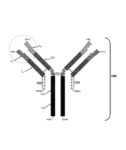
extension according to this embodiment is schematically illustrated in FIG. 1.
As shown,
antibody 100 includes two light chain polypeptides that include light chain
variable (V') and
constant (CO domains, and C-terminal extensions 102 and 104 having the
sequence GGGSC
extending from the C-terminal residue of each of the (CO domains.
In one embodiment, when X is GGGS (SEQ ID NO:1), a is 1, c is 1, and f is 0.
In one example, with reference to Formula I above, where X is a spacer having
the
sequence GGGS (SEQ ID NO:1) for purposes of illustration; a and c each = 1, b
= 2, and f= 0,
then the C-terminal extension has the sequence GGGSCC (SEQ ID NO:61).
In one example, with reference to Formula I above, where X is GGGS (SEQ ID
NO:1)
for purposes of illustration, where a = 2, b = 1, c = 1, and f = 0, the C-
terminal extension has the
sequence GGGSGGGSC (SEQ ID NO:62) when each spacer sequence is the same.
In one example, with reference to Formula I above, where Xis GGGS (SEQ ID
NO:1)
for purposes of illustration, and where a = 1, b = 1, c = 2, and f= 0, the C-
terminal extension has
the sequence GGGSCGGGSC (SEQ ID NO:63) (also referred to herein as "Cys2")
when each
spacer sequence is the same.
In one example, with reference to Formula I above, where X is GGGS (SEQ ID
NO:1)
for purposes of illustration, and where a = 1, b = 1, c = 4, and f = 0, the
extension would have the
sequence GGGSCGGGSCGGGSCGGGSC (SEQ ID NO:64) (also referred to herein as
"Cys4")
when each spacer sequence is the same.
28
CA 02934818 2016-06-22
WO 2015/095972
PCT/CA2014/051263
In one example, with reference to Formula I above, where X is GGGS (SEQ ID
NO:1)
for purposes of illustration, and where a - ------------------------------ 1,
b - 1, c - 1, d - 3, e - 1, and f= 1, the extension
would have the sequence GGGSCGGGSGGGSGGGSC (SEQ ID NO:65) when each spacer
sequence is the same.
In one example, with reference to Formula I above, where X is GGGS (SEQ ID
NO:1)
for purposes of illustration, and where a = 2, b = 1, c = 1, d = 1, e = 1, and
f= 1, the extension
would have the sequence GGGSGGGSCGGGSC (SEQ ID NO:66) when each spacer
sequence is
the same.
In one example, with reference to Formula 1 above, where Xis GGGS (SEQ ID
NO:1)
------------------------------------------------------------------- for
purposes of illustration, and where a - 1, b - 1, c - 1, d - 2, e - 1, and
f = 1, the extension
would have the sequence GGGSCGGGSGGGSC (SEQ ID NO:67) when each spacer
sequence is
the same.
In one example, with reference to Formula I above, where X is GGGS (SEQ ID
NO:1)
and X' is AKTTPKLEEGEFSEAR (SEQ ID NO:2) for purposes of illustration, and
where a = 1,
b = 1, c = 1, d= 2, e = 1, and f = 1, the extension would have the sequence
GGGSCAKTTPKLEEGEFSEARC (SEQ ID NO :89).
In one example, with reference to Formula I above, where c =2 and X is GGGS
(SEQ ID
NO:1) in the context of the first occurrence of (XaCb)c and X is
AKTTPKLEEGEFSEAR (SEQ
ID NO:2) in the context of the second occurrence of (XaCb)c, and X' is
AKTTPKLEEGEFSEAR
(SEQ ID NO:2) for purposes of illustration, and where a= 1, b = 1, c = 2, d=
2, e = 1, and f = 1,
the extension would have the sequence
GGGSCAKTTPKLEEGEFSEARCAKTTPKLEEGEFSEARC (SEQ ID NO:90).
As an example, with reference to Formula 1, where X is a spacer having the
sequence
AKTTPKLEEGEFSEAR (SEQ ID NO:2) for purposes of illustration, where a, b and c
are each
= 1; and f= 0, then the C-terminal extension has the sequence
AKTTPKLEEGEFSEARC (SEQ
ID NO:68).
In one example, with reference to Formula I above, where X is AKTTPKLEEGEFSEAR
(SEQ ID NO:2) for purposes of illustration, where a = 2, b = 1, c = 1, and f =
0, the C-terminal
extension has the sequence AKTTPKLEEGEFSEARAKTTPKLEEGEFSEARC (SEQ ID
NO:69) when each spacer sequence is the same.
29
CA 02934818 2016-06-22
WO 2015/095972 PCT/CA2014/051263
In one example, with reference to Formula I above, where X is AKTTPKLEEGEFSEAR
(SEQ ID NO:2) for purposes of illustration, and where a = 1, b = 1, c = 2, and
f= 0, the C-
terminal extension has the sequence AKTTPKLEEGEFSEARCAKTTPKLEEGEFSEARC
(SEQ ID NO:70) when each spacer sequence is the same.
In one example, with reference to Formula I above, where X is AKTTPKLEEGEFSEAR
(SEQ ID NO:2) for purposes of illustration, and where a = 1, b = 1, c = 4, and
f 0, the
extension would have the sequence
AKTTPKLEEGEFSEARCAKTTPKLEEGEFSEARCAKTTPKLEEGEFSEARCAKTTPKLEE
GEFSEARC (SEQ ID NO:71) when each spacer sequence is the same.
In one example, with reference to formula I above, where X is AKTTPKLEEGEFSEAR
(SEQ ID NO:2) for purposes of illustration, and where a ¨ ------------------
1, b ¨ 1, c ¨ 1, d ¨ 3, e ¨ 1, and f = 1,
the extension would have the sequence
AKTTPKLEEGEFSEARCAKTTPKLEEGEFSEARAKTTPKLEEGEFSEARAKTTPKLEEGEF
SEARC (SEQ ID NO:72) when each spacer sequence is the same.
In one example, with reference to Formula I above, where X is AKTTPKLEEGEFSEAR
(SEQ ID NO:2) for purposes of illustration, and where a ¨2, ---------------- b
¨ 1, c ¨ 1, d ¨ 1, e ¨ 1, and f = 1,
the extension would have the sequence
AKTTPKLEEGEFSEARAKTTPKLEEGEFSEARCAKTTPKLEEGEFSEARC (SEQ ID
NO:73) when each spacer sequence is the same.
In one example, with reference to Formula I above, where X is AKTTPKLEEGEFSEAR
(SEQ ID NO:2) for purposes of illustration, and where a= 1, b = 1, c = 1, d=
2, e = 1, and f 1,
the extension would have the sequence
AKTTPKLEEGEFSEARCAKTTPKLEEGEFSEARAKTTPKLEEGEFSEARC (SEQ ID
NO:74) when each spacer sequence is the same.
In another example, a C-terminal extension of the present disclosure may be
represented
by Formula II:
(XaCb)c(X'dCe)f(X"gCh), (II)
where
X, X', and X¨ represent a spacer of one or more amino acids, wherein the amino
acid
.. sequence of each X, X' and X" is independently selected from any amino acid
sequence of
interest, including any of the examples of spacer amino acid sequences
provided herein;
CA 02934818 2016-06-22
WO 2015/095972 PCT/CA2014/051263
C is a cysteine residue: and
a, b, c, d, e, f, g, h and i are integers independently selected from 0, 1, 2,
3, 4, 5, 6, 7, 8, 9,
10, 11, 12, 13, 14, 15, 16, 17, 18, 19 and 20, where the sum of b, e and h is
at least 1, and the
sum of c, f and i is at least 1. X, X', and X" may be the same or different.
Where c is greater
than 1, then each X of (XaCh)c may be the same or different amino acid
sequence within each
repeat unit of (XaCb)e. Where d or f is greater than 1, then each X' of
(X'dCe)f may the same or
different amino acid sequence within each repeat unit of (X'dCe)f. Where g or
i is greater than 1,
then each X¨ of (X"gCh), may the same or different amino acid sequence within
each repeat unit
of (X"gCh),.
A C-terminal extension of the present disclosure may include (e.g., consist
of) an amino
acid sequence selected from: EPKSCDKTHTC (SEQ ID NO:92) (also referred to
herein as
extension 1); EPKSCDKTHTCPPC (SEQ ID NO:93) (also referred to herein as
extension 2);
EPKSC (SEQ ID NO:94) (also referred to herein as extension 3); ESKYGPPC (SEQ
ID NO:95)
(also referred to herein as extension 4); ERKCCVECPPC (SEQ ID NO:96) (also
referred to
herein as extension 5); ERKC (SEQ ID NO:97) (also referred to herein as
extension 6);
DVITMDPKDNC (SEQ ID NO:98) (also referred to herein as extension 7);
DHVKPKETENTKQPSKSCHKPK (SEQ ID NO:99) (also referred to herein as extension
8);
ESSC (SEQ ID NO:100) (also referred to herein as extension 9); ESSCDVKLV (SEQ
ID
NO:101) (also referred to herein as extension 10); DHVKPKETENTKQPSKSC (SEQ ID
NO:102) (also referred to herein as extension 11); DVITMDPKDNCSKDAN (SEQ ID
NO:103)
(also referred to herein as extension 12); CAA, CCAA (SEQ ID NO:128), AACAA
(SEQ ID
NO:129), and GGGGSCAA (SEQ ID NO:130).
The present disclosure also provides nucleic acids encoding any of the C-
terminal
extensions described herein (e.g., C-terminal extension of Formula II, etc.),
as well as nucleic
acids encoding any of the light chain polypeptides described herein (e.g.,
light chain
polypeptides comprising a C-terminal extension of Formula II, etc.).
The present disclosure provides antibodies having at least one light chain
polypeptide
having a C-terminal extension of the present disclosure. In some embodiments,
the antibody has
two light chain polypeptides having a C-terminal extension of the present
disclosure. In some
embodiments, such antibodies can be conjugated to an agent/payload (e.g., a
drug) through
covalent attachment to at least one cysteinc in the C-terminal extension
present in at least one of
31
CA 02934818 2016-06-22
WO 2015/095972 PCT/CA2014/051263
the light chain polypeptides of the antibody. In one embodiment, the payload
is directly attached
to the cysteine of the C-terminal extension. In another embodiment, the
payload is attached via a
linker to the cysteine of the C-terminal extension. In one embodiment, the
payload is
preferentially attached to the cysteine of the C-terminal extension over a
cysteine outside the C-
terminal amino acid extension. In one embodiment, the payload is exclusively
attached to the
cysteine of the C-terminal extension.
NUCLEIC ACIDS, VECTORS AND HOST CELLS
The present disclosure provides nucleic acids encoding a light chain
polypeptide having a
C-terminal amino acid extension (referred to herein for convenience as a
"modified light chain
polypeptide"), as well as vectors and host cells containing such nucleic
acids. The modified light
chain polypeptide encoded by the nucleic acid may include any of the features
described above,
in any combination. For example, the C-terminal extension portion of the light
chain polypeptide
may include any of the C-terminal extension features described above with
respect to the length
of the extension, the amino acid sequence of the extension, the number of
spacers in the
extension and amino acid sequences thereof, extension configurations based on
combinations of
one or more spacers and one or more cysteine residues, and any other aspects
of the C-terminal
extensions described above and elsewhere herein.
A nucleic acid (e.g., DNA or RNA) encoding a C-terminal extension-containing
light
chain polypeptide can be operably linked to one or more regulatory elements,
such as a promoter
and enhancer, that allow expression of the nucleic acid in a host cell (e.g.,
a cell that is
genetically modified to synthesize the encoded modified light chain
polypeptide).
For example, where the host cell is a prokaryotic hose cell, example of
promoters
include, but are not limited to, a bacteriophage T7 RNA polymerase promoter; a
trp promoter; a
lac operon promoter; a hybrid promoter, e.g., a lac/tac hybrid promoter, a
tac/trc hybrid
promoter, a trp/lac promoter, a T7/lac promoter; a trc promoter; a tac
promoter, and the like; an
araBAD promoter; in vivo regulated promoters, such as an ssaG promoter or a
related promoter,
a pagC promoter; a nirB promoter; a sigma70 promoter, e.g., a consensus
sigma70 promoter; a
stationary phase promoter, e.g., a dps promoter, an spy promoter, and the
like; a promoter
derived from the pathogenicity island SPI-2; an actA promoter; an rpsM
promoter; a tet
promoter; an SP6 promoter; and the like. Examples of strong promoters for use
in prokaryotes
32
CA 02934818 2016-06-22
WO 2015/095972 PCT/CA2014/051263
such as Escherichia coli include, but are not limited to Trc, Tac, T5, T7, and
P
- Lambda. Non
limiting examples of operators for use in bacterial host cells include a
lactose promoter operator
(Lad I repressor protein changes conformation when contacted with lactose,
thereby preventing
the Lad repressor protein from binding to the operator), a tryptophan promoter
operator (when
complexed with tryptophan, TrpR repressor protein has a conformation that
binds the operator;
in the absence of tryptophan, the TrpR repressor protein has a conformation
that does not bind to
the operator), and a tac promoter operator.
In some embodiments, e.g., for expression in a yeast cell, the promoter can be
a
constitutive promoter such as an ADH1 promoter, a PGK1 promoter, an ENO
promoter, a PYK1
promoter and the like; or a regulatable promoter such as a GAL1 promoter, a
GAL10 promoter,
an ADH2 promoter, a PHO5 promoter, a CUP1 promoter, a GAL7 promoter, a MET25
promoter, a MET3 promoter, a CYC1 promoter, a HIS3 promoter, an ADH1 promoter,
a PGK
promoter, a GAPDH promoter, an ADC1 promoter, a TRP1 promoter, a URA3
promoter, a
LEU2 promoter, an ENO promoter, a TP1 promoter, and A0X1 (e.g., for use in
Pichia).
A nucleotide sequence encoding the modified light chain polypeptide can be
present in an
expression vector and/or a cloning vector. When it is desirable to express the
modified light
chain polypeptide and one or more other polypeptide components of an antibody,
e.g., to provide
for an antibody having a modified light chain polypeptide of the present
disclosure, the
corresponding nucleotide sequences encoding the two or more polypeptides may
be cloned in the
.. same or separate vectors. An expression vector can include a selectable
marker, an origin of
replication, and other features that provide for replication and/or
maintenance of the vector.
Expression vectors generally have convenient restriction sites located near
the promoter
sequence to provide for the insertion of nucleic acid sequences encoding
heterologous proteins.
Examples of expression vectors include, but are not limited to, viral vectors
(e.g. viral vectors
based on vaccinia virus; poliovirus; adenovirus; adeno-associated virus; SV40;
herpes simplex
virus; human immunodeficiency virus; a retroviral vector (e.g., Murine
Leukemia Virus, spleen
necrosis virus, and vectors derived from retroviruses such as Rous Sarcoma
Virus, Harvey
Sarcoma Virus, avian leukosis virus, human immunodeficiency virus,
myeloproliferative
sarcoma virus, and mammary tumor virus); and the like.
Also provided is a host cell that includes any of the nucleic acids encoding
the modified
light chain polypeptide, or vectors including the same. In certain aspects,
the host cell is a
33
CA 02934818 2016-06-22
WO 2015/095972 PCT/CA2014/051263
prokaryotic host cell or a eukaryotic host cell. The host cell may be an
isolated genetically
modified host cell (e.g., an in vitro cell) that is genetically modified
(e.g., transformed or
transfected) with a nucleic acid of the present disclosure. In some
embodiments, a genetically
modified host cell of the present disclosure can produce a modified light
chain polypeptide of the
.. present disclosure, which antibody light chain polypeptide can have any of
the features described
elsewhere herein.
Examples of host cells include cukaryotic host cells, such as a mammalian
cell, an insect
host cell, a yeast cell; and prokaryotic cells, such as a bacterial cell.
Introduction of a the nucleic
acid into the host cell can be effected, for example by calcium phosphate
precipitation, DEAE
dextran mediated transfection, liposome-mediated transfeetion,
electroporation, or other known
methods.
Examples of mammalian host cells include primary cells and immortalized cell
lines.
Suitable mammalian cell lines include human cell lines, non-human primate cell
lines, rodent
(e.g., mouse, rat) cell lines, and the like. Suitable mammalian cell lines
include, but are not
limited to, HeLa cells (e.g., American Type Culture Collection (ATCC) No. CCL-
2), CHO cells
(e.g., ATCC Nos. CRL9618, CCL61, CRL9096), 293 cells (e.g., ATCC No. CRL-
1573), Vero
cells, NIH 3T3 cells (e.g., ATCC No. CRL-1658), Huh-7 cells, BHK cells (e.g.,
ATCC No.
CCL10), PC12 cells (ATCC No. CRL1721), COS cells, COS-7 cells (ATCC No.
CRL1651),
RAT1 cells, mouse L cells (ATCC No. CCLI.3), human embryonic kidney (HEK)
cells (ATCC
No. CRL1573), HLHepG2 cells, and the like.
Examples of yeast host cells include, but are not limited to, Pichia pastoris,
Pichia
finlandica, Pichia trehalophila, Pichia koclamae, Pichia membranaefaciens,
Pichia opuntiae,
Pichia therm otolerans, Pichia salictaria, Pichia guercuum, Pichiapperi,
Pichia stiptis, Pichia
methanolica, Pichia sp., Saccharomyces cerevisiae, Saccharomyce.s sp.,
Hansenula polymorpha,
Kluyveromyces sp., Kluyveromyces lactis, Candida albicans, Aspergillus
nidulans, Aspergillus
niger, Aspergillns oryzae, Trichodenna reesei, Chrysosporium lucknowense,
Fusarium sp.,
Fusarium gram ineum, Fusarium venenatum, Neurospora crassa, Chlarnydomonas
reinhardtii,
and the like.
Examples of prokaryotic cells include, but are not limited to, any of a
variety of
laboratory strains of Eseherichia coli, Lactobacillus sp., Salmonella sp.,
Shigella sp., and the
like. Examples of Salmonella strains which can be employed in the present
invention include, but
34
CA 02934818 2016-06-22
WO 2015/095972 PCT/CA2014/051263
are not limited to, Salmonella typhi and S. typhimurium. Suitable Shigella
strains include, but are
not limited to, Shigella flexneri, Shigella sonnei, and Shigella disenteriae.
Typically, the
laboratory strain is one that is non-pathogenic. Non-limiting examples of
other suitable bacteria
include, but are not limited to, Bacillus subtilis, Pseudomonas pudita,
Pseudomonas aeruginosa,
Pseudomonas mevalonii, Rhodobacter sphaeroides , Rhodobacter capsulatus,
Rhodospirillum
rubrum, Rhodococcus sp., and the like.
METHODS OF PRODUCING ANTIBODY LIGHT CHAIN POLYPEPTIDES AND ANTIBODIES
As discussed above, the present disclosure provides methods of making modified
light
chain polypeptides (i.e., a light chain polypeptide having a C-terminal
extension of the present
disclosure), as well as antibodies containing one or more of such modified
light chain
polyp eptides.
According to one embodiment, provided is a method of making a light chain
polypeptide
of an antibody, the method including expressing in a host cell a nucleic acid
encoding a light
chain polypeptide that includes a C-terminal amino acid extension as described
herein. The light
chain polypeptide may be produced by any convenient method, e.g., conventional
synthetic
methods for protein synthesis, recombinant DNA methods, etc. The light chain
polypeptide
having a C-terminal amino acid extension may be produced in combination with
the production
of a heavy chain polypeptide of interest, or may be combined with a heavy
chain polypeptide
following production (e.g., by fusion of recombinant cells which separately
express a light chain
polypeptide of the present disclosure and a heavy chain polypeptide).
Recombinant methods can be used for production of the light chain polypeptide.
For
example, nucleic acids encoding a light chain polypeptide of interest may be
inserted into an
expression vector. The nucleic acid segment encoding the light chain
polypeptide may be
operably linked to one or more control sequences in the expression vector that
ensure the
expression of the light chain polypeptide. Expression control sequences
include, but are not
limited to, promoters (e.g., naturally-associated or heterologous promoters),
signal sequences,
enhancer elements, and transcription termination sequences. The expression
control sequences
can be eukaryotic promoter systems in vectors capable of transforming or
transfecting eukaryotic
host cells (e.g., COS or CHO cells). Once the vector has been incorporated
into the appropriate
CA 02934818 2016-06-22
WO 2015/095972 PCT/CA2014/051263
host, the host is maintained under conditions suitable for a desired level of
expression of the
nucleotide sequences, and the collection and purification of the light chain
polypeptide.
Because of the degeneracy of the genetic code, a variety of nucleic acid
sequences can
encode the desired light chain polypeptide. The nucleic acid sequence encoding
the light chain
polypeptide can be produced by de novo solid-phase DNA synthesis, by
polymerase chain
reaction (PCR) mutagenesis (e.g., overlapping PCR) of a nucleic acid that
encodes a light chain
polypeptide of a parental antibody that lacks a C-terminal amino acid
extension. An example
approach which utilizes overlapping PCR for generating nucleic acids that
encode light chain
polypeptides having C-terminal amino acid extensions is described in detail in
the Examples
section herein.
Suitable expression vectors are typically replicable in the host organisms
either as
episomes or as an integral part of the host chromosomal DNA. Conunonly,
expression vectors
contain selection markers (e.g., ampicillin-resistance, hygromycin-resistance,
tetracycline
resistance, kanamycin resistance or neomycin resistance) to permit detection
of those cells
transformed with the desired DNA sequences. Examples of expression vectors and
host cells are
discussed above. For example, the host cells can be prokaryotic cells, (e.g.,
Escherichia coli,
bacilli (such as Bacillus subtilis) Salmonella, Serratia, Pseudomonas species,
and the like), yeast
cells (e.g., Saccharomyces (e.g., S. cerevisiae), Pichia and the like), and
mammalian cells (e.g.,
CHO cell lines, Cos cell lines, HeLa cells, myeloma cell lines, transformed B-
cells, hybridomas
.. and the like).
Where the light chain polypeptide is chemically synthesized, the synthesis may
proceed
via liquid-phase or solid-phase. Solid phase polypeptide synthesis (SPPS), in
which the C-
terminal amino acid of the sequence is attached to an insoluble support
followed by sequential
addition of the remaining amino acids in the sequence, is an example of a
suitable method for the
chemical synthesis of the light chain polypeptide. Various forms of SPPS, such
as Fmoc and
Boc, are available for synthesizing the light chain polypeptide. Techniques
for solid phase
synthesis are available in the art. For example, small insoluble, porous beads
are treated with
functional units on which peptide chains are built. After repeated cycling of
coupling/deprotection, the free N-terminal amine of a solid-phase attached is
coupled to a single
N-protected amino acid unit. This unit is then deprotected, revealing a new N-
terminal amine to
36
CA 02934818 2016-06-22
WO 2015/095972 PCT/CA2014/051263
which a further amino acid may be attached. The peptide remains immobilized on
the solid-
phase and undergoes a filtration process before being cleaved off
Once synthesized (either recombinantly or chemically), the modified light
chain
polypeptides, either alone or as part of an antibody, can be purified
according to standard
procedures. The modified light chain polypeptide, either alone or as part of
an antibody, can be
substantially pure, e.g., at least about 80% to 85% pure, at least about 85%
to 90% pure, at least
about 90% to 95% pure, or 98% to 99%, or more, pure, e.g., free from
contaminants such as cell
debris, macromolecules other than the light chain polypeptide, and the like.
According to certain embodiments the light chain polypeptide is produced by in
vitro
translation. See, e.g., Yin et al. (2012) Aglycosylated antibodies and
antibody fragments
produced in a scalable in vitro transcription-translation system, Landes
Bioscience, Volume 4,
Issue 2.
The antibody may be a recombinant antibody, e.g., a chimeric, humanized, fully
human,
bispecific, deimmunized, and/or an in vitro generated antibody.
CONJUGATES
The present disclosure provides antibodies having at least one light chain
polypeptide
having a C-terminal extension, where at least one cysteine of the C-terminal
extension is
conjugated to an agent (e.g., drug).
The light chain polypeptide having a C-terminal extension of the antibody
portion of the
conjugates of the present disclosure may include any of the features described
above, and in any
combination. For example, the C-terminal extension of the antibody portion of
the conjugate
may include any of the C-terminal extension features described above with
respect to the length
of the extension, the amino acid composition of the extension, the number of
spacers in the
extension and amino acid sequences thereof, extension configurations based on
combinations of
one or more spacers and one or more cysteine residues, and any other aspects
of the C-terminal
extensions described above and elsewhere herein.
Accordingly, the antibody conjugates of the present disclosure have at least
one light
chain polypeptide having a C-terminal extension, where at least one cysteine
residue of the C-
terminal extension is conjugated to an agent. In some embodiments, each of the
light chain
polypeptides of the antibody conjugates of the present disclosure have a C-
terminal extension,
37
CA 02934818 2016-06-22
WO 2015/095972 PCT/CA2014/051263
where at least one cysteine residue of the C-terminal extensions of each of
the modified light
chain polypeptides is conjugated to an agent. For example, a first agent may
be conjugated to a
cysteine residue of the C-terminal extension of a first modified light chain
polypeptide, and a
second agent may be conjugated to a cysteine residue of the C-terminal
extension of a second
.. light chain polypeptide. In one embodiment, the agent is preferentially
attached to the cysteine
residue of the C-terminal extension rather than a cysteine residue outside the
C-terminal amino
acid extension. In certain aspects, the agent is exclusively attached to the
cysteine residue of the
C-terminal extension. Antibody conjugates of the present disclosure can
include any number of
agents conjugated to the C-terminal extension of the modified light chain
polypeptide(s),
.. according to the number of cysteines in the C-terminal extension available
for conjugation, e.g.,
1, 2, 3, 4, 5, 6, 7, 8, 9, 10, 11, 12, 13, 14, 15, 16, 17, 18, 19, or more
cysteines. In some
embodiments, the C-terminal extension of a modified light chain polypeptide(s)
of the antibody
is conjugated to an agent or agents via covalent binding to at least 1, 2, 3,
4, 5, or more cysteines
of the C-terminal extension. In some embodiments the C-terminal extension of a
modified light
.. chain polypeptide(s) of the antibody is conjugated to an agent or agents
via covalent binding to
no more than 2, 3, 4, 5, 6, 7, 8, 9 or 10 cysteines of the C-terminal
extension. In certain aspects,
the extension includes an agent that is conjugated to two or more adjacent
cysteine residues in
the extension, either directly or via one or more linkers. For example, the
extension may include
an agent that is conjugated to two adjacent cysteine residues in the
extension.
In certain embodiments, the number of agents conjugated to the C-terminal
extensions of
modified light chain polypeptide(s) of the antibody can be characterized by
the drug-to-antibody
ratio (DAR). In some instances, a distribution of a plurality of antibody
conjugates may be
characterized by measuring the average DAR of the antibody conjugates in the
distribution,
where the average DAR indicates the average number of agents conjugated to the
C-terminal
.. extensions of modified light chain polypeptide(s) of the antibodies in the
distribution. By
"average" is meant the arithmetic mean. In certain cases, average DAR is
assayed by
Hydrophobic Interaction Chromatography (HIC). Thus, an average DAR provides an
indication
of the average drug-to-antibody ratio of the antibody conjugates in a
distribution provided by the
DAR assay.
In some instances, the DAR of an antibody conjugate according to the present
disclosure
ranges from 0 to 20, such as from 1 to 17, or 1 to 15, or 1 to 12, or 1 to 10,
or 1 to 9, or 1 to 8, or
38
CA 02934818 2016-06-22
WO 2015/095972 PCT/CA2014/051263
1 to 7, or 1 to 6, or 1 to 5, or 1 to 4, or 1 to 3. For instance, a DAR of 0
indicates that the
antibody is unconjugated; a DAR of 1 indicates that one agent (e.g., drug) is
conjugated to a C-
terminal extension of a modified light chain polypeptide of the antibody; a
DAR of 2 indicates
that two agents are conjugated to one or more C-terminal extensions of
modified light chain
polypeptide(s) of the antibody; a DAR of 3 indicates that three agents are
conjugated to one or
more C-terminal extensions of modified light chain polypeptide(s) of the
antibody; a DAR of 4
indicates that four agents are conjugated to one or more C-terminal extensions
of modified light
chain polypeptide(s) of the antibody; etc. In some instances, the DAR of an
antibody conjugate
according to the present disclosure is 1, 2, 3, 4, 5, 6, 7, 8, 9, 10, 11, 12,
13, 14, 15, 16, 17, 18, 19
or 20.
In sonic instances, the average DAR of a distribution of antibody conjugates
according to
the present disclosure ranges from 0 to 20, such as from 1 to 17, or 1 to 15,
or 1 to 12, or 1 to 10,
or 1 to 9, or 1 to 8, or 1 to 7, or 1 to 6, or 1 to 5, or 1 to 4, or 1 to 3.
For instance, an average
DAR of 0 indicates that, on average, the antibodies in the distribution are
unconjugated; an
average DAR of 1 indicates that, on average, one agent (e.g., drug) is
conjugated to each
antibody in the distribution; an average DAR of 2 indicates that, on average,
two agents are
conjugated each antibody in the distribution; an average DAR of 3 indicates
that, on average,
three agents are conjugated to each antibody in the distribution; an average
DAR of 4 indicates
that, on average, four agents are conjugated to each antibody in the
distribution; etc. In some
instances, an average DAR of a distribution of antibody conjugates according
to the present
disclosure is 1, 2, 3, 4, 5, 6, 7, 8,9, 10, 11, 12, 13, 14, 15, 16, 17, 18, 19
or 20.
In certain embodiments, a sample containing antibody conjugates according to
the
present disclosure includes a mixture of distributions of unconjugated and
conjugated (e.g.,
mono-conjugated, di-conjugated, tri-conjugated, etc.) antibodies. The average
DAR of each
distribution of antibody conjugates may be assayed (e.g., by RIC). For
example, a sample
containing antibody conjugates may include a distribution of unconjugated
antibodies (i.e.,
antibody conjugates having an average DAR = 0), a distribution of mono-
conjugated antibodies
(i.e., antibody conjugates having an average DAR = 1), a distribution of di-
conjugated antibodies
(i.e., antibody conjugates having an average DAR = 2), a distribution of tri-
conjugated antibodies
(i.e., antibody conjugates having an average DAR = 3), etc.
39
An example antibody conjugate according to an embodiment of the present
disclosure is
schematically illustrated in FIG. 2. As shown, conjugate 200 includes an
antibody having two
light chain polypeptides that include light chain variable (V') and constant
(CO domains, and C-
terminal extensions having the sequence GGGSC extending from the C-terminal
residue of each
of the (C1) domains. Conjugate 200 further includes agents 202 and 204 linked
to the cysteine
residue of the extension via a linker.
Linkers
In certain aspects, the agent is conjugated to the cysteine residue via a
linker. Linkers that
find use in the conjugates of the present disclosure include maleimide or
maleimide-based
linkers; valine-citrulline linkers; hydrazone linkers; N-succinimidy1-4-(2-
pyridyldithio)butyrate
(SPDB) linkers; Succinimidy1-4-(N-maleimidomethyl)cyclohexane-1-carboxylate
(SMCC)
linkers; vinylsulfone-based linkers; linkers involving metal atom(s)
coordinated to cysteine;
linkers that include polyethylene glycol (PEG), such as, but not limited to
tetraethylene glycol;
linkers that include propanoic acid; linker that include caproleic acid;
linkers that include
Fleximer polymers (Mersana Therapeutics, Cambridge, MA) or the linkers used
to attach
drugs to Fleximer polymers (see, e.g., USPN 8,524,214); and linkers including
any combination
thereof.
In certain aspects, the linker is a chemically-labile linker, such as an acid-
cleavable linker
that is stable at neutral pH (bloodstream pH 7.3-7.5) but undergoes hydrolysis
upon
internalization into the mildly acidic endosomes (pH 5.0-6.5) and lysosomes
(pH 4.5-5.0) of a
target cell (e.g., a cancer cell). Chemically-labile linkers include, but are
not limited to,
hydrazone-based linkers. According to certain embodiments, the linker is an
enzyme-labile
linker, such as an enzyme-labile linker that is stable in the bloodstream but
undergoes enzymatic
cleavage upon internalization into a target cell, e.g., by a lysosomal
protease (such as cathepsin
or plasmin) in a lysosome of the target cell (e.g., a cancer cell). Enzyme-
labile linkers include,
but are not limited to, linkers that include peptidic bonds, e.g., valine-
citrulline linkers, such as a
maleimidocaproyl-valine-citruline-p-aminobenzyl (MC-vc-PAB) linker, and the
like. In certain
aspects, the linker is a non-cleavable linker, such as a linker that includes
a non-cleavable
thioether bond. Chemically-labile linkers, enzyme-labile, and non-cleavable
linkers are known
and described in detail, e.g., in Ducry & Stump (2010) B io conjugate Chem.
21:5-13.
Date Recue/Date Received 2021-03-01
CA 02934818 2016-06-22
WO 2015/095972 PCT/CA2014/051263
According to certain embodiments, the linker is (or includes) a sulfhydryl-
reactive
chemical group capable of reacting with one or more reduced sulfhydryl group
(or thiol, -SH) of
the cysteine residue(s) of the C-terminal amino acid extension. Sulfhydryl-
reactive chemical
groups that find use in linking the agent to the cysteine of the C-terminal
extension include, but
are not limited to, maleimides, haloacetyls, pyridyl disulfides, aziridines,
acryloyls, arylating
agents, vinylsulfones, TNB-thiols, metals, and disulfide reducing agents. Such
groups may
conjugate to sulfhydryls by alkylation (e.g., by the formation of a thioether
bond), disulfide
exchange (formation of a disulfide bond), or the like.
A2ents
The agents which are the payload of the antibody conjugates of the present
disclosure can
be any suitable agent. The agent selected for use in the antibody conjugates
of the present
disclosure will vary depending on the application for which the conjugate is
employed (e.g.,
killing, prevention of cell proliferation, hormone therapy, target imaging,
and/or gene therapy,
etc.). Agents of interest include, but are not limited to, therapeutic agents
(e.g., drugs (e.g.,
cytotoxic agents)), detectable agents (e.g., in vivo imaging agents), and/or
any other agent useful
for a particular antibody-based application of interest.
Non-limiting examples of such agents include toxins, fragments of toxins,
lectins,
alkylating agents, enzymes, antibiotics such as antibacterials, antifungals,
antimycoplasmals,
etc., antiviral agents, antimetabolites, antiproliferative or antineoplastic
agents, DNA,
radioopaque dyes, radioactive isotopes (e.g., 1123, 1131 as well as
radioactive metal ions), metal
ions, fluorogenic compounds, marker compounds, and compounds which alter cell
membrane
permeability. In certain aspects, the agent is (or includes) a member of a
specific binding pair,
e.g., biotin (which forms a specific binding pair with avidin/streptavidin).
According to certain embodiments, the agent is a therapeutic agent.
Therapeutic agents of
interest include agents capable of affecting the function of a cell/tissue to
which the conjugate
binds via specific binding of the antibody portion of the conjugate to an
antigen on the surface of
the cell/tissue. For example, the agent may boost the function of the
cell/tissue to which the
conjugate specifically binds. Alternatively, when the function of the
cell/tissue is pathological,
an agent that reduces the function of the cell/tissue may be employed. In
certain aspects, a
conjugate of the present disclosure includes an agent that reduces the
function of a target
cell/tissue by inhibiting cell proliferation and/or killing the cell/tissue.
Such agents may vary and
41
include cytostatic agents and cytotoxic agents (e.g., an agent capable of
killing a target cell tissue
with or without being internalized into a target cell).
In certain aspects, the therapeutic agent is a cytotoxic agent selected from
an enediyne, a
lexitropsin, a duocarmycin, a taxane, a puromycin, a dolastatin, a
maytansinoid, and a vinca
alkaloid. In some embodiments, the cytotoxic agent is paclitaxel, docetaxel,
CC-1065, CPT-11
(SN-38), topotecan, doxorubicin, morpholino-doxorubicin, rhizoxin,
cyanomorpholino-
doxorubicin, dolastatin-10, echinomycin, combretastatin, calicheamicin,
maytansine, maytansine
DM1, maytansine DM4, DM-1, an auristatin or other dolastatin derivatives, such
as auristatin E
or auristatin F, AEB (AEB-071), AEVB (5-benzoylvaleric acid-AE ester), AEFP
(antibody-
endostatin fusion protein), MMAE (monomethylauristatin E), MMAF
(monomethylauristatin F),
pyrrolobenzodiazepines (PBDs), eleutherobin, netropsin, or any combination
thereof.
According to certain embodiments, the agent is a protein toxin selected from
hemiasterlin
and hemiasterlin analogs such as HTI-286 (e.g., see USPN 7,579,323; WO
2004/026293; and
USPN 8,129,407), abrin, brucine, cicutoxin, diphtheria toxin, batrachotoxin,
botulism toxin,
shiga toxin, endotoxin, Pseudomonas exotoxin, Pseudomonas endotoxin, tetanus
toxin, pertussis
toxin, anthrax toxin, cholera toxin, falcarinol, fumonisin B1, fumonisin B2,
afla toxin,
maurotoxin, agitoxin, charybdotoxin, margatoxin, slotoxin, scyllatoxin,
hefutoxin, calciseptine,
taicatoxin, calcicludine, geldanamycin, gelonin, lotaustralin, ocratoxin A,
patulin, ricin,
strychnine, trichothecene, zearlenone, and tetradotoxin. Enzymatically active
toxins and
fragments thereof which may be employed include diphtheria A chain, non-
binding active
fragments of diphtheria toxin, exotoxin A chain (from Pseudomonas aeruginosa),
ricin A chain,
abrin A chain, modeccin A chain, alpha-sarcin, Aleurites fordii proteins,
dianthin proteins,
Phytolaca americana proteins (PAPI, PAPII, and PAP-S), Momordica charantia
inhibitor, curcin,
crotin, Sapaonaria officinalis inhibitor, gelonin, mitogellin, restrictocin,
phenomycin, enomycin
and the tricothecenes.
In certain aspects, the agent is a labeling agent. By "labeling agent" (or
"detectable
label") is meant the agent detectably labels the antibody, such that the
antibody may be detected
in an application of interest (e.g., in vitro and/or in vivo research and/or
clinical applications).
Detectable labels of interest include radioisotopes, enzymes that generate a
detectable product
(e.g., horseradish peroxidase, alkaline phosphatase, etc.), fluorescent
proteins, paramagnetic
atoms, and the like. In certain aspects, the antibody is conjugated to a
specific binding partner of
42
Date Recue/Date Received 2021-03-01
CA 02934818 2016-06-22
WO 2015/095972 PCT/CA2014/051263
detectable label (e.g., conjugated to biotin such that detection may occur via
a detectable label
that includes avidin/streptavidin).
According to certain embodiments, the agent is a labeling agent that finds use
in in vivo
imaging, such as near-infrared (NIR) optical imaging, single-photon emission
computed
tomography (SPECT)/CT imaging, positron emission tomography (PET), nuclear
magnetic
resonance (NMR) spectroscopy, or the like. Labeling agents that find use in
such applications
include, but are not limited to, fluorescent labels, radioisotopes, and the
like. In certain aspects,
the labeling agent is a multi-modal in vivo imaging agent that permits in vivo
imaging using two
or more imaging approaches (e.g., see Thorp-Greenwood and Coogan (2011) Dalton
Trans.
40:6129-6143).
In certain aspects, the labeling agent is an in vivo imaging agent that finds
use in near-
infrared (NIR) imaging applications, which agent is selected from a Kodak X-
SIGHT dye, Pz
247, DyLight 750 and 800 Fluors, Cy 5.5 and 7 Fluors, Alexa Fluor 680 and 750
Dyes, IRDye
680 and 800CW Fluors. According to certain embodiments, the labeling agent is
an in vivo
imaging agent that finds use in SPECT imaging applications, which agent is
selected from 99mTc,
"In, '231n,
201 133
In, In, Tl, and ¨Xe. In certain aspects, the labeling agent is an in
vivo imaging agent that
finds use in positron emission tomography (PET) imaging applications, which
agent is selected
from ric, 13N, 150, 18F, 64cu, 62cu, 1241, 76Br, 82Rb and 68Ga.
CONJUGATION METHODS
The present disclosure also provides methods of making antibody conjugates.
The
methods include conjugating an agent to an antibody that includes a light
chain polypeptide
including a C-terminal amino acid extension, which extension includes a
cysteine residue, where
the agent is conjugated to the cysteine residue (directly or indirectly (e.g.,
via a linker)) of the C-
terminal amino acid extension. In one embodiment, the method involves the
preferential (or
"biased") conjugation of agent to the cysteine residue of the C-terminal amino
acid extension
rather than a cysteine residue outside the C-terminal extension. In certain
aspects, the
conjugation includes conjugating a linker to a sulfhydryl group of the
cysteine residue, e.g.,
using maleimide reaction chemistry, haloacetyl reaction chemistry, pyridyl
disulfide reaction
chemistry, or any other suitable reaction chemistry as described herein. The
methods of making
the conjugate may further include reducing the cysteine residue to a
sulthydryl group (i.e., thiol)
43
CA 02934818 2016-06-22
WO 2015/095972 PCT/CA2014/051263
prior to the conjugating step, e.g., using a suitable reducing agent and
reaction conditions as
described above. Suitable reducing agents include, but are not limited to,
DTPA, cysteamine,
TCEP (tris(2-carboxyethyl)phosphine hydrochloride), combinations thereof, and
the like. In
certain embodiments, methods of making the conjugate include contacting the
antibody that
includes a light chain polypeptide including a C-terminal amino acid extension
with a reducing
agent. In certain embodiments, methods of making the conjugate include
contacting the antibody
that includes a light chain polypeptide including a C-terminal amino acid
extension with a first
reducing agent, followed by contacting the antibody that includes a light
chain polypeptide
including a C-terminal amino acid extension with a second reducing agent. An
alternative
embodiment of the present disclosure does not require a reduction step as the
cysteine within the
light chain extension is already in a reduced state as a synthesis product. In
certain embodiments,
the reduced antibody may be contacted with a suitable oxidizing agent.
Suitable oxidizing agents
include, but are not limited to, dehydroascorbic acid (DHAA), and the like.
The agent conjugated
to the antibody may be any useful agent. In certain aspects, the agent is a
therapeutic agent or a
labeling agent, which agents are described elsewhere herein.
In certain aspects, the agent is linked to the cysteine of the C-terminal
extension using
maleimide reaction chemistry. The maleimide group may react specifically with
sulfhydryl
groups when the pH of the reaction mixture is between pH 6.5 and 7.5,
resulting in the formation
of a stable thioether linkage. For example, a maleimidyl-modified agent (e.g.,
drug) may be
reacted with a reduced cysteine (e.g., sulfhydryl or thiol group) of a C-
terminal amino acid
extension to form a thioether linkage between the agent and the antibody. In
more alkaline
conditions (pH > 8.5), primary amines may compete with thiols for reaction
with maleimides,
and also increase the rate of hydrolysis of the maleimide group to a non-
reactive maleamic acid.
Maleimides do not react with tyrosines, histidines or methionines.
Bioconjugation approaches
that employ maleimide-based linkers are described in, e.g., in Hermanson,
G.T., Bioconjugate
Techniques, 2nd ed. San Diego, CA Academic Press 2008; Aslam & Dent,
Bioconjugation:
Protein Coupling Techniques for the Biomedical Sciences, London Macmillan
Reference Ltd
1998; Kalia & Raines, Advances in Bioconjugation, Curr. Org. Chem. 14(2):138-
147. Examples
of suitable conjugation approaches using a maleimide-based linker according to
embodiments of
the present disclosure are described in detail in the Examples section herein.
44
CA 02934818 2016-06-22
WO 2015/095972 PCT/CA2014/051263
According to certain embodiments, the agent is linked to the cysteine of the C-
terminal
extension using haloacetyl reaction chemistry. In certain aspects, a
haloacetyl linker that includes
an iodoacetyl or a bromoacetyl group is employed. In certain embodiments,
haloacetyls react
with sulfhydryl groups at physiologic pH. The reaction of the iodoacetyl group
proceeds by
nucleophilic substitution of iodine with a sulfur atom from a sulfhydryl
group, resulting in a
stable thioether linkage.
In certain aspects, the agent is linked to the cysteine of the C-terminal
extension using
pyridyl disulfide reaction chemistry. In certain embodiments, pyridyl
disulfides react with
sulthydryl groups over a broad pH range (with pH 4 to 5 being optimal) to form
disulfide bonds.
During the reaction, a disulfide exchange occurs between the sulfhydryl group
of the antibody
and a 2-pyridyldithiol group of a 2-pyridyldithiol-modified agent. As a
result, pyridine-2-thione
is released and can be measured spectrophotometrically (Amax = 343nm) to
monitor the
progress of the reaction.
To generate a reduced sulfhydryl in the cysteine of the C-terminal amino acid
extension
to which the agent may be attached (e.g., via a linker), the cysteine may be
contacted with a
suitable reducing agent under conditions sufficient to produce a reduced
sulfhydryl group. In
certain aspects, the reducing agent is selected from cysteamine hydrochloride,
2-
mercaptoethanol, dithiothreitol (DTT), 2-mercaptoethylamine, tris(2-
carboxyl)phosphine
(TCEP), cysteine HC1, N-ethylmaleimide, Nacystelyn, domase alfa, thymosin (34,
guaifenesin
TCEP HC1, and any combination thereof. Reaction conditions for such reducing
agents are
known in the art and may be optimized, e.g., to promote selectivity or "bias"
the reduction of the
cysteine(s) present in the C-terminal extension as opposed to the cysteine
residues present in the
parental antibody (e.g., the cysteine residues that participate in disulfide
bonding between CL and
CHI of the light and heavy chains, and/or between the hinge regions of the
heavy chains). An
alternative embodiment of the invention does not require a reduction step as
the cysteine within
the light chain extension is already in a reduced state as a synthesis
product.
Preferential reduction of the cysteine(s) of the C-terminal amino acid
extension over one
or more cysteine residues outside the C-terminal amino acid extension (or
exclusive reduction of
the cysteine(s) of the C-terminal amino acid extension) may be achieved by
selection of suitable
reduction conditions. In certain aspects, suitable reduction conditions
include suitable selection
of one or more of the following: a mild reducing agent and/or a reducing agent
having a steric
CA 02934818 2016-06-22
WO 2015/095972 PCT/CA2014/051263
bulk that confers upon the reducing agent a preference for reducing a cysteine
of the C-terminal
amino acid extension; concentrations of the reducing agent and substrate; the
temperature at
which the reduction reaction is carried out, the pH of the reduction reaction
mixture; the buffer
used in the reduction reaction; and/or conditions under which the cells
expressing the extended
C-terminal light chain polypeptides are cultured (e.g., to obtain free thiol
on the C-terminal
extension and/or to generate readily reduced intermolecular disulfides).
PHARMACEUTICAL COMPOSITIONS
As summarized above, the present disclosure provides compositions.
Compositions of the
.. present disclosure may include any of the antibodies, conjugates, nucleic
acids, vectors, and/or
host cells described above. Aspects of the present disclosure include
pharmaceutical
compositions. In certain embodiments, the pharmaceutical compositions include
any of the
antibodies described elsewhere herein (e.g., an antibody that includes a light
chain polypeptide
including a C-terminal amino acid extension that includes a cysteine residue
as described above)
or any of the conjugates described elsewhere herein (e.g., a conjugate that
includes an antibody
component having a light chain polypeptide including a C-terminal amino acid
extension that
includes a cysteine residue as described above), and a pharmaceutically
acceptable excipient.
The antibody or conjugate present in the pharmaceutical compositions may
include any of the
features described above with respect to the antibodies of the present
disclosure or the conjugates
of the present disclosure, in any combination. For example, the C-terminal
extension of the
antibody, or antibody portion of the conjugate, may include any of the C-
terminal extension
features described above with respect to the length of the extension, the
amino acid makeup of
the extension, the number of spacers in the extension and amino acid sequences
thereof,
extension configurations based on combinations of one or more spacers and one
or more cysteine
residues, and any other aspects of the C-terminal extensions described above
and elsewhere
herein.
The pharmaceutical compositions generally include a therapeutically effective
amount of
an antibody or conjugate of the present disclosure. An effective amount may be
administered in
one or more administrations.
The antibodies or conjugates of the present disclosure may be administered to
the patient
using any convenient means capable of resulting in the desired therapeutic
effect or diagnostic
46
CA 02934818 2016-06-22
WO 2015/095972 PCT/CA2014/051263
effect. Thus, the antibody or conjugate can be incorporated into a variety of
formulations for
therapeutic administration. More particularly, the antibody can be formulated
into
pharmaceutical compositions by combination with appropriate, pharmaceutically
acceptable
carriers or diluents, and may be formulated into preparations in solid, semi-
solid, liquid or
gaseous forms, such as tablets, capsules, powders, granules, ointments,
solutions, injections,
inhalants and aerosols.
Formulations of the antibodies or conjugates of the present disclosure
suitable for
administration to a patient (e.g., suitable for human administration) arc
generally sterile and may
further be free of detectable pyrogens or other contaminants contraindicated
for administration to
a patient according to a selected route of administration.
In pharmaceutical dosage forms, the antibody can be administered in the form
of their
pharmaceutically acceptable salts, or they may also be used alone or in
appropriate association,
as well as in combination, with other pharmaceutically active compounds. The
following
methods and excipients are merely examples and are in no way limiting.
For oral preparations, the antibodies or conjugates can be used alone or in
combination
with appropriate additives to make tablets, powders, granules or capsules, for
example, with
conventional additives, such as lactose, mannitol, corn starch or potato
starch; with binders, such
as crystalline cellulose, cellulose derivatives, acacia, corn starch or
gelatins; with disintegrators,
such as corn starch, potato starch or sodium carboxymethylcellulose; with
lubricants, such as talc
or magnesium stearate; and if desired, with diluents, buffering agents,
moistening agents,
preservatives and flavoring agents.
The antibodies or conjugates can be formulated into preparations for injection
by
dissolving, suspending or emulsifying them in an aqueous or non-aqueous
solvent, such as
vegetable or other similar oils, synthetic aliphatic acid glycerides, esters
of higher aliphatic acids
or propylene glycol; and if desired, with conventional additives such as
solubilizers, isotonic
agents, suspending agents, emulsifying agents, stabilizers and preservatives.
Pharmaceutical compositions of the present disclosure may be prepared by
mixing the
antibody or conjugate having the desired degree of purity with optional
physiologically
acceptable carriers, excipients, stabilizers, surfactants, buffers and/or
tonicity agents. Acceptable
carriers, excipients and/or stabilizers are nontoxic to recipients at the
dosages and concentrations
employed, and include buffers such as phosphate, citrate, and other organic
acids; antioxidants
47
including ascorbic acid, glutathione, cysteine, methionine and citric acid;
preservatives (such as
ethanol, benzyl alcohol, phenol, m-cresol, p-chlor-m-cresol, methyl or propyl
parabens,
benzalkonium chloride, or combinations thereof); amino acids such as arginine,
glycine,
ornithine, lysine, histidine, glutamic acid, aspartic acid, isoleucine,
leucine, alanine,
phenylalanine, tyrosine, tryptophan, methionine, serine, proline and
combinations thereof;
monosaccharides, disaccharides and other carbohydrates; low molecular weight
(less than about
residues) polypeptides; proteins, such as gelatin or serum albumin; chelating
agents such as
EDTA; sugars such as trehalose, sucrose, lactose, glucose, mannose, maltose,
galactose, fructose,
sorbose, raffinose, glucosamine, N-methylglucosamine, galactosamine, and
neuraminic acid;
10 and/or non-ionic surfactants such as TweenTm, Brij PluronicsTM, TritonTm-
X, or polyethylene
glycol (PEG).
The pharmaceutical composition may be in a liquid form, a lyophilized form or
a liquid
form reconstituted from a lyophilized form, wherein the lyophilized
preparation is to be
reconstituted with a sterile solution prior to administration. The standard
procedure for
.. reconstituting a lyophilized composition is to add back a volume of pure
water (typically
equivalent to the volume removed during lyophilization); however solutions
comprising
antibacterial agents may be used for the production of pharmaceutical
compositions for
parenteral administration.
Example antibody or conjugate concentrations in a pharmaceutical composition
according the present disclosure may range from about 1 mg/mL to about 200
mg/ml or from
about 50 mg/mL to about 200 mg/mL, or from about 150 mg/mL to about 200 mg/mL.
An aqueous formulation of the antibody or conjugate may be prepared in a pH-
buffered
solution, e.g., at pH ranging from about 4.0 to about 7.0, or from about 5.0
to about 6.0, or
alternatively about 5.5. Examples of buffers that are suitable for a pH within
this range include
phosphate-, histidine-, citrate-, succinate-, acetate-buffers and other
organic acid buffers. The
buffer concentration can be from about 1 mM to about 100 mM, or from about 5
mM to about 50
mM, depending, e.g., on the buffer and the desired tonicity of the
formulation.
A tonicity agent may be included in the antibody or conjugate formulation to
modulate
the tonicity of the formulation. Example tonicity agents include sodium
chloride, potassium
.. chloride, glycerin and any component from the group of amino acids, sugars
as well as
combinations thereof. In some embodiments, the aqueous formulation is
isotonic, although
48
Date Recue/Date Received 2021-03-01
CA 02934818 2016-06-22
WO 2015/095972 PCT/CA2014/051263
hypertonic or hypotonic solutions may be suitable. The term "isotonic" denotes
a solution having
the same tonicity as some other solution with which it is compared, such as
physiological salt
solution or serum. Tonicity agents may be used in an amount of about 5 mM to
about 350 mM,
e.g., in an amount of 100 mM to 350 mM.
A surfactant may also be added to the antibody or conjugate formulation to
reduce
aggregation of the formulated antibody or conjugate and/or minimize the
formation of
particulates in the formulation and/or reduce adsorption. Example surfactants
include
polyoxyethylensorbitan fatty acid esters (Tween), polyoxyethylene alkyl ethers
(Brij),
alkylphenylpolyoxyethylene ethers (Triton-X), polyoxyethylene-polyoxypropylene
copolymer
(Poloxamer, Pluronic), and sodium dodecyl sulfate (SDS). Examples of suitable
polyoxyethylenesorbitan-fatty acid esters are polysorbate 20, (sold under the
trademark Tween
2OTM) and polysorbate 80 (sold under the trademark Tween 80Tm). Examples of
suitable
polyethylene-polypropylene copolymers are those sold under the names Pluronic0
F68 or
Poloxamer 188TM. Examples of suitable Polyoxyethylene alkyl ethers are those
sold under the
trademark BrijTM. Example concentrations of surfactant may range from about
0.001% to about
1% w/v.
A lyoprotectant may also be added in order to protect the active ingredient
(e.g. the
antibody or conjugate) against destabilizing conditions during the
lyophilization process. For
example, known lyoprotectants include sugars (including glucose and sucrose);
polyols
(including mannitol, sorbitol and glycerol); and amino acids (including
alanine, glycine and
glutamic acid). Lyoprotectants can be included in an amount of about 10 mM to
500 nM.
In certain aspects, the formulation includes an antibody or conjugate of the
present
disclosure, and one or more of the above-identified agents (e.g., a
surfactant, a buffer, a
stabilizer, a tonicity agent) and is essentially free of one or more
preservatives, such as ethanol,
benzyl alcohol, phenol, m-cresol, p-chlor-m-cresol, methyl or propyl parabens,
benzalkonium
chloride, and combinations thereof. In other embodiments, a preservative is
included in the
formulation, e.g., at concentrations ranging from about 0.001 to about 2%
(w/v).
For example, the formulation can be a liquid (e.g., an aqueous solution or
emulsion) or
lyophilized formulation thereof, suitable for parenteral administration, and
can comprise: about 1
mg/mL to about 200 mg/mL of a subject antibody or conjugate; about 0.001 % to
about 1 % of at
least one surfactant; about 1 mM to about 100 mM of a buffer; optionally about
10 mM to about
49
CA 02934818 2016-06-22
WO 2015/095972 PCT/CA2014/051263
500 mM of a stabilizer; and about 5 mM to about 305 mM of a tonicity agent;
and has a pH of
about 4.0 to about 7Ø
An antibody or conjugate of the present disclosure can be utilized in an
aerosol
formulation to be administered via inhalation. The antibody can be formulated
into pressurized
acceptable propellants such as dichlorodifluoromethane, propane, nitrogen and
the like.
Unit dosage forms for oral administration such as syrups, elixirs, and
suspensions may be
provided wherein each dosage unit, for example, teaspoonful, tablespoonful, or
tablet, contains a
predetermined amount of the composition containing one or more inhibitors.
Similarly, unit
dosage forms for injection or intravenous administration may comprise the
antibody or conjugate
in a composition as a solution in sterile water, normal saline or another
pharmaceutically
acceptable carrier.
The term "unit dosage form," as used herein, refers to physically discrete
units suitable as
unitary dosages for human and animal subjects, each unit containing a
predetermined quantity of
compounds of the present invention calculated in an amount sufficient to
produce the desired
effect in association with a pharmaceutically acceptable diluent, carrier or
vehicle. The
specifications for the antibody or conjugate of interest may depend on the
particular antibody
employed and the effect to be achieved, and the pharmacodynamics associated
with each
antibody in the host.
In certain aspects, the pharmaceutical composition (optionally provided in
unit dosage
form) includes an antibody or conjugate of the present disclosure present at a
concentration of
from about 10 mg/mL to about 1000 mg/mL, e.g., from about 25 mg/mL to about
500 mg/mL,
from about 50 mg/mL to about 250 mg/mL, from about 75 mg/mL to about 200
mg/mL, or from
about 100 mg/mL to about 150 mg/mL (e.g., about 125 mg/mL).
In some embodiments, the antibody or conjugate is formulated in a controlled
release
formulation. Sustained-release preparations may be prepared using methods well
known in the
art. Suitable examples of sustained-release preparations include semipermeable
matrices of solid
hydrophobic polymers containing the antibody in which the matrices are in the
form of shaped
articles, e.g. films or microcapsules. Examples of sustained-release matrices
include polyesters,
copolymers of L-glutamic acid and ethyl-L-glutamate, non-degradable ethylene-
vinyl acetate,
hydrogels, polylactides, degradable lactic acid-glycolic acid copolymers and
poly-D-(-)-3-
hydroxybutyric acid.
CA 02934818 2016-06-22
WO 2015/095972 PCT/CA2014/051263
Controlled release within the scope of this invention can be taken to mean any
one of a
number of extended release dosage forms. The following terms may be considered
to be
substantially equivalent to controlled release, for the purposes of the
present invention:
continuous release, controlled release, delayed release, depot, gradual
release, long-term release,
.. programmed release, prolonged release, proportionate release, protracted
release, repository,
retard, slow release, spaced release, sustained release, time coat, timed
release, delayed action,
extended action, layered-time action, long acting, prolonged action, repeated
action, slowing
acting, sustained action, sustained-action medications, and extended release.
A suitable dosage can be determined by an attending physician or other
qualified medical
personnel, based on various clinical factors. As is well known in the medical
arts, dosages for
any one patient depend upon many factors, including the patient's size, body
surface area, age,
the particular antibody or conjugate to be administered, sex of the patient,
time, and route of
administration, general health, and other drugs being administered
concurrently. An antibody or
conjugate of the present disclosure may be administered in amounts between 1
ng/kg body
weight and 25 mg/kg body weight per dose, e.g. between 0.1 mg/kg body weight
to 10 mg/kg
body weight, e.g. between 0.5 mg/kg body weight to 8 mg/kg body weight, e.g.
between 1 mg/kg
body weight to 6 mg/kg body weight, e.g. between 2 mg/kg body weight to 5
mg/kg body
weight; however, doses below or above these example ranges are envisioned,
especially
considering the aforementioned factors. If the regimen is a continuous
infusion, it can also be in
the range of 1 ir.g to 10 mg per kilogram of body weight per minute.
Those of skill will readily appreciate that dose levels can vary as a function
of the
specific antibody, the severity of the symptoms and the susceptibility of the
subject to side
effects. Preferred dosages for a given compound are readily determinable by
those of skill in the
art by a variety of means.
Conventional and pharmaceutically acceptable routes of administration include
intravenous, intra-arterial, intramuscular, intranasal, intra-tracheal,
subcutaneous, intradennal,
topical application, nasal, oral, and other enteral and parenteral routes of
administration. Routes
of administration may be combined, if desired, or adjusted depending upon the
antibody or
conjugate and/or the desired effect. The pharmaceutical composition can be
administered in a
single dose or in multiple doses. In some embodiments, the composition is
administered
intravenously. In some embodiments, the composition is administered orally. In
some
51
CA 02934818 2016-06-22
WO 2015/095972 PCT/CA2014/051263
embodiments, the composition is administered via an inhalational route. In
some embodiments,
the composition is administered intranasally. In some embodiments, the
composition is
administered locally. In some embodiments, the composition is administered
intra-cranially.
METIIODS OF TREATMENT
The present disclosure provides methods of treating diseases or disorders. The
methods
may include administering to a patient in need thereof a therapeutically
effective amount of any
of the antibodies, conjugates, or pharmaceutical compositions described
elsewhere herein. The
antibody or conjugate may be administered alone (e.g., in monotherapy) or in
combination (e.g.,
.. in combination therapy) with one or more additional therapeutic agents.
In some embodiments, an effective amount of the antibody or conjugate is an
amount
that, when administered alone (e.g., in monotherapy) or in combination (e.g.,
in combination
therapy) with one or more additional therapeutic agents, in one or more doses,
is effective to
reduce the symptoms of a disease or disorder in an individual by at least
about 5%, at least about
10%, at least about 15%, at least about 20%, at least about 25%, at least
about 30%, at least
about 40%, at least about 50%, at least about 60%, at least about 70%, at
least about 80%, at
least about 90%, or more, compared to the symptoms in the individual in the
absence of
treatment with the antibody or conjugate.
The methods of the present disclosure may be employed to treat any diseases or
disorders
of interest. In certain aspects, the methods are employed to treat cancer. For
example, in some
embodiments, an antibody or conjugate of the present disclosure inhibits
growth, metastasis
and/or invasiveness of a cancer cell(s) in a host when the antibody or
conjugate is administered
in an effective amount. By "cancer cell" is meant a cell exhibiting a
neoplastic cellular
phenotype, which may be characterized by one or more of, for example, abnormal
cell growth,
abnormal cellular proliferation, loss of density dependent growth inhibition,
anchorage-
independent growth potential, ability to promote tumor growth and/or
development in an
immunocompromised non-human animal model, and/or any appropriate indicator of
cellular
transformation. "Cancer cell" may be used interchangeably herein with "tumor
cell", "malignant
cell" or "cancerous cell", and encompasses cancer cells of a solid tumor, a
semi-solid tumor, a
.. primary tumor, a metastatic tumor, and the like.
52
CA 02934818 2016-06-22
WO 2015/095972
PCT/CA2014/051263
In certain aspects, when the methods are for treatment of cancer, the antibody
or antibody
component of the conjugate specifically binds to an antigen on the surface of
a cancer cell. The
terms "antigen" and "epitope" are well understood in the art and refer to the
portion of a
macromolecule (e.g., a polypeptide) which is specifically recognized by a
component of the
immune system, e.g., an antibody or a T-cell antigen receptor. As used herein,
the term "antigen"
encompasses antigenic epitopes, e.g., fragments of an antigen which are
antigenic epitopes.
I-laptens are also examples of antigens. Epitopes can be recognized by
antibodies in solution, e.g.
free from other molecules. Epitopes can be recognized by T-cell antigen
receptor when the
epitope is associated with a class 1 or class 11 major histocompatibility
complex molecule.
Antigens of interest in the context of cancer treatment include tumor-specific
antigens,
e.g., antigens present on the surface of malignant cells and not present on
non-malignant cells. In
other aspects, the antigen bound by the antibody is a tumor-associated
antigen. By "tumor-
associated antigen" is meant an antigen expressed on malignant cells with
limited expression on
cells of normal tissues, antigens that are expressed at much higher density on
malignant versus
normal cells, or antigens that are developmentally expressed.
Any tumor-associated antigen or tumor-specific antigen may be targeted by an
antibody
or conjugate of the present disclosure. In certain aspects, when the methods
of the present
disclosure are for treatment of cancer, the antigen specifically bound by the
antibody or antibody
component of a conjugate of the present disclosure may include, but is not
limited to, HER2,
CD19, CD22, CD30, CD33, CD56, CD66/CEACAM5, CD70, CD74, CD79b, CD138, Nectin-
4,
Mesothelin, Transmembrane glycoprotein NMB (GPNMB), Prostate-Specific Membrane
Antigen (PSMA), 5LC44A4, CA6, CA-IX, or any other tumor-associated or tumor-
specific
antigens of interest.
By "specific binding" or "specifically binds" in the context of a
characteristic of an
antibody refers to the ability of an antibody to preferentially bind to a
particular antigen that is
present in a homogeneous mixture of different antigens. In certain
embodiments, a specific
binding interaction will discriminate between desirable and undesirable
antigens (or "target" and
"non-target" antigens) in a sample or organism (e.g., a human), in some
embodiments more than
about 10 to 100-fold or more (e.g., more than about 1000- or 10,000-fold). In
certain
embodiments, the affinity between an antibody and antigen when they are
specifically bound in
an antibody-antigen complex is characterized by a KD (dissociation constant)
of less than le M,
53
CA 02934818 2016-06-22
WO 2015/095972 PCT/CA2014/051263
less than 10-7M, less than 10M, less than 10-9M, less than 10-10M, less than
10-11M, or less
than about 10-1-2M or less.
Cancers which may be treated using the methods of the present disclosure
include, but
are not limited to, solid tumors, breast cancer, prostate cancer, pancreatic
cancer, colorectal
.. carcinoma, renal cell carcinoma, Hodgkin's lymphoma, Non-Hodgkin's
lymphoma, anaplastic
large cell lymphoma, acute myelogenous leukemia, multiple myeloma, and any
other type of
cancer which may be treated using an antibody-based or antibody-conjugate-
based therapy.
KITS
The present disclosure also provides kits. According to certain embodiments,
the kits may
include any of the antibodies, conjugates, or pharmaceutical compositions of
the present
disclosure having any of the features as described elsewhere herein.
Alternatively, or
additionally, the kits may include any reagents useful for producing an
antibody of the present
disclosure or light chain polypeptide thereof, or any of the conjugates of the
present disclosure.
For example, the kit may include a nucleic acid that encodes an antibody light
chain polypeptide
that include a cysteine-containing C-terminal amino acid extension. Such kits
may include, e.g.,
competent cells or cells already harboring nucleic acids encoding one or more
antibody light
and/or heavy chain polypeptides, a reducing agent for reducing the sulfhydryl
group of a cysteine
residue in the C-terminal light chain polypeptide extension, a linker for
conjugating an agent to a
reduced sulfhydryl of a cysteine residue, an agent (which may be attached to a
linker or separate
from a linker), reagents, buffers, purification columns, etc. that find use in
producing an antibody
or conjugate of the present disclosure, or any combinations thereof. The kits
find use, e.g., in
enabling one to practice the methods of the present disclosure, such as the
methods of treating a
disease or disorder, methods of making antibody light chain polypeptides,
and/or methods of
making antibody conjugates.
The kits for practicing the methods may include one or more pharmaceutical
compositions that include the antibodies or conjugates described herein. As
such, the kits may
include a single pharmaceutical composition present as one or more unit
dosages. In yet other
embodiments, the kits may include two or more separate pharmaceutical
compositions.
Components of the kits may be present in separate containers, or multiple
components
may be present in a single container. In certain embodiments, it may be
convenient to provide the
54
CA 02934818 2016-06-22
WO 2015/095972 PCT/CA2014/051263
components in a lyophilized form, so that they are ready to use and can be
stored conveniently at
room temperature.
In addition to the above-mentioned components, a kit of the present disclosure
may
further include instructions for using the components of the kit, e.g., to
treat a disease or disorder
using an antibody or conjugate of the present disclosure, or to make an
antibody or conjugate of
the present disclosure. The instructions are generally recorded on a suitable
recording medium.
For example, the instructions may be printed on a substrate, such as paper or
plastic, etc. As
such, the instructions may be present in the kits as a package insert, in the
labeling of the
container of the kit or components thereof (i.e., associated with the
packaging or subpackaging)
etc. In other embodiments, the instructions are present as an electronic
storage data file present
on a suitable computer readable storage medium, e.g. CD-ROM, diskette, Hard
Disk Drive
(HDD) etc. In yet other embodiments, the actual instructions are not present
in the kit, but means
for obtaining the instructions from a remote source, e.g. via the internet,
are provided. An
example of this embodiment is a kit that includes a web address where the
instructions can be
viewed and/or from which the instructions can be downloaded. As with the
instructions, this
means for obtaining the instructions is recorded on a suitable substrate.
The following examples are offered by way of illustration and not by way of
limitation.
EXAMPLES
EXAMPLE 1 ¨ CLONING OF NUCLEIC ACIDS ENCODING ANTIBODIES HAVING C-TERMINAL
LIGHT CHAIN POLYPEPTIDE CYSTEINE-CONTAINING EXTENSIONS
To generate human IgG1C and human IgGkC cloning vectors the human IgGC and
IgkC
regions were cloned into a pTT5 vector (Fig. 1.). To facilitate cloning of
variable regions in
frame with the constant regions, a restriction site was introduced into the 5'
end of the constant
region. The heavy chain constant region sequence was changed from GCC TCC to
GCT AGC
which introduced a NheI restriction site while maintaining the original amino
acid sequence. The
light chain polypeptide constant region sequence was changed from CGA ACT to
CGT ACG to
generate a BsiWI restriction site while maintaining the original amino acid
sequence. The
changes were introduced by designing mismatch PCR primers purchased from
Operon -
Eurofins. Since the pTT5 vector did not contain a BsiWI site, the IgkC 5'
primer was designed
with a 5' NheI overhang to facilitate cloning into the vector. The 3' primers
were designed with a
BamHI restriction site 3' of the stop codon.
To generate a cDNA template for the PCR reactions, RNA was extracted from
human
peripheral blood using Qiagen RNeasy0 miniprep kits and cDNA was synthesized
using an
oligodT primer and Superscript III reverse transcriptase from InvitrogenTM.
Following PCR, the fragments and the vectors were digested using the relevant
restriction
enzymes and separated on a 1% agarose gel. The digested fragments were
extracted from the gel
using Qiagen gel extraction kit and ligated into the vector using T4 DNA
ligase (New England
Biolabs). Competent DH5a E-coli (Invitrogen) were transformed and single-cell
colonies were
grown at 37 C over night on ampicillin selective LB plates. To isolate the
plasmids, single cells
colonies were inoculated into liquid LB-ampicillin media, grown overnight at
37 C, and
plasmids were isolated using Qiagen QIAprep spin miniprep kit. The clones
were screened
by miniprep DNA digests, and verified by sequence analysis using Geneious
sequence
alignment, assembly and analysis software from Biomatters. Primer sequences
used for the
cloning are provided in Table 1.
Table 1 - IgGC and IgkC cloning primers
hIgGC for NheI SEQ
ID NO:75 ATTAGCTAGCACCAAGGGCCCATCGGTCTT
hIgGC_rev_BamHI SEQ ID NO:76 GATATGGATCCTCATTTACCCGGAGACAGG
GA
hKC Jor_NheI_BsiW SEQ ID NO:77 TATGCTAGCGTCGTACGGTGGCTGCACCAT
CTGTCTTCATC
hKCJev_BainHI SEQ ID NO:78 GATGGATCCCTAACACTCTCCCCTGTTGAA
GC
To generate ERBB specific antibodies the 4D5 humanized variant 8, herceptinTm
V-
genes were synthesized as gBlocks gene fragments from IDTO (Integrated DNA
Technologies). The sequences synthesized are provided in Table 2.
Table 2 - HerceptinTM V sequences
HerceptinTM VH SEQ
ID MEFGLSWVFLVAILKGVQCEVQLVESGGGLVQ
amino acid sequence NO:79
PGGSLRLSCAASGFNIKDTYIHWVRQAPGKGLE
WVARIYPTNGYTRYADSVKGRFTISADTSKNTA
YLQMNSLRAEDTAVYYCSRWGGDGFYAMDY
WGQGTLVTVSSASTKGPSV
56
Date Recue/Date Received 2021-03-01
CA 02934818 2016-06-22
WO 2015/095972 PCT/CA2014/051263
Herceptin VH SEQ ID AGTCAGTCGGAATTCGCCACCATGGAGTTTGG
nucleotide sequence NO: 80
GCTGAGCTGGGTTTTCCTTGTTGCTATTTTAAA
AGGTGTCCAGTGTGAGGTGCAGCTGGTGGAG
AGCGGCGGCGGCCTGGTGCAGCCCGGCGGCA
GCCTGAGACTGAGCTGCGCCGCCAGCGGCTTC
AACATCAAGGACACCTACATCCACTGGGTGA
GACAGGCCCCTGGCAAGGGCCTGGAGTGGGT
GGCCAGAATCTACCCCACCAACGGCTACACC
AGATACGCCGACAGCGTGAAGGGCAGATTCA
CCATCAGCGCCGACACCAGCAAGAACACCGC
CTACCTGCAGATGAACAGCCTGAGAGCCGAG
GACACCGCCGTGTACTACTGCAGCAGATGGG
GCGGCGACGGCTTCTACGCCATGGACTACTGG
GGCCAGGGCACCCTGGTGACCGTGAGCAGCG
CTAGCACCAAGGGCCCATCGGTCTT
Herceptin Vk amino SEQ ID MDMRVPAQLLGLLLLWLRGARCDIQMTQSPSS
acid sequence NO:81
LSASVGDRVTITCRASQDVNTAVAWYQQKPGK
APKLLIYSASFLYSGVPSRFSGSRSGTDFTLTISSL
QPEDFATYYCQQHYTTPPTFGQGTKVEIKRTVA
APSV
Herceptin Vk nucleotide SEQ ID AGTCAGTCGGAATTCGCTACCATGGACATGA
sequence NO: 82 GGGTCCCCGCTCAGCTCCTGGGGCTCCTGCTA
CTCTGGCTCCGAGGTGCCAGATGTGACATCCA
GATGACCCAGAGCCCCAGCAGCCTGAGCGCC
AGCGTGGGCGACAGAGTGACCATCACCTGCA
GAGCCAGCCAGGACGTGAACACCGCCGTGGC
CTGGTACCAGCAGAAGCCCGGCAAGGCTCCC
AAGCTGCTGATCTACAGCGCCAGCTTCCTGTA
CAGCGGCGTGCCCAGCAGATTCAGCGGCAGC
AGAAGCGGCACCGACTTCACCCTGACCATCA
GCAGCCTGCAGCCCGAGGACTTCGCCACATAC
TACTGCCAGCAGCACTACACCACCCCTCCCAC
CTTCGGCCAGGGCACCAAGGTGGAGATCAAG
CGTACGGTGGCTGCACCATCTGTCT
The fragments were cloned into the mentioned constant region pTT5 vectors
using EcoRI
and Nhel digests for the heavy chain and EcoRI and BsiWI digests for the light
chain
polypeptide as described above.
To add a (GGGSC)x extension to the light chain polypeptide, where x is the
number of
repeats added, 3' end primers were designed and ordered from Eurofins MWG
Operon with the
desired sequences, shown in Table 3. Standard cloning procedures were
performed using the
previously described herceptin VK constructs as templates in the PCR reaction.
To generate the
57
4 cysteine construct a two-step overlapping approach was taken where the
primer Igk_rev_3_cys
was used in the first PCR, followed by amplification using the primer
Igk_rev_4_cys_Bam and
the PCR product from the first PCR as template. The 5' primer used in all the
reactions was
pTT5 _for (Table 3). The PCR fragments were digested, cloned and analyzed as
described above.
Table 3 - Cloning primers used to add cysteines to light chain polypeptide C-
terminus
IgK_rev_l_cys_Bam SEQ ID ACG TGG ATC CTC AAC AGC TTC CCC CTC
NO:83 CAC ACT CTC CCC TGT TGA AGC
IgK_rev_2_cys_Bam SEQ ID ACG TGG ATC CTC AAC AGC TTC CCC CTC
NO:84 CGC AGC TTC CTC CTC CAC ACT CTC CCC TGT
TGA AGC
IgK_rev_3_cys_Bam SEQ ID ACG TGG ATC CTC AAC AGC TTC CCC CTC
NO:85 CGC AGC TTC CTC CTC CGC AAG ATC CTC CTC
CAC ACT CTC CCC TGT TGA AGC
IgK_rev_3_cys SEQ ID ACA GCT TCC CCC TCC GCA GCT TCC TCC TCC
NO:86 GCA AGA TCC TCC TCC ACA CTC TCC CCT GTT
GAA GC
IgK_rev_4_cys_Bam SEQ ID ACG TGG ATC CTC AGC AGC TTC CTC CTC
NO:87 CAC AGC TTC CCC CTC CGC AGC T
pTT5_for SEQ ID TGC GCT AAG ATT GTC AGT TTC CA
NO:88
EXAMPLE 2¨ ANTIBODY PRODUCTION AND PURIFICATION
Qiagen maxipreps of the plasmids generated in Example 1 above were performed
to
isolate transfection ready plasmids. HEK293 cells were co-transfected with the
Herceptin heavy
chain and the Herceptin light chain-cys constructs using 293 fectin
(Invitrogen). The cells were
grown in Freestyle 293 expression media (Gibco) supplemented with 0.1%
Pluronic F68 (Gibco)
solution for 5 days. 24 hours post transfection the cells were supplemented
with 0,5% tryptone.
Supernatants were collected and the secreted antibodies were purified using a
protein A
SepharoseTM batch gravity protocol (GEHealthcare) followed by buffer exchange
into PBS PH
7.4 using an Amicon0 Ultra 15 filter with a 30 kDa MW cutoff (Millipore).
EXAMPLE 3¨ REDUCTION OF HERCEPTIN-VLCYSX DISULFIDE BONDS
Samples (1-5 mg) of Herceptin and Herceptin-VLCysX (X = 1, 2 or 4) in PBS were
applied to ZebaTM spin columns (Pierce, catalogue #87767) preconditioned with
100 mM
phosphate, 50 mM NaC1, 2 mM DTPA, pH 6.1 and buffer exchanged according to the
58
Date Recue/Date Received 2021-03-01
CA 02934818 2016-06-22
WO 2015/095972 PCT/CA2014/051263
manufacturer's instructions. The eluates were assayed using a bicinchoninic
acid assay (Pierce,
#23225) using Herceptin as a standard to establish protein concentration, and
with Ellman's
reagent using cysteine as a standard to establish the absence of free thiol
groups.
Herceptin or Herceptin VLCysX (5-30 }IM in 100 mM phosphate, 50 mM NaCl, 2 mM
DTPA, pH 6.1) was reduced by addition of cysteamine hydrochloride (from 5 to
10 mM) from a
1.0 M stock in the same buffer and incubation for 40-180 minutes at either
room temperature or
37 C. After cooling to room temperature cysteamine was removed from the
reaction mixture by
passage over a ZebaTM spin column (40 KDa MWCO) preconditioned with 100 mM
phosphate,
50 mM NaCl, 2 mM DTPA, pH 6.1. in order to ensure excess cysteamine had been
removed,
the eluate was assayed with Ellman's reagent employing a standard curve
generated by assay of
cysteine serial dilutions. This assay also provided some measure of the
average thiol content per
protein.
EXAMPLE 4¨ CONJUGATION OF HERCEPTIN-VLCYSX TO A CYTOTOXIC AGENT
After cooling the eluates from Example 3 above on ice, maleimide toxin (toxin
1 or toxin
2) was added from a 10 mM DMSO stock solution (generally 2.0 eq. per thiol,
with an equal
amount added to the reduced Herceptin control (Herceptin-toxin 2)). The
conjugation reaction
was allowed to proceed for between 30 and 70 minutes on ice before
purification and buffer
exchange in to 20 mM sodium citrate, pH 5.5. The purified conjugates were
sterile filtered
(Costar Spin-X 0.22 um centrifugal filters, # 8161) and assayed using the
BCA reagent for
total protein content.
"Toxin 1" as used herein is MC-vc-PABC-toxin, where the toxin is (S,E)-N-(4-
(aminomethyObenzylsulfony1)-2,5-dimethyl-44S)-N,3,3-trimethyl-2-((S)-3-methyl-
2-
(methylamino)-3-phenylbutanamido)butanamido)hex-2-enamide.
"Toxin 2" as used herein is MC-vc-toxin, where the toxin is (S,E)-N-(4-
aminobenzylsulfony1)-2,5-dimethy1-44(S)-N,3,3-trimethyl-2-((S)-3-methyl-2-
(methylamino)-3-
phenylbutanamido)butanamido)hex-2-enamide.
"Toxin 3" as used herein is MT-ye-toxin, where the toxin is (S,E)-N-(4-
aminophenylsulfony1)-2,5-dimethy1-44S)-N,3,3-trimethyl-24S)-3-methyl-2-
(methylamino)-3-
phenylbutanamido)butanamido)hex-2-enamide.
59
CA 02934818 2016-06-22
WO 2015/095972 PCT/CA2014/051263
"Toxin 4" as used herein is MP(T-ve-toxin)2, where the toxin is (S,E)-N-(4-
aminophenylsulfony1)-2,5-dimethy1-44S)-N,3,3-trimethyl-2-((S)-3-methyl-2-
(methylamino)-3-
phenylbutanamido)butanamido)hex-2-enamide.
"Toxin 5" as used herein is MT-vc-PABC-toxin, where the toxin is (S,E)-N-(4-
(aminomethyl)benzylsulfony1)-2,5-dimethyl-4-((S)-N,3,3-trimethyl-2-((S)-3-
methyl-2-
(methylamino)-3-phenylbutanamido)butanamido)hex-2-enamide.
"Toxin 6" as used herein is MT-ye-toxin, where the toxin is:
0 0
0
NH0 NH,
__ EXAMPLE 5¨ TESTING TIIE EFFECT OF HERCEPTIN VLCYSX ON CANCER CELL VIABILITY
The antibody drug conjugates generated in Example 4 above were tested at
varying
concentrations against the Her2 positive human mammary carcinoma cell line
HCC1954. On the
day prior to adding compounds, HCC1954 cells (100 [IL) were added to opaque-
walled clear-
bottomed 96-well tissue culture-treated microtiter plates using complete
growth medium at a
density of 2500 cells/100 tit of medium. The HCC1954 cells were incubated for
one night at
37 C/5% CO2 to allow the cells to attach to the microtiter plate surface.
Antibody drug
conjugates were diluted in complete growth medium at five-times the final
maximum
concentration desired and compounds were then titrated 1:3 in the same medium,
eight steps. A
control with no compound (growth medium alone) was included on each microtiter
plate in
__ sextuplicate. The prepared compounds titrations were added (twenty-five
JAL/well) in triplicate
to the HCC1954 cells. The cells and compound titrations were incubated at 37
C/5% CO2 for
three or five nights. After the incubation, cell viability was measured using
CellTiter-Glo
reagent by adding 30 [IL of prepared CellTiter-Glo to each assay well. The
mixture was
incubated for a minimum of twenty minutes prior to measurement of luminescence
using a
microplate luminometer (500ms integration time). The collected relative
luminescence units
(RLU) are converted to % cytotoxicity using the growth medium alone control
mentioned above
CA 02934818 2016-06-22
WO 2015/095972 PCT/CA2014/051263
(% Cytotoxicity = 1 - [Well RLU/average medium alone control RLU]). Data were
fit to curves
using non-linear regression methods available with Prism Graph Pad software. A
graph showing
the data from this study is provided in FIG. 3. The EC50 values of Her-VLCys2-
toxin 2, Her-
VLCysl-toxin 1 and Herceptin-toxin 2 are shown in Table 4.
Table 4 - EC50 values of Her-VLCys2-toxin, Her-VLCysl-toxin and Herceptin-
toxin
EC50 (nM)
Her-VLCys2-toxin 2 0.04
Her-VLCysl-toxin 1 0.12
Herceptin-toxin 2 22.68
EXAMPLE 6¨ CLONING OF NUCLEIC ACIDS ENCODING ADDITIONAL ANTIBODIES HAVING C-
TERMINAL LIGHT CHAIN POLYPEPTIDE CYSTEINE-CONTAINING EXTENSIONS
To generate additional light chain extensions, synthetic gBlocks0 gene
fragments from
IDT were ordered with the desired sequence and cloned in frame into the
Herceptin VK using a
Gibson assembly cloning kit (New England Biolabs). All clones were sequence
verified and
analyzed as described above in Example 1.
Light chain amino acid sequences and the nucleic acid sequences encoding the
same are
provided in Table 5 below.
Table 5 ¨ Amino acid sequences for additional antibody light chains, and
nucleic acid
sequences encoding the same
Name and SEQ ID NO Description Sequence (AA)
RTVAAPSVFIFPPSDEQLKSG
Herceptin-Igk-Ig extension' TASVVCLLNNEYPREAKVQ
Herceptin VL and hIgk constant region (Km3 WKVDNALQSGNSQESVTEQ
SEQ ID NO: 104 allotype) + a EPKSCDKTHTC tail (sequence
from human IgG1 hinge) DSKDSTYSLSSTLTLSKADY
EKHKVYACEVTHQGLSSPVT
KSFNRGECEPKSCDKTHTC
RTVAAPSVEIFPPSDEQLKSG
TASVVCLLNNFYPREAKVQ
Herceptin-Ig,k-Ig extension2
Herceptin VL and hIgk constant region (Km3 WKVDNALQSGNSQESVTEQ
SEQ ID NO: 105 allotype) + a EPKSCDKTHTCPPC tail
DSKDSTYSLSSTLTLSKADY
(sequence from human IgG1 hinge) EKHKVYACEVTHQGLSSPVT
KSFNRGECEPKSCDKTHTCP
PC
Herceptin-Igk-Ig extension3 Herceptin VL and hIgk constant region (Km3
RTVAAPSVFIFPPSDEQLKSG
allotype) + a EPKSC tail (sequence from
61
CA 02934818 2016-06-22
WO 2015/095972 PCT/CA2014/051263
Name and SEQ ID NO Description Sequence (AA)
SEQ ID NO: 106 human IgG1 hinge) TASVVCLLNNFYPREAKVQ
WKVDNALQSGNSQESVTEQ
DSKDSTYSLSSTLTLSKADY
EKHKVYACEVTHQGLSSPVT
KSFNRGECEPKSC
RTVAAPSVFIFPPSDEQLKSG
TASVVCLLNNFYPREAKVQ
Herceptin-Ig,k-Ig extension4 Herceptin VL and hIgk constant region (Km3
WKVDNALQSGNSQESVTEQ
allotype) + a ESKYGPPC tail (sequence
SEQ ID NO: 107 from human IgG4 hinge) DSKDSTYSLSSTLTLSKADY
EKHKVYACEVTHQGLSSPVT
KSFNRGECESKYGPPC
RTVAAPSVFIFPPSDEQLKSG
TASVVCLLNNFYPREAKVQ
Herceptin-Ig,k-Ig extension5 IIerceptin VL and bIgk constant region (Km3
WKVDNALQSGNSQESVTEQ
allotype) + a ERKCCVECPPC tail
SEQ ID NO: 108 (sequence from human IgG2 hinge) DSKDSTYSLSSTLTLSKADY
EKHKVYACEVTHQGLSSPVT
KSFNRGECERKCCVECPPC
RTVAAPSVFIFPPSDEQLKSG
TASVVCLLNNFYPREAKVQ
Ilerceptin-Ig,k-Ig extension6 Herceptin VL and bIgk constant region (Km3
WKVDNALQSGNSQESVTEQ
allotype) + a ERKC tail (sequence from
SEQ ID NO: 109 human IgG2 hinge) DSKDSTYSLSSTLTLSKADY
EKHKVYACEVTHQGLSSPVT
KSFNRGECERKC
RTVAAPSVFIFPPSDEQLKSG
TASVVCLLNNFYPREAKVQ
Herceptin-Igk-Ig extension7 Herceptin VL and hIgk constant region (Km3
WKVDNALQSGNSQESVTEQ
allotype) + a DVITMDPKDNC tail
SEQ ID NO: 110 (sequence from human TCRg hinge) DSKDSTYSLSSTLTLSKADY
EKHKVYACEVTHQGLSSPVT
KSFNRGECDVITMDPKDNC
RTVAAPSVFIFPPSDEQLKSG
TASVVCLLNNFYPREAKVQ
Herceptin-Ig,k-Ig extension8 Herceptin VL and hIgk constant region (Km3
WKVDNALQSGNSQESVTEQ
allotype) + a
DSKDSTYSLSSTLTLSKADY
DHVKPKETENTKQPSKSCHKPK tail
SEQ Ill NO: 111
(sequence from human TCRd hinge) EKHKVYACEVTHQGLSSPVT
KSFNRGECDHVKPKETENTK
QPSKSCHKPK
RTVAAPSVFIFPPSDEQLKSG
TASVVCLLNNFYPREAKVQ
Herceptin-lg,k-Ig extension9 Herceptin VL and hIgk constant region (Km3
WKVDNALQSGNSQESVTEQ
allotype) + a ESSC tail (sequence from
SEQ ID NO: 112 human TCRa hinge) DSKDSTYSLSSTLTLSKADY
EKHKVYACEVTHQGLSSPVT
KSFNRGECES Sc
62
CA 02934818 2016-06-22
WO 2015/095972 PCT/CA2014/051263
Name and SEQ ID NO Description Sequence (AA)
RTVAAPSVFIFPPSDEQLKSG
TASVVCLLNNFYPREAKVQ
Herceptin-Igk-Ig extension10 Herceptin VL and hIgk constant region (Km3
WKVDNALQSGNSQESVTEQ
allotype) + a ESSCDVKLV tail (sequence
SEQ ID NO: 113 from human TCRa hinge) DSKDSTYSLSSTLTLSKADY
EKHKVYACEVTHQGLSSPVT
KSFNRGECESSCDVKLV
RTVAAPSVFIFPPSDEQLKSG
TASVVCLLNNFYPREAKVQ
Herceptin-Igk-Ig exten si on 11 Herceptin VL and hIgk constant region (Km3
WKVDNALQSGNSQESVTEQ
allotype) + a DHVKPKETENTKQPSKSC DSKDSTYSLSSTLTLSKADY
SEQ ID NO: 114 tail (sequence from human TCRd hinge) EKHKVYACEVTHQGLS
SPVT
KSFNRGECDHVKPKETENTK
QPSKSC
RTVAAPSVFIFPPSDEQLKSG
TASVVCLLNNFYPREAKVQ
Herceptin-Igk-Ig extension12 Herceptin VL and hIgk constant region (Km3
WKVDNALQSGNSQESVTEQ
allotype) + a DVITMDPKDNCSKDAN tail DSKDSTYSLSSTLTLSKADY
SEQ ID NO: 115 (sequence from human TCRg hinge) EKHKVYACEVTHQGLSSPVT
KSFNRGECDVITMDPKDNCS
KDAN
Name Description Sequence (NT)
GGCCAGGGCACCAAGGTGG
AGATCAAGCGTACGGTGGC
TGCACCATCTGTCTTCATCT
TCCCGCCATCTGATGAGCA
GTTGAAATCTGGAACTGCC
TCTGTTGTGTGCCTGCTGAA
TAACTTCTATCCCAGAGAG
GCCAAAGTACAGTGGAAGG
TGGATAACGCCCTCCAATC
Herceptin-Igk-Ig extensionl Herceptin VL and hIgk constant region (Km3
GGGTAACTCCCAGGAGAGT
allotype) + a EPKSCDKTHTC SEQ ID NO: 116 from human IgG1 h tail
(sequenceinge) GTCACAGAGCAGGACAGCA
AGGACAGCACCTACAGCCT
CAGCAGCACCCTGACGCTG
AGCAAAGCAGACTACGAG
AAACACAAAGTCTACGCCT
GCGAAGTCACCCATCAGGG
CCTGAGCTCGCCCGTCACA
AAGAGCTTCAACAGGGGAG
AGTGTGAGCCAAAATCCTG
TGACAAGACTCACACGTGT
TGAGGATCCCCCGACCTCG
63
CA 02934818 2016-06-22
WO 2015/095972 PCT/CA2014/051263
Name Description Sequence (NT)
ACCTCTGGCT
GGCCAGGGCACCAAGGTGG
AGATCAAGCGTACGGTGGC
TGCACCATCTGTCTTCATCT
TCCCGCCATCTGATGAGCA
GTTGAAATCTGGAACTGCC
TCTGTTGTGTGCCTGCTGAA
TAACTTCTATCCCAGAGAG
GCCAAAGTACAGTGGAAGG
TGGATAACGCCCTCCAATC
GGGTAACTCCCAGGAGAGT
Herceptin-Ig,k-Ig extension2 Herceptin VL and hIgk constant region (Km3
GTCACAGAGCAGGACAGCA
allotype) + a EPKSCDKTHTCPPC tail
SEQ ID NO: 117 (sequence from human IgG1 hinge) AGGACAGCACCTACAGCCT
CAGCAGCACCCTGACGCTG
AGCAAAGCAGACTAC GAG
AAACACAAAGTCTACGCCT
GCGAAGTCACCCATCAGGG
CCTGAGCTCGCCCGTCACA
AAGAGCTTCAACAGGGGAG
AGTGTGAGCCTAAGTCATG
CGACAAGACCCACACCTGT
CCACCTTGTTGAGGATCCC
CCGACCTCGACCTCTGGCT
GGCCAGGGCACCAAGGTGG
AGATCAAGCGTACGGTGGC
TGCACCATCTGTCTTCATCT
TCCCGCCATCTGATGAGCA
GTTGAAATCTGGAACTGCC
TCTGTTGTGTGCCTGCTGAA
TAACTTCTATCCCAGAGAG
GCCAAAGTACAGTGGAAGG
TGGATAACGCCCTCCAATC
Herceptin-Ig,k-Ig extension3 Herceptin VI, and higk constant region (Km3
GGGTAACTCCCAGGAGAGT
allotype) + a EPKSC tail (sequence from GTCACAGAGCAGGACAGCA
SEQ ID NO: 118 human IgG1 hinge) AGGACAGCACCTACAGCCT
CAGCAGCACCCTGACGCTG
AGCAAAGCAGACTAC GAG
AAACACAAAGTCTACGCCT
GCGAAGTCACCCATCAGGG
CCTGAGCTCGCCCGTCACA
AAGAGCTTCAACAGGGGAG
AGTGTGAACCAAAGTCCTG
TTGAGGATCCCCCGACCTC
GACCTCTGGCT
Herceptin-Igk-Ig extension4 Herceptin VL and hlgk constant region (Km3
GGCCAGGGCACCAAGGTGG
allotype) + a ESKYGPPC tail (sequence AGATCAAGCGTACGGTGGC
64
CA 02934818 2016-06-22
WO 2015/095972 PCT/CA2014/051263
Name Description Sequence (NT)
SEQ ID NO: 119 from human IgG4 hinge) TGCACCATCTGTCTTCATCT
TCCCGCCATCTGATGAGCA
GTTGAAATCTGGAACTGCC
TCTGTTGTGTGCCTGCTGAA
TAACTTCTATCCCAGAGAG
GCCAAAGTACAGTGGAAGG
TGGATAACGCCCTCCAATC
GGGTAACTCCCAGGAGAGT
GTCACAGAGCAGGACAGCA
AGGACAGCACCTACAGCCT
CAGCAGCACCCTGACGCTG
AGCAAAGCAGACTACGAG
AAACACAAAGTCTACGCCT
GCGAAGTCACCCATCAGGG
CCTGAGCTCGCCCGTCACA
AAGAGCTTCAACAGGGGAG
AGTGTGAGTCTAAATATGG
ACCCCCGTGCTGAGGATCC
CCCGACCTCGACCTCTGGC
GGCCAGGGCACCAAGGTGG
AGATCAAGCGTACGGTGGC
TGCACCATCTGTCTTCATCT
TCCCGCCATCTGATGAGCA
GTTGAAATCTGGAACTGCC
TCTGTTGTGTGCCTGCTGAA
TAACTTCTATCCCAGAGAG
GCCAAAGTACAGTGGAAGG
TGGATAACGCCCTCCAATC
GGGTAACTCCCAGGAGAGT
Herceptin-Igk-Ig extension5 Herceptin VL and hIgk constant region (Km3
GTCACAGAGCAGGACAGCA
allotype) + a ERKCCVECPPC tail
SEQ ID NO: 120 (sequence from human IgG2 hinge) AGGACAGCACCTACAGCCT
CAGCAGCACCCTGACGCTG
AGCAAAGCAGACTAC GAG
AAACACAAAGTCTACGCCT
GCGAAGTCACCCATCAGGG
CCTGAGCTCGCCCGTCACA
AAGAGCTTCAACAGGGGAG
AGTGTGAGAGAAAGTGTTG
CGTAGAGTGTCCTCCCTGC
TGAGGATCCCCCGACCTCG
ACCTCTGGCT
GGCCAGGGCACCAAGGTGG
Herceptin-Ig,k-Ig extension6 Herceptin VL and hIgk constant region (Km3
AGATCAAGCGTACGGTGGC
SEQ ID NO: 121
allotype) + a ERKC tail (sequence from
TGCACCATCTGTCTTCATCT
human IgG2 hinge)
TCCCGCCATCTGATGAGCA
GTTGAAATCTGGAACTGCC
CA 02934818 2016-06-22
WO 2015/095972 PCT/CA2014/051263
Name Description Sequence (NT)
TCTGTTGTGTGCCTGCTGAA
TAACTTCTATCCCAGAGAG
GCCAAAGTACAGTGGAAGG
TGGATAACGCCCTCCAATC
GGGTAACTCCCAGGAGAGT
GTCACAGAGCAGGACAGCA
AGGACAGCACCTACAGCCT
CAGCAGCACCCTGACGCTG
AGCAAAGCAGACTACGAG
AAACACAAAGTCTACGCCT
GCGAAGTCACCCATCAGGG
CCTGAGCTCGCCCGTCACA
AAGAGCTTCAACAGGGGAG
AGTGTGAGCGGAAATGCTG
AGGATCCCCCGACCTCGAC
CTCTGGCT
GGCCAGGGCACCAAGGTGG
AGATCAAGCGTACGGTGGC
TGCACCATCTGTCTTCATCT
TCCCGCCATCTGATGAGCA
GTTGAAATCTGGAACTGCC
TCTGTTGTGTGCCTGCTGAA
TAACTTCTATCCCAGAGAG
GCCAAAGTACAGTGGAAGG
TGGATAACGCCCTCCAATC
GGGTAACTCCCAGGAGAGT
Herceptin-Igk-Ig extension7 Herceptin VL and higk constant region (Km3
GTCACAGAGCAGGACAGCA
allotype) + a DVIIMDPKDNC tail
SEQ ID NO: 122 (sequence from human TCRg hinge) AGGACAGCACCTACAGCCT
CAGCAGCACCCTGACGCTG
AGCAAAGCAGACTACGAG
AAACACAAAGTCTACGCCT
GCGAAGTCACCCATCAGGG
CCTGAGCTCGCCCGTCACA
AAGAGCTTCAACAGGGGAG
AGTGTGACGTTATAACCAT
GGACCCGAAAGACAATTGC
TGAGGATCCCCCGACCTCG
ACCTCTGGCT
GGCCAGGGCACCAAGGTGG
AGATCAAGCGTACGGTGGC
TGCACCATCTGTCTTCATCT
Herceptin VL and hIgk constant region (Km3
Herceptin-Igk-Ig extension8 TCCCGCCATCTGATGAGCA
allotype) + a
SEQ ID NO 123 DHVKPKETENTKQPSKSCHKPK tail GTTGAAATCTGGAACTGCC
:
(sequence from human TCRd hinge) TCTGTTGTGTGCCTGCTGAA
TAACTTCTATCCCAGAGAG
GCCAAAGTACAGTGGAAGG
TGGATAACGCCCTCCAATC
66
CA 02934818 2016-06-22
WO 2015/095972 PCT/CA2014/051263
Name Description Sequence (NT)
GGGTAACTCCCAGGAGAGT
GTCACAGAGCAGGACAGCA
AGGACAGCACCTACAGCCT
CAGCAGCACCCTGACGCTG
AGCAAAGCAGACTACGAG
AAACACAAAGTCTACGCCT
GCGAAGTCACCCATCAGGG
CCTGAGCTCGCCCGTCACA
AAGAGCTTCAACAGGGGAG
AGTGTGATCACGTGAAGCC
CAAGGAGACGGAGAATAC
CAAACAACCTTCCAAATCA
TGTCACAAACCAAAATGAG
GATCCCCCGACCTCGACCT
CTGGCT
GGCCAGGGCACCAAGGTGG
AGATCAAGCGTACGGTGGC
TGCACCATCTGTCTTCATCT
TCCCGCCATCTGATGAGCA
GTTGAAATCTGGAACTGCC
TCTGTTGTGTGCCTGCTGAA
TAACTTCTATCCCAGAGAG
GCCAAAGTACAGTGGAAGG
TGGATAACGCCCTCCAATC
Herceptin-Igk-Ig extension9 Herceptin VL and hIgk constant region (Km3
GGGTAACTCCCAGGAGAGT
allotype) + a ESSC tail (sequence from GTCACAGAGCAGGACAGCA
SEQ ID NO: 124 human TCRa hinge) AGGACAGCACCTACAGCCT
CAGCAGCACCCTGACGCTG
AGCAAAGCAGACTACGAG
AAACACAAAGTCTACGCCT
GCGAAGTCACCCATCAGGG
CCTGAGCTCGCCCGTCACA
AAGAGCTTCAACAGGGGAG
AGTGTGAAAGCAGCTGTTG
AGGATCCCCCGACCTCGAC
CTCTGGCT
GGCCAGGGCACCAAGGTGG
AGATCAAGCGTACGGTGGC
TGCACCATCTGTCTTCATCT
TCCCGCCATCTGATGAGCA
Herceptin-Igk-Ig extension10 Herceptin VL and hIgk constant region (Km3
GTTGAAATCTGGAACTGCC
allotype) + a ESSCDVKLV tail (sequence
TCTGTTGTGTGCCTGCTGAA
SEQ ID NO: 125 from human TCRa hinge)
TAACTTCTATCCCAGAGAG
GCCAAAGTACAGTGGAAGG
TGGATAACGCCCTCCAATC
GGGTAACTCCCAGGAGAGT
GTCACAGAGCAGGACAGCA
67
CA 02934818 2016-06-22
WO 2015/095972 PCT/CA2014/051263
Name Description Sequence (NT)
AGGACAGCACCTACAGCCT
CAGCAGCACCCTGACGCTG
AGCAAAGCAGACTAC GAG
AAACACAAAGTCTACGCCT
GCGAAGTCACCCATCAGGG
CCTGAGCTCGCCCGTCACA
AAGAGCTTCAACAGGGGAG
AGTGTGAGAGCAGCTGCGA
TGTGAAATTGGTCTGAGGA
TCCCCCGACCTCGACCTCT
GGCT
GGCCAGGGCACCAAGGTGG
AGATCAAGCGTACGGTGGC
TGCACCATCTGTCTTCATCT
TCCCGCCATCTGATGAGCA
GTTGAAATCTGGAACTGCC
TCTGTTGTGTGCCTGCTGAA
TAACTTCTATCCCAGAGAG
GCCAAAGTACAGTGGAAGG
TGGATAACGCCCTCCAATC
GGGTAACTCCCAGGAGAGT
Herceptin-Igk-Ig extension 11 Herceptin VL and hIgk constant region (Km3
GTCACAGAGCAGGACAGCA
allotype) + a DHVKPKETENTKQPSKSC AGGACAGCACCTACAGCCT
SEQ ID NO: 126 tail (sequence from human TCRd hinge)
CAGCAGCACCCTGACGCTG
AGCAAAGCAGACTAC GAG
AAACACAAAGTCTACGCCT
GCGAAGTCACCCATCAGGG
CCTGAGCTCGCCCGTCACA
AAGAGCTTCAACAGGGGAG
AGTGTGATCATGTGAAGCC
TAAAGAAACGGAGAATAC
AAAACAGCCCAGTAAGAGC
TGTTGAGGATCCCCCGACC
TCGACCTCTGGCT
GGCCAGGGCACCAAGGTGG
AGATCAAGCGTACGGTGGC
TGCACCATCTGTCTTCATCT
TCCCGCCATCTGATGAGCA
GTTGAAATCTGGAACTGCC
Herceptin-Igk-Ig extension12 Herceptin VL and hlgk constant region (Km3
TCTGTTGTGTGCCTGCTGAA
allotype) + a DVITMDPKDNCSKDAN tail
TAACTTCTATCCCACiAGAG
SEQ ID NO: 127 (sequence from human TCRg binge)
GCCAAAGTACAGTGGAAGG
TGGATAACGCCCTCCAATC
GGGTAACTCCCAGGAGAGT
GTCACAGAGCAGGACAGCA
AGGACAGCACCTACAGCCT
CAGCAGCACCCTGACGCTG
68
CA 02934818 2016-06-22
WO 2015/095972 PCT/CA2014/051263
Name Description Sequence (NT)
AGCAAAGCAGACTACGAG
AAACACAAAGTCTACGCCT
GCGAAGTCACCCATCAGGG
CCTGAGCTCGCCCGTCACA
AAGAGCTTCAACAGGGGAG
AGTGTGATGTGATTACTAT
GGACCCAAAGGATAATTGC
AGTAAGGACGCTAATTGAG
GATCCCCCGACCTCGACCT
CTGGCT
EXAMPLE 7¨ ANTIBODY PRODUCTION AND PURIFICATION
Qiagen maxipreps of the clones generated in Example 6 were performed to
isolate
transfection ready plasmids. HEK293 cells were co-transfected with the
Herceptin heavy chain
and the Herceptin light chain-cys constructs using 293 fectin (lnvitrogen).
The cells were grown
in Freestyle 293 expression media (Gibco) supplemented with 0.1% Pluronic F68
(Gibco)
solution for 5 days. 24 hours post transfection the cells were supplemented
with 0.5% tryptone.
Supernatants were collected and the secreted antibodies were purified from
supernatant using the
AKTAxpress machine and HiTrap Mab Select SuRe columns (cat# 11-0034-93)
followed by
buffer exchange into PBS PH 7.4 using an Amicon0 Ultra 15 filter with a 30 kDa
MW cutoff
(Millipore).
EXAMPLE 8¨ DOUBLE REDUCTION AND CONJUGATION
Herceptin VLSpacerX (30 p,M in PBS) was reduced by addition of 1.25 mM
diethylenetriaminepentaacetate (DTPA) in PBS from a 2.5mM, pH 6.7 stock
solution, followed
by addition of cysteamine hydrochloride (final concentration 1m1V1 ) from a
1.0 M stock. The
samples were incubated for 120 minutes at 37 C. After cooling to room
temperature, cysteamine
was removed from the reaction mixture by passage over a ZebaTM spin column (40
KDa
MWCO) preconditioned with PBS+1mM DTPA pH7.3.
After cooling the eluates, maleimide toxin (toxin 1, toxin 3, or toxin 4) was
added from a
10 mM DMSO stock solution (generally 5.0 eq. based on the antibody
concentration, with an
equal amount added to the reduced Herceptin control). The conjugation reaction
was allowed to
proceed for between 30 and 70 minutes on ice before passage over a ZebaTm spin
column (40
KDa MWCO) preconditioned with PBS.
69
CA 02934818 2016-06-22
WO 2015/095972
PCT/CA2014/051263
To achieve higher drug loading, the above described reduction and conjugation
procedure
was repeated using the conjugated material as starting material.
EXAMPLE 9 ¨ PREPARATION OF LIGHT CHAIN EXTENSION ANTIBODIES FOR CONJUGATION BY
REDUCTION AND REOXIDATION
Full length, light chain extended monoclonal antibodies were reduced with
about a 12
fold of TCEP (tris(2-carboxyethyl)phosphine hydrochloride for 2 hours at 37 C
in PBS and 1
mM DTPA with a final antibody concentration of 2.5 mg/mL. The reduced light
chain extension
antibody was loaded onto a ZcbaTM Spin Desalting Column, 40K MWCO, and eluted
with PBS.
The eluted reduced antibody was treated with 10 equivalents of 10 mM
dehydroascorbic acid
(DHAA) in PBS at room temperature, for 30 minutes.
EXAMPLE 10¨ CONJUGATION OF LIGHT CHAIN EXTENSION ANTIBODIES
The reoxidized antibodies from Example 9 were combined with 3.5 molar
equivalents
relative to the antibody, mixed, and let stand for about an hour at room
temperature to effect
conjugation and form the light chain extension antibody-drug conjugate (ADC),
including Tsp2-
.. Toxin 3, Tsp3-Toxin 3, Tsp4-Toxin 3, Tsp5-Toxin 3, Tsp6-Toxin 3, Tsp9-Toxin
3, Tsp10-Toxin
3, Tsp10-Toxin 4, Tsp10-Toxin 1, Tspll-Toxin 3, TVLCysl-Toxin 3 (DAR 1.06),
Bsp10-Toxin
3, Bsp1O-Toxin 4, Bsp1O-Toxin 1, Bsp1O-Toxin 6, and Bsp10-MC-vc-PABC-MMAE. The
conjugation mixture was loaded onto a ZebaTM Spin Desalting Column, 40K MWCO,
and eluted
with PBS.
EXAMPLE 11 ¨ ANTIBODY BINDING ASSAY
HER2 expressing MDA-MB-231 cells were trypsinized and counted, and 50,000
cells per
sample were incubated with unconjugated MAbs or with conjugated ADCs for 24
hours at 4 C
in 50 1 total volume. Antibodies were applied at 20000, 4000, 800, 160, 32,
6.4, 1.28 and 0.256
ng/ml in Leibovitz's L15 media supplemented with 10% Fetal bovine serum.
Following
.. incubation the cells were washed twice in ice cold PBS+1% FBS and incubated
with Alexa 647
labelled Goat anti-Human IgGFc (2ug/mL) secondary antibodies + 2.5ug/mL 7-
Aminoactinomycin D. The cells were incubated for 30 minutes and washed twice,
resuspended
in 50111 PBS+1%FBS and analyzed by flow cytometry. Binding results for
unconjugated
antibodies are shown in FIG. 4 (Panels A and B), while binding results for
ADCs are shown in
CA 02934818 2016-06-22
WO 2015/095972 PCT/CA2014/051263
FIG. 5 (Panels A and B). "TSP2" is an antibody having the light chain
polypeptide of SEQ ID
NO: 105. "TSP3" is an antibody having the light chain polypeptide of SEQ ID
NO: 106.
"TSP4" is an antibody having the light chain polypeptide of SEQ ID NO: 107.
"TSP5" is an
antibody having the light chain polypeptide of SEQ ID NO: 108. "TSP6" is an
antibody having
the light chain polypeptide of SEQ ID NO: 109. "TSP9" is an antibody having
the light chain
polypeptide of SEQ ID NO: 112. "TSPIO" is an antibody having the light chain
polypeptide of
SEQ ID NO: 113. "TSP11" is an antibody having the light chain polypeptide of
SEQ ID NO:
114. VLcysl is trastuzumab having the C-terminal light chain extension GGGSC
(SEQ ID
NO:60).
EXAMPLE 12¨ DIFFERENTIAL SCANNING C ALORIMETRY (DSC)
Differential Scanning Calorimetry (DSC) experiment was performed on sample:
Trastsuzumab with extension 10 (TSP10) and Trastuzumab (T) in PBS pH 7.4.
Before loading
the samples in the DSC cell, sample was equilibrated at room temperature,
diluted with buffer,
and degassed under vacuum with stirring for 8 minutes. The samples were
scanned from 10 to
100 C at a scan rate of 60 C/hr using a VPCapillary-DSC, MicroCal. The
reference cell
contained PBS buffer. Results are shown in FIG. 6.
EXAMPLE 13¨ ALExA488 CONJUGATION
5-maleimido-Alexa488 was conjugated to MAbs using the reduction/conjugation
methods described above. The antibodies were analyzed by SDS PAGE using the
samples at a
dilution between 1:15 to 1:40 from 100 g/m1 in PBS and loading 20 pi in each
lane. The gels
were imaged using a Typhoon Trio'IM imager (GE Healthcare Life Sciences)
measuring the
fluorescence of Alexa488. Results are shown in FIG. 7.
EXAMPLE 14 ¨ IN VIVO STUDY PROTOCOL
7-8 week old Female NOD/SCID gamma (NSG) mice (Jackson) was inoculated with
5*106 NCI-N87 tumor cells (ATCC Cat # CRL-5822) mixed 1:1 with matrigel in a
total volume
of 1004 Tumors were measured every Monday, Wednesday, and Friday. Once tumors
reached
150-200 mm3 in size, animals were assigned to treatment groups as shown in
Table 6 below to
counterbalance the average tumor size across groups. "T" is an abbreviation
for trastuzumab.
71
CA 02934818 2016-06-22
WO 2015/095972 PCT/CA2014/051263
Table 6 ¨ In vivo study groups
Group # Dose
Group Name (mg/kg) DAR
1 Vehicle N/A N/A
2 TSP6-Toxin 3 12 1.8
3 TSP4-Toxin 3 12 2.06
4 TSP10-Toxin 4 12 1.66
TSP10-Toxin 1 12 2.04
6 TSP10-Toxin 3 12 2.12
7 TSP10 12 N/A
Animals were treated with their respective test articles with a single
intravenous injection
at the concentrations indicated in Table 6, and tumor measurement continued
every Monday,
5 .. Wednesday, and Friday for up to 60 days or until the tumor size reached
800mm3. All animals
received doses as indicated in an Injection Record. Post Injection Clinical
Observation Record
(PICOR) forms were used to monitor acute toxicity post injection. No
significant body weight
loss was observed in any groups and no acute toxicity responses post injection
were noted in
PICOR files. Results of the in vivo study are shown in FIG. 8.
EXAMPLE 15¨ ANALYSIS OF ANTIBODY-DRUG CONJUGATES BY HYDROPHOBIC INTERACTION
CIIROMATOGRAPIIY (HIC)
HIC analysis of antibody drug conjugates were performed on a HP 1100 Series
HPLC
with a DAD at 280nm using a TSKgel Butyl-NPR column (2.5uM, 4.6mm x 3.5cm).
The method
was run at 1 mL/min from a linear gradient of 95% to 5% mobile phase A over 12
minutes with
re-equilibration back to 95% A for 3 minutes (A: 1.5M ammonium sulphate and
25mM sodium
phosphate monobasic at pH 4.4; B: 25% IPA in 25mM sodium phosphate at pH
4.73).
Chemstation software was used for data collection, analysis and peak area
quantification.
Results are shown in FIGs. 20-41,
EXAMPLE 16¨ ANALYSIS OF ANTIBODIES AND ANTIBODY-DRUG CONJUGATES BY NATIVE SIZE
EXCLUSION CHROMATOGRAPHY
Non-denaturing SEC analysis of recombinant antibodies and antibody drug
conjugates
were carried out on a HP1100 Series HPLC with DAD at 280nm using an Acquity
UPLC BEH
200 SEC column (1.7uM, 4.6mm x 150cm). The analysis was performed using an
isocratic
72
CA 02934818 2016-06-22
WO 2015/095972 PCT/CA2014/051263
elution over 20 minutes at 0.2mL/min with 25mM sodium phosphate and 150mM
sodium
chloride buffer at pH 6.8. Chemstation software was used for data collection,
analysis and peak
area quantification.
Individual aliquots of lmg/mL solutions of TSp10, TSp10-Toxin 3, TSp10-Toxin
1,
TSp10- Toxin 4, TSp6-Toxin 3, TSp4-Toxin 3 in PBS, pH 7, were prepared. Non-
denaturing
SEC-UV analysis (described previously) was performed at an injection volume of
10 iL at time
zero prior to incubation at 37 C. Aliquots of each sample were analyzed by
non-denaturing
SEC-UV after 191 hours of incubation, and after 330 hours of incubation.
Percent monomer peak
area of each species was adjusted against their respective zero time point
measurement.
For trastuzumab control, 99.36% of the protein sample was in the monomeric
state, while
one different aggregate species was present at 0.63%. For T-VLCysl, 98.39% of
the protein
sample was in the monomeric state, while other aggregate species were present
at 1.16%,
0.266% and 0.175%. For T-5P2, 96.5% of the protein sample was in the monomeric
state, while
two different aggregate species were present at 0.3%, 1.5% and 1.8%. For T-
5P3, 98.5% of the
protein sample was in the monomeric state, while two different aggregate
species were present at
1.17% and 0.35%. For T-SP4, 98% of the protein sample was in the monomeric
state, while
other different aggregate species were present at 1.4%, 0.39% and 0.22%. For T-
5P5, 94% of the
protein sample was in the monomeric state, while two different aggregate
species were present at
2.1% and 3.2%. For T-SP6, 89% of the protein sample was in the monomeric
state, while other
different aggregate species were present at 1.5% and 8.8%. For T-5P7, 95.95%
of the protein
sample was in the monomeric state, while other different aggregate species
were present at 3%,
0.51% and 0.50%. For T-5P9, 94.5% of the protein sample was in the monomeric
state, while
other different aggregate species were present at 0.6%, 3.8% and 1%. For T-
SP10, 97.3% of the
protein sample was in the monomeric state, while other different aggregate
species were present
at 2.2%, 0.27% and 0.22%. For T-SP-11, 77.7% of the protein sample was in the
monomeric
state, while other different aggregate species were present at 6.3%, 8.6%,
2.7% and 4.5%. For T-
SP12, 87.5% of the protein sample was in the monomeric state, while other
different aggregate
species were present at 2.2%, 1.6%, 8.6% and 4.5%.
73
CA 02934818 2016-06-22
WO 2015/095972 PCT/CA2014/051263
EXAMPLE 17¨ ANALYSIS OF ANTIBODIES AND ANTIBODY-DRUG CONJUGATES BY SIZE
EXCLUSION CHROMATOGRAPHY-INTACT MASS
Denaturing SEC-high resolution mass spectrometry (HRMS) for intact mass
analysis of
recombinant antibodies and antibody drug conjugates was performed on a Waters
Acquity H
Class UPLC with PDA detection at 280nm utilizing an Acquity UPLC BEH 200 SEC
column
(1.7uM, 4.6mm x 150cm). High resolution mass spectrometry detection was
achieved using a
MicroMass Q-TOF Premier with a scan range from 250-4900 mlz. The analysis was
performed
using an isocratic elution at 0.25m1/min over 11 minutes with 70/30 H20/ACN
with 0.1% TFA
and 0.1% FA. Data collection and analysis was done with MassLynx 4.1 with
spectra
deconvolved with MaxEntl.
Results for an antibody (T-VLcysl) having the extension of SEQ ID NO:60 are
shown in
FIG. 9, Panels A-C. Results for an antibody (T-VLcys2) having the extension of
SEQ ID NO:63
are shown in FIG. 10, Panels A-C. Results for an antibody (T-VLcys4) having
the extension of
SEQ ID NO:64 are shown in FIG. 11, Panels A and B. Results for an antibody
having the light
chain of SEQ ID NO:105 are shown in FIG. 12, Panels A and B. Results for an
antibody having
the light chain of SEQ ID NO:106 are shown in FIG. 13, Panels A-C. Results for
an antibody
having the light chain of SEQ ID NO:107 are shown in FIG. 14, Panels A-C.
Results for an
antibody having the light chain of SEQ ID NO:108 are shown in FIG. 15, Panels
A and B.
Results for an antibody having the light chain of SEQ ID NO:109 are shown in
FIG. 16, Panels
.. A-C. Results for an antibody having the light chain of SEQ ID NO:110 are
shown in FIG. 17,
Panels A-C. Results for an antibody having the light chain of SEQ ID NO:113
are shown in
FIG. 18, Panels A-C. Results for an antibody having the light chain of SEQ ID
NO:114 are
shown in FIG. 19, Panels A-C.
EXAMPLE 18 /N VITRO CELL PROLIFERATION ASSAYS
In vitro cell proliferation assays were performed using a procedure similar to
that
described in Example 5 above, by treating HER2 expressing HCC1954 cells, HER2
expressing
N87 cells, and HER2 antigen negative Jurkat cells with various trastuzumab
("T")-based ADCs
and controls. "Free Toxin 1" is Toxin 3 as defined above in its free form
(i.e., not conjugated to
an antibody). Results are shown in FIGs. 42-53 and summarized below in Tables
7 and 8.
74
CA 02934818 2016-06-22
WO 2015/095972
PCT/CA2014/051263
Table 7 ¨ In vitro cell proliferation assay results (HCC1954 and Jurkat cells)
Cell Line Sample DAR EC50 (nM)
HCC1954 Tsp2-Toxin3 1.9 0.029
Tsp3-Toxin3 1.9 0.051
Tsp4-Toxin3 2.1 0.044
Tsp5-Toxin3 1.5 0.110
Tsp6-Toxin3 1.8 0.061
Tsp9-Toxin3 1.3 0.187
Tsp1O-Toxin3 2.1 0.067
Tspll -Toxin3 2.7 0.087
TVLCys1-Toxin3 1.1 0.118
Tsp10
Trastuzumab
Free Toxinl 0.2989
Jurkat Tsp2-Toxin3 1.9
Tsp3-Toxin3 1.9
Tsp4-Toxin3 2.1
Tsp5-Toxin3 1.5
Tsp6-Toxin3 1.8
Tsp9-Toxin3 1.3
Tsp10-Toxin3 2.1
Tspll-Toxin3 2.7
TVLCys1-Toxin3 1.1
Tsp10
Trastuzumab
Free Toxinl 0.1787
Table 8 ¨ In vitro cell proliferation assay results (N87 cells)
Cell
Sample DAR EC50 (nM)
Line
N87 Tsp2-Toxin3 1.9 0.031
Tsp3-Toxin3 1.9 0.017
Tsp4-Toxin3 2.1 0.016
Tsp5-Toxin3 1.5 0.040
Tsp6-Toxin3 1.8 0.035
Tsp9-Toxin3 1.3 0.078
Tsp1O-Toxin3 2.1 0.018
Tspll-Toxin3 2.7 0.023
TVLCysl-Toxin3 1.1 0.048
Tsp10-Toxinl 1.9 0.068
CA 02934818 2016-06-22
WO 2015/095972 PCT/CA2014/051263
Tsp10-Toxin4 I 1.9 I 0.1032
In vitro cell proliferation assays were also performed by treating CD30
antigen positive
Karpas 299 cells with various Brentuximab ("B")-based ADCs and controls.
Results are shown
in FIGs. 54-58 and summarized below in Table 9.
Table 9 ¨ In vitro cell proliferation assay results (Karpas 299 cells)
Cell
Sample DAR EC50 (n111)
Line
Karpas Bsp10 N/A
299 2.2
Bsp1O-Toxin5 0.009
Bsp1O-Toxin6 1.9 0.041
Bsp10-Tomn3 2.0 0.012
Bsp10-Toxin4 2.0 0.007
Bsp10-MCvcPABC- 2.1
MMAE 0.024
B-MCvcPABC-MMAE 3-5 0.008
B (Brentuximab) N/A
EXAMPLE 19¨ DENATURING PAGE OF TRASTUZUMAB C-TERMINAL LIGHT CHAIN
EXTENSION VARIANTS
Trastuzumab light chain extension variants were purified on immobilized
protein A and
subjected to non-reducing denaturing or reducing (+DTT) denaturing
polyacrylamide gel
electrophoresis (PAGE). Results are shown in FIGs. 59-62.
EXAMPLE 20 ¨CONJUGATE STABILITY
ADC stability was assessed using a thermal stability assay. Individual
aliquots of
lmg/mL solutions of TSp10, TSp10-Toxin 3, TSp1O-Toxin 1, TSp10-Toxin 4, TSp6-
Toxin 3,
TSp9-Toxin 3 in PBS, pH 7, were prepared. Non-denaturing SEC-UV analysis
(described
previously) was performed at an injection volume of 10 [IL at time zero prior
to incubation at 37
C. Aliquots of each sample were analyzed by non-denaturing SEC-UV after 191
hours of
incubation, and after 330 hours of incubation. Percent monomer peak area of
each species was
adjusted against their respective zero time point measurement. Results are
shown in FIG. 63,
Panels A and B.
76
CA 02934818 2016-06-22
WO 2015/095972
PCT/CA2014/051263
Although the foregoing invention has been described in some detail by way of
illustration
and example for purposes of clarity of understanding, it is readily apparent
to those of ordinary
skill in the art in light of the teachings of this invention that certain
changes and modifications
may be made thereto without departing from the spirit or scope of the appended
claims.
Accordingly, the preceding merely illustrates the principles of the invention.
It will be
appreciated that those skilled in the art will be able to devise various
arrangements which,
although not explicitly described or shown herein, embody the principles of
the invention and are
included within its spirit and scope. Furthermore, all examples and
conditional language recited
herein are principally intended to aid the reader in understanding the
principles of the invention
and the concepts contributed by the inventors to furthering the art, and are
to be construed as
being without limitation to such specifically recited examples and conditions.
Moreover, all
statements herein reciting principles, aspects, and embodiments of the
invention as well as
specific examples thereof, are intended to encompass both structural and
functional equivalents
thereof. Additionally, it is intended that such equivalents include both
currently known
equivalents and equivalents developed in the future, i.e., any elements
developed that perform
the same function, regardless of structure. The scope of the present
invention, therefore, is not
intended to be limited to the exemplary embodiments shown and described
herein. Rather, the
scope and spirit of present invention is embodied by the appended claims.
77