Note: Descriptions are shown in the official language in which they were submitted.
AN INCISING IMPLANT FOR THE PROSTATIC URETHRA
FIELD OF THE DISCLOSED TECHNIQUE
The disclosed technique relates to systems and methods for
relieving a prostate enlargement (e.g., as a result of benign prostatic
hyperplasia), in general, and to systems and methods for creating
incisions in the inner wall tissues of the prostatic urethra.
BACKGROUND OF THE DISCLOSED TECHNIQUE
The prostate is a walnut-sized gland that forms part of the male
reproductive system. The prostate is located in front of the rectum and just
below the bladder, where urine is stored. The prostate surrounds a portion
of the urethra (hence referred to as the prostatic urethra), the canal
through which urine passes out of the body. Prostate enlargement can
result from a number of medical problems such as Benign Prostatic
Hyperplasia (BPH), prostatic Bladder Neck Obstruction (BNO) and the
like. The enlarged prostate applies pressure on the urethra (i.e., on the
prostatic urethra and possibly on neighboring areas, such as the bladder
neck and damages bladder function.
Infarction is a process resulting in a macroscopic area of
necrotic tissue in some organ caused by loss of adequate blood supply.
The inadequate blood supply can result from pressure applied to the blood
vessels. Even by applying a relative small but continuous pressure on a
tissue, one can block the tiny blood vessels within the tissue and induce
infarction.
PCT Patent Application Publication No. WO 2006/040767 Al, to
Kilemnik, and entitled "Prostate Treatment Stent" is directed at a tissue
dissecting implant. The implant is spring-shaped and includes a plurality of
-1-
Date Recue/Date Received 2021-05-21
rings elastically coupled there-between. Adjacent rings apply pressure on
tissues caught between the rings, thereby pinching the caught tissues and
inducing necrosis.
US Patent Application Publication No. 2011/0276081 to
Kilemnik, and entitled "Radial Cutter Implant" is directed at an implant for
applying radial forces on the tissues surrounding it. The implant includes
wires for applying radial pressure on the surrounding tissues. Each of the
wires extends in a different radial direction, and therefore, each wire
applies pressure on different tissues. The implant can further include a
io longitudinal central tube, such that the wires are coupled with a proximal
end and a distal end of the tube. The tube supports the wires and provides
structural stability to the implant. The distal end of the wires is positioned
within the bladder of the subject, and may irritate the bladder.
-2-
Date Recue/Date Received 2021-05-21
SUMMARY OF THE DISCLOSED TECHNIQUE
It is an object of the disclosed technique to provide methods and
systems for implanting an incising implant in a prostatic urethra of a
subject for creating longitudinal incisions in the inner walls of the
prostatic
urethra. In accordance with the disclosed technique, there is thus provided
an incising implant for creating incisions in the prostatic urethra of a
subject. The implant includes at least two closed-shaped wires. Each of
the wires has a proximal section, a distal section and two longitudinal
sections extending between the proximal section and the distal section.
io Each of the closed-shaped wires is elastic, and thereby compressible
into
a compressed configuration. Each of the longitudinal sections of each of
the wires is adjoined with another longitudinal section of another one of
the wires.
In accordance with another embodiment of the disclosed
technique, there is thus provided a method for implanting an incising
implant within a prostatic urethra of a subject. The method includes the
steps of enfolding the incising implant within a sheath and inserting the
sheath into a urethra of the subject. The incising implant is elastic. When
the implant is enfolded within the sheath the implant is compressed such
that the implant conforms to a diameter of the sheath. The sheath is
inserted into the urethra until a distal end of the sheath extends into a
bladder of the subject. The method further includes the steps of pushing
the incising implant within the sheath until the incising implant exits the
distal end of the sheath into the bladder, and pulling the incising implant
until the incising implant is implanted within the prostatic urethra.
-3-
Date Recue/Date Received 2021-05-21
BRIEF DESCRIPTION OF THE DRAWINGS
The disclosed technique will be understood and appreciated
more fully from the following detailed description taken in conjunction with
the drawings in which:
Figures 1A, 1B and 1C are schematic illustrations of an incising
implant for creating incisions in the inner wall tissues of the prostatic
urethra, constructed and operative in accordance with an embodiment of
the disclosed technique;
Figures 2A, 2B and 2C are schematic illustrations of an incising
io implant for
creating incisions in the inner wall tissues of the prostatic
urethra, constructed and operative in accordance with another
embodiment of the disclosed technique;
Figure 3 is a schematic illustration of an incising implant for
creating incisions in the inner wall tissues of the prostatic urethra,
constructed and operative in accordance with a further embodiment of the
disclosed technique;
Figures 4A and 4B are schematic illustrations of a proximal
niche of a proximal cap of an incising implant, constructed and operative
in accordance with yet another embodiment of the disclosed technique;
and
Figures 5A-5L are schematic illustrations of a method for
deploying and for extracting an incising implant, operative in accordance
with yet a further embodiment of the disclosed technique.
-4-
Date Recue/Date Received 2021-05-21
DETAILED DESCRIPTION OF THE EMBODIMENTS
The disclosed technique overcomes the disadvantages of the
prior art by providing an incising implant to be implanted in the prostatic
urethra (or at the vicinity thereof, for example, in the bladder neck). The
incising implant includes wires which apply radial force on the surrounding
inner wall tissues of the prostatic urethra. Over time, the wires induce
infraction and thereby produce longitudinal incisions in the surrounding
tissues. The incisions relieve constriction of the prostatic urethra.
In accordance with an embodiment of the disclosed technique,
io the incising implant is formed by three closed-shape wires (or more).
The
shape of each wire can be roughly divided into a proximal section, a distal
end section and two longitudinal sections extending between the proximal
and the distal sections. Each wire is made of an elastic material allowing it
to be compressed into a sheath, and to assume its original shape when
released from the sheath.
The longitudinal sections of each wire are adjoined with
longitudinal sections of adjacent wires. Thereby, the wires are coupled to
each other to form a frame of wires. Each wire forms a face of the wire
frame, and the adjoined longitudinal sections form the edges of the wire
frame. The edges of the wire frame apply radial pressure on the inner wall
tissues of the prostatic urethra, thereby creating longitudinal incisions that
relieve urethral constriction and increase the urinal passage.
The wires, when applying pressure on the surrounding tissues,
are pressed against one another (i.e., each wire is pressed against
adjacent wires to which it is adjoined at the respective longitudinal
sections). Thereby, the wires support each other. In other words, when a
wire applies a force on a tissue, the tissue applies an opposite force
having the same magnitude (in accordance with Newton's third law). The
wire is thus pressed against adjoined adjacent wires. These adjoined
wires, in turn, are pushed against other tissues. In this manner, the wire
-5-
Date Recue/Date Received 2021-05-21
frame is self-supporting, obviating the need for an additional supporting
element, such as a central support tube. Additionally, each edge of the
wire frame is formed by two adjoined wires, doubling the pressure applied
on the tissues and allowing for thinner wires.
In accordance with another embodiment of the disclosed
technique, there is thus provided a method for deploying an incising
implant in the prostatic urethra of the subject. The method involves
enfolding the incising implant within a sheath. The implant is elastic and
thereby conforms to the circumference of the enfolding sheath which is
io smaller than
the circumference of the implant. The sheath is inserted into
the urethra and is pushed until its distal end extends into the bladder of
the subject. The implant is pushed within the sheath until it extends from
the distal end of the sheath. Once released from the sheath the elastic
implant resumes its original, extended, configuration.
The implant can include a proximal cap having a proximal niche
(or a proximal protrusion). The proximal niche is a non-round niche that
can transfer rotary motion from a corresponding pin (or in case of a
proximal protrusion ¨ a corresponding niche). Thereby, the user can rotate
the implant within the bladder to a desired rotary orientation.
Thereafter, the implant is pulled back in the proximal direction
until it is positioned within the prostatic urethra (and/or the bladder neck).
The implant remains within the prostatic urethra for a period of time (e.g.,
several hours or several day), during which the implant creates
longitudinal incisions in the surrounding inner wall tissues of the urethra
for relieving urethral constrictions. After the period of time passes, a
sheath is inserted into the urethra and enfolds the implant, thereby
compressing the implant back to a compressed configuration. Following
this, the implant is removed from the urethra via the sheath.
The terms pressure and force (e.g., applying radial pressure or
applying radial force) are employed interchangeably herein below, to
-6-
Date Recue/Date Received 2021-05-21
describe the operation of the wires of the implant on the surrounding
tissues. That is, the wires are described as applying pressure on the
tissues, or as applying force on the tissues. Herein below, the terms
proximal and distal refer to directions relative to implantable device and
the delivery system. In particular, the distal end is the end of the device
(or
of the system) that is inserted into the body of the patient first and reaches
the deepest. The proximal end is the end closer to the exit from the body
of the patient.
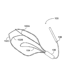
Reference is now made to Figures 1A, 1B and 1C, which are
io schematic
illustrations of an incising implant, generally referenced 100, for
creating incisions in the inner wall tissues of the prostatic urethra,
constructed and operative in accordance with an embodiment of the
disclosed technique. Figure 1A depicts the incising implant from a
top-view perspective (i.e., as would be seen in case the observer is
located distally to the implant), and Figures 1B and 1C depict the incision
implant from opposite isometric perspectives. Incising implant 100
includes three closed-shaped wires 102A, 102B and 102C (also referred
to herein below, together, as wires 102), an anchoring leaflet 104, a
proximal cap 106, and an extraction string 108.
The closed shape of each of wires 102 can roughly be divided
into a proximal section, a distal section and two longitudinal sections
extending between the proximal section and the distal section. For
example, the proximal section can be a U-shaped proximal end, from
which the longitudinal sections extend. The distal section is the section
connecting the longitudinal sections. Each of wires 102 is coupled with
adjacent ones of wires 102 on either side thereof. Specifically, the
longitudinal sections of each of wires 102 are coupled with longitudinal
sections of adjacent wires. For example, one longitudinal section of wire
102A is coupled with a longitudinal section of wire 102B, and the other
longitudinal section of wire 102A is coupled with a longitudinal section of
-7-
Date Recue/Date Received 2021-05-21
wire 102C. The other longitudinal section of wire 102B (not coupled with
102A) is coupled with the other longitudinal section of wire 102C (that is
also not coupled with 102A). Proximal cap 106 holds the proximal ends of
wires 102 together. Extraction string 108 is coupled with wires 102, or with
proximal cap 106.
The following paragraphs describe the use of incising implant
100. Thereafter, the components of incising implant 100 would be
elaborately described. Incising implant 100 is temporary implanted in the
prostatic urethra for creating longitudinal incisions in the inner wall
tissues
io of the prostatic urethra, thereby relieving urethra constriction.
Incising implant 100 is implanted by employing a sheath (not
shown) for inserting the implant into the urethra. Implant 100 is
compressed within the sheath such that the diameter of the circumference
of implant 100, illustrated by dotted circle 110, conforms to the inner
diameter of the sheath. Wires 102 are made of elastic material, such that
when released from the enfolding sheath they regain their original,
extended, shape (and the original circumference diameter of implant 100).
When positioned in the prostatic urethra, implant 100 is bound by the
inner diameter of the urethral walls surrounding it.
Wires 102 push against the surrounding tissues (i.e., apply a
radial outward force on the tissues). Over time, the force applied by wires
102 impairs the blood (and oxygen) supply to the tissues in contact with
wires 102, thereby inducing tissues necrosis and creating infarcted
incisions. Over time, the incisions become deeper until wires 102 reach
their full extent (i.e., until implant 100 regains its original circumference
diameter as illustrated by dotted circle 110). It is noted however, that
implant 100 can be removed before fully regaining its original shape, in
case the incisions are determined to be sufficiently deep to relieve the
constriction of the urethra.
-8-
Date Recue/Date Received 2021-05-21
Implant 100 is implanted such that wires 102 are aligned with
the longitudinal direction of the urethra. Therefore, wires 102 create
longitudinal incisions in the inner wall tissues of the prostatic urethra.
That
is, the longitudinal incisions are incisions running along the longitudinal
.. axis of the urethra. Put another way, longitudinal incisions are incisions
running along (and not across) the urinary passage.
The period of time required for creating incisions that are
sufficient to relieve urethra constriction depends on various factors, such
as the level of constriction, the materials of wires 102, the original fully
io extending shape of wires 102, and the like. Implant 100 can remain in
the
prostatic urethra for a predetermined period of time. Alternatively, implant
100 can remain implanted until the constriction is sufficiently relieved, as
determined by a physician, according to tests (e.g., observations of the
implant effect over time), or by the subject himself (e.g., according to what
the subject feels when urinating). For example, implant 100 can be
implanted for a time period ranging between a one hour and several
weeks. The incisions created by implant 100 are created over time without
causing pain or bleeding to the subject. After implant 100 is implanted, the
subject can be released and resume his regular lifestyle, without any
hindrances. After the required period of time, implant is removed from the
subject.
Incising implant 100 is implanted within the prostatic urethra to
relieve constriction of the urethra, caused for example by prostatic
enlargement. Implant 100 can be positioned in other, or in additional,
areas of the urinal passage, such as the bladder neck. Alternatively,
implant 100 can be implanted in any tubular organ that requires relief of a
constriction, such as tubular organs of the digestion system, blood
vessels, and the like.
Wires 102 (i.e., wires 102A, 102B and 102C) are closed-shaped
wires made of elastic material. The material of wires should be elastic
-9-
Date Recue/Date Received 2021-05-21
enough to allow wires to be compressed within a sheath, and to conform
to the inner diameter of the sheath, during insertion into the urethra. The
wires should regain their original, extended, shape (and the original
circumference diameter) once released from the sheath. Additionally, the
wires should be strong enough to apply a force on the surrounding tissues
to induce necrosis in the tissues (e.g., a force of 0.5 Newton), and thereby
to create infarcted longitudinal incisions. Wires 102 can be made, for
example, from Nickel Titanium alloy (Nitinol). Alternatively, implant 100 is
made of biodegradable materials, such that there is no need to remove
io implant 100 from the body of the patient.
The closed shape of wires can be roughly divided into three
sections, a proximal section, a middle section consisting of two
longitudinal sections, and a distal section (all not referenced). The
proximal section (or proximal end) is U-shaped. The longitudinal sections
extend from the arms of the U-shaped proximal end and are connected
via the distal section (or distal end). The distal section serves as a support
crosspiece connecting the longitudinal sections of the wire. Exemplary
closed shapes of the wires are illustrated in Figures 1A-1C, 2A-2C, 3, and
4.
The longitudinal sections of each of wires 102 are the sections
in contact with the surrounding tissues. That is, the longitudinal sections
are the sections pushing against the tissues for creating the incisions. The
longitudinal sections of each of wires 102 are coupled (i.e., adjoined) with
longitudinal sections of adjacent wires. For example, a first longitudinal
section of wire 102A is adjoined with a first longitudinal section of wire
102B, a second longitudinal section of wire 102A is adjoined with a first
longitudinal section of wire 102C, and a second longitudinal section of
wire 102B is adjoined with a second longitudinal section of wire 102C. In
this manner, the adjoined wires form together a supporting wire frame,
such that each closed-shape wire forms a face of the frame, and each
-10-
Date Recue/Date Received 2021-05-21
adjoined pair of longitudinal sections of adjacent wires forms an edge of
the frame.
When the longitudinal sections of wires 102 are pushed against
the surrounding tissues (i.e., as implant 100 tries to regain its original
shape while being bound by the urethra inner walls), the surrounding
tissues apply an opposite force on wires 102 in accordance with the third
law of Newton. Each of wires 102 is pushed against the adjacent wires to
which it is adjoined. The wire frame increases the structural stability of
implant 100 and allowing implant 100 to apply sufficient force for creating
io the incisions in the surrounding tissues. Thus, the wire frame obviates
the
need for an additional support element, such as a central support tube.
In the example set forth in Figures 1A-1C, wires 102 are
adjoined together by being wound (i.e., twisted) around each other. That
is, the first longitudinal section of wire 102A and the first longitudinal
section of wire 102B are wound around each other; the first second
longitudinal section of wire 102A and the first longitudinal section of wire
102C are wound around each other; and the second longitudinal section
of wire 102B and the second longitudinal section of wire 102C are wound
around each other. The twist coupling of wires 102 further provides
structural solidity to implant 100. Thereby, each of wires 102 can be made
thinner without compromising the robustness of implant 100. For example,
each of wires can be as thin as 0.5 millimeters (i.e., the cross section of
each of the wires is 0.5 millimeters).
The wounding of wires 102 can be achieved, for example, by
twisting the longitudinal sections around each other and thermally treating
implant 100 for stabilizing the winding. Wires 102 can be wound around
each other by being placed in a mold having rotating elements that grab
the longitudinal sections and wound them around each other.
In the example set forth in Figures 1A-C there are three wound
wires, each consisting of two longitudinal sections of two adjacent wires,
-11-
Date Recue/Date Received 2021-05-21
wound around each other. Thus, the wire frame has three longitudinal
edges creating three longitudinal incisions. In accordance with an
alternative embodiment of the disclosed technique, the implant can
include other numbers of closed-shaped wires, such as a single wire, two
wires, (for a wire frame of two longitudinal edges creating two longitudinal
incisions), four wires (for a wire frame of four longitudinal edges creating
four longitudinal incisions), five wires, and the like.
Proximal cap 106 is coupled with the proximal ends of wires 102
for coupling wires 102 together. Thereby, the wire frame is further
io strengthened. Put another way, proximal cap 106 helps to maintain the
structure of implant 102 (i.e., increases the structural stability) by further
adjoining wires 102 to each other.
In the example set forth in Figures 1A-1C, proximal cap 106
encases the proximal ends of wires 102. Thereby, proximal cap 106
shields tissues of the urethra from getting caught in the proximal ends of
wires 102. Additionally, proximal cap 106 serves to prevent wires 102 from
unwinding.
Proximal cap 106 can include a proximal non-round niche (e.g.,
niche 502 of Figures 4A and 4B). The non-round proximal niche of
proximal cap 106 is configured to receive a corresponding non-round pin,
and to transfer rotary motion of the pin to implant 100. Thereby, the user
can rotate implant 100 when implant is located within the bladder of the
subject, as would be detailed further herein below with reference to
Figures 4A-4B and 5A-5L.
Anchoring leaflet 104 serves as a one-way stopper allowing
implant to move from the bladder into the prostatic urethra and preventing
implant 100 from migrating back toward the bladder by being stuck
against one of the urethral sphincters. Leaflet 104 can be a wire leaflet
(e.g., as depicted in Figures 1A-1C), or any other form allowing it to slice
across the urethral sphincters in the proximal direction and preventing it to
-12-
Date Recue/Date Received 2021-05-21
slide across the urethral sphincters in the distal direction. For example,
leaflet can be bar-shaped. Leaflet can be coupled to implant elastically or
via an axis, or another coupling mechanism configured to enable leaflet to
serve as a one-way stopper for movement across the urethral sphincters.
Alternatively, other or additional anchoring elements can be employed for
anchoring implant in its place (moving in the proximal direction, the distal
direction, or both), such as barbs on wires 102.
Extraction string 108 enables the physician to extract implant
100. Specifically, the distal end of string 108 is coupled with implant 100,
io and the proximal end of string 108 extends outside of the body of the
subject. The physician can insert an extraction sheath into the urethra
along string 108 for enfolding implant 100. The physician can extract the
enfolded implant by pulling string 108. String 108 is strong enough for
pulling implant 100 without being torn (e.g., the thickness and materials of
string 108 allow pulling implant 100 via string 108). String 108 can be a
single strand or a woven bundle of strands for further fortifying it.
Incising implant 100 is deployed such that it does not extend
distally beyond the bladder neck of the subject (i.e., does not extend into
the bladder). Specifically, wires 102 do not come into contact with the
tissues of the bladder itself. Thereby, implant 100 does not irritate the
bladder of the patient.
In accordance with an embodiment of the disclosed technique,
the incising implant is colored in such a manner that enables the physician
to easily position it in order. For example, the wires of the implant are
color coded such that sections that should be positioned on top are
colored blue, and sections that should be positioned on the bottom are
colored white. The physician can observe the implant in the bladder via a
cystoscope, and rotate the implant to the desired orientation according to
the colors of the implant.
-13-
Date Recue/Date Received 2021-05-21
Reference is now made to Figures 2A, 2B and 2C, which are
schematic illustrations of an incising implant, generally referenced 100, for
creating incisions in the inner wall tissues of the prostatic urethra,
constructed and operative in accordance with another embodiment of the
disclosed technique. Figure 2A depicts the incising implant from an
isometric perspective, Figure 2B depicts the incising implant from a
top-view perspective, and Figure 2C depicts a closed-shaped wire of the
implant. Incising implant 200 includes three closed-shaped wires 202A,
202B and 202C (also referred to herein below, together, as wires 202),
io and an anchoring leaflet 204. The components of implant 200 are similar
to those of implant 100, and for the sake of brevity only the differences are
elaborated herein below.
The closed shape of each of wires 202 is depicted in Figure 2C.
The closed shape is truncated at the distal end thereof. That is, the distal
end of each of wires 202 is substantially perpendicular to the longitudinal
axis of implant 200. Thereby, the wires do not come into contact with the
tissues of the bladder, for avoiding bladder irritation.
Wires 202 are not wound around each other. Instead, wires 102
can be adjoined to one another (i.e., the longitudinal sections are adjoined
to longitudinal sections of adjacent wires) by various manners. For
example, the wires are welded together, glued together, or coupled by a
coupling mechanism or element (e.g., coupling thread binding the
longitudinal sections together).
In the example set forth in Figures 2A-2C (and in Figure 3
herein below), the incising implant is depicted without a proximal cap and
an extraction string. It is noted however, that the implant can include any
of the proximal cap, the extraction string, or both.
Reference is now made to Figure 3, which is a schematic
illustration of an incising implant, generally referenced 300, for creating
incisions in the inner wall tissues of the prostatic urethra, constructed and
-14-
Date Recue/Date Received 2021-05-21
operative in accordance with a further embodiment of the disclosed
technique. Incising implant 300 includes three closed-shaped wires 302A,
302B and 302C (also referred to herein below, together, as wires 302) and
an anchoring leaflet 306. The components of implant 300 are similar to
those of implant 100, and for the sake of brevity only the differences are
elaborated herein below. Implant 300 is depicted from a bottom-view
perspective (i.e., as seen by a proximally located observer). The closed
shape of wires 302 is triangular, such that together wires 302 form a
triangular-pyramid wire frame with the proximal ends of the wires forming
io the apex of the pyramid, and the distal ends forming the base of the
pyramid. The adjoined longitudinal sections of wires 302 form the
longitudinal edges of the triangular pyramid.
Reference is now made to Figures 4A and 4B, which are
schematic illustrations of a proximal niche, generally referenced 402, of a
proximal cap of an incising implant, constructed and operative in
accordance with yet another embodiment of the disclosed technique. The
proximal cap is detailed herein above with reference to proximal cap 106
of Figures 1A-1C. The niche has a non-round shape for allowing it to
transfer rotary motion from the corresponding pin inserted into the niche.
Thereby, the physician can rotate the incising implant from afar (e.g.,
when the implant is in the bladder). In the example set forth in Figure 4A,
the shape of niche 402 is rectangular, and in the example set forth in
Figure 4B, the shape of niche 402 is hexagonal. Alternatively, the niche
can have any shape allowing it to transfer rotary motion (i.e., rotations
around the central axis of the proximal cap), such as non-round shapes, a
slit, an array of niches (e.g., two holes), and the like.
Reference is now made to Figures 5A-5L, which are schematic
illustrations of a method for deploying and for extracting an incising
implant, operative in accordance with yet a further embodiment of the
disclosed technique. With reference to Figure 5A, an extraction string 510
-15-
Date Recue/Date Received 2021-05-21
(shown in Figure 5J) extends from the proximal end of implant 500.
Implant 500 includes a proximal cap having a non-round proximal niche
(both not shown). A guidewire 506 includes a distal head (i.e., distal pin),
which shape corresponds to the proximal niche of the proximal cap of
implant 500; and an inner channel (not show). The distal head of
guidewire 506 is inserted into the proximal niche of the proximal cap of
implant 500. Extraction string 510 runs through the inner channel of
guidewire 506. At the proximal end of extraction string 510 a proximal knot
512 (shown in Figure 5J) holds guidewire 506 attached to implant 500.
io Incising implant 500 is attached to the distal end of deployment sheath
502, such that guidewire 506 (and extraction string 510 running
therethrough) runs through sheath 502.
With reference to Figure 5B, a physician removes protective
cover 504 from implant 500, thereby implant 500 expands to its original
open configuration (as seen in any of drawings 1A-1C, 2A-2C, and 3). A
protective cover 504 keeps implant 500 sterile during storage prior to use.
With reference to Figure 5C, while holding guidewire 506, the physician
pushes sheath 502 over implant 500 thereby enfolding implant 500 within
sheath 502 for delivery into the urethra.
With reference to Figure 5D, the physician inserts a rigid
cystoscope 508 (e.g., size 20 French) into the urethra, for example, as in a
routine urethral catheterization procedure. With reference to Figure 5E,
the physician Inserts sheath 502, including compressed implant 500
therewithin, into cystoscope 508. The physician continues pushing implant
500 through cystoscope 508 by pushing guidewire 506, until implant 500
extends through the distal end of cystoscope 508. With reference to
Figure 5F, the physician removes sheath 502 from implant 500 and out of
cystoscope 508. With reference to Figure 5G, once released from sheath
502 and from cystoscope 508, implant 500 expands (i.e., regains its
original extended shape).
-16-
Date Recue/Date Received 2021-05-21
With reference to Figure 5H, the physician rotates implant 500
to the desired orientation by rotating guidewire 506 (and its distal head
inserted into the proximal niche of implant 500). An anchoring leaflet of
implant 500 (e.g., leaflet 104 of Figures 1A-1C) should be positioned
posteriorly. The wires of implant 500 can be color coded, such that the
sections that should be positioned on the top are colored, for example,
blue; and the sections that should be positioned on the bottom are
colored, for example, white. The physician rotates implant as detailed
further herein above with reference to proximal cap 106 of Figures 1A-1C,
io and proximal cap 400 of Figures 4A-4B.
With reference to Figure 51, while holding implant 500 in place
by using guide wire 506, the physician retracts cystoscope 508.
Thereafter, the physician pulls implant 500 via guidewire 506 until the
anchoring leaflet of implant 500 slides over a urethral sphincter and
implant is positioned within the prostatic urethra. With reference to Figure
5J, the physician cuts knot 512 at the proximal end of extraction string
510, and retracts guidewire 506 from the urethra.
Thereby, implant 500 is implanted within the prostatic urethra
and starts applying radial outward force on the surrounding tissues of the
inner walls of the urethra for creating longitudinal incisions. Implant 500 is
left within the prostatic urethra for a selected time period (e.g., ranging
between one hour and several weeks. Thereafter implant 500 is removed
as would be detailed below. Alternatively, implant 500 is made of
biodegradable materials and simply dissolves after a selected time period.
With reference to Figure 5K, the physician inserts cystoscope
508 through the urethra toward implant 500 over extraction string 510.
Alternatively, the physician can insert sheath 502 instead of cystoscope
508. The physician pushes cystoscope 508 until it enfolds implant 500.
With reference to Figure 5L, the physician extracts implant 500 enfolded
-17-
Date Recue/Date Received 2021-05-21
within cystoscope 508 by pulling implant via extraction string 510. Then,
the physician extracts cystoscope 508 from the urethra.
It will be appreciated by persons skilled in the art that the
disclosed technique is not limited to what has been particularly shown and
described hereinabove. Rather the scope of the disclosed technique is
defined only by the claims, which follow.
-18-
Date Recue/Date Received 2021-05-21