Note: Descriptions are shown in the official language in which they were submitted.
CA 02935211 2016-06-27
WO 2015/0959-17 PCT/BR201-
1/050054
1
MULTIPOTENT AND IMMUNOCOMPATIBLE STEM CELL CONCENTRATE
The present invention generally relates to a stem
cell concentrate isolated from mammalian vasculari zed
adipose tissue, obtained by the method described herein, as
well as pharmaceutical products comprising such a
concentrate, and use thereof in therapies for treating
mammalian diseases.
In the context of this document, the expression
"stem cells" is a synonym of multipotent stem cells,
mesenchymal stem cells, medicinal signaling cells, stromal
mesenchymal cells and adult stem cells.
Background of the Invention
It is known that stem cells are able to subdivide
for indefinite periods in culture, and to differentiate
into specialized cells. They can
also give rise to many
types of differentiated cells and be used to treat many
types of diseases.
The existence of adult stem cells in adipose tissue
of mammals is also known. Such cells have already been
isolated and cultivated to be used in cell therapies, such
as transplants.
Until now, however, the existing knowledge about
such stem cells has not solved many drawbacks. For example,
the low productivity of multiplication processes for such
cells compared to the needs of their use as well as the
little understanding concerning the stem cells' composition
used in cell therapies. In general, the fraction of stem
cells isolated from adipose tissue is named stromal
vascular fraction of adipose tissue that derives
perivascular cells and which also has paracrine activity
similar to mesenchymal stem cells.
Another existing problem is the large amount of
stem cells needed to achieve effectiveness in various
therapies, typically between 2 x 10⁶ and 1 x 10 cells per
kilogram of body weight of the patient.
Additionally, despite being muitipotent, stem cells
CA 02935211 2016-06-27
WO 2015/0959-17
PCT/BR2014/050054
2
derived from adipose tissue as known in the art have been
reported only in limited number of cases of use concerning
treatment of certain types of diseases and tissues.
Those facts limit the use and increase the cost of
stem cells derived from adipose tissue in cell therapies,
regardless of the fact that such cells are more abundant
than those obtained from other sources.
The present invention brings improvements and
solutions to the state of the art, as it provides:
- a high-productivity process for immunocompatible
multipotent stem cells from vascularized adipose tissue,
including culture and cell division;
- a synergic concentrate of immunocompatible and
multipotent cells that allows the use of a smaller amount
of stem cells in a variety of therapeutic treatments of
diseases in mammals (involving, for example,
osteoarticular, muscular, neurological, hematological,
dermatological, imuno-mediated urinary system diseases and
many others).
Brief description of the figures
- Figure 1 is a graph of increased hematocrit levels after
a single application of the cell concentrate of the
invention;
- Figure 2 is a graph of increased leukocyte levels after a
single application of the cell concentrate of the
invention;
- Figure 3 is a graph of increased platelet levels after a
single application of the cell concentrate of the
invention;
- Figure 4 is a graph of glycemic level variation in the
animal prior to treatment with the cell concentrate of the
invention;
- Figure 5 is a graph of glycemic level variation in the
animal after treatment with the cell concentrate of the
invention;
- Figure 6 is a graph of glycemic level variation in the
CA 02935211 2016-06-27
WO 2015/095947
PCT/BR2014/050054
3
animal after the second transplant with the cell
concentrate of the invention.
Description of the invention
In a first aspect, the present invention relates to
a multipotent and immunocompatible concentrate of stem
cells that secrete medicinal molecules, originated from
mammalian vascularized adipose tissue, comprising: immature
stem cells (ISC), mesenchymal stem cells (MSC) and
perivascular cells (pericytes).
The stem cell concentrate of the invention,
isolated from mammals, is immunopositive at least to
CD146+, a-SMA+, CD44+, CD73+, CD90+, CD105+, CD13+,
vimentin+, nestin+, Nanog+, Oct3/4+, Sox2+ markers, and
immunonegative at least to CD34-, CD45-, CD56-, CD144-,
CD14-, CD11b- and CD31- markers. Additionally, the stem
cell concentrate of the invention isolated from humans is
immunopositive to NG2+, PDGFRI3+, CD271+ (P75), CD29+,
S0X9+, SOX17+, FOX2+, CD140+ markers and immunonegative to
KLF4-.
Typically, the stem cell contents in the
concentrate of the invention are:
- ISC: 1- to 20
- MSC: 70,' to 100,,
- Pericytes: bE to 30-
The expression "stem cell contents" refers to the
percentages of cells that express the respective markers
(CTI, CTM or pericytes, respectively).
Particularly, without excluding any other
alternatives, the concentrates of the invention have about
5.- of ISC, 100ci. of MSC and 20 of pericytes, or values
close to those.
The number of immature stem cells (ISC),
mesenchymal stem cells (MSC) and pericytes obtained from
multiple niches of stem cells of adipose tissue varies
among patients and with the isolation method. Thus,
mixtures of concentrates obtained from 2 or more donors may
CA 02935211 2016-06-27
WO 21)15/095947
PCT/BR2014/050054
4
be used in order to reach average contents different from
the individual contents.
Alternatively, the stem cell
concentrate can be enriched with specifically cultured
pericytes.
The composition of cells in the concentrate of the
invention is synergic, since the combined effects of ISO,
MSC and pericytes unexpectedly results in the use of lesser
amounts of cells compared to the amounts of stem cells
currently used to achieve effectiveness in therapeutic
treatments, as described further on.
Variations of the stem cell composition in the
concentrate of the invention are also important since, as
depending on the treatment (of a disease, trauma, injury,
aesthetic-dermatological aspect, etc.), this composition
can be purified and/or enriched with cells that express
important markers for of the specific disease, trauma,
injury or other. A therapeutic composition can be adjusted
to the patient needs concerning the ratio of markers that
are expressed by the cells in the concentrate, as well as
regarding the dosage of stem cells, that is, a personalized
medicine is proposed.
The cell concentrate of the invention can be used
in the non-differentiated state of its stem cells, or the
cells can be induced to produce the precursors of specific
lineages (for example, glial cells) of interest for the
treatment, which can be purified and applied as a pure
population of precursors that are committed to a single
type cell differentiation for the benefit of the autocrine
effect of these cells. On the other hand, these cells have
paracrine effects generated by the cells when they are not
differentiated. For a more comprehensive therapeutic
effect, the composition of non-differentiated cells can be
used with the precursor cells.
These precursor cells have the ability to
differentiate, in vitro, at least into derivatives of two
germ layers: mesoderm (bone, cartiiage, muscle, etc.) and
CA 02935211 2016-06-27
WO 2015/095947
PCT/BR2014/050054
ectoderm (neural cells) . However their differentiation is
not limited only to mesoderm and ectoderm, since under
appropriate conditions, these cells can also produce the
cell types derived from endoderm, what characterizes them
5 as multipotent cells.
The cells contained in the concentrate of the
invention have similar phenotypic and functional
characteristics, but distinct in many aspects (cell
division, expression of some genes and proteins and
paracrine action), from those within the donor organism (in
vivo).
There are significant differences between the cell
lines in the concentrate of the invention and in the in
vivo adipose tissue. Post-differentiating levels of the
factors leptin and TMF-alpha secreted in cultures (in
vitro) of adipose tissue cells are much lower in comparison
with the in vivo tissue. In addition, an important early
regulator of the adipogenesis transcription, KLF4, has
significantly higher expression in vivo than in vitro.
Thus, it is not a matter of mere isolation of stem cells,
as they are found in the body tissue of the donor, but of
removing, treating and growing them specifically to attain
unpredictably improved characteristics, adequate to the
therapeutic purposes of the invention.
The stem cell concentrate of the invention does not
induce immune response, thus being able to be used in
autogenous transplants (the patient is the donor and the
receptor), allogeneic transpiants(the donor is related or
pertains to the same species of the patient) and xenogeneic
transplants (stem cells from different, unrelated species).
Moreover, the use of the inventive cell concentrate
in therapeutic treatments does not generate side effects,
such as those typically found in traditional therapeutic
treatments that employ synthetic compounds, whether in the
short or long term.
The stem cell concentrate of the invention is
CA 02935211 2016-06-27
WO 2015/095947
PCT/BR2014/050054
6
particularly suitable for cell therapies, particularly cell
transplants. The stem cell concentrate of the invention can
derive a wide spectrum of tissues derived from the three
embryonic germ layers (mesoderm, ectoderm and endoderm).
A particular aspect of the invention relates to the
standardization sought in its various aspects, to ensure
the success of the attained effects, whether with regard to
the large production of cells from vascularized adipose
tissue or to the potency and therapeutic efficiency
obtained with the use of smaller amounts of cells compared
to the state of the art.
An important aspect of the invention is the quality
of the donor, source of adipose tissue. In the veterinary
field, the isolation of stem cells from healthy young
animals provide effective results of high productivity,
potency and therapeutic efficiency in comparison with the
isolation of cells from adult and/or old animals, or
chronically ill, what provides less satisfactory effects.
Thus, for optimized embodiments of the invention, the
appropriate age of the donor mammal is between 20r. and 30,
of its full life cycle. Particularly for dogs and cats as
donors of vascularized adipose tissue, the optimal range of
donor age is 2-3 years. For horses, up to 6-7 years.
Still concerning the veterinary field, a suitable
body region of choice for adipose tissue harvesting biopsy
is the lateral region of the flank or the lateral surface
of the hind limb, but the harvest is not limited to these
regions, provided that the collection of vascularized
adipose tissue is performed. Advantageously, the harvesting
of adipose tissue can be performed during elective surgical
procedures in dogs and cats - for example, castration of
females (ovary salpingo hysterectomy) and pre-scrotal and
inguinal castration of males. This avoids the need for a
specific surgical procedure to harvest samples, avoiding
more pain and suffering of the patient. For enhanced
productivity of the process to obtain the stem cell
CA 02935211 2016-06-27
WO 2015/095947
PCT/BR2014/050054
7
concentrate of the invention, it is sufficient to harvest a
small fragment of vascularized visceral fat, for example,
between 0.1 and 0.3 cm3, such as a fragment of about 0.5 x
0.5 x 0.5 cm. The small size of the extracted sample also
facilitates, when necessary, the transportation of the
sample to the laboratory (for example, using a kit adapted
for this purpose) and its preservation until the beginning
of in vitro cell isolation.
The stem cell concentrate of the invention can be
cryopreserved, in a manner per se known to one skilled in
the art, to be stored for later use, when it will be
submitted to a thawing procedure, also known to the person
skilled in the art. For example, the stem cell concentrate
of the invention can be frozen in cryotubes in
concentrations of 1 x 10⁶ , 2 x 10⁶ or any other, and kept
at -80 C for 3 months, later transferred to liquid nitrogen
at -196 C. The culture medium used to cryopreserve cells
keeps them viable and preserves their differentiation
potential, for example, when comprised of 45 DMEM-h
(Dulbecco's Modified Eagle Medium - high glucose), of
fetal bovine serum and 101 of dimethyl sulfoxide.
With respect to the use of the invention in the
veterinary field, concerning the aspect of health of the
animal donor of adipose tissue, it is advantageous, to
ensure optimized effects of the invention, that the animals
(particularly dogs, cats and horses) are up to date with
vermifugation against nematodes and cestodes and already
vaccinated against:
- dogs: canine distemper virus, hepatitis, parovirus, 4
strains of ieptospirosis, parainfluenza, coranivirus,
parainfluenza, laryngotracheitis, giardia, cough of the
kennels and rabies;
- cats: rhinotracheitis virus, calicivirose, panleukopenia,
feline leukemia, chlamydiosis and rabies.
- horses: rabies virus, equine influenza,
encephalomyelitis.
CA 02935211 2016-06-27
WO 2015/095947
PCT/BR2014/050054
8
In another aspect, the invention relates to the use
of the stem cell concentrate of the invention in the
preparation of products useful in treating diseases,
trauma, injuries, aesthetic-dermatological aspects, etc.,
with stem cell transplantation. Without excluding any other
alternatives, examples of such products are
biopharmaceuticals, solutions, tablets,
ophthalmic
formulations, topical or mucosal formulations, etc.
In another aspect, the invention relates to
biopharmaceuticals, characterized in that they comprise
said stem cell concentrate and one or more biologically
acceptable ingredients, e.g., saline solution, biomaterials
(for example, polymeric biomaterials), growth factors (for
example, VEGF, TGF-beta, TTK), mono-nuclear cells,
platelet-rich plasma, fibrin, collagen membranes,
hydroxyapatite, bioactive molecules (for example, hormones
and mitogens). Particularly, a formulation for injection
contains at least the stem cell concentrate of the
invention and saline solution.
The invention takes advantage of particular aspects
of the process of obtaining the synergistic concentrate of
stem cells, for example, The isolation of a greater amount
of stem cells from multiple niches of adipose cells in the
same tissue fragment, which in explant culture releases in
vitro unlimited amounts of stem cells in culture. Thus, the
fragments of the adipose tissue initially plated in culture
bottles can be transferred to new culture bottles to
maintain the release of new stem cells.
Thus, without excluding any other alternative, an
advantageous process for obtaining the synergistic
composition of the stem cells of the invention comprises
the following steps:
A - obtaining a sample vascularized adipose tissue from a
mammal, particularly of a young and healthy mammal;
B - washing and cleaning of the tissue sample, for example,
with saline and antibiotics;
CA 02935211 2016-06-27
WO 2015/095947 PCT/BR2014/050054
9
C - performing fragmentation of the tissue sample;
- mild enzymatic digestion of the fragments of adipose
tissue to eliminate adipocytes, until inactivation of the
enzyme, for example, collagenase types I or IV;
E - centrifugation to obtain cells from the vascular
fraction of adipose tissue;
F - culturing of tissue explant - particularly 5 to 7
tissue fragments per 25 cm2 bottle of adherent material,
from 3 to 5 days, without changing the culture medium;
G - when reaching confluence between 70 and 90, dissociate
via enzymatic action the cell colonies that migrated out of
the tissue;
H - optionally, the tissue separated in step E can be
subjected to a new explant culture, starting a new step F;
I - culturing of isolated cells in step G, particularly up
to a maximum of 6 times for later use in cell therapy.
After step A, the harvested tissue sample is
cleaned and washed and may be placed in a container that
serves as a temporary storage medium for transportation to
another location for up to 48 hours. There are kits known
in the state of the art for this purpose.
As already mentioned, to ensure optimal results of
the invention, the vascularized adipose tissue mammal donor
is young and healthy. Advantageously, the age of the donor
should be up to a maximum of 30 of its total life cycle,
preferably up to 20',1,- The health should preferably be such
that essentially excludes any disease.
The process used allows avoiding any type of cell
fluid filtration, or avoids any type of cell selection
using size, granularity or specific markers, for example,
magnetic beads and specific antibodies.
One aspect of the enzymatic digestion of the
adipose tissue in step (D) is that vessels or fat from the
sample are not discarded after digestion. The enzyme is
suitable for smooth digestion of the connective tissue, for
example, type I or type IV coliagenase, such as the product
CA 02935211 2016-06-27
WO 2015/095947
PCT/BR2014/050054
GIBCOC,, available from Life Technologies, a USA company.
If it is desired to enrich the stem cell
concentrate of the invention with pericytes, the blood
vessels obtained from the smooth digestion are separated
5 from the sample of adipose tissue, and the explant growth
is performed only with them, according to steps F, G and I.
The pericytes then obtained can be added, in a later
moment, to the concentrate to be enriched.
In step F, it is preferred that the culture medium
10 is not changed during 3-5 days, and the equivalent amount
of the evaporated medium may be replenished. The
maintenance of the culture medium during this time provides
selection only of cells that can be adhered to the plastic
of the culture bottle, a characteristic of mesenchymal stem
cells. The confluence between 70 and 90 allows the
isolation of multiple colonies of fusiform cells with
mesenchymal morphology and substantially standardized size.
To favor good performance of the concentrate of the
invention, the growth medium in step F should be of high
quality, that is, standardized with minimal variation of
its pre-identified components. An appropriate example of
such medium, without excluding any other, is EMEM-h
(Dulbecco's Modified Eagle Medium-high glucose),
commercialized as GIBCOC', available from Life Technologies,
a US company, supplemented with 15 of fetal bovine serum
from Hyclone, a US company, supplemented with 15- fetal
bovine serum from the US company Hyclone, 1 of L-
glutamine, 11 of non-essential amino acids and 1, of a
penicillin/streptomycin mixture to neutralize collagenase,
all other ingredients from Life Technologies, a US company.
In step H, the fragment of the adipose tissue in
culture can be transferred multiple times in the culture
bottle, particularly up to 5 times, still releasing cells.
It is verified that, in phase I, the culture of
cells up to 6 passages is aimed at the safe use in cell
therapy, avoiding the induction of changes in the
CA 02935211 2016-06-27
WO 2015/0959-47
PCT/BR2014/050054
11
karyotype, proliferation or undifferentiated state. For
other applications in basic and applied science, the number
of passages can be higher than six passages.
In another aspect, the invention refers to stem
cell concentrates as described, characterized in that they
are for use in medical, veterinary or cosmetic therapy.
In another aspect, the invention relates to the use
of the stem cell concentrate of the invention in therapies
(autologous, heterologous and xenotransplants) in the
treatment of mammal diseases, such as joint, neurological,
hematological, ophthalmic and kidney (acute and chronic)
diseases, musculoskeletal disease, diabetes and acute
spinal cord injury.
Particularly in the veterinary field, pathology
therapies can still be cited, without excluding any other,
such as:
- hematopoietic diseases (medullary hypoplasia and aplasia)
- joint diseases (hip dysplasia, osteoarthritis,
degenerative process)
- bone fissures, gaps and fractures
- tendon and ligament laceration
- keratoconjunctivitis sicca, corneal ulcer
- neurological sequel derived from canine distemper virus
- myeloencephalitis derived from equine protozoal
- acute and chronic kidney disease
- masticatory myositis
- diabetes type I
- atopy
- purulent/necrotic skin lesion
- for dogs: neurologic sequel of canine distemper and other
neurodegenerative diseases, hip dysplasia, masticatory
myositis;
- for dogs and cats: medullary aplasia and hypoplasia; bone
fractures; degenerative osteoarticular diseases, acute
spinal cord injury, acute and chronic kidney disease, eye
diseases (for example, retinal degeneration,
CA 02935211 2016-06-27
WO 2015/095947
PCT/BR2014/050054
19
keratoconjunctivitis sicca), diabetes;
- for horses: tendon and ligament injuries, neurological
sequel caused by encephalomyelitis virus, osteoarthritis
and laminitis.
In another aspect, the present invention refers to
dosage forms for the treatment of mammal diseases and,
particularly, the amount of stem cell concentrate cells
varies, for example, between 1 x 10⁶ and 1 x 10- stem cells
Evaluation Of Tumorigenic Potential
The evaluation of tumorigenic potential of the cell
concentrate of the invention was made in a manner known to
one skilled in the art, for example, from information
available in Cell Cycle (2009). 15; 8(16), 2608-2612 and
"Teratoma formation: A tool for monitoring pluripotency in
stem cell research", in www.stembook.org.
A cell concentrate of the invention with 2 x 10⁶
cells was injected into the pelvic limb of 5 mice from nude
lineage. The mice were kept under normal conditions for 60
days. Then, they were euthanized and no neoformed mass in
the muscle or other body was observed.
Examples
Exemplary embodiments of the present invention are
described below. Such examples should be considered only as
illustrative of the particular embodiments of the
invention, and not in a restrictive sense, without imposing
limits of any type, beyond those comprised in the attached
claims.
Example I - Isolation of stem cells of the invention from
adipose tissue of dogs and cats.
In the examples given further on, obtaining
vascularized adipose tissue followed the procedure below.
Firstly, the region of choice, the lateral side of
the animal hind limb, was well washed with water and soap.
Then, the skin was shaved using a razor blade or knife and
the region was cleaned wir.h soap and water one more time.
In the sequence, the area was cleaned with gauze moistened
CA 02935211 2016-06-27
WO 2015/095947
PCT/BR2014/050054
13
in degerming chlorhexidine (solution of Riohex 2 de-
germing chlorhexidine, commercialized by Rioguimica, a
Brazilian company). This procedure was performed twice.
Then, 5 ml of lidocaine (anesthetic) was applied on the
harvesting region. This
region was firstly isolated with
surgical field cloth and then a 2-3 cm skin incision was
made with a scalpel. The fat fragment was removed,
measuring about 0.5 x 0.5 x 0.5 cm using a scalpel and an
ailis forceps, in order to ensure the presence of blood
vessels in the sample. After the harvesting, the site was
cleaned with sterile gauze to dry possible bleedings and
the skin sutured with thread, followed by disinfection
after suturing, for example, with rifocin.
The collected adipose tissue was firstly washed in
phosphate buffered saline solution (PBS) with 5,-
streptomycin/penicillin for removing blood, debris and
possible contaminants present in the sample. The washing
procedure was repeated 5 times. Then sterile gauze was used
to remove PBS in excess contained in the sample. The tissue
was transferred to a 60 mm Petri dish to perform tissue
fragmentation with a scalpel blade No. 22, thus obtaining
several smaller fragments. Then, 2 mL of 0.075 type I or
type IV collagenase (Gibco) were added to the culture
plate and then diluted in PBS at 37 C. The fragments with
collagenase were kept between 2 and 4 hours at 37 00 under
a 5- CO2 atmosphere. During incubation with collagenase,
the sample was homogenized at least every 2 hours with a
1000 uL graduated pipette.
After this period, 3 mL of the following culture
medium was added: DMEM supplemented with 15- fetal bovine
serum from Hyclone, a US company, supplemented with L-
glutamine, l non-essential amino acid and 1'
streptomycin/penicillin mix to neutralize collagenase (all
ingredients from Life Technologies, a US company). The
content was transferred to a 15 ml conical tube and
centrifuged during 5 minutes at 200 g. This procedure was
CA 02935211 2016-06-27
WO 2015/095947
PCT/BR2014/050054
14
performed twice for the complete inactivation of
collagenase. In the last centrifugation, the cells were re-
suspended in 1000 qL of the culture medium already
described and counted in a Neubauer chamber. Around 2 x 10⁶
cells were transferred to a 25 cm2 culture bottle prefilled
with 4,000 pL of the same culture medium.
The cells were kept at 37 C in 5 CO2 atmosphere.
After adhesion of the cells during a 3-5 day period, the
culture medium was discarded and a new one was added in the
bottle. After a period of 3 to 5 days, the first colonies
of fusiform fibroblastoid cells were formed (counted as
passage zero). After 7-9 days, the colonies reached
between 70 and 90 of confluence. At this stage, the first
cell replating was made by transferring the contents of a
25 cm2 bottle to four 25 cm2 bottles - starting the first
passage - which were then replated when a confluence
between 70 and 90 was reached, and were expanded ex vivo
until the 6-1' passage (from which a decline in the cell
proliferation rate was observed). These steps ensured the
isolation of the cell concentrate of the invention.
In the following examples, the cell concentrate of
the invention was used, obtained according to the procedure
above, containing approximately 5 ISC, 100- MSC and 20
pericytes obtained by mixing the concentrates from 3
distinct donors and was enriched with pericytes
specifically cultivated (the cell obtainment and isolation
was done with the procedure known by the person skilled in
the art).
In the examples with equines, the sample of adipose
tissue was removed from the tail or withers of the animals.
Example 2 - Canine Distemper
2.1 Animals used
The dogs used in this experiment were males and
females of different races, with ages ranging from 1 to 6
years. All animals presented clinical neurological symptoms
caused by invasion of canine distemper virus in the central
CA 02935211 2016-06-27
WO 2015/095947 PCT/BR2014/050054
nervous system, such as myoclonus, paraplegia, tetraplegia,
paraparesis, seizure and inability to stay on feet and
walk, according to Table I below. All dogs had been
previously subjected to conventional treatment against the
5 symptoms manifested in the viremic phase (digestive and
respiratory) and presented no gastrointestinal and
respiratory clinical symptoms. Transplants were performed
with a time interval of 30 days between each application.
2.2 Pre-transplant clinical evaluation
10 Patient anamnesis was performed and supplementary
blood tests, chest x-rays and abdominal ultrasound were
conducted to rule out the pre-existence of neoplasms.
2.3 Transplant
Transplant of the concentrate of the invention was
15 carried out intravenously in patients with neurologic
sequel of canine distemper. The cell number varied with the
animal weight (table I). Patients received on average three
transplants of cell concentrate of the invention at an
interval of 30 days between each transplant. This was
accomplished through intravenous access with a catheter and
the animal was maintained in fluid therapy with 0.9'1', saline
solution. Previously preserved cells of the invention were
thawed according to the appropriate procedure, and were re-
suspended into 2 mL of 0.9' physiological solution. The
infusion of stem cells was slowly carried out.
Table I - Neurological clinical signals of the patients
1
0
,u
-
..) -
7, a) w
0
0 in
0 a u
0
g F, ft
z
E 0 0 1,4
-
0
_p
z
1 mongrel 24 M 2 cpadriplegia 3 4x10⁶
2 mongrel 8 7 F 4 paraplegia 3 4x10'
3 mongrel 15 3 F 3 paraplegia 3 4H10'
4 doodle 32 4 F 3 Inability to 3 4x1Y
stand and, alien
CA 02935211 2016-06-27
WO 2015/095947 PCT/BR2014/050054
16
sugoorted,
presented severe
ataxia and
oaraoaresis of
both members
mongrel 21 25 M 5 Inability to 3 6x10'
stand and, when
su000rted,
presented severe
ataxia and
oaraoaresis of
both members
6 'mongrel 36 3 M 29 Inability to 2
4x10(
stand and, when
supported,
presented severe
ataxia and
oaraoaresis of
botn members
2.4 Post-transplant clinical evaluation
After treatment, the animals were monitored for 1
hour by the veterinarian to check for possible anaphylactic
reactions. None of the patients presented symptoms of
5 rejection after transplantation of the cell concentrate of
the invention. Clinical returns were carried out in 48
hours, 7 days and 21 days after application of the stem
cells and the time interval between two applications was 30
days. Most of the animals presented reduction of
neurological clinical signs and were able to walk again -
the less positive results occurred with the older animals
and with those with more time on sequels.
Table II below shows the evolution of reduction of
neurological sequel.
Table II. Evolution of reduction of neurologic sequel of
canine distemper after treatment with cells of the
invention concentrate.
dog DO D1 D2 D3
1 quadriplegia Unable to Unable to Unable to
stand and stand and stand and
CA 02935211 2016-06-27
WO 2015/095947
PCT/BR2014/050054
17
support weight support support
and, when weight and, weight and,
sustained, when when
presented mild sustained, sustained,
movements of presented presented
FL mild mild
Ataxia and movements movements
paresis. of FL and of FL and
No intentional HL. HL.
movement Ataxia and Ataxia and
HL paralysis paresis. paresis.
Paralysis Stand and Stand and Normal gait
support support
weight, weight,
minimum gait, minimum
ataxia and gait,
paraparesis ataxia and
paraparesis
3 quadriplegia Unable to Stands and Normal gait
stand and supports
support weight,
weight. normal
mild ataxia gait.
and Stands and
paraparesis of supports
of FL and HL. the body's
moderate weight.
ataxia and Ataxic gait
paresis of HL. and minimal
paraparesis
4 Unable to Unable to Stands and Normal gait
stand and stand and supports
support support weight weight.
weight and, and, when Mild ataxia
CA 02935211 2016-06-27
WO 2015/095947
PCT/BR2014/050054
18
when sustained, and paresis
sustained, presented mild of FL and
presented movements of HL.
mild FL and HL
movements of ataxia and
FL and HL. paresis.
Severe ataxia
and paresis.
Unable to Unable to Unable to Unable to
stand and stand and stand and, stand
and,
support support weight when when
weight and, and, when sustained, sustained,
when sustained, showed mild showed mild
sustained, presented mild ataxia and ataxia and
presented movements of paresis FL
paresis of
mild FL and HL. and HL FL and HL.
movements of Moderate
FL and HL. ataxia and
Severe ataxia paresis.
and paresis.
6 Quadriplegia Unable to Unable to
Unable to
stand and stand and stand and,
support weight when when
and, when sustained, sustained,
sustained, Presented showed mild
presented mild mild ataxia ataxia and
movements of and paresis
paresis of
FL and HL. FL and HL. FL and HL.
Moderate
ataxia and
paresis.
DO - neurological symptoms before the transplant. ls:
transplant.
D1 - neurological symptoms after the ls- transplant (30
days)
CA 02935211 2016-06-27
WO 2015/095947 PCT/BR2014/050054
19
D2 - neurological symptoms after the 2'd transplant (60
days)
D3 - neurological symptoms after the 3rd transplant (90
days)
fore limb= FL
hind limb=HL
Example 3 - Hypoplasia and aplasia of bone marrow
3.1 Animals used
A 30 Kg one-year old male Labrador retriever
presented weakness, loss of appetite and severe anemia,
according to the hemogram. The dog had been previously
submitted to monthly blood transfusions for a period of 5
months and was being treated with exogenous erythropoietin
in an attempt to keep the hematocrit levels acceptable for
a canine species.
3.2 Pre-transplant clinical evaluation
Dog anamnesis was performed and supplementary
blood tests, chest x-rays and abdominal ultrasound were
conducted to rule out the pre-existence of neoplasms. The
medullar biopsy diagnosed erythroid and granulocytic
hypoplasia, and megakaryocytic aplasia.
3.3 Transplant
A single transplant with 8 x 10⁶ cells of the
invention concentrate was performed intraosseously in the
femur bone. The animal was initially sedated with 4 mg/Kg
intramuscular tramadol hydrochloride (a pre-anesthetic
medication), followed by the induction to the effect of
intravenous 8 mg/kg propophol (2,6-diisopropylphenol),
maintained with isoflurane (2-chloro-2-(difluoromethoxy)-
1.1.1-trifluoro-ethane). Then, a broad trichotomy of
lumbosacral region and antisepsis with 2 chlorhexidine
were made. Then, the access to the femoral crest was
performed with a specific needle for spinal biopsy.
Cryopreserved cells of the invention were thawed, re-
suspended in 1 ml of 0.9 physiological solution and the
infusion of stem cells was carried out slowly.
CA 02935211 2016-06-27
WO 2015/095947
PCT/BR2014/050054
3.4 Post-transplant clinical evaluation
Clinical returns were carried out with peripheral
blood collection for hematocrit control and general
physical examination, at intervals of around 30 days. The
5 patient did not express symptoms of rejection after
transplant with the cells of the invention. After a single
transplant with the concentrate cells of the invention, the
dog did not need to receive blood transfusion for one year
- after this year, the aspirate examination of the bone
10 marrow indicated a hematopoietically active bone marrow
with a erythroid series slightly augmented, with normal
morphology, complete and orderly maturation and
predominance of mature forms, whereas the myeloid series
was slightly decreased, with normal morphology, complete
15 and orderly maturation and predominance of mature forms.
The animal remained well without receiving transfusion
since then (until the priority filing date of this patent
application).
The graphics of Figures 1, 2 and 3 present a
20 significant increase of hematocrit levels (figure 1),
leukocytes (figure 2) and platelets (figure 3) after a
single application of the inventive cells. The blood tests
were carried out with 30 day intervals.
Example 4 - Type I Diabetes
4.1 Animal used
A 2-year 8 Kg female dog with high levels of blood
glucose and earlier diagnosed of type I diabetes.
4.2 Pre-transplant clinical evaluation
Patient anamnesis was performed and supplementary
blood tests, chest x-rays and abdominal ultrasound were
conducted to rule out the pre-existence of neoplasms. The
glucose test was performed twice daily, confirming type I
diabetes. To reduce blood glucose levels, 3 to 6 units of
insulin were being administered to the patient daily.
4.3 Transplant
Four transplants of 4 x 10⁶ cells of the inventive
CA 02935211 2016-06-27
WO 2015/0959-17
PCT/BR201-1/050054
21
concentrate were performed intravenously at 30 days
intervals between each transplant. For this, the
cryopreserved cells of the invention were thawed and re-
suspended in 1 mL of 0.9-, physiological solution.
Intravenous infusion of stem cells was carried out slowly.
4.4 Post-transplant clinical evaluation
After the transplant, the glucose test was
conducted twice daily for 60 days in order to track the
reduction of glucose level. The results showed a reduction
in blood glucose levels and insulin dose reduction.
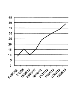
In figures 4, 5 and 6, letter "T" means date of the
transplant.
Figure 4 is a graph of the variation of glucose
level of the animal before the treatment with stem cells.
The gray color indicates the day that the patient did not
receive insulin, the dark gray color indicates that the
patient received 3 units of insulin per day, the white
color indicates that the patient received 6 units of
insulin once a day.
Figure 5 is a graph of the variation of glucose
level of the animal after the treatment with the stem
cells. The reductions in the number of days without insulin
and of the dose were observed. The light gray color
indicates the day in which the patient did not need to
receive insulin, the dark gray color indicates that the
patient received 3 units of insulin, the white color
indicates that the patient received 6 units of insulin once
per day. It is noted that the number of days without
insulin increased, as well as the reduction of the number
of days when the dog needed to receive 6 units of insulin.
Figure 6 is a graph of the variation of glucose
level of the animal after the second transplant with the
stem cells. A great reduction of the number of days without
administering insulin and of the dose is noted. The gray
color indicates the day when the patient did not receive
insulin, the white color indicates when the patient
CA 02935211 2016-06-27
WO 2015/(1959-17
PCT/BR2014/050054
22
received 3 units of insulin.
It is noted that after the second transplant, there
was a reduction of the daily dose of insulin from 6 to 3
units per kilo. Additionally, the interval of days without
having to take insulin increased.
Example 5 - Masticatory myositis
5.1 Animal used
A 11 year male labrador retriever dog showed
inability to open the mouth, along with swelling and
difficulty to feed.
5.2 Pre-transplant clinical evaluation
Patient anamnesis was performed and supplementary
blood tests, chest x-rays and abdominal ultrasound were
conducted to rule out the pre-existence of neoplasms. The
biopsy of masticatory muscle indicated myositis of the
masticatory muscles. The jaw opening of the animal was of
2.7 cm. The dog had been medicated with corticosteroids,
but did not adapt to this treatment.
5.3 Transplant
Three transplants of 8 x 10b stem cells of the
Inventive concentrate were performed intravenously with 30
day intervals between each transplant. For this,
cryopreserved cells of the invention were thawed, re-
suspended in 1 mL of 0.9- physiological solution, and
intravenous infusion of stem cells was performed slowly.
5.4 Post-transplant clinical evaluation
After the transplant the animal showed more
disposition, and was able of feed alone and to use the
mouth to catch objects, what was previously not possible.
The jaw opening capacity was amplified to 4.7 cm.
Example 6 - Tendon Injury of Athlete Horses
6.1 Animals used
Three male horses with ages ranging from 16 to 28
years showed focal lesions in the superficial digital
extensor tendon. The lesions were diagnosed with an
ultrasound machine, and classified according to the loss of
CA 02935211 2016-06-27
WO 2015/095947
PCT/BR2014/050054
23
linear pattern of the tendon fiber, as proposed by Nixon et
al., (Nixon AJ, Goodrich LR, Scimeca DM, Witte TI-I, Schnabel
LV, Watts AE, et al. Gene therapy in musculoskeletal
repair. Ann N Y Acad Sci 2007; 1117:310-327) in a scale
from 1 to 4. The loss
of linear pattern of the tendon
fiber corresponding to 10 to 20 was classified as grade 1,
while injuries between 25 to 50': were classified as grade
2.
In the table below, animals treated with the cell
concentrates of the invention are cited, as well as their
respective classifications of the scale of linear pattern
of the tendon fiber and, according to the tendon lesion,
the number of cells used in the transplant.
Table 1. Animals submitted to transplant with the stem cell
concentrate of the invention (according to example 1, with
equines being the donors of adipose tissue), percentage of
injury and No. of transplanted cells.
Animal * of injury Total number of
transplanted cells
1 10 6H10'
2 20 8H10'
3 30 8x10'
6.2 Transplant
The animals were subjected to trichotomy followed
by antisepsis with povidone-iodine, for further transplant
of the cell concentrate of the invention. The application
route of the cell concentrated was at the site of injury,
made possible with an ultrasound which allowed the exact
identification of the injury. The cell concentrate of the
invention (horses were the adipose tissue donors) was
applied with the aid of a 40 x 12 needle and 3 mL syringe.
A single cell transplant was performed.
6.3 Clinical evaluation of the injury after transplant
After 15 days, an evaluation of the stem cell
transplant of the inventive concentrate was performed on
CA 02935211 2016-06-27
WO 2015/095947
PCT/BR2014/050054
24
the injured tendon site, with ultrasound.
A reduction of the injury extension of the tendon
tissue and the arrangement of collagen fibers were
verified. A significant improvement in the linear pattern
of collagen fibers was observed.
Example 7. Acute and chronic kidney disease
7.1 Animal used
A 13-year old female pinscher dog with a history of
poor appetite and low weight. The biochemical examination
verified high levels of urea (86 mg/dL) and creatinine 2
(54 mg/dL). The patient was receiving fluid therapy twice a
day and 2 mL of oral serum, with special diet for kidney
patient with Royal renal (pâté and feed) and white meat
(chicken). The drug protocol used was: Ketosteril (amino
acids and analogues, marketed by Fresenius, a Brazilian
laboratory), Glutamax (glutamine supplement, marketed by
Vitafor, a Brazilian laboratory), omeprazole, bromopride
and Hemolitan (vitamin supplement marketed by Vetnil, a
Brazilian laboratory).
7.2 Pre-transplant clinical evaluation
Patient anamnesis was performed and supplementary
blood tests, chest x-rays and abdominal ultrasound were
conducted to rule out the pre-existence of neoplasms. The
biochemical test and urinalysis were also made, indicating
chronic kidney disease.
7.3 Transplant
Two transplants of 2 x 10 cells of the inventive
concentrate were performed intravenously with 30-day
intervals. Cryopreserved cells of the invention were thawed
and re-suspended into 2 mL of 0.9 physiological solution.
The infusion of cells of the invention was slowly carried
out. The patient kept the conventional treatment for the
disease during the treatment with stem cells.
7.4 Post-transplant clinical evaluation
Clinical returns were carried out in 48 hours, 7
days and 21 days after the transplant with the stem cell
CA 02935211 2016-06-27
WO 2015/0959-17
PCT/BR201-1/050054
concentrate of the invention, and the interval between
applications was 30 days.
The patient did not express
symptoms of rejection after cell transplant of the
invention. The biochemical tests and urinalysis conducted
5 showed a reduction in blood levels of urea and creatinine,
ionized calcium and the increase in hematocrit, as shown in
the table below.
Table. Values of renal function before and after the
transplant.
Dates DO D1 !D1 May D2 Jun June Augu Sept Typic
Apri Apri Apri 17"h June e 18m st embe al
1 1 1 2012 13'11 14- 2012 23th r value
12"-' 13"h 13'4' 2012 h 2012 21s' s
2012 2012 2012 201 2012
2
No. of 2H10 2H10"
transp
lanted
cells
Urea 86 78 51 34 10-
mg/dL 50
mg/d
Creatin 2.54 2.12 1.8 1.7 0.5-
in 2 1.5
mg/dL mg/d
Hemogr 25 43 38 45.4 50-
am (-) 53-
Ionize 6.08 6.13 4.96 4.5-
5.7
calcium mg/d
mg/di,
Potassi 5.6 4.3 5.1 3.7-
u.m 5.8
mEg/dL mEq/d
Sodium 155.0 150 141-
mEg/dL 0 153
mEq/d
Phosph 5.7 3.21 5.3 2.2-
orous 5,5
mg/do mg/d
Albumin 3.15 3.4
10 DO - laboratory data before the l transplant.
CA 02935211 2016-06-27
WO 2015/0959-17 PCT/BR20
i 4/050054
26
D1 - laboratory data after the 1' transplant (30 days)
D2 - laboratory data after the 2'd transplant (60 days)
8. Treatment of Spinal Disc Extrusion
8.1 Animal used
9 year-old 39 kg male Doberman dog showed absence
of deep pain and paraplegia.
8.2 Pre-treatment clinical evaluation
Patient anamnesis was performed and supplementary
blood tests, chest x-rays and abdominal ultrasound were
conducted to rule out the pre-existence of neoplasms, and
it was observed, by magnetic resonance imaging, the spinal
disc extrusion in the toraco-lumbar region at T12-13 T13-1.
8.3 Transplant
After 21 days, the spinal disc extrusion at the
toraco-lumbar region was verified. During this period (21
days), a single transplant of 4 x 10⁶ stem cells of the
inventive concentrate was performed. Cryopreserved cells of
the invention were thawed and re-suspended into 1 mL of
0.9 physiological solution. The infusion of stem cells was
slowly carried out into the epidural space using a 3 mL
syringe a 20 x 5.5 needle. After stem cell therapy, tramal
and dipyrone were administered during 5 days and cephalexin
was administered during 14 days. On the 10th day after
transplant, the animal underwent physiotherapy for a period
of 60 days, three times a week, and acupuncture once a
week.
8.4 Post-transplant clinical evaluation
Clinical returns were carried out in 48 hours, 7
days and 21 days after the transplant with the stem cell
concentrate of the invention. The patient did not express
symptoms of rejection after the transplant. After 30 days
from transplant, the animal could stand with some
difficulty walking and, after 60 days, the animal was
walking normally.
Example 9. Tendon laceration
9.1 Animal used
CA 02935211 2016-06-27
WO 2015/095947
PCT/BR2014/050054
27
A 42 kilo female german shepherd dog, with injury
on left foot caused by trauma. The animal had difficulties
in locomotion due to inability to support the left paw on
the ground.
9.2 Pre-transplant clinical evaluation
The rupture of the common calcaneal tendon was
observed by ultrasound.
9.3 Transplant
28 days after the traumatic rupture of the tendon,
the tendon suture was performed and, at the time of
surgery, the concentrate of the invention was applied
having 2 x 10⁶ stem cells of the inventive concentrate.
9.4 Post-transplant clinical evaluation
After 60 days an ultrasound was performed and the
complete tendinous fibers organization was observed, the
animal recovering the motor activity.
10. Keratoconjunctivitis sicca
10.1 Animal used
A two- year old male crossbreed dog, which featured
production deficiency of tear in the left eye, during 6
months.
10.2 Pre-treatment clinical evaluation
The patient anamnesis was performed and the
Schirmer tear test disclosed a level of tear production of
5 mm.
10.3 Transplant
A single transplant of the stem cell concentrate of
the invention was conducted subconjuntivally in the main
lacrimal gland and in the lacrimal gland of the third
eyelid. Cryopreserved cells of the invention were thawed
and re-suspended into 0.5 uL of 0.9, physiological
solution, 0.3 pL was transfused in the main gland and 0.2
pl in the gland of the third eyelid of the patient. During
the treatment, only artificial tears in the first 7 days
after the beginning of stem cell transplant were used.
10.4 Post-transplant clinical evaluation
CA 02935211 2016-06-27
WO 2015/095947 PCT/B
R2014/050054
28
The clinical returns were carried out 7, 14 and 21
days after the transplant with the cells of the invention.
The patient did not express symptoms of rejection after the
cell concentrate transplant of the invention. After 7 days,
the patient did not use artificial tears anymore and the
Schirmer tear test was 10 mm, and after 14 and 21 days, it
was 15 mm, which are considered normal values.
11. Ulcerative Keratitis
11.1 Animal used
A 8-year old female Schnauzer dog presented a great
width of bullous ulcerative keratitis in the right eye.
11.2 Pre-treatment clinical evaluation
The patient's anamnesis was performed, and on
inspection, the bullous ulceratitis of large span was
observed in the right eye. Before the treatment with stem
cells, the patient was treated with antibiotics during 7
days.
11.3 Transplant
Five transplants of stem cell concentrate of the
invention were performed along 5 days, directly on the
ulcerated cornea, with 1 x 10⁶ cells re-suspended in .5 pL
of saline solution. Cryopreserved cells of the invention
were thawed before use.
11.4 Post-transplant clinical evaluation
After a month of treatment with the stem cell
concentrate of the invention, the cornea transparency was
observed. When performing the visual acuity test (the test
threat) the animal manifested reaction, and therefore, it
was considered positive to the test.
Example 12. Neurologic
sequel derived from equine
protozoal myeloencephalitis (EPM)
12.1 Animal used
A 10-year-old female of the equine species,
undetermined race, positive serology for Sarcocystis
neuroma (title>80) with clinical signs of motor
discoordination , loss of balance and gait with compromise
CA 02935211 2016-06-27
WO 2015/0959-17
PCT/BR2014/050054
29
of mainly the hind limbs.
12.2 Pre-treatment clinical evaluation
For 6 months the patient presented motor
discoordination, loss of balance and gait, with mainly the
hind limbs compromised (severe gait ataxia). The viremic
phase of the PPM was not in course (sorologic examination
for Sarcocystis neuroma - title <80)
12.3 Transplant
A total of 8 x 10 cells was transplanted via
epidural (sacro-coccygeal epidural space)in the horse with
the neurological sequel. The cells were previously thawed
and re-suspended into 2 mL of physiological solution and
then transplanted via intrathecal administration.
12.4 Post-transplant clinical evaluation
Twenty days after the first transplant the animal
had a reduction on of the clinical signs of ataxia, motor
discoordination and loss of balance, with marching ability
recovery. After treatment the animal presented only light
gait ataxia.
Example 13. Atopy
13.1 Animal used
7 year-old male golden retriever, diagnosed with
atopy at the age of one. The animal was treated with
cyclosporine, corticoid and hypoallergenic topic
treatments, with no effective response after diagnosis.
13.2 Pre-treatment clinical evaluation
The dog presented severe clinical signs of atopy,
such as: pruritus level 9, pyoderma, otitis, and mutilation
of the elbows due to intense pruritus.
13.4 Transplant
Three transplants with 8 x 10 cells were performed
intravenously, with 30-day intervals. The cells were re-
suspended in 3m1 of physiological solution and injected
with the aid of a 3 ml syringe with a 24x12 needle.
13.4 Post-transplant clinical evaluation
The animal presented progressive improvement after
CA 02935211 2016-06-27
WO 2015/0959-17
PCT/BR2014/050054
the transplants. 30 days after the initial transplant the
animal showed pruritus reduction. The other symptoms showed
reduction after the 3"I application (pruritus, pyoderma,
otitis). After the end of the treatment the animal
5 presented controlled atopy symptoms, without medication,
with use of allergenic shampoo.
14. Treatment of cutaneous wounds
14.1 Animal used
A dog 19 month-old 12 kg mongrel, female presenting tissue
10 necrosis in the lower dorsal region and left lateral flank,
with high level of pain.
14.2 Pre-treatment clinical evaluation
Clinical diagnostic of necrotizing cutaneous wound,
with abundant purulent secretion and significant tissue
15 loss. Animal had been treated for 40 days with antibiotics
and topical skin healing solutions, without effective
results.
14.3 Transplant
Five consecutive cell transplants were performed.
20 The first transplant was performed via three different
ways: endovenous, local injection and topical (direct
application on the skin would). For this treatment a total
of 8 x 10⁶ cells of the invention were used, divided as
follows: 4 x 10⁶ cells were applied intravenously, 4 x 10⁶
25 cells equally divided between local injection and topical
application.
14.4 Post-transplant clinical evaluation
The animal had significant improvement after the
treatment with the invention celis. Initially one observed
30 reduction of the inflammatory process (6 days) and of the
tissue necrosis (15 days). The reepitalization process was
accelerated and after 60 days all affected cutaneous area
was reepitelized (neoformed cutaneous tissue).
With the aid of the teachings and examples
disclosed herein, the person skilled in the art can carry
out the invention in equivalent forms, i.e. not expressly
CA 02935211 2016-06-27
WO 2015/095947
PCT/BR2014/050054
31
described, but whose functions and results are of the same
nature as those of the invention, therefore within the
scope of the appended claims.