Note: Descriptions are shown in the official language in which they were submitted.
CA 02935506 2016-06-29
WO 2015/124159
PCT/DK2015/050035
1
A SET COMPRISING A SURGICAL INSTRUMENT
TECHNICAL FIELD
The invention relates to a set comprising a surgical instrument suitable for
use
in a minimal invasive surgery and/or for training the handling of such
surgical
instrument as well as a surgical system, a training kit, a method of training
and
performing minimal invasive surgery.
BACKGROUND ART
Minimal invasive surgery has been used increasingly in the last years due to
the benefits compared to conventional open surgery as it reduces the trauma
to the patient tissue, leaves smaller scars, minimizes post-surgical pain and
enables a faster recovery of the patient.
For example, in laparoscopic surgery (a form of minimal invasive surgery) the
surgeon accesses a body cavity, such as the abdominal or pelvic cavity,
through a series of small incisions. A laparoscope is inserted through an
incision, and conventionally connected to a monitor, enabling the surgeon to
see the inside of the abdominal or pelvic cavity. In order to perform the
surgery procedure, surgical instruments are inserted through incisions. In
addition, the body cavity around the surgical site is inflated with a fluid,
preferably gas e.g. carbon dioxide in order to create an 'air space within the
cavity for the surgeon to view the surgical site and move the laparoscopic
instruments.
In conventional open surgery the surgeon can use the normal visual-motor
relations, wherein the motor control is based on visual perception, such that
a
desired movement of a surgical instrument can be performed on basis of
vision. In other words, during conventional open surgery the normal link
between the visual perception and the motor system is conserved. However,
when performing minimal invasive surgery the surgeon has an indirect vision
of the surgical field which results in dissociation of the visual perception
and
the motor system of the surgeon. Consequently, the surgeon needs to acquire
CA 02935506 2016-06-29
WO 2015/124159
PCT/DK2015/050035
2
new skills in order to correctly connect his or hers visual perception and
motor
system (hand-eye coordination) during minimal invasive surgery.
Visual perception is the ability to interpret the surrounding environment by
processing information obtained by use of the eyes, in the present case the
surrounding environment can be the inside of a body cavity, such as the
abdominal or pelvic cavity.
The motor system of a person is the complex system which, among other
things, controls voluntary movements, enabling a surgeon to move body parts,
such as a hand and fingers, to control the movement of a surgical instrument
inside a body cavity.
Further, the remote vision of the surgical field is normally displayed on a
monitor in two dimensions whereas the surgical instrument is manipulated in
three dimensions; this results in a poor spatial and depth perception which
makes it even harder for a surgeon to acquire the new abilities for connecting
visual perception of the remote vision (in two-dimensions) and motor system
moving the surgical tools (in three-dimensions).
In addition, if the surgical tools are controlled via a surgical robot the
normal
three dimensional motor behaviors of the surgeon are processed and
changed by the robot, which makes it more difficult for the surgeon to
correctly
connect his or hers visual perception and motor system during minimal
invasive surgery.
Training in minimal invasive surgery is normally performed after basic
surgical
training and is based on apprenticeship where the skills are obtained in
direct
clinical surgery supervised by an experienced surgeon. This training method
poses considerable risk to the patient and requires a substantially amount of
time from the experienced surgeon.
Therefore, the use of simulators, for example laparoscopic simulators, is
preferred in order for an inexperienced surgeon to learn the basic skills
before
starting to do clinical surgery. Among the most important skills needed to
CA 02935506 2016-06-29
WO 2015/124159
PCT/DK2015/050035
3
master is; the ability to transform the information received by indirect
vision
into a three dimensional understanding.
Different approaches for providing an improved depth perception have been
provided for example as described in EP 2630915 in which a light instrument
for use in minimal invasive surgery is described. The light instrument
includes
an elongate tubular member and a metrology system mounted on the
elongate tubular member. The metrology system includes a mask, a zoom
lens assembly and a light element arranged such that the light element
propagates light beams through the mask and the zoom lens assembly to
project the patterns of the mask onto the surgical site of interest to provide
markings as references used for measuring by the surgeon.
US 2013/0296712 describes an apparatus for determining endoscopic
dimensional measurements, including a light source for projecting light
patterns on a surgical sight including shapes with actual dimensional
measurements and fiducials, and means for analyzing the projecting light
patterns on the surgical sight by comparing the actual dimensional
measurements of the projected light patterns to the surgical site.
WO 2013/163391 describes at system for generating an image which the
surgeon can use for measure the size of or distance between structures in the
surgical field by using an invisible light for marking a pattern to the
surgical
field.
The system comprises a first camera; a second camera; a light source
producing light at a frequency invisible to a human eye; a dispersion unit
projecting a predetermined pattern of light from the invisible light source;
an
instrument projecting the predetermined pattern of invisible light onto a
target
area; a band pass filter directing visible light to the first camera and the
predetermined pattern of invisible light to the second camera; wherein the
second camera images the target area and predetermined pattern of invisible
light, and computes a three-dimensional image.
CA 02935506 2016-06-29
WO 2015/124159
PCT/DK2015/050035
4
DISCLOSURE OF INVENTION
The object of the present invention is to provide a tool comprising a surgical
instrument suitable for use in minimal invasive surgery and/or for training
the
handling of such surgical instrument, which tool provides the surgeon with
good visual perception for handling the surgical instrument and preferably
with
enhanced visual perception when performing the surgery and which tool
simultaneously is relatively simple to use and can be produced at adequate
cost.
It is also an object to provide a method for performing minimal invasive
surgery and/or training therefor which method provides the surgeon with good
visual perception.
These and other objects have been solved by the invention or embodiments
thereof as defined in the claims and as described herein below.
It has been found that the invention or embodiments thereof have a number
of additional advantages which will be clear to the skilled person from the
following description.
The tool is provided in form of a correlated set for minimal invasive surgery
comprising a surgical instrument and a pattern generating member which can
be assembled as described below to form a surgical instrument assembly.
In an embodiment of the present invention a surgical instrument assembly for
use in minimal invasive surgery which enhances the surgeon's visual
perception such that the surgeon is able to connect his or hers visual
perception and motor system during minimal invasive surgery whereby an
intended movement of the surgical instrument can be performed on basis of
remote vision.
The correlated set for minimal invasive surgery comprises a surgical
instrument and a pattern generating member. The surgical instrument has a
distal end and a proximal end and comprises a handle portion at its proximal
CA 02935506 2016-06-29
WO 2015/124159
PCT/DK2015/050035
end, a surgical tool at its distal end and a body portion connecting the
handle
portion to the surgical tool. The pattern generating member comprises a
pattern light source and a projector. The pattern light source is operatively
connected to the projector for projecting a light pattern. At least the
projector
5 of the pattern generating member is configured for being at least
temporarily
fixed to the body portion of the surgical instrument such that a movement of
the surgical tool results in a correlated movement of the projector.
Thereby, when the tool is moved, the projector will move in a correlated way
which results in that the projected pattern as seen on a distally arranged
surface will change accordingly. This change of the projected pattern in
response to a surgeons' movement of the tool provides the surgeon with a
very good visual perception of the 3D space in which the surgical tool is
moved and thereby it will be simpler for the surgeon to handle the surgical
tool with high accuracy. Further the surgeon needs less time to orientate in
the 3D space which also makes it possibly to perform the surgical procedure
relatively fast.
The terms distal and proximal should be interpreted in relation to the
orientation of the surgical instrument.
The phrase "distal direction" means a direction with a vector oriented from
the proximal end to the distal end of the surgical instrument.
The phrase "proximal direction" means a direction with a vector oriented from
the distal end towards the proximal end of the surgical instrument.
The distal and proximal directions are determined when the body portion of
the surgical instrument is in straight position.
Any planes and angles to the distal and proximal directions are all well
determined when the body portion of the surgical instrument is in straight
position.
CA 02935506 2016-06-29
WO 2015/124159
PCT/DK2015/050035
6
The body portion of the surgical instrument has a longitudinal axis determined
in straight position which longitudinal axis is parallel to the distal and
proximal
directions.
The phrase "distal to" means "arranged at a position in distal direction to".
The phrase "distally arranged" means arranged distal to the distal end of the
surgical instrument.
The term "surgical instrument assembly" means an assembly comprising the
surgical instrument and pattern generating member.
The term "substantially" should herein be taken to mean that ordinary
product variances and tolerances are comprised.
The term "about" is generally used to ensure that what is within
measurement uncertainties are include. The term "about" when used in
ranges, should herein be taken to mean that what is within measurement
uncertainties are included in the range.
It should be emphasized that the term "comprises/comprising" when used
herein is to be interpreted as an open term, i.e. it should be taken to
specify
the presence of specifically stated feature(s), such as element(s), unit(s),
integer(s), step(s) component(s) and combination(s) thereof, but does not
preclude the presence or addition of one or more other stated features.
Throughout the description or claims, the singular encompasses the plural
unless otherwise specified or required by the context.
In an embodiment the surgical instrument for use in minimal invasive surgery
where a surgeon performs a surgical procedure within a body cavity with
indirect vision of a surgical field comprises, a handle portion for
manipulation
of the instrument, and a body portion extending from the handle portion and
comprising a surgical tool, wherein the body portion is adapted to be inserted
through an incision in a body into the body cavity. The pattern generating
member is fixed to the body portion and when the body part is inserted into
CA 02935506 2016-06-29
WO 2015/124159
PCT/DK2015/050035
7
the body cavity, it projects a light pattern on an area of the surgical field
such
that the contours of the surgical field and the position of the instrument can
be
deduced by the surgeon based on indirect vision of the light pattern.
Thus, it is possible for the surgeon to use the light pattern as a reference
in
order to connect the remote vision with the movement of the surgical tools.
The light pattern can be interpreted as a monocular reference which enables
the surgeon to determine the position of the surgical instrument and the
contours of the surgical field. Consequently, the present invention enables
the
surgeon to overcome the difficulties in connecting his or hers visual
perception
and motor system during minimal invasive surgery.
In an embodiment the indirect vision of the surgical field can be obtained
through an endoscope inserted through an incision in the body. The
endoscope is advantageously connected to a monitor for displaying the
surgical field in a two dimensions image.
The handle portion can comprise an actual handle for the surgeon to seize
and thereby control the surgical instrument directly. In another embodiment,
the handle portion is controlled by use of an actuator connected to a control
mechanism, for example a surgical robot, such that the surgeon can control
the surgical instrument indirectly.
The invention also comprises the correlated set in assembled condition i.e.
the surgical instrument assembly.
The surgical instrument assembly mentioned above can both be used in
training of surgeons in minimal invasive surgery and during an actual surgical
procedure. When used during training it will reduce the training time before a
minimal invasive surgeon is sufficiently skilled to perform live surgery.
Light pattern can for example be a grid or a plurality of light dots that
generate
the pattern. In an embodiment the light pattern comprises one or more cones
of light that forms a geometric shape, such as a square. The surgeon can then
CA 02935506 2016-06-29
WO 2015/124159
PCT/DK2015/050035
8
use the curves defined by the edges of the geometric shape to determine the
position of the surgical instrument and the contours of the surgical field.
When used during the actual surgical procedure it will help the surgeon,
hereby minimizing the risk of mistakes and secure a smoother surgical
procedure.
Advantageously the projector of the pattern generating member is configured
for being at least temporarily fixed to the body portion of the surgical
instrument such that at least any non-rotational movement of the surgical tool
results in a correlated movement of the projector.
The phrase "rotational movement of the surgical tool" is a movement that is
exclusively rotational with a rotation axis coincident with an axis of the
body
portion of the surgical tool when the body portion of the surgical instrument
is
straight. Any other movements of the surgical tool are non-rotational
movements.
It has been found that some surgeons may in certain situations be distracted
in case a mere rotational movement of the surgical tool results in a
correlated
movement of the projector and thereby the projected pattern.
In an embodiment the projector or the shape of the pattern is configured
such that a mere rotational movement of the surgical tool does not result in a
correlated movement of the projector and thereby the projected pattern.
In an embodiment the projector of the pattern generating member is
configured for being at least temporarily fixed to the body portion of the
surgical instrument such that any non-rotational movement of the surgical
tool results in a correlated movement of the projector. Thereby a very
improved visual perception in 3D can be obtained which ensures that the
surgeon can handle the surgical instrument assembly with high precision.
Advantageously the fixation of the pattern generating member is configured
for being controllable by the surgeon, such that the surgeon can switch on
CA 02935506 2016-06-29
WO 2015/124159
PCT/DK2015/050035
9
and off movement of the generated pattern in response to rotational
movement of the surgical instrument. In an embodiment at least the projector
of the pattern generating member is configured for being at least temporarily
fixed to the body portion of the surgical instrument using a fixing element
comprising a rotation element, such as a turntable or a rotation disc allowing
at least partially rotation of the projector, wherein the fixing element
preferably comprising a switch for blocking rotation of the rotation element.
The projector of the pattern generating member or the whole pattern
generating member is adapted for being temporarily or permanently fixed to
the body portion of the surgical instrument. The phrase "temporarily fixed" is
used to mean that the projector or the whole pattern generating member can
be dissembled from the surgical instrument after use, e.g. for cleaning for
reuse.
By having the surgical instrument and the pattern generating member with
the projector as separate units e.g. for assembling by the user, the user can
for example have a correlated set with several different pattern generating
members to choose between, thereby allowing the surgeon to select which
pattern generating member he/she wishes to use for a specific procedure.
Further it also allows a simpler production because the surgical instrument
and the pattern generating member of the correlated set may be produced
and sold separately.
In an embodiment the pattern generating member is detachably attached to
the body portion. The surgeon can then remove the pattern generating
member if needed. In addition, the pattern generating member can be
attached to an existing surgical instrument in order to obtain a surgical
instrument assembly.
The projector or the whole pattern generating member can be attachable to
the body portion of the surgical instrument by any suitable means which
provides a sufficiently safe attachment. The projector or the whole pattern
CA 02935506 2016-06-29
WO 2015/124159
PCT/DK2015/050035
generating member may for example be temporarily or permanently fixed to
the surgical instrument by a click lock, a sleeve lock, a screw lock, a turn
lock,
a wedge lock or combinations thereof.
By providing the pattern generating member as a detachable unit, the pattern
5 generating member or for example the projector of the pattern generating
member can be replaced whenever desired. Further, it has been found that it
is simpler to clean the surgical instrument for reuse without the pattern
generating member or parts thereof. In an embodiment the pattern
generating member is a disposable unit. In an embodiment the projector is a
10 disposal unit.
The pattern generating member may be selected by the surgeon for the
specific surgery ¨ i.e. different pattern may be preferred in different
surgeries,
such as different shape of pattern, different size of pattern and/or different
wavelength(s).
In an embodiment at least the projector of the pattern generating member is
temporarily fixed to the body portion of the surgical instrument by a sleeve
which surrounds the body portion in at least a length section thereof of the
surgical instrument. The sleeve may simultaneously hold and protect the
optical fiber. The sleeve may in principle be of any material. Where the
surgical instrument assembly is for use in training there is no specific
requirements to the sleeve material, however generally it is desired that the
sleeve is of a relatively light material e.g. with a density below 1 g/cm3,
such
as of about 0.97 g/cm3 or less. Preferably the sleeve is of polymer material
or
a composite material. For application in surgery the sleeve material is
preferably an olefin polymer, polyurethane or silicone. The sleeve preferably
has a low-friction outer surface in order to reduce friction between the
sleeve
and tissue during surgery. In an embodiment the sleeve comprises a low
friction coating e.g. of PTFE or a hydrophilic material e.g. a coating
comprising hydrogel (e.g. based on alkyl methacrylate) and/or
polyvinylpyrridine.
CA 02935506 2016-06-29
WO 2015/124159
PCT/DK2015/050035
11
The sleeve is preferably arranged to fit tight around the body ¨ i.e. the
inner
diameter of the sleeve is approximately as or slightly larger than the body.
The thickness of the sleeve material surrounding the body ¨ here referred to
as the sleeve wall ¨ may be any thickness taken into account that the surgery
instrument assembly should not be too bulky in particular the part of the
sleeve that is adapted to be inserted through an incision and/or through a
hole in the cover of a training kit. The thickness of the sleeve wall may vary
along the length of the body and/or in its annular or semi-annular extension
around the body. Examples of suitable sleeve wall thickness are from about
0.1 to about 10 mm, such as from about 2-5 mm.
In an embodiment the sleeve comprises two or more layers of equal or
different materials.
In an embodiment the sleeve comprises a first elongate through hole for the
body and a separate elongate hole for the optical fiber, such that the sleeve
provide a protection cover for the optical fiber from the end of the sleeve
closest to the handle when mounted and to the projector. The sleeve may
comprise additional through holes and/or pockets e.g. for other elements of
the pattern generation member e.g. such that further parts e.g. All parts of
the pattern generating member can be fixed to the surgical instrument e.g. to
the body of the surgical instrument by the sleeve. Thereby it becomes even
simpler to mount the pattern generation member.
The outer diameter of the sleeve, in particular the part of the sleeve that is
adapted to be inserted through an incision, is advantageously about 15 mm
or less, such as about 10 mm or less. For certain applications the outer
diameter may be larger.
In an embodiment the projector of the pattern generating member is
temporarily fixed to the body portion of the surgical instrument by a sleeve
where the sleeve comprises a fixing element arranged immediately adjacent
CA 02935506 2016-06-29
WO 2015/124159
PCT/DK2015/050035
12
to the handle. The fixing element advantageously comprises a rotation
element such as described above.
In embodiments where only the pattern light source is not fixed or adapted to
be fixed to the surgical instrument it is desired that the optical fiber
guiding
the light to the projector can be arranged on either side of the handle
selected by the surgeon and e.g. in a fixed position in order not to bother
the
surgeon during surgery.
In an embodiment at least the projector and the pattern light source of the
pattern generating member are temporarily fixed to the body portion of the
surgical instrument by a sleeve.
In an embodiment all elements of the pattern generating member are
temporarily fixed to the body portion of the surgical instrument by a sleeve.
The elements of the pattern generating member comprises the projector and
the pattern light source and optional power source and/or one or more
controlling elements such as the pattern light source control unit described
below.
In an embodiment at least the projector of the pattern generating member is
permanently fixed to the surgical instrument, preferably the pattern
generating member is integrated with the surgical instrument to form an
integrated surgical instrument assembly.
In an embodiment the pattern generating member including the pattern light
source and an optional battery is incorporated into the body of the surgical
instrument.
Advantageously the correlated movement between the surgical tool and the
projector is such that a given movement of the surgical tool results in a
given
predetermined movement of the projector.
The surgical instrument can in principle be any kind of surgical instrument
for
minimal invasive surgery. The term "surgical instrument" is herein used to
CA 02935506 2016-06-29
WO 2015/124159
PCT/DK2015/050035
13
denote an instrument which is for performing the actual invasive act contrary
to instruments for illumination or acquiring images and similar not invasive
instruments.
In an embodiment the surgical instrument is a laparoscopic instrument, an
arthroscopic instrument and/or a thoracoscopic instrument, a gastroscopic
instrument, a colonoscopic instrument, a laryngoscopic instrument, a
broncoscopic instrument, a cytoscopic instrument or a combination thereof.
In an embodiment the surgical instrument has a rigid body portion and
advantageously the body portion of the surgical instrument provides a rigid
interconnection between the handle portion and the surgical tool. For
example the body portion is rigid and the body portion preferably has length
extending from the handle portion to the surgical tool. The body portion is
preferably straight along its length. Such substantially rigid surgical
instrument advantageously is a laparoscopic instrument, an arthroscopic
instrument and/or a thoracoscopic instrument.
A laparoscopic instrument is usually applied for minimal invasive surgery in
the abdomen. An arthroscopic instrument is usually applied for minimal
invasive surgery at or inside of a joint. A thoracoscopic instrument is
usually
applied for minimal invasive surgery in the chest (heart, lungs, great
vessels).
In an embodiment the surgical instrument is an endoscopic surgical
instrument for use together with an endoscope where the surgical instrument
is arranged in a channel in the endoscope during the surgical procedure. Such
surgical instruments are in particular suitable in gastroscopy, colonoscopy,
laryngoscopy, broncoscopy and cystoscopy and advantageously such surgical
instrument has a flexible body portion. In an embodiment the body portion of
the surgical instrument provides a flexible interconnection between the handle
portion and the surgical tool.
In general the surgical instrument for minimal invasive surgery are relatively
similar, however often they differs in size in relation to their purpose, i.e.
CA 02935506 2016-06-29
WO 2015/124159
PCT/DK2015/050035
14
whether it is a laparoscopic instrument, an arthroscopic instrument and/or a
thoracoscopic instrument. Further as explained above the body portion of
surgical instrument may be rigid or flexible.
The body portion of the surgical instrument can in principle have any length
depending on the surgery to be performed e.g. from a few cm, such as 5 cm
to about 50 cm or even up to about 200 cm. Generally, flexible surgical
instrument will have longer body portions than rigid surgical instrument. The
length of the body portion is determined from the handle portion to the
surgical tool. In an embodiment the body portion has a length of from about
35 cm to about 40 cm.
Advantageously the surgical tool is adapted to perform a surgical intervention
at a surgery target site. In an embodiment the surgical tool is selected from
a
grasper, a suture grasper, a stapler, forceps, a dissector, scissors, a
suction
instrument, a clamp instrument, an electrode, a curette, ablators, scalpels, a
needle holders, a biopsy and retractor instrument or a combination thereof.
All of such surgical tools are well known to a skilled person and will not be
described in further details. The surgical tool is advantageously adapted to
be
operating in direct contact with the tissue. Such instruments require to be
controlled with a very high precision and accordingly the invention provides a
very valuable contribution for improving the control and operation of the
surgical instrument in proximal directions for high precision minimal invasive
surgery.
The pattern light source can in principle be any kind of light source capable
of
providing a desired pattern. The light source may be a coherent light source
or an incoherent light source.
The term "coherent light" is herein used to denote laser light whereas
"incoherent light" includes any non-laser lights irrespectively of its Degree
of
coherence. Incoherent light with a relatively high degree of coherence
(sometimes called partially coherent light) are often preferred because the
CA 02935506 2016-06-29
WO 2015/124159
PCT/DK2015/050035
incoherent light provides a sufficient bright pattern, while the incoherent
light
source generally can be obtained a much lower cost than coherent light.
In an embodiment the pattern light source is a coherent light source, such as
a semiconductor light source, such as a laser diode and/or a VCSEL light
5 source.
In an embodiment the pattern light source is an incoherent light source,
preferably the light source is a semiconductor light source, such as a light
emitting diode (LED).
Advantageously, the light pattern is generated by at least one laser and/or
10 LED. Lasers and LED's (light emitting diodes) are advantageous as they
can
generate light patterns that are well defined and it is possible to choose the
wavelength, and thus color, such that the pattern is enhanced in the remote
vision. For example such that the light pattern is clearly visible and
enhanced
on the monitor.
15 The pattern light source advantageously has a relatively narrow band
width
thereby providing a bright light in the narrow bandwidth, while simultaneously
emitting a relatively low light energy. This is advantageously both to avoid
undesired heating of the surgical target site and simultaneously have low risk
of blinding the surgeon.
In an embodiment the pattern light source has a band width (full width at half
maximum ¨FWHM) of up to about 50 nm, such as from 1 nm to about 40 nm.
Preferably the narrow band width of the pattern light source is about 25 nm
or less, such as about 10 nm or less.
In an embodiment the pattern light source has a center wavelength of about
450 nm to about 600 nm, such as about 532 nm.
In an embodiment the pattern light source has a red center wavelength within
the range 610 nm<A<760 nm.
CA 02935506 2016-06-29
WO 2015/124159
PCT/DK2015/050035
16
In an embodiment the pattern light source has an orange center wavelength
within the range 590 nm<A<610 nm.
In an embodiment the pattern light source has a yellow center wavelength
within the range 570 nm<A<590 nm.
In an embodiment the pattern light source has a green center wavelength
within the range 500 nm<A<570 nm.
In an embodiment the pattern light source has a blue center wavelength
within the range 450 nm<A<500 nm.
In principle the pattern light source can comprise any wavelengths such as
wavelengths within visible light or within invisible light. Where the light is
invisibly the light pattern is adapted to be read by a detector capable of
detecting the light wavelength in question. The detector may for example be
part of a computer connected to a monitor for showing the pattern and/or the
detector may be part of a robot as further described below.
In an embodiment the pattern light source has a band width comprising
visible light having wavelength in the range from about 400 nm to about 900
nm, preferably in the range from about 450 nm to about 700 nm, preferably
in the range from about 500 nm to about 650 nm.
In an embodiment the pattern light source has a band width comprising
invisible light, such as light having wavelength above about 900 nm and/or
below about 450 nm.
In an embodiment pattern generating member comprises two or more pattern
light sources having equal or different bandwidths, wherein the two or more
pattern light sources preferably are operatively connected to the projector.
-the two or more pattern light sources can be operated independent of each
other i.e. they can independently be switched on and off e.g. using a non-
CA 02935506 2016-06-29
WO 2015/124159
PCT/DK2015/050035
17
hand held unit or by a unit incorporated into the handle of the surgical
instrument.
In an embodiment-the two or more pattern light sources can be connected to
separate projectors.
Generally it is desired that the pattern light source (or sources) can be
switched on and off and optionally be modified in wavelength and/or intensity,
using a pattern light source control unit. In an embodiment the pattern light
source control unit is a non-hand held unit, such as a pedal or a voice
activated control unit ¨ thereby the surgeon can in a simple manner control
the light pattern. In an embodiment the pattern light source control unit is
incorporated into the handle of the surgical instrument for simple handling by
the surgeon.
Advantageously the pattern light source is arranged to provide a pattern
output power which is sufficient to generate a visible pattern, but not too
high
such that an undesired amount of heat may be generated. Preferably the
pattern light source is arranged to provide a pattern output power up to
about 5 mW such as from about 0.1 to about 4 mW, such as from about 0.5
to about 1.5 mW. Preferably the pattern output power is adjustable. The
pattern output power is determined as the output power of the projector.
The projector of the pattern generating member is adapted to project a
pattern. Advantageously the projector of the pattern generating member
comprises a phase optic element, a spatial light modulator, a multi-order
diffractive lens, a holographic lens, a Fresnel lens and/or a computer
regulated optical element.
The phase optic element may advantageously be a diffractive optic element
(DOE).
In an embodiment the phase optics element is capable of producing an image
having periodic intensity distribution.
CA 02935506 2016-06-29
WO 2015/124159
PCT/DK2015/050035
18
Diffractive optic elements are well known in the art and may for example
utilize a surface with a complex microstructure for its optical function. The
micro-structured surface relief profile has two or more surface levels. The
surface structures are either etched in fused silica or other glass types, or
embossed in various polymer materials. Additionally, diffractive optics can
realize almost the same optical functions as refractive optics such as lenses,
prisms or aspheres, but they are much smaller and lighter. DOEs are not
limited to laser applications; partially coherent light from LEDs or other
light
sources can also be modulated.
In an embodiment the DOE is as described in US 2013/0038836 e.g. as
shown in Fig. 1 and/or as described in section [015] of US 2013/0038836.
In an embodiment the diffractive optic elements comprises a "multi-order
diffractive" lens, such as a conventional diffractive-optic lens utilizing a
single
diffraction order in which the optical power of the lens is directly
proportional
to the wavelength of light.
The projector may comprise any type of beam manipulating element for
providing the desired pattern e.g. lenses and/or mirrors and/or splitters
and/or filters and/or collimator.
In an embodiment the projector comprises a spatial light modulator. The
spatial light modulator configured for modulating the light pattern for
example
by modulating the transparency of a pattern cover e.g. by a computer
modulation. In an embodiment the spatial light modulator is arranged for
modulating the intensity and/or the phase of the light from the pattern light
source to thereby modulate the emitted light pattern.
In order to ensure that the minimal invasive surgery can be performed with a
desired small incision it is generally desired that the part of the pattern
generating member to be mounted to the body portion of the surgical
instrument is relatively small.
CA 02935506 2016-06-29
WO 2015/124159
PCT/DK2015/050035
19
Advantageously the projector of the pattern generating member has a
maximal extending area perpendicular to the proximal direction when the
pattern generating member is fixed to the body portion of the surgical
instrument and the body portion is in straight position, which maximal
extending area is up to about 4 cm2, such as up to about 2 cm2, such as from
about 0.01 to about 1 cm2, such as from about 0.1 to about 0.5 cm2.
Where the whole pattern generating member is to be fixed to the body
portion of the surgical instrument it is desired that the whole pattern
generating member has a maximal extending area perpendicular to the
proximal direction when the pattern generating member is fixed to the body
portion of the surgical instrument and the body portion is in straight
position,
which maximal extending area is up to about 4 cm2, such as up to about 2
cm2, such as from about 0.01 to about 1 cm2, such as from about 0.1 to
about 0.5 cm2.
The pattern light source is operatively connected to the projector to transfer
light to the projector. The operatively connection can in principle be any
kind
of wave guiding element or elements, such as an optical fiber, one or more
lenses, mirrors, splitters, collimators, amplifiers or any other suitable
optical
element.
In an embodiment, the light source is not intended to be inserted into the
body cavity and the operatively connection e.g. at least one optical waveguide
is adapted to guide the light from the pattern light source through the
incision
in the body to the projector. Thus, the projector of the pattern generating
member that is inserted in to the body cavity can be made relatively small, as
the light can be generated outside the body cavity and guided via optical
waveguides to the body cavity where it can be projected onto the area of the
surgical field. Advantageously, the pattern light source can be detached from
the at least one optical waveguide. This simplifies the sterilization process
of
the surgical instrument as the pattern light source does not need to be
sterilized in case it does not enter the body cavity. In an example, there is
CA 02935506 2016-06-29
WO 2015/124159
PCT/DK2015/050035
one pattern light source that is either connected to one optical waveguide or
a plurality of optical waveguides. In another example, there are more than
one pattern light source.
In an embodiment the pattern light source is adapted for being arranged at a
5 distance to the projector, e.g. such that it need not be inserted through
the
incision in use. The pattern light source is in an embodiment incorporated
into
a pattern light source housing arranged to be positioned at a distance to the
surgical instrument and advantageously connected to the projector via
connection means comprising an optical fiber. Preferably the optical fiber is
10 protected by a polymer cover.
In an embodiment the pattern light source is (or is adapted for being)
connected to or incorporated into the handle of the surgical instrument. In
this embodiment the pattern light source control unit as discussed above is
advantageously also (or is adapted for being) connected to or incorporated
15 into the handle of the surgical instrument.
The pattern generating member is connected or adapted to be connected to a
power source. In an embodiment the power source is a battery.
The pattern light source and/or optional battery is/are in an embodiment
incorporated into or fixed to the body of the surgical instrument.
20 Advantageously the pattern light source is fixed to or incorporated into
the
handle of the of the surgical instrument and the battery is incorporated into
or fixed to the body of the surgical instrument, preferably near the proximal
end of the surgical instrument, such as closest to the handle than to the
surgical tool.
In an embodiment the pattern light source and the battery are incorporated
into a pattern light source housing which is adapted to be arranged external
during a surgery e.g. in the handle of the surgical instrument as described
above.
CA 02935506 2016-06-29
WO 2015/124159
PCT/DK2015/050035
21
In an embodiment where the pattern light source and the battery are
incorporated into a pattern light source housing, the pattern light source
housing is incorporated into or fixed to the body of the surgical instrument,
preferably near the proximal end of the surgical instrument, such as closest
to
the handle than to the surgical tool.
In an embodiment the pattern light source and the battery are incorporated
into a pattern light source housing together with the projector to form the
pattern generating member which is adapted to be mounted to the body
portion of the surgical instrument.
In an embodiment the pattern light source and optionally the the pattern light
source control unit is/are adapted for being or is/are connected to or
incorporated into the handle of the surgical instrument and the pattern light
source is connected to an external battery arranged at a distance from the
surgical instrument.
In an embodiment the pattern light source and/or a power source or a power
input connector is/are fixed to the body of the surgical instrument using a
sleeve as described above. In this embodiment preferably also other elements
such as the projector and optional control units are fixed to the body of the
surgical instrument using the sleeve.
In an embodiment the projector pattern light source, the power source in
form of one or more batteries or a power input connector, the projector and a
control unit as described above are fixed to the body of the surgical
instrument using the sleeve. One or more, such as all of the pattern light
source, the power source, the projector and the control unit is/are
advantageously fixed (temporally or permanently fixed) to the sleeve prior to
mounting the sleeve to the body of the surgical instrument, thereby providing
a very simple assembling of the elements to provide an assembled surgical
instrument ready for use. The pattern light source, the power source, the
projector and/or the control unit is/are preferably fixed to the sleeve by
being
CA 02935506 2016-06-29
WO 2015/124159
PCT/DK2015/050035
22
fully or partly integrated in the material of the sleeve, by being arranged
between layers of the sleeve by being mechanically attached to the sleeve e.g.
by being arranged in a pocket of the sleeve and e.g. fixed there by being
attached by being connected to another element (such as another of the
parts attached to the sleeve). In an embodiment one or more parts of the
pattern light source, the power source, the projector and/or the control unit
is/are mechanically attached to the sleeve by an adhesive, by welding, by
screws, by rivets or by any other suitable means.
In an embodiment the pattern light source and the battery is incorporated
into a pattern light source housing together with the projector to form the
pattern generating member and the pattern generating member is fixed
(temporally or permanently fixed) to the sleeve prior to mounting the sleeve
to the body of the surgical instrument.
In an embodiment, the pattern generating member projects the light pattern
on an area in front of the body portion covering 90 degree to the sides,
preferably 60 degree. In front of the body portion, is to be understood as
along the longitudinal axis of the body portion and away from the handle
portion (in distal direction), preferably in front of the surgical tool.
In an embodiment the projector of the pattern generating member is
configured such that when it is fixed to the body portion of the surgical
instrument it is arranged for emitting a pattern comprising a plurality of
projecting directions with angles relative to its longitudinal axis of from
about
5 degrees to about 85 degrees (or even up to 90 degrees), such as from
about 10 degrees to about 60 degrees, such as from about 15 degrees to
about 50 degrees.
When the projector of the pattern generating member is fixed to the body
portion of the surgical instrument it is adapted to emitting a pattern such
that
a movement of the surgical tool results in a correlated change of the pattern.
CA 02935506 2016-06-29
WO 2015/124159
PCT/DK2015/050035
23
The pattern may have any desired shape. In case the pattern consists of one
or more coaxial circles any non-rotational movements of the surgical tool
advantageously results in a correlated change of the pattern.
The change of the pattern may e.g. be a change of shape, position, size
and/or color.
In an embodiment the projector is fixed or adapted to be fixed to the body
portion of the surgical instrument such that the pattern remain substantially
stationary when the surgical instrument is subjected exclusively to
circumferential movement with the longitudinal axis of the body portion of the
surgical instrument as center of the circumferential movement.
In an embodiment the projector when fixed to the body portion of the
surgical instrument, is configured for emitting a pattern which pattern when
projected to a surface perpendicular to the distal direction is at most 10
fold
rotational symmetrical, preferably the pattern is at most 8 fold rotational
symmetrical.
Such pattern which is not fully rotational symmetrical but has up to 10 fold
rotational symmetry gives the user an even better visual information about
the position of the surgical instrument and in particular the surgical tool.
For
example the user can with a high precision see any twisting and angular
movements of the surgical tool.
In an embodiment, the light pattern is a grid. Thus, the light pattern is a
light
grid which is projected on an area of the surgical field when the surgical
instrument is in use. The changes in the grid lines can for example be used to
deduce the contours of the body cavity such as projected surface and/or the
contours of the surgical field. The changes in the angle and distance between
crossing and/or parallel grid lines when the surgical instrument is moved can
for example be used to determine the orientation of the surgical instrument.
CA 02935506 2016-06-29
WO 2015/124159
PCT/DK2015/050035
24
The phrases "surgical field", "surgical site" and "surgery target site" are
herein
used interchangeable. In certain situations, as it will be explained below,
the
surgical site is an artificial surgical site.
In an embodiment the light pattern comprises a plurality of light dots. When
the surgical instrument is moved, the position and /or the distance between
the dots will change, which enhances the surgeon's ability even further to
deduce the position of the surgical instrument and the area contours of the
surgical field.
In an embodiment the projector fixed to the body portion of the surgical
instrument is configured for emitting a pattern which pattern, when projected
to a surface perpendicular to the longitudinal axis of the body portion of the
surgical instrument, comprises a plurality of angled lines. Advantageously the
pattern comprises a grid of lines, such as a grid comprising one or more sets
of parallel lines.
Where the pattern has angled lines pattern when projected to a surface
perpendicular to the distal direction a tilting of the surgical instrument can
for
example be observed by a change of such angled lines e.g. by a deformation
of one or more of the lines, by change of line angles and/or by change of
distance between lines.
The pattern is advantageously sufficient large to ensure good visual
perception of the surgical tool and movement thereof ¨ even when the
surgical tool is in contact with tissue during a surgery.
In an embodiment the projector in mounted condition (i.e. fixed to the body
portion of the surgical instrument) is configured for emitting a pattern which
pattern, when projected to a projecting surface immediately distal to the
surgical tool and perpendicular to the longitudinal axis of the body portion
of
the surgical instrument, has a pattern beam size determined as the diameter
of a circle inscribing the pattern of at least about 0.5 cm, such as up to
about
60 cm, preferably from about 1 cm to about 25 cm. Thereby the user can be
CA 02935506 2016-06-29
WO 2015/124159
PCT/DK2015/050035
guided by the pattern and deformations of the pattern even when the surgical
tool is close to or even touching the tissue.
Advantageously the pattern projected to a projecting surface becomes larger
the larger the distance to the projecting surface. In an embodiment, the
5 pattern beam size increases proportionally on a projecting surface when
the
surgical instrument is moved to a distance between the surgical tool and the
projecting surface. Advantageously the pattern beam size increases such that
at a distance of 1 cm the pattern beam size is at least about 5 % larger than
at zero distance. In an embodiment the pattern beam size increases such that
10 at a distance of 1 cm the pattern beam size is at least about 10 %
larger,
such as up to 200 % larger than at zero distance.
The invention also comprises a surgical system suitable for performing
minimal invasive surgery or suitable for training for performing minimal
invasive surgery.
15 The surgical system comprises an illuminating element comprising an
illuminating light source for illuminating a surgery target site, a camera
element for acquiring images of the surgery target site, a monitor for
displaying the acquired images and at least one surgical instrument assembly.
The surgical instrument assembly comprises a surgical instrument and a
20 pattern generating member. The surgical instrument has a distal end and
a
proximal end and comprises a handle portion at its proximal end, a surgical
tool at its distal end and a body portion connecting the handle portion to the
surgical tool. The pattern generating member comprises a pattern light source
and a projector, wherein the pattern light source is operatively connected to
25 the projector for projecting a light pattern. At least the projector of
the
pattern generating member is at least temporarily fixed to the body portion of
the surgical instrument such that a movement of the surgical tool results in a
correlated movement of the projector.
CA 02935506 2016-06-29
WO 2015/124159
PCT/DK2015/050035
26
In an embodiment at least any non-rotational movement of the surgical tool
results in a correlated movement of the projector.
Advantageously the surgical instrument assembly is an assembled correlated
set as described above.
The camera element can be any element capable of acquiring images of a
surgery target site. In an embodiment the camera element is in form of an
endoscope, such as a rigid endoscope or a flexible endoscope. Such rigid and
flexible endoscopes are well known in the art and any such prior art
endoscopes may form the camera element of the surgical system.
The camera element is operatively connected e.g. wireless or by wire to the
monitor optionally via a computer and/or via the Internet. The camera may
e.g. comprise an objective lens and a lens system transmitting the image
from the objective lens to the monitor, typically a relay lens system in the
case of rigid endoscopes or a bundle of fiber optics in the case of a flexible
endoscope (fiberscope).
The illumination element can be a separate illumination element or it can e.g.
be incorporated in the camera element e.g. in the endoscope.
In an embodiment the surgical system comprises an endoscope comprising
both the illumination element and the camera element. Where the endoscope
comprises both the illumination element (or the illumination instrument) and
the camera element these parts are advantageously integrated into the
endoscope. In an embodiment the endoscope may comprise a channel for the
illumination element.
The illumination light source is normally outside the body and the
illumination
light is typically directed via an optical fiber system.
In an embodiment the camera element is an endoscope, comprising a channel
for the surgical instrument assembly.
CA 02935506 2016-06-29
WO 2015/124159
PCT/DK2015/050035
27
In an embodiment the illumination element comprises an illuminating
instrument and an illuminating light source operatively connected to the
illumination instrument, the illumination light source is preferably adapted
to
be positioned at a distance to the illumination instrument and is preferably
operatively connected to the illumination instrument via an optical fiber.
The illumination instrument is not operatively connected to the surgical
instrument to change position in dependence on the position of the surgical
tool of the surgical instrument.
The illumination instrument is the element from where the illumination is
emitted. The illumination element may be a projector for the illumination
light,
such as a projector comprising a phase optic element, a spatial light
modulator, a multi-order diffractive lens, a holographic lens, a Fresnel lens
and/or a computer regulated optical element as described above.
The illumination light can be any kind of light, e.g. the illumination light
as
used in prior art illumination elements. The illumination light source may be
a
coherent or an incoherent light source. In an embodiment illumination light
source is a halogen light source, an arc light source and/or a LED light
source.
In an embodiment illumination light source is an arc light source such as a.
xenon source for illumination.
In an embodiment the illumination light source is selected from a VCSEL light
source and a supercontinuum light source.
Outstanding illumination light sources are ones that combine high color
rendering, high luminance and as little ultraviolet and infrared radiation as
possible.
Generally it is desired that the illumination light source has a relatively
large
band width in order to provide a good illumination. Advantageously the
illumination light source has a band width comprising visible light having
wavelength in the range from about 400 nm to about 900 nm, preferably in
CA 02935506 2016-06-29
WO 2015/124159
PCT/DK2015/050035
28
the range from about 450 nm to about 700 nm, preferably in the range from
about 500 nm to about 650 nm. As described below the illumination element
may comprise a filter e.g. an on-off filter or a tunable filter in case the
surgeon wishes to observe certain tissue illuminated with narrow band width
light.
Preferably the illumination light source has a band width (full width at half
maximum ¨FWHM) of at least about 50 nm, such as from about 60 nm to
about 800 nm or larger.
Preferably the illumination light source and the pattern light source differ
from
each other such that the surgeon or a computer can distinguish light pattern
reflected light from illumination reflected light. Preferably the pattern
light
source has a narrow bandwidth relative to the bandwidth of the illumination
light source. In an embodiment the pattern light source has a bandwidth
which is about half or less than the bandwidth of the illumination light
source.
In an embodiment the pattern light source has a bandwidth which is 1/10 or
less than the bandwidth of the illumination light source.
In an embodiment the pattern light source comprises wavelength in its
bandwidth which has a higher power than in the bandwidth of the illumination
light, thereby the surgeon or the computer can distinguish light pattern
reflected light from illumination reflected light.
In an embodiment the pattern light source comprises wavelength in its
bandwidth which are not comprised in the bandwidth of the illumination light
source. Preferably the pattern light source comprises wavelength below 550
nm, and the illumination light source does not comprises wavelengths below
550 nm.
In an embodiment the illumination light source and or the illumination
instrument comprises an optical filter, such as a tunable optical filter
and/or
an on-of filter. Thereby a user can switch from a broad band illumination
CA 02935506 2016-06-29
WO 2015/124159
PCT/DK2015/050035
29
emitted to the surgery target site to a narrow band illumination e.g. to
observe certain tissue in certain light to reveal defects
In an embodiment, data of the wavelength of the reflected light of the light
illumination or of the light pattern may be obtained and a system may be
adapted to receive the data and determine properties of tissue in the surgical
site. By analyzing the reflected light, certain properties of the tissue may
be
determined. This can for example be the oxygen level in the tissue and
changes thereof, and the type of tissue. For example the reflected light can
be used to determine what kind of organ the tissue is part of, which indicates
to the surgeon what organs are which and thereby assisting the surgeon to
the area of interest.
In an embodiment the surgical system further comprises a marking element
comprising a marking instrument and a marking light source. The marking
element is applied for marking up the surgery target site e.g. with a marking
light pattern e.g. a dotted pattern or a grid. Such marking pattern is
independent of the position of the surgical tool and the pattern does not
change in a correlated way relative to movement of the surgical instrument or
the surgical tool of the surgical instrument. Preferably the marking pattern
is
kept substantially stationary (or simply switched on and of) during a surgery.
The marking instrument is e.g. a projector for the marking light and is e.g.
arranged on the endoscope.
The marking light source advantageously differs from the pattern light source
and the illumination light source, preferably the marking light source has a
band width (full width at half maximum ¨FWHM) of up to about 50 nm, such
as from 1 nm to about 20 nm.
In an embodiment the marking light source is as the invisible light source
described in WO 2013/163391.
In an embodiment the marking the marking light source comprises visible
light.
CA 02935506 2016-06-29
WO 2015/124159
PCT/DK2015/050035
In an embodiment the marking element is as the projector assembly of the
metrology system described in US 2013/0296712.
The invention also comprises a training kit suitable for training for
performing
a minimal invasive surgery.
5 The training kit comprises a surgical system as described above, an
artificial
surgical site and a cover for the artificial surgical site, wherein the cover
comprises one or more through holes for the surgical instrument assembly.
The cover may e.g. be shaped as a screen, a plate, a curtain, a curved shield
or any combinations thereof.
10 The camera element is configured for acquiring images of the artificial
surgical site and may e.g. be as described above. In an embodiment the
camera element is fixed to the cover on a side of the cover facing the
artificial
surgical site. In an embodiment the camera element is arranged to be
inserted through the one or more through holes.
15 The illumination element is configured for emitting light towards the
artificial
surgical site and may e.g. be as described above. In an embodiment the
illumination element is fixed to the cover on a side of the cover facing the
artificial surgical site. In an embodiment the illumination element is
arranged
to be inserted through the one or more through holes.
20 Advantageously the illumination element and the camera element
optionally in
form of a combined illumination element/camera element is/are fixed to the
cover. In an embodiment the illumination element and the camera element
optionally in form of a combined illumination element/camera element
have/has a proximal end and a distal end wherein the camera element and
25 illumination element carries a camera lens and an illuminating emitter
at
their/its distal end and wherein the one or more through holes are adapted,
such that the distal ends of the surgical instrument and the camera
element/illumination element can be inserted through the one or more holes.
CA 02935506 2016-06-29
WO 2015/124159
PCT/DK2015/050035
31
Advantageously the camera element is in data connection (wireless or by wire)
with a computer programmed to monitoring the movement of the surgical
tool of the surgical instrument. Preferably the computer is programmed to
monitoring the shape and size of the light pattern as a function of time and
based thereon determine the movements of the surgical tool. Advantageously
the computer further is programmed to evaluate the performance of the user.
The term "computer" is used to mean any kind of computer or systems of
computers. The computer may e.g. be a single computer or two or more
computers in data connection.
Where the surgical system also comprises a marking element e.g. emitting
narrow band UV or IR this generated marking pattern may e.g. provide a
reference grid for the computer to determine the positions and movements of
the surgical tool.
The invention also comprises a method of training for performing a minimal
invasive surgery comprising training handling of a surgical instrument for
minimal invasive surgery.
The training method comprises
= providing a surgical instrument assembly, wherein the surgical
instrument assembly comprises a surgical instrument and a pattern
generating member, the surgical instrument has a distal end and a
proximal end and comprises a handle portion at its proximal end, a
surgical tool at its distal end and a body portion connecting the handle
portion to the surgical tool, the pattern generating member comprises
a pattern light source and a projector, wherein the pattern light source
is operatively connected to the projector for projecting a light pattern,
at least the projector of the pattern generating member is at least
temporarily fixed to the body portion of the surgical instrument such
that a movement of the surgical tool results in a correlated movement
of the projector, and
CA 02935506 2016-06-29
WO 2015/124159
PCT/DK2015/050035
32
= performing a plurality of training steps, each step comprises arranging
the surgical instrument with its distal end pointing towards a training
surface such as a surgery target site or an artificial surgical site,
emitting a light pattern from the projector, such that a light pattern is
reflected from the training surface, moving the surgical instrument and
observing the corresponding changes of the light pattern.
In an embodiment, the projected light pattern is adapted to be able to change
shape, position, size and/or color. The surgeon can then ensure that the light
pattern has the optimal form and/or extension and/or position on the surgical
area for further enhancing the surgeon's ability to coordinate movement of the
surgical instrument based on the indirect vision. In an example the color of
the
light pattern is chosen such that specific objects in the surgical field
appear
clearer and/or with a larger contrast in relation to other objects in the
surgical
filed. This can be used to highlight an object in the surgical field, for
example
an organ which is the target for the surgical procedure.
In an embodiment the training method comprises the steps; provide a cavity,
provide a system for indirect vision of an area of the cavity, insert the
surgical
tool of the surgical instrument assembly and at least a section of the body
portion into the cavity, project the light pattern on a wall of the cavity
visible by
the system for indirect vision, move the instrument and observe, via the
system for indirect vision, the correlated changes of the light pattern.
In an embodiment, data on shape, position, size and/or color and/or changes
thereof of the light pattern is obtained and a computer is programmed to
receive the data and determine the position of the surgical tool. The position
of the tool is advantageously forwarded to the surgeon. The position of the
surgical tool can for example comprise information of the distance between
the surgical tool and a training surface, such as a surgical target site or an
artificial surgical site. This distance can be presented on a monitor used for
remote vision e.g. as a distance in mm. The distance can be given for any
distance between the surgical tool and another object in the body cavity.
CA 02935506 2016-06-29
WO 2015/124159
PCT/DK2015/050035
33
Thus, it is possible to train the handling of a surgical instrument assembly
based on remote vision of a section of the surgical instrument inside a
cavity.
Using this method for training, the handling of the surgical instrument
assembly, will enhance the ability to transform the information obtained by
indirect vision into a spatial understanding of the position of the instrument
and internal contours of the cavity. Hence, it will train the ability to
correctly
connect the visual perception and motor system (hand-eye coordination)
when observing via remote vision.
In an embodiment, the method is adapted such that data of the position of the
surgical tool and/or changes of the light pattern is obtained and forwarded to
a
computer, and wherein the computer is adapted to determine the abilities of
an operator of the surgical instruments based on an evaluation of the data of
the position and/or changes in the light pattern.
Advantageously the surgeon in training will move the surgical instrument
assembly during training in order to perform the task given. If the shape,
position, size and/or color and/or changes thereof of the light pattern is
recorded by the computer connected to the remote vision, (e.g. via a
laparoscope) the surgeon's ability to handle the surgical instrument assembly
can be determined. For example, if the light pattern has repetitive changes it
can indicate that the operator makes the same movements of the surgical
instruments several times and thus have difficulty doing a specific task, for
example aligning the instrument for grapping a specific item in the artificial
surgical site. Thus, the computer can so to speak, give a mark of the
performance of the surgeon in a training session based on data of the shape,
position, size and/or color and/or changes thereof of the light pattern. It is
to
be understood that the abovementioned feature also can be used during live
surgery for evaluating the surgeon's capabilities during the surgical
procedure.
In an embodiment the training method comprises at least one training step of
moving the surgical tool to and back and forth relative to the training
surface
and observing the corresponding changes of the light pattern.
CA 02935506 2016-06-29
WO 2015/124159
PCT/DK2015/050035
34
In an embodiment the training method comprises at least one training step of
moving the surgical tool by twisting and/or tilting relative to the training
surface and observing the corresponding changes of the light pattern.
Advantageously the surgical instrument assembly is part of a surgical system
as described above and/or the surgical instrument assembly is part of a
training kit as described above.
In an embodiment the training method comprises inserting the distal ends of
the camera element and illumination element through the one or more holes
of the cover towards the artificial surgical site, illuminating the artificial
surgical site by the illumination element, acquiring images of the artificial
surgical site by the camera element and transmitting the acquired images to
the monitor for displaying the acquired images, inserting the surgical tool of
the surgical instrument through a hole of the cover and emitting a light
pattern towards the artificial surgical site, moving the surgical instrument
relative to the training surface, such as a surgery target site or an
artificial
surgical site and observing the light pattern imaged on the monitor and
changes thereof corresponding to the respective movements of the surgical
instrument.
In an embodiment the method further comprises evaluating the performance
of the training person by monitoring the shape, position, size and/or color
and/or changes thereof of the light pattern as a function of time and based
thereon determine a sequence of movements of the surgical tool and evaluate
the performance. The performance advantageously is evaluated by
benchmarking the movements of the surgical instrument to a preset sequence
of movements.
The invention also comprises a method of performing a minimal invasive
surgery of a target surgical site in an internal body structure below a skin
area of a patient. The surgery method comprises providing access to the
surgical target site comprising providing an incision through the skin area,
CA 02935506 2016-06-29
WO 2015/124159
PCT/DK2015/050035
inserting an illumination element for illuminating a surgery target site, a
camera element for acquiring images of the surgery target site, and at least
one surgical instrument assembly through the incision. The camera element is
operatively coupled to a monitor for displaying the acquired images. The
5 laparoscopic instrument assembly comprises a surgical instrument with a
surgical tool and a pattern generating member with a pattern light source and
a projector for projecting a light pattern, which projector is correlated to
the
surgical tool such that movements of surgical tool results in a change of the
pattern of the pattern light source, and wherein the method comprising
10 handling the surgical instrument based at least partly on visualizing of
the
light pattern by the acquired images on the monitor.
Advantageously the surgical instrument assembly is part of a surgical system
as described above and/or the surgical instrument assembly is part of a
training kit as described above.
15 All features of the inventions including ranges and preferred ranges can
be
combined in various ways within the scope of the invention, unless there are
specific reasons not to combine such features.
BRIEF DESCRIPTION OF DRAWINGS
The invention will be explained more fully below in connection with a
20 preferred embodiment and with reference to the drawings in which:
Fig. 1 is a schematic view of an embodiment of a surgical instrument
assembly comprising an assembled set of a surgical instrument and a pattern
generating member.
Fig. 2 is a schematic view of a body portion of an embodiment of a surgical
25 instrument assembly comprising an assembled set of a surgical instrument
and a pattern generating member.
CA 02935506 2016-06-29
WO 2015/124159
PCT/DK2015/050035
36
Fig. 3 is a schematic view of a body portion of an embodiment of a surgical
instrument assembly comprising an assembled set of a surgical instrument
and a pattern generating member.
Fig. 4 is a schematic view of a light pattern generated by an embodiment of a
surgical instrument assembly comprising an assembled set of a surgical
instrument and a pattern generating member.
Fig. 5 is a schematic view of a light pattern generated by an embodiment of a
surgical instrument assembly comprising an assembled set of a surgical
instrument and a pattern generating member.
Fig. 6 is a schematic view of a projected light pattern by an embodiment of a
surgical instrument assembly.
Fig. 7 illustrates a correlated set of an embodiment of the invention
comprising a surgical instrument and a pattern generating member where the
pattern generation member arranged at a distance from the projector.
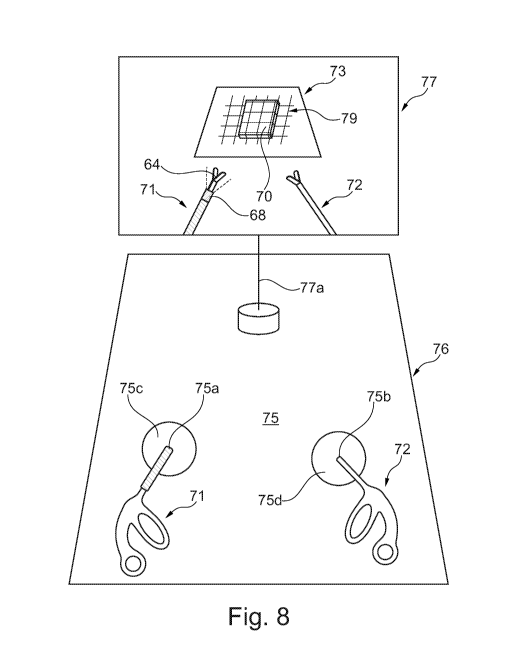
Fig. 8 illustrates a training kit of an embodiment of the invention comprising
a
surgical system, an artificial surgical site and a cover for the artificial
surgical
site.
Fig. 9 illustrates a surgical system in use during a minimal invasive surgery.
Fig. 10 illustrates another correlated set of an embodiment of the invention
comprising a surgical instrument and a pattern generating member.
Fig. 11 illustrates a further correlated set of an embodiment of the invention
comprising a surgical instrument and a pattern generating member.
The figures are schematic and are not drawn to scale. Fig. 1 discloses a
surgical instrument assembly 1, in the present case a laparoscopic instrument.
The surgical instrument assembly 1 comprises a surgical instrument with a
handle portion 2 and a body portion 3 with a surgical tool 4 in the present
CA 02935506 2016-06-29
WO 2015/124159
PCT/DK2015/050035
37
case forceps. The body portion interconnect the handle portion 2 which is
arranged at the proximal end of the surgical instrument and the surgical tool
4, which is arranged at the distal end of the surgical instrument . The body
portion is arranged in the distal/proximal direction.
In another embodiment the surgical tool 4 can be another surgical tool e.g.
scissors or as described above. The surgeon holds the handle portion 2 and
can in this way control the surgical instrument assembly and by pressing or
manipulating the handle portion the forceps can be controlled.
The surgical instrument assembly 1 further comprises a pattern generating
member 5 comprising a pattern light source 6 and optical fibers 7 which guide
the light to a projector 8 (light emitting portion) where optical fibers 7
ends
and the light is projected from the projector 8 in a desired pattern. The
pattern light source 6 is placed on or incorporated into the handle portion 2
of
the surgical instrument and is thus not inserted into the body cavity during
surgery. In an embodiment the pattern light source 6 is connected to a
battery which is also placed or incorporated into the handle portion 2. In an
embodiment the pattern light source 6 is connected to an external power
source such as a battery which is an external battery arranged at a distance
from the surgical instrument assembly.
The projector 8 has advantageously a spherically periphery geometry and is
configured for emitting a light pattern comprising a plurality of angled lines
and or a plurality of parallel lines. In an embodiment the projector 8 has for
example a spherical geometry, and is shaped for emitting a light pattern with
a spherically periphery where the light pattern forms a light pattern
resembling the light pattern generated by a mirror ball. It is to be
understood
that the mirror ball resembling pattern is preferably only a section of normal
mirror ball pattern, e.g. not a 360 degree pattern but preferably only a 90
degree section of the pattern and/or with an angle relative to the axis of the
body portion 3 up to 85 degrees.
CA 02935506 2016-06-29
WO 2015/124159
PCT/DK2015/050035
38
This light pattern makes it possible for the user of the surgical instrument
assembly 1 to deduce the position and movements of the surgical instrument
assembly 1 and thus the surgical tool 4, relative to the surface e.g. a
surgical
site where the light pattern is projected.
The light generated in the light source 6 may be generated by use of one or
more LED's and/or lasers or a combination thereof as explained above.
A section ¨ i.e. the distal end of the body portion 3 of the surgical
instrument
assembly 1 shown in fig. 1 is adapted to be inserted into a body cavity of a
patient through small incisions. The surgeon then seizes the handle portion 2
with his or hers hands in order to perform the surgical procedure. A not
shown endoscope, e.g. a laparoscope, may also be inserted into body cavity
in order to let the surgeon see the surgical field where the surgical
procedure
is to be performed. The endoscope can comprise an illuminating instrument
and a camera element e.g. a video camera such that the surgeon can see the
surgical field and the surgical tool 4 during the procedure via a monitor. The
projector 8 of the pattern generating member 5 is inserted into the body
cavity together with the surgical tool 4. The projector 8 will then project a
light pattern onto the surgical field. The light pattern will aid the surgeon
to
interpret the position and the contours of the surgical field. The light
pattern
aids the surgeon to connect his visual perception of the surgical area which
is
based on observing the monitor (two-dimensions) with his or hers motor
system which is used to control the surgical tools which are moved within the
body cavity (in three-dimensions).
The pattern generating member 5 or parts thereof can be made as an
optionally removable clip-on device and/or it can permanently attached to the
body portion 3. In an embodiment the entire pattern generating element 5 is
mounted to body portion 3 and is adapted for being inserted into the body
cavity.
CA 02935506 2016-06-29
WO 2015/124159
PCT/DK2015/050035
39
Fig. 2 shows a body portion 13 of a surgical instrument assembly 11. The
body portion 13 has a surgical tool 14 in the form of forceps which can be
operated from a not shown handle of the surgical instrument assembly 11. On
the body portion 13 there can be seen a pattern generating member 15 with
one or more projectors 16 which emits light in an angle in front of the
surgical
instrument 1, the angle is shown with a dotted line. The pattern generating
member 15 has in this embodiment plurality of projectors 6 which emits rays
of light that generates a dotted light pattern. The projectors 16 may be
optically connected to one or more pattern light sources of the pattern
generating member 15. By arranging the projector or projectors with a
desired angle the correlation between the movements of the surgical
instrument and the change of the projected pattern can be regulated.
Fig. 3 shows an alternative embodiment of a pattern generating member 25
mounted onto a body portion 23 of a surgical instrument 21. The pattern
generating member 25 comprises a projector 28 with a cylindrical geometry
and a plurality of light emitting areas 26 which emits light rays which forms
the light pattern. The light emitting areas 26 are arranged in rows and are
evenly distributed on the projector 28. The emitting areas 26 of the projector
28 may emit light from the same or from different not shown pattern light
sources. The light rays from the light emitting areas 26 is angled such
relative
to the axis of the body portion 23 that the light rays closest to the surgical
tool 24 is projected to have a smaller angle than the light rays further away
from the surgical tool 24. In this way the light pattern can cover a desired
large area.
The light source may be constructed so that substantially no light is
projected
in the direction of the laparoscope in order to prevent light hitting it and
thus
disturb the view. This can for example be done by turning off or blocking the
light which faces the endoscope. It can also be done by constructing the
surgical instrument so that there are no lights facing the laparoscope.
CA 02935506 2016-06-29
WO 2015/124159
PCT/DK2015/050035
Fig. 4 is an illustration of a light pattern 39 generated by an embodiment of
a
surgical instrument assembly 31assembled from a correlated set of a surgical
instrument and a pattern generating element according to the invention, the
surgical instrument assembly 31 is shown schematic. The light pattern 39
5 forms a grid which for illustrative purposes is shown as projected on a
board
37 with a bulge 30 in the upper left corner. It can be seen that the grid
makes the bulge highly visible as the contours of it stands out. Thus, a
surgical instrument 31 with a pattern generating member which projects a
light pattern on a surgical field can be used to enhance the visual perception
10 of the three dimensional contours of the surgical field. The operator
can in
other words deduce the three dimensional contours of the surgical field based
on a two dimensional vision of the surgical field.
Fig. 5 shows a schematic view of a light pattern 40 on a board 47, the pattern
49 is generated by a surgical instrument assembly 41. The light pattern 40
15 comprises a plurality of dots which are projected from the projector 48
on the
surgical instrument assembly 41. In similar way as for the light pattern shown
in fig. 4 it is possible for the operator to interpret variations in the light
pattern as variation in the surface whereon it is projected and/or to
interpret
movements of the surgical instrument assembly 41 as correlating changes of
20 the pattern 40.
The light pattern 40 (or 30 as exemplified in fig. 4) will change shape
depending on the position of the surgical instrument. For example, in fig. 4
the lines of the pattern 30 will become non parallel if the surgical
instrument
assembly 31 is turned. In addition, the distance between the lines will get
25 smaller if the surgical instrument is moved closer to the board 37 and
get
larger if the surgical instrument is moved away from the board 37. Thus, it is
possible for the operator to deduce the position and orientation of the
surgical
tool in relation a surface e.g. a surgical site whereon a light pattern is
projected based on the geometry light pattern.
CA 02935506 2016-06-29
WO 2015/124159
PCT/DK2015/050035
41
Fig. 6 shows a body portion 53 of an embodiment of a surgical instrument
assembly 51, with a surgical tool 54 and a pattern generating member 55.
The figure illustrate that the pattern generating member 55, in one
embodiment has a projector 58 which can project the light pattern in an angle
between 300 and 65 relative to the axis of the body portion 53. The angle a
is 30 and the angle 13 is 65 relative to the axis of the body portion 53.
The
light pattern is projected in front of the surgical instrument and can be of a
cylindrical geometry such that the projected light pattern will cover a
substantially circular area right in front of the instrument when the
instrument
is held with its distal direction perpendicular to a surface where the pattern
is
projected onto. It is to be understood that the angles for the light pattern
can
be any angle and will depend on the specific use of the surgical instrument.
The surgical instrument assembly shown in Fig. 7 comprises a correlated set
comprising a surgical instrument and a pattern generating member. The
surgical instrument comprises a handle portion 62 at its proximal end, a
surgical tool 64 at its distal end and a body portion 63 connecting the handle
portion 62 to the surgical tool 64. The pattern generating member comprises
a pattern light source 66 and a projector 68. The pattern light source 66 is
operatively connected to the projector 68 via a pattern light source control
unit 69. The pattern light source 66 comprises one or more buttons 66' e.g.
for adjusting the pattern light source 66. In the shown embodiment the
pattern light source control unit 69 is arranged at a distance from the
surgical
instrument ¨ e. g. on the floor in form of a pedal with a control button 69',
which can be operated by the surgeon. The pattern light source 66 is
connected to the pattern light source control unit 69 via an optical fiber 67b
and the pattern light source control unit 69 is connected to the projector 68
via an optical fiber 67a. The projector 68 of is at least temporarily fixed to
the body portion 63 of the surgical instrument such that a movement of said
surgical tool 64 results in a correlated movement of the projector 68. The
projector 68 is emitting a light pattern P as described above. The projector
68
is fixed to the body portion 63 of the surgical instrument by a sleeve 63a
CA 02935506 2016-06-29
WO 2015/124159
PCT/DK2015/050035
42
which surrounds the body portion 63 of the surgical instrument. The sleeve
simultaneously holds and protects the optical fiber 67a.
In an alternative embodiment the pattern light source control unit 69 is
mounted to or integrated with the handle 62.
The training kit shown in Fig. 8 comprises a surgical system, an artificial
surgical site 73 and a cover 76 for the artificial surgical site. The cover 76
comprises a rear side 75 and an opposite not shown front side facing the
artificial surgical site 73. The surgical system comprises a not shown
illuminating element placed on the front side of the cover 76 for illuminating
the artificial surgical site and a not shown camera element also arranged on
the front side of the cover 76 for acquiring images of the artificial surgical
site
73. The surgical system further comprises a monitor 77 for displaying the
acquired images and at least one surgical instrument assembly 71 e.g. as
shown in Fig. 7. The monitor 77 is connected to the camera element via the
wire 77a. The cover 76 comprises a through hole 75a for the surgical
instrument assembly 71. The through hole 75a comprises a periphery edge
75c of elastic material to provide an elastic seal to the surgical instrument
assembly 71. The cover 76 comprises an additional through hole 75b for a
further surgical instrument 72 of the surgical system. The through hole 75b
also comprises a periphery edge 75d of elastic material to provide an elastic
seal to the surgical instrument assembly 72.
The illuminating element is illuminating the artificial surgical site with a
broad
bandwidth light. The surgical system further comprises a not shown marking
element which e.g. in integrated with the illuminating element. The marking
element generates a marking pattern 79 which is stationary and used for
marking up the surgical site and is e.g. used as a reference pattern. The
marking pattern preferably is a narrow band width light pattern e.g. invisible
to the human eye. Advantageously the marking pattern can be switched on
and off by the surgeon.
CA 02935506 2016-06-29
WO 2015/124159
PCT/DK2015/050035
43
The projector 68 generates a pattern 70 which changes when the surgical
instrument assembly 71 as described above.
Advantageously the camera element is connected to a computer e.g.
integrated with the monitor 77, which computer is programmed to monitoring
the movement of the surgical tool 64 of the surgical instrument assembly 71.
The surgical system shown in Fig. 9 comprises an illuminating element and a
camera element integrated in an endoscope 88. The endoscope may
additional comprise a marking element. The endoscope 88 comprises an
illuminating light source for illuminating a surgery target sit 83. The
illumination light source may e.g. be incorporated into the endoscope or it
may be arranged external to the endoscope 88. The camera element of the
endoscope 88 is arranged for acquiring images of the surgery target site 83.
The surgical system further comprises a monitor 87 connected to the
endoscope with wire 87a.Alternatively the connection between the monitor 87
and the endoscope in wireless connected. The monitor 87 is arranged for
displaying the images acquired by the endoscope 88. The monitor may
additionally comprise a computer for analyzing the images. The surgical
system further comprises a surgical instrument assembly 81 e.g. as shown in
Fig. 7. The endoscope 88 and the surgical instrument assembly 81 are
arranged with their respective distal ends inserted through an incision in the
skin 86 of a patient. Below the skin 86 of the patient is formed an internal
body structure leading to the surgery target site 83 e.g. comprising an
intestine I. The surgical instrument assembly 81 is arranged such that the
projector 68 generates a light pattern80 onto the surgery target site 83,
thereby a movement of the surgical tool 64 results in a correlated movement
of the projector 68 and thereby the pattern will change in a correlated way.
The surgeon can follow the changes of the pattern 80 on the monitor 87 and
thereby he obtains a very good visual spatial perception for handling the
surgical instrument assembly 81 during the minimal invasive surgery.
CA 02935506 2016-06-29
WO 2015/124159
PCT/DK2015/050035
44
The surgical instrument assembly shown in Fig. 10 is a variation of the
surgical instrument assembly of Fig. 7 and it comprises a correlated set
comprising a surgical instrument and a pattern generating member. The
surgical instrument comprises a handle portion 62 at its proximal end, a
surgical tool 64 at its distal end and a body portion 63 connecting the handle
portion 62 to the surgical tool 64. The pattern generating member comprises
a pattern light source incorporated into a housing 90. The housing 90 is
mounted onto the body 63, either directly or by being attached to the sleeve
63a which surrounds the body portion 63 of the surgical instrument. The
projector 68 is fixed to the body portion 63 of the surgical instrument by the
sleeve 63a. The projector 68 is for example attached to the sleeve as
described above. The sleeve simultaneously holds and protects a not shown
optical fiber connecting the pattern generating member to the projector 68 for
supplying light. The projector 68 is emitting a light pattern P as described
above.
The pattern light source in the housing 90 is connected to a power source via
the plug 90a. In the shown embodiment the power source is in form of
battery casing 91 is connected to the pattern light source via an electrical
conducting wire 97a for supplying power. The battery casing 91 comprises an
on/off button 91'. The surgeon may use this on/off button 91' during use
(training or surgery) of the surgical instrument assembly, however, for safety
and to protect the battery casing 91 it is preferred that the on/off button
91'
is used only at the beginning and termination of an operation, Therefore the
surgical instrument assembly comprises an additional control source control
unit 92 which is arranged at a distance from the surgical instrument ¨ e. g.
on
the floor in form of a pedal with a control button 92', which can be operated
by the surgeon during use for switching the power and thereby the light
pattern on and off. The additional control source control unit 92 can be
operatively connected to control the power supply in any way e.g. by being
connected to the battery casing 91 via a wire 97b as shown. The housing 90
CA 02935506 2016-06-29
WO 2015/124159
PCT/DK2015/050035
may preferably comprise additional not shown means for adjusting the
pattern light source e.g. with respect to intensity, wavelength(s) and
similar.
The surgical instrument assembly shown in Fig. 11 is a variation of the
surgical instrument assembly of Fig. 10 and differs from the embodiment of
5 Fig. 10 in that the pattern generating member together with the power
source
and preferably the control unit is incorporated into the housing 99 which is
mounted onto the body 63 in the same way as described for the housing 90.
In this embodiment it is desired that a not shown on/off button is arranged
for being manually controlled during use.