Note: Descriptions are shown in the official language in which they were submitted.
CA 02935733 2015-12-16
WO 2015/103273 PCT/US2014/072769
WAVEFRONT MEASUREMENT PRE-SMOOTHING SYSTEMS AND
METHODS
CROSS-REFERENCE TO RELATED APPLICATIONS
[0001] This application claims priority to U.S. Application No. 61/922,605
filed December 31,
2013, the contents of which are incorporated herein by reference for all
purposes. Full Paris
Convention priority is hereby expressly reserved.
BACKGROUND OF THE INVENTION
[0002] Embodiments of the present invention relate to systems and methods for
treating vision
conditions in a patient, and in particular to techniques for develping targets
for use in such
treatments.
[0003] Current treatment planning procedures often involve taking several
wavefront
measurements, and using a measurement selection algorithm to select a single
one of the
wavefront measurements for generating the treatment target. The other non-
selected wavefront
measurements are not used for treatment planning purposes. Often, the single
wavefront
measurement which is selected for the treatment plan can include transient
small-scale spatial
variations.
[0004] Although vision treatment techniques that are based on wavefront
measurements
provide real benefits to patients in need thereof, still further improvements
are desirable.
Embodiments of the present invention provide solutions for at least some of
these outstanding
needs.
BRIEF SUMMARY OF THE INVENTION
[0005] Wavefront based laser vision correction procedures typically include a
diagnostic
measurement which evaluates certain aspects of the patient eye. For example, a
wavefront
measurement may capture details of the eye at a given instance in time. Often,
such
1
CA 02935733 2015-12-16
WO 2015/103273
PCT/US2014/072769
measurements contain certain high spatial frequency features. In some cases,
these features are
transient (e.g. tear film) and vary from measurement to measurement. In some
cases, these
features can be permanent (e.g. corneal scar) such that the high frequency
spatial structure is
consistent across multiple measurements. Embodiments of the present invention
encompass
systems and methods for pre-smoothing a wavefront measurement by dampening or
eliminating
such high frequency features (e.g. 0.3 mm and smaller), for example by using a
low pass filter.
Embodiments also encompass systems and methods for averaging multiple
measurements, which
provides an alternative approach to smoothing, by blurring out high frequency
features, and in
particular transient high frequency features. The modified or ocular wavefront
can then be used
to develop a treatment target which is applied to the eye. Accordingly, the
target shape that is
administered to the patient may not identically correspond to any given
wavefront measurement.
[0006] Often, the pre-smoothing techniques include a convolution protocol that
is implemeted
in the Fourier domain. It is possible to achieve a very high resolution using
a Fourier
decomposition of the wavefront measurement. Relatedly, the high resolution
allows for
improved accuracy for both small and large features of the wavefront. In some
instances, high
resolution Fourier techniques can be used initially in obtaining and
processing the wavefront data
(e.g. to obtain a Fourier spectrum of the wavefront), and then high frequency
information can be
dampened or discarded by a pre-smoothing process.
[0007] In one aspect, embodiments of the present invention encompass systems
and methods
for generating a vision treatment target for an eye of a patient. Exemplary
methods include
obtaining a wavefront measurement for the eye of the patient, processing the
wavefront
measurement, using a low pass filter, to obtain an ocular wavefront, and
generating the vision
treatment target based on the ocular wavefront. In some cases, the processing
step can include
applying a Fourier transform to the wavefront measurement to obtain a Fourier
spectrum of the
wavefront, convolving, in the Fourier domain, the Fourier spectrum of the
wavefront and a low
pass filter to obtain a Fourier spectrum convolution result, and applying an
inverse transform to
the convolution result to obtain the ocular wavefront. The ocular wavefront
can represent a low
pass filtered version of the wavefront measurement, such that high spatial
frequency features
present in the wavefront measurement are not present in the ocular wavefront.
In some cases, the
low pass filter is a Gaussian low-pass filter having a kernel size of 0.3 mm.
In some cases,
2
CA 02935733 2015-12-16
WO 2015/103273
PCT/US2014/072769
methods may include administering the vision treatment target to the eye of
the patient. In some
cases, methods may include processing the treatment target with a
deconvolution protocol to
obtain a deconvolved treatment target. In some cases, methods may include
administering the
deconvolved treatment target to the eye of the patient.
[0008] In another aspect, embodiments of the present invention encompass
systems and
methods for generating a vision treatment target for an eye of a patient.
Exemplary systems
include a processor, a first module, an second module, and a third module. The
first module can
include a tangible medium embodying machine-readable code executed on the
processor to
obtain a wavefront measurement for the eye of the patient. The second module
can include a
tangible medium embodying machine-readable code executed on the processor to
process the
wavefront measurement with a low pass filter to obtain an ocular wavefront.
The third module
can include a tangible medium embodying machine-readable code executed on the
processor to
generate the vision treatment target based on the ocular wavefront. In some
cases, the tangible
medium embodying machine-readable code of the second module, when executed on
the
processor, applies a Fourier transform to the wavefront measurement to obtain
a Fourier
spectrum of the wavefront, convolves, in the Fourier domain, the Fourier
spectrum of the
wavefront and a low pass filter to obtain a Fourier spectrum convolution
result, and applies an
inverse transform to the convolution result to obtain the ocular wavefront. In
some cases, the
ocular wavefront represents a low pass filtered version of the wavefront
measurement, such that
high spatial frequency features present in the wavefront measurement are not
present in the
ocular wavefront. According to some embodiments, the low pass filter is a
Gaussian low-pass
filter having a kernel size of 0.3 mm. In some embodiments, systems may also
include a fourth
module having a tangible medium embodying machine-readable code executed on
the processor
to administer the vision treatment target to the eye of the patient. In some
embodiments, systems
may also include a fourth module having a tangible medium embodying machine-
readable code
executed on the processor to process the treatment target with a deconvolution
protocol to obtain
a deconvolved treatment target. In some embodiments, systems may also include
a fifth module
having a tangible medium embodying machine-readable code executed on the
processor to
administer the deconvolved treatment target to the eye of the patient.
3
CA 02935733 2015-12-16
WO 2015/103273
PCT/US2014/072769
[0009] In yet another aspect, embodiments of the present invention encompass
computer
products embodied on tangible computer readable storage media. An exemplary
computer
product embodied on tangible computer readable storage medium may include code
for
obtaining a wavefront measurement for the eye of the patient, code for
processing the wavefront
measurement, using a low pass filter, to obtain an ocular wavefront, and code
for generating the
vision treatment target based on the ocular wavefront. In some cases, the code
for processing the
wavefront measurement includes code for applying a Fourier transform to the
wavefront
measurement to obtain a Fourier spectrum of the wavefront, code for
convolving, in the Fourier
domain, the Fourier spectrum of the wavefront and a low pass filter to obtain
a Fourier spectrum
convolution result, and code for applying an inverse transform to the
convolution result to obtain
the ocular wavefront. The ocular wavefront represents a low pass filtered
version of the
wavefront measurement, such that high spatial frequency features present in
the wavefront
measurement are not present in the ocular wavefront. In some cases, the low
pass filter is a
Gaussian low-pass filter having a kernel size of 0.3 mm. In some cases,
computer products may
also include code for administering the vision treatment target to the eye of
the patient. In some
cases, computer products may also include code for processing the treatment
target with a
deconvolution protocol to obtain a deconvolved treatment target. In some
cases, computer
products may also include code for administering the deconvolved treatment
target to the eye of
the patient.
BRIEF DESCRIPTION OF THE DRAWINGS
[0010] Figure 1 illustrates a laser ablation system according to an embodiment
of the present
invention.
[0011] Figure 2 illustrates a simplified computer system according to an
embodiment of the
present invention.
[0012] Figure 3 illustrates a wavefront measurement system according to an
embodiment of
the present invention.
[0013] Figure 3A illustrates another wavefront measurement system according to
an
embodiment of the present invention.
4
CA 02935733 2015-12-16
WO 2015/103273
PCT/US2014/072769
[0014] Figure 4 shows aspects of a method for generating at treatment target
or plan according
to embodiments of the present invention.
[0015] Figure 4A depicts aspects of wavefront and target representations
according to
embodiments of the present invention.
[0016] Figure 5 illustrates high order aberrations (HOA) or errors for
sequential wavefront
measurements for an eye, according to embodiments of the present invention.
[0017] Figures 6A and 6B show effects of wavefront smoothing using a low pass
filter,
according to embodiments of the present invention.
DETAILED DESCRIPTION OF THE INVENTION
[0018] Embodiments of the present invention can be readily adapted for use
with existing laser
systems and other optical treatment devices. Although system, software, and
method
embodiments of the present invention are described primarily in the context of
a laser eye
surgery system, it should be understood that embodiments of the present
invention may be
adapted for use in or in combination with alternative eye treatment
procedures, systems, or
modalities, such as spectacle lenses, intraocular lenses, accommodating IOLs,
contact lenses,
corneal ring implants, collagenous corneal tissue thermal remodeling, corneal
inlays, corneal
onlays, other corneal implants or grafts, and the like. Relatedly, systems,
software, and methods
according to embodiments of the present invention are well suited for
customizing any of these
treatment modalities to a specific patient. Thus, for example, embodiments
encompass custom
preformed lenses, intraocular lenses, custom contact lenses, custom corneal
implants, and the
like, which can be configured to treat or ameliorate any of a variety of
vision conditions in a
particular patient based on their unique ocular characteristics or anatomy.
Additionally, the
modified ablation target or target shape may be implemented via other non-
ablative laser
therapies, such as laser-incised custom lenticule shapes and subsequent
extraction and laser-
based corneal incision patterns.
[0019] In some instances, these techniques can be carried out in conjunction
with treatments
provided by any of a variety of laser devices, including without limitation
the WaveScan0
System and the STAR 540 Excimer Laser System both by Abbott Medical Optics
Inc., the
5
CA 02935733 2015-12-16
WO 2015/103273
PCT/US2014/072769
WaveLight Allegretto Wave Eye-Q laser, the Schwind AmarisTM lasers, the 217P
excimer
workstation by Technolas PerfectVision GmbH, the Mel 8OTM laser by Carl Zeiss
Meditec, Inc.,
and the like. In some cases, embodiments provide techniques for using laser
basis data during
refractive surgery treatment procedures which can be implemented in such laser
devices.
[0020] Exemplary systems and methods disclosed herein can be implemented via a
variety of
ophthalmic devices or solutions. For example, treatment techniques may be used
for any of a
variety of surgery modalities, including excimer laser surgery, femtosecond
surgery, and the like.
A variety of forms of lasers and laser energy can be used to effect a
correction or treatment,
including infrared lasers, ultraviolet lasers, femtosecond lasers, wavelength
multiplied solid-state
lasers, and the like. By way of non-limiting example, ophthalmic corrections
can involve a
cornea or lens reshaping procedure, such as, for example using a picosecond or
femtosecond
laser. Laser ablation procedures can remove a targeted amount stroma of a
cornea to change a
cornea's contour and adjust for aberrations. In some cases, a treatment
protocol can involve the
delivery of a series of discrete pulses of laser light energy, with a total
shape and amount of
tissue removed being determined by a shape, size, location, and/or number of
laser energy pulses
impinging on or focused within a cornea. In some cases, a surgical laser, such
as a non-
ultraviolet, ultra-short pulsed laser that emits radiation with pulse
durations as short as
nanoseconds and femtoseconds (e.g., a femtosecond laser, or a picosecond
laser) can be used to
treat the eye of a patient. Other pulse widths may be suitable as well. The
laser systems can be
configured to deliver near infrared light. Other wavelengths may be used as
well. The laser
systems can be configured to deliver laser light focused at a focus depth
(e.g. within corneal or
other ophthalmologic tissue) which may be controlled by the system. Laser
surgery with ultra-
short pulse lasers such as femtosecond lasers can be used to treat the eye.
These pulsed lasers
can make very accurate incisions of the eye and can be used in many ways to
treat the eye.
Additional types of incisions that can be performed with the short pulse
lasers include incisions
for paracentesis, limbal relaxing incisions, and refractive incisions to shape
the cornea, for
example.
[0021] In some cases, vision treatments can include focusing femtosecond laser
energy within
the stroma so as to ablate a volume of intrastromal tissue. By scanning the
focal spot within an
appropriate volume of the stromal tissue, it is possible to vaporize the
volume so as to achieve a
6
CA 02935733 2015-12-16
WO 2015/103273
PCT/US2014/072769
desired refractive alteration. Hence, embodiments of the present invention
encompass laser
surgical techniques that involve femtosecond laser photodisruption or
photoalteration treatments.
In some cases, a femtosecond laser can be used to perform the photodisruption,
thus providing an
easy, precise, and effective approach to refractive surgery
[0022] According to some embodiments, a femtosecond laser (or other laser) of
the optical
system can be used to incise the cornea or to cut a flap. A femtosecond laser
may be used to
make arcuate or other incisions in the cornea, which incisions may be
customized, intrastromal,
stable, predictable, and the like. Likewise, corneal entry incisions may be
made, which are
custom, multi-plane, and self-sealing.
[0023] Pulsed laser beams include bursts or pulses of light. Pulsed lasers,
such as non-
ultraviolet, ultra-short pulsed lasers with pulse durations measured in the
nanoseconds to
femtoseconds range, can be used in ophthalmic surgical procedures as disclosed
herein. For
example, a pulsed laser beam can be focused onto a desired area of
ophthalmologic material or
tissue, such as the cornea, the capsular bag, or the lens of the eye, to
photoalter the material in
this area and, in some instances, the associated peripheral area. Examples of
photoalteration of
the material include, but are not necessarily limited to, chemical and
physical alterations,
chemical and physical breakdown, disintegration, ablation, photodisruption,
vaporization, a the
like. Exemplary treatment systems can include a focusing mechanism (e.g. lens)
and/or a
scanning mechanism so as to guide or direct a focus of femtosecond energy
along a path within
the patient's eye (e.g. at one or more corneal subsurface locations).
[0024] According to some embodiments, the vergence weighting systems and
methods
disclosed herein can be implemented in connection with software, hardware, or
a combination of
software and hardware residing in a diagnostic device such as WaveScan0 and
iDesignTM
devices.
[0025] Turning now to the drawings, FIG. 1 illustrates a laser eye surgery
system 10 of the
present invention, including a laser 12 that produces a laser beam 14. Laser
12 is optically
coupled to laser delivery optics 16, which directs laser beam 14 to an eye E
of patient P. A
delivery optics support structure (not shown here for clarity) extends from a
frame 18 supporting
laser 12. A microscope 20 is mounted on the delivery optics support structure,
the microscope
often being used to image a cornea of eye E.
7
CA 02935733 2015-12-16
WO 2015/103273
PCT/US2014/072769
[0026] Laser 12 generally comprises an excimer laser, ideally comprising an
argon-fluorine
laser producing pulses of laser light having a wavelength of approximately 193
nm. Laser 12
will preferably be designed to provide a feedback stabilized fluence at the
patient's eye, delivered
via delivery optics 16. The present invention may also be useful with
alternative sources of
ultraviolet or infrared radiation, particularly those adapted to controllably
ablate the corneal
tissue without causing significant damage to adjacent and/or underlying
tissues of the eye. Such
sources include, but are not limited to, solid state lasers and other devices
which can generate
energy in the ultraviolet wavelength between about 185 and 205 nm and/or those
which utilize
frequency-multiplying techniques. Hence, although an excimer laser is the
illustrative source of
an ablating beam, other lasers may be used in the present invention.
[0027] Laser system 10 will generally include a computer or programmable
processor 22.
Processor 22 may comprise (or interface with) a conventional PC system
including the standard
user interface devices such as a keyboard, a display monitor, and the like.
Processor 22 will
typically include an input device such as a magnetic or optical disk drive, an
internet connection,
or the like. Such input devices will often be used to download a computer
executable code from
a tangible storage media 29 embodying any of the methods of the present
invention. Tangible
storage media 29 may take the form of a floppy disk, an optical disk, a data
tape, a volatile or
non-volatile memory, RAM, or the like, and the processor 22 will include the
memory boards
and other standard components of modern computer systems for storing and
executing this code.
Tangible storage media 29 may optionally embody wavefront sensor data,
wavefront gradients, a
wavefront elevation map, a treatment map, a corneal elevation map, and/or an
ablation table.
While tangible storage media 29 will often be used directly in cooperation
with an input device
of processor 22, the storage media may also be remotely operatively coupled
with processor by
means of network connections such as the internet, and by wireless methods
such as infrared,
Bluetooth, or the like.
[0028] Laser 12 and delivery optics 16 will generally direct laser beam 14 to
the eye of patient
P under the direction of a computer 22. Computer 22 will often selectively
adjust laser beam 14
to expose portions of the cornea to the pulses of laser energy so as to effect
a predetermined
sculpting of the cornea and alter the refractive characteristics of the eye.
In many embodiments,
both laser beam 14 and the laser delivery optical system 16 will be under
computer control of
8
CA 02935733 2015-12-16
WO 2015/103273
PCT/US2014/072769
processor 22 to effect the desired laser sculpting process, with the processor
effecting (and
optionally modifying) the pattern of laser pulses. The pattern of pulses may
by summarized in
machine readable data of tangible storage media 29 in the form of a treatment
table, and the
treatment table may be adjusted according to feedback input into processor 22
from an
automated image analysis system in response to feedback data provided from an
ablation
monitoring system feedback system. Optionally, the feedback may be manually
entered into the
processor by a system operator. Such feedback might be provided by integrating
the wavefront
measurement system described below with the laser treatment system 10, and
processor 22 may
continue and/or terminate a sculpting treatment in response to the feedback,
and may optionally
also modify the planned sculpting based at least in part on the feedback.
Measurement systems
are further described in U.S. Patent No. 6,315,413, the full disclosure of
which is incorporated
herein by reference.
[0029] Laser beam 14 may be adjusted to produce the desired sculpting using a
variety of
alternative mechanisms. The laser beam 14 may be selectively limited using one
or more
variable apertures. An exemplary variable aperture system having a variable
iris and a variable
width slit is described in U.S. Patent No. 5,713,892, the full disclosure of
which is incorporated
herein by reference. The laser beam may also be tailored by varying the size
and offset of the
laser spot from an axis of the eye, as described in U.S. Patent Nos.
5,683,379, 6,203,539, and
6,331,177, the full disclosures of which are incorporated herein by reference.
[0030] Still further alternatives are possible, including scanning of the
laser beam over the
surface of the eye and controlling the number of pulses and/or dwell time at
each location, as
described, for example, by U.S. Patent No. 4,665,913, the full disclosure of
which is
incorporated herein by reference; using masks in the optical path of laser
beam 14 which ablate
to vary the profile of the beam incident on the cornea, as described in U.S.
Patent No. 5,807,379,
the full disclosure of which is incorporated herein by reference; hybrid
profile-scanning systems
in which a variable size beam (typically controlled by a variable width slit
and/or variable
diameter iris diaphragm) is scanned across the cornea; or the like. The
computer programs and
control methodology for these laser pattern tailoring techniques are well
described in the patent
literature.
9
CA 02935733 2015-12-16
WO 2015/103273
PCT/US2014/072769
[0031] Additional components and subsystems may be included with laser system
10, as
should be understood by those of skill in the art. For example, spatial and/or
temporal
integrators may be included to control the distribution of energy within the
laser beam, as
described in U.S. Patent No. 5,646,791, the full disclosure of which is
incorporated herein by
reference. Ablation effluent evacuators/filters, aspirators, and other
ancillary components of the
laser surgery system are known in the art. Further details of suitable systems
for performing a
laser ablation procedure can be found in commonly assigned U.S. Pat. Nos.
4,665,913,
4,669,466, 4,732,148, 4,770,172, 4,773,414, 5,207,668, 5,108,388, 5,219,343,
5,646,791 and
5,163,934, the complete disclosures of which are incorporated herein by
reference. Suitable
systems also include commercially available refractive laser systems such as
those manufactured
and/or sold by Alcon, Bausch & Lomb, Nidek, WaveLight, LaserSight, Schwind,
Zeiss-Meditec,
and the like. Basis data can be further characterized for particular lasers or
operating conditions,
by taking into account localized environmental variables such as temperature,
humidity, airflow,
and aspiration.
[0032] FIG. 2 is a simplified block diagram of an exemplary computer system 22
that may be
used by the laser surgical system 10 of the present invention. Computer system
22 typically
includes at least one processor 52 which may communicate with a number of
peripheral devices
via a bus subsystem 54. These peripheral devices may include a storage
subsystem 56,
comprising a memory subsystem 58 and a file storage subsystem 60, user
interface input devices
62, user interface output devices 64, and a network interface subsystem 66.
Network interface
subsystem 66 provides an interface to outside networks 68 and/or other
devices, such as the
wavefront measurement system 30.
[0033] User interface input devices 62 may include a keyboard, pointing
devices such as a
mouse, trackball, touch pad, or graphics tablet, a scanner, foot pedals, a
joystick, a touchscreen
incorporated into the display, audio input devices such as voice recognition
systems,
microphones, and other types of input devices. User input devices 62 will
often be used to
download a computer executable code from a tangible storage media 29 embodying
any of the
methods of the present invention. In general, use of the term "input device"
is intended to
include a variety of conventional and proprietary devices and ways to input
information into
computer system 22.
CA 02935733 2015-12-16
WO 2015/103273
PCT/US2014/072769
[0034] User interface output devices 64 may include a display subsystem, a
printer, a fax
machine, or non-visual displays such as audio output devices. The display
subsystem may be a
cathode ray tube (CRT), a flat-panel device such as a liquid crystal display
(LCD), a projection
device, or the like. The display subsystem may also provide a non-visual
display such as via
audio output devices. In general, use of the term "output device" is intended
to include a variety
of conventional and proprietary devices and ways to output information from
computer system
22 to a user.
[0035] Storage subsystem 56 can store the basic programming and data
constructs that provide
the functionality of the various embodiments of the present invention. For
example, a database
and modules implementing the functionality of the methods of the present
invention, as
described herein, may be stored in storage subsystem 56. These software
modules are generally
executed by processor 52. In a distributed environment, the software modules
may be stored on
a plurality of computer systems and executed by processors of the plurality of
computer systems.
Storage subsystem 56 typically comprises memory subsystem 58 and file storage
subsystem 60.
[0036] Memory subsystem 58 typically includes a number of memories including a
main
random access memory (RAM) 70 for storage of instructions and data during
program execution
and a read only memory (ROM) 72 in which fixed instructions are stored. File
storage
subsystem 60 provides persistent (non-volatile) storage for program and data
files, and may
include tangible storage media 29 (FIG. 1) which may optionally embody
wavefront sensor data,
wavefront gradients, a wavefront elevation map, a treatment map, and/or an
ablation table. File
storage subsystem 60 may include a hard disk drive, a floppy disk drive along
with associated
removable media, a Compact Digital Read Only Memory (CD-ROM) drive, an optical
drive,
DVD, CD-R, CD-RW, solid-state removable memory, and/or other removable media
cartridges
or disks. One or more of the drives may be located at remote locations on
other connected
computers at other sites coupled to computer system 22. The modules
implementing the
functionality of the present invention may be stored by file storage subsystem
60.
[0037] Bus subsystem 54 provides a mechanism for letting the various
components and
subsystems of computer system 22 communicate with each other as intended. The
various
subsystems and components of computer system 22 need not be at the same
physical location but
may be distributed at various locations within a distributed network. Although
bus subsystem 54
11
CA 02935733 2015-12-16
WO 2015/103273
PCT/US2014/072769
is shown schematically as a single bus, alternate embodiments of the bus
subsystem may utilize
multiple busses.
[0038] Computer system 22 itself can be of varying types including a personal
computer, a
portable computer, a workstation, a computer terminal, a network computer, a
control system in
a wavefront measurement system or laser surgical system, a mainframe, or any
other data
processing system. Due to the ever-changing nature of computers and networks,
the description
of computer system 22 depicted in FIG. 2 is intended only as a specific
example for purposes of
illustrating one embodiment of the present invention. Many other
configurations of computer
system 22 are possible having more or less components than the computer system
depicted in
Fig. 2.
[0039] Referring now to FIG. 3, one embodiment of a wavefront measurement
system 30 is
schematically illustrated in simplified form. In very general terms, wavefront
measurement
system 30 is configured to sense local slopes of a gradient map exiting the
patient's eye. Devices
based on the Hartmann-Shack principle generally include a lenslet array to
sample the gradient
map uniformly over an aperture, which is typically the exit pupil of the eye.
Thereafter, the local
slopes of the gradient map are analyzed so as to reconstruct the wavefront
surface or map.
[0040] More specifically, one wavefront measurement system 30 includes an
image source 32,
such as a laser, which projects a source image through optical tissues 34 of
eye E so as to form
an image 44 upon a surface of retina R. The image from retina R is transmitted
by the optical
system of the eye (e.g., optical tissues 34) and imaged onto a wavefront
sensor 36 by system
optics 37. The wavefront sensor 36 communicates signals to a computer system
22' for
measurement of the optical errors in the optical tissues 34 and/or
determination of an optical
tissue ablation treatment program. Computer 22' may include the same or
similar hardware as
the computer system 22 illustrated in FIGS. 1 and 2. Computer system 22' may
be in
communication with computer system 22 that directs the laser surgery system
10, or some or all
of the components of computer system 22, 22' of the wavefront measurement
system 30 and
laser surgery system 10 may be combined or separate. If desired, data from
wavefront sensor 36
may be transmitted to a laser computer system 22 via tangible media 29, via an
I/O port, via an
networking connection 66 such as an intranet or the Internet, or the like.
12
CA 02935733 2015-12-16
WO 2015/103273
PCT/US2014/072769
[0041] Wavefront sensor 36 generally comprises a lenslet array 38 and an image
sensor 40. As
the image from retina R is transmitted through optical tissues 34 and imaged
onto a surface of
image sensor 40 and an image of the eye pupil P is similarly imaged onto a
surface of lenslet
array 38, the lenslet array separates the transmitted image into an array of
beamlets 42, and (in
combination with other optical components of the system) images the separated
beamlets on the
surface of sensor 40. Sensor 40 typically comprises a charged couple device or
"CCD," and
senses the characteristics of these individual beamlets, which can be used to
determine the
characteristics of an associated region of optical tissues 34. In particular,
where image 44
comprises a point or small spot of light, a location of the transmitted spot
as imaged by a beamlet
can directly indicate a local gradient of the associated region of optical
tissue.
[0042] Eye E generally defines an anterior orientation ANT and a posterior
orientation POS.
Image source 32 generally projects an image in a posterior orientation through
optical tissues 34
onto retina R as indicated in FIG. 3. Optical tissues 34 again transmit image
44 from the retina
anteriorly toward wavefront sensor 36. Image 44 actually formed on retina R
may be distorted
by any imperfections in the eye's optical system when the image source is
originally transmitted
by optical tissues 34. Optionally, image source projection optics 46 may be
configured or
adapted to decrease any distortion of image 44.
[0043] In some embodiments, image source optics 46 may decrease lower order
optical errors
by compensating for spherical and/or cylindrical errors of optical tissues 34.
Higher order
optical errors of the optical tissues may also be compensated through the use
of an adaptive optic
element, such as a deformable mirror (described below). Use of an image source
32 selected to
define a point or small spot at image 44 upon retina R may facilitate the
analysis of the data
provided by wavefront sensor 36. Distortion of image 44 may be limited by
transmitting a
source image through a central region 48 of optical tissues 34 which is
smaller than a pupil 50, as
the central portion of the pupil may be less prone to optical errors than the
peripheral portion.
Regardless of the particular image source structure, it will be generally be
beneficial to have a
well-defined and accurately formed image 44 on retina R.
[0044] In one embodiment, the wavefront data may be stored in a computer
readable medium
29 or a memory of the wavefront sensor system 30 in two separate arrays
containing the x and y
wavefront gradient values obtained from image spot analysis of the Hartmann-
Shack sensor
13
CA 02935733 2015-12-16
WO 2015/103273
PCT/US2014/072769
images, plus the x and y pupil center offsets from the nominal center of the
Hartmann-Shack
lenslet array, as measured by the pupil camera 51 (FIG. 3) image. Such
information contains all
the available information on the wavefront error of the eye and is sufficient
to reconstruct the
wavefront or any portion of it. In such embodiments, there is no need to
reprocess the
Hartmann-Shack image more than once, and the data space required to store the
gradient array is
not large. For example, to accommodate an image of a pupil with an 8 mm
diameter, an array of
a 20 x 20 size (i.e., 400 elements) is often sufficient. As can be
appreciated, in other
embodiments, the wavefront data may be stored in a memory of the wavefront
sensor system in a
single array or multiple arrays.
[0045] While the methods of the present invention will generally be described
with reference
to sensing of an image 44, it should be understood that a series of wavefront
sensor data readings
may be taken. For example, a time series of wavefront data readings may help
to provide a more
accurate overall determination of the ocular tissue aberrations. As the ocular
tissues can vary in
shape over a brief period of time, a plurality of temporally separated
wavefront sensor
measurements can avoid relying on a single snapshot of the optical
characteristics as the basis for
a refractive correcting procedure. Still further alternatives are also
available, including taking
wavefront sensor data of the eye with the eye in differing configurations,
positions, and/or
orientations. For example, a patient will often help maintain alignment of the
eye with
wavefront measurement system 30 by focusing on a fixation target, as described
in U.S. Patent
No. 6,004,313, the full disclosure of which is incorporated herein by
reference. By varying a
position of the fixation target as described in that reference, optical
characteristics of the eye may
be determined while the eye accommodates or adapts to image a field of view at
a varying
distance and/or angles.
[0046] The location of the optical axis of the eye may be verified by
reference to the data
provided from a pupil camera 52. In the exemplary embodiment, a pupil camera
52 images pupil
50 so as to determine a position of the pupil for registration of the
wavefront sensor data relative
to the optical tissues.
[0047] An alternative embodiment of a wavefront measurement system is
illustrated in FIG.
3A. The major components of the system of FIG. 3A are similar to those of FIG.
3.
Additionally, FIG. 3A includes an adaptive optical element 53 in the form of a
deformable
14
CA 02935733 2015-12-16
WO 2015/103273
PCT/US2014/072769
mirror. The source image is reflected from deformable mirror 98 during
transmission to retina
R, and the deformable mirror is also along the optical path used to form the
transmitted image
between retina R and imaging sensor 40. Deformable mirror 98 can be
controllably deformed by
computer system 22 to limit distortion of the image formed on the retina or of
subsequent images
formed of the images formed on the retina, and may enhance the accuracy of the
resultant
wavefront data. The structure and use of the system of FIG. 3A are more fully
described in U.S.
Patent No. 6,095,651, the full disclosure of which is incorporated herein by
reference.
[0048] The components of an embodiment of a wavefront measurement system for
measuring
the eye and ablations may comprise elements of a WaveScan system, available
from AMO
MANUFACTURING USA, LLC, MILPITAS, California. One embodiment includes a
WaveScan
system with a deformable mirror as described above. An alternate embodiment of
a wavefront
measuring system is described in U.S. Patent No. 6,271,915, the full
disclosure of which is
incorporated herein by reference. It is appreciated that any wavefront
aberrometer could be
employed for use with the present invention. Relatedly, embodiments of the
present invention
encompass the implementation of any of a variety of optical instruments
provided by AMO
WaveFront Sciences, LLC, including the COAS wavefront aberrometer, the
ClearWave contact
lens aberrometer, the Crystal Wave IOL aberrometer, and the like.
[0049] Relatedly, embodiments of the present invention encompass the
implementation of any
of a variety of optical instruments provided by WaveFront Sciences, Inc.,
including the COAS
wavefront aberrometer, the ClearWave contact lens aberrometer, the Crystal
Wave IOL
aberrometer, and the like. Embodiments of the present invention may also
involve wavefront
measurement schemes such as a Tscherning-based system, which may be provided
by
WaveFront Sciences, Inc. Embodiments of the present invention may also involve
wavefront
measurement schemes such as a ray tracing-based system, which may be provided
by Tracey
Technologies, Corp.
[0050] Wavefront Pre-Processing
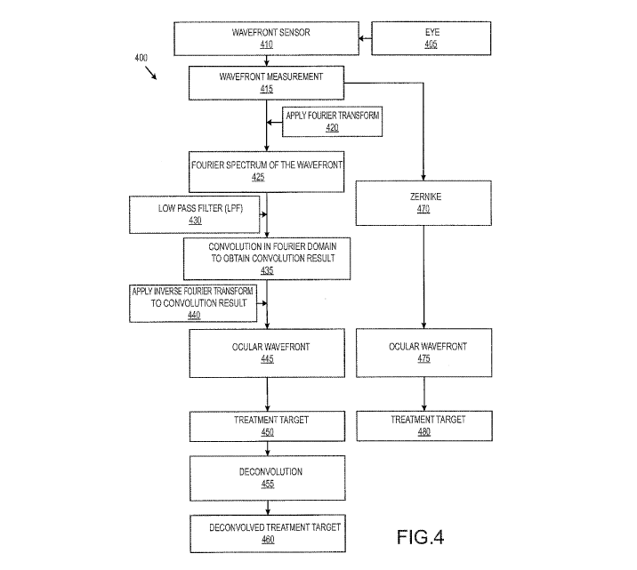
[0051] Embodiments of the present invention encompass systems and methods for
adjusting or
generating wavefront-based refractive treatment plans which involve the pre-
processing of
wavefront information or measurements. As depicted in FIG. 4, an exemplary
method 400 of
generating at treatment target or plan can include obtaining a wavefront
measurement 415 for an
CA 02935733 2015-12-16
WO 2015/103273
PCT/US2014/072769
eye 405 of a patient. Often, the eye may present high spatial frequency
features, including
transient features such as tear film, as well as permanent features such as
corneal scars. As
discussed elsewhere herein, it is possible to ignore or filter out certain
small-scale spatial
variations. The wavefront measurement 415 may be obtained, for example, by
evaluating the
eye 405 with a wavefront sensor device 410. A wavefront sensor 410 may include
an array of
lenslets, and the configuration of the lenslet array can determine the
resolution of the sensor. For
example, a lower density resolution sensor may have a lenslet array with wider
spacing, and a
higher density resolution sensor may have a lenslet array with narrower
spacing. The wavefront
sensor 410 can be configured to detect high spatial frequency features,
including rapid curvature
changes and the like. As discussed elsewhere herein, embodiments of the
present invention can
operate to ignore or suppress certain high spatial frequency information
obtained by the
wavefront device. In some instances, a filter can be used to suppress this
information, in a way
that is consistent for all measurements (e.g. independent of the resolution of
the sensor, and
independent of the wavefront diameter). Hence, a wavefront measurement having
a very high
resolution can be processed with a Fourier transform (or some other zonal
reconstruction
methods), and an LPF filter can be applied according to a defined spatial
scale for smoothing.
According to some embodiments, the wavefront measurement can include gradient
information.
For example, slope sensors of the wavefront sensor 410 can operate to generate
local gradient
measurements. Relatedly, the wavefront measurement can include a wavefront
gradient field
based on the local gradients.
[0052] As depicted as step 420, methods may include applying a Fourier
transform to the
wavefront measurement 415, so as to obtain a Fourier transform 425 of the
wavefront. For
example, by applying the Fourier transform of step 420, it is possible to
obtain a Fourier
transform of the local wavefront gradient measurements. A Fourier transform
can be used to
reconstruct wavefront data by decomposing the image into spatial frequency
components. As
noted elsewhere herein, the Fourier spectrum of the wavefront 425 can provide
a high resolution
representation of the wavefront.
[0053] By using the Fourier approach, it is possible to ensure that the shape
being measured by
the aberrometer is an accurate representation of the optics of the eye. The
Fourier transform
technique provides an alternative to Zernike polynomials, and can be used to
precisely
16
CA 02935733 2015-12-16
WO 2015/103273
PCT/US2014/072769
reconstructed the wavefront. Using the Fourier method, it is possible to
sample the wavefront
over fixed intervals of space that correspond to the equal spacing of the
lenslets in the sensor's
lenslet array. Hence, the Fourier technique is well suited for use with an
evenly spaced grid such
as the Hartmann-Shack sensor. Further, Fourier transform techniques are not
limited to a
circular reconstruction, and hence can accommodate elliptical pupil apertures.
What is more,
surgeons or operators are not required to specify the order of the
reconstruction, as typically is
the case with Zernike reconstruction. As discussed elsewhere herein, the
Fourier reconstruction
can use all the data present in the reconstruction, thus providing a high
resolution representation.
Relatedly, the Fourier technique is capable of accurately characterizing a
broad range of optical
aberrations.
[0054] According to some embodiments, a Fourier series can be used as a set of
basis
functions for the ocular wavefront reconstruction. Exemplary methods can
include obtaining the
wavefront slopes in x- and y- directions, and taking the Fourier transform of
these wavefront
slopes. Once the Fourier transform, or the Fourier spectrum of the wavefront,
is obtained, it is
then possible to obtain the ocular wavefront by an inverse Fourier transform
of the Fourier
transform of the wavefront.
[0055] According to step 430, methods may also include provising a low pass
filter (LPF),
which can operate to remove high spatial frequency data or variations of the
wavefront. In some
cases, a Gaussian kernel may be used. In some cases, a single parameter
Butterworth kernel may
be used. In some cases, a dual parameter or multiple parameter kernel may be
used. Exemplary
filter, kernels, and related techniques are discussed in U.S. Patent
Application No. 61/708,815
filed October 2, 2012, U.S. Patent Application No. 61/871,120 filed August 28,
2013, U.S.
Patent Application No. 14/044,650 filed October 2, 2013, and U.S. Patent
Application No.
61/901,216 filed November 7, 2013, the contents of each of which are
incorporated herein by
reference.
[0056] The low pass filter (LPF) can be used to remove or reduce small-scale
spatial variations
(e.g. high spatial frequency features) associated with the measured wavefront,
for example by
processing the measured wavefront information with LPF prior to using the
measured wavefront
information in the treatment planning protocol. Typically, although the small-
scale spatial
variations are removed, the low order and high order aberration information is
retained.
17
CA 02935733 2015-12-16
WO 2015/103273
PCT/US2014/072769
According to some embodiments, a Gaussian low-pass filter having a kernel size
of 0.3 mm can
be used. Such a filter produced little or no change in the low-order and high-
order aberrations,
and can smooth out any features smaller than 0.3 mm, thus improving the
robustness and
efficiency of the generated treatment.
[0057] In some cases, the smoothing kernel size may correspond to a spatial
frequency cutoff
in the frequency domain. For example, with a pupil that is 6 mm in diameter
(which is typical),
the Airy disk can be about 0.38 arc minutes, which corresponds to about 79
cycles per degree
(cutoff frequency) in the frequency domain. If the aberrometer lenslet spacing
is 0.175 mm or
so, a 0.3 mm in size on the cornea corresponds to about 0.3/0.175, or 1.7
times the lenslet
spacing. Therefore, 0.3 mm can correspond to 1.7 times smaller than the cutoff
frequency of 79
cpd, which is about 46 cpd. Hence, using a low pass filter kernel size of 0.3
mm for a 6 mm
pupil (or measured wavefront diameter) can supress or remove high spatial
frequency structures
that are higher than about 46 cpd.
[0058] According to some embodiments, this cutoff number can change with
different pupil
sizes. For example, with a smaller pupil, this 0.3 mm (i.e. kernel size) is a
bigger relative portion
so it corresponds to a lower frequency number. For example, for a 5 mm pupil,
0.3 mm
corresponds to only about 38 cpd. Hence, using a low pass filter kernel size
of 0.3 mm for a 5
mm pupil (or measured wavefront diameter) can supress or remove high spatial
frequency
structures that are higher than about 38 cpd.
[0059] For a very small pupil, say, 2 mm in diameter, 0.3 mm corresponds to 15
cpd, which is
not very high spatial frequency. 15 cpd corresponds to about 20/40 in visual
acuity. According
to some embodiments, for a pupil size of 4 mm or smaller, it may be desirable
to use a
smoothing kernel size that is different from 0.3 mm, since the smoothing in
principle may inhibit
the correction better than 30 cpd, or 20/20. Typically, however, patients
rarely present with a
pupil size (or a measured wavefront diameter) smaller than 4 mm.
[0060] Use of a 0.3 mm cutoff scale can be based on physiological parameters.
For example,
scales of this size or smaller can eventually disappear during after-treatment
healing. Hence, it
may not be desirable to ablate the cornea with a target that has features
smaller than 0.3 mm. In
some cases, a low pass filter can be applied to remove high spatial frequency
structures within a
certain range of sizes or dimensions, between an upper threshold and a lower
threshold. For
18
CA 02935733 2015-12-16
WO 2015/103273
PCT/US2014/072769
example, it is possible to apply a band-pass filter to limit certain sections
of frequencies. In
some cases, a lower threshold can have a value that is greater than the 0
frequency. In some
cases, a low pass filter can operate to limit a spatial frequency band, for
example to dampen
artificial noises introduced by a wavefront device.
[0061] In some instances, the low pass filter can be based on various factors,
including the
ability of the laser to ablate, the actual smoothing of the cornea after
surgery, tracking and/or
registration features of the laser delivery system, and the like. For example,
it is possible to
define the spatial dimention of the filter in a way that takes into account
the cell sizes, the
epithelial layer, or other biological parameters.
[0062] Embodiments of the present invention encompass systems and methods for
implementing the low pass filter in the Fourier domain. For example, when a
wavefront is
reconstructed from a Fourier spectrum, small-scale spatial features of the
measured wavefront
can be reduced or removed when the wavefront spectrum is multiplied by the LPF
spectrum.
[0063] As shown in step 435, methods can include performing a convolution in
the Fourier
domain (e.g. spectral domain or frequency domain). For example, methods may
include
multiplying the wavefront spectrum provided in step 425 and the LPF spectrum
provided in step
430, so as to obtain a convolution result. According to some embodiments, a
corresponding
convolution may also be performed in the spatial domain, rather than in the
Fourier domain. For
example, a convolution operation in the spatial domain can involve a
multiplication step. In
contrast, a convolution in the Fourier domain can involve a Fourier transform
of the objects to be
convolved, followed by a multiplication step that involves multiplying the
Fourier spectrum
components (e.g. convolution kernel or low pass filter multiplied by Fourier
transform of
wavefront) on a pixel by pixel basis, followed by an inverse Fourier transform
step. The inverse
Fourier transform can operate to transform the frequency domain function to a
spatial domain
function. The pre-smoothing techinque can operate to attenuate or suppress the
high spatial
frequency features.
[0064] As shown in step 440, methods can further include applying an inverse
Fourier
transform to the convolution result obtained in step 435. In this way, it is
possible to obtain the
ocular wavefront 445. Hence, the ocular wavefront can represent a
reconstructed gradient field,
which is provided by obtaining the inverse Fourier transform of the Fourier
transform.
19
CA 02935733 2015-12-16
WO 2015/103273
PCT/US2014/072769
According to some embodiments, the Fourier transform can be represented by the
convolution
result 435. Put another way, by applying the inverse Fourier transform, it is
possible to obtain
the ocular wavefront 445, which can be considered to be a low pass filtered
version (spatial
domain) of the wavefront measurement 415 (spatial domain).
[0065] Further, methods can include determining a treatment target 450 based
on the ocular
wavefront, and applying a deconvolution to the treatment target as indicated
by step 455, so as to
obtain a deconvolved treatment target 460. Exemplary deconvolution techniques
are discussed
in U.S. Patent Application No. 61/708,815 filed October 2, 2012, U.S. Patent
Application No.
61/871,120 filed August 28, 2013, U.S. Patent Application No. 14/044,650 filed
October 2,
2013, and U.S. Patent Application No. 61/901,216 filed November 7, 2013, the
contents of each
of which are incorporated herein by reference. In some instances, the
deconvolution technique
can be selected based on a model of what occurs in the eye following surgery.
For example, the
deconvolution procedure of step 455 can operate to account for healing and
biomechanical
changes. In some instances, a deconvolution process 455 can operate to amplify
small-scale
spatial features. Hence, a pre-smoothing protocol to obtain the ocular
wavefront 445 can be
helpful to avoid the presence of such small-scale spatial features, which may
pertain to transient
high spatial frequency information, when performing the deconvolution
[0066] According to some embodiments, a treatment target 450 can be used for
laser surgery,
without performing the deconvolution process of step 455.
[0067] FIG. 4 also depicts an alternative approach for determining a treatment
target 450. For
example, a Zernike representation for the wavefront measurement 415 can be
generated, as
indicated by step 470. In some cases, it is possible to represent a wavefront
using a number of
higher-order Zernike polynomials. Typically, this step involves taking into
account the pupil
size, and also involves specifying the order of the reconstruction (e.g. 6th,
8th, or 10th), which can
suppress high spatial frequency information. For example, Zernikes up to a
certain order do not
contain high spatial frequency information, and hence Zernike
decomposition/reconstruction
techniques can operate to reduce high frequency information. For example, in
some cases, it
may be difficult to represent a wavefront having very steep gradients or very
high spatial
frequency structure with Zernike polynomials, due to the presence of the of
fine spatial definition
in the wavefront measurement. Zernike approaches may require implementation
using a
CA 02935733 2015-12-16
WO 2015/103273
PCT/US2014/072769
particular aberrometer, and a particular pupil size, because for individual
reconstructions the
limit of high spatial frequency information may depend on the specific eye.
Accordingly,
Zernike techniques may be more difficult to implement from a system point of
view, because
requirements may vary depending on the number of orders needed for
reconstruction, the
hardware used (e.g. aberrometer), the pupil size of the eye, and the like. As
indicated in step
475, the alternative approach can also include obtaining an ocular wavefront
based on the
Zernike representation. For example, a reconstructed gradient field can be
generated based on a
Zernike representation which is expressed in the form of a polynomial
function. The alternative
approach can also include generating a treatment target 480 based on the
ocular wavefront 475.
Optionally, the treatment target 480 can be processed with a deconvolution
protocol so as to
obtain a deconvolved treatment target. In some cases, it is possible to
provide an ocular
wavefront 445 for generating a treatment target, and an ocular wavefront 475
for diagnostic
purposes. That is, the ocular wavefront 475 can be used to generate a map or
representation for
use in a diagnostic application (e.g. in conjunction with the development of a
treatment target
450 for administration to a patient), without generating a treatment target
480 based on the ocular
wavefront 475.
[0068] FIG. 4A depicts wavefront representations (top panel), ablation
treatment target
representations (middle panel), and deconvolved target representations (bottom
panel), for
original wavefronts (left side) and smoothed wavefronts (right side). The
wavefront and target
representations are depicted in terms of deviations from a 6th order Zernike
reconstruction
technique. As shown here, there is a wavefront smoothing effect on small-scale
features in a
Fourier wavefront and a corresponding ablation target (WFD=6.4mm, Sph=-7.25D,
Cy1=-1.5D).
For example, it can be seen that in the smoothed wavefront representations
(right side) there is a
lower amount of devisation from the Zernike reconstruction. The sharp
boundaries are
smoothed, and some small and sharp features are dampened or removed on the
right side panels,
as compared to the left side panels. Wavefront representations used for
treatment planning can
be created with Fourier decomposition, which can resolve small-scale features
down to 0.14 mm
size. Such features can also be reflected in the ablation target as shown in
FIG. 4A.
Deconvolution techniques (e.g. such as those shown in step 455 of FIG. 4), can
amplify such
small-scale features, which may lead to increased number of pulses and
treatment time or cause
other unwanted effects in a treatment plan. Removing such small-scale features
from the
21
CA 02935733 2015-12-16
WO 2015/103273
PCT/US2014/072769
wavefront measurement can be helpful, because such features can transiently
change from one
wavefront measurement to another, and corneal smoothing often erases any small-
scale features
of less than 0.3 mm size after several months. As discussed elsewhere herein,
pre-smoothing can
be implemented for Fourier reconstruction techniques, and can also be
implemented for zonal
reconstruction techniques. In such cases, artificial high frequency
information that is not ideal
may be introduced, and it may be desireable to not amplify that information
when developing a
treatment target.
[0069] After pre-smoothing of the wavefront, the de-convolved target does not
reveal sharp
small-scale features, which were in the original Fourier wavefront as shown in
FIG. 4A.
Moreover, such removal of high spatial frequency information from the
wavefront measurement
can save time and increase the efficiency of a laser treatment, regardless of
whether a
deconvolution protocol is applied. For example, by reducing or eliminating the
presence of such
features from the treatment target 450, the laser fitting algorithm does not
need to account for
these features when implementing the treatment. Hence, the number of smaller
pulses and/or the
ablation time can be reduced. In this way, the treatment target 450 can be
more easily
implemented by a laser, without requiring tiny fluctuations (e.g. that are
smaller than the width
of a laser pulse), such that the laser can more efficiently create the desired
surface shape on the
eye. Put another way, by providing a treatment target that does not require
extremely small laser
pulse sizes, it is easier to deliver the pulses in the desired locations
accurately, particularly in the
context of other system features such as eye tracking and iris registration
techniques, as the
capability of these system parameters may not match the requirements for the
accurate delivery
of extremely small pulse sizes at precise locations. According to some
embodiments of the
present invention, a treatment target 450 or 460 can be developed, where the
smallest pulse size
is no less than 0.5 mm in diameter.
[0070] According to some embodiments, a zonal reconstruction technique can be
used as an
alternative to a Fourier reconstruction technique. Hence, as depicted in FIG.
4B, an exemplary
method 400b of generating at treatment target or plan can include obtaining a
wavefront
measurement 415b for an eye 405b of a patient. Often, the eye may present high
spatial
frequency features, including transient features such as tear film, as well as
permanent features
such as corneal scars. As discussed elsewhere herein, it is possible to ignore
or filter out certain
22
CA 02935733 2015-12-16
WO 2015/103273
PCT/US2014/072769
small-scale spatial variations. The wavefront measurement 415b may be
obtained, for example,
by evaluating the eye 405b with a wavefront sensor device 410b. A wavefront
sensor 410b may
include an array of lenslets, and the configuration of the lenslet array can
determine the
resolution of the sensor. For example, a lower density resolution sensor may
have a lenslet array
with wider spacing, and a higher density resolution sensor may have a lenslet
array with
narrower spacing. The wavefront sensor 410b can be configured to detect high
spatial frequency
features, including rapid curvature changes and the like. As discussed
elsewhere herein,
embodiments of the present invention can operate to ignore or suppress certain
high spatial
frequency information obtained by the wavefront device. In some instances, a
filter can be used
to suppress this information, in a way that is consistent for all measurements
(e.g. independent of
the resolution of the sensor, and independent of the wavefront diameter).
Hence, a wavefront
measurement having a very high resolution can be processed with a Fourier
transform (or some
other zonal reconstruction methods), and an LPF filter can be applied
according to a defined
spatial scale for smoothing. According to some embodiments, the wavefront
measurement can
include gradient information. For example, slope sensors of the wavefront
sensor 410b can
operate to generate local gradient measurements. Relatedly, the wavefront
measurement can
include a wavefront gradient field based on the local gradients.
[0071] Techniques for wavefront construction using wavefront slope
measurements can
include modal reconstrution approaches (e.g. Fourier and Zernike) such as
those depicted in
FIG. 4, and zonal reconstruction approaches such as those depicted in FIG. 4B.
Modal
approaches can involve expanding the wavefront into a set of orthogonal basis
functions and
determining the coefficients of the set of basis functions based on the
discrete phase-slope
measurements. Zonal approaches can involve determining the wavefront directly
based on the
set of discrete phase-slope measurements. Exemplary modal and/or zonal
reconstruction
techniques are discussed in U.S. Patent No. 7,335,867 and U.S. Patent
Publication Nos.
2005/0012898, 2007/0058132, 2007/0091263, 2007/0222948, 2008/0140329,
2011/0149239,
and 2011/0301582, and the use of modal reconstruction with Zernike
polynomials, as well as a
comparison of modal and zonal reconstructions, has been discussed in detail by
W. H. Southwell,
"Wave-front estimation from wave-front slope measurements," J. Opt. Soc. Am.
70:998-1006
(1980). The content of each of the above references is incorporated herein by
reference.
23
CA 02935733 2015-12-16
WO 2015/103273
PCT/US2014/072769
[0072] As shown in FIG. 4B, embodiments of the present invention can involve
processing a
wavefront measurement 415b (e.g. a slope or gradient field obtained from an
aberrometer), by
applying a zonal reconstruction 416b to obtain a preliminary ocular wavefront
417b. In this way,
the preliminary ocular wavefront 417b, which can be in the spatial domain, is
based on direct use
of slope or gradient information for each lenslet of the aberrometer array.
Aberrometers having
fine lenslet spacing are well suited for use in detecting small features, thus
resulting in the
presence of high spatial frequency features in the ocular wavefront.
[0073] The preliminary ocular wavefront 417b obtained via zonal reconstruction
may include
high spatial frequency features as a result of the reconstruction. For
example, the zonal
reconstruction method may produce non-smooth connections between zones, or
there may be
large changes in curvature between zones or at the interface between two
adjacent zones. Hence,
an abrupt change (e.g. in height or tilt) in the reconstructed wavefront
between neighboring
zones can represent high spatial frequency information. As discussed elsewhere
herein, such
high spatial frequency features can be smoothed out with a low-pass filter. In
some
embodiments, another interpolation technique (e.g. other than zonal
reconstruction) can be used
to determine the preliminary ocular wavefront 417b, and high spatial frequency
features or
artifacts present in the preliminary ocular wavefront 417b can be smoothed out
with a low pass
filter.
[0074] As depicted as step 420b, methods may include applying a Fourier
transform to the
preliminary ocular wavefront 417b, so as to obtain a Fourier transform 425b of
the wavefront. In
this way, spatial domain representation of wavefront 417b can be converted to
the Fourier
domain. For example, by applying the Fourier transform of step 420b, it is
possible to obtain a
Fourier transform of the zonally reconstructed wavefront. A Fourier transform
can be used to
reconstruct wavefront data by decomposing the image into spatial frequency
components. As
noted elsewhere herein, the Fourier spectrum of the wavefront 425b can provide
a high
resolution representation of the wavefront.
[0075] According to step 430b, methods may also include provising a low pass
filter (LPF),
which can operate to remove high spatial frequency data or variations of the
wavefront. In some
cases, a Gaussian kernel may be used. In some cases, a single parameter
Butterworth kernel may
be used. In some cases, a dual parameter or multiple parameter kernel may be
used. Exemplary
24
CA 02935733 2015-12-16
WO 2015/103273
PCT/US2014/072769
filter, kernels, and related techniques are discussed in U.S. Patent
Application No. 61/708,815
filed October 2, 2012, U.S. Patent Application No. 61/871,120 filed August 28,
2013, U.S.
Patent Application No. 14/044,650 filed October 2, 2013, and U.S. Patent
Application No.
61/901,216 filed November 7, 2013, the contents of each of which are
incorporated herein by
reference.
[0076] The low pass filter (LPF) can be used to remove or reduce small-scale
spatial variations
(e.g. high spatial frequency features) associated with the measured wavefront,
for example by
processing the measured wavefront information with LPF prior to using the
measured wavefront
information in the treatment planning protocol. Typically, although the small-
scale spatial
variations are removed, the low order and high order aberration information is
retained.
According to some embodiments, a Gaussian low-pass filter having a kernel size
of 0.3 mm can
be used. Such a filter produced little or no change in the low-order and high-
order aberrations,
and can smooth out any features smaller than 0.3 mm, thus improving the
robustness and
efficiency of the generated treatment.
[0077] In some cases, the smoothing kernel size may correspond to a spatial
frequency cutoff
in the frequency domain. For example, with a pupil that is 6 mm in diameter
(which is typical),
the Airy disk can be about 0.38 arc minutes, which corresponds to about 79
cycles per degree
(cutoff frequency) in the frequency domain. If the aberrometer lenslet spacing
is 0.175 mm or
so, a 0.3 mm in size on the cornea corresponds to about 0.3/0.175, or 1.7
times the lenslet
spacing. Therefore, 0.3 mm can correspond to 1.7 times smaller than the cutoff
frequency of 79
cpd, which is about 46 cpd. Hence, using a low pass filter kernel size of 0.3
mm for a 6 mm
pupil (or measured wavefront diameter) can supress or remove high spatial
frequency structures
that are higher than about 46 cpd.
[0078] According to some embodiments, this cutoff number can change with
different pupil
sizes. For example, with a smaller pupil, this 0.3 mm (i.e. kernel size) is a
bigger relative portion
so it corresponds to a lower frequency number. For example, for a 5 mm pupil,
0.3 mm
corresponds to only about 38 cpd. Hence, using a low pass filter kernel size
of 0.3 mm for a 5
mm pupil (or measured wavefront diameter) can supress or remove high spatial
frequency
structures that are higher than about 38 cpd.
CA 02935733 2015-12-16
WO 2015/103273
PCT/US2014/072769
[0079] For a very small pupil, say, 2 mm in diameter, 0.3 mm corresponds to 15
cpd, which is
not very high spatial frequency. 15 cpd corresponds to about 20/40 in visual
acuity. According
to some embodiments, for a pupil size of 4 mm or smaller, it may be desirable
to use a
smoothing kernel size that is different from 0.3 mm, since the smoothing in
principle may inhibit
the correction better than 30 cpd, or 20/20. Typically, however, patients
rarely present with a
pupil size (or a measured wavefront diameter) smaller than 4 mm.
[0080] Use of a 0.3 mm cutoff scale can be based on physiological parameters.
For example,
scales of this size or smaller can eventually disappear during after-treatment
healing. Hence, it
may not be desirable to ablate the cornea with a target that has features
smaller than 0.3 mm. In
some cases, a low pass filter can be applied to remove high spatial frequency
structures within a
certain range of sizes or dimensions, between an upper threshold and a lower
threshold. For
example, it is possible to apply a band-pass filter to limit certain sections
of frequencies. In
some cases, a lower threshold can have a value that is greater than the 0
frequency. In some
cases, a low pass filter can operate to limit a spatial frequency band, for
example to dampen
artificial noises introduced by a wavefront device.
[0081] In some instances, the low pass filter can be based on various factors,
including the
ability of the laser to ablate, the actual smoothing of the cornea after
surgery, tracking and/or
registration features of the laser delivery system, and the like. For example,
it is possible to
define the spatial dimention of the filter in a way that takes into account
the cell sizes, the
epithelial layer, or other biological parameters.
[0082] Embodiments of the present invention encompass systems and methods for
implementing the low pass filter in the Fourier domain. For example, when a
wavefront is
reconstructed from a Fourier spectrum, small-scale spatial features of the
measured wavefront
can be reduced or removed when the wavefront spectrum is multiplied by the LPF
spectrum.
[0083] As shown in step 435b, methods can include performing a convolution in
the Fourier
domain (e.g. spectral domain or frequency domain). For example, methods may
include
multiplying the wavefront spectrum provided in step 425 and the LPF spectrum
provided in step
430b, so as to obtain a convolution result. According to some embodiments, a
corresponding
convolution may also be performed in the spatial domain, rather than in the
Fourier domain. For
example, a convolution operation in the spatial domain can involve a
multiplication step. In
26
CA 02935733 2015-12-16
WO 2015/103273
PCT/US2014/072769
contrast, a convolution in the Fourier domain can involve a Fourier transform
of the objects to be
convolved, followed by a multiplication step that involves multiplying the
Fourier spectrum
components (e.g. convolution kernel or low pass filter multiplied by Fourier
transform of
wavefront) on a pixel by pixel basis, followed by an inverse Fourier transform
step. The inverse
Fourier transform can operate to transform the frequency domain function to a
spatial domain
function. The pre-smoothing techinque can operate to attenuate or suppress the
high spatial
frequency features.
[0084] As shown in step 440b, methods can further include applying an inverse
Fourier
transform to the convolution result obtained in step 435b. In this way, it is
possible to obtain the
ocular wavefront 445b. Hence, the ocular wavefront can represent a
reconstructed gradient field,
which is provided by obtaining the inverse Fourier transform of the Fourier
transform.
According to some embodiments, the Fourier transform can be represented by the
convolution
result 435b. Put another way, by applying the inverse Fourier transform, it is
possible to obtain
the ocular wavefront 445b, which can be considered to be a low pass filtered
version (spatial
domain) of the preliminary ocular wavefront 417b (spatial domain).
[0085] Further, methods can include determining a treatment target 450b based
on the ocular
wavefront, and applying a deconvolution to the treatment target as indicated
by step 455b, so as
to obtain a deconvolved treatment target 460b. Exemplary deconvolution
techniques are
discussed in U.S. Patent Application No. 61/708,815 filed October 2, 2012,
U.S. Patent
Application No. 61/871,120 filed August 28, 2013, U.S. Patent Application No.
14/044,650 filed
October 2,2013, and U.S. Patent Application No. 61/901,216 filed November
7,2013, the
contents of each of which are incorporated herein by reference. In some
instances, the
deconvolution technique can be selected based on a model of what occurs in the
eye following
surgery. For example, the deconvolution procedure of step 455b can operate to
account for
healing and biomechanical changes. In some instances, a deconvolution process
455b can
operate to amplify small-scale spatial features. Hence, a pre-smoothing
protocol to obtain the
ocular wavefront 445b can be helpful to avoid the presence of such small-scale
spatial features,
which may pertain to transient high spatial frequency information, when
performing the
deconvolution
27
CA 02935733 2015-12-16
WO 2015/103273
PCT/US2014/072769
[0086] According to some embodiments, a treatment target 450b can be used for
laser surgery,
without performing the deconvolution process of step 455b.
[0087] As noted above, embodiments of the present invention encompass systems
and methods
for filtering out small-scale variations of a wavefront measurement. Often,
such variations are
transient, and can appear and disappear from one wavefront measurement to
another, particularly
when taking multiple multiple wavefront measurements from a single eye, one of
which can be
used for generating a treatment plan. For example, FIG. 5 depicts high order
aberrations (HOA)
or errors for sequential wavefront measurements for a myopic eye (Sphere = -
6.75D, Cylinder =
-1.5D). The wavefront errors can be shown in gm as deviations from an average
over multiple
sequential measurements. For treatment planning purposes, it may be desirable
to not take into
account such transient small-scale features. The techniques disclosed herein
provide approaches
for reducing, eliminating, or filtering out such transient small-scale spatial
variations from the
wavefront before using the wavefront in a treatment plan. In this way, the
refractive surgery can
be designed to address systemic errors in the wavefront rather than the
randomly appearing
fluctuations.
[0088] According to some embodiments, small-scale variations in the ablation
profile do not
affect the long-term treatment result or vision quality. Such variations can
disappear on the
cornea several months after surgery, for example when the epithelium covers up
the small-scale
variations during post-treatment cornea healing. The LASIK flap can also drape
small-scale
ablation features and smooth the cornea anterior surface. It is possible to
generate treatment plan
which does not involve such small-scale variations in the ablation profile.
For example, such
features can be filtered from a wavefront measurement before the treatment is
generated. In
other words, although the wavefront measurement may include high spatial
frequency
information (e.g. corresponding to small-scale variations in the measurement),
such information
can be disregarded or filtered when determining the laser treatment. By not
including such
small-scale spatial variations in the treatment plan, unnecessary ablation
features and/or ablation
features which are present for only a limited period of time can be avoided.
[0089] Wavefront measuring devices (e.g. aberrometers) typically have a finite
spatial
resolutions, for example from 0.1 to 0.2 mm. According to some embodiments,
Fourier
decomposition techniques can be used for wavefront reconstruction with high
resolutions.
28
CA 02935733 2015-12-16
WO 2015/103273
PCT/US2014/072769
Wavefront features in a restored wavefront having a scale smaller than the
aberrometer
resolution may represent artifacts, and such artifacts can be suppressed,
disregarded, or filtered
out when developing a refractive treatment plan based on the wavefront
measurement.
[0090] In some instances, certain small-scaled features in a treatment target
may be artifacts
which are introduced by inacurracies of a treatment planning algorithm. Such
artifacts may be
more prevalent in or near the transition zone. These artifact features do not
represent real
structures in the wavefront. Embodiments of the present invention encompass
treatment target
generation techniques which do not introduce such artifacts into the treatment
target.
[0091] Small-scaled wavefront errors may have little or no effect on vision
quality. In some
instances, sharp wavefront features representing stable corneal defects (e.g.
corneal scars) can be
resolved if the resolution of the aberrometer is sufficiently high.
[0092] Embodiments of the present invention encompass wavefront-based
treatment planning
systems and methods which can suppress or totally ignore high spatial
frequency information or
small-scale features in the wavefront measurement. Relatedly, systems and
methods as disclosed
herein can be used to generate ablation treatment plans which correct only
wavefront errors with
limited scales.
[0093] According to some embodiments, transient wavefront features can be
removed from a
treatment plan averaging multiple wavefront measurements. Because the
transient features
appear and disappear from one measurement to another, they can make little
contribution to the
average wavefront. For example, randomly appearing an disappearing features as
shown in the
multiple wavefront measurements of FIG. 5 can be considered as random noise,
and systems and
methods which operate to average multiple wavefront measurements can reduce
the effect os
such random noise.
[0094] According to some embodiments, small-scale transient wavefront features
can be
dampened by using a weighted average of multiple wavefront measurements. The
weights can
be selected inversely proportional to the measurement variances. For such
techniques, an
objective estimate of a variance can be provided either for each pixel or for
each amplitude of
measurement decomposition into a set of orthogonal functions. According to
some
embodiments, systems and methods that implement a weighted average of multiple
wavefront
29
CA 02935733 2015-12-16
WO 2015/103273
PCT/US2014/072769
measurements can involve estimating the weight for each pixel measurement
separately.
Exemplary weighted averaging techniques which can be implemented in
embodiments of the
present invention are discussed in U.S. Patent Application No. 13/796,513
filed March 12, 2013,
the content of which is incorporated herein by reference.
[0095] As discussed elsewhere herein, certain low pass filters can produce
little or no change
in the low-order and high-order aberrations, and yet can reduce or eliminate
high frequency
spatial features of the measured wavefront. For example, FIGS. 6A and 6B show
the effect of
wavefront smoothing using a low pass filter having a 0.3 mm kernel scale on
ablation targets
(n=98). Mean values and standard deviations are shown for changes of maximum
depth
(dHmax, gm), root mean squate of target change (RMS, gm), and ablated tissue
volume (dV%)
in FIG. 6A, and for changes in 4 mm spherical equivalent (dSE, D), and 6 mm
high order
aberrations: spherical aberration (dSA, gm), coma (dComa or dCM, gm), and
trefoil (dTrefoil or
dTR, gm) in FIG. 6B. Hence, it can be seen that such filters can smooth out
small-scale spatial
features (e.g. smaller than 0.3 mm), while introducing little or no change in
low-order and high-
order aberrations, and can also decrease maximum ablation depth and the volume
of ablated
tissue. As depicted in the statistics of profile changes presented in FIGS. 6A
and 6B, the
wavefront smoothing makes little effect on the ablation profile.
[0096] According to some embodiments, the low pass filter smoothing can be
applied to only
high order portions of the wavefront measurement, and the result can be re-
scaled to maintain the
same refraction. Accordingly, only high order aberrations can be affected and
changes of low
order aberrations can be minimal. In mathematical terms, the entire wavefront
can be
decomposed into low order and high order components. If convolved with a
kernel, the whole
wavefront can be expressed as a combination or summation of a convolution to
the low orders
and a convolution to the high orders. Accordingly, certain techniques can
involve convolving
the high order portion, and combining the convolved high order portion with
the unconvolved
low order portion. Often, some aspects of low order aberrations can be altered
following kernel
convolution, which can potentially change the refraction. Hence, it may be
desirable to pre-
smooth only the high order aberration portion of the wavefront measurement,
and then combine
the pre-smoothed high order aberration portion with the unsmoothed low order
aberration
portion.
CA 02935733 2015-12-16
WO 2015/103273
PCT/US2014/072769
[0097] In some instances, low pass filtering techniques can be used with
corneal topography
data, because very short-scale features often represent measurement noises,
transients (e.g. tear
film changes), or image restoration artifacts. The filtering techniques can be
used to reduce high
spatial frequency features of the topography data.
[0098] All patent filings (including patents, patent applications, and patent
publications),
scientific journals, books, treatises, technical references, and other
publications and materials
discussed in this application are incorporated herein by reference in their
entirety for all
purposes.
[0099] A variety of modifications are possible within the scope of the present
invention. A
variety of parameters, variables, factors, and the like can be incorporated
into the exemplary
method steps or system modules. While the specific embodiments have been
described in some
detail, by way of example and for clarity of understanding, a variety of
adaptations, changes, and
modifications will be obvious to those of skill in the art. Although the
invention has been
described with specific reference to a wavefront system using lenslets, other
suitable wavefront
systems that measure angles of light passing through the eye may be employed.
For example,
systems using the principles of ray tracing aberrometry, tscherning
aberrometry, and dynamic
skiascopy may be used with the current invention. The above systems are
available from
TRACEY Technologies of Bellaire, Texas, Wavelight of Erlangen, Germany, and
Nidek, Inc. of
Fremont, California, respectively. The invention may also be practiced with a
spatially resolved
refractometer as described in U.S. Patent Nos. 6,099,125; 6,000,800; and
5,258,791, the full
disclosures of which are incorporated herein by reference. Treatments that may
benefit from the
invention include intraocular lenses, contact lenses, spectacles and other
surgical methods in
addition to refractive laser corneal surgery.
[0100] Each of the calculations or operations discussed herein may be
performed using a
computer or other processor having hardware, software, and/or firmware. The
various method
steps may be performed by modules, and the modules may comprise any of a wide
variety of
digital and/or analog data processing hardware and/or software arranged to
perform the method
steps described herein. The modules optionally comprising data processing
hardware adapted to
perform one or more of these steps by having appropriate machine programming
code associated
therewith, the modules for two or more steps (or portions of two or more
steps) being integrated
31
CA 02935733 2015-12-16
WO 2015/103273
PCT/US2014/072769
into a single processor board or separated into different processor boards in
any of a wide variety
of integrated and/or distributed processing architectures. These methods and
systems will often
employ a tangible media embodying machine-readable code with instructions for
performing the
method steps described above. Suitable tangible media may comprise a memory
(including a
volatile memory and/or a non-volatile memory), a storage media (such as a
magnetic recording
on a floppy disk, a hard disk, a tape, or the like; on an optical memory such
as a CD, a CD-R/W,
a CD-ROM, a DVD, or the like; or any other digital or analog storage media),
or the like. While
the exemplary embodiments have been described in some detail, by way of
example and for
clarity of understanding, those of skill in the art will recognize that a
variety of modification,
adaptations, and changes may be employed.
[0101] The methods and apparatuses of the present invention may be provided in
one or more
kits for such use. The kits may comprise a system for determining a treatment
for an eye of a
patient, and instructions for use. Optionally, such kits may further include
any of the other
system components described in relation to the present invention and any other
materials or
items relevant to the present invention. The instructions for use can set
forth any of the methods
as described herein.
[0102] While the above provides a full and complete disclosure of exemplary
embodiments of
the present invention, various modifications, alternate constructions and
equivalents may be
employed as desired. Consequently, although the embodiments have been
described in some
detail, by way of example and for clarity of understanding, a variety of
modifications, changes,
and adaptations will be obvious to those of skill in the art. Accordingly, the
above description
and illustrations should not be construed as limiting the invention, which can
be defined by the
claims.
32