Note: Descriptions are shown in the official language in which they were submitted.
CA 2936827
SOFT EMBOLIC IMPLANT
FIELD OF THE INVENTION
[001] The present invention relates generally to the field of medical
treatment, and more
particularly to an embolic implant or embolic coil for occluding an aneurysm
or a blood vessel.
BACKGROUND OF THE INVENTION
[002] Coil embolization is a commonly practiced technique for treatment of
brain
aneurysm, arterio-venous malformation, and other conditions for which vessel
occlusion is a
desired treatment option, such as, for example, in the occlusion of a tumor
"feeder" vessel. A
typical occlusion coil is a wire coil having an elongate primary shape with
windings coiled
around a longitudinal axis. In a typical aneurysm coil embolization procedure,
a catheter is
introduced into the femoral artery and navigated through the vascular system
under
fluoroscopic visualization. The coil in its primary shape is positioned within
the catheter. The
catheter distal end is positioned at the site of an aneurysm within the brain.
The coil is passed
from the catheter into the aneurysm. Once released from the catheter, the coil
assumes a
secondary shape selected to optimize filling of the aneurysm cavity. Multiple
coils may be
introduced into a single aneurysm cavity for optimal filling of the cavity.
The deployed coils
serve to block blood flow into the aneurysm and reinforce the aneurysm against
rupture. While
the overall device is commonly referred to as a coil, some of the individual
components of the
device are also referred to as coils. For clarity, the device herein will most
often be referred to
as an embolic implant, though it will be understood that the terms embolic
coil and embolic
implant are interchangeable.
SUMMARY
[003] In one aspect, an embolic implant is provided, the embolic implant
comprising: a
proximal end and a distal end; a first coil and a second coil configured to
occlude blood flow in
an implanted state, the first and second coils extending from the proximal end
to the distal end
thereby defining a lumen; a proximal constraint assembly disposed at said
proximal end; and a
distal constraint assembly disposed at said distal end; a stretch resistant
fiber disposed in said
lumen and coupled to the proximal constraint assembly and the distal assembly
to prevent
elongation of the first coil; and a shape memory filament extending through
the lumen, wherein
-1-
Date recue / Date received 2021-11-22
CA 2936827
said shape memory filament comprises a first, constrained linear configuration
and a second
configuration that imparts a complex shape on the embolic implant.
BRIEF DESCRIPTION OF THE DRAWINGS
[004] Fig. la is a side elevation view of an embodiment according to the
invention, the
embodiment illustrated in its delivery configuration.
[005] Fig. lb is a side elevation view of the embodiment of Fig. la, the
device in its
secondary configuration.
-1a-
Date recue / Date received 2021-11-22
CA 02936827 2016-07-12
WO 2015/109007
PCT/US2015/011449
[006] Fig. 2 is a cutaway side elevation view of an embodiment according to
the
invention, the embodiment in its delivery configuration.
[007] Fig. 3 is a cutaway side elevation view of an alternative embodiment
according to
the invention, the embodiment illustrated in its delivery configuration.
[008] Fig. 4 is cutaway side view of a distal component of an embodiment
according to
the invention.
[009] Fig. 5 is a cutaway side view of a distal component of an alternative
embodiment
according to the invention.
[0010] Fig. 6 is a cutaway side view of a distal component of a yet another
embodiment
according to the invention.
[0011] Fig. 7 is cutaway side view of a proximal portion of an embodiment
according to
the invention.
[0012] Fig. 8 is a cutaway side view of a proximal portion of an
alternative embodiment
according to the invention.
[0013] Fig. 9 is a cutaway side view of another alternative embodiment
according to the
invention.
[0014] Fig. 10 is a cutaway side view of yet another alternative embodiment
according to
the invention.
[0015] Fig. 11 is a plan view of an embodiment according to the invention,
the
embodiment shown in a deployed configuration, outside of a vessel of a
subject.
[0016] Fig. 12 is a cutaway side view of an alternative embodiment
according to the
invention.
DETAILED DESCRIPTION OF THE INVENTION
[0017] Some embodiments of the invention are described below. For clarity,
not all
features of each actual implementation are described in this specification. In
the development
of an actual device, some modifications may be made that result in an
embodiment that still
falls within the scope of the invention.
[0018] Beginning with Fig. la, an embolic implant according to the
invention is
illustrated. Embolic implant 10 is shown in its linear, primary shape, from a
side elevation
view. Embolic implant 10 extends from its proximal end 12 to its distal end
14. Embolic
implant 10 is an elongate device of considerable length, but the illustration
of Fig. 1 is
truncated, so that the device's features can be enlarged to show detail. In
the illustration of
Fig. 1, primary coil 16 is visible. Primary coil 16 is formed of a wire coiled
to have a
-2-
CA 02936827 2016-07-12
WO 2015/109007 PCT/US2015/011449
primary coil diameter D1 of approximately 0.012 inches, although smaller
diameters, and
diameters as large as 0.035 inches, may instead be used. The pitch of the coil
may be
uniform as shown, or it may vary along the length of the coil, or different
sections of the coil
may be formed to have different pitches. The wire material selected for the
coil is preferably
one capable of fluoroscopic visualization, such as Platinum, Platinum/Iridium,
Platinum/Tungsten, Palladium, or other suitable material. In one embodiment,
the wire
forming the coil has a diameter of approximately 0.0010 ¨ 0.0020 inches.
Primary coil 16 is
formed from continuous turnings of a wire or other filament to form windings
18, which
extend essentially the length of primary coil 16, from proximal end 12 to
distal end 14.
Primary coil 16 terminates near distal end 14 in distal tip 24. Distal tip 24
is constructed to
provide an atraumatic tip at the leading end, or distal end 14 of embolic
implant 10. The
portion of distal tip 24 that is visible in Fig. 1 a can be molded or extruded
from a suitable
polymer such as polyester, low density polyethylene (LDPE), acrylic adhesive,
or other
material. Various alternative constructions of a distal tip according to the
invention are
discussed in detail below.
[0019] Also visible in Fig. 1a is proximal assembly 21. Proximal assembly
21, disposed
at proximal end 12, is made up of proximal constraint sphere 22, fiber 26, and
knot 28.
Proximal assembly 21 and various alternative embodiments of a proximal
assembly will be
discussed in greater detail below. Proximal constraint sphere 22 can be
fabricated from
stainless steel, a polymer, or any other suitable material. Fiber 26 can be
fabricated from
polyethylene, ultra high molecular weight polyethylene (UHMWPE),
polypropylene, or other
suitable material. While the term "sphere" is used throughout this disclosure,
it will be
understood that in an alternative embodiment according to the invention may
have a proximal
constraint element that is not spherical. A proximal constraint element may be
less than
perfectly spherical, elliptic, cubic, or other suitable shape. A proximal
constraint element
according to the invention is configured to be releaseably retained by a
delivery device (not
pictured).
[0020] As mentioned above, embolic implant 10 is shown in its linear,
delivery
configuration. Embolic implant 10 may be delivered into the vasculature of a
subject via a
delivery catheter or comparable implant tool (not pictured), while embolic
coil or embolic
implant 10 is in its delivery configuration. Once delivered to a treatment
site within the
vasculature, embolic implant 10 will be released from the delivery system, and
will revert to
a secondary configuration. A secondary configuration according to the
invention may be
curved, hooked, J-shaped, spiral, helical, complex, spherical, or any other
desirable three
-3-
CA 02936827 2016-07-12
WO 2015/109007 PCT/US2015/011449
dimensional configuration. In the example of embolic implant 10, the secondary
configuration is complex. Embolic implant 10 is illustrated in its complex
secondary
configuration in Fig. lb. In Fig. lb, embolic implant 10 is no longer in a
linear
configuration, but rather is coiled or turned about itself in a complex, three
dimensional array.
The three dimensional array is advantageous for distributing the implant in a
manner that will
occlude blood flow in a vessel or in an aneurysm. Primary coil 16 and proximal
end 12 are
visible in Fig. lb.
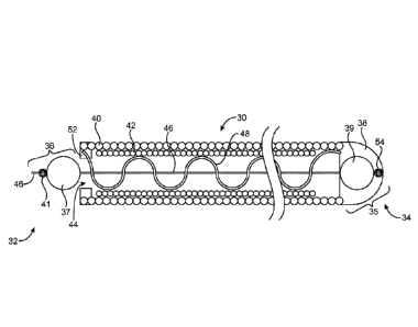
[0021] Referring now to Fig. 2, a cutaway side view reveals some of the
details of
embolic implant 30. In Fig. 2, embolic implant 30 is illustrated in its
primary delivery shape
or configuration. In use, embolic implant 30 is constrained in and delivered
in its primary
configuration via a catheter delivery system. As embolic implant 30 is
deployed from the
distal end of a catheter, it will revert to its secondary configuration. A
secondary
configuration according to the invention may be curved, hooked, J-shaped,
spiral, helical,
complex, or any three dimensional configuration that is suitable for the
therapeutic objectives
for use of the device. An example of a complex configuration according to the
invention is
illustrated in Fig. 11, and described in detail below.
[0022] In the delivery configuration illustrated in Fig. 2, embolic implant
30 has a
proximal end 32 and a distal end 34. A primary coil 40 and an inner coil 42
extend from the
proximal end 32 to the distal end 34, and surround lumen 44. Primary coil 40
is constructed
of thin Platinum wire, and inner coil is constructed of soft, kink resistant
Nitinol. Disposed at
proximal end 32 of embolic implant 30 is proximal constraint assembly 36.
Proximal
constraint assembly 36 is made of proximal constraint sphere 37, fiber 46, and
knot 41.
Proximal constraint sphere 37 may be constructed from gold or tin solder,
Platinum,
Titanium, stainless steel, or other suitable material. Proximal bond 52 is
disposed in an
annular fashion near proximal end 32, may be formed from polyester, acrylic
adhesive, or
other suitable material. The material forming proximal bond 52 may be reflowed
or
otherwise molded around the proximal end of primary coil 40. Proximal assembly
36
prevents proximal constraint sphere 37 from entering lumen 44. Proximal
constraint sphere
37 also plays a role in the delivery of embolic implant 30, and is configured
to be releaseably
retained by a delivery tool or device such as those disclosed in US Patent
Application No.
14/460,234.
[0023] Disposed at distal end 34 is distal assembly 35. Defining distal
assembly 35 are
distal tip 38, distal sphere 39, and distal knot 54. Within lumen 44, and
extending the length
of lumen 44, is fiber 46. In addition to being disposed in lumen 44, fiber 46
is disposed
-4-
CA 02936827 2016-07-12
WO 2015/109007 PCT/US2015/011449
within and through an internal channel or through hole (not visible) of
proximal constraint
element or proximal constraint sphere 37.
[0024] Fiber thus extends proximally through lumen 44, through proximal
constraint
sphere 37, and out of proximal constraint sphere 37 at proximal end 32. Fiber
46 is knotted to
form proximal knot 41. Fiber 46 is thus anchored at the proximal end 32.
Proceeding in the
opposite direction, fiber 46 extends distally of proximal constraint element
37, through lumen
44, and through distal sphere 39, which has, similar to proximal constraint
sphere 37, an
internal channel or through hole (not visible in Fig. 2). During construction
of implant 30,
fiber 46 is also knotted to form distal knot 54. Fiber 46 is thus anchored at
distal end 34.
Fiber 46 is stretch resistant, and may be constructed from a suitable polymer
such as
polyethylene, ultra high molecular weight polyethylene (UHMVVPE),
polypropylene, or other
suitable material. Because fiber 46 is stretch resistant, it will prevent
stretching of primary
coil 40 and inner coil 42, stretching which could potentially plastically
deform the coils and
interfere with the retractability of embolic implant 30 within a catheter, and
potentially
interfere with the ability of embolic implant 30 to reconfigure from its
linear delivery
configuration to its secondary configuration.
[0025] Distal sphere 39, which also may in the alternative have different
shape, is
retained by atraumatic distal tip 38. Atraumatic distal tip 38 is formed from
a polymeric
material such as polyester, an acrylic adhesive, or other suitable material.
The material is
injected, molded, reflowed, extruded, or otherwise placed around distal sphere
39, fiber 46
and distal knot 54 to securely bond the components one to another and to form
an atraumatic
distal tip. The embedding or other retention of distal sphere 39 also serves
to prevent distal
sphere 39 from entering lumen 44. Distal assembly 35, in conjunction with
proximal
assembly 36, thereby maintains tension upon fiber 46, and helps prevent
stretching and
distortion of primary coil 40 and inner coil 42.
[0026] Also disposed in lumen 44 is shape wire 48. Shape wire 48 is
anchored in and
extends from proximal bond 52, through lumen 44, and to distal tip 38. Wire 48
is formed
from Nitinol or another suitable shape memory material. Wire 48 confers the
desired complex
secondary configuration on embolic coil 30. The proximal end of shape wire 48
is retained by
proximal bond 52. The distal end of shape wire 48 is anchored to or secured by
atraumatic
distal tip 38. Because shape wire 48 is constructed of Nitinol, it is highly
kink resistant, and
confers softness on embolic implant 30, while at the same time reliably
conferring a desired
secondary shape on embolic implant 30. In the alternative, a relatively thin
platinum wire
-5-
CA 2936827
may be used to construct primary coil 40, also conferring softness on embolic
implant 30,
enhancing the safety of the device.
[0027] In an alternative embodiment (not pictured), shape wire 48 may be
ground or otherwise
formed so that it is of a smaller diameter at its proximal end relative to its
distal end. The diameter
of shape wire 48 may increase gradually or incrementally from proximal end 32
to distal end 34.
The resulting embolic coil would be of a more robust or a stiffer secondary
shape at the distal end
and a softer coil near the proximal end. The largest shape wire diameter would
be a diameter based
upon the level of robustness desired at the distal end of the device.
[0028] Alternative embodiments of the invention described above are
illustrated in Figures 3-10.
The embolic coils or embolic implants described below and illustrated in the
figures have some
elements in common with the embodiment illustrated in Fig. 2, though some of
the common elements
are arranged in alternative configurations than the configuration of Fig. 2.
In order to be concise, the
description of every detail of each element will not be repeated for each
embodiment.
[0029] The embodiment illustrated in Fig. 3 will now be described. Embolic
implant 60 has a
proximal end 62 and a distal end 64. Disposed at distal end 64 is distal
assembly 71. Embolic
implant 60 includes primary coil 66 and optional inner coil 68, both of which
surround lumen 69.
In the embodiment of Fig. 3, primary coil 66 is constructed of Platinum, and
inner coil 68 is
constructed of Nitinol, though the coils may be constructed of other suitable
materials and remain
within the scope of the invention. Inner coil 68 may optionally be processed
to impart shape
memory characteristics. Proximal constraint assembly 70 is disposed at
proximal end 62. Fiber 73
is threaded through a through hole (not visible) of proximal sphere 74, and
knotted to form a first
proximal knot 78. Fiber 73 is also knotted to form a second proximal knot 80.
In an alternative
embodiment, second proximal knot 80 is formed distal of the proximal end of
coil 66, permitting
some sliding movement of proximal constraint assembly 70. This sliding
movement would be
limited by proximal sphere 74 and knot 80. After formation of second proximal
knot 80, during
construction of implant 60, polyester, or an acrylic adhesive is reflowed,
molded, or otherwise
disposed at the proximal end of primary coil 66, to form proximal bond 85.
Proximal bond 85 is a
solid structure that anchors or secures fiber 73 and second proximal knot 80.
Proximal bond 85,
proximal sphere 74, fiber 73, first proximal knot 78, and second proximal knot
80 together define
proximal constraint assembly 70. Proximal bond 85 prevents proximal constraint
sphere 74 from
entering lumen 69, helping to maintain tension
-6-
Date Re9ue/Date Received 2021-06-10
CA 02936827 2016-07-12
WO 2015/109007 PCT/US2015/011449
on fiber 73, and preventing stretching and deformation of primary coil 66 and
secondary coil
68.
[0030] Also secured by or anchored to proximal bond 85, and extending
distally through
lumen 69, is shape wire 76. Wire 76 is embedded in or otherwise bonded to
proximal bond 85
near proximal end 62. Shape wire 76 is processed to impart a secondary shape
on embolic
implant 60. The profile of shape wire 76 may be altered to exhibit either a
consistent or
varied profile along its length. A larger profile shape wire will exhibit a
more robust shape,
and a smaller profile shape wire will exhibit a softer coil. Shape wire 76
extends distally and
is anchored to distal bond 86. Distal bond 86 may be formed using similar
techniques as
those used to form proximal bond 85. However, in the implant 60, distal bond
86 defines a
more ring-like structure than proximal bond 85. Distal bond 86 surrounds the
distal end of
primary coil 66.
[0031] Fiber 73 also extends distally, through lumen 69, and through a
through hole of
distal sphere 72. Fiber 73 is knotted to form distal knot 84 near distal end
64. Distal bond 86
prevents distal sphere 72 from entering lumen 69 at distal end 64. Both
proximal bond 85 and
distal bond 86 serve to maintain tension in stretch resistant member 73, and
to prevent
stretching and potential elongation of primary coil 66 and inner coil 68.
[0032] As mentioned above, prior to assembly of embolic implant 60, a
secondary
configuration is conferred upon wire 76. However, embolic implant 60 and wire
76 are
constrained in a generally linear, or delivery configuration by a delivery
catheter or
comparable device so that embolic coil 60 may be delivered intravascularly.
After delivery of
embolic implant 60 to a vessel or within an aneurysm of a subject, wire 76
will revert from its
linear delivery configuration to its secondary configuration (not pictured).
Consequently,
embolic implant 60 will also revert to its secondary configuration, such as,
for example, the
configuration illustrated in Fig. lb above.
[0033] Fig. 4 illustrates a component of an alternative embodiment
according to the
invention. Fig. 4 illustrates only the distal assembly 90, which in use would
be disposed at the
distal end of an embolic implant according to the invention. Distal assembly
90 can be used
as an alternative to the distal assemblies illustrated in Figs. 2-3, in the
fabrication of an
embolic coil or embolic implant. Distal assembly 90 includes the distal end of
fiber 92. Fiber
92 is knotted to form distal knot 94. Material such as polyester, acrylic
adhesive, or other
suitable material is reflowed, molded, injected, or otherwise formed around
fiber 92 and
distal knot 94 to form atraumatic distal tip 96. Distal tip 96 secures or
anchors fiber 92 and
distal knot 94, maintaining tension on fiber 92. Distal tip 96 also bonds to
the distal end of
-7-
CA 2936827
primary coil 98, a portion of which is shown in Fig. 4, and to the distal end
of shape wire 97, to
together form a component of distal assembly 90.
[0034] Fig. 5 illustrates yet another alternative embodiment of a distal
assembly according to
the invention. Distal assembly 100 includes fiber 102. Fiber 102 is passed
through a tubing segment
104 and knotted to form distal knot 106. Distal tip 108 is formed from a cured
material such as
polyester, acrylic adhesive, or other suitable material that is reflowed,
molded, injected, or
otherwise placed around and bonded with tubing segment 104, fiber 102, the
distal end of primary
coil 107, and distal knot 106. Distal end of shape wire 109 may also be
anchored to distal tip 108 or
mechanically locked with tubing segment 104. Distal assembly 100 defines an
atraumatic tip and
maintains tension on fiber 102.
[0035] Fig. 6 illustrates the distal end only of another alternative
embodiment according to the
invention. Distal assembly 110 is disposed at the distal end of primary coil
112, shown in cross
section in Fig. 6. A polymer, such as, for example, polypropylene, is melted
and reflowed to bond
to the distal end of primary coil 112, and to form atraumatic distal tip 114.
Also secured to distal tip
114 during the foregoing process are the distal ends of shape wire 115 and
fiber 116, which are
embedded in distal tip 114.
[0036] Fig. 7 illustrates an alternative embodiment of the proximal portion
only of an implant
according to the invention. Proximal assembly 120 includes proximal sphere
122. Proximal loop 124 is
formed from a wire that is formed into a loop, passed through a through hole
of proximal sphere 122,
and welded to proximal sphere 122. Polymer fiber 126 is in turn looped or
threaded through proximal
loop 124 to secure fiber 126 to proximal assembly 120. Proximal bond 128 is
bonded to the proximal
end of primary coil 130, in a fashion similar to the methods described above.
Shape wire 125, at its
proximal end, is also bonded to or secured by proximal bond 128. Proximal bond
128 prevents proximal
sphere 122 from entering lumen 131, and prevents stretching and/or permanent
deformation of primary
coil 130 and secondary coil 132, the proximal ends of which are shown in Fig.
7.
[0037] Fig. 8 illustrates another embodiment of a proximal assembly
according to the
invention. Proximal assembly 140 includes proximal constraint sphere 142.
Proximal constraint
sphere 142 includes a through hole (not visible) through which wire 144 is
threaded and then
welded to proximal constraint sphere 142. Wire 144 has a proximal end 146 and
a distal end 148.
At the distal end 148 of proximal assembly 140, wire 144 is flattened and
drilled or otherwise
processed to form hole 150. Fiber 152 is threaded through hole 150, and
looped. Alternatively, two
lengths of fiber 152 may be used in order to double the tensile strength of
fiber 152. Though not
-8-
Date Re9ue/Date Received 2021-06-10
CA 2936827
pictured in Fig. 8, fiber 152 extends distally through the lumen of an embolic
implant. Proximal
bond 154 is fonited in a similar fashion to that described above in relation
to previously described
embodiments, and bonds to the proximal end of primary coil 160 and optionally
to the proximal
end of shape wire 155. When it is a component of an embolic coil, proximal
constraint assembly
140 helps maintain tension on fiber 152 and prevents stretching and/or
deformation of the embolic
coil. Secondary coil 162 is also pictured.
[0038] Fig. 9 illustrates an alternative embodiment of an embolic implant
according to the
invention. Embolic implant 200 exhibits many advantages over prior art
implants, including proximal
softness that enhances safety. Embolic implant 200 in particular exhibits
progressively increasing
softness from its distal end 201 to its proximal end 205. In other words,
distal end 201 exhibits a more
robust secondary or three dimensional shape than does proximal end 205. And
proximal end 205
exhibits greater overall compliance and softness. An implant such as embolic
implant 200 can be
shape set to, upon release from the constraints of a delivery catheter, return
to a shape such as, for
example, the configuration illustrated in Fig. 11. Fig. 11 will be discussed
in greater detail below.
[0039] Embolic implant 200 is shown in cross section in Fig. 9, in order
that its features may be
readily viewed. Embolic implant 200 includes primary coil 202. Primary coil
may be constructed
from any of the materials suitable for the coils described above. Primary coil
202 includes an internal
lumen 203. Disposed in and extending through lumen 203 is fiber 204. Also
disposed in lumen 203
are elliptical hole washers 212, one near proximal end 205 and one near distal
end 201. (The term
elliptical hole washers is used herein to describe a washer having a round
hole and an elliptical hole.
It will be understood that any washer having a plurality of holes or apertures
may be used to form an
embodiment within the scope of the invention.) Fiber 204 is threaded through
elliptical holes 209 of
elliptical hole washers 212. Fiber 204 also traverses elliptical hole 209 at
proximal end 205, extends
beyond proximal end 205 and is attached to a proximal constraint assembly 207.
Proximal constraint
assembly 207 will be described in greater detail below.
[0040] Also extending through lumen 203 is primary shape wire 206. Each end
of primary shape
wire 206 extends through an elliptical hole washer 212, via apertures 213.
Further, each end of primary
shape wire 206 is optionally flattened or affixed to a broadened element 211
to prevent primary shape
wire 206 from passing back through apertures 213. Primary shape wire 206 is
most advantageously
constructed from Nitinol. Primary shape wire 206 is shape set to confer a
secondary shape on embolic
implant 200. Coupled to primary shape wire 206 is distal support wire 208.
Distal support wire 208 is
linked to shape wire 206 towards the distal end 201 of embolic implant 200. In
the example of Fig. 9,
-9-
Date Re9ue/Date Received 2021-06-10
CA 2936827
distal support wire 208 is attached to shape wire 206 at bonds 210. Where it
is coupled to distal support
wire 208, the stiffness or robustness of shape wire 206 is augmented by distal
support wire 208, and
both members confer the secondary configuration of embolic implant 200. The
absence of distal support
wire 208 near proximal end 205 permits primary shape wire 206 to exhibit a
softer, less robust
secondary shape, and creates the progressively increasing softness of proximal
end 205.
[0041] Also disposed at each end of primary coil 202 are weld joints 221.
In the example of
Fig. 9, weld joints 221 are constructed of a Platinum-Platinum bond. Weld
joints 221 each anchor
elliptical hole washers 212 to primary coil 202. After construction of weld
joints 221, atraumatic
tips 214 are formed from a molded polymer or adhesive. Fiber 204 is also
secured within
atraumatic tips 214. Atraumatic tip 214 disposed at distal end 201 also
secures distal peg 222,
which will be described in greater detail below.
[0042] Turning for now to proximal end 205, elliptical hole washer 212
prevents proximal
constraint assembly 207 from entering lumen 203. Proximal constraint assembly
accordingly helps
maintain tension on fiber 204. Several structures define proximal constraint
assembly 207. These
structures include fiber loop 217, proximal constraint element or proximal
constraint sphere 216,
adhesive 218, and proximal wire 220. Knot 215 is also pictured. Fiber loop 217
is threaded through
a hole in proximal constraint sphere 216. Proximal wire 220 is in turn
threaded through the
proximal end of loop 217. Loop 217 thereby links proximal constraint sphere
216 and proximal
wire 220, and forms a mechanical lock of fiber 204 at proximal end 205.
Adhesive 218 is molded
or applied to secure proximal wire 220, loop 217, and proximal constraint
sphere 216.
[0043] Returning now to distal end 201, fiber 204 is threaded distally
through embolic implant
lumen 203 and then through aperture 209 of washer 212 disposed at distal end
201. Fiber 204 is tied,
knotted, or otherwise linked to distal peg 222. Distal peg 222 can be formed
from stainless steel,
platinum, or other similarly rigid material. Distal peg 222 and fiber 204 are
embedded or otherwise
anchored or bonded to the distal atraumatic tip 214, forming a mechanical lock
adjacent to distal
elliptical hole washer 212. Together, proximal constraint assembly 207 and
distal peg 222 maintain
tension on fiber 204, which thereby enables embolic implant 200 to resist
stretching and elongation.
[0044] Turning now to Fig. 10, an alternative embodiment according to the
invention will be
described. Embolic implant 250 is shown in its linear, delivery configuration.
Following release
from the constraints of a delivery catheter (not pictured), embolic implant
will revert to a shape set
secondary configuration. The secondary configuration may be any of a number
-10-
Date Re9ue/Date Received 2021-06-10
CA 02936827 2016-07-12
WO 2015/109007 PCT/US2015/011449
of shapes according to the invention, including the complex shape illustrated
in Fig. lb
above, and Fig. 11 described below.
[0045] Embolic implant 250 has a proximal end 251 and a distal end 253.
Elliptical hole
washer 261is disposed at proximal end 251 and elliptical hole washer 262 is
disposed at distal
end 253. Embolic implant 250 includes a primary coil 252 that is shape set
during the
manufacturing process to impart a secondary, deployed configuration on embolic
implant
250. Primary coil 252 surrounds lumen 254. Disposed within lumen 254 is fiber
258. In a
fashion similar to that described in relation to Fig. 9, fiber 258 extends
through elliptical hole
257 of elliptical hole washer 262, and through elliptical hole 269 of
elliptical hole washer
261. After passing through elliptical hole washer 261, fiber 258 is looped
back upon itself to
form loop 271, brought into lumen 254, and knotted to itself to form knot 259.
[0046] Also disposed within lumen 254 is distal support wire 256. Distal
support wire
256 renders the secondary configuration of embolic implant 250 more robust in
the distal
region in which distal support wire 256 lies. (Embolic implant 250 is more
softly shaped near
its proximal end 251.) Distal support wire 256 is attached to fiber 258 at
bond 260. Distal
support wire 256 extends at its distal end through aperture 263 of elliptical
hole washer 262.
The distal end of distal support wire 256 is optionally flattened to form a
broadened element
280, or attached to a broadened element 280, to mechanically lock distal
support wire 256 to
elliptical hole washer 262.
[0047] Weld joint 264 is constructed at proximal end 251 in a fashion
similar to that
described above, and atraumatic tip 275 is formed from reflowed or molded
polymer,
adhesive, or a combination thereof. Weld joint 264 anchors primary coil 252
and elliptical
hole washer 261 at proximal end 251. At distal end 253, weld joint 267
similarly bonds
primary coil 252 and elliptical hole washer 262. A molded or reflowed polymer,
adhesive or
comparable material forms atraumatic tip 277.
[0048] Proximal constraint assembly 265 is similar to the proximal
constraint assembly
described above in relation to Fig. 9. Proximal constraint element or proximal
constraint
sphere 266 is linked to fiber 258 and proximal constraint wire 270. Fiber 258
is either
threaded through or wrapped around proximal constraint sphere 266 and looped,
and
proximal constraint wire 270 is threaded through loop 271. Adhesive 268
secures proximal
constraint sphere 266, fiber 258 and proximal wire constraint wire 270.
Elliptical hole washer
261 prevents proximal constraint sphere 266 from entering lumen 254, and
prevents
stretching and/or deformation of primary coil 252. At distal end 253, fiber
258 is tied or
otherwise coupled to distal peg 272, which is embedded in or otherwise bonded
or anchored
-11-
CA 02936827 2016-07-12
WO 2015/109007 PCT/US2015/011449
to distal atraumatic tip 275, Securing fiber 253 to distal peg 272 helps
maintain tension on
fiber 258.
[0049] As mentioned above, embolic implant 250 can be shape set to revert
to a
secondary configuration such as the configuration illustrated in Fig. 1B
above. An
advantageous step in shape setting implant 250 includes the step of shape
setting primary coil
252. Primary coil 252 is first formed by winding or otherwise forming
continuous turns of a
length of Platinum wire about a straight mandrel. The coiled wire can then be
heat set to
"remember" the primary coil shape. The Platinum primary coil formed from
continuous turns
can then be shaped around a fixture bearing a desired secondary shape. The
Platinum primary
coil is then heat set to "remember" the shape of the fixture. Of particular
advantage in
forming a low profile coil that readily fills empty space within an aneurysm
or within a frame
defined by another implant, is utilizing a fixture around which the primary
coil turns are
wrapped. In other words, the fixture is disposed within the lumen of the
primary coil during
the heat setting step, instead of the primary coil being first coiled, and
then wrapped around
the exterior of the fixture. Such a step results in smaller primary diameter
coils and lower
profile secondary configurations.
[0050] Turning now to FIG. 11, an embodiment according to the invention
will be
described. While Fig. lb above illustrates an example of a complex secondary
shape of an
implant according to the invention, Fig. 11 illustrates an alternative complex
secondary
shape, or secondary configuration of an embolic implant constructed according
to the
invention. Fig. 11 illustrates a plan, or topside view of implant 300 in its
deployed
configuration, outside a vessel of a subject, such as, for example, on a
laboratory bench top.
Similar to the embodiments described above, the embodiment illustrated in Fig.
11 also has a
delivery configuration, similar to that illustrated in Fig. 9, that is
generally linear, that permits
the device to be loaded into and delivered via a catheter or comparable
delivery tool (not
pictured). In its secondary configuration outside a vessel, implant 300 has a
proximal
segment 302. Proximal segment 302 is shaped by a relatively soft or flexible
shape wire (not
visible in Fig. 11). The shape wire imparting the secondary shape to proximal
segment 302 is
soft or flexible either because of a small diameter, a fine grind, or other
processing step which
produces a relatively soft filament. A wide range of flexibility, or softness,
of the filament is
within the scope of the invention, and the term "relatively" is used here to
mean in
comparison to distal segment 304, which will be discussed below.
[0051] Proximal segment 302 has a secondary (or deployed) configuration,
outside of a
vessel that is helical. Alternatively, a proximal segment may have a secondary
configuration
-12-
CA 2936827
that is complex, similar to the secondary configuration of distal segment 304,
described in more detail
below. In yet another alternative embodiment, a proximal segment according to
the invention may
have a straight or linear configuration. Though a wide range of outer
diameters of the helix of
proximal segment 302 are within the scope of the invention, in the example
illustrated here, the outer
diameter of proximal segment 302 is approximately 2-30 mm. In a preferable
embodiment, the outer
diameter of proximal segment 302 is less than the outer diameter of distal
segment 304, when both
proximal segment 302 and distal segment 304 are in their secondary
configurations. Techniques for
forming the secondary configuration of proximal segment 302 are known in the
art, and include, for
example, wrapping the shape wire disposed within proximal segment 302 around a
mandrel and heat
treating the segment so that it will return via shape memory behavior to the
helical shape. Alternative
techniques for achieving the shape memory objective are within the scope of
the invention.
[0052] Implant 300 also has a distal segment 304, as mentioned above.
Distal segment 304 also
includes, disposed within its interior and therefore not visible in Fig. 11, a
shape wire that is
fabricated from a wire, filament, or comparable structure that is stiffer
relative to that used to fabricate
proximal segment 302. (Alternatively, a coil may be shape set to return to the
configuration of Fig. 11
upon release from a constraint.) The shape of distal segment 304 may be formed
from a wire or
filament that is of greater thickness than that used to fabricate proximal
segment 302, in a fashion
similar to that described in relation to Fig. 2 above. As another example,
distal segment 304 may
include, similar to that pictured in Fig. 9 above, a support wire coupled to
the shape wire, the support
wire extending only the length of distal segment 304. As yet another example,
additional processing
steps such as annealing or other steps may be undertaken with respect to the
material used to fabricate
the filament that forms the support wire of distal segment 304. Regardless of
the technique used to
manufacture the shape wire of distal segment 304, the resulting secondary
structure is a stiffer or
more robust three dimensional object than that of proximal segment 302.
[0053] In addition, as can be viewed in Fig. 11, distal segment 304 has a
secondary
configuration that is more complex than the generally helical shape of
proximal segment 302. In an
alternative embodiment according to the invention, a distal segment may have a
secondary
configuration that is helical, similar to the secondary configuration
described in more detail above,
in relation to the description of proximal segment 302. In the example
illustrated in Fig. 11, the
deployed shape of distal segment 304 is characterized as having sides 306, top
308, and bottom
312. Primary coil 310 is visible in Fig. 11. Taken together, sides 306, top
308, and bottom 312
-13-
Date Re9ue/Date Received 2021-06-10
CA 02936827 2016-07-12
WO 2015/109007 PCT/US2015/011449
generally define a cubic shape having rounded corners. Therefore, distal
segment 304 can be
described as having the shape of a cube. The term "cube" is used here to
denote a three
dimensional shape having several faces, and a particular embodiment according
to the
invention may or may not have six faces. The corners and edges of each face
may be squared
or rounded, curved or straight. Each face may or may not be of equal
dimensions as each
other face. Further, as is visible in Fig. 11, the secondary shape of distal
segment 304 frames
some open "interior" space, and much of the coiled element defines the outer
edges of the
secondary shape of distal segment 304.
[0054] In addition to having a very different secondary shape than proximal
segment 302,
distal segment 304 also has a larger outside profile or outer diameter than
proximal segment
302. For example, in the embodiment illustrated in Fig. 11, distal segment 304
has an outer
diameter of approximately 3-32 mm. Techniques for shaping distal segment 304
include a
series of steps of wrapping the stretch resistant member of distal segment 304
around a
specialized mandrel or comparable tool, and heat treating the distal wire
member so that it
returns to the secondary shape imparted by the tool. Alternative techniques
for fabricating the
stretch resistant member disposed within distal segment 304 are within the
scope of the
invention.
[0055] The combination of both this larger outer diameter, the
concentration of material
at the outer edges of the shape, and the stiffer internal wire of distal
segment 304 cause distal
segment 304 to function much like an "anchor" for implant 300 within a vessel.
In other
words, distal segment 304 exerts some outward radial force against a vessel
wall when
implant 300 is deployed within a vessel. And, when deployed within a blood
vessel of a
subject, blood flow may carry proximal segment into the "interior" or distal
segment 304,
filling distal segment 304, and effectively preventing further blood flow
through implant 300.
In this respect, implant 300 effectively has an "anchor" segment and a
"filler" segment,
resulting in a soft, well packed embolic implant.
[0056] Turning now to Fig. 12, yet another alternative embodiment according
to the
invention will be described. Embolic implant 400 shares many of the same
features of the
embolic implant illustrated in Fig. 9. Embolic implant 400 exhibits many
advantages over
prior art implants, including softness that enhances safety. Embolic implant
400 in particular
boasts the feature of progressively increasing softness from its distal end
401 to its proximal
end 405. In other words, distal end 401 exhibits a more structured secondary
or three
dimensional shape than proximal end 405. And proximal end 405 exhibits greater
overall
compliance and softness. An implant such as embolic implant 400 can be shape
set to, upon
-14-
CA 2936827
release from the constraints of a delivery catheter, return to a secondary
shape such as, for example,
the configuration illustrated in Fig. 11 above.
[0057] Embolic implant 400 is shown in cross section in Fig. 12, in order
that its features may
be readily viewed. Embolic implant 400 has a primary diameter of approximately
0.018 inches.
Embolic implant includes primary coil 402. Primary coil may be constructed
from any of the
materials suitable for coils described above. Primary coil 402 includes an
internal lumen 403.
Primary coil 402 is shape set to exhibit a secondary shape following release
from a delivery
catheter (not pictured). Primary coil 402 may be constructed from Platinum or
other suitable shape
memory material. Extending through lumen 403 is fiber 404. Fiber 404 also
extends beyond
proximal end 405 and is attached to a proximal constraint assembly 407.
Proximal constraint
assembly 407 will be described in greater detail below. Also extending through
lumen 403 is
primary shape wire 406. Primary shape wire 406 is most advantageously
constructed from Nitinol.
Coupled to primary shape wire 406 is distal support wire 408. Distal support
wire 408 is linked to
primary shape wire 406 towards the distal end 401 of embolic implant 400. The
stiffness of shape
wire 406 is augmented by distal support wire 408, and both members confer the
secondary
configuration on embolic implant 400. The absence of distal support wire 408
at proximal end 405
creates the progressive softness of proximal end 405. In the example of Fig.
12, distal support wire
408 is bonded to primary shape wire 406 at bonds 410.
[0058] As mentioned above, primary shape wire 406 extends essentially the
length of embolic
implant 400. Each end of shape wire 406 extends through an elliptical hole
washer 412, via
apertures 413. Elliptical hole washers 412 are disposed at each end of primary
coil 402. Also
disposed at each end of primary coil 402 is a molded polymer or adhesive 421,
each of which
secures elliptical hole washers 412 to primary coil 402 and fiber 404, and
forms atraumatic tips
414. Both proximal end 405 and distal end 401 have atraumatic tips 414.
[0059] Several structures define proximal constraint assembly 407. As
mentioned above, fiber
404 extends beyond proximal end 405. Fiber 404 is looped back onto itself to
form loop 417. After
forming loop 417, fiber 404 extends back into lumen 403, and is secured to
itself via knot 415.
Loop 417 links proximal constraint sphere 416 and proximal wire 420. Adhesive
418 is molded or
applied to secure proximal wire 420, loop 417, and proximal constraint sphere
416. Elliptical hole
washer 412 and adhesive 421 prevent proximal constraint sphere 416 from
entering lumen 403.
-15-
Date Re9ue/Date Received 2021-06-10
CA 02936827 2016-07-12
WO 2015/109007 PCT/US2015/011449
[0060] Extending distally through lumen 403, fiber 404 is tied, knotted, or
otherwise
linked to distal peg 422. Distal peg 422 and fiber 404 are embedded or
otherwise anchored to
the distal atraumatic tip 414. Together, proximal constraint assembly 407 and
distal peg 422
maintain tension on fiber 404, which thereby enables embolic implant 400 to
resist stretching
and plastic deformation.
[0061] Unlike the embodiment of Figure 9, embolic implant 400 also includes
jacket 425.
Jacket 425 wraps or encases primary coil 402. Jacket 425 is preferably
constructed from a
thrombogenic material such as polyester, polypropylene, silk, or other
suitable material. The
thrombogenic material or materials may be monofilament or multi-filament
fibers. Jacket 425
may be constructed by wrapping, winding, braiding, threading or otherwise
arranging the
fiber or fibers in engagement with coil 402. Jacket 425 may be constructed to
form a
"sleeve" like structure that is placed over coil 402, or applied directly to
coil 402 to form
jacket 425.
[0062] The foregoing examples are not intended to limit the scope of the
invention. All
modifications, equivalents and alternatives are within the scope of the
invention. As an
example, a proximal constraint element or a distal constraint element
according to the
invention need not be a sphere, but may be a disc, a block, a tear drop, or of
any suitable
alternative shape.
-16-