Note: Descriptions are shown in the official language in which they were submitted.
CA 2936841
DEVICE AND METHOD TO TREAT VAGINAL ATROPHY
CROSS REFERENCE TO RELATED APPLICATIONS
[0001] This Application claims the benefit of US Provisional Patent
Application No. 61/933,712
(filed January 30, 2014); US Provisional Patent Application No. 61/947,715
(filed March 4, 2014);
and US Provisional Patent Application No. 61/982,475 (filed April 22, 2014).
[0002] <deleted>
BACKGROUND
[0003] The prior art has a variety of devices and methods directed to female
sexual functioning and
response. Many of these devices are designed to provide sexual pleasure to
women with normally
functioning sexual response.
[0004] In some women, sexual response is less than normal or totally absent.
For example,
estrogen-deficient women may experience vaginal discomfort during intercourse
due to reduced
lubrication and reduced resilience of vaginal tissue. Over time, atrophy of
vaginal tissue may
worsen, thereby diminishing sexual response even further.
[0005] The prior art also includes devices and methods to treat disorders of
the female sex organs.
For example, US 6,741,895 describes a vaginal probe that applies heat,
vibration, electrical
stimulation and/or pressure to vaginal nerves to treat female sexual
dysfunction. US 2007/0021809
describes devices and methods that apply topical heating or cooling to treat
inflammation or
irritation of a woman's genitals. There is no data showing that any of these
prior art devices and
methods provides a lasting benefit to women experiencing vaginal atrophy or
other diminished
sexual function, however.
SUMMARY OF THE DISCLOSURE
[0006] The invention provides devices and methods for improving the health of
vaginal tissue (e.g.,
through increasing vaginal blood flow) by applying energy, such as ultrasound
energy, to tissue in
and around the vagina. The benefits of this energy application persist after
the energy application
ceases; the therapy therefore provides an effective treatment for vaginal
atrophy
- 1 -
Date Re9ue/Date Received 2021-06-25
CA 02936841 2016-07-12
WO 2015/116512
PCT/US2015/012825
[0007] In particular, the invention provides methods and devices for
increasing blood flow,
lubrication, elasticity and resilience of the vagina and surrounding tissue
in, e.g., estrogen-
deficient women and/or women with vaginal atrophy or discomfort. According to
the invention,
energy is introduced locally into the woman's genital tissue. The applied
energy may be
chemical energy, electrical energy, ultrasound energy, RF energy, suction,
photonic energy,
electromagnetic radiation or light-based (e.g., pulsed or continuous laser).
The energy may be
applied intravaginally, near the vulva without penetrating the vagina, or
outside the subject's
body.
[0008] One aspect of the invention provides a method of treating vaginal
tissue atrophy in a
female subject. In some embodiments, the method includes the steps of:
engaging an energy
delivery element with tissue in or around the subject's vagina; applying
energy to the tissue from
the energy delivery element; and increasing blood flow to internal vaginal
tissue to an increased
level above a baseline level of blood flow to the internal vaginal tissue, the
increased level of
blood flow to the internal vaginal tissue persisting after the applying step
ceases.
[0009] In some embodiments, the energy applied by the energy delivery element
is ultrasound
energy delivered, e.g., at a frequency between 0.5 MHz and 4 MHz, an intensity
between 0.25
W/cm2 and 5 W/cm2 and/or at a duty cycle in a range of 20%-80%. In these
embodiments, the
energy delivery element may include an ultrasound probe and/or an ultrasound
coupling
medium. In these methods, the energy may be applied for a period between 30
seconds and 6
hours.
[0010] In some embodiments of the method, the engaging step includes the step
of engaging the
energy delivery element, such as the ultrasound coupling medium, with tissue
exterior to the
subject's vagina. Alternatively or additionally, the engaging step may include
the step of
engaging the energy delivery element with tissue inside the subject's vagina.
In some
embodiments, the engaging step may include the step of engaging the energy
delivery element
with tissue of the subject's abdomen or pelvis.
[0011] In some embodiments, the method includes the step of measuring a
physiologic
parameter of the subject's tissue in or around the subject's vagina and
controlling energy
delivery from the energy delivery element based on the measured parameter. The
measured
physiologic parameter may be, e.g., temperature, blood flow or vaginal
lubrication.
[0012] Another aspect of the invention provides a device for treating vaginal
tissue atrophy in a
female subject. The device may have an energy delivery element adapted to
engage tissue in or
around the subject's vagina and an energy source adapted to deliver energy to
the energy
delivery element to increase blood flow to internal vaginal tissue to an
increased level above a
- 2 -
CA 2936841
baseline level of blood flow to the internal vaginal tissue such that the
increased level of blood flow
to the internal vaginal tissue persists after energy application ceases.
[0013] In some embodiments, the energy applied by the energy delivery element
is ultrasound
energy delivered, e.g., at a frequency between 0.5 MHz and 4 MHz, an intensity
between 0.25
W/cm2 and 5 W/cm2 and/or at a duty cycle in a range of 20%-80%. In these
embodiments, the
energy delivery element may include an ultrasound probe and/or an ultrasound
coupling medium.
[0014] In some embodiments, the ultrasound coupling medium is disposed within
a container
adapted to cover a vaginal opening and tissue around the vaginal opening. The
container may be
adapted to conform to the subject's tissue. In some embodiments, the
ultrasound coupling medium
includes a gel on a surface of the energy delivery element.
[0015] In various embodiments the energy delivery element may be adapted to
engage tissue
exterior to the subject's vagina and/or inside the subject's vagina. In some
embodiments the energy
delivery element may be adapted to engage tissue of the subject's abdomen or
pelvis.
[0016] In some embodiments the device includes a sensor adapted and configured
to measure a
physiologic parameter of tissue in or around the subject's vagina when the
energy delivery element
is engaged with tissue in or around the subject's vagina, with the device
being further configured to
use information from the sensor to control energy delivery from the energy
delivery element. The
measure physiologic parameter may be, e.g., temperature, blood flow or vaginal
lubrication.
[0016A] Various embodiments of the claimed invention relate to a device for
treating vaginal
tissue atrophy in a female subject, comprising: an energy delivery element
comprising an
ultrasound coupling medium and adapted to conform to tissue in or around the
subject's vagina;
and an ultrasound transducer adapted to deliver energy to the energy delivery
element to
increase vaginal lubrication and blood flow to internal vaginal tissue to an
increased level
above a baseline level of blood flow to the internal vaginal tissue such that
the increased level
of blood flow to the internal vaginal tissue persists after energy application
ceases, the
ultrasound transducer comprising an ultrasound generator adapted to provide
ultrasound energy
at a frequency between 0.5 MHz and 4 MHz, the ultrasound transducer in contact
with the
ultrasound coupling medium, wherein the device is configured for self-
application.
- 3 -
Date Recue/Date Received 2022-01-26
CA 2936841
BRIEF DESCRIPTION OF THE DRAWINGS
[0017] The novel features of the invention are set forth with particularity in
the claims that follow.
A better understanding of the features and advantages of the present invention
will be obtained by
reference to the following detailed description that sets forth illustrative
embodiments, in which the
principles of the invention are utilized, and the accompanying drawings of
which:
[0018] Figure 1 is a cross-sectional view of an embodiment of the invention in
position on a
subject.
[0019] Figure 2 is an elevational view showing position of the device of
Figure 1 on the subject.
[0020] Figure 3 is an exploded view of another embodiment of a device
according to the invention.
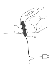
[0021] Figure 4 is a partial cross-section view of yet another embodiment of a
device according to
the invention in position in a subject.
- 3a -
Date Re9ue/Date Received 2021-06-25
CA 02936841 2016-07-12
WO 2015/116512
PCT/US2015/012825
[0022] Figure 5A is cross-sectional view of still another embodiment of a
device according to
the invention in position in a subject.
[0023] Figure 5B is a perspective view of the device of Figure 5A.
[0024] Figure 6 is a perspective view of another embodiment of a device
according to the
invention in position on a subject.
[0025] Figures 7, 8, 9 and 10 present data from use of the invention to treat
subjects.
DETAILED DESCRIPTION
[0026] The invention provides methods and devices that can be used to increase
blood flow,
lubrication, elasticity and resilience of the vagina and surrounding tissues
in estrogen-deficient
women (from natural causes or from the consequences of medical therapies), in
women with
vaginal atrophy, and/or in women who experience vaginal discomfort, whether
constantly or
specifically during sexual intercourse. In some embodiments, the device
applies energy locally
to genital tissue (e.g., vaginal tissue) to increase blood flow to the genital
area and to improve
and/or prevent deterioration of tissue health and natural lubrication. The
methods and devices
may also improve the symptoms of female sexual dysfunction.
[0027] In some embodiments, energy (including but not limited to thermal
energy, mechanical
energy, electrical energy, electromagnetic energy, radiofrequeney energy,
ultrasound energy and
chemical energy) is applied to the walls of the vagina from an external source
via, e.g.,
conductive, inductive, radiative or convective heat transfer to the tissue.
[0028] Figures 1 and 2 show one embodiment of a device according to an
embodiment of the
invention. The device includes an energy delivery element 4 adapted to engage
tissue around the
opening 7 of the vagina 6 of the subject 2. Also shown in these figures are
the subject's uterus 8,
bladder 10, urethral opening 9, vulva 12 and clitoris 11. As shown in cross-
section in Figure 1
and in phantom in Figure 2, in this embodiment energy delivery element 4
engages only exterior
genital tissue (to avoid irritation of sensitive vaginal tissue) and covers
the subject's vulva 12,
clitoris 11, urethral opening 9 and vaginal opening 7. Other embodiments could
cover more or
less of the subject's genitalia and/or the subject's mons pubis or proximal
thighs. In this
embodiment, energy delivery device is connected to an energy source 3, such as
an ultrasound
generator.
[0029] According to methods of this invention, energy may be applied from
energy source 3
through energy delivery element 4 to the patient's genitalia on an acute basis
or on a regular
basis, such as daily or weekly. In some embodiments, the energy delivery
element may be
incorporated into underwear. In some embodiments, the energy delivery element
may be
- 4 -
CA 02936841 2016-07-12
WO 2015/116512 PCT/US2015/012825
attached directly to the subject's body and held in place by suction,
adhesives or other securing
mechanisms. The energy delivery element may be reusable or disposable.
[0030] In some embodiments, the energy source may be combined with the energy
delivery
element. In some embodiments, the energy delivery element is flexible and may
include flexible
circuitry.
[0031] Figure 3 shows in an exploded view an embodiment of the device of this
invention
adapted to delivery ultrasound energy to tissue around a subject's vagina
(such as the vulva and
introitus) to increase blood flow in vaginal tissue. The energy delivery
element 30 is designed to
be applied completely externally without penetrating the vagina to avoid
irritation of sensitive
vaginal tissue. Energy delivery element 30 has a coupling pad 32 and a
transducer assembly 34;
both of these elements may be reusable or disposable.
[0032] The coupling pad 32 has a main structure 36 and a coupling structure
38. Main structure
36 may be deformable, yet rigid enough to support the coupling structure 38.
Main structure 36
may be formed from two plates or sections connected together. In one
embodiment, a top plate
(not shown) has a cut-out (not shown) to accommodate the coupling structure
38, and a bottom
plate 40 has cut-outs 42 to accommodate connection to the ultrasound
transducer 34. Main
structure 36 may be formed from biocompatible or non-allergenic materials such
as silicone
rubber, soft plastics, fabrics or flexible foams.
[0033] Main structure 36 is configured to attach to the subject's body in a
manner that maintains
intimate contact between the coupling structure 38 and tissue around the
subject's vagina, such
as the vulva. Attachment mechanisms (not shown) such as straps or tensile
supports wrapping
around the subject's waist, double-stick tape, glue or other adhesive
(including temperature-
sensitive adhesive) can be used to attach the main structure to the subject's
body. The
attachment mechanism may also employ an element inserted into the subject's
vaginal opening
or anus. The subject's underwear can also be used instead of, or in addition
to, such attachment
mechanisms, in the manner of a panty liner. Projections, wing-like features,
adhesive strips,
materials attachable to tissue with static electricity, or Velcro hook and
loop fasteners may also
be used. The attachment mechanism may also employ suction to maintain contact
between the
coupling structure and the subject's skin.
[0034] Main structure 36 may be shaped, e.g., as a rectangle or as a contoured
and filled-out
figure eight (e.g., like a feminine hygiene pad). Main structure 36 may be
shaped to fit in the
subject's underwear. Main structure 36 may be omitted entirely if another
supporting structure is
use, such as, e.g., by building the coupling structure into underwear worn by
the subject.
[0035] The coupling structure 38 can be a solid, but soft, deformable
structure that conforms to
the subject's vulva. In some embodiments, the coupling structure is entirely
external to the
- 5 -
CA 02936841 2016-07-12
WO 2015/116512
PCT/US2015/012825
vaginal canal; vaginal penetration may irritate sensitive vaginal tissue or
cause discomfort for
women with atrophy or vaginal discomfort. The coupling structure 38 ensures
safe and effective
energy delivery to vaginal tissue. The conforming design of the coupling
structure prevents skin
burns and ensures appropriate energy conduction between the transducer and the
target vaginal
tissue by minimizing air pockets between the two surfaces.
[0036] In one embodiment, coupling structure 38 is convex and shaped to
conform to the
subject's vulva and introitus (e.g., elliptical or ovoid). Coupling structure
38 is formed from
biocompatible, easily deformable and sonolucent material, such as gels like
pectin, gelatin, low-
durometer rubbers, low-durometer silicone or other non-porous soft materials.
In some
.. embodiments, the coupling structure is a deformable container containing a
fluid or semi-solid
such as water (e.g., deionized water, distilled water), oil (e.g., mineral
oil), gel, gelatin or other
sonolucent and biocompatible fluid or semi-solid. The container may be made
from silicone,
PTFE, nylon, HDPE or other plastic.
[0037] In some embodiments, coupling gel (not shown) is placed between the
coupling structure
38 and the subject's skin to enhance acoustic coupling with the subject's
tissue. The gel may be
pre-applied to the coupling structure at the time of manufacture and covered
with a protective
strip or other covering that is removed at the time of use. The gel may also
be applied to the
coupling structure by the user at the time of use.
[0038] In some embodiments, the transducer assembly 34 includes a damping back
plate 44
made, e.g. of semi-deformable, biocompatible or non-allergenic material(s)
such as, e.g.,
polymers like HDPE, PEEK, acrylic, polyethylene or nylon. Transducer assembly
34 also has
one or an array of piezo-ceramic or CMUT ultrasound transducers 46 to apply
diffuse or focused
ultrasound energy to the tissue around the subject's vagina through coupling
pad 32. The
transducer assembly 34 may have wire leads and a plug or other interface 48
that can connect the
transducer(s) 46 to an ultrasound generator (not shown). In other embodiments,
the transducer(s)
46 can connect to a generator wirelessly.
[0039] When the device is assembled for use, the transducers 46 are oriented
with respect to
coupling pad 32 to ensure the vulva and/or introitus are in the appropriate
acoustic field and the
ultrasound waves penetrate to the target vaginal tissue and appropriate
vascular bed. (In some
embodiments, the target of the ultrasound energy (unfocused or focused) may be
further tuned to
cover the lower third of the vaginal canal.) The orientation between the
transducers 46 and
coupling pad 32 can be achieved with geometric features 50 that mate between
the transducers
46 (or other part of the transducer assembly 34) and the backing plate 36 of
coupling pad 32,
such as a key and slot arrangement, a pin and hole arrangement, etc. The
orientation can also be
achieved through the design of the coupling structure 38. The orientation of
the transducers can
- 6 -
CA 02936841 2016-07-12
WO 2015/116512 PCT/US2015/012825
be tuned for each subject's anatomy, either manually (e.g., through different
size options) or
though automatic transducer repositioning based on closed-loop feedback during
use. The
orientation may also be adjusted by the user based on her anatomy by using
feedback from
reflected ultrasound energy displayed in a user interface to assist in
adjustment (e.g., blinking
.. lights of different colors similar to a tuning instrument). The surface-to-
surface interface
between the transducer face and the coupling pad to maintain good acoustic
coupling between
surfaces can be achieved by adhesives, spring-loaded features, mechanical snap
fits, and/or
elastic materials (e.g., silicone or elastic band) that wrap around the back
of the transducer.
Ultrasound gel may also be applied to the interface between the transducer
face and the coupling
pad to enhance acoustic coupling.
[0040] In some embodiments, the device has features that provide feedback to
the user regarding
the quality and sufficiency of the contact between the transducer face and the
coupling pad,
and/or contact between the coupling pad the subject's tissue, for reasons,
e.g., of the subject's
safety, device integrity and efficacy of treatment. These feedback mechanisms
can include
simple feature locks that provide "snap" sounds to inform the user that the
part is seated,
impedance or other sensors between the transducer assembly and the coupling
pad that provide
direct feedback to the user, and/or alarms on the ultrasound generator that
are based on sensor
feedback. Such feedback can notify the user of inadequate and/or unsafe
coupling between the
transducer and the coupling structure, or inadequate and/or unsafe coupling
between the coupling
pad and the subject's tissue. Feedback may also be used to prevent the device
from applying
energy to the subject unless the device is assembled and applied in a manner
providing sufficient
acoustic coupling and to cease applying energy if the acoustic coupling ceases
to be sufficient at
some point during the therapy.
[0041] Embodiments of the device discussed above employ a coupling pad that
does not
.. penetrate the vagina. Ultrasound energy is applied to the subject's
external anatomy near the
vulva and the introitus through the coupling pad; the malleability of the
coupling pad allows it to
fill spaces between the transducer(s) and the tissue to provide adequate
acoustic coupling so that
ultrasound energy is effectively transmitted to the tissue. In other
embodiments, the coupling
member and even the ultrasound transducer may be inserted into the vagina.
[0042] The devices described with respect to Figures 1-3 may be used in a
variety of ways to
improve vaginal health. The device may be used on an as-needed basis prior to
sexual relations
to induce lubrication. The device may be used for between 30 seconds and 6
hours to induce
enough lubrication for sexual intercourse. The device may also be used weekly,
multiple times a
week, daily or multiple times a day as a treatment or a preventative measure
to improve overall
vaginal health and rejuvenate the tissue (i.e., improving mucosal vascularity,
restoring tissue
- 7 -
CA 02936841 2016-07-12
WO 2015/116512
PCT/US2015/012825
elasticity, increasing constitutive lubrication, etc.). The device may also be
used for shorter
periods or longer periods of time. An automatic duty cycle or treatment
algorithm may be
employed to control overall energy delivery to the tissue and ensure safety
while providing
optimal therapy for desired outcomes. Alternatively, the device may be
customized by the user
to modify therapy as needed.
[0043] In some embodiments, the ultrasound therapy device described above may
be used to
treat vaginal atrophy and other conditions by generating and applying
ultrasound to tissue around
the subject's vagina at one or more frequencies between 0.5 MHz and 4 MHz, at
intensities
between 0.25 Wicm2 and 5 W/ cm2, continuously or at a duty cycle in the range
of 20%-80%.
Variations may include the use of other ultrasound waveform parameters.
[0044] The device may have sensors embedded within the coupling pad for
measurement of
various physiologic parameters. These parameters may include mucosal/dermal
blood flow
(measure, e.g., with Doppler ultrasound, Doppler laser imaging, temperature or
plethysmography); vaginal lubrication (e.g., using humidity sensors, absorbent
materials or other
methods for detecting lubrication and/or secretion); and other appropriate
parameters. The
sensors embedded with the device may allow for closed-loop feedback control of
the therapy
application.
[0045] For example, vulvar tissue temperature may be measured by a sensor in
the coupling pad.
If the temperature rises to a level that could potentially cause damage to the
subject, the feedback
loop would automatically adjust energy delivery parameters or even stop energy
delivery. As
another example, the device could increase energy delivery if the temperature
of the subject's
skin is not high enough. In yet another example, the device could measure
physiologic outcome
parameters (e.g., vaginal blood flow and/or lubrication) and automatically
increase or decrease
therapy delivery to achieve the desired outcome (e.g., vaginal blood flow
and/or lubrication).
[0046] The device may be controlled, e.g., via buttons, dials, and/or a
touchscreen interface on
the generator or controller unit. A remote control may also be used to
communicate control
commands to a tabletop generator. The device may also integrate the generator,
energy delivery
unit and control interface. A mobile application may be used to control the
device and/or obtain
data regarding the device and its use.
[0047] A user interface providing data regarding the device and its use may be
used for
biofeedback based on measured physiologic parameters. The biofeedback may be
structured in
the form of a game whereby the subject elicits and augments physiologic
responses to the energy
applied by the device to the vulvovaginal tissues with additional biofeedback
responses and/or
nervous control.
- 8 -
CA 02936841 2016-07-12
WO 2015/116512 PCT/US2015/012825
[0048] Other control interfaces may allow more direct control of transducer
function. Such a
user interface via mobile platform technology can also be used with a wireless
coupling pad to
allow the user to control the device discreetly over the course of routine
(and potentially
continuous) use without having to directly handle the coupling pad or the
generator.
[0049] The device may be portable, such as by being wearable. The device may
contain a
rechargeable energy source, such as batteries and/or a capacitor, that allows
the user to use the
device without being connected to line current. The ultrasound generator could
be rechargeable
and roughly the size of a smart phone or insulin pump to facilitate
portability.
[0050] The device may be used at home, and the patient may be able to apply
the therapy
.. herself. The device may be designed in such a way to be conducive to one-
hand self-application,
or it may be entirely hands-free, while still maintaining proper tissue
contact, orientation and
treatment efficacy. Alternatively, the device and therapy may be applied by a
partner.
[0051] Figure 4 shows an embodiment of the invention in which some of all of
the device 60 is
inserted into the vagina 6. As with the embodiments described above, energy
may be applied
acutely or on a continuous basis. The device may be controlled by an external
device, powered
by an external device or contain all power and control elements within the
inserted element 60.
In the embodiment shown in Figure 4, the insertable energy delivery element 60
connects with
an external energy source (not shown) via wire(s) 62.
[0052] Insertable devices may be elongated, as shown in Figure 4, ring-like,
as shown in Figures
.. 5A and 5B, or have other shapes, such as cylindrical, spherical, T-shaped,
custom shaped to
match the subject's vaginal cavity, etc. The insertable portion of the device
may be inflatable,
flexible and/or compressible for ease of insertion and for ensuring sufficient
contact with the
vaginal wall (made, e.g., of a biocompatible polymer like silicone or
polyurethane), and it may
attach to the vaginal wall (via, e.g., a mild, non-painful suction force). The
device may be
reusable or disposable. The energy applied by energy delivery element 60 may
be some form of
electric resistance heating, chemical heating, radiofrequency energy,
ultrasound energy,
vacuum/suction mechanical energy, photonic energy, electromagnetic radiation
or other methods
of transferring heat to tissue. For example, for ultrasound-based devices, the
ultrasound may be
delivered at one or more frequencies between 0.5 MHz and 4 MHz, at intensities
between 0.25
W/cm2 and 5 W/ cm2, continuously or at a duty cycle in the range of 20%-80%. A
device that
uses laser energy may utilize a pulsed laser or a continuous laser.
[0053] Figure 6 shows yet another embodiment of the invention in which an
energy delivery
element 70 (e.g., an ultrasound transducer and coupling medium) is in the form
of a patch
attached to the subject's abdomen or pelvis area 72. The energy delivery
element is controlled
by an external controller 76 to provide energy (such as ultrasound energy) to
the subject's vagina
- 9 -
CA 02936841 2016-07-12
WO 2015/116512 PCT/US2015/012825
and/or surround tissue 74. In embodiments employing ultrasound, the ultrasound
waves may be
focused or unfocused. The patch device may be reusable or disposable.
[0054] EXAMPLES
[0055] Example 1
[0056] In a controlled study, the legs of two female subjects were placed in
stirrups, and a
baseline measurement of the blood pulse amplitude of the subjects' internal
vaginal wall was
measured with a vaginal plethysmography probe for two minutes. The beat
average of the raw
signals from the probes for the two subjects are plotted on Figures 7 and 8 as
the first bar at each
of times 0, 0.5 min, 1 minute and 2 minutes. A speculum exam was then
performed on the
subjects, and the plethysmography probe was once again used to measure the
blood pulse
amplitude in the subjects' vaginal tissue for two minutes (not shown in
Figures 7 and 8).
Thereafter, a condom filled with warm water and coated on both sides with a
sonolucent gel was
placed on each subject's vaginal area to cover her vulva and introitus, and an
ultrasound
transducer was placed against the condom. Ultrasound energy was introduced via
the ultrasound
transducer at 1 MHz, 1.5 W/cm2 intensity, 50% duty cycle for 8 minutes. After
the 8 minute
treatment, the ultrasound transducer and condom were removed, and
plethysmography
measurements were obtained with the vaginal plethysmography probe at 0, 0.5
minutes, 1
minute, 2 minutes, 3 minutes, 5 minutes and 10 minutes after energy
application ceased. The
beat average of the raw signal from the probe for the two subjects are plotted
on Figures 7 and 8
as the second bar at each of times 0, 0.5 min, 1 minute and 2 minutes and as
the only bar at times
thereafter. The increase in vaginal tissue blood pulse amplitude after
treatment, and the
persistence of this increase, shows the efficacy of the therapy in increasing
blood flow to vaginal
tissue.
[0057] Example 2
[0058] As in Example 1, the legs of a female subject were placed in stirrups,
and a baseline
measurement of the blood pulse amplitude of the subject's internal vaginal
wall was measured
with a vaginal plethysmography probe for two minutes. The beat average of the
raw signal from
the probe is plotted on Figure 9 as the first bar at each of times 0, 0.5 min,
1 minute and 2
minutes. A speculum exam was then performed on the subject, and the
plethysmography probe
was once again used to measure the blood pulse amplitude in the subject's
vaginal tissue for two
minutes (not shown in Figure 9). Thereafter, a condom filled with warm water
and coated on
both sides with a sonolucent gel was placed on the subject's vaginal area to
cover her vulva and
introitus, and an ultrasound transducer was placed against the condom.
Ultrasound energy was
introduced via the ultrasound transducer at 1 MHz, 1.5 W/cm2 intensity, 50%
duty cycle for 8
minutes. After the 8 minute treatment, the ultrasound transducer and condom
were removed, and
-10-
CA 02936841 2016-07-12
WO 2015/116512
PCT/US2015/012825
plethysmography measurements were obtained with the vaginal plethysmography
probe at 0, 0.5
minutes, 1 minute, 2 minutes, 3 minutes, 5 minutes and 10 minutes after energy
application
ceased (not shown in Figure 9). The water-filled balloon was then re-applied
to the subject's
vaginal area to cover her vulva and introitus, and the ultrasound transducer
was once again
placed against the condom. Ultrasound energy was introduced via the ultrasound
transducer at 1
MHz, 1.5 W/cm2 intensity, 50% duty cycle for a second 8 minute treatment.
After this second
treatment, the ultrasound transducer and condom were removed, and
plethysmography
measurements were once again obtained with the vaginal plethysmography probe
at 0, 0.5
minutes, 1 minute, 2 minutes, 3 minutes, 5 minutes and 10 minutes after the
second energy
application ceased. The beat average of the raw signal from the probe after
the second
ultrasound application is plotted on Figure 9 as the second bar at each of
times 0, 0.5 min, 1
minute and 2 minutes and as the only bar at times thereafter. The increase in
vaginal tissue
blood pulse amplitude after treatment, and the persistence of this increase,
shows the efficacy of
the therapy in increasing blood flow to vaginal tissue.
[0059] Example 3
[0060] As in Examples 1 and 2, the legs of a female subject were placed in
stirrups, and a
baseline measurement of the blood pulse amplitude of the subject's internal
vaginal wall was
measured with a vaginal plethysmography probe for two minutes. A speculum exam
was then
performed on the subject, and the plethysmography probe was once again used to
measure the
blood pulse amplitude in the subject's vaginal tissue for two minutes (not
shown in Figure 10).
The beat average of the raw signal from the probe after the speculum exam is
plotted on Figure
10 as the first bar at each of times 0, 0.5 min, 1 minute and 2 minutes.
Thereafter, a condom
filled with warm water and coated on both sides with a sonolucent gel was
placed on each
subject's vaginal area to cover her vulva and introitus, and an ultrasound
transducer was placed
against the condom. Ultrasound energy was introduced via the ultrasound
transducer at 1 MHz,
1.5 W/cm2 intensity, 50% duty cycle for 8 minutes. After the 8 minute
treatment, the ultrasound
transducer and condom were removed, and plethysmography measurements were
obtained with
the vaginal plethysmography probe at 0, 0.5 minutes, 1 minute, 2 minutes, 3
minutes, 5 minutes
and 10 minutes after energy application ceased. The beat average of the raw
signal from the
probe is plotted on Figure 10 as the second bar at each of times 0, 0.5 min, 1
minute and 2
minutes and as the only bar at times thereafter. The increase in vaginal
tissue blood pulse
amplitude after treatment, and the persistence of this increase, shows the
efficacy of the therapy
in increasing blood flow to vaginal tissue.
- 11 -