Note: Descriptions are shown in the official language in which they were submitted.
CA 02937045 2016-07-15
WO 2015/116832
PCT/US2015/013551
SYSTEMS AND METHODS FOR USING EYE MOVEMENTS TO DETERMINE
STATES
STATEMENT REGARDING FEDERALLY SPONSORED RESEARCH
[0001] This invention was made with government support under Grant
PS-2010-0667 by the Spanish Ministry of Education. The government of Spain has
certain rights in the invention.
BACKGROUND
[0002] The
present disclosure generally relates to systems and methods for
acquiring data from a subject and, more particularly, to systems and methods
for
gathering and analyzing information about the subject's eye movements to
determine
or predict a state of the subject, including conditions such as hypoxia.
[0003] Human
brain function is highly vulnerable to hypoxic insults. Hypoxia
impairs vision, cognition, motor control, and can cause severe incapacitation
and
death. Reports on the effects of hypoxia on visual function (i.e. dark
adaptation,
central brightness contrast, color vision, and central acuity) have been
confounded by
subjective and environmental factors (i.e. changes in ambient light level and
non-compliance by flight crews in accurately reporting physiological
disabilities such
as color blindness). The few studies that have addressed the effects of
hypoxia on
objective oculomotor metrics, such as saccadic velocity, have obtained
inconsistent
results. The question of whether hypoxia modulates oculomotor metrics
therefore
remains open.
[0004] Acute
hypoxia, defined as decreased availability of oxygen in the body's
tissues that can lead to dyspnea, rapid pulse, syncope, visual dysfunction,
and mental
disturbances such as delirium or euphoria, is one of the most serious single
hazards in
military and civil aviation. Thus, international organizations such as the US
Federal
Aviation Administration and the European Aviation Safety Agency recommend
hypoxia training (i.e. performance training while reducing oxygen availability
to the
trainee) as a mandatory part of flight and cabin crew instruction. Altitude
chamber
training¨a well-established method to train aircrews to recognize early
symptoms and
signs of hypoxia¨has not eliminated in-flight hypoxic incidents, however. A
-1-
CA 02937045 2016-07-15
WO 2015/116832
PCT/US2015/013551
complicating factor is that there are wide individual differences in tolerance
to acute
and chronic exposures to reduced oxygen environments.
[0005] Early
and objective detection of the physiological effects of hypoxia can
preempt these symptoms, and is critical to prevent catastrophes in civil and
military
aviation. Considering the above, there continues to be a clear need for rapid,
accurate,
and non-invasive individualized systems and methods for detecting the presence
or
onset of hypoxia.
BRIEF SUMMARY
[0006] The
present invention overcomes drawbacks of previous technologies by
providing systems and methods that afford a number of advantages and
capabilities
not contemplated by, recognized in, or possible in traditional system or known
methodologies related to tracking or determining a subject's state, including
the
detection of hypoxia.
[0007] In one
embodiment, the present disclosure provides a system including a
sensing arrangement that collects eye movement data of a user, an alerting
arrangement that produces an alert to the user in response to receipt of an
alert signal,
and a control unit in communication with the sensing arrangement and the
alerting
arrangement. The control unit includes a data analysis module configured to
extract
one or more current eye movement dynamics from the eye movement data, and a
comparison module configured to receive the one or more current eye movement
dynamics from the data analysis module and compare the one or more current eye
movement dynamics to one or more baseline eye movement dynamics accessible by
the control unit, and to send the alert signal to the alerting arrangement in
response to
a determination that one or more of the compared current eye movement dynamics
diverges from one or more of the baseline eye movement dynamics by a threshold
amount.
[0008] The current eye movement dynamics may include one or more
intersaccadic drift velocities of the user and the data analysis module is
configured to
calculate the one or more intersaccadic drift velocities. The comparison
module may
be configured to compare one or more of the intersaccadic drift velocities to
one or
more threshold drift velocities of the baseline eye movement dynamics. One of
the
intersaccadic drift velocities may be a drift mean velocity. When the drift
mean velocity
is different from the one or more threshold drift velocities by more than the
threshold
-2-
CA 02937045 2016-07-15
WO 2015/116832
PCT/US2015/013551
amount, the alert signal may be an alert of the presence of hypoxia in the
user. One of
the intersaccadic drift velocities may be a current intersaccadic drift
velocity. When
the current intersaccadic drift velocity is different from the one or more
threshold drift
velocities by more than the threshold amount, the alert signal may be an alert
of the
onset of hypoxia in the user. The current intersaccadic drift velocity may be
collected
by the sensing arrangement within ten seconds of the comparison module sending
the
alert signal to the alerting arrangement. The data analysis module may
calculate the
current intersaccadic drift velocity by identifying, in the eye movement data,
a drift
period comprising a duration and a distance and determining the intersaccadic
drift
velocity from the duration and the distance.
[0009] The eye movement dynamics may include one or more saccade
parameters. The saccade parameters may include a saccadic peak velocity and a
magnitude. The comparison module may be configured to compare the current
intersaccadic drift velocity to one or more threshold drift velocities of the
baseline eye
movement dynamics.
[0010] The eye
movement data may be collected from both eyes of the user. The
baseline eye movement dynamics may be obtained from a data model stored in a
model data store accessible by the control unit. The data model may be a
standardized model generated from baseline measurements of one or more non-
user
subjects. The control unit may configured to calibrate the data model to the
user by
obtaining, from the sensing arrangement when the user is in a non-hypoxic
state, a
calibration set of eye movement data, comparing the calibration set to the
standardized model to determine a deviation of the calibration set from the
standardized model, and adapting the data model to the user based on the
deviation.
The calibration set may include a threshold-normal drift velocity for the
user, and
comparing the calibration set to the standardized model may include comparing
the
threshold-normal drift velocity for the user to a threshold-normal drift
velocity for the
standardized model. The control unit may further include a data model
generator
configured to generate the data model by obtaining, from the sensing
arrangement
when the user is in a non-hypoxic state, a portion of the eye movement data,
extracting
from the portion of the eye movement data a plurality of threshold eye
movement data
samples, and creating the data model from the threshold eye movement data
samples.
-3-
CA 02937045 2016-07-15
WO 2015/116832
PCT/US2015/013551
[0011] In
another embodiment, the present disclosure provides a method of
determining a physiological state of a user. The method includes recording
from the
user, during a time-on-duty of the user, eye movement data of one or both of
the user's
eyes without interrupting an activity of the user, comparing the eye movement
data to
one or more baseline measurements, and, if the eye movement data diverges from
one or more of the baseline measurements by a threshold amount, delivering an
alert
to the user. The eye movement data may include one or both of saccade
parameters
and intersaccadic drift parameters. The method may further include calculating
a
current intersaccadic drift velocity of the user from the eye movement data.
Comparing the eye movement data to the baseline measurements may include
comparing the current intersaccadic drift velocity to a threshold
intersaccadic drift
velocity of the baseline measurements. The alert may indicate to the user that
a
hypoxic condition of the user exists.
[0012] The
method may further include recording the baseline measurements from
the user in non-hypoxic conditions. The method may further include obtaining a
standardized data model of eye movement dynamics, recording one or more
threshold
eye movement data samples from the user in non-hypoxic conditions, determining
a
deviation of the threshold eye movement data samples from one or more eye
movement dynamics of the standardized model, and using the deviation to
calibrate
the standardized data model to include the baseline dynamics.
[0013] In one
embodiment of the present invention, systems and methods are
provided for monitoring, recording, and/or analyzing eye movements in situ to
determine whether oculomotor dynamics are being affected by the onset or
presence
of hypoxia. In one aspect, a sensor arrangement may include a camera and
recording
assembly for detecting and recording the eye movements.
[0014] In some
contemplated embodiments, systems and methods using in situ
testing of eye movement dynamics may be employed to identify the onset or
presence
of states or physiological conditions, such as fatigue, hypoxia, stroke,
intoxication,
seizure, and other conditions. The described study has shown that eye saccades
and
the velocity of intersaccadic eye drift are detectably affected by the onset
or presence
of these conditions. A system and method that implements the data recording
and
analysis approaches of the study may alert a user to the presence of these
states or
conditions in a testing environment. In particular, a system in accordance
with the
present invention may include devices and device assemblies that record
baseline
-4-
CA 02937045 2016-07-15
WO 2015/116832
PCT/US2015/013551
data of a subject and generate a data model representing the eye movement data
of
the subject, and further the system may include device and device assemblies
that
record eye movement data in situ and compare it to the data model to determine
if the
user is experiencing or about to experience any of the dangerous conditions.
[0015] In a
contemplated embodiment of the present invention, a system includes
a sensing arrangement that collects eye movement data of a user, and a control
unit in
communication with the sensing arrangement. The control unit may be configured
to
compare the eye movement data to one or more baseline measurements of eye
movement dynamics and, if the eye movement data diverges from one or more of
the
baseline measurements by a threshold amount, generate an alert for delivery to
the
user. Comparing the eye movement data to the baseline measurements may include
calculating a current intersaccadic drift velocity of the user and comparing
the current
intersaccadic drift velocity to one or more threshold drift velocities of the
baseline
measurements. The eye movement data may include one or more saccade
parameters, and comparing the eye movement data to the baseline measurements
may include calculating a current intersaccadic drift velocity of the user
from the
saccade parameters and comparing the current intersaccadic drift velocity to
one or
more threshold drift velocities of the baseline measurements.
[0016] In
another embodiment of the present invention, a method of determining a
physiological state of a user includes recording from the user eye movement
data of
one or both of the user's eyes, comparing the eye movement data to one or more
baseline measurements, and, if the eye movement data diverges from one or more
of
the baseline measurements by a threshold amount, delivering an alert to the
user.
The eye movement data may include one or both of saccade parameters and
intersaccadic drift parameters.
[0017] In
another embodiment of the present invention, systems and methods of
the present invention may be combined as a kit or apparatus, whose advantages
and
capabilities will be readily apparent from descriptions below.
[0018] The
foregoing and other advantages of the invention will appear from the
following description. In the description, reference is made to the
accompanying
drawings which form a part hereof, and in which there is shown by way of
illustration a
preferred embodiment of the invention. Such embodiment does not necessarily
represent the full scope of the invention, however, and reference is made
therefore to
the claims and herein for interpreting the scope of the invention.
-5-
CA 02937045 2016-07-15
WO 2015/116832
PCT/US2015/013551
BRIEF DESCRIPTION OF THE DRAWINGS
[0019] The present invention will hereafter be described with reference to
the
accompanying drawings, wherein like reference numerals denote like elements.
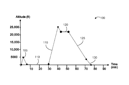
[0020] FIGS. 1A-B are charts illustrating an oculomotor performance
experiment in
accordance with the present invention.
[0021] FIGS. 2A-B are charts illustrating experimental results regarding
the
intersaccadic drift mean velocity of experiment subjects in a hypoxia group
(FIG. 2A)
and in a control group (FIG. 2B).
[0022] FIGS. 2C-D are charts illustrating experimental results regarding
the
average drift mean velocity of experiment subjects in a hypoxia group (FIG.
2C) and in
a control group (FIG. 2D).
[0023] FIGS. 3A-B are charts illustrating experimental results regarding
the
saccadic peak velocity of experiment subjects in the hypoxia group (FIG. 3A)
and in
the control group (FIG. 3B).
[0024] FIG. 4 is a diagram of a detection system in accordance with the
present
invention.
[0025] FIG. 5 is a flowchart illustrating a method for detecting hypoxia in
accordance with the present invention.
DETAILED DESCRIPTION
[0026] Systems and methods for detecting onset, presence, and progression
of
particular states, including hypoxia, through observation of eye movements are
described herein. These systems and methods can be further understood through
the
results of one or more experiments by the inventors. The results show that
acute
hypoxia affects oculomotor dynamics, including saccadic metrics and
intersaccadic
drift metrics, with increasing severity as the hypoxia progresses. The results
show, in
particular, that intersaccadic drift velocity increases as acute hypoxia
develops and
progresses, and that select oculomotor dynamics can be tracked against a
baseline to
alert a subject before the effects of hypoxia impair the subject's ability to
take
corrective action. What follows are descriptions of a particular study and its
results,
and methods for practical application of the findings in a detection system.
[0027] Study and Results
-6-
CA 02937045 2016-07-15
WO 2015/116832
PCT/US2015/013551
[0028] The systems and methods may be understood by way of example data
obtained through experimentation. The example data are offered for
illustrative
purposes only, and are not intended to limit the scope of the present
invention in any
way. Indeed, various modifications of the invention in addition to those shown
and
described herein will become apparent to those skilled in the art from the
foregoing
description and the following examples and fall within the scope of the
appended
claims. For example, specific duty conditions and pre-experimental training of
participants are provided, although it will be appreciated that the systems
and
methods may be applied in any oxygen-depleted environment and for any subject
without undue experimentation.
[0029] Materials and Methods
[0030] The study was conducted in conformity with the declaration of
Helsinki and
the Spanish Defence Medical Inspector General's Office's IRB (approval date:
07/26/2012). Written informed consent was obtained from each participant.
Participants attended the Spanish Defence Aero-medical Center (CIMA) for
aviation
medicine training. Most subjects were members of the Spanish Air Force flight
crew
(i.e., pilots and flight engineers). All subjects had normal or corrected-to-
normal vision
and underwent a full physical examination prior to study participation. Six
male
subjects, most of them aircrew operating rotary wing aircrafts receiving
hypoxia
training (mean age, height, and weight: 37 yrs ( 6.4); 176 cm ( 5.1); 85 kg
( 8.5)),
comprised the hypoxia group. Six different male subjects, receiving no hypoxia
training (mean age, height, and weight: 35 yrs ( 11); 181 cm ( 3.8); 83 kg (
7.3)),
comprised the control group.
[0031] The study followed a Pre/Post-Test design. The hypoxia training was
the
between-subjects factor and the eye movement metrics, including intersaccadic
drift
(hereafter drift) velocity, and saccadic velocity and magnitude, were the
dependent
variables. We also recorded the participants' subjective level of fatigue via
standardized questionnaires.
[0032] The CIMA altitude training chamber, manufactured by Environmental
Tectonics Corporation, USA, is a computer-controlled, man-rated, low-pressure
chamber that accommodates 10 subjects and one inside safety observer. A vacuum
pump removes pressure from the chamber to simulate the pressure of a
particular
altitude. The CIMA training involves various hypobaric training regimes; in
this study
we used training Type lb, which consists of depressurizing the hypobaric
chamber to
-7-
CA 02937045 2016-07-15
WO 2015/116832
PCT/US2015/013551
a simulated maximum altitude of 25,000 ft, to conduct a demonstration of acute
hypoxia. FIGS. 1A and 1B illustrate the details of this design. FIG. 1A is a
chart of a
simulated flight 100 showing altitude as a function of time, and FIG. 1B
illustrates the
associated timeline of administering the saccade tasks.
[0033]
Referring to FIG. 1A, an initial ear and sinus check ascent to 5,000 ft MSL,
at stage 105, is followed by a 30-minute denitrogenation period at ground
level, with
the subjects breathing 100% oxygen via a pressure-demand-type oxygen mask, at
stage 110. At stage 115, subjects experience an ascent to 25,000 ft, and at
stage 120
hypoxia conditions are administered at a rate of about 10 mins total, with
each subject
undergoing hypoxia for up to 3.25 min without supplemental oxygen. At stages
125
and 130, the subjects undergo a simulated descent to ground level. The total
duration
of the simulated flight was about 73 mins. Referring to FIG. 1B, the
experiment began
by administering a Pre-Test guided saccade task 150, followed be administering
the
simulated flight 100, and finished with administration of a Post-Test guided
saccade
task 155 that was the same task as the Pre-Test guided saccade task 150.
Control
subjects carried out their regular duties between the Pre- and Post- sessions.
All
participants filled in a self-rating scale of perceived fatigue before each
oculomotor
test.
[0034] As
stated above, each subject experienced hypoxia (equivalent to an
altitude of 22,000 ft) for a maximum of 3.25 min without supplemental oxygen.
All
subjects exhibited cognitive impairment during the hypoxia exposure, as
indicated by
the standard hypoxia demonstration sheet (i.e., pencil and paper test) known
in the
art. Pulse oximetry, measured with a non-invasive pulse oximeter on the
subject's
non-writing-hand's fingertip, confirmed a final oxyhemoglobin saturation
between 62 ¨
77% ( 5.9) Sp02 in each subject.
[0035] Before
and after the subjects entered the hypoxic chamber, we assessed
their oculomotor dynamics via the guided saccade task 150,155, in which we
displayed visual stimuli on a 21-in CRT screen (864 x 1152 pixels, refresh
rate 100 Hz)
located ¨70 cm in front of the subject, who sat on a comfortable chair. This
task
induced a total of 336 saccades, including vertical, diagonal, and horizontal
directions.
Subjects completed the task in ¨15 minutes. Eye position was sampled
binocularly at
500 Hz using the desktop configuration of the EyeLink 1000 eye tracking system
manufactured by SR Research, Ontario, Canada. Data recordation included
detecting
and classifying eye movements and calculating a linear regression on the
-8-
CA 02937045 2016-07-15
WO 2015/116832
PCT/US2015/013551
log-transformed saccadic peak velocities as a function of their magnitudes for
each
subject, where the slope reflected the effect of hypoxia on the saccadic peak
velocity-magnitude relationship.
[0036] The more
specific methods of recording and analyzing eye movements are
as follows. Eye movements were sampled binocularly at 500 Hz using the desktop
configuration of the Eyelink 1000 eye tracking system with a resolution of
0.01 RMS.
Blink periods were identified as portions of the raw data where pupil
information was
missing, and were removed. Additionally, portions of data where very fast
decreases
and increases in pupil area occurred (> 50 units/sample) were classified as
semi-blinks where the pupil is never fully occluded, and were removed. 200 ms
were
added before and after each blink/semi-blink to eliminate the initial and
final parts
where the pupil was still partially occluded. Saccades were identified with a
modified
version of the algorithm developed by Engbert and Klieg! (Engbert & Klieg!,
2003;
Engbert, 2006; Engbert & Mergenthaler, 2006) with A = 6 (used to determine the
velocity threshold for saccade detection) and a minimum saccadic duration of 6
ms. To
reduce the amount of potential noise, only binocular saccades (i.e., saccades
with a
minimum overlap of one data sample in both eyes) were considered.
Additionally, a
minimum intersaccadic interval of 20 ms was imposed, so that potential
overshoot
corrections might not be categorized as new saccades. To calculate saccade
properties such as magnitude and peak velocity, the values for the right and
left eyes
were averaged. Table 2 includes the descriptive statistics for saccades and
drift.
[0037] Drift
periods were defined as the eye-position epochs between saccades,
overshoots, and blinks. 10 ms were removed from the start and end of each
drift period
(because of imperfect detection of blinks and small saccades), and the
remaining
eye-position data was filtered with a low-pass Butterworth filter of order 13
and a
cut-off frequency of 30 Hz. To calculate drift parameters (such as mean
velocity and
duration), an additional 10 ms was removed from the beginning and end of each
drift
period of the filtered data, to reduce edge effects due to the filter. Drifts
shorter than
200 ms were discarded. Finally, because drifts are not generally conjugate,
data from
both the left and right eye was used. Thus, any given drift period had a
duration,
distance (length of the curve traced out by the drift), peak velocity, and
mean velocity
for each eye.
[0038]
Subjective fatigue is a well-known effect of both hypoxia and time on-duty
(TOD). Thus, the effects from hypoxia on oculomotor metrics were disambiguated
-9-
CA 02937045 2016-07-15
WO 2015/116832
PCT/US2015/013551
from those of TOD by requiring participants to complete a self-rating scale of
perceived fatigue, as is known in the art, before each oculomotor test.
[0039] All
subjects received a standard briefing on the effects of simulated altitude
and hypoxia on the day preceding the experiment. On the day of the training,
aircrews
underwent hypoxia training and two measuring sessions (see FIG. 1B) between
9.00
am and 12.30 pm (approximately three hours elapsed between the two sessions).
The
interval between the return to sea level and the start of the Post-Test
session was ¨30
minutes. Control subjects carried out their regular duties between the Pre-
and Post-
sessions.
[0040] The
oculomotor parameters were analyzed following two separate (one for
each dependent variable) 2 x 2 repeated-measures analyses of covariance
(ANCOVAs). Subjective scores of perceived fatigue served as covariates, and
measuring session and group served as factors. This analysis provided
statistical
control for the influence of TOD on the eye movement variables. For the
subjective
scores of perceived fatigue, a 2 x 2 repeated-measures analysis of variance
(ANOVA)
was used, with the two measuring sessions (Pre-Test vs. Post-Test) as the
within-subjects factor and the group (experimental vs. control group) as the
between-subjects factor.
[0041] Results
[0042] Table 1
includes aggregate collected data for the subjective, saccadic, and
intersaccadic drift parameters. The scores of the self-rating scale of
perceived fatigue
(Borg's Scale) range between 6 and 20. Higher scores indicate more subjective
fatigue. The eye movement data includes the calculated means and standard
deviations (in parentheses) from the mean values of each subject in each group
(n =
6). The adjusted means (in square brackets) refer to the group means after
controlling
for the effect of TOD (i.e. by considering the scores of the self-rating scale
of perceived
fatigue as covariates (ANCOVA adjusted means)).
TABLE 1
Pre-Test Post-Test
Control Experimental Control
Experimental
Group Group Group Group
Drift Mean Velocity (deg/sec) 2.31 [2.25] 2.30 [2.35] 2.50 [2.34]
2.85 [2.99]
(0.35) (0.75) (0.44) (1.09)
-10-
CA 02937045 2016-07-15
WO 2015/116832
PCT/US2015/013551
Slope 0.69 [0.69] 0.68 [0.68] 0.69 [0.69]
0.67 [0.67]
Saccadic Magnitude/Peak Velocity (deg/sec) (0.04) (0.04) (0.04)
(0.05)
Borg Scale 6.67 7.67 8.67 8.67
(2.9) (2.0) (2.1) (2.7)
[0043] Table 2
is a more detailed summary of the collected data for intersaccadic
drift parameters and saccadic parameters. Means and standard deviations were
calculated from the mean values of each subject for each group (n = 6). "*"
denotes
statistical significance for the "Group x Measuring Time" interaction. The
adjusted
means (in square brackets) refer to the group means after controlling for the
effect of
TOD by considering the scores of the self-rating scale of perceived fatigue as
covariates (ANCOVA adjusted means). All p-values < 0.05.
TABLE 2
Experimental Group Control Group
Pre-Test Post-Test Pre-Test Post-
Test
INTERSACCADIC DRIFT PARAMETERS
Mean Velocity (deg/sec) 2.29 [2.35] 2.85 [2.99] 2.31 [2.25]
2.48 [2.34]
(0.76) (1.10) (0.35) (0.44)
Peak Velocity (deg/sec) 6.26 [6.50] 7.88 [8.21] 7.26 [7.04]
7.00 [6.67]
(2.08) (2.21) (2.51) (1.59)
Distance (deg) 0.90 [0.92] 1.06 [1.10] 1.05
[1.02] 1.00 [0.95]
(0.27) (0.36) (0.18) (0.15)
Duration (ms) 0.42 [0.42] 0.39 [0.39] 0.47
[0.47] 0.44 [0.44]
(0.07) (0.06) (0.04) (0.05)
Number
2205[2180] 2164[2133] 2091[2117] 2132[2136]
(187) (194) (187) (194)
SACCADES PARAMETERS
Slope (peak velocity/magnitude) 0.69 [0.69] 0.68 [0.68] 0.69 [0.69]
0.67 [0.67]
(0.04) (0.04) (0.04) (0.05)
Slope (mean velocity/magnitude) 0.56 [0.56] 0.55 [0.54] 0.54 [0.54]
0.52 [0.52]
(0.02) (0.02) (0.04) (0.04)
Slope (duration/magnitude) 0.46 [0.46] 0.48 [0.49] 0.48
[0.48] 0.50 [0.50]
-11-
CA 02937045 2016-07-15
WO 2015/116832
PCT/US2015/013551
(0.02) (0.03) (0.05) (0.05)
Mean Velocity (deg/sec) 104.70 [105.69] 99.32 [99.60]
109.44 [108.46] 100.93 [100.65]
(11.23) (11.03) (15.35) (16.08)
Peak Velocity (deg/sec) 219.97 [222.48] 198.29 [198.81]
237.12 [234.61] 208.86 [208.33]
(36.69) (30.13) (37.74) (42.61)
Magnitude (deg) 4.73 [4.77] 4.29 [4.29] 5.42
[5.38] 4.94 [4.94]
(0.75) (0.88) (0.65) (0.93)
Duration (ms) 35.54 [35.78] 33.28 [33.56] 41.66
[41.41] 38.50 [38.21]
(4.06) (3.17) (5.74) (5.45)
Rate [N/s] 1.67 [1.68] 1.61 [1.64] 1.50
[1.48] 1.43 [1.40]
(0.39) (0.53) (0.18) (0.26)
[0044]
Referring to FIGS. 2A-B, there is illustrated the drift mean velocity
distributions before and after hypoxia training for the hypoxia group (FIG.
2A) or
equivalent TOD for the control group (FIG. 2B). Pre-Test is indicated by
reference
numerals 200 (FIG. 2A) and 210 (FIG. 2B), and Post-Test by reference numerals
205
(FIG. 2A) and 215 (FIG. 2B) for the subjects. In the hypoxia group, the
average drift
mean velocity increased from 2.35 deg/s to 2.99 deg/s after accounting for
TOD, an
increase of about 27%. In contrast, the control group average drift mean
velocity
changed by just 4% (from 2.25 to 2.34 deg/s), which is within the margin of
error.
[0045]
Referring to FIGS. 2C-D, there is illustrated the average drift mean velocity
in degrees per second as a function of degrees of horizontal gaze
eccentricity, before
and after hypoxia training for the hypoxia group (FIG. 2C) or equivalent TOD
for the
control group (FIG. 2D). Pre-Test is indicated by reference numerals 220 (FIG.
2C)
and 230 (FIG. 2D), and Post-Test by reference numerals 225 (FIG. 2C) and 235
(FIG.
2D) for the subjects. Mean drift velocity increased significantly from the Pre-
Test to the
Post-Test session in the hypoxia group, but not in the control group.
[0046] The
results show that hypoxia induced an increase in the mean velocity of
intersaccadic drift, suggesting a decrease in visual fixation stability. That
is, drift mean
velocity was significantly higher in the Post-Test session than in the Pre-
Test session
for the hypoxia group, but not for the control group, after controlling for
the effect of
TOD (i.e., by considering the scores of the self-rating scale of perceived
fatigue as
covariates [interaction between measuring session and group: F(1,8) = 10.192,
p <
0.013; np2 = 0.56]. Drift peak velocity and distance covered by drift were
also
significantly higher in the Post-Test than in the Pre-Test session for the
hypoxia group,
-12-
CA 02937045 2016-07-15
WO 2015/116832
PCT/US2015/013551
but not for the control group, consistent with the hypothesis of higher
fixation instability
with hypoxia (see Table 2). Drift duration was not affected (see Table 2).
There were
no significant main effect of hypoxia versus control groups or of Pre- versus
Post-Test
sessions [all F-values < 3]. Saccadic peak velocity decreased from the Pre-
Test to the
Post-Test session for both hypoxia and control groups [F(1, 10) = 7.32, p =
0.02] but
the effect was not statistically significant when controlled for the influence
of TOD. The
average slope of saccadic peak velocity to saccadic magnitude between hypoxia
and
control groups was not statistically significant. FIGS. 3A-B illustrated the
saccadic
magnitude/peak velocity relationships for one experimental subject (FIG. 3A)
and one
control subject (FIG. 3B) at two different measuring times: Pre-Test
(reference
numerals 300 and 310) and Post-Test (reference numerals 305 and 315). The
curves
are the power-law fits to the data from each measuring session.
[0047] The results show that the slope of the saccadic magnitude/peak
velocity
relationship decreased from the Pre-Test to the Post-Test session in both the
hypoxia
and the control groups, suggesting that this effect was due to TOD rather than
hypoxia. Indeed, when compensated for the influence of TOD (i.e., by
considering the
scores of the self-rating scale of perceived fatigue as covariates), no
significant effects
[all F-values < 1] on the saccadic magnitude/peak velocity were found for
either group.
Saccadic magnitude/duration and saccadic magnitude/mean velocity relationships
showed equivalent behaviors to the saccadic magnitude/peak velocity
relationship
(see Table 2). This is consistent with previous reports of the modulatory
effects of TOD
on saccade dynamics. Table 2 includes additional details about the effects of
measuring session and group on other saccadic parameters.
[0048] As regards the self-rating scale of perceived fatigue, the degree of
perceived fatigue increased from the Pre-Test to the Post-Test session in both
groups
[F(1, 10) = 5 p = 0.049; ilp2 = 0.34]. That is, increased TOD resulted in
increased
perceived fatigue in both groups. Neither the main effect of the group nor its
interaction
with the measuring sessions were significant [all F-values < 1]. See Table 1.
[0049] Discussion
[0050] The results show that short-term hypobaric hypoxia gives rise to
variations
in drift velocity. Hypoxia-triggered increases in drift speed may indicate a
decrease in
fixation stability, and the rapid compensations of the oculomotor system to
correct the
ensuing fixation errors. This hypothesis is consistent with the observation
that fixation
instability is one of most frequent symptoms in patients suffering from
cerebral
-13-
CA 02937045 2016-07-15
WO 2015/116832
PCT/US2015/013551
visual-impairment (CV!) after a perinatal hypoxia-ischemia episode. CVI
patients also
exhibit abnormal smooth pursuit behavior. Drift is thought to be under the
control of
smooth eye movements. Thus, our present observations of increased drift speed
after
short-term hypoxia, combined with previous reports of impaired fixation
instability and
smooth pursuit in CVI patients, may indicate a common neural pathway by which
decreased levels of oxygen in the brain lead to transitory or permanent
oculomotor
pathologies, depending on the duration of the hypoxia episode.
[0051] The
effect of hypoxia on saccadic velocity in our study was no longer
significant after controlling for the influence of fatigue due to TOD.
Therefore, the
decrease in saccadic peak velocities observed here is most parsimoniously
explained
by TOD, rather than hypoxia. Thus, the current study reconciles disparate
results from
previous studies.
[0052] One may
wonder if the present changes in drift velocity might have resulted
from increased head motion in the post-acute-hypoxia state. This possibility
seems
unlikely in light of previous research by some of the present co-inventors
showing that
the same eye-tracking system (EyeLink 1000, SR Research) and forehead/chin
rest
used here could detect variations in drift velocity independently of head
motion. Thus,
the most parsimonious explanation for the current results is that drift
velocity is indeed
sensitive to hypobaric hypoxia.
[0053] In
summary, short-term hypobaric hypoxia affected drift, but not saccade,
velocities. This dissociation may arise at the level of the frontal pursuit
area (FPA) in
the cerebral cortex, an area dedicated to the control of slow eye movements,
where
the first sensory-to-motor transformation of low velocity eye movement signals
takes
place. FPA appears to provide commands that drive smooth eye velocity, and
play an
important role in modulating the setting of gain control. Short-term hypobaric
hypoxia
episodes may interfere with these commands, increasing eye instability as a
result.
[0054] System
and Methods for Detecting Dangerous Physiological Conditions,
Including Hypoxia
[0055] Using
the approach of the present invention, a detection system may record
eye movement data from a user, compare the eye movement data to a data model
comprising threshold eye movement data samples, and from the comparison make a
determination whether or not the user's brain function is suffering or is
subject to
hypoxic insult or other dangerous physiological conditions, such as fatigue.
The
-14-
CA 02937045 2016-07-15
WO 2015/116832
PCT/US2015/013551
detection system may alert the user to take corrective action if onset or
presence of a
dangerous condition is detected.
[0056]
Referring to FIG. 4, an embodiment of a detection system 400 may include a
sensing arrangement 412 configured to detect and record eye movement dynamics
of
the user. The sensing arrangement 412 may include one or more sensors suitable
for
collecting the eye movement data. Such sensors may include a camera or other
imaging or motion tracking device capable of recording at a suitably high
speed and
level of detail so that the user's eye movement dynamics, including saccades
and
intersaccadic drift, are captured. A monocular arrangement of one or more
sensors for
one of the user's eyes may be used, or one or more sensors may be included for
each
eye to obtain binocular data. In some embodiments, the sensors may be
miniaturized
or otherwise compact, portable, and non-invasive so as not to interrupt an
activity of
the user while obtaining in situ measurements. The sensors may further be
vehicle-independent, and may be wireless, to facilitate integration of the
sensors into
any deployment of the detection system 400. For example, the sensing
arrangement
412 may include sensors that are integrated into eyewear, such as on the frame
or
within the lenses of a pair of glasses. This allows for eye movement data
collected
even as the user turns his head, and allows the sensors to be positioned close
to the
eyes. In another example, the sensors may be integrated into a heads-up
display for a
vehicle.
[0057] The
sensing arrangement 412 may further include integrated or discrete
devices for processing, storing, and transmitting collected data. Such devices
may
include a processor, volatile and/or permanent memory, a wired or wireless
transmitter, and associated power circuits and power supply for operating the
devices.
Software modules may define and execute instructions for operating the
sensors,
configuring databases, registers, or other data stores, and controlling
transmission of
the data. The collected data may be shared via transmission to a control unit
414 that
may be integrated with or disposed physically remotely from the sensing
arrangement
412. The eye movement data, or a subset thereof, may be transmitted in real-
time as
it is captured by the sensors, or it may be stored for later transmission.
[0058] The
control unit 414 may use the processing hardware (i.e., processor,
memory, and the like) of the sensing arrangement 412, or may include its own
processing hardware for analyzing the eye movement data and generating an
alert to
the user if needed. The control unit 414 may include a plurality of modules
that
-15-
CA 02937045 2016-07-15
WO 2015/116832
PCT/US2015/013551
cooperate to process the eye movement data in a particular fashion, such as
according to the methods described below. Each module may include software (or
firmware) that, when executed, configures the control unit 414 to perform a
desired
function. A data analysis module 416 may extract information from the eye
movement
data for comparison to the data model. The data analysis module 416 may
include one
or more data filters, such as a Butterworth or other suitable bandpass filter,
that retain
only desired signal elements of the eye movement data. The data analysis
module
416 may include program instructions for calculating, from the eye movement
data,
one or more eye movement dynamics, such as saccades and/or intersaccadic drift
velocities, of the user's eyes. The calculation may be performed substantially
in
real-time, such that a calculated intersaccadic drift velocity may be
considered the
current drift velocity of the user's eyes.
[0059] A comparison module 418 may receive the processed eye movement data
from the data analysis module 416 and may compare it to the data model as
described
in detail below. The control unit 414 may include or have access to a model
data store
420 that stores the data model. The model data store 420 may be a database,
data
record, register, or other suitable arrangement for storing data. In some
embodiments,
the data model may simply be a threshold drift velocity, and may thus be
stored as a
single data record in memory accessible by the comparison module 418. In other
embodiments, the data model may be a lookup table, linked list, array, or
other
suitable data type depending on the data samples for eye movement dynamics
needed to be stored in the data model.
[0060] In some
embodiments, the control unit 414 may include a data model
generator 422. The data model generator 422 is a module that receives eye
movement data collected by the sensing arrangement 412 during a modeling step
as
described below. The data model generator 422 may extract, or cause the data
analysis module 416 to extract, information from the collected eye movement
data that
will constitute the threshold eye movement data samples in the data model. The
data
model generator 422 may then create the data model from the threshold eye
movement data samples, and may store the data model in the data model store
420. In
other embodiments, the data model may be generated and stored in the data
model
store 420 by a separate modeling unit (not shown) of the system 400. The
modeling
unit may include its own sensing arrangement, processing hardware, and program
-16-
CA 02937045 2016-07-15
WO 2015/116832
PCT/US2015/013551
modules. One suitable modeling unit is described with respect to the above
study (i.e.,
using the EyeLink 1000).
[0061] The
control unit 414 may include or communicate with an alerting
arrangement 424 configured to produce an alert to the user according to the
results of
the data comparison in the comparison module 418. The alerting arrangement 424
may be any suitable indicator and associated hardware and software for driving
the
indicator. Suitable indicators include, without limitation: a visual display
such as one or
more light-emitting diodes, a liquid crystal display, a projector, and the
like; a bell,
buzzer, or other audible signaling means; and a piezoelectric or other
vibrating device.
In an embodiment, the alerting arrangement 424 may receive an alert signal
generated by, for example, the comparison module 418 when a threshold
deviation in
the eye movement dynamics is detected.
[0062] The
detection system 400 may be used to execute any suitable method of
detecting dangerous conditions that are indicated by eye movement data.
Referring to
FIG. 5, the detection system 400 may execute a method of detecting onset or
presence of hypoxia in the user. At step 500, the system may record baseline
measurements of the eye movement dynamics for the data model. The baseline
measurements are taken of a subject which may or may not be the user. It may
be
advantageous that the data model use baseline measurements of the user himself
in
order to individualize the operation of the system, but the baseline
measurements may
be taken from a non-user subject, or taken from a plurality of subjects and
averaged if
desired. The conditions in which the baseline measurements are recorded may
depend on the desired specificity of the data model. In some embodiments, the
baseline measurements may be taken in normal (i.e., sea-level or other typical
atmospheric oxygen supply) conditions. In other embodiments, the baseline
measurements may be taken in oxygen-depleted or known hypoxic conditions. In
still
other embodiments, as in the study described above, the baseline measurements
may
be taken continuously or at predetermined intervals as the subject is exposed
to a
progression from normal to hypoxic conditions.
[0063] At step
505, the system 400 may calculate one or more threshold drift
velocities from the recorded baseline measurements. The threshold drift
velocities
may depend on the format of the collected baseline measurements. For example,
where only normal-condition or only hypoxic-condition baseline measurements
were
taken, a single threshold drift velocity (i.e., threshold-normal or threshold-
hypoxic drift
-17-
CA 02937045 2016-07-15
WO 2015/116832
PCT/US2015/013551
velocity) may be calculated. Where progressive baseline measurements were
obtained, one or more threshold drift velocities reflecting the subject's
progression
into, and degree of, hypoxia may be calculated. At step 510, the system 400
may
generate the data model for the baseline-tested subject(s). The data model may
represent the progression of the intersaccadic drift velocity of the subject
from normal
conditions to hypoxic conditions, and further beyond a hypoxic threshold into
increasingly severe hypoxia. The data model may be generated and stored in any
suitable format that allows the system 400 to subsequently compare eye
movement
data collected in situ from the user against the data model to determine the
user's
current susceptibility to hypoxia.
[0064] The
steps 500, 505, 510 for obtaining the data model may be performed at
any suitable time before testing the user in situ for signs of hypoxia. In one
embodiment, the steps 500-510 may be performed far in advance and remotely
from
the test environment. In another embodiment, the steps 500-510 may be
performed in
the test environment, immediately preceding testing the user. For example, the
user
may activate the system 400, such as by donning and activating eyewear housing
the
sensing assembly 12, which initiates step 500 of recording the baseline
measurements in the present conditions. Typically, this would be in normal
conditions,
such as when the user is a scuba diver about to dive off of his boat or a
pilot preparing
to fly an aircraft, and only the normal or non-hypoxic eye movement data would
be
collected as baseline measurements. In still other embodiments, the data model
may
be created by the system 400 or another system using a different method than
described above.
[0065] At step
515, optionally the system 400 may calibrate itself to the user if the
data model or comparison method require it. For example, the data model may be
a
standardized model generated from baseline measurements of (a) non-user
subject(s), or the comparison method may determine the presence of hypoxia
from a
percentage deviation from the user's threshold-normal drift velocity value(s).
See
below. In such an embodiment, the system 400 calibrates (step 515) by
recording a
calibration set, such as ten seconds or less but preferably five seconds or
less, of eye
movement data of the user when the system 400 is activated in the test
environment
under normal conditions. The system 400 may compare the calibration data to
the
data model. In one embodiment, this involves determining a deviation of the
user's
-18-
CA 02937045 2016-07-15
WO 2015/116832
PCT/US2015/013551
threshold-normal drift velocity from the threshold-normal drift velocity of
the model.
The system 400 can then adapt the data model to the user.
[0066] At step 520, the system 400 may record in situ eye movement data from
the
user continuously or at predetermined intervals while the system 400 is
activated. At
step 525, the system 400 may calculate, in real-time or at predetermined
intervals, the
user's current drift velocity. At step 530, the system 400 may compare the
current drift
velocity to the data model to determine the user's progression (or lack
thereof) toward
hypoxia. Such progression may be calculated within any suitable paradigm.
Examples
include, without limitation: ratio or percentage by which the current drift
velocity
exceeds the user's or the data model's threshold-normal drift velocity; ratio
or
percentage by which the current drift velocity is below or above the threshold-
hypoxic
drift velocity; comparison of current drift velocity to points on a curve
between
threshold-normal and threshold-hypoxic values in the data model; and the like.
If the
user is neither hypoxic nor within a predetermined proximity to the threshold-
hypoxic
value of the data model, the system 400 returns to step 520 and continues
recording
current data. If the user's condition warrants (i.e., the current drift
velocity is above or
within a certain range of the threshold-hypoxic value), at step 535 the system
400 may
alert the user to take corrective action.
[0067] The described systems and methods may be implemented in any
environment and during any task that may subject the user to dangerous
conditions
that affect eye movements. The various configurations presented above are
merely
examples and are in no way meant to limit the scope of this disclosure.
Variations of
the configurations described herein will be apparent to persons of ordinary
skill in the
art, such variations being within the intended scope of the present
application. In
particular, features from one or more of the above-described configurations
may be
selected to create alternative configurations comprised of a sub-combination
of
features that may not be explicitly described above. In addition, features
from one or
more of the above-described configurations may be selected and combined to
create
alternative configurations comprised of a combination of features which may
not be
explicitly described above. Features
suitable for such combinations and
sub-combinations would be readily apparent to persons skilled in the art upon
review
of the present application as a whole. The subject matter described herein and
in the
recited claims intends to cover and embrace all suitable changes in
technology.
-19-