Note: Descriptions are shown in the official language in which they were submitted.
CA 02938152 2016-07-28
WO 2015/120364
PCT/US2015/014976
ANTIBODIES THAT BIND TO HUMAN TAU AND ASSAY FOR QUANTIFYING
HUMAN TAU USING THE ANTIBODIES
FIELD OF THE INVENTION
The present invention relates to antibodies that specifically bind human Tau
(h-Tau)
and are useful for quantitating h-Tau in biological samples. The invention
also relates to
assays that employ these antibodies.
BACKGROUND
Alzheimer's disease (AD), the most common cause of dementia, is a progressive
neurodegenerative disorder characterized by increasing loss of memory and
cognitive
function. Neuropathological features present in AD include amyloid plaques
made of AP
peptides, the most prominent being Af31 _42 peptide, and neurofibrillary
tangles (NFTs).
In particular, NFTs consist of paired helical filaments (PHFs) which in turn
are
composed of the microtubule associated protein h-Tau (h-Tau). Normally h-Tau
stabilizes a
key cellular network of microtubules that is essential for distributing
proteins and nutrients
within neurons. In AD patients, however, h-Tau becomes hyperphosphorylated,
disrupting
its normal functions, increasing its likelihood to aggregate into PHFs and
ultimately forming
NFTs. It is hypothesized that the formation of NFTs leads to the loss of
synapses and
neurons, and thus ultimately contributes to the development of dementia.
As the extracellular space of the brain is in direct contact with CSF,
biochemical
changes in the brain also affect CSF (Blennow et al., The Lancet Neurology,
Vol. 2, pp. 605-
613, 2003). Studies have shown elevated levels of h-Tau protein in the CSF of
AD patients
compared with normal subjects (Vandermeeren et al., J. Neurochem., Vol. 61,
pp. 1828-1834,
1993; Blennow et al., supra, Hampel et al., Exp. Gerontol, Vol. 45, pp. 30-40,
2010), and
thus h-Tau has been used as a biomarker to diagnose AD (Hampel et al. supra).
Elevated
levels of CSF h-Tau in AD patients have also been shown to correlate with NFT
pathology
(Tapiola et al., NeuroReport, Vol. 8, pp. 3961-3963, 1997).
Recent studies have also shown that measurement of elevated concentrations of
h-Tau
in CSF in combination with decreased concentrations of A31.42 in CSF can aid
in the
diagnosis of AD (Tapiola et al., Arch. Neurol., Vol. 66, pp. 382-389, 2009).
Further studies
have also demonstrated that the ratio of CSF h-Tau/ M3142 is useful in
identifying individuals
1
CA 02938152 2016-07-28
WO 2015/120364
PCT/US2015/014976
with amyloid plaque pathology (Fagan et al., Arch. Neurol., Vol. 68, pp. 1137-
1144, 2011).
The ratio of CSF h-Tau/A[31 -42 has also been shown to predict future
cognitive decline in non-
demented older adults and adults having mild AD (Fagan et al., Arch. Neurol.,
Vol. 64, pp.
343-349, 2007).
In view that the aforementioned CSF biomarkers have been shown to reflect
amyloid
pathology, neurodegeneration, and are able to prognosticate cognitive decline,
they may
become important in the identification of asymptomatic or mild symptomatic AD
patients,
who are most likely to benefit from novel therapeutic interventions.
A requisite for the aforementioned uses of these CSF biomarkers is the
accurate
quantification of the biomarker present in the CSF of the patient. h-Tau, in
particular, is a
difficult protein to quantitate for the following reasons. There are six
different isoforms of h-
Tau varying in size from 352-441 amino acids, all derived from a single gene
by alternate
mRNA splicing (Himmler et al., Mol. Cell Biol., Vol. 9, pp. 1381-1388, 1989;
see Figure 1).
The six h-Tau isoforms differ from one another by the number of (3 or 4)
microtubule
binding domains and the number of (0, 1, or 2) amino terminal inserts of 29
amino acids each
(Goedert etal., Neuron, Vol. 3 , pp.519-526, 1989). The heterogeneity in h-Tau
protein is
effected by post-translational modifications including phosphorylation,
ubiquination,
oxidation and others. In addition, h-Tau is present at low concentrations in
CSF ranging from
about 300ng/L in healthy individuals to 900ng/L in AD patients (Blennow and
Hampel,
Lancet Neurol., Vol. 2, pp. 605-613, 2003).
Immunoassays utilizing monoclonal antibodies have been developed to quantitate
h-
Tau in CSF (Hampel et al, supra; and Kang et al., Clinical Chem., Vol. 59, pp.
903-916,
2013). Given the molecular heterogeneity and low concentrations of h-Tau in
CSF, and the
importance of the h-Tau biomarker in the diagnosis of AD in patients at
different stages of
the disease and its use in to predict future cognitive decline, there remains
a continued need
to develop highly characterized assays that can accurately quantify all
isoforms of h-Tau in
CSF.
2
CA 02938152 2016-07-28
WO 2015/120364
PCT/US2015/014976
SUMMARY OF THE INVENTION
The present invention relates to antibodies, and in particular monoclonal
antibodies
(mAbs) that specifically bind to epitopes in a region of h-Tau that is
conserved in amino acid
sequence (amino acids 104-277) in the six known isofonns of h-Tau: h-Tau-441,
412, 410,
383, 381 and 352 (SEQ ID NOs 2 to 7, respectively, as shown in Table 1 below.
See also
Figure 1).
Table 1.
Amino Acid Sequences of: h-Tau Isoforms, Tau 166 Peptide, A131_42 Peptide,
Amyloid Beta A4 Protein Isoform A Precursor, Epitopes of h-Tau specifically
bound by
mAbs10H8, 19G10 and AT120, and h-Tau Reacting/Non-Reacting with mAbAT120.
Nucleic
Acid Sequence encoding Tau 166 Peptide
Human Amino Acid Sequence SEQ
Tau ID
Isoform or NO
Peptide
Tau Isofonn MHHHHHHD YDIPTTENL YFQGMAEPRQEFEVMEDHAGTYGLGDR
2-441 KDQGGYTMHQDQEGDTDAGLKESPLQTPTEDGSEEPGSETSDAKST
(including PTAEDVTAPLVDEGAPGKQAAAQPHTEIPEGTTAEEAGIGDTPSLED 1
HIS tag in EAAGHVTQARMVSKSKDGTGSDDKKAKGADGKTKIATPRGAAPP
bold f GQKGQANATRIPAKTPPAPKTPPSSGEPPKSGDRSGYSSPGSPGTPGS
oM
RSRTPSLPTPPTREPKKVAVVRTPPKSPSSAKSRLQTAPVPMPDLKN
and TEV
VKSKIGSTENLKHQPGGGKVQIINKKLDLSNVQSKCGSKDNIKHVP
cleavage GGGSVQIVYKPVDLSKVTSKCGSLGNIHHICPGGGQVEVKSEKLDFK
site in italic DRVQSKIGSLDNITHVPGGGNKKIETHKLTFRENAKAKTDHGAEIV
font) YKSPVVSGDTSPRHLSNVSSTGSIDMVDSPQLATLADEVSASLAKQ
GL
Tau Isoform MAEPRQEFEVMEDHAGTYGLGDRKDQGGYTMHQDQEGDTDAGL
2-441 KESPLQTPTEDGSEEPGSETSDAKSTPTAEDVTAPLVDEGAPGKQAA
(NCBI AQPHTEIPEGTTAEEAGIGDTPSLEDEAAGHVTQARMVSKSKDGTG
Accession SDDKKAKGADGKTKIATPRGAAPPGQKGQANATRIPAKTPPAPKTP 2
No NP PSSGEPPKSGDRSGYSSPGSPGTPGSRSRTPSLPTPPTREPKKVAVVR
.
TPPKSPSSAKSRLQTAPVPMPDLKNVKSKIGSTENLKHQPGGGKVQI
005901.2
INKKLDLSNVQSKCGSKDNIKHVPGGGSVQIVYKPVDLSKVTSKCG
SLGNIHHKPGGGQVEVKSEKLDFKDRVQSKIGSLDNITHVPGGGNK
KIETHKLTFRENAKAKTDHGAEIVYKSPVVSGDTSPRHLSNVSSTGS
IDMVDSPQLATLADEVSASLAKQGL
Tau Isoform MAEPRQEFEVMEDHAGTYGLGDRKDQGGYTMHQDQEGDTD
5-412 AGLKESPLQTPTEDGSEEPGSETSDAKSTPTAEAEEAGIG 3
(NCBI DTPSLEDEAAGHVTQARMVSKSKDGTGSDDKKAKGADGKT
Accession KIATPRGAAPPGQKGQANATRIPAKTPPAPKTPPSSGEPP
No. KSGDRSGYSSPGSPGTPGSRSRTPSLPTPPTREPKKVAVV
RTPPKSPSSAKSRLQTAPVPMPDLKNVKSKIGSTENLKHQ
NP0011165
PGGGKVQIINKKLDLSNVQSKCGSKDNIKHVPGGGSVQIV
39.1)
YKPVDLSKVTSKCGSLGNIHHKPGGGQVEVKSEKLDFKDR
VQSK1GSLDNITHVPGGGNKKIETHKLTFRENAKAKTDHG
AEIVYKSPVVSGDTSPRHLSNVSSTGSIDMVDSPQLATLA
DEVSASLAKQGL
3
CA 02938152 2016-07-28
WO 2015/120364
PCT/US2015/014976
Tau Isoform MAEPRQEFEVMEDHAGTYGLGDRKDQGGYTMHQDQEGDTD
8-410 AGLKESPLQTPTEDGSEEPGSETSDAKSTPTAEDVTAPLV
(NCBI DEGAPGKQAAAQPHTEIPEGTTAEEAGIGDTPSLEDEAAG
Accession HVTQARMVSKSKDGTGSDDKKAKGADGKTKIATPRGAAPP 4
No. NP GQKGQANATRIPAKTPPAPKTPPSSGEPPKSGDRSGYSSP
001190181. GSPGTPGSRSRTPSLPTPPTREPKKVAVVRTPPKSPSSAK
SRLQTAPVPMPDLKNVKSKIGSTENLKHQPGGGKVQIVYK
1) PVDLSKVTSKCGSLGNIHHKPGGGQVEVKSEKLDFKDRVQ
SKIGSLDNITHVPGGGNKKIETHKLTFRENAKAKTDHGAE
IVYKSPVVSGDTSPRHLSNVSSTGSIDMVDSPQLATLADE
VSASLAKQGL
Tau Isoform MAEPRQEFEVMEDHAGTYGLGDRKDQGGYTMHQDQEGDTD
3-383 AGLKAEEAGIGDTPSLEDEAAGHVTQARMVSKSKDGTGSD
(NCBI DKKAKGADGKTKIATPRGAAPPGQKGQANATRIPAKTPPA
Accession PKTPPSSGEPPKSGDRSGYSSPGSPGTPGSRSRTPSLPTP 5
No. NP PTREPKKVAVVRTPPKSPSSAKSRLQTAPVPMPDLKNVKS
058518.1) KIGSTENLKHQPGGGKVQIINKKLDLSNVQSKCGSKDNIK
HVPGGGSVQIVYKPVDLSKVTSKCGSLGNI
HHKPGGGQVEVKSEKLDFKDRVQSKIGSLDNITHVPGGGN
KKIETHKLTFRENAKAKTDHGAEIVYKSPVVSGDTSPRHL
SNVSSTGSIDMVDSPQLATLADEVSASLAKQGL
Tau Isoform MAEPRQEFEVMEDHAGTYGLGDRKDQGGYTMHQDQEGDTD
7-381 AGLKESPLQTPTEDGSEEPGSETSDAKSTPTAEAEEAGIG
(NCBI DTPSLEDEAAGHVTQARMVSKSKDGTGSDDKKAKGADGKT 6
Accession KIATPRGAAPPGQKGQANATRIPAKTPPAPKTPPSSGEPP
No. NP KSGDRSGYSSPGSPGTPGSRSRTPSLPTPPTREPKKVAVV
001190180. RTPPKSPSSAKSRLQTAPVPMPDLKNVKSKIGSTENLKHQ
PGGGKVQIVYKPVDLSKVTSKCGSLGNIHHKPGGGQVEVK
1) SEKLDFKDRVQSKIGSLDNITHVPGGGNKKIETHKLTFRE
NAKAKTDHGAEIVYKSPVVSGDTSPRHLSNVSSTGSIDMV
DSPQLATLADEVSASLAKQGL
Tau Isoform MAEPRQEFEVMEDHAGTYGLGDRKDQGGYTMHQDQEGDTD
4-352 AGLKAEEAGIGDTPSLEDEAAGHVTQARMVSKSKDGTGSD
(NCBI DKKAKGADGKTKIATPRGAAPPGQKGQANATRIPAKTPPA 7
Accession PKTPPSSGEPPKSGDRSGYSSPGSPGTPGSRSRTPSLPTP
No. NP PTREPKKVAVVRTPPKSPSSAKSRLQTAPVPMPDLKNVKS
058525.1) KIGSTENLKHQPGGGKVQIVYKPVDLSKVTSKCGSLGNIH
HKPGGGQVEVKSEKLDFKDRVQSKIGSLDNITHVPGGGNK
KIETHKLTFRENAKAKTDHGAEIVYKSPVVSGDTSPRHLS
NVSSTGSIDMVDSPQLATLADEVSASLAKQGL
Tau 166 MSYYHHHHHHDYDIPTTENLYFQGEEAGIGDTPSLEDEAAGHVTQA
peptide RMVSKSKDGTGSDDKKAKGADGKTKIATPRGAAPPGQKGQANATR
(including IPAKTPPAPKTPPSSGEPPKSGDRSGYSSPGSPGTPGSRSRTPSLPTPPT 8
HIS tag in REPKKVAVVRTPPKSPSSAKSRLQTAPVPMPDLKNVKSKIGSTENL
bold font KHQ
and TEV
cleavage
4
CA 02938152 2016-07-28
WO 2015/120364
PCT/US2015/014976
site in italic
font)
Tau 166 EEAGIGDTPSLEDEAAGHVTQARMVSKSKDGTGSDDKKAKGADGKT
peptide
KIATPRGAAPPGQKGQANATRIPAKTPPAPKTPPSSGEPPKSGDRSGYS 9
(AA104 ¨ SPGSPGTPGSRSRTPSLPTPPTREPKKVAVVRTPPKSPSSAKSRLQTAP
AA269 of h- VPMPDLKNVKSKIGSTENLKHQ
Tau)
Tau 166 GAAGAAGCAGGCATTGGAGACACCCCCAGCCTGGAAGACGAAGC
nucleic acid TGCTGGTCACGTGACCCAAGCTCGCATGGTCAGTAAAAGCAAAGA 10
CGGGACTGGAAGCGATGACAAAAAAGCCAAGGGGGCTGATGGTA
(Sequence AAACGAAGATCGCCACACCGCGGGGAGCAGCCCCTCCAGGCCAG
encoding AAGGGCCAGGCCAACGCCACCAGGATTCCAGCAAAAACCCCGCCC
GCTCCAAAGACACCACCCAGCTCTGGTGAACCTCCAAAATCAGGG
protein in
GATCGCAGCGGCTACAGCAGCCCCGGCTCCCCAGGCACTCCCGGC
bold font) AGCCGCTCCCGCACCCCGTCCCTTCCAACCCCACCCACCCGGGAGC
CCAAGAAGGTGGCAGTGGTCCGTACTCCACCCAAGTCGCCGTCTTC
CGCCAAGAGCCGCCTGCAGACAGCCCCCGTGCCCATGCCAGACCT
GAAGAATGTCAAGTCCAAGATCGGCTCCACTGAGAACCTGAAGCA
CCAG
Epitope of TREPK
10H8 mAb 11
Epitope of PKSGDR
19G10 12
mAb120
Epitope of PPTREPK 13
mAb AT120
described in
U.S. Patent
5,861,257
Peptide PPTREPKKVAVV
sequence 14
reacting
with mAb
AT120 as
described in
U.S. Patent
5,861,257
Peptide PTREPKKVAVV 15
sequence
that was
non-reactive
with mAb
AT120 as
described in
U.S. Patent
5,861,257
Epitope of GLMVGGVVIA 16
mAb 1-11-3
CA 02938152 2016-07-28
WO 2015/120364
PCT/US2015/014976
Epitope of EFRHDS
mAb 6E10 17
A131-42 DAEFRHDSGYEVHHQKLVFFAEDVGSNKGAIIGLMVGGVVIA
Peptide 18
Amylo id MLPGLALLLLAAWTARALEVPTDGNAGLLAEPQIAMFCGRLNMH
beta A4 MNVQNGKWD SDP S GTKTCIDTKEGILQYCQEVYPELQITNVVEAN
protein QPVTIQNWCKRGRKQCKTHPHFVIPYRCLVGEFV SD ALLVPDKCKF
isoform a LH QERMDVCETHLHWHTVAKETC S EKSTNLHDYGMLLPCGIDKFR
curs GVEFVCCPLAEESDNVDSADAEEDDSDVWW 19
pre
or
GGADTDYADGSEDKVVEVAEEEEVAEVEEEEADDDEDDEDGDEV
[Homo
EEEAEEPYEEATERTT SIATTTTTTTESVEEVVREVC SEQAETGPCRA
sapiens] MISRWYFDVTEGKCAPFFYG GCGGNRNNFDTEEYCMAVC G SAMS
(NCBI QSLLKTTQEPLARDPVKLPTTAASTPDAVDKYLETPGDENEHAHFQ
Accession KAKERLEAKHRERMSQVMREWEEAERQA
No. KNLPKADKKAVIQHFQEKVESLEQEAANERQQLVETHMARVEAML
NP 000475. NDRRRLALENYITALQAVPPRPRHVFNMLKKYVRAEQKDRQHTLK
1) HFEHVRMVDPKKAAQIRSQVMTHLRVIYERMNQSLSLLYNVPAVA
EEIQDEVDELLQKEQNYS DDVLANMISEPRISYGNDALMP S LTETKT
TVELLPVNGEFSLDDLQPWHSFGADSVP
ANTENEVEPVDARPAADRGLTTRPGSGLTNIKTEEISEVKMDAEFR
HD SGYEVHHQKLVFFAEDVGSNKG
AIIGLMVGGVVIATVIVITLVMLKK.KQYTSIHHGVVEVDAAVTPEER
HLSKMQQNGYENPTYKFFEQMQN
The inventors were concerned with developing a pair of antibodies that when
used
together in a h-Tau assay would possess both the requisite sensitivity to
quantitate all the six
isoforms of h-Tau in CSF (analytic performance) and the ability to
differentiate AD patients
from healthy controls (diagnostic performance), in particular for the purpose
of selecting
patients for treatment with an AD therapeutic agent. To generate a pair of
antibodies that
would possess the aforementioned characteristics, three immunogens were
employed: h-Tau
441, h-Tau 352, and Tau 166 peptide, which is a synthetic peptide spanning
amino acids 104
to 269 (SEQ ID NO: 9) of the conserved region of h-Tau. The inventors found
that in
utilizing Tau 166 peptide as an immunogen, more parental clones were generated
having
antibodies that specifically bound to the conserved region of h-Tau compared
to the amount
of parental clones generated utilizing the other two immunogens. In
particular, upon
screening the clones for antibodies specific for the conserved region of h-
Tau, two novel
mAbs 10H8 and 19G10 were identified that were specific for the conserved
region and which
were generated using the Tau 166 peptide as immunogen. The mAb 10H8
specifically binds
6
CA 02938152 2016-07-28
WO 2015/120364
PCT/US2015/014976
to a five amino acid epitope, TREPK (SEQ ID NO: 11) conesponding to amino
acids 220 to
224 of h-Tau. The mAb 19G10 specifically binds to a six amino acid epitope,
PKSGDR
(SEQ ID NO: 12) corresponding to amino acids 189 to 194 of h-Tau (See also
Tables 2 and 3
for amino acid sequences of epitopes of mAbs 10H8 and 19G10, respectively). In
particular,
the inventors found that when pairing mAb 10H8 as the capture antibody and mAb
19G10 as
the detection antibody in a h-Tau assay, the mAbs 10H8 and 19G10 paired in
this manner
provided both the highest clinical sensitivity and specificity, among 8
different antibody pairs
(Example 1), in terms of quantitating all isoforms of h-Tau in CSF, and the
best ability to
differentiate AD patients from normal, as compared to other paired mAbs
generated against
h-Tau. While it is known to utilize the isoforms h-Tau 441 and h-Tau 352 as
immunogens for
production of antibodies, it is believed to be the first time that Tau 166
peptide has been used
as an immunogen to selectively generate antibodies that recognize all isofonns
of h-Tau and
thus allow their quantitation in CSF.
In one aspect, the present invention provides an isolated antibody or antigen
binding
fragment thereof that specifically binds an epitope of h-Tau consisting of
amino acids 220 to
224 (SEQ ID NO: 11).
In one embodiment, the isolated antibody or antigen binding fragment binding
to the
epitope consisting of amino acids 220 to 224 of h-Tau comprises three light
chain CDRs of
SEQ ID NO: 20 (CDRL1), SEQ ID NO: 21 (CDRL2) and SEQ ID NO: 22 (CDRL3) and
three heavy chain CDRs of SEQ ID NO: 26 (CDRH1), SEQ ID NO: 27 (CDRH2) and SEQ
ID NO: 28 (CDRH3) or a variant of the antibody. In one embodiment, the variant
of the
antibody comprises 1, 2, 3, 4, 5, and 6 amino acid substitutions in one or
more of the above
recited CDRs, but retains the ability to bind an epitope of h-Tau consisting
of amino acids
220 to 224 (SEQ ID NO: 11).
In another embodiment, the isolated antibody or antigen binding fragment
binding to
the epitope consisting of amino acids 220 to 224 of h-Tau comprises a light
chain variable
region of SEQ ID NO: 24 and a heavy chain variable region of SEQ ID NO: 30 or
a variant
of the antibody. In one embodiment, the variant of the antibody comprises 1-20
amino acid
substitutions in one or both of these sequences, but retains the ability to
bind an epitope of h-
Tau consisting of amino acids 220 to 224 (SEQ ID NO: 11).
7
CA 02938152 2016-07-28
WO 2015/120364
PCT/US2015/014976
In another aspect, the present invention provides an isolated antibody or
antigen
binding fragment thereof that specifically binds an epitope of h-Tau
consisting of amino acids
189 to 194 (SEQ ID NO: 12).
In another embodiment, the isolated antibody or antigen binding fragment
thereof
binding to the epitope consisting of amino acids 189 to 194 of h-Tau comprises
three light
chain CDRs of SEQ ID NO: 32 (CDRL1), SEQ ID NO: 33 (CDRL2) and SEQ ID NO: 34
(CDRL3) and three heavy chain CDRs of SEQ ID NO: 38 (CDRH1), SEQ ID NO: 39
(CDRH2) and SEQ ID NO: 40 (CDRH3) or a variant of the antibody. In one
embodiment,
the variant of the antibody comprises 1, 2, 3, 4, 5, and 6 amino acid
substitutions in one or
more of the above recited CDRs, but retains the ability to bind an epitope of
h-Tau consisting
of amino acids 189 to 194 (SEQ ID NO: 12).
In another embodiment, the isolated antibody or antigen binding fragment
thereof
binding to the epitope consisting of amino acids 189 to 194 of h-Tau comprises
a light chain
variable domain of SEQ ID NO: 36 and a heavy chain variable domain of SEQ ID
NO: 42 or
a variant of the antibody. In one embodiment, the variant of the antibody
comprises 1-20
amino acid substitutions in one or both of these sequences, but retains the
ability to bind an
epitope of h-Tau consisting of amino acids 189 to 194 (SEQ ID NO: 12).
In other aspects, the present invention provides nucleic acids encoding the
variable
light and heavy chains of these antibodies, expression vectors comprising
these nucleic acids,
host cells comprising the expression vectors, and methods for producing the
antibody or
antigen binding fragment thereof.
In another aspect, the present invention provides an isolated Tau 166 peptide
(SEQ ID
NO: 9) for use, in particular, as an immunogen for producing the antibodies of
the present
invention. In an embodiment, the present invention also provides host cells
transfected with a
nucleic acid encoding the Tau 166 peptide.
In another aspect, the present invention provides methods for quantitating h-
Tau
utilizing one or both of the aforementioned antibodies and kits comprising
these antibodies
for use in diagnosing AD and selecting AD patients for treatment with an AD
therapeutic
agent, e.g., a BACE-1 inhibitor.
8
CA 02938152 2016-07-28
WO 2015/120364
PCT/US2015/014976
In an embodiment, the present invention provides a method for diagnosing
Alzheimer's disease in a patient suspected of having this disease, the method
comprising:
(a) quantifying the amount of human Tau in a cerebrospinal fluid sample of the
patient
by:
(1) capturing human Tau from the sample by contacting the sample with an
antibody or
antigen binding fragment thereof, specifically binding to the epitope
consisting of
amino acids 220-224 of h-Tau, selected from the group consisting of:
(i) an antibody or antigen binding fragment thereof comprising three light
chain
CDRs of SEQ ID NO: 20 (CDRL1), SEQ ID NO: 21 (CDRL2) and SEQ ID
NO: 22 (CDRL3) and three heavy chain CDRs of SEQ ID NO: 26 (CDRH1),
SEQ ID NO: 27 (CDRH2) and SEQ ID NO: 28 (CDRH3) or a variant of the
antibody, and
(ii) an isolated antibody or antigen binding fragment thereof comprising a
light
chain variable region of SEQ ID NO: 24 and a heavy chain variable region of
SEQ ID NO: 30 or a variant of the antibody, under conditions allowing
formation of a capture antibody/Tau complex, wherein the antibody or antigen
binding fragment is immobilized onto a solid support; and
(2) detecting the captured Tau by contacting the capture antibody/Tau complex
with a
detectably labeled antibody or antibody fragment, specifically binding to the
epitope
consisting of amino acids 189 to 194, selected from the group consisting of:
(i) an antibody or antigen binding fragment thereof comprising three light
chain
CDRs of SEQ ID NO: 32 (CDRL1), SEQ ID NO: 33 (CDRL2) and SEQ ID
NO: 34 (CDRL3) and three heavy chain CDRs of SEQ ID NO: 38 (CDRH1),
SEQ ID NO: 39 (CDRH2) and SEQ ID NO: 40 (CDRH3) or a variant of the
antibody, and
(ii) an antibody or antigen binding fragment thereof comprising a light
chain
variable domain of SEQ ID NO: 36 and a heavy chain variable domain of SEQ
ID NO: 42 or a variant of the antibody, under conditions allowing formation of
a capture antibody/Tau/detectable labeled antibody complex; and
(b) determining the concentration of human Tau in step (a), wherein a value
greater than
184 pg/mL indicates a diagnosis of AD in the patient.
9
CA 02938152 2016-07-28
WO 2015/120364
PCT/US2015/014976
In another embodiment of the aforementioned method of diagnosing Alzheimer's
disease in a patient suspected of having this disease, the method further
comprises
(c) quantifying the amount of A13142 in the cerebrospinal fluid sample of the
patient; and
(d) determining the ratio of human Tau/ A131-42 in the sample of the patient,
wherein a
ratio value greater than 0.215 indicates a diagnosis of AD in the patient.
In yet another aspect, a method of treating Alzheimer's disease in a patient
in need
thereof is provided. The method comprises:
(a) selecting a patient in need of treatment using the aforementioned
diagnostic
methods; and
(b) administering to the patient a therapeutically effective amount of an AD
therapeutic agent.
In an embodiment of the aforementioned method of treating Alzheimer's disease,
the AD therapeutic agent is a BACE-1 inhibitor having the structure
0
NH
NH
Ay
N¨ CH3
HN-
\ /
oH3 o
, a tautomer thereof, or a pharmaceutically acceptable
salt of the compound or the tautomer.
BRIEF DESCRIPTION OF THE DRAWINGS
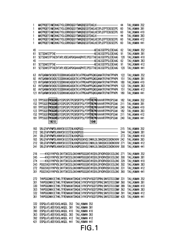
Figure 1 shows the amino acid sequences (SEQ ID NOS 2 to 7) of the six known h-
Tau
isoforms (h-Tau 441, h-Tau 412, h-Tau 410, h-Tau-381, h-Tau 383, and h-Tau
352,
respectively. The epitopes for mAbs 10H8 (epitope TREPK, SEQ ID NO: 11,
corresponding
to amino acids 220 to 224 of h-Tau) and 19G10 (epitope PKSGDR, SEQ ID NO: 12,
corresponding to amino acids 189 to 194 of h-Tau) are in bolded brackets.
Figure 2 shows the estimated ROC curves (100*Sensitivity vs. 100*(1-
Specificity)) for CSF
A1342, tau, and tau/A1342. Reference lines are drawn at 80% sensitivity and
60% specificity.
Figure 3 displays estimates of sensitivity, specificity, and total agreement
with PET
(Flutemetamol visual read) vs. prospective thresholds for CSF h-Tau/A[342
using log scaling.
Sensitivity (solid line) and Specificity (solid line) are displayed along with
95% lower
confidence limits (dashed lines). The estimate of Total Agreement (solid line)
is based on
CA 02938152 2016-07-28
WO 2015/120364
PCT/US2015/014976
nonparametric density estimation. Vertical lines (solid lines) show the CSF
window (0.169,
0.360) that achieves the acceptable sensitivity and specificity performance.
The value that
maximizes total agreement within this window (0.215) is also shown with a
vertical line and
identified on the top axis.
DETAILED DESCRIPTION OF THE INVENTION
Definitions
So that the invention may be more readily understood, certain technical and
scientific
terms are specifically defined below. Unless specifically defined elsewhere in
this document,
all other technical and scientific terms used herein have the meaning commonly
understood
by one of ordinary skill in the art to which this invention belongs.
As used herein, including the appended claims, the singular forms of words
such as
"a," "an," and "the," include their corresponding plural references unless the
context clearly
dictates otherwise.
"A131_42 peptide or API -42" refers to a 42 amino acid peptide corresponding
to amino
acids 672 to 713 (SEQ ID NO: 18) which is produced by proteolytic cleavage of
the amyloid
beta A4 protein isoform a precursor protein (SEQ ID NO: 19) by the p- and 7
¨secretases.
"Administration" or "administering" an AD therapeutic agent means providing an
AD
therapeutic agent to the patient in need of treatment.
"Alzheimer's disease or AD" as used herein refers to the spectrum of dementias
or
cognitive impairment resulting from neuronal degradation associated with the
formation or
deposition of AP plaques or NFTs in the brain, from the spectrum of
Alzheimer's disease
including but not limited to preclinical Alzheimer's disease, mild cognitive
impairment due
to Alzheimer's disease, early onset Alzheimer's disease, familial Alzheimer's
disease,
through the advance cognitive impairment of dementia due to Alzheimer's
disease (Jack et
al., Alzheimer's Dement., May 7 (3), pp. 257-262, 2011) and diseases
associated with the
presence of the ApoE4 allele.
"AD therapeutic agent" as used herein refers to a treatment or intervention
that
addresses one or more underlying pathophysiologies of AD or a symptom thereof
11
CA 02938152 2016-07-28
WO 2015/120364
PCT/US2015/014976
Examples of AD therapeutic agents include, but are not limited to, the BACE-1
inhibitors described herein, BACE-1 inhibitors CTS-21166 (CoMentis Inc.),
AZD3293
(AstraZeneca), E-2609 (Eisai), TAK-070 (Takeda), and HPP-854 (Transtech),
gamma
secretase inhibitors (e.g., as described in W02007/084595 and W02009/008980),
gamma
secretase modulators (as described e.g., in W02008/153793 and W02010/056849),
solanezumab (Eli Lilly), liraglutide (Lancaster University), bexarotene (brand
name
Targretine), ACC-001 (vaccine), muscarinic antagonists (e.g., m1 agonists
(such as
acetylcholine, oxotremorine, carbachol, or McNa343), or m2 antagonists (such
as atropine,
dicycloverine, tolterodine, oxybutynin, ipratropium, methoctramine,
tripitamine, or
gallamine); cholinesterase inhibitors (e.g., acetyl- and/or
butyrylchlolinesterase inhibitors
such as donepezil (AriceptO), galantamine (Razadyne0), and rivastigimine
(Exelone); N-
methyl-D-aspartate receptor antagonists (e.g., Namenda (memantine HC1,
available from
Forrest Pharmaceuticals, Inc.); combinations of cholinesterase inhibitors and
N-methyl-D-
aspartate receptor antagonists; non-steroidal anti-inflammatory agents; anti-
inflammatory
agents that can reduce neuroinflammation; anti-amyloid antibodies (such as
bapineuzemab,
Wyeth/Elan); vitamin E; nicotinic acetylcholine receptor agonists; CB1
receptor inverse
agonists or CB1 receptor antagonists; antibiotics; growth hormone
secretagogues; histamine
H3 antagonists; AMPA agonists; PDE4 inhibitors; GABAA inverse agonists;
inhibitors of
amyloid aggregation; glycogen synthase kinase beta inhibitors; promoters of
alpha secretase
activity; PDE-10 inhibitors; Tau kinase inhibitors (e.g., GSK3beta inhibitors,
cdk5 inhibitors,
or ERK inhibitors); Tau aggregation inhibitors (e.g., Rember0); RAGE
inhibitors (e.g., TTP
488 (PF-4494700)); anti-Abeta vaccine; APP ligands; agents that upregulate
insulin,
cholesterol lowering agents such as HMG-CoA reductase inhibitors (for example,
statins such
as Atorvastatin, Fluvastatin, Lovastatin, Mevastatin, Pitavastatin,
Pravastatin, Rosuvastatin,
Simvastatin) and/or cholesterol absorption inhibitors (such as Ezetimibe), or
combinations of
HMG-CoA reductase inhibitors and cholesterol absorption inhibitors (such as,
for example,
Vytorin0); fibrates (such as, for example, clofibrate, Clofibride, Etofibrate,
and Aluminium
Clofibrate); combinations of fibrates and cholesterol lowering agents and/or
cholesterol
absorption inhibitors; nicotinic receptor agonists; niacin; combinations of
niacin and
cholesterol absorption inhibitors and/or cholesterol lowering agents (e.g.,
Simcor0
(niacin/simvastatin, available from Abbott Laboratories, Inc.); LXR agonists;
LRP mimics;
H3 receptor antagonists; histone deacetylase inhibitors; hsp90 inhibitors; 5-
HT4 agonists
(e.g., PRX-03140 (Epix Pharmaceuticals)); 5-HT6 receptor antagonists; mGluR1
receptor
modulators or antagonists; mGluR5 receptor modulators or antagonists; mGluR2/3
12
CA 02938152 2016-07-28
WO 2015/120364
PCT/US2015/014976
antagonists; Prostaglandin EP2 receptor antagonists; PAT-1 inhibitors; agents
that can induce
Abeta efflux such as gelsolin; Metal-protein attenuating compound (e.g, PBT2);
and GPR3
modulators; and antihistamines such as Dimebolin (e.g., Dimebon , Pfizer).
"Antibody" as used herein may refer to any form of antibody that exhibits the
desired
biological activity. Thus, it is used in the broadest sense and specifically
covers, but is not
limited to, monoclonal antibodies (including full length monoclonal
antibodies), polyclonal
antibodies, multispecific antibodies (e.g., bispecific antibodies), humanized,
fully human
antibodies, chimeric antibodies and camelized single domain antibodies.
In general, the basic antibody structural unit comprises a tetramer. Each
tetramer
includes two identical pairs of polypeptide chains, each pair having one
"light" (about 25
kDa) and one "heavy" chain (about 50-70 kDa). The amino-terminal portion of
each chain
includes a variable region of about 100 to 110 or more amino acids primarily
responsible for
antigen recognition. The carboxy-terminal portion of the heavy chain may
define a constant
region primarily responsible for effector function. Typically, human light
chains are
classified as kappa and lambda light chains. Furthermore, human heavy chains
are typically
classified as mu, delta, gamma, alpha, or epsilon, and define the antibody's
isotype as IgM,
IgD, IgG, IgA, and IgE, respectively. Within light and heavy chains, the
variable and
constant regions are joined by a "J" region of about 12 or more amino acids,
with the heavy
chain also including a "D" region of about 10 more amino acids. See generally,
Fundamental
Immunology Ch. 7 (Paul, W., ed., 2nd ed. Raven Press, N.Y. (1989).
The variable regions of each light/heavy chain pair form the antibody binding
site.
Thus, in general, an intact antibody has two binding sites. Except in
bifunctional or
bispecific antibodies, the two binding sites are, in general, the same.
Typically, the variable domains of both the heavy and light chains comprise
three
hypervariable regions, also called complementarity determining regions (CDRs),
located
within relatively conserved framework regions (FR). The CDRs are usually
aligned by the
framework regions, enabling binding to a specific epitope. In general, from N-
terminal to C-
terminal, both light and heavy chains variable domains comprise FR1, CDR1,
FR2, CDR2,
FR3, CDR3 and FR4. The assignment of amino acids to each domain is, generally,
in
accordance with the definitions of Sequences of Proteins of Immunological
Interest, Kabat, et
al.; National Institutes of Health, Bethesda, Md. ; 5th ed.; NIH Publ. No. 91-
3242 (1991);
Kabat (1978) Adv. Prot. Chem. 32:1-75; Kabat, eta!,, (1977) J. Biol. Chem.
252:6609-6616;
13
CA 02938152 2016-07-28
WO 2015/120364
PCT/US2015/014976
Chothia, et al., (1987) J Mol. Biol. 196:901-917 or Chothia, et al., (1989)
Nature 342:878-
883.
As used herein, the term "hypervariable region" refers to the amino acid
residues of an
antibody that are responsible for antigen-binding. The hypervariable region
comprises amino
acid residues from a "complementarity determining region" or "CDR" (i.e.
CDRL1, CDRL2
and CDRL3 in the light chain variable domain and CDRH1, CDRH2 and CDRH3 in the
heavy chain variable domain). See Kabat et al. (1991) Sequences of Proteins of
Immunological Interest, 5th Ed. Public Health Service, National Institutes of
Health,
Bethesda, Md. (defining the CDR regions of an antibody by sequence); see also
Chothia and
Lesk (1987) J. Mol. Biol. 196: 901-917 (defining the CDR regions of an
antibody by
structure). As used herein, the term "framework" or "FR" residues refers to
those variable
domain residues other than the hypervariable region residues defined herein as
CDR residues.
As used herein, antibody 10H8 is the antibody produced by hybridoma subclone
MEB
clone 10H8.25.6.10H8 (murine IgG1 isotype) comprising the light chain and
heavy chain
variable regions (SEQ ID NOs: 24 and 30, respectively) set forth in Table 2
below.
Table 2. Characteristics of Monoclonal Antibody 10H8
Antibody Amino Acid Sequence or Nucleic Acid Sequence SEQ
ID NO
Feature
Light
Chain
CDRL1 RSSQNI IHSNGSTYLE 20
CDRL2 KVSNRFS 21
CDRL3 FQGSHVPWT 22
Leader MKLPVRLLVLMFWI PASSS
Sequence 23
Variable DVLMTQTPLSLPVSLGDQASISCRSSQNIIHSNGSTYLEWYLQ
Region KPGQSPKLLIYKVSNRFSGVPDRFSGSGSGTDFTLKISRVEAE 24
(CDRs in DLGIYYCFQGSHVPWTFGGGTKLEIK
bold font
and FRs in
italic font)
DNA GATGTTTTGATGACCCAAACTCCACTCTCCCTGCCTGTCAGTC
Sequence TTGGAGATCAAGCCTCCATCTCTTGCAGATCTAGTCAGAACAT
Encoding TATACATAGTAATGGAAGCACCTATTTAGAATGGTACCTGCAG
14
CA 02938152 2016-07-28
WO 2015/120364
PCT/US2015/014976
the AAACCGGGCCAGTCTCCAAAGCTCCTGATCTACAAAGTTTCCA 25
Variable ACCGATTTTCTGGGGTCCCAGACAGGTTCAGTGGCAGTGGATC
Region AGGGACAGATTTCACACTCAAGATCAGCAGAGTGGAGGCTGAG
GATCTGGGAATTTATTACTGCTTTCAAGGTTCACATGTTCCGT
(CDRs in GGACGTTCGGTGGAGGCACCAAGCTGGAAATCAAA
bold font
and FRs in
italic font)
Heavy
Chain
CDRH1 GFNI KDEYMN 26
CDRH2 WI DPENGDAAYASKFQG 27
CDRH3 FYSNYDGYFDV 28
Leader MKCSWVI FFLMAVV I GVNS 29
Sequence
Variable EVQLQQSGAELVRPGASVKLSCTASGFNIKDEYMNWVKQRPER 30
Region GLEWIGWIDPENGDAAYASKFQGKATMTADTSSNTAYLQLSSL
(CDRs in TSEDTAVYFCTFFYSNYDGYFDVWGAGTTVTVSS
bold font
and FRs in
italic font)
DNA GAGGTTCAGCTGCAGCAGTCTGGGGCTGAGCTTGTGAGGCCAG
Sequence GGGCCTCAGTCAAGTTGTCCTGCACAGCTTCTGGCTTTAACAT
Encoding TAAAGACGAGTATATGAACTGGGTGAAGCAGAGGCCTGAACGG 31
the GGCCTGGAGTGGATTGGATGGATTGATCCTGAAAATGGTGATG
Variable CTGCATATGCCTCGAAGTTCCAGGGAAAGGCCACTATGACTGC
Region AGACACATCCTCCAACACAGCCTACCTGCAGCTCAGCAGCCTG
(CDRs in ACATCTGAGGACACTGCCGTCTATTTCTGTACTTTCTTTTACA
bold font GTAACTACGACGGGTACTTCGATGTC TGGGGCGCAGGGACCAC
and FRs in GGTCACCGTCTCCTCA
italic font)
CA 02938152 2016-07-28
WO 2015/120364
PCT/US2015/014976
As used herein, antibody 19G10 is the antibody produced by hybridoma subclone
MEB.19G10.10.5 (murine isotype IgG2b) comprising the light chain and heavy
chain
variable regions (SEQ ID NOs: 36 and 42, respectively) set forth in Table 3
below.
Table 3. Characteristics of Monoclonal Antibody 19G10
Antibody Amino Acid Sequence or Nucleic Acid Sequence SEQ ID
Feature NO
Light
Chain
CDRL1 KSSQSLLYSNNQKNYLA 32
CDRL2 WASTRES 33
CDRL3 QQYYSYPLWT 34
Leader MDSQAQVLMLLLLWVSGTCG 35
Sequence
Variable D VMSQ S PS S LAVS IGEKVTMSCKSSQSLLYSNNQKNYLAWYQ 36
Region RKPGQS PKLL YWASTRES GVPDRFTGSGSGTDFTLT TSVKA
(CDRs in EDLAVYYCQQYY SY PLWT FGGGTKLE IK
bold font
and FRs in
italic font)
DNA GACATTGTGATGTCACAGTCTCCATCCTCCCTAGCTGTGTCAA 37
Sequence TTGGAGAGAAGGTTACTATGAGCTGCAAGTCCAGTCAGAGCCT
Encoding TTTATATAGTAACAATCAAAAGAACTACTTGGCC TGGTACCAG
the CGGAAACCAGGGCAGTCTCCTAAACTGCTGATTTACTGGGCAT
Variable CCACTAGGGAATCTGGGGTCCCTGATCGCTTCACAGGCAGTGG
Region ATCTGGGACAGATTTCACTCTCACCATCACCAGTGTGAAGGCT
(CDRs in GAAGACCTGGCAGTTTATTACTGTCAGCAATATTATAGTTATC
bold font CTCTGTGGACGTTCGGTGGAGGCACCAAGCTGGAAATCAAA
and FRs in
italic font)
Heavy
Chain
CDRH1 GFSLSTSGMGVG 38
CDRH2 HIWWDDDKYYNAVLKS 39
CDRH3 I GIDGPYAMDY 40
Leader MGRLTSSFLLLIVPAYVLS 41
Sequence
16
CA 02938152 2016-07-28
WO 2015/120364
PCT/US2015/014976
Variable QVT LKE SGPG L Q PSQ TL S L TC S FSGFSLSTSGMGVGW RQPS
Region GKGL EWLAHIWWDDDKYYNAVLKS RL T SKDTSKNQVFLKIAS 42
(CDRs in VD TAD TAT YYCARI GIDGPYAMDY WGQGTSVTVSS
bold font
and FRs in
italic font)
DNA CAGGTTACTCTGAAAGAGTCTGGCCCTGGGATATTGCAGCCCT
Sequence CCCAGACCCTCAGTCTGACTTGTTCTTTCTCTGGGTTTTCACT
Encoding GAGCACTTCTGGTATGGGTGTAGGC TGGATTCGTCAGCCTTCA 43
the GGGAAGGGTCTGGAATGGCTGGCACACATTTGGTGGGATGATG
Variable ATAAGTACTATAACGCAGTCCTGAAGAGCCGGCTCACAATCTC
Region CAAGGATACC TCCAAAAACCAGGT TT TCCTCAAGATCGCCAGT
(CDRs in GTGGACACTGCAGATACTGCCACATATTACTGTGCTCGAATAG
bold font GGATTGATGGTCCTTATGCTATGGACTAC TGGGGTCAAGGAAC
and FRs in CTCAGTCACCGTCTCCTCA
italic font)
As used herein an antibody is said to "specifically bind to an epitope on h-
Tau" if it binds to that epitope on the known six isoforms of h-Tau, but does
not bind to other
epitopes on h-Tau.
As used herein an antibody is said to "specifically bind to an epitope on the
N-
terminal or C-terminal of AI31.42" if it binds to that epitope but does not
bind to other epitopes
on AN-42.
As used herein "antibody fragment" or "antigen binding fragment" refers to
antigen
binding fragments of antibodies, i.e. antibody fragments that retain the
ability to bind
specifically to the antigen bound by the full-length antibody, e.g. fragments
that retain one or
more CDR regions. Examples of antibody binding fragments include, but are not
limited to,
Fab, Fab', F(ab')2, and Fv fragments; diabodies; linear antibodies; single-
chain antibody
molecules, e.g., sc-Fv; nanobodiese and multispecific antibodies formed from
antibody
fragments.
In an embodiment, the antibody or antigen binding fragment comprises a heavy
chain
constant region, e.g. a human constant region, such as 71, 72, 73, or 74 human
heavy chain
constant region or a variant thereof In another embodiment, the antibody or
antigen binding
fragment comprises a light chain constant region, e.g. a human light chain
constant region,
such as lambda or kappa human light chain region or variant thereof By way of
example,
and not limitation the human heavy chain constant region can be 71 and the
human light
chain constant region can be kappa.
17
CA 02938152 2016-07-28
WO 2015/120364
PCT/US2015/014976
"Biological sample" as used herein refers to any type of fluid or tissue
sample.
Typical examples that may be used in the assays herein are whole blood,
plasma, serum,
urine, cerebral spinal fluid (CSF) and extracts of brain tissue.
"Capture antibody" as used herein refers to an antibody that is used in the
disclosed
assays to retrieve from a biological sample all the isoforms making up h-Tau.
In one aspect,
the capture antibody as used herein specifically binds to the epitope on h-Tau
consisting of
amino acids TREPK ( amino acids 220 to 224, SEQ ID NO: 11). In an embodiment,
the
capture antibody binding to the aforementioned epitope on h-Tau is the mAb
10H8. In
another aspect, the capture antibody as used herein specifically binds to an
epitope of the N-
terminal and/or C-terminal of Af31_42. In one embodiment, the capture antibody
specifically
binds to an epitope on the C-terminal of A131.42 comprising amino acids
GLMVGGVVIA
(SEQ ID NO: 16, corresponding to amino acids 33 to 42 of SEQ ID NO: 18). In
another
embodiment, the capture antibody binding to the aforementioned epitope on
A131_42 is rabbit
mAb 1-11-3.
The phrase "control sequences" as used herein refers to DNA sequences
necessary for
the expression of an operably linked coding sequence in a particular host
organism. The
control sequences that are suitable for prokaryotes, for example, include a
promoter,
optionally an operator sequence, and a ribosome binding site. Eukaryotic cells
are known to
2,0 use promoters, polyadenylation signals, and enhancers.
"Detectably labeled antibody" refers to an antibody that is labeled with a
reagent
capable of detecting the antibody. The reagent may include, but is not limited
to, a
radioactive isotope, an enzyme, a biotin, dye, fluorescent label and
chemiluminescent label as
set forth below. The "detectably labeled antibody" is used to detect the
amount of h-Tau or
A31_42 which has been retained by the capture antibody. In one aspect, the
detectably labeled
antibody as used herein specifically binds to an epitope on h-Tau consisting
of amino acids
189 to 194 (PKSGDR, SEQ ID NO: 12). In an embodiment, the detectably labeled
antibody
specifically binding to the aforementioned epitope on h-Tau is the mAb 19G10.
In another
aspect, the detectably labeled antibody as used herein specifically binds to
an epitope on the
N-terminal and/or C-terminal of AB1_42. In an embodiment, the detectably
labeled antibody
specifically binds to an epitope on the N-terminal of A142 comprising amino
acids EFRHDS
18
CA 02938152 2016-07-28
WO 2015/120364
PCT/US2015/014976
(amino acids 3 to 8, SEQ ID NO:17). In another embodiment, the detectably
labeled
antibody binding to the aforementioned epitope of AI31_42 is mAb 6E10.
"Diabodies" refers to small antibody fragments with two antigen-binding sites,
which
fragments comprise a heavy chain variable domain (VH) connected to a light
chain variable
domain (VL) in the same polypeptide chain (VH-VL or VL-VH). By using a linker
that is too
short to allow pairing between the two domains on the same chain, the domains
are forced to
pair with the complementary domains of another chain and create two antigen-
binding sites.
Diabodies are described more fully in, e.g., EP 404,097; WO 93/11161; and
Holliger et al.
(1993) Proc. Natl. Acad. Sci. USA 90: 6444-6448. For a review of engineered
antibody
variants generally see Holliger and Hudson (2005) Nat. Biotechnol. 23:1126-
1136.
A "domain antibody" is an immunologically functional immunoglobulin fragment
containing only the variable region of a heavy chain or the variable region of
a light chain. In
some instances, two or more VH regions are covalently joined with a peptide
linker to create a
bivalent domain antibody. The two VH regions of a bivalent domain antibody may
target the
same or different antigens.
"Epitope" refers to the segment of amino acids on h-Tau capable of being
recognized
by, and bound by, an anti-h-Tau antibody of the present invention or other
anti-h-Tau
antibody, or a segment of amino acids on Aid1_42 capable of being recognized
by, and bound
by, an antibody.
A "Fab fragment" is comprised of one light chain and the CH1 and variable
regions of
one heavy chain. The heavy chain of a Fab molecule cannot form a disulfide
bond with
another heavy chain molecule. A "Fab fragment" can be the product of papain
cleavage of an
antibody.
An "Fc" region contains two heavy chain fragments comprising the CH1 and C12
domains of an antibody. The two heavy chain fragments are held together by two
or more
disulfide bonds and by hydrophobic interactions of the CH3 domains.
A "Fab' fragment" contains one light chain and a portion or fragment of one
heavy
chain that contains the VH domain and the C H1 domain and also the region
between the CH1
and C112 domains, such that an interchain disulfide bond can be formed between
the two
heavy chains of two Fab' fragments to form a F(ab') 2 molecule.
A "F(aW)2 fragment" contains two light chains and two heavy chains containing
a
portion of the constant region between the CH1 and C112 domains, such that an
interchain
19
CA 02938152 2016-07-28
WO 2015/120364
PCT/US2015/014976
disulfide bond is formed between the two heavy chains. A F(ab') 2 fragment
thus is composed
of two Fab' fragments that are held together by a disulfide bond between the
two heavy
chains. An "F(ab1)2 fragment" can be the product of pepsin cleavage of an
antibody.
The "Fv region" comprises the variable regions from both the heavy and light
chains,
but lacks the constant regions.
"h-Tau" as used herein refers to h-Tau which includes the six known isoforms
of h-
Tau. Quantification of h-Tau refers to the amount of h-Tau obtained from the
six known
iso forms of h-Tau.
"Isolated antibody" refers to the purification status and in such context
means the
molecule is substantially free of other biological molecules such as nucleic
acids, proteins,
lipids, carbohydrates, or other material such as cellular debris and growth
media. Generally,
the term "isolated" is not intended to refer to a complete absence of such
material or to an
absence of water, buffers, or salts, unless they are present in amounts that
substantially
interfere with experimental or therapeutic use of the binding compound as
described herein.
"Isolated nucleic acid molecule" means a DNA or RNA of genomic, mRNA, cDNA,
or synthetic origin or some combination thereof which is not associated with
all or a portion
of a polynucleotide in which the isolated polynucleotide is found in nature,
or is linked to a
polynucleotide to which it is not linked in nature. For purposes of this
disclosure, it should
be understood that "a nucleic acid molecule comprising" a particular
nucleotide sequence
does not encompass intact chromosomes. Isolated nucleic acid molecules
"comprising"
specified nucleic acid sequences may include, in addition to the specified
sequences, coding
sequences for up to ten or even up to twenty or more other proteins or
portions or fragments
thereof, or may include operably linked regulatory sequences that control
expression of the
coding region of the recited nucleic acid sequences, and/or may include vector
sequences.
A nucleic acid is "operably linked" when it is placed into a functional
relationship
with another nucleic acid sequence. For example, DNA for a presequence or
secretory leader
is operably linked to DNA for a polypeptide if it is expressed as a preprotein
that participates
in the secretion of the polypeptide; a promoter or enhancer is operably linked
to a coding
sequence if it affects the transcription of the sequence; or a ribosome
binding site is operably
linked to a coding sequence if it is positioned so as to facilitate
translation. Generally,
"operably linked" means that the DNA sequences being linked are contiguous,
and, in the
case of a secretory leader, contiguous and in reading phase. However,
enhancers do not have
CA 02938152 2016-07-28
WO 2015/120364
PCT/US2015/014976
to be contiguous. Linking is accomplished by ligation at convenient
restriction sites. If such
sites do not exist, the synthetic oligonucleotide adaptors or linkers are used
in accordance
with conventional practice.
as used herein refers to the "dissociation constant" of a particular antibody-
antigen interaction as is known in the art.
The term "monoclonal antibody or mAb", as used herein, refers to a population
of
substantially homogeneous antibodies, i.e., the antibody molecules comprising
the population
are identical in amino acid sequence except for possible naturally occurring
mutations that
may be present in minor amounts. In contrast, conventional (polyclonal)
antibody
preparations typically include a multitude of different antibodies having
different amino acid
sequences in their variable domains, particularly their CDRs, which are often
specific for
different epitopes. The modifier "monoclonal" indicates the character of the
antibody as
being obtained from a substantially homogeneous population of antibodies, and
is not to be
construed as requiring production of the antibody by any particular method.
For example, the
monoclonal antibodies to be used in accordance with the present invention may
be made by
the hybridoma method first described by Kohler et al. (1975) Nature 256: 495,
or may be
made by recombinant DNA methods (see, e.g., U.S. Pat. No. 4,816,567). The
"monoclonal
antibodies" may also be isolated from phage antibody libraries using the
techniques described
in Clackson et al. (1991) Nature 352: 624-628 and Marks et al. (1991) J. Mol.
Biol. 222: 581-
597, for example. See also Presta (2005) J. Allergy Clin. Immunol. 116:731.
"Polyclonal antibody" refers to an antibody which was produced among or in the
presence of one or more other, non-identical antibodies. In general,
polyclonal antibodies are
produced from collections of different B-lymphocytes, e.g. the B-lymphocyte of
an animal
treated with an immunogen of interest, which produces a population of
different antibodies
that are all directed to the immunogen. Usually, polyclonal antibodies are
obtained directly
from an immunized animal, e.g. spleen, serum or ascites fluid.
The term "salt(s)", as employed herein, denotes acidic salts formed with
inorganic
and/or organic acids, as well as basic salts formed with inorganic and/or
organic bases. In
addition, when a compound of the invention contains both a basic moiety, such
as, but not
limited to a pyridine or imidazole, and an acidic moiety, such as, but not
limited to a
carboxylic acid, zwitterions ("inner salts") may be formed and are included
within the term
"salt(s)" as used herein. Pharmaceutically acceptable (i.e., non-toxic,
physiologically
acceptable) salts are preferred, although other salts are also useful. Salts
of the BACE-1
21
CA 02938152 2016-07-28
WO 2015/120364
PCT/US2015/014976
inhibitors described herein may be formed, for example, by reacting the BACE-1
inhibitor
with an amount of acid or base, such as an equivalent amount, in a medium such
as one in
which the salt precipitates or in an aqueous medium followed by
lyophilization.
Exemplary acid addition salts include acetates, ascorbates, benzoates,
benzenesulfonates, bisulfates, borates, butyrates, citrates, camphorates,
camphorsulfonates,
fumarates, hydrochlorides, hydrobromides, hydroiodides, lactates, maleates,
methanesulfonates, naphthalenesulfonates, nitrates, oxalates, phosphates,
propionates,
salicylates, succinates, sulfates, tartarates, thiocyanates, toluenesulfonates
(also known as
tosylates,) and the like. Additionally, acids which are generally considered
suitable for the
formation of pharmaceutically useful salts from basic pharmaceutical compounds
are
discussed, for example, by P. Stahl et al, Camille G. (eds.) Handbook of
Pharmaceutical
Salts. Properties, Selection and Use. (2002) Zurich: Wiley-VCH; S. Berge et
al, Journal of
Pharmaceutical Sciences (1977) 66(1) 1-19; P. Gould, International J. of
Pharmaceutics
(1986) 33 201-217; Anderson et al, The Practice of Medicinal Chemistry (1996),
Academic
Press, New York; and in The Orange Book (Food & Drug Administration,
Washington, D.C.
on their website).
Exemplary basic salts include ammonium salts, alkali metal salts such as
sodium,
lithium, and potassium salts, alkaline earth metal salts such as calcium and
magnesium salts,
salts with organic bases (for example, organic amines) such as
dicyclohexylamines, t-butyl
amines, and salts with amino acids such as arginine, lysine and the like.
Basic nitrogen-
containing groups may be quarternized with agents such as lower alkyl halides
(e.g. methyl,
ethyl, and butyl chlorides, bromides and iodides), dialkyl sulfates (e.g.
dimethyl, diethyl, and
dibutyl sulfates), long chain halides (e.g. decyl, lauryl, and stearyl
chlorides, bromides and
iodides), aralkyl halides (e.g. benzyl and phenethyl bromides), and others.
All such acid salts and base salts are intended to be pharmaceutically
acceptable salts
and all acid and base salts are considered equivalent to the free forms of the
corresponding
BACE-1 inhibitor described herein.
The term "single-chain Fv" or "scFv" antibody refers to antibody fragments
comprising the VH and VL domains of an antibody, wherein these domains are
present in a
single polypeptide chain. Generally, the Fv polypeptide further comprises a
polypeptide
linker between the VH and VL domains which enables the scFv to form the
desired structure
for antigen binding. For a review of scFv, see Pluckthun (1994) THE
PHARMACOLOGY OF
22
CA 02938152 2016-07-28
WO 2015/120364
PCT/US2015/014976
MONOCLONAL ANTIBODIES, vol. 113, Rosenburg and Moore eds. Springer-Verlag, New
York, pp. 269-315. See also, International Patent Application Publication No.
WO 88/01649
and U.S. Pat. Nos. 4,946,778 and 5,260,203.
The term "treatment" or "treating" means any administration of an AD
therapeutic
agent to obtain a desired pharmacologic and/or physiologic effect. The effect
may be
prophylactic in terms of completely or partially preventing a disease or
symptom thereof,
and/or may be therapeutic in terms of a partial or complete cure for a disease
and/or adverse
effect attributable to the disease. Treatment includes (1) inhibiting the
disease in a patient,
e.g., a human, that is experiencing or displaying the pathology or
symptomatology of the
disease (i.e., arresting further development of the pathology and/or
symptomatology), or (2)
ameliorating the disease in a patient that is experiencing or displaying the
pathology or
symptomatology of the disease (i.e., reversing the pathology and/or
symptomatology).
The amount of an AD therapeutic agent that is effective to alleviate any
particular
disease symptom (also referred to as the "therapeutically effective amount")
may vary
according to factors such as the disease state, age, and weight of the
patient, and the ability of
the drug to elicit a desired response in the subject or patient. Whether a
disease symptom has
been alleviated can be assessed by any clinical measurement typically used by
physicians or
other skilled healthcare providers to assess the severity or progression
status of that symptom.
Physical and Functional Properties of the Exemplary Anti-h-Tau Antibodies and
Antigen-Binding Fragments
The present invention provides isolated anti-h-Tau antibodies and antigen
binding
fragments thereof and methods of quantifying h-Tau in a biological sample such
as CSF
using these antibodies and antigen binding fragments thereof. Examples of the
anti-h-Tau
antibodies of the present invention include but are not limited to: mAbs 10118
(see Table 2,
light chain and heavy chain variable regions of SEQ ID NOs: 24 and 30,
respectively) of
murine isotype IgGl, and 19G10 (see Table 3, light chain and heavy chain
variable regions of
SEQ ID NOs: 36 and 42, respectively) of murine isotype IgG2b.
The 10H8 and 19G10 antibodies specifically bind non-identical epitopes located
in a
conserved region shared by all six isoforms of h-Tau, which spans amino acids
104 to 277 of
h-Tau (See Figure 1).
23
CA 02938152 2016-07-28
WO 2015/120364
PCT/US2015/014976
In one aspect, an isolated antibody or antigen binding fragment thereof is
provided
which specifically binds an epitope on h-Tau consisting of amino acids 220 to
224 (TREPK)
as set forth in SEQ ID NO: 11. U.S. Patent 5,861,257 describes mAb AT120 which
specifically binds to an epitope on h-Tau comprising amino acids PPTREPK (SEQ
ID NO:
13) corresponding to amino acids Pro 218 to Lys 224 of h-Tau. The antibody of
the present
invention which specifically binds to epitope TREPK (SEQ ID NO: 11), as
exemplified by
mAb 10H8 is thought to be a different antibody from mAb AT120 described in
U.S. Patent
5,861,257 in view of the difference in their respective epitopes. In this
regard, epitope
mapping (see paragraph bridging columns 19-20 of U.S. patent 5,861,257) of mAb
AT120
indicated that while mAb AT120 reacted with the peptide sequence, PPTREPKKVAVV
(SEQ ID NO: 14), mAb AT120 did not react with the peptide sequence,
PTREPKKVAVV
(SEQ ID NO: 15). These and additional peptide mapping results of mAb AT120
indicated
that the epitope specifically bound by mAb AT120 was PPTREPK (SEQ ID NO: 13),
and not
the epitope TREPK (SEQ ID NO: 11) specifically bound by the antibody of the
present
invention as exemplified by mAb 10H8 (see Example 2 which shows epitope
mapping results
for mAb 10H8).
In another embodiment, the isolated antibody or antigen binding fragment
specifically
binding to the epitope of SEQ ID NO: 11 (TREPK) comprises three light chain
CDRs of SEQ
ID NO: 20 (CDRL1), SEQ ID NO: 21 (CDRL2) and SEQ ID NO: 22 (CDRL3) and three
heavy chain CDRs of SEQ ID NO: 26 (CDRH1), SEQ ID NO: 27 (CDRH2) and SEQ ID
NO: 28 (CDRH3) or a variant of the antibody. In one embodiment, the variant of
the
antibody comprises, 1, 2, 3, 4, 5, and 6 amino acid substitutions in one or
more of the above
recited CDRs, but retains the ability to bind an epitope of h-Tau consisting
of amino acids
220 to 224 (SEQ ID NO: 11).
In another embodiment, the isolated antibody or antigen binding fragment
thereof
binding to the epitope of SEQ ID NO: 11 (TREPK) comprises a light chain
variable region of
SEQ ID NO: 24 and a heavy chain variable region of SEQ ID NO: 30 or a variant
of the
antibody. In another embodiment, the variant of the antibody comprises 1-20
amino acid
substitutions in one or both sequences, but retains the ability to bind an
epitope of h-Tau
consisting of amino acids 220 to 224 (SEQ ID NO: 11)..
In another embodiment, the isolated antibody or antigen binding fragment
thereof
binding to the epitope of SEQ ID NO: 11 (TREPK) is a mAb or antigen binding
fragment
thereof In a particularly useful embodiment, the mAb is mAb 10H8 (variable
light and
24
CA 02938152 2016-07-28
WO 2015/120364
PCT/US2015/014976
heavy chains of SEQ ID NOs: 24 and 30, respectively, with a murine IgG1
isotype) produced
by hybridoma subclone_clone 10H8.25.6.10H8 or an antigen binding fragment of
mAb
10H8.
In another embodiment, the isolated antibody or antigen binding fragment
thereof
binding to the epitope of SEQ ID NO: 11 (TREPK) is of any class of
immunoglobulin , e.g.,
an IgA, IgD, IgE, IgG, and IgM, and several of these may be further divided
into subclasses
(isotypes), e.g., IgG-1, IgG-2, IgG-3 and IgG-4; IgA-1 and IgA-2. In a
particularly useful
embodiment, mAb 10H8 has a murine IgG1 isotype.
In another embodiment, the isolated antibody or antigen binding fragment
thereof
binding to the epitope of SEQ ID NO: 11 (TREPK) binds with a Kd value in the
low
micromolar (10-6) to nanomolar (10-7 to 10-9) range. In an embodiment, mAb
10H8 binds to
h-Tau with a Kd of about 17 nM (see Example 3).
In another aspect, an isolated antibody or antigen binding fragment thereof is
provided
which specifically binds an epitope on h-Tau consisting of amino acids 189 to
194
(PKSGDR) as set forth in SEQ ID NO: 12.
In an embodiment, the isolated antibody or antigen binding fragment
specifically
binding to the epitope of SEQ ID NO: 12 (PKSGDR) comprises three light chain
CDRs of
SEQ ID NO: 32 (CDRL1), SEQ ID NO: 33 (CDRL2) and SEQ ID NO: 34 (CDRL3) and
three heavy chain CDRs of SEQ ID NO: 38 (CDRH1), SEQ ID NO: 39 (CDRH2) and SEQ
ID NO: 40 (CDRH3) or a variant of the antibody. In one embodiment, the variant
of the
antibody comprises 1, 2, 3, 4, 5, and 6 amino acid substitutions in one or
more of the above
recited CDRs, but retains the ability to bind an epitope of h-Tau consisting
of amino acids
220 to 224 of SEQ ID NO: 11).
In another embodiment, the isolated antibody or antigen binding fragment
thereof
binding to the epitope of SEQ ID NO: 12 (PKSGDR) comprises a light chain
variable region
of SEQ ID NO: 36 and a heavy chain variable region of SEQ ID NO: 42 or a
variant of the
antibody. In one embodiment, the variant of the antibody comprises 1-20 amino
acid
substitutions in one or both of these sequences, but retains the ability to
bind an epitope of h-
Tau consisting of amino acids 189 to 194 (SEQ ID NO: 12).
In another embodiment, the isolated antibody or antigen binding fragment
thereof
binding to the epitope of SEQ ID NO: 12 (PKSGDR) is a mAb or antigen binding
fragment
thereof. In a particularly useful embodiment, the mAb is mAb 19G10 (variable
light and
CA 02938152 2016-07-28
WO 2015/120364
PCT/US2015/014976
heavy chains of SEQ ID NOs: 36 and 42, respectively, with a murine IgG2b
isotype)
produced by hybridoma subclone _clone 19G10.10.5.19G10 or an antigen binding
fragment
of mAb 19G10.
In another embodiment, the isolated antibody or antigen binding fragment
thereof
binding to the epitope of SEQ ID NO: 12 (PKSGDR) may be of any class of
immunoglobulin , e.g., an IgA, IgD, IgE, IgG, and IgM, and several of these
may be further
divided into subclasses (isotypes), e.g., IgG-1, IgG-2, IgG-3 and IgG-4; IgA-1
and IgA-2.
In a particularly useful embodiment, the isolated antibody or antigen binding
fragment
thereof binding to the epitope of SEQ ID NO: 12 (PKSGDR), e.g., mAb 19G10 has
an IgG2b
isotype.
In another embodiment, the isolated antibody or antigen binding fragment
thereof
binding to the epitope of SEQ ID NO: 12 (PKSGDR) binds with a Kd value in the
low
micromolar (10-6) to nanomolar (10-7 to 10-9) range. In a further embodiment,
mAb 19G10
binds to h-Tau with a Kd of about 6.3 nM (see Example 3).
Nucleic Acid Molecules, Vectors and Host Cells
In another aspect, isolated nucleic acids are provided which encode the
variable light
and heavy chains of an antibody or antigen binding fragment thereof that
specifically bind an
epitope on h-Tau consisting of amino acids 220 to 224 (TREPK, SEQ ID NO: 11).
In one
embodiment, an isolated nucleic acid is provided which encodes one or both of
an antibody
light chain variable region and an antibody heavy chain variable region,
wherein the antibody
light chain variable region is of SEQ ID NO: 24 and an antibody heavy chain
variable region
is of SEQ ID NO: 30.
In another aspect, isolated nucleic acids are provided which encode the
variable light
and heavy chains of an antibody or antigen binding fragment thereof that
specifically bind an
epitope on h-Tau consisting of amino acids 189 to 194 (PKSGDR, SEQ ID NO: 12).
In one
embodiment, an isolated nucleic acid is provided which encodes one or both of
an antibody
light chain variable region and an antibody heavy chain variable region,
wherein the antibody
light chain variable region is of SEQ ID NO: 36 and an antibody heavy chain
variable region
is of SEQ ID NO: 42.
26
CA 02938152 2016-07-28
WO 2015/120364
PCT/US2015/014976
In another aspect, expression vectors are provided which comprise the isolated
nucleic
acids of the invention, wherein the nucleic acid is operably linked to control
sequences that
are recognized by a host cell when the host cell is transfected with the
vector. Accordingly,
in one embodiment, an expression vector is provided comprising one or both of
the isolated
nucleic acids of SEQ ID NO: 25 and SEQ ID NO: 31, or one or both of the
isolated nucleic
acids of SEQ ID NO: 37 and SEQ ID NO: 43.
Also provided are host cells comprising an expression vector and methods for
producing the antibody or antigen binding fragment thereof disclosed herein
comprising
culturing a host cell harboring an expression vector encoding the antibody or
antigen binding
fragment in culture medium, and isolating the antigen or antigen binding
fragment thereof
from the host cell or culture medium.
Tau 166 peptide
In another aspect, an isolated peptide of SEQ ID NO: 9 known as Tau 166
peptide is
provided, which is employed as an immunogen to make the antibodies of the
present
invention. Tau 166 peptide can be produced using standard recombinant methods.
For
example, an isolated nucleic acid encoding the Tau 166 peptide may be cloned
into a suitable
expression vector. In an embodiment, the isolated nucleic acid encoding Tau
166 peptide is
SEQ ID NO: 10. The recombinant vector is then introduced into any suitable
host cell. In
one embodiment, the host cell is a sf9 (insect) cell. In another embodiment,
the host cell is E.
coli (see Example 1). Tau 166 peptide expressed from the host cell can then be
purified from
the host cell by standard methods (see e.g., Ausubel et al. (1991) Current
Protocols in
Molecular Biology Ch. 16 (John Wiley & Sons, NY).
Methods of Making Antibodies and Antigen Binding Fragments Thereof
To produce antibodies, a suitable animal, such as a mouse, rat, hamster,
monkey, or
other mammal, is immunized with the Tau 166 peptide to produce antibody-
secreting cells.
In an embodiment, the animal, e.g., mouse, is immunized with Tau 166 peptide
and an
adjuvant which is used to enhance the immunological response. Examples of
adjuvants
include, but are not limited to, Freund's adjuvant (complete and incomplete),
mineral salts
such as aluminum hydroxide or aluminum phosphate, surface active substances,
chitosan,
27
CA 02938152 2016-07-28
WO 2015/120364
PCT/US2015/014976
lysolecithin, pluronic polyols, polyanions, peptides, oil emulsions (see
Example 1 for
immunization protocol). In another embodiment, the immune response to Tau 166
peptide
may be enhanced by coupling the Tau 166 peptide to another immunogenic
molecule or
"carrier protein." Examples of carrier proteins include, but are not limited
to, keyhole limpet
hemocyanin (KLH), tetanus toxoid, diphtheria toxoid, ovalbumin, cholera
toxoid, and
immunogenic fragments thereof. For guidance in coupling peptide immunogens to
carrier
proteins, see, e.g., Ausubel et al. (1989) Current Protocols in Molecular
Biology Ch. 11.15
(John Wiley & Sons, NY); and Harlow and Lane (1988) Antibodies: A Laboratory
Manual
Ch. 5 (Cold Spring Harbor Laboratory, Cold Spring Harbor, N.Y.).
Hybridoma cells that produce parental (e.g. rodent) anti-h-Tau mAbs of the
present
invention may be produced by methods which are commonly known in the art.
These
methods include, but are not limited to, the hybridoma technique originally
developed by
Kohler, et al., (1975) (Nature 256:495-497), as well as the trioma technique
(Hering, et al.,
(1988) Biomed. Biochim. Acta. 47:211-216 and Hagiwara, et al., (1993) Hum.
Antibod.
Hybridomas 4:15), the human B-cell hybridoma technique (Kozbor, et al., (1983)
Immunology Today 4:72 and Cote, et al., (1983) Proc. Natl. Acad. Sci. U.S.A
80:2026-
2030), the EBV-hybridoma technique (Cole, et al., in Monoclonal Antibodies and
Cancer
Therapy, Alan R. Liss, Inc., pp. 77-96, 1985), and electric field based
electrofusion using a
Cyto Pulse large chamber cull fusion electroporator (Cyto Pulse Sciences,
Inc., Glen Burnie,
MD). Preferably, mouse splenocytes are isolated and fused with PEG or by
electrofusion to a
mouse myeloma cell line based upon standard protocols. The resulting
hybridomas may then
be screened for the production of antigen-specific antibodies. For example,
single cell
suspensions of splenic lymphocytes from mice immunized with the Tau 166
antigen may be
fused to SP2/0 nonsecreting mouse myeloma cells using e.g., a 50% polyethylene
glycol-
1500 (PEG-1500) solution in buffer, pH 8Ø Fused cells may be then plated
onto microtiter
plates and incubated in a hybridoma culture medium supplemented with HAT
(liquid mixture
of: sodium-hypoxanthine, aminopterin, and thymidine) for about two weeks. The
culture
supernatant from each individual plate may then be screened to identify
antibody-secreting
hybridomas by well-known methods such as enzyme-linked immunosorbent assay
(ELISA).
The antibody secreting hybridomas may be replated and screened again. If the
screened
hybridoma is still positive for the desired anti-h-Tau anatibodies, it can be
subcloned at least
twice. Subcloning can be carried out by limiting dilution, wherein the
hybridoma cells are
diluted in a culture medium by serial dilution to a final concentration of
cells, e.g., 2.5
28
CA 02938152 2016-07-28
WO 2015/120364
PCT/US2015/014976
cells/mL. An aliquot of the cells, e.g., 2001AL (about 1/2 cell per well) is
plated into each well
and incubated from about two weeks. Single hybridoma cells in each well may
then be
microscopically identified and the supernatant from that single hybridoma may
be screened
by ELISA for the anti-h-Tau antibody of the present invention. Desired
subclones are
selected and can be expanded for antibody production or frozen in a liquid
nitrogen freezer.
When needed for studies, a vial of the frozen hybridoma may be thawed and
grown in
hybridoma culture medium to produce antibodies which can be purified. The
procedure for
making the anti-h-Tau antibodies of the present invention is described in
Example 1.
The anti-h-Tau antibodies of the present invention may also be produced
recombinantly (e.g., in an E. colilT7 expression system). In this embodiment,
nucleic acids
encoding the antibody molecules of the invention (e.g., VH or VI) may be
inserted into a
pET-based plasmid and expressed in the E. colilT7 system. There are several
methods by
which to produce recombinant antibodies which are known in the art. One
example of a
method for recombinant production of antibodies is disclosed in U.S. Patent
No. 4,816,567.
Transformation can be by any known method for introducing polynucleotides into
a host cell.
Methods for introduction of heterologous polynucleotides into mammalian cells
are well
known in the art and include dextran-mediated transfection, calcium phosphate
precipitation,
polybrene-mediated transfection, protoplast fusion, electroporation,
encapsulation of the
polynucleotide(s) in liposomes, biolistic injection and direct microinjection
of the DNA into
nuclei. In addition, nucleic acid molecules may be introduced into mammalian
cells by viral
vectors. Methods of transforming cells are well known in the art. See, for
example, U.S.
Patent Nos. 4,399,216; 4,912,040; 4,740,461 and 4,959,455.
Anti-h-Tau antibodies can also be synthesized by any of the methods set forth
in U.S.
Patent No. 6,331,415.
Mammalian cell lines available as hosts for expression of the antibodies or
fragments
disclosed herein are well known in the art and include many immortalized cell
lines available
from the American Type Culture Collection (ATCC). These include, inter alio,
Chinese
hamster ovary (CHO) cells, NSO, 5P2 cells, HeLa cells, baby hamster kidney
(BHK) cells,
monkey kidney cells (COS), human hepatocellular carcinoma cells (e.g., Hep
G2), A549
cells, 3T3 cells, HEK-293 cells and a number of other cell lines. Mammalian
host cells
include human, mouse, rat, dog, monkey, pig, goat, bovine, horse and hamster
cells. Cell
lines of particular preference are selected through determining which cell
lines have high
expression levels. Other cell lines that may be used are insect cell lines,
such as 519 cells,
29
CA 02938152 2016-07-28
WO 2015/120364
PCT/US2015/014976
amphibian cells, bacterial cells, plant cells and fungal cells. When
recombinant expression
vectors encoding the heavy chain or antigen binding portion or fragment
thereof, the light
chain and/or antigen binding fragment thereof are introduced into mammalian
host cells, the
antibodies are produced by culturing the host cells for a period of time
sufficient to allow for
expression of the antibody in the host cells or, more preferably, secretion of
the antibody into
the culture medium in which the host cells are grown.
Antibodies can be recovered from the culture medium using standard protein
purification methods. Further, expression of antibodies of the invention (or
other moieties
therefrom) from production cell lines can be enhanced using a number of known
techniques.
For example, the glutamine synthetase gene expression system (the GS system)
is a common
approach for enhancing expression under certain conditions. The GS system is
discussed in
whole or part in connection with European Patent Nos. 0 216 846, 0 256 055,
and 0 323 997
and European Patent Application No. 89303964.4.
Diagnostic Assays, Methods of Treatment and Kits
In another aspect, a method of quantitating h-Tau in a biological sample is
provided,
the method comprising:
(a) contacting the biological sample with an anti-h-Tau antibody of the
present
invention, e.g., mAb 10H8 or mAb 19G10 or a variant of the antibody as
described above, or an antigen binding fragment thereof under conditions
allowing formation of an immune complex between h-Tau and the antibody or
antigen binding fragment thereoff, and
(b) detecting the immune complex formed.
The aforementioned method can be used to quantify h-Tau in a biological sample
as defined
above, e.g., CSF, plasma, whole blood, serum or extracts of brain tissue.
In an embodiment of the aforementioned method for quantifying h-Tau, an anti-h-
Tau
of the present invention, e.g., mAb 10H8 or mAb 19G10 or a variant of the
antibody, is
coated onto a solid phase and the biological sample is then contacted with the
solid phase.
Examples of solid phases that may be used in this method are microtiter wells,
plastic tubes,
membranes, latex particles, magnetic particles, microspheres, and beads. The h-
Tau in the
CA 02938152 2016-07-28
WO 2015/120364
PCT/US2015/014976
biological sample binds to the antibody, and the amount of h-Tau can be
determined by a
direct or indirect method.
The direct method comprises detecting the presence of the h-Tau/anti-h-Tau
antibody
complex itself and thus the presence and amount of h-Tau by attaching a label
to the antibody
or antigen binding fragment thereof. Examples of labels are radioisotopes
(such as 14C, 35S,
1125 and 3H), enzymes having detectable reaction products (e.g., luciferase,
beta-galactosidase,
etc.), fluorescent labels ( e.g., rhodamine, phycoerythrin, fluorescein
isothiocyanate, resorufin
etc.), chemiluminescent compounds (e.g., acridinium salts, luminol,
isoluminol, etc.) and
bioluminescent compounds (e.g., luciferin, aequorin, etc.) .
In the indirect method, the anti-h-Tau antibody of the present invention can
be labeled
indirectly by reacting the anti-h-Tau antibody with a substance having
affinity for the mouse
anti-h-Tau antibody (e.g.,goat anti-mouse or rabbit anti-mouse IgG) or a
second antibody that
has been labeled with a detectable reagent that is radioactive, fluorescent or
chemiluminescent as mentioned above, and detecting the presence of the second
antibody.
In another embodiment, the amount of h-Tau is quantified using a pair of anti-
h-Tau
antibodies, each specific for the conserved region of h-Tau spanning amino
acids 104 to 277
of h-Tau. One of the pair of antibodies is an anti-h-Tau antibody of the
present invention,
e.g., mAb 10H8 or mAb 19G10 or a variant of the two antibodies, and the other
antibody
making up the pair is also specific for h-Tau. Examples of well-known anti-h-
Tau antibodies,
in particular mAbs that bind to an epitope on the conserved region of h-tau
spanning amino
acids 104 to 277 are Tau 5, BT2 and HT7 (commercially available at Covance).
One of the
pair of antibodies may be used as a "capture" antibody and the other of the
pair may be used
as a "detectably labeled antibody". Accordingly, an embodiment uses a double
antibody
sandwich method for detecting h-Tau in a biological sample, wherein h-Tau is
sandwiched
between the capture antibody, i.e. mAb 10H8 or mAb 19G10 or a variant of the
antibody, and
the detectably labeled antibody, i.e., BT2, and wherein the capture antibody
is immobilized
onto a solid phase.
An embodiment of the double sandwich method is an enzyme-linked immunosorbent
assay (ELISA) incorporating the use of an anti-h-Tau antibody or antigen
binding fragment
thereof of the present invention. For example, the ELISA comprises the
following steps:
31
CA 02938152 2016-07-28
WO 2015/120364
PCT/US2015/014976
(a) coat a solid phase (e.g., surface of a microtiter plate well) with the
anti-h-Tau
antibody or antigen-binding fragment thereof of the present invention, e.g.,
mAb 10H8 or
mAb 19G10 or a variant of these antibodies;
(b) apply a sample to be tested for the presence of h-Tau to the solid phase;
(c) wash the plate, so that unbound material in the sample is removed;
(d) apply a detectably labeled anti-h-Tau antibodies (e.g., enzyme-linked
antibody)
which is also specific to the h-Tau antigen, e.g., Tau 5, BT2 or HT7;
(e) wash the solid phase, so that the unbound, labeled antibody is removed;
(f) if the labeled antibody is enzyme-linked, apply a chemical which is
converted by the
enzyme into a fluorescent signal; and
(g) detect the presence of the labeled antibody.
As an example of the ELISA, the detectably labeled antibody is labeled with
peroxidase which react with ABTS (e.g., 2,TLazino-bis(3-ethylbenzthiazoline-6-
sulphonic
acid)) or 3,3',5,5'-Tetramethylbenzidine to produce a color change which is
detectable.
In a particularly useful embodiment of the double antibody sandwich assay, the
amount of h-Tau in a sample of CSF is quantified utilizing a pair of anti-h-
Tau antibodies of
the present invention, e.g., mAb 10H8 and 19G10 or a variant of these
antibodies, each of
which specifically bind to non-identical epitopes on the conserved region of h-
Tau spanning
amino acids 104 to 277. The antibody that specifically binds the epitope of h-
Tau consisting
of amino acids 220 to 224 (TREPK, SEQ ID NO: 11), e.g., mAb 10H8 or a variant
of the
antibody, is used as the "capture antibody", and the antibody that
specifically binds the
epitope of h-Tau consisting of amino acids 188 to 194 (PKSGDR, SEQ ID NO: 12),
e.g.,
19G10 or a variant of the antibody, is used as the "detectably labeled
antibody". This method
for quantitating h-Tau in a CSF sample comprises:
(a) capturing h-Tau from the sample by contacting the sample with an antibody
specifically binding to the epitope of h-Tau consisting of amino acids 220 to
224
(TREPK, SEQ ID NO: 11) e.g., 10H8 or a variant of the antibody, or an antigen
binding fragment thereof under conditions allowing formation of a capture
antibody/h-Tau complex, wherein the antibody or antigen binding fragment
thereof is
immobilized onto a solid support; and
(b) detecting the captured h-Tau by contacting the capture antibody/h-Tau
complex with
a detectably labeled antibody specifically binding to the epitope of h-Tau
consisting
of amino acids 189 to 194 (PKSGDR, SEQ ID NO: 12), e.g., 19G10 or a variant of
32
CA 02938152 2016-07-28
WO 2015/120364
PCT/US2015/014976
the antibody, or an antigen binding fragment thereof under conditions allowing
formation of a capture antibody/h-Tau/detectably labeled antibody complex.
As mentioned above, examples of solid supports are microtiter wells, plastic
tubes,
membranes, latex particles, magnetic particles, magnetic microparticles,
microspheres, and
beads. Suitable materials for the solid support include but are not limited to
nylon,
nitrocellulose, polyaerylamide, cellulose acetate, polystyrene, polypropylene,
polymethacrylate, styrene, carboxylated styrene, and fiber-containing paper
such as filter
paper, chromatographic paper and glass fiber paper.
In a particularly useful embodiment, the solid support is magnetic
microspheres
(MagPlexe Microspheres which are carboxylated polystyrene micro-particles or
beads,
commercially available from Luminex Corporation, Austin, TX) (see e.g., U.S.
Patents
7,718,262, 6,514,295, 6,599,331, 6,632,526, 6,929,859, 7,445,844, 8,283,037
and 8,568,881).
Reagents for labeling the "detectably labeled antibody" include but are not
limited to a
radioactive isotope, an enzyme, a biotin, dye, fluorescent label and
chemiluminescent label.
In a particular useful embodiment the reagent is biotin which is attached to a
streptavidin-
phyco erythrin conjugate.
An example of the aforementioned method for quantifying h-Tau in a biological
sample,
e.g., CSF, employs a bead-based technology (Luminex Corporation, Austin, TX),
in which
mAb 10H8 (the "capture antibody"), is coupled onto magnetic microspheres. The
coupled
microspheres are incubated with different concentrations of h-Tau (used as a
standard) or
CSF samples, together in the wells of a 96-well plate, followed by addition of
biotinylated
mAb 19G10 (the "detectably labeled antibody") to form a mAbl0H8/h-
Tau/biotinylated mAb
19G10 complex. Detection of the biotinylated complex is carried out by
incubation with a
streptavidin-phycoerytherin conjugate which binds to the biotinylated
antibody. An xMAP
Technology instrument (FlexMap 3D, Luminex Corporation, Austin, TX) uses a
classification laser (638nM) to identify the specific microspheres, and a
second reporter laser
(532nM) to excite the phycoerytherin molecule bound to conjugate. The
fluorescent output
from the phycoerythrin bound to the complex is measured using a detector (565
nM-585 nM)
or CCD (Charged Coupled Device) imaging detector and is directly related to
the amount of
h-Tau in the CSF samples as read off a calibration curve prepared from the
different
concentration of h-Tau in the CSF sample.
33
CA 02938152 2016-07-28
WO 2015/120364
PCT/US2015/014976
In another aspect, a method for diagnosing AD in a patient suspected of having
this
disease is provided. This method can be used e.g., to select patients for
treatment with an AD
therapeutic agent, e.g., a BACE-1 inhibitor. The method comprises
(a) quantifying the amount of h-Tau in a biological sample obtained from the
patient by:
(i) capturing h-Tau from the sample by contacting the sample with an antibody
specifically binding to the epitope of h-Tau consisting of amino acids 220 to
224
(TREPK, SEQ ID NO: 11) e.g., 10H8 or a variant of the antibody, or an antigen
binding fragment thereof under conditions allowing formation of a capture
antibody/h-Tau complex, wherein the antibody or antigen binding fragment
thereof is
immobilized onto a solid support; and
(ii) detecting the captured h-Tau by contacting the capture antibody/h-Tau
complex
with a detectably labeled antibody specifically binding to the epitope of h-
Tau
consisting of 189 to 194 (PKSGDR, SEQ ID NO: 12), e.g., 19G10 or a variant of
the
antibody, or an antigen binding fragment thereof under conditions allowing
formation
of a capture antibody/Tau/detectably labeled antibody complex; and
(c) determining the concentration of h-Tau in the sample obtained in step (a),
wherein a
value greater than 184 pg/mL indicates a diagnosis of AD in the patient.
The method of diagnosing AD in a patient suspected of having AD by quantifying
the
amount of h-Tau is described, e.g., in Examples 4 and 5.
In an embodiment, the foregoing method for diagnosing Alzheimer's disease in a
patient suspected of having this disease comprises:
(a) quantifying the amount of human Tau in a cerebrospinal fluid sample of the
patient
by:
(1) capturing human Tau from the sample by contacting the sample with an
antibody or
antigen binding fragment thereof, specifically binding to the epitope
consisting of
amino acids 220-224 of h-Tau, selected from the group consisting of:
(i) an antibody or antigen binding fragment thereof comprising three
light chain
CDRs of SEQ ID NO: 20 (CDRL1), SEQ ID NO: 21 (CDRL2) and SEQ ID
NO: 22 (CDRL3) and three heavy chain CDRs of SEQ ID NO: 26 (CDRH1),
34
CA 02938152 2016-07-28
WO 2015/120364
PCT/US2015/014976
SEQ ID NO: 27 (CDRH2) and SEQ ID NO: 28 (CDRH3) or a variant of the
antibody, and
(ii) an isolated antibody or antigen binding fragment thereof
comprising a light
chain variable region of SEQ ID NO: 24 and a heavy chain variable region of
SEQ ID NO: 30 or a variant of the antibody, under conditions allowing
formation of a capture antibody/Tau complex, wherein the antibody or antigen
binding fragment is immobilized onto a solid support; and
(2) detecting the captured Tau by contacting the capture antibody/Tau complex
with a
detectably labeled antibody or antibody fragment, specifically binding to the
epitope
consisting of amino acids 189 to 194, selected from the group consisting of:
(i) an antibody or antigen binding fragment thereof comprising three light
chain
CDRs of SEQ ID NO: 32 (CDRL1), SEQ ID NO: 33 (CDRL2) and SEQ ID
NO: 34 (CDRL3) and three heavy chain CDRs of SEQ ID NO: 38 (CDRH1),
SEQ ID NO: 39 (CDRH2) and SEQ ID NO: 40 (CDRH3) or a variant of the
antibody, and
(ii) an antibody or antigen binding fragment thereof comprising a light
chain
variable domain of SEQ ID NO: 36 and a heavy chain variable domain of SEQ
ID NO: 42 or a variant of the antibody, under conditions allowing formation of
a capture antibody/Tau/detectable labeled antibody complex; and
(b) determining the concentration of human Tau in step (a), wherein a value
greater than
184 pg/mL indicates a diagnosis of AD in the patient.
In another embodiment of the foregoing methods for diagnosing AD by
quantifying the
amount of h-Tau, the method further comprises the steps of
(c) quantifying the amount of A131_42 in the CSF sample of the patient; and
(d) determining the ratio of h-Tau/A131 _42 in the sample of the patient,
wherein a ratio
value greater than 0.215 indicates a diagnosis of AD in the patient.
The amount of A13142 in the CSF sample can be quantified in step (c) utilizing
commercially available antibodies that specifically bind to an epitope on
either the N-
terminal and/or C-tenninal ends of A131_42 in immunoassays as described above
for
quantifying h-Tau. Examples of commercially available antibodies include, but
are
CA 02938152 2016-07-28
WO 2015/120364
PCT/US2015/014976
not limited to, mAb 6E10 (N-tenninal end, Covance), mAb 12F4 (C-tenninal end,
Covance), mAb 1-11-3 C-terminal end, BioLegend0), mAb G2-11(C-terminal end,
EMD Millipore), and mAb 4G8 (N-terminal end, BioLegend0).
In an embodiment, step (c) of the foregoing method of quantifying the amount
of API_
42 in CSF comprises:
(i) capturing A131-42 from the sample by contacting the sample with an
antibody
or antigen binding fragment thereof specifically binding to an epitope on the
C-
terminal end of AP1_42 under conditions allowing formation of a capture
antibody/
A131_42 complex, wherein the antibody or antigen binding fragment thereof is
immobilized onto a solid support; and
(ii) detecting the captured A131_42 by contacting the capture antibody/AP1-
42
complex with a detectably labeled antibody or antigen binding fragment thereof
specifically binding to an epitope on the N-terminal end of A131-42 under
conditions
allowing formation of a detectably labeled antibody/ AP1_42/capture antibody
complex.
As an example, A131_42 in CSF can be measured in step (c) using a sandwich
ELISA
system wherein a commercially available mAb such as mAb 6E10 is used as the
capture
antibody and alkaline phosphatase (AP)-conjugated mAb 12F4 as the detectably
labeled
antibody. In this system, a 96-well plate may be coated with mAb 6E10 by
incubating
overnight, and the plate then washed with buffer to remove unbound mAb 6E10.
Diluted
samples and a standard AB1_42 peptide at varying concentration may then be
incubated with
AP-conjugated detectably labeled antibody, followed by addition of CDP-Star
Chemiluminescent Substrate (Applied Biosystems). The chemiluminescence may be
measured with an EnVision0 plate reader (Perkin Elmer).
In a particularly useful embodiment, the method for quantifying A131_42 in CSF
in step
(c) employs a bead-based technology (Luminex Corporation, Austin, TX), in
which mAb 1-
11-3 (BioLegend0), used as the "capture antibody" is coupled onto magnetic
microspheres.
The coupled microspheres are incubated with different concentrations of
A131_42 (used as a
standard) or CSF samples, together in the wells of a 96-well plate, followed
by addition of
biotinylated mAb 6E10 (the "detectably labeled antibody") to form a mAb 1-11-
3/ AP1-42
!biotinylated mAb 6E10 complex (step (b) (ii)). Detection of the biotinylated
complex is
carried out by incubation with a streptavidin-phycoerytherin conjugate which
binds to the
biotinylated antibody. An xMAP technology instrument (FlexMap 3D, Luminex
Corporation,
36
CA 02938152 2016-07-28
WO 2015/120364
PCT/US2015/014976
Austin,TX) uses a classification laser (638nM) to identify the specific
microspheres, and a
reporter laser (532nM) to excite the phycoerytherin molecule bound to
conjugate. The
fluorescent output from the phycoerytherin bound to the complex is measured
using a
detector (565 ¨ 585nM) or CCD (Charged Coupled Device) imaging detector and is
directly
related to the amount of API -42 in the CSF samples as read off a calibration
curve prepared
from the different concentration of A131_42 in the CSF sample.
Following quantification of the amount of h-Tau and A131_42 in the CSF of the
patient,
the CSF h-Tau/ A131_42 ratio of the patient is determined ((step (d)). As
mentioned above,
recent studies have shown that the ratio of CSF h-Tau/A131_42 is useful in
identifying
individuals with amyloid plaque pathology (Fagan et al., Arch. Neurol., Vol.
68, pp. 1137-
1144, 2011). The CSF h-Tau/A131_42ratio has also been shown to predict future
cognitive
decline in non-demented older adults and adults having mild AD (Fagan et al.,
Arch. Neurol.,
Vol. 64, pp. 343-349, 2007). Accordingly, the CSF h-Tau/ AP1.42ratio can be
used to as an
aid in selecting patients for treatment with an AD modifying agent and was
determined as set
forth in Example 6 (See also Example 5 and Figures 2 and 3).
In another aspect, a method of treating AD in a patient in need thereof is
provided, the
method comprising:
(a) selecting a patient in need of treatment using the aforementioned
diagnostic
methods of quantifying h-Tau and/or the h-Tau/ AP142 ratio; and
(b) administering to the patient a therapeutically effective amount of an AD
therapeutic
agent.
In an embodiment, the AD therapeutic agent is selected from those described
above.
In one embodiment, the AD therapeutic agent is a BACE-1 inhibitor. BACE-1 has
become an accepted therapeutic target for the treatment of Alzheimer's
disease. For
example, McConlogue et al., J. Biol. Chem., Vol. 282, No. 36 (Sept. 2007),
have shown that
partial reductions of BACE-1 enzyme activity and concomitant reductions of AP
levels lead
to a dramatic inhibition of AP-driven AD-like pathology, making P-secretase a
target for
therapeutic intervention in AD. Ohno et al. Neurobiology of Disease, No. 26
(2007), 134-
145, report that genetic deletion of BACE-1 in 5XFAD mice abrogates AP
generation, blocks
amyloid deposition, prevents neuron loss found in the cerebral cortex and
subiculum (brain
regions manifesting the most severe amyloidosis in 5XFAD mice), and rescues
memory
37
CA 02938152 2016-07-28
WO 2015/120364
PCT/US2015/014976
deficits in 5XFAD mice. The group also reports that AP is ultimately
responsible for neuron
death in AD and concludes that BACE-1 inhibition has been validated as an
approach for the
treatment of AD. Roberds et al., Human Mol. Genetics, 2001, Vol. 10, No. 12,
1317-1324,
established that inhibition or loss of (3-secretase activity produces no
profound phenotypic
defects while inducing a concomitant reduction in A13. Luo et al., Nature
Neuroscience, Vol.
4, No. 3, March 2001, report that mice deficient in BACE-1 have normal
phenotype and
abolished 13-amyloid generation.
In an embodiment of the aforementioned method for treating AD, the BACE-1
inhibitor is a compound described in W02011044181, e.g., a compound selected
from the
group consisting of
0
NH 0
NH NH
HN
N_ Ay N HN ,CH3 NH
A1\1 ,CH3
\ /
Amik 0 _ ii .õ....0
gfr , sj-- \ /
ei-i3 b -
o-H
01 F30
F F 3 0
, ,
0 NH 0 0
NH NH NH
N¨ A ,CH3 NH
CH3 NH
CH3
it HN 11....0 Nz.--?--- HNAN' N_---.?--
-- HNAN'
,-,:- "-- ,---N ; \µ
L.,H3 0 CH3 0 CH3 0
___Ni/ it
F 0
F F Me F F
, , ,
0 0
NH NH
* HN
NH NH F ,
N_ )1, ,01-13 N_ HNij õ---.., õ..,n3
,
y.,,.0 410. 11._...,0
\ / \ /
,-
CH3 0 -
F OH3 '0
F F
, ,
0
N 0 NH
it
___?---
HN ,
HN_cH3
NH
\N...../ = H N A N,CH3
CH3 0 oH3 0
Me0 F F3C-0 F and
,
0 NH
NH A õ.CH3
1...../ 40. HN,.. ;:.0
e H3 0
H3C-0 F ,
38
CA 02938152 2016-07-28
WO 2015/120364 PCT/US2015/014976
or a tautomer, or the pharmaceutically acceptable salt of the compound or the
tautomer.
In another embodiment, the BACE-1 inhibitor is verubecestat, which has the
structure
0 NH
NH
N___.= A ,C H3 HN y.... _
CH3 0
F F or a tautomer thereof. Pharmaceutically acceptable
salts of
verubecestat are also contemplated. Suitable acceptable salts include, but are
not limited to,
the HC1 and the tosylate salts.
In another embodiment, the BACE-1 inhibitor is a compound described in
W02008/103351, e.g., a compound selected from the group consisting of
NH NH NH NH
F
F HN A N II
HN V- HN,.k..N---
H NA N -,
# -7 #
: .
0 0_ -
z - =
z -
._
_ _
S.,,
F
F F F F F
F F , F F
1
NH F NH NH
NH F
F .-frilN . NV 11 ''N-- == HNA N-.- F = HNAN
F . HN.
z -
:
z , F gi F IA z
_
411 IMP Vi SI
F F F F F F
F F
,
NH NH NH NH
.-'--
F 110 HN V- F = HN V- F . HNN. = HN.N
el 'LIP
_
F gal leri F,
F F F F
F , F , F and F ,or
a
39
CA 02938152 2016-07-28
WO 2015/120364
PCT/US2015/014976
a tautomer thereof, or a pharmaceutically acceptable salt of the compound or
the
tautomer.
It is also possible that the compounds referred to above having the moiety
NH
HN,-\ N /
I I
._n_rvvv-v-wv,
I I may exist in different tautomeric forms. All such forms are
embraced within
the scope of these BACE-1 inhibitors. Thus, for example, the BACE-1 inhibitors
conforming
NH NH2
HNN/
N N
I I I I
..n..rov-LArtivv, u-vvvvvv-v-vs
to the formula: I I , and their tautomers: I I
are both contemplated
as being within the scope of the BACE-1 inhibitors described above.
Suitable doses for administering the aforementioned AD therapeutic agent such
as a
BACE-1 inhibitor to patients may readily be determined by those skilled in the
art, e.g., by an
attending physician, pharmacist, or other skilled worker, and may vary
according to patient
health, age, weight, frequency of administration, use with other active
ingredients, and/or
indication for which the compounds are administered. Doses may range from
about 0.001 to
500 mg/kg of body weight/day of the AD therapeutic agent. In one embodiment,
the dosage
is from about 0.01 to about 25 mg/kg of body weight/day of the AD therapeutic
agent. In
another embodiment, the quantity of AD therapeutic agent in a unit dose of
preparation may
be varied or adjusted from about 1 mg to about 100 mg, preferably from about 1
mg to about
50 mg, more preferably from about 1 mg to about 25 mg, according to the
particular
application. In another embodiment, a typical recommended daily dosage regimen
for oral
administration can range from about 1 mg/day to about 500 mg/day, preferably 1
mg/day to
200 mg/day, in two to four divided doses.
In another embodiment, the AD therapeutic agent is the BACE-1 inhibitor having
the
structure
CA 02938152 2016-07-28
WO 2015/120364 PCT/US2015/014976
0 NH
N=JNH
CH3
HNAy-
\ /
OH3 0
, a tautomer thereof, or a pharmaceutically acceptable
salt of the compound or the tautomer, and optionally pharmaceutically
acceptable excipients
suitable for formulation, wherein the dose is 5 mg, 10 mg, 12 mg, 40 mg, 60 mg
or 100 mg
per dose, given from 1 to 4 times per day. In a useful embodiment, the dose of
this specific
BACE-1 inhibitor is 40 mg or 60 mg once per day.
As discussed above, the amount and frequency of administration of the
aforementioned AD therapeutic agent will be regulated according to the
judgment of the
attending clinician considering such factors as age, condition and size of the
patient as well as
severity of the symptoms being treated.
In an embodiment in which the AD therapeutic agent is a BACE-1 inhibitor, the
BACE-1 inhibitor can be used in combination with another AD therapeutic agent.
When
used in combination with one or more additional AD therapeutic agents, the
BACE-1
inhibitor may be administered together or sequentially. When administered
sequentially, the
BACE-1 inhibitor may be administered before or after the one or more
additional AD
therapeutic agents, as determined by those skilled in the art.
If formulated as a fixed dose, such combination products employ the BACE-1
inhibitor within the dosage range described herein and the other
pharmaceutically active
agent or treatment within its dosage range.
Accordingly, in an aspect, this invention includes combinations comprising an
amount
of at least one BACE-1 inhibitor, or tautomer, or a pharmaceutically
acceptable salt of the
BACE-1 inhibitor or tautomer, and an effective amount of one or more
additional AD
therapeutic agents described above.
The pharmacological properties of the aforementioned BACE-1 inhibitors may be
confirmed by a number of pharmacological assays as exemplified in
W02011/044181.
For preparing pharmaceutical compositions from the aforementioned AD
therapeutic
agents, inert, pharmaceutically acceptable carriers can be either solid or
liquid. Solid form
preparations include powders, tablets, dispersible granules, capsules, cachets
and
41
CA 02938152 2016-07-28
WO 2015/120364
PCT/US2015/014976
suppositories. The powders and tablets may be comprised of from about 5 to
about 95
percent active ingredient. Suitable solid carriers are known in the art, e.g.,
magnesium
carbonate, magnesium stearate, talc, sugar or lactose. Tablets, powders,
cachets and capsules
can be used as solid dosage forms suitable for oral administration. Examples
of
pharmaceutically acceptable carriers and methods of manufacture for various
compositions
may be found in A. Gennaro (ed.), Remington 's Pharmaceutical Sciences, 18th
Edition,
(1990), Mack Publishing Co., Easton, Pennsylvania.
Liquid form preparations include solutions, suspensions and emulsions. As an
example water or water-propylene glycol solutions may be used for parenteral
injection or
addition of sweeteners and opacifiers for oral solutions, suspensions and
emulsions. Liquid
form preparations may also include solutions for intranasal administration.
Aerosol preparations suitable for inhalation may include solutions and solids
in
powder form, which may be in combination with a pharmaceutically acceptable
carrier, such
as an inert compressed gas, e.g. nitrogen.
Also included are solid form preparations that are intended to be converted,
shortly
before use, to liquid form preparations for either oral or parenteral
administration. Such
liquid forms include solutions, suspensions and emulsions.
The aforementioned AD therapeutic agents may also be deliverable
transdermally.
The transdermal compositions can take the form of creams, lotions, aerosols
and/or emulsions
and can be included in a transdermal patch of the matrix or reservoir type as
are conventional
in the art for this purpose.
The aforementioned AD therapeutic agents may also be delivered subcutaneously.
In one embodiment, the AD therapeutic agent, e.g., a BACE-1 inhibitor, is
administered orally.
In some embodiments, it may be advantageous for the pharmaceutical preparation
comprising one or more AD therapeutic agents be prepared in a unit dosage
form. In such
forms, the preparation is subdivided into suitably sized unit doses containing
appropriate
quantities of the active component, e.g., an effective amount to achieve the
desired purpose.
42
CA 02938152 2016-07-28
WO 2015/120364
PCT/US2015/014976
In another aspect, a kit is provided to quantify the amount of h-Tau in a
biological
sample for purposes such as diagnosing Alzheimer's disease and to select
patients for AD
treatment as described above. The kit comprises:
(a) an isolated antibody or antigen-binding fragment thereof specifically
binding an
epitope on h-Tau consisting of amino acids 220 to 224, e.g., mAb 10H8 or
variant
of the antibody as described above, or antigen binding fragment thereof; and
(b) an isolated antibody or antigen binding fragment specifically binding an
epitope
on h-Tau consisting of amino acids 189 to 194, e.g., mAb 19G10 or a variant of
the antibody as described above, or antigen binding fragment thereof.
In an embodiment of the kit, the isolated antibody or antigen-binding fragment
thereof
of component (a) of the kit, e.g., mAb 10H8, is linked to a solid support as
described above
(e.g., magnetic microspheres), and component (b) of the kit, e.g., mAb 19G10,
is
biotinylated.
In another embodiment, the aforementioned kit may be used in conjunction with
a
second kit which includes a pair of antibodies specific for the A131_42
peptide as described
above. In a further embodiment of the second kit, the antibodies specific for
A131_42 are mAb
1-11-3 conjugated to magnetic microspheres, and biotinylated mAb 6E10.
The aforementioned kits can further include instructions for using the
antibodies for a
particular purpose, e.g., diagnosing AD patients for the purpose of selecting
patients for
treatment with an AD therapeutic agent, e.g, a BACE-1 inhibitor. The kit may
also include
buffers and other reagents that are routinely employed in a particular
application, and
substances for detecting labels, e.g., enzymatic substrates for enzyme labels,
secondary labels
such as a second antibody.
EXAMPLES
Example 1
Preparation of the Monoclonal Antibodies 10H8 and 19G10
1. Preparation of Tau 166 Antigen
Tau 166 peptide (antigen) was expressed in E. coli (BL21(DE3)pLysS) by
inoculating
colonies from a recent transformation into LB (Luria-Bertani) culture medium
containing
10Oug/mL ampicillin and 34ug/mL chloramphenicol. After inoculation, the
culture was
43
CA 02938152 2016-07-28
WO 2015/120364
PCT/US2015/014976
grown to saturation overnight at 37 C. The overnight culture was used to
inoculate 6 X 2L at
an initial optical density of 0.07 (A600). The culture was incubated at 37 C
with shaking at
225 RPM to an optical density of 0.6 (A600). Protein expression was induced
through the
addition of IPTG (Isopropy1-13-d-thiogalactopyranoside) to a final
concentration of 1mM at
the same temperature. The culture was harvested via centrifugation at 9180 x g
for 10
minutes at 4 hours post-induction (A600 = 0.63).
The harvested cell paste was suspended in 500mL of lysis buffer (TBS-Tris
Buffered
Saline) , pH 8.0 plus protease inhibitors). The resulting solution was briefly
homogenized,
and lysed via three passes through a microfluidizer. Lysates were clarified
via centrifugation
at 20,000 xg for 20 minutes.
Tau 166 peptide was purified from the lysate using Ni-NTA (NTA-
nitrilotriacetic
acid) chromatography on a 2.5cm Econo-column with a bed volume of 15mL. Ni-NTA
his-
bind columns (Novagen) were pre-equilibrated in Buffer A (10mM imidazole, pH
8.0). Tau
166 peptide in the soluble lysate fraction was batch bound to 15mL of Ni-NTA
resin at 4 C
for 2 hours. The resin was collected in the 2.5 cm Econo-column and washed in
the
following order; 10 column volumes of Buffer A, 10 column volumes of Buffer B
(10mM
imidazole, 0.5% triton X-100, pH 8.0), 10 column volumes of Buffer C (10 mM
imidazole,
0.5% Na-deoxycholate, pH 8.0), and finally 10 column volumes of Buffer A. Tau
166
peptide was eluted using a step gradient as follows: 10 column volumes of
Buffer D (25mM
imidazole, pH 8.0), 10 column volumes of Buffer E (300 mM imidazole, pH 8.0).
5mL
fractions were collected and analyzed by SDS-PAGE prior to pooling elution
fractions
containing the protein of interest and the Buffer D wash.
The Ni-NTA pool was injected onto a 2.6cm x 60cm SEC column (Superdex 200, GE
Healthcare) (pre-equilibrated with Buffer (PBS, pH 7.4). Five mL fractions
were collected
over 1.1 column volumes and analyzed by SDS-PAGE prior to pooling fractions
containing
the protein of interest. The protein from this pool was concentrated two-fold
using a 3000
kDa MWCO (Molecular weight cut-off) centrifugal device (PALL Corporation).
Final stocks
of Tau 166 peptide resuspended in PBS, pH 7.4 were analyzed for concentration
and aliquots
frozen at -80 C.
44
CA 02938152 2016-07-28
WO 2015/120364
PCT/US2015/014976
2. Immunization Protocol
Animals were immunized in preparation for use in the hybridoma fusion using a
38
day scheduled protocol. Briefly, the mice were injected on day 1 of the
protocol using 50 jig
of Tau 166 antigen in a complete Freund's adjuvant. Then, 28 days later the
mice were
injected again with 50 jig of antigen in an Incomplete Freund's adjuvant. 10
days later the
animals were bled and the immunological responsiveness of the animal was
determined
through measurement of the EC50 dilution titer of the serum to the screening
antigen, i.e.,
Tau 166 peptide. An animal with a titer value > 50,000 was selected to be used
in the fusion
protocol. The animal was boosted with 50 ug of antigen for 3 consecutive days
and then the
animal was sacrificed on the fourth day for use in the fusion protocol.
3. Fusion Protocol
The hybridoma fusion procedure utilized SP2/0 mouse myeloma cell line as the
fusion
partner for the selected animal splenocytes. The SP2/0 were used in their log
phase of
growth and are >90% viable at time of fusion. The mouse spleen was harvested
from the
selected mouse and was perfused, macerated and strained. The cells were
collected and
counted. The SP2/0 were counted and an adequate amount was collected to allow
for a
fusion ratio of SP2/0 to splenocytes of 1:5 to 1:2.
Both cell preparations were washed twice in DMEM/F12 separately, then were
combined and washed a third time. The supernatant was decanted and the
resulting pellet
was gently resuspended in the residual media. 1 ml of warmed PEG (polyethylene
glycol)
was added drop wise to the pellet over a 1 min period followed by 1 min of
rest. A total of
10 mL of media was then added to the suspension over the next 3 minutes and
the suspension
was incubated for 5 minutes in a 37 C water bath. The cells were spun down and
resuspended in fusion media containing 20% Fetalclone and 2X HAT (liquid
mixture of:
sodium-hypoxanthine, aminopterin, and thymidine) . The cells were then plated
onto 96 well
culture plates and incubated at 37 C. After 7 days an additional 105 vd of
media containing
20% Fetalclone and 1X HAT was added to the cultures and the plates were
incubated for an
additional 7 days. At this point 80% of the media was removed from the wells
and replaced
with fresh media. The plates were incubated for an additional 7 days and then
the supernatant
from each well was screened by ELISA for antibody reactivity to the screening
antigen(s).
CA 02938152 2016-07-28
WO 2015/120364
PCT/US2015/014976
4. ELISA Screening
For the screening of antigen, biotinylated Tau 166 peptide was bound to the
surface of
streptavidin coated 96 well culture plates and the wells were blocked with 150
1.1.1. of 1% BSA.
Screening was performed by incubating 50 I of 1% BSA block and 50 1 of
culture
supernatant from the fusion plates on the antigen coated and blocked plates.
Detection was
done using a goat anti-mouse IgG-HRP (horseradish peroxidase) conjugate and an
ABTS
(2,2'-Azinobis[3-ethylbenzothiazoline-6-sulfonic acid]) water soluble HRP
substrate.
Culture wells were considered 'positive' for antibody production if they
resulted in an
absorbance of greater than 0.5. The cells in these positive wells were
harvested from the 96
well plates and were plated into 0.5 ml of media containing 20% Fetalclone and
HT in 24
well culture plates. The positive wells were grown for 7 days and then were
expanded to a
second well with an additional 0.5 ml of HT media and were grown for an
additional 3 days.
The wells were screened against the screening antigen by ELISA, using both
goat anti-mouse
IgG-HRP and goat anti-mouse IgM-HRP to confirm that selected antibodies were
IgG. Wells
that were positive to only the IgG antibody with absorbance above 0.5 were
then considered
for sub-cloning.
Nine mAbs were developed using Tau 166 as the antigen and six of these tested
as eight
different pairs. All these pairs were tested for their diagnostic performance
by measuring h-Tau
concentration in the CSF of AD patients and NEV. ROC curves and their
corresponding AUC
(area under the curve) values were calculated. The antibody pair with the
highest AUC value was
found to be 10H8 and 19G10, indicating their superiority in distinguishing
normal subjects from
AD subjects over the rest of the pairs tested. Thus, these two mAbs were
selected for further
development.
5. Subcloning
The cells from wells screened as positive to mouse IgG expanded into 2 wells
of a 24
well plate were incubated until they were 50% confluent and greater than 90%
viable. The
cells were pooled and counted. Enough cells were then removed to create a
suspension
containing 40 ml of 5 cell/ml in 20% Fetalclone media. The remaining cells
were frozen
back. The cells were plated at 105 l/well on 3 x 96 well plates at the
equivalent of 0.5 cell
per well. The plates were incubated for approximately a week, then were fed
with an
additional 105 1 of media and were grown for another week or until they were
>25%
confluent. The wells containing single "colonies" of growing cells were
selected for
46
CA 02938152 2016-07-28
WO 2015/120364
PCT/US2015/014976
screening by ELISA with goat anti-mouse IgG-HRP (horseradish peroxidase) as
described
previously. The process was repeated using wells that were positive to the
screening antigen
until greater than 90% of the screened clones were positive to the Tau 166
antigen. At this
point a subset of the positive clones were expanded into 1 ml of media in a 12
well culture
plate. The cells were expanded into multiple wells and frozen back. A cell
bank was
produced from a selection of these clones.
47
CA 02938152 2016-07-28
WO 2015/120364
PCT/US2015/014976
6. GMP (Good Manufacturing Practice) Cell Bank Production
The GMP cell bank was grown from the frozen back subclone. A small volume of
culture was grown in 10% Fetalclone media to produce samples for use in pre-
banking
quality screening. This screening includes bioburden sterility testing and
mycoplasma
detection testing. Cells were frozen back for the QC testing and for the
generation of the cell
bank after test results were received.
An aliquot of the cells that have passed quality testing were grown up and
bulked to
an appropriate volume for banking vials at 5 x 10⁶ c/ml. The culture was
isotyped prior to
banking. The cells were counted every 24-72 hours and expanded by dilution to
5 x 10⁴ c/ml
in media. When an adequate volume of culture had been produced, cells were
counted and if
the viability was greater than 91%, the banking proceeded. Cell culture was
spun down and
the supernatant was discarded. Cells were resuspended in the appropriate
amount of cell
freezing media (90% Fetalclone, 10% DMSO). The cells were immediately placed
in
cryotubes, placed at -70 C for >24-72 hours, and then were stored in liquid
nitrogen. After at
least 24 hours in liquid nitrogen, post banking QC was performed.
Representative cell vials
from the beginning, middle and end of the fill process were thawed and grown.
Doubling
time of the culture from the beginning and end of the fill were performed.
Isotyping of the
culture grown from the mid-fill samples was performed and the culture was
harvested and
sent for post bank mycoplasma testing, and samples were sent for bioburden
sterility. The
bank was released when all quality results were in and negative.
7. 10H8 and 19G10 Antibody Production
Antibody production was done from the released GMP cell bank. A vial of banked
cells was thawed and cultured in 10% Fetalclone media. Culture was counted
every 24-72
hours and expanded as necessary by diluting the culture to 5 x 10⁴ cells/ml.
The culture was
expanded to the appropriate volume and counted and if the viability was
determined to be
greater than 30%, the supernatant was harvested. The culture was spun at 3000
x g for 20
minutes and the supernatant was decanted into a proper storage vessel. The
supernatant was
isotyped using the IsoStrip Mouse Monoclonal Isotyping Kit (Roche Applied
Science,
Indianapolis, IN) at this stage, and then stored at 2-8 C until purification.
48
CA 02938152 2016-07-28
WO 2015/120364
PCT/US2015/014976
8. Purification
Supernatant grown from culture from the GMP cell bank was purified using the
AKTA liquid chromatography system on a dedicated protein A column. The
antibody was
bound to the column using a pH 8.8 buffer and eluted using a pH 3.0 buffer.
The product
(10H8 or 19G10) was buffer dialyzed and concentrated and the final buffer was
PBS, pH 7.4.
The product (10H8 or 19G10) was tested and was greater than 90% pure by HPLC
and
protein concentration was measured by A280 (absorbance assay measuring protein
concentration at 280 nM). Product was stored in 500 ils aliquots at 2.0 mg/ml
and stored at -
C to -25 C. mAb 10H8 has variable light and heavy chain sequences of SEQ ID
NOs: 24
10 and 30, respectively, with a murine IgG1 isotype. mAb 19G10 has variable
light and heavy
chain sequences of SEQ ID NOs: 36 and 42, respectively, with a muring IgG2b
isotype.
Example 2
Epitope Mapping of 10H8 and 19G10
Epitope mapping of clones 10H8 and 19G10 was completed using JPT Peptide
Technologies' RepliTopeTm peptide microarrays. This technology consists in
using purified
synthetic peptides chemoselectively and covalently immobilized to the glass
surface. An
optimized hydrophilic linker moiety is inserted between the glass surface and
the antigen
derived peptide sequence to avoid false negatives caused by sterical
hindrance. The peptides
used spanned Tau mid region protein sequence of 166 amino acids between amino
acid 104
and amino acid 269 (Tau 166). Peptides of 15 amino acids in length that
extended
downstream either 1 or 2 amino acids at a time were used in this experiment.
The assay was performed using an automated TECAN HS4X00 microarray
processing station. Microan-ays were washed, incubated with blocking buffer
for 60 mm at 30
C, and subsequently with clones 10118 and 19G10 diluted in blocking buffer for
120 mm at
C. Microarrays were washed and incubated with secondary antibody diluted in
blocking
buffer for 45 min at 30 C and then dried. The quantification was performed
using high
resolution fluorescence scanner. The resulting images were analyzed and
quantified using
30 spot-recognition software GenePix (Molecular Devices). For each spot,
the mean signal
intensity was extracted and expressed as arbitraty florescence units.
49
CA 02938152 2016-07-28
WO 2015/120364
PCT/US2015/014976
The results for the epitope mapping of 10H8 and 19G10 are shown in Tables 4
and 5.
The first column shows the peptides containing putative epitopes used in the
RepliTopeTm
micromay experiment, whereas the second column shows the arbitrary
fluorescence units
from that experiment. Bolded numbers of arbitrary fluorescence units are
indicative of strong
reactivity, and thus presence of the epitope. Based on this reactivity, it was
concluded that the
epitope for mAb 10H8 consists of amino acids TREPK and for mAb 19G10 the
epitope
consists of amino acids PKSGDR.
Table 4
ID Fluorescent Intensity
PGSRSRTPSLPTPPT (SEQ ID
NO: 44) 570.33
SRSRTPSLPTPPTRE (SEQ ID
NO: 45) 552.00
SRTPSLPTPPTREPK (SEQ
ID NO: 46) 57972.33
TPSLPTPPTREPKKV (SEQ ID
NO: 47) 56714.33
SLPTPPTREPKKVAV (SEQ ID
NO: 48) 63929.00
PTPPTREPKKVAVVR (SEQ
ID NO: 49) 53033.33
PPTREPKKVAVVRTP (SEQ
ID NO: 50) 61139.33
TREPKKVAVVRTPPK (SEQ
ID NO: 51) 32352.00
EPKKVAVVRTPPKSP (SEQ
ID NO: 52) 2101.67
KKVAVVRTPPKSPSS (SEQ ID
NO: 53) 750.33
SLPTPPTREPKKVAV (SEQ ID
NO: 54) 64393.00
CA 02938152 2016-07-28
WO 2015/120364 PCT/US2015/014976
LPTPPTREPKKVAVV (SEQ
ID NO: 55) 59474.00
PTPPTREPKKVAVVR (SEQ
ID NO: 56) 64238.67
TPPTREPKKVAVVRT (SEQ
ID NO: 57) 60638.00
PPTREPKKVAVVRTP (SEQ
ID NO: 58) 60153.33
PTREPKKVAVVRTPP (SEQ
ID NO: 59) 64284.33
TREPKKVAVVRTPPK (SEQ
ID NO: 60) 58813.67
REPKKVAVVRTPPKS (SEQ
ID NO: 61) 1399.33
EPKKVAVVRTPPKSP (SEQ
ID NO: 62) 889.67
Epitope mapping result for clone 10H8 = TREPK
Table 5
ID Fluorescent Intensity
PPAPKTPPSSGEPPK (SEQ ID
NO: 63) 891.67
APKTPPSSGEPPKSG (SEQ ID
NO: 64) 760.67
KTPPSSGEPPKSGDR (SEQ
ID NO: 65) 55751.67
PPSSGEPPKSGDRSG (SEQ ID
NO: 66) 62047.67
SSGEPPKSGDRSGYS (SEQ ID
NO: 67) 62721.33
58008.00
GEPPKSGDRSGYSSP (SEQ ID
51
CA 02938152 2016-07-28
WO 2015/120364
PCT/US2015/014976
NO: 68)
PPKSGDRSGYSSPGS (SEQ ID
NO: 69) 53418.00
KSGDRSGYSSPGSPG (SEQ ID
NO: 70) 1814.00
GDRSGYSSPGSPGTP (SEQ ID
NO: 71) 588.33
SSGEPPKSGDRSGYS (SEQ ID
NO: 72) 55441.00
SGEPPKSGDRSGYSS (SEQ ID
NO: 73) 59686.33
GEPPKSGDRSGYSSP (SEQ ID
NO: 74) 58865.67
EPPKSGDRSGYSSPG (SEQ ID
NO: 75) 63089.00
PPKSGDRSGYSSPGS (SEQ ID
NO: 76) 58692.67
PKSGDRSGYSSPGSP (SEQ ID
NO: 77) 49555.33
KSGDRSGYSSPGSPG (SEQ ID
NO: 78) 1548.67
SGDRSGYSSPGSPGT (SEQ ID
NO: 79) 618.67
52
CA 02938152 2016-07-28
WO 2015/120364
PCT/US2015/014976
Epitope mapping result for clone 19G10 = PKSGDR
Example 3
Measurement of Relative Binding Affinity of mAbs 10H8 and 19G10
The binding affinity (Kd) of the 10H8 and 19G10 mAbs was determined by BIAcore
CM3 sensor chip (Biacore, Piscataway, NJ) using immobilized h-Tau 441 as the
capture
protein.
Table 6
Antibody Kor, (M-1s-1) Koff (s-1) Kd (nM)
IONS mAb 1.82E+04 9.84E+03 3.06E-04 1.50E-04
17 2.1
19G10 mAb 2.59E+04 7.05E+03 1.76E-04 1.37E-04
6.3 3.5
Example 4
Quantification of hTau and A13I-42
Preparation of h-Tau 441 standard for h-Tau assay
The sequence for h-Tau 441 (SEQ ID NO: 1) was cloned into the pet3A vector at
the
NDE I / BamH I cleavage site. The His-tag, TEV cleavage site, and the h-Tau
441 sequence
are shown in Table 1. The vector was transformed into in E. coli (BL21
(DE3)pLysS) and
protein expression induced through the addition of IPTG. Purification of h-Tau
441 was
completed using Ni-NTA His-bind columns (Novagen).
h-Tau Assay
The h-Tau assay employed a bead-based technology (Luminex Corporation, Austin,
TX), in which Tau specific mAb (mAb10118, Merck) was coupled onto magnetic
microspheres at a ratio of 100 g mAb 10118: 1.0mL MagPlexe microspheres, using
a two-
step carbodiimide reaction protocol. In the two-step procedure carboxyl groups
on the
surface of the microsphere are first activated with the carbodiimide
derivative EDC (1-Ethyl-
3-(3-dimethylaminopropyl)carbodiimide) to form an intermediate that is
stabilized with
sulfo-NHS (N-Hyroxysulfosuccinimide sodium salt). The intermediate then reacts
with a
protein's primary amide to form an amide bond, thus creating a stable
conjugated protein on
53
CA 02938152 2016-07-28
WO 2015/120364
PCT/US2015/014976
the surface of the microsphere. In the assay the coupled microspheres were
incubated with
calibrators (h-Tau 441: 15.6¨ 1000 pg/mL) or CSF samples, together in the
wells of a 96-
well plate for 2 hours at room temperature with shaking. Biotinylated mAb
specific to h-Tau
( mAb 19G10, Merck), labeled at a 20-fold molar excess, were then added to the
reaction and
incubated for 1 hour at room temperature with shaking, followed by 30 minute
incubation
with streptavidin-phycoerythrin (SAPE) conjugate (Moss, Inc., Pasadena, MD)
which bound
to the biotinylated antibody. Between each of the incubation steps 2X 1504,
wash (PBS-
TBN) was employed using a magnetic wash system. After completion of the
reactions the
microspheres were re-suspended in 100pI wash buffer and then analyzed
immediately on a
xMAP instrument (FlexMap 3D, Luminex Corporation, Austin, TX) that employs a
classification laser (638 nm) or classification excitation (621 nm) to
identify the specific
microspheres, and a reporter laser (532 nm) or reporter excitation (511 nm) to
excite the
phycoerithrin molecule bound to the conjugate. The fluorescent output is
directly related to
the concentration of h-Tau in the samples as read off a prepared calibration
curve.
AP _42 Assay
The A131_42 assay also employed a bead-based technology (Luminex Corporation,
Austin, TX), in which mAb 1-11-3 (BioLegend) was coupled onto magnetic
microspheres
(MagPlex microspheres) using a two-step carbodiimide reaction protocol. In
the two-step
procedure carboxyl groups on the surface of the microsphere are first
activated with the
carbodiimide derivative EDC (1-Ethy1-3-(3-dimethylaminopropyl)carbodiimide) to
form an
intermediate that is stabilized with sulfo-NHS (N-Hyroxysulfosuccinimide
sodium salt). The
intermediate then reacts with a protein's primary amide to form an amide bond,
thus creating
a stable conjugated protein on the surface of the microsphere. In the assay
the coupled
microspheres were incubated with calibrators (standard A131_42: 5.47 ¨700
pg/mL) or CSF
samples, together in the wells of a 96-well plate for 2 hours at room
temperature with
shaking. Biotinylated 6E10 mAb (Covance) labeled at a 20-fold molar excess,
was then
added to the reaction and incubated for 1 hour at room temperature with
shaking, followed by
minute incubation with streptavidin-phycoerythrin (SAPE) conjugate (Moss,
Inc.,
Pasadena, MD) which binds to the biotinylated antibody. Between each of the
incubation
30 steps 2X 1504, wash (PBS-TBN) was employed using a magnetic wash system.
After
completion of the reactions the microspheres were re-suspended in 100 L wash
buffer and
then analyzed immediately on an XMAP instrument that employs a classification
laser (638
nm) or classification excitation (621 nm) to identify the specific
microspheres, and a reporter
54
CA 02938152 2016-07-28
WO 2015/120364
PCT/US2015/014976
laser (532 nm) or reporter excitation (511 nm) to excite the phycoerithrin
molecule bound to
conjugate. The fluorescent output is directly related to the concentration of
API _42 analyte in
the samples as read off a prepared calibration.
Example 5
Correlation Between AD Diagnosis and h-Tau Levels and h-Tau/A1342 ratio
h-Tau levels in CSF of human individuals were determined in a set of
representative healthy
controls (n=188) and AD subjects (n=155) using the h-Tau assay described in
Example 4.
The CSF was collected per institutional guidelines. The healthy controls (HC)
and AD
patients were similar in gender (45% males for AD and 45% males in HC) and age
(mean age
64 years old in AD, and 67 years old in healthy volunteers). As shown in Table
7 below,
mean CSF h-Tau concentrations were higher in AD subjects (208 83) as compared
with
healthy control subjects (126 39). The mean ratio of h-Tau/A131-42 was 0.175
0.096 in
healthy controls, whereas it was 0.613 0.302 in AD. This raw data represents
the ability to
use the h-Tau levels or ratio of h-Tau/Ar31_42 to distinguish subjects that
are AD or HC.
Table 7
Tau Clinical Dx (N) Mean SD
Healthy Control 188 126 39
AD 155 208 83
Tau/AB42 Clinical Dx (N) Mean SD
Healthy Control 188 0.175 0.096
AD 155 0.613 0.302
55
CA 02938152 2016-07-28
WO 2015/120364
PCT/US2015/014976
Example 6
Method for Establishing Cut-Off Values for h-Tau and h-Tau/A13142 ratio
Statistical Analysis Plan
CSF samples were collected from 188 HC and 155 AD subjects from five
international sites and assayed using the h-Tau and A131_42 assays, described
above. A two-
step approach was used to establish the cut-off. First, a range of possible
cut-off values
which best differentiate AD vs. healthy controls was determined that
distinguish AD from
HC with at least 80% sensitivity and 60% specificity using the h-
Tau/Af31_42ratio. Receiver-
operator characteristic (ROC, see Pepe, M.S. The Statistical Evaluation of
Medical Tests for
Classification and Prediction. 2003 Oxford University Press: Oxford, Great
Britain.) curve
methodology was used to characterize the performance of the assays in CSF to
discriminate
between samples from HC and AD subjects. Second, Positron Emission Tomography
(PET)
imaging using VizamylTM (18F-Flutemetamol) as approved for imaging of the
brain to
estimate A13 neuritic plaque presence in adult patients with cognitive
impairment who are
being evaluated for AD and other causes of cognitive decline (General Electric
VizamylTM
package insert, Revised October, 2013) was performed and results were used to
select a
specific cut-off value within the established range. Images were scored as
either positive or
negative scans following the recommended methods for image orientation and
display of
these brain regions as described in the FDA approved label for VizamylTM. The
healthy
controls and AD subjects with amyloid PET imaging were used both to estimate
sensitivity
and specificity in the first step and estimate PET concordance in the second
step. The CSF
hTau and hTau/AI31-42 value within the window that met our sensitivity and
specificity criteria
and maximized concordance with amyloid PET was 184 pg/mL for hTau and a ratio
of 0.215
for hTau/ A131_42 (Figures 2 and 3).
56