Note: Descriptions are shown in the official language in which they were submitted.
CA 02938809 2016-08-04
WO 2015/118023
PCT/EP2015/052325
1
COMPOSITION AND METHOD FOR DETECTING MALIGNANT NEOPLASTIC
DISEASE
TECHNICAL FIELD
The present invention relates to the field of malignant neoplastic disease.
Particularly, it
relates to a composition and a method for an improved detection of malignant
neoplastic
disease, as well as its diagnostic and/or prognostic uses.
BACKGROUND OF THE INVENTION
There is an immense need for simple, accurate and cost effective methods that
can help
diagnose malignant disease, to aid in treatment decisions and management of
patients.
Currently available tumor markers, with a few exceptions, have been reduced to
therapy
monitoring due to lack of sensitivity and specificity. Essentially all tumor
markers are
unsatisfactory as single markers and there is a need to complement established
markers
with new and more specific markers. Especially the lack of tumor specificity
i.e.
increased marker values in various benign conditions such as liver chirrosis,
kidney
failure and general inflammation in various tissues is a fundamental problem
of currently
available tumor markers.
Keratins or fragments thereof circulating in serum, which are released from
apoptotic or
necrotic tumor and non-tumor cells, have been used as tumor markers for
monitoring
disease progression in several cancers. The expression of multiple keratins in
an
epithelial cell¨specific manner and the fact that keratin expression profiles
remain
relatively stable during neoplastic transformation explains why keratins are
commonly
used tumor markers.
The most commonly used keratin tumor markers are tissue polypeptide antigen
(TPA; a
mixture of Keratin 8 (K8), Keratin 18 (K18), and Keratin 19 (K19)), tissue
polypeptide¨
specific antigen (TPS; derived from K18), cytokeratin fragment 21-1 (CYFRA 21-
1;
derived from K19). TPA has been used as a potential serum marker to identify
individuals with various epithelial cell¨associated carcinomas, including
those involving
breast, colorectum, lung, and bladder. TPS has been used clinically in the
care of
individuals with cancers of the breast, ovary, prostate and CYFRA 21-1 in the
care of
CA 02938809 2016-08-04
WO 2015/118023
PCT/EP2015/052325
2
individuals with lung cancers. The main potential clinical uses of TPA, TPS,
and CYFRA
21-1 are to monitor treatment responses and tumor recurrence and to provide
prognostic
information. For the recommendation of routine use as a tumor marker, these
markers
have limited clinical utility because of their lack of organ specificity and
elevated serum
.. levels in nonmalignant diseases. High TPS and TPA levels are found in the
context of
several liver disorders and various inflammatory diseases. CYFRA 21-1 is the
most
specific of keratin markers and has gained widespread acceptance. However,
high
CYFRA levels accompany interstitial pulmonary fibrosis and renal failure. Also
in cardiac
heart failure CYFRA levels are leading to false positive results.
As mentioned above currently used tumor markers, not only CYFRA 21-1, give
problems
with false positive results but also e.g. Carcino Embryonal Antigen (CEA),
Cancer
Antigen 125 (CA125), Cancer Antigen 19-9 (CA 19-9), Cancer Antigen 15-3 (CA15-
3),
Neuron-Specific Enolase (NSE), Squamous Cell Carcinoma (SCC) to mention some.
There is thus an urgent need to find better diagnostic and prognostic markers,
means
.. and methods when diagnosing and prognosing malignant neoplastic diseases in
a
simple and reliable way, as well as means to perform an accurate and less
biased
method or assay for detecting malignant neoplastic diseases. Accordingly, it
is an object
of the present invention to provide means and methods to perform accurate and
less
biased diagnostic assays, in a simple and efficient way for routine testing
when
diagnosing or prognosing malignant neoplastic diseases.
SUMMARY OF THE INVENTION
This object may be achieved by providing a composition comprising at least two
targeting agents, wherein at least one first targeting agent recognizes a
keratin 7 (K7)
peptide and/or fragment(s) thereof, and at least one second targeting agent
recognizes a
keratin 19 (K19) peptide and/or fragment(s) thereof. Said first and second
targeting
agents are capable of binding specifically and simultaneously to a heterotypic
complex
of keratin 7 with keratin 19, and/or fragment(s) thereof.
The first and/or second targeting agents may be any kind of molecule that will
recognize
.. and bind to the targeted peptides or fragment(s) thereof. The first
targeting agent
recognizes a keratin 7 peptide and/or fragment(s) thereof. Keratin 7 (K7) is a
protein that
in humans is encoded by the KRT7 gene. Keratin 7 is a type ll keratin and is
specifically
CA 02938809 2016-08-04
WO 2015/118023
PCT/EP2015/052325
3
expressed in the simple epithelia lining the cavities of internal organs and
in the gland
ducts and blood vessels. K7 is a 51 kDa protein, and is 469 amino acids long.
The second targeting agent recognizes a keratin 19 peptide and/or fragment(s)
thereof.
Keratin 19 (K19) is a protein that in humans is encoded by the KRT19 gene. K19
is a
type I keratin specifically found in the periderm, the transiently superficial
layer that
envelops the developing epidermis. K19 is a 44 kDa protein, and is 400 amino
acids
long.
The first and/or second targeting agents are advantageously selected from the
group
consisting of ligands, inhibitors, peptidomimetic compounds, peptides,
proteins,
antibodies, antigen-binding fragment(s) of antibodies, and/or combinations
thereof. Both
of the first and second targeting agents are capable of binding specifically
and
simultaneously to a heterotypic complex of keratin 7 with keratin 19, and/or
fragment(s)
thereof.
As used herein the term "heterotypic complex of keratin 7 with keratin 19" is
intended to
mean a molecular entity originating from a parallel and in register dimer
between full-
length keratin 7 and full-length keratin 19, and fragments thereof. The term
further
includes overlapping dimers between keratin 7 and keratin 19 which form anti-
parallel
tetramers between keratin 7 and keratin 19, and fragments thereof. It further
includes
oligomers, protofilaments and filaments assembled from these tetramers and any
fragments thereof.
In the heterotypic complex of keratin 7 with keratin 19 there must be a
minimum of least
3, preferably at least 5 or at least 10, or at least 15, or at least 20, or at
least 25 or at
least 30, but more preferably more than 40 amino acids in an unbroken
sequence,
derived from the keratin 7 moiety of the heterotypic complex, and there must
be a
minimum of least 3, preferably at least 5 or at least 10, or at least 15, or
at least 20, or at
least 25 or at least 30, but more preferably more than 40 amino acids in an
unbroken
sequence, derived from the keratin 19 moiety of the heterotypic complex. Said
amino
acids must be unique to keratin 7 or keratin 19 respectively.
Advantageously said minimum of least 3, preferably at least 5 or at least 10,
or at least
15, or at least 20, or at least 25 or at least 30, but more preferably more
than 40 amino
acids in an unbroken sequence are from the 256-412 amino acid sequence in
keratin 7.
More preferably the unbroken sequence is from the 300-380 amino acid sequence
in
CA 02938809 2016-08-04
WO 2015/118023
PCT/EP2015/052325
4
keratin 7. Advantageously said minimum of least 3, preferably at least 5 or at
least 10,
or at least 15, or at least 20, or at least 25 or at least 30, but more
preferably more than
40 amino acids in an unbroken sequence are from the 244-400 amino acid
sequence in
keratin 19 More preferably the unbroken sequence is from the 311-375 amino
acid
sequence in keratin 19. When the first and second targeting agents bind
specifically and
simultaneously to said heterotypic complex of keratin 7 with keratin 19,
and/or
fragment(s) thereof, they will both be bound to the heterotypic complex of
keratin 7 with
keratin 19, and/or fragment(s) thereof and form a triplex comprising said
first and second
targeting agents and the heterotypic complex of keratin 7 with keratin 19,
and/or
fragment(s) thereof. Said first and second targeting agents will form said
triplex with the
heterotypic complex of keratin 7 with keratin 19, and/or fragment(s) thereof
during a time
period that is sufficiently long for the triplex to be detected by any means
or methods that
are described herein, or else by means or methods known to the person skilled
in the
art.
In one advantageous embodiment at least one of the first or second targeting
agents is
an antibody, antigen-binding fragment(s), or a variant, fusion, derivative or
combination
thereof. Advantageously both the first and second targeting agents are
antibodies,
antigen-binding fragment(s), or variants, fusions, derivatives or a
combination thereof
that will recognize and bind to heterotypic complex of keratin 7 with keratin
19, and/or
fragment(s) thereof, and form an antigen-antibody triplex. Said antigen-
antibody triplex
thus comprises the heterotypic complex of keratin 7 with keratin 19, and/or
fragment(s)
thereof, to which both the first and the second antibodies, antigen-binding
fragment(s),
or variants, fusions, derivatives or a combination thereof are bound. The
first antibody,
antigen fragment(s), variant, fusion, derivative or combination thereof is
bound to the
keratin 7-peptide moiety of the heterotypic complex of keratin 7 with keratin
19, and/or
fragment(s) thereof, and the second antibody, antigen fragment(s) or variant,
fusion,
derivative or a combination thereof is bound to the keratin 19-peptide moiety
of the
heterotypic complex of keratin 7 with keratin 19, and/or fragment(s) thereof.
When the first and second targeting agents are antibodies, antigen fragment(s)
or
.. variants, fusions, derivatives or a combination thereof, they will
advantageously bind to
different antigenic sites of the heterotypic complex of keratin 7 with keratin
19, and/or
fragment(s) thereof. Typically the first antibody, antigen fragment(s)
variant, fusion,
derivative or combination thereof, recognizes and binds specifically to an
antigenic site
CA 02938809 2016-08-04
WO 2015/118023
PCT/EP2015/052325
located on the keratin 7 moiety of the heterotypic complex of keratin 7 with
keratin 19,
and/or fragment(s) thereof. The second antibody, antigen fragment(s) variant,
fusion,
derivative or combination thereof, recognizes and binds specifically to an
antigenic site
located on the keratin 19 moiety of the heterotypic complex of keratin 7 with
keratin 19,
5 and/or fragment(s) thereof.
Said first and second targeting agents may bind to, and form a triplex with
the
heterotypic complex of keratin 7 with keratin 19, and/or fragment(s) thereof
in any order.
This means that in one embodiment the first targeting agent will first bind to
the keratin 7
moiety of the heterotypic complex of keratin 7 with keratin 19, and/or
fragment(s) thereof,
.. and form a duplex. Thereafter the second targeting agent will bind to the
keratin 19
moiety of said duplex and form a triplex.
In another embodiment the second targeting agent will first bind to the
keratin 19-moiety
of the heterotypic complex of keratin 7 with keratin 19, and/or fragment(s)
thereof and
form a duplex. Thereafter the first targeting agent will bind to the keratin 7-
moiety of said
duplex to form a triplex. In a further embodiment, both the first and second
targeting
agents will bind the heterotypic complex of keratin 7 with keratin 19, and/or
fragment(s)
thereof substantially at the same time.
In one advantageous embodiment the first and/or second targeting agent are
primary
antibodies, and or fragment(s) thereof, and in a further advantageous
embodiment said
first and/or second targeting agents are monoclonal or recombinant antibodies,
and or
fragment(s) thereof.
When the first targeting agent is an antibody, it is advantageously selected
from the
group consisting of the antibodies 0-35, 0-62, 0-68, 018, 035õ KS 7.18, LDS-
68,
LP1K, and RCK105 or antibody fragment(s) variants, fusions, derivatives or
combination
thereof, which recognize a keratin 7 peptide and/or fragment(s) thereof, and
has the
capacity to bind specifically to heterotypic complex of keratin 7 with keratin
19, and/or
fragment(s) thereof. Advantageously the first targeting agent recognizes a
sequence of
at least 3, or at least 5, or at least 7, or 10 or more amino acids located on
the keratin 7
moiety of the heterotypic complex of keratin 7 with keratin 19, and which are
unique to
.. the keratin 7 protein sequence. Advantageously said first targeting agent
recognizes a
sequence located within the 256-412 amino acid sequence in the keratin 7
moiety of the
heterotypic complex of keratin 7 with keratin 19. More preferably said
sequence is from
the 300-380 amino acid sequence in the keratin 7 moiety.
CA 02938809 2016-08-04
WO 2015/118023
PCT/EP2015/052325
6
Advantageously the antibody recognizing the keratin 7 peptide or fragments
thereof is
the Ks 7.18 monoclonal antibody produced by clone Ks 7.18 which is available
from
Progen Biotechnik, GmBH, Germany. Monoclonal antibody KS 7.18 binds
specifically to
amino acids 300-350 of the K7 moiety of the heterotypic complex of keratin 7
with keratin
19, or fragment(s) thereof.
Alternatively the antibody recognizing the keratin 7 peptide or fragments
thereof is the
RCK105 monoclonal antibody produced by clone RCK105 which is available from
Acris
Antibodies Gmbh.
When the second targeting agent is an antibody, it is advantageously selected
from the
group consisting of the antibodies A53-B/A2.26 aka, Ks19.1, BM-19.21, CCD003,
CCD004, CKSO4, CKS06, CKS14, K19.2, KM 4.62, LP2K, SA 21, and 5A45 or antibody
fragment(s) variants, fusions, derivatives or combinations thereof, which
recognize a
keratin 19 peptide and/or fragment(s) thereof, and has the capacity to bind
specifically to
heterotypic complex of keratin 7 with keratin 19, and/or fragment(s) thereof.
Advantageously the second targeting agent recognizes a sequence of at least 3,
or at
least 5, or at least 7, or 10 or more amino acids located on the keratin 19
moiety of the
heterotypic complex of keratin 7 with keratin 19, and which are unique to the
keratin 19
protein sequence. Advantageously said sequence is located within the 244-400
amino
acid sequence in keratin 19. More preferably the sequence is from the 311-375
amino
acid sequence in keratin 19.
Advantageously the antibody recognizing the keratin 19 peptide or fragment(s)
thereof is
the BM-19.21 monoclonal antibody produced by clone BM-19.21 which is available
from
Roche Diagnostics. BM 19.21 binds specifically to amino acids 346-367 of the
of the K19
moiety of the heterotypic complex of keratin 7 with keratin 19.
Alternatively the antibody recognizing the keratin 19 peptide or fragment(s)
thereof is the
Ks 19.2 monoclonal antibody which is available from Progen Biotechnik, GnnBH.
Ks 19.2
binds specifically to amino acids 352-368 of the of the K19 moiety of the
heterotypic
complex of keratin 7 with keratin 19.
Alternatively the antibody recognizing the keratin 19 peptide or fragment(s)
thereof is
the Ks 19.1 monoclonal antibody produced by clone Ks 19.1 aka A53-B/A2. Ks
19.1
binds specifically to amino acids 311-335 of the of the K19 moiety of the
heterotypic
complex of keratin 7 with keratin 19.
CA 02938809 2016-08-04
WO 2015/118023
PCT/EP2015/052325
7
Advantageously the first targeting agent is the Ks 7.18 monoclonal antibody
and the
second targeting agent is the BM-19.21 monoclonal antibody.
Alternatively the first targeting agent is the Ks 7.18 monoclonal antibody and
the second
targeting agent is the Ks 19.2 monoclonal antibody.
Alternatively the first targeting agent is the RCK105 monoclonal antibody and
the second
targeting agent is the Ks 19.1 monoclonal antibody.
A further aspect of the invention relates to an in vitro method for detecting
a heterotypic
complex of keratin 7 with keratin 19, and/or fragment(s) thereof in a
biological sample,
said method comprising the steps of:
a) contacting said biological sample with a composition as described herein;
or
alternatively
b) contacting said biological sample with a first targeting agent recognizing
a keratin
7 peptide and/or fragment(s) thereof; and
c) contacting said biological sample with a second targeting agent recognizing
a
keratin 19 peptide and/or fragment(s) thereof: and
d) detecting said heterotypic complex of keratin 7 with keratin 19, and/or
fragment(s) thereof; wherein steps b) and c) may be performed in any order or,
simultaneously.
In an advantageous embodiment of the method, the composition as disclosed
herein is
used to contact a biological sample under suitable conditions. Examples of
suitable
conditions under which said composition and biological sample are contacted
are
described elsewhere herein below. Thereafter the heterotypic complex of
keratin 7 with
keratin 19, and/or fragment(s) thereof is detected by any suitable method also
as
described herein.
Alternatively the first and second targeting agents are provided separately
and not as a
composition. However, the biological sample may be contacted with said first
and
second targeting agents in any order. Therefore, in one embodiment the
biological
sample will first contact the first targeting agent which will bind to the
keratin 7 moiety of
a heterotypic complex of keratin 7 with keratin 19, and/or fragment(s) thereof
present in
the biological sample, and form a duplex between the first targeting agent and
the
heterotypic complex. Thereafter said duplex between the first targeting agent
and the
CA 02938809 2016-08-04
WO 2015/118023
PCT/EP2015/052325
8
heterotypic complex thus formed will be contacted with the second targeting
agent which
will bind to the keratin 19 moiety of said formed duplex and thereby form a
triplex
comprising the first and second targeting agents bound to the heterotypic
complex.
In another embodiment the biological sample will first contact the second
targeting agent
which will first bind to the keratin 19 moiety of the heterotypic complex of
keratin 7 with
keratin 19, and/or fragment(s) thereof and form a duplex between the second
targeting
agent and the heterotypic complex. Thereafter said duplex comprising the
second
targeting agent and the heterotypic complex thus formed will be contacted with
the first
targeting agent which will bind to the keratin 7 moiety of said formed duplex
and thereby
form a triplex comprising the first and second targeting agent and the
heterotypic
complex.
In a still further embodiment, both the first and second targeting agents will
bind the
heterotypic complex of keratin 7 with keratin 19, and/or fragment(s) thereof
substantially
at the same time.
Thereafter the heterotypic complex of keratin 7 with keratin 19, and/or
fragment(s)
thereof is detected by any suitable method as described below.
The first and second targeting agents used in the in vitro method are
advantageously as
described hereinabove.
The biological sample may be any sample selected from the group consisting of
solid
tissue samples such as e.g. biopsy specimens, tissue cultures or cells derived
therefrom,
and the progeny thereof, clinical samples, cells in culture, cell
supernatants, cell lysates,
or body fluids. Heterotypic complex of keratin 7 with keratin 19, and/or
fragment(s)
thereof may or may not be expressed in the biological sample.
Advantageously the method is used for biological fluid samples such as e.g.
blood
serum, blood plasma, lymph, exudates, feces, gastric acid, gastric juice,
lymph, mucus,
pericardial fluid, peritoneal fluid, pleural fluid, pus, saliva, sputum,
synovial fluid, tears,
sweat, vaginal secretion, vomit and urine. In a particularly advantageous
embodiment
the method is used for blood, blood plasma and or blood serum samples
In an advantageous embodiment the method comprises a further step of comparing
an
expression of heterotypic complex of keratin 7 with keratin 19, and/or
fragment(s) thereof
in the biological sample to a positive and/or negative control, wherein the
positive control
comprises heterotypic complex of keratin 7 with keratin 19, and/or fragment(s)
thereof,
CA 02938809 2016-08-04
WO 2015/118023
PCT/EP2015/052325
9
and the negative control does not comprise heterotypic complex of keratin 7
with keratin
19, and/or fragment(s) thereof.
In an advantageous embodiment said positive control is a biological sample
obtained
from a subject who is suffering from malignant neoplastic disease, and said
negative
.. control is a biological sample obtained from healthy subjects who are not
suffering from
malignant disease.
In a further embodiment the method may comprise a step of scoring the amount
of
heterotypic complex of keratin 7 with keratin 19, and/or fragment(s) thereof.
The scoring
may be done by means of the detected targeting agent complexes according to a
.. standard scoring system known in the art or described herein.
Advantageously the method is performed on an automated reading device or the
detection is performed manually.
One further aspect of the invention is an in vitro method for
i) diagnosing and/or prognosing malignant neoplastic disease in a subject,
and/or
ii) predicting efficacy of treatment of malignant neoplastic disease in a
subject,
and/or
iii) assessing outcome of treatment of malignant neoplastic disease in a
subject,
and/or
iv) assessing recurrence of malignant neoplastic disease in a subject
wherein the subject is a mammal having, or is suspected of having, a malignant
neoplastic disease.
The subject is a mammal of the group consisting of humans, nonhuman primates
such
as chimpanzees and other apes and monkey species, farm animals such as cattle,
sheep, pigs, goats and horses, domestic mammals such as dogs and cats,
laboratory
animals including rodents such as mice, rats and guinea pigs. Advantageously
the
subject is a mammal, including humans and non-human mammals. In the most
advantageous embodiment, the subject is a human
The malignant neoplastic disease may be one of the group consisting of bile
duct cancer
(extrahepatic), bladder cancer, breast cancer, carcinoma of unknown (primary),
cervical
cancer, colon cancer, endometrial cancer, esophageal cancer, gallbladder
cancer,
CA 02938809 2016-08-04
WO 2015/118023
PCT/EP2015/052325
gastric (stomach) cancer, head and neck cancer, hepatocellular (liver) cancer,
hypopharyngeal cancer, kidney cancer, laryngeal cancer, liver cancer
(primary), lung
cancer (non-small cell), lung cancer (small cell), mesothelioma, non-small
cell lung
cancer, ovarian cancer, ovarian epithelial cancer (surface epithelial-stromal
tumor),
5 ovarian germ cell tumor, ovarian low malignant potential tumor,
pancreatic cancer,
prostate cancer, rectal cancer, renal cell carcinoma (kidney cancer), salivary
gland
cancer, small cell lung cancer, small intestine cancer, stomach cancer,
testicular cancer,
thyroid cancer, transitional cell cancer of the renal pelvis and ureter, or
uterine cancer
(endometrial).
10 Advantageously the malignant neoplastic disease is selected from the
group consisting
of lung cancer, bladder cancer, esophagus cancer, hepatocellular cancer,
pancreatic
cancer, gastric cancer and ovarian cancer. The method is particularly
advantageous
when the malignant neoplastic disease is selected from the group consisting of
lung
cancer, bladder cancer, esophagus cancer, and ovarian cancer.
When diagnosing and/or prognosing malignant neoplastic disease in a subject,
the
method comprises the steps of
a) obtaining a biological sample from a given subject
b) contacting said biological sample with a composition as described herein;
or
alternatively
c) contacting said biological sample with a first targeting agent recognizing
a
cytokeratin 7 peptide and/or fragment(s) thereof; and
d) contacting said biological sample with a second targeting agent recognizing
a
cytokeratin 19 peptide and/or fragment(s) thereof: and
e) detecting said heterotypic complex of cytokeratin 7 with cytokeratin 19,
and/or
fragment(s) thereof;
wherein steps c) and d) may be performed in any order or, simultaneously; and
f) comparing the amount of heterotypic complex of cytokeratin 7 with
cytokeratin19, and/or fragment(s) thereof detected to a positive and/or
negative
control, thereby diagnosing and/or prognosing the malignant neoplastic disease
in the subject.
Optionally, a scoring may be done of the detected antigen-antibody complexes
according to a standard scoring system known in the art or described herein.
CA 02938809 2016-08-04
WO 2015/118023
PCT/EP2015/052325
11
The sample may be any sample possibly comprising malignant neoplastic disease.
Further embodiments are wherein the positive control comprises cells from a
subject
who is suffering from the malignant neoplastic disease. Even further
embodiments are
wherein the negative control comprises cells from healthy subjects who are not
suffering
from malignant neoplastic disease.
When predicting outcome of treatment in a subject suffering from malignant
neoplastic
disease, the method comprises the steps of
a) obtaining a biological sample from a subject suffering from malignant
neoplastic
disease
b) detecting the expression of heterotypic complex of keratin 7 with keratin
19 in
said biological sample
c) comparing the expression of heterotypic complex of keratin 7 with keratin
19 to a
positive and/or negative control, and thereby predicting the outcome of
treatment
of the malignant neoplastic disease in said subject based on the detected
expression of heterotypic complex of keratin 7 with keratin 19, and/or
fragments
thereof.
When assessing efficacy of treatment of malignant neoplastic disease in a
subject who is
being treated for malignant neoplastic disease, the method comprises the steps
of
a) obtaining a biological sample from a subject who is undergoing treatment
for
malignant neoplastic disease
b) detecting the expression of heterotypic complex of keratin 7 with keratin
19 in
said biological sample
c) repeating steps a) and b) at one or more time points during treatment of
said
subject for malignant neoplastic disease, and wherein a change in relative
expression of heterotypic complex of keratin 7 with keratin 19 over time
indicates
the efficacy of treatment.
Thus, an indication of effective treatment is a relative change in decreasing
expression
of heterotypic complex of keratin 7 with keratin 19 relative a previous sample
analyzed in
the steps of repeating the method.
Optionally, a scoring may be done of the detected heterotypic complex of
keratin 7 with
keratin 19 according to a standard scoring system known in the art or
described herein.
CA 02938809 2016-08-04
WO 2015/118023
PCT/EP2015/052325
12
The sample may be any sample possibly comprising malignant neoplastic disease,
preferably a biological sample from a subject having malignant neoplastic
disease, and
that subject will be, is in-between or is currently under treatment.
When assessing recurrence of malignant neoplastic disease, the method
comprises the
steps of
a) obtaining a biological sample from a subject having previously had
malignant
neoplastic disease,
b) detecting the expression of heterotypic complex of keratin 7 with keratin
19 in
said biological sample,
c) repeating steps a) and b) at one or more time points post treatment of said
subject for malignant neoplastic disease, and wherein a change in relative
expression of heterotypic complex of keratin 7 with keratin 19 over time may
indicate recurrence of malignant neoplastic disease.
Thus, an indication of recurrence is a relative change in increasing amounts
of
heterotypic complex of keratin 7 with keratin 19 that identify malignant
neoplastic
disease, i.e. an over-time increase in expression of protein marker
heterotypic complex
of keratin 7 with keratin 19 relative a previous sample analyzed in the steps
of repeating
the method.
In a further aspect the invention provides a use of the in vitro method to
i) detect heterotypic complex of keratin 7 with keratin 19 and or fragment(s)
thereof,
and/or
ii) detect malignant neoplastic disease; and/or
iii) diagnose or prognose malignant neoplastic disease in a subject, and/or
iv) predict outcome of treatment of malignant neoplastic disease in a subject,
and/or
v) assess efficacy of treatment of malignant neoplastic disease in a subject,
and/or
vi) assess recurrence of malignant neoplastic disease in a subject.
In a further aspect the invention provides a kit for
81798287
13
i) detecting heterotypic complex of keratin 7 with keratin 19, and/or
fragment(s) thereof in a biological sample; and/or
ii) detecting malignant neoplastic disease in a subject; and/or
iii) diagnosing or prognosing malignant neoplastic disease in a subject;
and/or
iv) predicting outcome of treatment of malignant neoplastic disease in a
subject; and/or
v) assessing efficacy of treatment of malignant neoplastic disease in a
subject; and/or
vi) assessing recurrence of malignant neoplastic disease in a subject;
the kit comprising:
a) a composition as described herein; or alternatively
b) a first container comprising a first targeting agent recognizing a keratin
7
peptide and/or fragment(s) thereof; and
c) a second container comprising a second targeting agent recognizing a
keratin 19 peptide and/or fragment thereof, and
d) optionally instructions for performing a method as described herein.
In a further aspect, the invention provides a composition comprising at least
two
antibodies, antigen-binding fragment(s), or combinations thereof, wherein at
least
.. one first antibody, antigen-binding fragment, or combination thereof
specifically
binds a sequence located within the 256-412 amino acid sequence, with
reference
to NCBI Reference Sequence NP_005547.3, in the keratin 7 moiety of the
heterotypic complex of keratin 7 with keratin 19 and/or fragment(s) thereof,
and at
least one second antibody, antigen-binding fragment, or combination thereof
specifically binds a sequence located within the 244-400 amino acid sequence,
with reference to
Date Recue/Date Received 2022-01-07
81798287
13a
Nail Reference Sequence NP_002267.2, in the keratin 19 moiety of the
heterotypic
complex of keratin 7 with keratin 19 and/or fragment(s) thereof, said first
and second
antibodies, antigen-binding fragment(s), or combination thereof being capable
of
binding specifically and simultaneously to the heterotypic complex of keratin
7 with
keratin 19 and/or fragment(s) thereof.
In a further aspect, the invention provides an in vitro method for detecting a
heterotypic complex of keratin 7 with keratin 19 and/or fragment(s) thereof in
a
biological sample, said method comprising the steps of:
a) contacting said biological sample with (i) or alternatively with both (ii)
and (iii),
wherein (ii) and (iii) are contacted with said biological sample in any order
or
simultaneously:
(i) a composition comprising at least two antibodies, antigen-binding
fragment(s), or combinations thereof, wherein at least one first antibody,
antigen-binding fragment, or combination thereof specifically binds a
sequence located within the 256-412 amino acid sequence, with reference to
Nail Reference Sequence NP_005547.3, in the keratin 7 moiety of the
heterotypic complex of keratin 7 with keratin 19 and/or fragment(s) thereof,
and at least one second antibody, antigen-binding fragment, or combination
thereof specifically binds a sequence located within the 244-400 amino acid
sequence, with reference to Nail Reference Sequence NP_002267.2, in the
keratin 19 moiety of the heterotypic complex of keratin 7 with keratin 19
and/or fragment(s) thereof, said first and second antibodies, antigen-binding
fragment(s), or combination thereof are capable of binding specifically and
simultaneously to heterotypic complex of keratin 7 with keratin 19 and/or
fragment(s) thereof; or alternatively
(ii) a first antibody, antigen-binding fragment, or combination thereof
specifically binds a sequence located within the 256-412 amino acid
sequence, with reference to Nail Reference Sequence NP_005547.3, in the
Date Recue/Date Received 2022-01-07
81798287
13b
keratin 7 moiety of the heterotypic complex of keratin 7 with keratin 19
and/or
fragment(s) thereof; and
(iii) a second antibody, antigen-binding fragment, or combination thereof
specifically binds a sequence located within the 244-400 amino acid
sequence, with reference to NCB! Reference Sequence NP_002267.2, in the
keratin 19 moiety of the heterotypic complex of keratin 7 with keratin 19
and/or fragment(s) thereof;
and
b) detecting said heterotypic complex of keratin 7 with keratin 19 and/or
fragment(s) thereof.
In a further aspect, the invention provides use of the in vitro method as
described
herein to
i) detect malignant neoplastic disease in a biological sample; or
ii) diagnose or prognose malignant neoplastic disease in a subject, or
iii) predict outcome of treatment of malignant neoplastic disease in a
subject, or
iv) assess efficacy of treatment of malignant neoplastic disease in a subject,
or
v) assess recurrence of malignant neoplastic disease in a subject.
In a further aspect, the invention provides a kit for
i) detecting heterotypic complex of keratin 7 with keratin 19 and/or
fragment(s)
thereof in a biological sample; or
ii) detecting malignant neoplastic disease in a subject; or
iii) diagnosing or prognosing malignant neoplastic disease in a subject; or
Date Recue/Date Received 2022-01-07
81798287
13c
iv) predicting outcome of treatment of malignant neoplastic disease in a
subject;
or
v) assessing efficacy of treatment of malignant neoplastic disease in a
subject;
or
vi) assessing recurrence of malignant neoplastic disease in a subject;
the kit comprising packaging and:
a) the composition as described herein; or
b) a first container comprising a first targeting agent specifically binding
keratin
7 peptide and/or fragment(s) thereof, and a second container comprising a
second targeting agent specifically binding keratin 19 peptide and/or
fragment thereof.
BRIEF DESCRIPTION OF THE FIGURES
Figure 1 shows keratin protein structure and filament assembly.
Figure 2 shows a typical calibration curve for the K7/19 assay.
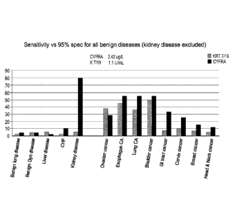
Figure 3 shows the sensitivity vs 95% specificity for benign diseases as
assayed by
the method of the invention.
Figure 4 shows a ROC-curve comparing the different tumor markers, CA125, HE4,
K7/19 and CYFRA in ovarian cancer.
Figure 5 shows ROC-curve comparing the combination CA125EIA & HE4 EIA with
the combination CA125EIA & K7/19 assay.
Date Recue/Date Received 2022-01-07
CA 02938809 2016-08-04
WO 2015/118023
PCT/EP2015/052325
14
DETAILED DESCRIPTION OF THE INVENTION
Before the present invention is described, it is to be understood that this
invention is not
limited to the particular embodiments described as methods, devices, and
formulations
may of course vary. It is also to be understood that the terminology used
herein is for the
purpose of describing particular embodiments only, and is not intended to
limit the scope
of the present invention which will be limited only by the appended claims. It
must be
noted that as used herein and in the appended claims, the singular forms "a,"
"an," and
"the" include plural referents unless the context clearly dictates otherwise,
and includes
reference to equivalent steps and methods known to those skilled in the art.
The terms used in this invention are, in general, expected to adhere to
standard
definitions generally accepted by those having ordinary skill in the art of
molecular
biology. A few exceptions, as listed below, have been further defined within
the scope of
the present invention.
"At least one" as used herein means one or more, i.e. 1, 2, 3,4, 5,6, 7, 8,9,
10 etc.
"Detection", "detect", "detecting" as used herein includes qualitative and/or
quantitative
detection (measuring levels) with or without reference to a control, and
further refers to
the identification of the presence, absence, or quantity of a given protein,
specifically
heterotypic complex of keratin 7 with keratin 19, and/or fragment(s) thereof.
"Diagnosis" as used herein encompasses the identification of the nature of a
disease.
"Prognosis" as used herein encompasses a forecast as to the probable outcome
of a
disease, the prospects as to recovery from a disease as indicated by the
nature and
symptoms of a disease.
"True positives" refers to those subjects having a localized or a metastasized
malignant
neoplasm.
"False negatives" refers to those subjects having either a localized or a
metastasized
malignant neoplasm and are not categorized as such by a diagnostic assay.
"True negatives" refers to those subjects who do not have a localized or a
metastasized
malignant neoplasm and who are categorized as such by a diagnostic assay.
"False positives" refers to those subjects who do not have a localized or a
metastasized
malignant neoplasm but are categorized by a conventional diagnostic assay as
having a
localized or metastasized malignant neoplasm.
CA 02938809 2016-08-04
WO 2015/118023
PCT/EP2015/052325
Depending on context, the term "false positives" may also refer to those
subjects who do
not have malignant neoplasm but are categorized by a diagnostic assay as
having
malignant neoplasm or a non-malignant disease.
"Sensitivity", as used herein in the context of its application to diagnostic
assays, refers
5 to the proportion of all subjects with localized or metastasized
malignant neoplasm that
are correctly identified as such (that is, the number of true positives
divided by the sum
of the number of true positives and false negatives).
"Specificity" of a diagnostic assay, as used herein in the context of its
application to
diagnostic assays, refers to the proportion of all subjects with neither
localized or
10 metastasized malignant neoplasm that are correctly identified as such
(that is, the
number of true negatives divided by the sum of the number of true negatives
and false
positives).
The terms "neoplasm" or "tumor" may be used interchangeably and refer to an
abnormal
mass of tissue wherein the growth of the mass surpasses and is not coordinated
with the
15 growth of normal tissue. A neoplasm or tumor may be defined as "benign"
or "malignant"
depending on the following characteristics: degree of cellular differentiation
including
morphology and functionality, rate of growth, local invasion and metastasis. A
"benign"
neoplasm is generally well differentiated, has characteristically slower
growth than a
malignant neoplasm and remains localized to the site of origin. In addition a
benign
neoplasm does not have the capacity to infiltrate, invade or metastasize to
distant sites.
A "malignant" neoplasm is generally poorly differentiated (anaplasia), has
characteristically rapid growth accompanied by progressive infiltration,
invasion, and
destruction of the surrounding tissue. Furthermore, a malignant neoplasm has
the
capacity to metastasize to distant sites. The term "metastasis" refers to the
spread or
migration of cancerous cells from a primary (original) tumor to another organ
or tissue,
and is typically identifiable by the presence of a "secondary tumor" or
"secondary cell
mass" of the tissue type of the primary (original) tumor and not of that of
the organ or
tissue in which the secondary (metastatic) tumor is located. For example a
carcinoma of
the lung that has migrated to bone is said to be metastasized lung cancer, and
consists
of cancer cells originating from epithelial lung cells growing in bone tissue.
"Healthy" refers to a subject possessing good health. Such a subject
demonstrates an
absence of any malignant or non-malignant disease. In the context of this
application, a
CA 02938809 2016-08-04
WO 2015/118023
PCT/EP2015/052325
16
"healthy individual" is only healthy in that they have an absence of any
malignant or non-
malignant disease; a "healthy individual" may have other diseases or
conditions that
would normally not be considered "healthy'.
"Subject" as used herein includes humans, nonhuman primates such as
chimpanzees
and other apes and monkey species, farm animals such as cattle, sheep, pigs,
goats
and horses, domestic mammals such as dogs and cats, laboratory animals
including
rodents such as mice, rats and guinea pigs, and the like. The term does not
denote a
particular age or sex. Thus, adult and newborn subjects, as well as fetuses,
whether
male or female, are intended to be covered. In preferred embodiments, the
subject is a
mammal, including humans and non-human mammals. In the most preferred
embodiment, the subject is a human.
"Monoclonal antibody" or "mAb" as used herein refers to an antibody of a
single amino
acid composition, that is directed against a specific antigen and that is
produced by a
single clone of B cells or hybridoma.
"Polyclonal antibody" as used herein refers to an antibody that is directed
against a
specific antigen that is derived from different B-cell lines.
"Fab" as used herein refers to an antibody fragment having a molecular weight
of about
50,000 Da and antigen binding activity, in which about a half of the N-
terminal side of the
H chain and the entire L chain, among fragments obtained by treating IgG with
a
protease, papaine, are bound together through a disulfide bond.
"F(ab')2" as used herein refers to an antibody fragment having a molecular
weight of
about 100,000 Da and antigen binding activity, which is slightly larger than
the Fab
bound via a disulfide bond of the hinge region, among fragments obtained by
treating
IgG with a protease, pepsin.
"Fabl " as used herein refers to an antibody fragment having a molecular
weight of about
50,000 Da and antigen binding activity, which is obtained by cutting a
disulfide bond of
the hinge region of the F(ab')2. As used herein, a single chain Fv ("scFv")
polypeptide is
a covalently linked VH:.VL heterodimer which is usually expressed from a gene
fusion
including VH and VL encoding genes linked by a peptide-encoding linker. The
human
scFv fragment of the invention includes CDRs that are held in appropriate
conformation,
preferably by using gene recombination techniques.
CA 02938809 2016-08-04
WO 2015/118023
PCT/EP2015/052325
17
"Hybridoma" as used herein denotes a cell, which is obtained by subjecting a B
cell
prepared by immunizing a non-human mammal with an antigen to cell fusion with
a
myeloma cell derived from a mouse or the like which produces a desired
monoclonal
antibody having an antigen specificity.
The terms "polypeptide," "protein," and "peptide" are used herein
interchangeably to refer
to amino acid chains in which the amino acid residues are linked by peptide
bonds or
modified peptide bonds. The amino acid chains can be of any length of greater
than two
amino acids. Unless otherwise specified, the terms "polypeptide," "protein,"
and "peptide"
also encompass various modified forms thereof. Such modified forms may be
naturally
occurring modified forms or chemically modified forms. Examples of modified
forms
include, but are not limited to, glycosylated forms, phosphorylated forms,
myristoylated
forms, palmitoylated forms, ribosylated forms, acetylated forms, ubiquitinated
forms, etc.
Modifications also include intra-molecular crosslinking and covalent
attachment to
various moieties such as lipids, flavin, biotin, polyethylene glycol or
derivatives thereof,
etc. In addition, modifications may also include cyclization, branching and
cross-linking.
Further, amino acids other than the conventional twenty amino acids encoded by
genes
may also be included in a polypeptide
As used herein a "biological sample" encompasses a variety of sample types
obtained
from any subject having or not having malignant neoplasm. A typical subject is
a human;
however, any mammal that has a malignant neoplasm that may develop cancer can
serve as a source of a biological sample useful in a disclosed method.
Exemplary
biological samples useful in the disclosed methods include but are not limited
to solid
tissue samples such as e.g. biopsy specimens, tissue cultures or cells derived
there
from, and the progeny thereof, clinical samples, cells in culture, cell
supernatants, cell
lysates, body fluids. For example, biological samples include samples obtained
from a
tissue or fluids collected from an individual suspected of having a malignant
neoplasm.
Examples of biological fluid samples include but are not limited to blood
serum, blood
plasma, lymph, exudates, feces, gastric acid, gastric juice, lymph, mucus,
pericardial
fluid, peritoneal fluid, pleural fluid, pus, saliva, sputum, synovial fluid,
tears, sweat,
vaginal secretion, vomit and urine.
Further examples are biopsies and/or fine needle aspirates. Samples may be
fresh or
processed post-collection (e.g., for archiving purposes). In some examples,
processed
samples may be fixed (e.g., formalin-fixed) and/or wax- (e.g., paraffin-)
embedded.
CA 02938809 2016-08-04
WO 2015/118023
PCT/EP2015/052325
18
Fixatives for mounted cell and tissue preparations are well known in the art
and include,
without limitation, 95% alcoholic Bouin's fixative; 95% alcohol fixative; B5
fixative,
Bouin's fixative, formalin fixative, Karnovsky's fixative (glutaraldehyde),
Hartman's
fixative, Hollande's fixative, Orth's solution (dichromate fixative), and
Zenker's fixative
(see, e.g., Carson, Histotechology: A Self-Instructional Text, Chicago: ASCP
Press,
1997). In some examples, the sample (or a fraction thereof) is present on a
solid
support.
Solid supports useful in a disclosed method need only bear the biological
sample and,
optionally, but advantageously, permit convenient detection of the proteins of
interest in
the sample. Exemplary supports include microscope slides (e.g., glass
microscope
slides or plastic microscope slides), coverslips (e.g., glass coverslips or
plastic
coverslips), tissue culture dishes, multi-well plates, membranes (e.g.,
nitrocellulose or
polyvinylidene fluoride (PVDF)) or BIACOREQ chips.
"Treatment" as used herein is defined as the management of a patient through
medical
or surgical means. The treatment improves or alleviates at least one symptom
of a
medical condition or disease and is required to provide a cure. The term
"treatment
outcome" or "outcome of treatment" as used herein is the physical effect upon
the
patient of the treatment.
The term "algorithm" as used herein refers to a mathematical formula that
provides a
relationship between two or more quantities. Such a formula may be linear, or
non-linear,
and may exist as various numerical weighting factors in computer memory.
"Lung cancer" refers to a neoplasm, e.g., malignant neoplasm, of the lung
within a given
subject, wherein the neoplasm is of epithelial origin i.e. carcinoma of the
lung. Lung
carcinomas are categorized by the size and appearance of the malignant cells
and the
term "lung cancer" includes both non-small cell lung cancer (NSCLC) and small
cell lung
cancer (SCLC). The term "lung cancer", when used without qualification,
includes both
localized and metastasized lung cancer. The term "lung cancer" can be
qualified by the
terms "localized" or "metastasized" to differentiate between different types
of tumor,
where "localized" refers to the original mother tumor, and the metastasized to
the tumors
that has spread from the original mother tumor.
The TNM System (tumor, node, metastases) may be used to stage NSCLC in an
initial
evaluation. Using the TNM descriptors, a group is assigned, ranging from
occult cancer,
CA 02938809 2016-08-04
WO 2015/118023
PCT/EP2015/052325
19
through stages 0, IA (one-A), IB, IIA, IIB, IIIA, IIIB and IV (four). This
stage group assists
with the choice of treatment and estimation of prognosis
Small-cell lung carcinoma has traditionally been classified as 'limited stage'
(confined to
one half of the chest and within the scope of a single tolerable radiotherapy
field) or
"extensive stage" (more widespread disease). However, the TNM classification
and
grouping are useful in estimating prognosis.
For both NSCLC and SOLO, the two general types of staging evaluations are
clinical
staging and surgical staging. Clinical staging is performed prior to
definitive surgery. It is
based on the results of imaging studies (such as CT scans and PET scans) and
biopsy
results. Surgical staging is evaluated either during or after the operation,
and is based on
the combined results of surgical and clinical findings, including surgical
sampling of
thoracic lymph nodes
"Ovarian cancer" refers to a neoplasm, e.g., malignant neoplasm, of the ovary
within a
given female subject, wherein the neoplasm is of epithelial origin.
The term "ovarian cancer", when used without qualification, includes both
localized and
metastasized ovarian cancer. The term "ovarian cancer" can be qualified by the
terms
"localized" or "metastasized" to differentiate between different types of
tumor, where
"localized" refers to the original mother tumor, and the metastasized to the
tumors that
has spread from the original mother tumor.
Ovarian cancer can be staged according to the AJCC/TNM System. This describes
the
extent of the primary tumor (T), the absence or presence of metastasis to
nearby lymph
nodes (N), and the absence or presence of distant metastasis (M). The extent
of primary
tumor contains three sub-categories T1, T2, T3. This closely resembles the
system that
is actually used by most gynecologic oncologists, called the FIGO system. Both
rely on
the results of surgery for the actual stage. In the FIGO system the tumor
stage is
classified from I-IV depending on how far the tumor has spread. Where stage IV
is worst
meaning that the tumor has spread to its estimated limit. Stages I-Ill
contains the sub-
categories A, B and C.
"Esophagus cancer" refers to a neoplasm, e.g., malignant neoplasm, of the
esophagus
within a given subject, wherein the neoplasm is of epithelial origin i.e.
carcinoma of the
esophagus. These carcinomas fall into two classes: either adenocarcinomas or
squamous cell carcinomas.
CA 02938809 2016-08-04
WO 2015/118023
PCT/EP2015/052325
The most common system used to stage esophageal cancer is the TNM system of
the
American Joint Committee on Cancer (AJCC). The TNM system is based on several
key
pieces of information: T refers to how far the primary tumor has grown into
the wall of the
esophagus and into nearby organs. N refers to cancer spread to nearby lymph
nodes. M
5 indicates whether the cancer has metastasized (spread to distant organs).
G describes
the grade of the cancer, which is based on how the patterns of cancer cells
look under a
microscope. Staging also takes into account the cell type of the cancer
(squamous cell
carcinoma or adenocarcinoma). For squamous cell cancers, the location of the
tumor
can also be a factor in staging.
10 "Bladder cancer" refers to a neoplasm, e.g., malignant neoplasm, of the
urinary bladder
within a given subject, wherein the neoplasm originates from the urothelium or
transitional epithelium. Bladder cancer refers thus to Transitional cell
(urothelial)
carcinoma, but it also refers to adenocarcinoma of the bladder.
The staging system most often used for bladder cancer is that of the American
Joint
15 Committee on Cancer (AJCC). This is also called the TNM system. The T
category
describing the tumor contains the sub-categories: TO, Ta, Tis, T1, T2a, T2b,
T3a, T3b,
T4.
Immunoassays such as immunohistochemistry (INC) and immunocytochemistry (ICC)
As used herein an "immunoassay" is a biochemical test that measures the
presence or
20 concentration of a substance in samples that frequently contain a
complex mixture of
substances. Such assays are based on the unique ability of an antibody to bind
with high
specificity to one or a very limited group of molecules or antigens.
Immunohistochemistry
(INC) and immunocytochemistry (ICC) are two exemplary immunoassay techniques
useful for detecting the antigen heterotypic complex of keratin 7 with keratin
19, and/or
fragment(s) thereof in the disclosed methods and uses.
Imnnunocytochemistry (ICC) is a common laboratory technique that uses
antibodies that
target specific peptides or protein antigens in the cell via specific
epitopes. These bound
antibodies can then be detected using several different methods. ICC allows
researchers
to evaluate whether or not cells in a particular sample express the antigen in
question. In
cases where an immunopositive signal is found, ICC also allows researchers to
determine which sub-cellular compartments are expressing the antigen.
CA 02938809 2016-08-04
WO 2015/118023
PCT/EP2015/052325
21
In immunohistochemistry (INC), samples are sections of biological tissue,
where each
cell is surrounded by tissue architecture and other cells normally found in
the intact
tissue. Immunocytochemistry is a technique used to assess the presence of a
specific
protein or antigen in cells (cultured cells, cell suspensions) by use of a
specific antibody,
which binds to it, thereby allowing visualization and examination under a
microscope. It
is a valuable tool for the determination of cellular contents from individual
cells. Samples
that can be analyzed include blood smears, aspirates, swabs, cultured cells,
and cell
suspensions.
Enzyme-linked immunosorbent assay (ELISA) and sandwich ELISA, are immunoassays
.. that are advantageously used in the methods disclosed herein. In a (direct)
ELISA an
unknown amount of antigen is affixed to a surface, and then a specific
antibody is
applied over the surface so that it can bind to the antigen. This antibody is
linked to an
enzyme, and in the final step a substance is added so that the enzyme can
convert to
some detectable signal, most commonly a colour change in a chemical substrate.
In a
sandwich ELISA a capture antibody that can bind to the antigen is affixed to
the surface.
The other steps are equivalent to the ELISA.
In an Enzyme Immuno Assay (EIA), which is similar to the sandwich ELISA,
streptavidin
is affixed to a surface and then the capture antibody is biotinylated,
otherwise the other
steps are performed equivalently as the ELISA. The EIA immunoassay is
advantageously used in the methods disclosed herein.
Western Blot (sometimes called the protein immunoblot) is a widely used
analytical
technique used to detect specific proteins in the given sample of tissue
homogenate or
extract. It uses gel electrophoresis to separate native or denatured proteins
by the length
of the polypeptide (denaturing conditions) or by the 3-D structure of the
protein (native/
.. non-denaturing conditions). The proteins are then transferred to a membrane
(typically
nitrocellulose or PVDF), where they are probed (detected) using antibodies
specific to
the target protein.
Targeting agents such as e.g. antibodies, antigen-binding fragments, or
variants, fusions
or derivatives specific for keratin 7 and keratin 19 and/or fragments thereof
are used to
detect the expression of heterotypic complex of keratin 7 with keratin 19,
and/or
fragment(s) thereof. The targeting agents of the invention are advantageously
provided
as a composition but may also be provided separately but subsequent to one
another.
The method of the invention thus provides antibodies antigen-binding
fragments, or
81798287
22
variants, fusions or derivatives binding to heterotypic complex of keratin 7
with keratin
19, and/or fragment(s) thereof. The antibodies can be detected, as further
described
herein, by direct labelling of the antibodies themselves, for example, with
radioactive
labels, fluorescent labels, hapten labels such as, biotin, or an enzyme such
as
horseradish peroxidase or alkaline phosphatase. Alternatively, an indirect
labelling is
used where the unlabeled primary antibody is used in conjunction with a
labelled
secondary antibody, comprising e.g. antiserum, polyclonal antiserum or a
monoclonal
antibody specific for the primary antibody. IHC and ICC protocols are well
known in the
art and are commercially available, see e.g. Antibodies: A Laboratory Manual,
Harlow
and Lane (Cold Spring Harbor Laboratory press, Cold Spring Harbor, NY 1988)
and
Current Protocols in Immunology, and Current Protocols in Molecular Biology,
both John
Wiley and Sons, Inc., N. Y.).
As revealed above, the present invention provides means and methods to improve
sensitivity and specificity of the diagnosis and/or prognosis of malignant
neoplastic
disease such as e.g. lung cancer, bladder cancer, esophagus cancer and/or
ovarian
cancer. More specifically, the present invention provides a composition that
gives better
sensitivity for detection of malignant neoplastic disease so as to improve the
detection of
malignant neoplastic protein markers in ELISA and EIA as well as in IHC and
ICC,
thereby giving a more consistent and reliable result when performing diagnosis
and/or
prognosis of patients having a malignant neoplastic disease such as e.g. lung
cancer,
bladder cancer, esophagus cancer and/or ovarian cancer.
Further, the methods according to the invention will improve the
identification of
malignant neoplastic diseases such as e.g. lung cancer, bladder cancer,
esophagus
cancer and/or ovarian cancer compared to available methods.
The targeting agents used in the methods herein thus show an improved
detection of
various malignant neoplastic diseases when compared to other known methods
such as
e.g. CYFRA 21-1 (see Examples given herein).
Examples 2-5 show that malignant neoplastic disease such as e.g. lung cancer,
bladder
cancer, esophagus cancer and/or ovarian cancer are identified by an expression
of
heterotypic complex of keratin 7 with keratin 19, and/or fragment(s) thereof.
The method detects malignant neoplastic disease such as e.g. lung cancer,
bladder
cancer, esophagus cancer and/or ovarian cancer with antibodies binding
specifically to
the heterotypic complex of keratin 7 with keratin 19, and/or fragment(s)
thereof.
Date Recue/Date Received 2021-05-04
CA 02938809 2016-08-04
WO 2015/118023
PCT/EP2015/052325
23
The proteins
The method of the present invention encompass the use of at least two
targeting agents
such as e.g. two primary antibodies, antigen-binding fragments, variants,
fusions,
derivatives or fragments thereof. At least one first primary antibody binds
specifically to
keratin 7 or fragments thereof and at least one second primary antibody binds
specifically to keratin 19 or fragments thereof. Both of said first and second
primary
antibodies are capable of binding specifically and simultaneously to
Heterotypic complex
of keratin 7 with keratin 19, and/or fragment(s) thereof and/or fragments
thereof.
Keratin 7 (K7) (NCB! Reference Sequence: NP_005547.3, NCBI gene ID: 3855) also
known as cytokeratin 7 (CK7) or sarcolectin (SCL) is a protein that in humans
is encoded
by the KRT7 gene. Keratin 7 is a type II keratin and is together with other
genes
encoding the type II keratins clustered in a region of chromosome 12q12-q13.
K7 is a 51
kDa protein which is 469 amino acids long and has the general structure as
seen in Fig.
1. Alternative splicing may result in several transcript variants. It is
specifically expressed
in the simple epithelia lining the cavities of the internal organs and in the
gland ducts and
blood vessels).
Keratin 19 (K19) (NCB! Reference Sequence: NP_002267.2, NCBI gene ID: 3880) is
also known as cytokeratin 19 (CK19) and is a type I keratin specifically found
in the
periderm, the transiently superficial layer that envelops the developing
epidermis. K19 is
a 44 kDa protein which is 400 amino acids long and has the general structure
as seen in
Fig. 1. K19 is in humans encoded by the KRT19 gene and is located in a region
of
chromosome 17q12-q21 gene.
In vivo keratins assemble as heteropolymers, i.e. a Type I and a Type ll
protein form
heterodimers, which in turn assemble to form tetramers. Two monomers form a
parallel
dinner by the winding of their a-helical rods into a coiled coil, oriented in
register and in
the same direction, and then two dimers join side-by-side in a staggered anti-
parallel
orientation to form a bidirectional tetramer (see Fig. 1). When keratins K7
and K19
assemble into heterodimers and tetramers heterotypic complexes of keratin 7
with
keratin 19 are formed.
As used herein the term "heterotypic complex of keratin 7 with keratin 19" is
intended to
include a molecular entity originating from a parallel and in register dimer
between full-
length keratin 7 and full-length keratin 19 and fragments thereof. It further
includes
overlapping dimers between keratin 7 and keratin 19 into anti-parallel
tetramers between
CA 02938809 2016-08-04
WO 2015/118023
PCT/EP2015/052325
24
keratin 7 and keratin 19 and fragments thereof. It further includes oligomers,
protofilaments and filament assembled from these tetramers and any fragments
thereof.
For a general structure of heterotypic complex of keratins see Fig. 1.
In the heterotypic complex of keratin 7 with keratin 19 there is
advantageously a
minimum of least 3, preferably at least 5 or at least 10, or at least 15, or
at least 20, or at
least 25 or at least 30, but more preferably more than 40 amino acids in an
unbroken
sequence, derived from the keratin 7 moiety of the heterotypic complex, and
there must
be a minimum of least 3, preferably at least 5 or at least 10, or at least 15,
or at least 20,
or at least 25 or at least 30, but more preferably more than 40 amino acids in
an
unbroken sequence, derived from the keratin 19 moiety of the heterotypic
complex. Said
amino acids must be unique to keratin 7 or keratin 19 respectively.
Advantageously said minimum of least 3, preferably at least 5 or at least 10,
or at least
15, or at least 20, or at least 25 or at least 30, but more preferably more
than 40 amino
acids in an unbroken sequence are from the 256-412 amino acid sequence in the
keratin
7 protein. More preferably the unbroken sequence is from the 300-380 amino
acid
sequence in keratin 7.
Advantageously said minimum of least 3, preferably at least 5 or at least 10,
or at least
15, or at least 20, or at least 25 or at least 30, but more preferably more
than 40 amino
acids in an unbroken sequence are from the 244-400 amino acid sequence in the
keratin
19 protein. More preferably the unbroken sequence is from the 311-375 amino
acid
sequence in keratin 19. The antibodies
In one aspect the present invention provides a method encompassing the use of
at least
two targeting agents, such as e.g. two primary antibodies, antigen-binding
fragments, or
variants, fusions or derivatives thereof to detect the presence of heterotypic
complex of
.. keratin 7 with keratin 19, and/or fragment(s) thereof in a biological
sample, wherein a
first primary antibody, antigen-binding fragment, or variant, fusion or
derivative thereof
binds specifically to the keratin 7 or fragments thereof of the heterotypic
complex, and a
second primary antibody, antigen-binding fragment, or variant, fusion or
derivative
thereof binds specifically to the keratin 19 or fragments thereof of the
heterotypic
complex. Advantageously, the at least two antibodies, antigen-binding
fragments, or
variants, fusions or derivatives thereof are present together in an antibody
cocktail, either
in a format ready-to-use by the user or in a concentrated solution which
requires a
dilution before its use, sometimes referred to as a stock solution.
CA 02938809 2016-08-04
WO 2015/118023
PCT/EP2015/052325
Thus, one aspect of the invention encompasses a composition comprising at
least one
first primary antibody, antigen-binding fragment, or variant, fusion or
derivative thereof
that recognizes a keratin 7 peptide and/or fragment(s) thereof, and at least
one second
primary antibody, antigen-binding fragments or variants fusions or derivative
thereof that
5 recognizes a keratin 19 peptide and/or fragment(s) thereof, wherein both
the first and the
second primary antibodies are capable of binding specifically and
simultaneously to
heterotypic complex of keratin 7 with keratin 19, and/or fragment(s) thereof.
By "binding specifically to" or "reacting specifically with" as used herein it
is intended to
equal "capable of binding selectively" or "binding specifically to". As used
herein the
10 expressions are intended to mean that the antibody or antigen-binding
fragment, or
variant, fusion or derivative thereof, including any anti-body derived binding
moiety,
which is capable of binding to an antigen of a molecule and further which
binds at least
10-fold more strongly the proteins than to another proteins for example at
least 50-fold
more strongly or at least 100- fold more strongly. The binding moiety may be
capable of
15 binding selectively to the protein under physiological conditions, e.g.
in vivo. Suitable
methods for measuring relative binding strengths include, immunoassays, for
example
where the binding moiety is an antibody. Alternatively, binding may be
assessed using
competitive assays or using Biacore(R) analysis (Biacore International AB,
Sweden).
By the term "binding specifically and simultaneously to" as used herein it is
intended to
20 mean that the first and second targeting agents bind specifically and
simultaneously to
the heterotypic complex of keratin 7 with keratin 19, and/or fragment(s)
thereof, and form
a triplex comprising said first and second targeting agents and the
heterotypic complex
of keratin 7 with keratin 19, and/or fragment(s) thereof. Said first and
second targeting
agents will form said triplex with the heterotypic complex of keratin 7 with
keratin 19,
25 and/or fragment(s) thereof during a time period that is sufficiently
long for the complex to
be detected by any means or methods that are described herein, or else by
means or
methods known to the person skilled in the art.
In one aspect embodiment, each of the first and second antibodies or antigen-
binding
fragments, or variants, fusions or derivatives thereof, bind to separate
antigenic sites of
the heterotypic complex of keratin 7 with keratin 19, and/or fragment(s)
thereof. Typically
the first primary antibody reacts specifically with an antigenic site present
on the K7
moiety of the heterotypic complex of keratin 7 with keratin 19, and/or
fragment(s) thereof,
and the second primary antibody reacts specifically with an antigenic site
present on the
CA 02938809 2016-08-04
WO 2015/118023
PCT/EP2015/052325
26
K19 moiety of the heterotypic complex of keratin 7 with keratin 19, and/or
fragment(s)
thereof.
Because the two monomers keratin 7 and keratin 19 of the heterotypic complex
between
keratin 7 and keratin 19 are aligned in parallel and in register with the
helical structures,
any two antibodies binding to corresponding positions starting from helix 1A
to helix 2B
(see Fig. 1), on keratin 7 and keratin 19 respectively will not bind
simultaneously. This
means that where the keratin 7 is coiled around keratin 19, corresponding
exposed
positions would not allow simultaneous binding of the two antibodies for
steric reasons.
This is further exemplified when e.g. amino acids 271- 291 on keratin 19 pair
up in the
coiled coil with amino acids 283-303 on keratin 7. If an antibody to keratin 7
binds within
the sequence of 283-303 on keratin 7, then an antibody to keratin 19 would not
be able
to bind to amino acids 271-291 on keratin 19 at the same time in the
heterotypic
complex because of steric hindrance.
A number of available antibodies that bind specifically to keratin 7 may be
used in the
methods presented herein, such as e.g. C-35, 0-62, 0-68, 018, 035, KS 7.18,
LDS-68,
LP1K, RCK105. However one antibody that is advantageously used in the methods
presented herein is the keratin 7 monoclonal antibody produced by clone Ks
7.18 which
is available from Progen Biotechnik, GnrIBH, Germany. Monoclonal antibody KS
7.18
binds specifically to amino acids 300-350 of the K7 moiety of the heterotypic
complex of
keratin 7 with keratin 19, and/or fragment(s) thereof.
Examples of available antibodies that will bind specifically to keratin 19 are
A53-B/A2.26
aka Ks19.1, BM-19.21, 000003, 00D004,CKSO4, CKS06, SA 21, SA 45, Ks19.2,
LP2K. However one antibody that is advantageously used in the present method
is the
Ks 19.2 monoclonal antibody which is available from Progen Biotechnik, GmBH
also
known as the BM-19.21 monoclonal antibody produced by clone BM-19.21 which is
available from Roche Diagnostics. BM-19.21 binds specifically to amino acids
346-367
of the of the K19 moiety of the heterotypic complex of keratin 7 with keratin
19, and/or
fragment(s) thereof. Several of the antibodies against keratin 19 are
described in:
Epitope Specificity of 30 Monoklonal Antibodies against Cytokeratin Antigens:
The
ISOBM TD-1 workshop, Tumor Biol 1998;19:132-152.
Advantageously, each of the first and second primary antibodies or antigen-
binding
fragments, or variants, fusions or derivatives thereof are provided in a
composition as an
CA 02938809 2016-08-04
WO 2015/118023
PCT/EP2015/052325
27
antibody cocktail, in aqueous form or in a freeze dried powder form. For the
latter, a re-
hydration step is required to put the antibodies in a usable liquid form
before use.
Alternatively the first and second primary antibodies are provided separately,
i.e. one
primary antibody is provided before the other when used in the methods
presented
herein. In one embodiment the first primary antibody recognizing a keratin 7
peptide
and/or fragment(s) thereof is provided to a sample containing heterotypic
complex of
keratin 7 with keratin 19, and/or fragment(s) thereof. The first primary
antibody will bind
to an antigenic site present on the K7 moiety of the heterotypic complex of
keratin 7 with
keratin 19, and/or fragment(s) thereof and form an antigen ¨ K7-antibody
complex.
Thereafter, the second primary antibody recognizing a keratin 19 peptide
and/or
fragment(s) thereof is provided to the sample containing heterotypic complex
of keratin 7
with keratin 19 ¨ K7-antibody. Said second primary antibody will bind to an
antigenic site
present on the K19 moiety of the heterotypic complex of keratin 7 with keratin
19 and
form a K19-antibody - antigen ¨ K7-antibody complex.
In an alternative embodiment, the second primary antibody recognizing a
keratin 19
peptide and/or fragment(s) thereof is provided to a sample containing
heterotypic
complex of keratin 7 with keratin 19, and/or fragment(s) thereof. The second
primary
antibody will bind to an antigenic site present on the K19 moiety of the
heterotypic
complex of keratin 7 with keratin 19 and form an antigen ¨ K19-antibody
complex.
Thereafter, the first primary antibody recognizing a keratin 7 peptide and/or
fragment(s)
thereof is provided to the sample containing heterotypic complex of keratin 7
with keratin
19¨ K19-antibody. Said first primary antibody will bind to an antigenic site
present on
the K7 moiety of the heterotypic complex of keratin 7 with keratin 19 and form
a K7-
antibody - antigen ¨ K19-antibody complex
The antibodies may be whole antibodies or fragments thereof, e.g. antigen-
binding
fragment, or variant, fusion or derivative thereof as long as they are capable
of binding to
the desired protein in vitro. Such binding specificity may be determined by
methods well
known in the art, such as e.g. ELISA, EIA, immunohistochemistry,
immunoprecipitation,
Western blots, chromatography and flow cytometry using transfected cells
expressing all
of the subunit or a heterodimer thereof (see Examples).
The term "antibody" includes substantially intact antibody molecules of any
species such
as rodents, e.g. murine, rat, guinea pig, or non-rodents such as rabbit, goat,
sheep, dog,
pig, camel, dromedary, donkey, horse or chicken, as well as chimaeric
antibodies,
CA 02938809 2016-08-04
WO 2015/118023
PCT/EP2015/052325
28
humanized antibodies, human antibodies (wherein at least one amino acid is
mutated
relative to the naturally occurring human antibodies), single chain
antibodies, bispecific
antibodies, antibody heavy chains, antibody light chains, homo-dimers and
hetero-
dimers of antibody heavy and/or light chains, and antigen binding fragments
and
derivatives of the same. For example, the antibody may be a monoclonal
antibody.
Antigenic specificity is conferred by variable domains and is independent of
the constant
domains, as known from experiments involving the bacterial expression of
antibody
fragments, all containing one or more variable domains. These molecules
include Fab-
like molecules; Fv molecules; single-chain Fv (ScFv) molecules where the V H
and V L
partner domains are linked via a flexible oligopeptide;) and single domain
antibodies
(dAbs) comprising isolated V domains. A general review of the techniques
involved in
the synthesis of antibody fragments which retain their specific binding sites
is to be found
in Winter & Milstein (1991) Nature 349, 293- 299.
Thus, by "antigen-binding fragment" is meant a functional fragment of an
antibody that is
capable of binding to any of the proteins K7or K19 or fragments thereof, and
heterotypic
complex of keratin 7 with keratin 19, and/or fragment(s) thereof. Exemplary
antigen-
binding fragments may be selected from the group consisting of Fv fragments
(e.g.
single chain Fv and disulphide-bonded Fv), Fab-like fragments (e.g. Fab
fragments, Fab'
fragments and F(ab) 2 fragments), single antibody chains (e.g. heavy or light
chains),
.. single variable domains (e.g. VH and VL domains) and domain antibodies
(dAbs,
including single and dual formats; i.e. dAb-linker-dAb).
Thus, in one embodiment the antibody or antigen-binding fragment, or variant,
fusion or
derivative thereof, comprises or consists of an intact antibody. In one
embodiment, the
antibody is a monoclonal antibody.
For example, the antibody or antigen-binding fragment, or a variant, fusion or
derivative
thereof, may consist essentially of an intact antibody. By "consist
essentially of we mean
that the antibody or antigen-binding fragment, variant, fusion or derivative
thereof
consists of a portion of an intact antibody sufficient to retain binding
specificity for any of
the two different proteins K7 and K19 or fragments thereof. In further
embodiments, the
two different proteins K7 and K19 are of human origin.
The term 'antibody' also includes all classes of antibodies, including IgG,
IgA, IgM, IgD
and IgE. In one embodiment, however, the antibody is an IgG molecule, such as
an
CA 02938809 2016-08-04
WO 2015/118023
PCT/EP2015/052325
29
IgGI, IgG2, IgG3, or IgG4 molecule. In one embodiment, the antibody is an IgGI
molecule. In a further embodiment, the antibody is an IgGI molecule with a
kappa light
chain.
In a further embodiment, the antibody may be a non-naturally occurring
antibody. Of
course, where the antibody is a naturally occurring antibody, it is provided
in an isolated
form (i.e. distinct from that in which it is found in nature).
Also included within the scope of the invention are modified versions of
antibodies and
antigen-binding fragments thereof, e.g. modified by the covalent attachment of
polyethylene glycol or other suitable polymer, and uses of the same.
Methods of generating antibodies and antibody fragments are well known in the
art. For
example, antibodies may be generated via any one of several methods which
employ
induction of in vivo production of antibody molecules, screening of
immunoglobulin
libraries or generation of monoclonal antibody molecules by cell lines in
culture. These
include, but are not limited to, the hybridoma technology, the human B-cell
hybridoma
technology, and the Epstein-Barr virus (EBV)- hybridoma technology.
For example, generating monoclonal or polyclonal antibodies to K7, or K19 may
be done
by immunization where the whole protein or a suitable fragment thereof can be
injected
into non-human mammals (such as mice or rabbits), followed by boost
injections, to
produce an antibody response. Serum isolated from immunized animals may be
isolated
for the polyclonal antibodies contained therein, or spleens from immunized
animals may
be used for the production of hybridomas and monoclonal antibodies.
In one example, a monoclonal antibody to one of the proteins can be prepared
from
murine hybridomas according to the classical method of Kohler and Milstein
{Nature,
256:495, 1975) or derivative methods thereof. Briefly, a mouse (such as
Balb/c) is
repetitively inoculated with a few micrograms of the selected protein or
peptide fragment
thereof or a suitable carrier conjugate thereof over a period of a few weeks.
The mouse
is then sacrificed, and the antibody-producing cells of the spleen isolated.
The spleen
cells are fused by means of polyethylene glycol with mouse myeloma cells, and
the
excess un-fused cells destroyed by growth of the system on selective media
comprising
aminopterin (HAT media). The successfully fused cells are diluted and aliquots
of the
dilution placed in wells of a microtiter plate where growth of the culture is
continued.
Antibody-producing clones are identified by detection of antibody in the
supernatant fluid
of the wells by immunoassay procedures, such as ELISA, as originally described
by
81798287
Engvall (Enzymol., 70:419, 1980), and derivative methods thereof.
Selected positive clones can be expanded and their monoclonal antibody product
harvested for use.
Commercial sources of antibodies include DAKO NS, Abeam, Lab Vision, BioCare
5 Medical, Cell Marque Corp. Abnova, Acris, Progen, Santa cruz biotech etc.
Polyclonal antibody-producing animals are identified by bleeding immunized
animals and
selection of appropriate animal with a suitable polyclonal antibody-titer
thereof.
In some embodiments, antibodies are purified before use. Purification of
antibodies are
done using techniques available in the art and known to the skilled person.
10 Generation of antibodies K7 and K19 are described in the art and
available from
commercial sources as described herein, or being available using techniques
known to a
skilled artisan using references enclosed herein.
The antibody or antigen-binding fragment or derivative thereof may also be
produced by
recombinant means. Suitable monoclonal antibodies to selected antigens and
proteins
15 may be prepared by techniques known to a skilled person. Antibody
fragments can also
be obtained using methods well known in the art. For example, antibody
fragments may
be prepared by proteolytic hydrolysis of the antibody or by expression in E.
coli or
mammalian cells (e.g. Chinese hamster ovary cell culture or other protein
expression
systems) of DNA encoding the fragment. Alternatively, antibody fragments can
be
20 obtained by pepsin or papain digestion of whole antibodies by
conventional methods.
Thus, in one embodiment at least one of the primary antibodies used in the
methods as
presented herein is a monoclonal antibody. In a further embodiment at least
one of the
primary antibodies used in the methods of the invention is a recombinant
antibody.
Alternatively, complex formation may be detected using duolink (0-link
bioscience).
25 Conventional immunofluorescence can reveal the presence of the
individual proteins,
but not the formation of heterodimers between different proteins. Using
Duolink it would
be possible to locate exactly where hetero dimerization takes place and
quantify the
relative extent of the interaction. Thus, it would be possible using the
duolink technique
to locate and quantify the heterotypic complex between keratin 7 and keratin
19 by using
30 one antibody specific for keratin 7 and one antibody specific for
keratin 19 wherein both
Date Recue/Date Received 2021-05-04
CA 02938809 2016-08-04
WO 2015/118023
PCT/EP2015/052325
31
antibodies bind simultaneously to the heterotypic complex between keratin 7
and keratin
19.
The antibodies or antigen-binding fragments or derivatives thereof, or
composition
comprising said antibodies or antigen-binding fragments or derivatives thereof
described
herein may be lyophilized for storage and reconstituted in a suitable carrier
prior to use.
Any suitable lyophilisation method (e.g. spray drying, cake drying) and/or
reconstitution
techniques can be employed. It will be appreciated by those skilled in the art
that
lyophilisation and reconstitution can lead to varying degrees of antibody
activity loss
(e.g. with conventional immunoglobulins, IgM antibodies tend to have greater
activity
loss than IgG antibodies) and that use levels may have to be adjusted upward
to
compensate. In one embodiment, the lyophilized (freeze dried) composition
loses no
more than about 20%, or no more than about 25%, or no more than about 30%, or
no
more than about 35%, or no more than about 40%, or no more than about 45%, or
no
more than about 50% of its activity (prior to lyophilisation) when re-
hydrated. It will be
further appreciated by persons skilled in the art that the antibodies and
antigen-binding
fragments, variants, fusions and derivatives thereof, described herein may
exist in
monomeric form or in the form of a homo-or hetero- multimer thereof (e.g.
dimer, trimer,
tetramer, pentamer, etc.).
Further provided herein is that the primary antibodies or fragments thereof
may be
labelled directly or indirectly, with a detectable moiety. By directly labeled
is meant that
the detectable moiety is attached to the antibody. By indirect labeled it is
meant that the
detectable moiety is attached to a linker, such as, for example, a secondary
or tertiary
antibody. The detectable moiety may be any moiety or marker known to those
skilled in
the art, or as described herein, and as being such a moiety being capable of
generating
a signal that allows the direct or indirect quantitative or relative
measurement of a
molecule to which it is attached.
A wide variety of detectable moieties, or labels, and conjugation techniques
are known
and reported extensively in both the scientific and patent literature.
Suitable labels
include radionuclides, enzymes, substrates, cofactors, inhibitors, fluorescent
agents,
chemiluminescent agents, magnetic particles and the like which are well-known
to the
skilled person. The detectable moiety may be a single atom or molecule which
is either
directly or indirectly involved in the production of a detectable species.
Optionally, the
detectable moiety is selected from the group consisting of a fluorescent
moiety, an
CA 02938809 2016-08-04
WO 2015/118023
PCT/EP2015/052325
32
enzyme linked moiety, a biotinylated moiety and a radiolabeled moiety, as
described
further herein, e.g. below. By "label", "detectable moiety" is meant any
detectable tag
that can be attached directly (e.g., a fluorescent molecule integrated into a
polypeptide)
or indirectly (e.g., by way of binding to a primary antibody with a secondary,
tertiary or
further antibody with an integrated fluorescent molecule) to the molecule of
interest.
Thus, a label, marker or detectable moiety is any tag that can be visualized,
for example,
with imaging methods.
By a "detectable moiety" we further include the meaning that the moiety is one
which,
when located at the target site following providing the composition of the
invention to a
biological sample, such as a tissue sample, or a biological fluid sample, may
be detected
in vitro. That includes that the detectable moiety is signal generating and it
is further
convenient and thus included in further embodiments if the detectable moiety
may be
detected and the relative amount and/or location of the moiety (for example,
the location
on an tissue sample) may be determined. Detectable moieties are well known in
the art.
Thus, antibodies or antigen-binding fragments or derivatives thereof, or the
composition
comprising such antibodies or antigen-binding fragments or derivatives thereof
are
useful in methods further exemplified herein by methods and uses for detection
of
heterotypic complex of keratin 7 with keratin 19, and/or fragment(s) thereof,
diagnosis or
prognosis malignant neoplastic disease such as e.g. lung cancer, bladder
cancer,
esophagus cancer and ovarian cancer in vitro of biological samples. In further
embodiments, immunochemical methods used are exemplified herein.
Suitable detectable moieties are well known in the art and the attachment or
linking of
these moieties to polypeptides and proteins is further well known in the art.
Further
examples of detectable moieties are an enzyme; an enzyme substrate; an enzyme
inhibitor; coenzyme; enzyme precursor; apoenzyme; fluorescent substance;
pigment;
chemiluminescent compound; luminescent substance; coloring substance; magnetic
substance; or a metal particle such as gold colloid; a radioactive substance
such as 1251,
1311, 32p, 3H, 35.-s, 14
or C; a phosphorylated phenol derivative such as a nitrophenyl
phosphate, luciferin derivative, or dioxetane derivative; or the like. The
enzyme may be a
dehydrogenase; an oxidoreductase such as a reductase or oxidase; a transferase
that
catalyzes the transfer of functional groups, such as an amino; carboxyl,
methyl, acyl, or
phosphate group; a hydrolase that may hydrolyze a bond such as ester,
glycoside,
ether, or peptide bond; a lyase; an isomerase; or a ligase. The enzyme may
also be
CA 02938809 2016-08-04
WO 2015/118023 PCT/EP2015/052325
33
conjugated to another enzyme. The enzyme may be detected by enzymatic cycling.
For
example, when the detectable label is an alkaline phosphatase, a measurement
may be
made by observing the fluorescence or luminescence generated from a suitable
substrate, such as an umbelliferone derivative. The umbelliferone derivative
may
comprise 4-methyl- umbellipheryl phosphate. The fluorescent or
chemiluminescent label
may be a fluorescein isothiocyanate; a rhodamine derivative such as rhodamine
B
isothiocyanate or tetramethyl rhodamine isothiocyanate; a dancyl chloride (5-
(dimethylamino)-1-naphtalenesulfonyl chloride); a dancyl fluoride; a
fluorescamine (4-
phenylspiro[furan-2(3H); ly-(3yH)-isobenzofuranl- 3;3y-dione); a
phycobiliprotein such as
a phycocyanine or phycoerythrin; an acridinium salt; a luminol compound such
as
lumiferin, luciferase, or aequorin; imidazoles; an oxalic acid ester; a
chelate compound of
rare earth elements such as europium (Eu), terbium (Tb) or samarium (Sm); or a
coumarin derivative such as 7-amino-4-methylcoumarin. The label may also be a
hapten, such as adamantine, fluoroscein isothiocyanate, or carbazole. The
hapten may
allow the formation of an aggregate when contacted with a multi-valent
antibody or
(streptavidin containing moiety. Further examples of detectable moieties
include, but are
, 111
not limited to, the following: radioisotopes (e.g. 3H, 14C, 35s, 1231, 1251,
1311 99Tc, In, 90y,
188R e), radionuclides (e.g. 110, 18F, 64Cu), fluorescent labels (e.g. FITC,
rhodamine,
lanthanide phosphors, carbocyanine), enzymatic labels (e.g. horseradish
peroxidase,
[beta]-galactosidase, luciferase, alkaline phosphatase), chemiluminescent,
biotinyl
groups and predetermined polypeptide epitopes recognized by a secondary
binding
entity (e.g. leucine zipper pair sequences, binding sites for secondary
antibodies, metal
binding domains, epitope or protein tags, carbohydrates). In some embodiments,
labels
are attached by spacer arms of various lengths to reduce potential steric
hindrance.
In indirect labelling, an additional molecule or moiety is brought into
contact with, or
generated at the site of, the antibody-antigen complexes, i.e. immune-
complexes,
between the primary antibody and the protein it binds to. For example, a
detectable
moiety such as an enzyme can be attached to or associated with the detecting
antibody
or detecting molecule as exemplified herein. The signal-generating molecule
can then
generate a detectable signal at the site of the immune-complex. For example,
an
enzyme, when supplied with suitable substrate, can produce a visible or
detectable
product at the site of the immune-complex.
CA 02938809 2016-08-04
WO 2015/118023
PCT/EP2015/052325
34
As another example of indirect labelling, an additional molecule (which can be
referred
to as a binding agent) that can bind to either the molecule of interest or to
the antibody
(primary antibody) of interest, such as a second antibody to the primary
antibody, can be
contacted with the immunocomplex. The additional molecule can have signal-
generating
.. molecule or detectable moiety.
The additional molecule may be an antibody, which can thus be termed a
secondary,
tertiary or further antibody. Binding of a secondary antibody to the primary
antibody can
form a so- called sandwich with the first (or primary) antibody and the
molecule of
interest. The immune- complexes can be contacted with the labelled, secondary
.. antibody under conditions effective and for a period of time sufficient to
allow the
formation of secondary immune complexes. The secondary immune complexes can
then
be generally washed to remove any non-specifically bound labelled secondary
antibodies, and the remaining label in the secondary immune complexes can then
be
detected. The additional molecule can also be or include one of a pair of
molecules or
moieties that can bind to each other, such as the biotin/avidin molecules, and
the
detecting antibody or detecting molecule should then include the other member
of the
pair.
Further examples of indirect labelling include the detection of primary
antibody-antigen
(immune-complexes) by a two-step approach. For example, a molecule (which can
be
referred to as a first binding agent), such as an antibody, that has binding
affinity for the
primary immune complex between the primary antibody-antigen complex can be
used to
form secondary complexes. After washing, the secondary complex can be
contacted
with another further molecule (which can be referred to as a second binding
agent) that
has binding affinity for the first binding agent, again under conditions
effective and for a
period of time sufficient to allow the formation of tertiary complexes. In
this example the
second binding agent may be linked to a detectable moiety, allowing detection
of the
tertiary complexes thus formed. This system may further comprise means to
provide for
signal amplification.
Other examples of primary, secondary or further binding agents with means for
signal
amplification are conjugated anti-immunoglobulins such as biotinylated
antibodies (e.g.,
conjugated with avidin/ streptavidin) or staphylococcal Protein A (binds IgG),
Protein G,
dextran, aptamers, proteins, peptides, small organic molecules, natural
compounds (e.g.
steroids), non-peptide polymers, or any other molecules that specifically and
efficiently
CA 02938809 2016-08-04
WO 2015/118023
PCT/EP2015/052325
bind to other molecules conjugated with a detectable moiety of not. In one
further
embodiment, a secondary, tertiary or further binding agent is an antibody such
as an
anti-mouse conjugate, e.g. a swine anti-mouse antibody. The conjugate may be a
conjugate to dextrane, HRP, biotin, alkali phosphatase, etc. as described
supra.
5 In one embodiment the detectable moiety is a swine anti-mouse antibody
conjugated
with dextran and HRP.
In a further embodiment a secondary, tertiary or further binding agent is an
antibody
such as an anti-rabbit conjugate, e.g. a goat anti-rabbit conjugate. The
conjugate may be
a conjugate to dextrane, HRP, biotin, etc. described supra.
10 In one embodiment, the detectable moiety is a goat anti-rabbit
conjugated with dextran.
In a further embodiment a secondary, tertiary or further binding agent is an
antibody
such as an anti-goat conjugate, such as e.g. a rabbit anti-goat conjugate. The
conjugate
may be a conjugate to dextrane, HRP1 biotin, etc. described supra.
In one embodiment the detectable moiety is rabbit anti-goat conjugated with
dextran and
15 Alkaline phosphatase, AP.
In still a further embodiment, the composition provided herein further
comprises a buffer.
Examples of buffers are Tris-buffers such as Tris-HCI, and PBS-buffers.
Suitable buffers
are available and known in the art.
Further additives to the buffers may be e.g. Tween 20, BSA, sodium azide,
glycerol,
20 and water, and a pH from about 5.5 to about 8.0, such as about 5.5, 5.6,
5.7, 5.8, 5.9,
6.0, 6.1, 6.2, 6.3, 6.4, 6.5, 6.6, 6.7, 6.8, 6.9, 7.0, 7.1, 7.2, 7.3, 7.4,
7.5, 7.6, 7.7, 7.8, 7.9
or 8Ø
The antibody composition may when in a liquid form be provided in a "ready-to-
use" form
or in a concentrated form which may be diluted before use in any appropriate
buffer
25 system upon use, for example at least 1x10, 1x20, 1x30, 1x40, 1x50,
1x60, 1x70, 1x80,
1x90, 1x100, 1x1000, 1x10000 and all ranges and values there between such as
in e.g.
the buffer systems provided here in or ant that may be apparent to a person
skilled in the
art.
In one advantageous embodiment the first primary antibody monoclonal antibody
Ks
30 7.18 recognizing keratin 7 peptide and/or fragment(s) thereof is
conjugated to biotin, and
the second primary antibody BM 19.21 recognizing a keratin 19 peptide and/or
fragment(s) thereof is conjugated to peroxidase (horse radish peroxidase
(HRP)).The
conjugation of biotin or HRP is known to a person skilled in the art.
CA 02938809 2016-08-04
WO 2015/118023
PCT/EP2015/052325
36
The composition according to the invention may also be used alone or in
combination
with other means for detecting malignant neoplasm, including, but not limited
to means
for detecting and measuring another tumor markers such as: CA125, HE4, CA 19-
9,
CEA, CYFRA 21-1, SCC, ProGRP, CA242, PSA or diagnostic means such as
ultrasound, CT-scans, chest radiography and/or IHC markers such as K5,K20,TTF-
1,
CDX2, MUC 2, p63, beta-catenin, p53, 34-beta-E12, ER, C056, NCAM, K6, K7, K19.
Methods and uses of the composition
The herein described antibodies or antigen-binding fragments or derivatives
thereof, or
the composition comprising such antibodies or antigen-binding fragments or
derivatives
thereof may be used in various immuno-methods, such as immunohistochemical
(INC)
and immunochemical methods. General protocols for such immuno-methods,
particularly
immunohistochemistry methods, are known in the art. In addition, methods and
uses of
the invention may be combined with other diagnostic methods to improve the
outcome of
the differential diagnosis.
The importance of accurately determining the presence or absence of malignant
neoplasms is evident. The impact on both the patient and health care system is
further
also evident. Thus, some embodiments of the methods and uses provide detecting
of
heterotypic complex of keratin 7 with keratin 19, and/or fragment(s) thereof
within a
given biological sample. The methods and uses comprises obtaining a biological
sample
from a subject, contacting said sample with the antibodies or antigen-binding
fragments
or derivatives thereof, or the composition comprising such antibodies or
antigen-binding
fragments or derivatives thereof disclosed herein specific for the two
proteins K7 and
K19, detecting an interaction between said antibodies and heterotypic complex
of keratin
.. 7 with keratin 19, or fragment(s) thereof, wherein the detection of an
interaction indicates
the presence or absence of heterotypic complex of keratin 7 with keratin 19,
and/or
fragment(s) thereof, thereby allowing for e.g. detection of heterotypic
complex of keratin
7 with keratin 19, and/or fragment(s) thereof, detection of malignant
neoplastic disease
in a biological sample, diagnosis, prognosis etc. according to any of the
methods
disclosed herein, the results of which one may determine if a subject is
healthy, or is
having a malignant neoplastic disease.
The composition and methods of the invention can be used when the malignant
neoplastic disease is bile duct cancer (extrahepatic), bladder cancer, breast
cancer,
CA 02938809 2016-08-04
WO 2015/118023
PCT/EP2015/052325
37
carcinoma of unknown (primary), cervical cancer, colon cancer, endometrial
cancer,
esophageal cancer, gallbladder cancer, gastric (stomach) cancer, head and neck
cancer, hepatocellular (liver) cancer, hypopharyngeal cancer, kidney cancer,
laryngeal
cancer, liver cancer (primary), lung cancer (non-small cell), lung cancer
(small cell),
mesothelioma, non-small cell lung cancer, ovarian cancer, ovarian epithelial
cancer
(surface epithelial-stromal tumor), ovarian germ cell tumor, ovarian low
malignant
potential tumor, pancreatic cancer, prostate cancer, rectal cancer, renal cell
carcinoma
(kidney cancer), salivary gland cancer, small cell lung cancer, small
intestine cancer,
stomach cancer, testicular cancer, thyroid cancer, transitional cell cancer of
the renal
pelvis and ureter, or uterine cancer (endometrial).
The composition and methods of the invention are suitable when the malignant
neoplastic disease is lung cancer, bladder cancer, esophagus cancer,
hepatocellular
cancer, pancreatic cancer, gastric cancer and ovarian cancer.
The composition and methods of the invention are particularly suitable when
the
malignant neoplastic disease is one of lung cancer, bladder cancer, esophagus
cancer
or ovarian cancer.
Benign disease tends to raise tumor marker values a bit for these individuals
in relation
to healthy individuals. Typical benign diseases elevating the marker
concentration are
diseases of the biliary tract, liver and kidney. Due to these elevated values
a border line
zone or a "gray zone" is frequently used for most markers and this grey zone
also
renders the markers unsuitable for diagnostic purposes. Tumor marker values in
the
borderline zone are very uncertain and cannot be classified as either tumor or
benign
disease. The subjects falling within this gray zone may be falsely suspected
as having or
not having cancer. Subjects falling within this gray zone will benefit from
the methods
provided herein. Within the disclosed methods and uses of heterotypic complex
of
keratin 7 with keratin 19, and/or fragment(s) thereof may be present at
elevated levels, at
decreased levels, or altogether absent within a sample taken from a subject in
a
particular clinical state (e.g., healthy or having a malignant neoplastic
disease such as
e.g. lung cancer, bladder cancer, esophagus cancer or ovarian cancer).
Accordingly, differential presence of the heterotypic complex of keratin 7
with keratin 19,
and/or fragment(s) thereof found in a given biological sample provides useful
information
regarding a probability of whether a subject being tested has malignant
neoplastic
diseases such as e.g. lung cancer, bladder cancer, esophagus cancer and
ovarian
CA 02938809 2016-08-04
WO 2015/118023
PCT/EP2015/052325
38
cancer or is healthy. A probability that a subject being tested has a
malignant neoplastic
diseases such as e.g. lung cancer, bladder cancer, esophagus cancer and
ovarian
cancer or is healthy depends on whether the quantity of heterotypic complex of
keratin 7
with keratin 19, and/or fragment(s) thereof in a test sample taken from said
subject is
statistically significant from a quantity of heterotypic complex of keratin 7
with keratin 19,
and/or fragment(s) thereof in a biological sample taken from healthy subjects
or a control
level known to exist in healthy subjects.
A difference in heterotypic complex of keratin 7 with keratin 19, and/or
fragment(s)
thereof found in a given biological sample may also be used to determine
whether a
subject known to have a malignant neoplastic disease such as e.g. lung cancer,
bladder
cancer, esophagus cancer or ovarian cancer is responding to a therapeutic
treatment
being administered. A quantity of the heterotypic complex of keratin 7 with
keratin 19,
and/or fragment(s) thereof detected in a sample taken at time of therapy is
compared to
a quantity of the heterotypic complex of keratin 7 with keratin 19, and/or
fragment(s)
thereof detected in a sample taken prior to an administration of treatment. In
addition, a
quantity of the heterotypic complex of keratin 7 with keratin 19, and/or
fragment(s)
thereof detected in a sample taken at time of therapy is compared to a
reference of the
heterotypic complex of keratin 7 with keratin 19, and/or fragment(s) thereof
indicative of
a healthy subject. Based on a comparison, one can determine whether said
subject is
responding to a therapeutic treatment, and to what degree the response is.
Furthermore, a difference in presence of the heterotypic complex of keratin 7
with keratin
19, and/or fragment(s) thereof found in a given biological sample may also be
used to
determine whether a subject known to have a malignant neoplastic disease such
as e.g.
lung cancer, bladder cancer, esophagus cancer or ovarian cancer will respond
to a given
therapeutic treatment. A quantity of the heterotypic complex of keratin 7 with
keratin 19,
and/or fragment(s) thereof detected in a sample taken from a subject diagnosed
as
having a malignant neoplastic disease such as e.g. lung cancer, bladder
cancer,
esophagus cancer or ovarian cancer is compared to reference panels of the
heterotypic
complex of keratin 7 with keratin 19, and/or fragment(s) thereof taken from
subjects with
similar diagnoses that have undergone different forms of treatment. Reference
panels of
the heterotypic complex of keratin 7 with keratin 19, and/or fragment(s)
thereof
generated from samples taken from subjects exposed to a given treatment,
wherein the
treatment resulted in a positive outcome are considered to indicate that the
given
CA 02938809 2016-08-04
WO 2015/118023
PCT/EP2015/052325
39
treatment had a positive effect on the subject and therefore would be deemed
successful. Reference panels of the heterotypic complex of keratin 7 with
keratin 19,
and/or fragment(s) thereof generated from samples taken from subjects exposed
to a
given treatment, wherein the treatment resulted in a neutral outcome are
considered to
indicate that the given treatment had no therapeutic effect on the subject and
would
therefore be deemed unsuccessful. Reference panels of the heterotypic complex
of
keratin 7 with keratin 19, and/or fragment(s) thereof generated from samples
taken from
subjects exposed to a given treatment, wherein the treatment resulted in a
negative
outcome are considered to indicate that the given treatment had no therapeutic
effect on
the subject and would be deemed unsuccessful. Based on the comparison, one
skilled in
the art would be able to administer the best mode of treatment for said
subject.
Additionally, differential presence of the heterotypic complex of keratin 7
with keratin 19,
and/or fragment(s) thereof found in a given biological sample may also be used
to
determine the stage of malignant neoplastic disease such as e.g. lung cancer,
bladder
cancer, esophagus cancer or ovarian cancer in a subject.
A quantity of the heterotypic complex of keratin 7 with keratin19 detected in
a sample
taken from a subject diagnosed as having a malignant neoplastic disease such
as e.g.
lung cancer, bladder cancer, esophagus cancer or ovarian cancer is compared to
reference biomarker panel taken from subjects known to have a specific stage
or grade
of malignant neoplastic disease such as e.g. lung cancer, bladder cancer,
esophagus
cancer or ovarian cancer. Based on the comparison, one would be able to
determine the
stage or grade at which the malignant neoplastic disease such as e.g. lung
cancer,
bladder cancer, esophagus cancer or ovarian cancer within said subject.
The heterotypic complex of keratin 7 with keratin 19, and/or fragment(s)
thereof may be
present at an elevated level, at a decreased level, or altogether absent
within a sample
taken from a subject in a particular clinical state (e.g., healthy or having
malignant
neoplastic disease such as e.g. lung cancer, bladder cancer, esophagus cancer
or
ovarian cancer).
Accordingly, any presence of the heterotypic complex of keratin 7 with keratin
19, and/or
fragment(s) thereof found in a given biological sample provides useful
information
regarding a probability of whether a subject being tested has a malignant
neoplastic
disease such as e.g. lung cancer, bladder cancer, esophagus cancer or ovarian
cancer
or is healthy. A probability that a subject being tested has malignant
neoplastic disease
CA 02938809 2016-08-04
WO 2015/118023
PCT/EP2015/052325
such as e.g. lung cancer, bladder cancer, esophagus cancer or ovarian cancer
or is
healthy depends on whether the quantity of the heterotypic complex of keratin
7 with
keratin 19, and/or fragment(s) thereof in a test sample taken from said
subject is
statistically significant from a quantity of the heterotypic complex of
keratin 7 with keratin
5 19, and/or fragment(s) thereof in a biological sample taken from healthy
subjects or a
control level known to exist in healthy subjects.
A subject that is said to have malignant neoplastic disease such as e.g. lung
cancer,
bladder cancer, esophagus cancer or ovarian cancer possesses morphological,
biochemical, and functional alterations of their tissue such that the tissue
can be
10 characterized as a malignant neoplastic disease such as e.g. lung
cancer, bladder
cancer, esophagus cancer or ovarian cancer. The stage to which a malignant
neoplastic
disease such as e.g. lung cancer, bladder cancer, esophagus cancer or ovarian
cancer
has progressed can be determined using known methods currently available and
presented herein.
15 .. Data analysis to analyze the presence or absence of the heterotypic
complex of keratin
7 with keratin 19, and/or fragment(s) thereof may include the steps of
determining signal
strength (e.g., intensity of peaks) of the detected biomarker and removing
"outliers" (data
deviating from a predetermined statistical distribution). An example is the
normalization
of peaks, a process whereby the intensity of each peak relative to some
reference is
20 .. calculated. For example, a reference can be background noise generated
by an
instrument and/or a chemical (e.g., energy absorbing molecule), which is set
as zero in
the scale. Then the signal strength detected for each protein can be displayed
in the
form of relative intensities in the scale desired (e.g., 100). In an
embodiment, an
observed signal for a given peak can be expressed as a ratio of the intensity
of that peak
25 over the sum of the entire observed signal for both peaks and background
noise in a
specified mass to charge ratio range. In an embodiment, a standard may be
admitted
with a sample so that a peak from the standard can be used as a reference to
calculate
relative intensities of the signals observed for the heterotypic complex of
keratin 7 with
keratin 19, and/or fragment(s) thereof detected.
30 The resulting data can be transformed into various formats for
displaying, typically
through the use of computer algorithms. Using any of the above display
formats, it can
be readily determined from a signal display whether the heterotypic complex of
keratin 7
with keratin 19, and/or fragment(s) thereof is detected in a sample.
CA 02938809 2016-08-04
WO 2015/118023
PCT/EP2015/052325
41
Exemplary method may be used to e.g. diagnose, prognose, staging of malignant
neoplastic disease such as e.g. lung cancer, bladder cancer, esophagus cancer
or
ovarian cancer, predict treatment outcome, predict the likelihood of malignant
neoplastic
disease such as e.g. lung cancer, bladder cancer, esophagus cancer or ovarian
cancer
recurrence etc. as further described herein.
Recurrence means the malignant neoplastic disease such as e.g. lung cancer,
bladder
cancer, esophagus cancer or ovarian cancer has returned after an initial (or
subsequent)
treatment(s). Representative initial treatments include radiation treatment,
chemotherapy, anti- hormone treatment and/or surgery.
Some methods disclosed herein are useful for prognosis of malignant neoplastic
disease
such as e.g. lung cancer, bladder cancer, esophagus cancer or ovarian cancer.
Prognosis is the likely outcome of the disease (typically independent of
treatment).
The methods disclosed herein may be used to prognose, i.e. to predict a likely
outcome
of the diseases such as e.g. a recurrence in a sample collected well prior to
such
recurrence. A poor (or poorer) prognosis is likely for a subject with a more
aggressive
cancer. In some method embodiments, a poor prognosis is less than 5 year
survival
(such as less than 1 year survival or less than 2 year survival) of the
patient after initial
diagnosis of the neoplastic disease. In some method embodiments, a good
prognosis is
greater than 2-year survival (such as greater than 3-year survival, greater
than 5-year
survival, or greater than 7-year survival) of the patient after initial
diagnosis of the
neoplastic disease.
Still other method embodiments predict treatment outcome of malignant
neoplastic
diseases such as e.g. lung cancer, bladder cancer, esophagus cancer or ovarian
cancer
in patients, and are useful for directing (e.g., selecting useful) treatment
modalities for
cancer patients. As discussed elsewhere in this specification, expression of
the
heterotypic complex of keratin 7 with keratin 19, and/or fragment(s) thereof
predicts that
treatment (e.g. immunotherapy, thermotherapy, laser therapy, surgery,
radiation therapy,
chemotherapy) is likely to fail (e.g., the disease will recur). Hence, the
heterotypic
complex of keratin 7 with keratin 19, and/or fragment(s) thereof can be used
by
caregivers to counsel cancer patients as to the likely success of treatment
(e.g.
immunotherapy, laser therapy, surgery, thermotherapy, radiation therapy,
chemotherapy). Taken in the context of the particular subject's medical
history, the
patient and the caregiver can make better informed decisions of whether or not
to treat
CA 02938809 2016-08-04
WO 2015/118023
PCT/EP2015/052325
42
(e.g., perform surgery) and/or whether or not to provide alternate treatment
(such as,
external beam radiotherapy, brachytherapy, chemotherapy, or watchful waiting).
The present invention thus relates to methods for diagnosis and prognosis of
malignant
neoplastic disease such as e.g. lung cancer, bladder cancer, esophagus cancer
or
ovarian cancer by detecting the heterotypic complex of keratin 7 with keratin
19, and/or
fragment(s) thereof expressed within a biological sample of a given subject,
wherein the
presence or absence of the heterotypic complex of keratin 7 with keratin 19,
and/or
fragment(s) thereof allows for the diagnosis or prognosis of a subject as
healthy or
having a malignant neoplastic disease such as e.g. lung cancer, bladder
cancer,
esophagus cancer or ovarian cancer. In one embodiment, the methods detect the
presence of heterotypic complex of keratin 7 with keratin 19, and/or
fragment(s) thereof
in a sample wherein the marker is not expressed in healthy, disease-free
individuals. In
related embodiments, the methods of the invention detect elevated levels of
heterotypic
complex of keratin 7 with keratin 19, and/or fragment(s) thereof that is
present at a
higher level in samples from individuals that have malignant neoplastic
disease such as
e.g. lung cancer, bladder cancer, esophagus cancer or ovarian cancer, as
compared to
normal, healthy individuals.
One aspect of the present invention is an in vitro method for detecting
heterotypic
complex of keratin 7 with keratin 19, and/or fragment(s) thereof in a
biological sample,
the method comprising the steps of
a) contacting said biological sample with a composition comprising at least
the first and
the second targeting agents or fragments thereof; or
b) contacting said biological sample with a first targeting agent recognizing
a keratin 7
peptide and/or fragment(s) thereof; and
c) contacting said biological sample with a second targeting agent recognizing
a keratin
19 peptide and/or fragment(s) thereof: and
d) detecting heterotypic complex of keratin 7 with keratin 19, and/or
fragment(s) thereof,
wherein steps b) and c) may be performed in any order or, simultaneously.
For example, in one embodiment the biological sample is contacted with a
composition
comprising at least two targeting agents, wherein at least one first targeting
agent
recognizes a keratin 7-peptide and/or fragment(s) thereof, and at least one
second
targeting agent recognizes a keratin 19-peptide and/or fragment(s) thereof.
Said first and
second targeting agents are capable of binding specifically and simultaneously
to
CA 02938809 2016-08-04
WO 2015/118023
PCT/EP2015/052325
43
heterotypic complex of keratin 7 with keratin 19, and/or fragment(s) thereof.
The specific
and simultaneous binding of said first and second targeting agents to
heterotypic
complex of keratin 7 with keratin 19, and/or fragment(s) thereof is thereafter
detected by
means of any methods well known to persons skilled in the art, or, alternately
by
advantageous methods as described herein.
In an alternative embodiment the biological sample is not contacted with the
first and
second primary antibodies as part of a composition but the first and second
primary
antibodies are provided separately, i.e. one primary antibody is provided
before the
other. For example, in one embodiment the first primary antibody recognizing a
keratin 7
peptide and/or fragment(s) thereof is contacted with a biological sample.The
first primary
antibody will bind to an antigenic site present on the K7 moiety of a
heterotypic complex
of keratin 7 with keratin 19, or fragment(s) thereof, present in said
biological sample. A
complex between the heterotypic complex of keratin 7 with keratin 19, and/or
fragment(s) thereof and the antibody recognizing a keratin 7 peptide and/or
fragment(s)
thereof will form. Thereafter, the second primary antibody recognizing a
keratin 19
peptide and/or fragment(s) thereof is provided to the biological sample
containing
heterotypic complex of keratin 7 with keratin 19, and/or fragment(s) thereof
bound to the
first primary antibody recognizing a keratin 7 peptide and/or fragment(s)
thereof. Said
second primary antibody will bind to an antigenic site present on the K19
moiety of the
heterotypic complex of keratin 7 with keratin 19, and/or fragment(s) thereof
and form a
K19-antibody - antigen ¨ K7-antibody complex, i.e. a triple complex comprising
heterotypic complex of keratin 7 with keratin 19, and/or fragment(s) thereof
and the two
primary antibodies recognizing keratin 7 and keratin 19 peptides, and/or
fragment(s)
thereof, respectively.
In an alternative embodiment, the second primary antibody recognizing a
keratin 19
peptide and/or fragment(s) thereof is provided to a biological sample.The
second primary
antibody will bind to an antigenic site present on the K19 moiety of the
heterotypic
complex of keratin 7 with keratin 19, and/or fragment(s) thereof and form an
antigen ¨
K19-antibody complex. Thereafter, the first primary antibody recognizing a
keratin 7
peptide and/or fragment(s) thereof is provided to the sample containing
heterotypic
complex of keratin 7 with keratin 19, and/or fragment(s) thereof ¨ K19-
antibody. Said first
primary antibody will bind to an antigenic site present on the K7 moiety of
the heterotypic
complex of keratin 7 with keratin 19, and/or fragment(s) thereof and form a K7-
antibody -
CA 02938809 2016-08-04
WO 2015/118023
PCT/EP2015/052325
44
antigen ¨ K19-antibody complex, i.e. a triple complex comprising heterotypic
complex of
keratin 7 with keratin 19, and/or fragment(s) thereof and the two primary
antibodies
recognizing keratin 7 and keratin 19 peptides, and/or fragment(s) thereof,
respectively.
In a further step the amount of heterotypic complex of keratin 7 with keratin
19, and/or
fragment(s) thereof detected may be compared to a positive and/or negative
control,
wherein the positive control comprises heterotypic complex of keratin 7 with
keratin 19,
and/or fragment(s) thereof and the negative control does not comprise
heterotypic
complex of keratin 7 with keratin 19, and/or fragment(s) thereof. In a further
embodiment,
the positive control comprises tissue or body fluids obtained from subjects
diagnosed
with malignant neoplastic disease, and the negative control is a biological
sample
obtained from healthy subjects who are not suffering from malignant disease.
Optionally, a scoring may be done of the detected antigen-antibody complexes
according to a standard scoring system known in the art or described herein.
The sample may, of course, be any biological sample which possibly may
comprise
heterotypic complex of keratin 7 with keratin 19, and/or fragment(s) thereof.
Examples of
such biological samples are tissue samples, body fluid samples or cell samples
from
humans, rodents, such as mice, rats, guinea pigs, or from goats, sheep, pigs,
camels,
dogs, cats, and even rabbits or otherwise as disclosed herein. In one
embodiment, the
sample is from a human. In still a further embodiment, the sample is a tissue
sample or a
human body fluid sample, such as e.g. blood serum, blood plasma, lymphõ
exudates,
feces, gastric acid, gastric juice, lymph, mucus, pericardial fluid,
peritoneal fluid, pleural
fluid, pus, saliva, sputum, synovial fluid, tears, sweat, vaginal secretion,
vomit and urine.
The biological samples may need to be prepared in order to work, and pre-
treatment of
the sample may be done according to methods known to the person skilled in the
art.
The in vitro method for detecting heterotypic complex of keratin 7 with
keratin 19, and/or
fragment(s) thereof in a biological sample as presented herein may
advantageously be
used for
i) diagnosing and/or prognosing malignant neoplastic disease in a subject, or
ii) predicting efficacy of treatment of malignant neoplastic disease in a
subject, or
iii) assessing outcome of treatment of malignant neoplastic disease in a
subject, or
iv) assessing recurrence of malignant neoplastic disease in a subject, wherein
the
subject is a mammal having, or is suspected of having, a malignant neoplastic
disease.
CA 02938809 2016-08-04
WO 2015/118023
PCT/EP2015/052325
The malignant neoplastic diseases may be any one of bile duct cancer
(extrahepatic),
bladder cancer, breast cancer, carcinoma of unknown (primary), cervical
cancer, colon
cancer, endometrial cancer, esophageal cancer, gallbladder cancer, gastric
(stomach)
cancer, head and neck cancer, hepatocellular (liver) cancer, hypopharyngeal
cancer,
5 kidney cancer, laryngeal cancer, liver cancer (primary), lung cancer (non-
small cell), lung
cancer (small cell), mesothelioma, non-small cell lung cancer, ovarian cancer,
ovarian
epithelial cancer (surface epithelial-stromal tumor), ovarian germ cell tumor,
ovarian low
malignant potential tumor, pancreatic cancer, prostate cancer, rectal cancer,
renal cell
carcinoma (kidney cancer), salivary gland cancer, small cell lung cancer,
small intestine
10 cancer, stomach cancer, testicular cancer, thyroid cancer, transitional
cell cancer of the
renal pelvis and ureter, uterine cancer (endometrial).
However, the methods as disclosed herein are useful for
i) diagnosing and/or prognosing malignant neoplastic disease in a subject, or
ii) predicting efficacy of treatment of malignant neoplastic disease in a
subject, or
15 iii) assessing outcome of treatment of malignant neoplastic disease in a
subject, or
iv) assessing recurrence of malignant neoplastic disease in a subject; when
the subject
is a mammal having, or is suspected of having, lung cancer, bladder cancer,
esophagus
cancer, hepatocellular cancer, pancreatic cancer, gastric cancer and ovarian
cancer.
The methods disclosed herein are particularly useful when the subject is a
mammal
20 having, or is suspected of having, lung cancer, bladder cancer,
esophagus cancer, and
ovarian cancer.
For example, one aspect of the invention is a method for detection in vitro of
malignant
neoplastic disease in a biological sample. The sample may be any biological
sample
possibly comprising malignant neoplastic disease, said method comprising the
steps of
25 a) contacting said biological sample with a composition as described
herein; or
b) contacting said biological sample with a first targeting agent recognizing
a
cytokeratin 7 peptide and/or fragment(s) thereof; and
c) contacting said biological sample with a second targeting agent recognizing
a
cytokeratin 19 peptide and/or fragment(s) thereof: and
30 d) detecting said Heterotypic complex of cytokeratin 7 with cytokeratin
19;
wherein steps b) and c) may be performed in any order or, simultaneously.
The specific and simultaneous binding of said first and second targeting
agents to
heterotypic complex of keratin 7 with keratin 19, and/or fragment(s) thereof
may be
CA 02938809 2016-08-04
WO 2015/118023
PCT/EP2015/052325
46
detected by means of any methods well known to persons skilled in the art, or,
alternately by advantageous methods as described herein, thereby detecting the
malignant neoplastic disease in the biological sample.
In a further step the amount of heterotypic complex of keratin 7 with keratin
19, and/or
fragment(s) thereof detected may be compared to a positive and/or negative
control,
wherein the positive control comprises cells from a subject who is suffering
from
malignant neoplastic disease, and the negative control comprises cells from
healthy
subjects who are not suffering from malignant neoplastic disease.
Optionally, a scoring may be done of the detected antigen-antibody complexes
according to a standard scoring system known in the art or described herein.
A further aspect is a method for in vitro diagnosing and/or prognosing
malignant
neoplastic disease in a biological sample, the method comprising the steps of
a) contacting said biological sample with a composition as defined herein; or
b) contacting said biological sample with a first targeting agent recognizing
a
cytokeratin 7 peptide and/or fragment(s) thereof; and
c) contacting said biological sample with a second targeting agent recognizing
a
cytokeratin 19 peptide and/or fragment(s) thereof: and
d) detecting Heterotypic complex of cytokeratin 7 with cytokeratin 19;
wherein steps b) and c) may be performed in any order or, simultaneously; and
e) comparing the amount of heterotypic complex of cytokeratin 7 with
cytokeratin19, and/or fragment(s) thereof detected to a positive and/or
negative
control, thereby diagnosing and/or prognosing the malignant neoplastic
disease.
Optionally, a scoring may be done of the detected antigen-antibody complexes
according to a standard scoring system known in the art or described herein.
The sample may be any sample possibly comprising malignant neoplastic disease.
Further embodiments are wherein the positive control comprises cells from a
subject
who is suffering from the malignant neoplastic disease. Even further
embodiments are
wherein the negative control comprises cells from healthy subjects who is not
suffering
from malignant neoplastic disease.
Thus, the method of diagnosis or prognosis of malignant neoplastic disease by
detecting
expression or not of heterotypic complex of keratin 7 with keratin 19, and/or
fragment(s)
thereof within a biological sample of a given subject may be wherein the
presence or
CA 02938809 2016-08-04
WO 2015/118023
PCT/EP2015/052325
47
absence of heterotypic complex of keratin 7 with keratin 19, and/or
fragment(s) thereof
allows for the diagnosis or prognosis of a subject as healthy or having
malignant
neoplastic disease. In one embodiment, the methods detect the presence of
heterotypic
complex of keratin 7 with keratin 19, and/or fragment(s) thereof in a sample
wherein the
marker is not expressed in healthy, disease-free individuals. In related
embodiments, the
methods of the invention detect elevated levels of heterotypic complex of
keratin 7 with
keratin 19, and/or fragment(s) thereof that are present at higher levels in
samples from
individuals that have malignant neoplastic disease such as e.g. lung cancer,
bladder
cancer, esophagus cancer, hepatocellular cancer, pancreatic cancer, gastric
cancer and
ovarian cancer as compared to normal, healthy individuals. This is further
visualized in
the Examples disclosed herein.
In one embodiment, the method of diagnosis or prognosis of malignant
neoplastic
disease comprises: obtaining a biological sample from a given subject,
contacting said
sample with the composition disclosed herein under specific binding
conditions, allowing
the antibodies binding to keratin 7 peptide and/or fragment(s) thereof and
keratin 19
peptide and/or fragment(s) thereof to bind to heterotypic complex of keratin 7
with keratin
19, and/or fragment(s) thereof, detecting the antibodies using a detection
method,
wherein the detection method generates a profile of the expression of said
heterotypic
complex of keratin 7 with keratin 19, and/or fragment(s) thereof within the
sample,
transforming the profile generated into a computer-readable form, and
comparing the
profile of said sample with a database containing profiles from comparable
samples
specific for healthy subjects, subjects having malignant neoplastic disease.
The outcome
of said comparison will allow for the determination of whether the subject
from which the
biological sample was obtained, is healthy or has a malignant neoplastic
disease based
on the presence, absence or comparative quantity of heterotypic complex of
keratin 7
with keratin 19, and/or fragment(s) thereof.
In further embodiments, the detection of heterotypic complex of keratin 7 with
keratin 19,
and/or fragment(s) thereof may be used in combination with another diagnostic
tool to
diagnose a subject as being healthy or having malignant neoplastic disease.
For
example, the detection of heterotypic complex of keratin 7 with keratin 19,
and/or
fragment(s) thereof may be used in combination with other diagnostic tools
specific for
lung cancer, bladder cancer, esophagus cancer, and ovarian cancer detection
such as,
CA 02938809 2016-08-04
WO 2015/118023
PCT/EP2015/052325
48
but not limited to, biopsy evaluation, radiography and symptomological
evaluation by a
qualified clinician.
Physicians routinely use chest radiograph and computed tomography to diagnose
patients for lung cancer. Bladder cancer is diagnosed using cystoscopy and
urine bound
markers like NMP 22 may also aid in the diagnosis, Esophagus cancer is usually
diagnosed by esophagogastroduodenoscopy (EGD),or possibly a barium swallow.
Ovarian cancer is diagnosed using pelvic examination, ultrasound and 0A125.
The
diagnosis must be confirmed with surgery.
If any of the tests are abnormal the doctors will order a biopsy to confirm
their findings.
The biopsy tissues are then examined by a pathologist Additional testing may
also be
used, such as CT-scan or combined CT and PET scans or magnetic resonance
imaging
(MRI) and other methods known in the clinical practice of mentioned cancers.
The composition of the present invention comprising antibodies binding
specifically to
heterotypic complex of keratin 7 with keratin 19, and/or fragment(s) thereof
is useful for
detecting ambiguous lesions since antibodies binding to heterotypic complex of
keratin 7
with keratin 19, and/or fragment(s) thereof will detect only subjects having
malignant
neoplastic disease and will score negative in ambiguous cases.
The amount of heterotypic complex of keratin 7 with keratin 19, and/or
fragment(s)
thereof in a sample may be determined using methods well known in the art.
Suitable
methods for assaying said protein (or antigen) levels in a biological sample
include
antibody-based techniques. For example, protein expression of the said
proteins in
tissues can be studied with classical immunochemical and/or immunohistological
methods. In these, the specific recognition is provided by the primary
antibody
(polyclonal or monoclonal) in the composition according to the invention. A
secondary
detection system can utilize fluorescent, enzyme, or other conjugated
secondary
antibodies, as discussed herein.
In one embodiment, the biological samples to be tested are identified as
samples
associated with malignant neoplastic disease by the up- or down-regulation of
heterotypic complex of keratin 7 with keratin 19, and/or fragment(s) thereof
level
compared to corresponding normal healthy cells. By "up regulated" we mean that
heterotypic complex of keratin 7 with keratin 19, and/or fragment(s) thereof
is increased
by at least 10% compared to expression of the protein in normal (healthy)
cells.
CA 02938809 2016-08-04
WO 2015/118023
PCT/EP2015/052325
49
Similarly, by "down regulated" we mean that the amount of heterotypic complex
of
keratin 7 with keratin 19, and/or fragment(s) thereof is decreased by at least
10%
compared to the expression of heterotypic complex of keratin 7 with keratin
19, and/or
fragment(s) thereof in normal (healthy) cells. For example, the level of
heterotypic
complex of keratin 7 with keratin 19, and/or fragment(s) thereof may be
increased by at
least 20%, 30%, 40%, 50%, or even 100% or more. Means to measure levels of
antigens in samples are enclosed herein and further known in the art.
Detecting the compound or antibody can be achieved using methods well known in
the
art of clinical imaging and diagnostics further described herein and in the
art. The
specific method required will depend on the type of detectable label attached
to the
antibodies of the composition according to the invention.
A further aspect is an in vitro method for predicting outcome of treatment in
a subject of
malignant neoplastic disease patients, the method comprising the steps of
a) detecting the expression of heterotypic complex of keratin 7 with keratin
19, and/or
fragment(s) thereof in a biological sample obtained from the subject,
b) comparing the expression of heterotypic complex of keratin 7 with keratin
19,
and/or fragment(s) thereof to a positive and/or negative control, and thereby
predicting the outcome of treatment of the malignant neoplastic disease in
said
subject based on the detected expression of heterotypic complex of keratin 7
with
keratin 19, and/or fragment(s) thereof.
Optionally, a scoring may be done of the detected heterotypic complex of
keratin 7 with
keratin 19, and/or fragment(s) thereof according to a standard scoring system
known in
the art or described herein.
The biological sample may be any sample possibly comprising malignant
neoplastic
disease, preferably a biological sample from a subject having malignant
neoplastic
disease.
A further aspect is an in vitro method of assessing efficacy of treatment of
malignant
neoplastic disease, the method comprising the steps of
a) providing a biological sample from a subject having malignant neoplastic
disease,
b) detecting heterotypic complex of keratin 7 with keratin 19, and/or
fragment(s)
thereof
CA 02938809 2016-08-04
WO 2015/118023
PCT/EP2015/052325
c) repeating steps a) and b) at one or more time points during treatment of
said
subject for malignant neoplastic disease, and wherein a change in relative
expression of heterotypic complex of keratin 7 with keratin 19, and/or
fragment(s) thereof over time indicates effective treatment.
5 Thus, an indication of effective treatment is a relative change in
decreasing expression
of heterotypic complex of keratin 7 with keratin 19, and/or fragment(s)
thereof relative an
in time previous sample analyzed in the steps of repeating the method.
Optionally, a scoring may be done of the detected antigen-antibody complexes
according to a standard scoring system known in the art or described herein.
10 The sample may be any sample possibly comprising malignant neoplastic
disease,
preferably a biological sample from a subject having malignant neoplastic
disease, and
that subject will be, is in-between or is currently under treatment.
A further aspect is an in vitro method of assessing recurrence of malignant
neoplastic
disease, the method comprising the steps of a) providing a biological sample
from a
15 subject having previously had malignant neoplastic disease,
b) detecting heterotypic complex of keratin 7 with keratin 19, and/or
fragment(s) thereof,
c) repeating steps a) and b) at one or more time points during treatment of
said subject
for malignant neoplastic disease, and wherein a change in relative expression
of
heterotypic complex of keratin 7 with keratin 19, and/or fragment(s) thereof
over time
20 indicates recurrence of malignant neoplastic disease.
Thus, an indication of recurrence is a relative change in increasing amounts
of
heterotypic complex of keratin 7 with keratin 19, and/or fragment(s) thereof
that identify
malignant neoplastic disease, i.e. an over-time increase in expression of
heterotypic
complex of keratin 7 with keratin 19, and/or fragment(s) thereof relative an
in time
25 previous sample analyzed in the steps of repeating the method.
The methods provided herein may be performed manually, or, preferably, on an
automated reading device. Thus, in one embodiment the methods are performed
manually. In further embodiments, the methods are performed on an automated
reading
device.
30 Uses of the composition
Further aspects of the present invention include uses of the methods and
composition
provided herein.
CA 02938809 2016-08-04
WO 2015/118023
PCT/EP2015/052325
51
A further aspect of the present invention is use of the methods provided
herein to detect
heterotypic complex of keratin 7 with keratin 19, and/or fragment(s) thereof.
A further aspect of the present invention is use of the methods provided
herein to detect
malignant neoplastic disease such as e.g. lung cancer, bladder cancer,
esophagus
cancer or ovarian cancer.
A further aspect of the present invention is use of the methods to diagnose or
prognose
malignant neoplastic disease such as e.g. lung cancer, bladder cancer,
esophagus
cancer or ovarian cancer.
A further aspect of the present invention is use of the methods to predict
outcome of
treatment of malignant neoplastic disease such as e.g. lung cancer, bladder
cancer,
esophagus cancer or ovarian cancer.
A further aspect of the present invention is use of the methods to assess
efficacy of
treatment of malignant neoplastic disease such as e.g. lung cancer, bladder
cancer,
esophagus cancer or ovarian cancer.
A further aspect of the present invention is use of the methods to assess
recurrence of
malignant neoplastic disease such as e.g. lung cancer, bladder cancer,
esophagus
cancer or ovarian cancer.
Kits
The present invention also provides kits for immunoassays such as
immunohistochemistry. Thus, a further aspect of the present invention provides
a kit for
immunoassays comprising
a) the composition of the present invention provided herein; or
b) a first container comprising a first targeting agent recognizing a keratin
7 peptide
and/or fragment(s) thereof as defined herein; and
c) a second container comprising a second targeting agent recognizing a
keratin 19
peptide and/or fragment thereof as defined herein, and
d) optionally instructions for performing methods as defined herein.
Further embodiments include visualization reagents to be able to detect the
composition
binding specifically to heterotypic complex of keratin 7 with keratin 19,
and/or
fragment(s) thereof. Examples of visualization and detection reagents are
known in the
art.
CA 02938809 2016-08-04
WO 2015/118023
PCT/EP2015/052325
52
In some kit embodiments, the primary antibody or antibodies can be directly
labelled as
described herein.
Other kit embodiments will include secondary or further detection such as
secondary
antibodies (e.g., goat anti-rabbit antibodies, rabbit anti-mouse antibodies,
anti-hapten
antibodies) or non-antibody hapten-binding molecules (e.g., avidin or
streptavidin) as
described herein. In such kits, the secondary or further detection means may
be directly
labelled with a detectable moiety. In other instances, the secondary (or
further) antibody
or binding agent will be conjugated to a hapten (such as biotin, DNP, and/or
FITC),
which is detectable by a detectably labelled cognate hapten binding molecule
(e.g.,
streptavidin (SA) horseradish peroxidase, SA alkaline phosphatase). Some kit
embodiments may include colorimetric reagents (e.g., DAB, and/or AEC) in
suitable
containers to be used in concert with primary or secondary (or higher order)
detection
means (e.g., antibodies or binding entities) that are labelled with enzymes
for the
development of such colorimetric reagents.
In some embodiments, a kit includes positive or negative control samples, such
as a cell
line, tissue or body fluid known to express or not express heterotypic complex
of keratin
7 with keratin 19, and/or fragment(s) thereof. Examples of control samples
include but
are not limited to normal (e.g., non-cancerous) cells or tissues, malignant
neoplastic
samples from subject known not to have or have had any recurrence of malignant
neoplastic disease following treatment (e.g., at least 5 years or at least 10
years
following treatment).
In some embodiments, a kit includes instructional materials disclosing, for
example,
means of use of the composition or further binding entities or detection
means, e.g. an
antibody that specifically binds heterotypic complex of keratin 7 with keratin
19, and/or
fragment(s) thereof or means of use for a particular reagent. The
instructional materials
may be written, in an electronic form (e.g., computer diskette or compact
disk) or may be
visual (e.g., video files). The kits may also include additional components to
facilitate the
particular application for which the kit is designed. Thus, for example, the
kit can include
buffers and other reagents routinely used for the practice of a particular
disclosed
method. Such kits and appropriate contents are well known to those of skill in
the art.
The kit may further comprise, in an amount sufficient for at least one assay,
the
composition according to the invention as a separately packaged reagent.
CA 02938809 2016-08-04
WO 2015/118023
PCT/EP2015/052325
53
Instructions for use of the packaged reagent are also typically included. Such
instructions typically include a tangible expression describing reagent
concentrations
and/or at least one assay method parameter such as the relative amounts of
reagent
and sample to be mixed, maintenance time periods for reagent/sample
admixtures,
temperature, buffer conditions and the like.
A further aspect of the present invention provides a kit for detection of
heterotypic
complex of keratin 7 with keratin 19, and/or fragment(s) thereof in a
biological sample in
vitro, the kit comprising
a) the composition of the present invention provided herein; or
b) a first container comprising a first targeting agent recognizing a keratin
7 peptide
and/or fragment(s) thereof as defined herein; and
c) a second container comprising a second targeting agent recognizing a
keratin 19
peptide and/or fragment thereof as defined herein, and
d) optionally instructions for performing methods as defined herein..
A further aspect of the present invention provides a kit for detection of
malignant
neoplastic disease such as e.g. lung cancer, bladder cancer, esophagus cancer
or
ovarian cancer in a biological sample in vitro, the kit comprising
a) the composition of the present invention provided herein; or
b) a first container comprising a first targeting agent recognizing a keratin
7 peptide
and/or fragment(s) thereof as defined herein; and
c) a second container comprising a second targeting agent recognizing a
keratin 19
peptide and/or fragment thereof as defined herein, and
d) optionally instructions for performing methods as defined herein.
A further aspect of the present invention provides a kit for diagnosing and/or
prognosing
malignant neoplastic disease such as e.g. lung cancer, bladder cancer,
esophagus
cancer or ovarian cancer in a biological sample in vitro, the kit comprising
a) the composition of the present invention provided herein; or
b) a first container comprising a first targeting agent recognizing a keratin
7 peptide
and/or fragment(s) thereof as defined herein; and
c) a second container comprising a second targeting agent recognizing a
keratin 19
peptide and/or fragment thereof as defined herein, and
d) optionally instructions for performing methods as defined herein.
CA 02938809 2016-08-04
WO 2015/118023
PCT/EP2015/052325
54
A further aspect of the present invention provides a kit for predicting
outcome of
treatment in a subject of malignant neoplastic disease such as e.g. lung
cancer, bladder
cancer, esophagus cancer or ovarian cancer patients, the kit comprising
a) the composition of the present invention provided herein; or
b) a first container comprising a first targeting agent recognizing a keratin
7 peptide
and/or fragment(s) thereof as defined herein; and
c) a second container comprising a second targeting agent recognizing a
keratin 19
peptide and/or fragment thereof as defined herein, and
d) optionally instructions for performing methods as defined herein.
A further aspect of the present invention provides a kit for assessing
efficacy of
treatment of malignant neoplastic disease such as e.g. lung cancer, bladder
cancer,
esophagus cancer or ovarian cancer, the kit comprising
a) the composition of the present invention provided herein; or
b) a first container comprising a first targeting agent recognizing a keratin
7 peptide
and/or fragment(s) thereof as defined herein; and
c) a second container comprising a second targeting agent recognizing a
keratin 19
peptide and/or fragment thereof as defined herein, and
d) optionally instructions for performing methods as defined herein.
A further aspect of the present invention provides a kit for assessing
recurrence of
malignant neoplastic disease such as e.g. lung cancer, bladder cancer,
esophagus
cancer or ovarian cancer, the kit comprising
a) the composition of the present invention provided herein; or
b) a first container comprising a first targeting agent recognizing a keratin
7 peptide
and/or fragment(s) thereof as defined herein; and
c) a second container comprising a second targeting agent recognizing a
keratin 19
peptide and/or fragment thereof as defined herein, and
d) optionally instructions for performing methods as defined herein.
Certain kit embodiments can include a carrier means, such as a box, a bag, a
satchel,
plastic carton (such as moulded plastic or other clear packaging), wrapper
(such as, a
sealed or sealable plastic, paper, or metallic wrapper), or other container.
In some examples, kit components will be enclosed in a single packaging unit,
such as a
box or other container, which packaging unit may have compartments into which
one or
more components of the kit can be placed. In other examples, a kit includes a
one or
CA 02938809 2016-08-04
WO 2015/118023
PCT/EP2015/052325
more containers, for instance vials, tubes, and the like that can retain, for
example, one
or more biological samples to be tested.
The kit embodiments include, for instance, syringes, cotton swabs, or latex
gloves, which
may be useful for handling, collecting and/or processing a biological sample.
Kits may
5 also optionally contain implements useful for moving a biological sample
from one
location to another, including, for example, droppers, syringes, and the like.
Still other kit
embodiments may include disposal means for discarding used or no longer needed
items (such as subject samples, etc.). Such disposal means can include,
without
limitation, containers that are capable of containing leakage from discarded
materials,
10 such as plastic, metal or other impermeable bags, boxes or containers.
Non-limiting examples which embody certain aspects of the invention will now
be
described.
EXAMPLE 1
Assay for determination of heterotypic complex of keratin 7 with keratin 19 in
body fluids
15 The purpose was to create an assay suitable for clinical evaluation.
Monclonal antibody Ks 7.18 specific for keratin 7 was conjugated to biotin and
monoclonal antibody BM 19.21 specific to keratin 19 was conjugated to
peroxidase
according to methods described in "Recent development in the periodate method
of
conjugating horseradish peroxidase (HRP) to antibodies" (M Barbara Wilson and
Paul K.
20 Nakane, Elsevier/North-Holland Biomedical Press, 1978).
A sandwich enzyme-immunoassay was carried out to determine the presence of
heterotypic complex of keratin 7 with keratin 19 or fragments thereof in body
fluids
obtained from patients diagnosed with malignant neoplastic disease, or
obtained from
patients diagnosed with non-malignant disease or from individuals considered
healthy. In
25 this procedure 100 pl of antibody solution in a BSA Iris buffer pH 7.2
containing the two
monoclonal antibodies, biotin conjugated Ks 7.18 (1pg/m1) and peroxidase
conjugated
BM 19.21 (2 pg/ml), was incubated together with 50 pl serum sample or
calibrator, i.e.
samples with known concentrations of heterotypic complex of keratin 7 with
keratin 19 or
fragments thereof, for one hour shaking at room temperature in streptavidin-
coated wells
30 of a micro-plate. After incubation the wells were washed six times with
0.05%
Tween/PBS to remove excess antibody. Subsequently 100 pl buffered TMB
substrate
(hydrogen peroxide and 3, 3', 5, 5' tetramethylbenzidine) was added to the
wells for the
CA 02938809 2016-08-04
WO 2015/118023
PCT/EP2015/052325
56
enzyme reaction to proceed at room temperature for 30 min. During the enzyme
reaction
blue color developed in well if antigen, i.e. heterotypic complex of keratin 7
with keratin
19 or fragments thereof is present in the serum sample. The color intensity
was
determined in a microplate reader at 620 nm. Calibration curves were
constructed for
each assay by plotting absorbance value versus the concentration for each
calibrator.
The concentrations of heterotypic complex of keratin 7 with keratin 19 or
fragments
thereof present in patient samples were determined from the calibration curve.
The
assay as described in this example is hereinafter called the "K7/K19 assay".
Figure 2
shows a typical calibrator curve.
EXAMPLE 2
Sensitivity of the K7/K19 assay for different forms of cancers
In total 398 sera from patients suffering from various forms of cancer were
tested using
the K7/19 assay described in Example 1. The results are summarised in table 1.
In conclusion the K7/K19 assay showed the highest clinical sensitivity for
bladder
cancer, ovarian cancer, lung cancer and oesophagus cancer compared to the
other
cancers studied, and especially samples from lung cancer, bladder cancer and
ovarian
cancer patients had high concentration of heterotypic complex of keratin 7
with keratin
19 (or fragment(s) thereof). In contrast the K7/K19 assay showed sensitivities
as low as
10% or less for Squamous Cell Carcinoma of the Cervix, and Breast, as well as
Head &
neck cancer and cancers of the gastrointestinal tract. Concentration values of
heterotypic complex of keratin 7 with keratin 19 (or fragment(s) thereof) for
these latter
cancer patients were in the same range as those of the controls groups
(healthy and
benign) in table 2.The K7/K19 assay is thus not generally suited for these
types of
cancers because of the low sensitivities. However, two breast cancer patients
and one
GI tract cancer patient had values higher than the highest values from the
samples in the
benign disease group. These high values may indicate an aggressive cancer or
poor
prognosis. That is to say that although the assay is not generally suited for
some
cancers, the few cases where the assay indeed gives high values may be of
clinical
value.
Table 1: Sensitivity versus 96% specificity for various cancers.
CA 02938809 2016-08-04
WO 2015/118023 PCT/EP2015/052325
57
Upper No of
Positive
Cancer grou
No. of 95th Mean positive at tests p
Samples percentile (U/mL) cut-off of
(U/mL) 1.1 U/mL (%)
Lung 119 38,1 8,0 43 36,1
Ovarian 40 13,3 2,38 15 37,5
Esophagus
40 4,3 1,30 18 45
(SCC)
Bladder 38 47,0 8,16 19 50
GI Tract 40 1,9 0,39 3 7,5
Cervix (SCC) 40 1,8 0,35 4 10
Breast 40 4,9 0,65 3 7,5
Head, neck 40 1,2 0,07 2 5
EXAMPLE 3:
Establishment of a reference range for healthy individuals.
One hundred and twelve (112) sera from apparently healthy individuals were
tested
according to the K7/K19 assay as described in Example 1 to establish a
reference range
for individuals having no apparent signs of malignant neoplastic disease. 95%
of the
individuals had a concentration of heterotypic complex of keratin 7 with
keratin 19 or
fragment(s) thereof that was less than 1.01 U/mL, or 95.5 % had a value of
1.09 U/mL or
less. Five (5) of the 112 healthy individuals (i.e. 4.5%) had a concentration
above 1.1
U/mL. The mean concentration of heterotypic complex of keratin 7 with keratin
19 or
fragment(s) thereof for the tested individuals was 0.15 U/mL (see table 2).
EXAMPLE 4:
Establishment of reference ranges for individuals with benign (non-cancerous)
neoplastic
disease.
CA 02938809 2016-08-04
WO 2015/118023 PCT/EP2015/052325
58
In total 239 sera from patients suffering from diseases, known to generally
give elevated
tumour marker values, were tested in the K7/19 assay according to example 1.
The
results are shown in the table 2 below.
In conclusion the 95th percentile value was not higher for the benign diseases
than for
the healthy blood donors. All groups gave 5% or less positive samples using a
cut value
of 1.09 U/mL and 99% of the samples from the benign group had values below 1.8
U/mL
(not shown).
Table 2: Reference values and % positive samples for healthy blood donors and
benign
diseases respectively
Upper No of
No of 95th Mean positive at Positive
Group tests
samples percentile (U/mL) cut-off of
(U/mL) 1.1 U/mL (%)
Healthy blood
112 1,01 0,15 5 4,5
donors
Benign disease
Lung 75 1,08 0,36 2 2,7
Gynecological 45 1,40 0,16 2 4,4
Liver 39 1,44 0,19 2 5,1
Kidney 40 1,52 0,55 2 5
CHF 40 0,85 0,25 1 2,5
All Benign 239 1,03 0,31 9 3,8
EXAMPLE 5A:
Test of possible cross-reactivity with heterotypic complex of keratin 8 with
keratin 19 or
keratin 19.
A calibrator containing heterotypic complex of keratin 8 with keratin19, i.e.
CYFRA 21-1
kit calibrator (described in: Lung cancer-associated keratin 19 fragments:
development
and biochemical characterisation of the new serum assay Enzymun-Test CYFRA 21-
1.
CA 02938809 2016-08-04
WO 2015/118023
PCT/EP2015/052325
59
Bodenmuller H, Ofenloch-Hahnle B, Lane EB, Dessauer A, Bottger V, Donie F, Int
J Biol
Markers. 1994 Apr-Jun; 9(2):75-81) having a keratin 19 concentration of 50
pg/L as
determined by the CYFRA 21-1 EIA kit (Fujirebio Diagnostics Inc.) was assayed,
as
described in Example 1, together with a blood donor sample, calibrator 0 in
the K7/K19
assay (which consists of calibrator matrix free of keratin 7/19) and a
positive control
consisting of sera from an ovarian cancer patient.
Table 3: results for samples in the K7/19 assay
Sample A620
Calibrator 0 0,040
CYFRA 21-1 Calibrator 50 pg/L 0.048
Blood donor 0.060
Ovarian cancer 0.267
Conclusion: No significant signal was detected in the assay, or the
concentration
detected was low enough for the CYFRA 21-1 Calibrator 50 pg/L, that any cross-
reactivity to K19 or heterotypic complex of keratin 8 with keratin 19 would
influence
clinical performance. This is evident since the highest calibrator from the
CYFRA kit
(50pg/L) was around the detection limit for the K7/19 assay and at the same
time also
lower than the blood donor sera and much lower than the positive control
consisting of
the ovarian cancer sera.
CA 02938809 2016-08-04
WO 2015/118023
PCT/EP2015/052325
EXAMPLE 5B
Comparison between Ks 19.2 and BM 19.21 clone
An assay was constructed and run as in example 1, but Keratin 19 monoclonal
antibody
clone Ks 19.2 was used instead of clone BM 19.21. This assay was compared to
the
5 assay having Keratin 19 monoclonal antibody clone BM 19.21. The assays
were run on
the same samples. Fifteen (15) sera from individuals suffering from cancer
(lung cancer,
esophagus cancer, bladder cancer or ovarian cancer) and 13 sera patients
suffering
from benign disease (liver-, kidney-, lung-, or CHF) were tested according to
the K7/K19
assay as described in Example 1.
10 Results:
There was a high correlation (r=0.97) between the assays and number of
positive
samples were:
Positive cancer Positive benign
Ks 7.18/BM 19.21 14/15 2/13
15 Ks 7.18/Ks 19.2 12/15 0/13
Conclusion: There is no significant difference between the two assays. Any of
the "BM
19.21like" monoclonal antibodies may be used to demonstrate the invention.
EXAMPLE 6
Comparison with the established Keratin19 marker CYFRA 21-1
20 .. The same samples assayed in examples 3 and 4 were tested with the CYFRA
21-1
Enzyme immunometric assay (EIA) according to the instructions for use as
included
(Fujirebio Diagnostics Inc.). The CYFRA 21-1 EIA provides a quantitative
determination
of soluble keratin 19 fragments in human serum, and is frequently used as an
aid in
monitoring disease progression during the course of disease and treatment in
lung
25 .. cancer patients. An upper 951h percentile reference limit of 2.42 pg/L
was calculated for
the CYFRA 21-1 EIA based on the benign diseases, with kidney disease excluded
(n=199). The results are shown graphically in figure 3. Figure 3 shows
Sensitivity vs
95% specificity for benign diseases (kidney disease excluded). The cut-off
values are
2.42 pg/L for the CYFRA 21-1 EIA and 1.1 U/mL for the K7/19 assay.
30 Conclusion: The K7/19 assay shows a superior specificity compared to the
CYFRA 21-1
EIA. From figure 3 it is seen that levels of the tumour marker CYFRA 12-1
(keratin 19) is
CA 02938809 2016-08-04
WO 2015/118023
PCT/EP2015/052325
61
elevated in patients suffering from Kidney disease, but also in patients with
cardiac heart
failure. The K7/19 assay is also more tissue restricted, but having comparable
sensitivities in ovarian cancer, oesophagus cancer (SCC) and urinary bladder
cancer.
The sensitivity in lung cancer, all histotypes, is lower than in CYFRA 21-1.
EXAMPLE 7
Evaluation of the K7/K19 assay for ovarian cancer and a comparison with
established
markers.
Serum samples from women visiting the hospital for an abnormal growth in the
lower
abdomen or pelvic region ( pelvic mass) were run in the following assays: CA
125 EIA,
HE4 EIA, CYFRA 21-1 EIA (all of which are commercially available from
Fujirebio
Diagnostics Inc.) and the K7/19 assay according to example 1. The CA 125 EIA
is
specific for the CA125 antigen and HE4 EIA is specific for the HE4 antigen.
Both tests
are frequently used to monitor patients with gynaecological malignancies such
as
epithelial ovarian cancer. The samples, being well characterized, are
described in table 3
below. It turned out that in this sample set 21 patients had ovarian cancer
and 60
patients had benign disease. A Receiver operating characteristic (ROC) Curve
was
created comparing the sensitivity vs. specificity profiles for the four
assays: CA 125 EIA,
HE4 EIA, CYFRA 21-1 EIA in ovarian cancer as seen in figure 4.
The CA125 EIA and K7/19 assay gave the highest sensitivity, 52%, at 95%
specificity
and the greatest area under the curve, thus demonstrating the higher ability
to
discriminate between ovarian cancer and benign pelvic masses compared to the
HE4
EIA or CYFRA 21-1 EIA.
CA 02938809 2016-08-04
WO 2015/118023
PCT/EP2015/052325
62
Table 4 Patient material for ovarian cancer study
No. of
Diagnosis Pathology samples
Epithelial Ovarian cancer serous 14
Epithelial Ovarian cancer endometrioid 2
Cancer - Non-Epithelial
granulosa, immature teratoma
Ovarian 2
Epithelial Ovarian cancer m ucinous 3
21
Benign adenofibroma 2
Benign simple cyst 6
Benign cystadenoma 7
Benign dermoid 3
Benign endometriosis 2
Benign endometrioma 3
Benign fibroma 2
Benign hydrosalpinx 2
Benign leiomyomata 1
Benign mature cystic teratoma 2
Benign mucinous cystadenoma 3
Benign myoma 2
Benign normal ovary with hemorrhagic
follicle 1
Benign ovarian cyst 1
Benign paratubal cyst 1
Benign myoma 2
CA 02938809 2016-08-04
WO 2015/118023
PCT/EP2015/052325
63
Benign serous cyst 4
Benign serous cystadenofibroma 2
Benign serous cystadenoma 7
Benign teratoma 2
Benign not specified 5
EXAMPLE 8
Combination of biomarkers from example 7.
Figure 5 shows a ROC-curve comparing the combination CA125 EIA & HE4 EIA with
the
5 combination CA125 EIA & K7/19 assay. If the 0A125 EIA was combined with
the K7/19
assay the diagnostic sensitivity is increased compared to when the CA 125 EIA
or K7/19
assay are used alone.
The sensitivity for the CA125 EIA & K7/19 assay combination is 62% at 95%
specificity
and a sensitivity of 100% is reached at a specificity of 70%.
10 Conclusion: examples 7 and 8 demonstrate that the K7/19 assay may be of
clinical use
in management of ovarian cancer patients. Due to the relatively high cancer
specificity of
the K7/19 assay the use of heterotypic complex of keratin 7 with keratin 19 as
a marker
for malignant neoplastic disease such as e.g. lung cancer, bladder cancer,
esophagus
cancer, and ovarian cancer can increase the sensitivity without decreasing the
15 specificity.
EXAMPLE 9
Lung cancer- differential diagnosis between NSCLC and SCLC
Twenty nine (29) sera from Small Cell Lung Cancer patients were tested
according to
20 the K7/K19 assay as described in Example 1. Twenty eight samples had
concentrations
of heterotypic complex of keratin 7 with keratin 19 (or fragment(s) thereof)
that were
below 1.1 U/mL. i.e. only one of samples was positive in the K7/19 assay. The
samples
were also previously analysed for ProGRP and 17 out of the 29 samples had
ProGRP
CA 02938809 2016-08-04
WO 2015/118023
PCT/EP2015/052325
64
concentrations above 200 pg/mL demonstrating that samples were taken from
individuals with active SCLC.
The K7/K19 assay is able to distinguish between Non-small cell lung cancer
(NSCLC)
and Small Cell Lung cancer (SCLC). This in combination with the high
specificity of the
K7/19 assay makes the K7/K19 assay suitable as a tool to aid in the
differential
diagnosis between Small Cell and Non-Small Cell Lung Cancer. The K7/K19 assay
will
be especially useful in combination with ProGRP, an assay with high
sensitivity and
specificity for Small Cell Lung cancer (Tang Jian-hua, et. al. Meta analysis,
Diagnostic
value of tumours marker pro-gastrin-releasing peptide in patients with small
cell lung
cancer: a systematic review, Chinese Medical Journal 2011;124(10): 1563-
1568.).
CYFRA 21-1 detects also SCLC and is therefore not useful for such an
application.
It can also be anticipated that combined measurement of ProGRP and K7/19 can
be of
value to differentiate between SCLC and combined SCLC (c-SCLC) and between
Large
cell Neuroendocrine carcinoma (LC-NET) and SCLC.
EXAMPLE 10
Diagnostic potential of the K7/K19 assay in Lung cancer.
The data from Example 3 (112 samples from healthy individuals), Example 4 (239
samples of benign diseases), and Example 5 (398 samples of malignant
neoplastic
diseases) were exploited using a cut-off value four times the cut-off value
used for the
benign diseases (kidney diseases were excluded in the CYFRA 21-1 EIA to not
discredit
this assay), giving cut-off values: 4.4 U/mL for the K7/19 assay and 9.7 pg/L
for the
CYFRA 21-1 EIA. With these limits the number of positive lung cancer samples,
other
positive cancer samples (i.e. not lung cancer samples) and positive samples
from
patients suffering from benign diseases are summarized in table 5.
Table 5 positive samples in the K7/19 assay and CYFRA 21-1 EIA using
diagnostic cut
off values
Assay No of positive No of positive No of positive
PPV Lung
lung cancer other cancer benign diseases cancer
samples out of samples out of a out of a total of
a total of 119 total of 280 240
K7/19 26 (22%) 22 (8%) 0 54%
CA 02938809 2016-08-04
WO 2015/118023
PCT/EP2015/052325
CYFRA 21-1 23(19%) 28 (10%) 1 45%
PPV Lung cancer = positive predictive value for lung cancer (i.e. positive
lung cancer
samples/ all positive samples)
Conclusion: Although the percentage of samples that are positive is quite low,
samples
being positive are with high probability cancer. In the case of K7/K19 assay
there is more
5 than a 50% probability that a sample that has a value for heterotypic
complex of keratin
7 with keratin 19 (or fragment(s) thereof) that is above 4.4 U/mL, is lung
cancer. For the
CYFRA 21-1 EIA this probability is lower and also the possibility of kidney
failure cannot
be ruled out (the positive benign sample). In this preliminary data set the
histological
types of lung cancer were not known.
EXAMPLE 11
Potential of the K7/K19 assay in biliary tract cancers
Sera from patients with advanced confirmed epithelial cancer in the biliary
tract or
confirmed advanced gastric cancer were run in the K7/19 assay as described in
Example 1.
Malignant disease Number of sera evaluated Positive sera in K7/K19
Pancreatic carcinoma 4 3
Gall bladder cancer 2 2
Primary liver cell cancer 1 1
Stomach cancer 4 3
Conclusion: A K7/K19 assay may be of clinical value also in the management of
patients
suffering from the above mentioned epithelial cancers.
EXAMPLE 12 Nytt
Alternative Assays for determination of heterotypic complex of keratin 7 with
keratin 19 in
serum.
CA 02938809 2016-08-04
WO 2015/118023
PCT/EP2015/052325
66
The purpose was to compare assays suitable for clinical evaluation having
major
different epitope locations for the antibodies compared to the Ks7.18/BM 19.21
assay
described in example 1.
Antibody Ks.19.1, specific for keratin 19, binds to the amino acid sequence
311-335 on
keratin 19 (Bottger et al. Eur. J. Biochem. 231, 475-485, 1995) and monoclonal
antibody
RCK 105 is specific for keratin 7 (Ramaekers et al. Exp Cell Res. 170(1):235-
49, 1987).
Monoclonal antibody RCK105 was conjugated to biotin and monoclonal antibody BM
Ks
19.1 was conjugated to peroxidase according to methods described in "Recent
development in the periodate method of conjugating horseradish peroxidase
(HRP) to
.. antibodies" (M Barbara Wilson and Paul K. Nakane, Elsevier/North-Holland
Biomedical
Press, 1978).
A sandwich enzyme-immunoassay was carried out, as in example 1, to determine
the
presence of heterotypic complex of keratin 7 with keratin 19 or fragments
thereof in
serum obtained from patients diagnosed with malignant neoplastic disease. The
two
assays were compared for the same 19 cancer samples.
There was a high correlation between the assays r=0.98, n=19 samples, and no
major
clinical discrepancy could be demonstrated as seen in the table below.
The results are summarized in the table below.
Mab combination (K7 / K19) Malignant disease Positive / examined sera
Ks 7.18/ BM 19.21 Ovarian cancer 7 I 7
Ks 7.18 / BM 19.21 Bladder cancer 6 / 6
Ks 7.18 / BM 19.21 Lung cancer 6 / 6
RCK 105 1 Ks 19.1 Ovarian cancer 5 7
RCK 105 1 Ks 19.1 Bladder cancer 6 / 6
RCK 105 / Ks 19.1 Lung cancer 5 / 6
Conclusion: An alternative assay for Keratin 7/19 may be constructed by using
a
keratin19 antibody that binds to amino acids 311-331 on keratin 19 in pair
with RCK 105,
having its epitope located to another part of keratin 7 than antibody Ks 7.18.
CA 02938809 2016-08-04
WO 2015/118023
PCT/EP2015/052325
67
EXAMPLE 13
Epitope location for antibody Ks 7.18 on keratin 7 could be established by
complete
inhibition by the Ks 19.1 keratin 19 antibody in the assay for heterotypic
complex of
keratin 7 with keratin 19.
An assay was constructed as in example 1, but with Ks 19.1 mab conjugated to
peroxidase instead of BM 19.21 mab. The results for the two different keratin
19
antibodies in combination with the Ks 7.18 antibody is shown in the table
below for the
defined samples.
Sample Ks 7.18 / Ks 19.1 Ks 7.18/ BM 19.21
(A620nm) (A620nm)
Calibrator 0 0.04 0.04
Blood donor sera 0.04 0.04
Cancer sera 1 0.04 0.32
Cancer sera 2 0.04 0.27
Pleura 1 from lung cancer 0.06 3.57
Pleura 2 from lung cancer 0.07 3.47
Conclusion:
Because the heterotypic complex between keratin 7 and keratin 19 is aligned in
parallel
and in register with the helical structures any two antibodies binding to
corresponding
exposed positions, starting from helix 1A to helix 2B, on keratin 7 and
keratin 19
respectively will not bind simultaneously. i.e. where the keratin 7 is coiled
around keratin
19 those corresponding positions would not allow simultaneous binding for
steric
reasons. This is further exemplified by that e.g. amino acids 271- 291 on
keratin 19 pair
up in the coiled coil with amino acids 283-303 on keratin 7.
The epitope for BM 19.21 is located on amino acid sequence 352-368 on keratin
19.
The epitope for Ks 19.1 antibody on keratin 19 has been located to amino acid
sequence
311-335. The corresponding sequence on Keratin 7 is 323-347.
CA 02938809 2016-08-04
WO 2015/118023
PCT/EP2015/052325
68
It can be concluded that the epitope for Ks 7.18 is located on amino acid
sequence from
300-350 on Keratin 7.