Note: Descriptions are shown in the official language in which they were submitted.
CA 02938921 2016-08-05
. .
. .
Codon-optimized Nucleotide Sequence Encoding MG53,
Its Recombinants and Use Thereof
Field of the Invention
The present invention can be applied within the fields of genetic engineering
field / bio-medical technology. Specifically, the present invention relates to
a
codon-optimized nucleotide sequence encoding MG53, its recombinant vectors
harboring the said sequences and host cells. The present invention also
relates a
method of increasing MG53 yield through codon-optimization and its use in
biomedical filed.
Background of the Invention
MG53 (mitsugumin 53, often called as MG53 in abbreviation or TRIM72) is a
muscle specific protein of tripartite motif family (TRIM) consisting of three
unique
motifs as to the RING, the B-BOX, and the coiled-coil domain. The three
domains
combine together and bind to unnecessary proteins toinduce afterwards
degradation
through ubiquitination.MG53 is also an essential factor of cell membrane
repair
mechanism.
In medical therapy aspect, MG53 treatment for cardiac diseases induced by cell
apoptosis has been well-recognized and accepted by experts of the field. MG53
has
beneficial effects on hearts, which means increased MG53 can protect hearts
from
injury. However, to our knowledge, MG53 yield cannot be elevated through in-
vitro
protein expression of wild-type human MG53 gene in eukaryotic cells, therefore
MG53 production is low-yield and becomes the bottleneck of MG53 's further
research and application.
Description of the Invention
The present invention provides a codon-optimized nucleotide sequence encoding
1
CA 02938921 2016-08-05
=
human MG53, SEQ ID:2, referred as the silently-mutated nucleotide sequence of
wild-type human MG53 (hereinafter referred to asMG53), which encodes the same
human MG53. Wide-type human MG53, SEQ ID NO:l.
MG53 has beneficial effects on hearts, which means increased MG53 can protect
hearts from injury. However, numerous studies proved that MG53 yield could not
be
elevated by means of in-vitro expression of wild-type human MG53 nucleotide
sequence, thus the costs for producing MG53 remained high, which prevented
this
in-vitro expression system from being applied in biomedical industry.
MG53 is a newly found member of the super family of tripartite motif-
containing
proteins which only expresses in skeletal muscle and myocardium. Early studies
demonstrate that MG53 in skeletal muscle and myocardium aids to repair cell
membrane injury as well as regulates the transportation of cellular vesicles
and the
regeneration of skeletal muscle as structural proteins. Besides, MG53 is able
to
mediate the association of Caveolin-3 and PI3K to activate the reperfusion
injury
salvage kinase pathway (RISK pathway), thus MG53 plays an important role in
protecting hearts during heart ischemia preconditioning. Based on massive
experimental data, the inventors of this application provide a codon-optimized
nucleotide sequence encoding MG53, referred to as SEQ ID NO:2, and constructs
in-vitro expressing systems with eukaryotic vectors and host cells. MG53 yield
will
rise by some 30% compared with the expressing systems based on wild-type MG53,
which makes this nucleotide sequence prominently superior to the wild-type
human
MG53 nucleotide sequence.
The present invention also provides a method of the codon-optimized MG53
nucleotide sequence in-vitro expression, wherein the method comprises the
following
steps:
1) Whole-Gene Synthesis of the MG53mut gene on the basis of SEQ ID NO:2;
2) Insert the said MG53 into the eukaryotic expressing vectors through
2
CA 02938921 2016-08-05
molecular cloning;
3) The recombinant vectors are transfected into the eukaryotic cells, and MG53
are produced by the said cells under the suitable conditions.
In the embodiments, the method of the said silently-mutated MG53 nucleotide
sequence (MG53mut) in-vitro expression is as follows:
(1) Whole-Gene Synthesis of the MG53mut on the basis of SEQ ID:2; Taking SEQ
ID:2 as a template, and PCR experiments are carried out with DNA polymerase
and specially-designed primers, and the PCR product is confirmed by agrose
gel electrophoresis;
(2) The PCR product is inserted into the vectors by means of molecular cloning
and tranfected into E coil competent TOP10: E.coli competent TOP10: thaw
the competent TOP10, followed by adding PCR product into the cell and
incubate on ice, followed by adding LB medium and shaking culture, then
spray the cells on LB medium plate with antibiotics, select the single colony
for DNA sequencing, the positive colony means successful construction of the
MG53mut expression plasmid;
(3) The MG53 protein will be obtained by transfecting the said MG53mut
expression plasmids into cells by ScreenFectA.
The present invention also provides a use of MG53mut in preparing the
pharmaceutical compositions for treating myocardial injury diseases, wherein
the
myocardial injury diseases includes myocardial defect, myocardial ischemia
injury,
myocardial ischemia/reperfusion injury, myocardial infarction, heart failure,
cardiac
arrhythmia and cardiac rupture.
The use of the MG53 in treating heart injury/myocardial injury,
preventing/treating myocardial ischemia and reperfusion injury has been
disclosed by
the patent CN 200910241451.3.
3
CA 02938921 2016-08-05
Over expression of MG53 in cardiomyocytes will protect the cells from injury
induced by hypoxia.
Methods Summary
Reagents and materials: Myc antibodies (Sigma-Aldrich, M4439). Unless
indicated otherwise, all chemicals are from Sigma-Aldrich.
Animal models: male rats of same age and source with normal blood pressure are
purchased from Vital River Laboratories, Beijing, China. All animal procedures
and
euthanasia are performed in accordance with protocols approved by the
Committee
for Animal Research of Peking University, China, and conformed to the Guide
for the
Care and Use of Laboratory Animals (NIH publication No. 86-23, revised 1985).
All
mice are maintained in a temperature-controlled barrier facility with a 12-h
light/dark
cycle and are given free access to food and water in the Center for
Experimental
Animals at Peking University, Beijing, China (an AAALAC-accredited
experimental
animal facility).
Plasmids and adenoviral vectors: full-length wild-type MG53 nucleotide
sequence is obtained from human muscular tissues by reverse transcription.
Silently-mutated MG53 nucleotide sequence is synthesized through gene
synthesis.
They are inserted into pcDNA4/TO/Myc-His B Expression Vector (Invitrogen) by
using BamHI and XhoI restrictive sites.
Cell culture, adenoviral infection and plasmid transfection: C2C12 myoblasts
(from Cell Resource Center, IBMS, CAMS/PUMC) are cultured at 37 C under 5%
CO2 in Dulbecco's modified Eagle's medium (DMEM) supplemented with 10% fetal
bovine serum (FBS, Sigma-Aldrich), 0.11 g/L sodium pyruvate, and 1%
penicillin-streptomycin. When C2C12 myoblasts reached 90% confluence, gene
transfer is performed by adenoviral infection or plasmid transfection. After
gene
transfer, cells are cultured in DMEM (2% horse serum) for 4 days to
differentiate into
myotubes.
4
CA 02938921 2016-08-05
Co-immunoprecipitation: Tissues or cells are lysed in lysis buffer A (30mM
HEPES at pH7.6,100mM NaCI, 0.5% Nonidet P-40, and protease inhibitors mixture)
for 10min at the temperature 4 C, and the lysates are centrifuged at 13,000
r.p.m. for
min at 4 C. Co-immunoprecipitation and Western blotting methods have been
5 described previously.
Statistics: using one-way analysis of variance (ANOVA) or repeated-measures
ANOVA, when appropriate, with the Bonferroni post-test. All P-values below
0.05 are
considered significant. Data are expressed as mean s.e.m.
Brief Description of the Drawings
10 SEQ ID:1 : Wild-type MG53 cDNA sequence (Homo sapiens)
SEQ ID:2 : silently-mutated human MG53 cDNA sequence
Fig.1: Sequence comparison between SEQ ID:1 and SEQ ID:2, MG53 represents
wild-type human MG53 cDNA, MG53mut represents silently-mutated MG53 cDNA,
and the consensus is the sequence shared byboth of them.
Fig.2 wild-type human MG53 and MG53mut recombinant plasmids in agrose gel
electrophoresis.
After the bacteria are amplified, the concentration and purity of the wild-
type
human MG53 and MG53mut recombinant plasmids are confirmed by agrose gel
electrophoresis.
Fig.3 Intravenous injection of MG53 will protect rats from myocardial
ischemia/reperfusion injury.
Heart pictures show that, compared with BSA group, Intravenous injection of
MG53 can significantly reduce the myocardial infarction areas (white areas)
induced
by myocardial ischemia/reperfusion. The blue areas are non-ischemic areas, and
the
5
CA 02938921 2016-08-05
other areas are ischemic areas. The red parts in the ischemic areas are non-
infarcting
areas, and the white parts are the infarcting areas.
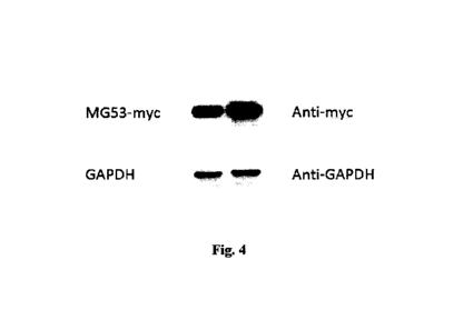
Fig.4 MG53mut reveals a higher MG53 yield than that of wild-type human
MG53
Wild-type human MG53 and MG53mut plasmids are transfected into host cells,
and 36-48 hours later, MG53 proteinsare harvested and confirmed by western
blot.
The top left and bottom left bands in the picture are respectively the MG53
level
and GAPDH abundance of wild-type human MG53 plasmids while the top right and
bottom right bands are respectively the MG53 level and GAPDH abundance of the
MG53mut plasmids.
Fig.5 MG53mut and wild-type human MG53 plasmids both have beneficial
effects on cardiomyocytes via MG53.
The expression plasmids of MG53mut and wild-type human MG53 are
tranfected into H9C2 rat heart cells, then the transfected cells and the
control group
will go through from the H202 ¨induced oxidative stress injury(simulating the
ischemia/reperfusion injury), and then the LDH(cellular injury biomarker) in
the
medium decreases significantly in the test groups, and the LDH concentration
of the
MG53mut group is lower than that in wild-type human MG53 group which means
both groups are exerting protective effects on the cardiomyocytes against
oxidative
stress, and the effect of MG53mut group is much stronger.
*means P<0.05, In the bar chart: the blank bar represents the H9C2 control
group
transfected with empty vectors, the diagonal filling bar represents the wild-
type
human MG53 group, and the black filling bar represents the MG53mut group. The
Y
axis means the LDH concentration in the medium (U/ml). The left three bars and
the
right bars are respectively groups free from and under hypoxia induced by
200um
H202 for 20 hours.
6
CA 02938921 2016-08-05
Embodiment
Example 1: MG53 cloning, the construction of wild-type human MG53 and
MG53mut expression vector.
The total RNAs are extracted from human muscular tissues by TRIZOL.
1) Add 1 ml TRIzol into 100mg human muscular tissues, and then homogenize
the tissues;
2) Equilibrate the homogenized tissues under room temperature, and add 0.2ml
chloroform, and then mix and equilibrate for 3min;
3) Centrifuge the homogenized tissue at 12000g for 15min, and retrieve the
supernatant;
4) Add 0.5m1 isopropanol into the supernatant, and centrifuge at 12000g for
10min at 4 C, and then discard the supernatant, and wash the RNA with 75%
alcohol;
5) Evaporate the RNA and dissolving it again, and then determine its
concentration.
Total RNAs of wild ¨type MG53 reverse transcription
We use the total RNAsextracts to obtain cDNA. Using the Oligod T as primers,
the reverse transcription is conducted by reverse transcriptase to obtain cDNA
library.
The cloning of wild-type MG53 cDNA library
Using the synthesized cDNA library as our template, the PCR is conducted by
specific primers to obtain MG53 coding sequence:
PCR program:
98 C3min
7
CA 02938921 2016-08-05
98 C 30sec
65 C 30sec 30 Cycles
72 C 90sec
72 C 5min
4 C Hold
using the program above can we obtain the wild-type MG53 nucleotide
sequence(sequence SEQ ID NO:1)
Primers:
Upperstream: 5'-ata ggatccgccaccatgtcggctgcgcccggcct-3';
Downstream: 5'-atactcgagacggcctcggcgccttcgggacc-3'; canying the BamHI and
Xhol restrictive sites.
PCR product is cut by BamHI and Xhol, and the same procedure is for the empty
vector pcDNA4/TO/myc-HisB. The cutting product is retrieved by gel
electrophoresis
and ligated to the vector by T4 ligase. The sequence is confirmed correct.
The MG53 coding sequence is analyzed and specially designed for silent
mutations. Then the MG53mut (SEQ ID NO:2) is obtained by whole-gene synthesis.
PCR product is cut by BamHI and Xhol, and the same procedure is for the empty
vector pcDNA4/TO/myc-HisB. The cutting product is retrieved by gel
electrophoresis
and ligated to the vector by T4 ligase. The sequence is confirmed correct.
To obtain the plasmids of MG53 and MG53mut, we use massive plasmids
extracting and confirm the quality and concentration of plasmids using agrose
gel
electrophoresis.
100m1 LB medium(lg peptone, 0.5g yeast extract, lg NaCl, pH 7.5 using NaOH)
for TOP10 culture, and centrifuge to obtain the bacteria, adding 5m1 lysis
buffer(50mM glucose, 25mM Tris-HC1 (pH 8.0), 1mM EDTA), vortexing, adding
8
CA 02938921 2016-08-05
. .
newly-prepared alkaline(200mM Na0H,1%SDS), following equilibrating for 5min,
adding 5m1 icy-cold 7.5M ammonium acetate(pH7.6), and centrifuge to retrieve
the
supernatant, then adding 9m1 isopropanol and discarding the supernatant,
dissolving
the sediments by 5m1 2M ammonium acetate(pH7.4), following the centrifuging to
obtain the supernatant, and more 5m1 isopropanol to settle, at last, the
plasmids are
extracted by phenol ¨ chloroform and settled by alcohol. The purity and
concentration
of plasmids are confirmed by agrose gel electrophoresis. See Fig.2.
2. MG53 protects heart from injury.
(1)MG53 and MG53mut gene are expressed in HEK293T cells.
HEK293T cells, cultured in 15cm dish with confluence 80%, are transfected by
constructed wild-type human MG53 and silently-mutated MG53mut plasmids with
ScreenFect A, Incellar TM). The concrete steps are as follows:
Replace the 293T medium with DMEM, and take one tube for mixing 100uL
ScreenFect A and 2000uL dilution buffer together; and another tube for mixing
3Oug
plasmids with 2000u1 dilution buffer; 5min later, mix the two mixtures above
together,
and the prepared mixture is equilibrated in room temperature for 30min.
Dripping the mixture into the cell culture; 4 hours later, replace the culture
with
traditional medium (DMEM with 10% FBS). The cells are lyzed after transfection
for
36-48 hours and western blot is conducted to determine the protein level by
corresponding antibodies. 48 hours later, every dish is lyzed by 1 ml Co-1P
buffer(30mM HEPES, pH 7.5, 100mM NaC1, 1mM EDTA, 0.5% NP40, protease
inhibitor(Roche, 04693132001, one tablet for 50m1 buffer)).
Purify the protein by Myc-tagged affinity method
The concrete steps are as follows:
9
CA 02938921 2016-08-05
. .
Wash the anti-myc beads (E6654-1m1, Sigma) 3times by icy-cold PBS, then
wash the beads one time by Co-IP buffer. Incubate the beads with lysates for 3
hours
at 4 C, and wash the then-beads 5 times with Co-IP buffer and remove the
supernatant
as much as you can. Then elute the protein by 0.1 M glycine (pH 2.8), the
elution
buffer is equilibrated by NaC1 and Tris-C1(pH 8.0). The protein is kept in -80
C for
use. Small amount of protein is taken to run the SDS-PAGE gel and dyed by
Coomassie brilliant blue to determine its purity and concentration compared to
BSA
marker.
Example 2: Rat myocardial infarction model establishment and evaluation
Rats (Vital River Laboratories, Beijing, China. SD Rats, body weight: 200g)
are
anesthetized with pentobarbital sodium (30mg/kg, i.v.) and ventilated by a
tracheostomy. A stemotomy is performed on the fourth rib line to open the
pericardial membrane and expose the heart. A reversible coronary artery snare
occluder was placed around the left anterior descending coronary artery.
Myocardial
I/R were performed by tightening the snare for 45 minutes and then loosening
it for
reperfusion. For MG53 treated group, at 5min before ischemia and lmin before
reperfusion, MG53 (3mg/KG, i.v.) is injected into the rats. 48 hours later,
the rats are
anesthetized with pentobarbital sodium (30mg/kg, i.v) and ventilated by a
tracheostomy. Open the chests and take off the hearts. The thoracic aorta is
ventilated
by a tube; and the heart is washed by backward reperfusion of saline. Then
snare
occluder is placed back to the coronary artery where it was and the artery is
dyed with
Alcian blue. The dyed areas are the non-ischemic areas, and the undyed areas
are the
ischemic areas. Then dye the heart section in 1%TTC at 37 C for 15min, the
white
parts are infracting areas.
Conclusion: MG53 (i.v.) protects rat heart from ischemia/reperfusion injury.
See
Fig.3
CA 02938921 2016-08-05
Example 3: MG53 and MG53mut expression vector tranfection and expression
HEK293T cells, cultured in 60mm dish with confluence 80%, are transfected by
constructed wild-type human MG53 and silently-mutated MG53mut plasmids with
ScreenFect A, Incellar TM). The concrete steps are as follows:
Replace the 293T medium with DMEM, then take one tube for mixing 1 OuL
ScreenFect A and 200u1 dilution buffer together; and another tube for mixing
3ug
plasmids with 200u1 dilution buffer; 5min later, mix the two mixtures above
together,
and the prepared mixture is equilibrated in room temperature for 30min.
Dripping the mixture steadily into the cell culture; 4 hours later, replace
the
culture with traditional medium (DMEM with 10% FBS). The cells are lyzed after
transfection for 36-48 hours and western blot is conducted to determine the
protein
level by Myc antibodies and GAPDH is to adjust the loading protein amount, see
Fig.4. Compared with wild-type MG53 plasmids in same amount of cells, MG53mut
plasmids are giving nearly 30% more MG53 yields.
Example 4: H9C2 heart cells injury induced by hypoxia.
H9C2 heart cells, cultured in 6-well plate, are tranfected by plasmids when
reaching confluence 80%: control group: pcDNA4/TO/myc-HisB; human MG53 WT
group: wild-type MG53 plasmids; human MG53mut group: silently-mutated MG53
plasmids. The transfection methods are the same. 30 hours later, the culture
is
replaced with fresh medium supplemented with 200um H202. 20 hours later, 40u1
medium is sampled and measured for LDH release with the use of a kit (lactate
dehydrogenase assay kit, LDH0360, Shanghai Jingyuan Medical Device Company.,
LTD.) see Fig.5.
11