Note: Descriptions are shown in the official language in which they were submitted.
COIL ASSEMBLY FOR MAGNETIC RESONANCE IMAGING
RELATED APPLICATIONS
This application claims priority to U.S. Provisional Application 61/879,050,
filed on September 17, 2013.
BACKGROUND
The present disclosure relates to magnetic resonance imaging.
SUMMARY
In one aspect, some implementations provide a magnetic resonance
imaging system that includes: a solenoid magnet configured to generate a
static
magnetic field; and an annular coil assembly housed within at least a portion
of
the solenoid magnet, the coil assembly including a gradient coil, wherein the
annular coil assembly has an aperture formed therein.
Implementations may include one or more of the following features. The
annular coil assembly and the magnet may be non-rotatable relative to each
other such that a position of the aperture within the magnet is fixed. The
annular
coil assembly and the magnet may be rotatable relative to each other such that
a
position of the aperture within the magnet is variable.
The magnetic resonance imaging system may further include a rotating
mechanism configured to rotate the annular coil relative to the magnet; and a
1
Date Recue/Date Received 2021-08-20
CA 02939405 2016-08-19
WO 2015/040473 PCT/1B2014/001864
2
locking mechanism configured to lock the annular coil such that the coil is
not
rotatable relative to the magnet.
A length of aperture may be shorter than a longitudinal length of the
annular coil assembly. A length of the aperture may be about the same as the
longitudinal length of the annular coil assembly.
The magnetic resonance imaging system may further include a patient
table slidable within the annular coil assembly.
The aperture may extend along a longitudinal direction of the annular coil
assembly. The aperture may be located in the upper hemisphere of the annular
coil assembly. The aperture may be located in the lower hemisphere of the
annular coil assembly. The aperture may open one or more of the 'x', 'y', or
'z'
axes of an annular coil assembly. For example, the aperture may be an opening
in the 'x' and 'y' axes (and shields) while the 'z' axis (and shield)
continues to
form a complete cylinder. The aperture may be sized to house at least a
portion
of a breathing apparatus, an intra-operative device, an infusion apparatus, a
display device, a projection screen, or a camera. The magnetic resonance
imaging system may further include a display device, a projection screen, or a
camera located within the aperture.
The annular coil assembly may further include a transmit coil. The
.. annular coil assembly may also include a receive coil. The magnetic
resonance
imaging system may further include a radio-frequency coil sized to encompass a
subject's head, wherein the radio-frequency coil is configured to receive
radio-
frequency signals emitted from within the subject's head, and wherein coil
CA 02939405 2016-08-19
WO 2015/040473 PCT/1B2014/001864
3
assembly is sized to house the radio-frequency coil.
The gradient coil of the annular coil assembly may be configured to
provide a gradient variation to the static magnetic field in more than one
spatial
direction, and wherein none of the more than one spatial direction are
directed at
the aperture of the annular coil assembly. The main magnet may be a portable
magnet transportable on a cart.
In another aspect, some implementations provide a method for imaging a
subject, the method including: placing a portion of the subject in an annular
coil
assembly housed within at least a portion of a solenoid magnet that is
configured
to generate a static magnetic field, wherein the annular coil assembly has an
aperture formed therein; and initiating an imaging sequence to image the
subject
using the annular coil assembly and the solenoid magnet.
Implementations may include one or more of the following features. The
method may further include rotating the annular coil assembly relative to the
solenoid magnet such that a portion of the subject is aligned with an
apparatus,
wherein at least a portion of the apparatus is housed within the aperture of
the
annular coil assembly. The method may additionally include fixing the annular
coil assembly relative to the magnet before initiating the imaging sequence.
The method may further include loading the patient on a slidable table;
and sliding the table into an inner bore of the solenoid magnet.
The method may further include passing a breathing tube through the
aperture of the annular coil assembly to the subject's face that is aligned
with the
CA 02939405 2016-08-19
WO 2015/040473 PCT/182014/001864
4
aperture; and providing anesthesia to the subject through the breathing tube
while the subject is being imaged.
The method may further include inserting a radio-frequency (RF) receiver
coil into the aperture of the annular coil assembly before initiating the
imaging
sequence.
The method may further include rotating the annular coil assembly relative
to the magnet causes the radio-frequency receiver coil to be placed at an
access
port on the subject's head through which an interventional procedure is being
performed; and initiating the imaging sequence further includes using the
radio-
frequency receiver coil to image the subject during the interventional
procedure
based on the access port.
The method may further include communicating with the subject while the
subject is being imaged using a display device or projection screen housed
within
the aperture of the annular coil assembly.
The method may further include monitoring the subject while the subject is
being imaged using a camera device housed within the aperture of the annular
coil assembly.
BRIEF DESCRIPTION OF THE DRAWINGS
Embodiments will now be described, by way of example only, with
reference to the drawings, in which:
Figure 1A shows an example magnetic resonance imaging system in
which the transmit coil and gradient coil are rotatably provided within a
solenoid
magnet, and where an aperture is provided within the transmit and gradient
coils.
CA 02939405 2016-08-19
WO 2015/040473 PCT/M2014/001864
Figure 1B shows an example implementation of a rotatable coil assembly
including the transmit and gradient coils.
Figures 2A illustrates the use of the aperture within the rotatable transmit
and gradient coil assembly for accommodating additional medical devices or
5 equipment.
Figure 2B provides a longitudinal view of the example illustration shown in
Figure 2A, shown as a cross-section taken along line A-A of Figure 2A.
Figure 2C is an illustration of an example embodiment showing the
rotation of the aperture to accommodate a patient oriented in a prone
position.
Figure 2D is an illustration showing the rotation of the aperture to
accommodate medical devices associated with a neurological interventional
procedure.
Figure 3A shows a top view of a patient residing within an example MRI
scanner according to one implementation, showing the close proximity of the
coil
assembly relative to the patient head.
Figures 3B and 3C show example embodiments in which the aperture is
employed to allow the patient to view an image or video.
Figure 30 shows an example embodiment illustrating the insertion of
patient, wearing a head coil, into a magnetic resonance imaging system having
a
coil assembly with an aperture formed therein.
Figure 3E is an illustration showing an example embodiment in which a
camera is positioned inside the solenoid and attached to the rotating coil
assembly, so that the patient can be visually monitored while inside the MRI
CA 02939405 2016-08-19
WO 2015/040473 PCT/1B2014/001864
6
scanner at multiple orientations.
Figure 4A is a photograph illustrating a wooden prototype of an example
system including a rotatable coil assembly recessed within a MRI magnet.
Figures 4B and 4C show examples of a portable magnetic resonance
imaging system according to an embodiment in which the rotatable coil assembly
is recessed within the magnet bore, showing (a) a front view and (b) a rear
view.
Figure SA shows an example of a mechanism for supporting or facilitating
the rotation of the coil assembly within the magnet bore.
Figure 5B is an illustration showing an example embodiment in which a
handle is attached to the rotatable coil assembly, in order to provide manual
or
automated rotation actuation.
Figure 5C is an illustration showing another example embodiment in
which dual handles are attached to the rotatable coil assembly, in order to
provide manual or automated rotation actuation.
Figure 50 is an illustration showing an example implementation of a
locking mechanism that enables the angular orientation of the rotatable coil
assembly to be locked at a plurality of configurations.
Figure SE is an illustration of an example implementation of a magnetic
resonance imaging system including a rotatable coil assembly, at least one
rotation mechanism (such as the handle as shown), and an optional locking
mechanism (such as the insertable locking member as shown).
Figure 5F shows an example implementation of a locking mechanism in
which a plurality of spring-loaded rods may be provided at the outer surface
of
CA 02939405 2016-08-19
WO 2015/040473 PCT/1B2014/001864
7
coil assembly, which may be removably received in corresponding holes within
the solenoid for locking a given angular orientation.
Figures 6A and 6B show example configurations of the gradient coil
according to two example implementations.
Figure 6C is an illustration of an example implementation of gradient coils
according to the embodiment shown in Figure 6B, showing the example X and Y
coil configurations.
DETAILED DESCRIPTION
Various embodiments and aspects of the disclosure will be described with
reference to details discussed below. The following description and drawings
are
illustrative of the disclosure and are not to be construed as limiting the
disclosure.
Numerous specific details are described to provide a thorough understanding of
various embodiments of the present disclosure, However, in certain instances,
well-known or conventional details are not described in order to provide a
concise discussion of embodiments of the present disclosure.
As used herein, the terms "comprises" and "comprising" are to be
construed as being inclusive and open ended, and not exclusive. Specifically,
when used in the specification and claims, the terms "comprises" and
"comprising" and variations thereof mean the specified features, steps or
components are included. These terms are not to be interpreted to exclude the
presence of other features, steps or components.
As used herein, the term "exemplary" means "serving as an example,
CA 02939405 2016-08-19
WO 2015/040473 PCT/1B2014/001864
8
instance, or illustration," and should not be construed as preferred or
advantageous over other configurations disclosed herein.
As used herein, the terms "about" and "approximately" are meant to cover
variations that may exist in the upper and lower limits of the ranges of
values,
such as variations in properties, parameters, and dimensions. In one non-
limiting
example, the terms "about" and "approximately" mean plus or minus 10 percent
or less.
According to selected embodiments of the present disclosure, magnetic
resonance imaging systems and devices are provided in which an aperture (e.g.
a gap, opening, cavity or window) is formed within a coil assembly including
the
gradient and transmit coils (and optionally a receive coil). In some
implementations, the coil assembly can be rotated relative to an axis within
the
bore of the main solenoid magnet. In some implementations, the coil assembly
may not rotate and the aperture can be fixed.
As described in further detail below, the rotatable aperture may be
employed to provide access and/or visibility to a patient who is being imaged.
The aperture may be employed to create a window or portal for medical staff to
view and monitor the patient. The aperture may also provide a spatial region
for
medical equipment or devices to be accommodate or housed during a magnetic
resonance imaging scan. In some implementations, the aperture may be located
in the upper half, for example, on top, of the inner surface of the coil
assembly.
These implementations may facilitate, for example, functional MR imaging of
the
brain, intra-operative imaging of neurological interventions. In some
CA 02939405 2016-08-19
WO 2015/040473 PCT/IB2014/001864
9
implementations, the aperture may be in the lower half, for example, at the
bottom, of the inner surface of the coil assembly. These implementations may
accommodate, for example, a mammography application, an MR-guided breast
biopsy procedure, etc. As shown in several examples below, the aperture can be
rotated within the solenoid magnet to accommodate various patient positions,
and can be located as needed for desired accessibility. For example,
typically,
the gradients and RF transmit coils are built as cylinders around the same
axis
as the solenoid magnet used to generate the main field. By opening up an
aperture in the gradient coils and RF transmit coil, it is possible to
generate extra
space inside the MRI without sacrificing performance. By then rotating these
gradients and RF coils, it is possible to locate the area of extra space where
it
would be of most use during a surgical procedure, for example, to allow more
room for an intubated patient with anesthesia equipment, or to allow an insert
imaging device such as a port coil to be used. This rotating is possible
because
all of MRI relies on the use of orthogonal planes. As the main magnetic field
(BO)
remains constant in the 'z' direction, as long as the directions of the
gradients
and RF remain substantially orthogonal then performance may be maintained.
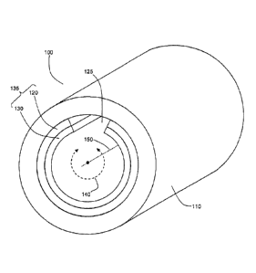
Referring now to Figure 1A, an example magnetic resonance imaging
system is shown in which a coil assembly 135, including transmit coil 130 and
.. gradient coil 120, is rotatably provided within solenoid magnet 110.
Aperture 125
is provided within the coil assembly, forming an opening or a gap in the coil
assembly 135. Coil assembly 135 may generally be shaped as an annular
structure and housed within the inner bore of solenoid magnet 110. Under
CA 02939405 2016-08-19
WO 2015/040473 PCT/1B2014/001864
rotation of coil assembly 135 relative to central axis 150, as shown at 140,
aperture 125 may be positioned at various angular locations within the inner
bore
of the solenoid magnet. The gradient, transmit, and receive coil system may
rotate either automatically or manually. In some implementations, annular coil
5 assembly 135 only includes gradient coil 120. In these implementations,
annular
coil assembly does not include transmit coil 130 or any receiver coil. For
these
implementations, radio-frequency (RF) signals are, for example, transmitted by
local coils for imaging a subject. In one instance, a head coil in a birdcage
configuration is used for both transmitting and receiving RF signals for
imaging
10 the subject. In another instance, a surface coil is used for
transmitting an RF
signal into the subject and a phased array coil configuration is used for
receiving
MR signals in response. The embodiments provided herein may be adapted for
intraoperative MRI, and MRI systems for use in an emergency room setting.
Figure 1B shows a detailed view of an example implementation of the coil
.. assembly 135, in which the aperture 125 is provided as an opening formed
along
the longitudinal direction of the coil assembly 135. In one instance, the
aperture
125 may only extend over a portion of the full longitudinal extent of the coil
assembly 135. In other instances, two or more apertures may be provided at
various axial and/or azimuthal positions of the coil assembly 135. In fact, a
.. variety of apertures with varying angular ranges may be formed on coil
assembly
135.
Referring now to Figure 2A, an example implementation illustrates the use
of the aperture within the rotatable transmit and gradient coil assembly for
CA 02939405 2016-08-19
WO 2015/040473 PCT/IB2014/001864
11
accommodating additional medical devices or equipment. This axial view
illustrates an example of providing an anesthetic mask for an intubated
patient.
In this example, patient 160 is positioned on patient support 180. Patient
support
180 may include a slidable patient table. An anesthetic mask 170 and
associated tubing 175 are provided on intubated patient 160. Aperture 125 is
oriented to provide additional room to house tubing 175, without comprising
valuable space within coil assembly 135 between transmit coil 130 and patient
160.
Figure 2B provides a longitudinal view of the example illustration shown in
Figure 2A, shown as a cross-section taken along line A-A of Figure 2A. This
longitudinal view shows how tubing 175 is received within the aperture that
would
have otherwise been occupied by gradient coil 120 and transmit coil 130. As
illustrated, tubing 175 takes up space towards the top of the inner bore of
the
solenoid magnet 110. This space overlaps with coil assembly 135 if coil
assembly is a full annular coil assembly. The aperture 125 on coil assembly
135
provides the space for tubing 175 without compromising gradient coil 120 and
transmit coil 130.
Figure 2C illustrates how the rotatable aspect of coil assembly 135 may be
employed to accommodate a patient 160 oriented in a prone position. Since
aperture 125 rotates with coil assembly 135, the additional medical hardware
or
devices associated with the patient (in this case, tubing 175) may be
accommodated in more than one angular position.
Figure 2D illustrates an example of rotating aperture 125 to accommodate
CA 02939405 2016-08-19
WO 2015/040473 PCT/1132014/001864
12
medical devices associated with a neurological interventional procedure,
illustrating the example case of a patient 160 having an access port 200
inserted
within his head. As shown in Figure 2D, the rotatable aperture 125 need not be
positioned over the patients face, and the extra space afforded by aperture
125
may be employed to position a local MRI receiver 210 (e.g., implemented as a
RF receiver coil) at the relevant surgical or diagnostic location. This may
prove
advantageous during port-based neurological surgical and diagnostic procedures
in allowing for magnetic resonance images to be obtained intra-operatively
without having to remove the access port prior to imaging. For example, the
close proximity of the local MRI receiver 210 may provide superior signal-to-
noise performance to improve sensitivity or to increase frame rate of an
intraoperative MRI imaging procedure.
Figure 3A shows a top-down view of a patient positioned within a
magnetic resonance imaging system according to one embodiment of the
present disclosure, in which the coil assembly 135 is recessed within solenoid
magnet 110. This example shows the close proximity that can be achieved
between the coil assembly and the patient's head 160, while still providing
ample
room for other portions of the patient's body that are not necessarily being
imaged. The dimensions provided in this figure are example ranges and are not
intended to be limiting.
Accordingly, embodiments of the present disclosure may enable a
reduction in size of a magnetic resonance imaging system, as the presence of
the rotatable aperture may enable a patient to be accommodated in a bore with
a
CA 02939405 2016-08-19
WO 2015/040473 PCT/I132014/001864
13
smaller diameter. For example, as described elsewhere in the present
disclosure,
the rotatable aperture may be employed to accommodate one or more additional
devices, such as diagnostic, therapeutic, imaging or communications devices,
without requiring an associated increase in the bore diameter. In other
.. embodiments, the rotatable aperture may be employed to provide the patient
with
the perception of additional room within the scanner by providing additional
room
in the vicinity of the patient's face (for example, within a small annular
segment
associated with the size of the patient's face), while still maintaining close
proximity between the coil assembly elsewhere.
This ability to perform magnetic resonance imaging within a smaller bore
system can lead to advantages in performance and/or cost. A typical magnetic
resonance imaging system may have a central bore (within the transmit coil)
diameter of approximately 60 cm. A wide-bore system may have a diameter of
approximately 70 cm. The cost of such a system is governed in part by the
radius
of the bore, because the radius affects the stored energy in the solenoid
magnet.
The stored energy varies as the cube of the radius. As such, reducing the size
of
the bore is advantageous as it allows for cost reduction and/or an increase of
the
achievable primary magnetic field.
Similarly, the performance of the gradient coil is also strongly dependent
on the radius, because the magnetic field from a wire drops according to an
inverse square law. Accordingly, a size reduction in the gradient coil radius
allows one to achieve a given performance with less current, thereby reducing
the system cost and complexity (and reducing associated heating and cooling
CA 02939405 2016-08-19
WO 2015/040473 PCT/IB2014/001864
14
requirements).
Accordingly, in some embodiments, the diameter of the transmit coil may
be reduced from the typical values noted above. In some example
implementations, the inner diameter of the transmit coil may be reduced to a
value that accommodates the insertion of a head, but is, for example, less
than
approximately 60 cm, less than approximately 50 cm, less than approximately 45
cm, less than approximately 40 cm, and less than approximately 35 cm. As
shown in Figures 2A and 2B, 3D and 5E, the coil assembly may be recessed
within the solenoid magnet, such that the patient body (e.g. the shoulders)
may
be inserted within a broader diameter region (for example, having a diameter
of
approximately 60 cm) associated with the coil assembly, while inserting the
head
within a narrower diameter region associated with the coil assembly. For
example, in one example implementation, shown in Figure 5E, the solenoid
magnet may have a longitudinal (axial) length of approximately 1 m, while the
region associated with the coil assembly (the gradient and transmit coils) may
have a longitudinal (axial) length of approximately 0.5 m.
Figures 3B and 3C show example embodiments in which the aperture is
employed to allow patient 160 to view an image or video, for example, via an
MRI-compatible display device or projection screen 250 located in aperture 125
within the inside of the solenoid magnet. As shown in the Figures, the display
or
projection device 250 may be attached to the coil assembly, for example via
member 255. Figures 3B and 3C shown in two different angular orientations,
illustrating how patient 160 may view and/or interact with the display or
projection
CA 02939405 2016-08-19
WO 2015/040473 PCT/1B2014/001864
device 250 at multiple orientations.
According, in some embodiments, an MRI with a video screen or image
projection may be embedded within the mechanism to facilitate communication
with the patient. This screen can be used to provide scan information to the
5 patient (such as instructions to not move, or to count down the scan time
remaining) or provide visual cues during scanning, for example fMRI studies.
Alternatively, this screen may be used to provide entertainment during the
scanning procedure. If an MR-compatible camera is added, this screen or image
projection may be used for two-way communications between a patient in the
10 scanner and another individual. The screen or image projection and
camera can
also potentially be mounted to the rotating items such that the patient
remains
visible regardless of the aperture orientation.
In one example embodiment, a timer may be visible to the patient inside of
the scanner. This timer would allow the patient to see an indication of time
15 remaining or time elapsed for their current scan, and could better hold
still,
leading to fewer image artifacts, If a general screen or area for image
projection
was available to the patient, the timer could be displayed here, along with
instructions to stay still, and soothing images, or other entertainment. The
screen or image projection could be used for fMRI studies. If the screen or
image projection were combined with a camera, two way visual contact could be
achieved between the patient in the scanner and the operator. This contact
could be used to allow medical staff to watch a medically distressed patient,
or a
child to be in visual contact with their caregiver. The medical staff could
explain
CA 02939405 2016-08-19
WO 2015/040473 PCT/1B2014/001864
16
the time to the patient, leading to a less confusing and isolating experience.
Figure 3D shows an example embodiment illustrating the insertion of
patient 160, supported by a table or stretcher 180, and wearing a head coil
230,
into a magnetic resonance imaging system having a coil assembly with an
aperture formed therein. In one instance, head coil 230 can be configured as a
radio-frequency receiver coil as a local coil. In this instance, head coil 230
is
configured to receive radio-frequency signals emitted from within the
subject's
head and in response to excitation radio frequency pulses sent from the
transmit
coil 130 within the annular coil assembly 135. In another instance, head coil
230
can be configured as a radio-frequency transmit and receiver coil. In the
example embodiment shown, the aperture includes a display device, screen
and/or camera 252. The coil assembly and associated aperture may be rotatable
to accommodate multiple patient orientations. The system includes an initial
gap
region 240 configured to accommodate the patient's shoulders and torso. The
receiving coil may be positioned about the patient with the aperture as
desired
prior to installing them within the magnet. In this embodiment, the rotating
coil
assembly 135 includes the gradient coil 120 and transmitting coil 130.
Figure 3E is an illustration showing an example embodiment in which a
camera 275 is positioned inside aperture 125 and attached to rotating coil
assembly 135 (for example, via attachment member 270), so that patient 160 can
be visually monitored while inside the scanner at multiple orientations. Such
an
embodiment may be optionally combined with the embodiment shown in Figures
3B and 3C to provide a display mechanism in addition to a camera, for example,
CA 02939405 2016-08-19
WO 2015/040473 PC171B2014/001864
17
to allow two-way visual communication or interaction between a patient in the
scanner and another individual. By positioning an MR compatible camera (e.g.,
for eye-tracking in fMRI studies) on the rotating element, it is possible for
the
anesthetist to maintain visual contact with the patient regardless of their
orientation.
Figure 4A is a photograph illustrating a wooden prototype of an example
system including a rotatable coil assembly 135 recessed within a MRI magnet
110. The rotatable coil assembly 135 had an aperture 125. Figures 4B and 4C
show examples of a portable magnetic resonance imaging system 280 according
to an embodiment in which the rotatable coil assembly is recessed within the
magnet bore, showing (a) a front view and (b) a rear view. In some instances,
the magnet is portable in that it can travel within a room or between rooms,
and
may be mounted on wheels, with or without a motorized base. The magnet may
have a tether cable attaching it to an equipment room. .
In some embodiments, support structures may be provided to support the
weight of the coil assembly in order to assist with, and/or guide, rotation of
the
coil assembly. Referring now to Figure 5A, an example mechanism is provided
for supporting or facilitating the rotation of the coil assembly (including
gradient
coil 120 and transmit coil 130) within the bore magnet 110. In the example
shown, a plurality of rotatable supports 300, such as rods, wheels, or
bearings
(which may be configured to be shock absorbing) are provided at various
azimuthal positions. Such supports may be retained by a suitable mechanism,
such as lateral retention mechanism 305. In some embodiments, such supports
CA 02939405 2016-08-19
WO 2015/040473 PCT/1B 201 4/001864
18
may be provided only in the vicinity of the lower portion of the system, where
the
weight of the coil assembly is received. Alternatively, pneumatic or air-
bearing
mechanisms may be employed.
A wide variety of mechanisms and means, both manual and automated,
.. may be employed to achieve or actuate rotation of the rotatable insert. In
one
illustration, an MRI-compatible motor may be employed to produce rotation of
the
rotatable coil assembly. A floating cable may be employed that extends out the
back of the magnet and is of sufficient length to support rotation.
Figure 5B is an illustration showing an example embodiment in which a
.. handle 350 is attached to the rotatable coil assembly, in order to provide
manual
or automated rotation actuation and relative positioning of aperture 125.
Figure
5C is an illustration showing another example embodiment in which dual handles
360 and 365 are attached to the rotatable coil assembly, in order to provide
manual or automated rotation actuation and relative positioning of aperture
125.
The handles may be connected to an automated mechanism, such as an
external motor, to automatically control the rotation of the coil assembly. In
some
embodiments, the handles are provided at the rear of the system, such that
they
do not interfere with the body of the patient (e.g. the patient's shoulders).
Figure 5D is an illustration showing an example implementation of a
locking mechanism that enables the angular orientation of the rotatable coil
assembly to be locked at a plurality of configurations. End portions 132 and
115
are provided on coil assembly 135 and solenoid 110, respectively. End portion
132 includes a plurality of first holes 410, and end portion 115 includes a
plurality
CA 02939405 2016-08-19
WO 2015/040473 PCT/IB2014/001864
19
of second holes 400. As coil assembly 135 is rotated, first holes 410 and
second
holes 400 align at different angular positions. A locking member 420 can be
inserted to lock an angular position at a location where holes align, such as
at
location 415. Figure 5E is an illustration of an example implementation of a
magnetic resonance imaging system including a rotatable coil assembly 135, at
least one rotation mechanism, such as handle 350, and an optional locking
mechanism, such as the insertable locking member 420.
In another embodiment, shown in Figure 5F, a plurality of spring-loaded
rods 430 may be provided at the outer surface of coil assembly, which may be
removably received in corresponding holes within the solenoid for locking a
given
angular orientation. The rods may be disengaged by a suitable mechanism. In
another embodiment, the rods may be received at a sufficiently shallow depth
that they may be disengaged by applying a sufficient torque to the coil
assembly.
Figures 6A and 6B show example configurations of the gradient coil 130
according to two example implementations. In Figure 6A, the Y gradient axis
505
is directed towards the aperture on coil assembly 350, and the presence of the
aperture leads to a performance degradation of the Y-gradient relative to that
of
the X-gradient.
Figure 66 illustrates another configuration in which the X and Y
orientations are rotated relative to those shown in Figure 14(a), thereby
improving the relative performance of the Y gradient. In one example
implementation, the X and Y axes are rotated such that they are angled at
approximately 45 degrees relative to the normal defined by the aperture and
CA 02939405 2016-08-19
WO 2015/040473 PCT/1B2014/001864
neither X nor Y axis is directed towards the aperture of coil assembly 130.
Figure 6C provides an illustration of an example implementation of
gradient coils according to the embodiment shown in Figure 6B, showing the
example X (520 and 525) and Y (530 and 535) coil configurations. The
reoriented
5 coil configurations improve the Y gradient at the expense of the X
gradient. The
Y gradient improves in performance because there is available continuous
surface area for current to flow on either side of the newly defined Y axis
(Y') in
Figure 66, while in Figure 6A there is only continuous surface area for
current
flow on one side of the Y axis (the lower half). Likewise, the X gradient will
suffer
10 slightly in performance because in Figure 6A there is ample surface area
for
current to flow on either side of the X axis, while in Figure 6B the surface
area on
one side of the X' axis has been diminished. A variation on this approach
allows
for the 'z' axis to remain a complete cylinder (to achieve full performance)
while
the 'x' and 'y' axes are rotated about the aperture. In this approach, a
substantial
15 aperture is formed without sacrificing gradient performance.
The various embodiments described above may provide one or more of
the following advantages. For example, various embodiments may provide for a
smaller head-only MRI system that fits closely around a patient's head, and
meets the requirement to fit anesthesia equipment, other MRI imaging coils
such
20 as a port coil, or to accommodate the variety of patient positions
possible during,
for example, neurosurgery (or spine surgery). Such embodiments may lower
costs (relative to larger size MRI systems), reduce difficulty in siting, and
reduce
difficulty in moving the device, all of which are linked to the size of an
MRI.
CA 02939405 2016-08-19
WO 2015/040473 PCT/1B2014/001864
21
More generally, embodiments may make an MRI a less uncomfortable
experience. Embodiments may make the patient feel less isolated within the
scanner with a greater ability to communicate with the outside world. Once the
scan starts, the patient can be provided with knowledge of how much time
remains in some embodiments. The reduced feeling of isolation, greater ability
to communicate, and/or knowledge of how much time remains can decrease
fidgeting, which may prevent some degradation of image quality. This may be
particularly advantageous with respect to children, as they have a tendency to
move about once isolated in the scanner. Similarly, this may be advantageous
for patients in medical distress, as it is difficult to have them be isolated
and away
from medical attention for the duration of their time in the scanner.
Furthermore, during an interventional neurosurgical procedure, it is
advantageous for the anesthetist to have visual access to the patient's face.
For
an intra-operative procedure to maintain this visual access, prone and other
patient positions can be challenging, but embodiments described above can
reduce or eliminate these challenges.
The specific embodiments described above have been shown by way of
example, and it should be understood that these embodiments may be
susceptible to various modifications and alternative forms. It should be
further
understood that the claims are not intended to be limited to the particular
forms
disclosed, but rather to cover all modifications, equivalents, and
alternatives
falling within the spirit and scope of this disclosure.