Note: Descriptions are shown in the official language in which they were submitted.
CA 02939790 2016-08-23
METHOD FOR ASSESSING BRAIN FUNCTION AND
PORTABLE AUTOMATIC BRAIN FUNCTION ASSESSMENT APPARATUS
This application is a division of Canadian Patent Serial No. 2,616,974
July 26, 2006.
DESCRIPTION OF THE INVENTION
Field of the Invention
[001] The invention relates to the field of emergency triage, and
specifically, a method and apparatus for performing emergency neurological
triage. Moreover, the invention relates to a method and apparatus for
assessing
brain function.
Background of the Invention
[002] The central nervous system (CNS) and the brain in particular,
perform the most complex and essential processes in the human body.
Surprisingly, contemporary health care lacks sophisticated tools to
objectively
assess their function. A patient's mental and neurological status is typically
assessed clinically by an interview and a subjective physical exam. The
clinical
laboratory currently has no capacity to assess brain function or pathology,
contributing little more than identification of poisons, toxins, or drugs that
may
have externally impacted the CNS. Brain imaging studies, such as computed
tomography imaging (CT), magnetic resonance imaging (MRI), though widely
used and useful, are structural/anatomical tests revealing little or nothing
about
brain function. In the immediate time of acute brain injury, stroke, or
seizure,
imaging studies typically reveal no abnormality, even when there is clear and
dramatically abnormal brain function. CT and MRI only detect the condition
after
the morphology or structure of the brain has changed. In some cases it can
take
- 1 -
CA 02939790 2016-08-23
from hours to days after the patient is present in an emergency room (ER)
before
overt changes are evident on the CT or MRI, and before severe neurological
pathology is visible. Electrical activity of the brain, however, is affected
immediately. New imaging modalities such as functional MRI (fMRI) measure the
changes in oxygen saturation in different parts of the brain. Radioisotope
imaging
such as positron emission tomography (PET) and single photon emission
computerized tomography (SPECT) assess chemical changes within the brain as
a measurement of function with limited sensitivity and specificity. All of
these
assessment tools play an important role in selected cases, but they are
costly, not
universally available, and they do not provide critical information at the
early
stages of acute care situations. None of the current techniques provides the
immediate, actionable information critical to timely intervention, appropriate
triage,
or the formulation of an appropriate plan of care.
[003] The CNS and brain, of all organs in the human body, are also the
most time sensitive and have the least capacity for repair. Currently,
emergency
room patients with altered mental status, acute neuropathy, or head trauma
must
undergo costly and time consuming tests to determine an appropriate course of
treatment. Unfortunately, in many cases, the clinical condition of patients
continue
to deteriorate as they wait for equipment to become available or for
specialists to
interpret tests. The task of the ER physician is to basically establish
whether the
brain is functioning normally, whether the abnormality is psychiatric or
organic in
nature, whether an organic abnormality is global or lateralized, and to
develop an
initial assessment of the diagnostic possibilities. The problem that faces ER
physicians is that their resources are quite literally limited to a flashlight
and a
rubber reflex hammer. Amazingly, all of the physician's decisions concerning
the
administration of emergency treatment or intervention, including CT scan,
spinal
tap, additional consultation or discharge are based on the results of this
simplistic
exam.
[004] Often, ER patients are sent for imaging studies, yet many functional
brain abnormalities, such as seizure, are not visible on a CT scan. Some
abnormalities which will eventually have anatomical and structural
consequences
often take time to become visible. This is true for many important conditions
such
- 2 -
CA 02939790 2016-08-23
as ischemic stroke, concussion, raised intracranial pressure, and others.
Thus,
while the location, expense, and limited availability of the CT scan can be
problematic, so indeed can the fact that it is a structural as opposed to
functional
test.
[005] One-third of over 200 physicians surveyed at the American College
of Emergency Physicians feel that the combination of a good clinical
laboratory, a
neurological exam, and a CT scan of the head, is not adequate for the
assessment of every patient with altered mental status or neurological
dysfunction. Consensus estimates from the CDC NHS database and practicing
ER physicians, is that patients requiring a mental status exam represent 15%
of
the more than 100 million ER visits annually in the U.S., and in some
institutions,
considerably more.
[006] There are more than 100 million ER visits per year in the US alone
(CDC/NCHS) database. In year 2000, more than 13 million of these patients
required a formal mental status exam and nearly 5 million had CT scans. This
data indicates the need for real-time functional brain state assessment which
can
be performed in the hospital, in an ambulance, at a sporting event, or any
other
location where acute neurological evaluation may be necessary.
[007] All of the brain's activity, whether reflexive, automatic, unconscious,
or conscious, is electrical in nature. Through a series of electro-chemical
reactions, mediated by molecules called neurotransmitters, electrical
potentials
(voltages) are generated and transmitted throughout the brain, traveling
continuously between and among the myriad of neurons. This activity
establishes
the basic electrical signatures of the electroencephalogram (EEG) and creates
identifiable frequencies which have a basis in anatomic structure and
function.
Understanding these basic rhythms and their significance makes it possible to
characterize the EEG as being within or beyond normal limits. At this basic
level,
the EEG serves as a signature for both normal and abnormal brain function.
[008] The electrical activity of the brain has been studied extensively since
the first recordings over 75 years ago, and especially since the advent of
computers. "Normal" electrical activity of the brain has been well
characterized in
hundreds of studies, with a narrow standard deviation. The frequencies of
- 3 -
'
, CA 02939790 2016-08-23
electrical activity of some parts of the brain are the normal response to
various
stimuli , such as acoustic, visual, or pain, known as "evoked potentials."
Evoked
potentials (EP) are particular waves that have characteristic shapes,
amplitudes
and duration of peaks within those wave shapes, and many other features, all
of
which have well established normative data, generated over decades of
research.
Normative data for all of the EEG and evoked response waves are remarkably
constant across different genders, ages, and ethnicities. Moreover, any
variability
that does exist is well described and explained.
[009] Neuroscientists have also characterized the EEG signature of
various different brain pathologies. Just as an abnormal electrocardiogram
(ECG)
pattern is a strong indication of a particular heart pathology, an irregular
brain
wave pattern is a strong indication of a particular brain pathology. A wide
array of
pathologies have been well characterized: acute and chronic, structural,
toxic,
metabolic, and even specific diagnoses such as: ischemic stroke, epileptic
seizures, concussion, alcohol, and drug overdose, psychiatric conditions, and
dementias including Alzheimer's disease. A large body of data, with continuing
refinements and contributions, constitutes the field of clinical
neurophysiology.
[010] Even though EEG-based neurometric technology is accepted today
and a tremendous body of data exists, application in the clinical environment
is
notably limited. Some of the barriers limiting its adoption include: the cost
of EEG
equipment, its lack of portability, the need for a technician to administer
the test,
the time it takes to conduct the test, and the need for expert interpretation
of the
raw data. More importantly, the technology is neither available nor practical
in the
acute care setting, especially at the point of care. A complete diagnostic EEG
instrument typically costs $80,000, fully equipped. Despite the high costs,
the
instrument produces essentially raw waveforms which must be carefully
interpreted by an expert. Moreover, use of the standard EEG equipment remains
extremely cumbersome. It can take 30 minutes or more to apply the required 19
electrodes. Once the patient is prepared for the test, the recording itself
can take
from 1 to 4 hours. Data is collected and analyzed by an EEG technician, and
are
then presented to a neurologist for interpretation and clinical assessment.
There
are some self-standing dedicated neurodiagnostic laboratories which focus
strictly
-4-
CA 02939790 2016-08-23
on detailed analysis of electrical brain data. Neither the specialized
centers, nor
the typically large hospital EEG machines are practical for the ER, operating
room
(OR), intensive care unit (ICU), or any other acute care medicine setting
where
patients are in the greatest need. Immediate, functional brain state
assessment is
needed to treat patients with acute neurological injury and disease for the
prevention of further damage and disability.
SUMMARY
[011] In accordance with the invention, there is provided a neurological
triage apparatus comprising a processor configured to process acquired
spontaneous and evoked signals using wavelets.
[012] Also in accordance with the invention, there is provided a method of
determining a neurological state of a subject comprising the steps of
acquiring
spontaneous signals through an electrode set, processing the acquired signals,
extracting desired features from the processed signals, and classifying the
extracted features into one or more diagnostic categories.
[013] Also in accordance with the invention, there is provided a method of
determining a neurological state of a subject comprising the steps of evoking
brain
response signals using audio, visual, electrical, or other stimulus means,
acquiring
the evoked signals through an electrode set, processing the acquired signals,
extracting desired features from the processed signals, and classifying the
extracted features.
[014] Also in accordance with the invention, there is provided a method of
determining a neurological state of a subject comprising the steps of
acquiring
spontaneous and evoked signals through the electrode set, processing the
acquired signals, extracting desired features from the processed signals, and
classifying the extracted features.
[015] Further in accordance with the invention, there is provided an
apparatus for diagnosing the neurological state of a subject, comprising a
processor, a memory operatively coupled to the processor, wherein the memory
stores one or more operating instructions, a multi-channel input-output
interface
operatively coupled to the processor, wherein the multi-channel input-output
- 5 -
CA 02939790 2016-08-23
interface is configured to receive external electrical signals through a set
of
electrodes placed on the subject; and the processor is configured to utilize
the
one or more operating instructions to perform one or more operations on
signals
received from the multi-channel input-output interface.
[015a] In one embodiment there is provided an apparatus for determining
the neurological state of a subject, comprising: a set of electrodes
configured to
be placed on the subject to collect brain electrical signals; a processor; a
memory
operatively coupled to the processor, wherein the memory stores one or more
operating instructions; and an input-output interface having at least one
channel
operatively coupled to the processor, wherein the input-output interface is
configured to receive brain electrical signals through the set of electrodes;
and
the processor is configured to utilize the one or more operating instructions
to
perform one or more operations on the signals received through the input-
output
interface, wherein the one or more operations comprise: de-noising the
received
signals; extracting features from the de-noised signals; classifying the
extracted
features; and determining a neurological state of the subject based on the
classification, wherein determining the neurological state comprises the steps
of:
determining if the neurological state is normal or abnormal; and determining
if an
abnormal neurological state is psychiatric or "functional" in nature, organic
in
nature, or an emergency or "Alert" condition.
[015b] Yet another embodiment provides an apparatus for providing an
automatic brain function assessment comprising; an electrode set; a processor;
wherein the electrode set and the processor are operatively connected through
an input-output interface having at least one channel; a display operatively
connected to the processor; a user interface operatively connected to the
processor; and internal memory, wherein the memory contains instructions for
providing a real-time assessment of whether a subject's brain function is
normal
or abnormal; and wherein the memory contains further instructions for
providing
a real-time assessment of whether an abnormal brain function is psychiatric or
"functional" in nature, organic in nature, or an emergency or "Alert"
condition.
6
CA 02939790 2016-08-23
[016] Also in accordance with the invention, there is provided a kit for
performing an emergency neurological diagnosis of a patient suffering from an
altered mental state, the kit including an apparatus for diagnosing the
neurological
state of a subject, instructions for using the apparatus, and a portable
carrying
case for the apparatus.
[017] Also in accordance with the invention, there is provided an
apparatus for providing an automatic brain function assessment comprising an
electrode set, a processor, wherein the electrode set and the processor are
operatively connected through a multi-channel input-output interface, a
display
operatively connected to the processor, a user interface operatively connected
to
the processor, and internal memory, wherein the memory contains instructions
for
providing a real-time assessment of a subject's brain function, and the memory
contains instructions for processing signals acquired by the electrode set
using
'wavelet-packet algorithms.
[018] Also in accordance with the invention, there is provided a method for
providing a triage assessment of a patient's brain function comprising the
steps of
measuring the spontaneous brain activity of the patient, stimulating the
patient
and measuring the evoked brain activity therefrom, processing the spontaneous
and evoked brain activity, wherein the processing is performed in real-time
using
wavelet-packet algorithms, providing a triage assessment of the patient based
on
the processed brain activity.
[019] Further in accordance with the invention, there is provided a triage
apparatus comprising an electrode set, an amplifier operatively connected to
the
electrode set, a processor operatively connected to the amplifier, wherein the
processor is configured to process external signals acquired by the electrode
set
using wavelets.
[020] Additional features and advantages of the invention will be set forth
in part in the description which follows, and in part will be obvious from the
- 6a -
CA 02939790 2016-08-23
description, or may be learned by practice of the invention. The features and
advantages of the invention will be realized and attained by means of the
elements and combinations particularly pointed out in the appended claims.
[021] It is to be understood that both the foregoing general description and
the following detailed description are exemplary and explanatory only and are
not
restrictive of the invention, as claimed.
[022] The accompanying drawings, which are incorporated in and
constitute a part of this specification, illustrate several embodiment of the
invention and together with the description, serve to explain the principles
of the
invention.
BRIEF DESCRIPTION OF THE DRAWINGS
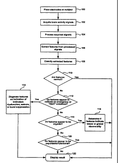
[023] Figure 1 is a flowchart illustrating the method of assessing the brain
state of a subject carried out by an apparatus according to an embodiment
consistent with the present invention.
[024] Figure 2 is a diagram illustrating an apparatus according to an
embodiment consistent with the present invention.
[025] Figure 3 is a diagram illustrating an electrode set according to an
embodiment consistent with the present invention.
DESCRIPTION OF THE EMBODIMENTS
[026] Reference will now be made in detail to present embodiments of the
invention, an example of which is illustrated in the accompanying drawings.
Wherever possible, the same reference numbers will be used throughout the
drawings to refer to the same or like parts.
[027] In accordance with embodiments consistent with the present
invention, Figure 1 shows a flowchart illustrating a method for assessing the
brain
state of a patient. This method may be implemented by an apparatus or device
which is manufactured to perform the method given herein. An electrode set is
placed on a subject (step 100). Typical electrode sets for acquiring EEG data
use
at least 19 electrodes. An electrode set consistent with an embodiment of the
- 7 -
=
CA 02939790 2016-08-23
present invention may comprise a reduced electrode set, with less than 19
electrodes.
[028] The electrodes measure the electrical fields that are produced as a
result of the subject's brain activity (step 102). The activity may be
spontaneous,
evoked or a combination thereof. In an embodiment consistent with the present
invention, the spontaneous brain activity is measured and an evoked response
is
measured. The spontaneous activity may comprise the subject's EEG signals.
The evoked response may be obtained by stimulating the subject using visual,
physical, auditory, or other stimulation. In an embodiment consistent with the
present invention, an auditory stimulus is given to the subject to obtain an
Auditory
Evoked Potential (AEP). Moreover, the Auditory Evoked Potentials may comprise
any of auditory brainstem response (ABR) potentials, auditory mid-latency
response (AMLR) potentials, or auditory late response (ALR) potentials,
including
P100 responses, and P300 responses.
[029] The spontaneous and evoked signals are acquired by the electrode
set and are subsequently subjected to a signal processor, wherein artifacts
are
removed from the signals (step 104). Artifacts that may be removed are a
result
of such factors as a disconnected electrode, electromyogram (EMG) artifacts
resulting from muscular movement, eye movement and other significant
artifacts.
In one embodiment, the artifacts may be removed by removing discrete artifact
sections from the signals. In another embodiment, the artifacts may be removed
by subtracting out any artifacts present in the acquired signals.
[030] The artifact-free signals are subjected to further processing to
extract statistical signal features (step 106). In one embodiment consistent
with
the present invention, a quantitative EEG algorithm may be used to extract
features. In another embodiment, a wavelet packet algorithm may be used for
feature extraction. In a further embodiment, spectral analysis and statistical
procedures may be performed to extract features. In yet a further embodiment,
diffusion geometric analysis may be performed to extract features. In yet
another
embodiment, microstate analysis may be performed to extract features. In a
further embodiment, wavelet-packet local discriminant basis algorithms may be
applied to extract features.
- 8 -
CA 02939790 2016-08-23
[031] Referring again to Figure 1, the extracted features are classified
according to one or more diagnostic categories, wherein a probability that
features
extracted from a subject can be classified in one or more diagnostic
categories is
determined (step 108). According to embodiments consistent with the invention,
classifying may be performed by applying discriminant analysis to the
extracted
features, or by applying wavelet-packets to the extracted features. Regardless
of
the classifying method used, the classification algorithm first determined if
the
results are normal (step 110). If the features extracted from the subject's
brain
waves are normal, then the device will display that the subject's brain
activity is
normal (step 122). If there is a higher probability that the subject's
extracted
features are not normal, the device will attempt to classify the extracted
features
as an emergency or "Alert" condition (step 112). If there is a high
probability that
the extracted features match features typical of someone in an emergency
mental
state or an "Alert" condition, the device will attempt to classify the
extracted
features as either brainstem dysfunction, active seizure, or burst suppression
(step 114). If the device determines that the extracted features have a high
probability of being one of the emergency states. the device will display this
result
so the subject can receive immediate treatment (step 122). If the extracted
features do not have a high probability of being an emergency, the device will
determine if the abnormality of the extracted features appears to be organic
in
nature (step 116). If the extracted features are determined to correlate with
an
extracted feature abnormality that is organic in nature, the device will then
attempt ,
to determine if the extracted feature abnormality is lateral or global in
nature (step
118), and will display the result (122). The extracted feature abnormalities
will be
tested to determine if they are psychiatric or "functional" in nature (step
120), and
this result will be shown (step 122).
[032] Figure 2 shows an apparatus consistent with an embodiment of the
present invention. An electrode set 200 is placed on the head of a subject
201. In
an illustrative embodiment, the subject is a human, but the subject can be an
animal as well. An electrode set 200 consistent with an embodiment of the
present invention may comprise a reduced electrode set, with less than 19
electrodes.
- 9 -
= CA 02939790 2016-08-23
[033] Figure 3 shows an electrode set 200 consistent with an embodiment
of the present invention. Electrode set 200 may comprise a plurality of
electrodes
which may be affixed to the head of a subject 201. In an illustrative
embodiment,
electrode set 200 comprises nine electrodes that may be affixed to the
forehead,
= shoulder and ear of the subject. This reduced electrode set 200 allows
for
placement on the forehead, and eliminates the need to place any electrodes
over
any hair that a subject may have on their head. This further eliminates any
conduction problems that arise due to the hair, and also eliminates the need
for
any hair removal. In an illustrative embodiment, the electrodes may be placed
on
the right mastoid 302, far right of the forehead 304, near right of the
forehead 306,
center top of the forehead 308, near left of the forehead 310, far left of the
forehead 312, left mastoid 314, and an ECG electrode on the left shoulder 316.
Additionally, in an illustrative embodiment, there is an electrode placed on
the
center of the forehead 318 that is grounded. The electrodes on the right and
left
mastoids 302, 314 and the center of the forehead 318 may be used in an
embodiment wherein an AEP signal is acquired. An illustrative embodiment
consistent with the present invention is able to use an electrode set 200 with
a
reduced number of electrodes because the signal processing algorithms
eliminate
the need for additional electrodes.
[034] Referring back to Figure 2, the electrodes measure the electrical
fields that are produced as a result of subject's 201 brain activity. The
activity
may be spontaneous, evoked or a combination thereof. In an embodiment
consistent with the present invention, the spontaneous brain activity is
measured,
for example the EEG of subject 210, and an evoked response is measured. The
evoked response may be obtained by stimulating subject 201 using visual,
physical, aural or other stimulation. In an embodiment consistent with the
present
invention, an auditory stimulus is given to subject 201 to obtain an Auditory
Evoked Response (AEP). In one embodiment of the present invention, a pulse
oximeter 203 is connected to subject 201 to monitor subject's 201 pulse and
blood
oxygen levels 209.
[035] Electrode headset 202 and pulse oximeter 203 can be connected to
a handheld device 205. Electrode headset 202 can be connected to handheld
-10-
CA 02939790 2016-08-23
device 205 through a low-voltage preamplifier 222. Low-voltage preamplifier
222
has a high noise tolerance and is designed to amplify the signals that are
transmitted to and from electrode headset 202. Handheld device 205 is designed
to be able to fit in one's hand. In one embodiment handheld device 205 may
have
a size of about 115mm x 190mm x 60mm, and a weight of less than about 600 g.
Handheld device 205 has a display 219, which can be an LCD screen, and can
further have a touch screen interface and a traditional user interface 220
such as
a keyboard. In one embodiment, handheld device 205, electrodes 200 and
electrode headset 202 may come in a kit, designed for performing neurological
triage of a patient suffering from an altered mental state, wherein the kit
includes
instructions for using handheld device 205, and comes in a portable carrying
case.
[036] Handheld device 205 contains analog and digital hardware on the
front end 221, and is controlled through processor 210. In one embodiment,
processor 210 is a Texas Instruments OMAP microcontroller/digital signal
processor. Front end 221 is separated from processor 210 by isolation barrier
208. Front end 221 acts as a multi-channel input/output interface for the
device,
further facilitating the bi-directional communication of transmitted and
received
signals to processor 210. In one embodiment consistent with the present
invention, the multi-channel input/output interface is a wireless multi-
channel
input/output interface.
[037] In an embodiment consistent with the present invention, a command
from a user, entered through user interface 220, will begin a test routine.
Analog
brain waves are acquired through electrode headset 202 and are transmitted
through cables to analog front end 204 of handheld device 205. Analog brain
waves are then converted to digital signals through an ADC contained in analog
front end 204 and transmitted to digital front end 206. Digital front end 206
transmits the digital signals to processor 210 where digital signals are
processed
in accordance with instructions contained in internal memory 211 of processor
210. In an embodiment consistent with the present invention, the signals are
processed to remove noise, processed to extract features, and processed to
classify the extracted features. In another embodiment, the instructions
contained
in internal memory 211 of processor 210 comprise instructions for performing
the
-11-
CA 02939790 2016-08-23
method illustrated in Figure 1. Processor 210 may then output results, which
may
be in real-time, concerning the assessment of subject's 201 brain in
accordance
with the classification. Outputs may be displayed on LCD screen 219 of
handheld
device 205, or may be saved to external memory 216, or may be displayed on PC
215 connected to handheld device 205 by serial or universal serial bus
connection. In one embodiment, display may display a representation of
subject's
201 brain based on the assessment. In another embodiment consistent with the
present invention, processor 210 transmits the raw, unprocessed brainwaves to
an external memory 216. External memory 216 may be a hard disk drive, an
optical disk drive, a floppy disk drive, or a removable, non-volatile memory
device.
In another embodiment, results are transmitted through serial bus to infrared
transmitter 217 which is configured to transmit data wirelessly to printer 218
to
wirelessly print results. Handheld device 205 contains an internal
rechargeable
battery 212 that is able to be charged during use or in between uses through
charger 213 connected to a typical AC outlet 214.
[038] In another embodiment, a test routine may require a stimulus to be
given to subject 200 to evoke a response. The command to produce a stimulus is
transmitted from the processor 210 to digital front end 206, where it is
converted
to an analog signal by a DAC contained therein. The analog signal is output
from
the analog front end 204 through the cables and to a stimulus emitter 224
which
stimulates subject 201. The stimulus can be auditory, sensory, or visual, or
other.
In a preferred embodiment, the stimulus is an auditory stimulus given through
transmitters that are placed in subject's ear. The stimulus emitter 224 may be
an
Etymotic Research ER 10D probe with dual speakers and a single microphone in
each ear. The evoked signal is acquired by electrode headset 202, and is
transmitted along with spontaneous signals to analog front end 204 of handheld
device 205, where it is converted to a digital signal and transmitted to
digital front
end 206. Digital front end 206 transmits the digital acquired signals to
processor
210, where evoked response signals are filtered out from spontaneous signals.
In
an embodiment consistent with the present invention, the evoked response
signals are filtered out using an adaptive wavelet based filter. More
specifically,
internal memory 211 can contain instructions that are executed by the
processor
-12-
CA 02939790 2016-08-23
210 which uses a Dual-Tree Complex Wavelet Transform as an invertible
transform to adaptively filter evoked signal response signals from spontaneous
response signals. The instructions further can contain an implementation of an
algorithm carried out by processor 210, wherein a complex wavelet transform is
computed for each sub-average, and then the phase variance of each normalized
wavelet coefficient wij is computed. The magnitude of each wavelet coefficient
is
selectively scaled according to the phase variance of the coefficients at this
location across the sub-averages. The scaling has the form:
= a1ff71 exp( jOu),
where W,j and RI are respectively the magnitude and phase of the unprocessed
complex ith wavelet coefficient at the jth scale, and where:
au = exp(-0.75(Pu /T.)4,
where Ft/ is the phase variance of coefficient wu across the sub-averages. The
filtered evoked signal is averaged and an automatic peak detection algorithm
is
implemented by processor 210 to determine the following peak locations and
latencies : Peak 1, Peak 2, and Interpeak 1-5 latency. These values are then
compared to normative data contained in internal memory 211 of processor 210.
[039] In an embodiment consistent with the present invention, processing
the signals comprises removing noise from the acquired signals, or "de-
noising."
Internal memory 211 of processor 210 contains instructions for instructing
processor 210 to perform an algorithm on acquired signals. In one embodiment,
the algorithm utilizes wavelet based signal processing using wavelet
transforms.
The wavelet transform, a member of the family of Fourier transforms, is a
process
of decomposing a given signal into a set of orthonormal basis functions called
wavelets. In traditional discrete Fourier transform (DFT), a signal is
decomposed
using complex sinusoids as basis functions, producing a frequency domain
representation of the signal. In contrast, a discrete wavelet transform (DWT)
uses
a family of specifically designed wavelets, or little waves, as basis
functions. A
family of wavelets is created by dilating the original wavelet function,
termed the
"mother wavelet." A wavelet transform decomposes the signal in both time and
frequency using ciifferent dilations of the mother wavelet. With the
application of
DWT, the one dimensional fine it signal x[n] is represented in two-dimensional
- 13 -
CA 02939790 2016-08-23
"wavelet coordinates." Individual levels of signal decomposition are created,
1
called scales. At each scale a set of coefficients is created by computing the
inner
product of the original signal x[n] with a scaled version of the mother
wavelet. The
mother wavelet function is designated by if, and its dilations are designated
by
T(j). The position index of a wavelet at scale j is called a translation. The
value
of the wavelet is completely described by the two dimensional sequence T(j,k),
where j is the scale index of the wavelet, and k is the translation index. The
DVVT
is the defined as:
N-1
C(j,k)= Ex[n]t Lk[n],where 'Pb[n]= 2 2 tif(2-jn¨ k)
re=0
[040] Coefficients C(j,k) are the wavelet coefficients at different scales j
and translations k of the inner product of the wavelet Y(j,k) with the
original signal
x[n]. In wavelet coordinates, information about both the frequency and the
location (time) of the signal energy is preserved. This is a process of noise
suppression that utilizes assumptions about smoothness and coherence
properties of both the underlying signal and the noise that contaminates it.
Similar
to filtering in the frequency domain, the wavelet coefficient thresholding
algorithm
reduces sets of wavelet coefficients in the wavelet domain. This process is
based
on the assumption that the underlying signal is smooth and coherent, while the
noise that is mixed with the signal is rough and incoherent. Smoothness of a
signal is a property related to its bandwidth, and is defined in relation to
how many
times a signal can be differentiated. The degree of smoothness is equal to the
number of continuous derivatives that can be calculated. A signal is coherent
if its
energy is concentrated in both time and frequency domains. An incoherent noise
is "spread out," and not concentrated. One measure of coherence is how many
wavelet coefficients are required to represent 99% of the signal energy. A
time-
frequency signal space is completely spanned by wavelet coefficients at all
scales
and translations. A well-concentrated signal decomposition in an appropriately
selected wavelet basis will require very few coefficients to represent 99% of
signal
= energy. However, a completely incoherent noise will require 99% of the
coefficients that span the entire space to represent 99% of its energy.
-14-
CA 02939790 2016-08-23
[041] This conventional wavelet de-noising process is a three step
process:
1. Wavelet transform the signal to obtain wavelet coefficients at
different scales
2. Threshold the coefficients and set to zero any smaller than a
threshold 8
3. Perform the inverse wavelet transform to approximate the original
signal
[042] In the de-noising process, the noise components of the signal are
attenuated by selectively setting the wavelet coefficients to zero. De-noising
is
thus a non-linear operation, because different coefficients are affected
differently
by the thresholding function. There are many parameters to control in this
algorithm: level of wavelet decomposition, threshold selection, using
different
thresholds at different wavelet coefficients that are kept by a fixed amount.
[043] In accordance with an embodiment of the present invention, the
de-noising process involves dividing the acquired signals into discrete
intervals, or
"frames," and then averaging the frames, and de-noising the averaged frames.
The greater amount of frames that are de-noised prior recomposing the signal,
the
better the results of the de-noising process. Preferably, the frames are
combined
by using two adjacent frames and calculating their linear average. This method
is
chosen for its simplicity, computational stability, and well-understood
behavior.
This dyadic linear average is then de-noised, and a new frame is created. The
overall idea is to generate as many permutations of the original arrangement
of
frames as possible, and keep averaging and de-noising those new combinations
of frames. This recombination process is a tree-like process, and may comprise
the dual-tree process described above, in which new levels of recombined
frames
are created. The average and de-noise operation creates frames at level k,
which
are no longer a linear combination of frames from level k-1.
[044] The many possible algorithms to accomplish this task can be
evaluated by different criteria: ease of implementation, computational
efficiency,
computational stability, etc. For the present invention, ease of
implementation is
used, because the key aspect of the invention is implementation of different
-15-
'
CA 02939790 2016-08-23
wavelet de-noising techniques and not combinatorics of frame rearrangements.
The goal of the preferred embodiment in frame rearranging is to produce enough
new frames to obtain acceptable performance.
[045] Processor 210 is further configured to execute instructions contained
in internal memory 211 to perform an algorithm for extracting signals from
processed signals. In one embodiment, processor 210 executes instructions
which performs a quantitative EEG (QEEG) feature extraction algorithm on the
processed signals. The algorithm utilizes Fast Fourier Transform (FFT)
Analysis
is applied to characterize the frequency composition of the processed signals,
typically dividing the signals into the traditional frequency bands: delta
(1.5-3.5
Hz), theta (3.5-7.5 Hz), alpha (7.5-12.5 Hz), beta (12.5-25 Hz), and gamma (25-
50
Hz). Higher EEG frequencies, up to and beyond 1000 Hz may also be used.
These features can include characteristics of the processed signals such as
absolute and relative power, symmetry, and coherence. In the context of
analyzing process brainwaves, absolute power is the average amount of power in
each frequency band and in the total frequency spectrum of the processed
signals, and is a measure of the strength of the brain's electrical activity.
Relative
power is the percentage of the total power contributed for a respective
electrode
and a respective frequency band and is a measure of how brain activity is
distributed. Symmetry is the ratio of levels of activity between corresponding
regions of the two brain hemispheres in each frequency band and is a measure
of
the balance of the observed activity. Coherence is the degree of
synchronization
of electrical events in corresponding regions of the two hemispheres and is a
measure of the coordination of the brain activity. These four basic categories
of
univariate features, resulting from the spectral analysis of the process
signals, are
believed to characterize independent aspects of brain activity and each is
believed
to be sensitive to a variety of different clinical conditions and changes of
state. A
full set of individual and pairwise features is calculated and transformed for
Gaussianity using, for example, the log function. Once a Gaussian distribution
has been demonstrated and age regression applied, statistical Z transformation
is
performed. The Z-transform is used to describe the deviations from age
expected
- 16 -
CA 02939790 2016-08-23
normal values:
Z= Probability that subject value lies within the normal range
Z= Subject Value - Norm for Age
Standard Deviation for Age
[046] The significance of the Z-transform is that it allows measures with
different metrics to be combined using the common metric of probability. Using
a
database of response signals from a large population of subjects believed to
be
normal, or to have other conditions, the distribution of these response
signals is
determined for each electrode. In particular, each extracted feature or factor
score is converted to a Z-transform score, or factor Z-score which
characterizes
the probability that the extracted feature value or factor score observed in
the
subject will conform to a normal value.
[047] Processor 210 is further configured to perform an algorithm wherein
the extracted features, or the Z-scores are classified. In one embodiment,
these
sets of univariate data is subjected to Gaussian normalization in order to
improve
the accuracy of any subsequent statistical analysis. The Z-scores are given a
selected discriminant score. Each discriminant score is a respective weighted
combination of a selected subset of Z-scores for monopolar and/or bipolar
univariate and multivariate features derived from the processed signals of a
subject. The processor 210 executes an algorithm wherein a respective
discriminant score is evaluated for each of two or more diagnostic categories
multiplying each of several selected Z-scores by a respective coefficient and
adding the resulting products. The coefficients typically differ as between
diagnostic categories and as between Z-scores. The probability is evaluated
that
the subject belongs to one of the two or more diagnostic categories through a
probability evaluating expression which is a function of the relevant
discriminant
scores, matching results against limits provided by internal memory 211 for
selected brain states.
[048] The diagnostic categories may be indicative of whether a subject is
exhibiting normal or abnormal brain function. Moreover, abnormal brain
function
may be further broken down into diagnostic categories which are indicative of
psychiatric or "functional" in nature, organic in nature, either lateral or
global, or an
-17-
=
= CA 02939790 2016-08-23
emergency or "Alert" condition, which may include seizure, abnormal braInstem
response, or burst suppression. Psychiatric or "functional" brain function may
further be broken down Into specific diagnostic categories indicative of
specific
types of psychiatric disorders. Similarly, organic lateral and global brain
functions
may further be broken down Into specific diagnostic categories indicative of
specific types of lateral and global abnormalities. The ability of the
apparatus to
determine a probability that subject 201 is experiencing a particular type of
abnormal brain function allows a medical professional to act accordingly. For
example, should a subject be diagnosed as having a high probability of having
a
brain function that is indicative of an organic abnormality, the apparatus
will further
determine whether the brain function has a higher probability of being
indicative of
a lateral or global abnormality, allowing a medical professional to
distinguish
between global abnormalities such as concussion, toxicity, encephalitis and
the
like, and lateral abnormalities such as ischemic and hemorrhagic strokes. This
probability that subject 201 belongs to a particular diagnostic category can
be
displayed on LCD display 219. For example, in the above scenario in which
subject 201 is exhibiting an organic, lateral abnormality, LCD display 219 can
further display that subject's brain function is 80% indicative of a
hemorrhagic
stroke, 15% indicative of an ischemic stroke, and 5% of a subdural hematoma.
Furthermore, if a subject 201 is diagnosed as having a high probability of
suffering
from an emergency or "Alert" condition, such as active seizure, a medical
professional may be able to provide immediate emergency care to subject 201.
[049] The novel apparatus and method allows the rapid triage assessment
of the neurological state of a subject, allowing for immediate diagnosis and
care of
victims of head injury and neurological maladies. The apparatus may further be
packaged in a portable kit with instructions on using the apparatus for
performing
rapid triage assessment.
[050] Other embodiments of the Invention will be apparent to those skilled
in the art from consideration of the specification and practice of the
invention
disclosed herein. The scope of the claims should not be limited by the
preferred
embodiments set forth herein, but should be given the broadest interpretation
consistent with the description as a whole.
- 18-