Note: Descriptions are shown in the official language in which they were submitted.
CA 02940268 2016-08-19
WO 2015/135883 PCT/EP2015/054861
Prosthetic monoleaflet valve and delivery device
The invention is directed to prosthetic valves and delivery de-
vices for prosthetic valves. It more precisely relates to stent-
valves wherein the valve component is made of a single function-
al leaflet.
The replacement of cardiac valves with prostheses is a complex
operation. The replacement is often carried out by an open heart
surgery. Such an operation requires the opening of the chest, as
well as the arrest of the patient's heart. During the last
years, minimally invasive systems have been established to per-
cutaneously deliver a stent prosthesis by catheter.
Stents to be delivered by catheter have to be crimped in order
to be mounted on or into the catheter. Upon arriving at an im-
plantation site, the stent is released and expands either
through self-expanding or with aid of auxiliary means such as
balloons or wires.
A critical aspect of a functional expandable stent-valve is an
attachment of valve leaflets to the stent. The attachment of the
valve leaflets is often achieved with stitches at a commissural
level of the prosthesis. Usually the leaflets are attached ver-
tical and parallel to a longitudinal axis of the stent. There-
with, crimping of the stent can be facilitated. However, stress
generated during opening and closing of the leaflets are mainly
occurring at the stitches. These unevenly distributed stress may
reduce the long-term durability of stent-valves. Furthermore,
especially in asymmetric stents and/or stents with multiple
leaflets, such an anchoring is problematic as the stitches might
interfere with each other.
1
CA 02940268 2016-08-19
WO 2015/135883 PCT/EP2015/054861
US 2013/0023984 suggests providing a stent with elongated pro-
jections at an outflow end. Leaflet tabs from each of two adja-
cent leaflets are extending through one common elongated projec-
tion. The passed through leaflet tabs are sutured together
around a small component such as a pin. Therewith, the leaflets
are attached to the stent. The sutures are arranged outside the
stent to reduce stress.
However, the leaflets are bending over an edge of the projec-
tions during opening and closing. This results in high stress
occurring on the leaflets adjacent to these edges of the projec-
tions. The stress reduces the long term durability of the leaf-
lets and hence the stent-valve.
WO 2009/079475 proposes a solution with a two-part foldable
stent-frame. The frame has an "upper part" and a "lower part"
wherein each part is shaped with rounded arc portions to support
leaflets. The leaflets are sandwiched between the upper and low-
er arc portions. A cloth is sewn to the leaflets. The leaflets
comprising the cloth are then sandwiched between the two arcs.
Afterwards, the cloth is wrapped over the arcs and sewn together
to attach the leaflets to the frame.
However, the leaflets are bending over edges of the arc portions
during opening and closing. This results in high stress occur-
ring at the leaflets adjacent to an inner side of arcs. The
stress reduces the long term durability of the leaflets and
hence the stent-valve.
Hence, there is a need for prosthetic valves which avoid the
disadvantage of the known state of the art. In particular, there
is a need for prosthetic valves comprising valve leaflets which
are attached to a stent or surgically to an annulus such that
2
CA 02940268 2016-08-19
WO 2015/135883 PCT/EP2015/054861
the stress is evenly distributed along parts of the leaflet and
ensure long time durability of the leaflets The attachment
should work for different stent-valves, e.g. symmetric or asym-
metric stent valves, self-expandable and balloon expandable
stent-valves, mono or multiple leaflet valves. The attachment
should also work if directly attached to the annulus of a heart
valve, i.e., without a stent component.
Herein, the invention is mainly described for a stent-valve com-
prising a mono leaflet valve for mitral valve replacement. The
device is also suitable for direct implantation without a stent-
component, e.g., by stitching the parts to the annulus.
The present invention provides a prosthetic valve, preferably an
expandable prosthetic valve, having a valve component, prefera-
bly a mitral valve component, made of a single functional leaf-
let wherein the prosthetic valve comprises at least two tissue
supports attachable to an inner surface of a stent component.
The valve component comprises one leaflet, and wherein at least
one tissue support comprises at least one attachment area. At
least a section of the attachment area extends essentially in-
wardly from the inner surface. The leaflet is attached to the at
least one tissue support at the section of said attachment ar-
ea(s) such that the leaflet is arranged between said attachment
area(s) or integrally formed with one tissue support and at-
tached to the second tissue support at the section of the at-
tachment area(s).
At least two tissue supports in the context of this application
means that there are at least two sections. At least two tissue
supports does include also two integrally formed sections (e.g.
a section covering an inner surface of an inflow end of the
3
CA 02940268 2016-08-19
WO 2015/135883 PCT/EP2015/054861
stent and attached to second section covering an inner surface
of an outflow end of the stent, connected sections), as well as
separated, not connected sections.
The tissue support may extend along the entire circumference or
only along parts of the circumference of the stent or the annu-
lus. The attachment area may circumferentially extend along the
complete tissue support or only along parts of the tissue sup-
port.
Preferably, the prosthetic valve further comprises a stent com-
ponent. The at least two tissue supports are attached to an in-
ner surface of the stent component and at least partly cover the
inner surface of the stent component.
The tissue supports may be arranged such that the entire length
of the stent is covered with tissue support or such that only
parts of the length of the stent are covered with tissue sup-
port.
The leaflet is not directly and fixedly attached to the valvular
stent but indirectly via one or more tissue supports. The at-
tachment of the functional leaflet as well as the whole func-
tional leaflet itself is completely inside the stent. Therefore,
the leaflet is not bending over any struts or the like, e.g.,
during systo-diastolic opening. Therewith, the stress occurring
along such bending lines are avoided.
The at least one leaflet is preferably elastic and the sutures
preferably arranged at minimum distance 1 mm, preferably in the
range of 1 mm to 5 mm, from the attachment area such that that
the leaflet elastically absorbs chain shock during systo-
diastolic leaflet opening and closure.
4
CA 02940268 2016-08-19
WO 2015/135883 PCT/EP2015/054861
In an embodiment according to the present invention, the tissue
supports as well as the leaflet preferably are elastic. With
elastic components, the arrangement of at least two support tis-
sues and the attached leaflet is elastic.
During cycles of leaflet opening and closing, pressures act on
the leaflets. These pressures result in net forces on the leaf-
let mainly in upstream or downstream direction. With an elastic
arrangement, the net forces, i.e. chain shocks can be elastical-
ly absorbed and cushioned. With the elastic absorption the leaf-
lets and therefore the stent-valve have a longer lifetime of.
Alternatively, the tissue supports and/or the leaflet is not
elastic or only slightly elastic. There is still an elastic
chain shock absorption if at least one of the different parts is
elastic.
The attachment of a leaflet, preferably a pericardium leaflet to
tissue support is much more elastic than the attachment of a
leaflet directly to a stent. The elasticity is about the same as
the elasticity of the material alone, e.g. pericardium.
It is also possible to use other biologic or synthetic materials
for leaflets and/or support tissues. The parts may also be of
different material, e.g. one synthetic one biologic.
A suture to attach the tissue support is preferably running all
along the stent. The suture is preferably arranged at a distance
in the range of 1 mm to 5 mm, to the attachment area. Such a
distance ensures the elastic absorption.
Preferably, the at least one leaflet is elastic and the sutures
are arranged at a minimum distance of 2 mm from the attachment
5
CA 02940268 2016-08-19
WO 2015/135883 PCT/EP2015/054861
area sucht that all stress generated by the leaflet all along an
insertion line to the tissue supports are evenly distributed to
the stent surface.
The leaflet may be sandwiched between at least two tissue sup-
ports and separately formed from these at least two tissue sup-
ports. In case of more than one leaflet, the leaflets may all be
circumferentially attached to the same tissue supports. Alterna-
tively, the leaflets are attached to multiple separate support
tissues arranged one besides the other in a circumferential di-
rection.
Such an anchoring results a symmetric arrangement with an at-
tachment area on both sides of the leaflet. The construction re-
suits in an optimal elastic shock absorption during systo-
diastolic leaflet opening as both sides absorb the chain shock
substantially identical.
The at least two tissue supports may be integrally formed and
folded twice such that a five layer tissue results of which the
middle one is the functional leaflet.
Preferably, the at least two tissue supports are, however, sepa-
rated. The at least two tissue supports and the leaflet arrange-
ment then results in a three layer tissue of which the middle
one is the functional leaflet.
The arrangement with separate leaflets needs less material and
is therefore more cost-efficient. Furthermore, the arrangement
results in a relatively thin overlapping area of the tissue sup-
ports and the functional leaflet. The thin area interferes only
slightly with medium passing through the stent allowing a less
turbulent flow-through as compared to thicker areas.
6
CA 02940268 2016-08-19
WO 2015/135883 PCT/EP2015/054861
The inner surface of the stent-component between neighbouring
sections of the attachment areas attached to the leaflet is pos-
sibly not covered with tissue support. Therefore, with the at
least two tissue supports separated, the inner surface of the
stent component between the sections of the at least two tissue
supports attached to the leaflet is not covered with another
tissue support. Because of the functional leaflet, the not cov-
ered inner surface is not in direct contact with medium passing
through the stent. Therefore, there is no mandatory requirement
of an additional coverage of this area. By not covering the ar-
ea, the construction can be kept simpler and more cost-
efficient, as there is less tissue support needed.
Edge areas of the tissue support without an attached leaflet,
are preferably overlapping. Therefore, the inner surface of the
stent between the tissue support areas not attached to a leaflet
is covered with tissue support(s).
In an alternative embodiment, the functional leaflet is inte-
grally formed with one tissue support. 'Integrally formed" in
the context of this application means that the two parts are
formed as one piece. The integrally formed leaflet is attached
to the attachment area of the at least one further tissue sup-
port. This attachment results in a two layer tissue, one of
which is the functional leaflet.
Such a two layer tissue arrangement is simple and relatively
thin. The integral arrangement of one tissue support and the
leaflet is very stable as the leaflet is only attached to one
tissue support.
7
CA 02940268 2016-08-19
WO 2015/135883 PCT/EP2015/054861
The leaflet may be integrally formed with the tissue support po-
sitioned at an inflow end of the stent-valve.
Alternatively, the leaflet may be integrally formed with the
tissue support positioned at an outflow end of the stent-valve.
In another preferred embodiment, the functional leaflet is inte-
grally formed with one tissue support in a way as to form a tub-
ular body that, for instance, is made of pericardium.
The tubular body has a posterior part that is adapted to be
fixed to the stent component and an anterior part that forms the
functional leaflet. The other tissue support is attached to the
anterior part of the tubular body (i.e. to the functional leaf-
let).
Advantageously the tubular made is obtained from a planar piece
of tissue with opposite ends being sutured. The suture is pref-
erably located on the tubular body posterior part.
The piece of tissue may have a substantially rectangular shape.
Preferably, the rectangle width varies along one side of its
length, in a way as to show a larger width in the middle of the
rectangle.
Preferably, the leaflet in the different embodiments described
herein is attached to the attachment area(s) with sutures.
The sutures are preferably non absorbable, biocompatible su-
tures, such as e.g. polyester (twisted or braided), monofila-
ments of polypropylene or Gore-tex threads. Further possible are
silk threads or thin metal wires. The leaflet might be attached
to the attachment areas using a sewing machine.
Alternatively, the leaflet may be attached with glue.
8
CA 02940268 2016-08-19
W02015/135883 PCT/EP2015/054861
The shape of the attachment, i.e., sutures is not dependent on
the stent component, as the leaflet is not directly attached to
the stent, but to the tissue supports. The suture have prefera-
bly a shape of linear interrupted stitches or rhomboid stitches.
The distance between the stitches is preferably as short as pos-
sible. The distance may range from 1.0 mm to 1.5 mm, but prefer-
ably not more than 2 mm.
Sutures have been shown to provide a stable and secure attach-
ment for leaflets and the like. Alternatively the leaflet is at-
tached differently, e.g. with clamps or staples.
Preferably the at least two support tissues are attached to the
inner surface of the stent-component with sutures.
The support tissues are preferably stitched to the stent
through simple interrupted stitches basically following the ver-
tical axis of the stent. The distance between the stitches is
preferably 1.0 mm to 1.5 mm.
Sutures have been shown to provide a stable and secure attach-
ment also for tissue supports, stent skirts, or the like to
stents. Various shapes of stitches can be considered, e.g. line-
ar or rhomboid shapes. Alternatively, the tissue supports are
attached differently, e.g. with clamps, staples or glue.
The leaflet and/or the tissue supports are preferably substan-
tially made of pericardium.
Substantially made of pericardium in the context of this appli-
cation means that the main constituent is of pericardium but
other substances may be present in smaller amounts. Pericardium
is known to provide long-term stability. Further, pericardium
has been shown to have a good biocompatibility.
9
Alternatively, other biocompatible materials may be used, e. g., biocompatible
plastics
or native animal valve leaflets.
The stent-valve is preferably self expandable. The stent-valve expands after
release
from a catheter to an expanded configuration without any auxiliary means.
Typically, the
self expanding stent-valve is formed out of a memory-shape material such as
Nitinol.
With a self expanding stent-valve no further means, except for the catheter,
have to be
introduced into the body for the process of expansion. There is less risk of
complication
without such means.
Alternatively, the stent-valve is not self expandable but expandable with the
aid of
auxiliary means such as a balloon or wires.
Preferably, the attachment area has a length of 2 to 10 mm. The attachment
area has to
be sufficiently large to allow the attachment of the leaflet in a way that the
leaflet is
retained in place without dislodgement.
Another aspect of the invention is a delivery device comprising a stent-valve
according
to the invention.
Another aspect of the present invention relates to a delivery system
comprising a
delivery device and the prosthetic valve as defined hereinabove.
Further aspects of the invention are described relating to the figures. The
figures show
schematically:
Fig 1: A longitudinal section through a stent valve
Fig 2: An inside view of a stent-valve according to the invention
Fig 3: Another view of a stent-valve according to the invention
Date Recue/Date Received 2021-08-03
CA 02940268 2016-08-19
W02015/135883 PCT/EP2015/054861
Fig 4: A longitudinal section through a leaflet and a tissue
support according to the invention
Fig 5: A longitudinal section through an alternative leaflet
and a tissue support according to the invention
Fig 6: A view of another embodiment of the invention showing
the preparation of a tissue support including a func-
tional leaflet.
Fig 7: The tissue support and leaflet of figure 6 with oppo-
site ends sutures.
Fig 8: The embodiment of figure 7 viewed from another perspec-
tive
Fig 9: The tissue support and leaflet of figure 6 with the
second tissue support before the attachment.
Fig 10: A stent-valve made of the tissue supports of figures 6
to 8.
Fig 11: A view of a stent suitable for the invention.
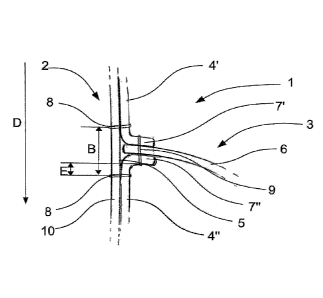
Figure 1 shows a longitudinal section through a stent-valve 1.
The stent-valve 1 is self expandable and comprises a stent com-
ponent 2 and a valve component 3. The stent component 2 is sub-
stantially made of Nitinol and comprises multiple crossing
struts 10. The stent-valve 1 is designed as a mitral valve pros-
thesis with a mitral valve component 3 comprising a mono leaflet
6. The stent-valve 1 and the leaflet 6 have a D-shape. The D-
shape better suits the native conditions of the mitral valve.
Two separate support tissues 4', 4" are attached to an inner
surface 5 of the stent component 3. Each tissue support 4', 4"
comprises an attachment area 7', 7" at its neighbouring end .
The attachment areas 7', 7" are extending essentially Inwardly
from the inner surface 5 towards a centre of the stent-valve 1.
The inwardly extending length of the attachment areas 7', 7" is
about 2 to 4 mm.
11
CA 02940268 2016-08-19
W02015/135883 PCT/EP2015/054861
The tissue supports 4', 4" are attached to struts 10 of the
stent component 2 with sutures 8. The sutures 8 attaching the
tissue supports 4', 4" to the stent component 2 have a distance
A (not shown, see Fig. 2) of about 1 to 2 mm. The sutures 8 at-
taching the tissue support 4', 4" to the stent component 2
nearest to the attachment areas 7', 7" have a distance B to
each other which is dependent on the thickness of the pericardi-
al tissue. With a tissue of about 0.3 mm, distance B is about 0.
9 to 5 mm, preferably 3 mm. The leaflet 6 is attached to the at-
tachment areas 7', 7" with sutures 9. Stitches of suture 9 have
a distance C (not shown, see Fig. 2) of about 1 mm to each oth-
er. The inner surface 5 between the sections of the attachment
areas 7', 7" attached to the leaflet is not covered with sup-
port tissue 4', 4". The distance E between the sutures 8 and
the attachment area 7', 7" is about 2 mm. The tissue supports
4', 4" are attached over the inner surface 5 of the stent com-
ponent 2.
The tissue supports 4', 4" and the leaflet 6 are substantially
made of pericardium. The tissue supports 4', 4" are about 0.3
mm thick.
Blood flows through the stent-valve 1 in direction D. The leaf-
let 6 opens concomitantly with a decreased pressure in the left
ventricle during diastole, and the blood flows through the
stent-valve 1. After the diastole, the leaflet 6 closes to pre-
vent blood backflow from the left ventricle into the left atri-
um. During opening of the leaflet 6 (and hence the valve), the
attachment areas 7', 7" bend together with the leaflet 6 sub-
stantially in direction D and back. The collective bending re-
sults in a smooth distribution of the stress acting on the leaf-
let 6 over the whole area of the leaflet 6 which is in contact
12
CA 02940268 2016-08-19
WO 2015/135883 PCT/EP2015/054861
with the attachment areas 7', 7" . Further, the arrangement pro-
vides an elastic chain shock absorption. The two tissue supports
4', 4" and the leaflet 6 cushion any forces during the systo-
diastolic cycle, i.e. systo-diastolic opening and closing of the
leaflet 6.
Figure 2 shows a view on the inside of a stent-valve 1. There
are two tissue supports 4', 4" attached to the inner surface 5
of the stent component 2. The sutures 8 to attach the tissue
supports 4', 4" are arranged along the struts 10 of the stent
component 2. The longitudinal cut through the tissue supports
4', 4" is only for illustrative reasons. In use, major parts of
the inner surface 5 of the stent-component 2 are covered with
tissue supports 4', 4". There might, however, be uncovered por-
tion. Uncovered portions might aid to an unproblematic, easy
crimping.
Figure 3 shows a larger section of the inside of a stent-valve
1. The leaflet 6 is arranged between two tissue supports 4',
4". In neighbouring sections, where no leaflet 6 is attached,
the tissue supports 4', 4" are overlapping. The leaflet 6 is
attached to the tissue supports 4', 4" along an attachment line
11. By attaching the leaflet 6 to the tissue supports 4', 4"
instead of directly to the stent component 2, the orientation of
the attachment line 11 is not dependent on the shape of the
stent component 2, i.e. the orientation of the struts 10 to
which the leaflet 6 might be to. The attachment line 11 can be
oriented in an oblique manner with respect to the flow direction
D.
Figures 4 and 5 show an alternative construction of the tissue
supports 4', 4" and the leaflet 6. The leaflet 6 is integrally
formed with the tissue support 4" at the outflow end (figure 4)
13
CA 02940268 2016-08-19
W02015/135883 PCT/EP2015/054861
or with the tissue support 4' at the inflow en (figure 5) of the
stent-valve 1. The leaflet 6 is attached to the other support
tissue 4', 4" with sutures 9. The stent component 2 is not
shown in figures 4 and 5.
Figures 6 to 10 show another construction comprising a single
piece of tissue, e.g. pericardium, which simultaneously forms a
first tissue support 4"' and a functional leaflet 6. A tubular
body is initially made from this single piece of tissue, with
two opposite end being sutured and wherein the suture 12 is lo-
cated on the posterior part.
The construction furthermore comprises a second tissue support
4"" which is attached the functional leaflet 6 on the anterior
part.
In this construction the attachment line 7' between the second
tissue support 4"" and the functional leaflet 6 forms an in-
clined angle.
Figure 11 shows a stent 1 according to the invention. The stent
component 2 is made of multiple crossing struts 10. The valve
component 3 is designed as a mono leaflet 6. The tissue supports
4', 4" are not shown. The leaflet is attached along an attach-
ment line 11 to the tissue supports 4', 4".
14