Note: Descriptions are shown in the official language in which they were submitted.
CA 02940610 2016-08-30
Catheter Stability Indication
BACKGROUND OF THE INVENTION
1. Field of the Invention.
[0001] This invention relates to tissue ablation systems. More particular-
ly, this invention relates to monitoring of contact between an invasive probe
and
tissue within the body.
2. Description of the Related Art.
[0002] Cardiac arrhythmias, such as atrial fibrillation, occur when re-
gions of cardiac tissue abnormally conduct electric signals to adjacent
tissue,
thereby disrupting the normal cardiac cycle and causing asynchronous rhythm.
[0003] Procedures for treating such arrhythmias include surgically dis-
rupting the origin of the signals causing the arrhythmia, as well as
disrupting the
conducting pathway for such signals. By selectively ablating cardiac tissue by
application of energy via a catheter, it is sometimes possible to cease or
modify
the propagation of unwanted electrical signals from one portion of the heart
to
another. The ablation process destroys the unwanted electrical pathways by
formation of non-conducting lesions.
[0004] Verification of physical electrode contact and contact stability
with the target tissue is important for controlling the delivery of ablation
energy.
Attempts in the art to verify electrode contact with the tissue have been
exten-
sive, and various techniques have been suggested. For example, U.S. Patent
No. 6,695,808 describes apparatus for treating a selected patient tissue or
organ
region. A probe has a contact surface that may be urged against the region,
thereby creating contact pressure. A pressure transducer measures the contact
pressure. This arrangement is said to meet the needs of procedures in which a
medical instrument must be placed in firm but not excessive contact with an
anatomical surface, by providing information to the user of the instrument
that is
indicative of the existence and magnitude of the contact force.
[0005] As another example, U.S. Patent No. 6,241,724 describes methods
for creating lesions in body tissue using segmented electrode assemblies. In
one embodiment, an electrode assembly on a catheter carries pressure trans-
1 of 29
CA 02940610 2016-08-30
ducers, which sense contact with tissue and convey signals to a pressure
contact
module. The module identifies the electrode elements that are associated with
the pressure transducer signals and directs an energy generator to convey ra-
diofrequency (RF) energy to these elements, and not to other elements that are
in contact only with blood.
[0006] A further example is presented in U.S. Patent No. 6,915,149. This
patent describes a method for mapping a heart using a catheter having a tip
electrode for measuring local electrical activity. In order to avoid artifacts
that
may arise from poor tip contact with the tissue, the contact pressure between
the
tip and the tissue is measured using a pressure sensor to ensure stable
contact.
[0007] U.S. Patent Application Publication 2007/0100332 describes sys-
tems and methods for assessing electrode-tissue contact for tissue ablation.
An
electromechanical sensor within the catheter shaft generates electrical
signals
corresponding to the amount of movement of the electrode within a distal por-
tion of the catheter shaft. An output device receives the electrical signals
for as-
sessing a level of contact between the electrode and a tissue.
[0008] Impedance-based methods for assessing catheter-tissue contact
that are known in the art typically rely on measurement of the magnitude of
the
impedance between an electrode on the catheter and a body-surface electrode.
When the magnitude is below some threshold, the electrode is considered to be
in contact with the tissue. This sort of binary contact is sensitive to
changes in the
impedance between the body-surface electrode and the skin.
[0009] U.S. Patent Application Publication Nos. 2008/0288038 and
2008/0275465, both by Sauarav et al., which are herein incorporated by refer-
ence, describe an electrode catheter system, which may comprise an electrode
adapted to apply electric energy. A measurement circuit adapted to measure
impedance may be implemented between the electrode and ground as the
electrode approaches a target tissue. A processor or processing units may be
implemented to determine a contact condition for the target tissue based at
least
in part on reactance of the impedance measured by the measurement circuit. In
another embodiment, the contact condition may be based on the phase angle of
the impedance.
2 of 29
CA 02940610 2016-08-30
SUMMARY OF THE INVENTION
[0010] Newer cardiac catheters include temperature-sensing elements
that provide information on the temperature distributions of the catheter tip
and
the relative orientation of the catheter tissue interface. This information
enables
an estimation of the size of an ablation lesion. The inventors have found that
such
temperature information in conjunction with strategically applied cooling
irriga-
tion of a target ablation site can be exploited prior to delivery of ablation
energy
to establish whether the catheter-tissue interface is stable or not.
[0011] A known difficulty in the use of ablation energy, e.g., radiofre-
quency energy for cardiac tissue ablation is controlling local heating of
tissue.
There are tradeoffs between the desire to create a sufficiently large lesion
to ef-
fectively ablate an abnormal tissue focus, or block an aberrant conduction pat-
tern, and the undesirable effects of excessive local heating. If the
radiofrequen-
cy device creates too small a lesion, then the medical procedure could be less
effective, or could require too much time. On the other hand, if tissues are
heat-
ed excessively then there could be local charring effects, coagulum, and or ex-
plosive steam pops due to overheating. Such overheated areas can develop
high impedance, and may form a functional barrier to the passage of heat. The
use of slower heating provides better control of the ablation, but unduly pro-
longs the procedure. Normally, irrigation precedes the ablation process.
Irriga-
tion lowers the temperature at the interface, since irrigation fluid is colder
than
the blood and the tissue.
[0012] The transient temperature pattern and its steady state differ when
the catheter is stable against the tissue and when it is not, When the
catheter is
stable only limited regions are cooled, whereas an unstable catheter-tissue in-
terface is characterized by a relatively more dispersed distribution of
irrigation
fluid. The temperature phenomena described in further detail herein are ob-
servable so long as the irrigation fluid is colder than the blood/tissue
tempera-
ture. Within this constraint, the temperature of the irrigation fluid and its
flow
rate mainly affect the magnitude of the differential signals, and their signal-
to-
noise ratio.
3 of 29
CA 02940610 2016-08-30
[0013] There is provided according to embodiments of the invention a
method, which is carried out by introducing a probe having a temperature sen-
sor on its distal portion into a fluid-filled body cavity of a subject, and
passing an
irrigating fluid through the probe, wherein the irrigating fluid exits the
probe at
its distal portion and wherein the temperature of the irrigating fluid is
different
from the temperature of the body cavity. The method is further carried out
while
passing the irrigating fluid by recording temperature readings of the tempera-
ture sensor, and making a determination from the temperature readings that
predetermined contact criteria between the probe and the interior wall of the
body cavity are satisfied, and thereafter alerting an operator that the
contact cri-
teria are satisfied.
[0014] According to a further aspect of the method, passing an irrigating
fluid is performed multiple times at different flow rates.
[0015] Yet another aspect of the method includes deriving a blood tern-
perature and an irrigation fluid temperature from the temperature readings at
respective flow rates.
[0016] According to an aspect of the method, the contact criteria com-
prise criteria for stable contact between the probe and the interior wall of
the
body cavity.
[0017] According to yet another aspect of the method, the contact criteria
comprise criteria for unstable contact between the probe and the interior wall
of
the body cavity.
[0018] According to still another aspect of the method, the contact crite-
ria comprise criteria for an absence of contact between the probe and the inte-
nor wall of the body cavity.
[0019] According to one aspect of the method, the probe has a plurality
of temperature sensors, and recording temperature readings is performed con-
currently with the temperature sensors.
[0020] An additional aspect of the method includes thermally insulating
the temperature sensors from the irrigating fluid passing through the probe.
[0021] According to a further aspect of the method, the temperature sen-
sors are disposed on an external surface of the probe.
4 of 29
CA 02940610 2016-08-30
[0022] According to yet another aspect of the method, the temperature
sensors are disposed internally in the probe.
[0023] According to another aspect of the method an ablation electrode
on the probe is activated while recording temperature readings.
[0024] In yet another aspect of the method recording temperature read-
ings includes recording a first temperature reading and thereafter recording a
second temperature reading. The contact criteria are satisfied when the second
temperature reading is lower than the first temperature reading, the method in-
cludes reporting contact between the probe with the interior wall.
[0025] According to still another aspect of the method, the second tem-
perature reading is at least 1 C lower than the first temperature reading.
[0026] According to a further aspect of the method, the second tempera-
ture reading is at least 4 C lower than the first temperature reading.
[0027] An additional aspect of the method the second temperature read-
ing further comprise transient elevations of between 1 to 4 C that are
between
0.3 to 5 seconds in duration, the method includes reporting an intermittent
con-
tact between the probe and the interior wall.
[0028] Another aspect of the method includes filtering the temperature
readings to remove effects of heart rate variations and respiratory
fluctuations.
[0029] There is further provided according to embodiments of the inven-
tion an apparatus, including a probe adapted for insertion into a fluid-filled
body cavity of a subject, the probe includes a temperature sensor on a distal
portion of the probe, The apparatus includes a pump for passing an irrigating
fluid through the probe, wherein the irrigating fluid exits the probe at the
distal
portion and wherein a temperature of the irrigating fluid is different from a
tem-
perature of the body cavity, and a processor operative for recording tempera-
ture readings of the temperature sensor while the pump is passing the
irrigating
fluid, making a determination from the temperature readings that predeter-
mined contact criteria between the probe and the interior wall of the body
cavi-
ty are satisfied, and thereafter alerting an operator that the contact
criteria are
satisfied.
[0030] An ablation electrode is provided on the distal portion of the
probe, which may be activated while recording temperature readings.
5 of 29
CA 02940610 2016-08-30
BRIEF DESCRIPTION OF THE SEVERAL VIEWS OF THE DRAWINGS
[0031] For a better understanding of the present invention, reference is
made to the detailed description of the invention, by way of example, which is
to
be read in conjunction with the following drawings, wherein like elements are
given like reference numerals, and wherein:
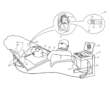
[0032] Fig. 1 is a pictorial illustration of a system for performing diagnos-
tic and therapeutic procedures in accordance with an embodiment of the inven-
tion;
[0033] Fig. 2 is a sectional view along the length of the distal segment of
a cardiac catheter, in accordance with an embodiment of the invention;
[0034] Fig. 3 is a detailed view of a portion of the distal segment of a car-
diac catheter in accordance with an alternate embodiment of the invention;
[0035] Fig. 4 is an isometric view of an insert for a catheter in accordance
with an embodiment of the invention;
[0036] Fig. 5 is a flow-chart of a method of determining catheter-tissue in-
terface stability, in accordance with an embodiment of the invention;
[0037] Fig. 6 is a diagram illustrating a calibration process in accordance
with an embodiment of the invention;
[0038] Fig. 7 is a chart indicating typical temperature tracings when the
procedure of Fig. 5 is performed in accordance with an embodiment of the in-
vention;
[0039] Fig. 8 is a chart that displays exemplary data in accordance with
an embodiment of the invention;
[0040] Fig. 9 is a graph showing average temperature measurement as a
function of flow of irrigation fluid in accordance with an embodiment of the
in-
vention
[0041] Fig. 10 is a graph showing average temperature measurement as
a function of flow of irrigation fluid in accordance with an embodiment of the
in-
vention;
[0042] Fig. 11 is a composite display comparing the graphs shown in
Fig. 9 and Fig. 10 in accordance with an embodiment of the invention;
6 of 29
CA 02940610 2016-08-30
[0043] Fig. 12 is a plot showing the difference between temperatures
during contact and non-contact between the catheter and tissue taken from the
data in Fig. 11 in accordance with an embodiment of the invention; and
[0044] Fig. 13 is a flow chart of a method of determining contact between
a catheter and a tissue in accordance with an alternate embodiment of the
inven-
tion.
DETAILED DESCRIPTION OF THE INVENTION
[0045] In the following description, numerous specific details are set
forth in order to provide a thorough understanding of the various principles
of
the present invention. It will be apparent to one skilled in the art, however,
that
not all these details are necessarily needed for practicing the present
invention.
In this instance, well-known circuits, control logic, and the details of
computer
program instructions for conventional algorithms and processes have not been
shown in detail in order not to obscure the general concepts unnecessarily.
[0046] Aspects of the present invention may be embodied in software
programming code, which is typically maintained in permanent storage, such as
a computer readable medium. In a client/server environment, such software
programming code may be stored on a client or a server. The software pro-
gramming code may be embodied on any of a variety of known non-transitory
media for use with a data processing system, such as a diskette, hard drive,
electronic media or CD-ROM. The code may be distributed on such media, or
may be distributed to users from the memory or storage of one computer sys-
tem over a network of some type to storage devices on other computer systems
for use by users of such other systems.
System Overview.
[0047] Turning now to the drawings, reference is initially made to Fig. 1,
which is a pictorial illustration of a system 10 for evaluating electrical
activity
and performing ablative procedures on a heart 12 of a living subject, which is
constructed and operative in accordance with a disclosed embodiment of the
invention. The system comprises a catheter 14, which is percutaneously
inserted
by an operator 16 through the patient's vascular system into a chamber or vas-
cular structure of the heart 12. The operator 16, who is typically a
physician,
7 of 29
CA 02940610 2016-08-30
brings the catheter's distal tip 18 into contact with the heart wall, for
example, at
an ablation target site. Electrical activation maps may be prepared, according
to
the methods disclosed in U.S. Patent Nos. 6,226,542, and 6,301,496, and in com-
monly assigned U.S. Patent No. 6,892,091, whose disclosures are herein incor-
porated by reference. One commercial product embodying elements of the sys-
tem 10 is available as the CARTO 3 System, available from Biosense Webster,
Inc., 3333 Diamond Canyon Road, Diamond Bar, CA 91765. This system may be
modified by those skilled in the art to embody the principles of the invention
described herein.
[0048] Areas determined to be abnormal, for example by evaluation of
the electrical activation maps, can be ablated by application of thermal
energy,
e.g., by passage of radiofrequency electrical current through wires in the
cathe-
ter to one or more electrodes at the distal tip 18, which apply the
radiofrequen-
cy energy to the myocardium. The energy is absorbed in the tissue, heating it
to
a point (typically about 50 C) at which it permanently loses its electrical
excita-
bility. When successful, this procedure creates non-conducting lesions in the
cardiac tissue, which disrupt the abnormal electrical pathway causing the ar-
rhythmia. The principles of the invention can be applied to different heart
chambers to diagnose and treat many different cardiac arrhythmias.
[0049] The catheter 14 typically comprises a handle 20, having suitable
controls on the handle to enable the operator 16 to steer, position and orient
the
distal end of the catheter as desired for the ablation. To aid the operator
16, the
distal portion of the catheter 14 contains position sensors (not shown) that
pro-
vide signals to a processor 22, located in a console 24. The processor 22 may
fulfill several processing functions as described below.
[0050] Ablation energy and electrical signals can be conveyed to and
from the heart 12 through one or more ablation electrodes 32 located at or
near
the distal tip 18 via cable 34 to the console 24. Pacing signals and other
control
signals may be conveyed from the console 24 through the cable 34 and the elec-
trodes 32 to the heart 12. Sensing electrodes 33, also connected to the con-
sole 24 are disposed between the ablation electrodes 32 and have connections
to the cable 34.
8 of 29
CA 02940610 2016-08-30
[0051] Wire connections 35 link the console 24 with body surface elec-
trodes 30 and other components of a positioning sub-system for measuring loca-
tion and orientation coordinates of the catheter 14. The processor 22 or
another
processor (not shown) may be an element of the positioning subsystem. The
electrodes 32 and the body surface electrodes 30 may be used to measure tis-
sue impedance at the ablation site as taught in U.S. Patent No. 7,536,218,
issued
to Govari et al., which is herein incorporated by reference. A temperature sen-
sor (not shown), typically a thermocouple or thermistor, may be mounted on or
near each of the electrodes 32. The sensors can vary in position. For example,
the sensors may be external or internal to the catheter a14. In any case the
sen-
sors are thermally insulated from irrigating fluid passing through the
catheter
using any conventional insulating material.
[0052] The console 24 typically contains one or more ablation power
generators 25. The catheter 14 may be adapted to conduct ablative energy to
the heart using any known ablation technique, e.g., radiofrequency energy, ul-
trasound energy, and laser-produced light energy. Such methods are disclosed
in commonly assigned U.S. Patent Nos. 6,814,733, 6,997,924, and 7,156,816,
which are herein incorporated by reference.
[0053] In one embodiment, the positioning subsystem comprises a mag-
netic position tracking arrangement that determines the position and
orientation
of the catheter 14 by generating magnetic fields in a predefined working vol-
ume and sensing these fields at the catheter, using field generating coils 28.
The
positioning subsystem is described in U.S. Patent No. 7,756,576, which is
hereby
incorporated by reference, and in the above-noted U.S. Patent No. 7,536,218.
[0054] As noted above, the catheter 14 is coupled to the console 24,
which enables the operator 16 to observe and regulate the functions of the
cath-
eter 14. Console 24 includes a processor, preferably a computer with appropri-
ate signal processing circuits. The processor is coupled to drive a monitor
29.
The signal processing circuits typically receive, amplify, filter and digitize
sig-
nals from the catheter 14, including signals generated by sensors such as elec-
trical, temperature and contact force sensors, and a plurality of location
sensing
electrodes (not shown) located distally in the catheter 14. The digitized
signals
are received and used by the console 24 and the positioning system to compute
9 of 29
CA 02940610 2016-08-30
the position and orientation of the catheter 14, and to analyze the electrical
sig-
nals from the electrodes.
[0055] In order to generate electroanatomic maps, the processor 22 typi-
cally comprises an electroanatomic map generator, an image registration pro-
gram, an image or data analysis program and a graphical user interface config-
ured to present graphical information on the monitor 29.
[0056] Typically, the system 10 includes other elements, which are not
shown in the figures for the sake of simplicity. For example, the system 10
may
include an electrocardiogram (ECG) monitor, coupled to receive signals from
one or more body surface electrodes, in order to provide an ECG synchroniza-
tion signal to the console 24. As mentioned above, the system 10 typically
also
includes a reference position sensor, either on an externally-applied
reference
patch attached to the exterior of the subject's body, or on an internally-
placed
catheter, which is inserted into the heart 12 maintained in a fixed position
rela-
tive to the heart 12. Conventional pumps and lines for circulating liquids
through
the catheter 14 for cooling the ablation site are provided. The system 10 may
re-
ceive image data from an external imaging modality, such as an MRI unit or the
like and includes image processors that can be incorporated in or invoked by
the processor 22 for generating and displaying images.
[0057] Reference is now made to Fig. 2, which is a sectional view along
the length of distal segment 54 of a cardiac catheter in accordance with an em-
bodiment of the invention. The distal segment 54 is in proximity to tissue 56,
and
is assumed to be immersed in fluid 58, so that tissue 56 has a surface 29
contact-
ing the fluid. Fluid 58 typically comprises a mixture of blood and saline
solution.
By way of example, distal segment 54 is assumed herein to be formed from an
insulating substrate 60 in the shape of a cylinder 62 closed by a generally
flat
surface 64 at one end. Cylinder 62 has an axis of symmetry 66. As shown in
Fig. 2, a curved section 68 joins flat surface 64 and cylinder 62. A typical
diame-
ter of cylinder 62 is 2.5 mm, and a typical radius of the curved section 68 is
0.5
mm.
[0058] Distal segment 54 comprises three electrodes 70, 72, 74, the elec-
trodes being insulated from each other. The electrodes 70, 72, 74 typically
com-
prise thin metal layers formed over insulating substrate 60. Typically, the
distal
10 of 29
CA 02940610 2016-08-30
tip has other electrodes, insulated from the electrodes 70, 72, 74, which for
sim-
plicity are not shown in the diagram. Tip electrode 70 has the shape of a cup
with a flat base, and is herein also referred to as the cup electrode. Cup
elec-
trode 70 typically has a thickness in a range from approximately 0.1 mm to ap-
proximately 0.2 30 mm. Second and third electrodes 70, 72, are usually in the
form of rings, and are also known as ring electrodes.
[0059] Electrodes 70, 72, 74 are connected to a controller in console 24
(Fig. 1) by wires (not shown). At least one of the electrodes is used to
ablate tis-
sue 56. Typically, during ablation, heat is generated in the ablating
electrode
and in the surrounding region. In order to dissipate the heat, small
irrigation
apertures 76 in the cup electrode. The apertures 76 typically have diameters
in
an approximate range 0.1 - 0.2 mm. An irrigation tube 78 supplies saline solu-
tion to the apertures 76, and the rate of flow of the saline solution through
the
apertures 76 (causing fluid 58 to be a mixture of blood and saline solution)
is
controlled by an irrigation module (not shown) in the console 24 (Fig. 1). The
sa-
line rate of flow is typically in the range of approximately 2 - 20 cc/minute,
but
may be higher or lower than this range.
[0060] A saline temperature sensor 80, typically a thermocouple, is lo-
cated in tube 78, and provides a signal to circuitry in the console 24 (Fig.
1)
module 56 enabling the console 24 to measure a temperature Ts of the saline so-
lution input to apertures 76. While the saline solution may be provided at
room
ambient temperature, e.g., in a range of approximately 19 - 25 C, the solution
may be heated slightly during its flow through the catheter, so that the final
irri-
gation temperature may be slightly higher.
[0061] Typically, one or more location sensing devices 82 are incorpo-
rated in the distal tip. Devices 82 are configured to provide signals to the
pro-
cessor 22 (Fig. 1) enabling the system to ascertain the position and/or
orienta-
tion of distal segment 54,
[0062] In one embodiment distal segment 54 comprises one or more
generally similar temperature sensors 84 (by way of example, two are shown in
the diagram) which are fixedly connected, by an insulator, to the outer
surface
of cup electrode 70, so as to protrude from the surface. Sensors 84 have a
typical
diameter of approximately 0.3 mm and a length of approximately 1.5 mm. In one
11 of 29
CA 02940610 2016-08-30
embodiment sensors 84 are thermistors NTC Type AB6, produced by General
Electric Company of Schenectady, New York. In an alternative embodiment,
sensors 84 comprise "F" type thermistors produced by Semitec USA Corpora-
tion of Torrance, 15 California. By way of example, the following description
as-
sumes there are three sensors 84 symmetrically distributed with respect to
axis
51, and located on a curved section 86 of the cup electrode. Curved section 86
of the cup electrode overlays curved section 68 of the distal tip. Curved
section
86 is in the shape of a partial toroid, typically a partial torus having a
tube radius
of approximately 0.5 mm.
[0063] A magnified section 88 of Fig. 2 illustrates one of sensors 84 in
more detail. As shown in section 88, an insulator 90 separates sensors 84 from
curved section 86 of the cup electrode 70. Insulator 90 is selected to provide
good thermal and electrical insulation, and in some embodiments insulator 90
may comprise an adhesive that bonds sensors 84 to curved section 86. Wires 92
90 connect sensors 84 to the console 24 (Fig. 1).
[0064] By having sensors 84 protrude from the outer surface of cup elec-
trode 70, the sensors 84 are able to intimately contact tissue 56. The proces-
sor 22 (Fig. 1) is thus able to use signals from the sensors 84 to provide
direct
temperature measurements of the tissue 56 In one embodiment the sensors 84
protrude from the outer surface of the electrode 70 by no more than 0.7 mm,
and
typically by approximately 0.5 mm.
[0065] Reference is now made to Fig. 3, which is a detailed view of a por-
tion of the distal segment of a cardiac catheter in accordance with an
alternate
embodiment of the invention. In this embodiment the sensors do not protrude
above the outer surface of the cap electrode or the outer surface of the
probe. In
the representative example of Fig. 3 sensor 94 is flush with outer surface 96
and
is insulated from fluid passing through lumen 98 by an insulating material
100.
An advantage of this embodiment is a reduction in the likelihood of thrombus
formation on the surface of the sensor 94.
[0066] Reference is now made to Fig. 4, which is an isometric view of an
insert 102 for a catheter in accordance with an embodiment of the invention.
The
insert 102 is adapted to be capped by cap electrode in the catheter similar to
the electrode 70 (Fig. 2), The cap electrode is omitted for clarity.
12 of 29
CA 02940610 2016-08-30
[0067] As can be seen, protrusions 104 include annular shoulders 106
configured to engage the inner surface of the ablation electrode. Shoulders
106
may have a surface that is complimentary to the internal surface of the cap
elec-
trode as appropriate. The width of shoulders 106 may be defined by the differ-
ence between the diameter of a base portion 108 and the diameter of inner por-
tion 110. The diameter of inner portion 110 is sized to mate with sensor
orifices
(not shown, The protrusions 104 are configured to either extend outward from
or
are flush with the outer surface of the cap electrode. Similarly, annular
shoul-
ders 106 extend radially outward from the surface of insert 102, such that the
depth of base portion 108 establishes a minimum separation between the inner
surface of the cap electrode and surface 112 on the body of insert 102.
[0068] In this embodiment, insert 102 includes three longitudinally ex-
tending arms 114, each having a hollow interior portion to allow routing of
leads
and wires to sensors 116. Arms 114 are connected at distal crown portion 118.
Passageways 120 may be formed between arms 114 as well as by a central
opening in crown portion 118. Depending on the intended use and the number
of sensors being provided, the configuration of insert 102 may be adapted as
desired, such as by featuring two or four arms, for example. In one aspect,
each
if the arms 114 may include at least two protrusions 104 to accommodate at
least
two sensors, such as one proximal and one distal.
[0069] Sensors 116 may be any combination of temperature sensors,
e.g., thermistor, thermocouple, fluoroptic probe, and the like, or electrical
sen-
sors, e.g., micro-electrodes. Any temperature sensor junctions located at or
near the end of protrusions 104 and may be potted with a thermally conductive
adhesive. Any wires or leads associated with sensors 116 may be routed
through arms 114 as appropriate. As will be appreciated, this configuration
iso-
lates sensors 116 from the cap electrode and the irrigation fluid. In one
aspect,
insert 102 serves to thermally insulate sensors 116. Accordingly, a more accu-
rate measurement of tissue and environmental temperature may be obtained by
reducing biasing from the cap electrode or the circulating irrigation fluid.
In an-
other aspect, insert 102 also serves to electrically insulate sensors 116 to
allow
more accurate measurement. Similarly, any wires and/or leads are also thermal-
ly and electrically insulated, as well as being sealed against corrosion from
the
13 of 29
CA 02940610 2016-08-30
irrigation fluid. In one aspect, each of the sensors 116 that are positioned
by the
protrusions 104 may be configured to sense a plurality of measurements. For
example, one or more sensors 116 may function both as a micro-thermistor and
a micro-electrode. According to one embodiment, thermistor wires as well as an
electrode lead wire may be connected to a shell cap electrode of each of the
sensors 116. Each wire may be isolated from each other by any suitable tech-
nique, such as by employing a suitable electrically nonconductive and non-
thermally insulative material to fill the interior of arms 114 after placement
of
sensors 116.
[0070] Insert 102 is stabilized within the cap electrode by portion 118,
which includes a disc-shaped base 122 and a distally projecting key 124. Base
122 may have a diameter corresponding to the inner diameter of the cap elec-
trode and may be secured in any suitable manner, such as by welding 126.
Key 124 is configured to fit within recess 128 of insert 102, formed by the
proxi-
mal portions of arms 114, to stabilize insert 102 against axial rotation and
possi-
ble displacement of sensors 116. Portion 118 may provide a fluid-tight seal
with
cap electrode while routing leads and wires associated with the cap electrode,
sensors 116 and irrigation fluid from lumens extending through the catheter
body. For example, central conduit 130 may be in communication with the lu-
men of the catheter to conduct irrigation fluid to passageways 120, for
circula-
tion within the interior of the cap electrode and eventual exit through
apertures,
e.g., apertures 76 (Fig. 2).
[0071] Catheters of the kind described with reference to Fig. 2 and Fig. 4
are described in further detail in commonly assigned U.S. Patent Application
Publication Nos. 2014/0171821 by Govari et al., 2011/0224664 to Bar-Tal et
al.,
and copending U.S. Application No. 14/551,229, entitled Irrigated Ablation
Cath-
eter with Multiple Sensors, which are herein incorporated by reference.
Operation.
[0072] Reference is now made to Fig. 5, which is a flow-chart of a method
of determining catheter-tissue interface stability, in accordance with an
embod-
iment of the invention. In the drawings herein, process steps are shown in a
par-
ticular linear sequence for clarity of presentation. However, it will be
evident
14 of 29
CA 02940610 2016-08-30
that many of them can be performed in parallel, asynchronously, with feedback
loops, or in different orders. Those skilled in the art will also appreciate
that a
process could alternatively be represented as a number of interrelated states
or
events, e.g., in a state diagram. Moreover, not all illustrated process steps
may
be required to implement the process.
[0073] In the discussion below, the temperature of the irrigation fluid is
lower than the temperature of the blood. An irrigation fluid at typical room
tem-
perature (25 C) is suitable. However, the principles of the invention are
appli-
cable, mutatis mutandis, when the irrigation fluid is warmer than the blood.
[0074] At initial step 132, a cardiac catheter is introduced into the heart
of a subject using well-known methods. At this stage, the catheter is still
free in
the cardiac chamber and out of contact with the wall of the heart. An optional
calibration may be now performed. The goal of the calibration is to establish
a
temperature threshold for differentiating between two conditions: A) catheter
in
the blood pool; and B) catheter in contact (whether intermediate or not) with
tis-
sue. It is necessary to know the blood temperature, the irrigation fluid
tempera-
ture. These can be assumed or measured. It is also necessary to know the flow
rate of the irrigation fluid.
[0075] Reference is now made to Fig. 6, which is a diagram illustrating
the optional calibration process in accordance with an embodiment of the inven-
tion. Blood and irrigating fluid baseline temperatures are represented as bro-
ken lines 134, 136, respectively. Temperatures measured during the procedure
described below with respect to Fig. 5 occur in an operational zone 138 that
lies
between the baseline temperatures. The blood temperature (line 134) may be
determined during introduction of the catheter and prior to initiating
irrigation
using sensors in the catheter tip. Measurement of fluid temperature can be per-
formed on the irrigation line or can be derived by providing irrigation fluid
at
known flow rates to the catheter, and measuring the observed temperatures. Ir-
rigating fluid temperature (line 134) may be determined using either of the
fol-
lowing two procedures. Both procedures establish baselines representing the
temperature of the irrigating fluid.
[0076] Returning to Fig. 5, in a first calibration option shown in block 140,
after introduction of the catheter irrigation is initiated at step 142. Then,
at
15 of 29
CA 02940610 2016-08-30
step 144 baseline temperature readings may be obtained directly from sensors
in the irrigation fluid lines outside the patient's body. These readings are
not in-
fluenced by blood temperature.
[0077] Additionally or alternatively, in step 142, the irrigating fluid base-
line readings may be taken concurrently with multiple temperature sensors in
the catheter tip and should be continuous for a predetermined time interval,
e.g., 2-5 sec, in order to establish a reliable pattern of variation. The
predeter-
mined time interval is not critical, and may be varied for particular
applications.
It may be desirable to flush the catheter with irrigation fluid after the time
has
elapsed. This alternative provides a baseline for a state in which the
catheter is
free in the cardiac chamber and being irrigated at a typical rate. The value
ob-
tained generally differs from that of the first alternative as there is some
influ-
ence of ambient blood temperature.
[0078] In a second option, shown in block 146, an irrigation fluid base-
line temperature is set by flushing the catheter at step 148 at different flow
rates,
typically with saline between 2 and 20 ml/sec and, at step 150, reading one or
more temperature sensors during each flushing.. Each flow of the flushings can
be expressed in an equation that depends on the two knowns (the given flow
rate and catheter build/design) and two unknowns blood and fluid temperature.
By providing several flows the blood and fluid temperatures nay be obtained by
solving a system of such equations. The geometry and other aspects of the cath-
eter design are important as they affects the parameters. The parameters of
the
equations are therefore empirical, and catheter-specific. Significant catheter
de-
sign issues include sensor locations (how well they sense the flow) and the de-
sign of the irrigation holes. Solution of the equations provides data on the
fluid
and the blood temperatures simultaneously. A precalibration process can be
used for the equations.
[0079] During flushing, the temperature quickly drops from an ambient
level to a threshold value (line 134; Fig. 6), which is close to the
temperature of
the fluid used for flushing. Additionally or alternatively, one or more
additional
sensors (not shown) may be located along the catheter in order to monitor the
saline temperature as it enters the catheter and to measure the temperature of
the blood. Using the information provided by the additional sensors, and
solving
16 of 29
CA 02940610 2016-08-30
the above-noted equations, it is possible to estimate the expected temperature
readings of temperature sensors 152 in the blood pool prior to determining tis-
sue contact with the catheter.
[0080] In either of the procedures described in blocks 140, 146, once ir-
rigation begins, the temperature readings from the catheter tip drop from the
blood temperature baseline (line 136; Fig. 6). For example, once a temperature
reading below a predetermined threshold value, e.g., 32 C, is observed, it may
be concluded that irrigation has begun. After completing step 144, or step 150
control proceeds to step 154.
[0081] In some embodiments the procedures of blocks 140, 146 are omit-
ted, as the transitions described below, e.g., in the discussion of Fig. 8,
can be
established without reference to baselines or threshold readings. The baseline
values may be assumed, e.g., based on experience or known information. It
would be known from other modalities if the patient were febrile or hypother-
mic. In this case, control proceeds from initial step 132 directly to step 154
as
shown by line 156.
[0082] Next, at step 154 contact is established between the tissue and the
ablation electrode, which is typically located at the distal tip when the new
posi-
tion is attained of the catheter. This may be accomplished by any known meth-
od, e.g., any of the methods described above and the methods taught in U.S. Pa-
tent Application Publication No. 20130172875, entitled "Contact Assessment
Based on Phase Measurement" and U.S. Patent Application Publication
No. 20140051959 entitled "Machine Learning in Determining Catheter Electrode
Contact", which are commonly assigned herewith and are herein incorporated
by reference. Irrigation is begun at step 158. When contact has been estab-
lished the temperature readings are intermediate between the blood and irriga-
tion fluid baselines, (see Fig. 8; time 160).
[0083] Next, at step 162, while continuing irrigation, a record of tempera-
ture readings is obtained. Statistics, such as the mean temperature, variance,
and the morphology of the temperature records are considered in step 162. If
the catheter-tissue interface is unstable, the readings will be unstable, even
bursty (as contact with a particular location occurs and is lost or as the
contact
point moves on the tissue. In the former case, the catheter tip is exposed to
am-
17 of 29
CA 02940610 2016-08-30
bient blood. In the latter case, the catheter tip contacts uncooled tissue. In
either
case the temperature will rise or fall as contact is lost and reestablished in
an
unstable manner. Typically transient elevations of between 1 to 4 C that are
0.3
to 5 sec in duration are seen when contact is intermittent or unstable. Such
fluc-
tuations may be due to respiration (5 sec per cycle, typically), heartbeat
(0.3 - 1
sec / cycle) and pump pulsations within the range of 0.3 to 5 sec / cycle.
[0084] Next, at decision step 164, it is determined if criteria for stable
contact based on the analysis of step 162 are satisfied. The criteria are
empiri-
cally determined case-by-case, according to irrigation flow rate and the tem-
perature of the irrigation fluid and the blood. If the determination at
decision
step 164 is affirmative, then control proceeds to final step 166. A stable
catheter-
tissue interface is reported, and ablation may begin.
[0085] If the determination at decision step 164 is negative then at deci-
sion step 168 it is determined if unstable or bursty readings temperature read-
ings were obtained. If the determination at decision step 168 is affirmative,
then
control proceeds to final step 170. An unstable electrode-tissue interface is
re-
ported.
[0086] If the determination at decision step 168 is negative then control
proceeds to final step 172. It is concluded that the catheter tip is free in
the
blood pool.
[0087] After performing one of final steps 166, 170, 172 the electrode is
classified as being in stable contact, in intermittent contact or not in
contact. The
classification of each electrode can be based solely on the sensor data or de-
rived from the behavior of several sensors. When multiple ablation electrodes
are present, the sequence that follows step 142 may be performed separately
for each electrode, and a respective contact status is reported for each of
them.
[0088] Reference is now made to Fig. 7, which is a chart indicating typi-
cal temperature tracings that are expected when a catheter is positioned in
the
heart, when the procedure of Fig. 5 is performed in accordance with an embod-
iment of the invention. During time interval 174, while the end of the
catheter is
free in the blood pool and out of contact with tissue, a relatively high
tempera-
ture is recorded by the three temperature sensors, and there is little
fluctuation.
As noted above, the actual number of temperature sensors may vary in different
18 of 29
CA 02940610 2016-08-30
= =
embodiments. During time interval 176, intermittent contact with tissue
exists.
The temperature is lower, and there is a greater degree of fluctuation than in
time interval 174. High frequency fluctuations are observed as the catheter al-
ternates between contact and non-contact states. The alternations are an
indica-
tion that the catheter is in close proximity to the tissue. Importantly, this
demon-
strates that there is not secure contact with the tissue In this event an
alert may
be generated for the operator, The fluctuation in this case correlates with me-
chanical movement of the cardiac wall as it moves against the catheter as the
heart beats.) During time interval 178, the catheter is forcefully in contact
with
the tissue. The temperature is lowest and fluctuation intermediate among the
time intervals 174, 176, 178. It is believed that the temperature is lowest
because
the irrigation fluid flowing from the catheter is cooling the tissue and the
isolated
sensor measures the tissue temperature. The transient temperature pattern and
its steady state differ when the catheter is stable against the tissue and
when it is
not, When the catheter is stable only limited regions are cooled, whereas an
un-
stable catheter-tissue interface is characterized by a relatively more
dispersed
distribution of irrigation fluid. Different measured temperatures are observed
at
the target site than when the catheter-tissue interface is stable and when it
is not.
The temperature phenomena described in further detail herein are observable
so long as the irrigation fluid is colder than the blood/tissue temperature.
Within
this constraint, the temperature of the irrigation fluid and its flow rate
mainly af-
fect the magnitude of the differential signals, and their signal-to-noise
ratio.
First Alternate Embodiment.
[0089] In this embodiment, the signals obtained from the temperature
sensors may be filtered using the signal processing circuitry of the system 10
(Fig. 1), e.g., by averaging the signal, or applying well-known filters
directed to
the frequencies of pump pulses, heart rate variations and respiratory fluctua-
tions that would obscure other significant temperature fluctuations. In the
first
embodimentõ high frequency fluctuations are intentionally not filtered, as
their
presence provides an excellent indication of intermittent contact with the
tissue.
Nevertheless, a filtering mode of operation is advantageous during periods of
intermittent contact, as shown in the following example.
19 of 29
CA 02940610 2016-08-30
Example 1.
[0090] This simulated example show the effect of dragging the electrode
along the tissue. It consists of data obtained from a test system in which
blood in
a heart chamber was simulated by a water-filled aquarium (temperature 34 C).
Water at a temperature of 24 C was pumped through a catheter, e.g., the cathe-
ters shown in Fig. 1, Fig. 2 having distal temperature sensors to simulate
abla-
tion site irrigation. Tissue contact was simulated by contacting the
operator's
hand to the distal portion of the catheter.
[0091] Reference is now made to Fig. 8, which is a chart that displays da-
ta from this example in accordance with an embodiment of the invention. The
data was recorded from three temperature sensors on the catheter. Tempera-
ture is plotted against time. Prior to time 180, the tip of the catheter was
free in
the aquarium, and the sensors recorded a temperature of 34 C, with very
little
variation. Irrigation was initiated at time 180. The temperature quickly
dropped
into a range of 25 ¨ 30 C, with somewhat greater variation than prior to
time 180. Heart rate and respiratory effects are necessarily omitted in this
ex-
ample.
[0092] At time 160 tissue contact was made with the catheter. Thereupon,
the temperature dropped precipitously by about 4 C to about 26 C, and there-
after declined more slowly, equilibrating at slightly above 24 C.
[0093] At time 182, an unstable catheter-tissue interface was simulated
by sliding the catheter along the hand. Thus resulted in a transient
elevation,
i.e., a temperature spike of about 4 C that was less than about 2 sec in
duration,
as the catheter contacted uncooled tissue. The temperature then gradually de-
dined and approached the temperature of the irrigation fluid. This maneuver
was repeated at times 184, 186. The spikes at times 182, 184, 186 reflect dis-
placement of the catheter tip from a relatively stable position at one
location. As
the tip was repositioned by sliding it to another location, there was a period
dur-
ing which the tip was no longer in stable tissue contact. During this period
the
temperature rose transiently. Then, as a new relatively stable position was at-
tained, the temperature dropped abruptly in the spiking pattern observed at
times 182, 184, 186. It should be noted that without filtering the sensor
signals,
20 of 29
CA 02940610 2016-08-30
the spikes at times 182, 184, 186 would be obscured by fluctuations (e.g.,
fluctu-
ations occurring during time interval 176; Fig. 7) caused by the above-noted
ar-
tifacts.
[0094] Then the catheter was held in place until time 188. The tempera-
ture remained equilibrated near the temperature of the irrigation fluid, and
met
predefined stability criteria that can be established by known methods, e.g.,
excursions that are less than a threshold value for a certain time interval.
Then at
time 188, the catheter was abruptly removed from the tissue. This maneuver was
associated with an immediate rise in temperature and a fluctuating tracing pat-
tern.
[0095] Without being bound by any particular theory, the following discus-
sion is offered as a possible explanation of the observed effects in order to
facilitate
understanding of the invention. When the catheter is in the blood pool it is
ex-
posed to the warm circulating blood (whether from the heart operation or from
the combination of circulating irrigating flow and the blood) that maintain
the
catheter at a relative high temperature (typically around 34 -35 ). The low
tem-
perature read by the sensors is an indication of the tissue being cooled by
the
catheter when it is in close proximity to the endocardial surface. The cooling
oc-
curs in a relatively small partially confined space. Therefore when the
catheter
slides on the tissue, it is exposed to higher temperatures of the blood pool
and/or tissue that was not cooled by the fluid; hence the spike at time 182.
When
the catheter alternates between contact and blood pool it shows the spikes pat-
tern of time interval 176 (Fig. 7).
Example 2.
[0096] This example shows the relationships between contact, non-
contact and flow rates. A pig was intubated and anesthetized, and catheterized
using an open irrigation catheter having the arrangement shown in Fig. f4 with
I
six thermocouple sensors and a contact force sensor. The conditions that were
tested were contact and non-contact, i.e., the tip of the catheter free in the
blood
pool. Contact status was verified by readings from the contact force sensor
and
by Carto mapping. In both contact and non-contact conditions three differ-
ent irrigation flows were measured (2, 10, 25 ml/sec).
21 of 29
CA 02940610 2016-08-30
=
[0097] Reference is now made to Fig. 9, which is a graph in accordance
with an embodiment of the invention showing average temperature measure-
ment from the 6 sensors, as a function of flow of irrigation fluid at room
tempera-
ture when there is no contact between the catheter tip and the tissue. It is
evi-
dent that the measured temperature drops as the flow rate increases. Trac-
ings 190, 192, and 193 correspond to flow rates of 2, 10, and 25 ml/sec,
respec-
tively. At the 2 ml/sec rate the measured temperature does not differ
significant-
ly from the blood temperature. At 25 ml/sec a more substantial drop is seen,
with an intermediate drop at 10 mUsec.
[0098] Reference is now made to Fig. 10, which is a graph in accordance
with an embodiment of the invention showing average temperature measure-
ment from the 6 sensors as a function of flow of irrigation fluid at room
tempera-
ture when there is contact between the catheter tip and the tissue. The same
conditions were used as in Fig. 9. Tracings 194, 196, 198 correspond to flow
rates of 2, 10, and 25 mUsec, respectively Contact was verified when a force
ex-
ceeding 15 gr was ready by the contact force sensor. As in Fig. 9 a
progression
in the temperature drop is seen as the flow rate increases.
[0099] Reference is now made to Fig. 11, which is a composite display
comparing the graphs shown in Fig. 9 and Fig. 10 in accordance with an embod-
iment of the invention. At very low flow rate there is a minimal effect of
flow rate
variation. Tracings 190, 192 are nearly identical. At intermediate flow rates
re-
sidual evidence of tissue cooling is seen. Thus, the temperature is almost the
same in contact and non-contact conditions. However, the greater the flow the
larger the temperature effect (i.e., the difference between the tracings 198,
193
(25 ml/sec) is larger than the difference between the tracings 196, 192.
[0100] Reference is now made to Fig. 12, which is a plot showing the dif-
ference between temperatures during contact and non-contact between the
catheter and tissue taken from the data in Fig. 11 in accordance with an embod-
iment of the invention. A non-linear relationship is shown. The actual
relation-
ship may vary according to the characteristics of the catheter and the tissue
be-
ing targeted; however a general non-linear increase in temperature difference
and flow rate is expected to persist in most if not all cases.
22 of 29
CA 02940610 2016-08-30
Second Alternate Embodiment.
[0101] Reference is now made to Fig. 13, which is a flow chart of a meth-
od of determining contact between a catheter and a tissue in accordance with
an
alternate embodiment of the invention. The method should be understood with
reference to Example 2 and Fig. 12.
[0102] The procedure begins with initial step 200. The catheter is intro-
duced into the chamber in a non-contacting relationship with tissue. Blood tem-
perature is determined as described above at zero flow rate. Temperature
measurements are taken in this and subsequent steps of the method using one of
the procedures described above in the discussion of Fig. 6.
[0103] Next, at step 202, while the catheter remains in a non-contacting
relationship with tissue, irrigation fluid is passed at a first flow rate.
This may be
10 ml/sec as described above, but other rates may be substituted.
[0104] Next, at step 204, while the catheter remains in a non-contacting
relationship with tissue, irrigation fluid is passed at a second flow rate.
This may
be 25 ml/sec as described above, but other rates may be substituted.
[0105] Next, at step 206 the catheter is brought into presumptive contact
with the target tissue, typically the wall of the cardiac chamber.
[0106] Next, at step 208, while the catheter remains in a presumptive
contact with tissue, irrigation fluid is passed at the first flow rate as in
step 202.
[0107] Next, at step 210, while the catheter remains in a presumptive
contact with tissue, irrigation fluid is passed at the second flow rate as in
step 204.
[0108] Next, at step 212 the respective temperature differences for the
measurements during non-contact (and presumptive contact are computed for
the first and second flow rates.
[0109] Next, at decision step 214, it is determined if the differences com-
puted in step 212 are significant. This may be done by optimizing a figure of
merit for a profile of the sort shown in Fig. 12, by employing the teachings,
mu-
tatis mutandis, of commonly assigned Application Serial No. 13/589,347,
entitled
Machine Learning in Determining Catheter Electrode Contact, which is herein in-
corporated by reference. Other techniques for such optimizations are well
23 of 29
CA 02940610 2016-08-30
known in the art. Alternatively, when the differences exceed a threshold value
for one or both of the differences computed in step 212 presumptive contact
may be confirmed. The actual values for the threshold are application depend-
ent, as noted above. Further alternatively, other characteristics of the
tempera-
ture differences may be used as decisional criterion, for example the maximum
value of A(temp)/L(flow) in the plot of Fig. 12.
[0110] If the determination at decision step 214 is affirmative, then con-
trol proceeds to final step 216. Confirmation of presumptive contact between
the
catheter and the tissue is reported.
[0111] If the determination at decision step 214 is negative, then control
proceeds to final step 218. Presumptive contact between the catheter and the
tissue cannot be confirmed. Presumably the catheter tip is still free in the
cham-
ber.
[0112] This method of determining contact is particularly useful when
conventional techniques of determining contact fail or are not available, for
ex-
ample when a fault in a contact force sensor or a malfunction in the mapping
processor or circuitry occurs during a procedure. Moreover, the method de-
scribed in Example 2 and Fig. 13 can be used to predict the temperature
threshold in Fig. 8.
[0113] It will be appreciated by persons skilled in the art that the present
invention is not limited to what has been particularly shown and described
hereinabove. Rather, the scope of the present invention includes both
combinations and sub-combinations of the various features described
hereinabove, as well as variations and modifications thereof that are not in
the
prior art, which would occur to persons skilled in the art upon reading the
foregoing description.
24 of 29