Note: Descriptions are shown in the official language in which they were submitted.
CA 02940685 2016-08-24
WO 2015/138460 PCT/US2015/019722
ANTI-EGFRvIll ANTIBODIES AND USES THEREOF
FIELD OF THE INVENTION
[0001] The present invention relates to human antibodies and antigen-binding
fragments
of human antibodies that specifically bind the deletion mutants of human
epidermal
growth factor receptor (EGFR), in particular, the class Ill deletion mutant,
EGFRvIll, and
therapeutic and diagnostic methods of using those antibodies.
BACKGROUND
[0002] Overexpression and/or gene amplification of the epidermal growth factor
(EGF)
receptor, or EGFR, have been reported in multiple human tumors, including
those in
breast, ovarian, bladder, brain, and various squamous carcinomas (Wong, A.J.
etal.,
1987, Proc. Natl. Acad. ScL USA, 84:6899-6903; Harris etal., 1992, Natl.
Cancer Inst.
Monogr. 11:181-187). However, targeting the EGFR as an anti-neoplastic
therapeutic
method has been problematic as many normal tissues also express this receptor
and
may get targeted along with the neoplastic targets. Meanwhile, it has been
reported that
many glioblastomas having EGFR gene amplification frequently contain gene
rearrangement (Ekstrand, A.J. et al., 1992, Proc. Natl. Acad. Sci. USA,
89:4309-4313;
Wong A.J. etal., 1992, Proc. NatL Acad. ScL USA, 89:2965-2969). In one study,
17 out
of 44 glioblastomas were found to have one or more alterations in the EGFR
coding
sequence and all of these cases contained amplified EGFR, while none of the 22
cases
without gene amplification showed any tumor-specific sequence abnormalities
(Frederick, L. etal., 2000, Cancer Res 60:1383-1387). The same study also
showed
that multiple types of EGFR mutations could be detected in individual tumors.
[0003] The class Ill variant of the EGFR (EGFRvIll) is the most frequently
found EGFR
variant in glioblastoma (Bigner etal., 1990, Cancer Res 50:8017-8022; Humphrey
etal.,
1990, Proc Nat! Acad Sci USA 87:4207-4211; Yamazaki et al., 1990, Jap J Cancer
Res
81:773-779; Ekstrand etal., 1992, Proc Nat! Acad Sc! USA 89:4309-4313;
Wikstrand et
al., 1995, Cancer Res 55:3140-3148; and Frederick etal., 2000, Cancer Res
60:1383-
1387). EGFRvIl I is characterized by a deletion of exons 2-7 of the EGFR gene,
resulting in an in-frame deletion of 801 base pairs of the coding region,
i.e., deletion of
6-273 amino acid residues (based on the residue numbers of mature EGFR), as
well as
the generation of a new glycine at the fusion junction (Humphrey et al., 1988,
Cancer
Res 48:2231-2238; Yamazaki etal., 1990, supra). EGFRvIll has been shown to
have a
ligand-independent, weak but constitutively active kinase activity as well as
enhanced
tumorigenicity (Nishikawa et al., 1994, Proc Nat! Acad Sci USA 91:7727-7731;
and
1
CA 02940685 2016-08-24
WO 2015/138460 PCT/US2015/019722
Batra etal., 1995, Cell Growth and Differentiation 6:1251-1259). In addition
to gliomas,
EGFRvIll has been detected in ductal and intraductal breast carcinoma
(Wikstrand et
al., 1995, Cancer Res 55:3140-3148), non-small cell lung carcinomas (Garcia de
Palazzo et al., 1993, Cancer Res 53:3217-3220), ovarian carcinomas (Moscatello
eta!,
1995, Cancer Res 55:5536-5539), prostate cancer (Olapade-Olaopa et al., 2000,
British
J Cancer 82:186-194), and squamous cell carcinoma of the head and neck
(Tinhofer et
al., 2011, Clin Cancer Res 17(15):5197-5204). In contrast, these and other
studies
report that normal tissues do not express EGFRvIll (Garcia de Palazzo et al.,
1993,
supra; Wikstrand et al., 1995, supra; and Wikstrand et al., 1998, J Neuro
Virol 4:148-
158). The highly tumor-specific nature of EGFRvIll makes it an especially
useful target
for treating cancers and tumors that express this molecule.
[0004] The nucleic acid and amino acid sequences of human EGFR are shown in
SEQ
ID NOs: 145 and 146, respectively, and the amino acid sequence of EGFRvIll is
shown
in SEQ ID NO:147. Antibodies to EGFRvIll are described in, for example, US
5,212,290, US 7,736,644, US 7,589,180 and US 7,767,792.
BRIEF SUMMARY OF THE INVENTION
[0005] The present invention provides antibodies and antigen-binding fragments
thereof
that bind EGFRvIll. The antibodies of the invention are useful, inter alia,
for targeting
tumor cells that express EGFRvIll. The anti-EGFRvIll antibodies of the
invention, and
antigen-binding portions thereof, may be used alone in unmodified form, or may
be
included as part of an antibody-drug conjugate or a bispecific antibody.
[0006] The antibodies of the invention can be full-length (for example, an
IgG1 or IgG4
antibody) or may comprise only an antigen-binding portion (for example, a Fab,
F(ab')2
or scFv fragment), and may be modified to affect functionality, e.g., to
eliminate residual
effector functions (Reddy et al., 2000, J. lmmunol. 164:1925-1933).
[0007] Exemplary anti-EGFRvIll antibodies of the present invention are listed
in Tables 1
and 2 herein. Table 1 sets forth the amino acid sequence identifiers of the
heavy chain
variable regions (HCVRs), light chain variable regions (LCVRs), heavy chain
complementarity determining regions (HCDR1, HCDR2 and HCDR3), and light chain
complementarity determining regions (LCD R1, LCDR2 and LCDR3) of the exemplary
anti-EGFRvIll antibodies. Table 2 sets forth the nucleic acid sequence
identifiers of the
HCVRs, LCVRs, HCDR1, HCDR2 HCDR3, LCDR1, LCDR2 and LCDR3 of the
exemplary anti-EGFRvIll antibodies.
[0008] The present invention provides antibodies or antigen-binding fragments
thereof
that specifically bind EGFRvIll, comprising an HCVR comprising an amino acid
2
CA 02940685 2016-08-24
WO 2015/138460 PCT/US2015/019722
sequence selected from any of the HCVR amino acid sequences listed in Table 1,
or a
substantially similar sequence thereof having at least 90%, at least 95%, at
least 98% or
at least 99% sequence identity thereto.
[0009] The present invention also provides antibodies or antigen-binding
fragments
thereof that specifically bind EGFRvIll, comprising an LCVR comprising an
amino acid
sequence selected from any of the LCVR amino acid sequences listed in Table 1,
or a
substantially similar sequence thereof having at least 90%, at least 95%, at
least 98% or
at least 99% sequence identity thereto.
[0010] The present invention also provides antibodies or antigen-binding
fragments
thereof that specifically bind EGFRvIll, comprising an HCVR and an LCVR amino
acid
sequence pair (HCVR/LCVR) comprising any of the HCVR amino acid sequences
listed
in Table 1 paired with any of the LCVR amino acid sequences listed in Table 1.
According to certain embodiments, the present invention provides antibodies,
or
antigen-binding fragments thereof, comprising an HCVR/LCVR amino acid sequence
pair contained within any of the exemplary anti-EGFRvIll antibodies listed in
Table 1. In
certain embodiments, the HCVR/LCVR amino acid sequence pair is selected from
the
group consisting of: 2/20, 18/26, 34/42, 50/58, 66/74, 82/90, 98/106, 114/122,
and
130/138.
[0011] The present invention also provides antibodies or antigen-binding
fragments
thereof that specifically bind EGFRvIll, comprising a heavy chain CDR1 (HCDR1)
comprising an amino acid sequence selected from any of the HCDR1 amino acid
sequences listed in Table 1 or a substantially similar sequence thereof having
at least
90%, at least 95%, at least 98% or at least 99% sequence identity.
[0012] The present invention also provides antibodies or antigen-binding
fragments
thereof that specifically bind EGFRvIll, comprising a heavy chain CDR2 (HCDR2)
comprising an amino acid sequence selected from any of the HCDR2 amino acid
sequences listed in Table 1 or a substantially similar sequence thereof having
at least
90%, at least 95%, at least 98% or at least 99% sequence identity.
[0013] The present invention also provides antibodies or antigen-binding
fragments
thereof that specifically bind EGFRvIll, comprising a heavy chain CDR3 (HCDR3)
comprising an amino acid sequence selected from any of the HCDR3 amino acid
sequences listed in Table 1 or a substantially similar sequence thereof having
at least
90%, at least 95%, at least 98% or at least 99% sequence identity.
[0014] The present invention also provides antibodies or antigen-binding
fragments
thereof that specifically bind EGFRvIll, comprising a light chain CDR1 (LCDR1)
3
CA 02940685 2016-08-24
WO 2015/138460
PCT/US2015/019722
comprising an amino acid sequence selected from any of the LCDR1 amino acid
sequences listed in Table 1 or a substantially similar sequence thereof having
at least
90%, at least 95%, at least 98% or at least 99% sequence identity.
[0015] The present invention also provides antibodies or antigen-binding
fragments
thereof that specifically bind EGFRvIll, comprising a light chain CDR2 (LCDR2)
comprising an amino acid sequence selected from any of the LCDR2 amino acid
sequences listed in Table 1 or a substantially similar sequence thereof having
at least
90%, at least 95%, at least 98% or at least 99% sequence identity.
[0016] The present invention also provides antibodies or antigen-binding
fragments
thereof that specifically bind EGFRvIll, comprising a light chain CDR3 (LCDR3)
comprising an amino acid sequence selected from any of the LCDR3 amino acid
sequences listed in Table 1 or a substantially similar sequence thereof having
at least
90%, at least 95%, at least 98% or at least 99% sequence identity.
[0017] The present invention also provides antibodies or antigen-binding
fragments
thereof that specifically bind EGFRvIll, comprising an HCDR3 and an LCDR3
amino
acid sequence pair (HCDR3/LCDR3) comprising any of the HCDR3 amino acid
sequences listed in Table 1 paired with any of the LCDR3 amino acid sequences
listed
in Table 1. According to certain embodiments, the present invention provides
antibodies, or antigen-binding fragments thereof, comprising an HCDR3/LCDR3
amino
acid sequence pair contained within any of the exemplary anti-EGFRvIll
antibodies
listed in Table 1.
[0018] The present invention also provides antibodies or antigen-binding
fragments
thereof that specifically bind EGFRvIll, comprising a set of six CDRs HCDR1-
HCDR2-HCDR3-LCDR1-LCDR2-LCDR3) contained within any of the exemplary anti-
EGFRvIll antibodies listed in Table 1. In certain embodiments, the HCDR1-HCDR2-
HCDR3-LCDR1-LCDR2-LCDR3 amino acid sequences set is selected from the group
consisting of: 4-6-8-12-14-16; 20-22-24-28-30-32; 36-38-40-44-46-48; 52-54-56-
60-62-
64; 68-70-72-76-78-80; 84-86-88-92-94-96; 100-102-104-108-110-112; 116-118-120-
124-126-128; and 132-134-136-140-142-144.
[0019] In a related embodiment, the present invention provides antibodies, or
antigen-
binding fragments thereof that specifically bind EGFRvIll, comprising a set of
six CDRs
(i.e., HCDR1-HCDR2-HCDR3-LCDR1-LCDR2-LCDR3) contained within an
HCVR/LCVR amino acid sequence pair as defined by any of the exemplary anti-
EGFRvIll antibodies listed in Table 1. For example, the present invention
includes
antibodies or antigen-binding fragments thereof that specifically bind
EGFRvIll,
4
CA 02940685 2016-08-24
WO 2015/138460 PCT/US2015/019722
comprising the HCDR1-HCDR2-HCDR3-LCDR1-LCDR2-LCDR3 amino acid sequences
set contained within an HCVR/LCVR amino acid sequence pair selected from the
group
consisting of: 18/26; 66/74; 274/282; 290/298; and 370/378. Methods and
techniques
for identifying CDRs within HCVR and LCVR amino acid sequences are well known
in
the art and can be used to identify CDRs within the specified HCVR and/or LCVR
amino
acid sequences disclosed herein. Exemplary conventions that can be used to
identify
the boundaries of CDRs include, e.g., the Kabat definition, the Chothia
definition, and
the AbM definition. In general terms, the Kabat definition is based on
sequence
variability, the Chothia definition is based on the location of the structural
loop regions,
and the AbM definition is a compromise between the Kabat and Chothia
approaches.
See, e.g., Kabat, "Sequences of Proteins of Immunological Interest," National
Institutes
of Health, Bethesda, Md. (1991); Al-Lazikani etal., J. Mol. Biol. 273:927-948
(1997); and
Martin et al., Proc. Natl. Acad. Sci. USA 86:9268-9272 (1989). Public
databases are
also available for identifying CDR sequences within an antibody.
[0020] The present invention also provides nucleic acid molecules encoding
anti-
EGFRvIll antibodies or portions thereof. For example, the present invention
provides
nucleic acid molecules encoding any of the HCVR amino acid sequences listed in
Table
1; in certain embodiments the nucleic acid molecule comprises a polynucleotide
sequence selected from any of the HCVR nucleic acid sequences listed in Table
2, or a
substantially similar sequence thereof having at least 90%, at least 95%, at
least 98% or
at least 99% sequence identity thereto.
[0021] The present invention also provides nucleic acid molecules encoding any
of the
LCVR amino acid sequences listed in Table 1; in certain embodiments the
nucleic acid
molecule comprises a polynucleotide sequence selected from any of the LCVR
nucleic
acid sequences listed in Table 2, or a substantially similar sequence thereof
having at
least 90%, at least 95%, at least 98% or at least 99% sequence identity
thereto.
[0022] The present invention also provides nucleic acid molecules encoding any
of the
HCDR1 amino acid sequences listed in Table 1; in certain embodiments the
nucleic acid
molecule comprises a polynucleotide sequence selected from any of the HCDR1
nucleic
acid sequences listed in Table 2, or a substantially similar sequence thereof
having at
least 90%, at least 95%, at least 98% or at least 99% sequence identity
thereto.
[0023] The present invention also provides nucleic acid molecules encoding any
of the
HCDR2 amino acid sequences listed in Table 1; in certain embodiments the
nucleic acid
molecule comprises a polynucleotide sequence selected from any of the HCDR2
nucleic
acid sequences listed in Table 2, or a substantially similar sequence thereof
having at
CA 02940685 2016-08-24
WO 2015/138460 PCT/US2015/019722
least 90%, at least 95%, at least 98% or at least 99% sequence identity
thereto.
[0024] The present invention also provides nucleic acid molecules encoding any
of the
HCDR3 amino acid sequences listed in Table 1; in certain embodiments the
nucleic acid
molecule comprises a polynucleotide sequence selected from any of the HCDR3
nucleic
acid sequences listed in Table 2, or a substantially similar sequence thereof
having at
least 90%, at least 95%, at least 98% or at least 99% sequence identity
thereto.
[0025] The present invention also provides nucleic acid molecules encoding any
of the
LCDR1 amino acid sequences listed in Table 1; in certain embodiments the
nucleic acid
molecule comprises a polynucleotide sequence selected from any of the LCD R1
nucleic
acid sequences listed in Table 2, or a substantially similar sequence thereof
having at
least 90%, at least 95%, at least 98% or at least 99% sequence identity
thereto.
[0026] The present invention also provides nucleic acid molecules encoding any
of the
LCDR2 amino acid sequences listed in Table 1; in certain embodiments the
nucleic acid
molecule comprises a polynucleotide sequence selected from any of the LCDR2
nucleic
acid sequences listed in Table 2, or a substantially similar sequence thereof
having at
least 90%, at least 95%, at least 98% or at least 99% sequence identity
thereto.
[0027] The present invention also provides nucleic acid molecules encoding any
of the
LCDR3 amino acid sequences listed in Table 1; in certain embodiments the
nucleic acid
molecule comprises a polynucleotide sequence selected from any of the LCDR3
nucleic
acid sequences listed in Table 2, or a substantially similar sequence thereof
having at
least 90%, at least 95%, at least 98% or at least 99% sequence identity
thereto.
[0028] The present invention also provides nucleic acid molecules encoding an
HCVR,
wherein the HCVR comprises a set of three CDRs (i.e., HCDR1-HCDR2-HCDR3),
wherein the HCDR1-HCDR2-HCDR3 amino acid sequence set is as defined by any of
the exemplary anti-EGFRvIll antibodies listed in Table 1.
[0029] The present invention also provides nucleic acid molecules encoding an
LCVR,
wherein the LCVR comprises a set of three CDRs (i.e., LCDR1-LCDR2-LCDR3),
wherein the LCDR1-LCDR2-LCDR3 amino acid sequence set is as defined by any of
the exemplary anti-EGFRvIll antibodies listed in Table 1.
[0030] The present invention also provides nucleic acid molecules encoding
both an
HCVR and an LCVR, wherein the HCVR comprises an amino acid sequence of any of
the HCVR amino acid sequences listed in Table 1, and wherein the LCVR
comprises an
amino acid sequence of any of the LCVR amino acid sequences listed in Table 1.
In
certain embodiments, the nucleic acid molecule comprises a polynucleotide
sequence
selected from any of the HCVR nucleic acid sequences listed in Table 2, or a
6
CA 02940685 2016-08-24
WO 2015/138460 PCT/US2015/019722
substantially similar sequence thereof having at least 90%, at least 95%, at
least 98% or
at least 99% sequence identity thereto, and a polynucleotide sequence selected
from
any of the LCVR nucleic acid sequences listed in Table 2, or a substantially
similar
sequence thereof having at least 90%, at least 95%, at least 98% or at least
99%
sequence identity thereto. In certain embodiments according to this aspect of
the
invention, the nucleic acid molecule encodes an HCVR and LCVR, wherein the
HCVR
and LCVR are both derived from the same anti-EGFRvIll antibody listed in Table
1.
[0031] The present invention also provides recombinant expression vectors
capable of
expressing a polypeptide comprising a heavy or light chain variable region of
an anti-
EGFRvIll antibody. For example, the present invention includes recombinant
expression vectors comprising any of the nucleic acid molecules mentioned
above, i.e.,
nucleic acid molecules encoding any of the HCVR, LCVR, and/or CDR sequences as
set forth in Table 1. Also included within the scope of the present invention
are host
cells into which such vectors have been introduced, as well as methods of
producing the
antibodies or portions thereof by culturing the host cells under conditions
permitting
production of the antibodies or antibody fragments, and recovering the
antibodies and
antibody fragments so produced.
[0032] The present invention includes anti-EGFRvIll antibodies having a
modified
glycosylation pattern. In some embodiments, modification to remove undesirable
glycosylation sites may be useful, or an antibody lacking a fucose moiety
present on the
oligosaccharide chain, for example, to increase antibody dependent cellular
cytotoxicity
(ADCC) function (see Shield et al. (2002) JBC 277:26733). In other
applications,
modification of galactosylation can be made in order to modify complement
dependent
cytotoxicity (CDC).
[0033] In another aspect, the invention provides a pharmaceutical composition
comprising a recombinant human antibody or fragment thereof which specifically
binds
EGFRvIll and a pharmaceutically acceptable carrier. In a related aspect, the
invention
features a composition which is a combination of an anti-EGFRvIll antibody and
a
second therapeutic agent. In one embodiment, the second therapeutic agent is
any
agent that is advantageously combined with an anti-EGFRvIll antibody. The
present
invention also provides antibody-drug conjugates (ADCs) comprising an anti-
EGFRvIll
antibody conjugated to a cytotoxic agent. Exemplary combination therapies, co-
formulations, and ADCs involving the anti-EGFRvIll antibodies of the present
invention
are disclosed elsewhere herein.
[0034] In yet another aspect, the invention provides therapeutic methods for
killing tumor
7
CA 02940685 2016-08-24
WO 2015/138460 PCT/US2015/019722
cells or for inhibiting or attenuating tumor cell growth using an anti-
EGFRvIll antibody or
antigen-binding portion of an antibody of the invention. The therapeutic
methods
according to this aspect of the invention comprise administering a
therapeutically
effective amount of a pharmaceutical composition comprising an antibody or
antigen-
binding fragment of an antibody of the invention to a subject in need thereof.
The
disorder treated is any disease or condition which is improved, ameliorated,
inhibited or
prevented by targeting EGFRvIll and/or by inhibiting ligand-mediated cell
signaling
through EGFRvIll.
[0035] Other embodiments will become apparent from a review of the ensuing
detailed
description.Other embodiments will become apparent from a review of the
ensuing
detailed description.
BRIEF DESCRIPTION OF THE FIGURES
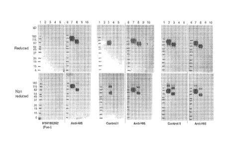
[0036] Figure 1 shows the results of western blot of EGFR and EGFRvIll using
anti-
EGFRvIll antibodies [i.e., H1H1863N2(Fuc-), and Controls land 11 in Figure la;
and
H1H1911, H1H1912, and H1 H1915 in Figure lb], or anti-His antibody, under
reduced
(upper panels) and non-reduced (lower panels) conditions. Lanes 1 and 6: 10 pl
of
BENCHMARKTm standard (INVITROGENTm); Lanes 2 and 7: 400 ng of hEGFR-mmh
(SEQ ID NO:154); Lane 3 and 8: 400 ng of hEGFRvIll-mmh (SEQ ID NO:152); and
Lanes 4, 5, 9 and 10: space. Control!: Human anti-EGFRvIll junctional peptide
antibody (IgG1) disclosed in US Patent No. 7,736,644; and Control II: Chimeric
anti-
EGFRvIII/EGFR antibody disclosed in US Patent No. 7,589,180.
[0037] Figure 2 shows the binding characteristics of H1H1863N2(Fuc-). The
EGFRvIll
junctional peptide or the peptide of residues 311-326 of EGFR ("EGFR311-326
peptide"), each of which was tagged via a linker with biotin at the C-
terminus, was
captured to streptavidin-coated OCTET tips on a FORTEB100 OCTET RED
instrument and reacted with H1H1863N2(Fuc-) or Control 1-111. Controls land
II: Same
as above; and Control III: Humanized anti-EGFRvIll antibody (hIgG1) disclosed
in US
Patent Application Publication No. 2010/0056762. (s): C-terminal biotin-
labeled
EGFRvIll junctional peptide (SEQ ID NO:149); and (M): C-terminal biotin-
labeled
EGFR311-326 peptide (SEQ ID NO:151).
[0038] Figure 3 shows the internalization of anti-EGFRvIll mAb by HEK293 cells
expressing EGFRvIll (HEK293/EGFRvIII). Cell-surface bound anti-EGFRvIll
antibodies
and control antibodies were detected by dye-conjugated secondary antibody
(Fab);
images were acquired at 40x and internalized vesicles were quantitated.
Controls! and
II: Same as above; and Control IV: Chimeric anti-EGFR antibody disclosed in US
8
Patent No. 7,060,808. (El): Internalization at 37 C; and (M): Internalization
at 4 C.
[0039] Figure 4 shows the binding and internalization of anti-EGFRvIll
antibody
H1H1863N2(Fuc-) by B16F10.9 tumors or B16F10.9 tumors expressing EGFRvIll
(B16F10.9/EGFRvIll) that were xenografted in severe combined immunodeficient
(SCID) mice. Cell-surface bound (Figure 4a) or cell-surface-bound plus
internalized
(Figure 4b) anti-EGFRvIll antibody or isotype control antibody, was detected
by
allophycocyanin conjugated anti-human Fc (hFc-APC) antibody using flow
cytometry.
Mean fluorescent intensities (MIF) at 10 minutes (El), 4 hours (0), and 24
hours (M),
post-antibody injection, are shown.
[0040] Figure 5 shows the results of pharmacokinetics analysis for anti-
EGFRvIll
antibody H1H863N2(Fuc+) (Fig. 5d) and control antibodies (as described above),
i.e.,
Control I (Fig. 5b), Control III (Fig. 5c), and Control IV (Fig. 5a), in wild-
type mice (I) or
mice expressing human EGFR (M).
DETAILED DESCRIPTION
[0041] Before the present invention is described, it is to be understood that
this invention
is not limited to particular methods and experimental conditions described, as
such
methods and conditions may vary. It is also to be understood that the
terminology used
herein is for the purpose of describing particular embodiments only, and is
not intended
to be limiting, since the scope of the present invention will be limited only
by the
appended claims.
[0042] Unless defined otherwise, all technical and scientific terms used
herein have the
same meaning as commonly understood by one of ordinary skill in the art to
which this
invention belongs. As used herein, the term "about," when used in reference to
a
particular recited numerical value, means that the value may vary from the
recited value
by no more than 1%. For example, as used herein, the expression "about 100"
includes
99 and 101 and all values in between (e.g., 99.1, 99.2, 99.3, 99.4, etc.).
[0043] Although any methods and materials similar or equivalent to those
described
herein can be used in the practice or testing of the present invention, the
preferred
methods and materials are now described.
Definitions
[0044] The term "EGFRvIll," as used herein, refers to the human EGFR class III
variant
having the amino acid sequence shown in SEQ ID NO:147, or a biologically
active
9
Date Recue/Date Received 2021-07-29
CA 02940685 2016-08-24
WO 2015/138460 PCT/US2015/019722
fragment thereof, which exhibits any characteristics specific for EGFRvIll, as
opposed to
those in common with normally expressed EGFR, unless specifically indicated
otherwise. EGFRvIll lacks amino acid residues 6 through 273 of mature EGFR
(i.e.,
SEQ ID NO:146 without the signal peptide, Le., residues 1-24) and contains a
new
glycine residue at position 6 between amino acid residues 5 and 274.
[0045] All references to proteins, polypeptides and protein fragments herein
are
intended to refer to the human version of the respective protein, polypeptide
or protein
fragment unless explicitly specified as being from a non-human species. Thus,
the
expression "EGFRvIll" means human EGFRvIll unless specified as being from a
non-
human species, e.g., "mouse EGFRvIll," "monkey EGFRvIll," etc.
[0046] As used herein, the expression "cell surface-expressed EGFRvIll" means
one or
more EGFRvIll protein(s), or the extracellular domain thereof, that is/are
expressed on
the surface of a cell in vitro or in vivo, such that at least a portion of a
EGFRvIll protein
is exposed to the extracellular side of the cell membrane and is accessible to
an
antigen-binding portion of an antibody. A "cell surface-expressed EGFRvIll"
can
comprise or consist of an EGFRvIll protein expressed on the surface of a cell
which
normally expresses EGFRvIll protein. Alternatively, "cell surface-expressed
EGFRvIll"
can comprise or consist of EGFRvIll protein expressed on the surface of a cell
that
normally does not express human EGFRvIll on its surface but has been
artificially
engineered to express EGFRvIll on its surface.
[0047] As used herein, the expression "anti-EGFRvIll antibody" includes both
monovalent antibodies with a single specificity, as well as bispecific
antibodies
comprising a first arm that binds EGFRvIll and a second arm that binds a
second
(target) antigen, wherein the anti-EGFRvIll arm comprises any of the HCVR/LCVR
or
CDR sequences as set forth in Table 1 herein. The expression "anti-EGFRvIll
antibody"
also includes antibody-drug conjugates (ADCs) comprising an anti-EGFRvIll
antibody or
antigen-binding portion thereof conjugated to a drug or toxin (i.e., cytotoxic
agent). The
expression "anti-EGFRvIll antibody" also includes antibody-radionuclide
conjugates
(ARCs) comprising an anti-EGFRvIll antibody or antigen-binding portion thereof
conjugated to a radionuclide.
[0048] The term "antibody", as used herein, means any antigen-binding molecule
or
molecular complex comprising at least one complementarity determining region
(CDR)
that specifically binds to or interacts with a particular antigen (e.g.,
EGFRv111). The term
"antibody" includes immunoglobulin molecules comprising four polypeptide
chains, two
heavy (H) chains and two light (L) chains inter-connected by disulfide bonds,
as well as
CA 02940685 2016-08-24
WO 2015/138460 PCT/US2015/019722
multimers thereof (e.g., IgM). Each heavy chain comprises a heavy chain
variable
region (abbreviated herein as HCVR or VH) and a heavy chain constant region.
The
heavy chain constant region comprises three domains, CH1, CH2 and CH3. Each
light
chain comprises a light chain variable region (abbreviated herein as LCVR or
VL) and a
light chain constant region. The light chain constant region comprises one
domain
(CL1). The VH and VL regions can be further subdivided into regions of
hypervariability,
termed complementarity determining regions (CDRs), interspersed with regions
that are
more conserved, termed framework regions (FR). Each VH and VI_ is composed of
three
CDRs and four FRs, arranged from amino-terminus to carboxy-terminus in the
following
order: FR1, CDR1, FR2, CDR2, FR3, CDR3, FR4. In different embodiments of the
invention, the FRs of the anti-EGFRvIll antibody (or antigen-binding portion
thereof) may
be identical to the human germline sequences, or may be naturally or
artificially
modified. An amino acid consensus sequence may be defined based on a side-by-
side
analysis of two or more CDRs.
[0049] The term "antibody", as used herein, also includes antigen-binding
fragments of
full antibody molecules. The terms "antigen-binding portion" of an antibody,
"antigen-
binding fragment" of an antibody, and the like, as used herein, include any
naturally
occurring, enzymatically obtainable, synthetic, or genetically engineered
polypeptide or
glycoprotein that specifically binds an antigen to form a complex. Antigen-
binding
fragments of an antibody may be derived, e.g., from full antibody molecules
using any
suitable standard techniques such as proteolytic digestion or recombinant
genetic
engineering techniques involving the manipulation and expression of DNA
encoding
antibody variable and optionally constant domains. Such DNA is known and/or is
readily available from, e.g., commercial sources, DNA libraries (including,
e.g., phage-
antibody libraries), or can be synthesized. The DNA may be sequenced and
manipulated chemically or by using molecular biology techniques, for example,
to
arrange one or more variable and/or constant domains into a suitable
configuration, or
to introduce codons, create cysteine residues, modify, add or delete amino
acids, etc.
[0050] Non-limiting examples of antigen-binding fragments include: (i) Fab
fragments; (ii)
F(ab')2 fragments; (iii) Fd fragments; (iv) Fv fragments; (v) single-chain Fv
(scFv)
molecules; (vi) dAb fragments; and (vii) minimal recognition units consisting
of the
amino acid residues that mimic the hypervariable region of an antibody (e.g.,
an isolated
complementarity determining region (CDR) such as a CDR3 peptide), or a
constrained
FR3-CDR3-FR4 peptide. Other engineered molecules, such as domain-
specific antibodies, single domain antibodies, domain-deleted antibodies,
chimeric
11
CA 02940685 2016-08-24
WO 2015/138460 PCT/US2015/019722
antibodies, CDR-grafted antibodies, diabodies, triabodies, tetrabodies,
minibodies,
nanobodies (e.g. monovalent nanobodies, bivalent nanobodies, etc.), small
modular
immunopharmaceuticals (SMIPs), and shark variable IgNAR domains, are also
encompassed within the expression "antigen-binding fragment," as used herein.
[0051] An antigen-binding fragment of an antibody will typically comprise at
least one
variable domain. The variable domain may be of any size or amino acid
composition
and will generally comprise at least one CDR which is adjacent to or in frame
with one
or more framework sequences. In antigen-binding fragments having a VH domain
associated with a VI_ domain, the VH and VI_ domains may be situated relative
to one
another in any suitable arrangement. For example, the variable region may be
dimeric
and contain VH-VH, VH-VL or VL-VL dimers. Alternatively, the antigen-binding
fragment of
an antibody may contain a monomeric VH or VI_ domain.
[0052] In certain embodiments, an antigen-binding fragment of an antibody may
contain
at least one variable domain covalently linked to at least one constant
domain. Non-
limiting, exemplary configurations of variable and constant domains that may
be found
within an antigen-binding fragment of an antibody of the present invention
include: (i)
VH-CH1; (ii) VH-CH2; (iii) VH-CH3; (iv) VH-CH1-CH2; (v) VH-CH1-CH2-CH3; (vi)
VH-CH2-CH3;
(vii) VH-CL; (viii) VL-CH1; (ix) VL-CH2; (x) VL-CH3; (xi) VL-CH1-0H2; (xii) VL-
CH1-0H2-CH3;
(xiii) VL-CH2-CH3; and (xiv) V[-C[. In any configuration of variable and
constant
domains, including any of the exemplary configurations listed above, the
variable and
constant domains may be either directly linked to one another or may be linked
by a full
or partial hinge or linker region. A hinge region may consist of at least 2
(e.g., 5, 10, 15,
20, 40, 60 or more) amino acids which result in a flexible or semi-flexible
linkage
between adjacent variable and/or constant domains in a single polypeptide
molecule.
Moreover, an antigen-binding fragment of an antibody of the present invention
may
comprise a homo-dimer or hetero-dimer (or other multimer) of any of the
variable and
constant domain configurations listed above in non-covalent association with
one
another and/or with one or more monomeric VH or VI_ domain (e.g., by disulfide
bond(s)).
[0053] As with full antibody molecules, antigen-binding fragments may be
monospecific
or multispecific (e.g., bispecific). A multispecific antigen-binding fragment
of an antibody
will typically comprise at least two different variable domains, wherein each
variable
domain is capable of specifically binding to a separate antigen or to a
different epitope
on the same antigen. Any multispecific antibody format, including the
exemplary
bispecific antibody formats disclosed herein, may be adapted for use in the
context of an
antigen-binding fragment of an antibody of the present invention using routine
12
CA 02940685 2016-08-24
WO 2015/138460 PCT/US2015/019722
techniques available in the art.
[0054] The antibodies of the present invention may function through complement-
dependent cytotoxicity (CDC) or antibody-dependent cell-mediated cytotoxicity
(ADCC).
"Complement-dependent cytotoxicity" (CDC) refers to lysis of antigen-
expressing cells
by an antibody of the invention in the presence of complement. "Antibody-
dependent
cell-mediated cytotoxicity" (ADCC) refers to a cell-mediated reaction in which
nonspecific cytotoxic cells that express Fc receptors (FcRs) (e.g., Natural
Killer (NK)
cells, neutrophils, and macrophages) recognize bound antibody on a target cell
and
thereby lead to lysis of the target cell. CDC and ADCC can be measured using
assays
that are well known and available in the art. (See, e.g., U.S. Patent Nos
5,500,362 and
5,821,337, and Clynes etal. (1998) Proc. Natl. Acad. Sci. (USA) 95:652-656).
The
constant region of an antibody is important in the ability of an antibody to
fix complement
and mediate cell-dependent cytotoxicity. Thus, the isotype of an antibody may
be
selected on the basis of whether it is desirable for the antibody to mediate
cytotoxicity.
[0055] In certain embodiments of the invention, the anti-EGFRvIll antibodies
of the
invention are human antibodies. The term "human antibody", as used herein, is
intended to include antibodies having variable and constant regions derived
from human
germline immunoglobulin sequences. The human antibodies of the invention may
include amino acid residues not encoded by human germline immunoglobulin
sequences (e.g., mutations introduced by random or site-specific mutagenesis
in vitro or
by somatic mutation in vivo), for example in the CDRs and in particular CDR3.
However, the term "human antibody", as used herein, is not intended to include
antibodies in which CDR sequences derived from the germline of another
mammalian
species, such as a mouse, have been grafted onto human framework sequences.
[0056] The antibodies of the invention may, in some embodiments, be
recombinant
human antibodies. The term "recombinant human antibody", as used herein, is
intended to include all human antibodies that are prepared, expressed, created
or
isolated by recombinant means, such as antibodies expressed using a
recombinant
expression vector transfected into a host cell (described further below),
antibodies
isolated from a recombinant, combinatorial human antibody library (described
further
below), antibodies isolated from an animal (e.g., a mouse) that is transgenic
for human
immunoglobulin genes (see e.g., Taylor et al. (1992) Nucl. Acids Res. 20:6287-
6295) or
antibodies prepared, expressed, created or isolated by any other means that
involves
splicing of human immunoglobulin gene sequences to other DNA sequences. Such
recombinant human antibodies have variable and constant regions derived from
human
13
CA 02940685 2016-08-24
WO 2015/138460 PCT/US2015/019722
germline immunoglobulin sequences. In certain embodiments, however, such
recombinant human antibodies are subjected to in vitro mutagenesis (or, when
an
animal transgenic for human Ig sequences is used, in vivo somatic mutagenesis)
and
thus the amino acid sequences of the VH and VI_ regions of the recombinant
antibodies
are sequences that, while derived from and related to human germline VH and VL
sequences, may not naturally exist within the human antibody germline
repertoire in
vivo.
[0057] Human antibodies can exist in two forms that are associated with hinge
heterogeneity. In one form, an immunoglobulin molecule comprises a stable four
chain
construct of approximately 1 50-1 60 kDa in which the dimers are held together
by an
interchain heavy chain disulfide bond. In a second form, the dimers are not
linked via
inter-chain disulfide bonds and a molecule of about 75-80 kDa is formed
composed of a
covalently coupled light and heavy chain (half-antibody). These forms have
been
extremely difficult to separate, even after affinity purification.
[0058] The frequency of appearance of the second form in various intact IgG
isotypes is
due to, but not limited to, structural differences associated with the hinge
region isotype
of the antibody. A single amino acid substitution in the hinge region of the
human IgG4
hinge can significantly reduce the appearance of the second form (Angal et al.
(1993)
Molecular Immunology 30:105) to levels typically observed using a human IgG1
hinge.
The instant invention encompasses antibodies having one or more mutations in
the
hinge, CH2 or CH3 region which may be desirable, for example, in production,
to improve
the yield of the desired antibody form.
[0059] The antibodies of the invention may be isolated antibodies. An
"isolated
antibody," as used herein, means an antibody that has been identified and
separated
and/or recovered from at least one component of its natural environment. For
example,
an antibody that has been separated or removed from at least one component of
an
organism, or from a tissue or cell in which the antibody naturally exists or
is naturally
produced, is an "isolated antibody" for purposes of the present invention. An
isolated
antibody also includes an antibody in situ within a recombinant cell. Isolated
antibodies
are antibodies that have been subjected to at least one purification or
isolation step.
According to certain embodiments, an isolated antibody may be substantially
free of
other cellular material and/or chemicals.
[0060] The anti-EGFRvIll antibodies disclosed herein may comprise one or more
amino
acid substitutions, insertions and/or deletions in the framework and/or CDR
regions of
the heavy and light chain variable domains as compared to the corresponding
germline
14
CA 02940685 2016-08-24
WO 2015/138460 PCT/US2015/019722
sequences from which the antibodies were derived. Such mutations can be
readily
ascertained by comparing the amino acid sequences disclosed herein to germline
sequences available from, for example, public antibody sequence databases. The
present invention includes antibodies, and antigen-binding fragments thereof,
which are
derived from any of the amino acid sequences disclosed herein, wherein one or
more
amino acids within one or more framework and/or CDR regions are mutated to the
corresponding residue(s) of the germline sequence from which the antibody was
derived, or to the corresponding residue(s) of another human germline
sequence, or to a
conservative amino acid substitution of the corresponding germline residue(s)
(such
sequence changes are referred to herein collectively as "germline mutations").
A
person of ordinary skill in the art, starting with the heavy and light chain
variable region
sequences disclosed herein, can easily produce numerous antibodies and antigen-
binding fragments which comprise one or more individual germline mutations or
combinations thereof. In certain embodiments, all of the framework and/or CDR
residues within the VH and/or VL domains are mutated back to the residues
found in the
original germline sequence from which the antibody was derived. In other
embodiments, only certain residues are mutated back to the original germline
sequence,
e.g., only the mutated residues found within the first 8 amino acids of FR1 or
within the
last 8 amino acids of FR4, or only the mutated residues found within CDR1,
CDR2 or
CDR3. In other embodiments, one or more of the framework and/or CDR residue(s)
are
mutated to the corresponding residue(s) of a different germline sequence
(i.e., a
germline sequence that is different from the germline sequence from which the
antibody
was originally derived). Furthermore, the antibodies of the present invention
may
contain any combination of two or more germline mutations within the framework
and/or
CDR regions, e.g., wherein certain individual residues are mutated to the
corresponding
residue of a particular germline sequence while certain other residues that
differ from
the original germline sequence are maintained or are mutated to the
corresponding
residue of a different germline sequence. Once obtained, antibodies and
antigen-
binding fragments that contain one or more germline mutations can be easily
tested for
one or more desired property such as, improved binding specificity, increased
binding
affinity, improved or enhanced antagonistic or agonistic biological properties
(as the
case may be), reduced immunogenicity, etc. Antibodies and antigen-binding
fragments
obtained in this general manner are encompassed within the present invention.
[0061] The present invention also includes anti-EGFRvIll antibodies comprising
variants
of any of the HCVR, LCVR, and/or CDR amino acid sequences disclosed herein
having
CA 02940685 2016-08-24
WO 2015/138460 PCT/US2015/019722
one or more conservative substitutions. For example, the present invention
includes
anti-EGFRvIll antibodies having HCVR, LCVR, and/or CDR amino acid sequences
with,
e.g., 10 or fewer, 8 or fewer, 6 or fewer, 4 or fewer, etc. conservative amino
acid
substitutions relative to any of the HCVR, LCVR, and/or CDR amino acid
sequences set
forth in Table 1 herein.
[0062] The term "epitope" refers to an antigenic determinant that interacts
with a specific
antigen binding site in the variable region of an antibody molecule known as a
paratope.
A single antigen may have more than one epitope. Thus, different antibodies
may bind
to different areas on an antigen and may have different biological effects.
Epitopes may
be either conformational or linear. A conformational epitope is produced by
spatially
juxtaposed amino acids from different segments of the linear polypeptide
chain. A linear
epitope is one produced by adjacent amino acid residues in a polypeptide
chain. In
certain circumstance, an epitope may include moieties of saccharides,
phosphoryl
groups, or sulfonyl groups on the antigen.
[0063] The term "substantial identity" or "substantially identical," when
referring to a
nucleic acid or fragment thereof, indicates that, when optimally aligned with
appropriate
nucleotide insertions or deletions with another nucleic acid (or its
complementary
strand), there is nucleotide sequence identity in at least about 95%, and more
preferably
at least about 96%, 97%, 98% or 99% of the nucleotide bases, as measured by
any
well-known algorithm of sequence identity, such as FASTA, BLAST or Gap, as
discussed below. A nucleic acid molecule having substantial identity to a
reference
nucleic acid molecule may, in certain instances, encode a polypeptide having
the same
or substantially similar amino acid sequence as the polypeptide encoded by the
reference nucleic acid molecule.
[0064] As applied to polypeptides, the term "substantial similarity" or
"substantially
similar" means that two peptide sequences, when optimally aligned, such as by
the
programs GAP or BESTFIT using default gap weights, share at least 95% sequence
identity, even more preferably at least 98% or 99% sequence identity.
Preferably,
residue positions which are not identical differ by conservative amino acid
substitutions.
A "conservative amino acid substitution" is one in which an amino acid residue
is
substituted by another amino acid residue having a side chain (R group) with
similar
chemical properties (e.g., charge or hydrophobicity). In general, a
conservative amino
acid substitution will not substantially change the functional properties of a
protein. In
cases where two or more amino acid sequences differ from each other by
conservative
substitutions, the percent sequence identity or degree of similarity may be
adjusted
16
upwards to correct for the conservative nature of the substitution. Means for
making this
adjustment are well-known to those of skill in the art. See, e.g., Pearson
(1994)
Methods Mol. Biol. 24: 307-331. Examples of groups of amino acids that have
side
chains with similar chemical properties include (1) aliphatic side chains:
glycine, alanine,
valine, leucine and isoleucine; (2) aliphatic-hydroxyl side chains: serine and
threonine;
(3) amide-containing side chains: asparagine and glutamine; (4) aromatic side
chains:
phenylalanine, tyrosine, and tryptophan; (5) basic side chains: lysine,
arginine, and
histidine; (6) acidic side chains: aspartate and glutamate, and (7) sulfur-
containing side
chains are cysteine and methionine. Preferred conservative amino acids
substitution
groups are: valine-leucine-isoleucine, phenylalanine-tyrosine, lysine-
arginine, alanine-
valine, glutamate-aspartate, and asparagine-glutamine. Alternatively, a
conservative
replacement is any change having a positive value in the PAM250 log-likelihood
matrix
disclosed in Gonnet et al. (1992) Science 256: 1443-1445. A "moderately
conservative"
replacement is any change having a nonnegative value in the PAM250 log-
likelihood
matrix.
[0065] Sequence similarity for polypeptides, which is also referred to as
sequence
identity, is typically measured using sequence analysis software. Protein
analysis
software matches similar sequences using measures of similarity assigned to
various
substitutions, deletions and other modifications, including conservative amino
acid
substitutions. For instance, GCG software contains programs such as Gap and
Bestf it
which can be used with default parameters to determine sequence homology or
sequence identity between closely related polypeptides, such as homologous
polypeptides from different species of organisms or between a wild type
protein and a
mutein thereof. See, e.g., GCG Version 6.1. Polypeptide sequences also can be
compared using FASTA using default or recommended parameters, a program in GCG
Version 6.1. FASTA (e.g., FASTA2 and FASTA3) provides alignments and percent
sequence identity of the regions of the best overlap between the query and
search
sequences (Pearson (2000) supra). Another preferred algorithm when comparing a
sequence of the invention to a database containing a large number of sequences
from
different organisms is the computer program BLAST, especially BLASTP or
TBLASTN,
using default parameters. See, e.g., Altschul et al. (1990) J. Mol. Biol.
215:403-410 and
Altschul et al. (1997) Nucleic Acids Res. 25:3389-402.
17
Date Recue/Date Received 2021-07-29
CA 02940685 2016-08-24
WO 2015/138460 PCT/US2015/019722
pH-Dependent Binding
[0066] The present invention includes anti-EGFRvIll antibodies with pH-
dependent
binding characteristics. For example, an anti-EGFRvIll antibody of the present
invention
may exhibit reduced binding to EGFRvIll at acidic pH as compared to neutral
pH.
Alternatively, anti-EGFRvIll antibodies of the invention may exhibit enhanced
binding to
EGFRvIll at acidic pH as compared to neutral pH. The expression "acidic pH"
includes
pH values less than about 6.2, e.g., about 6.0, 5.95, 5,9, 5.85, 5.8, 5.75,
5.7, 5.65, 5.6,
5.55, 5.5, 5.45, 5.4, 5.35, 5.3, 5.25, 5.2, 5.15, 5.1, 5.05, 5.0, or less. As
used herein, the
expression "neutral pH" means a pH of about 7.0 to about 7.4. The expression
"neutral
pH" includes pH values of about 7.0, 7.05, 7.1, 7.15, 7.2, 7.25, 7.3, 7.35,
and 7.4.
[0067] In certain instances, "reduced binding to EGFRvIll at acidic pH as
compared to
neutral pH" is expressed in terms of a ratio of the KD value of the antibody
binding to
EGFRvIll at acidic pH to the KD value of the antibody binding to EGFRvIll at
neutral pH
(or vice versa). For example, an antibody or antigen-binding fragment thereof
may be
regarded as exhibiting "reduced binding to EGFRvIll at acidic pH as compared
to neutral
pH" for purposes of the present invention if the antibody or antigen-binding
fragment
thereof exhibits an acidic/neutral KD ratio of about 3.0 or greater. In
certain exemplary
embodiments, the acidic/neutral KD ratio for an antibody or antigen-binding
fragment of
the present invention can be about 3.0, 3.5, 4.0, 4.5, 5.0, 5.5, 6.0, 6.5,
7.0, 7.5, 8.0, 8.5,
9.0, 9.5, 10.0, 10.5, 11.0, 11.5,12.0, 12.5, 13.0, 13.5, 14.0, 14.5, 15.0,
20Ø 25.0, 30.0,
40.0, 50.0, 60.0, 70.0, 100.0 or greater.
[0068] Antibodies with pH-dependent binding characteristics may be obtained,
e.g., by
screening a population of antibodies for reduced (or enhanced) binding to a
particular
antigen at acidic pH as compared to neutral pH. Additionally, modifications of
the
antigen-binding domain at the amino acid level may yield antibodies with pH-
dependent
characteristics. For example, by substituting one or more amino acids of an
antigen-
binding domain (e.g., within a CDR) with a histidine residue, an antibody with
reduced
antigen-binding at acidic pH relative to neutral pH may be obtained.
Anti-EGFRvIll Antibodies Comprising Fc Variants
[0069] According to certain embodiments of the present invention, anti-
EGFRvIll
antibodies are provided comprising an Fc domain comprising one or more
mutations
which enhance or diminish antibody binding to the FcRn receptor, e.g., at
acidic pH as
compared to neutral pH. For example, the present invention includes anti-
EGFRvIll
antibodies comprising a mutation in the CH2 or a CH3 region of the Fc domain,
wherein
the mutation(s) increases the affinity of the Fc domain to FcRn in an acidic
environment
18
CA 02940685 2016-08-24
WO 2015/138460 PCT/US2015/019722
(e.g., in an endosome where pH ranges from about 5.5 to about 6.0). Such
mutations
may result in an increase in serum half-life of the antibody when administered
to an
animal. Non-limiting examples of such Fc modifications include, e.g., a
modification at
position 250 (e.g., E or 0); 250 and 428 (e.g., L or F); 252 (e.g., UY/F/VV or
T), 254
(e.g., S or T), and 256 (e.g., S/R/Q/E/D or T); or a modification at position
428 and/or
433 (e.g., H/L/R/S/P/Q or K) and/or 434 (e.g., A, W, H, F or Y [N434A, N434W,
N434H,
N434F or N434Y]); or a modification at position 250 and/or 428; or a
modification at
position 307 or 308 (e.g., 308F, V308F), and 434. In one embodiment, the
modification
comprises a 428L (e.g., M428L) and 434S (e.g., N434S) modification; a 428L,
2591
(e.g., V259I), and 308F (e.g., V308F) modification; a 433K (e.g., H433K) and a
434
(e.g., 434Y) modification; a 252, 254, and 256 (e.g., 252Y, 2541, and 256E)
modification; a 250Q and 428L modification (e.g., 1250Q and M428L); and a 307
and/or
308 modification (e.g., 308F or 308P). In yet another embodiment, the
modification
comprises a 265A (e.g., D265A) and/or a 297A (e.g., N297A) modification.
[0070] For example, the present invention includes anti-EGFRvIll antibodies
comprising
an Fc domain comprising one or more pairs or groups of mutations selected from
the
group consisting of: 250Q and 248L (e.g., T250Q and M248L); 252Y, 2541 and
256E
(e.g., M252Y, 5254T and T256E); 428L and 434S (e.g., M428L and N4345); 2571
and
3111 (e.g., P257I and Q3111); 2571 and 434H (e.g., P257I and N434H); 376V and
434H
(e.g., D376V and N434H); 307A, 380A and 434A (e.g., 1307A, E380A and N434A);
and
433K and 434F (e.g., H433K and N434F). All possible combinations of the
foregoing Fc
domain mutations, and other mutations within the antibody variable domains
disclosed
herein, are contemplated within the scope of the present invention.
[0071] The present invention also includes anti-EGFRvIll antibodies comprising
a
chimeric heavy chain constant (CH) region, wherein the chimeric CH region
comprises
segments derived from the CH regions of more than one immunoglobulin isotype.
For
example, the antibodies of the invention may comprise a chimeric CH region
comprising
part or all of a CH2 domain derived from a human IgG1, human IgG2 or human
IgG4
molecule, combined with part or all of a CH3 domain derived from a human IgG1,
human
IgG2 or human IgG4 molecule. According to certain embodiments, the antibodies
of the
invention comprise a chimeric CH region having a chimeric hinge region. For
example, a
chimeric hinge may comprise an "upper hinge" amino acid sequence (amino acid
residues from positions 216 to 227 according to EU numbering) derived from a
human
IgG1, a human IgG2 or a human IgG4 hinge region, combined with a "lower hinge"
sequence (amino acid residues from positions 228 to 236 according to EU
numbering)
19
derived from a human IgG1, a human IgG2 or a human IgG4 hinge region.
According to
certain embodiments, the chimeric hinge region comprises amino acid residues
derived
from a human IgG1 or a human IgG4 upper hinge and amino acid residues derived
from
a human IgG2 lower hinge. An antibody comprising a chimeric CH region as
described
herein may, in certain embodiments, exhibit modified Fc effector functions
without
adversely affecting the therapeutic or pharmacokinetic properties of the
antibody. (See,
e.g., U.S. Provisional Appl. No. 61/759,578, filed February 1, 2013).
Antibody-Drug Conjugates (ADCs)
[0072] The present invention provides antibody-drug conjugates (ADCs)
comprising an
anti-EGFRvIll antibody or antigen-binding fragment thereof conjugated to a
therapeutic
moiety such as a cytotoxic agent, a chemotherapeutic drug, or a radioisotope.
[0073] Cytotoxic agents include any agent that is detrimental to the growth,
viability or
propagation of cells. Examples of suitable cytotoxic agents and
chemotherapeutic
agents that can be conjugated to anti-EGFRvIll antibodies in accordance with
this aspect
of the invention include, e.g., 1-(2ch10r0ethy1)-1,2-dimethanesulfonyl
hydrazide, 1,8-
dihydroxy-bicyclo[7.3.1]trideca-4,9-diene-2,6-diyne-13-one, 1-
dehydrotestosterone, 5-
fluorouracil, 6-mercaptopurine, 6-thioguanine, 9-amino camptothecin,
actinomycin D,
amanitins, aminopterin, anguidine, anthracycline, anthramycin (AMC),
auristatins,
bleomycin, busulfan, butyric acid, calicheamicins, camptothecin,
carminomycins,
carmustine, cemadotins, cisplatin, colchicin, combretastatins,
cyclophosphamide,
cytarabine, cytochalasin B, dactinomycin, daunorubicin, decarbazine,
diacetoxypentyldoxorubicin, dibromomannitol, dihydroxy anthracin dione,
disorazoles,
dolastatin, doxorubicin, duocarmycin, echinomycins, eleutherobins, emetine,
epothilones, esperamicin, estramustines, ethidium bromide, etoposide,
fluorouracils,
geldanamycins, gramicidin D, glucocorticoids, irinotecans, leptomycins,
leurosines,
lidocaine, lomustine (CCNU), maytansinoids, mechlorethamine, melphalan,
mercatopurines, methopterins, methotrexate, mithramycin, mitomycin,
mitoxantrone, N8-
acetyl spermidine, podophyllotoxins, procaine, propranolol, pteridines,
puromycin,
pyrrolobenzodiazepines (PD Bs), rhizoxins, streptozotocin, tallysomycins,
taxol,
tenoposide, tetracaine, thioepa chlorambucil, tomaymycins, topotecans,
tubulysin,
vinblastine, vincristine, vindesine, vinorelbines, and derivatives of any of
the foregoing.
According to certain embodiments, the cytotoxic agent that is conjugated to an
anti-
EGFRvIll antibody is a maytansinoid such as DM1 or DM4, a tomaymycin
derivative, or
a dolastatin derivative. Other cytotoxic agents known in the art are
contemplated within
Date Recue/Date Received 2021-07-29
the scope of the present invention, including, e.g., protein toxins such
ricin, C. difficile
toxin, pseudomonas exotoxin, ricin, diphtheria toxin, botulinum toxin,
bryodin, saporin,
pokeweed toxins (Le., phytolaccatoxin and phytolaccigenin), and others such as
those
set forth in Sapra et al., PharmacoL & Therapeutics, 2013, 138:452-469.
[0074] The present invention also includes antibody-radionuclide conjugates
(ARCs)
comprising anti-EGFRvIll antibodies conjugated to one or more radionuclides.
Exemplary radionuclides that can be used in the context of this aspect of the
invention
include, but are not limited to, e.g., 225Ac, 212Bi, 213Bi, 1311, 186Re,
227Th, 222Rn, 223Ra,
224Ra, and 90Y.
[0075] In certain embodiments of the present invention, ADCs are provided
comprising
an anti-EGFRvIll antibody conjugated to a cytotoxic agent (e.g., any of the
cytotoxic
agents disclosed above) via a linker molecule. Any linker molecule or linker
technology
known in the art can be used to create or construct an ADC of the present
invention. In
certain embodiments, the linker is a cleavable linker. According to other
embodiments,
the linker is a non-cleavable linker. Exemplary linkers that can be used in
the context of
the present invention include, linkers that comprise or consist of e.g., MC (6-
maleimidocaproyl), MP (maleimidopropanoyl), val-cit (valine-citrulline), val-
ala (valine-
alanine), dipeptide site in protease-cleavable linker, ala-phe (alanine-
phenylalanine),
dipeptide site in protease-cleavable linker, PAB (p-aminobenzyloxycarbonyl),
SPP (N-
Succinimidyl 4-(2-pyridylthio) pentanoate), SMCC (N-Succinimidyl 4-(N-
maleimidomethyl)cyclohexane-1 carboxylate), SIAB (N-Succinimidyl (4-iodo-
acetyl)aminobenzoate), and variants and combinations thereof. Additional
examples of
linkers that can be used in the context of the present invention are
disclosed, e.g., in US
7,754,681 and in Ducry, Bioconjugate Chem., 2010, 2/:5-13, and the references
cited
therein.
[0076] The present invention comprises ADCs in which a linker connects an anti-
EGFRvIll antibody or antigen-binding molecule to a drug or cytotoxin through
an
attachment at a particular amino acid within the antibody or antigen-binding
molecule.
Exemplary amino acid attachments that can be used in the context of this
aspect of the
invention include, e.g., lysine (see, e.g., US 5,208,020; US 2010/0129314;
Hollander et
al., Bioconjugate Chem., 2008,19:358-361; WO 2005/089808; US 5,714,586; US
2013/0101546; and US 2012/0585592), cysteine (see, e.g., US 2007/0258987; WO
2013/055993; WO 2013/055990; WO 2013/053873; WO 2013/053872; WO
2011/130598; US 2013/0101546; and US 7,750,116), selenocysteine (see, e.g., WO
2008/122039; and Hofer etal., Proc. Natl. Acad. Sci., USA, 2008, /05:12451-
12456),
21
Date Recue/Date Received 2021-07-29
formyl glycine (see, e.g., Carrico etal., Nat. Chem. Biol., 2007, 3:321-322;
Agarwal et
al., Proc. Natl. Acad. Sc., USA, 2013, /10:46-51, and Rabuka et al., Nat.
Protocols,
2012, 10:1052-1067), non-natural amino acids (see, e.g., WO 2013/068874, and
WO
2012/166559), and acidic amino acids (see, e.g., WO 2012/05982). Linkers can
also be
conjugated to an antigen-binding protein via attachment to carbohydrates (see,
e.g., US
2008/0305497, and Ryan etal., Food & Agriculture ImmunoL, 2001, /3:127-130)
and
disulfide linkers (see, e.g., WO 2013/085925, WO 2010/010324, WO 2011/018611,
and
Shaunak etal., Nat. Chem. Biol., 2006, 2:312-313).
[0077] Any method known in the art for conjugating a chemical moiety to a
peptide,
polypeptide or other macromolecule can be used in the context of the present
invention
to make an anti-EGFRvIll ADC as described herein. An exemplary method for
antibody-
drug conjugation via a linker is set forth in Example 12 herein. Variations on
this
exemplary method will be appreciated by persons of ordinary skill in the art
and are
contemplated within the scope of the present invention.
[0078] According to certain embodiments, the present invention provides ADCs,
wherein
an anti-EGFRvIll antibody as described herein (e.g., the antibody designated
H1H1863N2) is conjugated to a linker-drug composition as set forth in
W02014/145090
(e.g., compound "7," also referred to herein as "M0026") (see also Example 12,
herein).
Epitope Mapping and Related Technologies
[0079] The epitope to which the antibodies of the present invention bind may
consist of a
single contiguous sequence of 3 or more (e.g., 3, 4, 5, 6, 7, 8, 9, 10, 11,
12, 13, 14, 15,
16, 17, 18, 19, 20 or more) amino acids of an EGFRvIll protein. Alternatively,
the
epitope may consist of a plurality of non-contiguous amino acids (or amino
acid
sequences) of EGFRvIll. In some embodiments, the epitope is located on or near
the
ligand-binding domain of EGFRvIll. In other embodiments, the epitope is
located
outside of the ligand-binding domain of EGFRvIll, e.g., at a location on the
surface of
EGFRvIll at which an antibody, when bound to such an epitope, does not
interfere with
ligand binding to EGFRvIll.
[0080] The present invention, according to certain embodiments, includes anti-
EGFRvIll
antibodies that specifically bind EGFRvIll (and do not bind EGFR), wherein the
antibodies recognize the EGFRvIll junctional peptide (e.g., SEQ ID NO:148).
Such
antibodies may be referred to herein as "junctional peptide binders,"
"EGFRvIll peptide-
binding antibodies," and the like. The present invention, according to other
embodiments, includes anti-EGFRvIll antibodies that specifically bind EGFRvIll
(and do
22
Date Recue/Date Received 2021-07-29
CA 02940685 2016-08-24
WO 2015/138460 PCT/US2015/019722
not bind EGFR), wherein the antibodies do not recognize the EGFRvIll
junctional
peptide (e.g. do not recognize the junctional peptide of SEQ ID NO:148, and/or
do not
recognize the peptide of SEQ ID NO:165). Such antibodies may be referred to
herein
as "conformational binders," "EGFRvIll conformational epitope binders," and
the like.
[0081] Various techniques known to persons of ordinary skill in the art can be
used to
determine whether an antibody "interacts with one or more amino acids" within
a
polypeptide or protein. Exemplary techniques include, e.g., routine cross-
blocking
assay such as that described Antibodies, Harlow and Lane (Cold Spring Harbor
Press,
Cold Spring Harb., NY), alanine scanning mutational analysis, peptide blots
analysis
(Reineke, 2004, Methods Mol Biol 248:443-463), and peptide cleavage analysis.
In
addition, methods such as epitope excision, epitope extraction and chemical
modification of antigens can be employed (Tomer, 2000, Protein Science 9:487-
496).
Another method that can be used to identify the amino acids within a
polypeptide with
which an antibody interacts is hydrogen/deuterium exchange detected by mass
spectrometry. In general terms, the hydrogen/deuterium exchange method
involves
deuterium-labeling the protein of interest, followed by binding the antibody
to the
deuterium-labeled protein. Next, the protein/antibody complex is transferred
to water to
allow hydrogen-deuterium exchange to occur at all residues except for the
residues
protected by the antibody (which remain deuterium-labeled). After dissociation
of the
antibody, the target protein is subjected to protease cleavage and mass
spectrometry
analysis, thereby revealing the deuterium-labeled residues which correspond to
the
specific amino acids with which the antibody interacts. See, e.g., Ehring
(1999)
Analytical Biochemistry 267(2):252-259; Engen and Smith (2001) Anal. Chem.
73:256A-
265A.
[0082] The present invention further includes anti-EGFRvIll antibodies that
bind to the
same epitope as any of the specific exemplary antibodies described herein
(e.g.
antibodies comprising any of the amino acid sequences as set forth in Table 1
herein).
Likewise, the present invention also includes anti-EGFRvIll antibodies that
compete for
binding to EGFRvIll with any of the specific exemplary antibodies described
herein (e.g.
antibodies comprising any of the amino acid sequences as set forth in Table 1
herein).
[0083] One can easily determine whether an antibody binds to the same epitope
as, or
competes for binding with, a reference anti-EGFRvIll antibody by using routine
methods
known in the art and exemplified herein. For example, to determine if a test
antibody
binds to the same epitope as a reference anti-EGFRvIll antibody of the
invention, the
reference antibody is allowed to bind to a EGFRvIll protein. Next, the ability
of a test
23
CA 02940685 2016-08-24
WO 2015/138460 PCT/US2015/019722
antibody to bind to the EGFRvIll molecule is assessed. If the test antibody is
able to
bind to EGFRvIll following saturation binding with the reference anti-EGFRvIll
antibody,
it can be concluded that the test antibody binds to a different epitope than
the reference
anti-EGFRvIll antibody. On the other hand, if the test antibody is not able to
bind to the
EGFRvIll molecule following saturation binding with the reference anti-
EGFRvIll
antibody, then the test antibody may bind to the same epitope as the epitope
bound by
the reference anti-EGFRvIll antibody of the invention. Additional routine
experimentation (e.g., peptide mutation and binding analyses) can then be
carried out to
confirm whether the observed lack of binding of the test antibody is in fact
due to binding
to the same epitope as the reference antibody or if steric blocking (or
another
phenomenon) is responsible for the lack of observed binding. Experiments of
this sort
can be performed using ELISA, RIA, Biacore, flow cytometry or any other
quantitative or
qualitative antibody-binding assay available in the art. In accordance with
certain
embodiments of the present invention, two antibodies bind to the same (or
overlapping)
epitope if, e.g., a 1-, 5-, 10-, 20- or 100-fold excess of one antibody
inhibits binding of
the other by at least 50% but preferably 75%, 90% or even 99% as measured in a
competitive binding assay (see, e.g., Junghans et al., Cancer Res.
1990:50:1495-1502).
Alternatively, two antibodies are deemed to bind to the same epitope if
essentially all
amino acid mutations in the antigen that reduce or eliminate binding of one
antibody
reduce or eliminate binding of the other. Two antibodies are deemed to have
"overlapping epitopes" if only a subset of the amino acid mutations that
reduce or
eliminate binding of one antibody reduce or eliminate binding of the other.
[0084] To determine if an antibody competes for binding (or cross-competes for
binding)
with a reference anti-EGFRvIll antibody, the above-described binding
methodology is
performed in two orientations: In a first orientation, the reference antibody
is allowed to
bind to an EGFRvIll protein under saturating conditions followed by assessment
of
binding of the test antibody to the EGFRvIll molecule. In a second
orientation, the test
antibody is allowed to bind to an EGFRvIll molecule under saturating
conditions
followed by assessment of binding of the reference antibody to the EGFRvIll
molecule.
If, in both orientations, only the first (saturating) antibody is capable of
binding to the
EGFRvIll molecule, then it is concluded that the test antibody and the
reference
antibody compete for binding to EGFRyll I. As will be appreciated by a person
of
ordinary skill in the art, an antibody that competes for binding with a
reference antibody
may not necessarily bind to the same epitope as the reference antibody, but
may
sterically block binding of the reference antibody by binding an overlapping
or adjacent
24
CA 02940685 2016-08-24
WO 2015/138460 PCT/US2015/019722
epitope.
Preparation of Human Antibodies
[0085] The anti-EGFRvIll antibodies of the present invention can be fully
human
antibodies. Methods for generating monoclonal antibodies, including fully
human
monoclonal antibodies are known in the art. Any such known methods can be used
in
the context of the present invention to make human antibodies that
specifically bind to
human EGFRvIll.
[0086] Using VELOCIMMUNETm technology, for example, or any other similar known
method for generating fully human monoclonal antibodies, high affinity
chimeric
antibodies to EGFRvIll are initially isolated having a human variable region
and a mouse
constant region. As in the experimental section below, the antibodies are
characterized
and selected for desirable characteristics, including affinity, ligand
blocking activity,
selectivity, epitope, etc. If necessary, mouse constant regions are replaced
with a
desired human constant region, for example wild-type or modified IgG1 or IgG4,
to
generate a fully human anti-EGFRvIll antibody. While the constant region
selected may
vary according to specific use, high affinity antigen-binding and target
specificity
characteristics reside in the variable region. In certain instances, fully
human anti-
EGFRvIll antibodies are isolated directly from antigen-positive B cells.
Bioequivalents
[0087] The anti-EGFRvIll antibodies and antibody fragments of the present
invention
encompass proteins having amino acid sequences that vary from those of the
described
antibodies but that retain the ability to bind human EGFRvIll. Such variant
antibodies
and antibody fragments comprise one or more additions, deletions, or
substitutions of
amino acids when compared to parent sequence, but exhibit biological activity
that is
essentially equivalent to that of the described antibodies. Likewise, the anti-
EGFRvIll
antibody-encoding DNA sequences of the present invention encompass sequences
that
comprise one or more additions, deletions, or substitutions of nucleotides
when
compared to the disclosed sequence, but that encode an anti-EGFRvIll antibody
or
antibody fragment that is essentially bioequivalent to an anti-EGFRvIll
antibody or
antibody fragment of the invention. Examples of such variant amino acid and
DNA
sequences are discussed above.
[0088] Two antigen-binding proteins, or antibodies, are considered
bioequivalent if, for
example, they are pharmaceutical equivalents or pharmaceutical alternatives
whose
rate and extent of absorption do not show a significant difference when
administered at
CA 02940685 2016-08-24
WO 2015/138460 PCT/US2015/019722
the same molar dose under similar experimental conditions, either single does
or
multiple dose. Some antibodies will be considered equivalents or
pharmaceutical
alternatives if they are equivalent in the extent of their absorption but not
in their rate of
absorption and yet may be considered bioequivalent because such differences in
the
rate of absorption are intentional and are reflected in the labeling, are not
essential to
the attainment of effective body drug concentrations on, e.g., chronic use,
and are
considered medically insignificant for the particular drug product studied.
[0089] In one embodiment, two antigen-binding proteins are bioequivalent if
there are no
clinically meaningful differences in their safety, purity, and potency.
[0090] In one embodiment, two antigen-binding proteins are bioequivalent if a
patient
can be switched one or more times between the reference product and the
biological
product without an expected increase in the risk of adverse effects, including
a clinically
significant change in immunogenicity, or diminished effectiveness, as compared
to
continued therapy without such switching.
[0091] In one embodiment, two antigen-binding proteins are bioequivalent if
they both
act by a common mechanism or mechanisms of action for the condition or
conditions of
use, to the extent that such mechanisms are known.
[0092] Bioequivalence may be demonstrated by in vivo and in vitro methods.
Bioequivalence measures include, e.g., (a) an in vivo test in humans or other
mammals,
in which the concentration of the antibody or its metabolites is measured in
blood,
plasma, serum, or other biological fluid as a function of time; (b) an in
vitro test that has
been correlated with and is reasonably predictive of human in vivo
bioavailability data;
(c) an in vivo test in humans or other mammals in which the appropriate acute
pharmacological effect of the antibody (or its target) is measured as a
function of time;
and (d) in a well-controlled clinical trial that establishes safety, efficacy,
or bioavailability
or bioequivalence of an antibody.
[0093] Bioequivalent variants of anti-EGFRvIll antibodies of the invention may
be
constructed by, for example, making various substitutions of residues or
sequences or
deleting terminal or internal residues or sequences not needed for biological
activity.
For example, cysteine residues not essential for biological activity can be
deleted or
replaced with other amino acids to prevent formation of unnecessary or
incorrect
intramolecular disulfide bridges upon renaturation. In other contexts,
bioequivalent
antibodies may include anti-EGFRvIll antibody variants comprising amino acid
changes
which modify the glycosylation characteristics of the antibodies, e.g.,
mutations which
eliminate or remove glycosylation.
26
CA 02940685 2016-08-24
WO 2015/138460 PCT/US2015/019722
Species Selectivity and Species Cross-Reactivity
[0094] The present invention, according to certain embodiments, provides anti-
EGFRvIll
antibodies that bind to human EGFRvIll but not to EGFRvIll from other species.
The
present invention also includes anti-EGFRvIll antibodies that bind to human
EGFRvIll
and to EGFRvIll from one or more non-human species. For example, the anti-
EGFRvIll
antibodies of the invention may bind to human EGFRvIll and may bind or not
bind, as
the case may be, to one or more of mouse, rat, guinea pig, hamster, gerbil,
pig, cat,
dog, rabbit, goat, sheep, cow, horse, camel, cynomologous, marmoset, rhesus or
chimpanzee EGFRvIll. According to certain exemplary embodiments of the present
invention, anti-EGFRvIll antibodies are provided which specifically bind human
EGFRvIll and cynomolgus monkey (e.g., Macaca fascicularis) EGFRvIll. Other
anti-
EGFRvIll antibodies of the invention bind human EGFRvIll but do not bind, or
bind only
weakly, to cynomolgus monkey EGFRvIll.
Multispecific Antibodies
[0095] The antibodies of the present invention may be monospecific or
multispecific
(e.g., bispecific). Multispecific antibodies may be specific for different
epitopes of one
target polypeptide or may contain antigen-binding domains specific for more
than one
target polypeptide. See, e.g., Tutt et al., 1991, J. lmmunol. 147:60-69; Kufer
etal.,
2004, Trends Biotechnol. 22:238-244. The anti-EGFRvIll antibodies of the
present
invention can be linked to or co-expressed with another functional molecule,
e.g.,
another peptide or protein. For example, an antibody or fragment thereof can
be
functionally linked (e.g., by chemical coupling, genetic fusion, noncovalent
association
or otherwise) to one or more other molecular entities, such as another
antibody or
antibody fragment to produce a bi-specific or a multispecific antibody with a
second
binding specificity.
[0096] The present invention includes bispecific antibodies wherein one arm of
an
immunoglobulin binds human EGFRvIll, and the other arm of the immunoglobulin
is
specific for a second antigen. The EGFRvIll-binding arm can comprise any of
the
HCVR/LCVR or CDR amino acid sequences as set forth in Table 1 herein. In
certain
embodiments, the EGFRvIll-binding arm binds human EGFRvIll and blocks ligand
binding to EGFRvIll. In other embodiments, the EGFRvIll-binding arm binds
human
EGFRvIll but does not block ligand binding to EGFRvIll.
[0097] An exemplary bispecific antibody format that can be used in the context
of the
present invention involves the use of a first immunoglobulin (Ig) CH3 domain
and a
second Ig CH3 domain, wherein the first and second Ig CH3 domains differ from
one
27
CA 02940685 2016-08-24
WO 2015/138460 PCT/US2015/019722
another by at least one amino acid, and wherein at least one amino acid
difference
reduces binding of the bispecific antibody to Protein A as compared to a bi-
specific
antibody lacking the amino acid difference. In one embodiment, the first Ig
CH3 domain
binds Protein A and the second Ig CH3 domain contains a mutation that reduces
or
abolishes Protein A binding such as an H95R modification (by IMGT exon
numbering;
H435R by EU numbering). The second CH3 may further comprise a Y96F
modification
(by IMGT; Y436F by EU). Further modifications that may be found within the
second
CH3 include: D16E, L18M, N44S, K52N, V57M, and V82I (by IMGT; D356E, L358M,
N384S, K392N, V397M, and V422I by EU) in the case of IgG1 antibodies; N44S,
K52N,
and V82I (IMGT; N384S, K392N, and V422I by EU) in the case of IgG2 antibodies;
and
Q15R, N44S, K52N, V57M, R69K, E79Q, and V82I (by IMGT; Q355R, N384S, K392N,
V397M, R409K, E41 9Q, and V422I by EU) in the case of IgG4 antibodies.
Variations on
the bispecific antibody format described above are contemplated within the
scope of the
present invention.
[0098] Other exemplary bispecific formats that can be used in the context of
the present
invention include, without limitation, e.g., scFv-based or diabody bispecific
formats, IgG-
scFv fusions, dual variable domain (DVD)-Ig, Quadroma, knobs-into-holes,
common
light chain (e.g., common light chain with knobs-into-holes, etc.), CrossMab,
CrossFab,
(SEED)body, leucine zipper, Duobody, IgG1/IgG2, dual acting Fab (DAF)-IgG, and
Mab2
bispecific formats (see, e.g., Klein et al. 2012, mAbs 4:6, 1-11, and
references cited
therein, for a review of the foregoing formats). Bispecific antibodies can
also be
constructed using peptide/nucleic acid conjugation, e.g., wherein unnatural
amino acids
with orthogonal chemical reactivity are used to generate site-specific
antibody-
oligonucleotide conjugates which then self-assemble into multimeric complexes
with
defined composition, valency and geometry. (See, e.g., Kazane et al., J. Am.
Chem.
Soc. [Epub: Dec. 4, 2012]).
Therapeutic Formulation and Administration
[0099] The invention provides pharmaceutical compositions comprising the anti-
EGFRvIll antibodies or antigen-binding fragments thereof of the present
invention. The
pharmaceutical compositions of the invention are formulated with suitable
carriers,
excipients, and other agents that provide improved transfer, delivery,
tolerance, and the
like. A multitude of appropriate formulations can be found in the formulary
known to all
pharmaceutical chemists: Remington's Pharmaceutical Sciences, Mack Publishing
Company, Easton, PA. These formulations include, for example, powders, pastes,
ointments, jellies, waxes, oils, lipids, lipid (cationic or anionic)
containing vesicles (such
28
CA 02940685 2016-08-24
WO 2015/138460 PCT/US2015/019722
as LIPOFECTINTm, Life Technologies, Carlsbad, CA), DNA conjugates, anhydrous
absorption pastes, oil-in-water and water-in-oil emulsions, emulsions carbowax
(polyethylene glycols of various molecular weights), semi-solid gels, and semi-
solid
mixtures containing carbowax. See also Powell et al. "Compendium of excipients
for
parenteral formulations" FDA (1998) J Pharm Sci Technol 52:238-311.
[00100] The dose of antibody administered to a patient may vary depending upon
the
age and the size of the patient, target disease, conditions, route of
administration, and
the like. The preferred dose is typically calculated according to body weight
or body
surface area. In an adult patient, it may be advantageous to intravenously
administer
the antibody of the present invention normally at a single dose of about 0.01
to about 20
mg/kg body weight, more preferably about 0.02 to about 7, about 0.03 to about
5, or
about 0.05 to about 3 mg/kg body weight. Depending on the severity of the
condition,
the frequency and the duration of the treatment can be adjusted. Effective
dosages and
schedules for administering anti-EGFRvIll antibodies may be determined
empirically; for
example, patient progress can be monitored by periodic assessment, and the
dose
adjusted accordingly. Moreover, interspecies scaling of dosages can be
performed
using well-known methods in the art (e.g., Mordenti et al., 1991, Pharmaceut.
Res.
8:1351).
[00101] Various delivery systems are known and can be used to administer the
pharmaceutical composition of the invention, e.g., encapsulation in liposomes,
microparticles, microcapsules, recombinant cells capable of expressing the
mutant
viruses, receptor mediated endocytosis (see, e.g., Wu et al., 1987, J. Biol.
Chem.
262:4429-4432). Methods of introduction include, but are not limited to,
intradermal,
intramuscular, intraperitoneal, intravenous, subcutaneous, intranasal,
epidural, and oral
routes. The composition may be administered by any convenient route, for
example by
infusion or bolus injection, by absorption through epithelial or mucocutaneous
linings
(e.g., oral mucosa, rectal and intestinal mucosa, etc.) and may be
administered together
with other biologically active agents. Administration can be systemic or
local.
[00102] A pharmaceutical composition of the present invention can be delivered
subcutaneously or intravenously with a standard needle and syringe. In
addition, with
respect to subcutaneous delivery, a pen delivery device readily has
applications in
delivering a pharmaceutical composition of the present invention. Such a pen
delivery
device can be reusable or disposable. A reusable pen delivery device generally
utilizes
a replaceable cartridge that contains a pharmaceutical composition. Once all
of the
pharmaceutical composition within the cartridge has been administered and the
29
CA 02940685 2016-08-24
WO 2015/138460 PCT/US2015/019722
cartridge is empty, the empty cartridge can readily be discarded and replaced
with a
new cartridge that contains the pharmaceutical composition. The pen delivery
device
can then be reused. In a disposable pen delivery device, there is no
replaceable
cartridge. Rather, the disposable pen delivery device comes prefilled with the
pharmaceutical composition held in a reservoir within the device. Once the
reservoir is
emptied of the pharmaceutical composition, the entire device is discarded.
[00103] Numerous reusable pen and autoinjector delivery devices have
applications in
the subcutaneous delivery of a pharmaceutical composition of the present
invention.
Examples include, but are not limited to AUTOPENTm (Owen Mumford, Inc.,
Woodstock, UK), DISETRONICTm pen (Disetronic Medical Systems, Bergdorf,
Switzerland), HUMALOG MIX 75/2STM pen, HUMALOGTm pen, HUMALIN 70/3QTM pen
(Eli Lilly and Co., Indianapolis, IN), NOVOPENTM I, ll and III (Novo Nordisk,
Copenhagen, Denmark), NOVOPEN JUNIORTM (Novo Nordisk, Copenhagen,
Denmark), BDTM pen (Becton Dickinson, Franklin Lakes, NJ), OPTIPENTm, OPTIPEN
PROTM, OPTIPEN STARLETTm, and OPTICLIKTm (sanofi-aventis, Frankfurt, Germany),
to name only a few. Examples of disposable pen delivery devices having
applications in
subcutaneous delivery of a pharmaceutical composition of the present invention
include,
but are not limited to the SOLOSTARTm pen (sanofi-aventis), the FLEXPENTM
(Novo
Nordisk), and the KWIKPENTM (Eli Lilly), the SURECLICKTM Autoinjector (Amgen,
Thousand Oaks, CA), the PENLETTm (Haselmeier, Stuttgart, Germany), the EPIPEN
(Dey, L.P.), and the HUMIRATm Pen (Abbott Labs, Abbott Park IL), to name only
a few.
[00104] In certain situations, the pharmaceutical composition can be delivered
in a
controlled release system. In one embodiment, a pump may be used (see Langer,
supra; Sefton, 1987, CRC Crit. Ref. Biomed. Eng. 14:201). In another
embodiment,
polymeric materials can be used; see, Medical Applications of Controlled
Release,
Langer and Wise (eds.), 1974, CRC Pres., Boca Raton, Florida. In yet another
embodiment, a controlled release system can be placed in proximity of the
composition's target, thus requiring only a fraction of the systemic dose
(see, e.g.,
Goodson, 1984, in Medical Applications of Controlled Release, supra, vol. 2,
pp. 115-
138). Other controlled release systems are discussed in the review by Langer,
1990,
Science 249:1527-1533.
[00105] The injectable preparations may include dosage forms for intravenous,
subcutaneous, intracutaneous and intramuscular injections, drip infusions,
etc. These
injectable preparations may be prepared by methods publicly known. For
example, the
injectable preparations may be prepared, e.g., by dissolving, suspending or
emulsifying
CA 02940685 2016-08-24
WO 2015/138460 PCT/US2015/019722
the antibody or its salt described above in a sterile aqueous medium or an
oily medium
conventionally used for injections. As the aqueous medium for injections,
there are, for
example, physiological saline, an isotonic solution containing glucose and
other auxiliary
agents, etc., which may be used in combination with an appropriate
solubilizing agent
such as an alcohol (e.g., ethanol), a polyalcohol (e.g., propylene glycol,
polyethylene
glycol), a nonionic surfactant [e.g., polysorbate 80, HCO-50 (polyoxyethylene
(50 mol)
adduct of hydrogenated castor oi0], etc. As the oily medium, there are
employed, e.g.,
sesame oil, soybean oil, etc., which may be used in combination with a
solubilizing
agent such as benzyl benzoate, benzyl alcohol, etc. The injection thus
prepared is
preferably filled in an appropriate ampoule.
[00106] Advantageously, the pharmaceutical compositions for oral or parenteral
use
described above are prepared into dosage forms in a unit dose suited to fit a
dose of the
active ingredients. Such dosage forms in a unit dose include, for example,
tablets, pills,
capsules, injections (ampoules), suppositories, etc. The amount of the
aforesaid
antibody contained is generally about 5 to about 500 mg per dosage form in a
unit dose;
especially in the form of injection, it is preferred that the aforesaid
antibody is contained
in about 5 to about 100 mg and in about 10 to about 250 mg for the other
dosage forms.
Therapeutic Uses of the Antibodies
[00107] The present invention includes methods comprising administering to a
subject in
need thereof a therapeutic composition comprising an anti-EGFRvIll antibody or
an
antibody-drug conjugate comprising an anti-EGFRvIll antibody (e.g., an anti-
EGFRvIll
antibody or ADC comprising any of the HCVR/LCVR or CDR sequences as set forth
in
Table 1 herein). The therapeutic composition can comprise any of the anti-
EGFRvIll
antibodies, antigen-binding fragments thereof, or ADCs disclosed herein, and a
pharmaceutically acceptable carrier or diluent.
[00108] The antibodies and ADCs of the invention are useful, inter alia, for
the treatment,
prevention and/or amelioration of any disease or disorder associated with or
mediated
by EGFRvIll expression or activity, or treatable by blocking the interaction
between
EGFRvIll and an EGFR ligand or otherwise inhibiting EGFRvl II activity and/or
signaling,
and/or promoting receptor internalization and/or decreasing cell surface
receptor
number. For example, the antibodies and ADCs of the present invention are
useful for
the treatment of tumors that express EGFRvIll and/or that respond to ligand-
mediated
signaling. The antibodies and antigen-binding fragments of the present
invention may
also be used to treat primary and/or metastatic tumors arising in the brain
and
meninges, oropharynx, lung and bronchial tree, gastrointestinal tract, male
and female
31
CA 02940685 2016-08-24
WO 2015/138460 PCT/US2015/019722
reproductive tract, muscle, bone, skin and appendages, connective tissue,
spleen,
immune system, blood forming cells and bone marrow, liver and urinary tract,
and
special sensory organs such as the eye. In certain embodiments, the antibodies
and
ADCs of the invention are used to treat one or more of the following cancers:
renal cell
carcinoma, pancreatic carcinoma, head and neck cancer, prostate cancer,
malignant
gliomas, osteosarcoma, colorectal cancer, gastric cancer (e.g., gastric cancer
with MET
amplification), malignant mesothelioma, multiple myeloma, ovarian cancer,
small cell
lung cancer, non-small cell lung cancer, synovial sarcoma, thyroid cancer,
breast
cancer, or melanoma.
[00109] In the context of the methods of treatment described herein, the anti-
EGFRvIll
antibody may be administered as a monotherapy (i.e., as the only therapeutic
agent) or
in combination with one or more additional therapeutic agents (examples of
which are
described elsewhere herein).
[00110] According to specific embodiments, the present invention provides
methods for
treating a cancer, reducing tumor growth and/or causing tumor regression in a
patient.
The methods according to this aspect of the invention comprise administering
to a
patient a first antibody-drug conjugate (ADC) either alone or in combination
with a
second anti-EGFRvIll antibody or ADC. The first ADC will typically comprise an
antibody or antigen-binding fragment of an antibody and a cytotoxin, wherein
the
antibody or antigen-binding fragment of the first ADC specifically binds
EGFRvIll but
does not bind the junctional EGFRvIll peptide of SEQ ID NO:148 or the peptide
of SEQ
ID NO:165 (i.e., the first ADC comprises a conformational EGFRvIll-binding
antibody).
In embodiments in which a second antibody or ADC is administered, the second
antibody or ADC will typically comprise an antibody or antigen-binding
fragment of an
antibody and a cytotoxin, wherein the second antibody or antigen-binding
fragment
specifically binds EGFRvIll and also binds the junctional EGFRvIll peptide of
SEQ ID
NO:148 and/or the peptide of SEQ ID NO:165 (i.e., the second antibody or ADC
comprises an EGFRvIl I junctional peptide-binding antibody). When two separate
anti-
EGFRvIll ADCs are used in the context of this aspect of the invention, both
ADCs may,
in certain embodiments, comprise the same cytotoxic agent or same class of
cytotoxic
agent. In other embodiments where two separate anti-EGFRvIll ADCs are used,
each
ADC may comprise a different cytotoxic agent and/or a different class of
cytotoxic agent.
Non-limiting exemplary embodiments of this aspect of the invention are set
forth herein
at Example 14. According to certain embodiments, the antibody or antigen-
binding
fragment of the first ADC (i.e., the conformational EGFRvIll binding antibody)
comprises
32
CA 02940685 2016-08-24
WO 2015/138460 PCT/US2015/019722
heavy and light chain complementarity determining regions comprising SEQ ID
NOs: 36,
38, 40, 44, 46, and 48, or the heavy chain variable region comprising SEQ ID
NO: 34
and a light chain variable region comprising SEQ ID NO:42.
Combination Therapies and Formulations
[00111] The present invention includes compositions and therapeutic
formulations
comprising any of the anti-EGFRvIll antibodies described herein in combination
with one
or more additional therapeutically active components, and methods of treatment
comprising administering such combinations to subjects in need thereof.
[00112] The anti-EGFRvIll antibodies of the present invention may be co-
formulated with
and/or administered in combination with one or more additional therapeutically
active
component(s) selected from the group consisting of: a PRLR antagonist (e.g.,
an anti-
PRLR antibody or small molecule inhibitor of PRLR), an EGFR antagonist (e.g.,
an anti-
EGFR antibody [e.g., cetuximab or panitumumab] or small molecule inhibitor of
EGFR
[e.g., gefitinib or erlotinib]), an antagonist of another EGFR family member
such as
Her2/ErbB2, ErbB3 or ErbB4 (e.g., anti-ErbB2 [e.g., trastuzumab or T-DM1
{KADCYLAR], anti-ErbB3 or anti-ErbB4 antibody or small molecule inhibitor of
ErbB2,
ErbB3 or ErbB4 activity), a cMET anagonist (e.g., an anti-cMET antibody), an
IGF1R
antagonist (e.g., an anti-IGF1R antibody), a B-raf inhibitor (e.g.,
vemurafenib, sorafenib,
GDC-0879, PLX-4720), a PDGFR-a inhibitor (e.g., an anti-PDGFR-a antibody), a
PDGFR-13 inhibitor (e.g., an anti-PDGFR-13 antibody or small molecule kinase
inhibitor
such as, e.g., imatinib mesylate or sunitinib malate), a PDGF ligand inhibitor
(e.g., anti-
PDGF-A, -B, -C, or -D antibody, aptamer, siRNA, etc.), a VEGF antagonist
(e.g., a
VEGF-Trap such as aflibercept, see, e.g., US 7,087,411 (also referred to
herein as a
"VEGF-inhibiting fusion protein"), anti-VEGF antibody (e.g., bevacizumab), a
small
molecule kinase inhibitor of VEGF receptor (e.g., sunitinib, sorafenib or
pazopanib)), a
DLL4 antagonist (e.g., an anti-DLL4 antibody disclosed in US 2009/0142354 such
as
REGN421), an Ang2 antagonist (e.g., an anti-Ang2 antibody disclosed in US
2011/0027286 such as H1H685P), a FOLH1 antagonist (e.g., an anti-FOLH1
antibody),
a STEAP1 or STEAP2 antagonist (e.g., an anti-STEAP1 antibody or an anti-STEAP2
antibody), a TMPRSS2 antagonist (e.g., an anti-TMPRSS2 antibody), a MSLN
antagonist (e.g., an anti-MSLN antibody), a CA9 antagonist (e.g., an anti-CA9
antibody),
a uroplakin antagonist (e.g., an anti-uroplakin [e.g., anti-UPK3A] antibody),
a MUC16
antagonist (e.g., an anti-MUC16 antibody), a Tn antigen antagonist (e.g., an
anti-Tn
antibody), a CLEC12A antagonist (e.g., an anti- CLEC12A antibody), a TNFRSF17
antagonist (e.g., an anti-TNFR3F17 antibody), a LGR5 antagonist (e.g., an anti-
LGR5
33
CA 02940685 2016-08-24
WO 2015/138460 PCT/US2015/019722
antibody), a monovalent CD20 antagonist (e.g., a monovalent anti-CD20 antibody
such
as rituximab), a PD-1 antibody, a PD-L1 antibody, a 003 antibody, a CTLA-4
antibody
etc. Other agents that may be beneficially administered in combination with
the
bispecific antigen-binding molecules of the invention include, e.g.,
tamoxifen, aromatase
inhibitors, and cytokine inhibitors, including small-molecule cytokine
inhibitors and
antibodies that bind to cytokines such as IL-1, IL-2, IL-3, IL-4, IL-5, IL-6,
IL-8, IL-9, IL-11,
IL-12, IL-13, IL-17, IL-18, or to their respective receptors.
[00113] The present invention includes compositions and therapeutic
formulations
comprising any of the anti-EGFRvIll antibodies described herein in combination
with one
or more chemotherapeutic agents. Examples of chemotherapeutic agents include
alkylating agents such as thiotepa and cyclosphosphamide (CytoxanTm); alkyl
sulfonates
such as busulfan, improsulfan and piposulfan; aziridines such as benzodopa,
carboquone, meturedopa, and uredopa; ethylenimines and methylamelamines
including
altretamine, triethylenemelamine, trietylenephosphoramide,
triethylenethiophosphaoramide and trimethylolomelamine; nitrogen mustards such
as
chlorambucil, chlornaphazine, cholophosphamide, estramustine, ifosfamide,
mechlorethamine, mechlorethamine oxide hydrochloride, melphalan, novembichin,
phenesterine, prednimustine, trofosfamide, uracil mustard; nitrosureas such as
carmustine, chlorozotocin, fotemustine, lomustine, nimustine, ranimustine;
antibiotics
such as aclacinomysins, actinomycin, authramycin, azaserine, bleomycins,
cactinomycin, calicheamicin, carabicin, carminomycin, carzinophilin,
chromomycins,
dactinomycin, daunorubicin, detorubicin, 6-diazo-5-oxo-L-norleucine,
doxorubicin,
epirubicin, esorubicin, idarubicin, marcellomycin, mitomycins, mycophenolic
acid,
nogalamycin, olivomycins, peplomycin, potfiromycin, puromycin, quelamycin,
rodorubicin, streptonigrin, streptozocin, tubercidin, ubenimex, zinostatin,
zorubicin; anti-
metabolites such as methotrexate and 5-fluorouracil (5-FU); folic acid
analogues such
as denopterin, methotrexate, pteropterin, trimetrexate; purine analogs such as
fludarabine, 6-mercaptopurine, thiamiprine, thioguanine; pyrimidine analogs
such as
ancitabine, azacitidine, 6-azauridine, carmofur, cytarabine, dideoxyuridine,
doxifluridine,
enocitabine, floxuridine; androgens such as calusterone, dromostanolone
propionate,
epitiostanol, mepitiostane, testolactone; anti-adrenals such as
aminoglutethimide,
mitotane, trilostane; folic acid replenisher such as frolinic acid;
aceglatone;
aldophosphamide glycoside; aminolevulinic acid; amsacrine; bestrabucil;
bisantrene;
edatraxate; defofamine; demecolcine; diaziquone; elfornithine; elliptinium
acetate;
etoglucid; gallium nitrate; hydroxyurea; lentinan; lonidamine; mitoguazone;
34
CA 02940685 2016-08-24
WO 2015/138460 PCT/US2015/019722
mitoxantrone; mopidamol; nitracrine; pentostatin; phenamet; pirarubicin;
podophyllinic
acid; 2-ethylhydrazide; procarbazine; PSKTM; razoxane; sizofiran;
spirogermanium;
tenuazonic acid; triaziquone; 2,2',2"-trichlorotriethylamine; urethan;
vindesine;
dacarbazine; mannomustine; mitobronitol; mitolactol; pipobroman; gacytosine;
arabinoside ("Ara-C"); cyclophosphamide; thiotepa; taxanes, e.g. paclitaxel
(TaxolTm,
Bristol-Myers Squibb Oncology, Princeton, N.J.) and docetaxel (TaxotereTm;
Aventis
Antony, France); chlorambucil; gemcitabine; 6-thioguanine; mercaptopurine;
methotrexate; platinum analogs such as cisplatin and carboplatin; vinblastine;
platinum;
etoposide (VP-16); ifosfamide; mitomycin C; mitoxantrone; vincristine;
vinorelbine;
navelbine; novantrone; teniposide; daunomycin; aminopterin; xeloda;
ibandronate; CPT-
11; topoisomerase inhibitor RFS 2000; difluoromethylornithine (DMF0); retinoic
acid;
esperamicins; capecitabine; and pharmaceutically acceptable salts, acids or
derivatives
of any of the above. Also included in this definition are anti-hormonal agents
that act to
regulate or inhibit hormone action on tumors such as anti-estrogens including
for
example tamoxifen, raloxifene, aromatase inhibiting 4(5)-imidazoles, 4-
hydroxytamoxifen, trioxifene, keoxifene, LY 117018, onapristone, and
toremifene
(Fareston); and anti-androgens such as flutamide, nilutamide, bicalutamide,
leuprolide,
and goserelin; and pharmaceutically acceptable salts, acids or derivatives of
any of the
above.
[00114] The anti-EGFRvIll antibodies of the invention may also be administered
and/or
co-formulated in combination with antivirals, antibiotics, analgesics,
corticosteroids,
steroids, oxygen, antioxidants, COX inhibitors, cardioprotectants, metal
chelators, IFN-
gamma, and/or NSAIDs.
[00115] The additional therapeutically active component(s), e.g., any of the
agents listed
above or derivatives thereof, may be administered just prior to, concurrent
with, or
shortly after the administration of an anti-EGFRvIll antibody of the present
invention; (for
purposes of the present disclosure, such administration regimens are
considered the
administration of an anti-EGFRvIll antibody "in combination with an additional
therapeutically active component). The present invention includes
pharmaceutical
compositions in which an anti-EGFRvIll antibody of the present invention is co-
formulated with one or more of the additional therapeutically active
component(s) as
described elsewhere herein.
Administration Regimens
[00116] According to certain embodiments of the present invention, multiple
doses of an
anti-EGFRvIll antibody (or a pharmaceutical composition comprising a
combination of
CA 02940685 2016-08-24
WO 2015/138460 PCT/US2015/019722
an anti-EGFRvIll antibody and any of the additional therapeutically active
agents
mentioned herein) may be administered to a subject over a defined time course.
The
methods according to this aspect of the invention comprise sequentially
administering to
a subject multiple doses of an anti-EGFRvIl I antibody of the invention. As
used herein,
"sequentially administering" means that each dose of anti-EGFRvIll antibody is
administered to the subject at a different point in time, e.g., on different
days separated
by a predetermined interval (e.g., hours, days, weeks or months). The present
invention
includes methods which comprise sequentially administering to the patient a
single initial
dose of an anti-EGFRvIll antibody, followed by one or more secondary doses of
the
anti-EGFRvIll antibody, and optionally followed by one or more tertiary doses
of the anti-
EGFRvIll antibody.
[00117] The terms "initial dose," "secondary doses," and "tertiary doses,"
refer to the
temporal sequence of administration of the anti-EGFRvIll antibody of the
invention.
Thus, the "initial dose" is the dose which is administered at the beginning of
the
treatment regimen (also referred to as the "baseline dose"); the "secondary
doses" are
the doses which are administered after the initial dose; and the "tertiary
doses" are the
doses which are administered after the secondary doses. The initial,
secondary, and
tertiary doses may all contain the same amount of anti-EGFRvIll antibody, but
generally
may differ from one another in terms of frequency of administration. In
certain
embodiments, however, the amount of anti-EGFRvIll antibody contained in the
initial,
secondary and/or tertiary doses varies from one another (e.g., adjusted up or
down as
appropriate) during the course of treatment. In certain embodiments, two or
more (e.g.,
2, 3, 4, or 5) doses are administered at the beginning of the treatment
regimen as
"loading doses" followed by subsequent doses that are administered on a less
frequent
basis (e.g., "maintenance doses").
[00118] In certain exemplary embodiments of the present invention, each
secondary
and/or tertiary dose is administered 1 to 26 (e.g., 1, 11/2, 2, 21/2, 3, 31/2,
4, 41/2, 5, 51/2, 6,
61/2, 7, 71/2, 8, 81/2, 9, 91/2, 10, 101/2, 11, 111/2, 12, 121/2, 13, 13 , 14,
141/2, 15, 151/2, 16,
161/2, 17, 171/2, 18, 181/2, 19, 191/2, 20, 201/2, 21, 211/2, 22, 221/2, 23,
231/2, 24, 241/2, 25,
251/2, 26, 261/2, or more) weeks after the immediately preceding dose. The
phrase "the
immediately preceding dose," as used herein, means, in a sequence of multiple
administrations, the dose of anti-EGFRvIll antibody which is administered to a
patient
prior to the administration of the very next dose in the sequence with no
intervening
doses.
[00119] The methods according to this aspect of the invention may comprise
36
CA 02940685 2016-08-24
WO 2015/138460 PCT/US2015/019722
administering to a patient any number of secondary and/or tertiary doses of an
anti-
EGFRvIll antibody. For example, in certain embodiments, only a single
secondary dose
is administered to the patient. In other embodiments, two or more (e.g., 2, 3,
4, 5, 6, 7,
8, or more) secondary doses are administered to the patient. Likewise, in
certain
embodiments, only a single tertiary dose is administered to the patient. In
other
embodiments, two or more (e.g., 2, 3, 4, 5, 6, 7, 8, or more) tertiary doses
are
administered to the patient. The administration regimen may be carried out
indefinitely
over the lifetime of a particular subject, or until such treatment is no
longer
therapeutically needed or advantageous.
[00120] In embodiments involving multiple secondary doses, each secondary dose
may
be administered at the same frequency as the other secondary doses. For
example,
each secondary dose may be administered to the patient 1 to 2 weeks or 1 to 2
months
after the immediately preceding dose. Similarly, in embodiments involving
multiple
tertiary doses, each tertiary dose may be administered at the same frequency
as the
other tertiary doses. For example, each tertiary dose may be administered to
the patient
2 to 12 weeks after the immediately preceding dose. In certain embodiments of
the
invention, the frequency at which the secondary and/or tertiary doses are
administered
to a patient can vary over the course of the treatment regimen. The frequency
of
administration may also be adjusted during the course of treatment by a
physician
depending on the needs of the individual patient following clinical
examination.
[00121] The present invention includes administration regimens in which 2 to 6
loading
doses are administered to a patient at a first frequency (e.g., once a week,
once every
two weeks, once every three weeks, once a month, once every two months, etc.),
followed by administration of two or more maintenance doses to the patient on
a less
frequent basis. For example, according to this aspect of the invention, if the
loading
doses are administered at a frequency of once a month, then the maintenance
doses
may be administered to the patient once every six weeks, once every two
months, once
every three months, etc.
Diagnostic Uses of the Antibodies
[00122] The anti-EGFRvIll antibodies of the present invention may also be used
to
detect and/or measure EGFRvIll, or EGFRvIll-expressing cells in a sample,
e.g., for
diagnostic purposes. For example, an anti-EGFRvIll antibody, or fragment
thereof, may
be used to diagnose a condition or disease characterized by aberrant
expression (e.g.,
over-expression, under-expression, lack of expression, etc.) of EGFRvIll.
Exemplary
diagnostic assays for EGFRvIll may comprise, e.g., contacting a sample,
obtained from
37
CA 02940685 2016-08-24
WO 2015/138460 PCT/US2015/019722
a patient, with an anti-EGFRvIll antibody of the invention, wherein the anti-
EGFRvIll
antibody is labeled with a detectable label or reporter molecule.
Alternatively, an
unlabeled anti-EGFRvIll antibody can be used in diagnostic applications in
combination
with a secondary antibody which is itself detectably labeled. The detectable
label or
reporter molecule can be a radioisotope, such as 3H, 14C, 32-,
"S, or 1251; a fluorescent
or chemiluminescent moiety such as fluorescein isothiocyanate, or rhodamine;
or an
enzyme such as alkaline phosphatase, beta-galactosidase, horseradish
peroxidase, or
luciferase. Specific exemplary assays that can be used to detect or measure
EGFRvIll
in a sample include enzyme-linked immunosorbent assay (ELISA),
radioimmunoassay
(RIA), and fluorescence-activated cell sorting (FAGS).
[00123] Samples that can be used in EGFRvIll diagnostic assays according to
the
present invention include any tissue or fluid sample obtainable from a patient
which
contains detectable quantities of EGFRvIll protein, or fragments thereof,
under normal
or pathological conditions. Generally, levels of EGFRvIll in a particular
sample obtained
from a healthy patient (e.g., a patient not afflicted with a disease or
condition associated
with abnormal EGFRvIll levels or activity) will be measured to initially
establish a
baseline, or standard, level of EGFRvIll. This baseline level of EGFRvIll can
then be
compared against the levels of EGFRvIll measured in samples obtained from
individuals
suspected of having a EGFRvIll related disease or condition.
EXAMPLES
[00124] The following examples are put forth so as to provide those of
ordinary skill in
the art with a complete disclosure and description of how to make and use the
methods
and compositions of the invention, and are not intended to limit the scope of
what the
inventors regard as their invention. Efforts have been made to ensure accuracy
with
respect to numbers used but some experimental errors and deviations should be
accounted for. Unless indicated otherwise, molecular weight is average
molecular
weight, temperature is in degrees Centigrade, and pressure is at or near
atmospheric.
Example 1. Generation of Anti-EGFRvIll Antibodies
[00125] Anti-EGFRvIll antibodies were obtained by immunizing a VELOCIMMUNE
mouse (i.e., an engineered mouse comprising DNA encoding human immunoglobulin
heavy and kappa light chain variable regions) with an immunogen comprising the
extracellular domain of EGFRvIll. Antibodies of the first set include the
antibodies
designated as H1H2194P, H1H2195P, H2M1863N2, H2M1911N, H2M1912N,
H2M1915N, H2M1917N, H2M1918N, and H3M1913N (as shown in Tables 1 and 2).
38
[00126] The antibody immune response was monitored by an EGFRvIll-specific
immunoassay. When a desired immune response was achieved splenocytes were
harvested and fused with mouse myeloma cells to preserve their viability and
form
hybridoma cell lines. The hybridoma cell lines were screened and selected to
identify
cell lines that produce EGFRvIll-specific antibodies. Using this technique
several anti-
EGFRvIll chimeric antibodies (i.e., antibodies possessing human variable
domains and
mouse constant domains) were obtained. In addition, several fully human anti-
EGFRvIll
antibodies were isolated directly from antigen-positive B cells without fusion
to myeloma
cells, as described in US 2007/0280945A1.
[00127] Separately, H1H1863N2 with reduced fucosylation rH1H1863N2(Fuc-)1 was
also prepared in a CHO host cell line that was described as "8088" in US
Patent
Application No. 2010/0304436A1. Briefly, the light chain and heavy chain
sequences of
H1H1863N2 were cloned into expression vectors. Two million 8088 cells were
transfected with the light and heavy chain plasmids, and pR4004 vector
containing the
gene encoding Cre. Transfected cells that survived selection with 400 pg/ml
hygromycin
were adapted to grow in suspension in serum-free, fucose-free medium. Cells
that
expressed fluorescent protein EGFP but not DsRed or ECFP from the transfected
cells
were isolated by flow cytometry. The sorted cells were seeded in a shaker
flask at 4 x
cells/m1 and, three days later, the culture medium was collected and the
antibody
protein therein [i.e., H1H1863N2(Fuc-)] was purified by Protein A
chromatography. Mass
spectrometry analysis of the resulting H1H1863N2(Fuc-) confirmed that core
fucose was
removed relative to the H1H1863N2(Fuc+), original antibody. The designations,
"H1H1863N2" and "H1H1863N2(Fuc+)" herein, both indicate the original antibody
without
fucosylation modifications.
[00128] Certain biological properties of the exemplary anti-EGFRvIll
antibodies generated
in accordance with the methods of this Example are described in detail in the
Examples
set forth below.
Example 2. Heavy and Light Chain Variable Region Amino Acid and Nucleic Acid
Sequences
[00129] Table 1 sets forth the amino acid sequence identifiers of the heavy
and light
chain variable regions and CDRs of selected anti-EGFRvIll antibodies of the
invention.
The corresponding nucleic acid sequence identifiers are set forth in Table 2.
39
Date Recue/Date Received 2021-07-29
CA 02940685 2016-08-24
WO 2015/138460
PCT/US2015/019722
Table 1: Amino Acid Sequence Identifiers
SEQ ID NOs:
Antibody
Designation HCVR HCDR1 HCDR2 HCDR3 LCVR LCDR1 LCDR2 LCDR3
H1H2194P 2 4 6 8 10 12 14 16
H1H2195P 18 20 22 24 26 28 30 32
H2M1863N2 34 36 38 40 42 44 46 48
H2M1911N 50 52 54 56 58 60 62 64
H2M1912N 66 68 70 72 74 76 78 80
H2M1915N 82 84 86 88 90 92 94 96
H2M1917N 98 100 102 104 106 108 110 112
H2M1918N 114 116 118 120 122 124 126 128
H3M1913N 130 132 134 136 138 140 142 144
Table 2: Nucleic Acid Sequence Identifiers
SEQ ID NOs:
Antibody
Designation HCVR HCDR1 HCDR2 HCDR3 LCVR LCDR1 LCDR2 LCDR3
H1H2194P 1 3 5 7 9 11 13 15
H1H2195P 17 19 21 23 25 27 29 31
H2M1863N2 33 35 37 39 41 43 45 47
H2M1911N 49 51 53 55 57 59 61 63
H2M1912N 65 67 69 71 73 75 77 79
H2M1915N 81 83 85 87 89 91 93 95
H2M1917N 97 99 101 103 105 107 109 111
H2M1918N 113 115 117 119 121 123 125 127
H3M1913N 129 131 133 135 137 139 141 143
[00130] Antibodies are typically referred to herein according to the following
nomenclature: Fc prefix (e.g. "H1H," "H2M," "H3M," etc.), followed by a
numerical
identifier (e.g. "2194," "2195," "1863," etc.), followed by a "P" or "N"
suffix, as shown in
Tables 1 and 2. Thus, according to this nomenclature, an antibody may be
referred to
herein as, e.g., "H1H2194N," "H2M1911N," "H3M1913N," etc. The H1H, H2M and H3M
prefixes on the antibody designations used herein indicate the particular Fc
region
isotype of the antibody. For example, an "Hi H" antibody has a human IgG1 Fc,
an
"H2M" antibody has a mouse IgG2 Fc, and an "H3M" antibody has a mouse IgG3 Fc,
(all variable regions are fully human as denoted by the first 'H in the
antibody
designation). As will be appreciated by a person of ordinary skill in the art,
an antibody
CA 02940685 2016-08-24
WO 2015/138460 PCT/US2015/019722
having a particular Fc isotype can be converted to an antibody with a
different Fc
isotype (e.g., an antibody with a mouse IgG1 Fc can be converted to an
antibody with a
human IgG4, etc.), but in any event, the variable domains (including the CDRs)
¨ which
are indicated by the numerical identifiers shown in Tables 1 and 2 ¨ will
remain the
same, and the binding properties are expected to be identical or substantially
similar
regardless of the nature of the Fc domain.
Control Constructs Used in the Following Examples
[00131] Control constructs were included in the following experiments for
comparative
purposes: Control I: Human anti-EGFRvIll antibody (IgG1) with heavy and light
chain
variable domains having the amino acid sequences corresponding to SEQ ID
NOS:142
and 144, respectively, of the "13.1.2" antibody disclosed in US Patent No.
7,736,644;
Control II: Chimeric anti-EGFRvIll antibody (hIgG1) with heavy and light chain
variable
domains having the amino acid sequences corresponding to SEQ ID NOS:11 and 12,
respectively, of the "ch806" antibody disclosed in US Patent No. 7,589,180;
Control III:
Humanized anti-EGFRvIll antibody (hIgG1) with heavy and light chain variable
domains
having the amino acid sequences corresponding to SEQ ID NOS:42 and 47,
respectively, of the "hu806" antibody disclosed in US Patent Application
Publication No.
2010/0056762; Control IV: a chimeric anti-EGFR antibody with heavy and light
chain
variable domains having the amino acid sequences of the corresponding domains
of
"C225," as set forth in US 7,060,808; and Control V: Human anti-EGFRvIll
antibody
(IgG1) with heavy and light chain variable domains having the amino acid
sequences
corresponding to SEQ ID NOS: 2 and 19, respectively, of the "131" antibody of
US
Patent No. 7,736,644 B2. The "13.1.2" antibody is known to be specific for the
junctional peptide (SEQ ID NO:148) of EGFRvIll; and the "ch806" and "hu806"
antibodies are known to bind to residues 311-326 (SEQ ID NO:165) of EGFR (SEQ
ID
NO:146), which is amplified or overexpressed, or residues 44-59 of EGFRvIll
(SEQ ID
NO:147).
Example 3. EGFRvIll Binding Affinity Determination
[00132] Binding affinities and kinetic constants of human monoclonal anti-
EGFRvIll
antibodies were determined by surface plasmon resonance at 37 C. Measurements
were conducted on a T100 BIACORETM instrument. Antibodies, expressed as human
IgG1 Fc (Le., "Hi H" designations), were captured onto an anti-human Fc sensor
surface
(mAb-capture format), and soluble monomeric [EGFR-mmh (SEQ ID NO:154) and
EGFRvIll-mmh (SEQ ID NO:152)] or dimeric [EGFR-mFc (SEQ ID NO:155) and
41
CA 02940685 2016-08-24
WO 2015/138460
PCT[US2015/019722
EGFRvIll-mFc (SEQ ID NO:153)] proteins were injected over the surface. In the
receptor-capture format, either EGFRvIll-mFc or EGFR-mFc, was captured on the
BIACORETM chip and the respective antibodies flowed over. Kinetic association
(ka)
and dissociation (kd) rate constants were determined by processing and fitting
the data
to a 1:1 binding model using Scrubber 2.0 curve fitting software. Binding
dissociation
equilibrium constants (KD) and dissociative half-lives (t112) were calculated
from the
kinetic rate constants as: KD (M) = kJ / ka; and t112 (min) = In2/(601<d).
[00133] Results are shown in Tables 3 and 4. NB = no binding under the
conditions
tested; NT= not tested.
Table 3 (Binding kinetics of human Fc antibodies)
Binding at 372C / MAb-Capture Format
Ab Analyte ka (10-1s-1) kd (s-1) KD (M) T1/2
EGFRvIll-mmh 1.97E+04 8.95E-03 4.54E-07 1.3
H1H1863N2 EGFR-mmh NT NT NT NT
(Fuc+) EGFRvIll-mFc 7.28E+04 8.07E-04 1.11E-08 14
EGFR-mFc NT NT NT NT
EGFRvIll-mmh 3.02E+04 1.02E-02 3.39E-07 1.1
H1H1863N2 EGFR-mmh NB NB NB NB
(Fuc-) EGFRvIll-mFc 1.12E+05 6.42E-04 5.73E-09 18
EGFR-mFc NB NB NB NB
EGFRvIll-mmh NB NB NB NB
H1H1911N EGFR-mmh NB NB NB NB
EGFRvIll-mFc NB NB NB NB
EGFR-mFc NB NB NB NB
EGFRvIll-mmh 1.83E+04 1.64E-02 8.99E-07 0.7
H1H1912N EGFR-mmh NB NB .. NB NB
EGFRvIll-mFc 2.04E+04 9.71E-04 4.77E-08 12
EGFR-mFc NB NB NB NB
EGFRvIll-mmh 1.63E+02 1.14E-03 7.03E-06 10
H1H1913N EGFR-mmh NB NB NB NB
EGFRvIll-mFc 1.40E+04 3.16E-04 2.26E-08 37
EGFR-mFc NB NB NB NB
EGFRvIll-mmh NB NB NB NB
H1H1915N EGFR-mmh NB NB .. NB NB
EGFRvIll-mFc NB NB NB NB
EGFR-mFc NB NB NB NB
EGFRvIll-mmh 8.10E+04 1.37E-03 1.70E-08 8
H1H2194P EGFR-mmh 7.60E+04 9.60E-04 1.26E-08 12
EGFRvIll-mFc 9.54E+04 2.22E-04 2.33E-09 52
EGFR-mFc 8.10E+04 1.99E-04 2.43E-09 58
H1H21 EGFRvIll-mmh 6.48E+04 6.94E-04 1.07E-08 17
95P
EGFR-mmh 5.66E+04 5.23E-04 9.20E-09 22
42
CA 02940685 2016-08-24
WO 2015/138460 PCT/US2015/019722
EGFRvIll-mFc 1.02E+05 1.13E-04 1.10E-09 103
EGFR-mFc 9.20E+04 1.89E-04 2.05E-09 61
EGFRvIll-mmh 1.29E+05 1.53E-01 1.19E-06 0.1
Control I EGFR-mmh NB NB NB NB
EGFRvIll-mFc 7.15E+04 7.36E-03 1.03E-07 1.6
EGFR-mFc NB NB NB NB
EGFRvIll-mmh 4.90E+04 7.33E-03 1.50E-07 2
Control ll EGFR-mmh NB NB NB NB
EGFRvIll-mFc 2.02E+05 4.08E-04 2.02E-09 28
EGFR-mFc NB NB NB NB
EGFRvIll-mmh 8.57E+04 5.16E-03 6.02E-08 2.2
Control III EGFR-mmh NB NB NB NB
EGFRvIll-mFc 2.52E+05 2.98E-04 1.18E-09 39
EGFR-mFc NB NB NB NB
EGFRvIll-mmh 1.94E+05 1.59E-02 8.20E-08 1
C EGFR-mmh NB NB NB NB
ontrol V
EGFRvIll-mFc 1.91E+05 3.71E-04 1.95E-09 31
EGFR-mFc NT NT NT NT
Table 4 (Binding kinetics of human Fc antibodies)
Binding at 37 C / Receptor-Capture Format
Ab Receptor
ka kd (S-1) KD (M) T1/2
Captured
H1H1863N2 EGFRvIll-mFc 9.00E+05 2.06E-04 2.30E-10 56
(Fuc+) EGFR-mFc 2.11E+05 1.82E-01 8.65E-07 0.1
H1H1863N2 EGFRvIll-mFc 1.01E+06 2.15E-04 2.10E-10 54
(Fuc-) EGFR-mFc
1.99E+05 4.67E-01 2.34E-06 0.02
H1H1911N EGFRvIll-mFc 3.29E+04 6.43E-04 1.95E-08 18
EGFR-mFc 7.77E+03 1.74E-03 2.24E-07 7
H1H1912N EGFRvIll-mFc 9.90E+04 5.37E-04 5.40E-09 22
EGFR-mFc 3.99E+04 9.14E-04 2.29E-08 13
H1H1913N EGFRvIll-mFc 6.30E+04 1.00E-06 1.58E-11 11550
EGFR-mFc 5.93E+03 1.00E-06 1.69E-10 11550
H 1H 1915N EGFRvIll-mFc 1.00E+05 3.28E-04 3.20E-09
35
EGFR-mFc 4.35E+04 8.01E-03 1.84E-07 1.4
H 1H 2193N EGFRvIll-mFc 2.17E+05 5.85E-05 .. 2.68E-10 ..
197
EGFR-mFc 2.04E+05 9.15E-05 4.47E-10 126
H1H2194N EGFRvIll-mFc 1.88E+05 7.38E-05 3.94E-10 157
EGFR-mFc 1.87E+05 7.07E-05 3.80E-10 163
H1H2195N EGFRvIll-mFc 2.37E+05 2.53E-05 1.06E-10 456
EGFR-mFc 2.25E+05 5.20E-05 2.31E-10 222
C EGFRvIll-mFc
4.46E+05 4.04E-03 9.06E-09 2.9
ontrol I
EGFR-mFc NB NB NB NB
C l ll EGFRvIll-mFc 1.25E+06 7.31E-05 5.90E-11
158
ontro
EGFR-mFc 4.44E+05 1.46E-04 3.29E-10 79
Control III EGFRvIll-mFc 1.49E+06 1.00E-06 6.70E-13 11550
43
CA 02940685 2016-08-24
WO 2015/138460 PCT/US2015/019722
EGFR-mFc 2.86E+05 6.17E-05 2.15E-1O I 187
[00134] As shown in Tables 3 and 4, several antibodies showed selectivity for
EGFRvIll
and did not bind wild-type EGFR in the mAb-capture format. In the receptor
capture
format (Table 4) H1H863N2, H1H1915N and Control I showed the greatest
selectivity.
Experiment 4: Antibody Specificity Determined by ELISA
[00135] To further characterize anti-hEGFRvIll mAbs, their binding specificity
was
examined by ELISA. Plates were coated with one of the following: EGFR-mmh (SEQ
ID
NO:154); EGFRvIll-mmh (SEQ ID NO:152); and a junctional peptide (J-peptide)
(SEQ
ID NO:148). For the junctional peptides that were linked to biotin either at C-
terminal
(SEQ ID NO:149) or N-terminal (SEQ ID NO:150) via a linker, plates were pre-
coated
with avidin. Also, coated was an irrelevant peptide (control peptide) with or
without
biotin at its N-terminal. Anti-EGFRvIll antibodies as well as an isotype
control antibody
were added to coated plates and allowed to incubate for 1 hour at 25 C. The
plates
were then washed and bound anti-EGFRvIll mAbs were detected with anti-human Fc
antibodies conjugated with horse-radish peroxidase (HRP). Plates were
developed with
a tetra-methyl-benzidine (TMB) substrate solution to produce a colorimetric
reaction and
neutralized with sulfuric acid before reading absorbance at 450 nm on a
VICTORTm X5
plate reader. Data analysis used a sigmoidal dose-response model within
PRISMTm
software. The calculated ECK, value, defined as 50% of antibody concentration
required
to develop maximal response, was used as an indicator of binding potency. The
results
are shown in Table 5. NT: Not tested. Controls I-III: As described above.
Table 5
EC50 (nM)
N-term
C-term N-term Biotin
EGFR- EGFRvIl
Antibody J- bioti
mmh l-mmh n biotin
Control contro
I
(25 C) (25 C) peptide J- J- peptide
peptide peptide peptid
H1H1863N
>10 0.0766 >10 >10 >10 >10 >10
2 (Fuc-)
H1H1863N >10 0.113 >10 >10 >10 >10 >10
2 (Fuc+)
H1H1911N 9.06 0.0748 >10 >10 >10 >10 >10
H1H1912N 0.0405 0.0118 >10 >10 >10 >10 >10
H1H1913N 2.55 2.14 >10 >10 >10 >10 >10
44
CA 02940685 2016-08-24
WO 2015/138460 PCT/US2015/019722
H1H1915N >10 0.167 >10 >10 >10 >10 >10
H1H2193P 0.0040 0.0035 >10 >10 >10 >10 >10
H1H2194P 0.0037 0.0032 >10 >10 >10 >10 >10
H1H2195P 0.0052 0.0049 >10 >10 >10 >10 >10
Control I >10 0.0094 0.118 0.0153 0.0106 >10
>10
Control II 0.0095 0.0057 >10 >10 >10 >10 >10
Control III 0.0079 0.0048 NT NT NT NT NT
Isotype
>10 >10 >10 >10 >10 >10 >10
Control
[00136] Antibodies H1H1863N2, H1H1915 and Control I showed strong binding to
EGFRvIll but no binding (>10 nM) to wild-type EGFR. None of the antibodies,
except
Control I (having the sequences that correspond to the heavy and light chain
sequences
of the "13.1.2" antibody derived from mice immunized with junctional peptide
(US Patent
No. 7,736,644), showed binding to the junctional peptides.
Example 5: Western Blot of EGFR and EGFRvIll using Anti-EGFRvIll Antibodies
[00137] One of the antibodies, H1H1863N2, was tested for its binding
characteristics
with western blots under both reduced and non-reduced conditions. EGFR-mmh
(SEQ
ID NO:154) or EGFRvIll-mmh (SEQ ID NO:152) was loaded onto Tris-Glycine SDS
PAGE gels, run and then transferred to nitrocellulose. After blocking,
membranes were
cut in half and probed with either anti-EGFRvIll antibodies or anti-His
antibody. Controls
I and II are as described above.
[00138] As shown in Figure la, H1H1862N2 (Fuc-) does not bind reduced or non-
reduced EGFRvIll-mmh or EGFR-mmh and thus has a conformational epitope to
EGFRvIll. In contrast, Control ll binds both wildtype and variant III EGFR
under
reduced and non-reduced conditions, while Control I, a junctional peptide
binder, is
specific for EGFRvIll. Both Control I and II, in contrast to H1H1863N2, have
linear
binding epitopes. Figure lb shows other EGFRvIll antibodies, which show mixed
behaviors on Western blots.
Example 6: EGFR/EGFRvIll Peptide Binding and Antibody Competition Assays
[00139] H1H1863N2(Fuc-) was tested for its binding characteristics using
peptide
binding and antibody competition assays. For peptide binding experiments the
EGFRvIll junctional peptide (SEQ ID NO:148) tagged via a linker with biotin at
its C-
terminus [i.e., LEEKKGNYVVTDHGGGGSK (SEQ ID NO:149)-biotin] or the peptide
consisting of residues 311-326 of EGFR (the "EGFR 311-326 peptide"; SEQ ID
NO:165)
tagged via a linker with biotin at its C-terminus [i.e.,
CGADSYEMEEDGVRKCGGGGSK
CA 02940685 2016-08-24
WO 2015/138460 PCT[US2015/019722
(SEQ ID NO:151)-biotin] were captured to -0.4 nM of thickness using
streptavidin
coated OCTET tips on a FORTEBIO OCTET RED instrument. After peptide
capture, the coated tips were placed in 1 pM solutions of antibody and the
binding
responses were recorded (see Figure 2). Controls I-Ill are the same as those
described
above.
[00140] As predicted Control I bound the junctional peptide with C-terminal
biotin and
Controls II and III bound the EGFR 311-326 peptide with C-terminal biotin.
H1H1863N2(Fuc-) failed to bind either of the peptides.
[00141] For antibody cross competition, -200 resonance units (RU) of hEGFRvIll-
mmh
(SEQ ID NO:152) was captured onto a BIACORETM surface coated with a high-
density,
anti-penta-Histidine polyclonal antibody (cat. # 34660, QUIAGEN). Using a
coinjection
methodology, captured hEGFRvIll-mmh was saturated by a 5-minute injection of
500
nM of a first mAb immediately followed by another 5-minute injection of a
second mAb
(500 nM) which was supplemented with 500 nM of the first mAb. Significant
binding,
expressed as RU, of the second mAb was interpreted that it does not compete
for
binding with the first mAb. For control experiments isotype matched mAbs were
used
as either a first mAb or a second mAb. Results are shown in Table 6.
Table 6
BIACORETM Second Antibody Binding (RU)
Surface H1H1863N2(Fuc- Control I Control ll
Control Ill
(First Antibody) ) Binding Binding Binding Binding
Response Response Response Response
EGFRvIll alone 270 234 247 247
EGFRvIll -
H1H1863N2(Fuc-) 5 253 191 208
Complex
EGFRvIll - Control 291 5 258 272
I Complex
EGFRvIll - Control
225 252 6 25
ll Complex
EGFRvIll - Control 223 254 13 7
III Complex
46
CA 02940685 2016-08-24
WO 2015/138460
PCT/US2015/019722
[00142] H1H1863N2(Fuc-) did not compete with any of control antibodies I-Ill
for binding
to the hEGFRvIll-mmh capture surface. As expected controls ll and III, both of
which
are known to bind to residues 311-326 of EGFR, competed with each other for
binding
to the EGFRvIll-mmh capture surface.
Example 7: Cell Binding Selectivity of Anti-EGFRvIll Antibodies
[00143] To determine the specificity of the anti-EGFRvIll mAbs, their binding
to HEK293,
HEK293 cells expressing EGFRvIll (HEK293/EGFRvIll) and A431 cells, was
analyzed
by fluorescence activated cell sorting (FAGS). HEK293/EGFRvIll cells were
prepared
by transfecting HEK293 cells with neomycin resistant DNA vectors
constitutively
expressing full-length hEGFRvIll (SEQ ID NO:147) using LIPOFECTAMINETm 2000
transfection reagent (INVITROGENTm). At two days post-transfection, cells were
placed
under G418 selection for approximately two weeks. Populations positively
expressing
EGFRvIll were isolated via fluorescence activated cell sorting (FAGS). The
HEK293
cells expressing -3 x 10⁶ copies of EGFRvIll per cell were used in the
experiment.
Briefly, the anti-EGFRvIll antibodies at 10 pg/ml were incubated with cells
for 30
minutes at room temperature, washed, incubated with secondary antibody, i.e.,
phycoerythrin (PE)-labeled goat F(ab)2 against human IgG (cat # 109-116-170,
Jackson
ImmunoResearch Laboratories), followed by a final wash before FAGS analysis.
In
another set of experiment, anti-EGFRvIll antibodies were directly conjugated
via their
lysine residues with the fluorescent dye, ALEXA FLUOR 488 Dye (INVITROGENTm),
thereby eliminating the step using the secondary antibody. The results from
HEK293
cells and HEK293/EGFRvIll cells using directly labeled anti-EGFRvIll
antibodies are
shown in Table 7 and those using the secondary PE-labeled anti-Fc (human or
mouse)
are shown in Table 8. The results from A431 cells using directly labeled anti-
EGFRvIll
antibodies are shown in Table 9 and those using the secondary PE-labeled anti-
Fc
(human or mouse) are shown in Table 10. Controls I, II, Ill, IV and V are
described
above. MFI: Mean Fluorescence Intensity.
Table 7
Parental HEK Ratio
Antibody HEK293 293/EGFRvIll (EGFRvIll
MFI/parental
MFI MFI MFI)
Unstained 3548 4005 1.1
H1H1863N2 (Fuc -) 3776 361000 95.6
H1H1863N2 (Fuc +) 3805 360000 94.6
H1H1911N 3593 55064 15.3
47
CA 02940685 2016-08-24
WO 2015/138460 PCT/US2015/019722
H1H1912N 3727 122000 32.7
H1H1913N 4801 239000 49.8
H1H1915N 3461 73413 21.2
Control I 3559 258000 72.5
Control ll 3582 313000 87.4
Control IV 24954 439000 17.6
Table 8
Parental HEK Ratio
Antibody HEK293 293/EGFRvIll (EGFRvIll
MFI/parental
MFI MFI MFI)
Unstained 819 920 1.1
PE anti-human IgG 1027 1106 1.1
H1H1863N2 (Fuc -) 1 671 301000 180.1
H1H1911N 1812 107000 59.1
H1H2194P 981 18583 18.9
H1H2195P 1176 13517 11.5
Control I 1480 272000 183.8
Control ll 1015 313000 308.4
Control IV 23325 354000 15.2
Control V 11732 997062 85.0
Table 9
Fold Above
Antibody A431 MFI
Background
Unstained 6708 1.0
H1H1863N2 (Fuc -) 26036 3.9
H1H1911N 15984 2.4
H1H1912N 14343 2.1
H1H1915N 8440 1.2
Control I 9652 1.4
Control ll 15716 2.3
Control III 71514 10.7
Control IV 962000 143.4
Table 10
Fold Above
Antibody A431 MFI
Background
Unstained 1314 0.9
PE anti-human IgG 1428 1.0
H1H1863N2 (Fuc -) 3385 2.4
H1H1911N 3140 2.2
H1H2194P 2291 1.6
H1H2195P 2227 1.6
Control I 1448 1.0
48
CA 02940685 2016-08-24
WO 2015/138460 PCT/US2015/019722
Control ll 5576 3.9
Control IV 395000 276.6
Control V 4240 3.0
[00144] Several anti-EGFRvIll antibodies showed a distinct binding preference
for the
HEK293/EGFRVIII cell line over the parental HEK293 cells when either detected
using
directly labeled anti-EGFRvIll antibodies (Table 7) or a secondary PE labeled
anti-
human IgG (Table 8). Most antibodies when incubated with A431cells (30 minutes
at
4 C) displayed minimal to no binding, except for Controls III and IV
antibodies (Tables 9
and 10).
Example 8: Internalization of anti-EGFRvIll mAbs by HEK293/EGFRvIll cells
[00145] Anti-EGFRvIll mAbs (bug/m1) were incubated with HEK293/EGFRVIII (see
Example 7, supra) cells for 2 hours on ice followed by two PBS washes. Cells
were
then subjected to a 30-min incubation on ice with secondary DYLIGHTTm 488-
conjugated anti-human IgG Fab fragments (Jackson ImmunoResearch Laboratories)
followed by two additional PBS washes. Antibodies were allowed to internalize
for lh at
37 C in internalization buffer (PBS + FBS) or remained at 4 C. Cells were
fixed in 4%
formaldehyde, and nuclei stained with DRAQ5 DNA dye (Cell Signaling
Technology,
Inc.). Images were acquired at 40x on the IMAGEXPRESSTm high content system
(Molecular Devices) and internalized vesicles were quantitated using Columbus
software (Perkin Elmer). The results are shown in Tables 11 and Figure 3.
Table 11
Fluorescent Intensity of Fluorescent Intensity of
Ab vesicles 4 c vesicles 37 c
Mean SD Mean SD
H1H1863N2(Fuc-
) 29896 8333 617184 46823
H1H1911N 29834 11879 280439 61121
H1H1912N 4912 1774 370201 12205
Control I 21981 4613 263506 28067
Control ll 20339 5644 615239 144397
Control IV 92311 19386 1078196 106073
49
CA 02940685 2016-08-24
WO 2015/138460 PCT/US2015/019722
[00146] Robust internalization occurred at 37 C for H1H1863N2, Control II, and
Control
IV. Internalization was also observed for H1H1911N, H1H1912N and Control I.
Example 9: Binding of Anti-EGFRvIll Antibody to U87/EGFRvIll Tumor Xenograft
[00147] To further determine the specificity of H1H1863N2, human glioblastoma
cell line
U87 expressing EGFRvIll was prepared as described for HEK293/EGFRvIll cells in
Example 7. U87 cells expressing -1.5 x 10⁵ copies of EGFRvIll per cell
(U87/EGFRvIll)
were used in the experiment. U87/EGFRvIll cells (3 x 10⁶ cells) were
xenografted in
severe combined immunodeficient (SCID) mice and tumors were allowed to grow
until a
median size of 200-300 mm3 was obtained. Mice were then injected with
H1H1863N2(Fuc-) or isotype control via tail vein. At 10 minutes, 4 hours and
24 hours
post injection of the antibody, mice were sacrificed and tumors were removed
and
placed into PBS. Tumors were immediately dissociated and stained with an
allophycocyanin (APC)-conjugated anti-human Fc (hFc-APC) antibody. Stained
cells
were washed 3 times with flow PBS containing 2% fetal calf serum and 0.1%
sodium
azide. Tumors at the 10-min and 4-hour time points were fixed overnight and
then
measured by flow cytometer. Tumors collected at 24-hour time point were
measured
without being fixed. All samples were collected on an ACCURIO C6 FLOW
CYTOMETERO (Accuri Cytometers, Inc.) and the mean fluorescence intensity (MFI)
determined. The results are shown in Table 12. MFI values are the average of 2-
3
biological replicates the standard error of the mean (S EM).
Table 12
Time MFI SEM (U87/EGFRvIll)
Injection Post-
Isotype Control H1H1863N2(Fuc-)
minutes 708 4 2259 115
4 hours 741 34 10620 2881
24 hours 664 34 27923 3297
[00148] Compared to isotype-control, H1H1863N2(Fuc-) antibody bound
U87/EGFRvIll
tumor cells efficiently in a time-dependent manner.
Example 10: Binding of Anti-EGFRvIll Antibody to B16F10.9/EGFRvIll Tumor
Xenog raft
[00149] SCID mice were implanted with fifty thousand of murine melanoma cells
B16F10.9 or B16F10.9 over-expressing EGFRvIll (B16F10.9/EGFRvIII).
CA 02940685 2016-08-24
WO 2015/138460
PCT/US2015/019722
B16F10.9/EGFRvIll cells were prepared as described for HEK293/EGFRvIll cells
in
Example 7. B16F10.9 cells expressing -1.5x10⁵ copies of EGFRvIll per cell are
used
for this experiment. Tumors were allowed to grow for approximately 14 days,
until a
median size of 200-300 mm3 was obtained. Mice were then injected with
H1H1863N2(Fuc-) or isotype control via their tail vein. At 10 minutes, 4 hours
and 24
hours post injection of antibody, mice were sacrificed and tumors were removed
and
placed into PBS. Tumors were immediately dissociated and stained with an
allophycocyanin conjugated anti-human Fc (hFc-APC) antibody. Stained cells
were
washed 3X with flow PBS (1xPBS, 2% fetal calf serum, 0.1% sodium azide), fixed
and
permealized using standard methods. Flow cytometry was used to detect cell
surface-
bound H1H1863N2(Fuc-) and analysis was performed using FlowJo software (Tree
Star, Inc.). The results are shown in Table 13 and Figure 4a. To detect both
cell
surface-bound and intracellularly-bound antibodies, cells were stained a
second time
using the same anti-human Fc (hFc-APC) antibody following the fixation and
permeabilization steps. This allowed for intracellular antibody to be
detected. The
results are shown in Table 14 and Figure 4b. All samples were collected on an
ACCURI C6 FLOW CYTOMETER and the mean fluorescence intensity (MFI)
determined. MFI for each sample was reported after subtracting the MFI of the
unstained control. MFI values are the average of two biological replicates
(N=2) the
standard error of the mean (SEM). * N=1 for this time point.
Table 13
Time MFI SEM (B16F10.9/EGFRvIll) - Surface Staining
Post- B16F10.9 B16F10.9/EGFRvIll
Injection
Isotype H1H1863N2(Fuc- Isotype H1H1863N2(Fuc-
Control Control
minutes 74 67 56 2 128 49 2003 216
4 hour 80 15 195 52 54 21 4224 610
24 hour 79 21 155 42 72* 5692 595
Table 14
MFI SEM (B16F10.9/EGFRvIll) - Surface & Internal Staining
Time
B16F10.9 B16F10.9/EGFRvIll
Injection
Post-
Isotype H1H1863N2(Fuc- Isotype H1H1863N2(Fuc-
Control Control
10 minutes 132 + 92 117 18 155 44 2627 192
4 hour 165 22 422 106 120 22 7785 782
24 hour 135 11 281 51 132* 9578 852
51
CA 02940685 2016-08-24
WO 2015/138460 PCT/US2015/019722
[00150] H1H1863N2(Fuc-) bound efficiently to the surface of B16F10.9 cells
expressing
EGFRvIll in a time-dependent manner, while the binding of isotype control was
minimal.
The increase in total binding (i.e., cell surface bound plus internally bound)
of
H1H1863N2(Fuc-), compared to its binding to cell surface only, indicated that
the cell
surface-bound antibodies were effectively internalized by B16F10.0 cells.
Example 11: Pharmacokinetics of Anti-EGFRvIll Antibodies in Mice
[00151] To determine the in vivo selectivity of anti-EGFRvIll antibodies a
pharmacokinetic study using wild-type mice ("WT mice") naturally expressing
mouse
EGFR, and humanized EGFR mice ("hEGFR mice") expressing human EGFR, was
carried out. Mice were from cross-bred strains with a background containing
057BL6
(75%) and 129Sv (25%). Cohorts contained 5 each of either WT or hEGFR mice.
All
antibodies were administered subcutaneously at a dose of 0.2 mg/kg. Bleeds
were
collected at 0 hour, 6 hours, 1 day, 2 days, 3 days, 4 days, 7 days, 10 days,
14 days, 21
days, and 30 days after the administration. Serum levels of human antibodies
were
determined by sandwich ELISA. Briefly, a goat polyclonal anti-human IgG (Fc-
specific)
antibody (Jackson ImmunoResearch) was coated in 96-well plates at a
concentration of
one pg/mland incubated overnight at 4 C. After the plates were blocked with
BSA,
serum samples in six-dose serial dilutions and reference standards of the
respective
antibodies in twelve-dose serial dilutions were added to the plate and
incubated for one
hour at room temperature. After washing to remove unbound antibody, captured
human
antibodies were detected using the same goat polyclonal anti-human IgG (Fc-
specific)
antibody conjugated with horseradish peroxidase (HRP) (Jackson ImmunoResearch)
and developed by standard colorimetric tetramethylbenzidine (TMB) substrate
according
to the manufacturer's recommendation. Absorbances at 450 nm were recorded on a
plate reader and the concentration of hIgG in serum samples were calculated
using the
reference standard curve generated in the sample plate. Mouse anti-human
antibodies
(MAHA) were measured using standard methods and were generally low.
[00152] Figs. 5a-5d show the antibody concentration vs. time plots for the
four tested
antibodies. Control IV ("Mab C225") is known to bind human EGFR but not its
mouse
homologue. As expected, this antibody displayed fast clearance in hEGFR mice
and
slow clearance (i.e., no target-mediated clearance) in WT mice (Fig. 5a).
Control I
("Mab 13.1.2") is known to bind the EGFRvIll junctional peptide
"LEEKKGNYVVTDH"
that is not present in human or mouse EGFR. The antibody does not bind human
or
mouse EGFR in vivo. As expected, this antibody displayed identical slow
52
pharmacokinetic clearance rates in both types of mice (Fig. 5b) and no target-
mediated
clearance was observed. Control III antibody ("Mab hu806") showed increased
clearance in hEGFR mice relative to WT mice (Fig. 5c). This finding is
consistent with
its ability to bind hEGFR in vitro as determined by Biacore (see Example 3,
Table 4) and
FAGS (Example 7, Table 9). Fig. 5d shows the clearance of H1H1863N2(Fuc+).
This
antibody, similar to control I, displayed identical slow clearance rates in
both types of
mice. Thus, H1H1863N2 does not bind human or mouse EGFR in vivo.
Example 12: An Anti-EGFRvIll Antibody-Drug Conjugate Inhibits Tumor Growth in
in vivo EGFRvIll-Positive Breast Cancer Allograft Models
[00153] In this Example, two different antibody-drug conjugates of the
exemplary anti-
EGFRvIll antibody H1H1863N2 were tested for their ability to inhibit tumor
growth in
vivo. A first ADC was produced by conjugating H1H1863N2 to the maytansinoid
toxin
DM1 via a non-cleavable MCC linker (see, e.g., US 5,208,020 and US application
2010/0129314) to produce "H1H1863N2-MCC-DM1." A second ADC was produced by
conjugating H1H1863N2 to a modified version of DM1 attached to a novel
cleavable
linker, referred to as "M0026" (also known as "compound 7" in W02014/145090),
to yield
H1H1863N2-M0026." When tested for cytotoxicity in vitro against MMT/EGFRvIll
cells,
H1H1863N2-MCC-DM1 exhibited an IC50 of 12 nM whereas H1H1863N2-7 exhibited an
IC50 of 0.8 nM based on drug equivalents.
[00154] To compare the in vivo efficacy of the anti-EGFRvIll antibodies
conjugated to
DM1 and M0026, studies were performed in immunocompromised mice bearing
EGFRvIll positive breast cancer allografts.
[00155] Briefly, tumor allografts were established by subcutaneous
implantation of
0.5x10⁶ MMT/EGFRvIll cells into the left flank of female CB17 SCID mice
(Taconic,
Hudson, NY). Once tumors had reached an average volume of 140 mm3 (-Day 8),
mice
were randomized into groups of seven, and dosed with anti-EGFRvIll ADCs using
either
the MCC-DM1 or M0026 linker-drug format. Control reagents, including non-
binding
ADCs using either the MCC-DM1 or M0026 linker-drug format, and PBS vehicle
were
also assessed. ADCs were dosed at 1 and 5 mg/kg three times over one week and
thereafter monitored until an average tumor size of approximately 2000 mm3 was
attained in the group administered with vehicle alone. At this point the Tumor
Growth
Inhibition was calculated as described below.
[00156] Average tumor size relative to the vehicle treated group was
calculated as
follows: tumors were measured with calipers twice a week until the average
size of the
53
Date Recue/Date Received 2021-07-29
CA 02940685 2016-08-24
WO 2015/138460 PCT/US2015/019722
vehicle group reached 1000mm3; tumor size was calculated using the formula
(length x
width2)/2. Tumor growth inhibition was calculated according to the following
formula: (1 -
((lfinarTinitial)/(CfinarCinitial)))*1 00, where T (treated group) and C
(control group) represent
the mean tumor mass on the day the vehicle group reached 1000mm3. Results are
summarized in Table 15.
Table 15
Final Tumor Average Tumor
Treatment Group size at Day 8 Growth Inhibition
mm3 (mean SD) (%)
PBS Vehicle 2253 217
Control-MCC-DM1 1mg/kg 2827 278 -27
Control-MCC-DM1 5mg/kg 2402 256 -7
Control-M0026 lmg/kg 2729 470 -22
Control-M0026 5mg/kg 2787 503 -25
H1H1863N2-MCC-DM1 1mg/kg 931 292 62
H1H1863N2-MCC-DM1 5mg/kg 471 227 84
H1H1863N2-M0026 1mg/kg 679 265 74
H1H1863N2-M0026 5mg/kg 96 34 102
[00157] As summarized in Table 15, the greatest tumor inhibition was observed
in mice
dosed with 5 mg/kg Hi H1863N2-M0026, where regression of the initial tumor was
observed. The tumor growth inhibition of 102% resulting from treatment with 5
mg/kg
H1H1863N2-M0026 was significantly greater relative to that observed following
treatment of tumor with 5mg/kg H1H1862N2-MCC-DM1 (83%). The superiority of the
tumor growth inhibition induced by H1H1863N2-M0026 compared to H1H1863N2-mcc-
DM1 was maintained at the 1 mg/kg dose as well. No anti-tumor effect was
observed in
groups treated with Control ADC using MCC-DM1 or M0026.
[00158] This Example therefore shows that anti-EGFRvIll antibodies of the
present
invention, when administered in the form of antibody-drug conjugates, are
highly potent
at inhibiting tumor growth. The present Example additionally supports a role
for the
ADCs of the invention to actually promote tumor regression, especially in the
context of
anti-EGFRvIll antibodies of the invention (e.g., H1H1863N2) conjugated to the
novel
linker/drug molecule M0026.
[00159] The present invention is not to be limited in scope by the specific
embodiments
described herein. Indeed, various modifications of the invention in addition
to those
described herein will become apparent to those skilled in the art from the
foregoing
54
CA 02940685 2016-08-24
WO 2015/138460
PCT/US2015/019722
description and the accompanying figures. Such modifications are intended to
fall within
the scope of the appended claims.
Example 13: Anti-EGFRvIll-DM1 Antibodies Show Specificity for EGFRvIll-
Expressing Cells and Demonstrate Potent Cell Killing Activity
[00160] In this Example, the ability of anti-human EGFRvIll antibodies
conjugated to
maytansine toxin DM1 to reduce cell viability was determined using in vitro
cell based
assays.
[00161] Full length human EGFRvIll (SEQ ID NO:147) or wild-type human EGFR
(SEQ
ID NO:146) was stably introduced into HEK293 (293/hEGFRvIll, 293/hEGFRwt),
U251
(U251/hEGFRvIll) and MMT 060562 (MMT/hEGFRvIll) cell lines. All cells were
generated via lipofectamine 2000 based methodologies and were cultured in
complete
growth media in the presence of G418.
[00162] Cell surface expression of EGFR wt or EGFRvIll was measured via FACS
analysis. Briefly, 1x10⁶ cells were incubated with 10 pg/ml of anti-EGFRvIll
antibody
H1H1863N2, an anti-EGFRwt control mAb (Control IV) or isotype control for 30
min. on
ice in antibody dilution buffer. Following two washes with antibody dilution
buffer, cells
were incubated with 10 pg/m1 of PE conjugated anti-human secondary antibody
for 30
min on ice. After two additional washes, samples were run on an Accuri C6 (BD)
or
Hypercyt (Intellicyt) cytometer and analyzed data analyzed using FlowJo
software.
Results are summarized in Table 16. n.d. = not determined.
Table 16: Cell Surface Expression in EGFRwt and EGFRvIll Engineered Cell Lines
FACS Binding (MFI Fold Above lsotype Control)
H1H1863N2 Control IV
(anti- (Anti-
Secondary Isotype
Cell Line Unstained EGFRvIll) EGFRwt) Alone Control
HEK293 1X 1X 49x 1X 1X
HEK293/hEGFRwt 1X n.d. 332x 1X 1X
HEK293/hEGFRvIll 1X 264X n.d. 1X 1X
U251 1X 1X n.d. 1X 1X
U251/hEGFRvIll 1X 13X n.d. 1X 1X
MMT/ 1X 1X n.d. 1X 1X
MMT/hEGFRvIll 1X 280X n.d. 1X 1X
CA 02940685 2016-08-24
WO 2015/138460 PCT/US2015/019722
[00163] These results show that EGFRvIll surface expression was comparable in
the
HEK293/hEGFRvIll and MMT/hEGFRvIll cells lines, whereas U251/EGFRvIll
expression levels were approximately 20-fold lower than in the
HEK293/hEGFRvIll and
MMT/hEGFRvIll cell systems. EGFRvIll binding via H1H1863N2 was not detectable
in
the parental cell lines. In contrast, the anti-EGFRwt control antibody
(Control IV) bound
to HEK293 parental cells at 49-fold above the isotype control. Stable
incorporation of an
EGFRwt expression vector into HEK293 cells increased expression to 332 fold
above
background and was comparable to EGFRvIll expression in HEK293/hEGFRvIll and
MMT/hEGFRvIll cells.
[00164] The selective binding of anti-EGFRvIll antibody H1H1863N2 to EGFRvIll
was
assessed via FACS using HEK293 parental, HEK293/hEGFRwt, HEK293/hEGFRvIll,
and A431 cell lines. Results are shown in Table 17.
Table 17: Binding Specificity of anti-EGFRvIll Antibody to EGFRvIll-Expressing
Cell Lines
FAGS Binding (MFI Fold Above Isotype Control)
HEK293/ HEK293/
mAb HEK293 EGFRwt EGFRvIll A431
Control IV (Anti-EGFRwt) 83 251 855 621
H1H1863N2 (anti- 1 3 662 13
EGFRvIll)
Isotype Control 1 1 1 1
Secondary Ab Alone 1 1 1 1
Unstained Cells 1 1 1 1
[00165] As shown in Table 17, both H1H1863N2 and anti-EGFRwt control antibody
(Control IV) exhibited strong binding (>650 fold above background) to
HEK293/EGFRvIll cells relative to an isotype control. In contrast, H1H1863N2
bound
weakly to the wt-EGFR HEK293 cell line (3-fold above background) and
endogenously
expressing EGFR cell line A431 (13-fold above control). Anti-EGFR-wt Control
Antibody
bound strongly to the wt EGFR-expressing cells, confirming the selectivity of
Hi for EGFRvIll over wild-type EGFR.
[00166] Next, the ability of anti-human EGFRvIll antibodies conjugated to the
maytansine toxin DM1 to reduce cell viability was determined using in vitro
cell based
assays. Cells were seeded in PDL-coated 96 well plates at 250 ¨ 2000 cells per
well in
56
CA 02940685 2016-08-24
WO 2015/138460
PCT/US2015/019722
complete growth media and allowed to grow overnight. For cell viability
curves, ADCs
or free drug (DM1-SMe) was added to cells at final concentrations ranging from
500 nM
to 5 pM and incubated for 3 days. Cells were incubated with CCK8 (Dojindo) for
the
final 1-3 h and the absorbance at 450nm (0D450) was determined on the
Flexstation3
(Molecular Devices). Background 0D450 levels from digitonin (40 nM) treated
cells was
subtracted from all wells and viability is expressed as a percentage of the
untreated
controls. IC50 values were determined from a four-parameter logistic equation
over a
10-point response curve (GraphPad Prism). Results are shown in Tables 18A and
18B.
IC50 values are in nM and are normalized for the particular drug/antibody
ratio (DAR).
Table 18A: Cell Kill Potency of Anti-EGFRvIll-DM1 Antibody-Drug Conjugates
HEK293/ HEK293/
Cell Line HEK293 U251
hEGFRvIll hEGFRwt
IC50 IC50 IC50 IC50
ADC % Kill (3/0 Kill % Kill % Kill
(nM) (nM) (nM) (nM)
H1H1863N2- >100 90 1 97 >100 91 48 77
MCC-DM1
Anti-
EGFRwt- 76 94 0.2 97 -1.0 94 ND ND
MCC-DM1
DM1-SMe 0.31 97 0.6 99 0.57 95 1.8 81
Isotype
Control- >100 92 >100 96 >100 91 40 77
MCC-DM1
Table 18B: Cell Kill Potency of Anti-EGFRvIll-DM1 Antibody-Drug Conjugates
Cell Line U251/ hEGFRvIll MMT MMT/
hEGFRvIll
IC50 IC50 IC50
ADC % Kill % Kill % Kill
(nM) (nM) (nM)
H1H1863N2-
MCC-DM1 4 78 >150 40 3 100
Anti-
EGFRwt- ND ND ND ND ND ND
MCC-DM1
DM1-SMe 1.2 83 0.6 96 0.7 100
Isotype
Control- 35 76 >150 66 NK 72
MCC-DM1
[00167] As shown in Tables 18A and 18B, H1H1863N2-MCC-DM1 reduced the
viability
of HEK293/hEGFRvIll, U251/hEGFRvIll, and MMT/hEGFRvIll cell lines with 1050s
57
CA 02940685 2016-08-24
WO 2015/138460 PCT/US2015/019722
ranging from 1.0 to 4.0 nM. In contrast, an isotype control conjugated to DM1
reduced
the viability of 293/EGFRvIll and MMT/hEGFRvIll cells with 1050s greater than
100 nM
and U251/hEGFRvIll cells with an I050 of 35 nM. H1H1863N2-MCC-DM1 had no
impact on HEK293 cells expressing wild-type EGFR (293/hEGFRwt) or on the
control
parental cell lines suggesting specificity for EGFRvIll expressing cells.
[00168] Thus, this Example demonstrates that the EGFRvIll antibody H1H1863N2
has
specificity for EGFRvIll-expressing cell lines and demonstrates specific cell
killing ability
when conjugated to the DM1 toxin.
Example 14: Improved Cell Killing Potency Is Achieved When an EGFRvIll
Conformational-Binding Antibody-Drug Conjugate is Dosed in Combination with
an EGFRvIll Junctional Peptide-Binding Antibody-Drug Conjugate
[00169] In this example, the ability to enhance cell killing by co-
administering two
different types of anti-EGFRvIll antibody-drug conjugates was determined. For
this
Example the combinations tested consisted of two different anti-EGFRvIll
antibodies: (1)
an anti-EGFRvIll specific antibody that does not recognize the EGFRvIll
junctional
peptide ADC (referred to herein as a "conformational binder"); and (2) an anti-
EGFRvIll
specific antibody that does recognize the EGFRvIll junctional peptide
(referred to herein
as a "peptide binder"). As demonstrated in Example 6, the anti-EGFRvIll
antibody
H1H1863N2 does not bind to the EGFRvIll junctional peptide or residues 311-326
of
human EGFR and is therefore regarded as a "conformational binder".
Cross Competition in vitro
[00170] First, the ability of H1H1863N2 to cross compete with an antibody that
binds the
EGFRvIll junctional peptide was determined via a binding competition assay.
The
junctional peptide binding anti-EGFRvIll antibody used in this example was
Control V.
[00171] Cross competition was determined using a real time, label-free bio-
layer
interferometry (BLI) assay on an Octet HTX biosensor (ForteBio Corp., A
Division of Pall
Life Sciences). The entire experiment was performed at 25 C in buffer
comprised of
0.01M HEPES pH7.4, 0.15M NaCI, 3mM EDTA, 0.05% v/v Surfactant P20, 1.0mg/mL
BSA (Octet HBST buffer) with the plate shaking at a speed of 1000rpm. To
assess
whether two antibodies cross-competed for binding on recombinant human
EGFRvIll
(hEGFRvIll.mmh; SEQ ID:152), approximately -0.35nm of hEGFRvIll.mmh was
captured onto anti-penta-His coated Octet biosensors. The antigen-captured
biosensors were then saturated with the first anti-EGFRvIll monoclonal
antibody
(subsequently referred to as mAb-1) by immersion into wells containing a 50
g/mL
58
CA 02940685 2016-08-24
WO 2015/138460
PCT/US2015/019722
solution of mAb-1 for 5 minutes. The biosensors were then subsequently
submerged
into wells containing a 50 g/mL solution of a second anti-EGFRvIll monoclonal
antibody
(subsequently referred to as mAb-2) for 3 minutes. All the biosensors were
washed in
Octet HBST buffer in between each step of the experiment. The real-time
binding
response was monitored during the course of the experiment and the binding
response
at the end of every step was recorded. The response of mAb-2 binding to
hEGFRvIll
pre-complexed with mAb-1 was compared and competitive/non-competitive behavior
of
different anti-EGFRvIll monoclonal antibodies was determined.
[00172] Using this experimental cross-competition format, H1H1863N2 did not
exhibit
cross competition with the EGFRvIll junctional peptide binder tested, nor did
it cross
compete for binding to EGFRvIll with Control ll or Control IV. The results of
this cross
competition assay therefore indicate that H1H1863N2 has a distinct binding
epitope to
that of the EGFRvIll junctional peptide binder, as well as Controls II and IV.
Cell Killing Activity of Individual Anti-EGFRvIll Antibody-Drug Conjugates
[00173] Next, the ability of H1H1863N2-MCC-DM1 and an anti-EGFRvIll peptide-
binding
ADC to reduce cell viability when administered in combination was assessed.
The
ability of Control V to induce cell kill when conjugated to SMCC-DM1 (i.e.,
Control V-
MCC-DM1) was determined using an in vitro cell based assay as described in
Example
13. Results are summarized in Table 19.
Table 19: Cell Kill Potency of Anti-EGFRvIll-DM1 Antibody-Drug Conjugates
HEK293/ MMT/
Cell Line HEK293 hEGFRvIll MMT hEGFRvIll
(high) (high)
ADC IC50 % Kill IC50 % Kill IC50 % Kill IC50 %
Kill
DM1-SMe 0.19 98 0.25 99 0.15 100 0.18 99
(free DM1)
Isotype Ctrl -
MCC-DM1 200 91 150 92 110 68 250 72
H1H1863N2-
80 97 0.37 99 200 95 3.25 97
MCC-DM1
Control V-
90 95 0.25 100 200 89 0.35 97
MCC-DM1
[00174] As summarized in Table 19, anti-EGFRvIll ADCs reduced cell viability
of various
EGFRvIll overexpressing cell lines with IC50 values ranging from 0.25 nM to
3.25 nM.
59
CA 02940685 2016-08-24
WO 2015/138460 PCT/US2015/019722
Cell Killing Activity of Pairwise Combinations of Anti-EGFRvIll Antibody-Drug
Conjugates
[00175] Next, the cell killing potency of H1H1863N2-MCC-DM1 paired with the
anti-
EGFRvIll peptide-binding ADC was tested on EGFRvIll over-expressing cell lines
in a
1:1 ratio. Results are shown in Table 20.
Table 20: Cell Kill Potency of Pairwise Combinations of Anti-EGFRvIll-DM1 ADCs
HEK293/ MMT/
Cell Line: HEK293 MMT
hEGFRvIll hEGFRvIll
ADC 1 ADC 2 IC50 % I C50 % I C50 % I
C50 %
(nM) Kill (nM) Kill (nM) Kill
(nM) Kill
H1H1863N2-
None 250 87 1.52 95 250 59 11.1 98
MCC-DM1
Control V-
None 100 85 0.14 98 100 67 0.7 95
MCC-DM1
Control
H1H1863N2- V-MCC- 100 91 0.19 99 200 98 0.58 100
MCC-DM1
DM1
DM1-SMe
None 0.21 96 0.28 97 0.19 100 0.19 100
(Free DM1)
Isotype MCC-DM1 Ctrl-
None 200 93 95 93 150 32 100 36
[00176] As summarized in Table 20, the combination of H1H1863N2-MCC-DM1 (a
conformational epitope binder) and the Control V-MCC-DM1 (a junctional peptide
binder) resulted in cell killing potency that was at least equivalent to, or
in certain
instances, enhanced as compared with the single-ADC treatments. The lack of
interference between the two types of antibodies suggests the effective use of
two non-
competing antibodies with different cytotoxins, or different classes of
cytotoxins having
distinct mechanisms of action.
[00177] In summary, this example demonstrates that H1H1863N2 does not cross-
compete with the control EGFRvIll peptide binding antibody. This unique
epitope allows
for its combination with EGFRvIll peptide-binding ADCs to improve cell killing
potency.
This novel combination of EGFRvIll ADCs may allow for better therapeutic
efficacy.