Note: Descriptions are shown in the official language in which they were submitted.
1
NEEDLE ARRAY ASSEMBLY AND METHOD FOR DELIVERING
THERAPEUTIC AGENTS
BACKGROUND
Technical Field
In general, the disclosed embodiments relate to devices and
methods for the introduction and subsequent evaluation of therapeutic agents
to biological tissue, and in particular to the simultaneous introduction of a
plurality of agents to the tissue in viva
Description of the Related Art
Numerous cancer-related therapeutics are under phase I or phase
II clinical trial and evaluations at any particular time: however, most of
them will
fail to advance. In fact, it is estimated that more than 90% of cancer-related
therapeutics will fail phase I or II clinical trial evaluation. The failure
rate in
phase ll trials is almost 50%, and the cost of new drug development from
discovery through phase III trials is between $0.8 billion and $1.7 billion
and
can take between eight and ten years.
In addition, many patients fail to respond even to standard drugs
that have been shown to be efficacious. For reasons that are not currently
well
understood or easily evaluated, individual patients may not respond to
standard
drug therapy. One significant challenge in the field of oncology is to exclude
drug selection for individual patients having cell autonomous resistance to a
Date Tecu/Date Received 2020-07-09
2
candidate drug to reduce the risk of unnecessary side effects. A related
problem is that excessive systemic concentrations are required for many
oncology drug candidates in efforts to achieve a desired concentration at a
tumor site, an issue compounded by poor drug penetration in many under-
vascularized tumors (Tunggal et al., 1999 Clin. Canc. Res. 5:1583).
Clearly there is a need in the art for improved devices and
methods for testing and delivering cancer therapies, including improved
methodologies for performing efficient pre-clinical and clinical studies of
candidate oncology medicines, and for identifying therapeutics having
increased likelihood of benefitting individual subjects. The present invention
addresses these and similar needs, and offers other related advantages.
BRIEF SUMMARY
It is an aspect of the present invention to provide a device for
delivery of a fluid to a solid tissue, comprising: a plurality of needles
arranged in
an array; a plurality of reservoirs, each in fluid communication with a
respective
one of the plurality of needles; and a plurality of actuators operatively
coupled
to respective ones of the plurality of reservoirs and configured to control a
fluid
pressure within the reservoir. In certain embodiments each of the plurality of
actuators comprises one of a plurality of plungers, a first end of each of the
plurality of plungers being received in a respective one of the plurality of
reservoirs, and in certain further embodiments the plungers of the plurality
of
plungers are operatively coupled together at respective second ends so as to
be simultaneously depressable. Certain still further embodiments comprise a
plunger driver configured to depress all of the plurality of plungers at a
selectively variable rate. In other embodiments each of the plurality of
actuators comprises one of a plurality of fluid transmission lines having
first and
second ends, a first end of each of the plurality of fluid transmission lines
being
coupled to a respective one of the plurality of reservoirs. In other
embodiments
the device comprises a fluid pressure source, and each of the plurality of
Date recu/Date Received 2020-07-09
3
actuators comprises a fluid coupling between the fluid pressure source and a
respective one of the plurality of reservoirs. In further embodiments the
fluid
pressure source comprises at least one of a compressor, a vacuum
accumulator, a peristaltic pump, a master cylinder, a microfluidic pump, and a
valve. In another embodiment, each of the plurality of needles comprises a
plurality of ports distributed along its length.
In another embodiment there is provided a device for delivering a
fluid to a solid tissue, comprising a dispenser including a needle having a
plurality of ports distributed along a length thereof, a reservoir in fluid
communication with the dispensing needle, and a plunger having a first end
positioned in the reservoir; and a plunger driver coupled to a second end of
the
plunger and configured to depress the plunger at a selectably variable rate.
In
certain further embodiments the dispenser is one of a plurality of dispensers
arranged in a dispenser array, each comprising a needle, a reservoir, and a
plunger having first and second ends. In certain further embodiments the
plunger driver is coupled to the second end of the plunger of each of the
plurality of dispensers and is configured to depress each of the plungers
simultaneously. In certain other further embodiments the device comprises a
plurality of cylindrical tubes arranged in an array corresponding to the
dispenser
array, each of the plurality of cylindrical tubes being sized and positioned
to
receive the needle of a respective one of the plurality of dispensers.
In certain other embodiments the plunger driver comprises a
driver shaft coupled to the plunger and having a threaded region, the plunger
driver configured such that rotation of the driver shaft in a first direction
depresses the plunger a distance corresponding to a thread pitch of the
threaded region and a number of revolutions of the driver shaft. In certain
further embodiments the device comprises a motor having a rotor coupled to
the driver shaft of the plunger driver such that the rotor and the driver
shaft are
rotationally fixed with respect to each other, the motor being controllable to
rotate the rotor at a selectably variable rate. In certain other further
Date recu/Date Received 2020-07-09
4
embodiments the device comprises a motor having a rotor coupled to the driver
shaft of the plunger driver such that the rotor and the driver shaft are
rotationally fixed with respect to each other, the motor being controllable to
rotate the rotor to a selectable angle of rotation. Certain further
embodiments
comprise a controller coupled to the motor, the controller being programmable
to control direction and speed of rotation of the rotor and to control a
number of
degrees from a start of rotation to an end of rotation. In other embodiments
of
the above described device, the dispenser comprises a dispenser cylinder; a
first portion of the dispenser cylinder defines the reservoir; and a second
portion
of the dispenser cylinder defines the needle. In another embodiment the
plurality of ports are sized and positioned along the length of the needle so
as
to deliver a substantially equal amount of fluid at any given location along
the
length of the needle. In another embodiment the plurality of ports is evenly
distributed along a portion of the length of the needle.
In certain embodiments a size of each of the plurality of ports is
inversely related to a distance of the respective port from a tip-end of the
needle. In certain other embodiments a distribution density of the plurality
of
ports is inversely related to a distance of the respective port from a tip-end
of
the needle. In certain other embodiments the plurality of ports is distributed
in a
spiral pattern along the length of the needle. In certain other embodiments
the
plurality of ports is arranged in pairs of ports on opposite sides of the
needle,
with each pair of ports rotated 90 degrees with respect to adjacent pairs of
ports
along the length of the needle.
According to certain other embodiments disclosed herein, there is
provided a method, comprising placing an agent in a reservoir of each of a
plurality of dispenser needles; inserting each of the plurality of dispenser
needles into a selected region of solid tissue; and introducing the agent in
the
reservoirs into the selected region of solid tissue by simultaneously
overpressurizing each of the plurality of dispenser needles. In certain
further
embodiments the introducing comprises introducing the agent in the reservoirs
Date recu/Date Received 2020-07-09
5
into the selected region of solid tissue from a plurality of apertures along
each
of the plurality of dispenser needles. Certain other further embodiments
comprise at least one of imaging the solid tissue prior to the inserting,
imaging
the solid tissue concurrently with the inserting, and imaging the solid tissue
after
the inserting. In certain other further embodiments the inserting comprises
inserting an array of introducer needles into a subject; inserting each of the
plurality of dispenser needles into a respective one of the array of
introducer
needles; and extending a tip-end of each of the plurality of dispenser needles
beyond a tip end of the respective one of the array of introducer needles and
into the selected region of tissue. Certain further embodiments comprise
removing stylets from the introducer needles of the array prior to inserting
the
plurality of dispenser needles.
In certain embodiments the selected region of tissue is a portion
of a tumor in a subject, and in certain further embodiments the subject is one
of
a preclinical model and a human patient. In certain other embodiments the
method comprises excising at least the portion of the tumor after the
introducing. Certain further embodiments comprise at least one of imaging the
tumor prior to the excising, imaging the tumor concurrently with the excising,
and imaging the tumor after to the excising. In certain other embodiments the
excising comprises excising at least the portion of the tumor at a time that
is a
selected period of time after introducing the agent. In certain further
embodiments the selected period of time is one of a range of time, a minimum
period of time for excising, and a specific period of time for excising. In
certain
embodiments the selected period of time is a period exceeding 48 hours. In
certain embodiments the selected period of time is a range of between about 72
and about 96 hours. In certain embodiments the selected period of time is a
period exceeding one week.
According to certain other embodiments of the above described
method, the agent comprises a plurality of agents, and the placing comprises
placing each of the plurality of agents into the reservoir of a respective one
of
Date recu/Date Received 2020-07-09
6
the plurality of dispenser needles. In certain further embodiments the
plurality
of agents comprises at least one of a negative control composition and a
positive control composition. In certain other further embodiments the
plurality
of agents comprises at least one position marker. In certain other further
embodiments at least one of the plurality of agents is a candidate effective
agent. In certain other further embodiments at least one of the plurality of
agents comprises an indicator of efficacy, which in certain further
embodiments
comprises at least one of a nanoparticle, a nanostucture, and an indicator
dye.
In certain other embodiments at least one of the plurality of agents is
selected
'10 based on a clinically demonstrated efficacy of the respective agent. In
certain
other further embodiments of the above described method, the method
comprises assessing, with respect to at least one of the plurality of agents,
at
least one of efficacy, activity, and toxicity of the agent.
In another embodiment there is provided a method for identifying
relative efficacies of a plurality of agents for treating a subject,
comprising
injecting each of a plurality of candidate effective agents into a respective
location in an injection site in a solid tissue in a subject; excising from
the
subject at least the injection site of the solid tissue; and evaluating the
excised
injection site for an altered physiologic state at each of the respective
locations,
and therefrom identifying relative efficacies of the plurality of agents. In
certain
further embodiments the excising comprises one of excising at least 48 hours
after the injecting, excising at least 72 hours after the injecting, excising
72 to
96 hours after the injecting, and excising at least one week after the
injecting.
In another embodiment there is provided a method of operation of
a therapeutic device, comprising charging a reservoir of each of a plurality
of
needles with a respective one of a plurality of agents; injecting,
simultaneously,
each of the plurality of agents into a respective region of a solid tissue;
and
evaluating an effect of each of the plurality of agents on the respective
region.
In certain further embodiments the injecting comprises injecting the plurality
of
agents into the solid tissue in vivo, and in certain still further embodiments
the
Date recu/Date Received 2020-07-09
7
method comprises excising the solid tissue prior to the evaluating. In certain
embodiments the method comprises imaging the solid tissue, which in certain
further embodiments comprises imaging the solid tissue in vivo. In certain
other
embodiments the injecting comprises distributing each of the plurality of
agents
into the solid tissue along an axis in the respective region of the tissue. In
certain other embodiments the method further comprises assessing, with
respect to at least one of the plurality of agents, at least one of efficacy,
activity,
and toxicity of the agent.
Also provided herein according to certain embodiments is a
method of determining efficacy of a cancer treatment regimen, comprising
simultaneously introducing an agent to a plurality of positions in a solid
tumor in
a subject in vivo; removing the tumor from the subject; and evaluating an
effect
of the agent on the tumor in vitro. In certain further embodiments the agent
comprises a plurality of agents and the introducing comprises distributing
each
of the plurality of agents to a respective one of the plurality of positions
in the
tumor. In another embodiment there is provided a method, comprising
introducing an agent to a region of solid tissue in a subject by distributing
the
agent to a plurality of positions along an axis within the region of solid
tissue in
vivo; removing the region of solid tissue from the subject; and evaluating an
effect of the agent on the region of solid tissue in vitro. In a further
embodiment
the region of solid tissue comprises a tumor.
In certain embodiments the axis is one of a plurality of parallel
axes in the region of solid tissue, and wherein the introducing comprises
distributing the agent along each of the plurality of parallel axes. In
certain
further embodiments the introducing comprises simultaneously distributing the
agent along each of the plurality of parallel axes, and in certain other
further
embodiments the plurality of parallel axes is arranged in an array. In certain
other embodiments the method comprises introducing at least two position
markers to the region of solid tissue along a respective one of the plurality
of
parallel axes, and in certain further embodiments the introducing at least two
Date recu/Date Received 2020-07-09
8
position markers comprises distributing the at least two position markers
along
respective parallel axes within the region of solid tissue. In certain other
embodiments the at least two position markers each comprise a detectable
label that is selected from the group consisting of a radiolabel, a radio-
opaque
label, a fluorescent label, a colorimetric label, a dye, an enzymatic label, a
GCMS tag, avidin, and biotin.
In certain other embodiments of the above described method, the
agent is one of a plurality of agents and the axis is one of a plurality of
parallel
axes arranged in an array in the region of solid tissue, and wherein the
introducing comprises distributing each of the plurality of agents to a
plurality of
positions along a respective one of the plurality of parallel axes. In certain
other
embodiment the method comprises at least one of imaging the solid tissue prior
to the introducing, imaging the solid tissue concurrently with the
introducing,
and imaging the solid tissue after the introducing. In certain other
embodiments
the evaluating comprises sectioning the region of solid tissue into a
plurality of
sections normal to the parallel axes. In certain further embodiments the
evaluating comprises detecting within the solid tissue an altered physiologic
state that results from at least one of the plurality of agents. In certain
further
embodiments the detecting comprises, with respect to the at least one of the
plurality of agents, at least one of detecting a degree of permeation of the
agent
through the solid tissue, detecting a physicochemical effect of the agent on
the
tissue, and detecting a pharmacological effect of the agent on the tissue. In
certain other embodiments the evaluating comprises determining the effects of
at least two of the plurality of agents on a same position within the region
of the
solid tissue. In certain other embodiments the evaluating comprises
determining the effects of at least two of the plurality of agents on adjacent
positions within the region of the solid tissue.
In certain other embodiments the evaluating comprises
differentiating a degree of the effect of at least one of the plurality of
agents on
different sections of the solid tissue according to different characteristics
of the
Date recu/Date Received 2020-07-09
9
different sections of the solid tissue. In certain other embodiments the
evaluating comprises comparing a first effect of at least a first one of the
plurality of agents on the solid tissue with a second effect of at least a
second
one of the plurality of agents on the solid tissue. In certain other
embodiments
the evaluating comprises, with respect to at least one of the plurality of
agents,
assessing at least one of efficacy, activity, and toxicity on the region of
solid
tissue. In certain other embodiments the method comprises deselecting at
least one of the plurality of agents based on the evaluating. In certain other
embodiments the method comprises selecting at least one of the plurality of
agents based on the evaluating. In certain other embodiments the method
comprises prioritizing at least two of the plurality of agents based on the
evaluating. In certain other embodiments the method comprises distributing the
plurality of agents to a plurality of positions, each along a respective one
of a
plurality of parallel axes within a region of solid tissue within each of a
plurality
of subjects. In certain further embodiments the method comprises one of (i)
selecting at least one of the plurality of agents based on the evaluating,
(ii)
deselecting at least one of the plurality of agents based on the evaluating,
and
(iii) prioritizing at least two of the plurality of agents based on the
evaluating. In
certain other embodiments the method comprises one of (i) selecting at least
one of the plurality of subjects based on the evaluating, (ii) deselecting at
least
one of the plurality of subjects based on the evaluating, and (iii)
prioritizing at
least two of the plurality of subjects based on the evaluating. In certain
other
embodiments the evaluating comprises determining a level of altered
physiologic state of the solid tissue near at least one of the plurality of
parallel
axes.
Turning to another embodiment there is provided a fluid agent-
delivering device comprising (i) a plurality of needles arranged in an array,
each
of said needles having, independently, one or a plurality of ports distributed
along its length wherein at least one needle has said plurality of ports, (ii)
a
plurality of reservoirs containing the fluid agent, each of said reservoirs
being in
Date recu/Date Received 2020-07-09
10
fluid communication with a respective one of the plurality of needles, and
(iii) a
plurality of plungers, a first end of each plunger being received in a
respective
one of the plurality of reservoirs and a second end of each plunger being
depressable such that depressing each plunger results in injection of the
fluid
agent through the respective one of the plurality of needles.
In another embodiment of the presently disclosed invention there
is provided a method for selective delivery of a fluid agent to a solid
tissue,
comprising (a) introducing a plurality of needles of a fluid agent-delivering
device into the solid tissue; and (b) administering the fluid agent into the
solid
tissue by injection through said needles. In certain further embodiments the
solid tissue has been removed from a subject. In certain other further
embodiments the solid tissue is in a subject. In certain further embodiments
the
agent is delivered to the solid tissue in a therapeutically effective amount.
In
certain still further embodiments, outside the solid tissue, the agent is
either (i)
undetectable, or (ii) if detectable outside the solid tissue, the agent is
present at
less than a minimal dose. In certain embodiments the solid tissue comprises a
tumor. In certain further embodiments the tumor is selected from a benign
tumor and a malignant tumor. In certain other further embodiments the tumor is
selected from a primary tumor, an invasive tumor and a metastatic tumor. In
certain other further embodiments the tumor comprises at least one cancer cell
selected from a prostate cancer cell, a breast cancer cell, a colon cancer
cell, a
lung cancer cell, a brain cancer cell, and an ovarian cancer cell. In certain
other further embodiments the tumor comprises a cancer selected from
adenoma, adenocarcinoma, squamous cell carcinoma, basal cell carcinoma,
small cell carcinoma, large cell undifferentiated carcinoma, chondrosarcoma
and fibrosarcoma. In certain other embodiments the solid tissue is selected
from brain, liver, lung, kidney, prostate, ovary, spleen, lymph node, thyroid,
pancreas, heart, skeletal muscle, intestine, larynx, esophagus and stomach.
In certain other embodiments the fluid agent comprises an agent
that is selected from (a) a gene therapy agent; (b) a chemotherapy agent; (c)
a
Date recu/Date Received 2020-07-09
11
small molecule; (d) an antibody; (e) a protein; (f) one of a small interfering
RNA
and an encoding polynucleotide therefor; (g) one of an antisense RNA and an
encoding polynucleotide therefor; (h) one of a ribozyme and an encoding
polynucleotide therefor; (i) a detectable label; and (j) one of a therapeutic
protein, polypeptide, and a peptidomimetic. In certain further embodiments the
detectable label is selected from a radiolabel, a radio-opaque label, a
fluorescent label, a colorimetric label, a dye, an enzymatic label, a GCMS
tag,
avidin, and biotin. In certain embodiments the agent is selected from (i) a
gene
therapy agent that comprises at least one operably linked promoter, (ii) a
small
interfering RNA-encoding polynucleotide that comprises at least one operably
linked promoter; (iii) an antisense RNA-encoding polynucleotide that comprises
at least one operably linked promoter; and (iv) a ribozyme-encoding
polynucleotide that comprises at least one operably linked promoter. In
certain
further embodiments the operably linked promoter is selected from a
constitutive promoter and a regulatable promoter. In certain still further
embodiments the regulatable promoter is selected from an inducible promoter,
a tightly regulated promoter and a tissue-specific promoter.
In certain other embodiments there is provided a method for
altering a physiologic state in a solid tissue, comprising: (a) introducing a
plurality of needles of a fluid agent-delivering device into the solid tissue;
and
(b) administering the fluid agent into the solid tissue by injection through
said
needles.
In certain embodiments there is provided a method for obtaining
biological samples from a plurality of positions in a solid tissue, comprising
(a)
introducing a multiple needle device into the solid tissue, thereby placing a
plurality of needles at a plurality of positions in the tissue; and (b)
generating
negative pressure at a port of each needle of said multiple needle device
under
conditions and for a time sufficient to draw into said needles a plurality of
biological samples from said plurality of positions in the tissue, and thereby
obtaining biological samples from a plurality of positions in the tissue.
Date recu/Date Received 2020-07-09
12
In certain embodiments there is provided a method for obtaining
biological samples from a plurality of positions along an axis in a solid
tissue,
comprising (a) introducing a multiple needle device into the solid tissue,
thereby
placing a plurality of needles at a plurality of positions in the tissue; and
(b)
generating negative pressure at a plurality of ports located along a length of
each needle of said multiple needle device under conditions and for a time
sufficient to draw into said needles a plurality of biological samples from
said
plurality of positions in the tissue, and thereby obtaining biological samples
from
a plurality of positions along an axis in the tissue.
In certain embodiments there is provided a method of screening
subjects for eligibility to participate in a clinical trial of one or more
agents,
comprising (a) introducing one or more agents to a region of solid tissue in
one
or more subjects in vivo by distributing each of said agents to a plurality of
positions along an axis within the region in each subject; (b) removing the
region of solid tissue from each of said subjects; and (c) evaluating each
region
removed in (b) for an effect of each agent on the respective position along
the
axis within the region, wherein either (i) for any given agent or agents
presence
of a detectable effect of said agent or agents on the solid tissue region from
the
subject indicates eligibility of the subject for participation in a clinical
trial of the
agent or agents, (ii) for any given agent or agents absence of a detectable
effect of said agent or agents on the solid tissue region from the subject
indicates ineligibility of the subject for participation in a clinical trial
of the agent
or agents, or (iii) both (I) and (ii).
In certain embodiments there is provided a method of rating a
candidate agent for development into a therapeutic agent for treating a solid
tumor, comprising (a) introducing one or more candidate agents to a region of
a
solid tumor of known tumor type in each one or more subjects having a tumor of
the known tumor type, by distributing each of said candidate agents to a
plurality of positions along an axis within the region in each subject; (b)
removing the region of solid tumor from each of said subjects; and (c)
Date recu/Date Received 2020-07-09
13
comparing each region removed in (b) for an effect of each candidate agent on
the respective position along the axis within the region, wherein an agent
that
results in a greater beneficial effect when introduced to the tumor receives a
higher rating for development into a therapeutic agent for treating the solid
tumor, and an agent that results in a lesser beneficial effect when introduced
to
the tumor receives a lower rating for development into a therapeutic agent for
treating the solid tumor.
These and other aspects of the present invention will become
apparent upon reference to the following detailed description and attached
drawings.
BRIEF DESCRIPTION OF THE SEVERAL VIEWS OF THE DRAWINGS
Figure 1 is a schematic diagram of a needle array assembly for
injecting biological tissue with therapeutic agents according to various
embodiments.
Figures 2A-2D and 3 show delivery needles according to
respective embodiments.
Figures 4A and 4B show portions of a delivery needle and an
insertion needle in, respectively, an insertion position and a delivery
position.
Figure 5 is a diagrammatic view of a delivery assembly according
to an embodiment.
Figure 6 shows a portion of a needle array, including a reservoir,
according to an embodiment.
Figure 7 shows elements of a delivery assembly according to
another embodiment.
Figure 8 is a diagrammatic view of a delivery assembly according
to a further embodiment.
Figure 9 shows diagrammatically a portion of a tumor illustrating
principles of the invention.
Date recu/Date Received 2020-07-09
14
Figure 10 is a diagram of a data processing system according to
an embodiment.
DETAILED DESCRIPTION
The present invention is directed in certain embodiments as
described herein to devices and methods for delivery of fluids to solid
tissues,
and in particular embodiments, to solid tumors. The herein described
embodiments relate in part to certain surprising and heretofore unrecognized
advantages, disclosed in greater detail below, that derive from exquisite
control
of the location, amount and time of fluid delivery to solid tissue. These and
related embodiments feature the precise positioning of delivery needle outlet
apertures, including positioning of spatially defined multiple-needle arrays
andfor of needles having multiple outlet apertures at defined locations, and
further including the use of fluidics configurations that provide extremely
fine
control over fluid delivery events. The invention provides improved accuracy
and versatility to screening therapeutic compounds such as anti-cancer agents
for use in treating solid tumors, and permits early exclusion from a screening
program or a therapeutic regimen of candidate drugs to which tumor cells may
be resistant.
Accordingly, for example, certain embodiments contemplate direct
drug delivery to a solid tissue at low flow rates with low shear forces that
eliminate or reduce mechanochemical damage to tissues while permitting
precisely targeted therapeutic agent delivery to defined focal sites. These
and
related embodiments permit advantageous and selective delivery of a
therapeutic agent to a solid tissue in vivo in a therapeutically effective
amount,
while in further related embodiments the agent is undetectable outside the
solid
tissue or is present at less than a minimal dose. Hence, problems (e.g.,
toxicity, detrimental side-effects, etc.) associated with administering
excessively
high systemic concentrations in order to obtain a therapeutically effective
Date recu/Date Received 2020-07-09
15
concentration in a desired solid tissue are overcome by the presently
disclosed
embodiments.
Additionally, certain embodiments contemplate direct delivery of
multiple drugs, candidate drugs, imaging agents, positional markers,
indicators
of efficacy and appropriate control compositions to a plurality of spatially
defined locations along parallel axes in a solid tissue, such as a solid
tumor,
followed, after a desired time interval, by excision of the treated tissue and
evaluation or analysis of the tissue for effects of the treatments. Indicators
of
efficacy may be, for example, detectable indicator compounds, nanoparticles,
nanostructures or other compositions that comprise a reporter molecule which
provides a detectable signal indicating the physiological status of a cell,
such as
a vital dye (e.g., Trypan blue), a colorimetric pH indicator, a fluorescent
compound that may exhibit distinct fluorescence as a function of any of a
number of cellular physiological parameters (e.g., pH, intracellular Ca2+ or
other
physiologically relevant ion concentration, mitochondria; membrane potential,
plasma membrane potential, etc., see Haugland, The Handbook: A Guide to
Fluorescent Probes and Labeling Technologies (10th Ed.) 2005, Invitrogen
Corp., Carlsbad, CA), an enzyme substrate, a specific oligonucleotide probe, a
reporter gene, or the like. Control compositions may be, for example, negative
controls that have been previously demonstrated to cause no statistically
significant alteration of physiological state, such as sham injection, saline,
DMSO or other vehicle or buffer control, inactive enantiomers, scrambled
peptides or nucleotides, etc.; and positive controls that have been previously
demonstrated to cause a statistically significant alteration of physiological
state,
such as an FDA-approved therapeutic compound.
Typically and in certain preferred embodiments, the excised tissue
may be cut into a plurality of serial histological sections along parallel
planes
that are substantially normal (e.g., perpendicular or deviating from
perpendicular by as much as 1, 2, 3, 4, 5, 6, 7, 8, 9, 10, 11, 12, 13,14, 15,
20,
25, 30, 35 or more degrees) to the parallel axes, for analysis by any of a
Date recu/Date Received 2020-07-09
16
number of known histological, histochemical, immunohistological,
histopathologic, microscopic (including morphometric analysis and/or three-
dimensional reconstruction), cytological, biochemical, pharmacological,
molecular biological, immunochemical, imaging or other analytical techniques,
which techniques are known to persons skilled in the relevant art. See, e.g.,
Bancroft and Gamble, Theory and Practice of Histological Techniques (611'
Ed.),
2007 Churchill Livingstone, Oxford, UK; Kiernan, Histological and
Histochemical Methods: Theory and Practice, 2001 Cold Spring Harbor
Laboratory Press, Cold Spring Harbor, NY; M.A. Hayat (Ed.), Cancer Imaging -
Vols. 1 and 2, 2007 Academic Press, NY. Imaging may be performed before,
during or after dispenser needles are inserted into the solid tissue.
Positional
markers are known and include, as non-limiting examples, metal or plastic
clips,
fluorescent quantum dots, India ink, metal or plastic beads, dyes, stains,
tumor
paint (Veiseh et al., 2007 Canc. Res. 67:6882) or other positional markers,
and
may be introduced at desired positions. Markers may include any subsequently
locatable source of a detectable signal, which may be a visible, optical,
colorimetric, dye, enzymatic, GCMS tag, avidin, biotin, radiological
(including
radioactive radiolabel and radio-opaque), fluorescent or other detectable
signal.
A detectable marker thus may comprises a unique and readily
identifiable gas chromatography/mass spectrometry (GCMS) tag molecule.
Numerous such GCMS tag molecules are known to the art and may be
selected for use alone or in combination as detectable identifier moieties. By
way of illustration and not limitation, various different combinations of one,
two
or more such GCMS tags may be added to individual reservoirs of the device
described herein in a manner that permits the contents of each reservoir to be
identified on the basis of a unique GCMS "signature", thereby permitting any
sample that is subsequently recovered from an injection region to be traced
back to its needle of origin for identification purposes. Examples of GCMS
tags
include a,a,a-trifluorotoluene, oc-methylstyrene, o-anisidine, any of a number
of
distinct cocaine analogues or other GCMS tag compounds having readily
Date recu/Date Received 2020-07-09
17
identifiable GCMS signatures under defined conditions, for instance, as are
available from SPEX CertiPrep Inc. (Metuchen, NJ) or from SigmaAldrich (St.
Louis, MO), including Supelco products described in the Supelco 2005 gas
chromatography catalog and available from SigmaAldrich.
Through the use of the device described herein, which includes
configuration (e.g., by placing at least one positional marker in one or more
known locations) of the multiple needles in a manner that permits ready
identification of the effects at a particular location, if any, of the
contents
released from a particular needle at the tissue location these and related
embodiments thus contemplate methods of simultaneously comparing the
relative therapeutic efficacies and/or toxicities of a large number of
candidate
therapeutic agents. Such applications may find uses in methods of drug
screening and drug discovery, such as in preclinical animal models to identify
and functionally characterize potential new therapeutics. For instance, a
plurality of siRNAs may be administered intratumorally and their relative
abilities
to knock down expression of a desired target gene may be compared. Other
similar embodiments may find uses in clinical contexts, for example, to
"deselect", or eliminate from consideration, known therapeutic agents that
have
no effect in a particular tumor, thereby advantageously advancing the
therapeutic management of a patient by avoiding the loss of time and the
undesirable side-effects that may be associated with administering an
ineffectual treatment regimen.
The present invention provides compositions and methods that
are useful for the classification and/or stratification of a subject or
patient
population, including for use in drug discovery and in pharmacogenomics. In
these and related embodiments, correlation of one or more indicia of an
altered
physiological state with a position at which a given candidate agent has been
introduced in a solid tumor may be used to gauge the subject's responsiveness
to, or the potential efficacy of, a particular therapeutic treatment; related
embodiments contemplate this approach for "deselection", or elimination from
Date recu/Date Received 2020-07-09
18
consideration as potential therapies, of candidate agents in which no evidence
of an altered physiological state is detected at a site of introducing in the
tumor.
As described herein, determination of levels of at least one
indicator of altered physiologic state may also be used to stratify a patient
population for eligibility to participate in a clinical trial. These and
related
embodiments are contemplated as usefully providing advantages associated
with evaluation of candidate therapeutic compounds at an earlier stage of
development than is currently the case. For instance, it is not currently
standard clinical trial practice to establish biomarker parameters (which may
be
the basis for exclusion of subjects) prior to Phase Ill studies, whereas the
embodiments described herein may provide useful results even in the absence
of established biomarker criteria, for example, at Phase II. Accordingly it is
envisioned that through the practice of certain presently disclosed
embodiments, relevant information on the properties of a candidate agent may
be obtained earlier in a solid tumor oncology drug development program than
has previously been the case, including in a manner which may time-efficiently
and cost-effectively permit elimination from a clinical trial of subjects for
whom
no response or benefit can be expected based on a nonresponder result for a
particular candidate agent.
For example, stratification of a patient population according to
levels of at least one indicator of altered physiologic state, determined as
described herein, may provide a useful marker with which to correlate the
efficacy of any candidate therapeutic agent being used in cancer subjects,
and/or to classify subjects as responders, nonresponders or possible
responders.
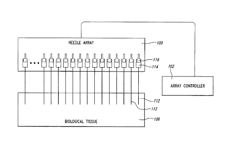
Referring first to Figure 1, a needle array assembly 100 is shown,
including a plurality of needles 112, a plurality of reservoirs 114, a
plurality of
delivery actuators such as, in the present example, plungers 116, and a
controller 102. Each of the plurality of needles 112 is fixed in position
relative to
the others of the plurality of needles, and the plungers are likewise
operatively
Date recu/Date Received 2020-07-09
19
coupled so as to be fixed in position and simultaneously actuable. Each of the
plurality of needles 112 is in fluid communication with a respective one of
the
plurality of reservoirs 114, and each of the plurality of plungers includes a
first
end positioned in a respective one of the plurality of reservoirs 114. The
controller 102 is operatively coupled to second ends of each of the plurality
of
plungers 116. The controller is configured to control actuation of the
plungers
within the reservoir with respect to speed, distance, and direction of
movement.
Movement of the plurality of plungers 116 in a first direction
creates a negative pressure in the respective reservoirs 114, drawing a
therapeutic agent or other fluid into the reservoirs via the respective needle
112, thereby charging the reservoirs. Each reservoir 114 can be charged with a
different agent, or some or all of the reservoirs can be charged with a common
agent. Movement of the plurality of plungers 116 in a second direction creates
a positive pressure, or overpressure, in the respective reservoirs 114,
forcing
the contents of the reservoirs out via the respective needles 112.
In this configuration, a relatively small amount of a plurality of
therapeutic agents can be simultaneously delivered directly to a region of
solid
tissue 106 for evaluation and analysis. In some embodiments, the amount of a
therapeutic agent delivered to the tissue is less than 1 pL per needle. The
evaluation of the tissue 106 and the efficacy of the different therapeutic
agents
delivered thereto can be used, for example, to screen potential therapeutic
agents for subsequent clinical trials or to make patient-specific treatment
decisions based on the relative efficacy of the therapeutic agents in the
tissue
106.
According to various embodiments, any number of needles can
be used. For example, as few as one, two, or three needles can be used, and
according to some embodiments, more than one thousand needles can be
used. According to an embodiment, each of the needles includes a plurality of
ports or apertures arranged along the length of the needle.
Date recu/Date Received 2020-07-09
20
Turning now to Figures 2A-2D, various configurations of needles
120 are shown. Figure 2A shows a delivery needle 120a including a plurality of
ports 122 in pairs on opposite sides of the needle, the pairs being evenly
spaced along its length. Each pair is rotated 90 degrees with respect to
adjacent pairs of ports along the length of the needle 120a. When fluid in a
reservoir in fluid communication with the needle 120a is subjected to an
overpressure, it is forced from the needle via the plurality of apertures 122.
Because the reservoir holding the fluid is to the right of the needle 120a, as
viewed in the figures, an overpressure in the reservoir will result in the
largest
volume of fluid being forced from the right-most ports 122, such that the
fluid
will be delivered in a progressively diminishing volume along its length
toward
the tip-end 124. The relative volume of fluid distributed from each of the
plurality of ports 122 along the needle 120a may be influenced by a number of
factors including, for example, viscosity of the fluid, the size and
concentration
of solids suspended therein, the density, permeability, and wettability of
tissue
in which the needle is positioned, the degree of overpressure, the size of the
ports, etc.
Figure 2B shows a delivery needle 120b according to another
embodiment, in which ports 122 are largest near the tip-end 124 of the needle
120b, and the relative size of each of the plurality of ports is inversely
related to
a distance of the respective port from the tip-end of the needle. Thus, while
an
overpressure of fluid in the needle will be greatest at the right-most port
122,
that will also be the smallest port, and, conversely, while the overpressure
will
be lowest at the left-most port 122, that port will also be the largest. By
appropriate sizing of each of the ports 122, the needle 120b can be configured
to deliver a substantially equal volume of fluid at any given location along
its
axis, or alternatively, the needle can be configured to deliver fluid
according to
any selected distribution profile along its axis, by appropriate selection of
the
size of the respective ports 122. The size of the apertures can vary along the
length of the needle from about 0.01 mm or less to about 0.25 mm or more.
Date recu/Date Received 2020-07-09
21
Figure 2C shows a delivery needle 120c according to an
embodiment in which a distribution density of the plurality of ports 122 is
inversely related to a distance of the respective port from the tip-end 124 of
the
need1e120c. In other words, the ports 122 closest to the tip-end 124 of the
needle 120c are the most closely spaced, while the spacing between the ports
grows increasingly greater as the distance from the tip-end increases.
Accordingly, when fluid in the associated reservoir is subjected to an
overpressure, the volume of fluid per port 122 will be greatest at the right-
most
port, but a lower volume of fluid per port will be offset toward the left by
the
progressively closer spacing of the ports. Thus, the overall distribution of
fluid
along the length of the needle 120c can be made to be substantially consistent
by distributing the ports 122 as described above, or can be made to conform to
another selected distribution profile by appropriate selection of the
distribution
density of the ports along the needle.
Turning to Figure 2D, a delivery needle 120d is shown according
to another embodiment. Ports 126 of the needle 120d are formed in a spiral
pattern, with each port rotated 90 with respect to adjacent ports. In the
embodiment shown in Figure 2D, the ports 126 are formed by wire-electrode
electrical discharge machining (wire EDM). In cutting the ports 126, the depth
and the length of each cut can be selected to control the port size, while the
pitch of the spiral can be selected to control the distribution density. Thus,
ports
126 configured as shown in Figure 2D can be differentially sized or spaced as
described with reference to Figures 2B and 2C.
In addition to wire EDM, the ports 122, 126 of the needles 120
can be formed by any appropriate method, including, for example, laser
cutting,
waterjet cutting, chemical etching, mechanical drilling or grinding, etc.
The tip-ends 124 of the needles 120 are shown as being closed
and pointed. According to some research, "pencil point" needles, such as, for
example, Sprotte and Whitacre needles, may be less damaging to biological
tissue than bevel-tipped needles. Additionally, fluids injected into tissue
using
Date recu/Date Received 2020-07-09
22
pencil point side-port needles tend to remain in the tissue rather than
leaking
from the tissue via a channel formed by the needle. Such considerations are
explored in more detail in U.S. Patent Application No. 2004/0191225 ¨ see also
U.S Patent No. 5,848,996. Nevertheless, the scope of the
invention is not limited to pencil point
needles. Bevel-tipped and blunt-tipped needles can also be employed
according to various embodiments. In particular, the inventors have conducted
tests using prototypes with blunt-tipped needles, which performed
satisfactorily.
Figure 3 shows a solid-core delivery needle 130 having a plurality
of annular grooves 132. The needle 130 is a "passive delivery" device,
meaning that a therapeutic agent is not delivered under pressure from a
reservoir but instead is carried into the tissue in the grooves 132. Other
passive delivery-type needles include, for example, needles with micro pits
over
their surfaces, needles coated with nanowire, and needles made from porous
materials. Such a needle is immersed in a liquid agent for sufficient time to
charge, and is then inserted into the target tissue. In the case of the needle
130 of Figure 3, the needle can be charged by being briefly dipped into the
agent. A porous needle will carry more of the agent, but may require more time
to charge, and may likewise need to be left in place in the tissue for a
longer
period to deliver its charge.
Embodiments are primarily described herein as using active
delivery needles, i.e., needles that actively force fluid into the surrounding
tissue. However, passive delivery needles such as those described above can
also be employed, according to the design parameters of a given application.
Figures 4A and 4B show a portion of an inserter needle 140 and a
portion of a delivery needle 120 similar to those described with reference to
Figures 2A-2D. In Figure 4A, the inserter needle 140 is positioned such that
the tip-end 124 of the delivery needle 120 extends slightly beyond an end of
the
inserter needle 140. In this configuration, the needle 120 and inserter needle
140 can be inserted through the skin of a subject, such as a patient or test
Date recu/Date Received 2020-07-09
23
model. The combination of the needle 120 and inserter needle 1406 are
configured to have sufficient stiffness to penetrate the skin without bending,
and
the tapered point of the tip-end 124 assists in the penetration. Additionally,
the
inserter needle 140 covers the ports 122 of the needle 120 and prevents
contamination of the contents of the needle by non-target tissue, and vice-
versa. When the tip-end 124 of the needle has penetrated to within a small
distance of the target tissue, the inserter needle 140 is held in position
while
insertion of the needle 120 continues until it is correctly positioned in the
target
tissue. The insertion distance of the inserter needle 140 can be selected such
that the needle 120 is correctly positioned once all of the ports 122 are
clear of
the inserter needle, as shown in Figure 2B. In this way, the needle 120 can be
provided with maximum protection and support, and the likelihood of
contamination can be minimized.
According to an alternate embodiment, a stylette is positioned in
the inserter needle to stiffen the needle and prevent collection of a tissue
plug
during insertion. Once the inserter needle 140 is positioned, the stylette is
removed and the delivery needle 120 is inserted.
Figure 5 is a diagrammatic view of a delivery assembly 150
according to another embodiment. The delivery assembly 150 includes a
needle array 152, an inserter assembly 154, an actuator assembly 156, a driver
assembly 158, a control assembly 160, and a frame 162. The frame 162
provides a substantially rigid structure to which other elements of the
assembly
150 are coupled.
The needle array 152 comprises a plurality of needle cylinders
166 and a needle block 168. In the embodiment shown, the needle block 168
is integral with the frame 162. Each of the plurality of needle cylinders 166
is
coupled, at a first end 170, in a respective needle aperture 174 extending in
the
needle block 168, and comprises a lumen 176, having, in the illustrated
embodiment, a nominal diameter of .15 mm, extending substantially the entire
length of the needle cylinder 166. Each needle cylinder 166 includes a
Date recu/Date Received 2020-07-09
24
reservoir 178 in a region toward the first end 170, a needle 120 in a region
toward a second end 180, and a tip-end 124 at the second end 180 of the
needle cylinder 166. In the embodiment shown, the tip-end 124 is tapered to a
point.
Each delivery needle 120 is defined by a plurality of ports 122
distributed along its length. The length of each of the plurality of needle
cylinders 166 and of the respective needles 120 varies according to the
embodiment. in one embodiment, each needle cylinder 166 is longer than 15
cm, while according to other embodiments the needle cylinders are each longer
than 10 cm, between 5 cm and 10 cm, and as short as 2 cm, respectively.
Likewise, according to various embodiments, each of the plurality of delivery
needles 120, defined by the portion of the respective needle cylinder 166
along
which the ports 122 are spaced, is longer than 1 cm, longer than 2 cm, longer
than 4 cm, and longer than 8 cm.
The inserter assembly 154 comprises a plurality of inserter
needles 140 coupled to an inserter block 192 in respective inserter apertures
190 extending therein in a configuration that corresponds to the arrangement
of
the needle cylinders 166 in the needle block 168, such that each of the
plurality
of needle cylinders 166 can be positioned within a respective one of the
plurality of inserter needles 140 as shown in Figure 5. The inserter assembly
154 is axially slidable over the needle cylinders 166 between a first
position, in
which only the tip-ends 124 of each of the needle cylinders 166 extend from
respective ones of the plurality of inserter needles 140, to a second
position, in
which the second ends 180 of each of the needle cylinders 166 extends from
the respective inserter needle 140 a distance sufficient to clear all of the
ports
122 of the respective delivery needle 120.
According to an embodiment, a spacer is provided, configured to
be positioned between the inserter block 192 and the needle block 168, sized
such that when the inserter block and the needle block are both engaged with
the spacer, the inserter block is maintained in the first position. Removal of
the
Date recu/Date Received 2020-07-09
25
spacer permits movement of the inserter block 192 and the needle block 168
relative to each other, to permit placement of the inserter block into the
second
position, relative to the needle block.
The actuator assembly 156 comprises a plurality of plungers 200
coupled at respective first ends 204 to a plunger block 206 in a configuration
that corresponds to the arrangement of the needle cylinders 166 and the
inserter needles 140 such that a second end 208 of each of the plurality of
plungers 200 can be positioned within the reservoir 178 of a respective one of
the plurality of the needle cylinders 166 as shown. An 0-ring 210 is provided
at
the second end 208 of each of the plurality of plungers 200 to sealingly
engage
the wall of the respective lumen 176. The actuator assembly 156 also
comprises an actuator 212 coupled to an actuator block 214, which in turn is
rigidly coupled to the plunger block 206. In the embodiment shown, the
actuator 212 comprises a micrometer device 220 having a thimble 222, a barrel
224, and a spindle 228 such as are well known in the art. The barrel 224 is
rigidly coupled to the frame 162 while the spindle 228 is rotatably coupled to
the
actuator block 568 so as to control translational movement of the actuator
block
relative to the frame 162. The micrometer device 568 is calibrated in .01mm
increments, with a spindle travel of .5mm per rotation of the thimble 222 and
a
maximum stroke of 15 mm. Thus, each complete rotation of the thimble moves
each of the plurality of plungers .5 mm within the lumen 178 of the respective
needle cylinder 166 and displaces about .0001 cm3 of volume, or .1 nL per
revolution. Thus, given a maximum stroke of 15 mm, the maximum dispensing
capacity of each of the plurality of needles 120 is about 3 nL.
The driver assembly 158 comprises a stepper motor 230 such as
is well known in the art, and that includes a motor casing 232, a motor shaft
234
coupled to a rotor of the motor 230, and other elements such as are well known
in the art. The motor casing 232 is rigidly coupled to the frame 162, and the
motor shaft 234 is slidably coupled to the thimble 222 of the micrometer
device
568 while being rotationally locked therewith, such as via a spline coupling,
for
Date recu/Date Received 2020-07-09
26
example. Accordingly, rotational force from the motor shaft 234 is transmitted
to the thimble 222, while axial movement of the thimble is not limited by the
motor shaft. Such couplings are well known in the mechanical arts. The
stepper motor 230 of the illustrated embodiment is configured to divide each
rotation into 125 steps. Thus, each incremental rotational step of the motor
230
rotates the thimble about 3 , displacing a volume of about .8 pL per reservoir
178.
The controller assembly 160 includes a controller 240 and a
control cable 242 that extends from the controller to the stepper motor 230.
Signals for controlling direction, speed, and degree of rotation of the motor
shaft
234 are transmitted from the controller 240 to the stepper motor 230 via the
control cable 242 in a manner that is well known in the field to which such
motors belong. According to an embodiment, the controller is programmable.
A user can program the controller to control a speed of delivery of a fluid
from
the delivery needles 120 by selecting the speed of rotation, and a volume of
fluid delivered by selecting the number of partial and complete rotations of
the
rotor. According to another embodiment, the controller is manually operated,
such that a user controls a rate and direction of rotation of the motor 230 in
real
time. According to a third embodiment, the driver and controller assemblies
are
omitted, and a user controls fluid delivery by manually rotating the thimble
222
of the actuator assembly 212.
Charging the reservoirs 178 can be accomplished in a number of
ways. For example, a charging vessel can be provided that includes a plurality
of cups or compartments in an arrangement that corresponds to the
arrangement of the needle cylinders 166. The user first places a selected
fluidic agent or combination of agents in each of the cups. The delivery
assembly 150 of Figure 5 is positioned with the needle cylinders pointing
downward as shown in the drawing, and the spindle 228 of the actuator 212
fully extended. The frame 162 is lowered until the needles 120 are fully
immersed in the fluids in the respective cups. The motor 230 is then
controlled
Date recu/Date Received 2020-07-09
27
to rotate in the reverse direction, drawing the spindle 228 inward and pulling
the
plungers 200 upward. This in turn creates a negative pressure in the
reservoirs
178 relative to ambient, drawing the fluids into the needle cylinders 166 via
the
needle ports 122. When the reservoirs are sufficiently charged, rotation of
the
rotor is halted and the needle array 152 is withdrawn from the charging vessel
In order to deliver the charge, according to one embodiment, each
of the needle cylinders 166 of the needle array 152 is positioned in a
respective
one of the inserter needles 140 of the inserter assembly 154 so that the tip-
ends 178 of the needles 120 protrude from the inserter needles 140,
substantially as described with reference to Figure 4A. The delivery assembly
150 is then positioned in axial alignment with a target tissue region of a
subject
and translated axially so that the tip-ends of the needles 120 penetrate the
subject's skin. Axial translation of the delivery assembly 150 continues until
the
tip-ends 124 of the needle cylinders 166 have penetrated to within a selected
distance of the target tissue region. The inserter assembly 154 is then held
in
position while the frame 162 and the elements coupled thereto continue to
move axially, such that the needles 120 extend into the target tissue region.
When the needles 120 are correctly positioned, movement of the delivery
assembly 150 is halted and the frame 162 is held in position relative to the
subject. The stepper motor 230 is then controlled to rotate the thimble 222 in
the forward direction so as to cause the spindle 228 to extend, driving the
plungers 200 into the needle cylinders 166 and creating an overpressure in the
respective reservoirs 178, thereby forcing fluid from the reservoirs to the
target
tissue region via the ports 122 of the delivery needles 120.
Delivery can be performed in a few seconds, or it can be
extended over minutes or hours under a relatively low overpressure to promote
complete absorption of the fluid into the surrounding tissue. According to the
embodiment described with reference to Figure 5, the stepper motor 230 can
be controlled to rotate the rotor fast enough to depress the plungers 200 the
full
Date recu/Date Received 2020-07-09
28
15 mm in less than one second, or slow enough that a single rotation can take
many hours.
Figure 6 shows a portion of a needle array 250 according to
another embodiment. A portion of a needle cylinder 166 is shown, together
with a portion of a needle block 252. The first end 170 of the needle cylinder
166 is coupled to a first portion 254 of an aperture 256 extending in the
needle
block 252. The aperture 256 includes the first portion 254, sized to receive
the
needle cylinder 166, and a second portion 258 having an increased diameter.
In the embodiment shown, the diameter of the second portion 258 has a
diameter of .75 mm. The second portion 258 defines a reservoir that is in
fluid
communication with the needle cylinder 166. The second end of a plunger 260
is positioned in the second portion 258, with an 0-ring 262 sealingly engaged
therein.
In the arrangement described with reference to the delivery
assembly 150 of Figure 5, the second end of each of the plurality of plungers
200 is positioned in a respective one of the needle cylinders 166, and the
reservoirs 178 are comprised by the needle cylinders. Thus, axial movement of
one of the plurality of plungers 200 within the lumen 176 of a respective
needle
cylinder 166 displaces a volume equal to a transverse cross-sectional area of
the lumen, multiplied by the distance of travel of the plunger. In contrast,
because of the diameter of the second portion 258 of the aperture 256 of
Figure
6, relative to the diameter of the lumen 176, axial movement of the plunger
260
displaces a volume that is greater than the volume displaced by a plunger 200
of Figure 5 by a factor of 25, for a given distance of travel. Conversely, to
displace an equal volume of the reservoir 178, a plunger 200 of Figure 5 must
travel 25 times as far as the plunger 260 in the second portion 258 of the
aperture. Thus, given otherwise identical elements, the delivery assembly 150
of Figure 5 is capable of accurately metering delivery of smaller quantities
of
fluid than a similar assembly having reservoirs and plungers configured as
described with reference to Figure 6, while the latter is capable of
delivering
Date recu/Date Received 2020-07-09
29
larger quantities of fluid for a given plunger stroke length; up to 75 nL over
a
stroke of 15 mm vs. 3 nL for the embodiment of Figure 5. Of course, the values
given are exemplary; one of ordinary skill will recognize that the needle
sizes as
well as reservoir sizes can be selected to accommodate particular
requirements.
Turning now to Figure 7, elements of a delivery assembly 270 are
shown according to another embodiment. A needle block 272 includes a large
plurality of needle apertures 274 extending therethrough, arranged in a
closely
spaced array. Needle cylinders 166 are provided separately, in various
assortments of lengths and numbers, sizes, and spacings of ports.
In use, a user selects a number of needles to be used for a
particular procedure, and selects the particular needle cylinders 166, placing
each in a respective one of the plurality of apertures 274 of the needle
block, in
an arrangement that is selected for the particular procedure. The user may
require only a small number of needles, such as one to five, for example, or
may require hundreds or thousands of needles. Furthermore, the needle
cylinders 166 can be of varying lengths and configurations. The user selects
the arrangement of the needle cylinders 166 in the needle block 272, and their
respective lengths and configurations, at least in part according to factors
such
as the size, shape, and position of a target tissue region in a subject's
body, the
desired distribution density of fluid in the target tissue region, the
permeability of
the target tissue, etc.
The needle cylinders 166 can be affixed in the apertures 274 by
any appropriate means, including, for example, soldering, brazing, and by
adhesive. Alternatively, the needle cylinders 166 can be sized and configured
to be fixed in place by an interference fit, such that, for example, the first
end of
each needle cylinder has a fractionally increased outer diameter. The user
drops each needle cylinder into a respective aperture, tip-end first, then
pulls
the needle cylinder into the aperture from the other side of the needle block
until the first end is firmly engaged in the aperture. A plunger block and an
Date recu/Date Received 2020-07-09
30
inserter block are also provided, with respective pluralities of apertures in
arrays
corresponding to the array of needle apertures of the needle block. The user
loads plungers 200 and inserter needles 140 concurrently with the needle
cylinders 166 for operation in a delivery assembly similar to that described
with
reference to Figure 5. Provided the method used to attach the needle cylinders
166 in place can be reversed, the needle block 272 can be reused repeatedly
for different procedures.
The needle block 272 shown in Figure 7 is about 5 cm in
diameter, 1 cm in thickness, and has approximately 1,600 apertures, spaced
about 1 mm apart. According to other embodiments, the needle block can be
any appropriate shape and size, from as small as 1 or 2 centimeters across to
as large as ten or more centimeters across, and can have any number of
apertures, from ten or fewer to several thousand. According to various
embodiments, the needle block is provided, for example, with 200, 400, and
800 apertures. The large number of apertures provides significant freedom to a
user to control spacing between needles as well as the particular pattern of
the
array. The needle block 272 is shown with a hexagonal grid array of apertures.
The apertures can also be arranged in other grid configurations, such as, for
example, rectangular and quincunx. The needle cylinders shown are about 5
cm in length, but this too is merely exemplary.
The delivery actuators of previous embodiments have been
described as plungers. However, any suitable actuator can be used to control
an amount of therapeutic agent delivered from the reservoirs into the needle.
For example, fluid pressure such as by compressed air or pressurized liguidcan
be used to control an amount of therapeutic agent delivered to a region of
biological tissue via the reservoirs and needles.
Referring now to Figure 8, a delivery assembly 300 is shown,
according to another embodiment. The delivery assembly 300 includes a
plurality of needle cylinders 302 comprising respective reservoirs 178 and
needles 120. Fluid couplings 312 place the needle cylinders 302 in fluid
Date recu/Date Received 2020-07-09
31
communication with a manifold 304. A fluid pressure source 306 and a fluid
vacuum source 308 can each be placed in fluid communication with the
manifold 304 by operation of a valve 310.
According to the embodiment of Figure 8, the needle cylinders
302 are not fixed with respect to each other, but can be individually
emplaced,
in a target tissue region, for example. The reservoirs are first charged, by
placing the delivery needles 120 in a selected fluid, e.g., a therapeutic
agent or
respective therapeutic agent, and the fluid vacuum source is placed in fluid
communication with the manifold, drawing a negative pressure into the
reservoirs and drawing the agent into the needles. The user then positions the
needles 120 in the target tissue region. When they are all in place, the
manifold
304 is pressurized, forcing fluid from the reservoirs of each of the needle
cylinders 166 via the ports 122 of the respective delivery needles. While
Figure
8 shows a simple fluid circuit, it will be understood that in practice such a
circuit
could include any of valves, pressure regulator, peristaltic pump,
microfluidic
pump, vacuum accumulator, compressor, controller, etc., all of which are well
known in the art, and within the abilities of one of ordinary skill to select
and
configure for a given application.
According to an embodiment, solid tissue into which a plurality of
therapeutic agents have been delivered is subsequently resected from the
subject and evaluated. For example, in a case where the target tissue is a
cancerous tumor, the plurality of agents injected therein can include some
agents whose efficacy or effect on such tumors is under investigation. By
injecting the various agents in vivo then waiting a selected period before
removing the tumor, the effect of the agents on the tumor in situ can be
investigated. This preserves the tumor microenvironment and distinguishes this
method from current ex vivo or in vitro therapeutics evaluation methods.
Assuming that the needles used are configured to deliver a substantially equal
amount of fluid at any given location along their length, as described above
with
reference to Figures 2B-2D, the agent delivered by each of the needles is
Date recu/Date Received 2020-07-09
32
evenly distributed to the surrounding tissue along the delivery axis on which
the
respective needle 120 was positioned during the delivery of the agent to the
tumor 320. Over time, each agent permeates outward from its delivery axis to a
greater or lesser degree, depending on factors such as, for example, the
density of the surrounding tissue, the viscosity and composition of the agent,
the wettability of the tissue by the respective agent, etc. Typically, the
portions
of the tissue into which the agents spread are approximately column-shaped
regions coaxial with the respective delivery axes.
According to various embodiments, a region of tissue is left in
place for some period of time before being resected. For example, 48-72 hours
following delivery is thought to be generally sufficient for a tumor to
exhibit a
detectable response. In other cases, the wait period may be hours, days, or
weeks. According to some embodiments, the tissue region is imaged using
known methods to precisely locate the target region of tissue prior to
insertion
of the needles. The region may be imaged repeatedly before and after delivery
of the plurality of agents to the region of tissue.
According to other embodiments, a plurality of agents are
delivered to a portion of tissue via respective ones of a plurality of needles
of a
needle array after the portion of tissue is resected.
Referring now to Figure 9, a portion of a tumor 320 is shown,
following an injection procedure and subsequent resection. The tumor 320 has
been sectioned into a plurality of slices 322 along planes that lie
substantially
normal to the delivery axes. Column-shaped delivery regions 324 define the
regions of permeation of the respective agents, and extend perpendicular to
the
planes of the sections 322.
Many of the regions 324 may not be easily detectable to a user,
so generally at least two readily detectable position markers 324a, 324b are
among the agents injected, at widely separated locations. The user can then
overlay a template on which the locations of each of the delivery axes is
marked, aligning the indicated marker positions of the template with the
Date recu/Date Received 2020-07-09
33
detectable position markers 324a, 324b of a given section 322, thereby
locating
the remaining delivery regions 324. The position markers 324a, 324b can be
any composition that is detectable by a user. Various exemplary position
markers are described in detail elsewhere in this disclosure. According to an
embodiment, the position markers are selected to resist permeation and
diffusion into the surrounding tissue and to remain concentrated in a narrow
column, as shown for example at 324a, so as to be detectable for an extended
period after the injection procedure, and to provide an accurate guide for
positioning the template.
In addition to position markers, control agents may also be among
the agents injected. For example, a negative control can comprise a substance
used as a vehicle in others of the agents, and a positive control can comprise
a
compound of most or all of the agents delivered individually at other delivery
axes.
Following sectioning of the tumor 320, a user conducts selected
assays on delivery regions 324 of various sections 322 of the tumor 320, as
described in more detail later. One benefit of the devices and methods
disclosed herein is that, in addition to evaluating the efficacy of a given
agent
on the tumor, the efficacy of agents at various delivery regions 324 can be
evaluated and compared. Additionally, the effect of a given agent on various
parts of the tumor can be evaluated, both vertically and horizontally. By
comparing the effect of an agent in a delivery region 324c at section 322a,
for
example, with its effect in the same region 324c at sections 322b and 322c,
the
effect of that agent on different tissue compositions that may occur
vertically
can be differentiated. Similarly, the same agent can be delivered at several
delivery axes in the array, e.g., 324c and 324d, and the relative effects at
those
locations in a given section 322 can then be compared, providing horizontal
differentiation. As is well known in the art, biological tissue is rarely
homogeneous over even relatively small distances. A given agent might have
substantially no effect on some tissue structures of a tumor, but might, on
the
Date recu/Date Received 2020-07-09
34
other hand, be extremely effective on others. Such differential effects can be
detected and evaluated as described above.
Another valuable aspect that can be evaluated is the effect of
multiple agents in regions where they interact within the tissue. Delivery
regions 324e and 324f are spaced more closely together than the others,
resulting in the respective agents interacting in a region 324ef where the
respective delivery regions overlap.
As discussed in the background section of this disclosure, clinical
trials for cancer related therapeutics are incredibly expensive and time
consuming. It is therefore very important to effectively screen for agents
that
have relatively greater potential as early in the process as possible. Agents
subjected to such screening are sometimes referred to as candidate effective
agents. One screening method involves placing each candidate agent in a
respective Petri dish with a growth medium. A cancerous tumor is reduced to a
homogeneous slurry and is distributed among the Petri dishes and incubated.
The dishes are later evaluated for indications of cell growth. Agents that
appear to have impeded growth of cancer cells may then be advanced for
further study.
However, this method is only marginally effective, for several
reasons. First, many cancers are known to be nonviable outside a live subject,
for reasons such as lack of a blood supply, etc., and fail to grow in vitro
under
any circumstances. Screening tests like that described are therefore
ineffective
with these. In some cases it is not known that a particular strain falls in
this
category prior to conducting the test. The result is that an expensive and
time
consuming test is inconclusive, and the tumor cannot be salvaged for an
alternative test. If the tumor is of a rare strain, it may be some time before
another is available for alternative testing.
Second, the reduction process can alter the response
characteristics of a tumor. The process involves essentially pureeing the
tumor,
which completely destroys any structural differentiation, and may render the
Date recu/Date Received 2020-07-09
35
cancer susceptible to some agents that would have no effect on the same strain
in vivo, resulting in a false positive, even though such agents might be
useless
for treating the cancer in patients. The result is that many such agents are
not
eliminated until later phases of study, after much more money and effort have
been expended.
Third, the same reduction process can also produce false
negatives, in which some agents may fail to inhibit cell growth in vitro, but
would
be effective in treating the same cancer in vivo. This results in the
premature
elimination of some agents that might otherwise have become effective
therapeutic options.
Many false positives or false negatives are generated in the
current ex vivo or in vitro art because tumor cells are separated from their
microenvironment, e.g., surrounding non-cancerous cells, blood, hormones,
paracrine factors, oxygen tension, cell-cell communications, and host immune
functions, all of which may influence whether certain therapeutic agents have
or
do not have activity in cancer cells.
Fourth, even where accurate, only the most general information
can be gleaned from such studies because the test conditions do not remotely
resemble the conditions in which the cancer normally lives and grows, and in
which it is treated therapeutically.
It is also known to inject a test agent into a tumor prior to
resection from a subject, for subsequent examination. However, in such tests a
single agent is typically injected, so they are not feasible for early
screening, but
are usually reserved for agents that have already demonstrated significant
potential. Even in animal models, it is expensive and time consuming to induce
a tumor and allow it to grow to a practical size, which makes extensive early
screening by this method impractical.
Finally, even where general efficacy of an agent in treating a
particular cancer type, subtype, variant, strain or the like has been
demonstrated, it is not uncommon for the cancer of a particular patient to be
Date recu/Date Received 2020-07-09
36
wholly unresponsive to the agent. The patient is thus exposed to the often
extreme discomfort and toxicity of the treatment ¨ not to mention the cost ¨
without significant benefit. Worse, because the agent's ineffectiveness may
not
be known for a long period while the treatment is ongoing, the opportunity to
shift to a different treatment that might have been completely successful may
be lost.
Where a similar idiosyncratic response ¨ or lack thereof ¨ occurs
in a subject of a drug study, the results of the study can be skewed. To avoid
this, it is typically necessary to resort to larger test groups to minimize
the
statistical impact of nonresponding study subjects, which further increases
the
cost of such studies.
The inventors have recognized that the inability in the known art
to accurately position an agent in tissue in vivo, especially with respect to
other
agents, and the inability to later identify the locations of agents in tissue,
prevent more extensive and beneficial use of in vivo injection, and likewise,
that
if such accuracy could be achieved, significant benefits in research and
therapy
could be realized.
It has been noted above that the volume of fluid that is delivered
by each delivery needle can be vanishingly small, much less than would be a
minimal dose required to produce a detectable effect in an adult. Depending on
the agent, the effect may nevertheless be detected on the very small region
immediately surrounding the delivery site. Accordingly, candidate effective
agents can be injected into a tumor, for example, in situ, without danger of
harming the subject. Additionally, a significant number of different agents
can
be simultaneously delivered to respective delivery axes within the tumor.
The procedures described above can be employed to resolve a
number of the problems and difficulties that contribute to the cost and delay
of
developing effective cancer therapies. For example, because the candidate
agents are delivered in vivo, the tumor is not otherwise disturbed, and so its
reaction to each agent will tend to be indicative of its reaction if exposed
to that
Date recu/Date Received 2020-07-09
37
agent in therapeutically effective quantities. The incidence of false
positives
and false negatives is significantly reduced.
Second, because relatively large numbers of agents can be
delivered to a tumor without significant danger to the subject, it is
practical to
use the procedure to screen large numbers of candidate agents early in the
testing process, perhaps eliminating those that show the least promise,
flagging
the most promising agents for additional study, or prioritizing candidates for
further study.
Third, again because of the large number of agents that can be
delivered to a tumor, potential study subjects can be screened for response to
particular agents, reducing or eliminating the number of subjects with
idiosyncratic responses.
Fourth, when employed in a therapeutic setting, a patient can be
tested for response to a large number of treatments and the most promising
can be identified early in the process, thereby reducing the number of
patients
who undergo ineffective treatments and improving the likelihood that a patient
will receive the most effective available treatment.
According to an embodiment, a plurality of candidate effective
agents are delivered, in vivo, along mutually parallel axes to a region of
solid
tissue, substantially as described above. The region of solid tissue is
subsequently resected from a subject and an effect of each of the plurality of
agents on the solid tissue is evaluated_ Based on the evaluation, one or more
of the plurality of agents are prioritized for further investigation.
According to alternate embodiments, one or more of the plurality
of agents are selected or deselected for further investigation, based on the
evaluation.
According to another embodiment a plurality of agents that have
been shown to have therapeutic benefits are delivered, in vivo, along mutually
parallel axes to a region of solid tissue. The region of solid tissue is
subsequently resected from a subject and an effect of each of the plurality of
Date recu/Date Received 2020-07-09
38
agents on the solid tissue is evaluated. Based on the evaluation, one or more
of the plurality of agents are prioritized for therapeutic treatment of the
subject.
According to alternate embodiments, one or more of the plurality of agents are
selected or deselected for therapeutic treatment of the subject, based on the
evaluation.
According to another embodiment a plurality of agents that are
currently under investigation for therapeutic efficacy are delivered, in vivo,
along
mutually parallel axes to a region of solid tissue in each of a plurality of
subjects. The respective regions of solid tissue are subsequently resected
from
each of the subjects and an effect of each of the plurality of agents on the
respective regions of solid tissue is evaluated. Based on the evaluation, one
or
more of the plurality of subjects are selected as candidates for further study
of
the therapeutic efficacy of one or more of the agents. According to alternate
embodiments, one or more of the plurality of subjects are deselected as
candidates for further study of the therapeutic efficacy of one or more of the
agents.
According to some embodiments, needle arrays are employed to
effectively and broadly deliver therapeutic agents to live and viable tissue
(e.g.,
solid tissue) in a subject, wherein imaging is not required and the tissue is
not
subsequently resected. The subject may be monitored according to
appropriate clinical criteria for assessing clinical improvement. In these and
related embodiments, it will be appreciated that significantly higher levels
of the
therapeutic agent will be achieved within the solid tissue than would be the
case if the agent were delivered systemically, although detectable amounts of
the therapeutic agent may be subsequently identified outside the solid tissue
(e.g., in the circulation). As a non-limiting example, one such embodiment
contemplates direct intramuscular introduction of a gene therapy agent for
treating muscular dystrophy (e.g., an engineered therapeutic virus, a
therapeutic agent-carrying nanopartide, etc.) to one or more skeletal muscle
injection sites in a subject, without the need for imaging, surgery, or
histology
Date recu/Date Received 2020-07-09
39
on biopsy specimens. Of course, periodic monitoring of the circulation for
leaked therapeutic agent and/or subsequent analysis of a biopsy specimen,
e.g., to assess the effects of the agent on the target tissue, may also be
considered.
In other embodiments it is contemplated that the target region in a
solid tissue may be imaged using known techniques to evaluate the effects of
the agents. The imaging can be by any suitable process or method, including,
for example, radiographic imaging, magnetic resonance imaging, positron
emission tomogoraphy, biophotonic imaging, etc. In some embodiments, the
target region may be imaged repeatedly before, during, and after the delivery
process.
According to the embodiment of Figure 10, a data processing
system 350 is used to carry out or direct operations, and includes a processor
354 and a memory 356. The processor 354 communicates with the memory
356 via an address/data bus 360 and also communicates with a needle array
assembly 362 and a patient scanning device 364. The patient scanning device
364 is used, according to an embodiment, to assist in placing the needles of
the
needle array assembly 362 in a patient in vivo and for non-invasive analysis
of
target tissue regions using imaging techniques, such as radiographic imaging
or
nuclear medical assays. The processor 354 can be a commercially available or
custom microprocessor, microcontroller, signal processor or the like. The
memory 356 can include any memory devices and/or storage media containing
the software and data used to implement the functionality circuits and
modules.
The memory 356 can any of include several categories of
software and data used in the data processing system, such as, for example,
an operating system 366, application programs 368; input/output device drivers
370; and data 372. The application programs 368 are illustrative of the
programs that implement the various features of the circuits and modules
according to some embodiments, and the data 372 represents the static and
dynamic data used by the application programs 368, the operating system 366,
Date recu/Date Received 2020-07-09
40
the input/output device drivers 370 and other software programs that may
reside in the memory 356.
According to various embodiments, the data processing system
350 may include several modules, including an array controller 376, a scanner
controller 378 and the like. The modules may be configured as a single module
or additional modules otherwise configured to implement the operations
described herein. For example, the array controller 376 can be configured to
control the needle array assembly 100 of Figure 1, by controlling the
actuators
116, and consequently, the release of therapeutic agents from the reservoirs
114 via the needles 112. The scanner controller 378 can be configured to
control the patient scanning device 364.
Certain embodiments described herein relate to introducing an
agent into a solid tissue in a subject, and/or excising all or a portion of a
solid
tissue from a subject, and/or obtaining one or more biological samples from a
solid tissue that may be in a subject, and/or screening one or more subjects
for
clinical trial eligibility, and/or any number of other methods that may
involve a
subject, which includes a subject or biological source.
The subject or biological source may be a human or non-human
animal, a transgenic or cloned or tissue-engineered (including through the use
of stem cells) organism, a primary cell culture or culture adapted cell line
including but not limited to genetically engineered cell lines that may
contain
chromosomally integrated or episomal recombinant nucleic acid sequences,
immortalized or immortalizable cell lines, somatic cell hybrid cell lines,
differentiated or differentiatable cell lines, transformed cell lines and the
like. In
certain preferred embodiments of the invention, the subject or biological
source
may be suspected of having or being at risk for having a malignant condition,
and in certain preferred embodiments of the invention the subject or
biological
source may be known to be free of a risk or presence of such disease.
Certain preferred embodiments contemplate a subject or
biological source that is a human subject such as a patient that has been
Date recu/Date Received 2020-07-09
41
diagnosed as having or being at risk for developing or acquiring cancer
according to art-accepted clinical diagnostic criteria, such as those of the
U.S.
National Cancer Institute (Bethesda, MD, USA) or as described in DeVita,
Hellman, and Rosenberg's Cancer: Principles and Practice of Oncology (2008,
Lippincott, Williams and Wilkins, Philadelphia/ Ovid, New York); Pizzo and
Poplack, Principles and Practice of Pediatric Oncology (Fourth edition, 2001,
Lippincott, Williams and Wilkins, Philadelphia/ Ovid, New York); and
Vogelstein
and Kinzler, The Genetic Basis of Human Cancer (Second edition, 2002,
McGraw Hill Professional, New York); certain embodiments contemplate a
human subject that is known to be free of a risk for having, developing or
acquiring cancer by such criteria.
Certain other embodiments contemplate a non-human subject or
biological source, for example a non-human primate such as a macaque,
chimpanzee, gorilla, vervet, orangutan, baboon or other non-human primate,
including such non-human subjects that may be known to the art as preclinical
models, including preclinical models for solid tumors and/or other cancers.
Certain other embodiments contemplate a non-human subject that is a
mammal, for example, a mouse, rat, rabbit, pig, sheep, horse, bovine, goat,
gerbil, hamster, guinea pig or other mammal; many such mammals may be
subjects that are known to the art as preclinical models for certain diseases
or
disorders, including solid tumors and/or other cancers (e.g., Talmadge et al.,
2007 Am. J. Pathol. 170:793; Kerbel, 2003 Canc. Biol. Therap. 2(4 Suppl
1):S134; Man et al., 2007 Canc. Met. Rev. 26:737; Cespedes et al., 2006 Clin.
TransL Onco/. 8:318). The range of embodiments is not intended to be so
limited, however, such that there are also contemplated other embodiments in
which the subject or biological source may be a non-mammalian vertebrate, for
example, another higher vertebrate, or an avian, amphibian or reptilian
species,
or another subject or biological source.
Biological samples may be provided by obtaining a blood sample,
biopsy specimen, tissue explant, organ culture, biological fluid or any other
Date recu/Date Received 2020-07-09
42
tissue or cell preparation from a subject or a biological source. In certain
preferred embodiments the biological sample may be obtained from a solid
tissue (e.g., a solid tumor) using the herein described device, for example,
by
introducing a multiple needle device into a solid tissue, thereby placing a
plurality of needles at a plurality of positions in the tissue, and generating
negative pressure at one or a plurality of ports of each needle of the
multiple
needle device under conditions and for a time sufficient to draw into the
needles
a plurality of biological samples from the plurality of positions in the
tissue.
Devices and methods disclosed here may find uses according to
certain preferred embodiments for the introduction of agents to, and/or the
withdrawal of biological samples from, a solid tissue, which may be present in
a
subject in vivo including a solid tissue that may be accessed further to a
surgical procedure, or that may be excised, for instance incident to a
surgical
procedure according to standard medical practices.
Solid tissues are well known to the medical arts and may include
any cohesive, spatially discrete non-fluid defined anatomic compartment that
is
substantially the product of multicellular, intercellular, tissue and/or organ
architecture, such as a three-dimensionally defined compartment that may
comprise or derive its structural integrity from associated connective tissue
and
may be separated from other body areas by a thin membrane (e.g., meningeal
membrane, pericardial membrane, pleural membrane, mucosal membrane,
basement membrane, omentum, organ-encapsulating membrane, or the like).
Non-limiting exemplary solid tissues may include brain, liver, lung, kidney,
prostate, ovary, spleen, lymph node (including tonsil), thyroid, pancreas,
heart,
skeletal muscle, intestine, larynx, esophagus and stomach. Anatomical
locations, morphological properties, histological characterization, and
invasive
andfor non-invasive access to these and other solid tissues are all well known
to those familiar with the relevant arts.
Certain particularly preferred embodiments as disclosed herein
relate to a method for selective delivery of a fluid-phase agent to a solid
tissue.
Date recu/Date Received 2020-07-09
43
As also noted above, such selective delivery obviates the need for excessive
systemic concentrations of therapeutic or candidate agents in order to achieve
therapeutically effective concentrations in the desired solid tissue, thereby
avoiding clinically detrimental toxicities to uninvolved tissues and also
avoiding
undesirable side-effects. Related embodiments contemplate the testing of
currently non-approved candidate agents through such selective delivery to a
solid tissue. Without wishing to be bound by theory, according to these
embodiments, direct effects of the candidate agent on the solid tissue (e.g.,
solid tumor) can be evaluated by in vivo administration followed by ex vivo
analysis of excised tissue, without threatening the health of the subject,
because the dose used for direct administration into the solid tissue is far
lower
than the minimal dose that would otherwise be administered systemically. (The
minimal dose is the smallest amount of the agent that will produce a desired
physiologic effect in the subject.) Given the minute volumes and low pressures
of the present modes of fluid administration, and full or partial patency of
the
solid tissue as a physical property that promotes retention of the
administered
fluid (also determinable by existing methodologies, e.g., by imaging and/or by
use of a detectable label as a tracer), the agent that is selectively
administered
to the solid tissue according to the present disclosure is either undetectable
outside the solid tissue, or if detectable outside the solid tissue, the agent
is
present at less (in a statistically significant manner) than the minimal dose.
Such considerations pertain in related embodiments, wherein
detection in a solid tissue of an altered physiologic state subsequent to
introducing an agent or a plurality of agents includes detecting a degree of
permeation of the agent(s) through the solid tissue, detecting a degree of
absorption of the agent(s) in the tissue, detecting a physicochemical effect
of
the agent(s) on the tissue, and/or detecting a pharmacological effect of the
agent(s) on the tissue. Assays, including fluorescence assays, of drug
permeation or penetration in solid tissues are known in the art and have been
described (e.g., Kerr et al., 1987 Cana Chemother. Pharmacol. 19:1 and
Date recu/Date Received 2020-07-09
44
references cited therein; Nederman et al., 1981 In Vitro 17:290; Durand, 1981
Canc. Res. 41:3495; Durand, 1989 JNCI 81:146; Tunggal et al., 1999 Clin.
Canc. Res. 5:1583) and may be configured further according to the present
disclosure, for instance, through the detection in histological serial
sections of a
detectable label that has been co-administered to the solid tissue, prior to
excision and sectioning, with an agent of interest.
In such embodiments, permeation or penetration refers to the
area of retention of an agent in the solid tissue in the immediate vicinity of
the
needle from which the agent was introduced exclusive of perfusion (entry into
and dispersion via any blood vessel), and may include retention of the agent
in
extracellular space or extracellular matrix or in association with a cell
membrane or intracellularly. Permeation may be distinct from a
physicochemical effect, which refers to microscopically detectable mechanical
disruption of tissue that results from the needle insertion or fluid injection
itself,
or from non-biological mechanical or chemical tissue disruption caused by the
agent (e.g., damage to cell membranes or disintegration of cell-cell
junctions).
Pharmacological effects include statistically significant alterations of a
cell or
tissue physiological state that are detectable as consequences of the
molecular
mechanism of action of the agent, for example, cytoskeletal reorganization,
extension or withdrawal of cellular processes, or evidence of biological
signal
transduction as may be detected using any of a number of known cytological,
biochemical, molecular biological or other read-outs. Comparison of serial
sections may permit distinguishing the nature of the effect that is detected
histologically.
Particularly preferred embodiments include those in which the
solid tissue comprises a tumor, wherein agent delivery may be made to, and/or
sample retrieval may be made from, the solid tumor. It will be appreciated by
persons familiar with the art from the disclosure herein that in the course of
practicing certain embodiments described herein, a selected region of a tumor
may comprise the site into which the needles of the presently described
devices
Date recu/Date Received 2020-07-09
45
are inserted, introduced or otherwise contacted with the tumor. The region may
be selected on any number of bases, including based on imaging that may be
conducted before, during or after a step of needle insertion, introduction or
contacting, or based on imaging conducted before, during or after excising the
solid tissue from a subject, or based on other criteria including but not
limited to
anatomic location, accessibility in the course of a surgical procedure, degree
of
vascularization or other criteria.
Solid tumors of any type are contemplated as being suitable for
intervention using the devices described herein. In certain preferred
embodiments, the solid tumor may be a benign tumor or a malignant tumor,
which may further be a primary tumor, an invasive tumor or a metastatic tumor.
Certain embodiments contemplate a solid tumor that comprise one of a prostate
cancer cell, a breast cancer cell, a colon cancer cell, a lung cancer cell, a
brain
cancer cell and an ovarian cancer cell, but the invention is not intended to
be so
limited and other solid tumor types and cancer cell types may be used. For
example, the tumor may comprise a cancer selected from adenoma,
adenocarcinoma, squamous cell carcinoma, basal cell carcinoma, small cell
carcinoma, large cell undifferentiated carcinoma, chondrosarcoma and
fibrosarcoma, or the like. As also noted elsewhere herein, art-accepted
clinical
diagnostic criteria have been established for these and other cancer types,
such as those promulgated by the U.S. National Cancer Institute (Bethesda,
MD, USA) or as described in DeVita, Hellman, and Rosenberg's Cancer:
Principles and Practice of Oncology (2008, Lippincott, Williams and Wilkins,
Philadelphia/ Ovid, New York); Pizzo and Poplack, Principles and Practice of
Pediatric Oncology (Fourth edition, 2001, Lippincott, Williams and Wilkins,
Philadelphia/ Ovid, New York); and Vogelstein and Kinzler, The Genetic Basis
of Human Cancer (Second edition, 2002, McGraw Hill Professional, New York).
Other non-limiting examples of typing and characterization of particular
cancers
are described, e.g., in Ignatiadis et al. (2008 Pathobiol. 75:104); Kunz (2008
Date recu/Date Received 2020-07-09
46
Curr. Drug Discov. TechnoL 5:9); and Auman et at. (2008 Drug Metab. Rev.
40:303).
An "altered physiologic state" may be any detectable parameter
that directly relates to a condition, process, pathway, dynamic structure,
state or
other activity in a solid tissue (and in preferred embodiments in a solid
tumor)
including in a region thereof or a portion therefrom, further including a
biological
sample obtained therefrom, and that permits detection of an altered (e.g.,
measurably changed in a statistically significant manner relative to an
appropriate control) structure or function in a biological sample from a
subject
or biological source. The methods of the present invention thus pertain in
part
to such correlation where an indicator of altered physiologic state may be,
for
example, a cellular or biochemical activity, including as further non-limiting
examples, cell viability, cell proliferation, apoptosis, cellular resistance
to anti-
growth signals, cell motility, cellular expression or elaboration of
connective
tissue-degrading enzymes, cellular recruitment of angiogenesis, or other
criteria
as provided herein.
"Altered physiologic state" may refer to any condition or function
where any structure or activity that is directly or indirectly related to a
solid
tissue function has been changed in a statistically significant manner
relative to
a control or standard, and may have its origin in direct or indirect
interactions
between a solid tissue constituent and an introduced agent, or in structural
or
functional changes that occur as the result of interactions between
intermediates that may be formed as the result of such interactions, including
metabolites, catabolites, substrates, precursors, cofactors and the like.
Additionally, altered physiologic state may include altered signal
transduction, respiratory, metabolic, genetic, biosynthetic or other
biochemical
or biophysical activity in some or all cells or tissues of a subject or
biological
source, in preferred embodiments in some or all cells of a solid tissue, and
in
more preferred embodiments in some or all cells of a tumor such as a solid
tumor in a solid tissue. As non-limiting examples, altered biological signal
Date recu/Date Received 2020-07-09
47
transduction, cell viability, cell proliferation, apoptosis, cellular
resistance to
anti-growth signals, cell motility, cellular expression or elaboration of
connective
tissue-degrading enzymes, cellular recruitment of angiogenesis, or other
criteria
including induction of apoptotic pathways and formation of atypical chemical
and biochemical crosslinked species within a cell, whether by enzymatic or non-
enzymatic mechanisms, may all be regarded as indicative of altered physiologic
state. Certain of these and other non-limiting examples are described in
greater detail herein.
According to certain presently contemplated embodiments, the
efficacy of a candidate agent may be identified by detecting an altered
physiologic state as provided herein, including by assessing any of a number
of
biological parameters characteristic of a cancer cell such as those reviewed
by
Hanahan and Weinberg (2000 Cell 100:57) and in the references cited therein.
Therein are disclosed methodologies for determining the effect of a candidate
agent on one or more traits exhibited by cancer cells, and detectable by any
of
a variety of techniques known to the art for determining one or more of (i) an
ability to evade apoptosis, (ii) acquisition of self-sufficiency in growth
signals,
(iii) insensitivity to growth-inhibitory signals, (iv) acquisition of tissue
invasive
and metastatic phenotype, (v) unlimited replicative potential, and (vi)
sustained
angiogenesis. Persons skilled in the art are familiar with multiple approaches
for detecting the presence of these alterations of physiologic state, which
can
be adapted to a particular excised tumor system. See, e.g., Bonificano et al.
(Eds.) Current Protocols in Cell Biology, 2007 John Wiley & Sons, NY; Ausubel
et al. (Eds.) Current Protocols in Molecular Biology, 2007 John Wiley & Sons,
NY; Coligan et al. (Eds.), Current Protocols in Immunology, 2007 John Wiley &
Sons, NY; Robinson et al. (Eds), Current Protocols in Cytometry, 2007 John
Wiley & Sons, NY. Non-limiting examples of parameters that may be assayed
to identify an altered physiologic state include assays of cell viability,
cell
division, apoptosis, necrosis, cell surface marker expression, cellular
activation
state, cellular elaboration of extracellular matrix (ECM) components or of ECM-
Date recu/Date Received 2020-07-09
48
degrading enzymes, morphometric analysis, extension or retraction of cellular
processes, cytoskeletal reorganization, altered gene expression, e.g., by in
situ
hybridization of immunohistochemistry (e.g., Shibata et al., 2002 J. Anat.
200:309) intracellular phosphoprotein localization (e.g., Gavet et al., 1998 J
Cell
Sci 111:3333), and the like.
Selection of agents that are known or candidate oncology agents
is understood and determinable by one skilled in the relevant arts (see, e.g.,
Berkowet al., eds., The Merck Manual, 16th edition, Merck and Co., Rahway,
N.J., 1992; Goodman et al., eds., Goodman and Gilman's The Pharmacological
Basis of Therapeutics, 10th edition, Pergamon Press, Inc., Elmsford, N.Y.,
(2001); DeVita, Hellman, and Rosenberg's Cancer: Principles and Practice of
Oncology (2008, Lippincott, Williams and Wilkins, Philadelphia/ Ovid, New
York); Pizzo and Poplack, Principles and Practice of Pediatric Oncology
(Fourth
edition, 2001, Lippincott, Williams and Wilkins, Philadelphia/ Ovid, New
York);
Avery's Drug Treatment: Principles and Practice of Clinical Pharmacology and
Therapeutics, 3rd edition, ADIS Press, LTD., Williams and Wilkins, Baltimore,
MD. (1987), Ebadi, Pharmacology, Little, Brown and Co., Boston, (1985);
Osolci al., eds., Remington's Pharmaceutical Sciences, 18th edition, Mack
Publishing Co., Easton, PA (1990); Katzung, Basic and Clinical Pharmacology,
Appleton and Lange, Norwalk, CT (1992)). Candidate agents may be selected
from resources that disclose listings of investigational therapeutics, for
instance,
the National Institutes of Health (Bethesda, MD) which maintains a database of
ongoing and planned clinical trials at its "ClinicalTrials.gov" website.
Candidate agents for use in screening methods and in
methods of rating candidate agents for development into therapeutic
agents such as a therapeutic agent for treating a solid tumor may be
provided as "libraries" or collections of compounds, compositions or
molecules. Such molecules typically include compounds known in the art
as "small molecules" and having molecular weights less than 105 daltons,
Date recu/Date Received 2020-07-09
49
preferably less than 104 daltons and still more preferably less than 103
daltons.
For example, a plurality of members of a library of test
compounds can be introduced as candidate agents to a region of a solid
tumor of known tumor type in each one or a plurality of subjects having a
tumor of the known tumor type, by distributing each of the candidate
agents to a plurality of positions along an axis within the region in each
subject, and after a selected period of time (e.g., a range of time, a
minimum time period or a specific time period) the region of solid tumor in
which the candidate agents have been introduced can be imaged or
removed from each subject, and each region compared by detecting an
effect (if any) of each agent on the respective position within the region,
for instance, by determining whether an altered physiologic state is
present as provided herein, relative to positions in the region that are
treated with control agents as provided herein, which would either produce
no effect (negative control) or a readily detectable effect (positive
control).
Candidate agents further may be provided as members of a
combinatorial library, which preferably includes synthetic agents prepared
according to a plurality of predetermined chemical reactions performed in a
plurality of reaction vessels. For example, various starting compounds may be
prepared employing one or more of solid-phase synthesis, recorded random
mix methodologies and recorded reaction split techniques that permit a given
constituent to traceably undergo a plurality of permutations and/or
combinations
of reaction conditions. The resulting products comprise a library that can be
screened followed by iterative selection and synthesis procedures, such as a
synthetic combinatorial library of peptides (see e.g., PCT/US91/08694,
PCT/US91/ 04666, or other compositions that
,may include small molecules as provided
herein (see e.g., PCT/US94/08542, EP 0774464, U.S. 5,798,035, U.S. 5,751,629.
Date recu/Date Received 2020-07-09
50
Those having ordinary skill in the art will appreciate that a diverse
assortment of such libraries may be prepared according to established
procedures, and tested for their influence on an indicator of altered
mitochondrial function, according to the present disclosure.
Other candidate agents may be proteins (including therapeutic
proteins), peptides, peptidomimetics, polypeptides, and gene therapy agents
(e.g., plasmids, viral vectors, artificial chromosomes and the like containing
therapeutic genes or polynucleotides encoding therapeutic products, including
coding sequences for small interfering RNA (siRNA), ribozymes and antisense
RNA) which in certain further embodiments may comprise an operably linked
promoter such as a constitutive promoter or a regulatable promoter, such as an
inducible promoter (e.g., IPTG-inducible), a tightly regulated promoter (e.g.,
a
promoter that permits little or no detectable transcription in the absence of
its
cognate inducer or derepressor) or a tissue-specific promoter. Methodologies
for preparing, testing and using these and related agents are known in the
art.
See, e.g., Ausubel (Ed.), Current Protocols in Molecular Biology (2007 John
Wiley & Sons, NY); Rosenzweig and Nebel (Eds), Current Protocols in Human
Genetics (esp. Ch. 13 therein, "Delivery Systems for Gene Therapy", 2008 John
Wiley & Sons, NY); Abell, Advances in Amino Acid Mimetics and
Peptidomimetics, 1997 Elsevier, NY.
Other candidate agents may be antibodies, including naturally
occurring, immunologically elicited, chimeric, humanized, recombinant, and
other engineered antigen-specific immunoglobulins and artificially generated
antigen-binding fragments and derivatives thereof, such as single-chain
antibodies, minibodies, Fab fragments, bi-specific antibodies and the like.
See,
e.g., Coligan et al. (Eds.), Current Protocols in Immunology (2007 John Wiley
&
Sons, NY); Harlow and Lane, Antibodies: A Laboratory Manual (1988 Cold
Spring Harbor Press, Cold Spring Harbor, NY); Harlow and Lane, Using
Antibodies (1999 Cold Spring Harbor Press, Cold Spring Harbor, NY).
Date recu/Date Received 2020-07-09
51
Pharmaceutically acceptable carriers for therapeutic use are well
known in the pharmaceutical art, and are described, for example, in
Reminotons Pharmaceutical Sciences, Mack Publishing Co. (A.R. Gennaro
edit. 1985). For example, sterile saline and phosphate-buffered saline at
physiological pH may be used. Preservatives, stabilizers, dyes and other
ancillary agents may be provided in the pharmaceutical composition. For
example, sodium benzoate, sorbic acid and esters of p-hydroxybenzoic acid
may be added as preservatives. Id. at 1449. In addition, antioxidants and
suspending agents may be used. Id. "Pharmaceutically acceptable salt" refers
to salts of drug compounds derived from the combination of such compounds
and an organic or inorganic acid (acid addition salts) or an organic or
inorganic
base (base addition salts). The agents, including drugs, contemplated for use
herein may be used in either the free base or salt forms, with both forms
being
considered as being within the scope of the certain present invention
embodiments.
The pharmaceutical compositions that contain one or more agents
may be in any form which allows for the composition to be administered to a
patient. According to certain preferred embodiments the composition will be in
liquid form and the route of administration will comprise administration to a
solid
tissue as described herein. The term parenteral as used herein includes
transcutaneous or subcutaneous injections, and intramuscular, intramedullar
and intrastemal techniques.
The pharmaceutical composition is formulated so as to allow the
active ingredients contained therein to be bioavailable upon administration of
the composition to a subject such as a human patient. Compositions that will
be administered to a patient may take the form of one or more doses or dosage
units, where for example, a pre-measured fluid volume may comprise a single
dosage unit, and a container of one or more compositions (e.g., drugs) in
liquid
form may hold a plurality of dosage units. A dose of a drug includes all or a
portion of a therapeutically effective amount of a particular drug that is to
be
Date recu/Date Received 2020-07-09
52
administered in a manner and over a time sufficient to attain or maintain a
desired concentration range of the drug, for instance, a desired concentration
range of the drug in the immediate vicinity of a delivery needle in a solid
tissue,
and where the absolute amount of the drug that comprises a dose will vary
according to the drug, the subject, the solid tissue and other criteria with
which
the skilled practitioner will be familiar in view of the state of the medical
and
pharmaceutical and related arts. In certain embodiments at least two doses of
the drug may be administered, and in certain other embodiments 3, 4, 5, 6, 7,
8,
9, 10 or more doses may be administered.
A liquid pharmaceutical composition as used herein, whether in
the form of a solution, suspension or other like form, may include one or more
of the following adjuvants: sterile diluents such as water for injection,
saline
solution, preferably physiological saline, Ringer's solution, saline solution
(e.g.,
normal saline, or isotonic, hypotonic or hypertonic sodium chloride), fixed
oils
such as synthetic mono or digylcerides which may serve as the solvent or
suspending medium, polyethylene glycols, glycerin, propylene glycol or other
solvents; antibacterial agents such as benzyl alcohol or methyl paraben;
antioxidants such as ascorbic acid or sodium bisulfite; chelating agents such
as
ethylenediaminetetraacetic acid; buffers such as acetates, citrates or
phosphates and agents for the adjustment of tonicity such as sodium chloride
or dextrose. The parenteral preparation can be enclosed in ampoules,
disposable syringes or multiple dose vials made of glass or plastic.
Physiological saline is a preferred adjuvant. An injectable pharmaceutical
composition is preferably sterile. It may also be desirable to include other
components in the preparation, such as delivery vehicles including but not
limited to aluminum salts, water-in-oil emulsions, biodegradable oil vehicles,
oil-
in-water emulsions, biodegradable microcapsules, hydrogels, and liposomes.
While any suitable carrier known to those of ordinary skill in the
art may be employed in the pharmaceutical compositions of this invention, the
type of carrier will vary depending on the mode of administration and whether
a
Date recu/Date Received 2020-07-09
53
conventional sustained drug release is also desired. For parenteral
administration, such as supplemental injection of drug, the carrier preferably
comprises water, saline, alcohol, a fat, a wax or a buffer. Biodegradable
microspheres (e.g., polylactic galactide) may also be employed as carriers for
the pharmaceutical compositions of this invention. Suitable biodegradable
microspheres are disclosed, for example, in U.S. Patent Nos. 4,897,268 and
5,075,109. In this regard, it is preferable according to certain embodiments
that
the microsphere be larger than approximately 25 microns, while other
embodiments are not so limited and contemplate other dimensions.
Pharmaceutical compositions may also contain diluents such as
buffers, antioxidants such as ascorbic acid, low molecular weight (less than
about 10 residues) polypeptides, proteins, amino acids, carbohydrates
including
glucose, sucrose or dextrins, chelating agents such as EDTA, glutathione and
other stabilizers and excipients. Neutral buffered saline or saline mixed with
nonspecific serum albumin are exemplary appropriate diluents. Preferably, an
agent (e.g., a therapeutic drug or a candidate drug) is formulated as a
lyophilizate using appropriate excipient solutions (e.g., sucrose) as
diluents.
For convenience, elements of various embodiments have been
described as components of a number of discrete assemblies. However, in
practice, elements of some of the assemblies may be omitted or grouped with
other assemblies. Accordingly, the particular arrangements of embodiments
disclosed above do not impose similar organization on other embodiments or
the claims.
When used to refer to agents delivered from needles, the term
fluid is to be read broadly to read on any substance capable of flowing
through
such a needle, including liquids, gases, colloids, suspended solids, etc.
The abstract of the present disclosure is provided as a brief
outline of some of the principles of the invention according to one
embodiment,
and is not intended as a complete or definitive description of any embodiment
Date recu/Date Received 2020-07-09
54
thereof, nor should it be relied upon to define terms used in the
specification or
claims. The abstract does not limit the scope of the claims.
Elements of the various embodiments described above can be
combined, and further modifications can be made, to provide further
embodiments without deviating from the spirit and scope of the invention.
Aspects of the embodiments can be modified, if necessary to employ concepts
of the various patents, applications and publications to provide yet further
embodiments.
These and other changes can be made to the embodiments in
light of the above-detailed description. In general, in the following claims,
the
terms used should not be construed to limit the claims to the specific
embodiments
disclosed in the specification, but should be construed to include all
possible
embodiments along with the full scope of equivalents to which such claims are
entitled.
Accordingly, the claims are not limited by the disclosure.
Date recu/Date Received 2020-07-09