Note: Descriptions are shown in the official language in which they were submitted.
CA 02941691 2016-09-06
WO 2015/132670 PCT/1B2015/000912
METHODS AND COMPOSITIONS FOR DETECTION OF TARGETS FOR
AUTOIMMUNE DISEASE
CROSS-REFERENCE TO RELATED APPLICATIONS
[0001] This application claims the benefit of U.S. Ser. No. 61/949,927, filed
Mar. 7,
2014, the content of which is incorporated herein by reference in its
entirety.
BACKGROUND OF THE INVENTION
[0002] Autoimmune diseases are a major cause of morbidity and mortality,
afflicting
approximately 3-10% of the population in Western countries. There are more
than 80
distinct autoimmune diseases, most of which are chronic conditions that often
manifest
debilitating and life-threatening complications. Current therapies for
autoimmune diseases
are suboptimal because they often cause generalized immunosuppression, which
predisposes the recipient to serious infections and cancer. Moreover, these
therapies fail
to correct the fundamental biological defect underlying autoimmune disease
pathogenesis:
loss of immunologic self-tolerance. The outcome of autoantigen recognition by
the
immune system (self-tolerance or autoimmune disease) is largely determined by
the
activation state of the dendritic cells (DCs) that uptake, process, and
present autoantigens
to autoreactive T cells. In particular, activation of dendritic cells to an
immunogenic
phenotype is necessary for the full activation of naïve autoreactive T cells,
and thus
necessary for the initiation of autoimmune disease. There is a need for better
understanding of the cellular and molecular triggers and mechanisms of DC
activation,
which could catalyze the development of novel therapies to induce self-
tolerance in
patients with autoimmune disease.
SUMMARY OF THE INVENTION
[0003] Accordingly, the present invention provides a method for identifying
one or more
target genes that modulate activation state of dendritic cells, the method
comprising: (a)
transfecting immature bone marrow-derived dendritic cells (BMDCs) with an
siRNA library;
and (b) detecting activation of the BMDCs, thereby identifying one or more
target genes
1
CA 02941691 2016-09-06
WO 2015/132670 PCT/1B2015/000912
that modulate activation state of dendritic cells. In one embodiment, the
detecting step (b)
is conducted on BMDCs that have not received any additional stimulation other
than the
transfecting step (a).
[0004] In a further embodiment and in accordance with the above, the detecting
step (b)
includes detection of a dendritic cell intrinsic effector function. In a still
further
embodiment, the dendritic cell intrinsic effector function comprises
expression of one or
more of a member selected from the group consisting of: IL-12/23-p40, CCD80,
CD86,
and MHCII.5.
[0005] In a still further embodiment and in accordance with any of the above,
the
detecting activation step (b) is conducted using flow cytometry.
[0006] In a yet further embodiment and in accordance with any of the above,
the
immature BMDCs transfected in step (a) are cultured to minimize baseline
activation.
[0007] In a further embodiment and in accordance with any of the above, the
methods of
the invention include, subsequent to the in vitro screening phase, a further
in vivo
validating phase involving validating the one or more target genes that
modulate activation
state of dendritic cells in an in vivo screen to determine whether the one or
more target
genes are a candidate target for treatment of autoimmune disease. In an
exemplary
embodiment, the in vivo screen includes the steps of (i) providing BMDCs that
lack the
candidate gene; (ii) exposing the BMDCs lacking the candidate gene to
activating stimulus;
(iii) transferring the BMDCs from step (iii) to an animal model; (iv)
assessing whether the
animal model develops an autoimmune disease, thereby validating the target
gene as a
candidate target for treatment of autoimmune disease. In a further embodiment,
the
animal model is a model for a member selected from the group consisting of
autoimmune
diabetes, multiple sclerosis, and rheumatoid arthritis.
[0008] In a yet further embodiment and in accordance with any of the above,
the siRNA
library used in the in vitro phase of the screen comprises siRNAs directed to
transmembrane receptors.
BRIEF DESCRIPTION OF THE DRAWINGS
[0009] FIG. 1 is an exemplary illustration of an embodiment of the invention.
[0010] FIG. 2 is an exemplary illustration of an embodiment of the invention.
[0011] FIG. 3 shows data on CD80 and CD86 expression in BMDCs before
optimization.
2
CA 02941691 2016-09-06
WO 2015/132670 PCT/1B2015/000912
[0012] FIG. 4 shows the effect of LDH and SGOT concentrations on BMDC
maturation
level.
[0013] FIG. 5 shows data from RIP-GP mice injected with stimulated and
unstimulated
BMDCs.
[0014] FIG. 6 shows BMDC viability as a function of electroporation pulse
voltage and
capacitance gradients.
[0015] FIG. 7 shows CD86 expression on BMDCs as a function of electroporation
pulse
voltage and capacitance gradients.
[0016] FIGS. 8A and B show siRNA transfection efficiency as a function of
[siRNA],
BMDC density, and pulse voltage and capacitance gradients.
[0017] FIG. 9 shows siRNA transfection efficiency as a function of [siRNA],
BMDC
density, and focused pulse voltage and capacitance gradients.
[0018] FIGS. 10A-B show transfection efficiency data showing that optimized
siRNA
transfection efficiency approaches 90% under conditions described herein.
[0019] FIG. 11 shows BMDC viability and maturation under identified
electroporation
conditions.
[0020] FIG. 12 shows data from transfection of CD11c-specific siRNA as
measured by
flow cytometry.
[0021] FIG. 13 shows the effect of transfection of SOCS1-specific siRNA on
BMDC
maturation.
[0022] FIG. 14 shows the effect of transfection of SOCS1-specific siRNA on
BMDC
maturation.
[0023] FIG. 15 shows data on fluorescence intensity of BMDCs under several
conditions.
[0024] FIG. 16 is a schematic illustration of an embodiment of the invention.
DETAILED DESCRIPTION OF THE INVENTION
[0025] The practice of the present invention may employ, unless otherwise
indicated,
conventional techniques and descriptions of organic chemistry, polymer
technology,
molecular biology (including recombinant techniques), cell biology,
biochemistry, and
immunology, which are within the skill of the art. Such conventional
techniques include
polymer array synthesis, hybridization, ligation, phage display, and detection
of
3
CA 02941691 2016-09-06
WO 2015/132670 PCT/1B2015/000912
hybridization using a label. Specific illustrations of suitable techniques can
be had by
reference to the example herein below. However, other equivalent conventional
procedures can, of course, also be used. Such conventional techniques and
descriptions
can be found in standard laboratory manuals such as Genome Analysis: A
Laboratory
Manual Series (Vols. l-IV), Using Antibodies: A Laboratory Manual, Cells: A
Laboratory
Manual, PCR Primer: A Laboratory Manual, and Molecular Cloning: A Laboratory
Manual
(all from Cold Spring Harbor Laboratory Press), Stryer, L. (1995) Biochemistry
(4th Ed.)
Freeman, New York, Gait, "Oligonucleotide Synthesis: A Practical
Approach"1984, IRL
Press, London, Nelson and Cox (2000), Lehninger, Principles of Biochemistry
3rd Ed., W.
H. Freeman Pub., New York, N.Y. and Berg et al. (2002) Biochemistry, 5th Ed.,
W. H.
Freeman Pub., New York, N.Y., all of which are herein incorporated in their
entirety by
reference for all purposes.
[0026] Note that as used herein and in the appended claims, the singular forms
"a,"
"an," and "the" include plural referents unless the context clearly dictates
otherwise. Thus,
for example, reference to "a polymerase" refers to one agent or mixtures of
such agents,
and reference to "the method" includes reference to equivalent steps and
methods known
to those skilled in the art, and so forth.
[0027] Unless defined otherwise, all technical and scientific terms used
herein have the
same meaning as commonly understood by one of ordinary skill in the art to
which this
invention belongs. All publications mentioned herein are incorporated herein
by reference
for the purpose of describing and disclosing devices, compositions,
formulations and
methodologies which are described in the publication and which might be used
in
connection with the presently described invention.
[0028] Where a range of values is provided, it is understood that each
intervening value,
to the tenth of the unit of the lower limit unless the context clearly
dictates otherwise,
between the upper and lower limit of that range and any other stated or
intervening value
in that stated range is encompassed within the invention. The upper and lower
limits of
these smaller ranges may independently be included in the smaller ranges is
also
encompassed within the invention, subject to any specifically excluded limit
in the stated
range. Where the stated range includes one or both of the limits, ranges
excluding either
both of those included limits are also included in the invention.
4
CA 02941691 2016-09-06
WO 2015/132670 PCT/1B2015/000912
[0029] In the following description, numerous specific details are set forth
to provide a
more thorough understanding of the present invention. However, it will be
apparent to one
of skill in the art that the present invention may be practiced without one or
more of these
specific details. In other instances, well-known features and procedures well
known to
those skilled in the art have not been described in order to avoid obscuring
the invention.
[0030] As used herein, the term "comprising" is intended to mean that the
compositions
and methods include the recited elements, but not excluding others.
"Consisting
essentially of" when used to define compositions and methods, shall mean
excluding other
elements of any essential significance to the composition or method.
"Consisting of" shall
mean excluding more than trace elements of other ingredients for claimed
compositions
and substantial method steps. Embodiments defined by each of these transition
terms are
within the scope of this invention. Accordingly, it is intended that the
methods and
compositions can include additional steps and components (comprising) or
alternatively
including steps and compositions of no significance (consisting essentially
of) or
alternatively, intending only the stated method steps or compositions
(consisting of).
[0031] All numerical designations, e.g., pH, temperature, time, concentration,
and
molecular weight, including ranges, are approximations which are varied ( + )
or ( -) by
increments of 0.1. It is to be understood, although not always explicitly
stated that all
numerical designations are preceded by the term "about". The term "about" also
includes
the exact value "X" in addition to minor increments of "X" such as "X + 0.1"
or "X ¨ 0.1." It
also is to be understood, although not always explicitly stated, that the
reagents described
herein are merely exemplary and that equivalents of such are known in the art.
[0032] A "composition" may include any substance comprising an agent or
compound
and is also intended to encompass any combination of an agent or compound and
other
substances, including a carrier, e.g., compound or composition, inert (for
example, a
detectable agent or label) or active, such as an adjuvant, diluent, binder,
stabilizer, buffers,
salts, lipophilic solvents, preservative, adjuvant or the like. Carriers also
include
pharmaceutical excipients and additives proteins, peptides, amino acids,
lipids, and
carbohydrates (e.g., sugars, including monosaccharides, di-, tri-, tetra-, and
oligosaccharides; derivatized sugars such as alditols, aldonic acids,
esterified sugars and
the like; and polysaccharides or sugar polymers), which can be present singly
or in
combination, comprising alone or in combination 1-99.99% by weight or volume.
CA 02941691 2016-09-06
WO 2015/132670 PCT/1B2015/000912
Exemplary protein excipients include serum albumin such as human serum albumin
(HSA), recombinant human albumin (rHA), gelatin, casein, and the like.
Representative
amino acid/antibody components, which can also function in a buffering
capacity, include
alanine, glycine, arginine, betaine, histidine, glutamic acid, aspartic acid,
cysteine, lysine,
leucine, isoleucine, valine, methionine, phenylalanine, asparagine, and the
like.
Carbohydrate excipients are also intended within the scope of this invention,
examples of
which include but are not limited to monosaccharides such as fructose,
maltose, galactose,
glucose, D-mannose, sorbose, and the like; disaccharides, such as lactose,
sucrose,
trehalose, cellobiose, and the like; polysaccharides, such as raffinose,
melezitose,
maltodextrins, dextrans, starches, and the like; and alditols, such as
mannitol, xylitol,
maltitol, lactitol, xylitol sorbitol (glucitol) and myoinositol.
[0033] "BMDC" as used herein refers to Bone marrow-derived dendritic cell.
[0034] The term pharmaceutically acceptable carrier (or medium), which may be
used
interchangeably with the term biologically compatible carrier or medium,
refers to reagents,
cells, compounds, materials, compositions, and/or dosage forms that are not
only
compatible with the cells and other agents to be administered therapeutically,
but also are,
within the scope of sound medical judgment, suitable for use in contact with
the tissues of
human beings and animals without excessive toxicity, irritation, allergic
response, or other
complication commensurate with a reasonable benefit/risk ratio.
Pharmaceutically
acceptable carriers suitable for use in the present invention include liquids,
semi-solid
(e.g., gels) and solid materials (e.g., cell scaffolds and matrices, tubes
sheets and other
such materials as known in the art and described in greater detail herein).
These semi-
solid and solid materials may be designed to resist degradation within the
body (non-
biodegradable) or they may be designed to degrade within the body
(biodegradable,
bioerodable). A biodegradable material may further be bioresorbable or
bioabsorbable,
i.e., it may be dissolved and absorbed into bodily fluids (water-soluble
implants are one
example), or degraded and ultimately eliminated from the body, either by
conversion into
other materials or breakdown and elimination through natural pathways.
[0035] As used herein, the term "patient" or "subject" intends an animal, a
mammal or
yet further a human patient. For the purpose of illustration only, a mammal
includes but is
not limited to a human, a simian, a murine, a bovine, an equine, a porcine or
an ovine.
6
CA 02941691 2016-09-06
WO 2015/132670 PCT/1B2015/000912
[0036] As used herein, the term "oligonucleotide" or "polynucleotide" refers
to a short
polymer composed of deoxyribonucleotides, ribonucleotides or any combination
thereof.
Oligonucleotides are generally at least about 10, 15, 20, 25, 30, 40, 50, 60,
70, 80, 90, 100
or more nucleotides in length. An oligonucleotide may be used as a primer or
as a probe.
[0037] As used herein, the term "sample" or "test sample" refers to any liquid
or solid
material containing nucleic acids. In suitable embodiments, a test sample is
obtained from
a biological source (i.e., a "biological sample"), such as cells in culture or
a tissue sample
from an animal, most preferably, a human.
[0038] "Substantially homogeneous" describes a population of cells in which
more than
about 50%, or alternatively more than about 60 %, or alternatively more than
70 %, or
alternatively more than 75 %, or alternatively more than 80%, or alternatively
more than 85
%, or alternatively more than 90%, or alternatively, more than 95 %, of the
cells are of the
same or similar phenotype. Phenotype can be determined by a pre-selected cell
surface
marker or other marker.
[0039] Although the present invention is described primarily with reference
to specific
embodiments, it is also envisioned that other embodiments will become apparent
to those
skilled in the art upon reading the present disclosure, and it is intended
that such
embodiments be contained within the present inventive methods.
[0040] In one aspect, the present invention provides methods for screening for
proteins
that encode proteins that are key to maintaining dendritic cells (DCs) in an
inactivated
state. Without being limited by mechanism, the screens of the present
invention are based
in part on the fact that the outcome of T cell antigen recognition, i.e.,
immunity versus
tolerance and the phenotype of the resulting immune response, depends on the
activation
state of DCs. Only DCs that have been activated by Pattern associated
molecular pattern
proteins (PAMPs) or Danger-associated molecular pattern proteins (DAMPs) via
their
Pattern recognition receptors (PRRs) ¨thereby upregulating costimulatory
molecules and
producing cytokines¨can initiate a primary immune response by activating naïve
T cells.
Thus, there is no autoimmune disease without T cell activation, and there is
no T cell
activation without DC activation. As such, the methods and compositions of the
present
invention screen for target genes that, when silenced, result in DC
activation, thus
identifying targets that are key for prevention and/or treatment of autoimmune
disease.
7
CA 02941691 2016-09-06
WO 2015/132670 PCT/1B2015/000912
[0041] In general, in the methods of the present invention, libraries of
siRNAs are used
to transfect dendritic cells. This screen is in preferred embodiments a high
volume, high
throughput screen in which different libraries are used to transfect thousands
to millions of
dendritic cells. After transfection, the cultures are screened for activated
dendritic cells
using methods such as a fluorescent detection (e.g., by utilizing a "knock-in"
of a
fluorescent protein for detection by automatic cell sorters and the like), and
targets are
identified that are key to maintaining dendritic cells in the inactivated
state. Potential
targets are then validated in an in vitro and/or an in vivo study. In vivo
studies include
screens to determine whether putative targets attenuate or exacerbate disease
symptoms
in mouse models. Models of particular use for in vivo validation studies in
accordance with
the invention include models of autoimmune diseases such as rheumatoid
arthritis, EAE,
and diabetes.
[0042] Figure 1 provides an overview of a general embodiment of the in vitro
phase of
the siRNA library screen in BMDCs. A small interfering RNA (siRNA) library is
electroporated into resting BMDCs. One gene is targeted in each BMDC sample.
After 48
hours of putative gene silencing, BMDCs are assayed in vitro for evidence of
maturation.
As will be appreciated, Figure 1 provides an overview of one exemplary
embodiment of the
in vitro phase of the screen, and as is described in further detail herein,
different elements
of the embodiment pictured in Figure 1 can be altered and/or optimized and be
encompassed by the presently disclosed invention. For example, instead of
siRNA
libraries, other types of libraries that dampen or eliminate expression can be
used,
including other kinds of RNA interference such as microRNA (miRNA) and short
hairpin
RNA (shRNA). In addition, instead of electroporation, other methods for
incorporating
these nucleic acid libraries into the BMDCs can be used, including without
limitation
lipofection.
[0043] Figure 2 provides an overview of a general embodiment of the in vivo
phase of
screening methods of the present invention. In this embodiment, BMDCs are
generated in
vitro from a mouse strain in which a candidate gene hit from the in vitro
screen is
genetically ablated. These knockout BMDCs are pulsed with Lymphocytic
choriomeningitis
virus (LCMV) glycoprotein (GP) peptides with or without Toll-like receptor
(TLR)
stimulation, then adoptively transferred into Rat insulin promoter-lymphocytic
choriomeningitis virus glycoprotein (RIP-GP) mice and monitored for the
development of
8
CA 02941691 2016-09-06
WO 2015/132670 PCT/1B2015/000912
autoimmune diabetes. In a different embodiment of the in vivo screen (not
shown), WT
BMDCs are generated in vitro, then the candidate gene hit is silenced by
siRNA. The
siRNA-transfected BMDCs are then pulsed with LCMV GP peptides with or without
TLR
stimulation, then adoptively transferred into RIP-GP mice and monitored for
the
development of autoimmune diabetes. The RIP-GP DC vaccination model of
autoimmune
diabetes is an example of an in vivo system in which autoreactive T cell fate
and disease
outcome are regulated by the activation state of the DC. Thus, it is a robust
in vivo system
for testing the activation state and immunogenicity of DCs.
[0044] The two phase screening approach outlined above provides an advantage
by
achieving a balance between (1) high-throughput, and (2) specificity,
sensitivity, and cost-
efficiency.
[0045] The invention disclosed herein readily lends itself to high efficiency,
high-
throughput siRNA library screening. The commercial availability of siRNA
libraries, the use
of a 96-well electroporator, and the implementation of adjustable multi-
channel pipetting
techniques all promote high efficiency workflow. The use of adjustable multi-
channel
pipettes is especially strategic because it enables the rapid transfer of
samples between
the 96-well electroporation plates and the pre-warmed, culture medium-
containing 24-well
plates. This rapid transfer also promotes cell viability by (1) minimizing the
electroporated
BMDCs' exposure time to potentially cell-damaging pH extremes near the
electroporator
electrodes, and (2) bathing the BMDCs in warm serum-containing medium as soon
as
possible after electroporation.
[0046] In general, methods of the invention include a step in which expression
of one or
more genes is attenuated or silenced. The phrase "attenuating expression" with
reference
to a gene or an mRNA as used herein means administering or expressing an
amount of
interfering RNA (e.g., an siRNA) to reduce translation of a target mRNA into
protein, either
through mRNA cleavage or through direct inhibition of translation. The terms
"inhibit,"
"silencing," and "attenuating" as used herein refer to a measurable reduction
in expression
of a target mRNA or the corresponding protein as compared with the expression
of the
target mRNA or the corresponding protein in the absence of an interfering RNA
of the
invention. The reduction in expression of the target mRNA or the corresponding
protein is
commonly referred to as "knock-down" and is reported relative to levels
present following
administration or expression of a non-targeting control RNA (e.g., a non-
targeting control
9
CA 02941691 2016-09-06
WO 2015/132670 PCT/1B2015/000912
siRNA). Knock-down of expression of an amount including and between 50% and
100% is
contemplated by embodiments herein. However, it is not necessary that such
knock-down
levels be achieved for purposes of the present invention.
[0047] Knock-down is commonly assessed by measuring the mRNA levels using
quantitative polymerase chain reaction (qPCR) amplification or by measuring
protein levels
by western blot or enzyme-linked immunosorbent assay (ELISA). Analyzing the
protein
level provides an assessment of both mRNA cleavage as well as translation
inhibition.
Further techniques for measuring knock-down include RNA solution
hybridization,
nuclease protection, northern hybridization, gene expression monitoring with a
microarray,
antibody binding, radioimmunoassay, and fluorescence activated cell analysis.
[0048] In one embodiment, a single interfering RNA is delivered to decrease
target
mRNA levels. In other embodiments, two or more interfering RNAs targeting the
mRNA
are administered to decrease target mRNA levels. In further embodiments,
libraries
containing over 1000, 2000, 3000, 4000, 5000, 6000, 7000, 8000, 9000, 10000
and more
interfering RNAs are used in screening methods of the present invention.
[0049] As used herein, the terms "interfering RNA" and "interfering RNA
molecule" refer
to all RNA or RNA-like molecules that can interact with RISC and participate
in RISC-
mediated changes in gene expression. Examples of other interfering RNA
molecules that
can interact with RISC include short hairpin RNAs (shRNAs), single-stranded
siRNAs,
microRNAs (miRNAs), picoRNAs (piRNAs), and dicer-substrate 27-mer duplexes.
Examples of "RNA-like" molecules that can interact with RISC include siRNA,
single-
stranded siRNA, miRNA, piRNA, and shRNA molecules that contain one or more
chemically modified nucleotides, one or more non-nucleotides, one or more
deoxyribonucleotides, and/or one or more non-phosphodiester linkages. Thus,
siRNAs,
single-stranded siRNAs, shRNAs, miRNAs, piRNA, and dicer-substrate 27-mer
duplexes
are subsets of "interfering RNAs" or "interfering RNA molecules."
[0050] The term "siRNA" as used herein refers to a double-stranded interfering
RNA
unless otherwise noted. Typically, an siRNA used in a method of the invention
is a double-
stranded nucleic acid molecule comprising two nucleotide strands, each strand
having
about 10 to about 28 nucleotides ¨ in further embodiments, the siRNA is about
10, 11, 12,
13, 14, 15, 16, 17, 18, 19, 20, 21, 22, 23, 24, 25, 26, 27, or 28 nucleotides
in length.
Typically, an interfering RNA used in a method of the invention has a length
of about 19 to
CA 02941691 2016-09-06
WO 2015/132670 PCT/1B2015/000912
49 nucleotides. The phrase "length of 19 to 49 nucleotides" when referring to
a double-
stranded interfering RNA means that the antisense and sense strands
independently have
a length of about 19 to about 49 nucleotides, including interfering RNA
molecules where
the sense and antisense strands are connected by a linker molecule. In further
embodiments, the length of the interfering RNA, including siRNA, is about 10-
100, 20-90,
30-80, 40-70, 50-60 nucleotides in length.
[0051] The interfering RNA used in a delivery system and method of the
invention can
be unmodified or can be chemically stabilized to prevent degradation in the
lysosome or
other compartments in the endocytic pathway.
[0052] Single-stranded interfering RNA has been found to effect mRNA
silencing.
Therefore, embodiments of the present invention also provide for
administration of a
single-stranded interfering RNA. The single-stranded interfering RNA has
similar lengths
as for the double-stranded interfering RNA cited above. The single-stranded
interfering
RNA has a 5' phosphate or is phosphorylated in situ or in vivo at the 5'
position. The term
"5' phosphorylated" is used to describe, for example, polynucleotides or
oligonucleotides
having a phosphate group attached via ester linkage to the 05 hydroxyl of the
sugar (e.g.,
ribose, deoxyribose, or an analog of same) at the 5' end of the polynucleotide
or
oligonucleotide.
[0053] Single-stranded interfering RNAs can be synthesized chemically or by in
vitro
transcription or expressed endogenously from vectors or expression cassettes
as
described herein in reference to double-stranded interfering RNAs. 5'
Phosphate groups
may be added via a kinase, or a 5' phosphate may be the result of nuclease
cleavage of
an RNA. A hairpin interfering RNA is a single molecule (e.g., a single
oligonucleotide
chain) that comprises both the sense and antisense strands of an interfering
RNA in a
stem-loop or hairpin structure (e.g., a shRNA). For example, shRNAs can be
expressed
from DNA vectors in which the DNA oligonucleotides encoding a sense
interfering RNA
strand are linked to the DNA oligonucleotides encoding the reverse
complementary
antisense interfering RNA strand by a short spacer. If needed for the chosen
expression
vector, 3' terminal T's and nucleotides forming restriction sites may be
added. The
resulting RNA transcript folds back onto itself to form a stem-loop structure.
[0054] Interfering RNAs may differ from naturally-occurring RNA by the
addition,
deletion, substitution or modification of one or more nucleotides. Non-
nucleotide material
11
CA 02941691 2016-09-06
WO 2015/132670 PCT/1B2015/000912
may be bound to the interfering RNA, either at the 5' end, the 3' end, or
internally. Such
modifications are commonly designed to increase the nuclease resistance of the
interfering
RNAs, to improve cellular uptake, to enhance cellular targeting, to assist in
tracing the
interfering RNA, to further improve stability, to reduce off-target effects,
or to reduce the
potential for activation of the interferon pathway. For example, interfering
RNAs may
comprise a purine nucleotide at the ends of overhangs. Conjugation of
cholesterol to the 3'
end of the sense strand of an siRNA molecule by means of a pyrrolidine linker,
for
example, also provides stability to an siRNA.
[0055] Further modifications include a biotin molecule, a peptidomimetic, a
fluorescent
dye, or a dendrimer, for example.
[0056] Nucleotides may be modified on their base portion, on their sugar
portion, or on
the phosphate portion of the molecule and function in embodiments of the
present
invention. Modifications include substitutions with alkyl, alkoxy, amino,
deaza, halo,
hydroxyl, thiol groups, or a combination thereof, for example. Nucleotides may
be
substituted with analogs with greater stability such as replacing a
ribonucleotide with a
deoxyribonucleotide, or having sugar modifications such as 2' OH groups
replaced by 2'
amino groups, 2' 0-methyl groups, 2' methoxyethyl groups, or a 2'-0, 4'-C
methylene
bridge, for example. Examples of a purine or pyrimidine analog of nucleotides
include a
xanthine, a hypoxanthine, an azapurine, a methylthioadenine, 7-deaza-adenosine
and 0-
and N-modified nucleotides. The phosphate group of the nucleotide may be
modified by
substituting one or more of the oxygens of the phosphate group with nitrogen
or with sulfur
(phosphorothioates). Modifications are useful, for example, to enhance
function, to
improve stability or permeability, to reduce off-target effects, or to direct
localization or
targeting.
[0057] In certain embodiments, an interfering molecule of the invention
comprises at
least one of the modifications as described above.
[0058] Interfering RNA target sequences (e.g., siRNA target sequences) within
a target
mRNA sequence can be selected using available design tools as discussed above.
Interfering RNAs corresponding to a target sequence are then tested in vitro
by
transfection of cells expressing the target mRNA followed by assessment of
knockdown as
described herein. The interfering RNAs can be further evaluated in vivo using
animal
models as described herein.
12
CA 02941691 2016-09-06
WO 2015/132670 PCT/1B2015/000912
[0059] In general, and in accordance with any of the description herein, the
screening
methods of the invention utilize BMDCs. In further embodiments, the cells used
are
immature BMDCs but are not bone marrow precursors ¨ in other words, the cells
used in
the methods described herein are fully dendritic cells, albeit immature
(inactivated) cells.
[0060] In certain embodiments, prior to transfection with the siRNA libraries,
the BMDCs
are maintained in culture conditions that minimize the baseline maturation
level to allow for
a high-dynamic range screen. In certain embodiments, Lactate dehydrogenase
(LDH)
levels in the BMDC cultures are maintained at about 200-800, 250-750, 300-700,
350-650,
400-600, 450-550 mU/mL. In further embodiments, Serum glutamic oxaloacetic
transaminase (SGOT) concentrations were maintained at about 5-60, 10-55, 15-
50, 20-45,
25-40 mU/mL. In still further embodiments, LDH levels are about 200, 300, 400,
500, 600,
700, 800, 900 mU/mL. In yet further embodiments, SGOT levels are maintained in
the
BMDC cultures at about 10, 20, 30, 40, 50, 60 mU/mL. Generally, the LDH and
SGOT
levels are controlled through choice of fetal bovine serum (FBS) used in the
cultures,
although any methods known in the art can be used to control these
concentration levels.
As will be appreciated, any combination of LDH and SGOT concentrations as
described
herein can be used in order to minimize baseline maturation levels of BMDCs in
accordance with present invention.
[0061] In further embodiments, GM-CSF was removed from the BMDC culture medium
on day 10, at the time when the BMDCs are transferred to tissue culture-
treated plates for
a further period of culture. This can serve to eliminate potential stimulatory
effects of GM-
CSF and thereby further minimize baseline BMDC maturation level.
[0062] In general, siRNA libraries are incorporated into BMDCs through
transfection by
electroporation. As will be appreciated, other methods of transfection known
in the art may
also be used.
[0063] In embodiments using electroporation, different exponential pulse
waveforms
may be used to optimize transfection efficiency. In some embodiments, an
exponential
pulse waveform of about 400 V/200 pF is used. As will be appreciated, this
waveform can
be empirically altered to customize transfection efficiency.
[0064] Transfection efficiency and effectiveness can also be affected by the
time point at
which electroporation is administered during the lifetime of a BMDC culture.
In certain
embodiments, electroporation of the siRNA libraries is conducted at day 7-8 of
the culture.
13
CA 02941691 2016-09-06
WO 2015/132670 PCT/1B2015/000912
In further embodiments, the electroporation is administered at about day 5-15,
6-14, 7-13,
8-12, 9-11 of the culture. In yet further embodiments, the electroporation is
administered at
about day 5, 6, 7, 8, 9, 10, 11, 12, 13, 14, 15, 16, 17, 18, 19, 20 of the
culture.
[0065] In further embodiments, the concentration of siRNA used during
transfection is
altered to maximize transfection efficiency. In still further embodiments,
about 4000-
10000, 5000-9000, 6000-7000 nM of siRNA is used. In yet further embodiments,
at least
2000, 2500, 3000, 3500, 4000, 4500, 5000, 5500, 6000, 7000, 8000, 9000, 10000
nM of
siRNA is used. In still further embodiments, about 3000, 3200, 3400, 3600,
3800, 4000,
4100, 4300, 4500, 4700, 4900, 5000, 5300, 5600, 5900, 6200, 6500, 7000 nM of
siRNA is
used.
[0066] In certain aspects, the screening methods of the present invention
identify siRNA
that effectively silence genes responsible for maintaining BMDCs in an
inactivated state.
Thus, the present screening methods utilize markers of BMDC activation. BMDC
activation in methods of the present invention is determined using robust in
vitro readouts.
In some embodiments, such readouts include assays for classic costimulatory
molecules,
including without limitation CD80 and CD86. Such molecules are effective
indicators of
BMDC activation, because full activation of naive T cells requires the
interaction of CD80
and CD86 on the BMDC surface with CD28 on the T cell surface. In addition,
CD80 and
CD86 are reliably and significantly upregulated on BMDCs that have been
stimulated via
TLRs, the prototypic PRRs. An additional advantage of using CD80 and CD86 as
assays
for BMDC activation is that detection of such molecules can be accomplished
using
methods amenable to high throughput large scale activity, including flow
cytometric
detection of cell-surface molecules stained with fluorochrome-conjugated
antibodies to
analyze BMDC surface expression of CD80 and CD86. Additional markers for BMDC
activation of use in methods of the present invention include Major
histocompatibility
complex class II (MHCII), dextran (DX) uptake, and IL-12/23-p40 production. As
will be
appreciated, any other known markers of BMDC activation can also be used in
the
methods of the present invention.
[0067] In further embodiments, the readouts of BMDC activation are conducted
in the
absence of any further stimulation other than the initial transfection of the
siRNA libraries.
In other words, the readout of BMDC activation identifies only activation
initiated by the
silencing of one or more genes by transfection of the siRNA libraries, and not
by any
14
CA 02941691 2016-09-06
WO 2015/132670 PCT/1B2015/000912
additional stimulation from other methods known in the art, including
stimulation with TLR
ligands, M. tuberculosis antigens, or M. tuberculosis infection.
[0068] In some embodiments, further validation studies of screens utilizing
the above-
described markers of BMDC activation. Such further validations include without
limitation
ELISA-based analyses, and T cell proliferation assays as a functional
correlate to BMDC-
derived IL-12/23-p40 production ( see Figure 16). In such assays, purified
CD3+ T cell
cells were co-cultured with siRNA-transfected BMDCs in the presence of low-
dose CD3
monoclonal antibody stimulation, and T cell proliferation was measured by flow
cytometric
analysis of Violet CellTrace dye dilution. As schematically illustrated in
Figure 16, BMDCs
generated in vitro from an IL-12/23-p40-YFP knock-in mouse are transfected
with an
siRNA library. After 48 hours of putative gene silencing, one portion of the
siRNA-
transfected BMDCs are analyzed for IL-12/23-p40-YFP expression by flow
cytometry. In
parallel, splenic CD3+ T cells are purified and stained with Violet CellTrace,
then co-
cultured with a second portion of the siRNA-transfected BMDCs in the presence
of low-
dose CD3 monoclonal antibody stimulation. After 72 hours of co-culture, T cell
proliferation
is analyzed by flow cytometry. Co-culture supernatants are harvested for
future cytokine
ELISA analysis.
[0069] In a still further embodiment and in accordance with any of the above,
the
methods of the invention include, subsequent to the in vitro screening phase,
a further in
vivo validating phase involving validating the one or more target genes that
modulate
activation state of dendritic cells in an in vivo screen to determine whether
the one or more
target genes are a candidate target for treatment of autoimmune disease. In an
exemplary
embodiment, the in vivo screen includes the steps of (i) providing BMDCs that
lack the
candidate gene; (ii) exposing the BMDCs lacking the candidate gene to
activating stimulus;
(iii) transferring the BMDCs from step (iii) to an animal model; (iv)
assessing whether the
animal model develops an autoimmune disease, thereby validating the target
gene as a
candidate target for treatment of autoimmune disease. In a further embodiment,
the
animal model is a model for a member selected from the group consisting of
autoimmune
diabetes, multiple sclerosis, and rheumatoid arthritis. In certain
embodiments, the
activating stimulus can be any known in the art to activate DCs, including
without limitation
polyinosinic-polycytidylic acid (Poly(I:C)), TLR ligands, M. tuberculosis
antigens, or M.
tuberculosis infection.
CA 02941691 2016-09-06
WO 2015/132670 PCT/1B2015/000912
Example 1: Generation of mice and cell lines
[0070] Wild-type C57BL/6 mice and gene-targeted IL-12/23-p40-eYFP (86.129-
lli 2btm 1 Lky , /J. 3
Stock Number 006412) knock-in mice were purchased from The Jackson
Laboratory. Homozygous RIP-GP ("Berlin'") mice were previously generated.
Heterozygous RIP-GP mice were generated by crossing male Berlin' + mice with
female
wild-type C57BL/6 mice. Gene-targeted SHP-deficient (Shp) mice on the C57BL/6
genetic background were previously generated. All mice were maintained, and
all
experiments were performed, at the Ontario Cancer Institute Animal Resource
Centre. All
procedures were approved by the University Health Network Animal Care
Committee.
[0071] BMDCs were generated in vitro according to the Lutz method (J Immunol
Methods 223, 77-92 (1999)). Briefly, bone marrow cells were harvested from the
femurs
and tibias of mice and cultured in 100 mm bacteriological Petri dishes (BD
Falcon) for ten
days in RPM! 1640 (Gibco) containing 10% heat-inactivated FBS (Life
Technologies), 55
pM of 2-mercaptoethanol (Gibco), and GM-CSF (40 ng/mL for the first 3 days, 20
ng/mL
for the remaining 7 days, PeproTech). Medium was changed on days 3, 6, and 8.
On day
10, non-adherent BMDCs were collected for further culture or analysis as
described
herein. In the case of further culture without electroporation, the non-
adherent BMDCs
were washed and re-cultured in 24-well plates at 2x10⁶/mL/well with or without
(1) LPS
(Sigma) at 1000, 100, 10, or 1 ng/mL or (2) polyinosinic-polycytidylic acid
(poly(I:C),
Invivogen) at 100 pg/mL for 16-20 hours. Then, BMDCs were collected for
further analysis
as described herein.
[0072] The Nuclear Receptors siGENOME siRNA library (Dharmacon) contained 54
siRNA pools (SMARTpools), each consisting of four synthetic siRNA duplexes
targeting a
single gene, arrayed in a 96-well plate at 0.5 nmol/well. The Cytokine
Receptors
siGENOME siRNA library contained 158 SMARTpools. A number of siRNA SMARTpools
were purchased and used individually, including Non-Targeting Pool #2, siGLO
Red
Transfection Indicator, CD11c, A20, SOCS1, NROB2. Lyophilized siRNA library
SMARTpools were resuspended in their original library plate in Opti-MEM buffer
(Gibco) at
mM, then placed on an orbital shaker for 30 minutes at room temperature
according to
Dharmacon's instructions. Stock solutions of lyophilized individual siRNA
SMARTpools
were prepared by resuspension in Opti-MEM buffer at 20 pM or 50 pM, followed
by orbital
shaking.
16
CA 02941691 2016-09-06
WO 2015/132670 PCT/1B2015/000912
[0073] On day 10 of BMDC culture, non-adherent BMDCs were collected, washed,
and
resuspended in Opti-MEM buffer at 20x106/mL. Lyophilized siRNA library
SMARTpools
arrayed in 96-well plates (0.5 nmol/well) were resuspended and shaken in Opti-
MEM at 10
mM as described above, then transferred to a 96-well Bio-Rad Gene Pulser
MXCell
electroporation plate. To each well of the electroporation plate were added
1.1x10⁶
BMDCs in 55 pL of Opti-MEM, producing a final cell density of 10.5x10⁶/mL and
a final
siRNA concentration of 4762 nM. An exponential waveform pulse of 400 V, 200
pF, and
1000 0 was delivered to each sample well at room temperature. Immediately
following
electroporation, BMDCs were transferred using adjustable multi-channel
pipettes to pre-
warmed 24-well plates containing 1 or 2 mL of complete 10% RPMI, then
incubated at
37 C in 5% CO2. Forty-eight hours later, BMDCs were collected by gentle
pipetting for
further experimentation or analysis. For optimization experiments in which
pulse voltage
and capacitance were varied, the resistance was always held constant at 1000
Q.
[0074] BMDC-T CELL CO-CULTURE: A BMDC-CD3+ T cell co-culture (1:10) with 72
hours of low-dose CD3 monoclonal antibody stimulation (0.1 mg/mL, BioLegend)
was
prepared in 96-well plates. BMDCs were generated in vitro and electroporated
with siRNA
as described above. After 48 hours of culture in 24-well plates as described
above,
supernatants were gently collected and frozen at -80 C for future cytokine
analysis. Two
mL of complete RPM! was added back to each well for cell resuspension. In
order to
promote high-throughput workflow, the number of cells in each well was not
determined.
Rather, based on previous data, cell recovery per well was assumed to be 60%,
considering viability and plastic adherence. Since each well was originally
seeded with
1.1x10⁶ electroporated BMDCs, it was assumed that 48 hours later, there were
0.66x10⁶
BMDCs in 2 mL in each well. Thus, 30 pL (0.01x10⁶ cells) of the BMDC
suspension in
each well was transferred to separate wells of a 96-well plate. In parallel,
CD3+ T cells
were purified from wild-type C57BL/6 spleens using a Pan T Isolation Kit II
(Miltenyi).
Twenty million CD3+ T cells at 10x106/mL were stained with Violet CellTrace
(VCT, Life
Technologies) at 2.5 pM for 20 minutes at 37 C and 5% CO2. Following quenching
with
complete RPMI, the CD3+ T cells were incubated for another 5 minutes at 37 C
and 5%
CO2. After resuspension at 0.476x10⁶/mL in complete RPM! containing 0.1 mg/mL
of CD3
monoclonal antibody, 0.1x10⁶ (210 pL) of the VCT-stained T cells were added to
each well
of the BMDC-containing 96-well plate above. After 72 hours of co-culture at 37
C and 5%
17
CA 02941691 2016-09-06
WO 2015/132670 PCT/1B2015/000912
002, all cells were collected, washed, and acquired on a FACSCanto flow
cytometer. Cell
division and proliferation analyses were performed using FlowJo software.
[0075] BMDC VACCINATION OF RAT INSULIN PROMOTER-LYMPHOCYTIC
CHORIOMENINGITIS VIRUS GLYCOPROTEIN (RIP-GP) MICE: BMDCs were prepared
in vitro as described above. On day 10, non-adherent BMDCs were collected,
washed, and
re-cultured in 24-well plates at 2x10⁶/mL/well with or without LPS at 10 ng/mL
(Sigma) for
16-20 hours. Then, the BMDCs in each well were pulsed with a triple-peptide
mix of LCMV
peptides (New England Peptide and Washington Biotechnology) for 2-3 hours as
follows:
10-6M gp33-41 (KAVYNFATM), 10-6 M gp276-286 (SGVENPGGYCL), and 1 pg/mL gp61-
80 (GLNGPDIYKGVYQFKSVEFD). BMDCs were collected by pipetting up and down,
washed with Hanks' Buffered Saline Solution (HBSS), and resuspended in HBSS at
10x106/mL. Two million BMDCs (0.2 mL) were injected intravenously into each
RIP-GP
mouse via the lateral tail vein. Blood glucose concentrations were measured
beginning on
day 6 and then every 2-3 days thereafter using an electronic glucometer and
chemstrips
(Accu-Chek). Diabetes was diagnosed after two consecutive blood glucose
readings of 15
mM or higher.
Example 2: Assays
[0076] FLOW CYTOMETRY ASSAYS
[0077] Surface staining: Cells were collected, centrifuged, transferred to
flow cytometry
tubes, and washed with cold PBS (without calcium and magnesium) containing 2%
FBS
and 0.09% sodium azide ("Staining Buffer"). After 10 minutes of Fc receptor
blockade with
CD16/CD32 monoclonal antibodies (BioLegend) at 4 C, cells were stained for 30
minutes
at 4 C in the dark with different combinations of fluorochrome-conjugated
monoclonal
antibodies, including: MHC II (1-All-E or I-Ab), CD80, and CD86 (all from BD
BioSciences),
and CD11c (eBioscience). Cells were washed with cold Staining Buffer,
centrifuged,
resuspended in Staining Buffer, and acquired on a FACSCanto flow cytometer
(BD).
[0078] Viability staining: Following surface staining as described above,
cells were
washed with cold Staining Buffer, centrifuged, and incubated with 50 pL of 7-
AAD (BD
Biosciences) for 15 minutes at 4 C in the dark. After adding 200 pL of
Staining Buffer to
each sample, cells were acquired on a FACSCanto flow cytometer.
[0079] Intracellular cytokine staining: Intracellular cytokine staining was
performed using
the BD Biosciences Cytofix/Cytoperm Fixation/Permeabilization kit according to
the
18
CA 02941691 2016-09-06
WO 2015/132670 PCT/1B2015/000912
manufacturer's instructions. Briefly, cells were incubated with GolgiPlug
(Brefeldin A) for 5-
6 hours. Following surface staining as described above, cells were washed with
cold
Staining Buffer and permeabilized by incubation in Cytofix/Cytoperm for 30
minutes at 4 C.
Cells were then washed in Perm/Wash buffer and stained with IL-12-p70-specific
monoclonal antibody (BD Biosciences) for 30 minutes at 4 C in the dark.
Following
additional washes in PermWash buffer, cells were acquired on a FACSCanto flow
cytometer.
[0080] Data analysis: Flow cytometry data was analyzed using FlowJo software
(Tree
Star). When more than one fluorochrome was used, single-stained compensation
controls
were acquired for compensation analysis, which was always manually performed
in
FlowJo. Cellular debris was excluded from analysis by setting an appropriate
gate in the
forward scatter (FSC)/side scatter (SSC) plot. All other gates were set based
on
fluorescence-minus-one (FMO) control samples. Except for IL-12/23-p4O-YFP
knock-in
BMDCs, all other BMDCs were gated on the CD11ch'gh population before analysis.
The
CD11ch'gh population frequency was approximately 90% of the FSC/SSC
population. IL-
12/23-p4O-YFP BMDCs were gated on the FSC/SSC population, then analyzed for
YFP
expression. When viability staining was performed, analyses were conducted on
live cells
by gating on the 7-AAD-negative population. All MFI values represent median
fluorescent
intensities. For some histogram overlays, the data were normalized to the peak
height at
the mode of the distribution (i.e., the number of cells in each bin of a given
histogram was
divided by the number of cells in the bin containing the largest number of
cells). Thus, the
y-axis depicts the percentage of the maximum number of cells (i.e., the number
of cells at
the mode of the distribution).
[0081] FITC-DEXTRAN ENDOCYTOSIS ASSAY: BMDCs were collected, centrifuged,
washed, and resuspended in 180 pL of complete RPMI. Twenty microlitres of
fluorescein
isothiocyanate-dextran (FITC-DX, 10 mg/mL, Sigma) was added to produce a final
FITC-
DX concentration of 1 mg/mL. Control samples (surface binding of FITC-DX but
no
endocytosis) were incubated for 30 minutes at 4 C in the dark while
experimental samples
(surface binding and endocytosis) were incubated for 30 minutes at 37 C and 5%
CO2 in
the dark. After three washes in ice cold Staining Buffer, cells were incubated
with Fc block
as described above, then surface-stained with MHC II (1-A/I-E)-specific
monoclonal
19
CA 02941691 2016-09-06
WO 2015/132670 PCT/1B2015/000912
antibody (BD Biosciences) for 30 minutes at 4 C in the dark. Cells were washed
and then
acquired on a FACSCanto flow cytometer.
[0082] CYTOKINE ELISA: BMDC culture supernatants were collected 48 hours after
siRNA library transfection and stored at -80 C for future cytokine ELISA
analysis. The
concentrations of IL-6, IL-12-p70, and TNF-a were determined by sandwich ELISA
analysis according to the manufacturer's guidelines (eBioscience Ready-SET-Go!
kits).
Example 3: Testing the Effect of FBS on BMDC Maturation Level
[0083] To maximize the dynamic range of the screen (i.e., to maximize the
probability of
detecting a true phenotypic change resulting from BMDC gene silencing), BMDCs
with
minimal baseline maturation were generated (i.e., as phenotypically immature
as possible,
while retaining the ability to become fully activated) prior to transfection.
Before
optimization, the baseline expression of CD80 and CD86 on the surface of the
BMDCs
was relatively high (Figure 3). For the data in Figure 3, resting BMDCs were
generated by
culturing WT C57BL/6 bone marrow cells in the presence of GM-CSF for 10 days.
On day
10, BMDCs were left unstimulated or stimulated with poly(I:C). After 16-20
hours of culture,
BMDCs were stained with fluorochrome-conjugated monoclonal antibodies and
analyzed
by flow cytometry. Cells were gated on the CD11chigh population. Top row
histograms:
Surface CD86 expression. Bottom row histograms: Surface CD80 expression.
Filled (solid)
histograms: Unstimulated BMDCs. Open (line) histograms: Poly(I:C)-stimulated
BMDCs.
Numbers indicate (1) the frequency of CD80+ and CD86+ populations, (2) the MFI
of
CD80 and CD86 expression, and (3) the fold-change in CD80+/CD86+ population
frequency and CD80/CD86 MFI upon poly(I:C) stimulation. Data are
representative of
many independent experiments.
[0084] The biochemical profile of the fetal bovine serum (FBS) used to
generate BMDCs
in vitro can have an effect on their differentiation and baseline maturation
level. A review of
the certificate of analysis of the FBS lot for the BMDC cultures used for the
data generated
in Figure 3 showed the LDH and SGOT concentrations in the FBS were 2136 mU/mL
and
148 mU/mL, respectively. These values were greatly in excess of the threshold
level for
BMDC-stimulating FBS determined by Lutz et al.
[0085] Several new FBS lots, all with LDH and SGOT concentrations between 300-
700
mU/mL and 12-50 mU/mL, respectively were tested for their effects on BMDC
differentiation and maturation. Figure 4 shows a representative experiment in
which
CA 02941691 2016-09-06
WO 2015/132670 PCT/1B2015/000912
BMDCs were generated using one of the new FBS lots (Invitrogen, Catalog No.
16000, Lot
No. 432023, [LDFI] = 332 mU/mL, [SGOT] = 31 mU/mL). Resting BMDCs were
generated
by culturing WT C57BL/6 bone marrow cells in the presence of GM-CSF for 10
days,
either with original (pre-optimization) FBS or new FBS containing [LDFI] and
[SGOT] within
the ranges recommended by Lutz et al. On day 10, BMDCs were left unstimulated
or
stimulated with poly(I:C). After 16-20 hours of culture, BMDCs were stained
with
fluorochrome-conjugated monoclonal antibodies and analyzed by flow cytometry.
Cells
were gated on the CD1lchIgh population. Figure 4: Top row histograms: Surface
CD86
expression on BMDCs cultured in original FBS. Bottom row histograms: Surface
CD86
expression on BMDCs cultured in new FBS. Filled in (solid) histograms:
Unstimulated
BMDCs. Open (line) histograms: Poly(I:C)-stimulated BMDCs. Numbers indicate
(1) the
frequency of the CD86+ population, (2) the MFI of CD86 expression, and (3) the
fold-
change in CD86+ population frequency and CD86 MFI upon poly(I:C) stimulation.
Data are
representative of at least three independent experiments.
[0086] As compared to the relatively high frequency of CD86+ BMDCs (54%) that
was
generated using the old FBS, the CD86+ frequency that was generated with the
new FBS
was reduced by more than one-third (to 34%). Importantly, the BMDCs generated
using
the new FBS lot retained the ability to become activated in response to TLR-
ligand
stimulation, as evidenced by the expected rise in the CD86+ frequency (old
FBS: 1.8-fold,
new FBS: 2.4-fold) and CD86 MFI (old FBS: 6.8-fold, new FBS: 9.1-fold). In
addition,
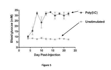
unstimulated BMDCs generated using the new FBS lot did not induce autoimmune
diabetes when they were LCMV peptide-pulsed and transferred into RIP-GP mice,
whereas poly(I:C)-stimulated, LCMV peptide-pulsed, RIP-GP-transferred BMDCs
did
induce diabetes (Figure 5). Resting BMDCs were generated by culturing WT
C57BL/6
bone marrow cells in the presence of GM-CSF for 10 days with the new
Invitrogen FBS
(Catalog No. 16000, Lot No. 432023). On day 10, BMDCs were left unstimulated
or
stimulated with poly(I:C). After 16-20 hours of culture, BMDCs were pulsed
with LCMV
triple-peptide mix for 2-3 hours, then adoptively transferred into RIP-GP
mice. Data are
representative of many independent experiments.
21
CA 02941691 2016-09-06
WO 2015/132670 PCT/1B2015/000912
Example 4: Electroporation Achieves High siRNA Transfection Efficiency with
Minimal
Effect on BMDC Maturation and Viability
[0087] Electroporation conditions that would simultaneously achieve (1)
maximum
siRNA transfection efficiency, (2) minimum BMDC maturation, and (3) maximum
BMDC
viability were investigated. Different sets of electroporation conditions were
tested,
including different pulse voltages and capacitances, siRNA concentrations, and
BMDC
densities. The overall strategy was to first exclude electroporation pulse
voltage and
capacitance parameter sets that resulted in (1) excessive BMDC death, as
measured by 7-
AAD positivity, and (2) excessive BMDC maturation, as measured by surface BMDC
expression of CD80 and CD86. Then, focusing on the subset of electrical
parameters that
resulted in reasonable BMDC viability and baseline maturation level, the
sample siRNA
concentration and BMDC density were adjusted in order to achieve maximum
transfection
efficiency, as measured by BMDC expression of a fluorescently labeled
oligonucleotide
siRNA duplex (Dharmacon siGLO Red Transfection Indicator).
[0088] To assess BMDC viability as a function of different electroporation
conditions,
unstimulated BMDCs were electroporated on day 10 without siRNA (i.e., mock
electroporation), then cultured for an additional 48 hours in order to
simulate a period of
siRNA-mediated gene silencing. At any given voltage, increasing the
capacitance resulted
in increased BMDC death, as measured by flow cytometric analysis of 7-AAD+
cells
(Figure 6). BMDC death was further increased when voltage and capacitance were
concurrently increased. At the same time, these data indicated that over a
considerable
range of voltages and capacitances, BMDC viability was relatively unaffected
(Figure 6).
[0089] To assess BMDC maturation level as a function of different
electroporation
conditions, unstimulated BMDCs were electroporated on day 10 without siRNA and
cultured for 48 hours in order to simulate a period of siRNA-mediated gene
silencing. Only
at a pulse voltage level of 400 V was there any increase in CD86 expression,
which was
further augmented as the pulse capacitance was increased (Figure 7).
[0090] On the basis of these BMDC viability and maturation data, the delivery
of a pulse
capacitance of 950 pF appeared to be excessive and undesirable, regardless of
the pulse
voltage level. This conclusion was supported by the data of Jantsch et al. (J
Immunol
Methods 337, 71-77 (2008).) , who used a pulse of 400 V/150 pF to achieve
excellent
gene silencing at the RNA and protein levels.
22
CA 02941691 2016-09-06
WO 2015/132670 PCT/1B2015/000912
[0091] To optimize siRNA transfection efficiency, the uptake of a
fluorescently labeled
oligonucleotide duplex (siGLO Red Transfection Indicator, Dharmacon) was
studied as a
function of siRNA concentration, BMDC density, and a refined, narrower
gradient of pulse
voltages and capacitances. siGLO Red uptake was analyzed by flow cytometry,
performed
immediately following electroporation.
[0092] Figure 8 shows representative results from this initial set of
experiments. Even at
the relatively high siRNA concentration of 500 nM, the transfection efficiency
was only
between 30-45%. However, when the siRNA concentration was increased greatly to
2000
nM, the transfection efficiency rose to 65-80%. When the BMDC density was
doubled from
1x10⁶/mL to 2x10⁶/mL (while holding pulse voltages and capacitances constant),
there
was no significant change in the transfection efficiency at either 500 nM or
2000 nM.
However, when the pulse capacitance and siRNA concentration were held constant
at 300
pF and 2000 nM, respectively, increasing the pulse voltage from 300 V to 400 V
raised the
transfection efficiency by more than 10% (from 70% to 80% at either BMDC
density).
Similarly, when the pulse voltage and siRNA concentrations were held constant
at 400 V
and 2000 nM, respectively, increasing the pulse capacitance from 200 pF to 300
pF raised
the transfection efficiency by more than 10% at both BMDC densities (70 to
80%). At the
siGLO concentration of 2000 nM and the BMDC density of either 1x10⁶/mL or
2x10⁶/mL,
there was no significant difference in transfection efficiency when the pulse
voltage and
capacitance were varied in opposite directions, i.e., to either 300 V/300 pF
or 400 V/200
pF. For the experiments in Figure 8, resting BMDCs were generated by culturing
WT
C57BL/6 bone marrow cells in the presence of GM-CSF for 10 days. On day 10,
BMDCs
were electroporated with either a fluorescently labeled siRNA (siGLO) or a non-
fluorescent, non-targeting (NT) control. Cells were analyzed by flow cytometry
following
electroporation. Cells were gated on the FSC/SSC population. Filled in (solid)
histograms:
NT control. Open (line) histograms: siGLO. Rows show different siGLO
concentrations.
Columns show different pulse voltages and capacitances. Numbers indicate the
transfection frequency, i.e., the frequency of siGLO+ BMDCs. (A) BMDC density
=
2x10⁶/mL. Data are representative of at least two independent experiments.
[0093] On the basis of these results, it appeared that (1) maximum
transfection
efficiency may require the siRNA concentration to be at least 2000 nM, (2)
there was no
advantage or disadvantage to doubling the BMDC density from 1x10⁶/mL to
2x10⁶/mL
23
CA 02941691 2016-09-06
WO 2015/132670 PCT/1B2015/000912
when the siGLO concentration was high at 2000 nM, and (3) applying a pulse of
400 V/300
pF achieved the highest transfection efficiency, but at the cost of (a)
slightly more BMDC
death (Figure 6) and (b) slightly more CD86 expression (Figure 7). By
comparison,
delivering a pulse of 400 V/200 pF resulted in marginally less transfection
efficiency, but
also slightly less BMDC death and maturation.
[0094] For the data in Figure 6, resting BMDCs were generated by culturing WT
C57BL/6 bone marrow cells in the presence of GM-CSF for 10 days. On day 10,
BMDCs
were left untreated or electroporated without siRNA (mock electroporation)
with different
pulse voltages and capacitances. After 48 hours of culture, BMDCs were stained
with 7-
AAD and analyzed by flow cytometry. Cells were gated on the FSC/SSC
population. (A)
Plots of the 7-AAD+ and 7-AAD- populations as a function of pulse voltage and
capacitance. (B) Plot of the 7-AAD+ and 7-AAD- populations in the untreated
(non-
electroporated) control. Numbers indicate the frequency of 7-AAD+ and 7-AAD-
populations. Data are representative of two independent experiments.
[0095] For the data in Figure 7: resting BMDCs were generated by culturing WT
C57BL/6 bone marrow cells in the presence of GM-CSF for 10 days. On day 10,
BMDCs
were left untreated or electroporated without siRNA (mock electroporation)
with different
pulse voltages and capacitances. After 48 hours of culture, BMDCs were stained
with
fluorochrome-conjugated monoclonal antibodies and analyzed by flow cytometry.
Cells
were gated on the 7-AAD-, CD11chigh population. Filled in (solid) histograms:
CD86
expression as a function of pulse voltage and capacitance. Line (open)
histograms: CD86
expression in the untreated (non-electroporated) control. Numbers indicate the
CD86+
frequency. Data are representative of at least two independent experiments.
[0096] These results led to new optimization experiments focusing on an even
narrower
range of electroporation conditions. Specifically, comparisons were made
between (1) a
pulse of 400 V/200 pF to a pulse of 400 V/150 pF, (2) an escalating gradient
of siRNA
concentrations from 2000 nM to 4762 nM, and (3) and escalating gradient of
BMDC
densities from 2x10⁶/mL to 20x106/mL.
[0097] Figure 9 shows representative results from the first set of these
experiments. At
the BMDC density of 2x10⁶/mL, electroporating 4000 nM of siRNA resulted in a
higher
transfection efficiency (85%) than either 3000 nM (80%) or 1000 nM (50%) of
siRNA. At
the siRNA concentration of 4000 nM, there was no advantage or disadvantage in
raising
24
CA 02941691 2016-09-06
WO 2015/132670 PCT/1B2015/000912
the BMDC density to 20x106/mL from 2x10⁶/mL (both 80-85%). Notably, when the
pulse
voltage was maintained at 400 V but the pulse capacitance was reduced from 200
pF to
150 pF to simulate Jantsch et al.'s conditions, the transfection efficiency
dropped
substantially from 85% to 70% (BMDC density held constant at 2x10⁶/mL). This
drop was
slightly attenuated when the BMDC density was raised to Jantsch et al.'s level
of
20x106/mL.
[0098] From these results, it appears that (1) maximum transfection efficiency
would
require at least 4000 nM of siRNA, (2) a pulse of 400 V/200 pF was superior to
a pulse of
400 V/150 pF, and (3) high transfection efficiency could be achieved over a 10-
fold range
of BMDC densities (2-20x106/mL).
[0099] For the data in Figure 9, resting BMDCs were generated by culturing WT
C57BL/6 bone marrow cells in the presence of GM-CSF for 10 days. On day 10,
BMDCs
were electroporated with either a fluorescently labeled siRNA (siGLO) or a non-
fluorescent, non-targeting (NT) control. Cells were analyzed by flow cytometry
following
electroporation. Cells were gated on the FSC/SSC population. Filled in (solid)
histograms:
NT control. Open (line) histograms: siGLO. Rows show different siGLO
concentrations.
Columns show different pulse voltages, pulse capacitances, and BMDC densities.
Numbers indicate the transfection frequency, i.e., the frequency of siGLO+
BMDCs. Data
are representative of at least two independent experiments.
[0100] To facilitate high-throughput screening, it was desirable to
electroporate a
sufficient number of cells to permit multiple functional assays from a single
electroporation
experiment. Thus, an experiment was designed to determine whether high
transfection
efficiency could be achieved at the BMDC density of 10.5x10⁶/mL (Figure 10).
At an
electroporation sample volume of 105 pL, this BMDC density corresponded to an
absolute
BMDC number of 1.1x10⁶/well. This number was advantageous, as it permitted
division of
the electroporated BMDCs from each well of the electroporation plate into two
separate
24-plate wells, each containing approximately 5.5x10⁵ BMDCs. After 48 hours of
putative
gene silencing, and allowing for an expected degree of BMDC death, these
divided
samples containing 5.5x10⁵BMDC5 each were suitable for at least three
functional assays
(e.g., flow cytometric analysis of cell surface markers, flow cytometric
analysis of FITC-DX
uptake, and ELISA analysis of cytokine production).
CA 02941691 2016-09-06
WO 2015/132670 PCT/1B2015/000912
[0101] Resting BMDCs were generated by culturing WT C57BL/6 bone marrow cells
in
the presence of GM-CSF for 10 days. On day 10, BMDCs were electroporated with
either
a fluorescently labeled siRNA (siGLO) or a non-fluorescent, non-targeting (NT)
control.
Cells were analyzed by flow cytometry following electroporation. Cells were
gated on the
FSC/SSC population. Filled in (solid) histograms: NT control. Line (open)
histograms:
siGLO. (A) Histograms showing transfection efficiency (i.e., the frequency of
siGLO+
BMDCs). (B) Summary data from multiple independent experiments.
[0102] To test transfection efficiency at the BMDC density of 10.5x10⁶/mL, the
siRNA
concentration was increased to 4762 nM in order to (1) promote technical
simplicity,
because each well of the Dharmacon siRNA libraries contained 0.5 nmol of pre-
spotted,
lyophilized siRNA (0.5 nmo1/105 pL = 4762 nM), and (2) more closely simulate
the
electroporation conditions of Jantsch et al., who used approximately 4800 nM
of siRNA.
Figure 10A shows that at the BMDC density of 10.5x10⁶/mL and the siRNA
concentration
of 4762 nM, the transfection efficiency was extremely high, approaching 90%.
Figure 10B
shows representative summary data from multiple transfection efficiency
experiments
performed using 4000 nM or 4762 nM of siRNA. On the basis of these results, it
appears
that the ideal electroporation conditions for the siRNA library screen
included the
combination of: (1) a pulse of 400 V/200 pF, (2) an siRNA concentration of
4762 nM, and
(3) a BMDC density of 10.5x10⁶/mL.
[0103] The optimized transfection efficiency allowed an investigation of BMDC
viability
and maturation, in order to confirm that the optimized electroporation
conditions would not
cause excessive BMDC death or maturation. Figure 11 shows that electroporating
10.5x10⁶/mL BMDCs with a pulse of 400 V/200 pF (without siRNA) resulted in no
change
in surface CD86 expression and only a minimal increase in CD80 expression.
Resting
BMDCs were generated by culturing WT C57BL/6 bone marrow cells in the presence
of
GM-CSF for 10 days. On day 10, BMDCs were left untreated or electroporated
without
siRNA (mock electroporation) with a pulse of 400 V/200 pF at a cell density of
10.5x10⁶/mL. After 48 hours of culture, BMDCs were stained with fluorochrome-
conjugated
monoclonal antibodies and analyzed by flow cytometry. Cells were gated on the
7-AAD-,
CD11chIgh population. Top row histograms: CD80 expression. Bottom row
histograms:
CD86 expression. Filled in (solid) histograms: Untreated samples. Line (open)
histograms:
26
CA 02941691 2016-09-06
WO 2015/132670 PCT/1B2015/000912
Mock-electroporated samples. Numbers indicate CD80+ and CD86+ frequencies.
Data are
representative of multiple independent experiments.
Example 5: Transfection of siRNA Targeting a Known Negative Regulator of BMDC
Activation Induces BMDC Maturation
[0104] Control experiments were conducted to confirm that gene expression in
BMDCs
could be successfully downregulated using siRNA. These experiments were
directed to
silencing the CD11c gene, whose protein product is highly expressed and easily
detectable on the BMDC surface. Figure 12 shows that transfection of CD11c-
specific
siRNA caused a greater than 80% reduction of surface CD11c expression, as
measured
by flow cytometry. Resting BMDCs were generated by culturing WT C57BL/6 bone
marrow cells in the presence of GM-CSF for 10 days. On day 10, BMDCs were
electroporated with CD11c-specific siRNA or NT siRNA (4762 nM) with a pulse of
400
V/200 pF at a cell density of 10.5x10⁶/mL. After 48 hours of culture, BMDCs
were stained
with fluorochrome-conjugated monoclonal antibodies and analyzed by flow
cytometry.
Cells were gated on the 7-AAD- population. Histograms show CD11c expression.
Filled in
(solid) histogram: NT siRNA-transfected samples. Line (open) histogram: CD11c
siRNA-
transfected sample. Data are representative of at least three independent
experiments.
[0105] Induction of BMDC maturation by transfecting siRNA targeting SOCS1, a
known
negative regulator of DC activation was also tested. Transfection of SOCS1-
specific siRNA
caused a substantial increase in BMDC maturation, as indicated by (1) an
increase in
BMDC surface expression of CD86 and CD80 (Figure 13), (2) an increase in the
MHC
iihigh/neg
A population frequency, and (3) a decrease in the MHCIllm/DXP s
population
frequency (Figure 14).
[0106] For the data in Figure 13, resting BMDCs were generated by culturing WT
C57BL/6 bone marrow cells in the presence of GM-CSF for 10 days. On day 10,
BMDCs
were electroporated with SOCS1-specific siRNA or NT siRNA (4762 nM) with a
pulse of
400 V/200 pF at a cell density of 10.5x10⁶/mL. After 48 hours of culture,
BMDCs were
stained with fluorochrome-conjugated monoclonal antibodies and analyzed by
flow
cytometry. Cells were gated on the 7-AAD-, CD11chigh population. Top row
histograms:
CD80 expression. Bottom row histograms: CD86 expression. Filled in (solid)
histograms:
NT siRNA-transfected samples. Line (open) histograms: SOCS1 siRNA-transfected
27
CA 02941691 2016-09-06
WO 2015/132670 PCT/1B2015/000912
samples. Numbers indicate CD80+ and CD86+ frequencies and CD80 and CD86 MFIs.
Data are representative of at least three independent experiments.
[0107] For the data in Figure 14, Resting BMDCs were generated by culturing WT
C57BL/6 bone marrow cells in the presence of GM-CSF for 10 days. On day 10,
BMDCs
were electroporated with SOCS1-specific siRNA or NT siRNA (4762 nM) with a
pulse of
400 V/200 pF at a cell density of 10.5x10⁶/mL. After 48 hours of culture,
BMDCs were
incubated with FITC-DX, then stained with fluorochrome-conjugated monoclonal
antibodies and analyzed by flow cytometry. Cells were gated on the 7-AAD-
population.
Density plots show MHC II expression and DX positivity. Data are
representative of at least
three independent experiments. Immature (top left quadrant rectangles) and
Mature
(bottom right quadrant rectangles) frequencies are identified.
Example 6: siRNA Library Screen Based On Bmdc Expression Of 11-12/23-P40
[0108] In a revised screening approach, it was investigated whether the
transfection of
siRNAs targeting various cytokine receptor genes (Dharmacon siGENOME siRNA
Cytokine Receptors library, 158 SMARTpools) would cause the upregulation of IL-
12/23-
p40 production by BMDCs. Upregulation of BMDC-derived IL-12/23-p40 would
indicate
that the putatively silenced genes were negative regulators of BMDC
activation.
[0109] A first step was to characterize the estimated dynamic range of this
screening
approach. To that end, the fluorescence intensity of BMDCs under several key
conditions
was measured (Figure 15). As compared to NT siRNA-transfected WT (i.e., eYFP')
BMDCs, a mild increase in eYFP expression in unstimulated (1) non-
electroporated p40-
eYFP KI BMDCs and (2) NT siRNA-transfected p40-eYFP KI BMDCs was detected.
Importantly, there was no significant difference in eYFP expression between
these two
groups. Moreover, when p40-eYFP KI BMDCs were transfected with siRNA targeting
A20
(TNFAIP3, a known negative regulator of DC activation), an upregulation of
eYFP
expression significantly above the basal level expressed by control NT siRNA-
transfected
p40-eYFP KI BMDCs was detected.
[0110] On the basis of (1) the reasonably low level of eYFP expression in NT
siRNA-
transfected p40-eYFP KI BMDCs (the control baseline), and (2) the significant
eYFP
upregulation in A20 siRNA-transfected p40-eYFP KI BMDCs, it appeared that BMDC-
derived IL-12/23-p40 production, as detected by eYFP expression in p40-eYFP KI
BMDCs,
28
CA 02941691 2016-09-06
WO 2015/132670 PCT/1B2015/000912
could be used as a robust indicator of BMDC maturation in an in vitro siRNA
screen to
identify genes that regulate BMDC activation.
[0111] For the data in Figure 15, resting BMDCs were generated by culturing
WT
C57BL/6 or IL-12/23-p40-YFP knock-in (p40-YFP KI) bone marrow cells in the
presence of
GM-CSF for 10 days. On day 10, BMDCs were left untreated or electroporated
with A20-
specific siRNA or NT siRNA (4762 nM) with a pulse of 400 V/200 pF at a cell
density of
10.5x10⁶/mL. After 48 hours of culture, BMDCs were analyzed by flow cytometry.
Cells
were gated on the FSC/SSC population. Data are representative of at least
three
independent experiments.
[0112] The present specification provides a complete description of the
methodologies,
systems and/or structures and uses thereof in example aspects of the presently-
described
technology. Although various aspects of this technology have been described
above with
a certain degree of particularity, or with reference to one or more individual
aspects, those
skilled in the art could make numerous alterations to the disclosed aspects
without
departing from the spirit or scope of the technology hereof. Since many
aspects can be
made without departing from the spirit and scope of the presently described
technology,
the appropriate scope resides in the claims hereinafter appended. Other
aspects are
therefore contemplated. Furthermore, it should be understood that any
operations may be
performed in any order, unless explicitly claimed otherwise or a specific
order is inherently
necessitated by the claim language. It is intended that all matter contained
in the above
description and shown in the accompanying drawings shall be interpreted as
illustrative
only of particular aspects and are not limiting to the embodiments shown.
Unless
otherwise clear from the context or expressly stated, any concentration values
provided
herein are generally given in terms of admixture values or percentages without
regard to
any conversion that occurs upon or following addition of the particular
component of the
mixture. To the extent not already expressly incorporated herein, all
published references
and patent documents referred to in this disclosure are incorporated herein by
reference in
their entirety for all purposes. Changes in detail or structure may be made
without
departing from the basic elements of the present technology as defined in the
following
claims.
29