Note: Descriptions are shown in the official language in which they were submitted.
CA 02942026 2016-09-09
WO 2015/135061
PCT/CA2015/000142
1
TREATMENT OF MALE ANDROGEN DEFICIENCY SYMPTOMS OR
DISEASES WITH SEX STEROID PRECURSOR COMBINED WITH SERM
FIELD OF THE INVENTION
[0001] The present invention relates to a novel treatment of low total
androgens
accompanied by one or more symptoms classically attributed to male
hypogonadism or low testosterone. The number of individuals over 65 years of
age has increased more than 10-fold compared with the 1990s (Shiqehara and
Namiki 2011). In the aging process, low testosterone is often accompanied by
decreased sense of well-being, depression, decreased libido and increased
erectile dysfunction (Lunenfeld and Nieschlaq 2007). The decrease in serum
testosterone levels associated with aging has been called late-onset
hypogonadism (LOH) (Wang, Nieschlaq et al. 2009b). The diagnosis of male
hypogonadism usually combined symptomatology in addition to low serum
testosterone reported as below 2.0-3.5 ng/mL.
[0002] The precise threshold testosterone level below which symptoms of
androgen deficiency and adverse health outcomes occur is not known and may
be age-dependent (Kelleher, Conway et al. 2004; Zitzmann, Faber et al. 2006;
Hall, Esche et al. 2008).
[0003] At a threshold of 3.0 ng testosterone/mL, symptoms occur more below
this value (Kelleher, Conway et al. 2004; Zitzmann, Faber et al. 2006; Bhasin,
Cunningham et al. 2010). The guidelines from the US Endocrine Society have
defined LOH as a serum testosterone less than 2.0 ng/mL in conjunction with
one or more signs and symptoms of classical hypogonadism (Bhasin
Cunningham et al. 2006). The American Society of Andrology recommends less
than 3.0 ng/mL in symptomatic men (American Society of Androloqy 2006). On
the other hand, according to the International Society for the Study of the
Aging
Male (ISSAM), symptomatic aged men should be considered hypogonadal at
less than 3.50 ng testosterone/mL (Wang, Nieschlag et al. 2009a).
CA 02942026 2016-09-09
WO 2015/135061
PCT/CA2015/000142
2
[0004] In parallel, the testosterone concentration below which testosterone
administration improves outcomes is unclear and may vary among individuals
and among target organs. Therefore, the available evidence does not support
use of an arbitrary threshold for testosterone level below which clinical
androgen
deficiency occurs and that confirms the diagnosis of hypogonadism in all
patients
(Bhasin, Cunningham et al. 2006).
[0005] A correlation between low physical vigor and low serum testosterone has
been repeatedly low (Xu, Gouras et al. 1998; Travison, Morley et al. 2006). It
is
also quite possible, as mentioned above, that various thresholds exist for the
various androgen-dependent targets (Bhasin, Woodhouse et al. 2005; Gray,
Singh et al. 2005; Zitzmann, Faber et al. 2006; Shigehara and Namiki 2011).
[0006] A novel component at the basis of the present invention is that
consideration should also be given to isolated or combined low intracrine
peripheral formation of androgens from low serum dehydroepiandrosterone
(DHEA) with a symptomatology similar to that attributed to hypogonadism.
Accordingly, the DHEA-derived androgen metabolites, especially androsterone
glucuronide (ADT-G), can be measured as described (Labrie, Belanger et al.
2006). The normal values of dehydroepiandrosterone (DHEA) and androgen
metabolite glucuronides, namely ADT-G (estimate of total androgenicity) and
other androgens and metabolites can be seen in (Labrie, Cusan et al. 2009;
Labrie 2010b; Ohlsson, Labrie et al. 2010; Labrie 2011; O'Connor, Lee et al.
2011). Values of serum DHEA below 2.0 ng/mL by themselves can be
considered low with normal testosterone but the concentration of serum
testosterone must also be taken into consideration and the symptoms of low
total
androgens results from the combination of low testosterone and/or low DHEA
resulting in low total androgens reflected by low androgen metabolites. Serum
ADT-G below 25 ng/mL can be considered a parameter of low total
hypoandrogenecity (Labrie, Diamond et al. 1997b).
[0007] Male hypogonadism can represent deficiency in spermatogenesis or a
CA 02942026 2016-09-09
WO 2015/135061
PCT/CA2015/000142
3
deficiency in testicular testosterone secretion. This second part will be
involved in
the present invention (please see (Corona, Rastrelli et al. 2012) for more
details).
[0008] Typically, late-onset hypogonadism (LOH) appearing in the aging male
combines low serum testosterone with one or more symptoms of
hypoandrogenecity. However, since up to 50% of total androgens derive from
DHEA, low DHEA can be as responsible as low testosterone of the signs and
symptoms of hypogonadism.
[0009] Consequently, the signs and symptoms of hypogonadism and/or low
peripheral androgen formation can be appropriate conditions for therapy. Free
testosterone can also be measured according to Vermeulen's formula
www.issam.ch/freetesto.htm, but is not usually very informative.
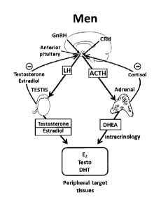
[0010] In addition to testosterone, the testis, through the action of
aromatase
secretes the estrogens estrone and estradiol (Figure 1). The secretion of
luteinizing hormone (LH) by the anterior pituitary gland is stimulated by the
pulsatile secretion of GnRH (Gonadotropin-Releasing Hormone) from the
hypothalamus while both testosterone and estradiol exert global inhibitory
effects
at the hypothalamo-pituitary level on LH secretion (Corona, Rastrelli et al.
2012).
LH then stimulates testosterone secretion by the Leydig cells in the testis
(Figure 1).
[0011] The guidelines of the US Endocrine Society recommend testosterone
treatment only in men with "consistent symptoms and signs and unequivocally
low serum testosterone levels". However, it has been found that only half the
men receiving testosterone replacement therapy were diagnosed with male
hypogonadism. In fact, 34% were treated for fatigue, 31% for erectile
dysfunction
and 12% for psychosexual dysfunction (Baillaroeon, Urban et al. 2013).
[0012] As mentioned above, it must be considered that up to 50% of total
androgens in men are made locally in peripheral tissues from DHEA that
CA 02942026 2016-09-09
WO 2015/135061
PCT/CA2015/000142
4
decreases with age by as much as 80% on average in men aged 75 years or
more (Labrie, Belanger et al. 1997b), thus providing a reason why low DHEA has
at least an equal role compared to low serum testosterone to explain the
symptoms and signs so-far attributed to male hypogonadism (Labrie. Belanger et
al. 1997a).
[0013] The Endocrine Society has a Clinical Practice Guideline on testosterone
therapy, namely Testosterone Therapy in Men with Androgen Deficiency
Syndrome (2006; revised 2010) at www.endocrine.org. It includes the revised
recommendations on the Prostate Specific Antigen exclusion criteria and PSA
follow-up guidance.
[0014] Low testosterone can be accompanied by any single or a combination of
the following signs or symptoms:
- loss of libido (interest in sex)
- difficulty in getting an erection (erectile dysfunction)
- tiredness and lack of energy (loss of energy, energy loss)
- depression
- loss of bone (decreased bone mineral density and increased risk of
fracture)
- loss of muscle and muscle weakness
- loss of body hair
- fertility problems
[0015] Additional benefits such as treatment or reduction of the likelihood or
risk
of acquiring the following medical problems, namely hypercholesterolemia,
hyperlipidemia, atherosclerosis, hypertension, Alzheimer's disease, loss of
memory, loss of cognition, dementia, insomnia, cardiovascular diseases,
insulin
resistance, Type 2 diabetes and obesity (especially abdominal obesity)
(Comhaire 2000; Ding, Song et al. 2006; Khaw, Dowsett et al. 2007; Bassil
Alkaade et al. 2009; Zitzmann 2009) are also provided by treatment with the
CA 02942026 2016-09-09
WO 2015/135061
PCT/CA2015/000142
invention.
[0016] Low serum testosterone in men is associated with low muscle mass,
decreased muscle strength and poor mobility (Roy, Blackman et al. 2002;
Schaap, Pluijrn et al. 2005). Testosterone supplementation in healthy older
men
increases muscle mass and strength and leg power, these being important
factors of mobility (Bhasin. Storer et al. 1996; Sih, Morley et al. 1997;
Snyder,
Peachey et al. 1999; Storer, Maoliano et al. 2003; Bhasin, Woodhouse et al.
2005; Page, Amory et al. 2005).
[0017] Symptoms/signs of androgen deficiency in aging males can be as stated
in the Clinical Practice Guideline of the Endocrine Society (Bhasin,
Cunningham
et al. 2006).
CA 02942026 2016-09-09
WO 2015/135061
PCT/CA2015/000142
6
Table 1A: Symptoms and signs suggestive of androgen deficiency in
aging men
= Reduced sexual desire (libido) and activity
= Decreased spontaneous erections
= Breast discomfort, gynecomastia
= Loss of body (axillary and pubic) hair, reduced shaving
= Very small or shrinking testes (especially <5 mL)
= Inability to father children, low or zero sperm counts
= Height loss, low trauma fracture, low bone mineral density
= Reduced muscle bulk and strength
= Hot flushes, sweats
Table 1B: Other symptoms and signs associated with androgen
deficiency that are less specific than those in Table 1A
= Decreased energy, motivation, initiative, aggressiveness, self-confidence
= Feeling sad or blue, depressed mood, dysthymia
= Poor concentration and memory
= Sleep disturbance, increased sleepiness
= Mild anemia (normochromic, normocytic, in the female range)
= Increased body fat, body mass index
= Diminished physical or work performance
(Bhasin, Cunningham et al. 2006)
[0018] Aging itself is often associated with a decline in sexual functioning
in men
(Vermeulen 2003; Ebert, Jockenhovel et al. 2005).
[0019] The diagnosis of male hypogonadism can be helped by the
ANDROTEST (Corona, Jannini et al. 2006; Corona, Mannucci et al. 2006).
Differential diagnosis can also be helped by the information provided by
(Corona,
Rastrelli et al. 2012). LOH has also been defined by the presence of at least
three sexual symptoms associated with a total testosterone level of less than
3.2 ng/mL (Wu, Tajar et al. 2010). In that study performed in a random
population sample of 3369 men aged 40 to 79 years, differences between
asymptomatic and symptomatic men in relation with serum testosterone were
minimal. One possible explanation could be, as indicated above, that serum
CA 02942026 2016-09-09
-7-
testosterone is not the exclusive source of androgenic activity which is, as
mentioned above, up to 50% from DHEA-derived androgens (Labrie, Dupont et
al. 1985; Labrie 2011).
[0020] The physiological role of testosterone in male sexual behavior is
poorly
understood. Many studies with attempts to correlate male sexual behavior and
the concentration of serum testosterone have given conflicting results. There
are wide variations between serum testosterone levels and erectile dysfunction
(Salmimies, Kockott et at. 1982; Gooren 1987; Bhasin, Cunningham et at. 2006;
Traish, Guay et at. 2009). It remains, however, that low serum testosterone
has
become standard clinical practice in the evaluation of sexual disorders in
men.
[0021] Diagnosis of late-onset male hypogonadism can be helped by
questionnaires, although clinical evaluation of the total clinical picture is
of major
importance. The instruments which can be used are, without limitation,
Androgen
Deficiency in Aging Males (ADAM) (Morley, Charlton et al. 2000), the Aging
Males Symptoms (AMS) Rating Scale (Moore, Huebler et al. 2004) and the
Massachusetts Male Ageing Study (MMAS) Questionnaire (Smith, Feldman et
al. 2000). Diagnosis can be helped with the Brief Sexual Function Inventory
(BSFI) (O'Leary, Fowler et al. 1995). The instrument covers sexual drive (two
items), erection (three items), ejaculation (two items), perception of
problems in
each area (three items) and overall satisfaction (one item).
[0022] There is an emerging medication for the treatment of male
hypogonadism (see the two following recent reviews: (Corona, Rastrelli et at.
2012; Kim, Crosnoe et al. 2013)). In addition to the existing exogenous
testosterone treatment, clinical data with selective estrogen receptor
modulators
(SERMs) are available. A SERM binds to the estrogen receptor in the
hypothalamus and pituitary gland in competition with estradiol. The
neutralization
of inhibitory action of estradiol in the hypothalamus increases GnRH
(gonadotropin-releasing hormone) secretion which stimulates LH secretion which
increases testosterone production by the testes. Several studies with
clomiphene
8
citrate have been performed. Clomiphene citrate increases serum testosterone
levels in the blood like the use of testosterone gels (Taylor and Levine
2010).
Clomiphene citrate improves sexual function in hypogonadal men (Guav,
Jacobson et al. 2003). Clomiphene citrate improves the testosterone-estradiol
ratio in hypogonadal men (Shabsioh, Kano et al. 2005). Clomiphene citrate
increases circulating testosterone and improves several hypogonadism-related
symptoms (decreased libido, lack of energy) in young hypogonadal men (Katz
Nabulsi et al. 2011).
[0023] Enclomiphene (Androxal; Repros) is under development for male
hypogonadism and infertility. Patents literature also indicates that SERMs or
antiestrogens could be useful for male androgen deficiency including male
hypogonadism (US 2006/0293294, US 2009/0215733, WO 01/91744,
W003/072092, W02006/024689 and W020131123218) and in combination
with other active agents (US 2007/0078091 and WO 2013/130832). Other
classes of compounds have been suggested to treat male hypogonadism,
namely gonadotropins, 5a-reductase inhibitors, testosterone precursors, non-
aromatizable androgens, aromatase inhibitors, selective estrogen receptor i3
agonists and selective androgen receptor modulators (SARMs). Gonadotropin
therapy remains one of the few effective treatments for infertility in men
with
secondary hypogonadism (Liu. Baker et al. 2009; Farhat, Al-zidiali et al.
2010).
Human chorionic gonadotropin is an LH analogue that stimulates Leydig cell
production of testosterone and it can be derived from urine as well as
recombinant sources.
[0024] In particular, the treatment includes the administration of a precursor
of
sex steroids in combination with a cell-specific selective estrogen receptor
modulator (SERM), in particular acolbifene.
[0025] The invention also provides kits and pharmaceutical compositions for
practicing the foregoing combination.
CA 2942026 2018-01-29
CA 02942026 2016-09-09
WO 2015/135061
PCT/CA2015/000142
9
[0026] It is known that a large number of diseases, conditions and undesirable
symptoms respond favorably to administering exogenous sex steroids, or
precursors thereof. For example, estrogens are believed to decrease the rate
of
bone loss while androgens have been shown to build bone mass by stimulating
bone formation.
[0027] Long-term testosterone treatment in hypogonadal men improves
metabolic syndrome components. It reduced total cholesterol, low-density
lipoprotein cholesterol, tryglycerides and increased HDL cholesterol levels.
It also
reduced blood glucose levels (Traish, Haider et al. 2013).
[0028] Treatment with dihydrotestosterone (DHT) for 2 years had no effect on
prostate volume but decreased fat mass, increased lean mass, suppressed
serum testosterone and decreased spinal bone mineral density, probably due to
inhibition of LH secretion. Many other studies have shown the benefits of
androgen replacement therapy with no significant change of prostatic volume or
urinary symptoms (Sih, Morley et al. 1997; Kenny, Prestwood et al. 2001; Marks
Mazer et al. 2006; Saad, Gooren et al. 2008; Takao, Tsujimura et al. 2009). In
a
10-year study with oral testosterone undecanoate, no increase in prostate size
and no evidence of cancer was noted (Gooren 1994).
[0029] In hypogonadal men, even an improvement of lower urinary tract
symptoms was observed (Pecherskv, Mazurov et al. 2002), for review see
(Amano, lmao et al. 2010; Shiciehara and Namiki 2011). Oral testosterone
undecanoate replacement for 8 months at doses of 40 to 160 mg/day did not
change the prostate size nor showed deterioration of voiding symptoms (Franchi
F, Luisi M et al. 1978). A study where 100 mg testosterone enanthate was
injected weekly for 3 months similarly did not change prostate volume or post
voiding residual volume (Tenover 1992).
[0030] In another study, androgen replacement therapy for 8 months increased
prostate volume by 18% with no change in uroflowmetry data (HoImang, Mann et
CA 02942026 2016-09-09
WO 2015/135061
PCT/CA2015/000142
at. 1993). No difference in prostate volume was observed in another study
(Behre, Bohmever et at. 1994).
[0031] Reduced libido and erectile dysfunction are considered as being the
most
proeminent symptoms of hypogonadism in men (Harman, Metter et al. 2001;
Matsumoto 2002). In the Massachusetts Male Aging Study, the prevalence of
complete erectile dysfunction increased 3-fold from 5% to 15% between the ages
40 and 70 years (Morley 2003).
[0032] In the European Male Aging Study (EMAS), on the other hand, a
correlation was found between low serum testosterone and the symptoms poor
morning erection, low sexual desire and erectile dysfunction (testosterone
range
2.3 to 3.7 ng/mL) leading to the LOH (Late-Onset Hypogonadism) definition in
men having the 3 symptoms and serum testosterone less than 3.2 ng/mL or
11 nmole per liter (Wu, Tajar et al. 2010). Testosterone controls gonadotropin
secretion, masculinization during sexual maturation, induction and maintenance
of sperm production, as well as libido and sexual function.
[0033] Both estrogens derived from androgens and androgens themselves exert
a global negative effect on GnRH/LH secretion (Figure 1). Estradiol, while
being
at much lower concentrations in the blood, is an efficient inhibitor of
GnRH/LH
secretion.
[0034] Serum testosterone levels vary significantly as a result of circadian
and
circannual rhythms, episodic secretion, and measurement variations.
Testosterone concentrations may be affected by illness and certain medications
(e.g. opiates and glucocorticoids).
[0035] In the TOM trial performed in men older than 65 years with chronic
conditions and limitations in mobility, twice as many adverse events (AEs)
were
reported in the testosterone gel versus the placebo groups (Basaria, Coviello
et
at. 2010). In that relatively small group (testosterone in older men with
mobility
CA 02942026 2016-09-09
WO 2015/135061
PCT/CA2015/000142
11
limitations, TOM) of 209 men with serum testosterone of 1.0 to 3.5 ng/mL with
a
high prevalence of chronic disease, namely hypertension, hyperlipidemia,
diabetes and obesity, a higher incidence of cardiovascular events in the
testosterone gel group stopped the trial. Greater improvement of leg-press and
chest-press strength and in stair climbing while carrying a load was seen in
the
testosterone-treated versus placebo groups (Basaria, CoyleIlo et al. 2010).
The
risk of cardiovascular AEs was greater in testosterone-treated men.
[0036] Testosterone replacement therapy is also associated with infertility as
side effect due to decreased sperm count as well as decrease in testicular-
size.
[0037] Testosterone injections have the advantage of low cost but have the
disadvantage of non physiological peak and trough levels over the weekly,
bi-weekly or long term dosing regimen.
[0038] In a group of 8709 Veteran Administration patients with serum
testosterone <3.0 ng/mL, after a median of 531 days post coronography, 1223 of
them started testosterone therapy (Vigen, O'Donnell et al. 2013). In that
retrospective observational study, the rates of deaths at 3 years were 15.4%
vs
18.5% in the control and testosterone groups, respectively. As stated, "this
signal warrants cautions testosterone prescribing...." (Capp la 2013).
[0039] Metaanalysis of testosterone therapy trials, except the TOM trial,
however, did not demonstrate adverse cardiovascular events (Calof, Singh et
al.
2005; Haddad, Kennedy et al. 2007; Fernandez-Balsells, Murad et al. 2010).
[0040] Testosterone replacement therapy has been associated with increased
sexual functioning and mood (Seftel, Mack et al. 2004; Wang, Cunningham et al.
2004).
[0041] In addition to improving sexual function (Wang, Swerdloff et al. 2000;
lsidori, Giannetta et al. 2005; Bolona, Uraga et al. 2007), the administration
of
CA 02942026 2016-09-09
WO 2015/135061
PCT/CA2015/000142
12
testosterone to men with symptomatic androgen deficiency increases bone
mineral density (Snyder, Peachev et al. 2000; Isidori, Giannetta et at. 2005)
increases fat-free mass (lsidori, Caprio et al. 1999; Snyder, Peachey et al.
2000;
Isidori, Giannetta et at. 2005) and strength (Sih, Morley et at. 1997),
improves
insulin resistance (Jones and Saad 2009; Jones, Arver et al. 2011) and
improves
the lipid profile (Mann, Holmang et al. 1993; Jones and Saad 2009; Jones,
Arver
et al. 2011).
[0042] A significant problem with testosterone replacement therapy is that it
suppresses testicular endogenous testosterone secretion and can result in
azoospermia or impairment of spermatogenesis as indicated by the labeling
accepted by the Food and Drug Administration (Kim, Crosnoe et at. 2013).
Exogeneous testosterone inhibits the hypothalamo-pituitary-testicular axis and
can result in infertility. Intramuscular testosterone has even been studied as
a
contraceptive agent (Liu, Swerdloff et at. 2006). In the present invention,
the low
testicular testosterone formation secondary to inhibition of LH secretion is
avoided by the use of a SERM, in particular acolbifene that stimulates LH
secretion instead of blocking endogenous LH and, secondarily, testosterone
secretion.
CA 02942026 2016-09-09
WO 2015/135061
PCT/CA2015/000142
13
SUMMARY OF THE INVENTION
[0043] It is an object of the present invention to provide a method of
preventing,
reducing or eliminating the incidence of male androgen deficiency symptoms or
diseases including male hypogonadism-associated symptoms and diseases due
to low testosterone and/or low peripheral androgen formation.
[0044] It is another object to provide methods of preventing, reducing or
eliminating the incidence of loss of libido, erectile dysfunction, tiredness,
loss of
energy, depression, bone loss, muscle loss, muscle weakness, fat accumulation,
memory loss, cognition loss, Alzheimer's disease, dementia, loss of body hair,
fertility problems, insomnia, gynecomastia, anemia, hot flushes, sweats,
decreased sense of well-being, obesity, osteoporosis, hypercholesterolemia,
hyperlipidemia, atherosclerosis, hypertension, insulin resistance,
cardiovascular
disease and type 2 diabetes.
[0045] It is another object to provide methods of reducing the risk of the
male
patients acquiring breast cancer.
[0046] It is another object to provide kits and pharmaceutical compositions
suitable for use in the above methods. Preferably, these products are packaged
with directions for using the contents thereof for preventing, reducing or
eliminating the incidence of male androgen deficiency symptoms or diseases
including male hypogonadism-associated symptoms and diseases.
[0047] It is another object to provide kits and pharmaceutical compositions
suitable for use in the above methods. Preferably, these products are packaged
with directions for using the contents thereof for preventing, reducing or
eliminating the incidence of loss of libido, erectile dysfunction, tiredness,
loss of
energy, depression, bone loss, muscle loss, muscle weakness, fat accumulation,
memory loss, cognition loss, Alzheimer's disease, dementia, loss of body hair,
fertility problems, insomnia, gynecomastia, anemia, hot flushes, sweats,
CA 02942026 2016-09-09
WO 2015/135061
PCT/CA2015/000142
14
decreased sense of well-being, obesity, osteoporosis, hypercholesterolemia,
hyperlipidemia, atherosclerosis, hypertension, insulin resistance,
cardiovascular
disease and type 2 diabetes.
[0048] In one embodiment, the invention provides a method of preventing,
reducing or eliminating the incidence of male androgen deficiency symptoms or
diseases including male hypogonadism-associated symptoms and diseases, said
method comprising administering to male patient in need of said prevention,
reduction or elimination, a therapeutically effective amount of a sex steroid
precursor or prodrug thereof in association with a therapeutically effective
amount of a selective estrogen receptor modulator or an antiestrogen or
prodrug
of either.
[0049] It is preferred that the sex steroid precursor is selected from the
group
consisting of dehydroepiandrosterone, dehydroepiandrosterone-sulfate, androst-
5-ene-313,1713-diol, 4-androstene-3,17-dione, and a prodrug of any of the
foregoing additional agents.
[0050] It is preferred that the selective estrogen receptor modulator is
selected
from the group comprising of Tamoxifen, Toremifene, CC 8490, SERM 3471,
HMR 3339, HMR 3656, Raloxifene, LY 335124, LY 326315, Arzoxifene
(LY 353381), Pipendoxifene (ERA 923), Bazedoxifene (TSE 424, WAY 140424),
Oporia (Lasofoxifene), EM-652, EM-800, EM-652-HCI (acolbifene, EM-1538),
4-hydroxy-Tamoxifen, 4-hydroxy-Toremifene, Droloxifene, LY 335563, GW-5638,
Idoxifene, Levormeloxifene, Iproxifen (TAT-59), Ospemifene (FC 1271),
Fispemifene, Centchroman, CHF 4227, LY 2066948, LY 2120310, Sivifene,
SR 16234, Clomiphene, Enclomiphene, Zuclomiphene, GW 7603, BL 3040,
SRI 16158, SR 16157, SRI 16137, SR 16137, Rad 1901,
(+)-3-(4-
hydroxypheny1)-24442-(1-piperidinypethoxy]phenylj-4-(trifluoromethyl)-2H-1-
benzopyran-7-ol, Femarelle, Nafoxidine and Endoxifen.
[0051] It is preferred that the antiestrogen is selected from the group
comprising
CA 02942026 2016-09-09
WO 2015/135061
PCT/CA2015/000142
of Faslodex (ICI 182780, fulvestrant, 7a49-
(4,4,5,5,5-pentafluoro-
pentylsulphinyl)nonyl]oestra-1,3,5(10)-triene-3, 17p-diol), ICI 164384,
CH 4893237, ZK 246965 and SH 646.
[0052] It is preferred that the selective estrogen receptor modulator has one
of
the following formulae selected from the group comprising of:
R2
G3
R1 _________________
/G1
R100-1-
G2
wherein R1 and R2 are independently hydrogen, hydroxyl, halogen, Cl-C6 alkyl
or
a moiety which is converted to hydroxyl in vivo;
wherein Z is either absent or selected from the group consisting of ¨CH2-, ¨0-
,
-S- and ¨NR3- (R3 being hydrogen or C1-06 alkyl);
wherein the R100 is a bivalent moiety which distances L from the B-ring by 4-
10
intervening atoms;
wherein L is a bivalent or trivalent moiety selected from the group of -SO-,
-CON<, -N<, and -SON<;
wherein G1 is selected from the group consisting of hydrogen, a C1 to
C5 hydrocarbon, a bivalent moiety which in combination with G2 and L is a 5-
to
7-membered heterocyclic ring, and halo or unsaturated derivatives of the
foregoing;
wherein G2 is either absent or selected from the group consisting of hydrogen,
a
CA 02942026 2016-09-09
WO 2015/135061
PCT/CA2015/000142
16
Ci to C5 hydrocarbon, a bivalent moiety which in combination with G1 and L is
a
5- to 7-membered heterocyclic ring, and halo or unsaturated derivatives of the
foregoing;
wherein G3 is selected from the group consisting of hydrogen, methyl, ethyl
and
trifluoromethyl;
R2
G3
0
0
0
0
or a pharmaceutically acceptable salt thereof,
wherein D is -OCH2CH2N(R3)R4 (R3 and R4 either being independently selected
from the group consisting of C1-C4 alkyl, or R3, R4 and the nitrogen atom to
which
they are bound, together being a ring structure selected from the group
consisting of pyrrolidinyl, 2,2-dimethylpyrrolidinyl, 2-methylpyrrolidinyl,
piperidino,
hexamethyleneimino, and morpholino);
wherein R1 and R2 are independently selected from the group consisting of:
hydrogen, hydroxyl, halogen, C1-C6 alkyl, and a moiety converted in vivo to
hydroxyl;
wherein G3 is selected from the group consisting of hydrogen, methyl, ethyl
and
trifluoromethyl;
CA 02942026 2016-09-09
WO 2015/135061
PCT/CA2015/000142
17
5'
6' (¨) 4'
4 ¨R2
6 3'
3 2'
R1-10
2"
3
8
R
6" 3
4"
5,,
or a pharmaceutically acceptable salt thereof;
wherein a benzopyran compound which is an optically active compound having
an absolute configuration S on carbon 2;
wherein R1 and R2 are independently selected from the group consisting of
hydroxyl, halogen, Ci-C6 alkyl, and a moiety convertible in vivo to hydroxyl;
wherein R3 is a species selected from the group consisting of saturated,
unsaturated or substituted pyrrolidinyl, saturated, unsaturated or substituted
piperidino, saturated, unsaturated or substituted piperidinyl, saturated,
unsaturated or substituted morpholino, nitrogen-containing cyclic moiety,
nitrogen-containing polycyclic moiety, and NRaRb (Ra and Rb being
independently hydrogen, straight or branched C1-C6 alkyl, straight or branched
C2-C6 alkenyl, or straight or branched C2-C6 alkynyl);
wherein a salt of an acid selected from the group consisting of acetic acid,
adipic
acid, benzenesulfonic acid, benzoic acid, camphorsulfonic acid, citric acid,
fumaric acid, hydroiodic acid, hydrobromic acid, hydrochloric acid,
hydrochlorothiazide acid, hydroxy-naphthoic acid, lactic acid, maleic acid,
methanesulfonic acid, methylsulfuric acid, 1,5-naphthalenedisulfonic acid,
nitric
acid, palmitic acid, pivalic acid, phosphoric acid, propionic acid, succinic
acid,
CA 02942026 2016-09-09
WO 2015/135061
PCT/CA2015/000142
18
sulfuric acid, tartaric acid, terephthalic acid, p-toluenesulfonic acid, and
valeric
acid.
[0053] In another embodiment, the invention provides a method which further
comprising administering as part of a combination therapy, a therapeutically
effective amount of human chorionic gonadotropin.
[0054] In another embodiment, the invention provides a pharmaceutical
composition comprising:
a) a pharmaceutically acceptable excipient, diluent or carrier;
b) a therapeutically effective amount of at least one sex steroid precursor or
prodrug thereof; and
c) a therapeutically effective amount of at least one SERM, antiestrogen or
prodrug.
[0055] In another embodiment, the invention provides a pill, a tablet, a
capsule,
a gel, a cream, an ovule, a rectal suppository, or an injection comprising:
a) a pharmaceutically acceptable excipient, diluent or carrier;
b) a therapeutically effective amount of at least one sex steroid precursor or
prodrug thereof; and
C) a therapeutically effective amount of at least one SERM, antiestrogen or
prodrug.
[0056] In another embodiment, the invention provides a kit comprising a first
container containing a pharmaceutical formulation comprising a therapeutically
effective amount of at least one sex steroid precursor or a prodrug thereof;
and
said kit further comprising a second container containing a pharmaceutical
formulation comprising a therapeutically effective amount of at least one
SERM,
antiestrogen or prodrug as part of combination therapy.
[0057] In another embodiment, the invention pertains to a method of
preventing,
reducing or eliminating the incidence of male androgen deficiency symptoms or
CA 02942026 2016-09-09
WO 2015/135061
PCT/CA2015/000142
19
diseases including male hypogonadism-associated symptoms and diseases by
increasing levels of a sex steroid precursor selected from the group
consisting of
dehydroepiandrosterone (DHEA), dehydroepiandrosterone-sulfate (DHEA-S),
androst-5-ene-3p,17p-diol (5-diol) and 4-androstene-3,17-dione in a patient in
need of said prevention, reduction or elimination of the incidence, and
further
comprising administering to said patient a therapeutically effective amount of
at
least one SERM, antiestrogen or prodrug as part of combination therapy.
[0058] In another embodiment, the invention pertains to a method of
preventing,
reducing or eliminating the incidence of male androgen deficiency symptoms or
diseases including male hypogonadism-associated symptoms and diseases by
increasing levels of circulating testicular testosterone by the action of SERM
or
antiestrogen in a patient in need of said prevention, reduction or elimination
of
the incidence, and further comprising administering to said patient a
therapeutically effective amount of at least one sex steroid precursor or
prodrug
as part of combination therapy.
[0059] In another embodiment, the invention provides a method of preventing,
reducing or eliminating the incidence of male androgen deficiency symptoms or
diseases including male hypogonadism-associated symptoms and diseases by
increasing the levels of circulating androgen metabolites consisting of
androsterone glucuronide (ADT-G), androstane-3a,17p-dioI-3-glucuronide
(3a-dioI-3G) and androstane-3a,1713-di01-17-glucuronide (3a-dio1-17G), said
method comprising administering to male patient in need of said prevention,
reduction or elimination, a therapeutically effective amount of a sex steroid
precursor or prodrug thereof in association with a therapeutically effective
amount of a selective estrogen receptor modulator or an antiestrogen or
prodrug
of either.
CA 02942026 2016-09-09
WO 2015/135061
PCT/CA2015/000142
[0060] As used herein, "Pure SERM" means that the SERM does not have any
estrogenic activity in breast or uterine tissue at physiological or
pharmacological
concentrations.
[0061] In another embodiment, the invention provides a kit comprising a first
container containing a therapeutically effective amount of at least one
precursor
of sex steroids and further comprising a second container containing a
therapeutically effective amount of at least one SERM.
[0062] In another embodiment, the invention provides, in one container, a
pharmaceutical composition comprising:
a) a pharmaceutically acceptable excipient, diluent or carrier;
b) a therapeutically effective amount of at least one precursor of sex
steroids; and
c) a therapeutically effective amount of at least one SERM.
[0063] In another embodiment, the invention provides a method of preventing,
reducing or eliminating the incidence of male androgen deficiency symptoms or
diseases including male hypogonadism-associated symptoms and diseases, said
method comprising administering to male patient in need of said prevention,
reduction or elimination, a therapeutically effective amount of a sex steroid
precursor or prodrug thereof in association with a therapeutically effective
amount of a selective estrogen receptor modulator or an antiestrogen or
prodrug
of either, wherein the selective estrogen receptor modulator or antiestrogen
stimulates LH secretion which increases the level of circulating testosterone.
[0064] In another embodiment, the invention provides a pharmaceutical
composition for preventing, reducing or eliminating the incidence of male
androgen deficiency symptoms or diseases including male hypogonadism-
associated symptoms and diseases comprising:
a) a pharmaceutically acceptable excipient, diluent or carrier;
CA 02942026 2016-09-09
WO 2015/135061
PCT/CA2015/000142
21
b) at least one sex steroid precursor or prodrug thereof; and
C) at least one selective estrogen receptor modulator or an antiestrogen or
prodrug of either;
wherein said pharmaceutical composition is provided in packaging that directs
use of said composition for prevention, reduction or elimination of at least
one
male androgen deficiency symptom or disease.
[0065] In another embodiment, the invention provides a kit for preventing,
reducing or eliminating the incidence of male androgen deficiency symptoms or
diseases including male hypogonadism-associated symptoms and diseases,
comprising (i) a first container having therein at least one sex steroid
precursor or
a prodrug thereof; (ii) a second container having therein at least one
selective
estrogen receptor modulator, or an antiestrogen or prodrug of either of the
foregoing; and (iii) instructions for using the kit for the prevention,
reduction or
elimination of at least one male androgen deficiency symptom or disease.
[0066] It is preferred that the sex steroid precursor is
dehydroepiandrosterone
and the selective estrogen receptor modulator is acolbifene.
[0067] As used herein, compounds administered to a patient "in association
with" other compounds are administered sufficiently close to administration of
said other compound that a patient obtains the physiological effects of both
compounds simultaneously, even though the compounds were not administered
in close time proximity. When compounds are administered as part of a
combination therapy they are administered in association with each other.
Preferred SERM (acolbifene) discussed herein is preferably used in combination
with preferred sex steroid precursors dehydroepiandrosterone,
dehydroepiandrosterone-sulfate, androst-5-ene-313,1713-diol or 4-androstene-
3,17-dione, especially dehydroepiandrosterone.
CA 02942026 2016-09-09
WO 2015/135061
PCT/CA2015/000142
22
[0068] The applicant believes that the addition of a precursor of sex steroids
to
acolbifene treatment will increase intracellular levels of testosterone (as
well
demonstrated in patients with prostate cancer where intracellular androgens,
especially dihydrotestosterone, is coming from endogenous DHEA (Labrie
Dupont et al. 1985; Labrie, Cusan et al. 2009; Labrie 2011)).
[0069] As used herein, a SERM is a compound that functions as an estrogen
receptor antagonist (antiestrogen) in breast tissue, yet provides estrogenic
or
estrogen-like effect on bone tissue and on serum cholesterol levels (i.e. by
reducing serum cholesterol). Non-steroidal compounds that function as estrogen
receptor antagonists in vitro or in human or rat breast tissue (especially if
the
compound acts as an antiestrogen on human breast cancer cells) is likely to
function as a SERM. Conversely, steroidal antiestrogens tend not to function
as
SERMs because they tend not to display any beneficial effect on serum
cholesterol. Non-steroidal antiestrogens we have tested and found to function
as
SERMs include EM-800, EM-652.HCI, raloxifene, tamoxifen, 4-hydroxy-
tamoxifen, toremifene, 4-hydroxy-toremifene,
droloxifene, LY 353 381,
LY 335 563, GW-5638, lasofoxifene, bazedoxifene (TSE 424; WAY-TSE 424;
WAY 140424; 1-[[4 [2-(hexahydro-1H-azepin-1-yl)ethoxy]phenylimethy11-2-(4-
hydroxyphenyl)-3 methyl-1H-indo1-5-ol), pipendoxifene (ERA 923;
2-(4-
hydroxypheny1)-3-methy1-14[442-(1- piperidinypethoxylphenyl]methy1]-1H-indo1-
5-ol) ospemifene and idoxifene, but are not limited to these compounds.
[0070] But we have found also that all SERMs do not react in the same manner
and may be divided into two subclasses: "pure SERMs" and "mixed SERMs".
Thus, some SERMs like EM-800 and EM-652.HCI do not have any estrogenic
activity in breast and endometrial tissues at physiological or pharmacological
concentrations and have hypocholesterolemic and hypotriglyceridemic effects in
the rat. These SERMS may be called "pure SERMs''. The ideal SERM is a pure
SERM of the type EM-652.HCI because of its potent and pure antiestrogenic
activity in the mammary gland. Others, like raloxifene, tamoxifen,
droloxifene,
CA 02942026 2016-09-09
WO 2015/135061
PCT/CA2015/000142
23
4-hydroxy-tamoxifen (1¨(4-dimethylaminoethoxyphenyI)-1-(4-hydroxylpheny1)-
2¨phenyl¨but-1¨ene), toremifene, 4-hydroxy-toremifene [(Z)-(2)-244-(4¨chloro-
1¨(4¨hydroxypheny1)-2¨phenyl-1¨butenyl)phenoxy]¨N, N¨dimethylethanamine),
LY 353 381, LY 335 563, GW-5638, lasofoxifene, idoxifene, bazedoxifene and
ospemifene have some estrogenic activities in the breast and endometrium. This
second series of SERMs may be called "mixed SERMs". The unwanted
estrogenic activities of these "mixed SERMs" may be inhibited by addition of
pure
"SERMs" as shown in Figures 2 and 3 in vitro tests and in Figure 4 in an in
vivo
test of breast cancer. Since human breast carcinoma xenografts in nude mice
are the closest available model of human breast cancer, we have thus compared
the effect of EM-800 and tamoxifen alone and in combination on the growth of
ZR-75-1 breast cancer xenografts in nude mice.
[0071] In one embodiment, the invention uses selective estrogen receptor
modulators of the following molecular structure
0¨R2
R1 -0I
I NI
0
wherein R1 and R2 are independently hydrogen, hydroxyl or a moiety which is
converted to hydroxyl in vivo, and n=1 or 2.
[0072] The applicant believes that it is very important that SERMs of the
invention act as pure antiestrogens in breast because SERMs have to counteract
potential side-effects of estrogens, particularly those formed from the
exogenous
precursors of sex steroids which can increase the proliferation of this
tissue.
24
Particularly, the applicant believes that benzopyran derivatives of the
invention
having the absolute configuration 2S at position 2 is more suitable than its
racemic mixture. Thus, in
US 6,060,503, optically active benzopyran
antiestrogens having 2S configuration are disclosed to treat estrogen-
exacerbated breast and endometrial cancer and these compounds are shown to
be significantly more efficient than racemic mixtures (See Figures 1-5 of
US 6,060,503).
[0073] The enantiomer of 2S configuration being difficult to be industrially
obtained as a pure state, the applicant believes that less than 10%,
preferably
less than 5% and more preferably less than 2% by weight of contamination by
the 2R enantiomer is preferred.
[0074] Prodrug forms of active pharmaceutical ingredient are well known in the
art. See, e.g. H. Bundgaard "5. Design and Application of Prodrugs" (In A
textbook of Drug Design and Development. Edited by P. Krogsgaard-Larsen and
H. Bundgaard; Harwood Academic Publishers GmbH, Chur, Switzerland, 1991). In
particular, see page 114 defining prodrug: a prodrug is a pharmacologically
inactive
derivative of a parent drug molecule that requires spontaneous or enzymatic
transformation within the body in order to release the active drug, and that
has
improved delivery properties over the parent drug molecule. In the present
application,
the prodrugs of sex steroid precursor are derivatives of the 3- and/or 17-
hydroxyl
group(s) and/or 3- and/or 17-ketone group(s), and the prodrugs of selective
estrogen
receptor modulators and antiestrogens are derivatives of the hydroxyl group.
The
prodrug forms of the hydroxyl group are esters, carbonate esters, phosphate
esters,
ethers, and a-acyloxyalkyl ethers, and the prodrug forms of the ketone group
are
ketals, imines, enol esters, oxazolidines and thiazolidines but not limited by
these
examples (see page 154). The previous-cited SERM EM-800 (diester derivative,
dipivaloate) is a prodrug of EM-652 (Gauthier, Caron et al. 1997).
CA 2942026 2018-01-29
CA 02942026 2016-09-09
WO 2015/135061
PCT/CA2015/000142
[0075] Serum testosterone is higher in the morning and decreases to a
minimum concentration after sleep (Trenell, Marshall et al. 2007). Serum
testosterone should be monitored (with the judgment of the treating physician
concerning its frequency) at months 1 and 2 of treatment and then every
3 months to assure proper increases in serum testosterone. Similar
measurements should be made for DHEA. Serum DHEA also follows a circadian
rythm being lowest in the morning. For proper comparison, it is preferable to
measure serum testosterone and DHEA at the same time of the day at different
treatment time intervals, i.e., at month 1 and 2 and then every 3 months.
CA 02942026 2016-09-09
WO 2015/135061
PCT/CA2015/000142
26
BRIEF DESCRIPTION OF THE DRAWINGS
[0076] Figure 1 is a schematic representation of the hypothalamo-pituitary-
testicular and hypothalamo-pituitary-adrenal axes. GnRH,
gonadotropin-
releasing hormone; CRH, corticotropin-releasing hormone; LH, luteinizing
hormone; ACTH, adrenocorticotropin; DHEA,
dehydroepiandrosterone;
E2, estradiol; DHT, dihydrotestosterone; Testo, testosterone.
[0077] Figure 2 shows the effect of increasing concentrations of EM-800
(prodrug of acolbifene, free salt), (Z)-4-0H-tamoxifen, (Z)-4-0H-toremifene
and
raloxifene on alkaline phosphatase activity in human endometrial cancer
lshikawa cells. Alkaline phosphatase activity was measured after a 5-day
exposure to increasing concentrations of indicated compounds in the presence
or absence of 1.0 nM E2. The data are expressed as the means SEM of four
wells. When SEM overlaps with the symbol used, only the symbol is shown
(Simard, Sanchez et al. 1997).
[0078] Figure 3 shows the blockade of the stimulatory effect of (Z)-4-0H-
tamoxifen, (Z)-4-0H-toremifene, droloxifene and raloxifene on alkaline
phosphatase activity by the antiestrogen EM-800 (prodrug of acolbifene, free
salt) in human lshikawa (endometrial) carcinoma cells. Alkaline phosphatase
activity was measured after a 5-day exposure to 3 or 10 nM of the indicated
compounds in the presence or absence of 30 or 100 nM EM-800. The data are
expressed as the means SD of eight wells with the exception of the control
groups were data are obtained from 16 wells (Simard, Sanchez et al. 1997).
[0079] Figure 4 shows that the stimulatory effect of tamoxifen on the growth
of
human breast cancer ZR-75-1 xenografts is completely blocked by simultaneous
administration of EM-652.HCI (acolbifene). Acolbifene, by itself, in agreement
with its pure antiestrogenic activity has no effect on tumor growth in the
absence
of tamoxifen.
CA 02942026 2016-09-09
WO 2015/135061
PCT/CA2015/000142
27
[0080] Figure 5. Schematic
representation of the adrenal and intracrine
steroidogenic pathways, DHEA, dehydroepiandrosterone; DHEA-S, DHEA-
sulphate; DHT, dihydrotestosterone; HSD, hydroxysteroid dehydrogenase.
[0081] Figure 6. Comparison of the serum concentrations of testosterone (A),
total androgenic pool (sum of ADT-G, 3a-dioI-3G and 30c-dio1-17G) (B) and EiS
(C) in castrated 69-80-year-old men (n=34) and intact 55-65-year-old
postmenopausal women (n=377) (Labrie, Belanger et al. 2006; Labrie, Cusan et
al. 2009).
[0082] Figure 7 shows the effect of 12-month
treatment with
dehydroepiandrosterone (DHEA) alone or in combination with Flutamide or EM-
800 (prodrug of acolbifene, free salt) on trabecular bone volume in
ovariectomized rats. Intact animals are added as additional controls. Data are
presented as mean SEM ** p<0.01 versus OVX Control.
[0083] Figure 8 shows the effect of 12-month
treatment with
dehydroepiandrosterone (DHEA) alone or in combination with Flutamide or EM-
800 (prodrug of acolbifene, free salt) on trabecular number in ovariectomized
rats. Intact animals are added as additional controls. Data are presented as
mean SEM ** p<0.01 versus OVX Control.
[0084] Figure 9 shows proximal tibia metaphyses from intact control (A),
ovariectomized control (B), and ovariectomized rats treated with DHEA alone
(C)
or in combination with Flutamide (D) or EM-800 (prodrug of acolbifene, free
salt)
(E). Note the reduced amount of trabecular bone (T) in ovariectomized control
animals (B), and the significant increase in trabecular bone volume (T)
induced
after DHEA administration (C). The addition of Flutamide to DHEA partially
blocked the effect of DHEA on the trabecular bone volume (D), whereas the
combination of DHEA and EM-800 (prodrug of acolbifene, free salt) provided
CA 02942026 2016-09-09
WO 2015/135061
PCT/CA2015/000142
28
complete protection against the ovariectomy-associated bone loss. Modified
trichrome Masson-Goldner, magn.x80. T: Trabeculae, GP: Growth Plate.
[0085] Figure 10 shows the effect of treatment with DHEA (10 mg,
percutaneously, once daily) or EM-800 (prodrug of acolbifene, free salt) (75
pg,
orally, once daily) alone or in combination for 9 months on serum
triglycerides (A)
and cholesterol (B) levels in the rat. Data are expressed as the means SEM.
**: p<0.01 experimental versus respective control.
[0086] Figure 11 shows the effect of 37-week treatment with increasing doses
(0.01, 0.03, 0.1, 0.3, and 1 mg/kg) of EM-800 (prodrug of acolbifene, free
salt) or
raloxifene administered on total serum cholesterol levels in the
ovariectomized
rat. Comparison is made with intact rats and ovariectomized animals bearing an
implant of 1713-estradiol (E2); ** p<0.01, experimental versus OVX control
rats.
[0087] Figure 12: Schematic representation of the role of testicular and
adrenal
sources of sex steroids in men and the effect of adding acolbifene to
counteract
the inhibitory effect of estrogens at the hypothalamo-pituitary level on the
secretion of LH. ACTH, adrenocorticotropin; CRH, corticotropin-releasing
hormone; DH EA, dehydroepiandrosterone; DHT,
dihydrotestosterone;
E2, 1713-estradiol; LH, luteinizing hormone;
GnRH, gonadotropin-releasing
hormone.
[0088] Figure 13: Male cynomolgus monkeys were dosed orally with 2.5, 10 or
40 mg acolbifene/day for 13 weeks. Control monkeys received vehicle alone
(0.4% methylcellulose). End of study serum testosterone concentrations were
determined using a validated gas chromatography mass spectrometric assay.
Results are expressed as the mean SEM of 4 monkeys per group. P values
(versus control) were calculated using a two-sided t test assuming equality of
variances.
CA 02942026 2016-09-09
WO 2015/135061
PCT/CA2015/000142
29
[0089] Figure 14: Male cynomolgus monkeys were dosed orally with 3.13, 12.5
or 50 mg EM-800 (prodrug of acolbifene, free salt)/day for 52 weeks. Control
monkeys received vehicle alone (0.4% methylcellulose). End of study serum
testosterone concentrations were determined using a validated gas
chromatography mass spectrometric assay. Results are expressed as the
mean SEM of 5 (EM-800 study) monkeys per group. P values (versus control)
were calculated using a two-sided t test assuming equality of variances.
[0090] Figure 15 shows the effects of antiestrogens on ZR-75-1 tumor growth.
Effect of treatment with the antiestrogens tamoxifen, EM-652.HCI (acolbifene)
and the combination of tamoxifen and EM-652.HCI for 161 days, on the growth of
human ZR-75-1 breast tumors in ovariectomized nude mice. Tumor size is
expressed as the percentage of initial tumor area (Day 1=100%). Data is
expressed as means SEM (n=18-30 tumors/group); #/#p<0.01 vs EM-652.HCI
(acolbifene); "p<0.01 vs OVX. Antiestrogens were administered orally once
daily
at the dose of 20014/mouse in absence of estrogen stimulation.
[0091] Figure 16 Effect on uterine weight of increasing daily doses of the
antiestrogens CS-115-1 (EM-343) and EM-762 administered orally or
percutaneously by application on the skin for 9 days to ovariectomized mice
simultaneously treated by twice daily subcutaneous injection of estrone.
[0092] Figure 17 shows the effect on uterine weight of increasing
concentrations
of EM-652.HCI (acolbifene), lasofoxifene (free base; active and inactive
enantiomers) and raloxifene administered orally for 9 days to ovariectomized
mice simultaneously treated with estrone. *p<0.05, "p<0.01 versus El-treated
control.
[0093] Figure 18 shows the effect on uterine weight of 1 pg and 10 pg of EM-
652.HCI (acolbifene), lasofoxifene (free base; active and inactive
enantiomers)
CA 02942026 2016-09-09
WO 2015/135061
PCT/CA2015/000142
and raloxifene administered orally for 9 days to ovariectomized mice. **p<0.01
versus OVX control.
[0094] Figure 19 shows the effects of antiestrogens on ZR-75-1 tumor growth.
Effect of treatment with 7 antiestrogens for 161 days, on estrone-induced
growth
of human ZR-75-1 breast tumors in ovariectomized nude mice. Tumor size is
expressed as the percentage of initial tumor area (Day 1=100%). Data is
expressed as means SEM (n=18-30 tumors/group); 14 p<0.01 vs EM-652.HCI
(acolbifene); ** p<0.01 vs OVX. Antiestrogens were administered orally once
daily at the dose of 50 Ag/mouse under estrone stimulation obtained with
subcutaneous 0.5-cm silastic implants containing 1:25 ratio of estrone and
cholesterol.
[0095] Figure 20 shows the effects of antiestrogens on ZR-75-1 tumor growth.
Effect of treatment with 7 antiestrogens for 161 days, on the growth of human
ZR-75-1 breast tumors in ovariectomized nude mice. Tumor size is expressed as
the percentage of initial tumor area (Day 1=100%). Date is expressed as
means SEM (n=18-30 tumors/group); ## p<0.01 vs EM-652.HCI (acolbifene);
**p<0.01 vs OVX. Antiestrogens were administered orally once daily at the dose
of 100 lig/mouse in absence of estrogen stimulation.
[0096] Figure 21 shows the effects of the combination
of
dehydroepiandrosterone and the SERM acolbifene on various parameters. The
addition of acolbifene to dehydroepiandrosterone will treat or reduce the
indicated negative effects of low androgens.
CA 02942026 2016-09-09
WO 2015/135061
PCT/CA2015/000142
31
DETAILED DESCRIPTION OF THE INVENTION
Beneficial Effects of DHEA
[0097] We feel that the increased understanding of androgen and estrogen
formation and action in peripheral target tissues called intracrinology
(Labrie
1991; Labile, Simard et al. 1992a; Labile, Simard etal. 1992b; Labrie, Simard
et
al. 1994; Labrie, Durocher et al. 1995; Luu-The, Dufort et al. 1995; Labrie
Simard et al. 1996b; Labrie, Belanger et al. 1997a; Labrie, Belanger et al.
1997b;
Labrie, Diamond et al. 1997b; Labrie, Luu-The et al. 1997) as well as our
recent
observations indicating the predominant role of androgens over that of
estrogens
in the prevention of bone loss after ovariectomy in the rat (Martel, Sourla et
al.
1998) and the observation of a similar situation in postmenopausal women
(Labrie, Diamond et al. 1997a) have paved the way for a timely and potentially
highly significant progress in the field of sex steroid replacement therapy
and
aging. Such a possibility is well supported by our observations.
[0098] The present invention is thus based upon the recent progress achieved
in our understanding of sex steroid physiology in men and women (Labrie 1991;
Labrie, Simard et al. 1992a; Labrie, Simard et al. 1992b; Labrie, Simard et
al.
1994; Labrie, Durocher et al. 1995; Luu-The, Dufort et al. 1995; Labrie,
Simard et
al. 1996b; Labile, Belanger et al. 1997a; Labile, Belanger et al. 1997b,
Labrie,
Diamond et al. 1997b; Labrie, Luu-The et al. 1997).
[0099] The pool of androgens in men decreases progressively from the age of
30 years in parallel with the decrease in the serum concentration of DHEA and
DHEA-S (Labrie, Belanger et al. 1997b). Since serum DHEA is responsible for
up to 50% of the androgens present in peripheral tissues (Labrie, Dupont et
al.
1985; Labrie, Cusan etal. 2009; Labrie 2010b; Labile 2011) such a decrease of
the biosynthesis of androgens from DHEA with aging is likely to play an
important role in the appearance of LOH (Late Onset Hypogonadism) and all the
problems mentioned earlier related to low androgens.
CA 02942026 2016-09-09
WO 2015/135061
PCT/CA2015/000142
32
DHEA, an important source of peripheral androgens made by the intracrine
mechanisms in men
[0100] Humans, with some other primates, are unique among animal species in
having adrenals that secrete large amounts of the inactive precursor steroids
DHEA and DHEA-S, which are converted into potent androgens and/or
estrogens in peripheral tissues. It is
remarkable that man, in addition to
possessing very sophisticated endocrine and paracrine systems, has largely
invested in sex steroid formation in peripheral tissues (Labrie, Dupont et al.
1985;
Labrie, Belanger et al. 1988; Labrie 1991; Labrie, Belanger et al. 1997a)
(Figures 1, 2 and 5).
[0101] In men, the 95% (or more) fall in serum testosterone induced by
castration and the clinical benefits of this partial elimination of androgens
with
advanced prostate cancer (Huggins and Hodges 1941) have led to erroneously
believe that castration eliminates 95% (or more) of androgens and that
castration
alone is an appropriate treatment for prostate cancer.
[0102] In men, the finding that 25-50% of androgens are left in the prostate
after
castration (Labrie, Dupont et al. 1985; Belanger, Belanger et al. 1989;
Nishiyama, Hashimoto et al. 2004; Mostaghel, Page et al. 2007) explains why
the addition of a pure (non-steroidal) anti-androgen to castration achieves a
more
complete blockade of androgens and has been the first treatment shown to
prolong life in prostate cancer (Labrie, Dupont et al. 1982; Labrie, Dupont et
al.
1985; Caubet, Tosteson et al. 1997; Prostate Cancer Triallists' Collaborative
Group 2000; Labrie, Belanger et al. 2005). The androgens remaining at
relatively
high levels after castration also explain why combined androgen blockades or
the
blockade of the androgens of both testicular and adrenal origins at start of
treatment can provide cure for most patients when the treatment is started at
the
localized stage of the cancer (Labrie, Candas et al. 2002; Akaza 2006; Ueno
Namiki et al. 2006), thus clearly demonstrating the major role of
extratesticular
androgens or intracrinology in men.
CA 02942026 2016-09-09
WO 2015/135061
PCT/CA2015/000142
33
[0103] Transformation of the adrenal precursor steroid DHEA into androgens
and/or estrogens in peripheral target tissues depends upon the levels of
expression of the various steroidogenic and metabolizing enzymes in each cell
of
these tissues. This situation of a high secretion rate of adrenal precursor
sex
steroids in men and women is thus completely different from all animal models
used in the laboratory (namely rats, mice, guinea pigs and all others except
monkeys), where the secretion of sex steroids takes place exclusively in the
gonads (Labrie, Dupont et al. 1985; Labrie, Belanger et al. 1988; Belanger,
Belanger et al. 1989; Labrie, Belanger et al. 1997a).
[0104] The androgens testosterone and DHT as well as E2 made in peripheral
tissues from DHEA of adrenal origin exert their action locally in the same
cells
where their synthesis takes place (Figure 5). This sophisticated Mechanism
permits to maintain biologically active levels of intracellular estrogens
and/or
androgens in specific tissues in need of these sex steroids while the same
steroids leak in the blood at very low levels, thus sparing the other tissues
from a
potentially negative influence. Following their cell-specific local formation
and
immediate availability for local intracellular action, testosterone and DHT
(the
most active natural androgen) and E2 are inactivated and transformed in the
same cells into water-soluble glucuronide or sulphate derivatives which can
then
diffuse quantitatively into the general circulation where they can be measured
by
mass spectrometry (Labrie, Belanger et al. 2006) before their elimination by
the
kidneys.
[0105] It should also be noted that the importance of the intracrine formation
of
androgens and estrogens extends to non-malignant diseases such as acne,
seborrhoea, hirsutism and androgenic alopecia as well as to osteoporosis and
vulvovaginal atrophy (Cusan, Dupont et al. 1994; Labrie, Belanger et al.
1997a;
Labrie, Archer et al. 2009b; Labrie, Archer et al. 2009a; Labrie, Archer et
al.
2009c). Practically all tissues possess, at various levels, a battery of
steroidogenic enzymes that can transform DHEA. Each tissue, however,
CA 02942026 2016-09-09
WO 2015/135061
PCT/CA2015/000142
34
possesses a highly tissue-specific set of steroidogenic and steroid-
inactivating
enzymes which require experimentation to be known.
[0106] While the serum levels of testosterone are reduced by 97.4% following
castration in 69-80-year-old men (Labrie, Cusan et al. 2009), the sum of the
metabolites of androgens, the only accurate and valid parameter of total
androgenic activity measurable in the circulation (Labrie, Belanger et al.
2006), is
only reduced by 58.9% (Labrie, Cusan et al. 2009), thus indicating that a very
important proportion (41.1%) of androgens remains in men after complete
elimination of testicular androgens. Such data are in close agreement with the
concentration of intraprostatic DHT that shows that, on average, 39% of DHT is
left in the prostate after castration in various studies, namely 45% (Labrie,
Dupont et al. 1985), 51% (Belanger, Brochu et al. 1986), 25% (Nishiyama,
Hashimoto et al. 2004) and 35% (Mostaghel, Page et al. 2007) (see Figure 4 in
(Labrie 2010b)).
[0107] With the knowledge of the major importance of androgens of adrenal
origin in men, it is of interest to compare the data mentioned above for men
with
the serum levels of the same steroids measured in intact postmenopausal
women. As can be seen in Figures 6A and 6B, the serum levels of testosterone
and of the total androgen metabolites are almost superimposable in castrated
men and postmenopausal women of comparable age. Most interestingly, it can
also be seen that the serum levels of estrone sulphate (EIS) are also
comparable (Figure 6C). It could also be seen that the serum levels of El and
E2
are also comparable, thus indicating that similar amounts of estrogens of
adrenal
origin are found in both men and women (Labrie, Cusan et al. 2009).
[0108] The above-summarized data show that -40% of androgens are made in
peripheral tissues in the absence of testicles in 69-80-year-old men. Since
serum DHEA decreases markedly with age starting in the thirties (Labrie,
Dupont
et al. 1985), and testicular androgen secretion decreases only slightly, it is
most
CA 02942026 2016-09-09
WO 2015/135061
PCT/CA2015/000142
likely that androgens of adrenal origin have an even greater relative and
absolute
importance at younger ages.
[0109] As mentioned above, the local synthesis and action of sex steroids in
peripheral target tissues has been called intracrinology (Labrie, Belanger et
al.
1988; Labrie 1991). Recent and rapid progress in this area has been made
possible by the elucidation of the structure of most of the tissue-specific
genes
that encode the steroidogenic enzymes responsible for the transformation of
DHEA-S and DHEA into androgens and/or estrogens locally in peripheral tissues
(Labrie, Simard et at. 1992a; Labrie, Sugimoto et al. 1992; Labrie, Durocher
et al.
1995; Luu-The, Zhanp et al. 1995; Labrie, Simard et at. 1996a; Labrie, Luu-The
et at. 1997) (Figure 5).
[0110] The major importance of DHEA and DHEA-S in human sex steroid
physiology is illustrated by the estimate that up to 50% of total androgens in
adult
men derive from these adrenal precursor steroids (Labrie, Dupont et at. 1985;
Belanger, Brochu et al. 1986; Labrie, Belanger et al. 1993).
[0111] Concerning the breast, DHEA is known to prevent the development (Luo
Sourla et al. 1997) and to inhibit the growth (Li, Yan et al. 1993) of
dimethylbenz(a)anthracene mammary tumors in the rat. DHEA, in addition,
inhibits the growth of human breast cancer xenografts in nude mice (See
example 1 and (Couillard, Labrie et at. 1998). Thus, contrary to estrogens and
progestins which exert stimulatory effects, DHEA is expected to inhibit both
the
development and the growth of breast cancer in women.
[0112] As well demonstrated in our previous studies, supplementation with
physiological amounts of exogenous DHEA permits the biosynthesis of
androgens and estrogens only in the appropriate target tissues which contain
the
specific steroidogenic enzymes. The active androgens and estrogens thus
synthesized remain in the cells of origin and very little leakage occurs into
the
circulation.
CA 02942026 2016-09-09
WO 2015/135061
PCT/CA2015/000142
36
[0113] In fact, the most striking effects of DHEA administration are on the
circulating levels of the glucuronide derivatives of the metabolites of DHT,
namely ADT-G and 3a-diol-G, these metabolites being produced locally in the
peripheral intracrine tissues which possess the appropriate steroidogenic
enzymes to synthesize DHT from the adrenal precursors DHEA and DHEA-S
and, thereafter, to further metabolize DHT into inactive conjugates (Labne
1991;
Labrie, Simard et at. 1996a). This local biosynthesis and action of androgens
in
target tissues eliminates the exposure of other tissues to androgens and thus
minimizes the risks of undesirable masculinizing or other androgen-related
side
effects. The same applies to estrogens although we feel that a reliable
parameter
of total estrogen secretion (comparable to the glucuronides for androgens) is
not
yet available.
DHEA, muscle and lean body mass
[0114] Since 40-50% of androgens in 60-70-year-old men originate from
adrenal DHEA (Labile, Cusan et at. 2009), it is reasonable to believe that
adrenal
DHEA has an importance comparable to testicular testosterone in the control of
muscle mass and strength in men.
[0115] There is no doubt that androgens play the predominant role in muscle
growth, development and function. Androgens are well known to increase muscle
mass in normal men (Bhasin, Storer et at. 1996; Bhasin, Woodhouse et al.
2001), this effect being related to the ban of androgens by the International
Olympic Committee. In fact, the major form of sports doping remains
androgenicanabolic abuse. At suitable doses, exogenous androgens enhance
muscle mass and strength in all men and women athletes (Handelsman 2006).
As a result, since the early 1970s, exogenous androgens have been banned for
men and women in sports.
[0116] The marked decline in serum DHEA in aging women and men has led to
the suggestion that a series of changes associated with aging, including loss
of
CA 02942026 2016-09-09
WO 2015/135061
PCT/CA2015/000142
37
muscle mass and strength, may be due to declining DHEA with age (Labrie,
Belanger et al. 1998; Lamberts 2003). The beneficial effects of DHEA in
rodents
on body composition are well known (Tagliaferro, Davis et al. 1986; Han,
Hansen
et al. 1998). Several age-related changes observed in men, especially loss of
muscle and bone mass, as well as sexual function and increase in fat mass are
similar to those observed in androgen deficiency (Matsumoto 2002; Morley and
Perry 2003).
[0117] Based on cross-sectional data, maximal muscle strength at the age of 70
years is 30-50% of peak muscle strength found at the age of 30 years (Murray
and Pitt 1985; Kallman, Plato et al. 1990). The age associated muscle strength
loss seems to be correlated with a reduced cross-sectional area of the muscles
(Larsson, Grimby et al. 1979; Kaftan, Plato et al. 1990). Age-related
sarcopenia
increases the risk of falls, fractures, disability and life-threatening
complications
(Evans 1997; Frontera, Hughes et al. 2000; Melton, Khosla et al. 2000; Hughes,
Frontera et al. 2002; lannuzzi-Sucich, Prestwood et al. 2002).
[0118] Following studies where apparently too low doses of testosterone were
used (Elashoff, Jacknow et al. 1991), a series of recent studies have
unequivocally demonstrated a dose¨response stimulatory effect of androgens on
muscle size and strength (Bhasin, Storer et al. 1996; Bhasin, Storer et al.
1997;
Bross, Casaburi et al. 1998; Bhasin, Woodhouse et al. 2001; Storer, Magliano
et
al. 2003; Bhasin, Woodhouse et al, 2005) have compared the efficacy of
increasing doses of testosterone on androgen-sensitive parameters in 60-75-
year-old and 19-35-year-old men. All men were treated with a GnRH agonist to
eliminate endogenous and variable levels of testicular androgens. The weekly
doses of testosterone enanthate were 25, 50, 125, 300 and 600 mg for 20
weeks. The effects observed in both young and old men were dose related. The
increases in fat-free mass and muscle strength were correlated with the
testosterone dose and were not different in old and young men. The best
tolerance was achieved with the 125 mg dose, a dose giving high normal serum
CA 02942026 2016-09-09
WO 2015/135061
PCT/CA2015/000142
38
testosterone levels, low levels of adverse effects and an increase in fat-free
mass and muscle strength (Bhasin, Woodhouse et al. 2005). The effects of
androgens on the muscle are well recognized in hypogonadal men (Bhasin
Storer et al. 1997; Snyder, Peachey et al. 2000) and men receiving
glucocorticoid therapy (Crawford, Liu et al. 2003).
[0119] In a study of 558 men aged 20-95 years, serum DHEA-S was found to
be an independent predictor of muscle strength and mass in men aged 60-79
years (Valenti, Denti et al. 2004). These results are in agreement with
another
study showing a correlation between serum DHEA-S and muscle power (Kostka
Arsac et al. 2000; Bonnefoy, Patricot et al. 2002).
[0120] The administration of a daily dose of 50 or 100 mg DHEA for 6 or 12
months, respectively, improved knee extension strength in older men (Yen
Morales et al. 1995). No significant effect, however, was found following the
administration of DHEA in 60-80-year-old women but the number of subjects
was small. Muscle mass increase following DHEA administration has been
observed by (Yen, Morales et al. 1995; Diamond, Cusan et al. 1996; Morales,
Haubrich et al. 1998; Gebre-Medhin, Husebye et al. 2000; Villareal, Holloszy
et
al. 2000; Gordon, Grace et al. 2002; Johannsson. Burman et al. 2002) while
others found no significant effect (Yen, Morales et al. 1995; Ca!lies,
Fassnacht et
al. 2001; Percheron, Howe' et al. 2003) in women.
[0121] Lean body mass has been reported to be increased by DHEA treatment
(Diamond, Cusan et al. 1996; Morales, Haubrich et al. 1998; Gebre-Medhin,
Husebve et al. 2000; Villareal, Holloszv et al. 2000; Nair, Rizza et al. 2006;
Gurnell, Hunt et al. 2008).
[0122] Postural imbalance and falls are increasingly associated with hip
fractures during aging (Cummings and Nevitt 1989). In fact, it is estimated
that
80% of fractures in the elderly occur in the absence of peripheral
osteoporosis
(Sins, Chen et al. 2004) Such data stress the major importance of preventing
39
falls in older adults by maintaining muscle mass and strength (Chang. Morton
et
at 2004). A large proportion of fractures thus result from falls due to loss
of
muscle mass and strength which should be preventable, up to an unknown
extent, by appropriate DHEA replacement.
Role of androgens and estrogens in bone ohysiology
[0123] A predominant role of androgens on bone physiology is well documented
(Labrie, Diamond et al. 1997b; Martel. Sourla et al. 1998) In fact, both
testosterone and DHT increased the transcription of a (I) procollagen mRNA in
osteoblast-like osteosarcoma cells (Benz, Haussier et at. 1991). Treatment
with
DHT has also been shown to stimulate endochondral bone development in the
orchiectomized rat (Kapur and Reddi 1989). Moreover, bone mineral density
measured in the lumbar spine, femoral trochanter and total body was increased
more by estrogen + testosterone implants than by E2 alone over a 24-month
treatment period in postmenopausal women (Davis, McCloud et at. 1995).
[0124] Moreover, in established osteoporosis, anabolic steroids have been
reported to help prevent bone loss (Hennernan and Wallach 1957). Similarly,
subcutaneous E2 and testosterone implants have been found to be more efficient
than oral estrogen in preventing osteoporosis in postmenopausal women
(Savvas. Studd et at. 1988). Although the difference observed in that study
has
been attributed to the different routes of administration of the estrogen, the
cause
of the difference could well be the action of testosterone. As index of
increased
bone formation, an increase in serum osteocalcin, a marker of bone formation
has been found in postmenopausal women receiving methyftestosterone plus
estrogen, compared with estrogen alone (Raisz, VViita et at. 1996). A similar
stimulatory effect on serum osteocalcin has been observed following treatment
of
postmenopausal women with percutaneous DHEA for 12 months (Labile
Diamond et al. 1997a). Moreover, androgen therapy, as observed with
nandrolone decanoate, has been found to increase vertebral bone mineral
density in postmenopausal women (Need, Horowitz et al. 1989). Although
CA 2942026 2018-01-29
49
androgens are gaining increasing support due to their unique actions in
postmenopausal women, virilizing effects are observed with the use of
testosterone (Burger, Hailes et al. 1984; Studd, Collins et at. 1977).
[0125] We have shown that DHEA exerts beneficial effects on bone in both the
female rat (Luo, Sourla et at. 1997), and postmenopausal women (Labrie
Diamond et al. 1997a). Thus, in intact female rats, treatment with DHEA
increases bone mineral density (BMD) of total skeleton, lumbar spine and femur
(Luo, Sourla et at. 1997) (Figures 7, 8 and 9).
[0126] That the SERMs raloxifene and toremifene increase bone mineral density
has been demonstrated (Smith 2006). Clomiphene citrate has shown positive
results on serum testosterone and symptoms/signs of hypogonadism (Shabsigh.
Kanq et al. 2005; Whitten, Nangla et al. 2006).
DHEA and abdominal obesity
[0127] Abdominal obesity is associated with an increased risk of insulin
resistance, type 2 diabetes and atherosclerosis (Shimokata. Tobin et at. 1989;
Cefalu, Wang et al. 1995; Ferrannini, Natali etal. 1997; Kopelman 2000). Among
other factors, hormonal changes, especially the declining secretion of DHEA
and
DHEA-S by the adrenals is thought to be a factor involved (Tchernof, Labrie et
at.
1996). In rat and mouse models, DHEA administration reduces visceral fat
accumulation in diet-induced (Yen, Allan et at. 1977; Cleary and Zisk 1986;
Mohan, lhnen et at. 1990; Hansen, Han et al. 1997) obesity. A beneficial
effect of
DHEA has also been observed on the decrease in insulin resistance that occurs
with age (Han, Hansen et al 1998)
[0128] In a study performed in postmenopausal women who received a DHEA
cream for 12 months, we have found that insulin resistance was decreased while
subcutaneous fat at the level of the thigh was also decreased (Diamond, Cusan
et at. 1996). Moreover, the daily administration of 50 mg DHEA for 6 months in
CA 2942026 2018-01-29
CA 02942026 2016-09-09
WO 2015/135061
PCT/CA2015/000142
41
65 to 78-year-old men and women decreased abdominal visceral fat by 10.2% in
women and 7.4% in men (Villareal and Holloszv 2004). In the same study,
abdominal subcutaneous fat was decreased by 6% in both women and men.
Moreover, the responsiveness of serum insulin to the glucose tolerance test
was
decreased by 13% with no change in the glucose response, thus leading to a
34% improvement in the insulin sensitivity index following DHEA
administration.
An improvement in DHEA action has also been found in middle-aged men
suffering from hypercholesterolemia (Kawano, Yasue et al. 2003).
[0129] In a previous study performed by the same group, DHEA administration
for 6 months decreased total body fat mass by 1.4 kg while fat-free mass was
increased by 0.9 kg (Villareal, Holloszy et at. 2000).
[0130] Of 25 randomized small size clinical trials enrolling 1353 elderly men
with
a men follow-up of 36 weeks, DHEA was associated with a decrease of fat mass
which was strictly associated with its conversion into its biologically active
androgen metabolites (Corona, Rastrelli et at. 2013). No significant effect
was
seen on lipid and glycemic metabolism, bone, sexual function and quality of
life.
DHEA and sexual function
[0131] Community-based studies suggest self-reported sexual dysfunctions in
women which range from 8% to 50%. In fact, low libido and sexual dysfunction
increases with age in women from the third decade (Laumann, Paik et al. 1999)
as well as after ovariectomy (Nathorst-Boos and von Schoultz 1992). While
phychosocial and health factors are involved in low arousal and sexual desire
(Dennerstein. Dudley et al. 1997) it is believed that low androgens play an
independent role (Bachmann, Bancroft et at. 2002; Miller, Rosner et at. 2004).
[0132] Androgens are known to play a role in women's arousability, pleasure as
well as intensity and ease of orgasm. Androgens are also involved in the
42
neurovascular smooth muscle response of swelling and increased lubrication
(Basson 2004).
[0133] In addition, the detailed benefits of androgens added to ERT or HRT
have been described on general well-being, energy, mood, and general quality
of life (Sherwin and Gelfand 1985; Sherwin 1988). Improvements in the major
psychologic and psychomatic symptoms, namely irritability, nervousness,
memory, and insomnia have been observed following addition of androgens to
estrogen replacement therapy (ERT) (Notelovitz, Watts et al. 1992).
[0134] Loss of libido and/or sexual satisfaction are common in early
postmenopause. The addition of androgens to hormone replacement therapy
(HRT) is known to have beneficial effects on these problems. (Shifren
Braunstein et al. 2000) have found that transdermal testosterone administered
by
patch improved sexual frequency, pleasure and mood in surgically menopausal
women. The effect was seen at a daily 30014 dose of testosterone, a dose that
led to serum testosterone levels in the upper limit of normal. Testosterone
treatment has also been studied in non androgen-deficient women complaining
of decreased libido (Goldstat. Brioanti et al. 2003). Such treatment with
testosterone improved libido, sexual function as well as quality of life
compared
to placebo. Similarly, in menopausal women with normal levels of androgens,
the
addition of methyltestosterone to estrogen increased sexual desire and
frequency as compared to estrogen alone (Lobo, Rosen et al. 2003). Among
women with dysfunction of sexual interest, desire, androgen therapy has been
suggested for those having free serum testosterone levels within the lower
quantile of the reference range (Bachmann, Bancroft et al. 2002). In fact,
there is
increased use of testosterone to treat hypoactive sexual desire disorder
(HSDD)
(Sherwin and Gelfand 1987; Davis. McCloud et al. 1995; Shifren, Braunstein et
al. 2000; Goldstat, Bridanti et al. 2003). These randomized clinical trials
demonstrate that testosterone is effective in women with HSDD.
CA 2942026 2018-01-29
CA 02942026 2016-09-09
WO 2015/135061
PCT/CA2015/000142
43
[0135] A clear example of nature of androgen deficiency of adrenal origin is
provided by cases of adrenal insufficiency. (Mt, Ca!lies et al. 1999) have
studied the effect of DHEA, 50 mg daily and placebo for 4 months in a
population
of women suffering from adrenal insufficiency. Treatment with DHEA raised
serum testosterone in the low normal range. Such treatment increased the
frequency of sexual thoughts, interest and satisfaction. Well-being,
depression
and anxiety were also improved. In a study where DHEA was administered at a
high 300 mg daily dose, a greater subjective mental (p<0.016) and physical
(p<0.030) was observed in response to an erotic video (Hackbert and Heiman
2002).
[0136] Since it is now understood that serum testosterone does not reflect the
total androgen pool (Labrie, Belanger et al. 2006), it is not surprising that
serum
testosterone needs to be increased to supraphysiological levels to improve
sexual function since the serum levels represent only a fraction of total
androgens, which are up to 50% made intracellularly and not reflected by
circulating testosterone levels.
[0137] Since androgens appear so crucial for sexual dysfunction in women and
practically 100% of androgens in women originate from DHEA (Labrie 2010a;
Labrie, Martel et al. 2011) and women benefit from DHEA administration (Labrie
Archer et al. 2009a), it is reasonable to believe that DHEA administration in
men
having symptoms of loss of libido and sexual dysfunction (or other symptoms of
androgen deficiency) in the presence or absence of low serum testosterone will
similarly have beneficial effects from DHEA administration.
DHEA and cardiovascular disease
[0138] There is convincing evidence that androgens have beneficial effects on
cardiovascular disease (CVD) in men (Alexandersen. Haarbo et al. 1996; Anker,
Chua et al. 1997) (Beer, Jakubowicz et al. 1996; Anker, Clark et al. 1997; Hak
Witteman et al. 2002). This is in agreement with the observation that high
serum
CA 02942026 2016-09-09
WO 2015/135061
PCT/CA2015/000142
44
DHEA is associated with decreased deaths and CVD (Alexandersen, Haarbo et
al. 1996).
[0139] Clinical trials suggest that testosterone replacement therapy in men
may
help testosterone deficient men with angina (English, Steeds et al. 2000;
Malkin,
Pugh et al. 2004), congestive cardiac failure (Pugh, Jones et al. 2004; Malkin
Pugh et al. 2006) and type 2 diabetes (Kapoor, Malkin et al. 2005; Kapoor,
Goodwin et al. 2006). Moreover, in the human, data indicate that DHEA inhibits
atherosclerosis (Eich, Nestler et al. 1993; Kurzman, Panciera et al. 1998;
Hayashi, Esaki et al. 2000; Komesaroff 2008), reduces cardiovascular risk
markers (Mortola and Yen 1990; Beer, Jakubowicz et al. 1996) and improves
endothelial function (Kawano, Yasue et al. 2003; Williams, Dawood et al.
2004).
A protective role of DHEA against atherosclerosis has also been observed in
primates (Christopher-Hennings, Kurzman et al. 1995) and is particularly well
known in rabbits (Gordon, Bush et al. 1988; Eich, Nestler et al. 1993).
[0140] Apart from the TOM trial, metaanalysis of a series of trials did not
show
adverse cardiovascular outcome (Calof, Singh et al. 2005; Haddad, Kennedy et
al. 2007; Fernandez-Balsells, Murad et al. 2010). Shores et al, 2012 observed
a
39% decrease in mortality risk in patients treated with testosterone and a 20%
lower incidence of heart disease (Shores, Smith et al. 2012).
[0141] In the Testosterone in Older Men with Mobility Limitation (TOM) trial,
the
men who experienced cardiovascular events had greater increases in serum free
testosterone level than those who did not (Basaria, Davda et al. 2013).
[0142] Low serum DHEA-S has been found to be positively associated with the
incidence of cardiovascular events (Mitchell, Sprecher et al. 1994), the
extent
(Herrington, Gordon et al. 1990) as well as the incidence (Herrington, Naniee
et
al. 1996), of angiographic coronary stenosis, thus suggesting a protective
role of
DHEA-S on CVD. Moreover, low serum testosterone has been associated with
an increased risk of coronary artery disease in men (Turhan, Tulunay et al.
2007)
CA 02942026 2016-09-09
WO 2015/135061
PCT/CA2015/000142
while low DHEA levels have been reported to predispose to earlier death from
CVD (Barrett-Connor, Khaw et al. 1986; Tivesten, Vandenput et al. 2009;
Ohlsson, Labrie et al. 2010).
DHEA and the brain
[0143] In addition to the traditional symptoms of menopause (Raven and Hinson
2007), the DHEA decline with age has been linked to loss of memory and
cognitive function (Flood and Roberts 1988; Grimley Evans, Malouf et al.
2006).
[0144] A role of DHEA has been proposed in the etiology and treatment of
neuronal damage induced by Alzheimer's disease (Simpkins, Green et al. 1997;
Weill-Engerer, David et al. 2002; Yau, Rasmuson et al. 2003). The hippocampus
is a brain region involved in learning, cognition and memory. This brain area
shows pronounced changes during aging and in Alzheimer's disease (Beck and
Handa 2004). Estrogens and DHEA which can form estrogens locally in the brain
have been shown to enhance memory and learning functions (McEwen, Gould et
al. 1995; Foy 2001; Vallee, Mayo et al. 2001). Studies have shown that DHEA-S
can influence brain function and positively affect memory mood and energy and
indirectly physical activity (Wolkowitz, Reus et al. 1999; Hunt, GumsII et al.
2000;
Huppert and Van Niekerk 2001).
[0145] the human, tests of long-term memory have been improved by DHEA
administration (Barrett-Connor and Edelstein 1994). In addition, the oral
administration of 25 mg DHEA per day for 12 months in aging males with partial
androgen deficiency improved mood and fatigue in addition to joint pain
(Genazzani, Inglese et al. 2004).
[0146] A role of androgens has been proposed on depression, memory loss,
loss of cognition and brain cell activity (Azad, Pitale et al. 2003; Hajszan,
MacLusky et al. 2007; Almeida, Yeap et al. 2008). Estrogens which can also be
synthesized in brain from DHEA have been shown to have a beneficial role in
CA 02942026 2016-09-09
WO 2015/135061
PCT/CA2015/000142
46
Alzheimer's disease, memory loss and loss of cognition (Rocca, Bower et al.
2007). Three metaanalyses have shown a 20 to 40% decreased risk of
Alzheimer's disease in women who used estrogen after menopause (Yaffe 1998;
Hodervorst, Williams et at. 2000; LeBlanc, Janowsky et at. 2001). Estrogen
reduces beta-amyloid deposition in the brain whereas progesterone has the
opposite effect (Xu, Gouras et al. 1998; Huang, Guan et at. 2004). There is
now
solid evidence from clinical studies that there is a critical age window for
the
beneficial effects of estrogens on neuroprotection (Rocca, Bower et at. 2007),
cardiovascular disease (Manson, Bassuk et at. 2006) and overall mortality
(Rocca, Grossardt et at. 2006).
[0147] An association between lack of estrogen and cognitive impairment or
dementia is supported by laboratory data. Among them estrogen improves
synapse formation on dendritic spines in the hippocampi of oophorectomized
rats
(McEwen and Alves 1999; Monk and Brodatv 2000). Moreover, estrogen
improves cerebral blood flow and glucose metabolism and it may act as an
antioxidant (Gibbs and Aggarwal 1998; McEwen and Alves 1999; Monk and
Brodatv 2000). Estrogen has also been found to prevent B-Amyloid 1-42 from
inducing a rise in intracellular calcium and from causing mitochondrial damage
(Chen, Nilsen et at. 2006; Morrison, Brinton et at. 2006).
[0148] More and more evidence suggests a role of sex steroids, namely
estradiol and testosterone in neuroprotection on the brain (Pike, Carroll et
at.
2009). Data from cell culture and animal studies support testosterone as
neuroprotective (Holland, Bandelow et at. 2011) and same data suggests a
beneficial effect in older men on cognition (Tan and Pu 2003). In a recent
preclinical study, testosterone reduced neuronal and vascular aging in
hippocampal cells while (Ota, Akishita et at. 2012) decreasing cognitive
decline.
[0149] Lower serum testosterone levels were found in old men with Alzheimer's
disease compared to controls (Hooervorst, Bandelow et at. 2004).
CA 02942026 2016-09-09
WO 2015/135061
PCT/CA2015/000142
47
Low DHEA in longevity
[0150] Low DHEA-S has been associated to low longevity (Kushnir, Blamires et
al. 2010; Labrie 2010b; Araujo and VVittert 2011; Traish, Kanq et al. 2011;
Maqgi,
Buvat et al. 2013).
Other potential benefits of DHEA
[0151] The 70 to 95% reduction in the formation of DHEA and DHEA-S by the
adrenals during aging results in a dramatic reduction in the formation of
androgens and estrogens in peripheral target tissues, which could well be
involved in the pathogenesis of age-related diseases such as insulin
resistance
(Coleman, Leiter et al, 1982; Schriock, Buffington et al. 1988) and obesity
(Nestler, Barlascini et al. 1988; MacEwen and Kurzman 1991; Tchernof, Despres
et al. 1995). DHEA has been found to exert antioncogenic activity in a series
of
animal models (Schwartz, Pashko et al. 1986; Gordon, Shantz et al. 1987; Li
Yan et al. 1993). DHEA has also been shown to have immuno modulatory
effects in vitro (Suzuki, Suzuki et al. 1991) and in vivo in fungal and viral
diseases (Rasmussen, Arrowood et al. 1992), including HIV (Henderson, Yang et
al. 1992). On the other hand, a stimulatory effect of DHEA on the immune
system has been described in postmenopausal women (Casson, Andersen et al.
1993).
DHEA and lipids
[0152] Following administration of various doses of DHEA for variable periods
of
time, small but significant decreases in total and high-density lipoprotein
(HDL)
cholesterol have been reported (Nestler, Barlascini et al. 1988; Monola and
Yen
1990; Arit, CaHies et al. 1999; Barnhart, Freeman et al. 1999; Petri, Lahita
et al.
2002; Petri, Mease et al. 2004) while, in other studies, low-density
lipoprotein
(LDL) cholesterol was also decreased in addition to total and HDL cholesterol
(Gebre-Medhin, Husebve et al. 2000; Dhatariva, Bigelow et al. 2005). A small
CA 02942026 2016-09-09
WO 2015/135061
PCT/CA2015/000142
48
decrease in serum HDL cholesterol has been reported in previous studies with
DHEA administered at the daily dose of 50 mg (Mt, Ca!lies et al. 1999;
Barnhart,
Freeman et al. 1999; Hunt, Gurnell et al. 2000), 4-6 g 10% DHEA cream (Labrie
Belanger et al. 1997a), 1600 mg (Mortola and Yen 1990) or 25 mg (Casson
Santoro et al. 1998) while, in other studies, no significant effect was seen
at
50 mg/day (Morales, Nolan et al. 1994; Barnhart, Freeman et al. 1999;
Villareal
Holloszv et al. 2000) or 25 mg/day (Kawano, Yasue et al. 2003; Lovas, Gebre-
Medhin et al. 2003).
[0153] DHEA, contrary to estrogens, does not increase triglycerides (Diamond,
Cusan et al. 1996). In fact, a decrease in triglycerides is often seen with
DHEA
(Lasco, Frisina et al. 2001; Chang, Lan et al. 2002; Dhatariva, Bigelow et al.
2005). Increased HDL and decreased LDL cholesterol have also been reported
(Lasco, Frisina et al. 2001) while a decrease in total cholesterol only has
been
reported (Libe, Barbetta et al. 2004; Williams, Dawood et al. 2004). DHEA
administration in postmenopausal women has also been reported to decrease
serum Apolipoprotein A and increase HDL cholesterol (Casson, Santoro et al.
1998; Morales, Haubrich et al. 1998). DHEA has been found to decrease serum
Lp(A) (Barnhart, Freeman et al. 1999), an effect which should be beneficial
for
CVD (Lobo 1991).
[0154] The decrease in triglycerides and HDL cholesterol levels under the
influence of androgens has been reported to result from increased hepatic
lipase
activity which results in increased clearance of HDL (Haffner, Kushwaha et al.
1983; Hazzard, Haffner et al. 1984; Kantor, Bianchini et al. 1985). The
increased
reverse cholesterol transport (removal of cholesterol from peripheral tissues
via
increased HDL clearance) seems responsible for the decreased HDL and
triglyceride levels rather than decreased HDL production (Wu and von
Eckardstein 2003). The relatively small (when present) inhibitory effect of
DHEA
on total cholesterol, HDL cholesterol and sometimes LDL cholesterol could also
CA 02942026 2016-09-09
WO 2015/135061
PCT/CA2015/000142
49
involve the effects of DHEA-derived androgens on hepatic lipase activity, thus
impairing hepatic cholesterol formation (Tan, Shiu et al. 1998).
[0155] The consensus is that DHEA has only small and no clinically significant
effects on lipids (Mt, Justl et al. 1998; Morales, Haubrich et al. 1998; Gebre-
Medhin, Husebye et al. 2000; Lasco, Frisina et al. 2001; Poretsky, Brillon et
al.
2006; Gurnell, Hunt et al. 2008; Lovas and Husebve 2008). Our preclinical
studies, however, shown inhibitory effect on serum triglycerides and no effect
on
cholesterol (Figure 10) while EM-800 (a prodrug of acolbifene, free salt)
decreases both serum triglycerides (Figure 10) and cholesterol (Figures 10
and 11).
Benefits of DHEA: Combination of estrogen-like and androgenic effects
[0156] The present invention is based upon the recent progress achieved in our
understanding of sex steroid physiology in men and women and the recognition
that women, at menopause, are not only deprived from estrogen due to the
arrest of estrogen secretion by the ovaries, but have already been submitted
for
a few years to a decreasing exposure to androgens. In fact, normal women
produce an amount of androgens equivalent to approximately 50% of the
androgens secreted in men (Labrie, Belanger et at. 1997a). The pool of
androgens in men and women decreases progressively from the age of 30 years
in parallel with the decrease in the serum concentration of DHEA and DHEA-S
(Labrie, Belanger et al. 1997b). The addition of a SERM like acolbifene is to
increase the serum levels of testosterone (Figure 12) as well as the positive
effect on bone loss protection as well as on other benefits of SERM
administration. In Figure 12, a schematic representation of the effects of
DHEA
and acolbifene is presented by blocking the negative feedback effect of
estrogens on GnRH/LH secretion, as further illustrated in Figures 13 and 14
obtained in the cynomolgus male monkey, increased serum testosterone levels
are observed.
CA 02942026 2016-09-09
WO 2015/135061
PCT/CA2015/000142
[0157] That acolbifene increased LH secretion in the human is indicated by the
increase in serum E2 levels from 222 to 2030 pg/mL at 6 months of daily oral
administration of 20 mg acolbifene in a premenopausal women with advanced
breast cancer.
Beneficial Effects of Acolbifene
[0158] It can be seen in Figure 15 that the approximately 100% stimulatory
effect of tamoxifen on tumor growth was completely blocked by simultaneous
treatment with EM-652.HCI (acolbifene) EM-652.HCI in accordance with its pure
antiestrogenic activity did not exert any stimulatory effect on the growth of
the
human breast cancer ZR-75-1 xenografts in nude mice.
[0159] We have also noted a correlation between the beneficial effect of SERMs
have on serum cholesterol and beneficial estrogenic or estrogen-like effects
on
bone. SERMs have also a beneficial effect on hypertension, insulin resistance,
diabetes, and obesity (especially abdominal obesity). Without intending to be
bound by theory, it is believed that SERMs, many of which preferably have two
aromatic rings linked by one to two carbon atoms, are expected to interact
with
the estrogen receptor by virtue of the foregoing portion of the molecule that
is
best recognized by the receptor. Preferred SERMs have side chains which may
selectively cause antagonistic properties in breast and usually uterine
tissues
without having significant antagonistic properties in other tissues. Thus, the
SERMs may desirably functions as antiestrogens in the breast while
surprisingly
and desirably functioning as estrogens (or providing estrogen-like activity)
in
bone and in the blood (where concentrations of lipid and cholesterol are
favorably affected). The favorable effect on cholesterol and lipids translates
to a
favorable effect against atherosclerosis which is known to be adversely,
affected
by improper levels of cholesterol and lipids (Figures 10 and 11).
[0160] Cardiovascular symptoms, Alzheimer's disease, loss of cognitive
functions and insomnia involve certainly estrogen receptors situated in the
CA 02942026 2016-09-09
WO 2015/135061
PCT/CA2015/000142
51
nervous central system. Probably, decreased levels of estrogens (or androgens)
in the brain, can explain at least in part, these conditions. Exogenous
estrogens
and particularly those (i.e. estradiol) formed by the administration of sex
steroid
precursors can pass through the brain barrier and bind to the estrogen
receptor
to restore the normal estrogenic action. On the other hand, SERMs of the
invention, and more particularly those of acolbifene family, cannot pass
through
the brain barrier as shown in example 8. Thus, they cannot antagonise the
positive effect of estrogens in brain but they antagonise the neoative effects
of
estrogens in the breast, rending this combination (SERM + sex steroid
precursor)
particularly attractive for the treatment or reduction of the risk of
acquiring the
above-mentioned conditions.
[0161] As mentioned earlier, a role for androgens has also been suggested for
all these symptoms. In fact, DHEA can provide both estrogens and androgens in
the brain according to physiological needs.
Overall additive benefits of combining a sex steroid precursor and a SERM
or an antiestrogen
[0162] No adverse effect of EM-652 (acolbifene) has been seen on any
parameter while it should exert marked beneficial effects for the prevention
and
treatment of gynecomastia, breast cancer and osteoporosis.
[0163] Preferred SERMs or antiestrogens discussed herein relate: (1) to all
diseases stated to be susceptible to the invention; (2) to both therapeutic
and
prophylactic applications; and (3) to preferred pharmaceutical compositions
and
kits.
[0164] A patient in need of treatment or of reducing the risk of onset of a
given
disease is one who has either been diagnosed with such disease or one who is
susceptible of acquiring such disease.
CA 02942026 2016-09-09
WO 2015/135061
PCT/CA2015/000142
52
[0165] Except where otherwise stated, the preferred dosage of the active
compounds (concentrations and modes of administration) of the invention is
identical for both therapeutic and prophylactic purposes. The dosage for each
active component discussed herein is the same regardless of the disease being
treated (or of the disease whose likelihood of onset is being reduced).
[0166] Except when otherwise noted or where apparent from context, dosages
herein refer to weight of active compounds unaffected by pharmaceutical
excipients, diluents, carriers or other ingredients, although such additional
ingredients are desirably included, as shown in the examples herein. Any
dosage form (capsule, pill, tablet, injection or the like) commonly used in
the
pharmaceutical industry is appropriate for use herein, and the terms
"excipient",
"diluent", or "carrier" include such nonactive ingredients as are typically
included,
together with active ingredients in such dosage forms in the industry. For
example, typical capsules, pills, enteric coatings, solid or liquid diluents
or
excipients, flavorants, preservatives, or the like may be included.
[0167] All of the active ingredients used in any of the therapies discussed
herein
may be formulated in pharmaceutical compositions which also include one or
more of the other active ingredients.
Alternatively, they may each be
administered separately but sufficiently simultaneous in time so that a
patient
eventually has elevated blood levels or otherwise enjoys the benefits of each
of
the active ingredients (or strategies) simultaneously. In some
preferred
embodiments of the invention, for example, one or more active ingredients are
to
be formulated in a single pharmaceutical composition. In other embodiments of
the invention, a kit is provided which includes at least two separate
containers
wherein the contents of at least one container differs, in whole or in part,
from the
contents of at least one other container with respect to active ingredients
contained therein.
CA 02942026 2016-09-09
WO 2015/135061
PCT/CA2015/000142
53
[0168] Combination therapies discussed herein also include use of one active
ingredient (of the combination) in the manufacture of a medicament for the
treatment (or risk reduction) of the disease in question where the treatment
or
prevention further includes another active ingredient of the combination in
accordance with the invention. For example in one embodiment, the invention
provides the use of a SERM in the preparation of a medicament for use, in
combination with a sex steroid precursor in vivo, in the treatment of any of
the
diseases for which the present combination therapy is believed effective.
[0169] The limitations of bone mineral density (BMD) measurements are well
known. As an example, BMD measurements showed no change in rats treated
with the steroidal antiestrogen ICI 182780 (Wakeling 1993) while inhibitory
changes were seen by histomorphometry (Gallagher, Chambers et al. 1993).
Similar differences were reported with tamoxifen (Jordan, Phelps et al. 1987;
Sibonga, Evans et al. 1996).
[0170] It should be indicated that reduced bone mineral density is not the
only
abnormality associated with reduced bone strength. It is thus important to
analyze the changes in biochemical parameters of bone metabolism induced by
various compounds and treatments in order to gain a better knowledge of their
action (Table 2).
[0171] It is particularly important to indicate that the combination of DHEA
and
acolbifene exerted unexpected beneficial effects on important biochemical
parameters of bone metabolism. In fact, DHEA alone did not affect the urinary
hydroxyproline/creatinine ratio, a marker of bone resorption. Moreover, no
effect
of DHEA could be detected on daily urinary calcium or phosphorus excretion
(Luo, Sauna et al. 1997). EM-800 (prodrug of acolbifene free salt) decreased
the
urinary hydroxyproline/creatinine ratio by 48% while, similarly to DHEA, no
effect
of EM 800 (prodrug of acolbifene, free salt) was seen on urinary calcium or
phosphorus excretion. EM-800, moreover, had no effect on serum alkaline
CA 02942026 2016-09-09
WO 2015/135061
PCT/CA2015/000142
54
phosphatase activity, a marker of bone formation while DHEA increased the
value of the parameter by about 75% (Luo. Sourla et al. 1997).
[0172] One of the unexpected effects of the combination of DHEA and EM-800
relates to the urinary hydroxyproline/creatinine ratio, a marker of bone
resorption,
which was reduced by 69% when both DHEA and EM-800 were combined, this
value being statistically different (p<0.01) from the 48% inhibition achieved
by
EM-800 alone while DHEA alone did not show any effect. Thus, the addition of
DHEA to EM-800 increases by 50% the inhibitory effect of EM-800 on bone
reabsorption. Most importantly, another unexpected effect of the addition of
DHEA to EM-800 (prodrug of acolbifene, free salt) was the approximately 84%
decrease in urinary calcium (from 23.17 1.55 to 3.71 0.75 prno1/24h/100 g
(p<0.01) and the 55% decrease in urinary phosphorus (from 132.7 6.08 to
59.06 4.76 pimo1/24h/100 g (p<0.01) respectively (Luo, Sourla et al. 1997).
[0173] Importantly, the combination of acolbifene and DHEA in ovariectomized
rats treated for 12 months had beneficial effects on bone morphometry.
Trabecular bone volume is particularly important for bone strength and to
prevent
bone fractures (Figure 7). Thus, in the above-mentioned study, trabecular bone
volume of the tibia increased from 4.1 0.7% in ovariectomized rats to
11.9 0.6% (p<0.01) with DHEA alone while the addition of EM-800 to DHEA
further increased trabecular bone volume to 14.7 1.4%, a value similar to
that
found in intact controls (Figure 7).
[0174] From a value of 0.57 0.08 per mm in ovariectomized rats, treatment
with DHEA resulted in a 137% increase in trabecular bone number compared to
ovariectomized controls (Figure 8). The stimulatory effect of DHEA thus
reached
1.27 0.1 per mm while simultaneous treatment with EM-800 and DHEA
resulted in an additional 28% increase in trabecular bone number (p<0.01)
compared to that achieved by DHEA alone (Figure 8). Similarly, the addition of
EM-800 to DHEA treatment, resulted in an additional 15% (p<0.05) decrease in
CA 02942026 2016-09-09
WO 2015/135061
PCT/CA2015/000142
trabecular bone separation, compared to that achieved with DHEA alone, thus
leading to values not different from those seen in intact controls.
[0175] As complement to the numerical data presented in Figures 7 and 8,
Figure 9 illustrates the increase in trabecular bone volume in the proximal
tibia
metaphysis induced by DHEA in ovariectomized treated animals (C) compared to
ovariectomized controls (B), as well as the partial inhibition of the
stimulatory
effect of DHEA after the addition of Flutamide to DHEA treatment (D). On the
other hand, administration of DHEA in combination with EM-800 resulted in a
complete prevention of the ovariectomy-induced osteopenia (E), the trabecular
bone volume being comparable to that seen in intact controls (A).
CA 02942026 2016-09-09
WO 2015/135061
PCT/CA2015/000142
56
Table 2
URINE SERUM
GROUP CALCIUM PHOSPHORUS HP/Cr tALP
(pmo1/24h/100g) (pmo1/24h/100g) (pmol/mmol) (IU/L)
CONTROL 23.17 1.55 132.72 6.08 13.04 2.19
114.25 14.04
DHEA (10 mg) 25.87 3.54 151.41 14.57 14.02 1.59
198.38 30.76*
EM-800 (75 pg) 17.44 4.5 102.03 25.13 6.81 0.84**
114.11 11.26
DHEA + EM-800 3.71 0.75** 59.06 4.76** 4.06 0.28**
204.38 14.20**
Table 3
Effect of 12-month treatment with dehydroepiandrosterone (DHEA)
administered alone or in combination with Flutamide (FLU) or EM-800 on
bone markers and serum lipids.
Group Alkaline OH-proline
phosphatase
Cholesterol
Triglycerides
/creatinine
IU/L pmol/mmol mmol/L mmol/L
Intact Control 30 3** 15.4 1.3 2.28 0.12 1.4
0.2
OVX Control 51 4 11.7 1.2 2.29 0.16
1.1 0.1
OVX + DHEA 201 25** 7.3 1.0* 1.78 0.16* 0.8
0.1
OVX + DHEA + FLU 103 10** 14.5 1.2 2.27 0.15 0.8
0.1
OVX + DHEA + EM-800 202 17** 6.4 1.0** 0.63 0.09*" 1.0
0.2
* p<0.05; ** p<0.01 versus OVX Control
CA 02942026 2016-09-09
WO 2015/135061
PCT/CA2015/000142
57
[0176] The importance of the androgenic component of the stimulatory effect of
DHEA on bone histomorphometry is also supported by the effect of DHEA on
markers of bone formation and resorption. The concentration of serum alkaline
phosphatase, a marker of bone formation (Lauffenburger, Olah et al. 1977;
Meunier, Salson et al. 1987) was increased from 51 4 IU/L in OVX controls to
201 25 IU/L in DHEA-treated animals, suggesting a stimulatory effect of DHEA
on bone formation (Table 3). FLU reversed by 65% the stimulatory effect of
DHEA on this parameter while EM-800 had no significant effect. On the other
hand, since hydroxyproline released during collagen degradation is not
reutilized
in collagen synthesis, it is a useful marker of collagen metabolism or
osteoclastic
bone resorption. In the present study, the urinary hydroxyproline/creatinine
ratio
decreased from 11.7 1.2 pmol/mmol in OVX controls to 7.3 1.0 pmol/mmol
(p<0.05) in DHEA-treated rats (Table 3). The administration of FLU completely
prevented the inhibitory effect of DHEA on this parameter while EM-800 had no
statistically significant influence on the effect of DHEA.
[0177] Moreover, serum cholesterol was reduced by 22% from 2.29 0.16 to
1.78 0.16 mmo1/1 (p<0.05) by DHEA treatment, an effect neutralized by
concomitant treatment with the pure antiandrogen FLU. The addition of the pure
antiestrogen EM-800, on the other hand, decreased total serum cholesterol
further to 0.63 0.09 mmo1/1 (p<0.01), thus reaching a 65% inhibitory effect.
No
statistically significant change was observed in serum triglyceride levels
with any
of the treatments used (Table 3).
[0178] It is also of interest to note that the potent inhibitory effect of EM-
800
(prodrug of acolbifene, free salt) on serum cholesterol is not prevented by
simultaneous treatment with DHEA (Luo, Sourla et al. 1997).
[0179] Cancellous bone strength and subsequent resistance to fracture do not
only depend upon the total amount of cancellous bone but also on the
trabecular
microstructure, as determined by the number, size, and distribution of the
CA 02942026 2016-09-09
WO 2015/135061
PCT/CA2015/000142
58
trabeculae. The loss of ovarian function in postmenopausal women is
accompanied by a significant decrease in total trabecular bone volume (Melsen
Melsen et al. 1978; Kleerekoper, Villanueva et al. 1985), mainly related to a
decrease in the number and, to a lesser degree, in the width of trabeculae
(Weinstein and Hutson 1987).
[0180] In order to facilitate the combination therapy aspect of the invention,
for
any indication discussed herein, the invention contemplates pharmaceutical
compositions which include the SERM and the sex steroid precursor in a single
composition for simultaneous administration. The composition may be suitable
for administration in any traditional manner including but not limited to oral
administration, subcutaneous injection, intramuscular injection or
percutaneous
administration. In other embodiments, a kit is provided wherein the kit
includes
one or more SERM and sex steroid precursor in separate or in one container.
The kit may include appropriate materials for oral administration, e.g.
tablets,
capsules, syrups and the like and for transdermal administration, e.g.,
ointments,
lotions, gels, creams, sustained release patches and the like.
[0181] Applicants believe that administration of SERMs or antiestrogens and
sex steroid precursors has utility in the treatment and/or reduction of the
incidence of any of the symptoms mentioned above. The active ingredients of
the invention (whether SERM, antiestrogen or precursor or otherwise) may be
formulated and administered in a variety of ways. When administered together
in
accordance with the invention, the active ingredients may be administered
simultaneously or separately.
[0182] Active ingredient for transdermal or transmucosal is preferably from
0.01% to 5%, DHEA or 5-diol.
[0183] That the SERM can be administered percutaneously is indicated by the
comparable efficacy of acolbifene analogs to antagonize the stimulatory effect
of
59
estradiol on uterine weight whether acolbifene analogs are administered orally
or
percutaneously in mice (Figure 16).
[0184] When formulated as an ointment, lotion, gel, cream, or suppository or
the like, the active compound is admixed with a suitable carrier which is
compatible with human skin or mucosa and which enhances transdermal or
transmucosal penetration of the compound through the skin or mucosa.
Suitable carriers are known in the art and include but are not limited to
Klucel
HF and Glaxal base. Some are commercially available, e.g., Glaxal base
available from Glaxal Canada Limited Company. Other suitable vehicles can
be found in Koller and Burl, S.T.P. Pharma (Koller and Bun 1987). The carrier
is preferably one in which the active ingredient(s) is (are) soluble at
ambient
temperature at the concentration of active ingredient that is used. The
carrier
should have sufficient viscosity to maintain the inhibitor on a localized area
of
skin or mucosa to which the composition has been applied, without running or
evaporating for a time period sufficient to permit substantial penetration of
the
precursor through the localized area of skin or mucosa and into the
bloodstream where it will cause a desirable clinical effect. The carrier is
typically a mixture of several components, e.g. pharmaceutically acceptable
solvents and a thickening agent. A mixture of organic and inorganic solvents
can aid hydrophylic and lipophylic solubility, e.g. water and an alcohol such
as
ethanol.
[0185] When formulated as an ovule or a rectal suppository or the like, the
active compound is admixed with a suitable carrier which is compatible with
human rectal mucosa. Preferred carriers are hard fats (mixture of glycerides
of
saturated fatty acids), particularly VVitepsol, and specially Witepsol H-15
base
(available from Medisca, Montreal, Canada). Any other lipophilic base such as
Fattibase, Wecobee, cocoa butter, theobroma oil or other combinations of
VVitepsol bases could used.
[0186] Preferred sex steroid precursors are dehydroepiandrosterone (DHEA)
(available, for example, from Proquina, Orizaba, Veracruz, Mexico).
CA 2942026 2018-01-29
CA 02942026 2016-09-09
WO 2015/135061
PCT/CA2015/000142
[0187] The carrier may also include various additives commonly used in
ointments, lotions and suppositories and well known in the cosmetic and
medical
arts. For example, fragrances, antioxidants, perfumes, gelling agents,
thickening
agents such as carboxymethylcellulose, surfactants, stabilizers, emollients,
coloring agents and other similar agents may be present.
[0188] Treatment in accordance with the invention is suitable for indefinite
continuation. The SERM, or antiestrogenic compound and the sex steroid
precursor can also be administered, by the oral route, and may be formulated
with conventional pharmaceutical excipients, e.g. spray dried lactose,
microcrystalline cellulose, and magnesium stearate into tablets or capsules
for
oral administration.
[0189] The active substances can be worked into tablets or dragee cores by
being mixed with solid, pulverulent carrier substances, such as sodium
citrate,
calcium carbonate or dicalcium phosphate, and binders such as polyvinyl
pyrrolidone, gelatin or cellulose derivatives, possibly by adding also
lubricants
such as magnesium stearate, sodium lauryl sulfate, "Carbowax" or polyethylene
glycol. Of course, taste-improving substances can be added in the case of oral
administration forms.
[0190] As further forms, one can use plug capsules, e.g. of hard gelatin, as
well
as closed soft-gelatin capsules comprising a softener or plasticizer, e.g.
glycerin.
The plug capsules contain the active substance preferably in the form of
granulate, e.g. in mixture with fillers, such as lactose, saccharose,
mannitol,
starches, such as potato starch or amylopectin, cellulose derivatives or
highly
dispersed silicic acids. In solf-
gelatin capsules, the active substance is
preferably dissolved or suspended in suitable liquids, such as vegetable oils
or
liquid polyethylene glycols.
[0191] The lotion, ointment, gel or cream should be thoroughly rubbed into the
skin so that no excess is plainly visible, and the skin should not be washed
in
61
that region until most of the transdermal penetration has occurred preferably
at
least 4 hours and, more preferably, at least 6 hours.
[0192] A transdermal patch may be used to deliver precursor in accordance with
known techniques. It is typically applied for a much longer period, e.g., 1 to
4 days, but typically contacts active ingredient to a smaller surface area,
allowing
a slow and constant delivery of active ingredient.
[0193] A number of transdermal drug delivery systems that have been
developed, and are in use, are suitable for delivering the active ingredient
of the
present invention. The rate of release is typically controlled by a matrix
diffusion,
or by passage of the active ingredient through a controlling membrane.
[0194] Mechanical aspects of transdermal devices are well known in the rat,
and
are explained, for example, in United States Patents 5,162,037, 5,154,922,
5,135,480, 4,666,441, 4,624,665, 3,742,951, 3,797,444, 4,568,343, 5,064,654,
5,071,644, 5,071,657. Additional background is provided by European Patent
0279982 and British Patent Application 2185187.
[0195] The device may be any of the general types known in the art including
adhesive matrix and reservoir-type transdermal delivery devices. The device
may include drug-containing matrixes incorporating fibers which absorb the
active ingredient and/or carrier. In a reservoir-type device, the reservoir
may be
defined by a polymer membrane impermeable to the carrier and to the active
ingredient.
[0196] In a transdermal device, the device itself maintains active ingredient
in
contact with the desired localized skin surface. In such a device, the
viscosity of
the carrier for active ingredient is of less concern than with a cream or gel.
A
solvent system for a transdermal device may include, for example, oleic acid,
CA 2942026 2018-01-29
62
linear alcohol lactate and dipropylene glycol, or other solvent systems known
in
the art The active ingredient may be dissolved or suspended in the carrier.
[0197] For attachment to the skin, a transdermal patch may be mounted on a
surgical adhesive tape having a hole punched in the middle. The adhesive is
preferably covered by a release liner to protect it prior to use. Typical
material
suitable for release includes polyethylene and polyethylene-coated paper, and
preferably silicone-coated for ease of removal. For applying the device, the
release liner is simply peeled away and the adhesive attached to the patient's
skin. United States Patent 5,135,480 describes an alternative device having a
non-
adhesive means for securing the device to the skin.
[0198] It is necessary only that SERM, antiestrogen and sex steroid precursor
be administered in a manner and at a dosage sufficient to allow blood serum
concentration of each to obtain desired levels. In
accordance with the
combination therapy of the invention, concentration of the SERM is maintained
within desired parameters at the same time that sex steroid precursor
concentration is maintained within desired parameters
[0199] One preferred sex steroid precursor is DHEA, although DHEA-S and
analogs discussed below are also especially effective for the reasons stated
below.
[0200] A selective estrogen receptor modulator of the invention has a
molecular
formula with the following features: a) two aromatic rings spaced by 1 to 2
intervening carbon atoms, both aromatic rings being either unsubstituted or
substituted by a hydroxyl group or a group converted in vivo to hydroxyl; and
b) a
side chain possessing an aromatic ring and a tertiary amine function or salt
thereof.
[0201] One preferred SERM of the invention is acolbifene:
CA 2942026 2018-01-29
CA 02942026 2016-09-09
WO 2015/135061
PCT/CA2015/000142
63
0OH
0
HO 0 1/4/ 0
H,
[0202] Acolbifene (also called EM-652.HCI; EM-1538) is the hydrochloride salt
of the potent antiestrogen EM-652. It is disclosed in US patent 6,710,059 B1.
Another preferred SERM is lasofoxifene (Oporia; CP-336,156; (-)-cis-(5R,6S)-6-
pheny1-5-[4-(2-pyrrolidin-1-ylethoxy)pheny1]-5,6,7,8-tetrahydronaphthalen-2-
ol, D-
(-)-tartrate salt) (available from Pfizer Inc., USA).
[0203] Another preferred SERM is bazedoxifene (TSE 424; WAY-TSE 424;
WAY 140424; 1-[[4-[2-(hexahydro-1H-azepin-1-yl)ethoxy]phenyl]methyl]-2-(4-
hydroxy pheny1)-3-methy1-1H-indol-5-ol, acetate) developed by VVyeth Ayers
(USA) and disclosed in JP10036347 (American home products corporation) and
approved in USA for the prevention of postmenopausal osteoporosis and non-
steroidal estrogen derivatives described in WO 97/32837. Other preferred
SERMs of the invention include tamoxifen ((Z)-2-[4-(1,2-dipheny1-1-butenyl)
phenoxy ]-N,N-dimethylethanamine) (available from Zeneca, UK), toremifene
((Z)-244-(4-Chloro-1,2-dipheny1-1-butenyl)phenoxy]-N,N-dimethylethanamine)
available from Orion, Finland, under the trademark Fareston or Schering-
Plough), droloxifene ((E)-3-[1-[4-[2¨(Dimethylamino) ethoxy] phenyI]-2-phenyl-
1-butenyll phenol) and, from Eli Lilly and Co., USA: raloxifene ([2¨(4¨
hydroxypheny1)-6¨hydroxybenzo[b]thien-3-yl] [4-[2-(1¨piperidinyl) ethoxy]
phenyl] - methanone hydrochloride), LY 335124, LY 326315, LY 335563 (6¨
hydroxy-3-[4-[2¨(1¨piperid i nyl) ethoxy]
phenoxyl]-2¨(4¨hydroxyphenyl)
benzo[b]thiopene hydrochloride) and arzoxifene (LY 353381, 6¨hydroxy-3-[4-[2¨
CA 02942026 2016-09-09
WO 2015/135061
PCT/CA2015/000142
64
(1¨piperidinyl)ethoxy]phenoxyl] - 2 ¨ (4 ¨ methoxyphenyl) benzo [b] thiophene
hydrochloride). Other
preferred SERMs are idoxifene ((E)-1424441¨(4¨
lodopheny1)-2¨pheny1-1-butenyl]phenoxy]ethyl]pyrrolidine) (SmithKline
Beecham, USA), levormeloxifene (3,4-trans-2,2-dimethy1-3-pheny1-444-(2-
(pyrrolidin-1-ypethoxy)phenyl]-7-methoxychroman) (Novo Nordisk, A/S,
Denmark) which is disclosed in Shalmi et at. WO 97/25034, WO 97/25035, WO
97/25037,WO 97/25038; and Korsgaard et al. WO 97/25036), GW5638
(described by Willson et at., 1997) and indole derivatives (disclosed by
Miller et
al., EP 0802183A1) Are also included, Iproxifen (TAT 59; (E)-4-[1-[4-[2-
(dimethylamino)ethoxy]pheny1]-2-[4-(1-methylethyl)pheny1]-1-butenyl]phenol
dihydrogen phosphate) from Taiho (Japan), ospemifene (FC 1271; ((Z)-244-(4-
chloro-1,2-dipheny1-1-butenyl)phenoxyl]ethanol) from available from Orion-
Farmos Pharmaceuticaõ Finland, SERM 3471, HMR 3339 and HMR 3656 from
Sanofi-Aventis (France), pipendoxifene (ERA 923) developed by VVyeth-Ayers,
nonsteroidal estrogen derivatives described in WO 97/3283, fispemifene
developed by QuatRx (USA) and CC 8490 developed by Celgene in USA.
[0204] Any SERM used as required for efficacy, as recommended by the
manufacturer, can be used. Appropriate dosages are known in the art. Any
other non steroidal antiestrogen commercially available can be used according
to
the invention. Any compound having activity similar to SERMs (example:
raloxifene can be used).
[0205] SERMs administered in accordance with the invention are preferably
administered in a dosage range between 0.01 to 5 mg/kg of body weight per day
(preferably 0.05 to 1.0 mg/kg), with 5 mg per day, especially 10 mg per day,
in
two equally divided doses being preferred for a person of average body weight
when orally administered, or in a dosage range between 0.003 to 3.0 mg/kg of
body weight per day (preferably 0.015 to 0.3 mg/mL), with 1.5 mg per day,
especially 3.0 mg per day, in two equally divided doses being preferred for a
person of average body weight when parentally administered (i.e.
intramuscular,
CA 02942026 2016-09-09
WO 2015/135061
PCT/CA2015/000142
subcutaneous or percutaneous administration). Preferably the SERMs are
administered together with a pharmaceutically acceptable diluent or carrier as
described below.
[0206] One preferred antiestrogen of the invention is fulvestrant (Faslodex;
ICI 182 780; 7c-[9-(4,4,5,5,5-pentafluoro-pentylsulphinyl)nonyl]oestra-
1,3,5(10)-
triene-3,17p-diol) which is intramuscularly administered with the dosage of
250 mg per month available from AstraZeneca Canada Inc., Mississauga,
Ontario, Canada. Other preferred antiestrogen is SH 646 from Schering AG,
Germany
[0207] With respect to all of the dosages recommended herein, the attending
clinician should monitor individual patient response and adjust dosage
accordingly.
EXAMPLES
Example 1
Example of synthesis of the preferred compound of the invention.
[0208] Synthesis of acolbifene ((S)-(+)-7-hydroxy-3-(4'-hydroxypheny1)-4-
methy1-
2-(4"-(2"'-piperidinoethoxy)pheny1)-2H-1-benzopyran hydrochloride, EM-01538,
(EM-652. HCl)).
CA 02942026 2016-09-09
WO 2015/135061 PCT/CA2015/000142
66
Scheme 1
01-1 aTHP
OH 0 0 OH
9H 0 0
0 0 OH
HO--.6
Step A Ato Step C ,
.H0 * A
1 2 HO '' C ". 3 THPO -
OHO 0OTHP
0 I
0
THPO 8 0 I 0 3THP
Step D HO
0
ITI___,,..' 0
______________ _
I Step E
0
0
NCO. rõ
o ICY OTHP THPO 0 * r
,ft e
0) 5
0 0
THPO 190 0 I
0
3 H
CH3 0 OH CH3 0
Steps G, H, ( ''-
Step HI-1 .. Op 10)
H + H
HO ..*-"" 0 0 jNO
0 -03SH2C HO 0 '''/CL
SH
-03A
0 0
0
12 13
EM-343-(-)-CSA EM-652-(-9-CSA
0 CH3 OH
Step! 0 ''',
- H- NO
HO 0 9C1,
j CI
0
EM-01538
CA 02942026 2016-09-09
WO 2015/135061
PCT/CA2015/000142
67
Step A: BF3'Et20, toluene; 100 C; 1 hour.
Step C: 3,4-dihydropyran, p-toluenesulfonic acid monohydrate, ethyl acetate;
25 C under nitrogen, 16 hours, and then crystallization in isopropanol.
Steps D, E, and F:
(1) piperidine, toluene, Dean & Stark apparatus, reflux under nitrogen;
(2) 1,8-diazabicyclo[5, 4, O]undec-7-ene, DMF, reflux 3 hours;
(3) CH3MgCI, THF, -20 to 0 C and then room temperature for 24 hours;
Steps G, H: (1S)-(+)-10-camphorsulfonic acid, acetone, water, toluene, room
temperature, 48 hours.
Step HH: 95% ethanol, 70 C, then room temperature 3 days.
Step HHR: Recycling of mother liquor and wash of step HH
(S)-10-camphorsulfonic acid, reflux; 36 hours, then room temperature for
16 hours.
Step I:
(1) DMF aq., Na2CO3, ethyl acetate;
(2) Ethanol, dilute HCl;
(3) Water.
CA 02942026 2016-09-09
WO 2015/135061
PCT/CA2015/000142
68
[0209] Synthesis of 2-
tetrahydropyranyloxy-4-hydroxy-T-(4"-
tetrahydropyranyloxy phenyl) acetophenone (4). A suspension of
2,4-dihydroxy-2'-(4"-hydroxyphenyl)acetophenone 3 (97.6 g, 0.4 mole)
(available
from Chemsyn Science Laboratories, Lenexa, Kansas) in 3,4-dihydropyran
(218 mL, 3.39 mole) and ethyl acetate (520 mL) was treated with
p-toluenesulfonic acid monohydrate (0.03 g, 0.158 mmole) at about 25 C. The
reaction mixture was stirred under nitrogen with no external heating for about
16 hours. The mixture was then washed with a solution of sodium bicarbonate
(1 g) and sodium chloride (5 g) in water (100 mL). The phases were separated
and the organic phase was washed with brine (20 mL). Each wash was back
extracted with 50 mL ethyl acetate. All the organic phases were combined and
filtered through sodium sulfate. Solvent (about 600 mL) was removed by
distillation at atmospheric pressure and isopropanol (250 mL) was added.
Additional solvent (about 300 mL) was distilled at atmospheric pressure and
isopropanol (250 mL) was added. Additional solvent (about 275 mL) was
distilled at atmospheric pressure and isopropanol (250 mL) was added. The
solution was cooled at about 25 C with stirring and after about 12 hours, the
crystalline solid was filtered, washed with isopropanol and dried (116.5 g,
70%).
[0210] Synthesis of 4-hydroxy-4-methyl-2-(4'42"-piperidinoFethoxy)pheny1-
3-(4--tetrahydropyranyloxy)pheny1-7-tetrahydropyranyloxy-chromane (10).
A solution of 2-tetrahydropyranyloxy-4-hydroxy-2'-(4"-tetrahydropyranyloxy
phenyl)acetophenone 4 (1 kg, 2.42 mole), 4-[2-(1-
piperidino)ethoxy]
benzaldehyde 5 (594 g, 2.55 mole) (available from Chemsyn Science
Laboratories, Lenexa, Kansas) and piperidine (82.4 g, 0.97 mole) (available
from
Aldrich Chemical Company Inc., Milwaukee, Wis.) in toluene (8 L) was refluxed
under nitrogen with a Dean & Stark apparatus until one equivalent of water
(44 mL) was collected. Toluene (6.5 L) was removed from the solution by
distillation at atmospheric pressure. Dimethylformamide (6.5 L) and
1,8-diazabicyclo[5,4,0] undec-7-ene (110.5 g, 0.726 mole) were added. The
CA 02942026 2016-09-09
WO 2015/135061
PCT/CA2015/000142
69
solution was agitated for about 8 hours at room temperature to isomerize the
chalcone 8 to chroma none 9 and then added to a mixture of water and ice (8 L)
and toluene (4 L). The phases were separated and the toluene layer washed
with water (5 L). The combined aqueous washes were extracted with toluene
(3 x 4 L). The combined toluene extracts were finally washed with brine (3 x 4
L)
concentrated at atmospheric pressure to 5.5 L and then cooled to-10 C. With
continued external cooling and stirring under nitrogen, a 3M solution of
methylmagnesium chloride in THF (2.5 L, 7.5 mole) (available from Aldrich
Chemical Company Inc., Milwaukee, Wis.) was added, maintaining the
temperature below 0 C. After all the Grignard reagent was added, the external
cooling was removed and the mixture allowed warm to room temperature. The
mixture was stirred at this temperature for about 24 hours. The mixture was
again cooled to about -20 C and with continued external cooling and stirring,
saturated ammonium chloride solution (200 mL) was added slowly, maintaining
the temperature below 20 C. The mixture was stirred for 2 hours and then
added the saturated ammonium chloride solution (2 L) and toluene (4 L) and
agitated for five minutes. The phases were separated and the aqueous layer
extracted with toluene (2 x 4 L). The combined toluene extracts were washed
with dilute hydrochloric acid until the solution became homogenous and then
with
brine (3 x 4 L). The toluene solution was finally concentrated at atmospheric
pressure to 2 L. This solution was used directly in the next step.
[0211] Synthesis of (2R,S)-7-hydroxy-3-(4'-hydroxypheny1)-4-methyl-2-(4-
[2"1-piperidino]ethoxy)phenyl)-2H-1-benzopyran (1S)-10-camphorsulphonic
acid salt ( 12). To the toluene solution of 4-hydroxy-4-methyl-2-(4'-[-2"-
piperidino]-ethoxy)-phenyl-3-(4--tetrahydropyranyloxy)phenyl-7-
tetrahydropyranyloxy chromane (10) was added acetone (6 L), water (0.3 L) and
(S)-10-camphorsulphonic acid (561 g, 2.42 mole) (available from Aldrich
Chemical Company Inc. Milwaukee Wis). The mixture was agitated under
nitrogen for 48 hours after which time the solid (2R,S)-7-hydroxy-3-(4'-
hydroxypheny1)-4-methyl-2-(4"42w-piperidinojethoxy)pheny1)-2H-1-benzopyran
CA 02942026 2016-09-09
WO 2015/135061
PCT/CA2015/000142
(1S)-10-camphor sulphonic acid salt (12) was filtered, washed with acetone and
dried (883 g). This material was used in the next (HH) step without further
purification.
[0212] Synthesis of (25)-7-hydroxy-3-(4'-hydroxypheny1)-4-methyl-2-(4"-
[2"1-piperidino]ethoxy)pheny1)-2H-1-benzopyran (15)-10-cam phorsulphonic
acid salt (13, (+)-EM-652(15)-CSA salt). A suspension of (2R,S)-7-hydroxy-3-
(4'-hydroxypheny1)-4-methyl-2-(4"-[2--piperidino]ethoxy)phenyl)-2H-benzoPyran
(1S)-10-camphorsulphonic acid salt 12 (759 g) in 95% ethanol was heated with
stirring to about 70 C until the solid had dissolved. The solution was allowed
to
cool to room temperature with stirring then seeded with a few crystals of (2S)-
7-
hydroxy-3-(4'-hydroxypheny1)-4-methyl-2-(4"-[2w-piperidino]ethoxy)pheny1)-2H-1-
benzopyran (1S)-10-camphorsulphonic acid salt 13. The solution was stirred at
room temperature for about three days in total. The crystals were filtered,
washed with 95% ethanol and dried (291 g, 76%). The de of the product was
94.2% and the purity 98.8%.
[0213] Synthesis of acolbifene ((S)-(+)-7-hydroxy-3-(4'-hydroxypheny1)-4-
methyl-2-(4"-(2"1-piperidinoethoxy)pheny1)-2H-1-benzopyran hydrochloride,
EM-01538, (EM-652.HCI)). A suspension of compound 13 (EM-652-(+)-CSA
salt, 500 mg, 0.726 mmol) in dimethylformamide (11 pL, 0.15 mmol) was treated
with an 0.5 M aqueous sodium carbonate solution (7.0 mL, 3.6 mmol), and
stirred for 15 min. The suspension was treated with ethyl acetate (7.0 mL) and
stirred during 4 h. The organic phase was then washed with an aqueous
saturated sodium carbonate solution (2 x 5 mL) and brine (1 x 5 mL) dried over
magnesium sulfate, and concentrated. A solution of the resulting pink foam
(EM-652) in ethanol (2 mL) was treated with 2 N hydrochloric acid (400 pL,
0.80 mmol), stirred for 1 h, treated with distilled water (5 mL), and stirred
during
30 min. The resulting suspension was filtered, washed with distilled water (5
mL),
dried in air and under high vacuum (65 C) to give a creamy powder (276 mg,
77%): Fine off-white powder; Scanning Calorimetry: Melting peak onset at
CA 02942026 2016-09-09
WO 2015/135061
PCT/CA2015/000142
71
219 C, AH = 83 J/g; []24D = 154 in methanol 10 mg/mL; 1H NMR (300 MHz,
CD30D) o(ppm) 1.6 (broad, 2H, H-4"), 1.85 (broad, 4H, H-3¨ and 5¨), 2.03 (s,
3H, CH3), 3.0 and 3.45 (broad, 4H, H-2"and 6"), 3.47 (t, J=4.9Hz, 2H, H-3"),
4.26 (t, J=4.9Hz, 2H, H-2"), 5.82 (s, 1H, H-2), 6.10 (d, J=2.3Hz, 1H, H-8),
6.35
(dd, J=8.4, 2.43 Hz, 1H, H-6), 6.70 (d, J=8.6 Hz, 2H, H-3', and H-5'), 6.83
(d,
J=8.7Hz, 2H, H-3" and H-5"), 7.01 (d, J=8.5 Hz, 2H, H-2' and H-6'), 7.12 (d,
J=8.4Hz, 1H, H-5), 7.24 (d, J=8.6Hz, 2H, H-2" and H-6"); 13C RMN (CD30D, 75
MHz) 6 ppm 14.84, 22.50, 23.99, 54.78, 57.03, 62.97, 81.22, 104.38, 109.11,
115.35, 116.01, 118.68, 125.78, 126.33, 130.26, 130.72, 131.29, 131.59,
134.26,
154.42, 157.56, 158.96, 159.33. Elemental Composition: C, H, N, Cl: Theory;
70.51, 6.53, 2.84, 7.18, %, Found: 70.31, 6.75, 2.65, 6.89%.
CA 02942026 2016-09-09
WO 2015/135061
PCT/CA2015/000142
72
Example 2
MATERIALS AND METHODS
Animals
[0214] Female BALB/c mice (BALB/cAnNCrIBR) weighing 18-20g were obtained
from Charles-River, Inc. (St-Constant, Quebec, Canada) and housed 5 per cage
in a temperature (23 1 C)- and light (12 h light/day, lights on at 7:15)-
controlled
environment. The mice were fed rodent chow and tap water ad libitum. The
animals were ovariectomized (OVX) under Isoflurane anesthesia via bilateral
flank incisions and randomly assigned to groups of 10 animals. Ten mice were
kept intact as controls.
Treatments
[0215] In the first experiment, the tested compounds (Figures 17 and 18),
namely EM-652.HCI (acolbifene), lasofoxifene (as free base; active and
inactive
enantiomers) and raloxifene, were administered orally by gavage once daily at
doses of 1, 3 or 10 pg/animal for 9 days, starting 2 days after ovariectomy.
In the
second experiment (Table 4), TSE 424 was administered orally by gavage once
daily at doses of 1, 3, 10 or 30 pg/animal for 9 days, starting 2 days after
ovariectomy. In both experiments, to evaluate the antiestrogenic activity,
treatment with estrone (E1, 0.06 pg, s.c. injection, twice daily) was started
5 days
post-ovariectomy and was administered for a 6 day-period. Compounds were
dissolved in ethanol (4% final concentration) and administered in 0.4%
methylcellulose. Mice in the intact and OVX control groups received the
vehicle
alone (4% ETON-0.4% methylcellulose) during the 9-day period. The animals
were killed by exsanguination at the abdominal aorta on the 11th morning
following ovariectomy. The uteri and vagina were rapidly dissected, weighed,
and kept in 10% buffered formalin for further histologic examination.
CA 02942026 2016-09-09
WO 2015/135061
PCT/CA2015/000142
73
ARTICLE 1: RESULTS
Experiment 1:
[0216] As illustrated in Figure 17, EM-652.HCI (acolbifene) administered at
the
daily oral doses of 1 pg, 3 pg, and 10 pg caused respective 24%, 48%, and 72%
inhibitions of estrone-stimulated uterine weight (p<0.01 for all doses versus
control) while raloxifene administered at the same doses caused respective 6%
(NS), 14% (p<0.01) and 43% (p<0.01) inhibitions of this parameter.
Lasofoxifene
(as free base), on the other hand, had no inhibitory effect at the lowest dose
used while it caused respective 25% (p<0.01) and 44% (p<0.01) inhibitions of
estrone-stimulated uterine weight at the daily doses of 3 pg and 10 pg. The
inactive enantiomer of lasofoxifene exerted no inhibitory effect on this
parameter
at any dose used.
[0217] When compounds were administered alone (in the absence of estrone)
to ovariectomized mice at the daily oral doses of 1 pg and 10 pg, EM-652.HCI
had no significant stimulatory effect on uterine weight at both doses used,
while
treatment with 10 pg of lasofoxifene and raloxifene caused respective 93%
(p<0.01) and 85% (p<0.01) stimulations of uterine weight (Figure 18), thus
indicating an estrogenic effect of these latter compounds on this parameter.
Similarly, EM-652.HCI exerted no significant stimulatory effect on vaginal
weight
(Figure 18) while administration of 10 pg of lasofoxifene and raloxifene
caused
respective 73% (p<0.01) and 56% (p<0.01) stimulations of vaginal weight. On
the other hand, the inactive enantiomer of lasofoxifene had no stimulatory
effect
on uterine and vaginal weight.
CA 02942026 2016-09-09
WO 2015/135061
PCT/CA2015/000142
74
Experiment 2:
[0218] As shown in Table 4, TSE 424 administered at the daily oral doses of
1 pg, 3 pg, 10 pg or 30 pg caused respective 12% (NS), 47%, 74%, and 94%
inhibitions of estrone-stimulated uterine weight (p<0.01 for the three highest
doses versus El-control). When the compound was administered alone (in the
absence of estrone) to ovariectomized mice at the daily oral doses of 3 pg and
30 pg, TSE 424 had no significant stimulatory effect on uterine weight at both
doses used (Table 4).
TABLE 4
Effect on uterine weight of increasing concentrations of TSE 424
administered orally for 9 days to ovariectomized mice simultaneously
treated or not with estrone. "p<0.01 versus El-treated control.
TREATMENT UTERINE WEIGHT
(mg)
INTACT 54.6 12.5"*
OVX 15.6 1.3**
OVX +E1 118.3 6.0
OVX + El + TSE 424 1pg 105.5 6.1
OVX + E1 + TSE 424 3pg 69.7 4.4**
OVX + El + TSE 424 10pg 42.1 2.7**
OVX + El + TSE 424 30pg 21.7 1.7*"
OVX + TSE 424 3pg 18.3 1.2
OVX + TSE 424 30pg 17.7 1.6
CA 02942026 2016-09-09
WO 2015/135061
PCT/CA2015/000142
Example 3
PREVENTIVE EFFECTS ON BONE LOSS, SERUM LIPIDS AND TOTAL
BODY FAT
Animals and treatment
[0219] Ten to twelve week-old female Sprague-Dawley rats (Crl:CD(SD)Br)
(Charles River Laboratory, St-Constant, Canada) weighing approximately
220-270 g at start of treatment were used. The animals were acclimatized to
the
environmental conditions (temperature: 22 I 3 C; humidity: 50 20%; 12-h
light-
12-h dark cycles, lights on at 07:15h) for at least 1 week before starting the
experiments. The animals were housed individually and were allowed free
access to tap water and a pelleted certified rodent feed (Lab Diet 5002,
Ralston
Purina, St-Louis, MO). Experiments were conducted in an animal facility
approved by the Canadian Council on Animal Care (CCAC) and the Association
for Assessment and Accreditation of Laboratory Animal Care (AAALAC) in
accordance with the CCAC Guide for Care and Use of Experimental Animals.
[0220] In a first experiment, one hundred fifty-four rats were randomly
distributed between 11 groups of 14 animals each as follows: 1) Intact
control;
2) OVX control; 3) OVX + E2 (1 mg/kg); 4) OVX + EM-652.HCI (2.5 mg/kg);
5) OVX + E2 + EM-652.HCI; 6) OVX + dehydroepiandrosterone (DHEA;
mg/kg); 7) OVX + DHEA + EM-652.HCI; 8) OVX + DHEA +
E2;
9) OVX + DHEA + E2 + EM-652.HCI; 10) OVX + GW
5638;
11) OVX + E2 GW 5638. On Day 1 of the study, the animals of the appropriate
groups were bilaterally ovariectomized (OVX) under isoflurane anesthesia. The
DHEA was applied topically on the dorsal skin as a solution in 50% ethanol-50%
propylene glycol while the other tested compounds were administered as
suspension in 0.4% methylcellulose by oral gavage. Treatments were initiated
on
Day 2 of the study and were performed once daily during 3 months.
CA 02942026 2016-09-09
WO 2015/135061
PCT/CA2015/000142
76
[0221] In the second experiment, one hundred thirty-two rats were randomly
distributed between 9 groups of 14 or 15 animals each as follows: 1) Intact
control; 2) OVX control; 3) OVX + Premarin (0.25 mg/kg); 4) OVX + EM-652.HCI
(2.5 mg/kg); 5) OVX + Premarin + EM-652.HCI; 6) OVX + TSE 424 (2.5 mg/kg);
7) OVX + Premarin + TSE 424; 8) OVX + lasofoxifene (tartrate salt; racemate;
2.5 mg/kg); 9) OVX + Premarin + lasofoxifene. On Day 1 of the study, the
animals of the appropriate groups were bilaterally OVX under isoflurane
anesthesia. Tested compounds were administered as suspension in 0.4%
methylcellulose by oral gavage. Treatments were initiated on Day 2 of the
study
and were performed once daily during 26 weeks. In both experiments, animals
not receiving a test article were treated with the appropriate vehicle alone
during
the same period.
Bone mineral density measurements
[0222] After 3 months (experiment 1) or 26 weeks (experiment 2) of treatment,
individual rats under Isoflurane anesthesia had their whole body skeleton and
lumbar spine scanned using dual energy x-ray absorptiometry (DEXA;
QDR 4500A, Hologic, Waltham, MA) and a Regional High Resolution Scan
software. The bone mineral density (BMD) of the lumbar spine (vertebrae L2 to
L4) and the total body composition (fat percentage) were determined.
Serum assays
[0223] After 3 months (experiment 1) or 26 weeks (experiment 2) of treatment,
blood samples were collected at the jugular vein from overnight fasted animals
(under Isoflurane anesthesia). Samples were processed for serum preparation
and frozen at -80 C until assay. Serum cholesterol levels and alkaline
phospatase activity (ALP) were determined using the Boehringer Mannheim
Diagnostic Hitachi 911 Analyzer (Boehringer Mannheim Diagnostic Laboratory
Systems).
CA 02942026 2016-09-09
WO 2015/135061
PCT/CA2015/000142
77
Statistical analyses
[0224] Data are expressed as means SEM. Statistical significance was
determined according to the multiple-range test of Duncan-Kramer (Kramer
1956).
RESULTS
[0225] As shown in Table 5, 3 months after ovariectomy, BMD of the lumbar
spine was 10% lower in OVX control animals than in intact controls (p<0.01).
At
the doses used, the administration of estradiol and EM-652.HCI alone prevented
lumbar spine BMD loss by 98% (p<0.01) and 65% (p<0.05), respectively, while
the combined treatment with E2 and EM-652.HCI prevented the OVX-induced
decrease in lumbar spine BMD by 61% (p<0.05). On the other hand, while the
administration of DHEA alone prevented lumbar spine BMD by 43% (p<0.05), the
combined treatment with DHEA + E2 + EM-652.HCI prevented the OVX-induced
decrease in lumbar spine BMD by 91% and led to BMD value not different from
intact controls.
[0226] In Table 6, 26 weeks after ovariectomy, BMD of the lumbar spine was
18% lowered compared to intact controls (p<0.01). The administration of
Premarin, EM-652.HCI, TSE 424 and lasofoxifene alone prevented lumbar spine
BMD by 54%, 62%, 49% and 61%, respectively (all p<0.01 versus OVX
controls). The addition of Premarin to EM-652.HCI, TSE 424 or lasofoxifene led
to lumbar spine BMD values not significantly different from those obtained
with
the administration of each SERM alone (Table 6). Similarly, the addition of
DHEA
to E2 or to EM-652.HCI completely prevented the OVX-induced decrease in
lumbar spine BMD (Table 5). The positive effect of DHEA on BMD is also
supported by its effect on serum alkaline phosphatase activity (ALP), a marker
of
bone formation and turnover. ALP activity was increased from 73 6 IU/L in
OVX
control animals to 224 18 IU/L, 290 27 IU/L, 123 8 IU/L and 261 20
IU/L
(all p<0.01) in DHEA-, DHEA + EM-652.HCI-, .. DHEA
+ E2- .. and
CA 02942026 2016-09-09
WO 2015/135061
PCT/CA2015/000142
78
DHEA + E2 EM-652.HCI-treated animals, respectively, thus suggesting a
stimulatory effect of DHEA on bone formation (Table 7).
[0227] In addition to the preventive effects on bone loss, the administration
of
EM-652.HCI, TSE 424, lasofoxifene, GW 5638, DHEA and E2 exerts some
beneficial effects on total body fat percentage and serum lipids. After three
months of ovariectomy, total body fat was increase by 22% (p<0.05; Table 7).
The administration of EM-652.HCI completely prevented the OVX-induced fat
percentage increase while the addition of DHEA and/or E2 to the SERM led to
fat
percentage values below those observed in intact control animals. After
26 weeks of ovariectomy, the 40% fat increase induced by estrogen deficiency
was reversed by 74%, 78%, 75% and 114% following the administration of
Premarin, EM-652.HCI, TSE 424 or lasofoxifene, respectively, while the
addition
of Premarin to each SERM completely prevented the OVX-induced fat
percentage increase (Table 8).
[0228] As shown in Table 7, three months after ovariectomy, a 22% increase in
serum cholesterol levels was observed in OVX control rats compared to intact
controls (p<0.01). In fact, serum cholesterol was increased from
2.01 0.11 mmol/L in intact animals to 2.46 V 0.08 mmol/L in OVX controls.
The
administration of E2 or DHEA alone decrease serum cholesterol levels to
1.37 0.18 mmol/L and 1.59 0.10 mmol/L, respectively, while
the
administration of EM-652.HCI alone or in combination with E2 and/or DHEA led
to
cholesterol levels significantly lower (between 0.65 to 0.96 mmol/L) than
those
found in intact animals (2.01 0.11 mmol/L). Similarly, the administration of
GW 5638, TSE 424 and lasofoxifene alone or in combination with E2 or Premarin
completely prevented the OVX-induced increase on serum cholesterol levels and
led to values lower than those found in intact animals (Tables 7 and 8).
CA 02942026 2016-09-09
WO 2015/135061 PCT/CA2015/000142
79
TABLE 5
EFFECT ON PREVENTION OF BONE LOSS FOLLOWING 3 MONTH-
TREATMENT WITH ESTRADIOL, EM-652.HCI, GW 5638 OR DHEA,
ADMINISTERED ALONE OR IN COMBINATION, TO OVARIECTOMIZED
FEMALE RATS
ARTICLE II. ARTICLE III.
LUMBAR SPINE
ARTICLE IV. TREATMENT BMD Prevention
(g/cm2) of Bone
Loss (%)
1) Intact 0.2461 0.0049** 100
OVX 0.2214 0.0044
OVX + E2 0.2457 0.0049** 98
OVX + EM-652.HCI 0.2374 0.0027* 65
OVX + EM-652.HCI + E2 0.2364 0.0037* 61
Section 1.02 OVX + DHEA 0.2321 0.0034 43
Section 1.03 OVX + DHEA + EM 652.HCI 0.2458 0.0037** 99
Section 1.04 OVX + DHEA + E2 0.2496 0.0029** 114
Section 1.05 OVX + DHEA + E2 EM-652.HCI 0.2439 0.0043** 91
Section 1.06 OVX + GW 5638 0.2299 0.0060 34
Section 1.07 OVX + GW 5638 + E2 0.2344 0.0054 53
*, p<0.05; **, p<0.01, experimental versus OVX control rats.
CA 02942026 2016-09-09
WO 2015/135061 PCT/CA2015/000142
TABLE 6
EFFECT ON PREVENTION OF BONE LOSS FOLLOWING 26 WEEK-
TREATMENT WITH PREMARIN, EM-652.HCI, TSE 424 OR LASOFOXIFENE,
ADMINISTERED ALONE OR IN COMBINATION WITH PREMARIN, TO
OVARIECTOMIZED FEMALE RATS
ARTICLE V. ARTICLE VI. LUMBAR SPINE
ARTICLE VII. TREATMENT BM D Prevention of
(g/cm2) Bone Loss
(%)
1) Intact 0.2482 0.0067** 100
OVX 0.2035 0.0035
OVX + Premarin 0.2277 0.0028** 54
OVX + EM-652.HCI 0.2311 0.0040¨ 62
OVX + Premarin + EM-652.HCI 0.2319 0.0057** 64
Section 1.08 OVX + TSE 424 0.2252 0.0058** 49
Section 1.09 OVX + Premarin + TSE 424 0.2223 0.0046** 42
Section 1.10 OVX + lasofoxifene 0.2307 0.0040** 61
Section 1.11 OVX + Premarin + lasofoxifene 0.2357 0.0035¨ 72
**, p<0.01, experimental versus OVX control rats.
CA 02942026 2016-09-09
WO 2015/135061
PCT/CA2015/000142
81
TABLE 7
EFFECT ON TOTAL BODY FAT PERCENTAGE, SERUM CHOLESTEROL
LEVELS AND ALKALINE PHOSPHATASE ACTIVITY FOLLOWING 3 MONTH-
TREATMENT WITH ESTRADIOL, EM-652.HCI, GW 5638 OR DHEA,
ADMINISTERED ALONE OR IN COMBINATION, TO OVARIECTOMIZED
FEMALE RATS
ARTICLE VIII ARTICLE IX ARTICLE X ARTICLE XI
TOTAL FAT CHOLES- ALP
TEROL
ARTICLE XII TREATMENT (%) (mmol/L) (IU/L)
1) intact 24.0 1.5" 2.01 0.11** 39 2**
OVX 29.2 t 1.5 2.46 0.08 73 6
OVX + E2 19.5 2.5** 1.37 0.18** 59 4
OVX + EM-652.HCI 23.2 1.4** 0.87 0.04** 91 6*
OVX + EM-652.HCI + E2 20.4 1.4** 0.96 0.07** 92 5"
Section 1.12 OVX + DHEA 17.3 1.5** 1.59 0.10** 224 18**
Section 1.13 OVX + DHEA + EM- 18.0 1.1** 0.65 0.06**
290 27**
652.HCI
Section 1.14 OVX + DHEA + E2 15.8 1.3** 1.08 0.08**
123 8**
Section 1.15 OVX + DHEA + E2 + 19.2 1.6** 0.71 0.08**
261 20**
EM-652.HCI
Section 1.16 OVX + GW 5638 21.9 1.4** 1.14 0.08** 72 6
Section 1.17 OVX + GW 5638 + E2 23.2 1.2** 0.91 0.07**
80 6
*, p<0.05; **, p<0.01, experimental versus OVX control rats.
CA 02942026 2016-09-09
WO 2015/135061
PCT/CA2015/000142
82
TABLE 8
EFFECT ON TOTAL BODY FAT PERCENTAGE, SERUM CHOLESTEROL
LEVELS AND ALKALINE PHOSPHATASE ACTIVITY FOLLOWING 26 WEEK-
TREATMENT WITH PREMARIN, EM-652.HCI, TSE 424 OR LASOFOXIFENE,
ADMINISTERED ALONE OR IN COMBINATION WITH PREMARIN, TO
OVARIECTOMIZED FEMALE RATS
ARTICLE XIII ARTICLE XIV
ARTICLE XV ARTICLE XVI
TOTAL FAT CHOLESTEROL ALP
ARTICLE XVII TREATMENT (%) (mmol/L) (IU)
1) Intact 25.5 t 1.8** 2.11 0.11** 33 t 2*
OVX 35.7 t 1.6 2.51 t 0.09 60 t 6
OVX + Premarin 28.2 t 1.8** 1.22 t 0.07** 49 3
OVX + EM-652.HCI 27.7 t 1.4** 0.98 t 0.06** 78 t 4
OVX + EM-652.HCI + Premarin 25.7 t 2.2** 1.10 t 0.07** 81 t 6
Section 1.18 OVX + TSE 424 28.0 1.8** 1.15 0.05**
85 t 6
Section 1.19 OVX + TSE 424 +
25.7 1.7** 1.26 t 0.14** 98 t 22**
Premarin
Section 1.20 OVX + lasofoxifene 24.1 t 1.3** 0.60 0.02**
116 t 9**
Section 1.21 OVX + lasofoxifene +
23.8 t 1.9** 0.81 t 0.12** 107 t 6"*
Premarin
*, p<0.05; **, p<0.01, experimental versus OVX control rats.
CA 02942026 2016-09-09
WO 2015/135061
PCT/CA2015/000142
83
EXAMPLE 4
PREVENTIVE EFFECTS ON BONE LOSS FOLLOWING TREATMENT WITH
THE SERMs EM-652.HCI (ACOLBIFENE), TSE-424 AND ERA-923,
ADMINISTERED ALONE AND IN COMBINATION WITH DHEA TO
OVARIECTOMIZED FEMALE RATS
Animals and treatment
[0229] Ten to twelve week-old female Sprague-Dawley rats (Crl:CD(SD)Br)
(Charles River Laboratory, St-Constant, Canada) weighing approximately
220-270 g at start of treatment were used. The animals were acclimatized to
the
environmental conditions (temperature: 22 3 C; humidity: 50 20%; 12-h
light-
12-h dark cycles, lights on at 07:15h) for at least 1 week before starting the
experiments. The animals were housed individually and were allowed free
access to tap water and a pelleted certified rodent feed (Lab Diet 5002,
Ralston
Purina, St-Louis, MO). Experiments were conducted in an animal facility
approved by the Canadian Council on Animal Care (CCAC) and the Association
for Assessment and Accreditation of Laboratory Animal Care (AAALAC) in
accordance with the CCAC Guide for Care and Use of Experimental Animals.
[0230] One hundred twenty-six rats were randomly distributed between 9 groups
of 14 animals
each as follows: 1) Intact control; 2) OVX control;
3) OVX + EM-652.HCI (2.5 mg/kg); 4) OVX + TSE-424 (EM-4803, 2.5 mg/kg);
5) OVX + ERA-923 (EM-3527, 2.5 mg/kg); 6) OVX + dehydroepiandrosterone
(DHEA; 80 mg/kg); 7) OVX + DHEA + EM-652.HCI; 8) OVX + DHEA + TSE-424;
9) OVX + DHEA + ERA-923. On Day 1 of the study, the animals of the
appropriate groups were bilaterally ovariectomized (OVX) under isoflurane
anesthesia. The DHEA was applied topically on the dorsal skin as a solution in
50% ethanol-50% propylene glycol while the tested SERMs were administered
as suspension in 0.4% methylcellulose by oral gavage. Treatments were
initiated
on Day 2 of the study and were performed once daily during 5 weeks.
CA 02942026 2016-09-09
WO 2015/135061
PCT/CA2015/000142
84
Bone mineral density measurements
[0231] After 5 weeks of treatment, individual rats under lsoflurane anesthesia
had their lumbar spine, femur and tibia scanned using dual energy x-ray
absorptiometry (DEXA; QDR 4500A, Hologic, Waltham, MA) and a Regional
High Resolution Scan software. The bone mineral density (BMD) of the lumbar
spine (vertebrae L2 to L4), distal femoral metaphysis (DFM) and proximal
tibial
metaphysis (PTM) were determined.
Statistical analyses
[0232] Data are expressed as means SEM. Statistical significance was
determined according to the multiple-range test of Duncan-Kramer {Kramer,
1956 #37421.
RESULTS
[0233] As shown in Table 9, after 5 weeks of ovariectomy, BMD of the lumbar
spine was 9% lower in Ovx control animals than in intact controls. At the dose
used the administration of the SERMs: EM-652.HCI, TSE-424 or ERA-923 alone
prevented lumbar spine BMD loss by 86%, 53% and 78%, respectively. On the
other hand, the administration of DHEA alone prevented lumbar spine BMD loss
by 44%, while the combined treatment with DHEA+EM-652.HCI, DHEA+TSE-424
or DHEA+ERA-923 prevented the OVX-induced decrease in lumbar spine BMD
by 94%, 105% and 105%, respectively.
[0234] Bone mineral density of the distal femoral metaphysis (DFM) was
decreased by 10% after 5 weeks of ovariectomy (Table 9). The administration of
the SERMs: EM-652.HCI, TSE-424 or ERA-923 alone prevented DFM BMD loss
by 95%, 70% and 83%, respectively. On the other hand, the administration of
DHEA alone prevented DFM BMD loss by 71%, while the combined treatment
with DHEA+EM-652.HCI, DHEA+TSE-424 or DHEA+ERA-923 completely
prevented the OVX-induced decrease in DFM BMD and led to DFM BMD values
CA 02942026 2016-09-09
WO 2015/135061
PCT/CA2015/000142
higher than those observed in intact control animals. Similar results were
obtained on proximal tibial metaphysis BMD (Table 9).
0
l,1
0
--,
TABLE 9
u.
EFFECT ON PREVENTION OF BONE LOSS FOLLOWING 5 WEEK-TREATMENT WITH THE SERMs
,...,
ui
=
EM-652.HCI, TSE-424 AND ERA-923, ADMINISTERED ALONE OR IN COMBINATION WITH
DHEA, TO c,
OVARIECTOMIZED FEMALE RATS
LUMBAR SPINE DISTAL
FEMORAL PROXIMAL TIBIAL METAPHYSIS
(L2-L4) METAPHYSIS (DFM) (PFM)
TREATMENT BMD Prevention of BMD Prevention BMD
Prevention of Bone
(g/cm2) Bone Loss (%) (g/cm2) of Bone
(g/cm2) Loss (%)
P
Loss ( A )
2
Intact
.
0.2261 0.0046 100 0.3024 0.0040 100
0.2828 0.0032 100 .
co
.
cr,
-
Ovx 0.2051 0.0037 0.2709 0.0036
0.2560 0.0028
-
-
-
Ovx+EM-652.HCI 0.2232 0.0031 86 0.3008
0.0055 95 0.2806 0.0035 92 0,
,
Ovx+TSE-424 0.2162 0.0035 53 0.2929
0.0042 70 0.2750 0.0039 71 0
Ovx+ERA-923
0.2214 0.0029 78 0.2969 0.0029 83 0.2805
0.0034 91
Ovx+DHEA
0.2144 0.0028 44 0.2934 0.0046 71 0.2672
0.0041 42
Ovx+DHEA+EM- 0.2249 0.0023 94 0.3122
0.0045 131 0.2867 0.0047 115
652.HCI
Ovx+DHEA+TSE-424
0.2271 0.0030 105 0.3099 0.0040 124 0.2833
0.0034 102
Iv
Ovx+DHEA+ERA-923
(")
0.2271 0.0030 105 0.3072 0.0053 115
0.2817 0.0034 96 1-q
n
t.,
,-,
u,
7-:
c,
=
,-,
4,
K)
CA 02942026 2016-09-09
WO 2015/135061 PCT/CA2015/000142
87
Example 5
EFFECT OF COMPOUNDS OF THE INVENTION ON ALKALINE
PHOSPHATASE ACTIVITY IN HUMAN ENDOMETRIAL ADENOCARCINOMA
ISHIKAWA CELLS
MATERIALS
MAINTENANCE OF STOCK CELL CULTURES
[0235] The human lshikawa cell line derived from a well differentiated
endometrial adenocarcinoma was kindly provided by Dr. Erlio Gurpide, The
Mount Sinai Medical Center, New York, NY. The lshikawa cells were routinely
maintained in Eagle's Minimum Essential Medium (MEM) containing 5% (vol/vol)
FBS (Fetal Bovine Serum) and supplemented with 100 U/mL penicillin,
100 gg/mL streptomycin, 0.1 mM non-essential amino acids solution. Cells
were plated in Falcon T75 flasks at a density of 1.5 x 10⁶ cells at 37 C.
Cell culture experiments
[0236] Twenty four hours before the start of the experiment, the medium of
near confluent lshikawa cells was replaced by fresh estrogen-free basal medium
(EFBM) consisting of a 1:1 (v:v) mixture of phenol red-free Ham's F-12 and
Dulbecco's Modified Eagle's Medium (DMEM) supplemented with 100 U/mL
penicillin, 100 gg/mL streptomycin, 2 mM glutamine, and 5% FBS treated twice
with dextran-coated charcoal to remove endogenous steroids. Cells were then
harvested by 0.1% pancreatin (Sigma) and 0.25 mM HEPES, resuspended in
EFBM and plated in Falcon 96, well flat-bottomed microtiter plates at a
density
of 2.2 x10⁴ cells/well in a volume of 100 gl and allowed to adhere to the
surface
of the plates for 24 h. Thereafter, medium was replaced with fresh EFBM
containing the indicated concentrations of compounds in a final volume of
200 tl. Cells were incubated for five days, with a medium change after 48 h.
CA 02942026 2016-09-09
WO 2015/135061 PCT/CA2015/000142
88
ALKALINE PHOSPHATASE ASSAY
[0237] At the end of the incubation period, microtiter plates were inverted
and
growth medium was decanted. The plates were rinsed with 200 ill by well of
PBS (0.15 M NaCI, 10 mM sodium phosphate, pH 7.4). PBS was then removed
from the plates while carefully leaving some residual PBS, and the wash
procedure was repeated once. The buffered saline was then decanted, and the
inverted plates were blotted gently on a paper towel. Following replacement of
the covers, the plates were placed at -80 C for 15 min followed by thawing at
room temperature for 10 min. The plates were then placed on ice, and 50 of
an ice-cold solution containing 5 mM p-nitrophenyl phosphate, 0.24 mM MgCl,
and 1 M diethanolamine (pH 9.8) were added. Plates were then warmed to
room temperature, and the yellow color from the production of p-nitrophenyl
was
allowed to develop (8 min). Plates were monitored at 405 nm in an enzyme-
linked immunosorbent assay plate reader (BIO-RAD, model 2550 EIA Reader).
Calculations
[0238] Dose-response curves as well as IC50 values were calculated using a
weighted iterative nonlinear squares regression.
0
l,1
0
Table 10
.
u.
Maximal Inhibition of
Maximal inhibition f...)
vi
NAME CODE NAME STRUCTURE stimulation of 1nM E2-induced of 1nM
E2-induced
c,
alkaline stimulation of
stimulation of ,--,
phosphatase alkaline
alkaline
phosphatase
phosphatase
% of 1nM E2 IC50 (nM)
stimulation * (nb of (nb
of experiments)
(nb of experiments)
experiments)
EM-652.HCI EM-652.HCI; OH 1.88 0.26 1.52 0.22
98.97 0.174
(acolbifene) (EM-1538) 0 .I (22)
(18) (18)HO
P
. .
. -
OH- EM-880 Ho a---r,,-- 29.6 2.1
72.1 7.6 -- 75.73 3.52
\
.
0,
i
toremifene (6) (3) (3)
.
I
.
oi
GW-5638 EM-1796 I --.....õ COOH 7.75 5.5 No
inhibition
(2)
Iv
n
raloxifene EM-1105 ci- H 12.8 1.7 3.39 0.9
94.31 1.74 1-q
=,--->fra n
l.)
LY 156758 o (8) (6) (6)
1--,
vi
1 /
H OH
0
--,
4,
l,.)
0
tµ.1
Maximal Inhibition of
Maximal inhibition
NAME CODE NAME STRUCTURE stimulation of 1nM E2-
induced of 1nM E2-induced
alkaline stimulation of
stimulation of
phosphatase alkaline
alkaline
phosphatase
phosphatase
% of 1 n M E2 IC50 (nM)
stimulation * (nb of (nb of
experiments)
(nb of experiments)
experiments)
LY 353381 EM-1665 0- 15.5 0.25 1.87 0.07 90.25
0.127
(5) (2)
(2)
10>0
HO
lasofoxifene EM-3114 17.9 4.24
85.14
VD
F.
(free base) (1) (1)
(1)
0,
HO
ERA-923 EM-3527 HO 0.6 5.84 100.16
OH (1) (1) (1)
*V. of 1nM E2 stimulation =
(")
OD 405nm compound-OD 405nm basal/ OD 405nm 1nM E2-OD 405nm basal
1-3
Please see also Labrie et al. 1999.
CA 02942026 2016-09-09
WO 2015/135061 PCT/CA2015/000142
91
Example 6
EFFECT OF EM-652.HCL (ACOLBIFENE), TSE 424, AND LASOFOXIFENE
ON THE PROLIFERATION OF HUMAN BREAST CANCER MCF-7 CELLS
Methods:
Maintenance of Stock Cell Cultures
[0239] MCF-7 human breast cancer cells were obtained from the American
Type Culture Collection # HTB 22 at passage 147 and routinely grown in phenol
red-free Dulbecco's Modified Eagle's-Ham's F12 medium, the supplements
mentioned above and 5% FBS. The MCF-7 human breast adenocarcinoma cell
line was derived from the pleural effusion of a Caucasian 69-year-old female
patient. MCF-7 cells were used between passages 148 and 165 and
subcultured weekly.
Cell Proliferation Studies
[0240] Cells in their late logarithmic growth phase were harvested with 0.1%
pancreatin (Sigma) and resuspended in the appropriate medium containing
50 ng bovine insulin/mL and 5% (v/v) FBS treated twice with dextran-coated
charcoal to remove endogenous steroids. Cells were plated in 24-well Falcon
plastic culture plates (2 Cm2/well) at the indicated density and allowed to
adhere
to the surface of the plates for 72 h. Thereafter, medium was replaced with
fresh
medium containing the indicated concentrations of compounds diluted from
1000 x stock solutions in 99% redistilled ethanol in the presence or absence
of
2. Control cells received only the ethanolic vehicle (0.1% Et0H,v/v). Cells
were
incubated for the specified time intervals with medium changes at 2-or 3-day
intervals. Cell number was determined by measurement of DNA content.
CA 02942026 2016-09-09
WO 2015/135061 PCT/CA2015/000142
92
Calculations and Statistical Analysis
[0241] Dose-response curves as well IC50 values were calculated using a
weighted iterative nonlinear least-squares regression. All results are
expressed
as means SEM.
Table 11
Experiment 1
NAME CODE NAME Maximal Inhibition of 1nM E2
stimulation of DNA stimulation of DNA
by tested by tested
compounds compounds
% of 1nM E2 IC50 (nM)
stimulation *
EM-652. HCl;
EM-652.HCI N.S. 0.796
EM-1538
TSE 424 EM-4803 N.S. 3.68
Experiment 2
NAME CODE NAME Stimulation of Inhibition of 1nM E2
DNA by tested stimulation of DNA
compounds by tested
compounds
% of 1nM E2 IC50 (nM)
stimulation *
EM-652.HCI EM-652.HCI; N.S. 0.205
EM-1538
lasofoxifene EM-3114 N.S. 0.379
(free base)
CA 02942026 2016-09-09
WO 2015/135061 PCT/CA2015/000142
93
Example 7
COMPARISON OF THE EFFECTS OF EM-652.HCL (ACOLBIFENE),
TAMOXIFEN, TOREMIFENE, DROLOXIFENE, IDOXIFENE, GW-5638, AND
RALOXIFENE ON THE GROWTH OF HUMAN RZ-75-1 BREAST TUMORS IN
NUDE MICE.
[0242] The objective of this example was to compare the agonistic and
antagonistic effects of EM-652.HCI and six other oral antiestrogens (SERMs) on
the growth of the well-characterized estrogen-sensitive ZR-75-1 breast cancer
xenografts in ovariectomized nude mice.
MATERIALS AND METHODS
Human ZR-75-1 breast cancer cells
[0243] ZR-75-1 human breast cancer cells were obtained from the American
Type Culture Collection (Rockville, MD) and cultured in phenol red-free
RPMI-1640 medium. The cells were supplemented with 2 mM L-glutamine,
1 mM sodium pyruvate, 100 IU penicillin/mL, 100 pg streptomycin/mL, and 10%
(v/v) fetal bovine serum and incubated under an humidified atmosphere of 95%
air/5% CO2 at 37 C. Cells were passaged weekly and harvested at 85-90%
confluence using 0.083% pancreatin/0.3 mM EDTA.
Animals and tumor inoculation
[0244] Homozygous female nu/nu Br athymic mice (28- to 42-day old) were
obtained from Charles River, Inc. (Saint-Constant, Quebec, Canada). The mice
(5 per cage) were housed in vinyl cages equipped with air filter lids, which
were
kept in laminar airflow hoods and maintained under pathogen-limiting
conditions.
The photoperiod was 12 hours of light and 12 hours of darkness (lights on at
07:15). Cages, bedding and food (Agway Pro-Lab R-M-H Diet #4018) were
autoclaved before use. Water was autoclaved and provided ad libitum. Bilateral
CA 02942026 2016-09-09
WO 2015/135061 PCT/CA2015/000142
94
ovariectomy was performed under isoflurane-induced anesthesia. At the time of
ovariectomy, an implant of estradiol (E2) was inserted subcutaneously to
stimulate initial tumor growth. E2 implants were prepared in 1 cm-long
Silastic
tubing (inside diameter: 0.062 inch; outside diameter: 0.095 inch) containing
0.5 cm of a 1:10 (w/w) mixture of estradiol and cholesterol. One week after
ovariectomy, 2 x 10 6 ZR-75-1 (passage 93) cells were inoculated
subcutaneously in 0.1 mL of RPMI-1640 medium + 30% Matrigel on both flanks
of each ovariectomized (OVX) mouse through a 2.5-cm-long 22-gauge needle.
After four weeks, the E2 implants were replaced in all animals by
estrone-containing implants of the same size (Ei:chol, 1:25, w:w).
Randomization and treatments were started one week later.
Treatments
[0245] One day prior to initiation of treatments, 255 mice bearing ZR-75-1
tumors of an average area of 24,4 0,4 mm2 (range 5,7 to 50,7 mm2) were
randomly assigned to 17 groups (with respect to tumor size), each containing
15 mice (total of 29 or 30 tumors). The 17 groups included two control groups
(OVX and OVX + Estrone), seven groups supplemented with an estrone implant
and treated with an antiestrogen and eight other groups that received an
antiestrogen alone. The estrone implants were then removed from the animals
in the ovariectomized control group (OVX) and in groups that were to receive
the
antiestrogen alone. Estrone-containing implants in the nine other groups were
changed thereafter every 6 weeks. EM-652-1-1CI, raloxifene, droloxifene,
idoxifene and GW 5638 were synthesized in the medicinal chemistry division of
the Oncology and Molecular Endocrinology Research Center. tamoxifen was
purchased from Plantex (Netanya, Israel) while toremifene citrate was
purchased from Orion (Espoo, Finland). Under estrone stimulation, the
antiestrogens were given at the daily oral dose of 50 pg (2 mg/kg, on average)
suspended in 0.2 mL of 0.4% (w/v) methylcellulose. In the absence of estrone
stimulation, animals were treated with 200 pg (8 mg/kg on average) of each
CA 02942026 2016-09-09
WO 2015/135061 PCT/CA2015/000142
antiestrogen once daily by the oral route. Animals in both control groups
received 0.2 mL of the vehicle alone. The antiestrogen suspensions at the
appropriate concentration were prepared each month, stored at 4 C and used
under constant agitation. Powder stock were hermetically stored at 4 C
(idoxifene, raloxifene, toremifene, GW 5638, droloxifene) or at room
temperature
(tamoxifen, EM-652-HC1).
Tumor measurements and Necropsy
[0246] Two perpendicular diameters were recorded and tumor area (mm2) was
calculated using the formula: L/2 x W/2 x 7E. The area measured on the first
day
of treatment was taken as 100%.
[0247] After 161 days of treatment, the remaining animals were anesthetized
with isoflurane and killed by exsanguination. To further characterize the
effect of
the estrogen and antiestrogens, estrogen-responsive tissues, such as the
uterus
and vagina, were immediately removed, freed from connective and adipose
tissue and weighed. The uteri were prepared to evaluate endometrial thickness
by image analysis performed with Image Pro-Plus(Media Cybernetics, Maryland,
USA). In brief, uteri were fixed in 10% formalin and embedded in parafin.
Hematoxylin- and eosin-stained sections of mice uteri were analyzed. Four
images per uterus (2 per uterine horn) were analyzed. Mean epithelial cell
height
was measured in all animals of each group.
Response criteria
[0248] Tumor response was assessed at the end of the study or at death of
each animal, if it occurred during the course of the experiment. In this case,
only
data of mice that survived for at least half of the study (84 days) were used
in
the tumor response analysis. In brief, complete regression identifies those
tumors that were undetectable at the end of the experiment; partial regression
corresponds to the tumors that regressed 50(:)/0 of their original size;
stable
CA 02942026 2016-09-09
WO 2015/135061
PCT/CA2015/000142
96
response refers to tumors that regressed <50% or progressed <50%; and
progression refers to tumors that progressed 50% compared with their original
size.
Statistical analyses
[0249] The change in total tumors surface areas between Day 1 and Day 161
were analyzed according to an ANOVA for repeated measurements. The model
included the treatment, time, and time-treatment interaction effects plus the
term
to account for the strata at randomization. The significance of the different
treatments effects at 161 days was thus tested by the time-treatment
interaction.
Analysis of the residuals indicated that the measurements on the original
scale
were not fitted for analysis by an ANOVA nor any of the transformations that
were tried. The ranks were therefore selected for the analyses. The effect of
the
treatments on the epithelial thickness was assessed by a one¨way ANOVA
including also the strata at randomization. A posteriori pairwise comparisons
were performed using least square means statistics. The overvall type 1 error
rate (a) was controlled at 5% to declare significance of the differences. All
calculations were performed using Proc MIXED on the SAS Software (SAS
Institute, Carry, NC).
RESULTS
Antagonistic effects on ZR-75-1 tumor growth
[0250] Estrone alone (OVX+Ei) caused a 707% increase in ZR-75-1 tumor size
during the 23 week-treatment period (Figure 19). Administration of the pure
antiestrogen EM-652-1-1CI (acolbifene) at the daily oral dose of 50 pg to
estrone-
stimulated mice completely prevented tumor growth. In fact, not only tumor
growth was prevented but after 23 weeks of treatment, tumor size was 26%
lower than the initial value at start of treatment (p<0.04). This value
obtained
after treatment with EM-652=HCI was not statistically different from that
observed
CA 02942026 2016-09-09
WO 2015/135061 PCT/CA2015/000142
97
after ovariectomy alone (OVX) where tumor size decreased by 61% below initial
tumor size. At the same dose (50 pg) and treatment period, the six other
antiestrogens did not decrease initial average tumor size. Tumors in these
groups were all significantly higher than the OVX control group and to the
EM-652.HCI-treated group (p<0.01). In fact, compared to pretreatment values,
23 weeks of treatment with droloxifene, toremifene, GW 5638, raloxifene,
tamoxifen and idoxifene led to average tumor sizes 478%, 230%, 227%, 191%,
87% and 86% above pretreatment values, respectively (Figure 19).
Agonistic effects on ZR-75-1 tumor growth
[0251] After 161 days of treatment with a daily dose of 200 pg of tamoxifen,
in
the absence of estrone supplementation, the average tumor size increased to
196% over baseline (p<0.01 vs OV)() (Figure 20). On the other hand, the
average tumor size of mice treated with idoxifene increased (125%) (p<0.01)
while tumor size in mice treated with toremifene increased by 86% (p<0.01)
(Figure 20). The addition of 200 pg of EM-652-1-1CI to 200 pg of tamoxifen
completely inhibited the proliferation observed with tamoxifen alone (Figure
15).
On the other hand, treatment with EM-652.HCI (p=0.44), raloxifene (p=0,11),
droloxifene (p=0.36) or GW 5638 (p=0.17) alone did not significantly change
ZR-75-1 tumor size compared to the OVX control group, at the end of the
experiment (Figure 20).
Effects of antiestrogens on thickness of uterine epithelial cells
[0252] The height of the endometrial epithelial cells was measured as the most
direct parameter of agonistic and antagonistic effect of each compound in the
endometrium.
CA 02942026 2016-09-09
WO 2015/135061 PCT/CA2015/000142
98
Effect of daily 50 IA of antiestrogen in the presence of estrone stimulation
on thickness of uterine epithelial cells
[0253] At the daily oral dose of 50 pg, EM-652-1-1CI (acolbifene) inhibited
the
stimulatory effect of estrone on epithelial height by 70%. The efficacy of the
six
other antiestrogens tested were significantly lower (p<0.01). In fact,
droloxifene,
GW 5638, raloxifene, tamoxifen, toremifene and idoxifene inhibited estrone
stimulation by 17%, 24%, 26%, 32%, 41% and 50%, respectively (Table 12).
Effect of daily 200 mg of antiestroden in absence of estrone stimulation on
thickness of uterine epithelial cells
[0254] In the absence of estrone stimulation, EM-652=FICI and droloxifene were
the only compounds tested that did not significantly increase the height of
epithelial cells (114% and 101% of the OVX control group value, respectively).
Tamoxifen (155%), toremifene (135%) and idoxifene (176%) exerted a
significant stimulation of uterine epithelial height (p<0.01 vs OVX control
group).
Raloxifene (122%) and GW 5638 (121%) also exerted a statistically significant
stimulation of uterine epithelial height (p<0.05 vs OVX control group (Table
12).
The agonistic and antagonistic effects of each antiestrogen measured on
uterine
and vaginal weight were in accordance with the pattern observed on uterine
epithelium thickness (Data not shown).
CA 02942026 2016-09-09
WO 2015/135061
PCT/CA2015/000142
99
Table 12
ENDONIETRIAL
EPITHELIUM THICKNESS
GROUP ii (pm) SEM
OVX CONTROL 14 18.31 0.04
OVX + Ei CONTROL 8 40.58 h* 4' 0.63
OVX + El + EM-652=HC1 14 25.06' 0.07
OVX + El + TAMOX1FEN 10 33.44 " 0.04
OVX + El + TOREMIFENE 13 31.47 h. d 0.04
OVX + E + RALOXIFENE 17 34.77h. d + 0.06
OVX + Ei + DROLOX1FENE 12 36.71 " 0.12
OVX +F, + IDOXIFENE 12 29.35 " 0.05
OVX + E1 + CIW 5638 12 35.30 h' 0.07
OVX + EM-652=HC1 12 20.79 0.10
OVA + TAMOXIEEN 11 28.47 h- d 0.05
OV X EM-652=HC1 + TAMOXIFEN 13 27.95 h'd
OVX + TOREMIFENE 13 24.75 h' 0.04
OVX + RALOXIFENE 12 22.33 0.05
OVX + DROLOX1FENE 13 18.50 0.07
OVX + IDOXIFENE 11 37.14 " + 0.05
OVA + GlAi 5638 13 /7.72 0.05
a.) Experimental versus OVX control mice: P<0.05; P< 0.01.
d Experimental versus EM-652.HC1 treated-mice: 'P<0.05; d P< 0.01,
CA 02942026 2016-09-09
WO 2015/135061 PCT/CA2015/000142
100
Example 8
RADIOACTIVITY IN THE BRAIN OF FEMALE RATS FOLLOWING A SINGLE
ORAL DOSE OF 14C-EM-800 (20 MG/KG)
[0255] Example 8 shows the radioactivity in brain of rats following single
oral
dose of 14C-EM-800 (20 mg/kg), a SERM of the present invention. For
comparison purposes, values for the blood, plasma, liver and uterus from each
of these animals were included (Table 14). Tissue Distribution and Excretion
of
Radioactivity Following a Single Oral Dose of 14C-EM-800 (20 mg/2 mUkg) to
Male and Female Long-Evans Rats. These numbers indicate that the amount of
total drug-derived radioactivity in the brain of female Long-Evans rats was
very
low (ng equiv/g tissue) and was not detected after 12 hr post dose. At 2
hours,
radioactivity in the brain was 412 lower than in liver, 21 times lower than in
the
uterus, 8.4 times lower that in the blood and 13 times lower than in plasma.
Since an unknown proportion of total brain radioactivity is due to
contamination
by blood radioactivity, the values shown in Table 13 for brain radioactivity
are an
overestimate of the level of 14C (EM-800) ¨ related radioactivity in the brain
tissue itself. Such data suggest that the level of the antiestrogen in the
brain
tissue is too low, to counteract the effect of exogenous estrogen. It is
important
to note that some of the radioactivity detected in the brain tissue may be due
to
residual blood in the tissue (Table 14). Additionally, the radiochemical
purity of
the 14C-EM-800 used for this study was minimally 96.25%.
CA 02942026 2016-09-09
WO 2015/135061 PCT/CA2015/000142
101
Table 13
Mean Concentration of Drug-Derived Radioactivity (ng EM-800 equiv/g
tissue) in Selected Tissues of Female Long-Evans Rats Following a Single
Oral Dose of 14C-EM-800 (20 mg/kg) a
Time Brain Blood Plasma
(hr)
Mean b (%CV) Mean b (%CV) Mean b (%CV)
2 17.6 (29) 148.7 (22) 224.6 (20) 1
4 17.1 (29) 66.9 (45) 103.2 (39)
6 15.6 (8) 48.3 (29) 74.1 (31)
8 16.8 (31) 41.1 (12) 64.1 (14)
12 10.0 ' (87) 28.7 (54) 40.7 (55)
,
24 0 (NC) 4=7d (173) 10.1 (86)
36 0 (NC) 0 (NC) 0 (NC)
48 0 (NC) 0 (NC) 0 (NC)
72 0 (NC) 0 (NC) 0 (NC)
96 0 (NC) 0 (NC) 0 (NC)
168 0 (NC) 0 (NC) 0 (NC)
a: Values from report tables for LREM 1129 (EM-800: Tissue Distribution and
Excretion of
Radioactivity Following a Single Oral Dose of 14C-EM-800 (20 mg/2 mUkg) to
Male and Female
Long-Evans Rats).
b: Limit of quantification (LOQ) of 1.2 ng EM-800 equivalent.
c: One sample below the LOQ; 0 used in calculation of mean.
d: Two samples below the LOQ; 0 used in calculation of mean.
%CV: Coefficient of variation expressed as a percent, where n = 3.
NC: Not calculated.
Table 14
0
t.,
Mean Concentration of Drug-Derived Radioactivity (pg EM-800 equiv/g tissue) in
Selected Tissues of Female Long- .
u.
Evans Rats Following a Single Oral Dose of 14C-EM-800 (20 mg/kg) a
.
,...,
ui
Time Brain Liver Uterus Blood
Plasma =
c,
(hr) Mean b (%CV) Mean b (%CV) Mean b (%CV) Mean b
(%CV) Mean b (%CV)
2 0.0176 (29) 7.2547 (30) 0.3675 (36) 0.1487
(22) 0.2246 (20)
4 0.0171 (29) 3.2201 (48) 0.2866 (83) 0.0669
(45) 0.1032 (39)
6 0.0156 (8) 2.7462 (8) 0.2757 (19) 0.0483
(29) 0.0741 (31)
8 0.0168 (31) 2.7748 (8) 0.3332 (46) 0.0411
(12) 0.0641 (14) p
2
12 0.0100 c (87) 1.8232 (38) 0.2407 (25) 0.0287
(54) 0.0407 (55) .
,.
24 0 (NC) 0.6391 (52) 0.0837 (54) 0.0047 d
(173) 0.0101 (86) .
r.)
.
36 0 (NC) 0.4034 (22) 0.0261 (15) 0
(NC) 0 (NC)
48 0 (NC) 0.2196 (37) 0.0238 (44) 0
(NC) 0 (NC) .
-
72 0 (NC) 0.1326 (4) 0 (NC) 0
(NC) 0 (NC)
96 0 (NC) 0.0944 (15) 0 (NC) 0
(NC) 0 (NC)
168 0 (NC) 0.0348 (14) 0 (NC) 0
(NC) 0 (NC)
a: Values from report tables for LREM 1129 (EM-800: Tissue Distribution and
Excretion of Radioactivity Following a Single Oral Dose of 14C-EM-
800 (20 mg/2 mL/kg) to Male and Female Long-Evans Rats).
Iv
n
B: Limit of quantification (LOQ) of 1.2 ng EM-800 equivalent.
n
C: One sample below the LOQ; 0 used in calculation of mean.
D: Two samples below the LOQ; 0 used in calculation of mean.
!A
%CV: Coefficient of variation expressed as a percent, where n = 3.
NC: Not calculated.
--,
4,
IJ
CA 02942026 2016-09-09
WO 2015/135061 PCT/CA2015/000142
103
EXAMPLE 9
Animals
[0256] Female BALB/c mice (BALB/cAnNCrIBR), approximately 50 days old
and weighing 18-20 g, were obtained from Charles-River, Inc. (St-Constant,
Quebec, Canada) and housed 4-5/cage in a temperature (23 1 C)- and light
(12 h light/day, lights on at 7:15)-controlled environment. The mice were fed
rodent chow and tap water ad libitum. The animals were ovariectomized (OVX)
under general anesthesia (Avertin) via bilateral flank incisions and randomly
assigned to groups of 9-10 animals.
Treatments
[0257] CS-115-1 (racemic EM-652) and EM-762 (racemic EM-800) were
administered orally by gavage or by topical application on the dorsal skin
once
daily at different doses, namely 0.75, 2.5, 7.5, 25 or 75 nmol of
compound/gavage or application/animal. Treatment with the antiestrogens
(0.2 mL/mouse/gavage or application) was initiated 2 days after ovariectomy,
while treatment with estrone (0.06 pg, subcutaneous injection (s.c.), twice
daily)
was started 3 days later (5 days post-ovariectomy). Thereafter, estrone and
antiestrogens were administered in combination for a 6 day-period. For oral
administration, compounds were dissolved in a 50:50 (vol/vol) mixture of
polyethylene glycol 600 (PEG-600) and ethanol and administered in a 1% (w/v)
gelatin-0.9% NaCI solution (final concentration of PEG-600:ETOH was 8%)
while for the percutaneous administration, compounds were solubilized in
50% ETON-50% propylene glycol. Mice in the OVX control group received the
oral vehicle alone during the 9-day period. The animals were killed by
cervical
dislocation on the 11th morning following ovariectomy. Uteri were rapidly
dissected and weighed.
[0258] As can be seen on Figure 16, comparable effects are observed after
administration of acolbifene derivatives by the oral and percutaneous routes.
CA 02942026 2016-09-09
WO 2015/135061 PCT/CA2015/000142
104
PHARMACEUTICAL COMPOSITION EXAMPLES
[0259] Set forth below, by way of example and not of limitation, are several
pharmaceutical compositions utilizing preferred active SERM acolbifene
(EM-652.HCI; EM-1538) and preferred active sex steroid precursor
dehydroepiandrosterone (DHEA, Prasterone). Other compounds of the invention
or combination thereof, may be used in place of (or in addition to) acolbifene
or
dehydroepiandrosterone. The concentration of active ingredient may be varied
over a wide range as discussed herein. The amounts and types of other
ingredients that may be included are well known in the art.
Example A
Pharmaceutical composition for orally administration (capsules)
Ingredient Weight %
(by weight of total composition)
Acolbifene 5.0
DHEA 10.0
Lactose hydrous 70.0
Starch 4.8
Cellulose microcrystalline 9.8
Magnesium stearate 0.4
Example B
Pharmaceutical composition for orally administration (tablets)
Ingredient Weight %
(by weight of total composition)
Acolbifene 5.0
DHEA 15.0
Gelatin 5.0
Lactose 58.5
Starch 16.5
CA 02942026 2016-09-09
WO 2015/135061 PCT/CA2015/000142
105
Example C
Topical administration (cream)
Ingredient Weight %
(by weight of total composition)
DHEA 1.0
Acolbifene 0.2
Emulsifying Wax, NF 18.0
Light mineral oil, NF 12.0
Benzyl alcohol 1.0
Ethanol 95% USP 33.8
Purifed water, USP 34.0
Example D
Rectal administration
Rectal suppository or ovule
Ingredient Weight %
(by weight of total composition)
DHEA 0.25 to 2.0
Acolbifene 0.25 to 3.0
Witepsol H-15 base 95.0 to 99.5
[0260] DHEA suppositories were prepared using Witepsol H-15 base (Medisca,
Montreal, Canada). Any other lipophilic base such as Hard Fat, Fattibase,
Wecobee, cocoa butter, theobroma oil or other combinations of Witepsol bases
could used. Preferred SERMs are EM-800 and acolbifene
KIT EXAMPLES
[0261] Set forth below, by way of example and not of limitation, are several
kits
utilizing preferred active SERM acolbifene, preferred antiestrogen Faslodex
and
preferred active a sex steroid precursor DHEA. The concentration of active
ingredient may be varied over a wide range as discussed herein. The amounts
and types of other ingredients that may be included are well known in the art.
CA 02942026 2016-09-09
WO 2015/135061
PCT/CA2015/000142
106
Example D
Kit
The SERM and sex steroid precursor are orally administered
SERM composition for oral administration (capsules)
Ingredient Weight %
(by weight of total composition)
Acolbifene 5.0
Lactose hydrous 80.0
Starch 4.8
Cellulose microcrystalline 9.8
Magnesium stearate 0.4
+
DHEA composition for oral administration
(Gelatin capsule)
Ingredient Weight %
(by weight of total composition)
DHEA 25.0
Lactose hydrous 27.2
Sodium Starch Glycolate 20.0
Microcrystalline Cellulose, Colloidal 27.2
Silicon Dioxide, Silica Colloidal
Anhydrous and Light Anhydrous Silicic
Acid
Colloidal Silicon Dioxide 0.1
Magnesium stearate 0.5
[0262] Other SERMs may be substituted for acolbifene in the above
formulations, as well as other sex steroid precursors may be substituted for
DHEA. More than one SERM or more than one sex steroid precursor may be
included in which case the combined weight percentage is preferably that of
the
weight percentage for the single sex steroid precursor or single SERM given in
the examples above.
CA 02942026 2016-09-09
WO 2015/135061
PCT/CA2015/000142
107
Example E
Kit
The SERM is orally administered and the sex steroid precursor is rectally
administered
SERM composition for oral administration (capsules)
Ingredient Weight %
(by weight of total composition)
Acolbifene 5.0
Lactose hydrous 80.0
Starch 4.8
Cellulose microcrystalline 9.8
Magnesium stearate 0.4
Rectal suppository
Ingredient Weight %
(by weight of total composition)
DHEA 0.25 to 2.0
Witepsol H-15 base 98 to 99.75
[0263] DHEA suppositories were prepared using Witepsol H-15 base (Medisca,
Montreal, Canada). Any other lipophilic base such as Hard Fat, Fattibase,
Wecobee, cocoa butter, theobroma oil or other combinations of Witepsol bases
could used
CA 02942026 2016-09-09
WO 2015/135061 PCT/CA2015/000142
108
Example F
Kit
The SERM and the sex steroid precursor are rectally administered
Rectal suppository
Ingredient Weight %
(by weight of total composition)
DHEA 0.25 to 2.0
VVitepsol H-15 base 98 to 99.75
Rectal suppository
Ingredient Weight %
(by weight of total composition)
Acolbifene 0.3 to 3.0
Hard Fat 97.0 to 99.7
[0264] Acolbifene suppositories were prepared using Hard Fat (Witepsol). Any
other bases such as Fattibase, Wecobee, cocoa butter, theobroma oil or other
combinations of Hard Fat could be used.
CA 02942026 2016-09-09
WO 2015/135061
PCT/CA2015/000142
109
Example G
The SERM is orally administered and the sex steroid precursor is
percutaneously administered
SERM composition for oral administration (capsules)
Ingredient Weight %
(by weight of total composition)
Acolbifene 5.0
Lactose hydrous 80.0
Starch 4.8
Cellulose microcrystalline 9.8
Magnesium stearate 0.4
+
sex steroid precursor composition for transdermal administration (gel)
Weight %
Ingredient (by weight of total
composition)
DHEA 2.0
Caprylic-capric Triglyceride (Neobee M-5) 5.0
Hexylene Glycol 15.0
Transcutol (diethyleneglycol monomethyl ether) 5.0
Benzyl alcohol 2.0
Cyclomethicone (Dow corning 345) 5.0
Ethanol (absolute) 64.0
Hydroxypropylcellulose (1500 cps) (KLUCEL) 2.0
or
CA 02942026 2016-09-09
WO 2015/135061
PCT/CA2015/000142
110
Sex steroid precursor composition for transdermal administration (cream)
Weight %
Ingredient (by weight of total
cornposition)
Formulation EM-760-48-1.0%
Cyclometicone 5.0%
Light mineral oil 3.0%
2-ethylhexyl stearate 10.0%
Cutina E24 1.0%
DC emulsifier 10 3.0%
BHT 0.09%
Propyleneglycol 46.01%
Ethanol 95 10.0%
DHEA 1.0%
Eau purifiee 15.0%
MgSO4 0.65%
Ethanol 95 5.25%
Total 100.0%
Example H
Kit
The antiestrogen is intramuscularly administered and sex steroid precursor is
orally administered
Commercially available steroidal Antiestrogen Faslodex
DHEA composition for oral administration (Gelatin capsule)
Ingredient Weight %
(by weight of total
composition)
DHEA 25.0
Lactose hydrous 27.2
Sodium Starch Glycolate 20.0
Microcrystalline Cellulose, Colloidal Silicon
Dioxide, Silica Colloidal Anhydrous and Light 27.2
Anhydrous Silicic Acid
Colloidal Silicon Dioxide 0.1
Magnesium stearate 0.5
CA 02942026 2016-09-09
WO 2015/135061 PCT/CA2015/000142
111
[0265] Other SERMs (toremifene, ospemifene, raloxifene, arzoxifene,
lasofoxifene, TSE-424, ERA-923, EM-800, SERM 3339, GW-5638) may be
substituted for acolbifene in the above formulations, as well as other sex
steroid
precursors may be substituted for DHEA. More than one SERM or more than
one precursor may be included in which case the combined weight percentage
is preferably that of the weight percentage for the single precursor or single
SERM given in the examples above.
[0266] The invention has been described in terms of preferred embodiments
and examples, but is not limited thereby. Those of skill in the art will
readily
recognize the broader applicability and scope of the invention which is
limited
only by the patent claims herein.
Recommandations
[0267] We suggest men having a palpable prostatic nodule or induration or with
a serum PSA above 3 ng/ml_ to have further urological evaluation before
treatment as suggested by the Guidelines of the Endocrine Society (Bhasin
Cunningham et al. 2006).
[0268] Similarly, treatment is not recommended in men with erythrocytosis
(hematocrit >50%), untreated obstructive sleep apnea, severe untreated benign
prostatic hyperplasia with IPSS score >19 or uncontrolled heart failure.
CA 02942026 2016-09-09
WO 2015/135061 PCT/CA2015/000142
112
REFERENCES
Akaza, H. (2006). "Trends in primary androgen depletion therapy for patients
with localized and locally advanced prostate cancer: Japanese
perspective." Cancer Sci 97(4): 243-247.
Alexandersen, P., J. Haarbo, et al. (1996). "The relationship of natural
androgens
to coronary heart disease in males: a review." Atherosclerosis 125(1): 1-13.
Almeida, 0. P., B. B. Yeap, et al. (2008). "Low free testosterone
concentration as a
potentially treatable cause of depressive symptoms in older men." Arch
Gen Psychiatry 65(3): 283-289.
Amano, T., T. Imao, et al. (2010). "Testosterone replacement therapy by
testosterone ointment relieves lower urinary tract symptoms in late onset
hypogonadism patients." Aging Male 13(4): 242-246.
American Society of Andrology (2006). "Testosterone replacement therapy for
male aging: ASA position statement." I Androl 27(2): 133-134.
Anker, S. D., T. P. Chua, et al. (1997). "Hormonal changes and
catabolic/anabolic
imbalance in chronic heart failure and their importance for cardiac
cachexia." Circulation 96(2): 526-534.
Anker, S. D., A. L. Clark, et al. (1997). "Tumor necrosis factor and steroid
metabolism in chronic heart failure: possible relation to muscle wasting."
Am Coll Cardiol 30(4): 997-1001.
Araujo, A. B. and G. A. Wittert (2011). "Endocrinology of the aging male."
Best
Pract Res Clin Endocrinol Metab 25(2): 303-319.
Ant, W., F. Callies, et al. (1999). "Dehydroepiandrosterone replacement in
women with adrenal insufficiency." N. Engl. I. Med. 341(14): 1013-1020.
Ant, W., H. G. Justl, et al. (1998). "Oral dehydroepiandrosterone for adrenal
androgen replacement: pharmacokinetics and peripheral conversion to
androgens and estrogens in young healthy females after dexamethasone
suppression." J. Clin. Endocrinol. Metab. 83(6): 1928-1934.
Azad, N., S. Pitale, et al. (2003). "Testosterone treatment enhances regional
brain
perfusion in hypogonadal men." I Clin Endocrinol Metab 88(7): 3064-
3068.
Bachmann, G., J. Bancroft, et al. (2002). "Female androgen insufficiency: the
Princeton consensus statement on definition, classification, and
assessment." Fertil Steril 77(4): 660-665.
Baillargeon, J., R. J. Urban, et al. (2013). "Trends in androgen prescribing
in the
United States, 2001 to 2011." JAMA Intern Med 173(15): 1465-1466.
Barnhart, K. T., E. Freeman, et al. (1999). "The effect of
dehydroepiandrosterone
supplementation to symptomatic perimenopausal women on serum
endocrine profiles, lipid parameters, and health-related quality of life." I
Clin Endocrinol Metab 84(11): 3896-3902.
CA 02942026 2016-09-09
WO 2015/135061 PCT/CA2015/000142
113
Barrett-Connor, E. and S. L. Edelstein (1994). "A prospective study of
dehydroepiandrosterone sulfate and cognitive function in an older
population: the Rancho Bernardo Study." I Am Geriatr Soc 42(4): 420-423.
Barrett-Connor, E., K. T. Khaw, et al. (1986). "A prospective study of
dehydroepiandrosterone sulfate, mortality and cardiovascular disease."
N. Engl. I. Med. 315(24): 1519-1524.
Basaria, S., A. D. Coviello, et al. (2010). "Adverse events associated with
testosterone administration." N Engl I Med 363(2): 109-122.
Basaria, S., M. N. Davda, et al. (2013). "Risk factors associated with
cardiovascular events during testosterone administration in older men
with mobility limitation." I Gerontol A Biol Sci Med Sci 68(2): 153-160.
Bassil, N., S. Alkaade, et al. (2009). "The benefits and risks of testosterone
replacement therapy: a review." Ther Clin Risk Manag 5(3): 427-448.
Basson, R. (2004). "A New Model of Female Sexual Desire." Endocrine News 29:
22.
Beck, S. G. and R. J. Handa (2004). "Dehydroepiandrosterone (DHEA): a
misunderstood adrenal hormone and spine-tingling neurosteroid?"
Endocrinology 145(3): 1039-1041.
Beer, N. A., D. J. Jakubowicz, et al. (1996). "Dehydroepiandrosterone reduces
plasma plasminogen activator inhibitor type 1 and tissue plasminogen
activator antigen in men." Am I Med Sci 311(5): 205-210.
Behre, H. M., J. Bohmeyer, et al. (1994). "Prostate volume in testosterone-
treated
and untreated hypogonadal men in comparison to age-matched normal
controls." Clin Endocrinol (Oxf) 40(3): 341-349.
Belanger, A., M. Brochu, et al. (1986). "Levels of plasma steroid glucuronides
in
intact and castrated men with prostatic cancer." I. Clin. Endocrinol.
Metab. 62: 812-815.
Belanger, B., A. Belanger, et al. (1989). "Comparison of residual C-19
steroids in
plasma and prostatic tissue of human, rat and guinea pig after castration:
unique importance of extratesticular androgens in men." I. Steroid
Biochem. 32: 695-698.
Benz, D. J., M. R. Haussler, et al. (1991). "High-affinity androgen binding
and
androgenic regulation of a1(I)-procollagen and transforming growth
factor-b steady state messenger ribonucleic acid levels in human
osteoblast-like osteosarcoma cells." Endocrinology 128: 2723-2730.
Bhasin, S., G. R. Cunningham, et al. (2006). "Testosterone therapy in adult
men
with androgen deficiency syndromes: an endocrine society clinical
practice guideline." I Clin Endocrinol Metab 91(6): 1995-2010.
Bhasin, S., G. R. Cunningham, et al. (2010). "Testosterone therapy in men with
androgen deficiency syndromes: an Endocrine Society clinical practice
guideline." I Clin Endocrinol Metab 95(6): 2536-2559.
CA 02942026 2016-09-09
WO 2015/135061 PCT/CA2015/000142
114
Bhasin, S., T. W. Storer, et al. (1996). "The effects of supraphysiologic
doses of
testosterone on muscle size and strength in normal men." N Engl I Med
335(1): 1-7.
Bhasin, S., T. W. Storer, et al. (1997). "Testosterone replacement increases
fat-free
mass and muscle size in hypogonadal men." J Clin Endocrinol Metab
82(2): 407-413.
Bhasin, S., L. Woodhouse, et al. (2001). "Testosterone dose-response
relationships in healthy young men." Am I Physiol Endocrinol Metab
281(6): E1172-1181.
Bhasin, S., L. Woodhouse, et al. (2005). "Older men are as responsive as young
men to the anabolic effects of graded doses of testosterone on the skeletal
muscle." J Clin Endocrinol Metab 90(2): 678-688.
Bolona, E. R., M. V. Uraga, et al. (2007). "Testosterone use in men with
sexual
dysfunction: a systematic review and meta-analysis of randomized
placebo-controlled trials." Mayo Clin Proc 82(1): 20-28.
Bonnefoy, M., M. C. Patricot, et al. (2002). "[Relation between physical
activity,
muscle function and IGF-1, testosterone and DHEAS concentrations in
the elderly]." Rev Med Interne 23(10): 819-827.
Bross, R., R. Casaburi, et al. (1998). "Androgen effects on body composition
and
muscle function: implications for the use of androgens as anabolic agents
in sarcopenic states." Baillieres Clin Endocrinol Metab 12(3): 365-378.
Burger, H. G., J. Hailes, et al. (1984). "The management of persistent
menopausal
symptoms with oestradiol-testosterone implants: clinical, lipid and
hormonal results." Maturitas 6: 351-358.
Callies, F., M. Fassnacht, et al. (2001). "Dehydroepiandrosterone replacement
in
women with adrenal insufficiency: effects on body composition, serum
leptin, bone turnover, and exercise capacity." I Clin Endocrinol Metab
86(5): 1968-1972.
Calof, 0. M., A. B. Singh, et al. (2005). "Adverse events associated with
testosterone replacement in middle-aged and older men: a meta-analysis
of randomized, placebo-controlled trials." I Gerontol A Biol Sci Med Sci
60(11): 1451-1457.
Cappola, A. R. (2013). "Testosterone therapy and risk of cardiovascular
disease
in men." lama 310(17): 1805-1806.
Casson, P. R., R. N. Andersen, et al. (1993). "Oral dehydroepiandrosterone in
physiologic doses modulates immune function in postmenopausal
women." Am. T. Obstet. Gynecol. 169: 1536-1539.
Casson, P. R., N. Santoro, et al. (1998). "Postmenopausal
dehydroepiandrosterone administration increases free insulin-like
growth factor-I and decreases high-density lipoprotein: a six-month trial."
Fertil Steril 70(1): 107-110.
CA 02942026 2016-09-09
WO 2015/135061 PCT/CA2015/000142
115
Caubet, J. F., T. D. Tosteson, et al. (1997). "Maximum androgen blockade in
advanced prostate cancer: a meta-analysis of published randomized
controlled trials using nonsteroidal antiandrogens." Urology 49: 71-78.
Cefalu, W. T., Z. Q. Wang, et al. (1995). "Contribution of visceral fat mass
to the
insulin resistance of aging." Metabolism 44(7): 954-959.
Chang, D. M., J. L. Lan, et al. (2002). "Dehydroepiandrosterone treatment of
women with mild-to-moderate systemic lupus erythematosus: a
multicenter randomized, double-blind, placebo-controlled trial." Arthritis
Rheum 46(11): 2924-2927.
Chang, J. T., S. C. Morton, et al. (2004). "Interventions for the prevention
of falls
in older adults: systematic review and meta-analysis of randomised
clinical trials." Bmj 328(7441): 680.
Chen, S., J. Nilsen, et al. (2006). "Dose and temporal pattern of estrogen
exposure
determines neuroprotective outcome in hippocampal neurons:
therapeutic implications." Endocrinology 147(11): 5303-5313.
Christopher-Hennings, J., I. D. Kurzman, et al. (1995). "The effect of high
fat diet
and dehydroepiandrosterone (DHEA) administration in the rhesus
monkey." In Vivo 9(5): 415-420.
Cleary, M. P. and J. Zisk (1986). "Antiobesity effect of two different levels
of
dehydroepiandrosterone in lean and obese middle-aged female Zucker
rats." Int. J. Obes. 10: 193-204.
Coleman, D. L., E. H. Leiter, et al. (1982). "Therapeutic effects of
dehydroepiandrosterone (DHEA) in diabetic mice." Diabetes 31: 830-833.
Comhaire, F. H. (2000). "Andropause: hormone replacement therapy in the
ageing male." Eur Urol 38(6): 655-662.
Corona, G., E. A. Jannini, et al. (2006). "Inventories for male and female
sexual
dysfunctions." Intl Impot Res 18(3): 236-250.
Corona, G., E. Mannucci, et al. (2006). "ANDROTEST: a structured interview for
the screening of hypogonadism in patients with sexual dysfunction." I
Sex Med 3(4): 706-715.
Corona, G., G. Rastrelli, et al. (2013). "Dehydroepiandrosterone
supplementation
in elderly men: a meta-analysis study of placebo-controlled trials." I Clin
Endocrinol Metab 98(9): 3615-3626.
Corona, G., G. Rastrelli, et al. (2012). "Emerging medication for the
treatment of
male hypogonadism." Expert Opin Emerg Drugs 17(2): 239-259.
Couillard, S., C. Labrie, et al. (1998). "Effect of dehydroepiandrosterone and
the
antiestrogen EM-800 on the growth of human ZR-75-1 breast cancer
xenografts." I. Natl. Cancer Inst. 90: 772-778.
Crawford, B. A., P. Y. Liu, et al. (2003). "Randomized placebo-controlled
trial of
androgen effects on muscle and bone in men requiring long-term
systemic glucocorticoid treatment." I Clin Endocrinol Metab 88(7): 3167-
3176.
CA 02942026 2016-09-09
WO 2015/135061 PCT/CA2015/000142
116
Cummings, S. R. and M. C. Nevitt (1989). "A hypothesis: the causes of hip
fractures." I Gerontol 44(4): M107-111.
Cusan, L., A. Dupont, et al. (1994). "Comparison of flutamide and
spironolactone in the treatment of hirsutism: a randomized controlled
trial." Fertil. Steril. 61: 281-287.
Davis, S. R., P. McCloud, et al. (1995). "Testosterone enhances estradiol's
effects
on postmenopausal density and sexuality." Maturitas 21: 227-236.
Dennerstein, L., E. C. Dudley, et al. (1997). "Sexuality, hormones and the
menopausal transition." Maturitas 26(2): 83-93.
Dhatariya, K., M. L. Bigelow, et al. (2005). "Effect of dehydroepiandrosterone
replacement on insulin sensitivity and lipids in hypoadrenal women."
Diabetes 54(3): 765-769.
Diamond, P., L. Cusan, et al. (1996). "Metabolic effects of 12-month
percutaneous
DHEA replacement therapy in postmenopausal women." J. Endocrinol.
150: S43-S50.
Ding, E. L., Y. Song, et al. (2006). "Sex differences of endogenous sex
hormones
and risk of type 2 diabetes: a systematic review and meta-analysis." Jama
295(11): 1288-1299.
Ebert, T., F. Jockenhovel, et al. (2005). "The current status of therapy for
symptomatic late-onset hypogonadism with transdermal testosterone
gel." Eur Urol 47(2): 137-146.
Eich, D. M., J. E. Nestler, et al. (1993). "Inhibition of accelerated coronary
atherosclerosis with dehydroepiandrosterone in the heterotopic rabbit
model of cardiac transplantation." Circulation 87(1): 261-269.
Elashoff, J. D., A. D. Jacknow, et al. (1991). "Effects of anabolic-androgenic
steroids on muscular strength." Ann Intern Med 115(5): 387-393.
English, K. M., R. P. Steeds, et al. (2000). "Low-dose transdermal
testosterone
therapy improves angina threshold in men with chronic stable angina: A
randomized, double-blind, placebo-controlled study." Circulation
102(16): 1906-1911.
Evans, W. (1997). "Functional and metabolic consequences of sarcopenia." I Nu
tr
127(5 Suppl): 9985-1003S.
Farhat, R., F. Al-zidjali, et al. (2010). "Outcome of gonadotropin therapy for
male
infertility due to hypogonadotrophic hypogonadism." Pituitary 13(2):
105-110.
Fernandez-Balsells, M. M., M. H. Murad, et al. (2010). "Clinical review 1:
Adverse effects of testosterone therapy in adult men: a systematic review
and meta-analysis." I Clin Endocrinol Metab 95(6): 2560-2575.
Ferrannini, E., A. Natali, et al. (1997). "Insulin resistance,
hyperinsulinemia, and
blood pressure: role of age and obesity. European Group for the Study of
Insulin Resistance (EGIR)." Hypertension 30(5): 1144-1149.
CA 02942026 2016-09-09
WO 2015/135061 PCT/CA2015/000142
117
Flood, J. F. and E. Roberts (1988). "Dehydroepiandrosterone sulfate improves
memory in aging mice." Brain Res 448(1): 178-181.
Foy, M. R. (2001). "17beta-estradiol: effect on CA1 hippocampal synaptic
plasticity." Neurobiol Learn Mem 76(3): 239-252.
Franchi F, Luisi M, et al. (1978). "Long-term study of oral testosterone
undecanoate in hypogonadal males." Intl Androl. 1: 270-278.
Frontera, W. R., V. A. Hughes, et al. (2000). "Aging of skeletal muscle: a 12-
yr
longitudinal study." I Appl Physiol 88(4): 1321-1326.
Gallagher, A., T. J. Chambers, et al. (1993). "The estrogen antagonist ICI
182780
reduces cancellous bone volume in female rats." Endocrinology 133: 2787-
2791.
Gauthier, S., B. Caron, et al. (1997). "(S)-(+)-417-(2,2-dimethy1-1-
oxopropoxy)-4-
methy1-24442-(1-piperidinyl)-ethoxy]phenyl]-2H-1-benzopyran-3-y11-
phenyl 2,2-dimethylpropanoate (EM-800): a highly potent, specific, and
orally active nonsteroidal antiestrogen." I. Med. Chem. 40: 2117-2122.
Gebre-Medhin, G., E. S. Husebye, et al. (2000). "Oral dehydroepiandrosterone
(DHEA) replacement therapy in women with Addison's disease." Clin
Endocrinol (Oxf) 52(6): 775-780.
Genazzani, A. R., S. Inglese, et al. (2004). "Long-term low-dose
dehydroepiandrosterone replacement therapy in aging males with partial
androgen deficiency." Aging Male 7(2): 133-143.
Gibbs, R. B. and P. Aggarwal (1998). "Estrogen and basal forebrain cholinergic
neurons: implications for brain aging and Alzheimer's disease-related
cognitive decline." Horm Behav 34(2): 98-111.
Goldstat, R., E. Briganti, et al. (2003). "Transdermal testosterone therapy
improves well-being, mood, and sexual function in premenopausal
women." Menopause 10(5): 390-398.
Gooren, L. J. (1987). "Androgen levels and sex functions in testosterone-
treated
hypogonadal men." Arch Sex Behav 16(6): 463-473.
Gooren, L. J. (1994). "A ten-year safety study of the oral androgen
testosterone
undecanoate." I Andro115(3): 212-215.
Gordon, C. M., E. Grace, et al. (2002). "Effects of oral
dehydroepiandrosterone
on bone density in young women with anorexia nervosa: a randomized
trial." J Clin Endocrinol Metab 87(11): 4935-4941.
Gordon, G. B., D. E. Bush, et al. (1988). "Reduction of atherosclerosis by
administration of dehydroepiandrosterone. A study in the
hypercholesterolemic New Zealand white rabbit with aortic intimal
injury." I. Clin. Invest. 82: 712-720.
Gordon, G. B., L. M. Shantz, et al. (1987). "Modulation of growth,
differentiation
and carcinogenesis by dehydroepiandrosterone." Adv. Enzyme Regul. 26:
355-382.
CA 02942026 2016-09-09
WO 2015/135061 PCT/CA2015/000142
118
Gray, P. B., A. B. Singh, et al. (2005). "Dose-dependent effects of
testosterone on
sexual function, mood, and visuospatial cognition in older men." J Clin
Endocrinol Metab 90(7): 3838-3846.
Grimley Evans, J., R. Malouf, et al. (2006). "Dehydroepiandrosterone (DHEA)
supplementation for cognitive function in healthy elderly people."
Cochrane Database Syst Rev(4): CD006221.
Guay, A. T., J. Jacobson, et al. (2003). "Clomiphene increases free
testosterone
levels in men with both secondary hypogonadism and erectile
dysfunction: who does and does not benefit?" Int I Impot Res 15(3): 156-
165.
Gurnell, E. M., P. J. Hunt, et al. (2008). "Long-term DHEA replacement in
primary adrenal insufficiency: a randomized, controlled trial." T Clin
Endocrinol Metab 93(2): 400-409.
Hackbert, L and J. R. Heiman (2002). "Acute dehydroepiandrosterone (DHEA)
effects on sexual arousal in postmenopausal women." I Womens Health
Gend Based Med 11(2): 155-162.
Haddad, R. M., C. C. Kennedy, et al. (2007). "Testosterone and cardiovascular
risk in men: a systematic review and meta-analysis of randomized
placebo-controlled trials." Mayo Clin Proc 82(1): 29-39.
Haffner, S. M., R. S. Kushwaha, et al. (1983). "Studies on the metabolic
mechanism of reduced high density lipoproteins during anabolic steroid
therapy." Metabolism 32(4): 413-420.
Hajszan, T., N. J. MacLusky, et al. (2007). "Effects of androgens and
estradiol on
spine synapse formation in the prefrontal cortex of normal and testicular
feminization mutant male rats." Endocrinology 148(5): 1963-1967.
Hak, A. E., J. C. Witteman, et al. (2002). "Low levels of endogenous androgens
increase the risk of atherosclerosis in elderly men: the Rotterdam study." I
Clin Endocrinol Metab 87(8): 3632-3639.
Hall, S. A., G. R. Esche, et al. (2008). "Correlates of low testosterone and
symptomatic androgen deficiency in a population-based sample." I Clin
Endocrinol Metab 93(10): 3870-3877.
Han, D. H., P. A. Hansen, et al. (1998). "DHEA treatment reduces fat
accumulation and protects against insulin resistance in male rats." I
Gerontol A Biol Sci Med Sci 53(1): B19-24.
Handelsman, D. J. (2006). "Clinical review: The rationale for banning human
chorionic gonadotropin and estrogen blockers in sport." T Clin Endocrinol
Metab 91(5): 1646-1653.
Hansen, P. A., D. H. Han, et al. (1997). "DHEA protects against visceral
obesity
and muscle insulin resistance in rats fed a high-fat diet." Am J Physiol
273(5 Pt 2): R1704-1708.
CA 02942026 2016-09-09
WO 2015/135061 PCT/CA2015/000142
119
Harman, S. M., E. J. Metter, et al. (2001). "Longitudinal effects of aging on
serum
total and free testosterone levels in healthy men. Baltimore Longitudinal
Study of Aging." T Clin Endocrinol Metab 86(2): 724-731.
Hayashi, T., T. Esaki, et al. (2000). "Dehydroepiandrosterone retards
atherosclerosis formation through its conversion to estrogen: the possible
role of nitric oxide." Arterioscler Thromb Vasc Biol 20(3): 782-792.
Hazzard, W. R., S. M. Haffner, et al. (1984). "Preliminary report: kinetic
studies
on the modulation of high-density lipoprotein, apolipoprotein, and
subfraction metabolism by sex steroids in a postmenopausal woman."
Metabolism 33(9): 779-784.
Henderson, E., J. Y. Yang, et al. (1992). "Dehydroepiandrosterone (DHEA) and
sysnthetic DHEA analogs are modest inhibitors of HIV-1 IIIB replication."
Aids Res. Hum. Retroviruses 8: 625-631.
Hennernan, P. M. and S. Wallach (1957). "The role of androgens and estrogens
and their metabolic effects. A review of the prolonged use of estrogens
and androgens in postmenopausal and senile osteoporosis." AMA: Arch.
Int. Med. 100: 715-723.
Herrington, D. M., G. B. Gordon, et al. (1990). "Plasma dehydroepiandrosterone
and dehydroepiandrosterone sulfate in patients undergoing diagnostic
coronary angiography." I. Am. Coll. Cardiol. 16: 862-870.
Herrington, D. M., N. Nanjee, et al. (1996). "Dehydroepiandrosterone and
cardiac allograft vasculopathy." J Heart Lung Transplant 15(1 Pt 1): 88-93.
Hogervorst, E., S. Bandelow, et al. (2004). "Low free testosterone is an
independent risk factor for Alzheimer's disease." Exp Gerontol 39(11-12):
1633-1639.
Hogervorst, E., J. Williams, et al. (2000). "The nature of the effect of
female
gonadal hormone replacement therapy on cognitive function in post-
menopausal women: a meta-analysis." Neuroscience 101(3): 485-512.
Holland, J., S. Bandelow, et al. (2011). "Testosterone levels and cognition in
elderly men: a review." Maturitas 69(4): 322-337.
Holmang, S., P. Mann, et al. (1993). "Effect of long-term oral testosterone
undecanoate treatment on prostate volume and serum prostate-specific
antigen concentration in eugonadal middle-aged men." Prostate 23(2): 99-
106.
Huang, J., H. Guan, et al. (2004). "Estrogen regulates neprilysin activity in
rat
brain." Neurosci Lett 367(1): 85-87.
Huggins, C. and C. V. Hodges (1941). "Studies of prostatic cancer. I. Effect
of
castration, estrogen and androgen injections on serum phosphatases in
metastatic carcinoma of the prostate." Cancer Res. 1: 293-307.
Hughes, V. A., W. R. Frontera, et al. (2002). "Longitudinal changes in body
composition in older men and women: role of body weight change and
physical activity." Am J Clin Nutr 76(2): 473-481.
CA 02942026 2016-09-09
WO 2015/135061 PCT/CA2015/000142
120
Hunt, P. J., E. M. Gurnell, et al. (2000). "Improvement in mood and fatigue
after
dehydroepiandrosterone replacement in Addison's disease in a
randomized, double blind trial." I Clin Endocrinol Metab 85(12): 4650-
4656.
Huppert, F. A. and J. K. Van Niekerk (2001). "Dehydroepiandrosterone (DHEA)
supplementation for cognitive function." Cochrane Database Syst Rev
2(2): CD 000304.
Iannuzzi-Sucich, M., K. M. Prestwood, et al. (2002). "Prevalence of sarcopenia
and predictors of skeletal muscle mass in healthy, older men and
women." J Gerontol A Biol Sci Med Sci 57(12): M772-777.
Isidori, A. M., M. Caprio, et al. (1999). "Leptin and androgens in male
obesity:
evidence for leptin contribution to reduced androgen levels." I Clin
Endocrinol Metab 84(10): 3673-3680.
Isidori, A. M., E. Giannetta, et al. (2005). "Effects of testosterone on body
composition, bone metabolism and serum lipid profile in middle-aged
men: a meta-analysis." Clin Endocrinol (Oxf) 63(3): 280-293.
Johannsson, G., P. Burman, et al. (2002). "Low dose dehydroepiandrosterone
affects behavior in hypopituitary androgen-deficient women: a placebo-
controlled trial." I Clin Endocrinol Metab 87(5): 2046-2052.
Jones, T. H., S. Arver, et al. (2011). "Testosterone replacement in
hypogonadal
men with type 2 diabetes and/or metabolic syndrome (the TIMES2
study)." Diabetes Care 34(4): 828-837.
Jones, T. H. and F. Saad (2009). "The effects of testosterone on risk factors
for,
and the mediators of, the atherosclerotic process." Atherosclerosis 207(2):
318-327.
Jordan, V. C., E. Phelps, et al. (1987). "Effects of antiestrogens on bone in
castrated and intact female rats." Breast Cancer Res. Treat. 10: 31-35.
Kallman, D. A., C. C. Plato, et al. (1990). "The role of muscle loss in the
age-
related decline of grip strength: cross-sectional and longitudinal
perspectives." I Gerontol 45(3): M82-88.
Kantor, M. A., A. Bianchini, et al. (1985). "Androgens reduce HDL2-cholesterol
and increase hepatic triglyceride lipase activity." Med Sci Sports Exerc
17(4): 462-465.
Kapoor, D., E. Goodwin, et al. (2006). "Testosterone replacement therapy
improves insulin resistance, glycaemic control, visceral adiposity and
hypercholesterolaemia in hypogonadal men with type 2 diabetes." Eur
Endocrinol 154(6): 899-906.
Kapoor, D., C. J. Malkin, et al. (2005). "Androgens, insulin resistance and
vascular disease in men." Clin Endocrinol (Oxf) 63(3): 239-250.
Kapur, S. P. and A. H. Reddi (1989). "Influence of testosterone and
dihydrotestosterone on bone-matrix induced endochondral bone
formation." Calcif. Tissue Int. 44: 108-113.
121
Katz, D. J., 0. Nabulsi, et al. (201 1). "Outcomes of clomiphene citrate
treatment in young hypogonadal men." BJU Int 110(4): 5 73-5 78.
Kawano, H., H. Yasue, et ai. (2003). Dehydroepiartdrosterone supplementation
improves endothelial function and insulin sensitivity in men." J Clin
Endocrinol Metab 88(7): 3190-3195.
Kelleher, S., A. J. Conway, et al. (2004). "Blood testosterone threshold for
androgen deficiency symptoms." J Clin Endocrinol Metab 89(8): 3813-
3817.
Kenny, A. M., K. M. Prestwood, et al. (2001). "Effects of transdermal
testosterone
on bone and muscle in older men with low bioavailable testosterone
levels." I Gerontol A Bid Sci Med Sci 56(5): M266-272.
Khaw, K T., M. Dowsett, et al. (2007). "Endogenous testosterone and mortality
due to all causes, cardiovascular disease, and cancer in men: European
prospective investigation into cancer in Norfolk (EPIC-Norfolk)
Prospective Population Study." Circulation 116(23): 2694-2701.
Kim, E. D., L. Crosnoe, et al. (2013). "The treatment of hypogonadism in men
of
reproductive age." Fertil Steril 99(3): 718-724.
Kleerekoper, M., A. R. Villanueva, et al. (1985). "The role of three-
dimensional
trabecular microstructure in the pathogenesis of vertebral compression
fractures." Calcif. Tissue Int. 37: 594-597.
Komesaroff, P. A. (2008). "Unravelling the enigma of dehydroepiandrosterone:
moving forward step by step." Endocrinology 149(3): 886-888.
Kopelman, P. G. (2000). "Obesity as a medical problem." Nature 404(6778): 635-
643.
Kostka, T., L. M. Arsac, et al. (2000). "Leg extensor power and
dehydroepiandrosterone sulfate, insulin-like growth factor-I and
testosterone in healthy active elderly people." Eur I Appl Physiol 82(1-2):
83-90.
Kramer, C. Y. (1956). "Extension of multiple range tests to group means with
unique numbers of replications." Biometrics 12: 307-310.
Kroller and Bun i (1987). "S, T. P. PHARMA." 3: 115-124.
Kurzman, I. D., D. L. Panciera, et al. (1998).
Koller, C. and P. Bun i (1987). "Proprietes et interet pharmaceutique des -
gels thermoreversibles a base de poloxamers et poloxamines." S.
T. P. PHARMA 3(2): 115-124.
Kurzman, I. D., D. L. Panciera, et al. (1998). "The effect of
dehydroepiandrosterone combined with a low-fat diet in spontaneously
obese dogs: a clinical trial." Obes Res 6(1): 20-28.
Kushnir, M. M., T. Blamires, et al. (2010). "Liquid chromatography-tandem mass
spectrometry assay for androstenedione, dehydroepiandrosterone, and
testosterone with pediatric and adult reference intervals." Clin Chem
56(7):1138-1147.
Labrie, C., A. Belanger, et al. (1988). "Androgenic activity of
dehydroepiandrosterone and androstenedione in the rat ventral
prostate." Endocrinology 123: 1412-1417.
Labrie, F. (1991). "Intracrinology." Mol. Cell. Endocrinol. 78: C113-C118.
CA 2942026 2018-01-29
CA 02942026 2016-09-09
WO 2015/135061 PCT/CA2015/000142
122
Labrie, F. (2010a). "DHEA after Menopause - Sole source of sex steroids and
potential sex steroid deficiency treatment." Menopause Management 19:
14-24.
Labrie, F. (2010b). DHEA, important source of sex steroids in men and even
more in women. Neuroendocrinology, The Normal Neuroendocrine
System, Progress in Brain Research. L. Martini, Chrousos G.P, Labrie F,
Pacak K and D. Pfaff, eds., Elsevier. 182 (Chapter 4): 97-148.
Labrie, F. (2011). "Blockade of testicular and adrenal androgens in prostate
cancer treatment." Nat Rev Urol 8(2): 73-85.
Labrie, F., D. Archer, et al. (2009a). "Effect on intravaginal
dehydroepiandrosterone (Prasterone) on libido and sexual dysfunction in
postmenopausal women." Menopause 16: 923-931.
Labrie, F., D. Archer, et al. (2009b). "Intravaginal dehydroepiandrosterone
(Prasterone) , a physiological and highly efficient treatment of vaginal
atrophy." Menopause 16: 907-922.
Labrie, F., D. Archer, et al. (2009c). "Serum steroid levels during 12-week
intravaginal dehydroepiandrosterone administration." Menopause 16:
897-906.
Labrie, F., A. Belanger, et al. (2006). "Androgen glucuronides, instead of
testosterone, as the new markers of androgenic activity in women." I
Steroid Biochem Mol Biol 99: 182-188.
Labrie, F., A. Belanger, et al. (1997a). "Physiological changes in
dehydroepiandrosterone are not reflected by serum levels of active
androgens and estrogens but of their metabolites: intracrinology." J Clin
Endocrinol Metab 82(8): 2403-2409.
Labrie, F., A. Belanger, et al. (19971)). "Marked decline in serum
concentrations
of adrenal C19 sex steroid precursors and conjugated androgen
metabolites during aging." I Clin Endocrinol Metab 82: 2396-2402.
Labrie, F., A. Belanger, et al. (1993). "Science behind total androgen
blockade:
from gene to combination therapy." Clin. Invest. Med. 16:475-492.
Labrie, F., A. Belanger, et al. (1998). "DHEA and the intracrine formation of
androgens and estrogens in peripheral target tissues: its role during
aging." Steroids 63(5-6): 322-328.
Labrie, F., A. Belanger, et at. (2005). "Gonadotropin-releasing hormone
agonists
in the treatment of prostate cancer." Endocrine Reviews 26(3): 361-379.
Labrie, F., B. Candas, et al. (2002). "Can combined androgen blockade provide
long-term control or possible cure of localized prostate cancer?" Urology
60(1): 115-119.
Labrie, F., L. Cusan, et al. (2009). "Comparable amounts of sex steroids are
made
outside the gonads in men and women: strong lesson for hormone
therapy of prostate and breast cancer." I Steroid Biochem Mol Biol 113:
52-56.
123
Labrie, F., P. Diamond, et al. (1997a). "Effect of 12-month DHEA replacement
therapy on bone, vagina, and endometrium in postmenopausal women."
J. Gun. Endocrinol. Metab. 82: 3498-3505.
Labrie, F., A. Dupont, et al. (1985). Complete androgen blockade for the
treatment of prostate cancer. Important Advances in Oncology. V. T.
de Vita, S. Hellman and S. A. Rosenberg. Philadelphia, J.B. Lippincott:
193-217.
Labrie, F., A. Dupont, et al. (1982). "New hormonal therapy in prostatic
carcinoma: combined treatment with an LHRH agonist and an
antiandrogen." Gin. Invest. Med. 5: 267-275.
Labrie, F., V. Luu-The, et al. (1997). "The key role of 17b-HSDs in sex
steroid
biology." Steroids 62: 148-158.
Labrie, F., C. Martel, et al. (2011). "Wide distribution of the serum
dehydroepiandrosterone and sex steroid levels in postmenopausal
women: role of the ovary?" Menopause 18(1): 30-43.
Labrie, F., J. Simard, et al. (1992a). "Structure, function and tissue-
specific gene
expression of 3b-hydroxysteroid dehydrogenase/5-ene-4-ene isomerase
enzymes in classical and peripheral intracrine steroidogenic tissues." L
Steroid Biochem. Mol. Biol. 43: 805-826.
Labrie, F., J. Simard, et al. (1996a). The 313-hydroxysteroid
dehydrogenase/isomerase gene family: lessons from type II 30-HSD
congenital deficiency. Signal Transduction in Testicular Cells. Ernst
Schering Research Foundation Workshop. V. Hansson, F. 0. Levy and K.
Tasken. Berlm, Heidelberg, New York, Springer-Verlag. Suppl. 2: 185-
218.
Labrie, F., J. Simard, et al. (1992b). "Structure and tissue-specific
expression of
3b-hydroxysteroid dehydrogenase/ 5-ene-4-ene isomerase genes in
human and rat classical and peripheral steroidogenic tissues." J. Steroid
Biochem. Mol. Biol. 41: 421-435.
Labrie, F., J. Simard, et al. (1994). "Structure, regulation and role of 3b-
hydroxysteroid dehydrogenase, 17b-hydroxysteroid dehvdrogenase and
aromatase enzymes in formation of sex steroids in classical and
peripheral intracrine tissues." Hormone, Enzymes and Receptors: 451-
474.
CA 2942026 2018-01-29
CA 02942026 2016-09-09
WO 2015/135061 PCT/CA2015/000142
124
Labrie, F., Y. Sugimoto, et al. (1992). "Structure of human type II 5a-
reductase."
Endocrinology 131: 1571-1573.
Labrie, Y., F. Durocher, et al. (1995). "The human type II 1713-hydroxysteroid
dehydrogenase gene encodes two alternatively-spliced messenger RNA
species." DNA Cell Biol. 14: 849-861.
Lamberts, S. W. (2003). "The endocrinology of gonadal involution: menopause
and andropause." Ann Endocrinol (Paris) 64(2): 77-81.
Larsson, L., G. Grimby, et al. (1979). "Muscle strength and speed of movement
in
relation to age and muscle morphology." I Appl Physiol 46(3): 451-456.
Lasco, A., N. Frisina, et al. (2001). "Metabolic effects of
dehydroepiandrosterone
replacement therapy in postmenopausal women." Eur J Endocrinol 145:
457-461.
Lauffenburger, T., A. J. Olah, et al. (1977). "Bone remodeling and calcium
metabolism: a correlated histomorphometric, calcium kinetic, and
biochemical study in patients with osteoporosis and Paget's disease."
Metabolism 26: 589-606.
Laumann, E. 0., A. Paik, et al. (1999). "Sexual dysfunction in the United
States:
prevalence and predictors." Tama 281(6): 537-544.
LeBlanc, E. S., J. Janowsky, et al. (2001). "Hormone replacement therapy and
cognition: systematic review and meta-analysis." Tama 285(11): 1489-1499.
Li, S., X. Yan, et al. (1993). "Prevention by dehydroepiandrosterone of the
development of mammary carcinoma induced by 7,12-
dimethylbenz(a)anthracene (DMBA) in the rat." Breast Cancer Res. Treat.
29: 203-217.
Libe, R., L. Barbetta, et al. (2004). "Effects of dehydroepiandrosterone
(DHEA)
supplementation on hormonal, metabolic and behavioral status in
patients with hypoadrenalism." I Endocrinol Invest 27(8): 736-741.
Liu, P. Y., H. W. Baker, et al. (2009). "Induction of spermatogenesis and
fertility
during gonadotropin treatment of gonadotropin-deficient infertile men:
predictors of fertility outcome." J Clin Endocrinol Metab 94(3): 801-808.
Liu, P. Y., R. S. Swerdloff, et al. (2006). "Rate, extent, and modifiers of
spermatogenic recovery after hormonal male contraception: an integrated
analysis." Lancet 367(9520): 1412-1420.
Lobo, R. A. (1991). "Clinical review 27: Effects of hormonal replacement on
lipids and lipoproteins in postmenopausal women." J. Clin. Endocrinol.
Metab. 73(5): 925-930.
Lobo, R. A., R. C. Rosen, et al. (2003). "Comparative effects of oral
esterified
estrogens with and without methyltestosterone on endocrine profiles and
dimensions of sexual function in postmenopausal women with
hypoactive sexual desire." Fertil Steril 79(6): 1341-1352.
Lovas, K., G. Gebre-Medhin, et al. (2003). "Replacement of
dehydroepiandrosterone in adrenal failure: no benefit for subjective
CA 02942026 2016-09-09
WO 2015/135061 PCT/CA2015/000142
125
health status and sexuality in a 9-month, randomized, parallel group
clinical trial." J Clin Endocrinol Metab 88(3): 1112-1118.
Lovas, K. and E. S. Husebye (2008). "Replacement therapy for Addison's
disease:
recent developments." Expert Opin Investig Drugs 17(4): 497-509.
Lunenfeld, B. and E. Nieschlag (2007). "Testosterone therapy in the aging
male."
Aging Male 10(3): 139-153.
Luo, S., A. Sourla, et al. (1997). "Combined effects of dehydroepiandrosterone
and EM-800 on bone mass, serum lipids, and the development of
dimethylbenz(a)anthracene (DMBA)-induced mammary carcinoma in the
rat." Endocrinology 138:4435-4444.
Luu-The, V., I. Dufort, et al. (1995). "Structural characterization and
expression
of the human dehydroepiandrosterone sulfotransferase gene." DNA Cell
Biol. 14: 511-518.
Luu-The, V., Y. Zhang, et al. (1995). "Characteristics of human types 1, 2 and
3
17b-hydroxysteroid dehydrogenase activities: oxidation-reduction and
inhibition." J. Steroid Biochem. Mol. Biol. 55: 581-587.
MacEwen, E. G. and I. D. Kurzman (1991). "Obesity in the dog: role of the
adrenal steroid dehydroepiandrosterone (DHEA)." I. Nutr. 121: S51-S55.
Maggi, M., J. Buvat, et al. (2013). "Hormonal causes of male sexual
dysfunctions
and their management (hyperprolactinemia, thyroid disorders, GH
disorders, and DHEA)." I Sex Med 10(3): 661-677.
Malkin, C. J., P. J. Pugh, et al. (2004). "Testosterone replacement in
hypogonadal
men with angina improves ischaemic threshold and quality of life." Heart
90(8): 871-876.
Malkin, C. J., P. J. Pugh, et al. (2006). "Testosterone therapy in men with
moderate severity heart failure: a double-blind randomized placebo
controlled trial." Eur Heart J 27(1): 57-64.
Manson, J. E., S. S. Bassuk, et al. (2006). "Postmenopausal hormone therapy:
new
questions and the case for new clinical trials." Menopause 13(1): 139-147.
Mann, P., S. Holmang, et al. (1993). "Androgen treatment of abdominally obese
women." Obes. Res. 1: 245-251.
Marks, L. S., N. A. Mazer, et al. (2006). "Effect of testosterone replacement
therapy on prostate tissue in men with late-onset hypogonadism: a
randomized controlled trial." Jama 296(19): 2351-2361.
Martel, C., A. Sourla, et al. (1998). "Predominant androgenic component in the
stimulatory effect of dehydroepiandrosterone on bone mineral density in
the rat." I. Endocrinol. 157: 433-442.
Matsumoto, A. M. (2002). "Andropause: clinical implications of the decline in
serum testosterone levels with aging in men." I Gerontol A Biol Sci Med
Sci 57(2): M76-99.
McEwen, B. S. and S. E. Alves (1999). "Estrogen actions in the central nervous
system." Endocr Rev 20(3): 279-307.
CA 02942026 2016-09-09
WO 2015/135061 PCT/CA2015/000142
126
McEwen, B. S., E. Gould, et al. (1995). "Oestrogens and the structural and
functional plasticity of neurons: implications for memory, ageing and
neurodegenerative processes." Ciba Found Symp 191: 52-66; discussion
66-73.
Melsen, F., B. Melsen, et al. (1978). "Histomorphometric analysis of normal
bone
from the iliac crest." Acta Pathol. Microbiol. Scand. 86: 70-81.
Melton, L. J., 3rd, S. Khosla, et al. (2000). "Epidemiology of sarcopenia."
Mayo
Clin Proc 75 Suppl: S10-12; discussion S12-13.
Meunier, P. J., C. Salson, et al. (1987). "Skeletal distribution and
biochemical
parameters of Paget's disease." Clin. Orthop. 217: 37-44.
Miller, K. K., W. Rosner, et al. (2004). "Measurement of free testosterone in
normal women and women with androgen deficiency: comparison of
methods." I Clin Endocrinol Metab 89(2): 525-533.
Mitchell, L. E., D. L. Sprecher, et al. (1994). "Evidence for an association
between
dehydroepiandrosterone sulfate and nonfatal, premature myocardial
infarction in males." Circulation 89(1): 89-93.
Mohan, P. F., J. S. Ihnen, et al. (1990). "Effects of dehydroepiandrosterone
treatment in rats with diet-induced obesity." I. Nutr. 120: 1103-1114.
Monk, D. and H. Brodaty (2000). "Use of estrogens for the prevention and
treatment of Alzheimer's disease." Dement Geriatr Cogn Disord 11(1): 1-
10.
Moore, C., D. Huebler, et al. (2004). "The Aging Males' Symptoms scale (AMS)
as outcome measure for treatment of androgen deficiency." Eur Urol
46(1): 80-87.
Morales, A. J., R. H. Haubrich, et al. (1998). "The effect of six months
treatment
with a 100 mg daily dose of dehydroepiandrosterone (DHEA) on
circulating sex steroids, body composition and muscle strength in age-
advanced men and women." Clin Endocrinol (Oxf) 49(4): 421-432.
Morales, A. J., J. J. Nolan, et al. (1994). "Effects of replacement dose of
dehydroepiandrosterone in men and women of advancing age." T. C1M.
Endocrinol. Metab. 78: 1360-1367.
Morley, J. E. (2003). "Testosterone and behavior." Clin Geriatr Med 19(3): 605-
616.
Morley, J. E., E. Charlton, et al. (2000). "Validation of a screening
questionnaire
for androgen deficiency in aging males." Metabolism 49(9): 1239-1242.
Morley, J. E. and H. M. Perry, 3rd (2003). "Andropause: an old concept in new
clothing." C1M Geriatr Med 19(3): 507-528.
Morrison, J. H., R. D. Brinton, et al. (2006). "Estrogen, menopause, and the
aging
brain: how basic neuroscience can inform hormone therapy in women." I
Neurosci 26(41): 10332-10348.
127
Mortola, J. F. and S. S. Yen (1990). "The effects of oral
dehydroepiandrosterone
on endocrine-metabolic parameters in postmenopausal women.' I Clin
Endocrinol Metab 71(3): 696-704.
Mostaghel, E. A., S. T. Page, et al. (2007). "Intraprostatic androgens and
androgen-regulated gene expression persist after testosterone
suppression: therapeutic implications for castration-resistant prostate
cancer." Cancer Res 67(10): 5033-5041.
Murray, R. and P. Pitt (1985). "Treatment of advanced prostatic cancer,
resistant
to conventional therapy, with aminoglutethimide." Eur I Cancer Clin
Oncol 21(4): 453-458.
Nair, K. S., R. A. Rizza, et al. (2006). "DHEA in elderly women and DHEA or
testosterone in elderly men." N Engl I Med 355(16): 1647-1659.
Nathorst-Boos, J. and B. von Schoultz (1992). "Psychological reactions and
sexual
life after hysterectomy with and without oophorectomy." Gynecol Obstet
Invest 34(2): 97-101.
Need, A. G., M. Horowitz, et al. (1989). "Effects of nandrolone decanoate and
antiresorptive therapy on vertebral density in osteoporotic
postmenopausal women." Arch. Intern. Med. 149: 57-60.
Nestler, J. E., C. 0. Barlaseini, et al. (1988). "Dehydroepiandrosterone
reduces
serum low density lipoprotein levels and body fat but does not alter
insulin sensitivity in normal men." 1. Clin. Endocrinol. Metab. 66: 57-61.
Nishiyama, T., Y. Hashimoto, et al.- (2004). "The influence of androgen
deprivation therapy on dihydrotestosterone levels in the prostatic tissue
of patients with prostate cancer." Clin Cancer Res 10(21): 7121-7126.
Notelovitz, M., N. Watts, et al. (1992). Effects of estrogen plus low dose
androgen vs estrogen alone on menopausal symptoms in
oophorectomized/hysterectomized women. North Am. Menopause
Soc., Cleveland.
O'Connor, D. B., D. M. Lee, et al. (2011). "The Relationships between Sex
Hormones and Sexual Function in Middle-Aged and Older European
Men." f Clin Endocrinol Metab 96(10): E1577-E1587.
O'Leary, M P.. F. J. Fowler, et al. (1995). "A brief male sexual function
inventory
for urology." Urology 46(5): 697-706.
Ohlsson, C., F. Labrie, et at. (2010). "Low serum levels of
dehydroepiandrosterone sulfate predict all-cause and cardiovascular
mortality in elderly men." I Clin Endocrinol Metab 95:4406-4414.
Ota, H., M. Akishita, et al. (2012). "Testosterone deficiency accelerates
neuronal
and vascular aging of SAMP8 mice: protective role of eNOS and SIRT1."
PLoS One 7(1): e29598.
Page, S. T., J. K. Amory, et al. (2005). "Exogenous testosterone (T) alone or
with
finasteride increases physical performance, grip strength, and lean body
CA 2942026 2018-01-29
CA 02942026 2016-09-09
WO 2015/135061 PCT/CA2015/000142
128
mass in older men with low serum T.' J Clin Endocrinol Metab 90(3):
1502-1510.
Pechersky, A. V., V. I. Mazurov, et al. (2002). "Androgen administration in
middle-aged and ageing men: effects of oral testosterone undecanoate on
dihydrotestosterone, oestradiol and prostate volume." Intl Androl 25(2):
119-125.
Percheron, G., J. Y. Hogrel, et al. (2003). "Effect of 1-year oral
administration of
dehydroepiandrosterone to 60- to 80-year-old individuals on muscle
function and cross-sectional area: a double-blind placebo-controlled trial."
Arch Intern Med 163(6): 720-727.
Petri, M. A., R. G. Lahita, et al. (2002). "Effects of prasterone on
corticosteroid
requirements of women with systemic lupus erythematosus: a double-
blind, randomized, placebo-controlled trial." Arthritis Rheum 46(7): 1820-
1829.
Petri, M. A., P. J. Mease, et al. (2004). "Effects of prasterone on disease
activity
and symptoms in women with active systemic lupus erythematosus."
Arthritis Rheum 50(9): 2858-2868.
Pike, C. J., J. C. Carroll, et al. (2009). "Protective actions of sex steroid
hormones
in Alzheimer's disease." Front Neuroendocrinol 30(2): 239-258.
Poretsky, L., D. J. Brillon, et al. (2006). "Endocrine effects of oral
dehydroepiandrosterone in men with HIV infection: a prospective,
randomized, double-blind, placebo-controlled trial." Metabolism 55(7):
858-870.
Prostate Cancer Triallists' Collaborative Group (2000). "Maximum androgen
blockade in advanced prostate cancer: an overview of the randomised
trials." Lancet 355: 1491-1498.
Pugh, P. J., R. D. Jones, et al. (2004). "Testosterone treatment for men with
chronic heart failure." Heart 90(4): 446-447.
Raisz, L. G., B. Wiita, et al. (1996). "Comparison of the effects of estrogen
alone
and estrogen plus androgen on biochemical markers of bone formation
and resorption in postmenopausal women." J Clin Endocrinol Metab
81(1): 37-43.
Rasmussen, K. R., M. J. Arrowood, et al. (1992). "Effectiveness of
dehydroepiandrosterone in reduction of cryptosporidial activity in
immunosuppressed rats." Antimicrob. Agents Chemother. 36: 220-222.
Raven, P. W. and J. P. Hinson (2007). "Dehydroepiandrosterone (DHEA) and the
menopause: an update." Menopause Int 13(2): 75-78.
Rocca, W. A., J. H. Bower, et al. (2007). "Increased risk of cognitive
impairment
or dementia in women who underwent oophorectomy before
menopause." Neurology 69(11): 1074-1083.
CA 02942026 2016-09-09
WO 2015/135061 PCT/CA2015/000142
129
Rocca, W. A., B. R. Grossardt, et al. (2006). "Survival patterns after
oophorectomy in premenopausal women: a population-based cohort
study." Lancet Oncol 7(10): 821-828.
Roy, T. A., M. R. Blackman, et al. (2002). "Interrelationships of serum
testosterone and free testosterone index with FFM and strength in aging
men." Am I Physiol Endocrinol Metab 283(2): E284-294.
Saad, F., L. J. Gooren, et al. (2008). "A dose-response study of testosterone
on
sexual dysfunction and features of the metabolic syndrome using
testosterone gel and parenteral testosterone undecanoate." J Androl 29(1):
102-105.
Salmimies, P., G. Kockott, et al. (1982). "Effects of testosterone replacement
on
sexual behavior in hypogonadal men." Arch Sex Behav 11(4): 345-353.
Savvas, M., J. W. W. Studd, et al. (1988). "Skeletal effects of oral oestrogen
compared with subcutaneous oestrogen and testosterone in
postmenopausal women." Br. Med. T. 297: 331-333.
Schaap, L. A., S. M. Pluijm, et al. (2005). "The association of sex hormone
levels
with poor mobility, low muscle strength and incidence of falls among
older men and women." Clin Endocrinol (Oxf) 63(2): 152-160.
Schriock, E. D., C. K. Buffington, et al. (1988). "Divergent correlations of
circulating dehydroepiandrosterone sulfate and testosterone with insulin
levels and insulin receptor binding." T. Clin. Endocrinol. Metab. 66: 1329-
1331.
Schwartz, A. G., L. Pashko, et al. (1986). "Inhibition of tumor development by
dehydroepiandrosterone and related steroids." Toxicol. Pathol. 14: 357-
362.
Seftel, A. D., R. J. Mack, et al. (2004). "Restorative increases in serum
testosterone
levels are significantly correlated to improvements in sexual functioning."
J Androl 25(6): 963-972.
Shabsigh, A., Y. Kang, et al. (2005). "Clomiphene citrate effects on
testosterone/estrogen ratio in male hypogonadism." I Sex Med 2(5): 716-
721.
Sherwin, B. B. (1988). "Affective changes with estrogen and androgen
replacement therapy in surgically menopausal women." T. Affect. Disord.
14: 177-187.
Sherwin, B. B. and M. M. Gelfand (1985). "Differential symptom response to
parenteral estrogen and/or androgen administration in the surgical
menopause." Am. J. Obstet. Gynecol. 151: 153-160.
Sherwin, B. B. and M. M. Gelfand (1987). "The role of androgen in the
maintenance of sexual functioning in oophorectomized women."
Psychosom Med. 49: 397-409.
139
Shifren, J. L, G. D. Braunstein, et al. (2000). "Transdermal testosterone
treatment
in women with impaired sexual function after oophorectomy." N Engl
Med 343(10): 682-688.
Shigehara, K. and M. Namiki (2011). "Late-onset hypogonadism syndrome and
lower urinary tract symptoms." Korean I Urol 52(10): 657-663.
Shimokata, H., J. D. Tobin, et al. (1989). "Studies in the distribution of
body fat: I.
Effects of age, sex, and obesity."I Gerontol 44(2): M66-73.
Shores, M. M., N. L. Smith, et al. (2012). "Testosterone treatment and
mortality
in men with low testosterone levels." I Clin Endocrinol Metab 97(6): 2050-
2058.
Sibonga, J. D., G. L. Evans, et al. (1996). "Ovarian status influences the
skeletal
effects of tamoxifen in adult rats.' Breast Cancer Res. Treat. 41: 71-79.
Sih, R., J. E. Morley, et al. (1997). "Testosterone replacement in older
hypogonadal men: a 12-month randomized controlled trial." J Clin
Endocrinol Metab 82(6): 1661-1667.
Simard, J., R. Sanchez, et al. (1997). "Blockade of the stimulatory effect of
estrogens, OH-Tamoxifen, OH-Toremifene, Droloxifene and Raloxifene
on alkaline phosphatase activity by the antiestrogen EM-800 in human
endometrial adenocarcinoma Ishikawa cells." Cancer Res. 57: 3494-3497.
Simpkins, J. W., P. S. Green, et al. (1997). "Role of estrogen replacement
therapy
in memory enhancement and the prevention of neuronal loss associated
with Alzheimer's disease." Am I Med 103(3A): 19S-25S.
Sins, E. S., Y. T. Chen, et al. (2004). "Bone mineral density thresholds for
pharmacological intervention to prevent fractures." Arch Intern Med
164(10): 1108-1112.
Smith, K. W., H. A. Feldman, et al. (2000). "Construction and field validation
of a
self-administered screener for testosterone deficiency (hypogonadism) in
ageing men." Clin Endocrinol (Oxf) 53(6): 703-711.
Smith, M. R. (2006). "Treatment-related osteoporosis in men with prostate
cancer." Clin Cancer Res 12(20 Pt 2): 6315s-6319s.
Snyder, P. J., H. Peachey, et al. (2000). "Effects of testosterone replacement
in
hypogonadal men." 1 Clin Endocrinol Metab 85(8): 2670-2677.
Snyder, P. J., H. Peachey, et al. (1999). "Effect of testosterone treatment on
body
composition and muscle strength in men over 65 years of age." I Clin
Endocrinol Metab 84(8): 2647-2653.
Storer, T. W., L. Magliano, et al. (2003). "Testosterone dose-dependently
increases maximal voluntary strength and leg power, but does not affect
fatigability or specific tension." I Clin Endocrinol Metab 88(4): 1478-1485.
Studd, J. W., W. P. Collins, et at (19 7 7). "Oestradiol and testosterone
implants in the treatment of psychosexual problems in the post-
menopausal woman." Br. J. Obstet. Gynecol. 84: 3 14-3 1 5.
CA 2942026 2018-01-29
CA 02942026 2016-09-09
WO 2015/135061 PCT/CA2015/000142
131
Suzuki, T., N. Suzuki, et al. (1991). "Dehydroepiandrosterone enhances IL2
production and cytotoxic effector function of human T cells." Clin.
Immunol. Immunopathol. 61: 202-211.
Tagliaferro, A. R., J. R. Davis, et al. (1986). "Effects of
dehydroepiandrosterone
acetate on metabolism, body weight and composition of male and female
rats." I. Nutr. 116: 1977-1983.
Takao, T., A. Tsujimura, et al. (2009). "Lower urinary tract symptoms after
hormone replacement therapy in Japanese patients with late-onset
hypogonadism: a preliminary report." Intl Uro116(2): 212-214.
Tan, K. C., S. W. Shiu, et al. (1998). "Effects of testosterone replacement on
HDL
subfractions and apolipoprotein A-I containing lipoproteins." Clin
Endocrinol (Oxf) 48(2): 187-194.
Tan, R. S. and S. J. Pu (2003). "A pilot study on the effects of testosterone
in
hypogonadal aging male patients with Alzheimer's disease." Aging Male
6(1): 13-17.
Taylor, F. and L. Levine (2010). "Clomiphene citrate and testosterone gel
replacement therapy for male hypogonadism: efficacy and treatment
cost." I Sex Med 7(1 Pt 1): 269-276.
Tchernof, A., J. P. Despres, et al. (1995). "Reduced testosterone and adrenal
C19
steroid levels in obese men." Metabolism 44: 513-519.
Tchernof, A., F. Labrie, et al. (1996). "Obesity and metabolic complications:
contribution of DHEA and other steroid hormones." I. Endocrinol. 150:
S155-S164.
Tenover, J. S. (1992). "Effects of testosterone supplementation in the aging
male."
Clin Endocrinol Metab 75(4): 1092-1098.
Tivesten, A., L. Vandenput, et al. (2009). "Low serum testosterone and
estradiol
predict mortality in elderly men." J Clin Endocrinol Metab 94(7): 2482-
2488.
Traish, A. M., A. Guay, et al. (2009). "The dark side of testosterone
deficiency: I.
Metabolic syndrome and erectile dysfunction." J Androl 30(1): 10-22.
Traish, A. M., A. Haider, et al. (2013). "Long-term testosterone therapy in
hypogonadal men ameliorates elements of the metabolic syndrome: an
observational, long-term registry study." Intl C1M Pract.
Traish, A. M., H. P. Kang, et al. (2011). ''Dehydroepiandrosterone (DHEA)--a
precursor steroid or an active hormone in human physiology." I Sex Med
8(11): 2960-2982; quiz 2983.
Travison, T. G., J. E. Morley, et al. (2006). "The relationship between libido
and
testosterone levels in aging men." I Clin Endocrinol Metab 91(7): 2509-
2513.
Trenell, M. I., N. S. Marshall, et al. (2007). "Sleep and metabolic control:
waking
to a problem?" Clin Exp Pharmacol Physiol 34(1-2): 1-9.
CA 02942026 2016-09-09
WO 2015/135061 PCT/CA2015/000142
132
Turhan, S., C. Tulunay, et al. (2007). "The association between androgen
levels
and premature coronary artery disease in men." Coron Artery Dis 18(3):
159-162.
Ueno, S., M. Namiki, et al. (2006). "Efficacy of primary hormonal therapy for
patients with localized and locally advanced prostate cancer: a
retrospective multicenter study." Int J Uro113(12): 1494-1500.
Valenti, G., L. Denti, et al. (2004). "Effect of DHEAS on skeletal muscle over
the
life span: the InCHIANTI study." J Gerontol A Biol Sci Med Sci 59(5): 466-
472.
Vallee, M., W. Mayo, et al. (2001). "Role of pregnenolone,
dehydroepiandrosterone and their sulfate esters on learning and memory
in cognitive aging." Brain Res Brain Res Rev 37(1-3): 301-312.
Vermeulen, A. (2003). "Diagnosis of partial androgen deficiency in the aging
male." Ann Endocrinol (Paris) 64(2): 109-114.
Vigen, R., C. I. O'Donnell, et al. (2013). "Association of testosterone
therapy with
mortality, myocardial infarction, and stroke in men with low testosterone
levels." 'Tama 310(17): 1829-1836.
Villareal, D. T. and J. 0. Holloszy (2004). "Effect of DHEA on abdominal fat
and
insulin action in elderly women and men: a randomized controlled trial."
JAMA 292(18): 2243-2248.
Villareal, D. T., J. 0. Holloszy, et al. (2000). "Effects of DHEA replacement
on
bone mineral density and body composition in elderly women and men."
Clin Endocrinol (Oxf) 53(5): 561-568.
Wakeling, A. E. (1993). "The future of new pure antiestrogens in clinical
breast
cancer." Breast Cancer Res. Treat. 25: 1-9.
Wang, C., G. Cunningham, et al. (2004). "Long-term testosterone gel (AndroGel)
treatment maintains beneficial effects on sexual function and mood, lean
and fat mass, and bone mineral density in hypogonadal men." I Clin
Endocrinol Metab 89(5): 2085-2098.
Wang, C., E. Nieschlag, et al. (2009a). "Investigation, treatment, and
monitoring
of late-onset hypogonadism in males: ISA, ISSAM, EAU, EAA, and ASA
recommendations." f Androl 30(1): 1-9.
Wang, C., E. Nieschlag, et al. (2009b). "ISA, ISSAM, EAU, EAA and ASA
recommendations: investigation, treatment and monitoring of late-onset
hypogonadism in males." Aging Male 12(1): 5-12.
Wang, C., R. S. Swerdloff, et al. (2000). "Transdermal testosterone gel
improves
sexual function, mood, muscle strength, and body composition
parameters in hypogonadal men." I Clin Endocrinol Metab 85(8): 2839-
2853.
Weill-Engerer, S., J. P. David, et al. (2002). "Neurosteroid quantification in
human brain regions: comparison between Alzheimer's and
nondemented patients." I Clin Endocrinol Metab 87(11): 5138-5143.
CA 02942026 2016-09-09
WO 2015/135061 PCT/CA2015/000142
133
Weinstein, R. S. and M. S. Hutson (1987). "Decreased trabecular width and
increased trabecular spacing contribute to bone loss with aging." Bone 8:
137-142.
Whitten, S. J., A. K. Nangia, et al. (2006). "Select patients with
hypogonadotropic
hypogonadism may respond to treatment with clomiphene citrate." Fertil
Steril 86(6): 1664-1668.
Williams, M. R., T. Dawood, et al. (2004). "Dehydroepiandrosterone increases
endothelial cell proliferation in vitro and improves endothelial function
in vivo by mechanisms independent of androgen and estrogen
receptors." J Clin Endocrinol Metab 89(9): 4708-4715.
Wolkowitz, 0. M., V. I. Reus, et al. (1999). "Double-blind treatment of major
depression with dehydroepiandrosterone." Am I Psychiatry 156(4): 646-
649.
Wu, F. C., A. Tajar, et al. (2010). "Identification of late-onset hypogonadism
in
middle-aged and elderly men." N Engl f Med 363(2): 123-135.
Wu, F. C. and A. von Eckardstein (2003). "Androgens and coronary artery
disease." Endocr Rev 24(2): 183-217.
Xu, H., G. K. Gouras, et al. (1998). "Estrogen reduces neuronal generation of
Alzheimer beta-amyloid peptides." Nat Med 4(4): 447-451.
Yaffe, K. (1998). "Estrogen therapy in postmenopausal women: effects on
cognitive function and dementia." IAMA 279: 688-695.
Yau, J. L., S. Rasmuson, et al. (2003). ''Dehydroepiandrosterone 7-hydroxylase
CYP7B: predominant expression in primate hippocampus and reduced
expression in Alzheimer's disease." Neuroscience 121(2): 307-314.
Yen, S. S., A. J. Morales, et al. (1995). "Replacement of DHEA in aging men
and
women. Potential remedial effects." Ann. N.Y. Acad. Sci. 774: 128-142.
Yen, T. T., J. A. Allan, et al. (1977). "Prevention of obesity in Avy/ a mice
by
dehydroepiandrosterone." Lipids 12: 409-413.
Zitzmann, M. (2009). "Testosterone deficiency, insulin resistance and the
metabolic syndrome." Nat Rev Endocrinol 5(12): 673-681.
Zitzmann, M., S. Faber, et al. (2006). "Association of specific symptoms and
metabolic risks with serum testosterone in older men." J Clin Endocrinol
Metab 91(11): 4335-4343.