Note: Descriptions are shown in the official language in which they were submitted.
CA 02942390 2016-09-09
WO 2015/138939
PCT/US2015/020509
SYSTEM AND METHOD FOR LOW-FIELD, MULTI-CHANNEL IMAGING
CROSS-REFERENCE TO RELATED APPLICATIONS
[0001] The
present application is based on, claims priority to, and incorporates
herein by reference, U.S. Provisional Patent Application Serial No.
61/953,384, filed
March 14, 2014, and entitled, "SYSTEM AND METHOD FOR LOW-FIELD, MULTI-
CHANNEL IMAGING."
STATEMENT REGARDING FEDERALLY SPONSORED RESEARCH
[0002] This
invention was made with government support under W81XWH-11-
2-076 awarded by the Department of Defense. The government has certain rights
in
the invention.
BACKGROUND OF THE INVENTION
[0003] The
present disclosure relates to systems and methods for magnetic
resonance imaging (MRI). More particularly, the present disclosure relates to
coil
systems for low-field MRI (IfMRI)
[0004] When a
substance such as human tissue is subjected to a uniform
magnetic field (polarizing field Bo), the individual magnetic moments of the
excited
nuclei in the tissue attempt to align with this polarizing field, but precess
about it in
random order at their characteristic Larmor frequency. If the substance, or
tissue, is
subjected to a magnetic field (excitation field B1) which is in the x-y plane
and which
is near the Larmor frequency, the net aligned moment, Mz, may be rotated, or
"tipped", into the x-y plane to produce a net transverse magnetic moment M. A
signal is emitted by the excited nuclei or "spins", after the excitation
signal B1 is
terminated, and this signal may be received and processed to form an image.
[0005] When
utilizing these "MR" signals to produce images, magnetic field
gradients (Gx, Gy, and Gz) are employed. Typically, the region to be imaged is
scanned by a sequence of measurement cycles in which these gradients vary
according to the particular localization method being used. The resulting set
of
received MR signals are digitized and processed to reconstruct the image using
one
of many well known reconstruction techniques.
[0006] MRI is
performed by exciting and detecting emitted MR signals using
transmit and receive coils, respectively (often referred to as radio frequency
(RF)
coils).
Transmit/receive coils may include separate coils for transmitting and
-1-
CA 02942390 2016-09-09
WO 2015/138939
PCT/US2015/020509
receiving, multiple coils for transmitting and/or receiving, or the same coils
for
transmitting and receiving. Transmit/receive coils are also often referred to
as Tx/Rx
or Tx/Rx coils to generically refer to the various configurations for the
transmit and
receive magnetic component of an MRI system. These terms are used
interchangeably herein.
[0007]
Presently, MRI systems deployed in clinical environments are high-field
systems because high-field systems have historically been the only MRI
solution
capable of producing clinically useful images. However, high-field MRI systems
are
large, costly and require specialized facilities. As a result, the size and
expense of
high-field MRI systems limit their use and render them unavailable in numerous
clinical situations that could benefit from MRI. In many clinical settings,
for example,
including traumatic brain injury situations, time-critical diagnostic imaging
is needed
to properly triage and begin treatment. However, in many scenarios, access to
high-
field MRI scanners is limited. Thus, other imaging or monitoring systems are
needed, such as when high-field MRI scanners are not suitable, are
impractical, or
unavailable.
SUMMARY OF THE DISCLOSURE
[0008] The
present disclosure overcomes the aforementioned drawbacks by
providing a system and method for low-field, magnetic resonance (IfMRI) or
nuclear
magnetic resonance imaging. The present disclosure provides a system and
method that serves the needs for many clinical settings and is free from many
of the
system requirements of high-field scanners, such as are common today. In
particular, a coil system for use with a IfMRI system is provided that
achieves a
desired signal resolution for clinical applications. For example, the coil
system may
be particularly contoured to achieve a high filling factor relative to
particular anatomy.
Also, particular elements of the coil system may be designed, such as by
having a
selected number of turns, to achieve a high bandwidth. Furthermore, the coil
system
may employ a decoupling strategy or mechanism that improves operation at low
fields. Furthermore, the coil can have geometry that is particularly useful
with either
longitudinal or transverse MRI scanner magnetic field orientation.
[0009] In
accordance with one aspect of the invention, a coil system is
disclosed for performing parallel magnetic resonance imaging (pMRI) process
using
a low-field magnetic resonance imaging (IfMRI) system. The system includes a
-2-
CA 02942390 2016-09-09
WO 2015/138939
PCT/US2015/020509
substrate configured to follow a contour of a portion of a subject to be
imaged by the
IfMRI system using a pMRI process. The system also includes a plurality of
coils
coupled to the substrate, each coil in the plurality of coils having a number
of turns
and an associated decoupling mechanism selected to operate the plurality of
coils to
effectuate the pM RI process using the IfMRI system.
[0010] In accordance with another aspect of the invention, a magnetic
resonance imaging (MRI) system is disclosed that includes a magnet system
configured to generate a low-field static magnetic field about at least a
region of
interest (ROI) of a subject arranged in the MRI system and a plurality of
gradient
coils configured to establish at least one magnetic gradient field with
respect to the
low-field static magnetic field. The system also includes a radio frequency
(RF)
system including a local coil. The local coil includes a substrate configured
to follow
a contour of a portion of the subject including the ROI and a plurality of
coils coupled
to the substrate, each coil in the plurality of coils having a number of turns
and an
associated decoupling mechanism selected to operate the plurality of coils to
effectuate a parallel imaging process using the low-field static magnetic
field.
[0011] The foregoing and other advantages of the invention will appear
from
the following description.
BRIEF DESCRIPTION OF THE DRAWINGS
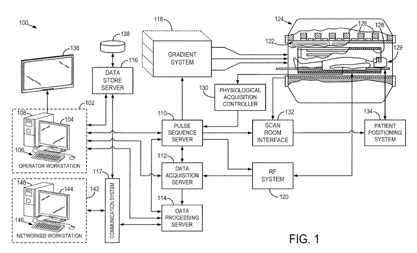
[0012] Fig. 1 is a block diagram of an MRI system.
[0013] Fig. 2 is a block diagram of an RF system of an MRI system.
[0014] Fig. 3 is a block diagram of an RF system of an MRI system
configured
for performing parallel MRI (pMR1) processes
[0015] Fig. 4 is a schematic illustration of a low-field MRI (IfMRI)
system in
accordance with the present disclosure.
[0016] Fig. 5A is a cross-sectional view of a coil system in accordance
with
the present disclosure and for use with the IfMRI system of Fig. 4.
[0017] Fig. 5B is a perspective view of a coil system in accordance with
the
present disclosure and for use with the IfMRI system of Fig. 4.
[0018] Fig. 6 illustrates a passive decoupling circuit for transmit and
receive, in
accordance with some embodiments.
[0019] Fig. 7 schematically depicts a bi-planar arrangement for a BO
magnet.
[0020] Fig. 8 illustrates an outline of a human body showing the
longitudinal
-3-
CA 02942390 2016-09-09
WO 2015/138939
PCT/US2015/020509
axis of the body.
DETAILED DESCRIPTION
[0021] Clinical
MRI scanners are predominantly high-field systems, with the
majority of installed MRI scanners operating at 1.5 or 3 tesla (T). The trend
in MRI is
to further increase the field strength to improve image quality and/or reduce
scan
times. Additionally, to further reduce scan times, high-field MRI systems
often
employ parallel acquisition techniques utilizing multiple transmit and/or
receive coils.
Specifically, multiple receive coils or channels operate simultaneously to
detect MR
signals in parallel, reducing the amount of time it takes to capture data by a
factor
related to the number of independent receive coils that are operated in
parallel.
However, while high-field MRI can provide high resolution images at relatively
short
scan times, the cost of manufacturing, deploying and maintaining a high-field
MRI
installment is often prohibitive, resulting in significantly limited
availability of high-field
MRI systems and preventing their use in many clinical situations.
[0022] Low-
field MRI (e.g., systems that operate at .2T and below) provides a
relatively low cost, high availability alternative to high-field MRI. However,
low-field
MRI presents a number of challenges resulting from the low-field strengths
employed, including significantly reduced signal-to-noise ratio (SNR). In
particular,
the SNR of an MR signal is related to the strength of the main magnetic field
BO,
which is a significant factor driving high-field MRI and the trend towards
higher field
strengths. Low-field MRI produces relatively weak MR signals resulting in
substantially lower SNR, generally requiring significant averaging over
numerous
measurements at each "location" of a region of interest (e.g., multiple
measurements
for each location in k-space) to achieve acceptable SNR. The SNR in high-field
MRI
is such that only a single measurement is needed at each location due
predominantly to the high field strengths involved. While averaging improves
SNR,
the need to acquire numerous measurements at each location increases scan
times.
As such, there is a trade-off between SNR and scan time.
[0023] The
inventors have appreciated that parallel MR techniques can be
used to perform averaging of an increased number of measurements without
needing to increasing the scan time in the low-field context. According to
some
embodiments, a plurality of receive coils are provided, wherein parallel
measurements obtained via the plurality of receive coils are used to increase
the
-4-
CA 02942390 2016-09-09
WO 2015/138939
PCT/US2015/020509
number of measurements that are averaged to perform low-field MRI. Rather than
utilizing the increased amount of data acquired simultaneously via parallel
receive
coils to reduce scan times, as in high-field MRI, the increased data
acquisition
capability is used to increase the number of measurements that are averaged
together to increase the SNR. That is, the time savings resulting from
parallel MR is
used to average over an increased number of measurements to increase the SNR.
According to some embodiments, the acceleration achieved using parallel MR is
used in part to increase the number of measurements that are averaged and in
part
to reduce scan times.
[0024] The
inventors have further appreciated that the low field strengths
employed in low-field MRI facilitate the design of parallel receive coils that
are not
applicable and/or possible in the high-field context. For
example, to transmit
excitation pulse sequences and to detect emitted MR signal, transmit/receive
coils
must resonate at a frequency dependent on the strength of the BO field.
Accordingly,
transmit/receive coils in the high-field regime must resonate at significantly
higher
frequencies than their low-field counterparts. Because of the inverse
relationship
between the length of a conducting path and the wavelength of the resonant
frequency/frequencies in a resonant circuit (i.e., the frequencies at which a
coil can
produce and detect magnetic fields), the conducting paths of high-field
transmit/receive coils are required to be very short. Thus, high-field MRI
receive coils
are single turn, short path conducting loops.
[0025] The
inventors have recognized that the low frequencies involved in low-
field MRI permit the conducting paths of parallel receive coils to be quite
long,
allowing for coil designs that are not suitable (or useable) for high-field
MRI due to
the constraints on conductive path length imposed by the high frequencies
involved
in high-field MRI. According to some embodiments, a plurality of multi-turn
receive
coils are provided to produce a multi-channel receive coil array for use in
low-field
MRI. The plurality of coils may be provided in a three-dimensional geometry
about a
region of interest. The plurality of coils may be arranged in an overlapping
relationship to facilitate decoupling of adjacent receive coils and may be
arranged
over a surface to detect MR signals emitted from a region of interest, some
examples of which are discussed in further detail below.
[0026]
Additionally, clinical high-field MRI systems typically generate a BO field
via a solenoid coil wound about a cylindrical bore into which the patient
being imaged
-5-
CA 02942390 2016-09-09
WO 2015/138939
PCT/US2015/020509
is inserted. As such, the BO field is oriented along the longitudinal axis of
the bore
and the body inserted into the bore. To perform MRI, transmit/receive coils
must
produce a B1 field perpendicular to the BO field and detect emitted MR signals
in this
transverse direction. This
places further restrictions on the geometry for
transmit/receive coils designed for high-field MRI.
[0027] Low-
field MRI facilitates the design of "open" systems in which the BO
field is generated using, for example, bi-planar coils between which a patient
being
imaged is placed such that the BO field is oriented perpendicular to the
longitudinal
axis of the body. Accordingly, transmit/receive coils are arranged to produce
and/or
detect magnetic fields transverse to this BO field, allowing for geometries
that are not
effective in traditional high-field MRI systems. As a result, bi-planar BO
magnets (or
other arrangements that produce a BO field that is transverse to the axis of
the body)
allow for the design of parallel receive coils that detect magnetic fields in
the axial
direction of the body, some examples of which are described in further detail
below.
Receive coils configured as such are not useable with BO coils that produce
magnetic fields aligned with the axis of the body, such as those commonly used
in
high-field MRI.
[0028] The
inventors have further appreciated that the low-field context also
facilitates the use of different materials to produce parallel receive coils.
For
example, conductive paths in receive coils for high-field MRI are typically
fabricated
from sheets of copper. In the low-field context, conductive paths can be
formed using
wire, for example, single strand wire, multi-strand wires (e.g., Litz wires),
etc. The
term "wire" is used herein to describe conductors having a cross-section
characteristic of extrusion such that the cross-section has an axis of
symmetry (e.g.,
such as a generally circular cross-section, rectangular cross-section, etc.),
as
opposed to conductors formed by milling or cutting copper sheets. A wire may
be
single stranded wire of suitable gauge, or multi-stranded wire such as a Litz
wire.
According to some embodiments, each receive coil in a parallel receive coil
array is
formed using wire wound to form a plurality of turns (e.g., 5, 10, 20, 30 or
more
turns) and arranged about a region of interest.
[0029]
Furthermore, in parallel MR, separate transmit and receive coils
inductively couple, adversely affecting the quality of images that can be
acquired. A
common decoupling scheme used in high-field MRI involves the use of PIN diodes
to
effectively detune the receive coils when transmitting and detune the transmit
coil
-6-
CA 02942390 2016-09-09
WO 2015/138939
PCT/US2015/020509
while receiving. However, due to unavailability of suitable PIN diodes that
operate
correctly at frequencies characteristic of low-field MRI, this solution is
generally not
available in the low-field regime. To decouple transmit and receive coils in
the low-
field context, the inventors have developed a passive decoupling scheme that
does
not rely on PIN diodes. According to some embodiments, a crossed-diode
configuration is utilized to decouple transmit and receive coils, examples of
which are
described in further detail below. Such a solution has the benefit of not
requiring any
active elements.
[0030] The
inventors have further appreciated that, due to the predominant
source of noise in low-field MRI being the noise produced by each of the
receive
coils in a parallel array (i.e., the so-called Johnson noise), the noise
regime makes
available certain techniques for reducing noise and increasing SNR. This is in
contrast to high-field MRI where the predominant source of noise is produced
by the
body inserted into the scanner (e.g., via loading effects of the body). As
such, SNR
can be increased in the low-field context using techniques that would have
little or no
impact on noise in the high-field noise regime.
[0031]
According to some embodiments, SNR is improved by reducing
resistive losses in the receive coils. For example, receive coils may be
formed using
multi-strand wires such as a Litz wire. The inventors have appreciated that
using a
Litz wire may substantially reduce resistive losses, thereby decreasing the
noise of
the transmit/receive coil and increasing SNR. According to some embodiments,
receive coils are cooled to reduce the amount of thermal noise and increase
SNR.
Techniques for reducing noise in the low-field context can be used alone or in
any
combination, as the aspects are not limited in this respect.
[0032]
Following below are more detailed descriptions of various concepts
related to, and embodiments of, methods and apparatus for parallel MR, for
example, for use in low-field MRI. It should be appreciated that the
embodiments
described herein may be implemented in any of numerous ways. Examples of
specific implementations are provided below for illustrative purposes only. It
should
be appreciated that the embodiments and the features/capabilities provided may
be
used individually, all together, or in any combination of two or more, as
aspects of
the technology described herein are not limited in this respect.
[0033]
Referring particularly now to Fig. 1, an example of a magnetic
-7-
CA 02942390 2016-09-09
WO 2015/138939
PCT/US2015/020509
resonance imaging (MRI) system 100 is illustrated. The MRI system 100 includes
an
operator workstation 102, which will typically include a display 104, one or
more input
devices 106, such as a keyboard and mouse, and a processor 108. The processor
108 may include a commercially available programmable machine running a
commercially available operating system. The operator workstation 102 provides
the
operator interface that enables scan prescriptions to be entered into the MRI
system
100. In general, the operator workstation 102 may be coupled to four servers:
a
pulse sequence server 110; a data acquisition server 112; a data processing
server
114; and a data store server 116. The operator workstation 102 and each server
110, 112, 114, and 116 are connected to communicate with each other. For
example, the servers 110, 112, 114, and 116 may be connected via a
communication system 117, which may include any suitable network connection,
whether wired, wireless, or a combination of both. As an
example, the
communication system 117 may include both proprietary or dedicated networks,
as
well as open networks, such as the internet.
[0034] The
pulse sequence server 110 functions in response to instructions
downloaded from the operator workstation 102 to operate a gradient system 118
and
a radiofrequency ("RF") system 120. Gradient waveforms necessary to perform
the
prescribed scan are produced and applied to the gradient system 118, which
excites
gradient coils in an assembly 122 to produce the magnetic field gradients G,
Gy,
and Gz used for position encoding magnetic resonance signals. The gradient
coil
assembly 122 forms part of a magnet assembly 124 that includes a polarizing
magnet 126 and a whole-body RF coil 128 and/or local coil, such as a head coil
129.
[0035] RF
waveforms are applied by the RF system 120 to the RF coil 128, or
a separate local coil, such as the head coil 129, in order to perform the
prescribed
magnetic resonance pulse sequence. Responsive magnetic resonance signals
detected by the RF coil 128, or a separate local coil, such as the head coil
129, are
received by the RF system 120, where they are amplified, demodulated,
filtered, and
digitized under direction of commands produced by the pulse sequence server
110.
The RF system 120 includes an RF transmitter for producing a wide variety of
RF
pulses used in MRI pulse sequences. The RF transmitter is responsive to the
scan
prescription and direction from the pulse sequence server 110 to produce RF
pulses
of the desired frequency, phase, and pulse amplitude waveform. The generated
RF
-8-
CA 02942390 2016-09-09
WO 2015/138939
PCT/US2015/020509
pulses may be applied to the whole-body RF coil 128 or to one or more local
coils or
coil arrays, such as the head coil 129.
[0036] The RF
system 120 also includes one or more RF receiver channels.
Each RF receiver channel includes an RF preamplifier that amplifies the
magnetic
resonance signal received by the coil 128/129 to which it is connected, and a
detector that detects and digitizes the / and Q quadrature components of the
received magnetic resonance signal. The magnitude of the received magnetic
resonance signal may, therefore, be determined at any sampled point by the
square
root of the sum of the squares of the / and Q components:
m = V/2 _______________ + Q2
(1);
[0037] and the
phase of the received magnetic resonance signal may also be
determined according to the following relationship:
r
q) = tan-i¨Q
I I (2).
[0038] The
pulse sequence server 110 also optionally receives patient data
from a physiological acquisition controller 130. By way of example, the
physiological
acquisition controller 130 may receive signals from a number of different
sensors
connected to the patient, such as electrocardiograph ("ECG") signals from
electrodes, or respiratory signals from a respiratory bellows or other
respiratory
monitoring device. Such signals are typically used by the pulse sequence
server 110
to synchronize, or "gate," the performance of the scan with the subject's
heart beat
or respiration.
[0039] The
pulse sequence server 110 also connects to a scan room interface
circuit 132 that receives signals from various sensors associated with the
condition
of the patient and the magnet system. It is also through the scan room
interface
circuit 132 that a patient positioning system 134 receives commands to move
the
patient to desired positions during the scan.
[0040] The
digitized magnetic resonance signal samples produced by the RF
system 120 are received by the data acquisition server 112. The data
acquisition
server 112 operates in response to instructions downloaded from the operator
workstation 102 to receive the real-time magnetic resonance data and provide
buffer
storage, such that no data is lost by data overrun. In some scans, the data
-9-
CA 02942390 2016-09-09
WO 2015/138939
PCT/US2015/020509
acquisition server 112 does little more than pass the acquired magnetic
resonance
data to the data processor server 114. However, in scans that require
information
derived from acquired magnetic resonance data to control the further
performance of
the scan, the data acquisition server 112 is programmed to produce such
information
and convey it to the pulse sequence server 110. For example, during prescans,
magnetic resonance data is acquired and used to calibrate the pulse sequence
performed by the pulse sequence server 110. As another example, navigator
signals may be acquired and used to adjust the operating parameters of the RF
system 120 or the gradient system 118, or to control the view order in which k-
space
is sampled. In still another example, the data acquisition server 112 may also
be
employed to process magnetic resonance signals used to detect the arrival of a
contrast agent in a magnetic resonance angiography (MRA) scan. By way of
example, the data acquisition server 112 acquires magnetic resonance data and
processes it in real-time to produce information that is used to control the
scan.
[0041] The data
processing server 114 receives magnetic resonance data
from the data acquisition server 112 and processes it in accordance with
instructions
downloaded from the operator workstation 102. Such processing may, for
example,
include one or more of the following: reconstructing two-dimensional or three-
dimensional images by performing a Fourier transformation of raw k-space data;
performing other image reconstruction algorithms, such as iterative or
backprojection
reconstruction algorithms; applying filters to raw k-space data or to
reconstructed
images; generating functional magnetic resonance images; calculating motion or
flow
images; and so on.
[0042] Images
reconstructed by the data processing server 114 are conveyed
back to the operator workstation 102 where they are stored. Real-time images
are
stored in a data base memory cache (not shown in Fig. 1), from which they may
be
output to operator display 112 or a display 136 that is located near the
magnet
assembly 124 for use by attending physicians. Batch mode images or selected
real
time images are stored in a host database on disc storage 138. When such
images
have been reconstructed and transferred to storage, the data processing server
114
notifies the data store server 116 on the operator workstation 102. The
operator
workstation 102 may be used by an operator to archive the images, produce
films, or
send the images via a network to other facilities.
[0043] The MRI
system 100 may also include one or more networked
-10-
CA 02942390 2016-09-09
WO 2015/138939
PCT/US2015/020509
workstations 142. By way of example, a networked workstation 142 may include a
display 144; one or more input devices 146, such as a keyboard and mouse; and
a
processor 148. The networked workstation 142 may be located within the same
facility as the operator workstation 102, or in a different facility, such as
a different
healthcare institution or clinic.
[0044] The
networked workstation 142, whether within the same facility or in a
different facility as the operator workstation 102, may gain remote access to
the data
processing server 114 or data store server 116 via the communication system
117.
Accordingly, multiple networked workstations 142 may have access to the data
processing server 114 and the data store server 116. In this manner, magnetic
resonance data, reconstructed images, or other data may exchanged between the
data processing server 114 or the data store server 116 and the networked
workstations 142, such that the data or images may be remotely processed by a
networked workstation 142. This data may be exchanged in any suitable format,
such as in accordance with the transmission control protocol (TCP), the
internet
protocol (IF), or other known or suitable protocols.
[0045] With
reference to Figs. 2 and 3, the RF system 120 of Fig. 1 will be
further described. In particular, with reference to Fig. 2, the generalities
of the RF
system 120 will be described and, with reference to Fig. 3, an example of an
RF
system 120 adapted for parallel imaging applications will be described.
[0046]
Referring to Fig. 2, the RF system 120 includes a transmission channel
202 that produces a prescribed RF excitation field. The base, or carrier,
frequency
of this RF excitation field is produced under control of a frequency
synthesizer 210
that receives a set of digital signals from the pulse sequence server 110.
These
digital signals indicate the frequency and phase of the RF carrier signal
produced at
an output 212. The RF carrier is applied to a modulator and up converter 214
where
its amplitude is modulated in response to a signal, R (t), also received from
the
pulse sequence server 110. The signal, R (t), defines the envelope of the RF
excitation pulse to be produced and is produced by sequentially reading out a
series
of stored digital values. These stored digital values may be changed to enable
any
desired RF pulse envelope to be produced.
[0047] The
magnitude of the RF excitation pulse produced at output 216 is
attenuated by an exciter attenuator circuit 218 that receives a digital
command from
-11-
CA 02942390 2016-09-09
WO 2015/138939
PCT/US2015/020509
the pulse sequence server 110. The attenuated RF excitation pulses are then
applied to a power amplifier 220 that drives the RF transmission coil 204.
[0048] The MR
signal produced by the subject is picked up by the RF receiver
coil 208 and applied through a preamplifier 222 to the input of a receiver
attenuator
224. The receiver attenuator 224 further amplifies the signal by an amount
determined by a digital attenuation signal received from the pulse sequence
server
110. The received signal is at or around the Larmor frequency, and this high
frequency signal is down converted in a two step process by a down converter
226.
The down converter 226 first mixes the MR signal with the carrier signal on
line 212
and then mixes the resulting difference signal with a reference signal on line
228 that
is produced by a reference frequency generator 230. The down converted MR
signal is applied to the input of an analog-to-digital ("ND") converter 232
that
samples and digitizes the analog signal. The sampled and digitized signal is
then
applied to a digital detector and signal processor 234 that produces 16-bit in-
phase
(I) values and 16-bit quadrature (Q) values corresponding to the received
signal.
The resulting stream of digitized I and Q values of the received signal are
output to
the data acquisition server 112. In addition to generating the reference
signal on line
228, the reference frequency generator 230 also generates a sampling signal on
line
236 that is applied to the ND converter 232.
[0001]
Referring to Fig. 3, the RF system 120 may be connected to the
whole-body RF coil 128 or, as shown in Fig. 3, a transmission section of the
RF
system 120 may connect to one or more transmit channels 302 of an RF coil
array
304 and a receiver section of the RF system 120 may connect to one or more
receiver channels 106 of the RF coil array 304, which may be, for example, a
head
coil 129, such as illustrated in Fig. 1. The transmit channels 302 and the
receiver
channels 306 are connected to the RF coil array 304 by way of one or more
transmit/receive ("T/R") switches 308. As will be described in further detail,
a
decoupling mechanism 309 may be provided. In alternative configurations of the
RF
system 128 in which the receive coils are a separate collection of coils than
the
transmit coils, T/R switches 308 are not needed and are not used. Instead, in
such a
configuration the receive array is "detuned" during transmission so that it
does not
couple to the transmitter. Likewise, during reception, the transmitter is
detuned. In
this manner, the transmit and receive paths do not mix.
-12-
CA 02942390 2016-09-09
WO 2015/138939
PCT/US2015/020509
[0002]
Referring particularly to Fig. 3 and also with reference to Fig. 1, the RF
system 120 operates the one or more transmit channels 302 to produce a
prescribed
RF excitation field. The base, or carrier, frequency of this RF excitation
field is
produced under control of a frequency synthesizer 310 that receives a set of
digital
signals from the pulse sequence server 110. These digital signals indicate the
frequency and phase of the RF carrier signal produced at an output 312. The RF
carrier is applied to a modulator and up converter 314 where its amplitude is
modulated in response to a signal, R(t), also received from the pulse sequence
server 110. The signal, R(t), defines the envelope of the RF excitation pulse
to be
produced and is produced by sequentially reading out a series of stored
digital
values. These stored digital values may be changed to enable any desired RF
pulse
envelope to be produced.
[0003] The
magnitude of the RF excitation pulse produced at output 316 may
be attenuated by an exciter attenuator circuit 318 that receives a digital
command
from the pulse sequence server 110. The attenuated RF excitation pulses are
then
applied to a power amplifier 320 that drives the RF coil array 304.
[0049] The MR
signal produced by the subject is picked up by the RF coil
array 302 and applied to the inputs of the set of receiver channels 306. A
preamplifier 322 in each receiver channel 306 amplifies the signal, which is
then
attenuated by a receiver attenuator 324 by an amount determined by a digital
attenuation signal received from the pulse sequence server 110. The received
signal is at or around the Larmor frequency, and this high frequency signal is
down
converted in a two step process by a down converter 326. The down converter
326
first mixes the MR signal with the carrier signal on line 312 and then mixes
the
resulting difference signal with a reference signal on line 328 that is
produced by a
reference frequency generator 330. The down converted MR signal is applied to
the
input of an analog-to-digital ("ND") converter 332 that samples and digitizes
the
analog signal. As an alternative to down conversion of the high frequency
signal, the
received analog signal can also be detected directly with an appropriately
fast
analog-to-digital ("ND") converter and/or with appropriate undersampling. The
sampled and digitized signal is then applied to a digital detector and signal
processor
334 that produces 16-bit in-phase (/) values and 16-bit quadrature (Q) values
-13-
CA 02942390 2016-09-09
WO 2015/138939
PCT/US2015/020509
corresponding to the received signal. The resulting stream of digitized / and
Q
values of the received signal are output to the data acquisition server 112.
In
addition to generating the reference signal on line 328, the reference
frequency
generator 330 also generates a sampling signal on line 336 that is applied to
the ND
converter 332.
[0050] The
basic MR systems and principles described above may be used to
inform the design of other MR systems that share similar components but
operate at
very-different parameters. In one example, a low-field magnetic resonance
imaging
(IfMRI) system utilizes much of the above-described hardware, but has
substantially
reduced hardware requirements and a smaller hardware footprint. For example,
referring to Fig. 4, a system is illustrated that, instead of a 1.5T or
greater static
magnetic field, utilizes a substantially smaller magnetic field, for example,
a BO field
of .2T or less. According to some embodiments, a 6.5 mT electromagnet-based
scanner 400 is provided that is capable of imaging objects up to, for example,
15.6
cm in diameter. However, it should be appreciated that any field strength in
the low
field regime may be used, as the parallel receive coil techniques described
herein
are not limited for use with any particular field strength. The system 400 may
use a
multi-channel array 402 to implement a parallel imaging process, such as a
sensitivity encoding (SENSE) imaging procedure.
[0051] The
system 400 is a relatively transportable and rapidly deployable
human imaging system. Current research for low field human imaging is limited
and
generally uses superconducting quantum interference device (SQUID) sensors. At
conventional magnetic field strengths, body noise dominates, resulting in
strongly
correlated noise on each receive coil in the parallel array. At low field,
uncorrelated
Johnson noise dominates. The present disclosure recognizes that this
phenomenon
provides a benefit to parallel imaging and accelerated imaging using, for
example,
SENSE. However, to perform such parallel imaging techniques, a multi-channel
coil
is required. Thus, the present disclosure provides a multi-channel coil array
402 that
is particularly advantageous, such as for IfMRI.
[0052] As
illustrated in Fig. 4, the multi-channel array 402 may be designed to
perform parallel imaging in vivo, for example, in the human head. As a non-
limiting
example, the present disclosure will describe an optimized 8-channel array,
though
any number of channels may be used to provide a multi-channel receive array.
-14-
CA 02942390 2016-09-09
WO 2015/138939
PCT/US2015/020509
Specifically, referring to Figs. 5A and 5B, the multi-channel array 402 may
include a
tight fitting substrate or housing 500. In this example, the substrate or
housing 500
may be formed as a helmet that tightly follows contours of the associated
anatomy.
An array of coils 502 may be arranged on or within the housing or substrate
500. In
the illustrated example, eight coils 504, 506, 508, 510, 512, 514, 516, 518
are
shown. Other numbers of coils may be selected, for example, based on anatomy
or
to achieve a desired fill factor or acceleration, which can in turn be used to
increase
SNR, as discussed in further detail below.
[0053] As a non-
limiting example, the coils 504-518 may be 30-turn, receive-
only coils (24 AWG, 4x12 cm and 4x14 cm loops). However, receive coils in a
parallel array may be of any size and contain any number of turns provided
they
satisfy design constraints of the system. It should be appreciated that the
exemplary
coils illustrated have a conducting length that far exceeds the limit imposed
by the
high frequencies of the high-field regime. For example, a 14 cm loop having 30
turns
will have a conducting path of approximately 320 cm, which is an order of
magnitude
or more greater than the limit of clinical high-field systems. Thus, the
relaxed
constraints on conductive length of the receive coils allow for greater
flexibility in both
the size and shape of the receive coils and the number of turns employed. Each
receive coil may be formed, for example, by winding a wire using a respective
desired number of turns (which may be different or the same for each coil) to
produce a receive coil of the desired shape and size and having desired
operating
characteristics. The plurality of receive coils can be designed to have
different size,
shape and turn characteristics so that the coils can be arranged in a three-
dimensional geometry to provide adequate coverage for a region of interest.
[0054]
Furthermore, as a non-limiting example, the coils 504-518 may be tiled
about the substrate or housing 500. In the illustrated, non-limiting example,
the coils
are tilted symmetrical about the sagittal plane. The shapes and sizes of the
coils
504-518 may be selected by tiling coils across the substrate or housing 500.
In the
illustrated example of a helmet, the coils 504-518 may be shaped and sized to
tile
coils across the substrate or housing 500, while avoiding the flat sides 520
by the
ears because these will be perpendicular to BO field and, therefore, collect
minimal
signal. Similarly, when extended to other anatomy, placement and selection of
the
coils 504-518 can follow these principles.
[0055] In one
non-limiting example, a helmet-shaped coil system was created.
-15-
CA 02942390 2016-09-09
WO 2015/138939
PCT/US2015/020509
The substrate or housing was formed using 3D printing (Fortus 360mc,
Stratasys,
Eden Prairie, MN, USA), such that the substrate followed closely to the
underlying
anatomy. In this non-limiting example, the coil were 4x14 cm and 4x12 cm and
all
were formed with 24 AWG and 30 turns.
[0056]
Referring again to Fig. 3, high-performance, low-impedance pre-amps
322 are not readily available for frequency ranges associated with IfMRI
(e.g., BO
fields of .2T or less). As such, the transmit and receive coils 304 may
include a
decoupling mechanism 309 that passively decouples using crossed diodes 340 in
series 342 with respect to the transmit channels 302 and in parallel 344 with
respect
to the receive channels 306. As illustrated, the crossed diodes 340 are formed
by
two diodes, in parallel or series, but pointing in opposite directions.
Passive
decoupling diodes 340 may be associated with at each element in the coil array
304.
Active detuning solutions based around PIN diodes are not desirable because
the
carrier lifetime of commercial PIN diodes precludes operation at 276 kHz.
[0057] Fig. 6
illustrates a circuit diagram of one embodiment of a passive
decoupling scheme for a transmit coil and for each of a plurality of parallel
receive
coils, respectively. For the transmit coil, crossed diodes 610a are connected
in
series with the transmit coil 650, in conjunction with a parallel tune (CT_TX)
and
series match (CM-TX) circuit. For each receive coil, crossed diodes 610b are
connected in parallel with the respective receive coil 675 along with a
parallel tune
(CT_RX) and series match (CM-RX) circuit. Crossed-diodes 610a allow the
transmit
pulse to reach the transmit coil when the pulse voltage is greater than the
bias
voltage, a condition met during transmit of pulse sequences. During receive,
however, the voltage induced in transmit coil 650 from precessing
magnetization is
too small to forward bias the crossed diodes, resulting in the transmit coil
being
decoupled during receive. The crossed diodes 610b on the receive circuit short
to
ground any induced voltage greater than the bias voltage, a condition met
during
transmit, thereby decoupling the respective receive coil 675 during transmit.
As
such, the transmit coil is allowed to resonate (e.g., via biased crossed-
diodes 610a)
when transmit pulses are generated, which transmit pulses cause an induced
voltage greater than the bias voltage of crossed diodes 610b, thus decoupling
the
receive coils. The above described decoupling circuit has no active components
and
therefore provides a passive decoupling technique.
[0058] In one,
non-limiting example, all coils were tuned to 276.0 kHz and
-16-
CA 02942390 2016-09-09
WO 2015/138939
PCT/US2015/020509
were matched to at least -27 dB and geometrically decoupled from their nearest
neighbors by at least -30 dB. Decoupling from next-nearest neighbors was at
least -
6 dB. A 30 cm diameter solenoid was used for transmit operations. It should be
appreciated that the frequency to which the coils are tuned will depend on the
field
strength selected for the BO magnetic field.
[0059] As also
discussed above, low-field MRI systems can be constructed
using a bi-planar configuration for the BO magnet. For example, Fig. 7
schematically
depicts a magnet 700 to illustrate a bi-planar coil configuration that can be
used to
generate a BO field for low-field MRI. As illustrated, the BO magnet includes
coils
710a and 710b that, when operated, produce a BO field oriented in the
direction
indicated by arrow 705. When a subject is placed between coils 710a and 710b,
BO
is perpendicular to the longitudinal axis of the body of the subject. FIG. 8
illustrates
the longitudinal axis 700 of the human body, which is perpendicular to the BO
field of
magnet 700 both when the subject is placed between the BO coils in a upright
or
supine position.
[0060]
Accordingly, low-field MRI systems having a BO field oriented as shown
in FIG. 7 (perpendicular to the longitudinal axis of the body) permit the use
of the
receive coil geometries described herein. By contrast, high-field MRI systems
are
predominantly produced using a solenoid BO magnet such that the BO field is
oriented along the longitudinal axis of the body of a subject and the bore
into which
the subject is inserted, thus requiring a B1 excitation field in a
perpendicular
direction. The receive coils illustrated in Fig. 5B are arranged to detect
magnetic
fields substantially aligned with the longitudinal axis of the wearer of the
head coil
and therefore these coils are ineffective for receive for a solenoid-based BO
magnet.
[0061] The
inventors have further appreciated that parallel receive coils,
examples of which have been described herein, may be used to increase the SNR
in
MR signal acquisition in the low-field context. As discussed above, the small
SNR of
low-field MRI is a significant challenge in performing low-field MRI. A
technique for
addressing the low SNR is to repeat MR data acquisition at a given "location"
multiple times (e.g., by repeating a pulse sequence with the same operating
parameters) and averaging the obtained MR signal that results. The term
"average"
is used herein to describe any type of scheme for combining the signals,
including
absolute average (e.g., mean), weighted average, or any other technique that
can be
used to increase the SNR by combining MR data from multiple acquisitions.
-17-
CA 02942390 2016-09-09
WO 2015/138939
PCT/US2015/020509
However, while averaging improves SNR, the repeat acquisitions increase total
acquisition times. According to some embodiments, the MR data acquired by a
plurality of receive coils in parallel is used to increase the number of
measurements
averaged together for a given location to increase the SNR. As a result, multi-
channel receive coils are utilized to increase SNR without necessarily
increasing the
total acquisition time.
[0062] As an
example, consider a low-field MRI acquisition using a single-
channel receive coil in which N measurements are averaged together to obtain
the
value at each location (e.g., 25, 50, 100 measurements, etc.) for a total
acquisition
time T. Using multi-channel receive coils according to the techniques
described
herein, a factor K acceleration is achieved, for example. Instead of reducing
the
scan time to T/K, the SNR may be increased by acquiring and averaging KN
measurements for each location for the same total acquisition time T. The
acceleration can be, in this sense, "traded in" for an equivalent factor more
measurements over which to average, thus increasing the SNR.
[0063] It
should be appreciated that not all of the acceleration need be
exchanged to increase the number of measurements that are averaged. For
example, some of the factor of K acceleration achieved may be used to reduce
the
scan time and some may be used to increase SNR. As such, the techniques
described herein in this respect can be used to increase SNR and/or reduce
scan
times, and the acceleration achieved using parallel MR can be allocated as
seen fit
for a particular imaging application.
[0064] Imaging
studies were performed using the above-described system.
In particular, axial and sagittal images were acquired using a 3D b-SSFP
sequence
with 50 percent incoherent undersampling of k-space at 6.5 mT (276 kHz).
Imaging
parameters were: TR/TE = 33.2/21.6 ms, acquisition matrix = (64x64x9), voxel
size
= (3x3x6) mm3, number of averages (NA) = 200, and flip angle = 70 . The
readout
duration was 7.04 ms with a 9091 Hz bandwidth. The total acquisition time was
30
min. The resulting images revealed recognizable anatomic features in the head
including the skull, cortical structures (gyri/sulci) and the corpus callosum.
[0065] Thus
systems and methods have been demonstrated for using an
optimized multi-channel, local coil, including but not limited to the above-
described 8-
channel helmet array, combined with fast acquisition techniques and
undersampling
-18-
CA 02942390 2016-09-09
WO 2015/138939
PCT/US2015/020509
strategies to enable 3D imaging using IfMRI. With (3.3x4x17) mm3 total voxel
size,
2 times greater spatial resolution was acquired than very-recently published
work
using a SQUID detector in an ultra-low field MRI system with a 80 mT
prepolarization
field. In addition, the 3D dataset (9 slices) was acquired more than seven
times
faster than the single-slice 2D brain dataset of the SQUID-detected work.
[0066] The
present disclosure recognizes that, at low frequency, detector coils
for low field NMR and MRI operate in the Johnson noise dominated regime. To
this
end, the present disclosure recognizes that desirable or optimal coil
parameters and
decoupling strategies when working within these imaging constraints benefit
from a
deviation from known engineering art in this regime. As such, the present
disclosure
provides an array of receive coils suitable for MRI and NMR at low frequency.
The
coil may be formed to a close-fitting, substrate or housing that is well
matched to the
human head. Advantageously, the substrate may be 3D printed and contoured to
comfortably fit human anatomy and maximize filling factor.
Individual receive
elements may be tailored so that the number of turns per coil maximizes the
induced
signal and results in a coil that meets the desired minimum receive coil
bandwidth
specification. One non-limiting example is coil having a Q of approximately
20. The
present disclosure also provides coil decoupling strategies that have been
specifically tailored for low frequency operation. This includes passive,
crossed
diodes at each element to decouple from transmit and nearest neighbor
decoupling.
Coil tiling geometry can be optimized for either longitudinal or transverse
MRI
scanner magnetic field. A transmit coil may also be integrated with the above-
described coil design.
[0067] As such,
the present disclosure provides much-needed new resources
for a variety of clinical applications, for example, including diagnosis or
analysis of
critical brain injury triage, monitoring of the progression of ischemic stroke
(including
measuring brain midline shift), and imaging of patients excluded from
conventional
MRI due to metal implants, pacemakers, and the like. When combined with
hyperpolarized contrast agents, the present disclosure provides systems and
methods that can be used for molecular imaging in the brain using low-field
scanners.
[0068] The
present invention has been described in terms of one or more
embodiments, and it should be appreciated that many equivalents, alternatives,
variations, and modifications, aside from those expressly stated, are possible
and
-19-
CA 02942390 2016-09-09
WO 2015/138939
PCT/US2015/020509
within the scope of the invention.
-20-