Note: Descriptions are shown in the official language in which they were submitted.
CA 02942770 2016-09-14
WO 2015/143512
PCT/BR2015/000025
1
ENHANCEMENT OF RECOMBINANT PROTEIN EXPRESSION
WITH COPPER
Cross-Reference to Related Applications
[0001] This application is based on and claims priority of
61/969,215 filed 23 March 2014.
Statement Regarding Federally Sponsored Research or Development: Not
applicable
BACKGROUND
1. FIELD
[0002] Recombinant proteins have been made by cell culturing based on the
batch method or perfusion since the 1980s. The present invention provides
improved
cell expression, particularly in mammalian cells, by the use of copper
additives. This
invention is applicable to many mammalian cell cultures, such as CHO, BHK and
human cell lines, particularly CHO, and to the expression of many recombinant
proteins, such as recombinant Factor VIII B Domain Deleted rFVIII and
recombinant Factor VII/Factor Vila (rFVII/rFVIIa).
2. RELATED BACKGROUND ART
[0003] Copper is essential for cell growth and survival. Because of
copper's
essential nutrient value, its chemical role as a catalyst of oxidative stress
and its
CA 02942770 2016-09-14
WO 2015/143512
PCT/BR2015/000025
2
propensity to precipitate, it is critical to understand, monitor and formulate
it for use in
specific cell culture systems and applications.
[0004] Copper is a transition metal that exists, in vitro, in an
equilibrium as
reduced (cuprous), Cu (I) and oxidized (cupric), Cu (II), copper. In its free
form and in
some chelates, it can participate actively in redox cycling. It oxidizes a
number of
important media components, such cysteine and ascorbate, for optimization of
the cell
culture process.
[0005] In vitro, Cu (I) will spontaneously form complexes with reduced
cysteine, glutathione and presumably organic sulfhythyls. In addition to
forming
cupri-cystine complexes, Cu (11) will form complexes with other amino acids
through
coordination of their alpha-amino nitrogen and carboxyl-oxygen groups. Binding
of
Cu (II) to histidine is important because this appears to be an intermediate
involved in
the movement of Cu (II) from albumin to the cell. Before the copper can cross
the cell
membrane it must be reduced to Cu (I).
[0006] Copper can cause the loss of the cysteine and cystine from cell
culture
media by oxidation and precipitation. In vitro, cysteine is freely soluble and
exists
almost exclusively as a neutral amino acid. It is unstable and undergoes non-
enzymatic
autoxidation in the presence of di-molecular oxygen to form cystine. Cupric
copper
accelerates the autoxidation of cysteine to cystine. Cupric copper can form
chelate-
precipitates with cystine. The depletion of cysteine from cell culture will
stop the
synthesis of proteins and glutathione, an important reducing agent. Reduced
CA 02942770 2016-09-14
WO 2015/143512
PCT/BR2015/000025
3
glutathione can complex with Cu (I) and inhibit its participation in the
formation of
hydroxyl free radicals. This interaction involves the cysteine sulfur atom. In
vivo, Cu
(I):glutathione complexes mediate the safe movement of Cu (I) that enters the
cytoplasm, probably through the copper transporter 1 pore, to intra-cellular
binding
proteins such as metallothionein. The formation of Cu (I): glutathione
complexes is
spontaneous and non-enzymatic, [Dierick, P.J. (1980, In vitro interaction of
organic
copper (II) compounds with soluble glutathione S-transferases from rat liver.
[Res.
Commun. Chem Pathol. Pharmacol. 51, 285-288.]
BRIEF DESCRIPTION OF THE DRAWINGS
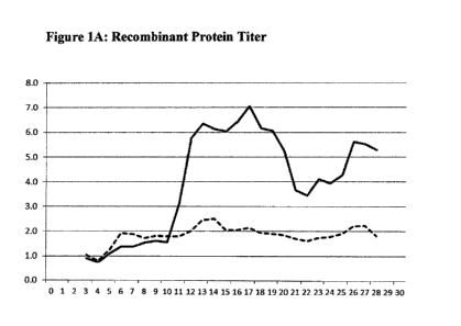
[0007] Figures lA and 2A show the influence of high copper levels in the
culture on
Recombinant Protein Expression. In both figures, the Y-axis represents
normalized
data on Recombinant Protein Titer obtained. The dashed line represents data
obtained
using medium with no additional copper added, i.e. only a basal level of 0.087
micromolar copper naturally present in the media. The X ¨axis represents
bioreactor
days. The solid line represents the protein titer obtained when additional
copper is
added.
[0008] Figures 1B and 2B show the influence of high copper levels on
recombinant
protein specific productivity. In both figures, the Y-axis represents
normalized data
on Recombinant Protein Specific Productivity versus bioreactor days on the X-
axis.
The dashed line again represents data obtained using medium with no additional
copper added, i.e. only a basal level of 0.087 micromolar copper naturally
present in
CA 02942770 2016-09-14
WO 2015/143512
PCT/BR2015/000025
4
the media. The solid line represents the protein specific productivity
obtained when
additional copper is added.
[0009] Figures 3A and 3B show Recombinant Protein Titer and Recombinant
Protein Specific Productivity, respectively, versus bioreactor days for the
basal level
of copper found in the medium and for various levels of copper added (0.315,
0.629
and 1.259 micromolar).
[0010] Figure 4 is a surface plot of normalized Specific Productivity (qp)
vs.
osmolality and copper concentration.
DETAILED DESCRIPTION
[0011] This data was generated in 2013 when the process was operated using an
external membrane-based cell retention device, using medium without copper
supplementation. Baseline cultures represented as (-) Copper were executed
with
copper levels found in normal medium in 16-160 nanomolar range. The first
experimental evidence of the added benefits of copper were obtained when two
(2)
bioreactors received medium with copper supplemented. The addition of copper
occurred on day ten (10) and showed an immediate influence on recombinant
protein
expression as evidenced in the graph showing the dramatic increase in protein
expression. However, the cupric ion source, such as cupric sulfate or cupric
chloride
or other cupric salt with similar characteristics, may be added to the medium
prior to
adding the cells with similar results. Figure 1 shows the influence of adding
40.9
CA 02942770 2016-09-14
WO 2015/143512
PCT/BR2015/000025
micromolar copper to the culture medium. A four (4) to five (5) fold increase
in
protein expression was demonstrated through duplicate bioreactors operating at
the
same conditions as the baseline runs. The addition of about 40 micromolar
copper in
the form of cupric ion appears to give optimal results, but other additional
concentrations within the range of 0.5 micromolar to about 10.0 micromolar
appear to
give similar results.
[0012] To better understand the influence of high levels of copper during the
initial
experimental runs, additional runs were executed using a reduced quantity of
copper.
Figure 2 represents data generated using a copper addition of 7.87 micromolar.
This
data demonstrates that with all other factors equal to baseline bioreactors,
the addition
of 7.87 micromolar resulted in a three (3) to four (4) fold increase in
protein
expression.
[0013] Further bioreactor experimentation was carried out to demonstrate
the
influence of more reasonable copper levels on protein expression. Figure 3
represents
data generated through duplicate bioreactors operated at varying levels of
copper
concentration through the course of the bioreactor run. All other parameters
were
maintained equivalent to the baseline runs. This data demonstrates when
compared to
the 7.87 micromolar copper addition as detailed in Figure 2, that copper
concentrations of 0.315, 0.63 and 1.26 micromolar will result in three (3) to
four (4)
fold increases equivalent to 7.87 micromolar.
CA 02942770 2016-09-14
WO 2015/143512
PCT/BR2015/000025
6
[0014] Figure 4 shows the specific productivity on the Z (vertical) axis
with the
copper concentration and osmolality on the X and Y-axis respectively. This
data was
generated using a six day, 250 InL shake flask, batch cell culture model to
determine/demonstrate the effect of added copper. The specific productivity
may also
be increased with increased osmolality of the medium, but the greatest effect
is seen
with the addition of copper ion. A response surface Design of Experiment was
performed where the cultures were seeded at 0.5e6 cells/mL into basal medium
supplemented with cupric chloride and or, optionally, sodium chloride to
adjust the
copper levels to between 0.087 to 3.78 micrmolar and osmolality to between 270
to
380 mOsmo respectively. Five different levels of each factor were chosen
(0.087,
0.787, 1.495, 2.927, and 3.78 micromolar copper and 270, 310, 350, 360, 380
mOsmo). Cultures were then sampled daily for viable cell concentration
determination
for six days. Product concentration evaluation was performed on days 4-6. The
specific productivity represents the average specific productivity between
days 4 and 6
of the batch culture normalized to average specific productivity of the center
point in
the study (310 mOsmoõ 1.49 micrornolar Cu). As seen in Figure 4 there is a
clear
increase in specific productivity with both increases in osmolality and
increases in
copper concentration. From a statistical analysis of the data from the
response surface
CA 02942770 2016-09-14
WO 2015/143512
PCT/BR2015/000025
7
design experiment, both Cu and osmolality exhibited a highly significant
effect, P=
0.000 (where any P<0.05 is considered significant), on specific productivity,
but there
was also a statistically significant interaction between the two P = 0.003,
see Table 1.
[0015] Per the equation developed to model this data, the specific
productivity
increased from 0.134 to 0.355 with an increase in copper concentration from
0.087 to
3.78 micromolar at an osmolality of 270 and from 1.2 to 2.15 at an osmolality
of 380.
Similarly there is a clear increase in specific productivity from 0.143 to
1.22 with an
increase osmolality from 270 to 380 at 0.087 micromolar copper and from 0.355
to
2.158 at 3.78 micromolar copper.
Table 1
Term Coef SE Coef
Constant 1.28562 0.03053 42.107 0.000
Osmo 0.71634 0.03372 21245 0.000
Cu ppb 0.28843 0.03492 8.260 0.000
Osmo*Osmo 0.10210 0.04882 2.091 0.063
Cu ppb*Cu ppb -0.31375 0.05114 -6.135 0.0000
Osmo*Cu ppb 0.18223 0.04553 4.002 0.003
[0016] Table one gives the coefficients for the regression model equation
which fits
the specific productivity data collected as a function of osmolality and
copper
concentration. The equation consists of a constant, two linear terms (Osmo, Cu
ppb),
and three nonlinear terms (Osmo*Osmo, Cu ppb*Cu ppb, Osmo*Cu ppb) as shown in
the first column in table 1. The "Osmo" term represents the osmolality of the
culture
CA 02942770 2016-09-14
WO 2015/143512
PCT/BR2015/000025
8
where as the "Cu ppb" term represents the copper concentration. The
coefficients for
each term are listed in the second row (Coef) with the standard error of those
coefficients listed in the third row (SE Coef). The forth row is the T
statistic of the
coefficients and is the quotient of the Coefficient divided by the standard
error of the
coefficient. The larger the magnitude of the T value the larger the
significance of the
coefficient. The fifth column represents the p-value for each term and a value
of less
than 0.05 is considered to indicate statistical significance. As can be seen
in table 1 all
but the Osmo*Osmo term have,a p-value less than 0.05 and are therefore
considered
significant. The final regression equation is shown below.
Qp = 1.28562 + 0.71634*Osmo + 0.28843*Cu ppb + 0.10210*Osmo*Osmo -
3.1375*Cu ppb*Cu ppb + 0.18223*Osmo*Cu ppb
SUMMARY
[0017] A method of increasing cell expression of mammalian cells, comprising
the
use of copper additives to the cell culture medium is provided herein. From
about 0.5
micromolar to about 10.0 micromolar copper is preferably added to the cell
culture
medium. A similar addition of 0.5 micromolar copper to about 10.0 micromolar
copper provides an increased cell specific productivity. Cupric ion is
particularly
preferred as the copper additive. The manufacturing system is composed of the
augmented cell culture medium and mammalian cells. Preferred mammalian cells
for
use in the cell culture medium are CHO, BHK or human mammalian cells. Unstable
CA 02942770 2016-09-14
WO 2015/143512
PCT/BR2015/000025
9
recombinant proteins are particularly good candidates for expression utilizing
a
membrane-based cell retention system with copper additives. This system is
useful
with perfusion cell cultures to produce coagulation proteins, chosen from the
group
consisting of recombinant Factor VIII, B Domain Deleted recombinant Factor
VIII,
recombinant Factor IX and rFVII or rFVIla.
[0018] The addition of other bulk ions such as sodium and potassium that
increase the osmolality of the medium further enhance protein expression.
[0019] The method is preferably used in combination with a membrane-
based cell retention system and perfusion cell culture.
[0020] Most preferred is the use of this improved method of recombinant
protein expression applied to increasing the expression of B-Domain
Deleted recombinant FVIII in mammalian cells with the addition of about
0.5 to about 10.0 micromolar cupric ion to the cell culture medium used
with a manufacturing system, composed of perfusion cell culture used in
combination with an external membrane-based cell retention system.