Note: Descriptions are shown in the official language in which they were submitted.
CA 02943430 2016-09-21
WO 2015/161069 PCT/US2015/026162
CORNEAL INLAY DELIVERY DEVICES AND METHODS
CROSS REFERENCE TO RELATED APPLICATIONS
[0001] This application claims the benefit of U.S. Provisional
Application No. 61/980,504,
filed April 16, 2014, which is incorporated by reference herein.
[0002] This application is related to the following applications, the
disclosures of which are
incorporated herein by reference: U.S. Pat. No. 8,162,953, issued 4/24/2012;
U.S. Pub. No.
2013/0253527, published 9/26/2013; and U.S. Pub. No. 2013/0123916, published
5/16/2013.
INCORPORATION BY REFERENCE
[0003] All publications and patent applications mentioned in this
specification are herein
incorporated by reference to the same extent as if each individual publication
or patent
application was specifically and individually indicated to be incorporated by
reference.
BACKGROUND
[0004] Delivery devices have been described for delivering ophthalmic
devices such as
intraocular lenses and corneal implants into the eye. Alternative designs and
methods of use are
needed that can easily and accurately deliver corneal implants, such as a,
without limitation,
small diameter, hydrophilic, corneal implants.
SUMMARY
[0005] One aspect of the disclosure is a corneal inlay inserter
comprising a distal region, the
distal region having at least one aperture through at least one of an anterior
and a posterior
surface thereof; and a fluid channel in fluid communication with the at least
one aperture.
[0006] In some embodiments the inserter comprises first and second devices
adapted and
configured to stably interface one another, wherein the fluid channel is
within the first device,
the second device comprising the distal region having the at least one
aperture through an
anterior or a posterior surface thereof. The distal end can have a plurality
of apertures through
the anterior or posterior surface.
[0007] In some embodiments the inserter comprises an elongate body
comprising the distal
region and the fluid channel therein. The distal region can have a plurality
of apertures through
the anterior or posterior surface. The fluid channel can extend from a
proximal end of the
elongate body to the at least one aperture.
[0008] In some embodiments the inserter further comprises a corneal
inlay secured over the
at least one aperture.
- I -
CA 02943430 2016-09-21
WO 2015/161069 PCT/US2015/026162
[0009] In some embodiments the distal region does not have any apertures
through the
posterior surface thereof.
[0010] In some embodiments the distal region does not have any apertures
through the
anterior surface thereof.
[0011] In some embodiments the distal region has at least one aperture
through the anterior
surface and at least one aperture through the posterior surface, the two
apertures being in fluid
communication.
[0012] One aspect of the disclosure is a method of delivering a corneal
implant into the eye,
comprising: providing a corneal inserter comprising a distal region, the
distal region having at
least one aperture through at least one of an anterior and a posterior surface
thereof, a corneal
implant secured to the inserter over the at least one aperture, and a fluid
delivery channel in fluid
communication with the at least one aperture; and delivering a fluid through
the fluid delivery
channel and through the at least one aperture to repel the corneal implant
from the distal region
and onto corneal tissue.
[0013] In some embodiments the inserter comprises a first device comprising
the distal
region, the method further comprising, prior in time to delivering the fluid,
securing a second
device with a fluid reservoir to the first device. Securing the second device
to the first device
can comprise securing the second device to a proximal end of the first device.
Securing the
second device to the first device can comprise securing the second device to a
posterior side of
the first device. The method can further comprise advancing the first device
into a corneal
pocket so that the posterior side is closer to the retina than an anterior
side of the first device.
[0014] In some embodiments the method further comprises advancing the
inserter into a
corneal pocket before delivering the fluid, wherein delivering the fluid
repels the corneal implant
into the corneal pocket.
[0015] In some embodiments delivering the fluid comprises delivering as
little as about 10
microliters of fluid through the fluid delivery channel, as little as 4
microliters, or even as little as
1 microliter, although more fluid may be delivered. In some embodiments the
inserter, including
the corneal implant, are adapted such that as little as 10 microliters of
fluid, or as little as 4
microliters, or even as little as I microliter, is all that is needed to be
delivered to release the
corneal implant from the inserter, even though more fluid can be used. This is
in contrast to, for
example, intraocular lens delivery systems, which are not adapted so that the
intraocular lens can
be released or delivered from the delivery device with a relatively very
little amount of fluid.
[0016] One aspect of the disclosure is a corneal inlay inserter with an
elongate body having
a fluid delivery channel therein, the fluid delivery channel in fluid
communication with a
- 2 -
CA 02943430 2016-09-21
WO 2015/161069
PCT/US2015/026162
proximal region and at least one aperture extending through at least one of an
anterior surface
and a posterior surface of a distal region of the elongate body.
[0017] In some embodiments the fluid delivery channel is in fluid with a
plurality of
apertures extending through at least one of the anterior surface and the
posterior surface of a
distal region of the inserter. The fluid delivery channel can be in fluid
communication with at
least one aperture extending only through the anterior surface and not the
proximal surface. The
fluid delivery channel can be in fluid communication with at least one
aperture extending only
through the posterior surface and not the anterior surface.
[0018] In some embodiments the inserter further comprises a corneal
inlay secured to the
inserter in a position over the at least one aperture on one of the anterior
surface and the posterior
surface. The corneal inlay can be a hydrophilic inlay, which in any of the
embodiments herein
can allow for the fluid delivery of the inlay away from or off of the
inserter. The corneal inlay
can have a diameter of between 1 mm and 5 mm. The corneal inlay can have a
thickness
between about 10 microns and about 100 microns.
[0019] In some embodiments the corneal implant has a diameter greater than
the greatest
linear dimension of the at least one aperture measured across the aperture in
the proximal to
distal direction.
[0020] In some embodiments a proximal end of the inserter is configured
to interface with a
fluid delivery device, the fluid delivery device adapted to advance fluid
through the fluid
delivery channel and out of the at least one aperture.
[0021] In some embodiments the inserter further comprises a securing
member positioned
over the at least one aperture, the securing member and the distal region
defining a volume in
which a corneal inlay can be disposed, the securing member configured to be
movable relative to
the distal region to provide access to the volume.
[0022] In some embodiments the at least one aperture has a greatest linear
dimension
measured across the aperture in the proximal to distal direction of .02 mm and
1.0mm.
[0023] In some embodiments the anterior and posterior surfaces are
substantially parallel
with each other.
[0024] One aspect of the disclosure is a method of delivering a corneal
implant into the eye,
comprising: providing a corneal implant inserter with a fluid delivery channel
therein in fluid
communication with at least one aperture in at least one of an anterior
surface and a posterior
surface of a distal region of the inserter, and a corneal implant secured to
the inserter over the at
least aperture on one of the anterior surface and the posterior surface; and
delivering a fluid
through the fluid delivery channel and through the at least one aperture to
repel the corneal
implant from the distal region and into the eye.
- 3 -
CA 02943430 2016-09-21
WO 2015/161069 PCT/US2015/026162
[0025] In some embodiments the method further comprises securing a fluid
reservoir to a
proximal end of the inserter, wherein the delivering step comprises delivering
fluid from the
fluid reservoir and into the fluid delivery channel.
[0026] In some embodiments the method further comprises advancing the
corneal implant
inserter into a corneal pocket before delivering the fluid, wherein delivering
the fluid repels the
corneal implant into the corneal pocket.
[0027] In some embodiments delivering the fluid comprises delivering as
little as 10
microliters of fluid, as little as 4 microliters, or even as little as 1
microliter, through the fluid
delivery channel, although more may be delivered. In some embodiments the
inserter, including
the corneal implant, are adapted such that as little as 10 microliters of
fluid, as little as 4
microliters, or even as little as 1 microliter, is all that is needed to be
delivered to release the
corneal implant from the inserter, even though more fluid can be used. This is
in contrast to, for
example, intraocular lens delivery systems, which are not adapted so that the
intraocular lens can
be released or delivered from the delivery device with a very little amount of
fluid.
[0028] One aspect of the disclosure is a corneal inlay inserter comprising
a distal region
having at least one aperture extending from an anterior surface to a posterior
surface, and a
hydrophobic member adapted to be moved from a first position to a second
position closer to the
at least one aperture.
[0029] In some embodiments the hydrophobic member is secured indirectly
to the distal
region but movable relative to the distal region.
[0030] In some embodiments the inserter further comprises an actuatable
member, the
hydrophobic member adapted to be moved closer to the at least one aperture
upon actuation of
the actuatable member.
[0031] One aspect of the disclosure is an ophthalmic device inserter
comprising a distal
region and an ophthalmic device retained at the distal region, and a repelling
member adapted to
be moved relative to the ophthalmic device, the repelling member having one or
more physical
properties adapted to repel the ophthalmic device away from distal region when
moved towards
the ophthalmic device without making direct physical contact with the
ophthalmic device. The
ophthalmic device can be hydrophilic.
[0032] In some embodiments the ophthalmic device is a corneal implant, the
distal region
having an anterior surface, a posterior surface, and at least one aperture
extending from the
anterior surface to the posterior surface, the corneal implant secured to the
anterior surface or the
posterior surface over at least one aperture; and the repelling member can be
adapted to be
moved relative to the corneal implant to repel the corneal implant from the
anterior surface or
posterior surface.
- 4 -
CA 02943430 2016-09-21
WO 2015/161069 PCT/US2015/026162
[0033] In some embodiments the repelling member is secured indirectly to
the distal region
but movable relative to the distal region.
[0034] In some embodiments the inserter further comprises an actuatable
member, the
repelling member adapted to be moved closer to the ophthalmic device upon
actuation of the
actuatable member.
[0035] One aspect of the disclosure is a method of delivering an
ophthalmic device into the
eye, comprising: providing an ophthalmic device and an ophthalmic device
inserter, the inserter
comprising a distal region and a repelling member having one or more physical
properties
adapted to repel the ophthalmic device; and repelling the ophthalmic device
from the distal
region of the inserter and into the eye by moving the repelling member towards
the ophthalmic
device without making direct physical contact with the ophthalmic device.
[0036] In some embodiments the distal region comprises at least one
aperture extending
from an anterior surface to a posterior surface, the ophthalmic device
retained to the anterior
surface or the posterior surface over at least one aperture; and wherein
repelling the ophthalmic
device comprises moving the repelling member closer to the ophthalmic device
on the other of
the anterior and posterior surfaces.
BRIEF DESCRIPTION OF FIGURES
[0037] Figure 1 illustrates an exemplary inserter with a fluid channel
in fluid
communication with a plurality of apertures in an anterior surface of a distal
region.
[0038] Figure 2 illustrates the inserter from Figure 1 attached to a
fluid reservoir.
[0039] Figure 3 illustrates an inserter with a fluid channel in fluid
communication with one
aperture in an anterior surface of a distal region.
[0040] Figures 4A-4H illustrate an exemplary inserter comprising a first
device with a fluid
delivery channel therein and a second device with at least one aperture
through an anterior
surface and a posterior surface. The first and second device are configured to
stably interface
with each other.
[0041] Figures 5A-5C illustrate an exemplary inserter that includes an
actuatable repelling
member adapted to repel the implant away from the inserter and into the eye.
DETAILED DESCRIPTION
[0042] The disclosure herein describes devices that are adapted for
positioning ophthalmic
devices, such as corneal implants, onto or into corneal tissue. These types of
devices may be
generally referred to herein as inserters.
- 5 -
CA 02943430 2016-09-21
WO 2015/161069 PCT/US2015/026162
[0043] Corneal implants can correct vision impairment by creating a
change in curvature of
the anterior surface of a cornea and/or creating multifocalities within the
cornea due to intrinsic
properties of the implant. "Corneal implants" as used herein includes corneal
onlays and corneal
inlays. An onlay is an implant that is placed over the stromal part of the
cornea such that the
outer layer of the cornea, i.e., the epithelium, can grow over and encompass
the implant. An
inlay is an implant that is implanted within corneal tissue beneath a portion
of the corneal tissue
by, for example, cutting a flap in the cornea and inserting the inlay beneath
the flap, or by
placing it within a pocket created within the cornea. Both inlays and onlays
can alter the
refractive power of the cornea by changing the shape of the anterior cornea,
by having a different
index of refraction than the cornea, or both. When the disclosure herein
refers to an "inlay," it is
understood that the devices and methods can be used for other types of corneal
implants as well.
[0044] There is a need for improved devices, systems and methods for
inserting corneal
implants onto corneal tissue, including inserting them within a corneal
pocket.
[0045] A corneal "pocket" is generally referred to as a recess formed
within the corneal
tissue for receiving a corneal implant. Methods of creating and accessing
pockets are known,
such as may be found described in U.S. Pub. No. 2003/0014042, published
January 16, 2003,
entitled "Method of Creating Stromal Pockets for Corneal Implants," U.S. Pub.
No.
2013/0253527, published September 26, 2013, which are fully incorporated by
reference herein.
Pockets can be made by, for example, a Femtosecond laser or a Blade Pocket
Maker. Additional
exemplary methods and devices for creating corneal pockets, or corneal
channels, can be found
in U.S. Pub. No. 2012/0046680, filed August 23, 2010, the disclosure of which
is fully
incorporated by reference herein. Any techniques for creating and accessing
pockets can be used
to create pockets described herein.
[0046] Exemplary devices and methods for positioning inlays into corneal
pockets can be
found described in U.S. Pat. No. 8,162,953 (see, e.g., Figures 7-11 and the
descriptions thereof).
In U.S. Pat. No. 8,162,953 the delivery device includes a holding space at a
distal end thereof
adapted to house an inlay, and fluid is used to deploy the inlay from the
holding space and into
the pocket. Additional exemplary devices and methods for positioning inlays in
pockets by
delivering fluid through a delivery device with a holding space can be found
described in U.S.
Pub. No. 2013/0253527.
[0047] In some additional embodiments devices and methods can use
selective adhesion
forces between the inlay and the device. For inlays made primarily of a
hydrogel material and of
small size (such as some of the inlays described in U.S. Pub. No.
2011/0218623, published
9/8/2011), relatively strong forces act on the fluid within the inlay. These
embodiments make
use of these characteristics of the inlay and the adhesion forces seen between
a fluid and various
- 6 -
CA 02943430 2016-09-21
WO 2015/161069 PCT/US2015/026162
surface geometries. Selective adhesion or transfer of the inlay during
different stages of the
delivery process can be manipulated by using different material and/or surface
geometries.
Examples of delivery devices and methods that utilize selective, or
"preferential," adhesion can
be found in U.S. Pub. No. 2013/0123916 published May 16, 2013 (see, e.g.,
relative adhesion
between "moderate" and "minimal" bodies, which may be referred to herein as
"fine mesh" and
"course mesh" materials). Embodiments in U.S. Pub. No. 2013/0123916, for
example, are
described primarily as being used to deposit an inlay on a corneal bed after a
flap has been
created and lifted. In U.S. Pub. No. 2013/0123916 a preferential adhesion
between two materials
is controlled in different stages of delivery until the inlay contacts the
stromal bed and adheres to
it. In a procedure that delivers an inlay into a pocket, tissue and/or liquid
in the eye are
constantly contacting the inlay and device material as the device and inlay
are advanced towards
and into the pocket. Thus, preferential adhesion of the inlay is preferably
controlled such that
there is a strong attraction between the inlay and the delivery device during
insertion into the
pocket and a reduction or transfer mechanism such that once the inlay is ready
for transfer by
position or such, the user can selectively transfer the inlay into the corneal
pocket.
[0048] One aspect of the disclosure is a corneal inlay inserter
comprising a distal region, the
distal region having at least one aperture through at least one of an anterior
and a posterior
surface thereof; and a fluid channel in fluid communication with the at least
one aperture.
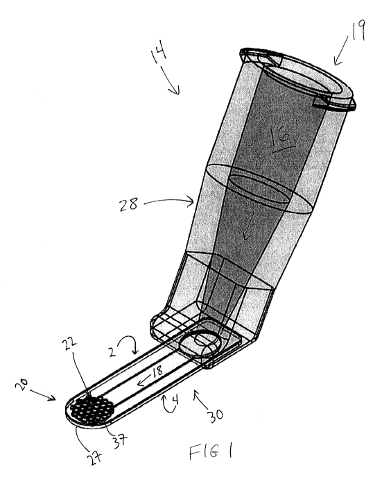
[0049] Figures 1 and 2 illustrate an example of an inserter 14, which
includes a distal
region, the distal region having at least one aperture through at least one of
an anterior and a
posterior surface thereof; and a fluid channel in fluid communication with the
at least one
aperture. In this embodiment inserter 14 has a proximal region 19 with a
proximal end, the
proximal region 19 configured to be secured to a fluid reservoir, but is other
embodiments the
inserter can be integral with the fluid reservoir (i.e., not configured to be
detachable without
physical deformation or breaking). In this embodiment fluid reservoir 12 (see
Figure 2) is a
syringe, which includes fluid chamber 17. The connection between inserter 14
and fluid
reservoir 12 can be any suitable method (such as a luer fitting).
[0050] Inserter 14 has an elongate body 30 with a distal region 20, the
distal region 20
including at least one aperture through at least one of an anterior and a
posterior surface of the
distal region. Distal region 20 includes anterior surface 2 and posterior
surface 4. Anterior and
posterior refer to relative positions of the inserter when it is in use. In
this context anterior refers
to a position closer to the anterior surface of the cornea than the retina,
and posterior refers to a
relative position closer to the retina than the anterior surface of the
cornea. In this embodiment
anterior surface 2 may be thought of as a "top" surface, and posterior surface
4 may be thought
of as a "bottom" surface of distal region 20.
- 7 -
CA 02943430 2016-09-21
WO 2015/161069
PCT/US2015/026162
[0051] Inserter 14 also includes fluid channel 18. In this embodiment
fluid inserter 14
includes elongate body 30, which includes fluid channel 18 therein in
communication with the at
least one aperture 22. Fluid channel 18 extends through elongate body 30 to a
location directly
below, or posterior to, the at least one aperture. In this embodiment distal
region 20 has a
plurality of apertures 22 in anterior surface 2, and no apertures in posterior
surface 4. In some
embodiments the distal region has at least one aperture in the posterior
surface and none in the
anterior surface, while in some embodiments distal region 20 has at least one
aperture in each of
the anterior surface and the posterior surface.
[0052] In this embodiment inserter 14 includes fluid reservoir adaptor
28, which is not
integral with elongate body 30 but rather is secured thereto. In alternative
embodiments adaptor
28 is integral with elongate body 30, and is considered an extension of
elongate body 30 in the
general anterior direction. In this embodiment adaptor 28 includes the
proximal region 19 that is
adapted to be secured to fluid reservoir 12. Adaptor 28 includes fluid channel
16 therein in fluid
communication with fluid channel 18 in elongate body 30. When fluid reservoir
12 secured to
inserter 14, fluid chamber 17, fluid channel 16, fluid channel 18, and the at
least one aperture 22
are all in fluid communication. In embodiments in which adaptor 28 is integral
with elongate
body 30, fluid channel 16 is simply an extension of fluid channel 18 towards
proximal region 19.
[0053] In this embodiment adaptor 28 is secured to elongate body 30.
Elongate body 30 can
be secured to the posterior, or bottom, of adaptor 28 using any number of
techniques such as an
adhesive. Adaptor 28 can include a receiving portion that is configured to
receive the proximal
end of elongate body 30 therein.
[0054] In this embodiment the inserter is configured to retain an
ophthalmic device, in this
embodiment a corneal inlay, on anterior side 2 of distal region 20 over the at
least one aperture in
anterior side 2. In this embodiment distal region 20 includes a plurality of
aperture in the
anterior surface 2 of distal region 20. In this embodiment the inlay is
retained on anterior side 2
on at least some of the plurality of apertures due to adhesion forces. In this
embodiment the
plurality of apertures 22 functions similarly to a "moderate body" mesh in
U.S. Pub. No.
2013/0123916, also referred to as "preferential material"). The distal end 20
is thus configured
so that the corneal inlay adheres to it, and is retained by it. Whereas in
U.S. Pub. No.
2013/0123916 the corneal inlays are generally described as adhering to a
"posterior" side of the
moderate body for placement onto the corneal bed, in this embodiment the
corneal inlay is
disposed positioned on anterior side 2 of the plurality of apertures and is
retained thereon.
[0055] In the embodiments herein, unless indicated otherwise, the
anterior and posterior
surfaces are substantially parallel with each other. Substantially parallel
does not require them to
be precisely parallel, but upon inspection one of ordinary skill in the art
would understand them
- 8 -
CA 02943430 2016-09-21
WO 2015/161069 PCT/US2015/026162
to be substantially parallel. For example, the two surfaces extending
proximally to distally in the
figures herein are all substantially parallel. The surfaces are substantially
parallel even if there is
a slight degree of curve to them.
[0056] While a "minimal" or "course" mesh material (as described in U.S.
Pub. No.
2013/0123916), or any other material that has less preferential adhesion for
the corneal inlay
than distal end 20, is not shown in the embodiment in Figures 1 and 2, it can
be assumed that the
corneal inlay could in some embodiments be positioned between a fine mesh and
a course mesh
for storage or packaging, and the course mesh could then be moved relative to
the fine mesh to
provide access to the inlay, as is described in U.S. Pub. No. 2013/0123916.
For example, a
packaging and storage device could include both fine and course mesh
materials, and the course
mesh material is moved away from the fine mesh, and the inlay will
preferentially adhere to the
fine mesh. Thus, the preferential adhesion principals described in U.S. Pub.
No. 2013/0123916
can be utilized in this embodiment, or any of the embodiments herein.
[0057] Distal region 20, including the region that defines the
apertures, can be, for example
without limitation, titanium. Additionally, any of the mesh configuration
described in U.S. Pub.
No. 2013/0123916 can be used for the mesh configuration of distal region 20.
[0058] In alternative embodiments the inlay can be retained on proximal
side 4 of inserter
14. For example, in some applications it may be desired to position the
corneal implant on the
posterior side 4 for placement in the eye. In those embodiments inserter can
include a plurality
of apertures in proximal side 4 that are in fluid communication with fluid
channel 18. There may
be added benefits to having at least one aperture on the side of the distal
region 20 opposite the
side on which the corneal implant is retained. The opposite side can thus have
at least one
aperture therein in fluid communication with fluid channel 18 even though the
implant is not on
that side.
[0059] In this embodiment the fluid reservoir can be secured to inserter
14, and then held by
a user when it is time to advance the implant 37 onto corneal tissue, such as
into a pocket. In an
exemplary method of use for delivery into a corneal pocket, once inserter 14
is prepared for
insertion, the user will introduce distal end 20, on which the corneal inlay
is adhered, into the
already prepared corneal pocket. Once the desired inlay location is achieved,
the user will
actuate plunger 15 (which can be any other actuation mechanism) to advance
fluid from the
reservoir chamber 17, through fluid channels 16 and 18, and out of apertures
22 in the anterior
direction. The flow of fluid out of the apertures 22 causes, either
hydraulically and/or through
the reduction of adhesion forces between the corneal inlay and the distal
region 20, repels the
inlay away from the distal region 20, separating it from the distal region and
thus delivering the
inlay into the corneal pocket. The inserter is then removed from the pocket.
Any of the inserters
-9 -
CA 02943430 2016-09-21
WO 2015/161069 PCT/US2015/026162
herein can also be used, or modified to be used, to deliver the ophthalmic
device on a corneal bed
formed by creating a corneal flap, or in any other suitable delivery
procedure.
[0060] In some embodiments inserter 14 is packaged and stored with a
corneal implant
(such as with two mesh materials in place to secure the implant, as described
in applications
incorporated herein by reference), and then attached to the fluid reservoir
when the inlay is ready
for use.
[0061] Distal end 20 of inserter 14 can be formed by securing a top, or
anterior, piece of
material the plurality of apertures formed therein, to a bottom piece with a
channel formed
therein. When the two pieces are secured together, fluid is directed down now
formed fluid
channel 18 towards the apertures in the direction of the arrows as shown in
Figure 1. The distal
region 20 of inserter 14 can be manufactured in other ways to create fluid
channel 18 as well.
The exemplary rounded distal end 27 of distal portion 20 is closed so that
fluid can only escape
the distal end through the apertures, which helps the inlay disassociate from
the distal region 20.
In some embodiments, however, there may be advantage to having apertures on
both sides. In
this embodiment the fluid thus acts to break the adhesion between the implant
and the distal
region 20 and the inlay drifts off of and away from distal region.
[0062] Any of the fine mesh materials (also referred to as moderate
materials), the
orientation of the apertures, and techniques for manufacturing them described
in U.S. Pub. No.
2013/0123916 can be used in making the distal region 20 and/or the elongate
body 30, or any
distal region herein.
[0063] In alternative embodiments to that shown in Figures 1 and 2, the
device can also be
adapted so that the implant adheres to the posterior side, or "bottom" of the
distal region. The
fluid channel could be on top of, or anterior to, the implant, and the fluid
would displace the
implant from the mesh in the downward, or posterior, direction. Any of the
devices herein can
be adapted so that the inlay is positioned on either the anterior side or the
posterior side of the
distal region.
[0064] The apertures herein on the anterior side and/or posterior side
of the distal region are
differentiated from distal ports, through which intraocular lenses or other
ophthalmic devices are
commonly pushed through during delivery into the eye.
[0065] In some embodiments inlay 37 (or any inlays used with any of the
inserters herein)
has a diameter of between 1 mm and 5 mm. In some embodiments the inlay has a
central
thickness between about 10 microns and about 100 microns. In some embodiments
herein the
inlay has a water content of least 60%, and is comprised of a hydrogel. As can
be seen in Figure
1 and in the applications incorporated by reference herein, the diameter of
the inlay is greater
than the greatest linear dimension of the at least one aperture measured
across the aperture in the
- 10 -
CA 02943430 2016-09-21
WO 2015/161069 PCT/US2015/026162
proximal to distal direction. In any of the embodiments herein the at least
one aperture, such as
all of them, has a greatest linear dimension measured across the aperture in
the proximal to distal
direction of between .02 mm and 1.0mm, such as between .02 mm and .75 mm.
[0066] Figure 3 illustrates an alternative inserter 50 that is adapted
to position implant 58
within a corneal pocket. Inserter 50 is similar to common cannulas, but has a
generally flattened
elongate body 54. Inserter 50 includes elongate body 54 and distal region 52,
wherein distal
region 52 has only one aperture 56 therein on anterior side 51 and does not
have any apertures on
posterior side 53. This is an example of at least one aperture but not more
than one. A fluid
channel (not labeled in Figure 3) extends through inserter 50 from aperture 56
through elongate
body 54 and into handle portion 57. Fluid can be advanced through the fluid
channel (not
shown) and out of aperture 56 using known techniques, such as with a plunger.
Anterior surface
51 and posterior surface 53 are substantially parallel in this embodiment as
well.
[0067] In an exemplary method of use, inlay 58, once positioned on
distal region 52 over
aperture 56, adheres to distal region 52 due to some adhesion forces. The
adhesion forces may
not be as great as those present in the embodiment in Figures 1 and 2,
however. The inserter,
with the inlay adhered thereto, is then advanced into a corneal pocket as
described herein. To
dissociate inlay 56 from inserter 50, fluid is advanced through the fluid
channel and out of
aperture 56, causing the inlay to be displaced from the inserter 50 and into
the pocket. Again,
inserter 50 could have the aperture(s) on the proximal side 53 such that the
inlay is displaced
from inserter 50 in a downward, or posterior, direction.
[0068] The embodiment shown in Figures 4A-4H is an example of an
inserter that includes
a distal region, the distal region having at least one aperture through an
anterior surface and a
posterior surface thereof, and a fluid channel in fluid communication with the
at least one
aperture. In this embodiment inserter 60 comprises first 72 and second 64
separate devices that
are adapted and configured to stably interface one another, wherein the fluid
channel is within
first device 72, and second device 64 comprises distal region 66 having the at
least one aperture
68 through the anterior and posterior surfaces thereof. The fluid channel (not
shown for clarity)
is thus in fluid communication with the at least one aperture 68 when the
first 72 and second 64
devices and secured to one another. In this embodiment the second device 64
can be a mesh
device such as any of the meshes described herein, and can be used in any of
the methods
described herein. For example, a corneal implant can be secured to an anterior
surface of the
distal region of the mesh (e.g., a fine mesh) due to adhesion forces.
[0069] Figures 4A- 4H illustrate an exemplary inserter 60 that includes
first device 72 with
distal region 74 that includes side ridges 75 at the periphery of distal
region 74 positioned and
configured so that distal region 74 of first device 72 can stably interface
distal region 66 of
- 11 -
CA 02943430 2016-09-21
WO 2015/161069 PCT/US2015/026162
second device 64 in at least one direction. Second device 64 includes a fine
mesh configuration
described herein, and a corneal implant 69 secured thereon (see Figure 4G).
Figures 4B, 4C, and
4H illustrate the inserter after the first and second devices are stably
interfacing each other. First
device 72 of the inserter is configured so that it can be attached at its
proximal end 71 to a fluid
reservoir 75 (Figures 4A-4C), such as a syringe with plunger, with the luer
lock.
[0070] Figure 4A shows the exemplary inserter, including first device
72, second device 64,
and fluid reservoir 75. Figure 4B is a perspective view showing inserter 60
after first device 72
and second device 64 are stably interfacing, and after reservoir 75 has been
secured to first
device 72. Figure 4D shows a top view of first device 72. Figure 4E shows a
side view of first
device 72, and Figure 4F shows a perspective view of first device 72. Figure
4G shows the distal
regions 66 and 74, of second device 64 and first device 72, respectively, not
in an interfacing
relationship. Figure 4H is a close-up perspective view of the distal regions
66 and 74 in an
interfacing relationship. The first device interfaces the second device at
interface regions 80,
thereby stabilizing the second device 64 relative to the first device 72 in at
least one direction.
[0071] In use, after the first and second devices are secured to one
another (such as shown
in Figures 4B, 4C, and 4H), fluid is delivered through the fluid channel in
first device 72 (e.g.,
using a syringe in fluid reservoir 75), out of the apertures 76 in the second
device 72, and then
through the mesh aperture(s) 68 in second device 64, causing the corneal
implant 69 to be
disassociated from (i.e., drift off of) distal region 66 of second device 64,
as is described
elsewhere herein.
[0072] Figures 4C and 4H show first device 72 with fluid channel therein
positioned
posterior to the mesh of first device 64 and inlay. In some embodiments first
device 72 includes
a fluid pillow at its distal end. In these embodiments, as fluid is delivered
from the syringe to the
fluid (e.g., saline) pillow, fluid is delivered through the mesh apertures
more gently due to the
presence of the pillow. Thus the pillow can be used to advance fluid through
the mesh aperture
in a more controlled and gentle manner. In this embodiment the pillow can be a
perforated
device, similar in concept to a teabag.
[0073] It is understood that other types of mechanisms can be used to
secure the first and
second device together and still fall within the subject matter of this
disclosure. For example, the
distal region of the second device 64 can be placed under (posterior to)
restraining clips in the
distal region of the first device 72.
[0074] In some embodiments herein an ophthalmic device inserter
comprises a distal region
and an ophthalmic device retained at the distal region, and a repelling member
adapted to be
moved relative to the ophthalmic device, the repelling member having one or
more physical
properties that cause it to repel the ophthalmic device away from the distal
region when moved
- 12 -
CA 02943430 2016-09-21
WO 2015/161069 PCT/US2015/026162
towards the ophthalmic device without making direct physical contact with the
ophthalmic
device. The inserter and ophthalmic device in Figures 5A-5C is an example of
such an inserter
and ophthalmic device.
[0075] Inserter 40 includes a handle portion 42 secured to distal
region 44. Inserter 40 also
includes an actuatable moveable member 49 that includes repelling member 46
(see Figures 5B
and 5C) extending distally from the base of actuatable member 49. Distal
region 44 is coupled
to handle 42 via connector 91, to which distal region 44 is secured.
[0076] Distal region 44 can be considered very similar or even the same
as the mesh
materials (moderate or minimal) incorporated by reference herein. Distal
region includes a
plurality of apertures 45 therethrough, from anterior surface 41 to proximal
surface 43.
Ophthalmic device 48 and distal region 44 are adapted such that ophthalmic
device 48 is retained
on distal region 44 due to adhesion forces. Rather than simply using fluid
flow to deliver the
implant, however, in this embodiment a repelling member is brought into closer
proximity to the
implant, which causes the implant to be repelled from distal region 44 and
into the eye. In this
exemplary embodiment inserter includes an actuable member 49 that is coupled
to repelling
member 46, and when actuated repelling member 46 is moved closer to implant 48
along the
posterior surface 43 of distal region 44. In some embodiments in which the
implant has a high
enough water content, repelling member 46 is a hydrophobic material that when
moved into
closer proximity of the implant, it repels the implant away from distal end 44
in the anterior
direction, away from distal end 44 and into the eye. Figure 5C shows a
posterior view of
repelling member 46 after it has been moved into closer proximity to implant
48, relative to an
initial position shown in Figure 5B prior to actuation. In Figure 5C,
repelling member is
disposed on the posterior side of at least some of the apertures 45. In
alternative embodiments
the implant is secured to posterior side of the distal region, and the
inserter is constructed and
arranged such that the repelling member moves into closer proximity to the
implant on the
anterior side. In these embodiments the implant is repelled away from the
distal end in the
posterior direction into the eye.
[0077] In some embodiments the repelling member is a hydrophobic
material that, when
moved into closer proximity to the implant, will repel the fluid and/or the
implant away from the
hydrophobic material, thus deploying the implant from the inserter and into
the pocket. A
material such as Teflon can be used as the hydrophobic material. Other
hydrophobic materials
can also be used, however. In this embodiment the implant is adhered to one
material that can be
considered hydrophilic, while repelling member 46 is hydrophobic. Repelling
member 46 can
alternatively be actuated in any conceivable way towards the implant/distal
region interface, such
as via an actuator on the handle that causes the actuatable member 49 to move.
- 13 -
CA 02943430 2016-09-21
WO 2015/161069 PCT/US2015/026162
[0078] The repelling action in embodiments in which the repelling member
is a
hydrophobic material is at least partially based on the lotus effect, named
after the lotus leaf.
The lotus effect refers generally to self-cleaning properties that are result
of very high water
repellence (superhydrophobicity), as exhibited by the leaves of the lotus
flower. Part of the
reason the lotus leaf is so repellent is due to air trapped in its nodule-
covered surface. The effect
relies on surface tension, therefore there needs to be a surface between air
and water. The
hydrophobic members herein can have surfaces that are configured (such as
through
modification) to trap air in order to increase the efficiency of the repelling
action.
[0079] The embodiment in Figures 5A-5C is an example of using a non-
fluid member to
deliver the implant, and one that does not come into direct contact with the
implant to deliver the
implant from the inserter.
[0080] The embodiment in Figures 5A-5C can be used in any of the methods
of use herein
to deploy the implant in a corneal pocket.
- 14 -