Note: Descriptions are shown in the official language in which they were submitted.
81800138
ACCOMMODATING INTRAOCULAR LENS
CROSS-REFERENCE TO PRIORITY DOCUMENTS
[0001] The present application claims the benefit of priority to
co-pending U.S.
Provisional Application Serial No. 61/972,183, filed March 28, 2014 and co-
pending U.S.
Provisional Application Serial No. 61/977,568, filed April 9, 2014.
BACKGROUND
[0002] The present disclosure relates generally to the field of
ophthalmics, more
particularly to ophthalmic devices, including intraocular lenses (IOLs) such
as
accommodating intraocular lenses.
[0003] A healthy young human eye can focus an object in far or
near distance,
as required. The capability of the eye to change back and forth from near
vision to far vision
is called accommodation. Accommodation occurs when the ciliary muscle
contracts to
thereby release the resting zonular tension on the equatorial region of the
capsular bag. The
release of zonular tension allows the inherent elasticity of the lens to alter
to a more globular
or spherical shape, with increased surface curvatures of both the anterior and
posterior
lenticular surfaces.
[0004] The human lens can be afflicted with one or more
disorders that degrade
its functioning in the vision system. A common lens disorder is a cataract
which is the
opacification of the normally clear, natural crystalline lens matrix. The
opacification can
result from the aging process but can also be caused by heredity or diabetes.
In a cataract
procedure, the patient's opaque crystalline lens is replaced with a clear lens
implant or IOL.
[0005] In conventional extracapsular cataract surgery, the
crystalline lens
matrix is removed leaving intact the thin walls of the anterior and posterior
capsules together
with zonular ligament connections to the ciliary body and ciliary muscles. The
crystalline lens
1
CA 2944010 2020-03-24
CA 02944010 2016-09-26
WO 2015/148673 PCT/US2015/022501
core is removed by phacoemulsification through a curvilinear capsularhexis
i.e., the removal
of an anterior portion of the capsular sac.
[0006] After a healing period of a few days to weeks, the capsular sac
effectively shrink-wraps around the IOL due to the capsularhexis, the collapse
of the walls of
the sac and subsequent fibrosis. Cataract surgery as practiced today causes
the irretrievable
loss of most of the eye's natural structures that provide accommodation. The
crystalline lens
matrix is completely lost and the integrity of the capsular sac is reduced by
the capsularhexis.
The "shrink-wrap" of the capsular sac around the IOL can damage the zonule
complex, and
thereafter the ciliary muscles may atrophy. Thus, conventional IOL' s, even
those that profess
to be accommodative, may be unable to provide sufficient axial lens spatial
displacement
along the optical axis or lens shape change to provide an adequate amount of
accommodation
for near vision.
[0007] It is known to implant a combination of lenses to address
refraction
errors in the existing lens in the case of phakic IOLs or improve the
refractive results of
standard IOL after cataract surgery in the case of pseudophakic patients.
These "piggyback"
TOI,s can be placed anterior to the previously implanted TOT, or natural lens
to improve the
refractive results of cataract surgery in the case of pseudophakes or to
change the refractive
status of the eye in the case of phakic eyes, usually to correct high myopia.
Generally, these
lenses are implanted in the sulcus and are non-accommodating.
SUMMARY
[0008] In some implementations, disclosed is an accommodating
intraocular
lens device for treatment of an eye. The lens device includes a stabilization
haptic configured
to be positioned within a region of an eye. The lens device includes a lens
body having a
sealed chamber containing a fixed volume of optical fluid. The lens body
includes a shape
changing membrane configured to outwardly bow in a region surrounding the
optical axis of
the eye; a shape deformation membrane configured to undergo displacement
relative to the
first shape changing membrane; and a static element. An inner surface of the
shape changing
membrane, an inner surface of the shape deformation membrane and an inner
surface of the
2
CA 02944010 2016-09-26
WO 2015/148673 PCT/US2015/022501
static element collectively form the sealed chamber. The lens device also
includes a force
translation arm having a first end configured to contact an outer surface of
the shape
deformation membrane of the lens body and a second end configured to engage a
ciliary
structure of the eye. The force translation arm is configured to move relative
to the lens body
upon movement of the ciliary structure.
[0009] The shape deformation membrane can be configured to undergo
inward
displacement towards the optical axis of the eye relative to the shape
changing membrane
during accommodation. Inward movement of the force translation arm can cause
inward
movement of at least one or more regions of the shape deformation membrane
towards the
optical axis of the eye causing a deformation of the sealed chamber. Inward
movement of the
shape deformation membrane can cause the optical fluid in the sealed chamber
to press
against the inner surface of the shape changing membrane and causes outward
bowing of the
shape changing membrane. The lens device can further include an internal
support located
within the sealed chamber. The internal support can mechanically isolate
optical components
of the lens from distortion during movement of the force translation arm. The
internal support
can include a plurality of internal supports spaced apart from one another
within the sealed
chamber. The internal support can include a tapered geometry to avoid contact
during inward
movement of the shape deformation membrane.
[0010] The stabilization haptic can be bonded to the lens body. The
stabilization haptic can be molded as part of the lens body. The lens device
can further
include an exterior support. The internal support can be coupled to a
perimeter region of the
shape changing membrane. The internal support can form a partition within the
sealed
chamber dividing the sealed chamber into a deformable region and a central
region. The
deformable region can be located outside an optic zone. The deformable region
can be
located inside an optic zone. Inward movement of the force translation arm can
cause inward
movement of the shape deformation membrane and a deformation of the deformable
region.
Inward movement of the shape deformation membrane can compress the sealed
chamber.
The optical fluid in the sealed chamber can be non-compressible and can press
against the
inner surface of the shape changing membrane and cause outward bowing of the
shape
changing membrane. The internal support can be further coupled to a region of
the static
3
CA 02944010 2016-09-26
WO 2015/148673 PCT/US2015/022501
element. The internal support can include a channel extending through the
internal support
providing fluid communication between the deformable region and the central
region of the
sealed chamber.
[0011] The lens device can further include an exterior support. The
exterior
support can be rigid and can be configured to prevent distortion caused by
movement of the
force translation arms relative to the lens body. The stabilization haptic can
be bonded to an
external surface of the exterior support. The stabilization haptic can be
molded as part of the
exterior support. The first end of the force translation arm can extend
through a channel in a
peripheral wall of the exterior support such that the first end is positioned
against the shape
deformation membrane. The exterior support can include a central annular
region and
opposed side regions. The lens body can include a central portion and opposed,
deformable
portions. The central portion can align with the central annular region of the
exterior support
and the deformable portions of the lens body extend within the opposed side
regions of the
exterior support. An outer surface of the shape deformation membrane can be
exposed
through the central annular region. An outer surface of the static element can
be exposed
through the central annular region. A first of the force translation arms can
extend through a
first opening in a first sidewall of the exterior support into a first
channel. A second of the
force translation arms can extend through a second opening in a second
sidewall of the
exterior support into a second channel. The first channel and the second
channel can be on
opposite sides of the central annular region. The force translation arms can
be configured to
move back and forth within the first and second channels.
[0012] The shape deformation membrane can have a first surface coupled
to the
shape changing membrane and a second surface coupled to the static element and
a sidewall
extending between the first surface and the second surface. The sidewall of
the shape
deformation membrane can be aligned with and bonded to an inner surface of the
central
region of the exterior support such that the lens body is fixedly positioned
relative to the
exterior support. The central portion can surround the optical axis and
deformable portions
are located outside the central portion. An outer surface of the shape
changing membrane
near the central portion can be aligned with and bonded to an inner surface of
the central
annular region of the exterior support. The deformable portions can be freely
movable within
4
CA 02944010 2016-09-26
WO 2015/148673
PCT/US2015/022501
the exterior support. The deformable portions can be configured to undergo
inward,
collapsible movement or displacement relative to the central portion during
accommodation.
The first ends of the force translation arms cam be configured to be
positioned against the
deformable portions. Upon contraction, the ciliary structure can press against
the second ends
causing the first ends of the force translation arms to press upon the
defolmable portions and
cause inward, collapsible movement of the deformable portions towards the
central portion.
Inward, collapsible movement of the deformable portions towards the central
portion can
cause the region of the shape changing membrane to outwardly bow. Inward,
collapsible
movement of the deformable portions towards the central portion can cause the
optical fluid
in the sealed chamber to press against the inner surface of the shape changing
membrane
causing the outward bowing of the shape changing membrane.
[0013] The central
portion of the lens body can be generally circular and the
deformable portions of the lens body have a shape selected from the group
consisting of
bellowed, pleated, trapezoidal, cylindrical, elliptical, conical, spherical,
and hemi-spherical.
The deformable portions of the lens body can move relative to the central
portion of the lens
body in response to a force applied by the ciliary structure onto the force
translation arms.
The deformable portions can move a distance between about 50 um and about 500
urn. The
distance the deformable portions move can cause at least a change in power of
the lens body
by at least 3 diopters. The force applied can be between about 0.1 gf to about
5 gf. The
stabilization haptic can be configured to maintain alignment of the optics and
resist
movement of the device following implantation in the eye. The stabilization
haptic can
further include a biting element to improve fixation of the haptic within the
eye. The biting
element can include a grooved edge and/or a hole. The stabilization haptic can
be open-loop,
closed-loop, plate-style, plate loop, monobloc-plate style, j-loop, c-loop,
modified J-loop,
multi-piece, single-piece, angulated, planar, or offset haptics. The
stabilization haptic can be
coaxial or coplanar with the force translation arms. The stabilization haptic
can be positioned
on a different plane than the force translation arms. The stabilization haptic
can be flexible,
foldable or formed of a shape memory material. The stabilization haptic can be
positioned
within a ciliary sulcus or the capsular bag of the eye.
81800138
[0014] The lens body can include a deformable portion that is located
outside the optic zone. The deformable portion can be a region of the shape
deformation
membrane. The lens body can include a deformable portion that is located
inside the
optic zone. The deformable portion can be a region of the shape deformation
membrane.
The shape deformation membrane can be annular. Outward bowing of the shape
changing membrane can be manually adjustable after implantation of the device
in the
eye. The static element can be a static lens having an optical power. The
static lens can
be positioned posteriorly relative to the eye and the shape changing member
can be
positioned anteriorly relative to the eye. The shape changing membrane can
have a
constant thickness. The region of the shape changing membrane can be a reduced
thickness region prone to give way upon increased internal pressure within the
sealed
chamber or upon application of pressure by the optical fluid against the inner
surface of
the shape changing membrane. The optical fluid can include a non-compressible
liquid
or gel of high clarity and transmission in the visible spectrum. The optical
fluid can be
silicone oil or fluorosilicone oil.
[0015] The force translation arms can have a length configured to
extend
between the shape deformation membrane of the lens body and the ciliary
structure. The
length can be adjustable prior to insertion of the device in the eye or after
insertion of
the device in the eye. The adjustment can be mechanical. The force translation
arms can
include two portions coupled together. The two portions can be coupled
together by a
hinge, piston, crimp, threads, or cam mechanism. The two portions can be
coupled
together by a chemical material. The ciliary structure can include at least
one of ciliary
muscle, ciliary body, ciliary process, and zonules.
[0015a] According to one aspect of the present invention, there is
provided
an accommodating intraocular lens device for implantation in an eye, the
device
comprising: a stabilization haptic configured to be positioned within a
capsular bag of
the eye; a lens body having a sealed chamber containing a fixed volume of
fluid, the
lens body comprising: a shape changing membrane formed of a first silicone
material
having a variable thickness between a perimeter region of the lens body and a
central
region of the lens body, wherein the shape changing membrane is configured to
outwardly bow in the central region of the lens body surrounding the optical
axis of the
eye, the lens body comprising one or more supports coupled near the perimeter
region
6
Date Recue/Date Received 2021-10-01
81800138
and formed of a second silicone material, the second silicone material having
a rigidity
greater than the first silicone material of the shape changing membrane such
that the
one or more supports limit distortion of the lens body; and a force
translation arm
having a length between an outer region and an inner region of the force
translation arm
that, upon implantation in the eye, the outer region of the force translation
aim extends
beyond the perimeter region of the lens body to engage a ciliary structure of
the eye
outside the capsular bag when the lens device is implanted in the eye,
wherein, upon
movement of the ciliary structure, the force translation arm is inwardly
movable relative
to the lens body and to apply a compressive force on the perimeter region of
the lens
body with the inner region of the force translation arm.
[0015b] According to another aspect of the present invention, there is
provided an accommodating intraocular lens device for implantation in an eye,
the
device comprising: an optical, lens body having a sealed chamber within an
optic zone
of the lens device, the sealed chamber containing a fixed volume of fluid, the
lens body
comprising: an anterior lens element having a variable thickness between a
perimeter
region of the lens body and a central region of the lens body surrounded by
the
perimeter region, the central region of the lens body configured to outwardly
bow; one
or more supports coupled near the perimeter region of the lens body and formed
of a
silicone material having a rigidity greater than a material of the anterior
lens element
such that the one or more supports limit distortion of the lens body; a static
element
positioned posterior relative to the anterior lens element; and a force
translation arm
having a length between an inner region and an outer region so that the outer
region
extends beyond the perimeter region of the lens body to engage a ciliary
structure of the
eye outside the capsular bag when at least a portion of the lens device is
implanted
within the capsular bag of the eye so that an optical axis of the lens body is
substantially
aligned with a visual axis of the eye, wherein the force translation arm is
movable
relative to the lens body to apply a compressive force against the perimeter
region of the
lens body with the inner region of the force translation arm at a first
location to cause
the central surface to outwardly bow.
[0015c] According to another aspect of the present invention, there is
provided an accommodating intraocular lens device for implantation in an eye,
the
device comprising: an optical, lens body having a sealed chamber containing a
fixed
6a
Date Recue/Date Received 2021-10-01
81800138
volume of fluid, the lens body comprising: an anterior lens element comprising
a
perimeter region and a central surface surrounded by the perimeter region, the
anterior
lens element having a variable thickness between the perimeter region and the
central
surface, the central surface configured to outwardly bow; one or more supports
coupled
near the perimeter region and formed of a silicone material having a rigidity
greater
than a material of the anterior lens element such that the one or more
supports limit
distortion of the lens body; a static element positioned posterior relative to
the anterior
lens element; and the fixed volume of fluid within the sealed chamber; a
stabilization
haptic coupled to the lens body and configured to be positioned within the
capsular bag;
and a force translation arm having a length between an outer region and an
inner region
of the force translation arm that, upon implantation in the eye, the outer
region of the
force translation arm extends beyond the perimeter region to engage a ciliary
structure
of the eye outside the capsular bag when the stabilization haptic of the lens
device is
implanted within the capsular bag of the eye so that an optical axis of the
lens body is
substantially aligned with a visual axis of the eye, wherein the force
translation arm is
movable relative to the lens body to apply a compressive force against the
perimeter
region of the anterior lens element with the inner region of the force
translation arm to
cause the central surface of the anterior lens element to outwardly bow.
[0015d] According to another aspect of the present invention, there is
provided an accommodating intraocular lens device for implantation in an eye,
the
device comprising: an optical, lens body having a sealed chamber containing a
volume
of fluid, the lens body comprising: an anterior lens element comprising a
central surface
and a perimeter region, the anterior lens element having a variable thickness
between
the perimeter region and the central surface; one or more supports coupled
near the
perimeter region and formed of a material having a rigidity greater than a
material of
the anterior lens element such that the one or more supports limit distortion
of the lens
body; and a static element positioned posterior relative to the anterior lens
element;
wherein the anterior lens element and the static element define the sealed
chamber of
the lens body containing the volume of fluid, and wherein the sealed chamber
is
configured to be deformed near the perimeter region of the anterior lens
element
causing the central surface to bow outwardly along an optical axis of the lens
body; a
static, stabilization haptic coupled to the lens body and configured to insert
within the
capsular bag when the lens device is implanted in the eye such that the
optical axis of
6b
Date Recue/Date Received 2021-10-01
81800138
the lens body is substantially aligned with a visual axis of the eye; and a
force
translation arm having an end configured to extend outside the capsular bag
when the
lens device is implanted in the eye, the end in direct contact with a ciliary
structure of
the eye, wherein the force translation arm is movable relative to the lens
body to cause
deformation of the sealed chamber.
[0015e] According to another aspect of the present invention, there is
provided an accommodating intraocular lens, comprising: a first portion
configured to
be inserted into a capsular bag; a second portion configured to be implanted
inside an
eye, but outside the capsular bag, wherein the intraocular lens comprises an
optical
element comprising a fluid chamber containing a fluid, a wall portion formed
of a
material having a variable thickness between a perimeter region of the optical
element
and a central region of the optical element, one or more supports coupled near
the
perimeter region formed of a material having a rigidity greater than the
material of the
wall portion such that the one or more supports limit distortion of the
optical element, at
least one haptic, and at least one force translation element operatively
coupled to the
optical element, wherein the first portion of the intraocular lens comprises
the at least
one haptic for engaging a portion of the capsular bag to anchor the
intraocular lens
within the eye, wherein the second portion of the intraocular lens comprises
the at least
one force translation element for engaging a portion of a ciliary tissue, and
wherein the
at least one force translation element is capable of changing an optical power
of the
intraocular lens by applying a radially inward compressive force to the
perimeter region
of the optical element to expand the central region of the optical element.
[0016] More details of the devices, systems and methods are set forth
in the
accompanying drawings and the description below. Other features and advantages
will be
apparent from the description and drawings.
BRIEF DESCRIPTION OF THE DRAWINGS
[0017] These and other aspects will now be described in detail with
reference
to the following drawings. Generally speaking the figures are not to scale in
absolute
terms or
6c
Date Recue/Date Received 2021-10-01
CA 02944010 2016-09-26
WO 2015/148673 PCT/US2015/022501
comparatively but are intended to be illustrative. Also, relative placement of
features and
elements may be modified for the purpose of illustrative clarity.
[0018] Fig. lA is a perspective cut-away view of an eye with an
opacified lens
capsule;
[0019] Fig. 1B is a perspective cut-away view of the eye of Fig. IA
with a
curvilinear capsularhexis and the crystalline lens matrix removed with the
implantation of a
traditional 3-piece IOL;
[0020] FIG. 1C is a cross-sectional view of an anterior angle of an
eye;
[0021] FIG. 2A is a perspective view of an implementation of an
accommodating intraocular lens ("AIOL");
[0022] FIG. 2B is an exploded view of the AIOL of FIG. 2A;
[0023] FIG. 2C is a top plan view of the AIOL of FIG. 2A;
[0024] FIG. 2D is a bottom plan view of the AIOL of FIG. 2A;
[0025] FIG. 2E is a cross-sectional view of the AIOL of FIG. 2C taken
along
line E-E;
[0026] FIG. 2F is a cross-sectional view of the AIOL of FIG. 2D taken
along
line F-F;
[0027] FIG. 2G is a side view of the AIOL of FIG. 2A;
[0028] FIG. 2H is a cross-sectional view of the AIOL of FIG. 2G taken
along
line H-H;
[0029] FIG. 3A is a perspective view of a lens body of the AIOL of
FIG. 2A;
[0030] FIG. 3B is an exploded view of the lens body of FIG. 3A;
[0031] FIG. 3C is a top plan view of a static lens of the lens body of
FIG. 3A;
7
CA 02944010 2016-09-26
WO 2015/148673 PCT/US2015/022501
[0032] FIG. 3D is a cross-sectional view of the static lens of FIG. 3C
taken
along line D-D;
[0033] FIG. 3E is a cross-sectional view of a shape changing membrane
of the
lens body of FIG. 3A;
[0034] FIG. 3F is a detail cross-sectional view of the shape changing
membrane
of FIG. 3E of circle F;
[0035] FIGs. 4A-4E are various schematic side views of a shape
changing
membrane;
[0036] FIG. 5A is a schematic cross-sectional view of an
implementation of a
lens body and FIG. 5B is a schematic, top plan view of the lens body of FIG.
5A;
[0037] FIG. 5C is a schematic cross-sectional view of another
implementation
of a lens body;
[0038] FIG. 5D is a schematic cross-sectional view of an
implementation of a
lens body and FIG. 5E is a schematic, top plan view of the lens body of FIG.
5D;
[0039] FIG. 5F is a schematic cross-sectional view of an
implementation of a
lens body and FIG. 5G is a schematic, top plan view of the lens body of FIG.
5F;
[0040] FIG. 5H is a schematic, top plan view of another implementation
of a
lens body;
[0041] FIG. 51 is a schematic cross-sectional view of an
implementation of a
lens body and FIG. 5J is a schematic, top plan view of the lens body of FIG.
51;
[0042] FIG. 6 is a schematic top plan view of an implementation of a
force
translation arm extending between a ciliary structure and a lens body;
[0043] FIG. 7 is a schematic top plan view of an implementation of a
force
translation arm extending between a ciliary structure and a lens body;
8
CA 02944010 2016-09-26
WO 2015/148673 PCT/US2015/022501
[0044] FIG. 8A is a schematic top plan view of an implementation of a
force
translation arm extending between a ciliary structure and a lens body;
[0045] FIG. 8B is a schematic top plan view of an implementation of a
force
translation arm extending between a ciliary structure and a lens body;
[0046] FIG. 9 is a schematic top plan view of an implementation of a
force
translation arm extending between a ciliary structure and a lens body;
[0047] FIG. 10 is a schematic top plan view of an implementation of a
force
translation arm extending between a ciliary structure and a lens body;
[0048] FIG. 11 is a schematic top plan view of an implementation of a
force
translation arm extending between a ciliary structure and a lens body;
[0049] FIG. 12 is a schematic top plan view of an implementation of a
force
translation arm extending between a ciliary structure and a lens body;
[0050] FIG. 13 is a schematic side view of an implementation of a
force
translation arm extending between a ciliary structure and a lens body;
[0051] FIGs. 14A-14B are a schematic top plan view of an
implementation of a
power adjustment mechanism for the devices described herein;
[0052] FIG. 15 is a schematic top plan view of an implementation of a
power
adjustment mechanism for the devices described herein;
[0053] FIG. 16 is a schematic top plan view of an implementation of a
power
adjustment mechanism for the devices described herein;
[0054] FIG. 17 is a schematic top plan view of an implementation of a
power
adjustment mechanism for the devices described herein;
[0055] FIG. 18 shows a shape deformation membrane 140 having a
deformable
portion 182 and a central portion 180;
9
CA 02944010 2016-09-26
WO 2015/148673 PCT/US2015/022501
[0056] FIG. 19 illustrates the optical power (D) achieved in a lens
body upon
movement (urn) of a shape deformation membrane upon application of a force
(gO;
[0057] FIG. 20 is a cross-sectional, partial perspective view of an
accommodating intraocular lens device positioned within the eye;
[0058] FIG. 21 is a cross-sectional perspective view of the device of
FIG. 20
positioned within the eye shown without the iris such that the haptic is
visible;
[0059] FIG. 22 is a cross-sectional, side view of the device of FIG.
20 in an
unaccommodated state;
[0060] FIG. 23 is a cross-sectional, side view of the device of FIG.
20 in an
accommodated state;
[0061] FIG. 24 is a cross-sectional, side view of an accommodating
intraocular
lens device positioned within the eye shown without the iris such that the
haptic is visible;
[0062] FIG. 25A is a perspective view of another implementation of an
accommodating intraocular lens;
[0063] FIGs. 25B and 25C are side views of the lens of FIG. 25A;
[0064] FIGs. 25D and 25E are cross-sectional partial view of the lens
of FIG.
25A in a disaccommodated, relaxed state and an accommodated, actuated state,
respectfully;
[0065] FIG. 25F is a detailed view of FIG. 25D;
[0066] FIG. 25G is a detailed view of FIG. 25E;
[0067] FIG. 26A is a perspective view of another implementation of an
accommodating intraocular lens;
[0068] FIG. 26Bis a cross-sectional view of the lens of FIG. 26A;
[0069] FIG. 26C is a detailed view of FIG. 26B;
CA 02944010 2016-09-26
WO 2015/148673 PCT/US2015/022501
[0070] FIG. 26D is a perspective view of an accommodating intraocular
lens;
[0071] FIG. 26E is a top view of the lens of FIG. 26D;
[0072] FIG. 26F is a cross-sectional side view of the lens of FIG.
26D;
[0073] FIGs. 27A and 27C are cross-sectional partial perspective views
of
another implementation of an accommodating intraocular lens in a
disaccommodated, relaxed
state and an accommodated, actuated state, respectfully;
[0074] FIGs. 27B and 27D are cross-sectional partial side views of the
lens of
FIGs. 27A and 27C in a disaccommodated, relaxed state and an accommodated,
actuated
state, respectfully.
[0075] It should be appreciated that the drawings herein are exemplary
only and
are not meant to be to scale.
DETAILED DESCRIPTION
[0076] The present disclosure relates generally to the field of
ophthalmics, more
particularly to ophthalmic devices, including intraocular lenses (IOLs) such
as
accommodating intraocular lenses (AIOLs). The devices described herein can be
switched
back and forth repeatedly between accommodation to disaccommodation, just as
in a young
accommodative natural eye. The devices described herein can provide focusing
power in both
the distance and accommodative ranges by mechanically and functionally
interacting with eye
tissues typically used by a natural lens such as the ciliary body, ciliary
processes, and the
zonules, to effect accommodation and disaccommodation. The forces generated by
these
tissues are functionally translated to the devices described herein causing a
power change to
more effectively accommodate. The devices described herein are configured to
be adjusted
for size and fit prior to, during, as well as at any time after implantation.
The devices
described herein can be implanted in the eye to replace a diseased, natural
lens. It should be
appreciated, however, the devices can also be implanted as a supplement of a
natural lens
11
CA 02944010 2016-09-26
WO 2015/148673 PCT/US2015/022501
(phakic patient) or an intraocular lens previously implanted within a
patient's capsular bag
(pseudophakic patient).
[0077] With reference to FIG. 1A, the human eye 10 includes a cornea
12, iris
14, sulcus 16, ciliary muscle 18, zonulcs 20, a lens 21 contained within a
capsular bag 22.
Accommodation occurs when the ciliary muscle 18 contracts to thereby release
the resting
zonular tension on the equatorial region of the capsular bag 22. The release
of zonular
tension allows the inherent elasticity of the lens 21 to alter to a more
globular or spherical
shape, with increased surface curvatures of both the anterior lenticular
surface 23 and
posterior lenticular surface 24. In addition, the human lens can be afflicted
with one or more
disorders that degrade its functioning in the vision system. A common lens
disorder is a
cataract which consists of the opacification of the normally clear, natural
crystalline lens
matrix 26. The opacification can result from the aging process but can also be
caused by
heredity or diabetes. FIG. lA shows a lens capsule comprising a capsular bag
22 with an
pacified crystalline lens nucleus 26.
[0078] In a cataract procedure, the patient's opaque crystalline lens
is replaced
with a clear lens implant or TOT 30. In conventional extracapsular cataract
surgery as
depicted in FIG. 1B, the crystalline lens matrix 26 is removed leaving intact
the thin walls of
the anterior and posterior capsules together with zonular ligament connections
to the ciliary
body and ciliary muscles 18. The crystalline lens core is removed by phaco
emulsification
through a curvilinear capsularhexis as illustrated in FIG. 1B, i.e., the
removal of an anterior
portion 23 of the capsular sac. FIG. I B depicts a conventional 3-piece IOL 30
just after
implantation in the capsular bag 22. The capsular bag 22 after a healing
period of a few days
to weeks can effectively shrink-wrap around a conventional 3-piece IOL 30 due
to the
capsularhexis, the collapse of the walls of the sac 22 and subsequent
fibrosis. Cataract
surgery as practiced today causes the irretrievable loss of most of the eye's
natural structures
that provide accommodation. The crystalline lens matrix 26 is completely lost
and the
integrity of the capsular sac 22 is reduced by the capsularhexis. The fibrosis
of the capsular
bag limits the dynamic movement of a lens placed in that bag. Thus,
conventional IOL's,
even those that profess to be accommodative, may be unable to provide
sufficient axial lens
12
CA 02944010 2016-09-26
WO 2015/148673 PCT/US2015/022501
spatial displacement along the optical axis or lens shape change to provide an
adequate
amount of accommodation for near vision.
[0079] It is known to implant a combination of lenses to address
refraction
errors in the existing lens in the case of phakic IOLs or improve the
refractive results of
standard IOL after cataract surgery in the case of pseudophakic patients.
These "piggyback"
IOLs can be placed anterior to the previously implanted IOL or natural lens to
improve the
refractive results of cataract surgery in the case of pseudophakes or to
change the refractive
status of the eye in the case of phakic eyes, usually to correct high myopia.
Generally, these
lenses are implanted in the ciliary sulcus and arc non-accommodating. As best
shown in FIG.
1C, the ciliary sulcus 16 is the space between the posterior surface of the
base of the iris 14
and the anterior surface of the ciliary body.
[0080] Accommodating IOLs are beneficial also for patients not
suffering from
cataracts, but who wish to reduce their dependency on glasses and contacts to
correct their
myopia, hyperopia and presbyopia. Intraocular lenses used to correct large
errors in myopic,
hyperopic, and astigmatic eye are called "phakic intraocular lenses" and are
implanted
without removing the crystalline lens. In some cases, aphakic IOLs (not phakic
TOT s) are
implanted via lens extraction and replacement surgery even if no cataract
exists. During this
surgery, the crystalline lens is extracted and an IOL replaces it in a process
that is very similar
to cataract surgery. Refractive lens exchange, like cataract surgery, involves
lens
replacement, requires making a small incision in the eye for lens insertion,
use of local
anesthesia and lasts approximately 30 minutes. The accommodating IOLs
described herein
can be used in patients for refractive lens exchange.
[0081] Described herein are accommodating IOLs (AIOLs") that can
achieve
the desired optical power change, for example in the range of 3 diopter (D) to
about 5 D,
independent of the capsular bag. The devices described herein can include one
or more force
translation arms configured to be positioned in the eye to harness movements
of one or more
ciliary structures and translate the movements into functional forces to drive
shape change of
the lens body for accommodation and disaccommodation. The devices described
herein can
further include one or more stabilization haptics that can be separate from
the force
13
81800138
translation arms and positioned, for example, within the ciliary sulcus. The
devices described
herein obviate known issues that tend to occur due to capsular fibrosis
described above. It
should be appreciated that the devices described herein can be configured to
harness
movements of one or combinations of ciliary structures including, but not
limited to, the
ciliary muscle, the ciliary body, ciliary process, and zonulcs. For the sake
of brevity, ciliary
structure is used throughout to refer to the one or more ciliary structures
for which
movements can be harnessed by the force translation arms to effect
accommodation of the
lens body as will be described in more detail herein.
[0082] The devices described herein can be implanted in the eye
to replace a
diseased, natural lens. In some implementations, the devices described herein
can be
implanted as aphakic IOLs via refractive lens exchange procedures. The
intraocular lenses
described herein can also be implanted as a supplement of a natural lens
(phakic patient) or
an intraocular lens previously implanted within a patient's capsular bag
(pseudophakic
patient). The lenses described herein can be used in combination with
intraocular lenses
described in U.S. Patent Publication Nos. 2009/0234449, 2009/0292355 and
2012/0253459.
As such, the lenses described herein can be used independently or as so-called
"piggyback"
lenses. Piggyback lenses can be used to correct residual refractive errors in
phakic
or pseudophakic eyes. The primary IOL used to replace the
natural lens is generally thicker and usually has a power that
can be in the range of 10D to 25D. The thicker, larger power lenses
generally do not
accommodate. In contrast, the supplemental lens need not possess a full range
of diopters
(D). The supplemental lens can be relatively thin compared to the primary IOL
and can
undergo more accommodation. Shape change and movement of the thinner lens is
generally
more easily accomplished relative to a thick primary lens. The AIOLs described
herein can
be used independently and need not be used in combination as piggyback lenses
with the
natural lens or an implanted IOL. The AIOLs described herein can be configured
to be
positioned in the sulcus 16 and/or the capsular bag 22.
[0083] The devices and systems described herein can incorporate
any of a
variety of features described herein and that elements or features of one
implementation of a
14
CA 2944010 2020-03-24
81800138
device and system described herein can be incorporated alternatively or in
combination with
elements or features of another implementation of a device and system
described herein as
well as the various implants and features described in U.S. Patent Publication
Nos.
2009/0234449, 2009/0292355 and 2012/0253459. For the sake of brevity,
explicit descriptions of each of those combinations may be omitted
although the various combinations are to be considered herein.
Additionally, the devices and systems deschbedheiem can be fiositioned in the
eye and need
not be implanted specifically as shown in the figures or as described herein.
The various
devices can be implanted, positioned and adjusted etc. according to a variety
of different
methods and using a variety of different devices and systems. The various
devices can be
adjusted before, during as well as any time after implantation. Provided arc
some
representative descriptions of how the various devices may be implanted and
positioned,
however, for the sake of brevity explicit descriptions of each method with
respect to each
implant or system may be omitted.
[0084] Turning now to FIGs. 2A to 2H, the accommodating
intraocular lens
("AIOL") 100 can include a lens body 105 positioned within and coupled to an
exterior
support 110 and having one or more force translation arms 115. One or more
stabilization
haptics 120 can be incorporated. The exterior support 110 can include a
central annular
region 125 within which a central portion 103 of the lens body 105 can be
positioned and
opposed, side regions 130 within which deformable portions 107 of the lens
body 105 extend.
An anterior surface of the lens body 105 can be exposed through an opening of
the central
annular region 125 of the exterior support 110 from the anterior side of the
device. Similarly,
a posterior surface of the lens body 105 can be exposed through the opening of
the central
annular region 125 of the exterior support 110 from the posterior side of the
device. The
opposed, side regions 130 of the exterior support 110 can each include a
channel 132
extending from an opening 133 or slot through a sidewall 134 of the side
regions 130 into the
central annular region 125 (best shown in FIG. 2H). It should be appreciated
that although
two, opposing force translation arms are shown in the figures, the devices
described herein
can have one, two, three, four or more force translation arms 115. In some
implementations,
a force translation arm 115 can extend through the opening 133 of one of the
side regions 130
CA 2944010 2020-03-24
CA 02944010 2016-09-26
WO 2015/148673 PCT/US2015/022501
and a second force translation arm 115 can extend through the opening 133 of
the opposing
side region 130. The force translation arms 115 can each include an outer,
contact portion
135 configured to contact or engage at least a portion of a ciliary structure
and an inner,
contact portion 137 configured to contact or be positioned against at least a
portion of the
lens body 105. Contact portion 135 of each force translation arm 115 can
remain external to
the exterior support 110 such that it can remain in contact with the ciliary
structure during
accommodation and disaccommodation. Contact portion 137 of each force
translation arm
115 can translate within channel 132 by extending through opening 133. The
force
translation arms 115 can move freely back and forth within channel 132 through
the openings
133 as the ciliary structure moves to effect accommodative shape change of the
lens body 105
as will be described in more detail below.
[0085] For example, and without limiting this disclosure to any
particular
theory or mode of operation, the ciliary muscle 18 is an annular structure or
sphincter. In
natural circumstances, when the eye is viewing an object at a far distance,
the ciliary muscle
18 within the ciliary body relaxes and the inside diameter of the ciliary
muscle 18 gets larger.
The ciliary processes pull on the zonules 20, which in turn pull on the lens
capsule 22 around
its equator. This causes a natural lens to flatten or to become less convex,
which is called
disaccommodation. During accommodation, the ciliary muscle 18 contracts and
the inside
diameter of the ciliary muscle 18 gets smaller. The ciliary processes release
the tension on
the zonules 20 such that a natural lens will spring back into its natural,
more convex shape
and the eye can focus at near distances. As will be described in more detail
below, the
devices described herein are configured to harness that inward/anterior
movement of the
ciliary muscle 18 (or one or more ciliary structures) with the force
translation arms 115. As
will be described in more detail herein, the contact portion 135 of the force
translation arms
115 can be implanted such that they are either in resting contact or readily
in contact upon
contraction of the ciliary muscle 18 with at least one of the ciliary
structures (i.e. zonules,
ciliary processes, and/or ciliary body). Contraction of the ciliary muscle and
inward/anterior
movement of one or more of the ciliary structures towards the optical axis
applies a force
against the contact portions 135 of the force translation arms 115. The force
translation arms
115 transfer the force to the lens body 105 by sliding inward through channels
132 toward
16
CA 02944010 2016-09-26
WO 2015/148673 PCT/US2015/022501
central annular region 125. Contact portions 137 of the force translation arms
115 are
configured to abut the deformable portions 107 of the lens body 105 causing
shape change in
the central portion 103 of the lens body 105 into a more spherical or convex
shape thereby
increasing the power of the lens suitable for near vision focus.
[0086] The exterior support 110 can be formed of a biocompatible
plastic,
including but not limited to silicone, polydimethylsiloxane (PDMS),
polyurethane, PMMA,
PVDF, polyamide, polypropylene, polycarbonate, PEEK, etc. and combinations
thereof. The
exterior support 110 can be configured to prevent distortion caused by
movement of the force
translation arms 115 through the channels 132. In some implementations, the
exterior
support 110 can be rigid. In other implementations, the exterior support 110
can be foldable
such that the device can be implanted in the eye through a smaller incision
than the non-
foldable, rigid version.
[0087] The exterior support 110 can be bonded or coupled to one or
more
stabilization haptics 120. In some implementations, the stabilization haptics
120 can be
coupled to the exterior support 110 via an element 121 encircling at least a
portion of the
central annular region 125 of the exterior support 110 (best shown in FIG.
21). In other
implementations, the stabilization haptics 120 can be coupled directly to the
exterior support
110 without element 121 (see FIG. 24). The stabilization haptics 120 can be
static haptics
configured to maintain alignment of the optics of the device and to resist
movement of the
device once implanted and undergoing accommodative shape change. The
stabilization
haptics 120 can be positioned and engaged within the sulcus 16 and/or the
capsular bag to
maintain the stability of the device 100 during motion of the force
translation arms 115 to
prevent and/or limit anterior, posterior, rotational movements of the device.
The haptics 120
can include biting elements 160 near their terminal ends having a grooved edge
162 and a
hole 164 to improve fixation of the haptic within the eye (see FIG. 2B). The
haptics 120 can
be any of a variety of haptic designs or combination of haptic designs
including, but not
limited to open-loop, closed-loop, plate-style, plate loop, monobloc-plate
style, j-loop, c-loop,
modified J-loop, multi-piece, single-piece, angulated, planar, offset, etc.
The haptics 120
can be coaxial or coplanar with the force translation arms 115. The haptics
120 can also be
17
CA 02944010 2016-09-26
WO 2015/148673 PCT/US2015/022501
positioned along a different axis than the force translation arms 115, for
example, offset from
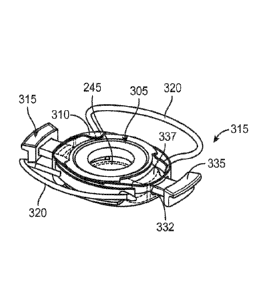
the force translation arms 115 or angulated relative to the force translation
arms 115. In some
implementations, the haptics 120 can be positioned at an angle in the range of
0-20 degrees or
other degree angle relative to the force translation arms 115. Haptics 120
considered herein
can include the Rayner designed haptics (Rayner Irrtraocular Lenses Ltd, East
Sussex, UK),
NuLens designed haptics (NuLens Ltd., Israel), Staar lens designs (Staar
Surgical, Monrovia,
CA), and others. In some implementations, the haptics 120 can be formed of a
biocompatible
polymer such as silicone, polyurethane, F'MMA, PVDF, PDMS, polyamide,
polypropylene,
polycarbonate, PEEK, etc. or a combination of such materials. The haptics 120
can be
formed of a material or configured to be foldable. In some implementations,
the haptics 120
are formed of a shape memory material.
[0088] Now with respect to FIGs. 2B, 3A-3F, the lens body 105 can
include a
shape deformation membrane 140 forming ring-like shape such that it forms a
continuous
loop or band of material near the periphery of the lens body 105. The shape
deformation
membrane 140 can have a first end or surface 141, a second end or surface 142,
and a
sidewall 143 between the first surface 141 and the second surface 142 having
an inner surface
and an outer surface. The shape deformation membrane 140 can be coupled on the
first
surface 141 to a shape changing membrane 145, for example on an anterior side
of the AIOL
100. The second surface 142 of the shape deformation membrane 140 can be
coupled to a
static element 150 that does not undergo a shape change, for example on a
posterior side of
the AIOL 100. The element 150 can be optically clear and provide support
function without
affecting the optics of the AIOL. The element 150 can also be or include a
static lens. It
should be appreciated that the anterior membrane can have an anterior support
that defines
the diameter of the shape changing membrane 145 and is configured to couple
the shape
deformation membrane to the shape changing membrane 145. The inner surfaces of
the
shape changing membrane 145, the shape deformation membrane 140 and the static
element
150 can collectively form a fixed volume, constant pressure, sealed chamber
155 configured
to contain a fixed volume of optical fluid therein. The shape deformation
membrane 140, the
shape changing membrane 145, and the static element 150 can each include a
central portion
and deformable portions such that upon coupling together they form the sealed
chamber 155
18
CA 02944010 2016-09-26
WO 2015/148673 PCT/US2015/022501
and the central portion 103 and the deformable portions 107 of the lens body
105. The sealed
chamber 155 can be a generally planar chamber formed by inner-facing surfaces
of the shape
changing membrane 145, the static element 150 and the sidewall 143 of the
shape
deformation membrane 140 and can have a variety of shapes as will be discussed
in more
detail below.
[0089] An outer surface of the sidewall 143 of the shape deformation
membrane 140 can be aligned with and bonded to an inner surface of the central
region 125
of the exterior support 110 such that the lens body 105 is fixedly positioned
within the central
region 125. It should be appreciated that the orientation of the lens body 105
within the
device 100 and within the eye can vary such that the shape changing membrane
145 can be
positioned anteriorly and the static element 150, such as a static lens,
positioned posteriorly
relative to the eye anatomy. Similarly, the shape changing membrane 145 can be
positioned
posteriorly and the static element 150 positioned anteriorly relative to the
eye anatomy.
Further, it should be appreciated that the shape changing membrane 145 and/or
the static
element 150 can create a sealed chamber 155 within the device 100 by coupling
directly to
the exterior support 110 rather than the surfaces 141, 142 of the shape
deformation
membrane 140. Further, the lens can include an anterior support coupled to and
defining the
diameter of the shape changing membrane 145.
[0090] FIGs. 3C and 3D illustrate an implementation of the static
element 150
having a static lens. The static lens can be formed of silicone, urethane,
acrylic material, a
low modulus elastomer, or combinations thereof. The static lens can be a
static optic to
correct to emmetropic state, or can be of an appropriate power for an aphakic
patient (usually
10D to 30D). The static lens can have zero power and form a posterior support
to the lens
body 105. If the AIOL 100 is being used in conjunction with a separate
capsular IOL (e.g. as
a "piggyback" lens), the power can be in the range of about -5D to about +5D
to correct for
residual refractive or other optical aberrations in the optical system of the
eye. In some
implementations, the static lens can have a flat surface 151 and a curved
surface 152. The
static lens also can be positioned inside the lens body 105 as described above
such that the
flat surface 151 is in contact with the fluid of the eye and the curved
surface 152 forms the
19
CA 02944010 2016-09-26
WO 2015/148673 PCT/US2015/022501
inner surface facing the sealed chamber 155 of the lens body 105. In other
implementations,
the static lens can be positioned outside the lens body 105 such that the flat
surface 151 forms
the inner surface facing the sealed chamber 155 of the lens body 105 and the
curved surface
152 is in contact with the fluid of the eye. The relative refractive indices
of the static lens
and the fluid surrounding it (whether that is the fluid of the eye or optical
fluid within the
sealed chamber 155) will determine the shape of the static lens for any given
power. The
static lens can be piano-convex, convex-plano, convex-convex, concave-convex
or any other
combination. The static lens can be a spherical lens, aspheric lens,
diffractive lens or any
combination of both, for example, in order to reduce or compensate for any
aberrations
associated to the flexible lens.
[0091] The shape changing membrane 145 can be a flexible optic formed
of an
optically clear, low modulus elastomer such as silicone. The shape changing
membrane 145
can have a constant thickness such that it is a planar element (see FIG. 4A)
or a variable
thickness (see FIGs. 3E-3F; and also FIGs. 4B-4E) such that the shape changing
membrane
145 has a reduced thickness portion that is relatively more prone to give way,
for example
upon an increased force applied against an inner surface of the membrane 145
during
deformation of the sealed chamber 155. It should be appreciated that the
structure of the
shape changing membrane 145 can vary. In some implementations, the shape
changing
membrane 145 can have a linear gradient thickness (FIG. 4B), curved gradient
thickness
(FIG. 4C), 2, 3 or more thicknesses with a step including radiused or right
angles (FIG. 4D),
or multiple materials (FIG. 4E), for example materials configured to flex near
the
accommodating zone (i.e. the region of the membrane 145 undergoing a shape
change) and
other materials configured to reinforce the optic zone and limit distortion.
[0092] In some implementations, the reduced thickness portions of the
shape
changing membrane 145 can be found near region 170 of the shape changing
membrane 145
surrounding, within, or parallel to the optical axis A. The reduced thickness
region 170 can
be configured to give way due to increased pressure applied by the optical
fluid within the
sealed chamber 155 on an internal surface of the shape changing membrane 145
causing an
outward bowing of the outer face (e.g., anterior face). Region 172 of the
shape changing
81800138
membrane 145 can have a thickness greater than region 170 and can be more
resistant to
reshaping under such internal pressure applied by the optical fluid in the
sealed chamber 155.
The regions 172 of the shape changing membrane 145 can continue to provide
distance
vision correction even when the region 170 is reshaped for near vision. Region
170 of the
shape changing membrane 145 can be formed of a material that is relatively
more susceptible
to outward bowing than the material of region 172. Region 170 can be injection
molded in
combination with the regions 172 to provide a relatively seamless and
uninterrupted outer
face. The material of the regions 172 can be generally consistent, though the
region 170 can
have different stiffness or elasticity that causes it to bow outward farther
than the surrounding
region. The shape changing membrane 145 can be configured to have varied
multifocal
capabilities to provide the wearer of the AIOLs described herein with enhanced
vision over a
wider range of distances, for example, as described in U.S. Publication No.
2009/0234449.
[0093] Again with respect to FIG. 2H, the shape deformation
membrane 140
can include central portion 180 and deformable portions 182. In some
implementations, the
deformable portions 182 can be coupled to the central portion 180 by a hinge
such that the
deformable portions 182 are collapsible relative to the central portion 180.
The central
portion 180 can be aligned with the deformable region 170 of the shape
changing membrane
145 (and a central portion of the static element 150) to create the central
portion 103 of the
lens body 105 that is surrounding, within, or parallel to the optical axis A.
The outer surface
of the sidewall 143 of the central portion 180 can be aligned with and bonded
to an inner
surface of the central annular region 125 such that the central portion 103 of
the lens body
105 is fixedly attached relative to the central annular region 125 of the
exterior support 110.
The deformable portions 107 of the lens body 105, in contrast, can be freely
moveable within
the channels 132 of the side regions 130 of the exterior support 110 such that
the deformable
portions 107 of the lens body 105 can undergo inward, collapsible movement or
displacement
relative to the central portion 103 during accommodation as well be described
in more detail
above.
21
CA 2944010 2020-03-24
CA 02944010 2016-09-26
WO 2015/148673 PCT/US2015/022501
[0094] Still with respect to FIG. 2G-2H, the deformable portions 182
are
configured to come in contact with contact portion 137 of the force
translation arms 115 and
be moved relative to the central portion 180. For example, during
accommodation the force
translation arms 115 can be urged by the one or more ciliary structures
towards the optical
axis A. Contact portion 135 can be positioned to engage the one or more
ciliary structures
and contact portion 137 can be positioned against the deformable portion 182
of the shape
deformation membrane 140. Contraction can cause the deformable portion 182 of
the
membrane 140 to undergo movement relative to the central portion 180 of the
shape
deformation membrane 140. This movement can be a compression, contraction,
collapse,
indentation, stretch, deformation, hinging or other type of movement that is
generally toward
the optical axis A. This movement of the deformable portions 182 of the shape
deformation
membrane 140 (and thus, the deformable portions 107 of the lens body 105) can
cause
flexure of the shape change membrane 145 in the optic zone 101 without
imposing stress or
squeezing on the optic zone. The deformable portions 182 can be located inside
or outside
the optic zone. The optic zone as used herein generally refers to a region of
the lens body
105 that surrounds the optical axis and is optically clear for vision. The
optic zone is
configured to have a corrective power although the entire optic zone may not
have the same
corrective power. For example, a central region of the optic zone may have
corrective power
and a peripheral region of the optic zone may not have corrective power.
[0095] As mentioned above, the sealed chamber 155 of the lens body 105
can
be filed with clear, biocompatible optical fluid. The optical fluid can be a
non-compressible
liquid or gel that is clear and transparent in the visible spectrum, for
example, silicone fluids
and gels, functionalized silicone fluids and gels (for example, halogen, i.e.,
fluorinated
silicones, aromatic, i.e., phenyl functionalized silicones, etc.), hydrocarbon
and
functionalized hydrocarbons, such as long chain hydrocarbons, halogenated
hydrocarbons,
such as fluorinated and partially fluorinated hydrocarbons, aqueous systems,
both fluids and
gels, whose refractive index (RI) has been increased by the additions of water-
soluble or
water swellable polymers, bio-polymer swellable additives such as cellulose,
as well as
organic or inorganic additives that form nanostructures to increase refractive
index. In some
implementations, the optical fluid within the sealed chamber 155 has a
refractive index
22
CA 02944010 2016-09-26
WO 2015/148673 PCT/US2015/022501
higher than 1.37. In other implementations, the optical fluid within the
sealed chamber 155
has a refractive index between 1.37-1.57. In other implementations, the
optical fluid within
the sealed chamber 155 has a refractive index between 1.37-1.60.
[0096] The optical fluid within the sealed chamber 155 can cause
flexure of the
shape changing membrane 145 upon movements of the deformable portions 182 of
the shape
deformation membrane 140 (and thus, the deformable portions 107 of the lens
body 105).
Inward movement of the deformable portions 182 can result in the non-
compressible optical
fluid contained within the fixed-volume sealed chamber 155 of the lens body to
press against
the surfaces of the sealed chamber 155 including the inner surface of the
shape changing
membrane 145 and the inner surface of the sidewall 143 of the shape
deformation membrane
140. Because the shape changing membrane 145 has a region near the region 170
configured
to bow outward upon application of a force, the pressure of the optical fluid
against the inner
wall of the shape changing membrane 145 results in outward bowing and
reshaping of the
outer surface of the shape changing membrane 145 upon inward movement of
deformable
portions 107. The accommodative portion of the optic zone becomes more convex
increasing
the power of the AIOL 100.
[0097] It should be appreciated that this shape change of the shape
changing
membrane 145 occurs without actual flow of optical fluid from one chamber to
another
chamber. Rather, a force being applied on the shape deformation membrane 140
to deform
the sealed chamber 155 in a first region can cause a reactive deformation of
the sealed
chamber 155 in at least a second region as the optical fluid inside the sealed
chamber 155
changes shape along with the changing shape of the sealed chamber 155. The
sealed
chamber 155 has a fixed volume, a constant pressure and is deformable. The
optical fluid has
a fixed volume, is non-compressible, and changes shape depending on the shape
of the sealed
chamber 155. Inward deformation of one or more portions of the chamber 155
(e.g. the
deformable portions 107) can cause a reactive outward deformation of another
portion of the
chamber 155 (e.g. region 170 of the shape changing membrane 145) due to the
non-
compressible optical fluid inside the sealed chamber 155. The optical fluid
therefore does
not actually flow between separate chambers of the AIOL, but rather changes
shape alone
23
CA 02944010 2016-09-26
WO 2015/148673 PCT/US2015/022501
with the changing shape of the sealed chamber causing the accommodative
portion of the
optic zone of the shape changing membrane 145 to bow outward increasing the
power of the
AlOL 100.
[0098] The shape deformation membrane 140, shape change membrane 145,
and static element 150 together can form a lens body 105 having any of a
variety of shapes.
The central portion 103 of the lens body 105 can be generally circular and the
deformable
portions 107 can have any of a variety of shapes including bellowed, pleated,
trapezoidal,
cylindrical, elliptical, conical, spherical, hemi-spherical and the like (see
for example, FIGs.
5B, 5E, 5G). Further, it should be appreciated that the deformable portions
107 can have any
of a variety of cross-sectional shapes along a variety of axes (see for
example FIGs. 5A, 5C,
5D, and 5F). The lens body 105 can also be a circular elastomeric ring having
a central
portion 103 and the deformable region within the optic zone such that the
contact portion 137
of the force translation arms 115 contacts the shape deformation membrane 140
within the
optic zone as shown in FIGs. 5H, 51-5J, and also FIG. 25F). The deformable
portion 107 of
the lens body 105 can be located outside or inside the optic zone (see for
example, FIG. 5H),
as well as outside or inside the lens body 105. The lens body 105 can have
more than two
deformable portions 107, including three, four or more deformable portions
107.
[0099] The shape deformation membrane 140 can be formed of an
optically
clear, low modulus elastomer such as silicone, urethane, or flexible inelastic
film such as
polyethylene. The central portion 180 of the shape deformation membrane 140
can be made
of an elastic material. The deformable portions 182 of the shape deformation
membrane 140
can be formed of elastic or inelastic materials.
[00100] Again with respect to FIGs. 2B and 2H, the devices described
herein can
include a force translation arm 115 configured to extend through an opening
133 in a sidewall
134 of the side regions 130 of the exterior support 110. As described above, a
force
translation arm 115 can extend through the opening 133 of one of the side
regions 130 and a
second force translation arm 115 can extend through the opening 133 of the
opposing side
region 130. It should be appreciated however that the devices described herein
can include
less than as well as more than two force translation arms 115. For example,
the devices
24
CA 02944010 2016-09-26
WO 2015/148673 PCT/US2015/022501
described herein can include one, three, four or more force translation arms
115 arranged
evenly around the device. In some implementations, the force translation arms
115 can be a
rigid polymer such as silicone, polyurethane, PMMA, PVDF, F'DMS, polyamide,
polypropylene, polycarbonate, etc., or combinations thereof. In some
implementations, the
force translation arms 115 can be an element reinforced with PMMA.
[00101] In some implementations, the force translation arms 115 can each
include an outer, contact portion 135 and an inner, contact portion 137 that
can have any of a
variety of shapes (see for example FIGs. 2B and 2H). Contact portion 135 can
be configured
to abut, contact, engage, functionally couple or be in close association with
one or more
ciliary structures, including but not limited to the ciliary body, ciliary
processes, ciliary
muscle, the zonules, or a combination thereof to drive shape change of the
optics during
accommodation and disaccommodation. Contact portion 135 of each force
translation arm
115 can remain external to the exterior support 110 such that it can remain in
contact with the
ciliary structure during accommodation and disaccommodation. In some
implementations,
the contact portion 135 can have an outer surface having a curved contour that
can match a
curved contour of a region of the eye in which the contact portion 135
associates. In some
implementations, the contact portion 135 can have indentations, grooves,
teeth, combs or
other surface features to improve, for example, contact and interdigitation
with ciliary
processes or zonular processes. The outer surface of the contact portion 135
can also have
sharpened or beveled edges on an upper and/or lower edge. The contact portions
135 of the
force translation arms 115 can incorporate features that improve their
connection with the
ciliary structures without causing damage. Generally, the contact portions 135
avoid piercing
or causing trauma to the ciliary structures. In some implementations, the
contact portions
135 can interfere with the ciliary structures such that movement can be
transferred without
causing trauma to the tissues themselves.
[00102] .. Contact portion 137 can be coupled to contact portion 135. In some
implementations, the contact portion 137 can be an elongate element coupled to
and
extending out from an inner surface of contact portion 135 (see e.g. FIG. 2B).
The contact
portion 137 can be shaped to be positioned within channel 132 such that at
least a portion of
CA 02944010 2016-09-26
WO 2015/148673 PCT/US2015/022501
the force translation arms 115 can translate within channel 132. Contact
portion 137 can abut
against at least a region of the lens body 105, such as the deformable portion
182 of the shape
deformation membrane 140. For example, as the ciliary muscle 18 contracts
during
accommodation it constricts towards the optical axis. The ciliary structure
can make contact
an outer surface of contact portion 135 such that the force translation arms
115 moves within
the channel 132 and contact portion 137 presses against the deformable portion
107 of the
lens body 105 and causes movement of the deformable portion 107 relative to
central portion
103 thereby driving the accommodating shape change of the shape changing
membrane 145
as described above.
[00103] The position of the force translation arms 115 relative to the one
or more
ciliary structures can vary. Further, the force translation arms 115 can have
a fixed length or
can be adjustable. The adjustment of the force translation arms 115 can be
performed prior
to, during, or any time after insertion in the eye. It should be appreciated
that the various
components and features described for the various force translation arms can
be incorporated
with one or more various components and features described with respect to the
various
devices herein. Any of the devices and systems described herein can
incorporate any of a
variety of features and components described herein. Components or features of
one
implementation of a device and system described herein can be incorporated
alternatively or
in combination with components or features of another implementation of a
device and
system described herein. For the sake of brevity, explicit descriptions of
each of those
combinations may be omitted although the various combinations are to be
considered herein.
[00104] FIG. 6 shows an implementation of a force translation arm 115
having a
fixed length. The force translation arm 115 can have an outer contact portion
135 configured
to contact one or more ciliary structures, such as the ciliary body. The
contact portion 135
can be coupled to an inner contact portion 137 by an elongate element 136. The
overall
length of the force translation arm 115 can be fixed and an appropriate size
selected for each
patient based on pre-op measurements.
[00105] FIG. 7 shows an implementation of a force translation arm 115
having a
length that can be adjusted, for example before, during or any time subsequent
to
26
CA 02944010 2016-09-26
WO 2015/148673 PCT/US2015/022501
implantation. In this implementation, the force translation arm 115 has a
contact portion 135
and a contact portion 137. Contact portion 135 can have a first elongate
element 738
extending out from an inner surface of the contact portion 135 and contact
portion 137 can
have a second elongate element 739 extending out from an outer surface of the
contact
portion 137. The mechanical adjustment interface between the first elongate
element 738 and
the second elongate element 739 can be a threaded engagement where an outer
surface of a
region of the first or second elongate elements 738, 739 can have threads
configured to
engage corresponding threads on an inner surface of a region of the first or
second elongate
elements 738, 739. For example, the second elongate element 739 can have
threads on an
outer surface and be configured to insert into a chamber 731 of the first
elongate element 738
to engage with corresponding threads. This threaded engagement between the two
portions
of the force translation arm 115 allows for on-the-fly adjustments to be made
for optimal
sizing, for example prior to insertion while the patient is on the table,
during or any time after
implantation of the device within the eye
[00106] The first and second elongate elements 738, 739 can engage one
another
according to other various mechanical configurations. For example, FIG. 8A
shows another
implementation of a force translation arm 115 having a length that can be
adjusted. In this
implementation, the force translation arm 115 has a contact portion 135 and a
contact portion
137. Contact portion 135 can have a first elongate element 738 extending out
from an inner
surface of the contact portion 135 and contact portion 137 can have a second
elongate
element 739 extending out from an outer surface of the contact portion 137.
The first
elongate element 738 and the second elongate element 739 can be aligned
adjacent to one
another until a desired overall length of the force translation arm 115 is
achieved.
Alternatively, the first and second elongate elements 738, 739 can be aligned
coaxial with
one another such that one of the elongate elements inserts through a bore into
the chamber
731 of the opposite elongate element (see FIG. 8B). In both configurations, a
region of the
first and second elongate elements 738, 739 can be mechanically fixed together
such as by
crimping at a crimp site 861 once the desired length is achieved. This type of
adjustment can
be performed, for example, prior to, during, or any time after implantation of
the device
within the eye.
27
CA 02944010 2016-09-26
WO 2015/148673 PCT/US2015/022501
[00107] In another interrelated implementation as shown in FIG. 9, once
desired
length between contact portions 135, 137 is achieved, the first and second
elongate elements
738, 739 can be fixed together such as by a sliding cam mechanism 963. The
first elongate
element 738 can have an irregularly shaped shaft having an end that is
configured to contact a
corresponding end of the second elongate element 739 also having an irregular
shape. As the
end of the first elongate element 738 is passed beyond the end of the second
elongate element
the two irregularly shaped shafts snap into locking engagement with one
another.
[00108] In another interrelated implementation as shown in FIG. 10, the
first and
second elongate elements 738, 739 can engage one another forming a piston
system. The
first elongate element 738 can include a chamber 731 and an end of the second
elongate
element 739 can extend through a bore into the chamber 731. The chamber 731
can be filled
to desired volume with an incompressible material 1090 to adjust the effective
length of the
elements 738, 739 relative to one another. The chamber 731 can be filled
during the surgical
procedure to fine-tune the effective length of the force translation arms 115.
[00109] In another interrelated implementation as shown in FIG. 11, contact
region 135 can be coupled to contact region 137 by a flexible hinge mechanism
1192. In
some implementations, the contact region 135 is coupled to a first elongate
element 738 and
contact region 137 is coupled to a second elongate element 739. The first
elongate element
738 mates with the second elongate element 739 via the hinge mechanism 1192.
It should be
appreciated that the contact region 135 can have more than one elongate
element 738 and
contact region 137 can have more than one elongate element 739 that each
couple together,
respectively, by a hinge mechanisms 1192. The flexible hinge mechanism(s) 1192
can be
adjusted prior to or during the procedure. In some implementations, the hinge
mechanism
1192 can be fixed in place via thermal/radiation/chemically induced curing.
The hinge
mechanism 1192 can be configured to rotate in a direction such that the
elongate force
translation aims 738, 739 fold outward or inward. It should also be
appreciated that one or
more hinge mechanisms 1192 can fold along one or more axes to provide not only
adjustment
between the optical axis A and the ciliary structure, but also adjustment in
an anterior and/or
posterior direction.
28
CA 02944010 2016-09-26
WO 2015/148673 PCT/US2015/022501
[00110] In another interrelated implementation as shown in FIG. 12, the
coupling between first and second elongate elements 738, 739 can additionally
or
alternatively involve a chemical linkage. For example, the first and second
elongate elements
738, 739, can be associated and then chemically fixed together using a
chemical material
such as an adhesive or an activating material such as thermo/radiation cured
polymers or
other materials. The material can be introduced at the interface between the
first and second
elongate elements 738, 739. In some implementations, the first elongate
element 738 can
include a chamber 731 that can be at least partially filled with material 1090
such that the
material 1090 surrounds the outer surface of the second elongate element 739
inserted
through a bore into the chamber 731. Once a desired length adjustment is
achieved, the
material 1090 can be activated to fix the interface between the first and
second elongate
elements 738, 739. The activation can be performed, for example, on the table
prior to
insertion in the eye or after insertion of the device in the eye such that
measurement,
adjustment and fixation are performed after implantation of the device 100.
[00111] In an interrelated implementation as shown in FIG. 13, the force
translation arm 115 can include contact portion 137 coupled by an elongate
element 739 to a
contact portion that can be a membrane 1394 having an internal volume 1396
configured to
contain a material. The material can include a volume adjustable material that
can be locked
in situ such as a thermosensitive glue, shape-memory alloys, shape-memory
polymers,
curable polymer, thermotradio activated material or other material that allows
for on-the-fly
adjustment in volume and space.
[00112] It should be appreciated that the force translation arms 115 need
not
move relative to the exterior support 110 and the lens body 105. For example,
the force
translation arms 115 can be configured to generate an electric current
generated upon ciliary
structure motion and contact. For example, the force translation arms 115 can
incorporate a
piezoelectric system that generates an electric charge in response to the
mechanical stress
applied by the ciliary structures. The current generated by the force
translation arms 115 can
be used to cause accommodation in the lens body 105. For example, an external
surface of
29
CA 02944010 2016-09-26
WO 2015/148673 PCT/US2015/022501
contact portion 135 can include a piezoelectric disk that generates a voltage
and cause
accommodation of the lens.
[00113] As mentioned herein the overall length of the force translation
arms 115
can be adjusted and fine-tuned before, during or after implantation for
individual patients, as
described above, to achieve customized and optimized contact between the force
translation
arms 115 and the ciliary structures such that shape change is in turn
optimized. It should be
appreciated that the shape change achieved in the lens body 105 can also be
adjusted and
fine-tuned any time after implantation of the device 100. In some
implementations and as
shown in FIGs. 14A-14B and similar to the implementation shown in FIG. 9, the
device 100
can incorporate a cam 1498. The cam 1498 can be positioned between the contact
portion
137 of the force translation arm 115 and the deformable portion 107 of the
lens body 105
such that they swing about one another. The position of the cam 1498 can be
changed such
as by rotation by a lever or other element such as a twisting mechanism. The
cam 1498 is
shown in a first "relaxed" position in FIG. 14A and in a max "active" position
in FIG. 14B.
[00114] In an interrelated implementation as shown in FIG. 15, a rod, shim,
spacer, wedge or other adjustment element 1502 can be incorporated to adjust
the relative
contact between the contact portion 137 of the force translation arm 115 and
the deformable
portion 107 of the lens body 105. The adjustment element 1502 can be inserted
through a
corresponding aperture in the exterior support 110 such that the further it is
inserted the
greater pressure it creates on the deformable portion 107 of the lens body 105
and the greater
the shape change. The adjustment element 1502 can be locked into position upon
reaching a
desired power adjustment. The adjustment element 1502 can also be released
such that the
power adjustment can be further fine-tuned by withdrawal of the adjustment
element 1502
from the exterior support 110. The position of the adjustment element 1502
relative to the
exterior support 110 can be adjusted in a variety of on-the-fly amounts
depending on the
depth of penetration of the adjustment element 1502 towards or away from the
exterior
support 110. Alternatively, the adjustment element 1502 can have a stepped
profile such that
it can be "clicked" into position one or two or more pre-set amounts. Further,
one or more
CA 02944010 2016-09-26
WO 2015/148673 PCT/US2015/022501
portions of the adjustment element 1502 can be coated with a thermo-sensitive
adhesive for
fixation following adjustment.
[00115] In an interrelated implementation as shown in FIGs. 16 and 17, the
pressure applied to the lens body 105 can be adjusted separately from that
applied by the
force translation arms 115 onto the deformable portions 107 of the lens body
105. For
example, an adjustable element 1602 such as a screw, lever, or rod can be
inserted through
the exterior support 110 to make contact with a region of the lens body 105,
such as against
the shape deformation membrane 140 near the central portion 103 of the lens
body. The
adjustable element 1602 can apply additional force on the shape deformation
membrane 140
such that the optical fluid within the sealed chamber 155 is further urged
against the shape
changing membrane 145. The adjustable element 1602 can be incrementally
adjustable in
order to fine-tune the pressure applied such that power adjustment can be
achieved. This
mechanism can also provide a general solution to power adjustment in the AIOL
without
accommodation. FIG. 17 illustrates a method of using a material that can be
expanded or
shrunk in situ in order to change the base power of the lens body 105. In this
implementation, rather than inserting a screw or other mechanical feature
against the lens
body 105, a tension on the shape change membrane 145 (or the static element
150) can be
adjusted. For example, the material can be a thermal sensitive material that
upon thermal
activation can create a bleb. Changes in tension and volume on the lens body
can occur
depending on whether a bleb, an indentation, or a flattening is formed upon
activation of the
material.
[00116] FIG. 18 shows a shape deformation membrane 140 having two
deformable portions 182 and a central portion 180. The deformable portions 182
can be
compressible or collapsible or otherwise configured to undergo movement
relative to the
central portion 180 toward (and also away from) the optical axis A. In this
implementation,
the deformable portions 182 are generally rectangular shaped and can be
displaced or move
in response to a force applied from an outer surface of the sidewall 143 of
the shape
deformation membrane 140 that is in the direction of arrow A without the
central portion 180
undergoing a movement or displacement. This displacement of the shape
deformation
31
CA 02944010 2016-09-26
WO 2015/148673 PCT/US2015/022501
membrane 140 of the closed system can result in the optical fluid contained
within the sealed
chamber pressing against the inner surface of the shape changing membrane and
an outward
bowing of a shape changing membrane coupled to the shape changing membrane
maintaining
pressure within the closed system constant.
[00117] Table 1 below illustrates the relationship between the displacement
of
the deformable portions 182 (displacement from each side) and the outward
bowing (and
thus, dioptric change or accommodation) of the shape changing membrane that
would result.
The lens diameter in mm is the region of the shape changing membrane that is
configured to
bow outwardly in response to the optical fluid pressing against it from within
the sealed
chamber. The optical fluid can be a silicone oil having a refractive index
between 1.37 -
1.57. The compressible portion length is the length (arrow L) of the
deformable portion 182
of the membrane 140 and the compressible portion height (arrow H) is the
thickness of the
sidewall 143 of the shape deformation membrane 140 (see FIG. 18). Displacement
from each
compressible portion (i.e. the deformable portion(s) 182 relative to the
central portion 180 of
the shape deformation membrane 140 or, in terms of the lens body 105, the
deformable
portion(s) 107 relative to the central portion 103) equals the volume V of the
sealed chamber
155 divided by the product of the length L of the compressible portion, the
height H of the
compressible portion and 2 or V/(L*H*2). The volume of the lens bowing is:
h
= (3a2 h2)
6
The lens height (h) can be calculated from Pythagoras equation: (r_h)2+a2 r2.
Hence: h=r-
Ar2-a2).
For example, if the refractive index of the optical fluid is 1.4 and the
diameter of the lens is
3mm, a 28 micron movement from each deformable portion 182 creates a
sufficient amount
of pressure applied by the optical fluid against the shape changing membrane
to form a 1D
lens and if the diameter of the lens is 3mm, a 84 micron movement of each
deformable
portion 182 creates a sufficient amount of pressure applied by the optical
fluid against the
shape changing membrane to form a 3D lens.
32
CA 02944010 2016-09-26
WO 2015/148673 PCT/US2015/022501
[00118] Table 1
Lens Refractive Compressible Compressible Displacement Diopters
Diameter Index of portion Length portion height (mm) from each (D)
(mm) optical fluid side
(mm) (mm)
3 1.4 2 0.6 0.026 1
3.5 1.4 2 0.6 0.048 1
4 1.4 2 0.6 0.082 1
3 1.4 2 0.6 0.052 2
3.5 1.4 2 0.6 0.096 2
4 1.4 2 0.6 0.164 2
3 1.4 2 0.6 0.078 3
3.5 1.4 2 0.6 0.144 3
4 1.4 2 0.6 0.246 3
3.5 1.4 2.5 0.7 0.099 3
4 1.57 1.8 0.5 0.089 3
[00119] FIG. 19 illustrates the optical power (D) achieved in a lens body
upon
movement (um) of a shape changing membrane upon application of a force (gf).
The devices
described herein were evaluated using an optical bench test for the evaluation
of intraocular
lenses (IOLA PLUS, Rotlex, Israel) and calibrated load cell (Advanced Force
Torque
Indicator (AFTI), Mecmesin, UK) and were shown to achieve about 3D change upon
100 um
movement and application of 1 gf.
33
CA 02944010 2016-09-26
WO 2015/148673 PCT/US2015/022501
[00120] FIG. 20 shows a cross-sectional, partial perspective view of an
AIOL
100 positioned within an eye and FIG. 21 is a cross-sectional perspective view
of the AIOL
100 positioned within the eye shown without the iris such that the haptic 120
is visible. FIG.
22 is a cross-sectional, side view of the AIOL 100 positioned in the eye and
in an
unaccommodated state. FIG. 23 is a cross-sectional, side view of the AIOL 100
positioned in
the eye and in an accommodated state. As with the various implementations
described
throughout, the AIOL 100 can include a lens body 105 having a sealed chamber
155 formed
of the inner surfaces of the shape deformation membrane 140, the shape
changing membrane
145 and the static element 150 and configured to contain optical fluid
therein. The lens body
105 can be positioned within and coupled to a support 110. The AIOL 100 can
include a
force translation arm 115 and a stabilization haptic 120. The stabilization
haptic 120 can be
positioned posterior to the iris 14 within the sulcus 16 (see FIG. 24) such
that the AIOL 100
is stabilized and fixed by the interaction of the haptic 120 within the sulcus
16. The AIOL
100 can also be implanted such that the stabilization haptic 120 is positioned
within the
capsular bag. An anterior surface of the central portion of the shape changing
membrane 145
can be aligned within a central annular region 125 of the support 110 and can
be configured
to bow outwardly upon contraction of the ciliary muscle 18, i.e. during
accommodation. The
AIOL 100 is shown implanted within an eye having undergone capsularhexis of
the capsular
bag 22 such that the static element 150 is positioned on a posterior-most side
of the device
105 and remains generally external to the capsularhexis (see FIG. 20). The
static element
150 can be a static lens powered for distance as described herein.
[00121] The devices described herein can be actuated into an accommodated
(or
unaccommodated) shape in direct response to ciliary structure movements, for
example
movements of the ciliary body and/or ciliary muscle. This direct ciliary
translation of
accommodation of the devices described herein can involve movement of optical
fluid within
the sealed chamber. As described above, and as shown also in FIGs. 20-23, the
force
translation arms 115 can directly contact one or more ciliary structures to
cause actuation of
the force translation arms 115 such that the contact portion 137 can be
positioned within the
channel 132 in a first configuration in which the force translation arm 115 is
generally
positioned away from a central axis CA of the device 105 (see FIG. 22) to a
second
34
CA 02944010 2016-09-26
WO 2015/148673 PCT/US2015/022501
configuration in which the force translation arm 115 is urged by the ciliary
structure towards
the central axis CA of the device 105 (see FIG. 23). The shape changing
membrane 145 can
be generally planar when the force translation arms 115 are in the first
configuration (i.e.
unaccommodated) and the shape changing membrane 145 can be bowed outwardly
when the
force translation arms 115 are in the second configuration (i.e.
accommodated). This can be
due to the contact portion 137 of the force translation arms 115 pressing
against shape
deformation membrane 140 such that the deformable portion 107 of the lens body
collapses
or moves inward towards the central portion 103 of the lens body 105. The
collapse of the
deformable portion 107 can cause optical fluid within the sealed chamber 155
to press against
the internal surfaces of the chamber 155 until the anterior surface of the
shape changing
membrane 145 takes on a more spherical or convex shape such as due to an
outward bowing
along the optical axis (see FIG. 23).
[00122] FIGs 25A-25G illustrate an interrelated implementation of an
accommodating intraocular lens ("AIOL") 200 according to the descriptions
provided herein.
It should be appreciated that the features and components of the devices
described herein can
be interrelated and used in combination or in the alternative. For the sake of
brevity some of
the descriptions regarding the components of the various implementations of
devices
described herein are not reiterated although it should not be construed to
mean those previous
descriptions do not apply to the following implementations.
[00123] The AIOL 200 can include a lens body 205, a support 210, force
translation arms 215, and one or more stabilization haptics 220. The support
210 can include
an internal and/or external support 210. In some implementations, the support
210 is an
external support 210 having a central annular region with which a central
portion of the lens
body 205 is aligned. The support 210 can include channels 232 or slots through
a peripheral
sidewall that extend into the central annular region (best shown in FIG. 25F).
A force
translation arm 215 can extend through a channel 232 on one side of the
support 210 and a
second force translation arm 215 can extend through a channel 232 on an
opposing side of
the support 210. The force translation arms 215 can each include an outer,
contact portion
235 configured to contact at least a portion of a ciliary structure and an
inner, contact portion
CA 02944010 2016-09-26
WO 2015/148673 PCT/US2015/022501
237 configured to contact at least a portion of the lens body 205. Contact
portion 235 of each
force translation arm 215 can remain external to the support 210 such that it
can remain in
contact with the ciliary structure during accommodation and disaccommodation.
Contact
portion 237 of each force translation arm 215 can translate within channel
232. The force
translation arms 215 can move freely back and forth within channel 232 as the
ciliary
structure moves to effect accommodative shape change of the lens body 205 as
will be
described in more detail below.
[00124] As with previous implementations, the support 210 can be formed of
a
rigid polymer, including but not limited to silicone, polyurethane, PMMA,
PVDF, PDMS,
polyamide, polypropylene, polycarbonate, etc, or combinations thereof. The
support 210 can
be configured to prevent distortion caused by movement of the force
translation arms 215
through the channels 232. The support 210 can be an exterior support located
outside the
sealed capsule 255 as shown in FIGs. 25A-25G or the support 210 can be located
inside the
sealed capsule 255 as shown in FIGs. 26A-26F and 27A-27D, which will be
described in
more detail below. In some implementations, one or more stabilization haptics
220 can be
bonded to the exterior support 210. In other implementations, the one or more
stabilization
haptics 220 can be bonded to a portion of the lens body 205 and the support
210 be located
within the sealed chamber of the lens body 205. In other implementations, the
one or more
stabilization haptics 220 can be molded as part of the lens body 205 or the
exterior support
210. The stabilization haptics 220 can be static haptics configured to
maintain alignment of
the optics of the device and to resist movement of the device once implanted
and undergoing
accommodative shape change as described in more detail above. In some
implementations,
the haptic(s) 220 can be placed in the ciliary sulcus or the capsular bag.
[00125] The lens body 205 can include a shape deformation membrane 240, a
shape changing membrane 245 and a static element 250, which can include a
static lens. The
shape deformation membrane 240, the shape changing membrane 245 and the static
element
250 in combination with the support 210 create a generally planar, sealed
chamber 255 that is
configured to contain optical fluid therein. The shape deformation membrane
240 can be a
ring- shape membrane coupled to an inner surface of the similarly ring-shaped
support 210.
36
CA 02944010 2016-09-26
WO 2015/148673 PCT/US2015/022501
A region of the shape changing membrane 245 can be coupled to a first surface
of the support
210 and a region of the static element 250 can be coupled to a second,
opposite surface of the
support 210. It should be appreciated that the orientation of the lens body
205 within the
AIOL 200 and within the eye can vary such that the shape changing membrane 245
can be
positioned anteriorly and the static element 250 positioned posteriorly
relative to the eye
anatomy. Similarly, the shape changing membrane 245 can be positioned
posteriorly and the
static element 250 positioned anteriorly relative to the eye anatomy.
[00126] The static element 250 is best shown in FIGs. 25B and 25C can be or
include a static lens formed of silicone, urethane, or a low modulus elastomer
as described
above in other embodiments. The shape changing membrane 245 can be a flexible
optic
formed of an optically clear low modulus elastomer such as silicone. The shape
changing
membrane 245 can have a constant thickness such that it is a planar element or
a variable
thickness such that the shape changing membrane 245 has a reduced thickness
portion that is
relatively more prone to give way due to increased internal pressure as
described in more
detail above. It should be appreciated that the structure of the shape
changing membrane 245
can vary as described herein. The reduced thickness portion can be configured
to give way
due to increased internal pressure applied by the optical fluid within the
sealed chamber 255
causing an outward bowing of the outer face (e.g., anterior face).
[00127] Now with respect to FIG. 25F and 25G, the support 210 can include
channels 232 through which the shape deformation membrane 240 can be accessed
by the
contact portions 237 of the force translation arms 215. For example, during
accommodation
the force translation arms 215 can be urged by the one or more ciliary
structures towards the
optical axis A. Inward/anterior movement of the ciliary structure can be
harnessed by contact
portions 235 causing the force translation arms 215 to move inward through
channels 232
toward the optical axis A. Contact portions 237 of the force translation arms
215 can contact
the shape deformation membrane 240 and cause the shape deformation membrane
240 to
undergo movement relative to the shape changing membrane 245. This movement
can be a
compression, indentation, stretch, deformation, or other type of movement that
is generally
toward the optical axis A. This movement of the shape deformation membrane 240
can
37
CA 02944010 2016-09-26
WO 2015/148673 PCT/US2015/022501
cause flexure of the shape change membrane 245 into a more spherical or convex
shape in
the optic zone 201 thereby increasing the power of the lens for near vision
focus without
imposing stress or squeezing on the optic zone as will be described in more
detail below.
[00128] .. As mentioned above, the sealed chamber 255 of the lens body 205 can
be filled with optical fluid that can be a clear, biocompatible optical fluid.
The optical fluid
can be a non-compressible liquid or gel that is clear and transparent in the
visible spectrum,
for example, silicone fluids and gels, functionalized silicone fluids and gels
(for example,
halogen, i.e., fluorinated silicones, aromatic, i.e., phenyl functionalized
silicones, etc.),
hydrocarbon and functionalized hydrocarbons, such as long chain hydrocarbons,
halogenated
hydrocarbons, such as fluorinated and partially fluorinated hydrocarbons,
aqueous systems,
both fluids and gels, whose refractive index (RI) has been increased by the
additions of
water-soluble or water swellable polymers, bio-polymer swellable additives
such as cellulose,
as well as organic or inorganic additives that form nanostructures to increase
refractive index.
In some implementations, the optical fluid within the sealed chamber 255 has a
refractive
index higher than 1.37. In other implementations, the optical fluid within the
sealed chamber
255 has a refractive index between 1.37-1.57. In other implementations, the
optical fluid
within the sealed chamber 255 has a refractive index between 1.37-1.60.
[00129] .. The optical fluid within the sealed chamber 255 can cause flexure
of the
shape changing membrane 245 upon movements of the shape deformation membrane
240.
Inward movement of the shape deformation membrane 240 can result in the non-
compressible optical fluid contained within the fixed volume, sealed chamber
255 to press
against the surfaces of the sealed chamber 255 including the inner surface of
the shape
changing membrane 245. Because the shape changing membrane 245 has a region
near the
central portion configured to bow outward upon application of a force, the
pressure of the
optical fluid against the inner wall of the shape changing membrane 245
results in outward
bowing and reshaping of the outer surface of the shape changing membrane 245.
FIGs. 25D
and 25E are cross-sectional, partial views of a device in a relaxed,
disaccommodated
(unaccommodated) state and an actuated, accommodated state, respectfully. The
optic zone
portion surrounding, within or parallel to the optical axis A becomes more
convex increasing
38
CA 02944010 2016-09-26
WO 2015/148673 PCT/US2015/022501
the power of the AIOL 200. It should be appreciated that this shape change of
the shape
changing membrane 245 can occur without actual flow of fluid from one part of
the lens body
205 to another as described herein. Rather, the compression of one region of
the sealed
chamber 255 having a fixed volume filled with a corresponding fixed volume of
non-
compressible optical fluid drives a reactive shape change of another region of
the sealed
chamber 255 formed by the shape changing membrane 245.
[00130] The AIOL 200 can include force translation arms 215 configured to
extend through channels 232 in the support 210. As described above, a force
translation arm
215 can extend through the channel 232 of one side and a second force
translation arm 215
can extend through the channel 232 of the opposing side. It should be
appreciated however
that the devices described herein can include more than two force translation
arms 215. For
example, the devices described herein can include three, four or more force
translation arms
215 arranged evenly around the device. In some implementations, the force
translation arms
215 can be a rigid polymer such as silicone, polyurethane, PMMA, PVDF, PDMS,
polyamide, polypropylene, polycarbonate, etc. or combinations thereof. For
example, the
force translation arms 215 can be an element of a first material reinforced
with a second
material, such as PMMA.
[00131] In some implementations, the force translation arms 215 can each
include an outer, contact portion 235 and an inner, contact portion 237 that
can have any of a
variety of shapes as described herein. Contact portion 235 can be configured
to abut, contact,
engage, functionally couple or be in close association with one or more
ciliary structures,
including but not limited to the ciliary body, ciliary processes, ciliary
muscle, the zonules, or
a combination thereof to drive shape change of the optics during accommodation
and
disaccommodation. Contact portion 235 of each force translation arm 215 can
remain
external to the support 210 such that it can remain in contact with the
ciliary structure during
accommodation and disaccommodation. In some implementations, the contact
portion 235
can have an outer surface having a curved contour that can match a curved
contour of a
region of the eye in which the contact portion 235 associates. In some
implementations, the
contact portion 235 can have indentations, grooves, teeth, combs or other
surface features to
39
CA 02944010 2016-09-26
WO 2015/148673 PCT/US2015/022501
improve, for example, contact and interdigitation with ciliary processes or
zonular processes.
The outer surface of the contact portion 235 can also have sharpened or
beveled edges on an
upper and/or lower edge.
[00132] Contact portion 237 can be coupled to contact portion 235. In some
implementations, the contact portion 237 can be an elongate element coupled to
and/or
extending out from an inner surface of contact portion 235 (see e.g. FIG. 25D
and 25E). The
contact portion 237 can be shaped to be positioned within channel 232 such
that at least a
portion of the force translation arms 215 can translate within channel 232.
Contact portion
237 can abut against the shape deformation membrane 240 as described above.
For example,
as the ciliary muscle 18 contracts during accommodation it constricts towards
the optical axis
A. The ciliary structure can make contact an outer surface of contact portion
235 such that
the force translation arms 215 moves within the channel 232 and contact
portion 237 presses
against the shape deformation membrane 240 of the lens body 205 and causes
movement of
the shape deformation membrane 240 relative to the shape changing membrane 245
thereby
driving the accommodating shape change of the shape changing membrane 245 as
described
above. The shape deformation membrane 240 can be located inside or outside the
optic zone
of the lens body.
[00133] FIGs 26A-26F illustrate an interrelated implementation of an
accommodating intraocular lens ("AIOL") 300 having an internal support 312
that will be
described in more detail below. It should be appreciated that the features and
components of
the devices described herein can be interrelated and used in combination or in
the alternative.
For the sake of brevity some of the descriptions regarding the components of
the various
implementations of devices described herein are not reiterated although it
should not be
construed to mean those previous descriptions do not apply to the following
implementations.
[00134] The AIOL 300 can include a lens body 305, a support 310, and force
translation arms 315. The support 310 can include an internal and/or an
external support 310.
In some implementations, the AIOL 300 has only an internal support sufficient
to support the
lens without any additional exterior support. The lens body 305 can be
positioned within and
coupled to a central region of the support 310. An anterior surface of the
lens body 305 can
CA 02944010 2016-09-26
WO 2015/148673 PCT/US2015/022501
be exposed through an anterior opening of the support 310 and a posterior
surface of the lens
body 305 can be exposed through a posterior opening of the support 310. It
should be
appreciated that orientation of the components of the lens body 305 within the
AIOL 300 and
within the eye can vary and that use of the terms "anterior" and "posterior"
are not intended
to be limiting.
[00135] The support 310 can include channels 332 or slots in a sidewall
through
which the force translation arms 315 can extend. The force translation arms
315 can move
freely back and forth within the channels 332 as the ciliary structures move
to effect
accommodative shape change of the lens body 305. For example, a first force
translation arm
315 can extend through a first channel 332 on one side of the support 310 and
a second force
translation arm 315 can extend through a second channel 332 on an opposing
side of the
support 310. The force translation arms 315 can each include an outer, contact
portion 335
configured to contact at least a portion of a ciliary structure and an inner,
contact portion 337
configured to translated within channel 332 and make contact with the lens
body 305. The
contact portion 335 of each force translation arm 315 can remain external to
the support 310
such that it can remain in contact with the ciliary structure during
accommodation and
disaccommodation.
[00136] The support 310 can be formed of a material configured to prevent
distortion caused by movement of the force translation arms 315 as well as
prevent
inadvertent movements of the force translation arms 315 (e.g. perpendicular to
the direction
of the force translation arm 315 inward/outward movement). In some
implementations, one
or more stabilization haptics 320 can be bonded to the support 310. In other
implementations, the one or more stabilization haptics 320 can be bonded to a
portion of the
lens body 305 and the support 310 be located within the sealed chamber of the
lens body 305.
In other implementations, the one or more stabilization haptics 320 can be
molded as part of
the lens body 305 or the exterior support 310. The stabilization haptics 320
can be static
haptics configured to maintain alignment of the optics of the device and to
resist movement
(e.g. vertical movement) of the AIOL 300 once implanted and undergoing
accommodative
41
CA 02944010 2016-09-26
WO 2015/148673 PCT/US2015/022501
shape change as described in more detail above. In some implementations, the
one or more
haptic(s) 320 can be placed in the ciliary sulcus and/or the capsular bag.
[00137] The lens body 305 can include a shape deformation membrane 340, a
shape changing membrane 345, and a static element 350 (or static lens) sealed
together into a
generally planar lens body 305 having a sealed chamber 355. The sealed chamber
355 is
configured to contain optical fluid therein, for example a fluorosilicone oil
or other optical
fluid described herein. The shape deformation membrane 340 can be a ring-
shaped silicone
structure (e.g. PDMS) coupled on a first surface (e.g. anterior surface) to a
perimeter of the
shape changing membrane 345 or an anterior support defining a diameter of the
shape
changing membrane 345 The shape deformation membrane 340 can be coupled on an
opposite surface (e.g. posterior surface) to a perimeter of the static element
350. It should be
appreciated that the components of the lens body can be coupled in any of a
variety of
configurations between themselves as well as with the support 310. The outer
wall of the
shape deformation membrane 340 can have regions configured to engage with the
force
translation arms 315 such that as the force translation arms 315 move, the
regions likewise
move. Movement of the shape deformation membrane 340 changes the shape of the
sealed
chamber 355 causing accommodation and disaccommodation as will be described in
more
detail below. In some implementations, the shape deformation membrane 340 can
have a
first region on the outer wall that engages with a first force translation arm
315 and a second
region on the outer wall that engages with a second force translation arm 315.
Each of the
first and second regions on the outer wall of the shape deformation membrane
340 can
include a surface feature 341 configured to engage with a corresponding
feature 338 on the
force translation arm 315 (best shown in FIG. 26C).
[00138] The support 310 can be formed of a harder material (or materials)
than
the shape deformation membrane 340 to prevent inadvertent movements of the
moving parts
of the device. The ARYL 300 can alternatively or in additionally include one
or more ribs or
internal supports 312 located within the sealed chamber 355 of the lens body
305. The
internal supports 312 can act to mechanically isolate the optical components
of the lens from
optical distortion during movement of the moving parts of the AIOL 300. In
some
42
CA 02944010 2016-09-26
WO 2015/148673 PCT/US2015/022501
implementations, the one or more internal supports 312 connect the anterior
and posterior
supports. In some implementations, a first surface of the internal support 312
can be coupled
to a perimeter region of the shape changing membrane 345 (best shown in FIGs.
26B and
26C) or an anterior support defining the diameter of the shape changing
membrane 345. In
some implementations, the internal support 312 is additionally coupled to the
static element
350 (or static lens) such that the internal support 312 is coupled on a first
surface to a
perimeter region of the shape changing membrane 345 and coupled on a second,
opposite
surface to a perimeter region of the static element 350 (see FIGs. 27A-27D).
In either
implementation (i.e. coupled to one or both of the anterior and posterior
surfaces), at least a
portion of the internal support 312 is separated a distance from the shape
deformation
membrane 340 such that the internal support 312 partitions the sealed chamber
355 into a
deformable region 307 and a central region 303 within which optical fluid is
contained. If the
internal support 312 is coupled to just the shape changing membrane 345, a
channel 342 can
extend under the internal support 312 allowing for fluid communication between
the
deformable region 307 and the central region 303 of the sealed chamber 355
(best shown in
FIG. 26B). If the internal support 312 is coupled to both the shape changing
membrane 345
and the static element 350, one or more channels 342 can extend through the
internal support
312 itself to allow for fluid communication between the deformable region 307
and the
central region 303 of the sealed chamber 355 (best shown in FIG. 27B). The one
or more
channels 342 can include a cylindrical bore that extends from a region of an
outer wall
through to a region of an inner wall of the internal support 312. It should be
appreciated that
the channels 342 can have any of a variety of shapes and sizes. Alternatively,
a plurality of
internal supports 312 can be contained within the sealed chamber 355 that are
spaced apart
from one another creating one or more channels 342 between them for fluid
communication
within the sealed chamber 355 between the deformable region 307 and the
central region 303
of the sealed chamber. The AIOLs described herein can incorporate 1, 2, 3, 4,
5 or more
internal supports 312 within the sealed chamber 355.
[00139] During accommodation, inward/anterior movement of the ciliary
structure can be harnessed by contact portions 335 of the force translation
arms 315 causing
the force translation arms 315 to move inward toward the optical axis A.
Inward movement
43
CA 02944010 2016-09-26
WO 2015/148673 PCT/US2015/022501
of the force translation arms 315 urges the shape deformation membrane 340 to
undergo
movement or deformation relative to the shape changing membrane 345, for
example,
towards the optical axis. Inward movement, collapse, compression or
deformation of the
deformable regions 307 of the shape deformation membrane 340 toward the
optical axis
causes the non-compressible optical fluid contained within the fixed volume,
sealed chamber
355 to press against the surfaces of the sealed chamber 355 including the
inner surface of the
shape changing membrane 345. The shape changing membrane 345 can have a region
surrounding the optical axis configured to bow outward or flex upon
application of a force
into a more spherical or convex shape in the optic zone thereby increasing the
power of the
lens for near vision focus without imposing stress or squeezing on the optic
zone. The
pressure of the optical fluid against the shape changing membrane 345 reshapes
the outer
surface. It should be appreciated that this shape change can occur without
flow of fluid from
one part of the lens body 305 to another. Rather, the compression of the fixed
volume sealed
chamber 355 (and deformable region 307) filled with non-compressible optical
fluid drives
the shape change of the membrane 345. The deformable region 307 and central
region 303
of the sealed chamber 355 can both be within the optic zone such that
deformation of the
shape deformation membrane 340 (and the deformable region 307) occurs inside
the optic
zone. Alternatively, the deformable region 307 can be located outside the
optic zone such
that deformation of the shape deformation membrane 340 (and the deformable
region 307)
occurs outside the optic zone.
[00140] .. The internal support 312 can have a tapered geometry such that the
supports 312 do not come into contact with moving parts of the lens such as
the shape
deformation membrane 340. For example as shown in 26C, the support 312 can
have a wider
dimension near where the support 312 couples with the shape changing membrane
345 and
an outer wall that tapers away from the shape changing membrane 345 such that
the support
312 has a narrower dimension near where the shape deformation membrane 340
deforms to
the greatest degree during accommodation. In another example as shown in FIG.
27D, the
support 312 can have a wider dimension on both anterior and posterior ends of
the AIOL 300
where the support 312 couples with the shape changing membrane 345 and the
static element
44
CA 02944010 2016-09-26
WO 2015/148673 PCT/US2015/022501
350, respectively, that tapers towards a center region. In this
implementation, the support 312
forms a tapered spool shape.
[00141] The dimensions of the components of the devices described herein
can
vary. The devices can be configured to be implanted through an incision that
is less than
about 4 mm. In some implementations, the overall diameter of the device is
approximately 8
mm, although this can vary. For example, a device having flexible or foldable
stabilization
haptics can have a first diameter during implantation that is smaller than the
diameter it
achieves after implantation following unfolding or expansion of the
stabilization haptics. In
some implementations, the exterior support can be made from a flexible
material(s) such that
the exterior support can bend during implantation of the device. In some
implementations,
the central, optic zone portion of the lens body can have a diameter that is
about 2.5 mm,
about 3.0 mm, about 3.5 mm, about 4.0 mm, about 4.5 mm, about 5.0 mm, about
5.5 mm,
about 6.0 mm, about 6.5 mm, or greater diameter. In some implementations, the
accommodating diameter, or the region of the central optic zone that undergoes
a shape
change, is greater than 3.0 mm.
[00142] As described above, the deformable regions of the lens body can
move
or collapse relative to the central region of the lens body upon application
of a degree of force
on the shape changing membrane. The force applied to achieve movement of the
shape
changing membrane of the lens body to effect accommodation can be as low as
about 0.1
grams of force (go. In some implementations, the force applied can be between
about 0.1 gf
to about 5.0 gf or between about 0.5 gf to about 1.5 gf or between about 1.0
gf to about 1.5
gf. The movements of the deformable regions of the lens body relative to the
central portion
of the lens body in response to forces applied to achieve accommodation can be
as small as
about 50um. The movements of the deformable regions of the lens body relative
to the
central portion of the lens body (or e.g. region 107 relative to central
region 103 or the
deformable portion 182 of the shape deformation membrane 140 relative to the
central
portion 180 of the shape deformation membrane 140) in response to forces
applied can be
between about 50 um to about 500 um, between about 50 um to about 150 um, or
between
about 100 um to about 150 um. These ranges of forces applied and that result
in these ranges
81800138
of movement can provide the devices described herein with an accommodating
capability that
is within a dynamic range of greater than 3D. In some implementations, the
power is
between 4D and 6D for about 100-150 um movement. The devices described herein
can
have an accommodating range that is at least 3D for about 100um movement of
the shape
changing membrane and about a force of at least 0.1 gf applied to the shape
changing
membrane. In other implementations, the devices can have an accommodating
range that is
at least 3D for about 50 urn movement and at least about 1.0 gf.
[00143] Suitable materials or combinations of materials for the
preparation of
the various components of the devices disclosed herein are provided
throughout. It should be
appreciated that other suitable materials are considered. U.S. Patent
Publication Nos.
2009/0234449, 2009/0292355 and 2012/0253459, provide further examples of other
materials suitable for forming certain components for the devices described
herein.
[00144] The various devices described herein can be implanted
according to a
variety of surgical methods known in the art. Depending upon the features and
components
of the device, they can be implanted using various techniques or using various
implements.
The devices described herein can be used alone or in combination with another
intraocular
lens or the patient's natural lens. As described herein the power of the lens
body as well as
the relative position of the force translation arms and/or stability haptics
can be adjusted
and/or fine-tuned prior to implantation, during implantation or any time after
implantation. It
should also be appreciated that the devices described herein can be inserted
through a small
incision, such as an incision that is no greater than 3.5 mm. The devices
described herein can
be implanted such that the device is positioned outside the lens capsule, for
example anterior
to the capsule and posterior to the iris. The devices described herein can be
implanted such
that the central portion of the lens body is aligned with the optical axis of
the eye. The force
translation arms can be positioned relative to the one or more ciliary
structures such as the
ciliary body or the ciliary muscle. The force translation arms can be
positioned such that they
abut with the ciliary structure (or very closely associated to the ciliary
structure without
abutting) without causing compression of the lens body including the
deformable region of
46
CA 2944010 2020-03-24
CA 02944010 2016-09-26
WO 2015/148673 PCT/US2015/022501
the lens body when the ciliary structure is in the resting, disaccommodated
state
(unaccommodated). However, the force translation arms can be positioned close
enough to
the ciliary structure such that upon contraction of the ciliary muscle the
lens body undergoes
accommodation and upon relaxation of the ciliary muscle the lens body
undergoes
disaccommodation and the materials of the lens body rapidly return to their
resting state. The
relative position and length of the force translation arms can be adjusted
according to the
various methods described above using one or more of the various features for
adjustment
described herein. The stabilization haptics can be positioned within the
ciliary sulcus (or
other region) to further stabilize the device within the eye. The resting
power of the lens
body can also undergo further adjustment and fine-tuning according to the
various methods
described herein and using one or more of the various features for power
adjustment
described herein.
[00145] While this
specification contains many specifics, these should not be
construed as limitations on the scope of what is claimed or of what may be
claimed, but
rather as descriptions of features specific to particular embodiments. Certain
features that are
described in this specification in the context of separate embodiments can
also be
implemented in combination in a single embodiment. Conversely, various
features that are
described in the context of a single embodiment can also be implemented in
multiple
embodiments separately or in any suitable sub-combination. Moreover, although
features
may be described above as acting in certain combinations and even initially
claimed as such,
one or more features from a claimed combination can in some cases be excised
from the
combination, and the claimed combination may be directed to a sub-combination
or a
variation of a sub-combination. Similarly, while operations are depicted in
the drawings in a
particular order, this should not be understood as requiring that such
operations be performed
in the particular order shown or in sequential order, or that all illustrated
operations be
performed, to achieve desirable results. Only a few examples and
implementations are
disclosed. Variations, modifications and enhancements to the described
examples and
implementations and other implementations may be made based on what is
disclosed.
47
CA 02944010 2016-09-26
WO 2015/148673 PCT/US2015/022501
[00146] In the descriptions above and in the claims, phrases such as "at
least one
of' or "one or more of' may occur followed by a conjunctive list of elements
or features.
The term "and/or" may also occur in a list of two or more elements or
features. Unless
otherwise implicitly or explicitly contradicted by the context in which it is
used, such a
phrase is intended to mean any of the listed elements or features individually
or any of the
recited elements or features in combination with any of the other recited
elements or features.
For example, the phrases "at least one of A and B;" "one or more of A and B;"
and "A and/or
B" arc each intended to mean "A alone, B alone, or A and B together." A
similar
interpretation is also intended for lists including three or more items. For
example, the
phrases "at least one of A, B, and C;" "one or more of A, B, and C;" and "A,
B, and/or C" are
each intended to mean "A alone, B alone, C alone, A and B together, A and C
together, B and
C together, or A and B and C together."
[00147] Use of the term "based on," above and in the claims is intended to
mean,
"based at least in part on," such that an unrecited feature or element is also
permissible.
48