Note: Descriptions are shown in the official language in which they were submitted.
CA 02944060 2016-09-26
WO 2015/153409 PCT/US2015/023257
SYSTEM AND METHOD FOR PREDICTING FETAL AND
MATERNAL HEALTH RISKS
PRIORITY TO RELATED APPLICATIONS
[0001] This application claims the benefit of priority to US 61/973,565, filed
on April 1, 2014,
the entire contents of which are expressly incorporated herein by reference.
FIELD OF THE INVENTION
[0002] The invention relates to a continuous recursive algorithm housed in a
computer for
predicting fetal, childhood and maternal health risks.
[0003] All publications, patents, patent applications, and other references
cited in this application
are incorporated herein by reference in their entirety for all purposes and to
the same extent as if
each individual publication, patent, patent application or other reference was
specifically and
individually indicated to be incorporated by reference in its entirety for all
purposes. Citation of
a reference herein shall not be construed as an admission that such is prior
art to the present
invention.
BACKGROUND OF THE INVENTION
[0004] Advances in both ultrasound technology and quantitative analysis of the
placenta have
permitted detailed assessment of key prenatal placental landmarks such as
centrality of the cord
insertion site, chorionic surface vascularization, the fetal-placental scaling
exponent 0 (measure
of placental vascular fractal structure), placental thickness and its
variability, and placental
roundness. Abnormal placental growth has been linked to adverse pregnancy
outcomes
including preeclampsia, intrauterine growth restriction, preterm labor, and
stillbirth. There is
increasing evidence linking abnormal placental and fetal development, referred
to as fetal
programming, to long-term health consequences in the offspring, extending even
into adulthood.
Indeed, birth weight has already been linked to later cardiovascular health
and type 2 diabetes. It
is believed that fetal programming is a result of inefficient fetal-placental
nutrient exchange but
the exact mechanism is not well understood. Often aspects of these important
placental growth
patterns can be identified by ultrasonographic examination at the end of the
first trimester.
1
CA 02944060 2016-09-26
WO 2015/153409 PCT/US2015/023257
[0005] The ability to identify at risk placental growth patterns early in
pregnancy, e.g., before the
pregnancy is clinically compromised, would significantly impact both obstetric
care and also
initiate preventative measures even before birth. And despite growing evidence
that deviations
from normal placental morphology and growth trajectory early in pregnancy mark
risk for
adverse pregnancy outcomes for both the child and mother, an accessible and
user-friendly,
evidence-based algorithm to predict risk does not exist. Thus, there is a need
for a recursive
placental growth model to predict fetal, childhood and maternal health risks.
SUMMARY OF THE INVENTION
[0006] The present invention is directed to a method for predicting a
prenatal, neonatal, obstetric
or childhood clinical event, disease or disorder from data collected during a
pregnancy,
comprising the steps of:
-inputting periodically collected pregnancy data comprising placental and
obstetric data
into a database housed in a computer;
-applying a continuous recursion modeling algorithm to said inputted pregnancy
data to
generate fetal and placental growth data during said pregnancy;
-generating data showing any deviations from model predictions of normal fetal
and
placental growth when compared to said generated fetal and placental growth
data during said
pregnancy; and
-predicting a prenatal, neonatal, obstetric or childhood risk of an adverse
clinical event,
disease or disorder from said deviating data.
[0007] The invention is further directed to a method for generating in-utero
fetal and placental
growth curves from data collected during a pregnancy, comprising the steps of:
-inputting placental and obstetric data collected from said pregnancy into a
database
housed in a computer; and
-applying a continuous recursion modeling algorithm to said pregnancy data to
generate
said in-utero fetal and placental growth curves during said pregnancy.
2
CA 02944060 2016-09-26
WO 2015/153409 PCT/US2015/023257
[0008] The invention is also directed to a computer programmed to predict a
prenatal, neonatal,
obstetric or childhood clinical event, disease or disorder from data collected
during a pregnancy,
comprising software which:
-applies a continuous recursion modeling algorithm to data collected during
said
pregnancy, and inputted into said computer, to generate in-utero fetal and
placental growth data;
and
-outputs data showing any deviations of said in-utero fetal and placental
growth data
from model predictions of normal in-utero fetal and placental growth.
[0009] The invention still further is directed to an article of manufacture
for predicting a
prenatal, neonatal, obstetric or childhood clinical event, disease or disorder
from data
periodically collected during a pregnancy, comprising a non-transitory
computer-readable
storage medium, and code stored on the medium, the code, when executed on a
processor,
controlling the processor for measuring in-utero fetal and placental growth
during said
pregnancy, wherein the processor applies a continuous recursion modeling
algorithm to said data
periodically collected during said pregnancy to show any deviations of said
data periodically
collected during said pregnancy from model values of normal placental volume
to predict said
prenatal, neonatal, obstetric or childhood clinical event, disease or
disorder.
BRIEF DESCRIPTION OF THE DRAWINGS
100101 The drawings described below are for illustrative purposes only and are
not intended to
limit the scope of the invention.
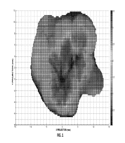
[0011] Figure 1 shows a placental thickness map.
[0012] Figure 2 shows the sigmoidal curve when gestational day is plotted
against placental
volume.
[00131 Figure 3 shows placental weight and volume validation.
3
CA 02944060 2016-09-26
WO 2015/153409 PCT/US2015/023257
[0014] Figure 4 shows value of r influenced early fetal growth while the value
of K impacted
later fetal growth.
[0015] Figure 5 shows the number of pregnancy complications by quartile of the
parameter r.
[0016] Figure 6 is a screen shot of the placental-growth curve software used
in an embodiment
of the invention.
[0017] Figure 7 is a table showing that placental quantifiers can be measured
in the first
trimester of pregnancy and are related to placental evaluations at term.
[0018] Figure 8 is a table showing baseline characteristics and available
measurements in a
pooled database.
[0019] Figure 9 is a table showing correlation between # and additional
pregnancy
complications.
[0020] Figure 10 is a table showing the physical and other characteristics of
the women who
completed the study of Example 1.
100211 Figure 11 is a table showing the breakdown of parameter estimates (X
SD) by study,
pregnancies without complications, pregnancies with complications, and total
pregnancies.
DETAILED DESCRIPTION
[0022] The invention is based in part on the discovery that by applying
recursive algorithmic
models from placental measures collected at multiple times during gestation,
dynamic changes in
placental growth can be calculated, and normal versus at risk deviations in
time dependent
growth can be identified. The flexibility of recursion models allows broad
model utility
prospectively and retrospectively. Forward model simulations validate the use
of placental
morphology measures to predict adverse pregnancy outcomes. Reverse simulations
can identify
4
CA 02944060 2016-09-26
WO 2015/153409 PCT/US2015/023257
combinations of timing, number, and magnitude of gestational stressors that
contributed to
clinically unanticipated adverse outcomes. Identifying the placental origins
of clinically
unpredicted pregnancy complications permits optimal inter-pregnancy
evaluation, counseling,
and future pregnancy management. Models can be programmed into user-friendly
computer
interfaces. For example, Figure 6 shows a screenshot of the computer output in
an embodiment
of the invention. Such models can be used, for example, in Phase 2 clinical
work.
[0023] Thus, in one embodiment of the invention, provided is a method for
predicting a prenatal,
neonatal, obstetric or childhood clinical event, disease or disorder from data
collected during a
pregnancy, comprising the steps of:
-inputting periodically collected pregnancy data comprising placental and
obstetric data
into a database housed in a computer;
-applying a continuous recursion modeling algorithm to said inputted pregnancy
data to
generate fetal and placental growth data during said pregnancy;
-generating data showing any deviations from model predictions of normal fetal
and
placental growth when compared to said generated fetal and placental growth
data during said
pregnancy; and
-predicting a prenatal, neonatal, obstetric or childhood risk of an adverse
clinical event,
disease or disorder from said deviating data.
[0024] In another embodiment of the invention, provided is a method for
predicting a prenatal,
neonatal, obstetric or childhood clinical event, disease or disorder from data
collected during a
pregnancy, further comprising the step of performing a clinical intervention
if said deviating data
so warrants.
[0025] In another embodiment of the invention, provided is a method for
predicting a prenatal,
neonatal, obstetric or childhood clinical event, disease or disorder from data
collected during a
pregnancy, wherein a continuous recursion modeling algorithm is housed in a
computer.
[0026] In another embodiment of the present invention, provided is a method
for predicting a
prenatal, neonatal, obstetric or childhood clinical event, disease or disorder
from data collected
CA 02944060 2016-09-26
WO 2015/153409 PCT/US2015/023257
during a pregnancy, wherein said prenatal, neonatal, obstetric or childhood
clinical event, disease
or disorder is preeclampsia, intrauterine growth restriction, preterm labor,
stillbirth, type 2
diabetes, high diastolic blood pressure, high systolic blood pressure,
increased presence of
placental knots, fibrotic chorionic villi, intrauterine growth restrict,
intraventicular hemorrhage,
placental edema, fetal acute inflammation, chorioamnionitis, amnion necrosis,
acute fetal
inflammation, acute maternal inflammation or acute amnionitis.
[0027] In a further embodiment of the present invention, provided is a method
for generating in-
utero fetal and placental growth curves from data collected during a
pregnancy, comprising the
steps of:
-inputting placental and obstetric data collected from said pregnancy into a
database
housed in a computer; and
-applying a continuous recursion modeling algorithm to said pregnancy data to
generate
said in-utero fetal and placental growth curves during said pregnancy.
[0028] In a still further embodiment of the invention, provided is a computer
programmed to
predict a prenatal, neonatal, obstetric or childhood clinical event, disease
or disorder from data
collected during a pregnancy, comprising software which:
-applies a continuous recursion modeling algorithm to data collected during
said
pregnancy, and inputted into said computer, to generate in-utero fetal and
placental growth data;
and
-outputs data showing any deviations of said in-utero fetal and placental
growth data
from model predictions of normal in-utero fetal and placental growth.
[0029] In another embodiment of the present invention, provided is a computer
wherein
software further predicts a prenatal, neonatal, obstetric or childhood risk of
an adverse clinical
event, disease or disorder from said outputted data showing deviations.
[0030] In another embodiment of the present invention, provided is an article
of manufacture for
predicting a prenatal, neonatal, obstetric or childhood clinical event,
disease or disorder from
data periodically collected during a pregnancy, comprising:
6
CA 02944060 2016-09-26
WO 2015/153409 PCT/US2015/023257
-a non-transitory computer-readable storage medium, and code stored on the
medium, the
code, when executed on a processor, controlling the processor for measuring in-
utero fetal and
placental growth during said pregnancy, wherein the processor applies a
continuous recursion
modeling algorithm to said data periodically collected during said pregnancy
to show any
deviations of said data periodically collected during said pregnancy from
model values of normal
placental volume to predict said prenatal, neonatal, obstetric or childhood
clinical event, disease
or disorder.
[0031] Placental growth in volume and mass has been well-established to follow
a sigmoidal
pattern (Figure 2), with early exponential growth followed by an inflection
point and finally
more limited log-like growth. Sigmoidal curves are common biological
phenomena, from
population dynamics, to plant growth, and cancer cell dynamics. Different
classes of
mathematical models mechanistically describe sigmoidal growth, the most
popular of which is
the logistic growth model. The logistic model assumes that initial growth is
exponential but is
eventually limited by a bound referred to as a carrying capacity. The carrying
capacity for
placental volume represents a theoretical cap on total possible volume
capacity for which the
placenta can hold, which may be higher than placental volume at term. Indeed,
placental size
continued to grow in late-term pregnancies providing observed evidence that
the carrying
capacity need not be attained during pregnancy.
[0032] The ability to identify at risk placental growth patterns early in
pregnancy, before the
pregnancy is obviously clinically compromised, will revolutionize obstetric
care and has the
potential to impact pediatric practice. Thus, the invention provides for the
development and
validation of evidence-based models to predict placental dysfunction and
pregnancy
complications from placental metrics obtained in early pregnancy. In one
embodiment, the
invention provides for a database containing information from over 2000
pregnancies and
includes 3D ultrasound images of the placenta obtained at 11-14 weeks of
gestation. From these
images, 19 different placental morphology metrics can be calculated and
analyzed together with
data extracted from 2D digital placental images and placental histopathology
samples collected
at birth. The clinical histories of these pregnancies are available, including
adverse outcomes
7
CA 02944060 2016-09-26
WO 2015/153409 PCT/US2015/023257
such as premature membrane rupture, preeclampsia, pre-term labor, placental
abruption, chronic
inflammation, and gestational diabetes mellitus.
[0033] The associations of placental metrics from early gestation and
delivery, and adverse
pregnancy outcomes are used to construct pregnancy risk prediction models
based on algorithms
that identify individual placental metrics as outside of receiver operating
characteristic (ROC)
determined cut off values. The risk models validate the prospective,
predictive value of novel
measures of placental structure for adverse pregnancy outcomes.
[0034] In another embodiment, a second class of recursive models are developed
from placental
measures collected at multiple times during gestation to reflect the dynamic
changes in placental
growth, and identify normal versus at risk deviations in time dependent
growth. The flexibility of
recursion models allows broad model utility prospectively and retrospectively.
As discussed
above, forward model simulations validate the use of placental morphology
measures to predict
adverse pregnancy outcomes. Reverse simulations can identify combinations of
timing, number,
and magnitude of gestational stressors that contributed to clinically
unanticipated adverse
outcomes. Identifying the placental origins of clinically unpredicted
pregnancy complications
permits optimal inter-pregnancy evaluation, counseling, and future pregnancy
management.
Models will be programmed into user-friendly interfaces for Phase 2 clinical
use.
[0035] In a further embodiment, the invention provides for an evidence-based
algorithm for
prediction of risk of placental dysfunction and adverse pregnancy outcomes
that includes
demographic and environmental covariates (maternal age, gestational age,
height, body weight,
race, parity, and trimester specific objectively determined energy intake) and
placental
morphology metrics obtained from 11-14 week 3D ultrasound data pooled from
Washington
University, St. Louis, New York University, and the University of
Pennsylvania.
[0036] In a method of the invention, recursion modeling is applied to predict
dynamics of
placental growth. Using placental morphology measures obtained at multiple
times during
gestation, recursive formulations can model dependency of placental "state"
during a given
8
CA 02944060 2016-09-26
WO 2015/153409 PCT/US2015/023257
gestational week on the "state" of the placenta in the previous week(s) and
account for the
observed range of normal versus dysfunctional placental growth patterns. The
inventive
algorithms can be validated on a reserved test database containing first
trimester placental
measures and data regarding pregnancy outcomes. This step will produce
objective criteria for
the determination of "healthy" and "at risk" patterns of placental growth
remote to delivery, and
clinically unanticipated adverse outcomes. The inventive models yield
individualized pregnancy
risks, providing the basis for a personalized and proactive management plan
for each pregnancy.
[0037] Placental quantifiers such as, for example, thickness, roundness, and
cord insertion site
can be measured in the first trimester of pregnancy and are related to
placental evaluations at
term. Measures of irregular placental shape obtained between 11-13 weeks (see
Figure 7) were
negatively correlated with placental weight at term. The Placental Morphology
Index (PMI) was
negatively correlated with both the placental weight and chorionic plate area
at term. First
trimester metrics (cord insertion site, geometrical center, cord eccentricity)
are correlated with
the cord insertion site at term. Interestingly, the cord insertion site
measures between 11-13
weeks of gestation are also related to thickness and mean chorionic vascular
density suggesting
non-central cord insertion site is a biomarker for a sparser chorionic
vascular tree, inefficient
placental-fetal nutrient exchange and a smaller baby at term.
[0038] Further, it was observed that non-central cord insertion site, non-
round placental shape,
and variable placental thickness are also related to a sparser chorionic
vascular tree and lower
placental efficiency. Cord displacement is positively correlated with mean
thickness. On the
other hand, deformation of the placental chorionic surface shape corresponds
to lower but more
variable placental disk thickness. A placenta with thin regions reflecting
reduced villous
arborization and variable fetal stem branching will tend to be less
functionally efficient, and will
yield a smaller baby for given placental weight.
[0039] Placental volume, placental quotient, placental morphology index, and
mean cord
diameter predicts small for gestational age (SGA), preeclampsia and
spontaneous preterm birth.
A recent study conducted by the inventors evaluated placental volume,
placental quotient, and
9
CA 02944060 2016-09-26
WO 2015/153409 PCT/US2015/023257
the PMI in weeks 11-13 of pregnancy and related these measures to pregnancy
outcomes at term.
Placental volume served as a proxy for placental weight. The placental
quotient adjusts placental
volume for gestational age. Mean cord diameter represents lateral placental
growth. PMI
indicates placental thickness (higher PMI is related to a flatter placenta).
Deviations of all four
measures from normal values were significantly correlated to adverse pregnancy
outcomes:
SGA, preeclampsia, and spontaneous preterm birth.
[0040] The proportion of the placenta that is metabolically active (a ) and
the fetal-placental
scaling exponent (a ) predicts preeclampsia, inflammation, placental
abruption, and pre-term
membrane rupture. The fetal-placental relationship is nonlinear and follows an
allometric
scaling law: PW = of (PW = placental weight and FW = fetal weight). In term
pregnancies
with normal outcomes, a = 1 and fi = 0.75 Across gestation, the value of Cl
decreases to 1 and
a should remain close to 0.75 by the end of the second trimester. Deviations
in a and are
highly sensitive indicators of pregnancy complications. In fact, the inventors
have noted that of
over 400 pre-term births, a and are predictors of preeclampsia, chorionic
inflammation,
placental abruption, and pre-term membrane rupture.
[0041] More specifically, placental growth is a recursive process which varies
over time. A
recursive dynamic model that predicts time-varying placental vascular tree
formation was
recently co-developed by one of the inventors. Model simulations suggested
that deviations
from normal placental morphology (round, regular, centrally inserted cord
placement) early in
pregnancy are amplified over the course of gestation. Cord displacement,
placental disk
diameter, chorionic plate area, perimeter, and maximal radius calculated from
the cord insertion
point were found to have power-law distributions, indicating that small early
perturbations in
morphology recursively are amplified in future placental growth,
experimentally supporting
conclusions derived from the recursion model. The initial recursion model can
be further
advanced using new topological visualizations of placental growth with graphic
display of
variation in arborization (Figure 1). The topological visualization can be
analyzed much as a
geologist analyzes strata and provide novel opportunities for model
development that will time
onset of deviations from normal growth trajectory. The relationship of 3D
villous arborization to
CA 02944060 2016-09-26
WO 2015/153409 PCT/US2015/023257
the networks of distributing chorionic surface arteries and draining venous
vasculature may be
key to understanding variation in placental function (see also U.S. Patent No.
8,565,507, which is
incorporated herein by reference).
[0042] In a further embodiment, the invention provides for a synopsis of the
fetal-maternal in-
utero processes as early as 11 weeks. Using existing 3 D ultrasound recordings
and at birth
placenta and clinical pregnancy data, the invention provides for the first
class of models that
combine 19 different placental measures to identify and estimate risk for
adverse pregnancy
outcomes.
[0043] In a further embodiment of the invention, ultrasound and pregnancy data
were pooled
from approximately 2,335 pregnancies (Figure 8). All placental measures were
obtained from
singleton pregnancies. At Washington University St. Louis (WUSL) 3D power
Doppler
placental images were obtained in women (n=750) between 11 and 14 weeks of
pregnancy and
again in the second trimester. Gestational weight gain and fetal biometric
parameters were
recorded. Placental measures in the Pregnancy, Infection and Nutrition (PIN)
study conducted at
the University of North Carolina (n=967), were obtained at delivery. Placentas
were weighed to
the nearest gram, photographed and histology samples obtained. New York
University (NYU):
Eighty of the women in this study (n=135) were recruited 11-14 weeks in
gestation when they
appeared for routine aneuploidy screening. At delivery, placenta were weighed
to the nearest
gram, photographed and histology samples obtained. (98 have 11-14 week and
delivery
measures). At Case Western Reserve University (CWRU), (n=83) women were
recruited at the
time of elective cesarean delivery at term. Placenta weight to the nearest
gram was recorded by
the water displacement method. In an on-going project at the University of
Pennsylvania
(UPenn) (NIH R03HD069742), two placenta measures were collected during
gestation and one
at delivery (n=600). The dataset also contains gestational weight gain and
fetal biometric
parameters. The University of Connecticut (UConn) study was conducted in over
400 women
who gave birth between 22-32 weeks of gestation. Study data contained adverse
pregnancy
outcomes, maternal and neonate demographics, and placental morphology
measures. The
Pennington Biomedical Research Center (PBRC) placental data can be collected
as an ancillary
to the existing U01. Ultrasound recordings of the placenta can be collected
during the first,
11
CA 02944060 2016-09-26
WO 2015/153409 PCT/US2015/023257
second, third trimesters and digital photographs at term are being collected
in 80 overweight and
obese women. Weekly body weights, body composition, total energy expenditures
by the doubly
labeled water method, resting metabolic rate, energy intake assessed by two
different objective
methods (model and energy balance), and appetite hormones can also collected.
[0044] The adverse pregnancy risk model will be constructed from data obtained
from the sub-
population with placental measures at more than one time point; WUSL (n=81),
NYU (n=80),
UPenn (n=300), PBRC (n=80). Cases with placental measures collected at only
one time point
will be used to determine ranges of normal versus at risk placental measure at
those specific time
points.
[0045] A key time point in growth is the inflection point, when the curve
switches from
exponential growth behavior to a log-like behavior. The invention disclosed
herein uses the
logistic growth model and, for example, two placental volume databases, one
with 5 longitudinal
measures of placental volume determined by three-dimensional ultrasound and
the second with 2
measures (one early and one at term) to first, calculate the timing of the
inflection point in
healthy pregnancies, second, whether deviations of inflection timing predict
pregnancy
complications and finally, generating predictions utilizing solely early
pregnancy data. In a
further embodiment, the invention couples the dynamic placental volume model
with a placental-
fetal scaling law to arrive at a dynamic fetal growth model that generates
fetal growth curves
after input of placental growth parameters.
[0046] Assumptions, Definitions and Mathematical Embodiments of the Invention
Assumption Assumption Statement
1 The early rate of placental volume growth is directly
proportional to
placental volume.
2 Initial growth is limited by a saturation value beyond
which the
placental volume cannot increase.
3 The self-limiting component of the model is described by
multiplying the term which exhibits exponential growth by a limiting
factor, (1 ¨14
12
CA 02944060 2016-09-26
WO 2015/153409
PCT/US2015/023257
4 Both
the proportionality constant, r , and the carrying capacity, K,
are independent of time.
Variable and Parameter Defmitions
Variable/Parameter Definition Units
P (t) The placental volume on day mL
t of gestation
The placental volume growth 1/d
rate in early gestation is
directly proportional to the
current placental volume. The
value of r is the
proportionality constant.
The carrying capacity of the mL
placenta which is the absolute
possible limiting volume the
placenta cannot exceed.
P0 The volume of the placenta in mL
the first trimester (-84 days).
Time of Inflection Point The time point when as the Gestational day
placental volume curve shifts
from concave up to concave
down in the S shape.
g/g3/4
a
ct=(1)1acental Weight)
3
[0047] Placental volume (mL) increases over gestation and thus is a time-
varying quantity. In
order to express this dependency of placental volume on time, placental volume
(mL) was
d P --
denoted on gestational day .1- by PviCt). The derivative of Pi-(t) , denoted
dt represents the
growth rate of placental volume and is expressed in units mL/d where d
represents days.
[0048] The inventive placental volume model is a differential equation that
relates the derivative
of placental volume to a function of placental volume, f CPv) (formulation of
IWO is described
in the next section):
dP--
f(Pv)
13
CA 02944060 2016-09-26
WO 2015/153409 PCT/US2015/023257
[0049] The solution of the differential equation yields a value that
represents the expected or
predicted placental volume on any given gestational day, t . Described below
is the derivation
of the function f CPO. and the solution of the placental volume model.
The Placental Volume Model
[00501 Every differential equation model entails a number of assumptions.
These assumptions
serve two purposes. The first is to sufficiently simplify the model so that it
can be solved
mathematically. The second reflects what is known about the specific mechanics
of the model.
For example, placental volume is known to increase sigmoidally over gestation.
Therefore the
model assumes this growth pattern. The list that follows outlines some
assumptions underlying
the placental volume model formulation:
(Al) The early growth rate of placental volume is directly proportional to
placental volume,
rP , where r is the proportionality constant. Conceptually, this assumption is
made because
early placental growth is due to cell division which is well known to follow
this growth pattern.
(A2) The increase in placental volume over gestation is eventually limited by
a maximum
value beyond which placental volume cannot increase. This saturation value is
referred to as the
"placental carrying capacity" in mL and denoted by the value, K . The carrying
capacity is not
the placental volume at term, but rather the upper bound beyond which
placental volume cannot
increase.
(A3) This self-limiting property of placental growth is captured by
multiplying the early
P--)
growth term, TP:r , by a limiting factor (1 ¨ KJwhich has the property that,
when P is close to
K , the factor is close to zero.
(A4) Both the proportionality constant, r ,and the carrying capacity, K ,are
time-independent.
Formulating these assumptions, Applicants arrived at the placental volume
growth model:
dP-- ( 4 Pv
rPv11. ¨
dr
=
The initial values and parameters must be non-negative: i (t0) and r. K 0,
where ta represents the gestational day at first placental volume measurement.
In
14
CA 02944060 2016-09-26
WO 2015/153409 PCT/US2015/023257
one embodiment, the first measured placental volume was obtained at
approximately
12 weeks (84 days) so to = 84 days and the initial condition is (34) , the mL
of
placental volume at 84 days.
The Placental Volume Model Solution
dP
= ¨
The model, dt K , can be solved explicitly for the solution Pa).
Kpoe-s4r
P7(0. ______________________________________________
e-rtic e -rr pip p0-24r
where PG represents the initial measurement of placental volume (here measured
at
approximately 84 days of gestation). This explicit solution has three
parameters, r ,K and Po
(bolded in the formula), which are calculated from the data. Once these values
are entered, the
solution yields an expected placental volume for gestational day, otherwise
stated, a prediction
for /AL ).
Determination of Model Parameters and Timing of Inflection
[0051] To fit three parameters in a model, here the values 0f ',k , and Po, ,
a minimum of three
placental volume measurements across gestation are needed. The multi-point
database contains
five placental volume measurements in each of the 11 pregnancies, and so three
measurements
from the five to fit the parameters can be used
Parameter fitting method using the multi-point study data
[0052] Below is provided an example of model solution from an individual
placenta. In the
given example, placental volume at week 12 (84 days) was 54.8 mL, at week 17
(119 days)
placental volume was 130.9 mL and at week 32 (224 days), 380.9 mL.
Step 1: Set the initial value, P0, equal to the measured volume at 12 weeks.
In this example, Pa = 54.8 Fitting in this value into the solution yields:
7102K -24r
P v(t) ¨ _______________________________________________
115e-rtK -4- 71 02e-84r ¨ 7102e-rr
CA 02944060 2016-09-26
WO 2015/153409 PCT/US2015/023257
Step 2: Use the 32-week measured placental volume to solve for E in terms
of!.
Substituting t = 224 days, provides for:
71.02Ke
Pv(224) =
12se-r224K - 102e-84r - 7102e-r224
or for this example:
7102,Ke-247
330.9 =
125e-r2241i." - 7102e-94' -
This is an algebraic equation which can be solved for K :
=C8733 .2(e-2 _ e-224r)
K = ______________________________________________
548e-24 - 3309C2:4 r
Substituting this expression of K back into the formula for P(t) yields:
2(38733.2(e-24v - e-2241
3261.e-(-24) 548e-16gr 3 sCge-308 r
Step 3: Apply the 17-week (119 day) placental volume measurement to solve for
L.
Substituting t 119 days, provides for:
203733,2(e-24r
Pri-(1149)
-r(11.9-rS4.)
3161e 548e-1-66r - 3809e-2 9
or
203733,2u-24- - e-22-4r)
130.9= ___________________________________________________
326? '-(_O) - 548e - 3309e-3 8 r
which can be solved for r :
r = 0.032 .
Step 4: Substitute the value of '2.- into the formula for E to solve for E.
? c8733.2(e-24r _ e-ZZ4r)
K - _________________________________________ =4O98
548e-84 r r mL
So, the predictive formula for placental volume becomes:
1156,8
P - ____________________________________________
355.0e-0.22.7 3.8
16
CA 02944060 2016-09-26
WO 2015/153409 PCT/US2015/023257
Graphing this function from t=0 to 280 (40 weeks) yields the S shaped
(sigmoidal) (Figure 2).
Step 5: Calculate the gestational day of inflection by setting P(t) = ¨2
From the model, the timing of inflection of the sigmoidal curve can be
calculated. At the point
t-P7( ) ¨
of inflection, the placental volume is half the carrying capacity.
Substituting = 2 yields
an algebraic formula:
1156.8
355.0e -113 38
_________________________________________ ¨ 204.9
Solving for t yields the timing of inflection: t = 142,8 days or 20.4 weeks.
Parameter fitting method using the early pregnancy study data
[0053] Unfortunately, obtaining placental volume measurements is time-
consuming and it is rare
to have more than one measured volume during gestation, especially when the
number of study
participants is larger than N=10. Since only one measured gestational
placental volume is in the
early pregnancy database, and a delivery measure of placental weight,
additional assumptions are
needed to estimate the three parameters.
[0054] The first point was a measured placental volume obtained approximately
at 12-weeks (84
days) of gestation. The second point was a placental weight at term. Although
placental density
is not well established and may have high inter-individual variance, a density
of 1 was applied
for conversion, which is consistent with that of adipose tissue ( 0.9 g/mL5)
and muscle (1.06
g/mL6).
[0055] Recall that two placental volume measurements are insufficient to fit
all three parameters,
r, K, and 13_0. The timing of inflection in the multi-point study was
clustered between 19-21
weeks of gestation. Therefore, it was assumed that the timing of inflection in
the early
pregnancy study should also occur at ¨ 20 weeks gestation.
[0056] Similar to the step by step description of parameter estimates in the
multi-point study,
data from a subject was applied to illustrate the calculations. For this
example placental volume
17
CA 02944060 2016-09-26
WO 2015/153409 PCT/US2015/023257
at 84 days was Pv(84) = 117.9 mL, gestational age at term was 259 days, and
placental
volume at term was 355.0 mL.
Step 1: Set the initial value, P0, equal to the measured volume at 12 weeks.
In the case of this example, Po =--- 117-9 . Filling in this value into the
solution yields:
17.9Ke -24r
Pr(t) = ______________________________________________
e-rtK - 117 .9e-rz. 117.9e-24r
Step 2: Solve for 11- in terms of by setting t=gestational age at term and
Pv(t) == placental
volume at term.
For this example t 259 and P 355 mL:
117 9Ke-s4v
3 5 5 - ________________________ - 117.9e-:59 - 117.9e-84r
which yields:
41845(e-9hr -
-
1179e-24r - 3550e-:5"
Substituting K into the solution,:
418543e-94r(e-24r -
Pr (t)
137.1e-(t+24) 1179e-16ir 355e5-342r
Step 3: Solve for!- by assuming the gestational age at inflection is 20 weeks
(140 days) and
solving Pv(140) = 7 for E- .
t = 140 days is set as equal to-2 , the value of P at the point of inflection.
18
CA 02944060 2016-09-26
WO 2015/153409 PCT/US2015/023257
4185.4.5e-24'167-24' e-259r)
(418545(e-94r - a-251
9)
-137 ie-(1.40+84) .9e-1.6gr 355e-342r 2)1179e-
24r - 3 550e -25gr
P(14U)
This equation contains only one unknown parameter, r . Solving for r yields,
r = C,02
Now that all constants are known, the final formula for P(t) is expressed by:
24955.7
P 7 = _______________
579.3e-c1.021- 61.2
Derivation of Dynamic Fetal Weight Model
[0057J In another embodiment, the invention applies the validated fetal-
placental scaling law
which states that placental weight is proportional to fetal weight to a
fractional power
Pw = cferW4' where Pw represents the grams of placental weight and FW
represents grams
of fetal weight. The value of the scaling exponent, /9, has been determined as
3/4 while the time-
varying proportionality constant aG9 is known to be 1 at term8.
Step 1 Let a- represent the density of the placenta (assume P = 1 for
numerical calculations)
and substitute ew = PP17 into the fetal-placental scaling law:
att) .0
tx(t)F P ; - = a (OF ET P Pv =
Step 2 Calculate the derivative of Pv in terms of =1 :
= a(t)
d; p ____________________________________________ F
dt
(9
Pv = ¨ r
Step 3: Substitute the scaling law expression, P into the placental
volume
differential equation:
d P
= r ev r a(t) II-7 aWF I V g
P - --7,- - -
dt
FLIT
Step 4: Equate the expression in Step 2 with the expression in Step 3 and
solve for dt
19
CA 02944060 2016-09-26
WO 2015/153409 PCT/US2015/023257
dFill CC F = r a (t) ( a (t)F14 )
(t)fl F WP-1 ________________________________ Flci- 1 -
P dt p K
a (0 cif' (t) D a V) õ
fl Fiey,Lf-1 r W ____ -
citp phj P
diFTY =r FW 1 - ________________________________ - re(t),oFW
a (OF W 16 a (t)
d t p pit j
r I a(t)FT 17'iir a(t)
=, _______________________ = FW I - ________________ FW
0 art`J a(t)j9
After input of the parameters, r K. and'AO, the solution to this model
generates a predicted
fetal growth curve, FIl'(t).
EXAMPLES
100581 The disclosure is further illustrated by the following examples, which
are not to be
construed as limiting this disclosure in scope or spirit to the specific
procedures herein described.
It is to be understood that the examples are provided to illustrate certain
embodiments and that
no limitation to the scope of the disclosure is intended thereby. It is to be
further understood that
resort may be had to various other embodiments, modifications, and equivalents
thereof which
may suggest themselves to those skilled in the art without departing from the
spirit of the present
disclosure and/or scope of the appended claims.
Subjects
100591 The first database included longitudinal measures of placental volume
at weeks 12, 17,
22, 27, and 32 weeks of pregnancy in twelve healthy women. This database is
referred to as the
multi-point study. From these data, the parameters of the logistic growth
model were calculated,
and then solved for the gestational age at inflection. The second database was
comprised of 54
women that included measures of a placental volume at 11-14 weeks of pregnancy
and the
delivered placental weight. Since the only in utero measurement of the
placenta was early in
pregnancy, this study is referred to as the early pregnancy study. This
database contained 11
complicated pregnancies. With these 11 "abnormal outcomes", tests were
performed to
CA 02944060 2016-09-26
WO 2015/153409 PCT/US2015/023257
determine whether deviations from the inventive placental volume growth model
predicted at
risk pregnancies. Both studies were approved by their institutional review
boards.
Example 1
The Multi-point study
[0060] Pregnant women who answered posted advertisements were recruited from
two private
practice obstetrical offices in Northern New Jersey. Subjects were eligible
for the study if they
were between 18 and 35 years old and were less than 12 weeks pregnant at
enrollment confirmed
by first trimester ultrasonography. Women were excluded for: 1. History of
smoking and/or drug
abuse, 2. A history of gestational diabetes or preeclampsia in a prior
pregnancy, 3. Medical co-
morbidities (i.e. chronic hypertension, diabetes, asthma, etc), and 4. Known
uterine anomalies or
fibroids. Data from enrolled participants were excluded from the analysis if
gestational diabetes
or preeclampsia was diagnosed during the study period. 20 women responded and
13 were
qualified to participate in the study. Of these, one was diagnosed with
gestational diabetes during
the study pregnancy. Physical and other characteristics of the 12 women who
completed the
study are shown in Figure 10.
[0061] Subjects underwent measurements of maternal height and weight at weeks
12, 17, 22,
27, and 32 weeks of gestation. For placental volume measurement, the entire
view of the
placenta was identified by 2-D ultrasonography, and the volume box was
adjusted to scan the
entire placenta. The sweep angle was set at 85 and was aimed so that the
probe was
perpendicular to the placental plate. Placental volume scans were then
obtained by 3D
ultrasonography. All volume scans were stored on a removable hard drive for
volume calculation
at a later date. Three scans were obtained at each time point, and the average
of the three
volumes scans was used for each time point. All images were acquired using
Voluson E8
Ultrasound machines (GE Medical Systems, Milwaukee, WI, USA) with a 4- to 8-
MHz
transducer. All ultrasounds were performed by one of two perinatologists.
[0062] In order to estimate placental volume, evaluation of the entire
placenta was performed
using the rotational technique in the virtual organ computer-aided analysis
(VOCAL) program
included in the 4DVIEW 6.0 software (GE, Austria) computer software.
Measurements were
21
CA 02944060 2016-09-26
WO 2015/153409 PCT/US2015/023257
performed on the sagittal section ("A" plane used as the reference image) by
manually
contouring the surface of the placenta, rotating the image 6 degrees and
contouring the surface
again. This process was repeated 30 times until completing an 180 rotation.
After completion of
the rotation, the VOCAL software yielded placental volume estimation as well
as a computed 3D
reconstruction of the placental. Care was taken to exclude the uterine wall
during manual
contouring. Manual contours were performed by the one physician.
Calculation of missing 32-week placental volume
[0063] Only six out of the 11 women in the multi-point study had a 32-week
placental volume
measure. By comparing the six data points at 32 weeks with 27 week data, it
was found that 32
week placental volume was highly correlated to 27 week volume (R2 = 0.79).
Missing data was
imputed using the regression formula: 1'32 = C.52P27 166.6 , where P27, P32
represent 27-
week and 32-week volumes, respectively.
Example 2
The early pregnancy study
[0064] A more detailed description of the original study appears in Schwartz
N, Coletta J, Pessel
C, Feng R, Timor-Tritsch IE, Parry S, et al. Novel 3-dimensional placental
measurements in
early pregnancy as predictors of adverse pregnancy outcomes. J Ultrasound Med.
2010;29(8):1203-12, which is incorporated by reference in its entirety. The
original study
recruited pregnant women between 11 to 14 weeks' gestation from the
Philadelphia metropolitan
region. The study was designed to determine whether early measurements of
placental
morphology predicted pregnancy outcomes. From the 98 subjects in the original
study, 54 had
both measurements of placental volume at 11-14 weeks and placental weight at
(term) delivery,
who were the subsample in this analysis. A transabdominal probe (Voluson E8;
GE Healthcare,
Milwaukee, WI) was used to obtain a 3D volume sweep of the placenta. The
volumes were
obtained using power Doppler imaging (quality, maximum; pulse repetition
frequency, 0.6 kHz;
and gain adjusted to just below the snow artifact) with the sweep angle opened
to ensure
inclusion of the entire placenta. The volume was reacquired if an obvious
fetal motion artifact
occurred during the sweep or if it appeared that a substantial portion of the
placenta was
excluded from the sweep. Volumes were stored for offline analysis postpartum.
22
CA 02944060 2016-09-26
WO 2015/153409 PCT/US2015/023257
[0065] The placental volume sets were then manipulated using 4D View software
(GE
Healthcare, Kretztechnik, Zipf, Austria). The placental volume was isolated
using virtual
computer- aided analysis, which involves manual tracing the perimeter of the
placenta in
successive images as obtained by automatic rotation of the image 6 times
around the y-axis in
30 intervals to achieve a 1800 rotation. With these traced perimeters, the
software reconstructs
the shape and volume of the object.
[0066] Demographic data, such as maternal age, parity, race, and body mass
index (BMI), as
well as pregnancy outcome data, including gestational age at delivery, birth
weight, and
pregnancy complications, were collected from the hospital medical records.
Gestational age at
delivery was based on first trimester sonographic dating if a definite last
menstrual period was
not available or if there was a greater than 7-day discrepancy between
menstrual dating and first-
trimester sonographic biometric measurements. Preeclampsia was defined as the
finding of a
systolic blood pressure of 140 mm Hg or higher or a diastolic blood pressure
of 90 mm Hg or
higher on 2 occasions 6 hours apart in the presence of substantial
proteinuria, defined as a 24-
hour urine collection containing greater than 300 mg of protein or urine
dipstick with a 1+
protein value or higher. Birth weight percentiles were determined on the basis
of the curve of
Alexander et al. , with small for gestational age (SGA) defined by birth
weight at or below the
10th percentile for the completed gestational week.
Example 3
The logistic model for placental volume
[0067] The logistic model solutions are sigmoidal curves. Sigmoidal growth
curves are
experimentally observed in placental growth, which has made the logistic model
a natural
choice for placental growth models (8, 16). Specifically, the logistic model
is a differential
equation originating from population ecology (17). If (0 is definded as the mL
of placental
volume on day t- of gestation then the model is given by the differential
equation:
fil3v
dt =
23
CA 02944060 2016-09-26
WO 2015/153409 PCT/US2015/023257
where r is exponential growth rate during early placental growth and K is the
carrying
capacity. The carrying capacity, K , represents the upper bound for placental
volume past which
the placental volume cannot increase. In order to simulate the model, an
initial value of placental
volume, Po , (preferably measured during early pregnancy) is required. A
complete
mathematical and biological background of the logistic growth model was
provided above.
Example 4
Parameter calculations in the multi-point study
[0068] All parameter calculations were performed in Maple 12 (Waterloo, Canada
2012)
interfaced with Microsoft Excel 2011 (Seattle, WA 2011). Three parameters that
need to be
determined; Po , r , and the carrying capacity, K . Using the 12-week (84 day)
measured
placental volume for Fi , this value was substituted into the solution of the
logistic model:
Pvt) = e -rt. _ pa e -2Air
The 32-week (224 days) placental volume measurement was used to calculate K ,
setting
Pv(224) = Plp and solving for IC :
_
K _____________________________________________
p e -
_
- 7;2
Next, the 17-week (119 days) placental volume measurement was used to solve
for r .
Specifically, r is calculated by solving the algebraic equation:
A-Poe-24r
_____________________________________________ =p17
e -r(119)Gic ¨ Po) + Poe -s4"
where P17 is the 17-week measured placental volume.
Calculation of the gestational age at inflection point
[0069] The inflection point occurs when the second derivative is zero, which
is calculated
directly from the differential equation:
24
CA 02944060 2016-09-26
WO 2015/153409 PCT/US2015/023257
d2P_ dP dP _ 2F
_ \ = 0
dt2 if art ¨ c*k K)
After input of Par r. and , solving for t in the algebraic equation:
ic:Pse-94r
e P0) po e -84r 2
yields the time (as gestational age in days) of inflection.
Example 5
Placental volume model validation
[0070] Placental volume data from weeks 22 and weeks 27 in the multi-point
study was not
applied to determine parameters and therefore can be used to determine model
accuracy. A
Bland Altman analysis was performed in Microsoft Excel 2011 (Seattle, WA 2011)
to test
model agreement with the placental volume at weeks 22 and weeks 27.
Example 6
Parameter calculations in the early pregnancy study
[0071] From the analysis of the multi-point study, the timing of the
inflection point in healthy
pregnancies was determined to be between 19-21 weeks of gestation. The
gestational age was
set at inflection point at 20 weeks (140 days) and assumed a first placental
volume measurement
at 12 weeks (84 days). Similar to the analysis in multi-point study, 84 days
was set as initial
time and Po equal to the initial placental volume measurement. The experiment
computed r by
substituting t 140 and solving the equation:
_
e o po) pc, e 2
for the non-zero solution of r . Finally, the experiment used the at term
placental weight data to
solve for K , by setting t = GA (gestational age at delivery) and solving the
algebraic equation:
Kpo e ¨24r
= P
-24r 'err al
¨
CA 02944060 2016-09-26
WO 2015/153409 PCT/US2015/023257
where Pfinca represents the final volume at term (converted from placental
weight using a
density of approximately 1). Expanded details with a numerical example were
provided above.
Example 7
Estimating pregnancy risk as deviations from model predictions
[00721 While analysis of parameters using at term measurements is informative,
it is not
desirable for risk detection during pregnancy. By applying average parameter
estimates from the
multi-point study where all pregnancies were normal, the experiment examined
whether
deviations from model predictions from the early pregnancy study was related
to pregnancy
complications. In order to rely solely on early pregnancy data to estimate
model parameters, the
experiment used the exponential model:
P(t) = Poet-84)
with r set as the average value from the multi-point data set (r "3 ) and P0
set as the first
trimester ultrasound measured placental volume in the early pregnancy study.
As calculated
earlier in the methods, the value of placental volume at the inflection point
is T. Assuming the
inflection point must occur at 20 weeks (119 days), then solving the equation:
K. 7 witerci9-24)
results in a rough estimate for K . Now that P., r , and K are known, a
predictive placental
volume curve can be simulated and compared to actual placental volume at term.
The deviation
of the actual placental volume at term from the model predictions was
calculated to determine
whether the actual volume "fell off the curve". The number of pregnancy
complications were
grouped by quartiles of distance that the actual placental volume deviated
from the predicted
curve.
Example 8
Prediction of pregnancy complications in the early pregnancy study
[0073] If parameter estimates were derived from both 12-week placental volume
and at term
placental weight, the values of r and K were grouped by quartiles and the
number of pregnancy
complications summed by quartile. When only the 11-14 week data was used to
fit parameters,
26
CA 02944060 2016-09-26
WO 2015/153409 PCT/US2015/023257
the values of r , and the difference between actual and predicted placental
volume at term (error)
were grouped into quartiles and the number of pregnancy complications were
summed
accordingly.
Example 9
Fetal growth model
[0074] The experiment applied the validated fetal-placental scaling law which
states that
placental weight is proportional to fetal weight to a fractional power PY,:" =
a(OF TV where Pw
represents the grams of placental weight and Fig represents grams of fetal
weight. The value of
the scaling exponent, P. has been well-established as 3/4 while the time-
varying proportionality
constant a(t) is known to be 1 at term.
By substituting the fetal-placental relationship into the placental volume
differential
equation model (expanded calculations shown above), a differential equation
model was derived
in terms of fetal growth that rely on the placental growth parameters, r and K
:
dF ( a ____ (t)F a õ
V.
dt ¨ F
K (0,0
where FIV(t) represents fetal weight on the t th gestational day.
Once r I 3 a(t) , and K were inputted, the model was simulated to generate a
fetal growth
curve. For numerical simulations an explicit formula for ae) and fetal weight
at 12 weeks is
required. For this purpose, a best fit curve (R2=0.97) for data was applied:
a(t) = 247.97t-074
Twelve week fetal weight was estimated as 20 g and all numerical simulations
were performed
in Maple 12 (Waterloo, CANADA 2012).
Example 10
Validation and analysis of fetal growth model
[0075] The multi-point study contained all required information (r .K ,
gestational age at term,
and birthweights) to compare actual versus predicted birth weights. A Bland
Altman analysis
was performed in Microsoft Excel (Seattle, WA 2011) to validate the fetal
growth model.
27
CA 02944060 2016-09-26
WO 2015/153409 PCT/US2015/023257
Fetal growth curves were generated for different combinations of r and K in
the
estimated data determined parameter ranges. The ranges of r (0.02-0.04) and K
(378-840 mL)
were separated into low r (7- = 0.02 ), average r (r = 0,03 ), high r (r= 0.04
) and low K
(K = 378 mL), average R.' (K = 582 mL) and high K (K = 850 mL) and fetal
growth
curves were simulated to term (gestational age of 40 weeks). The resulting
predicted birth
weight was classified into small for gestational age (SGA), average for
gestational age (AVA) or
large for gestational age (LGA) in the ranges <2500 g, between 2500 and 4000
g, and above
4000 g respectively.
Example 11
Results
Parameter Estimates
[0076] Figure 11 contains the breakdown of parameter estimates (X SD) by
study, pregnancies
without complications, pregnancies with complications, and total pregnancies.
In the early
pregnancy study, the mean value of r was lower in the pregnancies with
complications than in
the uncomplicated pregnancies. When r was grouped by quartiles, eight out of
the eleven
(73%) pregnancies with complications were in the two lower quartiles of r .
Similar associations
were not found for the carrying capacity, K. and first trimester placental
volume, Pet .
Estimation of gestational age at inflection point
[0077] The gestational age at inflection ranged from 19.4- 28.8 weeks with a
median at 20.8
weeks and a mean of 22.2 3.4 weeks (Figure 11).
Placental volume model validation
[0078] The correlation between actual placental volume and predicted placental
volume at 22
weeks was R2=0.75 which reduced to R2=0.62 at 27 weeks (Figure 3 Panels A and
B). There
was an overestimation of placental volume at both 22 and 27 weeks with a bias
of -37.5 mL at 22
weeks (95% confidence interval of [-92.4, 17.1]) and a bias of -59.0 mL at 27
weeks (95%
confidence interval of [-158.0, 40.1]).
28
CA 02944060 2016-09-26
WO 2015/153409 PCT/US2015/023257
Parameter estimates, model predictions and pregnancy risks relying solely on
early pregnancy
data (exponential model)
[0079] The values for K were higher than estimated with the logistic model.
This is expected
since the exponential model will overestimate placental volume because it
lacks any growth
limiting parameter. Thus, the difference between actual final volume and
predicted final volume
will trend downward and negative. Arrangement of the error by quartiles as
shown in Figure 5
revealed that seven of the eleven (64%) of the pregnancies with complications
were located in
the two bottom quartiles of error. The two bottom quartiles of error
represented the highest
deviations between actual and predicted.
Fetal growth model validation
[0080] The correlation between actual birth weight and predicted birth weight
was R2=0.36
which (Figure 3, Panel C). The model overestimated birth weight (bias = -675.1
g, confidence
interval: [-2157.8, 807.7]).
Fetal growth model analysis
[0081] Only 3 combinations of r and K resulted in a birth weight AGA; low r
and average
K , average r and low K , low r and average K , and low r and high K (Figure
9). The
value of r influenced early fetal growth while the value of K impacted later
fetal growth
(Figure 4). While the theoretical simulations of all combinations of r and K
were possible, the
experimental data revealed that low r was most often paired with high K ,
average " was
paired with a K value around 400 mL, and high r was paired with low K . In
both studies,
there were no cases of low T values paired with low K values. The model
correctly classified 7
of the 11(64%) of the birth weights with the four misclassifications resulting
in a predicted LGA
versus an actual AGA at term.
[0082] Thus, as shown in the Examples above, the invention rigorously
calculated the gestational
age at point of inflection for the growth of placenta at approximately 20
weeks. The point of
29
CA 02944060 2016-09-26
WO 2015/153409 PCT/US2015/023257
inflection has also been referred to as the maximal growth rate of the
placenta. The inventors
applied this point of inflection and the logistic growth model to classify
risks in a dataset of 54
pregnancies. It was discovered that that lower values of the initial growth
rate, 7. was associated
with pregnancy complications. Also discovered was an association with
deviations from
placental volume model predictions and pregnancy complications.
[0083] The combination of the inventively developed dynamic placental volume
model and a
well-established fetal-placental scaling law resulted in a dynamic fetal
growth model which
exhibited the influence of placental parameters on fetal growth. This analysis
advances the field
which has predominately relied on statistical relationships between placental
measures and birth
outcomes by permitting users to input placental parameters and observe the
fetal growth curve at
any gestational time point. The fetal growth model informs how placental
growth affects fetal
development at various stages during pregnancy. Interestingly, the fetal model
indicated how
different birth weights can be arrived at using various combinations of
placental growth
parameters. Additionally, the lack of any data with low values of r and K
indicated that some
parameter combinations are not physiologically feasible.
REFERENCES
1. Salafia CM, Misra DP, Yampolsky M, Charles AK, Miller RK. Allometric
metabolic
scaling and fetal and placental weight. Placenta. 2009;30(4):355-60. Epub
2009/03/07. doi:
SO143-4004(09)00017-4 [pi]
10.1016/j.placenta.2009.01.006. PubMed PMID: 19264357.
2. Salafia CM, Yampolsky M, Misra DP, Shlakhter 0, Haas D, Eucker B, et al.
Placental
surface shape, function, and effects of maternal and fetal vascular pathology.
Placenta.
2010;31(11):958-62. Epub 2010/10/12. doi: S0143-4004(10)00339-5 [pi]
10.1016/j.placenta.2010.09.005. PubMed PMID: 20933281; PubMed Central PMCID:
PMC2964412.
3. Yampolsky M, Salafia CM, Shlakhter 0, Haas D, Eucker B, Thorp J.
Centrality of the
umbilical cord insertion in a human placenta influences the placental
efficiency. Placenta.
2009;30(12):1058-64. Epub 2009/11/03. doi: 10.1016/j.placenta.2009.10.001.
PubMed PMID:
19879649; PubMed Central PMCID: PMC2790011.
4. Yampolsky M, Salafia CM, Shlakhter 0, Haas D, Eucker B, Thorp J.
Modeling the
variability of shapes of a human placenta. Placenta. 2008;29(9):790-7. Epub
2008/08/05. doi:
S0143-4004(08)00186-0 [ph]
10.1016/j.placenta.2008.06.005. PubMed PMID: 18674815; PubMed Central PMCID:
PMC2570048.
CA 02944060 2016-09-26
WO 2015/153409 PCT/US2015/023257
5. Yampolsky M, Salafiaa CM, Shlakhtera 0, Misraa5 DP, Haasa D, Euckera B,
et al.
Variable placental thickness affects placental functional efficiency
independent of other placental
shape abnormalities. Journal of Developmental Origins of Health and Disease
2011;2(4):205-
2011.
6. Schwartz N, Mandel D, Shlakhter 0, Coletta J, Pessel C, Timor-Tritsch
IE, et al.
Placental morphologic features and chorionic surface vasculature at term are
highly correlated
with 3-dimensional sonographic measurements at 11 to 14 weeks. J Ultrasound
Med.
2011;30(9):1171-8. Epub 2011/08/31. doi: 30/9/1171 [pi]. PubMed PMID:
21876086.
7. Hafner E, Philipp T, Schuchter K, Dillinger-Paller B, Philipp K, Bauer
P. Second-
trimester measurements of placental volume by three-dimensional ultrasound to
predict small-
for-gestational-age infants. Ultrasound Obstet Gynecol. 1998;12(2):97-102.
Epub 1998/09/23.
doi: 10.1046/j.1469-0705.1998.12020097.x. PubMed PMID: 9744052.
8. Rizzo G, Capponi A, Cavicchioni 0, Vendola M, Arduini D. First trimester
uterine
Doppler and three-dimensional ultrasound placental volume calculation in
predicting pre-
eclampsia. Eur J Obstet Gynecol Reprod Biol. 2008;138(2):147-51. Epub
2007/10/06. doi:
S0301-2115(07)00364-8 [pi]
10.1016/j.ejogrb.2007.08.015. PubMed PMID: 17916401.
9. Hafner E, Metzenbauer M, Hofinger D, Munkel M, Gassner R, Schuchter K,
et al.
Placental growth from the first to the second trimester of pregnancy in SGA-
foetuses and pre-
eclamptic pregnancies compared to normal foetuses. Placenta. 2003;24(4):336-
42. Epub
2003/03/27. PubMed PMID: 12657506.
10. Schwartz N, Coletta J, Pessel C, Feng R, Timor-Tritsch IE, Parry S, et
al. Novel 3-
dimensional placental measurements in early pregnancy as predictors of adverse
pregnancy
outcomes. J Ultrasound Med. 2010;29(8):1203-12. Epub 2010/07/28. doi:
29/8/1203 [pi].
PubMed PMID: 20660454.
11. Odibo AO, Zhong Y, Longtine M, Tuuli M, Odibo L, Cahill AG, et al.
First-trimester
serum analytes, biophysical tests and the association with pathological
morphometry in the
placenta of pregnancies with preeclampsia and fetal growth restriction.
Placenta.
2011;32(4):333-8. Epub 2011/02/18. doi: S0143-4004(11)00027-0 [pi]
10.1016/j.placenta.2011.01.016. PubMed PMID: 21324404.
12. Kim YM, Chaiworapongsa T, Gomez R, Buj old E, Yoon BH, Rotmensch S, et
al. Failure
of physiologic transformation of the spiral arteries in the placental bed in
preterm premature
rupture of membranes. Am J Obstet Gynecol. 2002;187(5):1137-42. Epub
2002/11/20. PubMed
PMID: 12439491.
13. Kim YM, Bujold E, Chaiworapongsa T, Gomez R, Yoon BH, Thaler HT, et al.
Failure of
physiologic transformation of the spiral arteries in patients with preterm
labor and intact
membranes. Am J Obstet Gynecol. 2003;189(4):1063-9. Epub 2003/10/31. PubMed
PMID:
14586356.
14. McMaster-Fay RA. Failure of physiologic transformation of the spiral
arteries of the
uteroplacental circulation in patients with preterm labor and intact
membranes. Am J Obstet
Gynecol. 2004;191(5):1837-8; author reply 8-9. Epub 2004/11/18. doi:
10.1016/j.ajog.2004.05.091. PubMed PMID: 15547578.
15. Odibo AO, Goetzinger KR, Huster KM, Christiansen JK, Odibo L, Tuuli MG.
Placental
volume and vascular flow assessed by 3D power Doppler and adverse pregnancy
outcomes.
Placenta. 2011;32(3):230-4. Epub 2011/02/08. doi: S0143-4004(11)00021-X [pi]
31
CA 02944060 2016-09-26
WO 2015/153409 PCT/US2015/023257
10.1016/j.placenta.2011.01.010. PubMed PMID: 21295850; PubMed Central PMCID:
PMC3125967.
16. Barker DJ, Bull AR, Osmond C, Simmonds SJ. Fetal and placental size and
risk of
hypertension in adult life. BMJ. 1990;301(6746):259-62, Epub 1990/08/04.
PubMed PMID:
2390618; PubMed Central PMCID: PMC1663477.
17. Barker DJ, Thornburg KL, Osmond C, Kajantie E, Eriksson JG. The surface
area of the
placenta and hypertension in the offspring in later life. The International
journal of
developmental biology. 2010;54(2-3):525-30. Epub 2009/10/31. doi:
10.1387/ijdb.082760db.
PubMed PMID: 19876839.
18. Risnes KR, Romundstad PR, Nilsen TI, Eskild A, Vatten U. Placental
weight relative to
birth weight and long-term cardiovascular mortality: findings from a cohort of
31,307 men and
women. Am J Epidemiol. 2009;170(5):622-31. Epub 2009/07/30. doi:
10.1093/aje/kwp182.
PubMed PMID: 19638481.
19. Moore VM, Miller AG, Boulton TJ, Cockington RA, Craig IH, Magarey AM,
et al.
Placental weight, birth measurements, and blood pressure at age 8 years. Arch
Dis Child.
1996;74(6):538-41. Epub 1996/06/01. PubMed PMID: 8758133; PubMed Central
PMCID:
PMC1511556.
20. Higgins MF, Russell NM, Mooney BE, McAuliffe FM. Clinical and
ultrasound features
of placental maturation in pre-gestational diabetic pregnancy. Early Hum Dev.
2012;88(10):817-
21. Epub 2012/07/04. doi: 10.1016/j.earlhumdev.2012.06.001. PubMed PMID:
22749772.
21. Salafia CM, Yampolsky M. Metabolic scaling law for fetus and placenta.
Placenta.
2009;30(5):468-71. Epub 2009/03/17. doi: S0143-4004(08)00438-4 [pi]
10.1016/j.placenta.2008.12.013. PubMed PMID: 19285342; PubMed Central PMCID:
PMC2699210.
22. Salafia CM, Ghidini A, Minior VK. Uterine allergy: a cause of preterm
birth? Obstet
Gynecol. 1996;88(3):451-4. Epub 1996/09/01. doi: 0029-7844(96)00219-0 [pi]
10.1016/0029-7844(96)00219-0. PubMed PMID: 8752257.
23. Egbor M, Ansari T, Morris N, Green CJ, Sibbons PD. Pre-eclampsia and
fetal growth
restriction: how morphometrically different is the placenta? Placenta.
2006;27(6-7):727-34. Epub
2005/08/30. doi: SO143-4004(05)00180-3 [pi]
10.1016/j.placenta.2005.06.002. PubMed PMID: 16125226.
24. Schuchter K, Metzenbauer M, Hafner E, Philipp K. Uterine artery Doppler
and placental
volume in the first trimester in the prediction of pregnancy complications.
Ultrasound Obstet
Gynecol. 2001;18(6):590-2. Epub 2002/02/15. doi: 596 [pi]
10.1046/j.0960-7692.2001.00596.x. PubMed PMID: 11844195.
25. Hafner E, Metzenbauer M, Hofinger D, Stonek F, Schuchter K, Waldhor T,
et al.
Comparison between three-dimensional placental volume at 12 weeks and uterine
artery
impedance/notching at 22 weeks in screening for pregnancy-induced
hypertension, pre-
eclampsia and fetal growth restriction in a low-risk population. Ultrasound
Obstet Gynecol.
2006;27(6):652-7. Epub 2006/03/04. doi: 10.1002/uog.2641. PubMed PMID:
16514618.
26. Semczuk-Sikora A, Krzyzanowski A, Stachowicz N, Robak J, Kraczkowski J,
Kwiatek
M, et al. [Maternal serum concentration of angiogenic factors: PIGF, VEGF and
VEGFR-1 and
placental volume in pregnancies complicated by intrauterine growth
restriction]. Ginekol Pol.
2007;78(10):783-6. Epub 2008/01/19. PubMed PMID: 18200969.
32
CA 02944060 2016-09-26
WO 2015/153409 PCT/US2015/023257
27. Higgins M, Felle P, Mooney EE, Bannigan J, McAuliffe FM. Stereology of
the placenta
in type 1 and type 2 diabetes. Placenta. 2011;32(8):564-9. Epub 2011/05/31.
doi: S0143-
4004(11)00169-X [pi]
10.1016/j.placenta.2011.04.015. PubMed PMID: 21621839.
28. Gauster M, Desoye G, Totsch M, Hiden U. The placenta and gestational
diabetes
mellitus. Curr Diab Rep. 2012;12(1):16-23. Epub 2011/11/22. doi:
10.1007/s11892-011-0244-5.
PubMed PMID: 22102097.
29. Akhter F, Ferdausi R. Quantitative macroscopic study on preterm
placenta in gestational
diabetes mellitus and pregnancy induced hypertension. Mymensingh Med J.
2011;20(2):280-6.
Epub 2011/04/28. PubMed PMID: 21522101.
30. McNamara JM, Odibo AO. Sonographic evaluation and the pregnancy
complicated by
diabetes. Curr Diab Rep. 2011;11(1):13-9. Epub 2010/11/04. doi: 10.1007/s11892-
010-0158-7.
PubMed PMID: 21046292.
31. Treacy A, Higgins M, Kearney JM, McAuliffe F, Mooney EE. Delayed
villous
maturation of the placenta: quantitative assessment in different cohorts.
Pediatr Dev Pathol.
2013;16(2):63-6. Epub 2012/11/10. doi: 10.2350/12-06-1218-0A.1. PubMed PMID:
23137099.
32. Salafia CM, Yampolsky M, Shlakhter A, Mandel DH, Schwartz N. Variety in
placental
shape: when does it originate? Placenta. 2012;33(3):164-70. Epub 2012/01/06.
doi: S0143-
4004(11)00574-1 [pi]
10.1016/j.placenta.2011.12.002. PubMed PMID: 22217910.
33. Yampolsky M, Salafia CM, Shlakhter 0. Probability distributions of
placental
morphological measurements and origins of variability of placental shapes.
Placenta.
2013;34(6):493-6. Epub 2013/04/09. doi: 10.1016/j.placenta.2013.03.003. PubMed
PMID:
23562224.
34. Gill JS, Salafia CM, Grebenkov D, Vvedensky DD. Modeling oxygen
transport in human
placental terminal villi. J Theor Biol. 2011;291:33-41. Epub 2011/10/01. doi:
S0022-
5193(11)00464-4 [pi]
10.1016/j jtbi.2011.09.008. PubMed PMID: 21959313.
35. Costa A, Costantino ML, Fumero R. Oxygen exchange mechanisms in the
human
placenta: mathematical modelling and simulation. J Biomed Eng. 1992;14(5):385-
9. Epub
1992/09/01. PubMed PMID: 1405555.
36. Groome U. A theoretical analysis of the effect of placental metabolism
on fetal
oxygenation under conditions of limited oxygen availability. Biosystems.
1991;26(1):45-56.
Epub 1991/01/01. PubMed PMID: 1760534.
37. Thomas DM, Clapp JF, Shernce S. A foetal energy balance equation based
on maternal
exercise and diet. J R Soc Interface. 2008;5(21):449-55 PMID: 1789522. Epub
2007/09/27. doi:
J8JK099G66V7546L [pi]
10.1098/rsif.2007.1161. PubMed PMID: 17895222; PubMed Central PMCID:
PMC2607387.
38. de Paula CF, Ruano R, Campos JA, Zugaib M. Placental volumes measured
by 3-
dimensional ultrasonography in normal pregnancies from 12 to 40 weeks'
gestation. J Ultrasound
Med. 2008;27(11):1583-90. Epub 2008/10/24. doi: 27/11/1583 [pi]. PubMed PMID:
18946097.
39. Redmer DA, Milne JS, Aitken RP, Johnson ML, Borowicz PP, Reynolds LP,
et al.
Decreasing maternal nutrient intake during the final third of pregnancy in
previously
overnourished adolescent sheep: effects on maternal nutrient partitioning and
feto-placental
development. Placenta. 2012;33(2):114-21. Epub 2011/12/14. doi: S0143-
4004(11)00555-8 [pi]
10.1016/j.placenta.2011.11.023. PubMed PMID: 22154692.
33
CA 02944060 2016-09-26
WO 2015/153409 PCT/US2015/023257
40. Thomas L, Wallace JM, Aitken RP, Mercer JG, Trayhurn P, Hoggard N.
Circulating
leptin during ovine pregnancy in relation to maternal nutrition, body
composition and pregnancy
outcome. J Endocrinol. 2001;169(3):465-76. Epub 2001/05/26. doi: JOE04159
[pi]. PubMed
PMID: 11375117.
41. van Abeelen AF, de Rooij SR, Osmond C, Painter RC, Veenendaal MV,
Bossuyt PM, et
al. The sex-specific effects of famine on the association between placental
size and later
hypertension. Placenta. 2011;32(9):694-8. Epub 2011/07/12. doi: S0143-
4004(11)00233-5 [pi]
10.1016/j.placenta.2011.06.012. PubMed PMID: 21742377.
42. Hasegawa J, Nakamura M, Hamada S, Okuyama A, Matsuoka R, Ichizuka K, et
al.
Gestational weight loss has adverse effects on placental development. J Matern
Fetal Neonatal
Med. 2012. Epub 2012/02/22. doi: 10.3109/14767058.2012.664666. PubMed PMID:
22348351.
43. Schoeller DA, Thomas D, Archer E, Heymsfield SB, Blair SN, Goran MI, et
al. Self-
report-based estimates of energy intake offer an inadequate basis for
scientific conclusions. Am J
Clin Nutr. 2013;97(6):1413-5. Epub 2013/05/22. doi: 10.3945/ajcn.113.062125.
PubMed PMID:
23689494.
44. Lichtman SW, Pisarska K, Berman ER, Pestone M, Dowling H, Offenbacher
E, et al.
Discrepancy between self-reported and actual caloric intake and exercise in
obese subjects. N
Engl J Med. 1992;327(27):1893-8. Epub 1992/12/31. doi:
10.1056/NEJM199212313272701.
PubMed PMID: 1454084.
45. Schoeller DA. How accurate is self-reported dietary energy intake? Nutr
Rev.
1990;48(10):373-9. Epub 1990/10/01. PubMed PMID: 2082216.
46. Butte NF, Wong WW, Treuth MS, Ellis KJ, O'Brian Smith E. Energy
requirements
during pregnancy based on total energy expenditure and energy deposition. Am J
Clin Nutr.
2004;79(6):1078-87. Epub 2004/05/26. PubMed PMID: 15159239.
47. Goldberg GR, Prentice AM, Coward WA, Davies HL, Murgatroyd PR, Wensing
C, et al.
Longitudinal assessment of energy expenditure in pregnancy by the doubly
labeled water
method. Am J Clin Nutr. 1993;57(4):494-505. Epub 1993/04/01. PubMed PMID:
8460604.
48. Thomas DM, Navarro-Barrientos JE, Rivera DE, Heymsfield SB, Bredlau C,
Redman
LM, et al. Dynamic energy-balance model predicting gestational weight gain. Am
J Clin Nutr.
2012;95(1):115-22 PMCID: 3238455. Epub 2011/12/16. doi: ajcn.111.024307 [pi]
10.3945/ajcn.111.024307. PubMed PMID: 22170365; PubMed Central PMCID:
PMC3238455.
49. Thomas DM, Halawani M, Phelan S, Butte NF, Redman LM. Development of a
pregravid weight calculator: Insights into the validity of self-reported
pregravid weight in
overweight and obese pregnant women. Under Review at Obstetrics & Gynecology.
2013.
50. Thomas DM, Urena B. A model describing the evolution of West Nile-like
encephalitis
in New York City. Mathematical and Computer Modelling. 2001;34(7-8):771-81.
51. Dudley NJ. A systematic review of the ultrasound estimation of fetal
weight. Ultrasound
Obstet Gynecol. 2005;25(1):80-9. Epub 2004/10/27. doi: 10.1002/uog.1751.
PubMed PMID:
15505877.
* * *
[0084] It is to be understood that the invention is not limited to the
particular embodiments of the
invention described above, as variations of the particular embodiments may be
made and still fall
within the scope of the appended claims.
34