Note: Descriptions are shown in the official language in which they were submitted.
CA 02944307 2016-10-05
SYSTEM FOR REPLACING A NATURAL AORTIC VALVE
FIELD OF THE INVENTION
The present invention relates to implantable devices. More particularly, it
relates to a
system for replacing a natural aortic valve comprising a catheter and a valve
prosthesis for
cardiac implantation or for implantation in other body ducts.
BACKGROUND OF THE INVENTION
There are several known prosthetic valves that have been previously described.
U.S.
Patent No. 5,411,552 (Andersen et al.), entitled VALVE PROSTHESIS FOR
IMPLANTATION
IN THE BODY AND CATHETER FOR IMPLANTING SUCH VALVE PROSTHESIS,
discloses a valve prosthesis comprising a stent made from an expandable
cylinder-shaped thread
structure comprising several spaced apices. The elastically collapsible valve
is mounted on the
stent with the commissural points of the valve secured to the projecting
apices, which prevents
the valve from turning inside out. Deployment of the valve can be achieved by
using an
inflatable balloon which in its deflated state is used to carry about it the
valve structure to its
position and, when inflated, deploys the stent in position to its final size.
See, also, U.S. Patent
No. 6,168,614 (Andersen et al.) entitled VALVE PROSTHESIS FOR IMPLANTATION IN
THE BODY and U.S. Patent No. 5,840,081 (Andersen et al.), entitled SYSTEM AND
METHOD FOR IMPLANTING CARDIAC VALVES.
In PCT/EP97/07337 (Letac, Cribier et al.), published as WO 98/29057, entitled
VALVE
PROSTHESIS FOR IMPLANTATION IN BODY CHANNELS, there is disclosed a valve
prosthesis comprising a collapsible valve structure and an expandable frame on
which the valve
structure is mounted. The valve structure is composed of a valvular tissue
compatible with the
human body and blood, the valvular tissue being sufficiently supple and
resistant to allow the
valve structure to be deformed from a closed state to an opened state. The
valvular tissue forms a
continuous surface and is provided with guiding means formed or incorporated
within, the
guiding means creating stiffened zones which induce the valve structure to
follow a patterned
movement in its expansion to its opened state and in its turning back to its
closed state. The valve
structure can be
CA 02944307 2016-10-05
WO 03/047468 PC7717802/32.588
2
extended to an internal cover which is fastened to the lower part of the valve
structure to
prevent regurgitation.
There are several known methods currently used for replacing aortic valves and
several types of artificial prosthetic devices. Mechanical valves are commonly
used in
several different designs (single and double flap) manufactured by well-known
companies
such as St. Jude, Medtronic, Sulzer, and others. Some of the main
disadvantages of these
devices are: a need for permanent treatment of anticoagulants, noisy
operation, and a need
for a large-scale operation to implant.
There is a wide range of biologically based valves made of natural valves or
composed of biological materials such as pericardial tissue. These too are
made and
marketed by well-known companies such as Edwards Lifesciences, Medtronic,
Sulzer,
Sorin, and others.
Polymer valves are new and are not yet in use, but several companies are in
the
process of developing such products. A new type of prosthesis is being
considered, based
on artificial polymer materials such as polyurethane..
The present invention introduces several novel structural designs for
implantable
valves. An aspect of the present invention deals with the possibility of
implanting the
valve percutaneously, i.e., inserting the valve assembly on a delivery device
similar to a
catheter, then implanting the valve at the desired location via a large blood
vessel such as
the femoral artery, in a procedure similar to other known interventional
cardiovascular
procedures. The percutaneous deployment procedure and device has an impact on
the
product design in several parameters, some of which are explained hereinafter.
The percutaneous implantation of medical devices and particularly prosthetic
valves is a preferred surgical procedure for it involves making a very small
perforation in
the patient's skin (usually in the groin or armpit area) under local
anesthetic and sedation,
as opposed to a large chest surgery incision, which requires general
anesthesia, opening a
CA 02944307 2016-10-05
WO 03/047468 PCT/US02/32588
3
large portion of the chest, and cardiopulmonary bypass. This percutaneous
procedure is
therefore considered safer.
The present invention provides a series of new concepts in the field of aortic
valves and other human valves.
SUMMARY OF THE INVENTION
It is therefore thus provided, in accordance with a preferred embodiment of
the
present invention, a valve prosthesis device suitable for implantation in body
ducts, the
device comprising:
a support stem, comprised of a deployable construction adapted to be initially
crimped in a narrow configuration suitable for catheterization through the
body duct to a
target location and adapted to be deployed by exerting substantially radial
forces from
within by means of a deployment device to a deployed state in the target
location, the
support stent provided with a plurality of longitudinally rigid support beams
of fixed
length; and
a valve assembly comprising a flexible conduit having an inlet end and an
outlet,
made of pliant material attached to the support beams providing collapsible
slack portions
of the conduit at the outlet,
whereby when flow is allowed to pass through the valve prosthesis device from
the inlet to the outlet the valve assembly is kept in an open position,
whereas a reverse
flow is prevented as the collapsible slack portions of the valve assembly
collapse
inwardly providing blockage to the reverse flow.
Furthermore, in accordance with another preferred embodiment of the present
invention, the support stent comprises an annular frame.
Furthermore, in accordance with another preferred embodiment of the present
invention, said valve assembly has a tricuspid configuration.
Furthermore, in accordance with another preferred embodiment of the present
invention, said valve assembly is made from biocompatible material.
CA 02 944307 2016-10-05
WO 03/047468 PC17US02/32588
4
Furthermore, in accordance with another preferred embodiment of the present
invention, the valve assembly is made from pericardial tissue, or other
biological tissue.
Furthermore, in accordance with another preferred embodiment of the present
invention, said valve assembly is made from biocompatible polymers.
Furthermore, in accordance with another preferred embodiment of the present
invention, the valve assembly is made from materials selected from the group
consisting
of polyurethane and polyethylene terephthalate (PET).
Furthermore, in accordance with another preferred embodiment of the present
invention, said valve assembly comprises a main body made from PET
(polyethylene
terephthalate) and leaflets made from polyurethane.
Furthermore, in accordance with another preferred embodiment of the present
invention, said support stent is made from nickel titanium.
Furthermore, in accordance with another preferred embodiment of the present
invention, the support beams are substantially equidistant and substantially
parallel so as
to provide anchorage for the valve assembly.
Furthermore, in accordance with another preferred embodiment of the present
invention, the support beams are provided with bores so as to allow stitching
or tying of
the valve assembly to the beams.
Furthermore, in accordance with another preferred embodiment of the present
invention, the support beams are chemically adhered to the support stent.
Furthermore, in accordance with another preferred embodiment of the present
invention, said valve assembly is riveted to the support beams.
Furthermore, in accordance with another preferred embodiment of the present
invention, said valve assembly is stitched to the support beams.
Furthermore, in accordance with another preferred embodiment of the present
invention, said beams are manufactured by injection using a mold, or by
machining.
Furthermore, in accordance with another preferred embodiment of the present
invention, said valve assembly is rolled over the support stent at the inlet.
Furthermore, in accordance with another preferred embodiment of the present
invention, said valve device is manufactured using forging or dipping
techniques.
CA 02 944307 2016-10-05
WO 03/047468 PCT/IIS02/32588
Furthermore, in accordance with another preferred embodiment of the present
invention, said valve assembly leaflets are longer than needed to exactly
close the outlet,
thus when they are in the collapsed state substantial portions of the leaflets
fall on each
other creating better sealing.
Furthermore, in accordance with another preferred embodiment of the present
invention, said valve assembly is made from coils of a polymer, coated by a
coating layer
=
of same polymer.
Furthermore, in accordance with another preferred embodiment of the present
invention, said polymer is polyurethane.
Furthermore, in accordance with another preferred embodiment of the present
invention, the support stent is provided with heavy metal markers so as to
enable tracking
and determining the valve device position and orientation.
Furthermore, in accordance with another preferred embodiment of the present
invention, the heavy metal markers are selected from gold, platinum, iridium,
or tantalum.
Furthermore, in accordance with another preferred embodiment of the present
invention, the valve assembly leaflets are provided with radio-opaque material
at the
outlet, so as to help tracking the valve device operation in vivo.
Furthermore, in accordance with another preferred embodiment of the present
invention, said radio-opaque material comprises gold thread.
Furthermore, in accordance with another preferred embodiment of the present
invention, the diameter of said support stent, when fully deployed is in the
range of from
about 19 to about 25 mm.
Furthermore, in accordance with another preferred embodiment of the present
invention, the diameter of said support stent may be expanded from about 4 to
about 25
Furthermore, in accordance with another preferred embodiment of the present
invention, the support beams are provided with bores and wherein the valve
assembly is
attached to the support beams by means of U-shaped rigid members that are
fastened to
CA 02944307 2016-10-05
WO 03/047468 PC1711802/32588
6
the valve assembly and that are provided with extruding portions that fit into
matching
bores on the support beams.
Furthermore, in accordance with another preferred embodiment of the present
invention, the support beams comprise rigid support beams in the form of frame
construction, and the valve assembly pliant material is inserted through a gap
in the frame
and a fastening rod is inserted through a pocket formed between the pliant
material and
the frame and holds the valve in position.
Furthermore, in accordance with another preferred embodiment of the present
invention, the main body of the valve assembly is made from coiled wire coated
with
coating material.
Furthermore, in accordance with another preferred embodiment of the present
invention, the coiled wire and the coating material is made from polyurethane.
Furthermore, in accordance with another preferred embodiment of the present
invention, a strengthening wire is interlaced in the valve assembly at the
outlet of the
conduit so as to define a fault line about which the collapsible slack portion
of the valve
assembly may flap.
Furthermore, in accordance with another preferred embodiment of the present
invention, the strengthening wire is made from nickel titanium alloy.
Furthermore, in accordance with another preferred embodiment of the present
invention, there is provided a valve prosthesis device suitable for
implantation in body
ducts, the device comprising a main conduit body having an inlet and an outlet
and pliant
leaflets attached at the outlet so that when a flow passes through the conduit
from the inlet
to the outlet the leaflets are in an open position allowing the flow to exit
the outlet, and
when the flow is reversed the leaflets collapse so as to block the outlet,
wherein the main
body is made from PET and collapsible leaflets are made form polyurethane.
=
CA 02944307 2016-10-05
-7-
Furthermore, in accordance with another preferred embodiment of the present
invention, support beams made from polyurethane are provided on the main body
and
wherein the leaflets are attached to the main body at the support teams.
Furthermore, in accordance with another preferred embodiment of the present
invention, said support beams are chemically adhered to the main body.
Furthermore, in accordance with another preferred embodiment of the present
invention, there is provided a valve prosthesis device suitable for
implantation in body
ducts, the device comprising:
a support stent, comprised of a deployable construction adapted to be
initially
crimped in a narrow configuration suitable for catheterization through the
body duct to a
target location and adapted to be deployed by exerting substantially radial
forces from
within by means of a deployment device to a deployed state in the target
location, the
support stent provided with a plurality of longitudinally rigid support beams
of fixed
length;
a valve assembly comprising a flexible conduit having an inlet end and an
outlet,
made of pliant material attached to the support beams providing collapsible
slack portions
of the conduit at the outlet; and
substantially equidistant rigid support beams interlaced or attached to the
slack
portion of the valve assembly material, arranged longitudinally.
Furthermore, in accordance with another preferred embodiment of the present
invention, there is provided a crimping device for crimping the valve device
described
above, the crimping device comprising a plurality of adjustable plates that
resemble a
typical SLR (Single Lens Reflex) camera variable restrictor, each provided
with a blade, that
are equally dispersed in a radial symmetry but each plate moves along a line
passing off an
opening in the center, all plates equidistant from that center opening.
CA 02944307 2016-10-05
WO 03/047468 PCT/US02/32588
8
Furthermore, in accordance with another preferred embodiment of the present
invention, the multiple plates are adapted to move simultaneously by means of
a lever and
transmission.
Furthermore, in accordance with another preferred embodiment of the present
invention, there is provided a method for deploying an implantable prosthetic
valve
device from the retrograde approach (approaching the aortic valve from the
descending
aorta) or from the antegrade approach (approaching the aortic valve from the
left ventricle
after performing a trans-septal puncture) at the natural aortic valve position
at the
entrance to the left ventricle of a myocardium of a patient, the method
comprising the
steps of:
(a) providing a balloon catheter having a proximal end and a distal end,
having
a first and second independently inflatable portions, the first inflatable
portion located at
=
the distal end of the catheter and the second inflatable portion adjacently
behind the first
inflatable portion;
(b) providing a guiding tool for guiding the balloon catheter in the
vasculature
of the patient;
(c) providing a deployable implantable valve prosthesis device adapted to
be
mounted on the second inflatable portion of the balloon catheter;
(d) for the retrograde approach, guiding the balloon catheter through the
patient's aorta using the guiding tool, the valve device mounted over the
second inflatable
portion of the balloon catheter until the first inflatable pardon of the
balloon catheter is
inserted into the left ventricle, whereas the second inflatable portion of the
balloon
catheter is positioned at the natural aortic valve position;
(e) for the antegrade approach, guiding the balloon catheter through the
patient's greater veins, right atrium, left atrium, and left ventricle using
the guiding tool,
the valve device mounted over the second inflatable portion of the balloon
catheter until
the first inflatable portion of the balloon catheter is inserted into the left
ventricle,
CA 02944307 2016-10-05
WO 03/047468 PCT/US02/32588
9
whereas the second inflatable portion of the balloon catheter is positioned at
the natural
aortic valve position;
(1) inflating the first inflatable portion of the balloon catheter so as
to
substantially block blood flow through the natural aortic valve and anchor the
distal end
of the balloon catheter in position;
(g) inflating the second inflatable portion of the balloon catheter so as
to
deploy the implantable prosthetic valve device in position at the natural
aortic valve
position;
(h) deflating the first and second inflatable portions of the balloon
catheter;
and
(i) retracting the balloon catheter and removing it from the patient's
body.
Furthermore, in accordance with another preferred embodiment of the present
invention, the guiding tool comprises a guide wire.
Furthermore, in accordance with another preferred embodiment of the present
invention, there is provided a method for deploying an implantable prosthetic
valve
device at the natural aortic valve position at the entrance to the left
ventricle of a
myocardium of a patient, the method comprising the steps of:
(a) providing a balloon catheter having a proximal end and a distal end,
having
a first and second independently inflatable portions, the first inflatable
portion located at
the distal end of the catheter and the second inflatable portion adjacently
behind the first
inflatable portion;
(b) providing a guiding tool for guiding the balloon catheter in the
vasculature
of the patient;
(c) providing a deployable implantable valve prosthesis device adapted to
be
mounted on the first inflatable portion of the balloon catheter, and a
deployable annular
stent device adapted to be mounted over the second inflatable portion of the
balloon
CA 02944307 2016-10-05
WO 03/047468 PCT/11502/32588
to
catheter, the deployable implantable valve prosthesis device and the
deployable annular
stent kept at a predetermined distant apart;
(d) guiding the balloon catheter through the patient's aorta using the
guiding
tool, the valve device mounted over the first inflatable portion of the
balloon catheter and
the deployable annular stout mounted over the second inflatable portion of the
balloon
catheter, until the first inflatable portion of the balloon catheter is
positioned at the natural
aortic valve position;
(e) inflating the second inflatable portion of the balloon catheter so that
the
deployable stent device is deployed within the aorta thus anchoring the
deployable
annular stent and the coupled valve device in position;
(f) inflating the first inflatable portion of the balloon catheter so as to
deploy
the implantable prosthetic valve device in position at the natural aortic
valve position;
=
(g) deflating the first and second inflatable portions of the balloon
catheter;
and
(h) retracting the balloon catheter and removing it from the patient's
body.
Furthermore, in accordance with another preferred embodiment of the
present invention, a valve prosthesis device suitable for implantation in body
ducts
comprises:
an expandable support frame, the support frame provided with a plurality of
longitudinally rigid support beams of fixed length; and
=
a valve assembly comprising a flexible conduit having an inlet end and an
outlet,
made of pliant material attached to the support beams providing collapsible
slack portions
of the conduit at the outlet,
whereby when flow is allowed to pass through the valve prosthesis device front
the inlet to the outlet the valve assembly is kept in an open position,
whereas a reverse
CA 02944307 2016-10-05
wo 03/047468 PCT/US02/32588
11
flow is prevented as the collapsible slack portions of the valve assembly
collapse
inwardly providing blockage to the reverse flow.
Furthermore, in accordance with another preferred embodiment of the present
invention, the support frame comprises a deployable construction adapted to be
initially
crimped in a narrow configuration suitable for catheterization through the
body duct to a
target location and adapted to be deployed by exerting substantially radial
forces from
within by means of a deployment device to a deployed state in the target
location.
Furthermore, in accordance with another preferred embodiment of the present
invention, the support beams have a U-shaped cross section.
Furthermore, in accordance with another preferred embodiment of the present
invention, a holder is used to secure the plaint material to the support
beams.
Furthermore, in accordance with another preferred embodiment of the present
invention, the support frame comprises three segments that form a circular
assembly
when assembled.
Furthermore, in accordance with another preferred embodiment of the present
invention, the support beams point inwardly with respect to a central
longitudinal axis of
the device.
Furthermore, in accordance with another preferred embodiment of the present
invention, the device is further provided with a restricting tapered housing,
for housing it
in a crimped state.
Furthermore, in accordance with another preferred embodiment of the present
invention, hooks are provided to secure the device in position after it is
deployed.
Furthermore, in accordance with another preferred embodiment of the present
invention, the support beams comprise longitudinal bars having a narrow slit
used as the
= commissural attachment so that extensions the pliant material are tightly
inserted through
it.
CA 02944307 2016-10-05
WO 05/047468 PCT/US02/32588
12
Furthermore, in accordance with another preferred embodiment of the present
invention, the extensions of the pliant material are wrapped about rigid bars
serving as
anchorage means.
Furthermore, in accordance with another preferred embodiment of the present
invention, extensions of the pliant material are sutured to each other at the
rigid bars.
Furthermore, in accordance with another preferred embodiment of the present
invention, a bottom portion of the pliant material is attached to the inlet
Furthermore, in accordance with another preferred embodiment of the present
invention, the support beams are each provided with a rounded pole, forming a
loop
through which the pliant material is inserted.
Furthermore, in accordance with another preferred embodiment of the present
invention, the pliant material is provided with longitudinal bars attached to
the pliant
material at positions assigned for attachment to the support frame, in order
to prevent
localized stress from forming.
Furthermore, in accordance with another preferred embodiment of the present
invention, the device is further provided with longitudinal bars having
protrusions that are
inserted in bores in the pliant material, a sheet of PET and through bores
provided on the
support beams.
Furthermore, in accordance with another preferred embodiment of the present
invention, pliant material is sutured leaving the slack portions free of
sutures.
Furthermore, in accordance with another preferred embodiment of the present
invention, a connecting member with a split portion is used to connect
leaflets of the
pliant material to the support beams, the split connecting member compressing
the pliant
material in position.
Furthermore, in accordance with another preferred embodiment of the present
invention, a portion of the connecting member is perpendicular to the split
portion.
CA 02944307 2016-10-05
WO 03/047468 PCT/US02/32588
13
Furthermore, in accordance with another preferred embodiment of the present
invention, the support frame is provided with metallic members coupled to the
stent and
rigid members are positioned on two opposite sides of the metallic member and
held
= against each other holding portion of the pliant material between them,
sutured, the
metallic members wrapped with PET.
Furthermore, in accordance with another preferred embodiment of the present
invention, the device is further provided with spring in order to reduce wear
of the pliant
material.
Furthermore, in accordance with another preferred embodiment of the present
invention, the spring is provided with a spiral.
Furthermore, in accordance with another preferred embodiment of the present
invention, the spring is made from stainless steel.
Furthermore, in accordance with another preferred embodiment of the present
invention, the spring is attached to slots provided on the support frames.
Furthermore, in accordance with another preferred embodiment of the present
invention, the pliant material is sutured to the support frame forming
pockets.
Furthermore, in accordance with another preferred embodiment of the present
invention, attachment bars are provided on the stent support at a portion of
the stent close
to the outlet, onto which the pliant material is coupled, and wherein the
pliant material is
attached circumferentially to the inlet, leaving slack pliant material.
Furthermore, in accordance with another preferred embodiment of the present
invention, the outlet is tapered with respect to the inlet.
Furthermore, in accordance with another preferred embodiment of the present
invention, the support frame at the outlet is wider in diameter than the
pliant material
forming the outlet.
Furthermore, in accordance with another preferred embodiment of the present
invention, the pliant material is reinforced using PET.
CA 02944307 2016-10-05
WO 03/047468 PCT/US02/32588
14
Furthermore, in accordance with another preferred embodiment of the present
invention, the support frame is a tube having an inner wall, having sinusoidal
fold lines,
wherein the pliant material is sutured to the inner wall of the tube along
suture lines.
Furthermore, in accordance with another preferred embodiment of the present
invention, additional piece of PET is added below the suture lines.
Furthermore, in accordance with another preferred embodiment of the present
invention, the device is incorporated with an angioplasty balloon.
Finally, in accordance with another preferred embodiment of the present
invention, balloon has a central longitudinal axis that runs along a flow path
through the
device, and a perimeter, the balloon comprising four inflatable portions, one
portion
located along a central axis and the other three located on the perimeter, the
pliant
material in the form of leaflets is distributed about the perimeter.
BRIEF DESCRIPTION OF THE FIGURES
To better understand the present invention and appreciate its practical
applications,
the following Figures are provided and referenced hereafter. It should be
noted that the
Figures are given as examples only and in no way limit the scope of the
invention as
defined in the appended claims.
Figure 1 illustrates an implantable prosthetic tricuspid valve in accordance
with a
preferred embodiment of the present invention, suitable for percutaneous
deployment
using a stent or similar deploying means, in its deployed-inflated position;
Figure 2 depicts an implantable valve according to the present invention
mounted
over a deploying stent with an inflatable balloon;
Figure 3 illustrates an implantable valve according to the present invention
mounted over a stent with an inflatable balloon, in a crimped position;
CA 02944307 2016-10-05
WO 03/047468 PCT/US02/32588
Figure 4 depicts implantable valve deployment in a natural aortic valve
position in
accordance with the present invention;
Figure 5 demonstrates manufacturing a polyurethane implantable valve using a
dipping technique according with the present invention;
Figures 6a to 6e illustrate manufacturing of an implantable valve by forging
according to the present invention;
Figures 7a and 7b demonstrate composite valve, which has polyurethane (PU)
leaflets and PET tubular-crown shaped construction, according to the present
invention;
Figures Sa and 8b depict a manufacture process of a composite valve made of
flexible PU leaflets, rigid PU construction for mounting and a PET tubular
end;
Figures 9 to 9i demonstrate different methods of attachment between the valve
and
stent according to the present invention;
Figure 10 illustrates a dipping mandrel with an extra portion, which improves
the
sealing ability of the valve, according to the present invention;
Figures 1 la to 11c illustrate a valve mounted on a stent with an extra
support,
which improves the force distribution on the valve material and facilitates
prolonged
durability of the valve, according to the present invention;
Figures I2a to 12c depict a valve with rigid supports according to the present
invention, located substantially in the center of its leaflets. This design
allows the valve
leaflets to perform without outer support;
= Figures 13a to 13c illustrate the manufacturing of a reinforced PU tube
composed
of strong fiber from PU, PET or other and a softer PU coating, for serving as
the
supporting structure;
WO 03/047468 PC'17US02/32588
16
Figures 14 to 14c demonstrate incorporation of heavy metal markers on the
stent;
according to the present invention. These markers allow orientation control
while
positioning the device at the required location;
Figures 15a to 15c demonstrate a valve with radio-opaque coating, according to
the present invention, which allows imaging of the valve motion under
angiogram;
Figures 16a to 16c illustrate a procedure, which helps in accurate positioning
the
valve device with respect to the longitudinal orientation;
Figures 17a and 17b desciibe a valve device according to the present
invention,
comprising one valve assembly mounted on a stent and an additional portion
with a stent
only. This allows placing the device in a way that coronaries are not blocked,
longitudinal positioning thus becomes less sensitive and the extra stent
decreases the risk
of device migration within the vasculature;
Figures 18a and 18b demonstrate a crimping device according to the present
invention, which can crimp a valve device in the operating theater as part of
the
implantation procedure;
Figures 19a to 19c depict a crimping machine according to the present
invention,
similar to the one described in figure 18 with a different mechanical method;
Figures 20a and 20b demonstrate a valve according to the present invention,
made of a tube mounted on a stent. During systole the tube is fully open and
during
diastole the tube collapses according to the mounting geometry providing tight
sealing;
Figure 21 depicts a stent structure according to the present invention, with
built-in
mounting portions of constant length, which allow valve mounting;
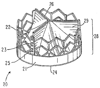
Figure 22 depicts yet another prefented embodiment a valve assembly in
accordance with the present invention, having dilated supports;
CA 2944307 2018-06-26
CA 02944307 2016-10-05
WO 03/047468 PCT/US02/31588
17
Figures 23a to 23e depict stages in a method of manufacturing an implantable
prosthetic valve in accordance with another preferred embodiment of the
present
invention;
Figures 24a to 24c illustrate a support frame of an implantable prosthetic
valve
having means for mounting valve leaflets in accordance with a preferred
embodiment of
the present invention that can form a tricuspid valve. Figure 24a depicts an
isometric
view of the frame, and Figure 24b depicts a cross-sectional view of the means
for
mounting a valve leaflet in details, provided with a valve leaflet Figure 24c
depicts
further details of attachment means for the attachment method;
Figures 25a to 25d illustrate an implantable prosthetic valve in accordance
with
another preferred embodiment of the present invention. Figures 25a and 25b
depict an
isometric view and an upper view of the .valve assembly, respectively, and
Figures 25c
and 25d illustrate upper views of two optional constructions for the means for
mounting
leaflets;
Figures 26a to 26c illustrate a tricuspid valve in accordance with yet another
preferred embodiment of the present invention, provided with a self-expandable
frame.
Figure 26a is the valve in its fully expanded diameter, Figure 26b is a
tapered tool which
assists in inserting the valve into an introducing tube, and Figure 26c shows
the valve
assembly inside a restriction tube, ready to be inserted into a introducing
sheath;
Figure 27 illustrates an isometric view of an implantable prosthetic valve in
accordance with another preferred embodiment of the present invention having
hooks
designated to anchor the valve assembly to body ducts;
Figure 28 illustrates a partial view of an implantable prosthetic valve in
accordance with yet another preferred embodiment of the present invention. The
conmissural attachment is showed in details;
Figures 29a and 29b illustrate an isometric view and an upper cross-sectional
view, respectively, of an attachment assembly of a valve's frame to leaflets
in accordance
with a preferred embodiment of the present invention;
WO 03/047468 PC1/US02/32588
18
Figures 30a to 30c illustrate an isometric view, a cross-sectional view and a
flattened view, respectively, of an attachment assembly of a valves frame to
leaflets in
accordance with another preferred embodiment of the present invention. Figure
30c is a
side view showing two pieces of pericardium before the attachment to the
frame;
Figures 31a and 31b illustrate an exploded view and an isomenic view,
respectively, of a commissural attachment in accordance with a preferred
embodiment of
the present invention depicting the attachment technique;
Figures 32a and 32b illustrate an isometric view of an attachment between
leaflets
and the frame in accordance with yet another preferred embodiment of the
present
invention;
Figures 33a to 33d illustrate different views and portions of an attachment
between a pericardium and a frame in accordance with yet another preferred
embodiment
of the present invention, demonstrating another method of attachment in
accordance with
the preferred embodiment;
Figures 34a to 34c illustrate an isometric view of an attachment between a
pericardium and a valve in accordance with yet another preferred embodiment of
the
present invention demonstrating another method of attachment. In Figures 34b
and 34c, a
deployed portion and the folded portion, respectively, are shown;
Figures 35a to 35c illustrate an isometric and cross-sectional upper views,
respectively, of attachment techniques between a pericardium leaflet and a
valve's frame
in accordance with another preferred embodiment of the present invention;
Figures 36a and 36b illustrate an isometric view of a commissural assembly in
accordance with a preferred embodiment of the present invention demonstrating
a method
of forming one;
Figures 37a to 37c illustrates a commissural assembly in accordance with
another
preferred embodiment of the present invention, where the connecting bar
functions as a
CA 2944307 2018-06-26
CA 02944307 2016-10-05
WO 03/047468 PCT/US02/32588
19
flexible support and has integral attachment means to the frame. Figure 37b is
an
isometric view of the connecting bar;
Figures 38a to 38g illustrate isometric views of flexible conmfissural
supports and
the method of attaching them to a pericardium and a frame and valve in
accordance with
preferred embodiments of the present invention;
=
Figures 39a to 39b illustrate an isometric view of a commissural attachment in
accordance with yet another preferred embodiment of the present invention,
demonstrating the attachment of the pericardium to the support by means of a
shaped
compressing member;
Figures 40a to 40c illustrate an isometric view of a bicuspid valve mounted on
a
frame in accordance with yet another preferred embodiment of the present
invention.
Figures 40b and 40c depicts a cross-sectional side view and an isometric view,
respectively, of the pericardium that is sutured to a PET tube in the form of
pockets;
Figures 41a to 41d illustrate isometric views of an implantable prosthesis
tricuspid
valve in accordance with yet another preferred embodiment of the present
invention;
Figures 42a and 42b illustrate an isometric view of an implantable prosthetic
valve
in accordance with yet another preferred embodiment of the present invention,
having a
different commissural attachment. Figure 42b depicts the attachment in
details;
Figures 43a and 43b illustrate an isometric view of an implantable prosthetic
valve
in accordance with yet another preferred embodiment of the present invention.
Figure
43a depicts the commissure that are pm-sutured in a tapered shape;
Figures 44a to 44c illustrate an isometric view of an implantable prosthetic
valve
in accordance with yet another preferred embodiment of the present invention,
with
additional pieces of PET used for sealing and protecting the pericardium;
Figures 45a to 45d illustrate an isometric view of an implantable prosthetic
valve
in accordance with yet another preferred embodiment of the present invention,
having
leaflets sutured to a pre-shaped PET tube and optional leaflet-tube
attachments in details;
CA 02944307 2016-10-05
wo 03/047468 PCT/US02/32588
Figures 46a and 46b illustrate an, exploded view and an upper cross-sectional
view
of an implantable prosthetic valve assembly in accordance with yet another
preferred
embodiment of the present invention;
Figures 47a to 47c illustrate a partial cross-sectional side view of an
inflating
balloon in accordance with a preferred embodiment of the present invention.
The balloon
is a part of an implantable prosthetic valve delivery system. Figures 47b and
47c are
cross sectional upper views in the inflated and deflated positions,
respectively; and
Figures 48a and 48b illustrate a partial cross-sectional side view and an
upper
cross-sectional view of an inflating balloon in accordance with another
preferred
=
embodiment of the present invention.
DETAILED DESCRIPTION OF THE INVENTION
A main aspect of the present invention is the introduction of several novel
designs
for an implantable prosthetic valve. Another aspect of the present invention
is the
disclosure of several manufacturing methods for implantable prosthetic valves
in
accordance with the present invention. A further aspect of the present
invention is the
provision of novel deployment and positioning techniques suitable for the
valve of the
=
present invention.
Basically the implantable prosthetic valve of the present invention comprises
a
leafed-valve assembly, preferably tricuspid but not limited to tricuspid
valves only,
consisting of a conduit having an inlet end and an outlet, made of pliant
material arranged
so as to present collapsible walls at the outlet. The valve assembly is
mounted on a
support structure such as a stent adapted to be positioned at a target
location within the
body duct and deploy the valve assembly by the use of deploying means, such as
a
balloon catheter or similar devices. In embodiments suitable for safe and
convenient =
percutaneous positioning and deployment the annular frame is able to be posed
in two
positions, a crimped position where the conduit passage cross-section
presented is small
so as to permit advancing the device towards its target location, and a
deployed position
where the frame is radial extended by forces exerted from within (by deploying
means) so
CA 02944307 2016-10-05
WO 03/047468 PCT/1JS02/32588
21
as to provide support against the body duct wall, secure the valve in position
and open
itself so as to allow flow through the conduit
The valve assembly can be made from biological matter, such as a natural
tissue,
pericardial tissue or other biological tissue. Alternatively, the valve
assembly may be
made form bioconapatible polymers or similar materials. Homograph biological
valves
need occasional replacement (usually within 5 to 14 years), and this is a
consideration the
surgeon must take into account when selecting the proper valve implant
according to the
patient type. Mechanical valves, which have better durability qualities, carry
the
associated risk of long-term anticoagulation treatment.
The frame can be made from shape memory alloys such as nickel titanium (nickel
titanium shape memory alloys, or NiTi, as marketed, for example, under the
brand name
Nitinol), or other biocompatible metals. The percutaneously implantable
embodiment of
the implantable valve of the present invention has to be suitable for crimping
into a
narrow configuration for positioning and expandable to a wider, deployed
configuration
so as to anchor in position in the desired target location.
The support stent is preferably annular, but may be provided in other shapes
too,
depending on the cross-section shape of the desired target location passage.
Manufacturing of the implantable prosthetic valve of the present invention can
be
done in various methods, by using pericardium or, for example, by using
artificial
materials made by dipping, injection, electrospinning, rotation, ironing, or
pressing.
The attachment of the valve assembly to the support stent can be accomplished
in
several ways, such as by sewing it to several anchoring points on the support
frame or
stent, or riveting it, pinning it, adhering it, or welding it, to provide a
valve assembly that
is cast or molded over the support frame or stent, or use any other suitable
way of
attachment
To prevent leakage from the inlet it is optionally possible to roll up some
slack
wall of the inlet over the edge of the frame so as to present rolled-up sleeve-
like portion at
the inlet
CA 02944307 2016-10-05
WO 03/047468 PCT/US02/32588
22
Furthermore, floating supports may be added to enhance the stability of the
device
and prevent it from turning inside out.
An important aspect of certain embodiments of the present invention is the
provision of rigid support beams incorporated with the support stent that
retains its
longitudinal dimension while the entire support stent may be longitudinally or
laterally
extended.
The aforementioned embodiments as well as other embodiments, manufacturing
methods, different designs and different types of devices are discussed and
explained
below with reference to the accompanying drawings. Note that the drawings are
only
given for the purpose of understanding the present invention and presenting
some
preferred embodiments of the present invention, but this does in no way limit
the scope of =
the present invention as defined in the appended claims.
Reference is now made to Figure 1, which illustrates a general tricuspid
implantable prosthetic valve 20 in accordance with a preferred embodiment of
the present
invention, suitable for percutaneous deployment using an expandable stout or
similar
deploying means, shown in its deployed position. A valve assembly 28 comprises
a
conduit having an inlet 24 and an outlet 26, the outlet walls consisting of
collapsible
pliant material 29 that is arranged to collapse in a tricuspid arrangement The
valve
assembly 28 is attached to an annular support stent 22, the one in this figure
being a net-
like frame designed to be adapted to crimp evenly so as to present a narrow
configuration
and be radially deployable so as to extend to occupy the passage at the target
location for
implantation in a body duct. Support beazns 23 are provided on annular support
stent 22
to provide anchorage to valve assembly 28. Support beams 23 are optionally
provided
with bores 25 to allow stitching of valve assembly 28 to support beams 23 by
thread,
wires, or other attachment means.
In the embodiment shown in Figure 1, a cuff portion 21 of the valve assembly
28
is wrapped around support stent 22 at inlet 24 to enhance the stability.
Preferably cuff
portion 21 of valve material 28 is attached to support beams 23.
CA 02944307 2016-10-05
WO 03/047468 PCT/US02/32588
23
=
Note that the entire valve structure is adapted to be radially crimped and
radially
expanded, and this lends to provide ease of navigation through narrow passages
in the
vasculature during positioning of the device and adequate deployment on the
final
location. This is made possible by the provision of a collapsible support
stent structure.
However, the support beams remain at all times constant at their length and
thus are
suitable for serving as the pliable valve assembly's anchorage. The valve
assembly is
attached to the support stent at the support beams, and due to their constant
length there is
no need for slack material as the attachment points (25) remain at constant
distances
regardless of the position of the valve device (crimped or deployed). This is
an important
feature for this means that the manufacturer of the valve device can make sure
the valve
assembly is secured and fastened to the support stent at all times. In prior
art implantable
valve devices the entire support structure changes its dimensions from its
initial first
crimped position and final deployed position, and this means that in the
attachment of the
valve assembly to the support structure one must take into consideration these
dimension
changes and leave slack material so that upon deployment of the device the
valve
assembly does not tear or deforrn. In the valve device of the present
invention there is no
relative movement between the valve assembly and the support beams (along the
longitudinal central axis of the device). As a result, the valve device of the
present
invention acquires greater durability and is capable of withstanding the harsh
conditions
prevailing within the vasculature and especially the millions of cycles of
stress applied by
the blood pressure.
The Diced attachment of the valve assembly to the support stent in the valve
device
of the present invention results in greater stability, enhanced safety, better
sealing and
consequently longer lifespan. The novel design of the valve device of the
present
invention leads to longitudinal strength and rigidity whereas its collapsible
support
structure results in radial flexibility.
Figure 2 depicts an implantable valve 30 mounted on a deployable stent 32. The
valve assembly 34 is attached to the deployable support stent 32 (dotted
lines) along three
substantially equidistant and substantially parallel support beams 40 of
constant length,
CA 02944307 2016-10-05
WO 03/047468 PCT/US02/325821
24
which are part of stent 32. The attachment of valve assembly 34 to stilt 32 is
facilitated
by the support beams 40 to which valve assembly 34 is stitched with thread or
fiber 46
(through bores 42 of support beams 40). Outlet leafs 38, which are a slack
portion of the
valve assembly, dangle inwardly, and the whole device is carried by an
inflatable balloon
48, which serves as the deploying device. A portion of the valve assembly 34
at an inlet
zone 45 is optionally rolled over support stent 32 at the inlet, making up a
rolled sleeve,
which enhances the sealing of the device at the valve inlet
Figure 3 demonstrates an implantable valve mounted to a dent 50 with an
inflatable balloon 52, in a crimped position. The support stent 50 is
initially crimped
about the balloon 52 so that is presents a narrow cross-section and is thus
suitable for
percutaneous catheterization and deployment.
Figure 4 depicts an implantable valve deployment in a natural aortic valve
position. The implantable valve is advanced while mounted over the balloon 52
until it
reaches the desired target location 54 in a body duct, for example, aorta 56.
The balloon
is inflated and the support stent 50 expands radially to take up its position.
Figure 5 demonstrates the manufacture of a polyurethane valve in a dipping
technique. A dipping mandrel 60 is provided with a tubular portion 62 with
surfaces 64
that correspond to the collapsible valve leaflets to be manufactured. Mandrel
60 is dipped
into a dissolved polyurethane bath 66 and is coated with a polyurethane
coating in the
desired form of the valve. Then, after the polyurethane coating has hardened
sufficiently,
the completed valve is removed from mandrel 60.
Figures 6a to 6e illustrate manufacturing an implantable valve by forging. A
suitable tubularly shaped material 74 is placed tightly on a tubular portion
68 of mandrel
67, covering the cusp portion 69. Flexible inserts 76 are pressed to mandrel
67, forging
the tubular material to mandrel shape 80. A tapered ring 70 holds the flexible
inserts in
place as the whole mold is placed in a hot oven regulated to a desired
temperature, which
is lower than the material's melting point Figure 6e illustrates a sectional
side view of
the mandrel and a cross cut portion of the mold. The mold is made to press
inwardly on
CA 02944307 2016-10-05
WO 03/047468 PCT/US02/32588
the mandrel, which is covered with the valve material. As a result the
material takes up
the desired shape. The materials used can vary, for example, polyurethane
(PU),
polyethylene terphthalate (PET), or any other suitable material, which may be
formed by
heating.
Figures 7a and 7b demonstrate a method of manufacturing a composite valve,
which has PU leaflets and PET tubular construction with a crown shape. PU is
an
excellent fatigue resistant material but is sensitive to tear. The PU is
reinforced by the
PET crown to allow safe attachment to a stoat by means of stitching, riveting,
or any
other suitable attachment method. A PET crown 86 is placed on a mandrel 87,
which is
then (turned and) dipped in a container of dissolved PU. The manufactured
device is a
valve assembly having leaflets 88 composed of pure PU, and thus fatigue
resistant, and a
main body made of PET with protruding attachment portions 90 suitable for
attachment
built in the PU.
Figures 8a and 8b demonstrate a method of manufacturing a composite valve,
which is based on flexible PU 92 for as the main body of the valve, rigid PU
support
beams 94 serving for the attachment area, and PET sleeve 96 portions for the
valve inlet
The need for a rigid portion for attachment (support beams 94) is explained by
the
tendency of the flexible, fatigue resistant material to tear as already
explained. The
advantage of the stiff PU support beams is that they are chemically adhered to
the main
body, and this improves the overall durability of the valve due to reduction
of inner forces
and friction in the attachment area specially attachment between two different
materials.
The valve is dipped in the method mentioned with reference to Figure 5, and
the rigid PU
support beam 94 is created by way of mold injection, machining or any other
suitable
way. The rigid PU support beam 941s placed on the valve and then dipped into
the
container of dissolved PU. This is done while the valve is positioned on the
mandrel (not
shown). This method provides the ability to composite several materials into
one body
and, by that, gain the advantage of the various properties of the materials as
they are
needed in different areas of the prosthesis.
CA 02944307 2016-10-05
WO 03/047468 PCT/US02/32588
26
Figures 9 to 9i demonstrate different methods of attachment between a valve
assembly and the support stents. A valve assembly 99 shown in Fig. 9 is
incorporated
into valve 100 shown in Fig_ 9a, where a support stent 102 is attached to
valve assembly
99 through support beam 106. A detail is shown in Fig. 9b, where, in cross-
section, it can
be seen that layer 108 is an optional inner support made of stainless steel or
rigid
polymeric material, valve assembly 99 comprises a PET layer 105 coated with a
PU layer
104, with the outer support beam 106. Connector 107 is a connecting wire made
of a
strong material, such as stainless steel. Figure 9c illustrates an alternative
arrangement
for attachment by a rivet 109, and in Figure 9d the attachment is achieved by
a suture
110.
Figures 9e to 9g show an attachment method comprising shaped rigid members
116, preferably made from metal, which tightly hold the PU valve material 118
by fitting
in between a PU U-shaped nest 120 and are attached to a stent 122 by extruding
portions
124 that are provided on U-shaped rigid member 116, which fit the bores 126 of
the
support beam 128 of the stent 122. Figures 9h and 9i show another attachment
method,
where rigid support beams in the form of frame construction 132 are provided,
and the
valve assembly pliant material 135 made of a tubular material is inserted
through a gap
137 in the frame. After insertion, a fastening rod 133 is inserted through the
pocket
formed between the pliant material and the frame and holds the valve in
position.
Figure 10 illustrates a dipping mandrel 139 with an extending portion 141,
which
improves the sealing ability of the valve. Since the valve is attached to a
collapsible stent
and is itself collapsible, it is difficult to determine the exact shape of the
valve after
crimping and deploying. It is of major importance that sealing will be
achieved. By
adding the extension 141 the leaflets are made longer than needed to exactly
close the
outlet, and therefore when they are in the collapsed state, substantial
portions of the
= leaflets fall on each other creating better sealing.
Figures ha to 11c illustrate a valve assembly mounted on a support stent 144
with
interlaced strengthening wire 146, which improves the force distribution on
the valve
material and facilitates prolonged durability of the valve. The support is in
the form of a
CA 02944307 2016-10-05
PCT/US02/32588
WO 03/047468
27
wire, which has a crown shape as the shape of the three cusp valve base 148,
it also has
the ability to be crimped 150 to a small diameter, together with the stent,
valve and
balloon, as shown in Fig. 11b. The forces applied to the valve edge 148 while
working,
are applied to the attachment points, by making the attachment line longer we
reduce the
force on each attachment point In this support method the valve is attached by
suturing
152 the entire line to the extra support wire 146. This wire can be made of
stainless steel,
nickel titanium alloy such as nitinol, or polymeric material. The support
suture renders
the valve assembly default fault lines where the valve material more readily
flexes, thus
ensuring proper operation of the valve flaps (leaflets). Optionally the valve
assembly
shown in Figures 11a to Ile can be mounted on a support stent such as the one
described
herein or similar supporting structures. The strengthening wire is interlaced
in the valve
assembly at the outlet of the conduit so as to define a fault line about which
the
collapsible slack portion 154 of the valve assembly may flap.
Figures 12a to 12e depict a valve device provided with a stein 159 and
substantially equidistant rigid support beams 160, interlaced or attached to
the slack
portion of the valve assembly material 161, arranged longitudinally. This
design allows
the valve leaflets to perform without outer support. The support in standard
valves is by
tying the upper edge of the cusp to a rigid embodiment, so that it reacts to
the load as a
suspension bridge. In this new design the prevention of collapsing is achieved
similar to
an Indian tent, i.e., the rigid supports lean on each other 162 when the valve
is closed but
do not interfere in opening 164 when the valve is open.
Figures 13a to 13c illustrate the manufacturing of a valve assembly in
accordance
with another preferred embodiment of the present invention. At first a
polyurethane
thread line 170 is fed from a PU supply 172, and coiled around a cylindrical
drum 174 to
form coil 176. Then, drum 174 with coil 176 is dipped in a PU bath 177, and a
second
layer 178 of the PU coats coil 176, making it a stronger construction capable
of
withstanding tearing forces both laterally and in other directions.
Incorporating two
different types of materials - such as PU and PET - may render greater
durability and
- - -
CA 02944307 2016-10-05
WO 03/047468 PCT/US02/32588
28
endurance to the valve assembly. This material is an alternative material to
be used in the
forging method shown in Figure 6.
Figures 14 to 14c demonstrate the incorporation of heavy metal markers on the
stent, which markers allow observation and thereby adjustment of orientation
while
placing the device in the required location. Heavy metals are radiopaque, that
is, they are
conspicuous on an angioscopic image, which is a two-dimensional image. Since
the
coronary artery ostia 237 and 238 are located near the typical valve
deployment location
and must stay open, it is extremely important to make sure that the deployed
valve
assembly is not blocking a coronary ostium. In some cases the stent is lower
than the
ostium and in those cases it will stay open, but in some cases as shown in
these figures it
is necessary to make sure that the stent portion 239 that is connecting the
valve supports
235 is opposite the coronary ostia, and in that way the blood supply is
preserved through
=
the stent struts. Two heavy metal markers 232 are attached at the outlet side,
one marker
230 at the inlet side. It is possible to adjust the angiogscopic view to the
plane of the left
coronary as shown in Figure 14b and anatomically locate the other accordingly.
lithe
two upper markers 232 are placed in the radiographic two dimensional image,
one on top
of the other, and the low marker 230 on the opposite side, we make sure that
the
coronaries are open to blood flow as seen in Figure 14c. Gold, platinum,
iridium or
tantalum are all biocompatible materials suitable for the markers described
above.
Figures 15a to 15c illustrate a valve with a portion of radio-opaque material
267
such as a thread of gold at the sealing edge. When a valve is implanted, it is
very
important to have clear indications of how the valve is functioning in vivo;
pressure
measurements, flow visualization, and doppler measurements are utilized. It is
also
possible to examine the valve by ultrasound methods, however, observing the
opening
and closing of the valve cusps on a monitor. Fig. 15b is an angiographic image
268 of the
open valve, while image 169 in Figure 15e is the closed position as seen on
the
angiogram.
Figures 16a to 16c illustrate a procedure, which helps in placing the device
in the
=
longitudinal position. It is very important to place the device in the correct
longitudinal
CA 02944307 2016-10-05
WO 03/017468 PCT/US02/32588
29
position, for if it is too deep in the left ventricle it may interfere with
the mital valve
function by improper closing or function of the valve. If it is positioned too
high it may
migrate, it may leak via the sinus cavities, which are located around it,
and/or it may
block the coronaries. It is a necessary task to position the valve prosthesis
in a narrow
target location. In Figure 14 a method of lateral orientation placement is
shown, and
Figures 16a to 16e illustrate a longitudinal positioning. The valve device
(the valve
assembly and the support stent) is placed on an inflatable balloon catheter,
comprising
double independently inflatable chambers 303, 305, and is inserted into the
left ventricle
302 in the crimped position and guided over a guiding stylet or guide wire
300. The
balloon, which is larger than the annulus diameter when inflated, is inflated
in the left
ventricle 302, and then the whole device is pulled slightly backwards. The
balloon is
supported on the inner part of the annulus 303, allowing positioning of the
device in the
exact desired position. In addition, it temporarily blocks the blood flow, and
that
improves the ability to hold the device in place while inflating it. The next
step is
inflating the second balloon 305, which deploys the valve device in the
desired location.
The method for deploying an implantable prosthetic valve device at the natural
aortic valve position at the entrance to the left ventricle of a myocardium of
a patient, as
depicted in Figures 16a, 16b and 16c, comprises the steps of:
(a) providing a balloon catheter having a proximal end and a distal end,
having
a first and second independently inflatable portions, the first inflatable
portion located at
the distal end of the catheter and the second inflatable portion adjacently
behind the first
inflatable portion;
(b) providing a guiding tool for guiding the balloon catheter in the
vasculature
of the patient;
(c) providing a deployable implantable valve prosthesis device adapted to
be
mounted on the second inflatable portion of the balloon catheter
(d) guiding the balloon catheter through the patient's aorta using the
guiding
tool, the valve device mounted over the second inflatable portion of the
balloon catheter
CA 02944307 2016-10-05
WO 03/047468 PCINS02/32588
until the first inflatable portion of the balloon catheter is inserted into
the left ventricle,
whereas the second inflatable portion of the balloon catheter is positioned at
the natural
aortic valve position;
(e) inflating the first inflatable portion of the balloon catheter so as to
substantially block blood flow through the natural aortic valve and anchor the
distal end
of the balloon catheter in position;
(f) inflating the second inflatable portion of the balloon catheter so as
to
deploy the implantable prosthetic valve device in position at the natural
aortic valve
position;
(g) deflating the first and second inflatable portions of the balloon
catheter;
and
(h) retracting the balloon catheter and removing it from the patient's
body.
Figure 17 describes a positioning of a valve device 310 using an additional
deployable stent 320. There are several problems that may be encountered while
deploying the stent and valve in the aortic valve location: blockage of
coronaries may
occur that is dangerous if the diameter of the stent is similar to that of the
coronaries
aortic root 309. Secondly, migration of the whole device may also occur, which
is a
dangerous possibility, and there is the problematic challenge of exact
positioning of the
valve device that is very difficult to accomplish, as already explained. The
newly special
designed device with a double diameter inflatable balloon and double stent
design allows
placement of the device in a way that coronaries will not be blocked because
of a safe
difference that is kept between the diameters, longitudinal placing is less
sensitive
because of the small diameter which ensures prevents over expansion of the
valved
prosthesis. The distal stent 320, which contains no valve, is expanded into
the ascending
aorta, while the proximal stent 310 is placed simultaneously in the annular
position. This
placement method is less challenging due to the smaller diameter of the
proximal stent
310 which ensures that the mitre valve is not deformed by over-expansion as
the
CA 02944307 2016-10-05
=
WO 031047468 PCT/US02/32588
31
dimensions are preserved, and the additional stent decreases the risk of
device migration.
It is safer to over dilate in the aorta, which is not true for the annulus.
The method for deploying an implantable prosthetic valve device at the natural
aortic valve Position at the entrance to the left ventricle of a myocarditun
of a patient, as
depicted in Figures 17a and 17b, comprises the steps of:
(a) providing a balloon catheter having a proximal end and a distal end,
having
a first and second independently inflatable portions, the first inflatable
portion located at
the distal end of the catheter and the second inflatable portion adjacently
behind the first
inflatable portion;
(b) providing a guiding tool for guiding the balloon catheter in the
vasculature
of the patient;
(c) providing a deployable implantable valve prosthesis device adapted to
be
mounted on the first inflatable portion of the balloon catheter, and a
deployable annular
stent device adapted to be mounted over the second inflatable portion of the
balloon
catheter, the deployable implantable valve prosthesis device and the
deployable annular
stent kept at a predetermined distant apart;
(d) guiding the balloon catheter through the patient's aorta using the
guiding
tool, the valve device mounted over the first inflatable portion of the
balloon catheter and
=
the deployable annular stent mounted over the second inflatable portion of the
balloon
catheter, until the first inflatable portion of the balloon catheter is
positioned at the natural
aortic valve position;
(e) inflating the second inflatable portion of the balloon catheter so that
the
deployable stent device is deployed within the aorta thus anchoring the
deployable
annular stent and the coupled valve device in position;
(f) inflating the first inflatable portion of the balloon catheter so as to
deploy
the implantable prosthetic valve device in position at the natural aortic
valve position;
CA 02 944307 2016-10-05
WO 03/047468 PCT/U502/32588
32
=
(g) deflating the first and second inflatable portions of the balloon
catheter;
and
(h) retracting the balloon catheter and removing it from the patient's
body.
Figures 18a and 18b illustrate an accessory crimping device that is adapted to
crimp a valve device in the operating theater as part of the implantation
procedure. The
crimping device 330 comprises several adjustable plates that resemble a
typical SLR
camera variable restrictor. It is comprised of simultaneously movable plates
332 each
provided with a blade 334, that are equally dispersed in a radial symmetry but
each plate
moves along a line passing off an opening in the center, all plates
equidistant from that
center opening 336. Initially (see Figure 18a) the plates are drawn apart
providing a large
enough opening for the implantable valve to be positioned within that opening.
When the
plates are drawn towards the center (see Figure 18b), the opening 336 reduces
in size but
still retains the annular shape, and this facilitates the crimping of the
valve frame to a
small dimension suitable for percutaneous positioning.
Figures 19a depicts a crimping method for the support stein of the valve
prosthesis
device of the present invention, whereby stent 340 is crimped, that is,
compressed or
curled. In Figure 19b a crimping device 343 is shown, comprising a body having
an
annular void in which an expanded stoat is positioned. Lever 346 is connected
to the end
347 of the stent and as the lever is pulled the stent is curled or compressed
about axle 345
into a compressed position 349 (Figure 19c).
Figures 20a and 20b depict a valve made of a simple tube mounted to a stern
352.
During systole period the tube is fully open and during diastole period the
tube collapses
according to the mounting geometry 357 and achieves sealing.
Figure 21 describes a newly designed support stent 360 in its open position.
Three
of the longitudinal struts 362 are full and thick and always stay with their
original
constant size, serving as anchoring support. Each of these struts 362 is
provided with a
plurality of bores 364, which are later used for mounting the valve assembly
(not shown)
and tying it to stent 360. Between struts 362 a web-like construction is
provided, which
CA 02944307 2016-10-05
WO 03/047468 PCT/US02/32588
33
is capable of being crimped to a narrow state and capable of being deployed
again to a
wider state.
Figure 22 illustrates another preferred embodiment of an implantable
prosthetic
valve according to the present invention. It comprises a metal tube 370,
having three
portions with a thicker wall 371 than in the rest of the tube 370, these areas
form the
longitudinal columns 372 in the construction, after the tube is cut to its
final form. The
advantage of such a construction is in its superior bending strength, in
specific required
portions of the construction, with minimal interference to the crimped volume
of the
whole construction.
Figure 23a to 23c depict a new method of manufacturing an artificial or
biological
crimpable valve device. A piece of fabric material 370 (Fig. 23a), is dipped
in PU to
create a portion which is later formed into valve leaflets 371 (Fig. 23b).
This composite
material 371 is then attached to an additional piece of fabric such as PET 372
by means of
stitching, suturing or other attaching technique 373 (Fig. 23c). The resulting
fabric 375 is
cut along stitching line 373 leaving enough material to later suture the valve
assembly to
the support construction. It is then formed to a tubular shape and stitched
374 (Fig. 23d).
The tubular valve is then attached to a support construction 380 by suturing
the bottom
part around the valve 379 tightly to prevent leakage, and around the cut
fabric line 376
(Fig. 23e). This open wall structure 378 allows blood flow to the coronary
arteries. The
valve is later placed with the coronary artery between the support columns
385.
Additional variations of this can be made by replacing the composite material
371/370
with a biological patch such as a suitable pericardium patch. In some cases it
is possible
to make the same valve without cutting the fabric 372 with the shaped cut 376,
and by
that create a valve with an outer tubular shape. The embodiment of Figs. 23a
to 23c is
easy to manufacture as it is generally flat throughout most of the production
process and
only at the final stage of mounting on the support stent is it given a three-
dimensional
form.
Reference is now made to Figure 241 illustrating a frame of an implantable
prosthetic valve having means for mounting valve leaflets in accordance with a
preferred
CA 02944307 2016-10-05
WO 03/047468 PCT/US02/32588
34
embodiment of the present invention that can form a tricuspid valve. Figure
24a depicts
an isometric view of the frame and Figure 24b depicts a cross sectional view
of the means
for mounting valve leaflets 430 in detail. A frame 420, which is suitable for
crimping and
expanding, has three support beams 422 for mounting leaflets positioned
substantially
symmetrically about the circumference of the frame. Frame 420 is shown in
Figure 24a
in its deployed state. Support beam 422 has a "U" shaped lateral cross
section, or profile
(shown clearly in Figure 24b) that is designed to attach to a commissure of
the valve
structure. The "U" shape can be produced by extrusion, wire cutting or by
welding the
"U" profile to the frame's struts 421 at junction points 424. Support beam 422
is provided
with a series of bores 425 positioned along its back wall. Bores 425 are
designated for
stitching the valve assembly by threads, wires, or other attaching means.
Figure 24b is a detailed cross-sectional view of one of the support beam 422.
Two
pericardial leaflets 430 are inserted through a U-shaped, or forked holder 428
that
compresses and restricts the leaflets in the U-shaped profile. Leaflets 430
are folded to
both sides of the support beam 422. When holder 428 is compressed toward the
support
beam 422, leaflets 430 are caught in-between holder 428 and support beam 422
so that
the leaflets are kept in place. Figure 24c is an exploded view of the holder,
bar 426 has a
series of bores compatible for attachment to the frames support beam 422,
attaefunent
being achieved by suture 423 or any other attachment means. This attachment
method
allows attaching the leaflets to the frame without puncturing it with sutures
and needles.
It is also important that the leaflets are firmly held in place by the holder
428 so that it has
no relative movement in respect to the rigid frame; hence avoiding wear due to
movements. Leaflets that are made from pericardium are known to better
withstand inner
movements and stresses and less to wear by movement against rigid, hard or
sharp bodies.
It is noted again that the entire valve structure is adapted to be radially
crimped
and radially expanded This feature imparts the valve with the ability and ease
to navigate
through narrow passages in the vasculature during positioning of the device.
After final
positioning of the valve, the valve is deployed_ This is made possible by the
provision of
a collapsible support frame structure. However, the length of the attaching
means (the
CA 02944307 2016-10-05
WO 03/047468 PCT/US02/32588
height of the valve) remains at all times constant; thus suitable for serving
as the pliable
valve assembly's anchorage. The leaflets are attached to the support frame at
the
attaching means, and due to their constant length there is no need for slack
material as
these attachment points that remain at constant distances regardless of the
position of the
valve assembly (crimped or deployed). This is an important feature for this
means that the
manufacturer of the valve device can make sure the valve assembly is secured
and
fastened to the support frame at all times. In prior art implantable valve
devices, the entire
support structure changes its dimensions from its initial fast crimped
position to final
deployed position and this means that in the attachment of the valve leaflets
to the support
structure one must take into consideration these dimension changes and leave
slack
material so that upon deployment of the device, the valve assembly does not
tear or
deform. In the valve device of the present invention there is no relative
movement
between the valve leaflets and the support beams (along the longitudinal
central axis of
the device). As a result, the valve device of the present invention acquires
greater
durability and is capable of withstanding the harsh conditions prevailing
within the
vasculature and especially the millions of cycles of stress applied by the
blood pressure.
The fixed attachment of the valve leaflets to the support frame in the valve
assembly device of the present invention renders it greater stability,
enhanced safety,
better sealing and consequently longer lifespan. The novel design of the valve
device of
the present invention renders it longitudinal strength and rigidity whereas
its collapsible
support structure renders it radial flexibility.
Figures 25a to 25d illustrate an implantable prosthetic valve in accordance
with
another preferred embodiment of the present invention. Figures 25a and 25b
depict an
isometric view and an upper view of the valve assembly, respectively and
Figures 25c
and 25d illustrate upper views of two optional constructions for the means for
mounting
leaflets. Pericardial leaflets 430 are mounted on a deployable support frame
432. The
frame is preferably made of three segments that form a circular support frame
when
assembled (Figure 25b). Pericardial leaflets 430 are attached to deployable
support frame
432 along three substantially equidistant and substantially parallel beams
440, which are
CA 02944307 2016-10-05
WO 03/047468 PCT1US02/32588
36
integral parts of support frame 432. Leaflets 430 are attached to support
frame 32 at
support beams 440 by suturing 446 leaflets 446 to support beams 440 through
bores 442
in beams. The frame segments that are preferably made from stainless steel are
pre-
shaped 432 and can be formed in different ways. Figure 25c illustrates support
frame
segments 432a having beams 435a pointing inwardly. Figure 25d illustrates
support
frame segments 432b having beams 435b that are outwardly pointing. The
advantages of
this technique are the possibility to manufacture the frame segments from
sheets (as
opposed to tube) and the ease of assembly of the frame segments with the
pericardial
leaflets.
Figures 26a to 26c illustrate a tricuspid valve in accordance with yet another
preferred embodiment of the present invention, provided with a self-expandable
frame.
Figure 26a is an isometric view of an implantable prosthetic valve 430 mounted
on a self-
expandable frame 445. Implantable prosthetic valve 430 comprised of three
valve leaflets
is mounted on self-expandable frame 445 so that each leaflet extends along an
equidistant
portion of the frame and is sutured at both opposite sides to substantially
equidistant and
substantially parallel beams 440. By using a tapered tube 448 the whole
assembly is
crimped into a restriction tube 449. Figure 26b shows the crimped valve
assembly 447 in
its final crimped diameter ready for insertion to the body. After insertion
into the desired
location in the body the valve is released from the restriction tube and as it
is made of self
expandable material (like a shape-memory alloy), it expands back to the
original diameter
and is anchored in place. In order to reduce the diameter of the device from
its fully
expanded diameter to its crimped diameter a special tapered tube is used,
shown in Figure
26c.
=
Figure 27 illustrates an isometric view of an implantable prosthetic valve in
accordance with another preferred embodiment of the present invention having
hooks
designated to anchor the valve assembly to body ducts. An implantable
prosthetic valve
450 is placed in a natural aortic valve position 452. Implantable prosthetic
valve 450
comprises preferably three leaflets 430 mounted on a metallic support frame
455. The
lower part of support frame 455 is provided with attachment means, preferably
with
CA 02944307 2016-10-05
WO 031047468 PCMTS02/32588
37
hooks 453. Hooks 453 assures that the valve assembly stays in place after
deployment,
and cannot migrate to another position.
Figure 28 illustrates a partial view of an implantable prosthetic valve in
accordance with yet another preferred embodiment of the present invention. The
commissural attachment is shown in details. This figure demonstrates an
attachment
technique that is used in order to attach pericardium leaflet 430 to a
metallic frame 420.
A longitudinal bar 456 having a narrow slit 457 is used as the commissural
attachment so
that extensions 463 of pericardium leaflet 430 are tightly inserted through
slit 457.
Pericardium extensions 463 that are extended beyond slit 457 are wrapped about
a rigid
bar 458 that acts as an anchoring means. Every two extensions originating from
two sides
of slit 457 are sutured to each other by a suture 459 at the side of rigid bar
458 opposite
the slit. An additional suture 462 attaches the bottom circumference of
support frame 420
to leaflet 420 in order to obtain sealing. The advantages of the described
attachment are
that no sutures or suture holes are applied in the leaflet working area, there
are no
concentrated stress points similar to stress point caused by suturing, and the
force
distribution is along the longitudinal bar 456. The narrow passage that is
maintained
through slit 457 forces the leaflets to be static in respect to the support so
as to reduce
abrasion.
The embodiments that will be shown herein after are optional configurations of
attachment between the leaflets and the support frame.
Figures 29a and 29b illustrate an isometric view and an upper cross sectional
view,
respectively, of an attachment assembly of a valve's frame to leaflets in
accordance with
a preferred embodiment of the present invention. The attachment is similar in
principle to
the attachment shown in Figure 28, however, longitudinal bar 456 is further
provided
with an additional pole 465 that is attached to longitudinal bar 456 so as to
establish an
integral part. Pole 465 is rounded so as to make sure the leaflets will not be
abraded or
cut by sharp corners. In the cross sectional view shown in Figure 29b,
adjacent leaflets
460 can be seen compressed together and the main protection goal is clearly
shown.
CA 02944307 2016-10-05
WO 03/047468 PCT/US02/32588
38
Figures 30a to 30c illustrate an isometric view, a cross-sectional view and a
flatten
view, respectively, of an attachment assembly of a valves frame to leaflets in
accordance
with another preferred embodiment of the present invention. Using the method
demonstrated in Figures 30a to 30c, the pericardial leaflets are pre-cut to
the desired
shape 430 and are provided with longitudinal bars 470 that are sutured to the
leaflets
creating a longitudinal clamping effect (Figure 30c). This allows distribution
of forces
along the whole length of the attachment means as opposed to concentrating the
stresses
in suture holes. In Figures 30a and 30b, an additional rigid portion 458 is
added, creating
a round ending, which prevents the leaflets from being bent drastically at the
attachment
point to portions of the frame 420. The attachment to frame 420 is performed
using
sutures 459.
Figures 31a and 3 lb illustrate an exploded view and an isometric view,
respectively, of a commissural attachment in accordance with a preferred
embodiment of
the present invention depicting the attachment technique. A method of
assembling
pericardial leaflets 430 to a frame 420 is demonstrated. A rigid bar 476
provided with
integral protrusions 478 is inserted through bores 479 that are pre-cut in
pericardial
leaflets 430. Integral protrusions 478 pass through a sheet of preferably PET
(braided
polyester) fabric 475, and finally through bores 442 that are provided in
longitudinal bar
440 (the attachment means) of frame 420. After the assembling of the parts, as
shown in
Figure 3 lb, the parts are tightly assembled and bar protrusions 478 are
attached to bar 440
by welding, riveting or any other technique. The PET sheet 475 is folded and
sutured
tightly around bar 476 using suture 472.
Figures 32a to 32c illustrate an isometric view of an attachment between
leaflets
and the frame in accordance with yet another preferred embodiment of the
present
invention. An optional method of attachment is demonstrated, in which a
pericardium
leaflet 430 and bars 480 are sutured in area far as possible from the working
area of the
leaflets. The pericardium is first sutured using a suture 484 to bar 480 as
seen in Figure
32b, and then folded and compressed. In order to firmly hold the pericardial
leaflets in
place between bars 480, an integral connecting member 482 connects the two
bars,
CA 02944307 2016-10-05
WO 03/047468 PC17US02/32.588
39
allowing the bent portions of the bars to be in parallel position, with the
leaflets caught in
between. Then, an additional suture 483 connects the bottom side of the bar to
the
leaflets so that while the valve is waking, the leaflets do not bear high
stresses.
Figures 33a to 33d illustrate different views of portions of an attachment
between
a pericardium and a frame in accordance with yet another preferred embodiment
of the
present invention, demonstrating another method of attachment in accordance
with the
preferred embodiment. A connecting member 490 (shown in a deployed position in
Figure 33d) is used to connect two pericardial leaflets 492 at the line of the
conunissurel.
After being connected between them, pericardial leaflets 492 are being
connected to
frame bar 480. Here again, the principal of compressing the leaflets between
two bent
portions bars 491 of connecting member 490 and tightening them using suture
484
without punctures in the working areas of the pericarditun is applied.
However,
connecting member 490 is provided with a portion 493 that is positioned
perpendicular to
the two bent portions bars 491 that holds the two leaflets together. Portion
493 is the
connecting member to frame's bar 480. In Figure 33a, the junction point 495
between the
portions of connecting member 491 is placed at the upper part (outlet) of the
frame so as
to achieve a rigid connection to the frame. In Figure 33b, junction point 495
is placed at
the bottom part (inlet) of the frame so that the junction point also functions
as a spring.
Comprehensive explanation of the benefits of springs in commissures is
discussed and
shown in respect with Figures 37 to 39.
Figures 34a to 34c illustrate an isometric view of an attachment between a
pericardium and a valve in accordance with yet another prefeiled embodiment of
the
present invention demonstrating another method of attachment. In Figures 34b
and 34c, a
deployed portion and the folded portion, respectively, are shown. An optional
design for
the attachment between the flame and the leaflets is depicted. A connecting
member 480
(shown clearly in Figure 34b) is being produced into a flat configuration
using laser-
cutting. Connecting member 480, which is a part of the frame's attachment
means, is
bent and then is ready for assembly with the leaflets. Connecting member 480
comprises
the main body as well as a connection bar 497 and a flexible element 498
allowing
CA 02944307 2016-10-05
WO 031047468 PCT/US02/32580
flexibility to the commissural. Leaflets 430 are threaded through
corresponding holes
481 in the structured connecting member 480 and are sutured using a suture
482.
Reference is now made to Figures 35a, 35b, and 35c illustrating isometric and
cross-sectional upper views, respectively, of attachment techniques between a
pericardium leaflet and a valve's frame in accordance with other preferred
embodiments
of the present invention. Figures 35b and 35c depict different techniques of
commissural
attachments: in Figure 35b two pieces of pericardial leaflets 500 are wrapped
around a
metallic member 505 that is connected to a frame 501. Rigid members 503 are
positioned
from both sides of metallic member 505 and then tightened together and
connected by a
suture 502. All metallic pieces are wrapped by PET fabric 508 in order to
avoid direct
contact between the metallic pieces and the delicate pericardial leaflets. The
advantage of
this structure is that after tightening the suture, the whole commissure
becomes static with
no relative movement between the portions. This improves the valve assembly's
resistance to abrasion. In addition, there are no needle holes or sutures in
the working
area. Figure 35c depicts a similar structuce, however, there is no use of
rigid sidebars.
After wrapping the metallic member 505 with pericardial leaflets 500, a piece
of PET 508
is used for tightening it to a tight bundle. In this case, the suture line 502
is the borderline
of the working area so it should be designed so that stresses are in the best
possible
distribution.
Figures 36a and 36b focus on the connection of the commissural assembly to
frame's protrusion 509, which is an integral part of the frame and is the
basis for the
corrunissural attachment This example shows the use of four rigid longitudinal
bars 503
connected by a suture 502.
Figures 37a to 37c illustrate a commissural assembly in accordance with
another
preferred embodiment of the present invention, where the connecting bar
functions as a
flexible support and has integral attachment means to the frame. Figure 37b is
an
isometric view of the connecting bar. Connecting bar 520 is flexible and
comprises a
resilient material shaped in a "U" shape. Connecting bar 520 is a part of
commissural
assembly 527 shown in Figure 37a. Connecting bar 520 is provided with
protruding
CA 02944307 2016-10-05
WO 03/047468 PCT/US02/32588
41
elements 521 that are acting as the means of attachment to the frame's bar
480.
Protruding elements are designated to be inserted in corresponding bores 442
in bar 480.
It is optional to provide rods 527 which are integral parts of the "U" shaped
member and
replace the suture 526 that connects the pericardium leaflet and the
connecting bar
together, which is shown in Figure 37a. Figure 37c depict another mcthod of
attaching
the flexible connecting bar 520 to the frame 480 by means of welding 523. Here
the
pericardial leaflets SOO are attached to the connecting bar 520 by suture 526
inserted
through a PET fabric 508 and two connecting bars 503, which together create a
tight
bundle.
Figures 38a to 38g illustrate isometric views of flexible conunissural
supports and
the method of attaching them to a pericardium and a frame a valve in
accordance with
preferred embodiments of the present invention. Figures 38a to 38c demonstrate
incorporation of different design options of commissural springs. The main
purpose of a
commissural spring is to reduce the impact applied to the pericardial leaflets
when the
valves leaflets are closed. If the structure is of a rigid nature, high stress
will be applied
each time the valve closes. If a spring is added to the structure, the spring
will bear the
highest portion of the impact; thus reducing the stress applied to the
leaflets during the
time the valve is closed. In Figure 38a, a simple stainless steel spring 530
is connected to
frame's bar 480 by threading a portion of the spring into slots 538 as shown
in more
detail in Figures 38e and 38f. In Figure 38b, there is a similar spring 530
with leafteta
500 connected to it by one of the attachment methods, the commissural support
itself 530
is connected to the frames bar 480 by spot welding, laser welding or other
attachment
means. Figure 38c depicts a similar spring 534 having an additional spiral.
The purpose
of such a spiral is to reduce stress in the spring and to allow the fatigue
requirements,
which in the case of heart valves are of at least 200 trillion cycles.
Figure 38d illustrates an isometric view of a flexible c,ommissural support in
accordance with yet another preferred embodiment of the present invention,
demonstrating the attachment of the pericardium to the support. Figures 38e to
38g are
the details of the attachment to the frame. A cornmissural spring of a
different design 539
CA 02944307 2016-10-05
WO 03/047468 PCTIUS02/32588
42
comprises a stainless steel wire of a small diameter in respect with the
springs described
in Figures 38a to 38c. One advantage of this structure is the distribution of
stresses in the
spring and the ability to form a structure, which can be crimped to a small
diameter.
Another advantage in this structure is that there are no open edges of the
spring, which
can be dangerous when operated; the open edges are protected in the frame's
bar as
shown in Figures 380 to 38g, which show possible attachment methods of the
spring to
the frame. In Figure 38e, a frame's flat bar 480 cut in shape with slots for
crimping the
spring 536. Figure 38f shows pre-bending of the slots 527 and Figure 38g shows
the
spring legs 539 assembled firmly into the slots 538.
Figure 39a illustrates a technique of commissural assembly using a shaped
compressing member 511. The compression member 511 holds pericardial leaflets
500
firmly while pressing it in the pivot points 513. A radial edge 514 is made in
order to
protect the pericardium from abrasion. The whole assembly is held tightly
inside the
compressing member 516. The Conirnissural assembly is connected to the frame
by
protrusion member 518, which fit bores in the frames bar 480. Figure 391) is
an isometric
view of the same detail.
Figures 40a to 40c illustrate an isometric view of a bicuspid valve mounted on
a
frame in accordance with yet another preferred embodiment of the present
invention.
Figures 40b and 40e depict a cross-sectional side view and an isometric view,
respectively, of the pericardium that is sutured to a PET tube in the form of
pockets. The
valve assembly (in this case bicuspid) comprises a crixnpable frame 540, two
pericardial
leaflets 545, a PET skirt 543 and a connecting suture 547. The focus in this
drawing is on
the pocket shape of the pericardium leaflet shown best in Figures 401, and
40c. One of
the main goals in valve design, in general, is to distribute the stresses in a
homogenous
way in the pericardium material and the attachment areas. The design of the
pericardium
leaflet as a pocket assists in distributing the stresses along suture line
547; pericardium
leaflet 545 is sutured to PET skirt 543 along connecting suture 547. PET skirt
543 is
sutured to the circumference of crimpable frame 540 at the bottom side 549 and
at the top
542 using one of the commissural attachments that are described herein before
regarding
CA 02944307 2016-10-05
WO 03/047468 PCIIUS02/32588
43
other embodiments. When hydrodynamic pressure is applied on leaflets 545, the
leaflets
will meet in the center 546 of frame 540 so as to seal the valve assembly. The
shape of
the leaflets in the valve assembly is determined by the boundary conditions,
which in this
case are the suture lines. The suture lines can be designed to have an optimal
shape
regarding the stress distribution in accordance with geometrical restrictions.
Reference is now made to Figures 41a to 41d illustrating isometric views of an
implantable prosthesis tricuspid valve in accordance with yet another
preferred
embodiment of the present invention. Figure 41a illustrates valve assembly 553
in an
open state. Valve assembly 553 comprises a frame 555 (rigid or arimpable),
pericardial
leaflets 550 and bars 551. It is emphasized that in the shown embodiment, the
goal is to
distribute the stresses on the commissural arrangement in an optimal way.
Pericardial
leaflets 550 are attached to bars 551 that act as attachment means. The
attachment means
are positioned at the top third of the valve; the bottom circumference is
attached to the
frame in order to obtain full sealing. The middle part of the pericardium is
left stack. The
pre-cut pericardium is cut in greater dimensions than the frame; e.g., the
height of the
pericardium leaflet is greater than the height of the frame, for example, if
the frame height
is 15 mm, the pericardium will be cut to a height of 18 min so as to establish
a slack
portion in the middle area of the valve assembly 553. Figure 4Ib depicts the
valve
assembly in a closed state. The slack portion of the pericardium collapses
toward the
middle while creating a small pocket shape 554, which assists in the stress
distribution.
Figure 41c shows the detailed commissural and the short bar attachment as well
as the
circumference sealing ,area at the bottom portion of the pericardium assembly.
It is
shown in the figures that bars 551, which are relatively short, allow firm
attachment of
the top portion of the cornmissural, slack portion in the middle, and a good
sealing
surface at the bottom portion 556.
Reference is now made to Figures 42a and 42b illustrating an isometric view of
an
implantable prosthetic valve in accordance with yet another preferred
embodiment of the
present invention, having a different conunissural attachment. Figure 42b
depicts the
attachment in details. In the embodiment shown in Figure 42a, similar valve
assembly is
CA 02944307 2016-10-05
WO 03/047468 PCT/US02/32588
44
illustrated, while the short bar is arranged in a manner that is similar to
the structure
shown in Figure 28 and described herein before. Relatively short bars 559 act
as the
attachment means to the frame bar 558. Suture 557 attaches short bars 559 to a
member
558, the suture can be made from an elastic material so that to add
flexibility to the
commissures and to render the valve assembly the benefits already explained
herein.
Reference is now made to Figures 43a and 43b illustrating an isometric view of
an
implantable prosthetic valve in accordance with yet another preferred
embodiment of the
present invention. Figure 43a depicts commissures that are pre-sutured in a
tapered
shape. The valve assembly shown in Figure 43a comprises a frame 560,
pericardial
leaflets 563, and attachment means 561. Pericardial leaflets 563 are shown to
be in an
open state so as to establish an open valve assembly while dashed lines 565
show the
valve in a closed sealed state. The attachment to the commissures can be
performed using
one of the explained techniques. Specifically to the embodiment shown in
Figures 43a
and 43b, the focus is on the formation of a tapered valve in which the
attachment means is
in the shape of long bars 561 that are attached to the pericardium in an
angular way in
apposition to the parallel attachment Attaching the bars in an angular way
when the
pericardium is flattened will create a tapered tube when built up to the three
dimensional
shape. When the whole prosthetic valve is inflated by a balloon, the
pericardium leaflet,
at the top circumference of the frame, is stretched and the frame is expanded
to the full
diameter. After deflating the balloon, the frame stays in its expended size
but the
pericardial leaflets regains their pre-stretched shape. This process creates a
permanent
clearance distance 562 between the pericardial leaflets 563 and frame 560.
This is of
major importance in the protection of the pericardium from abrading against
the frame.
Reference is now made to Figures 44a to 44c illustrating an isometric view of
an
implantable prosthetic valve in accordance with yet another preferred
embodiment of the
present invention, with additional pieces of PET used for sealing and
protecting the
pericardium. The illustrated implantable valve assembly resembles the valve
shown in
Figure 43, however, it is emphasized that in the attachment of the pericardial
leaflets 570
to frame 575, there is use of PET. Fig= 44c shows in a cross-sectional view,
the way
CA 02944307 2016-10-05
WO 03/047463 PCTIUS02/32588
the PET is assembled to the pericardium and the frame in a manner that
protects the
pericardium against wear. PET 571 and 572 are used for connecting pericardial
leaflets '
570 to frame 575, while they are assembled in between the leaflets and the
frame. A
suture 577 connects pericardium leaflet 570 in between two layers of PET,
while the
inner layer of PET 572 is short and the outer layer is longer. Bottom
attachment suture
576, connects the three layers, the leaflet and both PET layers to the frame
and forms a
strong sealing line. An upper suture 578 connects the outer PET layer 571 to
frame 575.
When the valve assembly closes and the pericardial leaflets come closer to
each other at
the top of the assembly, there is a tendency of the bottom attachment to move
and rotate
about an attachment point 577. Upper suture line 578 keeps the outer PET layer
tight and
prevents a part of this rotational movement, which can rapidly cause an
abrasion failure.
Figures 45a to 45d illustrate an isometric view of an implantable prosthetic
valve
in accordance with yet another preferred embodiment of the present invention,
having
leaflets sutured to a pm-shaped PET tube and optional leaflet-tube attachments
in details.
A novel technique of mounting pericardial leaflets 580 to a pre shaped PET
tube 585 is
shown. The tube is shaped so as to have a folding 586 with substantially
sinusoid pattern
586 that is similar to the optimal connection line of valve leaflets in the
natural valve.
This shape allows the pericardial leaflets to be sutured to the interior of
the PET tube.
The preferred suturing techniques are shown in the cross sectional views of
PET tubes in
Figures 45b, 45c, and 45d. Generally, in order to protect the pericardial
leaflets from
tearing, an additional piece 583 of PET is added below the suture lines.
Similar
variations are shown in Figures 45e and 45d.
Reference is now made to Figure 46a illustrating an exploded view of an
implantable prosthetic valve assembly in accordance with yet another preferred
embodiment of the present invention, where the leaflets are mounted on a pre-
cut and pre-
shaped tube and the outlet of the valve is cut in a conunissural shape. Figure
46a is view
of the attachment A pre-shaped PET tube 590 is cut to have substantially
sinusoidal
shape 596 and then bent in order to provide a suturing area. The pericardium
leaflet 593
is pre-cut and assembled to PET tube 590 by means of suturing 502. In this
case as well
CA 02944307 2016-10-05
WO 03/047468 PCT/US02/32588
46
as in the former case, an additional protective layer of PET or pericardium
594 is added.
Figure 46b is a cross-section of the attachment detail after being tightened
Figures 47a to 47c illustrate a partial cross-sectional side view of an
inflating
balloon in accordance with a preferred embodiment of the present invention.
The balloon
is a part of an implantable prosthetic valve delivery system. Figures 47b and
47c are cross
sectional upper views in the inflated and deflated positions, respectively.
The specially
designed balloon shown in the figures preferably comprises four inflating
members, three
substantially identical and symmetrical sections 600 and a central section
602.
Pericardial leaflets 612 are positioned between sections 600 and separate
them. A frame
610 circles the inflating members and a balloon shaft 619 that is positioned
in the center
of the delivery system while a cornmissural connection 613 connects
pericardial leaflets
612 to frame 610. The inflated balloon sections 600 are placed between frame
610 and
pericardial leaflets 612 so that when the inflating members are inflated, they
push leaflets
612 toward each other and frame 610 so as to establish a fully closed
position. This
technique better preserves the leaflets since there is no contact between the
leaflets and
the frame besides in the comrnissural connection. The preservation of the
leaflets is even
improved in times of inflation as well as after inflating the valve and
establishing a closed
position. In Figure 47a the fourth inflating member of the balloon, central
section 602 is
clearly shown. Through central section 602, the inlet 617 of the valve is
inflated while
the inflated central section assures that the whole valve is fully inflated to
substantially
round shape. Figure 47c shows the assembly in a crimped position. Frame 610 is
crimped and sections 600 are deflated. Pericardial leaflets 612 are also shown
in a
crimped configuration.
Figures 48a and 48b illustrate a partial cross-sectional side view and an
upper
cross sectional view of an inflating balloon in accordance with another
preferred
embodiment of the present invention. The inflating balloon comprises of a
central
inflating balloon 620 and three protection sheets 622. In the lateral cross-
section shown
in Figure 48b, the parts of inflated assembly 625 are clearly shown,
protection sheets 622
protects the pericarclial leaflets 624 from being pushed against the frame 625
when the
CA 02944307 2016-10-05
WO 03/047468 PCT/US02/32588
47
device is inflated. The advantage of this arrangement is in the protection of
the
pericardial leaflets.
The preferred' embodiments representing an implantable prosthetic valve in
accordance with the present invention are relatively easy to manufacture as
they are
generally flat throughout most of the production process and only at the final
stage of
mounting the other elements of the valve assembly on the support frame, a
three
dimensional form is established.
A typical size of an aortic prosthetic valve is from about 19 to about 25 mm
in
diameter. A maximal size of a catheter inserted into the femoral artery should
be no more
than 8 ram in diameter, The present invention introduces a device, which has
the ability
to change its diameter from about 4 mm to about 25 mm. Artificial valves are
not new;
however, artificial valves in accordance with the present invention posses the
ability to
change shape and size for the purpose of delivery and as such are novel. These
newly
designed valves require new manufacturing methods and technical inventions and
improvements, some of which were described herein.
As mentioned earlier, the material of which the valve is made from can be
either
biological or artificial. In any case new technologies are needed to create
such a valve.
To attach the valve to the body, the blood vessels determine the size during
delivery, and the requirements for it to work efficiently, there is a need to
mount it on a
collapsible construction which can be crimped to a small size, be expanded to
a larger
size, and be strong enough to act as a support for the valve function. This
construction,
which is in somewhat similar to a large "stent", can be made of different
materials such as
Nitinol, biocompatible stainless steel, polymeric material or a combination of
all. Special
requirement for the stent are a subject of some of the embodiments discussed
herein.
The mounting of the valve onto a collapsible stent is a new field of problems.
New
solutions to this problem are described herein.
Another major aspect of the design of the valve of the present invention is
the
attachment to the body.
CA 02944307 2016-10-05
WO 03/047468 PCT/US02/32588
48
In the traditional procedure the valve is sutured in place by a complicated
suturing
procedure. In the case of the percutaneous procedure there is no direct access
to the
implantation site therefore different attachment techniques are needed.
Another new problem that is dealt herein is the delivery procedure, which is
new
and unique. Positioning of the device in the body in an accurate location and
orientation
requires special marking and measuring methods of the device and surgical site
as was
disclosed herein.
Artificial polymer valves require special treatment and special conditions
when
kept on a shelf, as well as a special sterilization procedure. One of the
consequences of
the shelf treatment is the need to crimp the valve during the implantation
procedure. A
series of devices and inventions to allow the crimping procedure are disclosed
herein.
It should be clear that the description of the embodiments and attached
Figures set
forth in this specification serves only for a better understanding of the
invention, without
limiting its scope as covered by the following claims.
It should also be clear that a person skilled in the art, after reading the
present
specification could make adjustments or amendments to the attached Figures and
above
described embodiments that would still be covered by the following claims.