Note: Descriptions are shown in the official language in which they were submitted.
USE OF BACTERIA, BACTERIAL PRODUCTS, AND OTHER
IMMUNOREGULATORY ENTITIES IN COMBINATION WITH ANTI-CTLA-4
AND/OR ANTI-PD-1 ANTIBODIES TO TREAT SOLID TUMOR
MALIGNANCIES
10
BACKGROUND
The prognosis for patients who present with advanced cancers of the pancreas,
colon, lung, breast, ovary, brain or prostate is dismal. This tragic situation
has
stimulated an avalanche of research, resulting in a revolution in
understanding cancer
pathogenesis, significant gains in the applications of conventional
chemotherapeutic
agents, and some promising new agents. Unfortunately, this revolution has not
yet
had a major impact on the treatment of common solid tumors. Many believe that
the
best hope for future therapeutic gains lies in combining novel approaches with
more
conventional agents, such as the spores of Clostridium novyi (C. novyi), a
strain of
anaerobic bacteria.
The rationale for using anaerobic bacteria lies in the unique angiogenic state
that exists within tumors. It is recognized that solid tumors require
angiogenesis to
grow, and as they grow, parts of the tumors are poorly vascularized. These
avascular
regions tend to have lower therapeutic drug concentrations. In addition, those
drug
molecules that do make it to the avascular regions usually rely on both oxygen
and
actively replicating cells for full potency.
It has previously been shown that a solid tumor malignancy can be treated by
using some species of anaerobic bacteria. C. novyi is a Gram-positive,
endospore-
forming, obligate anaerobic bacterium. Clostridium novyi-NT (C. novyi-NI) is
an
attenuated form of C. novyi that lacks a major toxin. The use of C. novyi-NT
has
been previously reported for the treatment of cancer (Agrawal et al. (2004)
Proc. Natl.
Acad. Sci. U.S.A. 101(42):15172-15177; Bettegowda et al. (2003) Proc. NatL
Acad.
Sci. U.S.A.100(25):15083-15088; Bettegowda et al. (2006) Nat. Biotechnol.
24(12):1573-1580; Cheong et al. (2006) Science 314(5803):1308-1311; Dang et
al.
1
Date Recue/Date Received 2021-07-09
CA 02944456 2016-09-29
WO 2015/153639
PCT/US2015/023633
(2004) Cancer Biol. Ther. 3:326-337; Dang et al. (2004) Proc.. Natl. Acad.
Sc!. U.S.A.
98(26):15155-15160; Diaz et al. (2005) Toxicol. Sci. 88(2):562-575; Krick et
al.
(2012) Am. J. Vet. Re. 73(1):112-118).
Immunotherapy is also a promising approach to eradicate metastatic cancers.
Recent clinical studies of neutralizing antibodies targeting two important
checkpoints
for T-cell mediated immunity, CTLA-4 and PD-1, have shown clinical responses
in
patients with solid tumor malignancies.
SUMMARY
In one aspect, the presently disclosed subject matter provides a method for
treating a solid tumor in a subject, the method comprising administering to
the subject
a therapeutically effective amount of at least one antibody selected from the
group
consisting of an anti-CTLA-4 antibody and an anti-PD-1 antibody combined with
at
least one member of the group consisting of a bacterium, bacterial product,
and an
immunoregulatory entity, to treat the solid tumor. In particular aspects, the
bacterium
is a lethal toxin-depleted, anaerobic bacterium. In another particular aspect,
the
bacterial product is a component of the bacterium, for example a bacterial
membrane
component.
In certain aspects, the presently disclosed subject matter provides a kit for
treating a solid tumor, the kit comprising at least one antibody selected from
the group
consisting of an anti-CTLA-4 antibody, an anti-PD-1 antibody, and at least one
member of the group consisting of a bacterium, bacterial product, and an
immunoregulatory entity.
In other aspects, the presently disclosed subject matter provides a method of
treating cancer in a subject, the method comprising administering to the
subject a
therapeutically effective amount of a combination of at least one anti-CTLA-4
antibody and at least one anti-PD-1 antibody to treat the cancer.
Certain aspects of the presently disclosed subject matter having been stated
hereinabove, which are addressed in whole or in part by the presently
disclosed
subject matter, other aspects will become evident as the description proceeds
when
taken in connection with the accompanying Examples and Figures as best
described
herein below.
2
CA 02944456 2016-09-29
WO 2015/153639
PCT/US2015/023633
BRIEF DESCRIPTION OF THE DRAWINGS
Having thus described the presently disclosed subject matter in general terms,
reference will now be made to the accompanying Figures, which are not
necessarily
drawn to scale, and wherein:
FIG. 1 shows data from BALB/c mice bearing subcutaneous CT26 tumors
treated with all anti-CTLA-4 antibody and/or anti-PD-1 antibodies with or
without C.
novyi-NT spores;
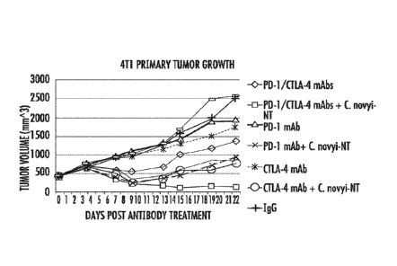
FIG. 2A and FIG. 2B show data from BALB/c mice bearing subcutaneous 4T1
tumors treated with an anti-CTLA-4 antibody and/or anti-PD-1 antibodies with
or
without C novyi-NT spores: A) tumor growth; and B) survival data;
FIG. 3A through FIG. 3C show the response to intratumoral C. novyi-NT
treatment in rat orthotopic brain tumor model: (A) Kaplan-Meier curves showing
survival of F344 Fisher rats after orthotopic implantation of a syngeneic
glioma cell
line (F98). Red line, C. novyi-NT spores injected into tumor 12-15 days after
tumor
implantation; black line, control; (B) bioluminescence (Xenogen imaging
system) in
three representative F344 Fisher rats after orthotopic implantation of F98
glioma cell
line. Images acquired on day 0 (pretreatment ¨ day of C. novyi-NT spore
injection),
day 1 after intratumoral injection of C. novyi-NT spores, and day 2 after
intratumoral
injection of C. novyi-NT spores; and (C) luciferase activity (millions) on day
0
(pretreatment), day 1 after intratumoral injection of C. novyi-NT spores, and
day 2
after intratumoral injection of C. novyi-NT spores;
FIG. 4A through FIG. 4D show germinated C. novyi-NT bacteria within
microscopic rat brain tumor lesions. Gram stain showed vegetative C. novyi-NT
bacteria (yellow arrowheads) localized in tumor (T) and stellate micro-
invasion (S),
but not in normal brain tissue (Br) of F344 Fisher rat: (A) interface of tumor
and
normal brain, scale bar 30 !um; (B) interface of tumor and normal brain, scale
bar 10
pm; (C) interface of normal brain, tumor, and stellate micro-invasion of
neoplastic
tissue, scale bar 30 pm; and (D) C. novyi-NT germination evident in stellate
micro-
invasive lesion, scale bar 10 jtm;
FIG. 5A through FIG. 5F show photographic and CT images from dog 11-R01
showing a partial response to C. novyi-NT therapy. Images span pre-treatment
to day
70 after first intratumoral dose of C. novyi-NT spores: (A) pre-treatment
image of the
peripheral nerve sheath tumor; (B) abscess formation on day 3 of the study,
with
extent confined to tumor; (C) medical debridement following spontaneous
abscess
3
CA 02944456 2016-09-29
WO 2015/153639
PCT/US2015/023633
rupture and discharge of necrotic and purulent material allowed healing by
second
intention.; (D) the wound had healed completely by day 70 of the study, and
77.6%
reduction in the largest diameter of the tumor was noted; (E) pre-treatment CT
image,
taken 4 days before first treatment showed extent of tumor (yellow circle) at
the
intersection of the pinna and cranium; and (F) post-treatment CT image on day
10 of
the study showed almost complete de-bulking of tumor;
FIG. 6A through FIG. 6F show photographic and CT images from dog 04-R03
showing a complete response to C. novyi-NT therapy. Images span pre-treatment
to
day 60 after first intratumoral dose of C. novyi-NT spores: (A) pre-treatment
image of
the soft tissue sarcoma; (B) tumor localized abscess formed on day 15 of the
study,
one day after a third dose of C. novyi-NT spores; (C) tumor de-bulking was
complete
by day 27 of the study and healthy granulation tissue had formed; (D) the
wound had
healed completely by day 60 of the study, and no residual tumor was noted
(complete
response); (E) pre-treatment CT image, taken 5 days before first treatment,
showing
extent of tumor (yellow circle) on antebrachium; and (F) post-treatment CT
image on
day 62 of the study showing complete loss of tumor mass;
FIG. 7 shows survival analysis of dogs treated with intratumoral injection of
C. novyi-NT. Kaplan-Meier curve showing time to progression of dogs that
experienced either a complete or partial response to intratumoral injected C
novyi-
NT. Dogs are censored if progression free at last known assessment;
FIG. 8A through FIG. 8D show CT and MRI images from the human patient:
(A) post-treatment CT with contrast on day 3 demonstrating evidence of intra-
and
extramedullary air collection; (B) pre-treatment MRI (Ti with gadolinium
contrast) of
the right upper humerus showing a contrast enhancing mass involving the soft
tissue
and possibly adjacent bone; (C) post-treatment MRI on day 4 demonstrating
diminished contrast enhancement in the tumor mass compared to baseline; and
(D)
post-treatment MRI on day 29 showing a homogeneous non-enhancing mass
consistent with ongoing necrosis. Tumor is highlighted with yellow arrowheads;
FIG. 9A through FIG. 9D show extensive tumor necrosis in the human patient
treated with C. novyi-NT spores: (A, B) pre-treatment tumor biopsy showing
viable
tumor (leiomyosarcoma) cells, scale bars 100 and 30 gm, respectively; and (C,
D)
post-treatment tumor biopsy, 4 days after intratumoral injection of C. novyi-
7'/T
spores, showing extensive necrosis of tumor cells, scale bars 100 and 30 gm,
respectively;
4
CA 02944456 2016-09-29
WO 2015/153639
PCT/US2015/023633
FIG. 10 shows summary data for samples sequenced; and
FIG. 11 shows somatic alterations in canine sarcomas.
DETAILED DESCRIPTION
The presently disclosed subject matter now will be described more fully
hereinafter with reference to the accompanying Figures, in which some, but not
all
embodiments of the inventions are shown. Like numbers refer to like elements
throughout. The presently disclosed subject matter may be embodied in many
different forms and should not be construed as limited to the embodiments set
forth
herein; rather, these embodiments are provided so that this disclosure will
satisfy
applicable legal requirements. Indeed, many modifications and other
embodiments of
the presently disclosed subject matter set forth herein will come to mind to
one skilled
in the art to which the presently disclosed subject matter pertains having the
benefit of
the teachings presented in the foregoing descriptions and the associated
Figures.
Therefore, it is to be understood that the presently disclosed subject matter
is not to be
limited to the specific embodiments disclosed and that modifications and other
embodiments are intended to be included within the scope of the appended
claims.
The presently disclosed subject matter provides methods and kits for treating
tumors. It was hypothesized that abrogation of the negative regulations
mediated
through the PD-1 and CTLA-4 pathways could enhance the anticancer immune
response elicited by an intratumoral bacterial infection, thus leading to
cures for
metastatic tumors. It has been shown herein below that by combining with an
anti-
CTLA-4 antibody and/or anti-PD-1 antibodies, the therapeutic effect of an
antitumor
bacterium is substantially enhanced. In a subcutaneous mouse tumor model,
essentially 100% of the tumors were eradicated by this approach. In a
metastatic
tumor model, the number of metastases was markedly reduced leading to
significant
survival benefit. In addition, in both tumor models, combining the anti-CTLA-4
and
anti-PD-1 antibodies resulted in better outcomes than using either of the
antibodies
alone.
Accordingly, the presently disclosed methods and kits use anti-CTLA-4 and/or
anti-PD-1 antibodies in combination with bacteria, bacterial products, or
other
immunoregulatory entities to antagonize the negative regulatory mechanisms of
the
antitumor immune responses induced by the immunoregulatory entities. In
addition,
5
CA 02944456 2016-09-29
WO 2015/153639
PCT/US2015/023633
the presently disclosed methods and kits can be used to treat cancer by
combining
anti-CTLA-4 and anti-PD-1 antibodies.
I. METHODS FOR TREATING CANCER
In some embodiments, the presently disclosed subject matter provides a
method for treating a solid tumor in a subject, the method comprising
administering to
the subject a therapeutically effective amount of at least one antibody
selected from
the group consisting of an anti-CTLA-4 antibody and an anti-PD-1 antibody
combined with at least one member of the group consisting of a bacterium,
bacterial
product, and an immunoregulatory entity, to treat the solid tumor. Examples of
antibodies that can be used in the presently disclosed methods include, but
are not
limited to, ipilimumab and tremelimumab against CTLA-4 and nivolumab against
PD-1.
CTLA-4 (Cytotoxic T-Lymphocyte Antigen 4; e.g., GenBank Accession No.
AAD00698.1), also known as CD152 (Cluster of Differentiation 152), is a T cell
surface molecule that is a negative regulator of T cell activation. CTLA-4 was
originally identified by differential screening of a murine cytolytic T cell
cDNA
library (Brunet et al. (1987) Nature 328:267-270). CTLA-4 is also a member of
the
immunoglobulin (Ig) superfamily and comprises a single extracellular Ig
domain.
CTLA-4 transcripts have been found in T cell populations having cytotoxic
activity,
suggesting that CTLA-4 might function in the cytolytic response (Brunet et al.
(1987)
Nature 328:267-270; Brunet et al. (1988) Immunol. Rev. 103-21-36). Researchers
have reported the cloning and mapping of a gene for the human counterpart of
CTLA-
4 (Dariavach et al. (1988) Eur. J. Immunol. 18:1901-1905) to the same
chromosomal
region (2q33-34) as CD28 (Lafage-Pochitaloff et al. (1990) Immunogenetics
31:198-
201). Sequence comparison between this human CTLA-4 DNA and that encoding
CD28 proteins reveals significant homology of sequence, with the greatest
degree of
homology in the juxtamembrane and cytoplasmic regions (Brunet et al. (1987)
Nature
328:267-270; Dariavach et al. (1988) Eur. J. Immunol. 18:1901-1905). Some
studies
have suggested that CTLA-4 has an analogous function as a secondary
costimulator
(Linsley et al. (1992) J. Exp. Med. 176:1595-1604; Wu et al. (1997) J. Exp.
Med.
185:1327-1335; U.S. Patent Nos. 5,977,318; 5,968,510; 5,885,796; and
5,885,579).
However, others have reported that CTLA-4 has an opposing role as a dampener
of T
cell activation (Krurnmel (1995) J Exp. Med. 182:459-465); Krurnmel et al.
(1996)
6
CA 02944456 2016-09-29
WO 2015/153639
PCT/US2015/023633
Immunol. 8:519-523; Chambers et al. (1997) immunity 7:885-895). It has been
reported that CTLA-4 deficient mice suffer from massive lymphoproliferation
(Chambers et al. (1997) Immunity 7:885-895). It has been reported that CTLA-4
blockade augments T cell responses in vitro (Walunas et al. (1994) Immunity
1:405-
413) and in vivo (Kearney (1995)1 Immunol. 155:1032-1036), exacerbates
antitumor
immunity (Leach (1996) Science 271 :1734-1736), and enhances an induced
autoimmune disease (Luhder (1998)1. Exp. Med. 187:427-432). It has also been
reported that CTLA-4 has an alternative or additional impact on the initial
character
of the T cell immune response (Chambers (1997) Curr. Opin. Immunol. 9:396-404;
Bluestone (1997) 1 Immunol. 158:1989-1993; Thompson (1997) Immunity 7:445-
450).
PD-1 (Programmed Cell Death Protein 1; e.g. GenBank Accession No.
NP 005009.2), also known as CD279 (Cluster of Differentiation 279), is a cell
surface membrane protein that is expressed mainly on a subset of activated T
lymphocytes, and that in humans is encoded by the PDCD1 gene (Entrez Gene
GeneID: 5133; see also Ishida et al. (1992) EMBO J. 11:3887; Shinohara et al.
(1994)
Genomics 23:704; U.S. Patent No. 5,698,520). PD-1 is a member of the
immunoglobulin gene superfamily, has an extracellular region containing
immunoglobulin superfamily domain, a transmembrane domain, and an
intracellular
region including an immunoreceptor tyrosine-based inhibitory motif (ITIM;
lshida et
al. (1992) EMBO J. 11:3887; Shinohara et al. (1994) Genomics 23:704). These
features also define a larger family of polypeptides, called the
immunoinhibitory
receptors, which also includes gp49B, PIR-B, and the killer inhibitory
receptors
(KIRs) (Vivier and Daeron (1997) Immunol. Today 18:286). It is often assumed
that
the tyrosyl phosphorylated ITIM motif of these receptors interacts with 5H2-
domain
containing phosphatases, which leads to inhibitory signals. A subset of these
immunoinhibitory receptors bind to MHC polypeptides, for example the KIRs, and
CTLA-4 bind to B7-1 and B7-2. It has been proposed that there is a
phylogenetic
relationship between the MHC and B7 genes (Henry et al. (1999) Immunol. Today
20(6):285-8). Like CTLA-4, PD-1 is rapidly induced on the surface of T-cells
in
response to anti-CD3 (Agata et al. (1996) Int. Immunol. 8:765). In contrast to
CTLA-
4, however, PD-1 is also induced on the surface of B-cells (in response to
anti-IgM).
PD-1 is also expressed on a subset of thymocytes and myeloid cells (Agata et
al.
(1996) Int. Immunol. 8:765; Nishimura et al. (1996) mt. Immunol. 8:773).
7
CA 02944456 2016-09-29
WO 2015/153639
PCT/US2015/023633
Two types of human PD-1 ligands have been identified: PDL1 and PDL2.
PD-1 ligands comprise a signal sequence, and an IgV domain, an IgC domain, a
transmembranc domain, and a short cytoplasmic tail. Both PDL1 (NCBI Reference
Sequence: NP_001254635.1; Freeman et al. (2000)J. Exp. Med. 192:1027) and PDL2
(NCBI Reference Sequence: NP_079515.2; Latchman et al. (2001) Nat. Immunol.
2:261) are members of the B7 family of polypeptides. Both PDL1 and PDL2 are
expressed in placenta, spleen, lymph nodes, thymus, and heart. Only PDL2 is
expressed in pancreas, lung and liver while only PDL1 is expressed in fetal
liver.
Both PD-1 ligands are upregulated on activated monocytes and dendritic cells.
The
fact that PD-1 binds to PDL1 and PDL2 places PD-1 in a family of inhibitory
receptors with CTLA-4.
"Functional variants" of CTLA-4 or PD-1 include functional fragments,
functional mutant proteins, and/or functional fusion proteins. A functional
variant of
a selected polypeptide refers to an isolated and/or recombinant protein or
polypeptide
which has at least one property, activity and/or functional characteristic of
the
selected polypeptide (e.g., CTLA-4 or PD-1). As used herein, the term
"activity,"
when used with respect to a polypeptide, e.g., CTLA-4 or PD-1, includes
activities
which are inherent in the structure of the wild-type protein.
For example, with respect to CTLA-4 or PD-1, the term "activity" includes the
ability of CTLA-4 or PD-1 to modulate an inhibitory signal in an activated
immune
cell, e.g., by engaging a natural CTLA-4 or PD-1 ligand on an antigen
presenting cell.
PD-1 transmits an inhibitory signal to an immune cell in a manner similar to
CTLA-4.
Modulation of an inhibitory signal in an immune cell results in modulation of
proliferation of, and/or cytokine secretion by, an immune cell. Thus, the term
"CTLA-4 activity" or "PD-1 activity" includes the ability of CTLA-4 or PD-1 to
bind
its natural ligand(s), the ability to modulate immune cell costimulatory or
inhibitory
signals, and the ability to modulate the immune response.
As used herein, the term "costimulate," as used with reference to activated
immune cells, includes the ability of a costimulatory polypeptide to provide a
second,
non-activating receptor mediated signal (a "costimulatory signal") that
induces
proliferation or effector function. For example, a costimulatory signal can
result in
cytokine secretion, e.g., in a T cell that has received a T cell-receptor-
mediated signal.
Immune cells that have received a cell-receptor mediated signal, e.g., via an
activating
receptor are referred to herein as "activated immune cells." As used herein,
the term
8
CA 02944456 2016-09-29
WO 2015/153639
PCT/US2015/023633
"costimulatory receptor" includes receptors which transmit a costimulatory
signal to a
immune cell. As used herein, the term "inhibitory receptors" includes
receptors which
transmit a negative signal to an immune cell (e.g., CTLA-4 or PD-1). An
inhibitory
signal as transduced by an inhibitory receptor can occur even if a
costimulatory
receptor (such as CD28) is not present on the immune cell and, thus, is not
simply a
function of competition between inhibitory receptors and costimulatory
receptors for
binding of costimulatory polypeptides (Fallarino et al. (1998) J. Exp. Med.
188:205).
Transmission of an inhibitory signal to an immune cell can result in
unresponsiveness
or anergy or programmed cell death in the immune cell. Preferably transmission
of an
inhibitory signal operates through a mechanism that does not involve
apoptosis. As
used herein the term "apoptosis" includes programmed cell death which can be
characterized using techniques which are known in the art. Apoptotic cell
death can
be characterized, e.g., by cell shrinkage, membrane blebbing and chromatin
condensation culminating in cell fragmentation. Cells undergoing apoptosis
also
display a characteristic pattern of internucleosomal DNA cleavage.
Generally, fragments or portions of CTLA-4 or PD-1 encompassed by the
presently disclosed subject matter include those having a deletion (i.e. one
or more
deletions) of an amino acid (i.e., one or more amino acids) relative to the
wild-type
CTLA-4 or PD-1 (such as N-terminal, C-terminal or internal deletions).
Fragments or
portions in which only contiguous amino acids have been deleted or in which
non-
contiguous amino acids have been deleted relative to wild-type CTLA-4 or PD-1
are
also envisioned. Generally, mutants or derivatives of CTLA-4 or PD-1
encompassed
by the present presently disclosed subject matter include natural or
artificial variants
differing by the addition, deletion and/or substitution of one or more
contiguous or
non-contiguous amino acid residues, or modified polypeptides in which one or
more
residues is modified, and mutants comprising one or more modified residues.
Preferred mutants are natural or artificial variants of CTLA-4 or PD-1
differing by the
addition, deletion and/or substitution of one or more contiguous or non-
contiguous
amino acid residues.
Generally, a functional variant of CTLA-4 or PD-1 has an amino acid
sequence which is at least about 80% identical, at least about 81% identical,
at least
about 82% identical, at least about 83% identical, at least about 84%
identical, at least
about 85% identical, at least about 86% identical, at least about 87%
identical, at least
about 88% identical, at least about 89% identical, at least about 90%
identical, at least
9
CA 02944456 2016-09-29
WO 2015/153639
PCT/US2015/023633
about 91% identical, at least about 92% identical, at least about 93%
identical, at least
about 94% identical, at least about 95% identical, at least about 96%
identical, at least
about 97% identical, at least about 98% identical, or at least about 99%
identical to
the wild-type amino acid sequence for CTLA-4 or PD-1 over the length of the
variant.
"Sequence identity" or "identity" in the context of proteins or polypeptides
refers to the amino acid residues in two amino acid sequences that are the
same when
aligned for maximum correspondence over a specified comparison window.
Thus, "percentage of sequence identity" refers to the value determined by
comparing two optimally aligned sequences over a comparison window, wherein
the
portion of the amino acid sequence in the comparison window may comprise
additions or deletions (i.e., gaps) as compared to the reference sequence
(which does
not comprise additions or deletions) for optimal alignment of the two
sequences. The
percentage is calculated by determining the number of positions at which the
identical
amino acid residue occurs in both sequences to yield the number of matched
positions, dividing the number of matched positions by the total number of
positions
in the window of comparison and multiplying the results by 100 to yield the
percentage of sequence identity. Useful examples of percent sequence
identities
include, but are not limited to, 50%, 55%, 60%, 65%, 70%, 75%, 80%, 85%, 90%,
or
95%, or any integer percentage from 50% to 100%. These identities can be
determined using any of the programs described herein.
Sequence alignments and percent identity or similarity calculations may be
determined using a variety of comparison methods designed to detect homologous
sequences including, but not limited to, the MegAlignTM program of the
LASERGENE bioinformatics computing suite (DNASTAR Inc., Madison, Wis.).
Within the context of this application it will be understood that where
sequence
analysis software is used for analysis, that the results of the analysis will
be based on
the "default values" of the program referenced, unless otherwise specified. As
used
herein "default values" will mean any set of values or parameters that
originally load
with the software when first initialized. The "Clustal V method of alignment"
corresponds to the alignment method labeled Clustal V (described by Higgins
and
Sharp (1989) (ABIOS 5:151-153; Higgins et al. (1992) Comput. Appl. Biosei.
8:189-
191) and found in the MegAlignTM program of the LASERGENE bioinformatics
computing suite (DNASTAR Inc., Madison, Wis.).
CA 02944456 2016-09-29
WO 2015/153639
PCT/US2015/023633
It is well understood by one skilled in the art that many levels of sequence
identity are useful in identifying proteins or polypeptides (e.g., from other
species)
wherein the proteins or polypeptides have the same or similar function or
activity.
Useful examples of percent identities include, but are not limited to, 50%,
55%, 60%,
65%, 70%, 75%, 80%, 85%, 90%, or 95%, or any integer percentage from 50% to
100%. Indeed, any integer amino acid identity from 50% to 100% may be useful
in
describing the present presently disclosed subject matter, such as 51%, 52%,
53%,
54%, 55%, 56%, 57%, 58%, 59%, 60%, 61%, 62%, 63%, 64%, 65%, 66%, 67%,
68%, 69%, 70%, 71%, 72%, 73%, 74%, 75%, 76%, 77%, 78%, 79%, 80%, 81%,
82%, 83%, 84%, 85%, 86%, 87%, 88%, 89%, 90%, 91%, 92%, 93%, 94%, 95%,
96%, 97%, 98% or 99%.
The term "antibody," also known as an immunoglobulin (Ig), is a large Y-
shaped protein produced by B cells that is used by the immune system to
identify and
neutralize foreign objects such as bacteria and viruses by recognizing a
unique portion
(epitope) of the foreign target, called an antigen. As used herein, the term
"antibody"
also includes an "antigen-binding portion" of an antibody (or simply "antibody
portion"). The term "antigen-binding portion," as used herein, refers to one
or more
fragments of an antibody that retain the ability to specifically bind to an
antigen (e.g.,
PD-1 or CTLA-4). It has been shown that the antigen-binding function of an
antibody
can be performed by fragments of a full-length antibody. Examples of binding
fragments encompassed within the term "antigen-binding portion" of an antibody
include: (i) a Fab fragment, a monovalent fragment consisting of the VL, VH,
CL and
CHI domains; (ii) a F(ab')2 fragment, a bivalent fragment comprising two Fab
fragments linked by a disulfide bridge at the hinge region; (iii) a Fd
fragment
consisting of the VH and CHI domains; (iv) a Fv fragment consisting of the VL
and
VH domains of a single arm of an antibody; (v) a dAb fragment (Ward et al.
(1989)
Nature 341:544-546), which consists of a VH domain; and (vi) an isolated
complementarity determining region (CDR). Furthermore, although the two
domains
of the Fv fragment, VL and VH, are coded for by separate genes, they can be
joined,
using recombinant methods, by a synthetic linker that enables them to be made
as a
single protein chain in which the VL and VH regions pair to form monovalent
polypeptides (known as single chain Fv (scFv); e.g., Bird et al. (1988)
Science
242:423-426; Huston et al. (1988) Proc. Natl. Acad Sci. USA 85:5879-5883; and
Osbourn et al. (1998) Nature Biotechnology 16:778). Such single chain
antibodies
11
CA 02944456 2016-09-29
WO 2015/153639
PCT/US2015/023633
are also intended to be encompassed within the term "antigen-binding portion"
of an
antibody. Any VH and VL sequences of specific scFv can be linked to human
immunoglobulin constant region cDNA or genomic sequences, in order to generate
expression vectors encoding complete IgG polypeptides or other isotypes. VH
and
V1 can also be used in the generation of Fab, Fv or other fragments of
immunoglobulins using either protein chemistry or recombinant DNA technology.
Other forms of single chain antibodies, such as diabodies are also
encompassed.
Diabodies are bivalent, bispecific antibodies in which VH and VL domains are
expressed on a single polypeptide chain, but using a linker that is too short
to allow
for pairing between the two domains on the same chain, thereby forcing the
domains
to pair with complementary domains of another chain and creating two antigen
binding sites (e.g., Holliger et al. (1993) Proc. Natl. Acad. Sci. USA 90:6444-
6448;
Poljak et a/. (1994) Structure 2:1121-1123).
Still further, an antibody or antigen-binding portion thereof may be part of
larger immunoadhesion polypeptides, formed by covalent or noncovalent
association
of the antibody or antibody portion with one or more other proteins or
peptides.
Examples of such immunoadhesion polypeptides include use of the streptavidin
core
region to make a tetrameric scFv polypeptide (Kipriyanov et al. (1995) Human
Antibodies and Hybridomas 6:93-101) and use of a cysteine residue, a marker
peptide
and a C-terminal polyhistidine tag to make bivalent and biotinylated scFv
polypeptides (Kipriyanov et al. (1994) Ho!. Immunol. 31:1047-1058). Antibody
portions, such as Fab and F(ab`)2 fragments, can be prepared from whole
antibodies
using conventional techniques, such as papain or pepsin digestion,
respectively, of
whole antibodies. Moreover, antibodies, antibody portions and immunoadhesion
polypeptides can be obtained using standard recombinant DNA techniques, as
described herein.
Antibodies may be polyclonal or monoclonal; xenogeneic, allogeneic, or
syngeneic; or modified forms thereof (e.g. humanized, chimeric, etc.).
Antibodies
may also be fully human. Preferably, antibodies of the presently disclosed
subject
matter bind specifically or substantially specifically to PD-1 or CTLA-4 or
functional
variants thereof. The terms "monoclonal antibodies" and "monoclonal antibody
composition," as used herein, refer to a population of antibody polypeptides
that
contain only one species of an antigen binding site capable of immunoreacting
with a
particular epitope of an antigen, whereas the term "polyclonal antibodies" and
12
CA 02944456 2016-09-29
WO 2015/153639
PCT/US2015/023633
"polyclonal antibody composition" refer to a population of antibody
polypeptides that
contain multiple species of antigen binding sites capable of interacting with
a
particular antigen. A monoclonal antibody composition typically displays a
single
binding affinity for a particular antigen with which it immunoreacts.
The term "humanized antibody", as used herein, is intended to include
antibodies made by a non-human cell having variable and constant regions which
have been altered to more closely resemble antibodies that would be made by a
human cell. For example, by altering the non-human antibody amino acid
sequence
to incorporate amino acids found in human germline immunoglobulin sequences.
The
humanized antibodies of the presently disclosed subject matter may include
amino
acid residues not encoded by human germline immunoglobulin sequences (e.g.,
mutations introduced by random or site-specific mutagenesis in vitro or by
somatic
mutation in vivo), for example in the CDRs. The term "humanized antibody", as
used
herein, also includes antibodies in which CDR sequences derived from the
germline
of another mammalian species, such as a mouse, have been grafted onto human
framework sequences.
An "isolated antibody", as used herein, is intended to refer to an antibody
that
is substantially free of other antibodies having different antigenic
specificities (e.g., an
isolated antibody that specifically binds CTLA-4 or PD-1 is substantially free
of
antibodies that specifically bind antigens other than CTLA-4 or PD-1.
Moreover, an
isolated antibody may be substantially free of other cellular material and/or
chemicals.
An isolated CTLA-4 or PD-1 or functional variant thereof (or a nucleic acid
encoding such polypeptides), can be used as an immunogen to generate
antibodies
that bind to the respective CTLA-4 or PD-1 or functional variant thereof using
standard techniques for polyclonal and monoclonal antibody preparation. A full-
length CTLA-4 or PD-1 can be used, or alternatively, the presently disclosed
subject
matter relates to antigenic peptide fragments of CTLA-4 or PD-1 ligands or
functional
variants thereof for use as immunogens. An antigenic peptide of a CTLA-4 or PD-
1
or a functional variant thereof comprises at least 8 amino acid residues and
encompasses an epitope present in the respective full length molecule such
that an
antibody raised against the peptide forms a specific immune complex with the
respective full length molecule. Preferably, the antigenic peptide comprises
at least 10
amino acid residues, more preferably at least 15 amino acid residues, even
more
preferably at least 20 amino acid residues, and most preferably at least 30
amino acid
13
CA 02944456 2016-09-29
WO 2015/153639
PCT/US2015/023633
residues. Preferred epitopes encompassed by the antigenic peptides are regions
of a
CTLA-4 or PD-1 or a functional variant thereof that are located on the surface
of the
protein, e.g., hydrophilic regions. A standard hydrophobicity analysis of the
polypeptide molecule can be performed to identify hydrophilic regions. Highly
preferred epitopes encompassed by the antigenic peptides are the regions of
the
polypeptide molecule which are in the extracellular domain, and therefore are
involved in binding. In one embodiment such epitopes can be specific for a
given
polypeptide molecule from one species, such as mouse or human (i.e., an
antigenic
peptide that spans a region of the polypeptide molecule that is not conserved
across
species is used as immunogen; such non conserved residues can be determined
using
an alignment such as that provided herein).
An immunogen comprising CTLA-4 or PD-1 or a functional variant thereof
typically is used to prepare antibodies by immunizing a suitable subject,
(e.g., rabbit,
goat, mouse or other mammal) with the immunogen. An appropriate immunogenic
preparation can contain, for example, a recombinantly expressed or chemically
synthesized molecule or fragment thereof to which the immune response is to be
generated. The preparation can further include an adjuvant, such as Freund's
complete or incomplete adjuvant, or similar immunostimulatory agent.
Immunization
of a suitable subject with an immunogenic preparation induces a polyclonal
antibody
response to the antigenic peptide contained therein.
Polyclonal antibodies can be prepared as described above by immunizing a
suitable subject with a polypeptide immunogen. The polypeptide antibody titer
in the
immunized subject can be monitored over time by standard techniques, such as
with
an enzyme linked immunosorbent assay (ELISA) using immobilized polypeptide. If
desired, the antibody directed against the antigen can be isolated from the
mammal
(e.g., from the blood) and further purified by well known techniques, such as
protein
A chromatography to obtain the IgG fraction. At an appropriate time after
immunization, e.g., when the antibody titers are highest, antibody-producing
cells can
be obtained from the subject and used to prepare monoclonal antibodies by
standard
techniques, such as the hybridoma technique originally described by Kohler and
Milstein (1975) Nature 256:495-497; Brown et al. (1981) J. Immunol. 127:539-
46;
Brown et al. (1980) J. Biol. Chem. 255:4980-83; Yeb et al. (1976) Proc. Natl.
Acad.
Sci. 76:2927-31; and Yeh et al. (1982) Int. J. Cancer 29:269-75), a human B
cell
hybridoma technique (Kozbor et al. (1983) Immunol. Toddy 4:72), the EBV-
14
CA 02944456 2016-09-29
WO 2015/153639
PCT/US2015/023633
hybridoma technique (Cole et al. (1985) Monoclonal Antibodies and Cancer
Therapy,
Alan R. Liss, Inc., pp. 77-96) or trioma techniques. The technology for
producing
monoclonal antibody hybridomas is well known (see generally Kenneth, R. H. in
Monoclonal Antibodies: A New Dimension In Biological Analyses, Plenum
Publishing
Corp., New York, N.Y. (1980); Lerner (1981) Yale J. Biol. Med. 54:387-402;
Gefter
etal. (1977) Somatic Cell Genet. 3:231-36). Briefly, an immortal cell line
(typically a
myeloma) is fused to lymphocytes (typically splenocytes) from a mammal
immunized
with an immunogen as described above, and the culture supernatants of the
resulting
hybridoma cells are screened to identify a hybridoma producing a monoclonal
antibody that binds to the polypeptide antigen, preferably specifically.
Any of the many well known protocols used for fusing lymphocytes and
immortalized cell lines can be applied for the purpose of generating an anti-
PD-1
ligand monoclonal antibody (e.g., Galfre, G. etal. (1977) Nature 266:55052;
Kenneth, R. H. in Monoclonal Antibodies: A New Dimension In Biological
Analyses,
Plenum Publishing Corp., New York, N.Y. (1980); Lerner (1981) Yale J. Biol.
Med.
54:387-402; Gefter etal. (1977) Somatic Cell Genet. 3:231-36). Moreover, the
ordinary skilled worker will appreciate that there are many variations of such
methods
which also would be useful. Typically, the immortal cell line (e.g., a myeloma
cell
line) is derived from the same mammalian species as the lymphocytes. For
example,
murine hybridomas can be made by fusing lymphocytes from a mouse immunized
with an immunogenic preparation of the present presently disclosed subject
matter
with an immortalized mouse cell line. Preferred immortal cell lines are mouse
myeloma cell lines that are sensitive to culture medium containing
hypoxanthine,
aminopterin and thymidine ("HAT medium"). Any of a number of myeloma cell
lines
can be used as a fusion partner according to standard techniques, e.g., the P3-
NS1/1-
Ag4-1, P3-x63-Ag8.653 or Sp2/0-Ag14 myeloma lines. These myeloma lines are
available from the American Type Culture Collection (ATCC), Rockville, Md.
Typically, HAT-sensitive mouse myeloma cells are fused to mouse splenocytes
using
polyethylene glycol ("PEG"). Hybridoma cells resulting from the fusion are
then
selected using HAT medium, which kills unfused and unproductively fused
myeloma
cells (unfused splenocytes die after several days because they are not
transformed).
Hybridoma cells producing a monoclonal antibody of the presently disclosed
subject
matter are detected by screening the hybridoma culture supernatants for
antibodies
that bind a given polypeptide, e.g., using a standard ELISA assay.
CA 02944456 2016-09-29
WO 2015/153639
PCT/US2015/023633
As an alternative to preparing monoclonal antibody-secreting hybridomas, a
monoclonal specific for one of the above described polypeptides antibody can
be
identified and isolated by screening a recombinant combinatorial
immunoglobulin
library (e.g., an antibody phage display library) with the appropriate
polypeptide to
thereby isolate immunoglobulin library members that bind the polypeptide. Kits
for
generating and screening phage display libraries are commercially available
(e.g., the
Pharmacia Recombinant Phage Antibody System, Catalog No. 27-9400-01; and the
Stratagene SurJZAPTM Phage Display Kit, Catalog No. 240612). Additionally,
examples of methods and reagents particularly amenable for use in generating
and
screening an antibody display library can be found in, for example, U.S.
Patent No.
5,223,409; PCT Patent App. Pub. No. WO 92/18619; PCT Patent App. Pub. No. WO
91/17271; PCT Patent App. Pub. No. 92/20791; PCT Patent App. Pub. No. WO
92/15679; PCT Patent App. Pub. No. WO 93/01288; PCT Patent App. Pub. No. WO
92/01047; PCT Patent App. Pub. No. WO 92/09690; PCT Patent App. Pub. No. WO
90/02809; Fuchs et al. (1991) Biotechnology (NY) 9:1369-1372; Hay et al.
(1992)
Hum. Antibod. Hybridomas 3:81-85; Huse et al. (1989) Science 246:1275-1281;
Griffiths et al. (1993) E/14-B0 J. 12:725-734; Hawkins et al. (1992) J. _WI.
Biol.
226:889-896; Clarkson et al. (1991) Nature 352:624-628; Gram et al. (1992)
Proc.
Natl. Acad. Sci. USA 89:3576-3580; Garrard et al. (1991) Biotechnology (NY)
9:1373-
1377; Hoogenboom et al. (1991) Nucleic Acids Res. 19:4133-4137; Barbas et al.
(1991) Proc. Natl. Acad. Sci. USA 88:7978-7982; and McCafferty et al. (1990)
Nature 348:552-554.
Additionally, recombinant anti-CTLA-4 antibodies or anti-PD-1 antibodies,
such as chimeric and humanized monoclonal antibodies, comprising both human
and
non-human portions, which can be made using standard recombinant DNA
techniques, are within the scope of the presently disclosed subject matter.
Such
chimeric and humanized monoclonal antibodies can be produced by recombinant
DNA techniques known in the art, for example using methods described in PCT
Patent App. Pub. No. PCT/US86/02269; European Patent App. No. 184,187;
European Patent App. No. 171,496; European Patent App. No. 173,494; PCT
Application WO 86/01533; U.S. Patent No. 4,816,567; European Patent App. No.
125,023; Better et al. (1988) Science 240:1041-1043; Liu et al. (1987) Proc.
Natl.
Acad. Sci. USA 84:3439-3443; Liu et al. (1987) J. Immunol. 139:3521-3526; Sun
et
al. (1987) Proc. Natl. Acad. Sci. 84:214-218; Nishimura et al. (1987) Cancer
Res.
16
CA 02944456 2016-09-29
WO 2015/153639
PCT/US2015/023633
47:999-1005; Wood et al. (1985) Nature 314:446-449; and Shaw et al. (1988)1
Natl.
Cancer Inst. 80:1553-1559); Morrison, S. L. (1985) Science 229:1202-1207; Oi
et al.
(1986) Biotechniques 4:214; U.S. Patent No. 5,225,539; Jones et al. (1986)
Nature
321:552-525; Verhoeyan et al. (1988) Science 239:1534; and Beidler et al.
(1988)J.
Immunol. 141:4053-4060.
In addition, humanized antibodies can be made according to standard
protocols such as those disclosed in U.S. Patent No. 5,565,332. In another
embodiment, antibody chains or specific binding pair members can be produced
by
recombination between vectors comprising nucleic acid molecules encoding a
fusion
of a polypeptide chain of a specific binding pair member and a component of a
replicable generic display package and vectors containing nucleic acid
molecules
encoding a second polypeptide chain of a single binding pair member using
techniques known in the art, e.g., as described in U.S. Pat. Nos. 5,565,332,
5,871,907,
or 5,733,743. The use of intracellular antibodies to inhibit protein function
in a cell is
also known in the art (e.g., Carlson (1988) Mol. Cell. Biol. 8:2638-2646;
Biocca et al.
(1990) EMBO J. 9:101-108; Werge etal. (1990) FEBS Lett. 274:193-198; Carlson
(1993) Proc. Natl. Acad. Sci. USA 90:7427-7428; Marasco et al. (1993) Proc.
NatL
Acad. Sci. USA 90:7889-7893; Biocca etal. (1994) Biotechnology (N)) 12:396-
399;
Chen et al. (1994) Hum. Gene Ther. 5:595-601; Duan etal. (1994) Proc. IVatl.
Acad.
Sci. USA 91:5075-5079; Chen etal. (1994) Proc. Natl. Acad. Sci. USA 91:5932-
5936;
Beerli et al. (1994)1 Biol. Chem. 269:23931-23936; Beerli et al. (1994)
Biochem.
Biophys. Res. Commun. 204:666-672; Mhashilkar et a/. (1995) EMBO J. 14:1542-
1551; Richardson et al. (1995) Proc. Natl. Acad. Sci. USA 92:3137-3141; PCT
Publication No. WO 94/02610; and PCT Publication No. WO 95/03832).
Additionally, fully human antibodies could be made against CTLA-4 or PD-1
or a functional variant thereof. Fully human antibodies can be made in mice
that are
transgenic for human immunoglobulin genes, e.g. according to Hogan, et al.,
"Manipulating the Mouse Embryo: A Laboratory Manual," Cold Spring Harbor
Laboratory. Briefly, transgenic mice are immunized with purified CTLA-4 or PD-
1
or a functional variant thereof. Spleen cells are harvested and fused to
myeloma cells
to produce hybridomas. Hybridomas are selected based on their ability to
produce
antibodies which bind to CTLA-4 or PD-1 or a functional variant thereof. Fully
human antibodies would reduce the immunogenicity of such antibodies in a
human.
17
CA 02944456 2016-09-29
WO 2015/153639
PCT/US2015/023633
In one embodiment, an antibody for use in the instant presently disclosed
subject matter is a bispecific antibody. A bispecific antibody has binding
sites for two
different antigens within a single antibody polypeptide. Antigen binding may
be
simultaneous or sequential. Triomas and hybrid hybridomas are two examples of
cell
lines that can secrete bispecific antibodies. Examples of bispecific
antibodies
produced by a hybrid hybridoma or a trioma are disclosed in U.S. Patent No.
4,474,893. Bispecific antibodies have been constructed by chemical means
(Staerz et
al. (1985) Nature 314:628, and Perez et al. (1985) Nature 316:354) and
hybridoma
technology (Staerz and Bevan (1986) Proc. Natl. Acad. Sci. USA, 83:1453, and
Staerz
and Bevan (1986) Immunol. Today 7:241). Bispecific antibodies are also
described in
U.S. Patent No. 5,959,084. Fragments of bispecific antibodies are described in
U.S.
Patent No. 5,798,229.
Bispecific agents can also be generated by making heterohybridomas by
fusing hybridomas or other cells making different antibodies, followed by
identification of clones producing and co-assembling both antibodies. They can
also
be generated by chemical or genetic conjugation of complete immunoglobulin
chains
or portions thereof such as Fab and Fv sequences. The antibody component can
bind
to CTLA-4 or PD-1 or a functional variant thereof. In one embodiment, the
bispecific
antibody could specifically bind to both PD-1 ligand or a functional variant
thereof
and a PD-1 polypeptide or a functional variant thereof.
Yet another aspect of the presently disclosed subject matter pertains to
antibodies that are obtainable by a process comprising, immunizing an animal
with an
immunogenic CTLA-4 or PD-1 or a functional variant thereof, or an immunogenic
portion thereof unique to the CTLA-4 or PD-1, and then isolating from the
animal
antibodies that specifically bind to the polypeptide.
In some embodiments, the presently disclosed subject matter provides a
method to treat a solid tumor using a bacterium, bacterial product, and/or
other
immunoregulatory entity. In other embodiments, the bacterium or bacterial
product
thereof is an anaerobic bacterium or bacterial product thereof. Suitable
genera
include but are not limited to Bifidobacteria, Lactobacilli, and Clostridia,
such as
Clostridium novyi or Clostridium sordellii (C. sordellii). In still other
embodiments,
the bacterium or bacterial product thereof is an obligate anaerobic bacterium
or
bacterial product thereof. An "anaerobic bacterium" as used herein is a
bacterium that
does not require oxygen for growth. An "obligate anaerobic bacterium" as used
18
CA 02944456 2016-09-29
WO 2015/153639
PCT/US2015/023633
herein is a bacterium that not only does not require oxygen for growth, but is
harmed
by normal levels of atmospheric oxygen. In further embodiments, the anaerobic
bacterium or bacterial product thereof is Clostridium novyi or bacterial
product
thereof.
In some embodiments, the bacterium or bacterial product thereof is a toxin-
depleted, anaerobic bacterium or bacterial product thereof. In other
embodiments, the
toxin-depleted, anaerobic bacterium or bacterial product thereof is
Clostridium novyi-
NT or bacterial product thereof
Decreasing the natural production of toxins is desirable in using bacteria
therapeutically. While toxin-depleted strains need not be totally non-
toxigenic, it is
desirable that at least one of the toxin genes is mutated, deleted, or
otherwise
inactivated to render the bacteria less harmful to the subject. As used
herein, the term
"toxic" refers to acting as or having the effect of a poison. Preferably the
toxicity is
reduced by a factor of at least 2, 5, 10, 50, 100, 1000, or more. If a toxin
gene is
episomal or on a bacteriophage, then curing of the episome or bacteriophage
can be
used to delete the toxin gene. Techniques are well known in the art for
mutagenesis,
curing, and screening of mutants.
In some embodiments, part of or all of a toxin gene of a wild-type form of the
toxin-depleted, anaerobic bacterium or bacterial product thereof is deleted to
produce
a "toxin-depleted" bacterium or bacterial product thereof. For example, the
lethal a-
toxin gene is deleted in C. novyi-NT. In other embodiments, the toxicity of
the toxin-
depleted, anaerobic bacterium is reduced by a factor of at least 2 compared to
a
corresponding wild-type bacterium. In still other embodiments, the toxicity of
the
toxin-depleted, Clostridium novyi is reduced by a factor of at least 2
compared to a
corresponding Clostridium novyi. The term "wild-type" as used herein refers to
the
normal, non-mutated version, such as of a bacterium or a gene. The term
"deletion"
as used herein refers to a change in nucleotide sequence wherein one or more
nucleotides are removed.
In some embodiments, the bacterial product is at least one bacterial membrane
component. Bacterial membrane components may include, for example, bacterial
membrane proteins attached to or associated with the membrane of Clostridium
novyi,
suitably a protein having a domain which is considered to be exposed on the
outside
of the bacterium and thus visible to the immune system of a human when
infected
with the bacteria. Reference to a bacterial membrane protein herein includes
variants
19
CA 02944456 2016-09-29
WO 2015/153639
PCT/US2015/023633
of naturally occurring bacterial membrane proteins such as deletion,
insertion, and
substitution mutations of a given bacterial membrane protein or to a protein
that has
an amino acid sequence which is at least about 80% identical, at least about
81%
identical, at least about 82% identical, at least about 83% identical, at
least about 84%
identical, at least about 85% identical, at least about 86% identical, at
least about 87%
identical, at least about 88% identical, at least about 89% identical, at
least about 90%
identical, at least about 91% identical, at least about 92% identical, at
least about 93%
identical, at least about 94% identical, at least about 95% identical, at
least about 96%
identical, at least about 97% identical, at least about 98% identical, or at
least about
99% identical to the wild-type amino acid sequence for a given bacterial
membrane
over the length of the variant, the variant being suitably immunogenic.
In some embodiments, other immunoregulatory entities can be combined with
antibodies against CTLA-4 and/or PD-1. Such immunoregulatory entities may
include, for example, immunostimulatory cytokines such as GM-CSF, Interleukin-
12
(IL-12), and IL-15. Additional examples for bacterial products used for
immunostimulatory purposes include inactivated bacteria or bacterial
components
such as Freund's complete adjuvant and Coley's toxin.
In some embodiments, at least one member of the group consisting of a
bacterium, bacterial product, and an immunoregulatory entity is administered
intravenously or intratumorally. In other embodiments, at least one antibody
is
administered by at least one method selected from the group consisting of
intravenously, intramuscularly, subcutaneously, and intratumorally.
A "cancer" in a subject refers to the presence of cells possessing
characteristics typical of cancer-causing cells, for example, uncontrolled
proliferation,
loss of specialized functions, immortality, significant metastatic potential,
significant
increase in anti-apoptotic activity, rapid growth and proliferation rate, and
certain
characteristic morphology and cellular markers. In some circumstances, cancer
cells
will be in the form of a tumor; such cells may exist locally within an animal,
or
circulate in the blood stream as independent cells, for example, leukemic
cells. A
cancer can include, but is not limited to, head cancer, neck cancer, head and
neck
cancer, lung cancer, breast cancer, prostate cancer, colorectal cancer,
esophageal
cancer, stomach cancer, leukemia/lymphoma, uterine cancer, skin cancer,
endocrine
cancer, urinary cancer, pancreatic cancer, gastrointestinal cancer, ovarian
cancer,
cervical cancer, and adenomas. A "tumor," as used herein, refers to all
neoplastic cell
CA 02944456 2016-09-29
WO 2015/153639
PCT/US2015/023633
growth and proliferation, whether malignant or benign, and all precancerous
and
cancerous cells and tissues. A "solid tumor", as used herein, is an abnormal
mass of
tissue that generally does not contain cysts or liquid areas. A solid tumor
may be in
the brain, colon, breasts, prostate, liver, kidneys, lungs, esophagus, head
and neck,
.. ovaries, cervix, stomach, colon, rectum, bladder, uterus, testes, and
pancreas, as non-
limiting examples. In some embodiments, the solid tumor regresses or its
growth is
slowed or arrested after the solid tumor is treated with the presently
disclosed
methods. In other embodiments, the solid tumor is malignant.
In some embodiments, the presently disclosed subject matter provides a
.. method of treating cancer in a subject, the method comprising administering
to the
subject a therapeutically effective amount of a combination of at least one
anti-CTLA-
4 antibody and at least one anti-PD-lantibody to treat the cancer. It has been
found
that the combination of anti-CTLA-4 and anti-PD-lantibodies to treat the
cancer
results in a better outcome than if the antibodies are administered
separately. In other
.. embodiments, the combination of anti-CTLA-4 and anti-PD-Iantibodies is
administered by at least one method selected from the group consisting of
intravenously, intramuscularly, subcutaneously, and intratumorally.
As used herein, the term "treating" can include reversing, alleviating,
inhibiting the progression of, preventing or reducing the likelihood of the
disease,
.. disorder, or condition to which such term applies, or one or more symptoms
or
manifestations of such disease, disorder or condition.
The subject treated by the presently disclosed methods in their many
embodiments is desirably a human subject, although it is to be understood that
the
methods described herein are effective with respect to all vertebrate species,
which
.. are intended to be included in the term "subject." Accordingly, a "subject"
can
include a human subject for medical purposes, such as for the treatment of an
existing
condition or disease or the prophylactic treatment for preventing the onset of
a
condition or disease, or an animal subject for medical, veterinary purposes,
or
developmental purposes. Suitable animal subjects include mammals including,
but
not limited to, primates, e.g., humans, monkeys, apes, and the like; bovines,
e.g.,
cattle, oxen, and the like; ovines, e.g., sheep and the like; caprines, e.g.,
goats and the
like; porcines, e.g., pigs, hogs, and the like; equines, e.g., horses,
donkeys, zebras, and
the like; felines, including wild and domestic cats; canines, including dogs;
lagomorphs, including rabbits, hares, and the like; and rodents, including
mice, rats,
21
CA 02944456 2016-09-29
WO 2015/153639
PCT/US2015/023633
and the like. An animal may be a transgenic animal. In some embodiments, the
subject is a human including, but not limited to, fetal, neonatal, infant,
juvenile, and
adult subjects. Further, a "subject" can include a patient afflicted with or
suspected of
being afflicted with a condition or disease. Thus, the terms "subject" and
"patient"
are used interchangeably herein.
More particularly, as described herein, the presently disclosed compositions
comprising at least one antibody selected from the group consisting of an anti-
CTLA-
4 antibody and an anti-PD-1 antibody combined with at least one member of the
group consisting of a bacterium, bacterial product, and an immunoregulatory
entity
can be administered to a subject for therapy by any suitable route of
administration,
including orally, nasally, transmucosally, ocularly, rectally, intravaginally,
parenterally, including intramuscular, subcutaneous, intramedullary
injections, as well
as intrathecal, direct intraventricular, intravenous, intra-articular, intra-
sternal, intra-
synovial, intra-hepatic, intralesional, intracranial, intraperitoneal,
intranasal, or
intraocular injections, intracisternally, topically, as by powders, ointments
or drops
(including eyedrops), including buccally and sublingually, transdermally,
through an
inhalation spray, or other modes of delivery known in the art. The presently
disclosed
compositions can also be administered intratumorally, such that the
compositions are
directly administered into a solid tumor, such as by injection or other means.
The phrases "systemic administration," "administered systemically,"
"peripheral administration" and "administered peripherally" as used herein
mean the
administration of compositions comprising at least one antibody selected from
the
group consisting of an anti-CTLA-4 antibody and an anti-PD-1 antibody combined
with at least one member of the group consisting of a bacterium, bacterial
product,
and an immunoregulatory entity , a compound, drug or other material other than
directly into the central nervous system, such that it enters the patient's
system and,
thus, is subject to metabolism and other like processes, for example,
subcutaneous
administration.
The phrases "parenteral administration" and "administered parenterally" as
used herein mean modes of administration other than enteral and topical
administration, usually by injection, and includes, without limitation,
intravenous,
intramuscular, intarterial, intrathecal, intracapsular, intraorbital,
intraocular,
intracardiac, intradermal, intraperitoneal, transtracheal, subcutaneous,
subcuticular,
22
CA 02944456 2016-09-29
WO 2015/153639
PCT/US2015/023633
intraarticular, subcapsular, subarachnoid, intraspinal and intrasternal
injection and
infusion.
The presently disclosed pharmaceutical compositions can be manufactured in
a manner known in the art, e.g. by means of conventional mixing, dissolving,
granulating, dragee-making, levitating, emulsifying, encapsulating, entrapping
or
lyophilizing processes.
In some embodiments, the presently disclosed pharmaceutical compositions
can be administered by rechargeable or biodegradable devices. For example, a
variety
of slow-release polymeric devices have been developed and tested in vivo for
the
controlled delivery of drugs, including proteinacious biopharmaceuticals.
Suitable
examples of sustained release preparations include semipermeable polymer
matrices
in the form of shaped articles, e.g., films or microcapsules. Sustained
release matrices
include polyesters, hydrogels, polylactides (U.S. Patent No. 3,773,919; EP
58,481),
copolymers of L-glutamic acid and gamma ethyl-L-glutamate (Sidman et al.,
Biopolymers 22:547, 1983), poly (2-hydroxyethyl-metbacrylate) (Langer et al.
(1981)
J. Biomed. Mater. Res. 15:167; Langer (1982), Chem. Tech. 12:98), ethylene
vinyl
acetate (Langer et al. (1981) J. Biomed. Mater. Res. 15:167), or poly-D-(-)-3-
hydroxybutyric acid (EP 133,988A). Sustained release compositions also include
liposomally entrapped compositions comprising at least one antibody selected
from
the group consisting of an anti-CTLA-4 antibody and an anti-PD-1 antibody
combined with at least one member of the group consisting of a bacterium,
bacterial
product, and an immunoregulatory entity which can be prepared by methods known
per se (Epstein et al. (1985) Proc. Natl. Acad. Sci. U.S.A. 82:3688; Hwang et
al.
(1980) Proc. Natl. Acad. Sci. U.S.A. 77:4030; U.S. Patent Nos. 4,485,045 and
4,544,545; and EP 102,324A). Ordinarily, the liposomes are of the small (about
200-
800 Angstroms) unilamelar type in which the lipid content is greater than
about 30
mol % cholesterol, the selected proportion being adjusted for the optimal
therapy.
Such materials can comprise an implant, for example, for sustained release of
the
presently disclosed compositions, which, in some embodiments, can be implanted
at a
particular, pre-determined target site, such as at a solid tumor.
In another embodiment, the presently disclosed pharmaceutical compositions
may comprise PEGylated therapeutics (e.g., PEGylated antibodies or bacterial
products). PEGylation is a well established and validated approach for the
modification of a range of antibodies, proteins, and peptides and involves the
23
CA 02944456 2016-09-29
WO 2015/153639
PCT/US2015/023633
attachment of polyethylene glycol (PEG) at specific sites of the antibodies,
proteins,
and peptides (Chapman (2002) Adv. Drug Deily. Rev. 54:531-545). Some effects
of
PEGylation include: (a) markedly improved circulating half-lives in vivo due
to either
evasion of renal clearance as a result of the polymer increasing the apparent
size of
the molecule to above the glomerular filtration limit, and/or through evasion
of
cellular clearance mechanisms; (b) improved pharmacokinetics; (c) improved
solubility¨ PEG has been found to be soluble in many different solvents,
ranging
from water to many organic solvents such as toluene, methylene chloride,
ethanol and
acetone; (d) PEGylated antibody fragments can be concentrated to 200 mg/ml,
and the
ability to do so opens up formulation and dosing options such as subcutaneous
administration of a high protein dose; this is in contrast to many other
therapeutic
antibodies which are typically administered intravenously; (e) enhanced
proteolytic
resistance of the conjugated protein (ewmin.gham-Rundles et.al. (1992)J
Immunol.
Meth. 152:177-190); (f) improved bioavailability via reduced losses at
subcutaneous
injection sites; (g) reduced toxicity has been observed; for agents where
toxicity is
related to peak plasma level, a flatter pharmacokinetic profile achieved by
sub-
cutaneous administration of PEGylated protein is advantageous; proteins that
elicit an.
immune response which has toxicity consequences may also benefit as a result
of
PEGylation; and (h) improved thermal and mechanical stability of the PEGylated
molecule.
Pharmaceutical compositions for parenteral administration include aqueous
solutions of compositions comprising at least one antibody selected from the
group
consisting of an anti-CTLA-4 antibody and an anti-PD-1 antibody combined with
at
least one member of the group consisting of a bacterium, bacterial product,
and an
immunoregulatory entity. For injection, the presently disclosed pharmaceutical
compositions can be formulated in aqueous solutions, for example, in some
embodiments, in physiologically compatible buffers, such as Hank's solution,
Ringer's solution, or physiologically buffered saline. Aqueous injection
suspensions
can contain substances that increase the viscosity of the suspension, such as
sodium
carboxymethyl cellulose, sorbitol, or dextran. Additionally, suspensions of
compositions comprising at least one antibody selected from the group
consisting of
an anti-CTLA-4 antibody and an anti-PD-1 antibody combined with at least one
member of the group consisting of a bacterium, bacterial product, and an
immunoregulatory entity or vehicles include fatty oils, such as sesame oil, or
synthetic
24
CA 02944456 2016-09-29
WO 2015/153639
PCT/US2015/023633
fatty acid esters, such as ethyl oleate or triglycerides, or liposomes.
Optionally, the
suspension also can contain suitable stabilizers or agents that increase the
solubility of
the compositions comprising at least one antibody selected from the group
consisting
of an anti-CTLA-4 antibody and an anti-PD-1 antibody combined with at least
one
member of the group consisting of a bacterium, bacterial product, and an
immunoregulatory entity to allow for the preparation of highly concentrated
solutions.
For nasal or transmucosal administration generally, penetrants appropriate to
the particular barrier to be permeated are used in the formulation. Such
penetrants are
generally known in the art.
For inhalation delivery, the agents of the disclosure also can be formulated
by
methods known to those of skill in the art, and may include, for example, but
not
limited to, examples of solubilizing, diluting, or dispersing substances such
as, saline,
preservatives, such as benzyl alcohol, absorption promoters, and
fluorocarbons.
Additional ingredients can be added to compositions for topical
administration, as long as such ingredients are pharmaceutically acceptable
and not
deleterious to the epithelial cells or their function. Further, such
additional
ingredients should not adversely affect the epithelial penetration efficiency
of the
composition, and should not cause deterioration in the stability of the
composition.
For example, fragrances, opacifiers, antioxidants, gelling agents,
stabilizers,
surfactants, emollients, coloring agents, preservatives, buffering agents, and
the like
can be present. The pH of the presently disclosed topical composition can be
adjusted
to a physiologically acceptable range of from about 6.0 to about 9.0 by adding
buffering agents thereto such that the composition is physiologically
compatible with
a subject's skin.
Regardless of the route of administration selected, the presently disclosed
compositions comprising at least one antibody selected from the group
consisting of
an anti-CTLA-4 antibody and an anti-PD-1 antibody combined with at least one
member of the group consisting of a bacterium, bacterial product, and an
immunoregulatory entity are formulated into pharmaceutically acceptable dosage
forms such as described herein or by other conventional methods known to those
of
skill in the art.
In general, the "effective amount" or "therapeutically effective amount" of an
active agent or drug delivery device refers to the amount necessary to elicit
the
desired biological response. As will be appreciated by those of ordinary skill
in this
CA 02944456 2016-09-29
WO 2015/153639
PCT/US2015/023633
art, the effective amount of an agent or device may vary depending on such
factors as
the desired biological endpoint, the agent to be delivered, the composition of
the
encapsulating matrix, the target tissue, and the like.
The term "combination" is used in its broadest sense and means that a subject
is administered at least two agents, more particularly at least one antibody
selected
from the group consisting of an anti-CTLA-4 antibody and an anti-PD-1 antibody
combined with at least one member of the group consisting of a bacterium,
bacterial
product, and an immunoregulatory entity. More particularly, the term "in
combination" refers to the concomitant administration of two (or more) active
agents
for the treatment of a, e.g., single disease state. As used herein, the active
agents may
be combined and administered in a single dosage form, may be administered as
separate dosage forms at the same time, or may be administered as separate
dosage
forms that are administered alternately or sequentially on the same or
separate days.
In one embodiment of the presently disclosed subject matter, the active agents
are
combined and administered in a single dosage form. In another embodiment, the
active agents are administered in separate dosage forms (e.g., wherein it is
desirable to
vary the amount of one but not the other). The single dosage form may include
additional active agents for the treatment of the disease state.
Further, the presently disclosed compositions can be administered alone or in
combination with adjuvants that enhance stability of the agents, facilitate
administration of pharmaceutical compositions containing them in certain
embodiments, provide increased dissolution or dispersion, increase activity,
provide
adjuvant therapy, and the like, including other active ingredients.
Advantageously,
such combination therapies utilize lower dosages of the conventional
therapeutics,
thus avoiding possible toxicity and adverse side effects incurred when those
agents
are used as monotherapies.
The timing of administration of a compound of anti-CTLA-4 and/or anti-PD-1
antibodies in combination with bacteria, bacterial products, or other
immunoregulatory entities and, optionally, additional agents can be varied so
long as
.. the beneficial effects of the combination of these agents are achieved.
Accordingly,
the phrase "in combination with" refers to the administration of anti-CTLA-4
and/or
anti-PD-1 antibodies in combination with bacteria, bacterial products, or
other
immunoregulatory entities and, optionally, additional agents either
simultaneously,
sequentially, or a combination thereof Therefore, a subject administered a
26
CA 02944456 2016-09-29
WO 2015/153639
PCT/US2015/023633
combination of anti-CTLA-4 and/or anti-PD-1 antibodies in combination with
bacteria, bacterial products, or other immunoregulatory entities and,
optionally,
additional agents can receive anti-CTLA-4 and/or anti-PD-1 antibodies in
combination with bacteria, bacterial products, or other immunoregulatory
entities and,
optionally, additional agents at the same time (i.e., simultaneously) or at
different
times (i.e., sequentially, in either order, on the same day or on different
days), so long
as the effect of the combination of all agents is achieved in the subject.
When administered sequentially, the agents can be administered within 1, 5,
10, 30, 60, 120, 180, 240 minutes or longer of one another. In other
embodiments,
agents administered sequentially, can be administered within 1, 2, 3, 4, 5,
10, 15, 20
or more days of one another. Where the compound of anti-CTLA-4 and/or anti-PD-
1
antibodies in combination with bacteria, bacterial products, or other
immunoregulatory entities and, optionally, additional agents are administered
simultaneously, they can be administered to the subject as separate
pharmaceutical
compositions, each comprising either anti-CTLA-4 and/or anti-PD-1 antibodies
in
combination with bacteria, bacterial products, or other immunoregulatory
entities and,
optionally, additional agents, or they can be administered to a subject as a
single
pharmaceutical composition comprising all agents.
When administered in combination, the effective concentration of each of the
agents to elicit a particular biological response may be less than the
effective
concentration of each agent when administered alone, thereby allowing a
reduction in
the dose of one or more of the agents relative to the dose that would be
needed if the
agent was administered as a single agent. The effects of multiple agents may,
but
need not be, additive or synergistic. The agents may be administered multiple
times.
In some embodiments, when administered in combination, the two or more
agents can have a synergistic effect. As used herein, the terms "synergy,"
"synergistic," "synergistically" and derivations thereof, such as in a
"synergistic
effect" or a "synergistic combination" or a "synergistic composition" refer to
circumstances under which the biological activity of a combination of an
agent, e.g.,
anti-CTLA-4 and/or anti-PD-1 antibodies in combination with bacteria,
bacterial
products, or other immunoregulatory entities, and at least one additional
therapeutic
agent is greater than the sum of the biological activities of the respective
agents when
administered individually.
27
CA 02944456 2016-09-29
WO 2015/153639
PCT/US2015/023633
Synergy can be expressed in terms of a "Synergy Index (SI)," which generally
can be determined by the method described by F. C. Kull et al. Applied
Microbiology
9, 538 (1961), from the ratio determined by:
QaQA QbQB = Synergy Index (SI)
wherein:
QA is the concentration of a component A, acting alone, which produced an
end point in relation to component A;
Qa is the concentration of component A, in a mixture, which produced an end
point;
QB is the concentration of a component B, acting alone, which produced an
end point in relation to component B; and
Qh is the concentration of component B, in a mixture, which produced an end
point.
Generally, when the sum of Qa/QA and Qb/QB is greater than one, antagonism
is indicated. When the sum is equal to one, additivity is indicated. When the
sum is
less than one, synergism is demonstrated. The lower the SI, the greater the
synergy
shown by that particular mixture. Thus, a "synergistic combination" has an
activity
higher that what can be expected based on the observed activities of the
individual
components when used alone. Further, a "synergistically effective amount" of a
component refers to the amount of the component necessary to elicit a
synergistic
effect in, for example, another therapeutic agent present in the composition.
As used herein, the term "reduce" or "inhibit," and grammatical derivations
thereof, refers to the ability of an agent to block, partially block,
interfere, decrease,
reduce or deactivate a pathway or mechanism of action. Thus, one of ordinary
skill in
the art would appreciate that the term "reduce" encompasses a complete and/or
partial
loss of activity, e.g., a loss in activity by at least 10%, in some
embodiments, a loss in
activity by at least 20%, 30%, 50%, 75%, 95%, 9no,/o,
and up to and including 100%.
In another aspect, the presently disclosed subject matter provides a
pharmaceutical composition including anti-CTLA-4 and/or anti-PD-1 antibodies
in
combination with bacteria, bacterial products, or other immunoregulatory
entities and,
optionally, additional agents, alone or in combination with one or more
additional
therapeutic agents in admixture with a pharmaceutically acceptable excipient.
More particularly, the presently disclosed subject matter provides a
pharmaceutical composition comprising an anti-CTLA-4 and/or anti-PD-1
antibodies
28
CA 02944456 2016-09-29
WO 2015/153639
PCT/US2015/023633
in combination with bacteria, bacterial products, or other immunoregulatory
entities
and, optionally, additional agents and a pharmaceutically acceptable carrier.
In therapeutic and/or diagnostic applications, the compounds of the disclosure
can be formulated for a variety of modes of administration, including systemic
and
topical or localized administration. Techniques and formulations generally may
be
found in Remington: The Science and Practice of Pharmacy (20th ed.)
Lippincott,
Williams and Wilkins (2000).
Use of pharmaceutically acceptable inert carriers to formulate the compounds
herein disclosed for the practice of the disclosure into dosages suitable for
systemic
administration is within the scope of the disclosure. With proper choice of
carrier and
suitable manufacturing practice, the compositions of the present disclosure,
in
particular, those formulated as solutions, may be administered parenterally,
such as by
intravenous injection. The compounds can be formulated readily using
pharmaceutically acceptable carriers well known in the art into dosages
suitable for
oral administration. Such carriers enable the compounds of the disclosure to
be
formulated as tablets, pills, capsules, liquids, gels, syrups, slurries,
suspensions and
the like, for oral ingestion by a subject (e.g., patient) to be treated.
For nasal or inhalation delivery, the agents of the disclosure also may be
formulated by methods known to those of skill in the art, and may include, for
example, but not limited to, examples of solubilizing, diluting, or dispersing
substances, such as saline; preservatives, such as benzyl alcohol; absorption
promoters; and fluorocarbons.
Pharmaceutical compositions suitable for use in the present disclosure include
compositions wherein the active ingredients are contained in an effective
amount to
achieve its intended purpose. Determination of the effective amounts is well
within
the capability of those skilled in the art, especially in light of the
detailed disclosure
provided herein. Generally, the compounds according to the disclosure are
effective
over a wide dosage range. For example, in the treatment of adult humans,
dosages
from 0.01 to 1000 mg, from 0.5 to 100 mg, from 1 to 50 mg per day, and from 5
to 40
mg per day are examples of dosages that may be used. A non-limiting dosage is
10 to
30 mg per day. The exact dosage will depend upon the route of administration,
the
form in which the compound is administered, the subject to be treated, the
body
weight of the subject to be treated, and the preference and experience of the
attending
physician.
29
CA 02944456 2016-09-29
WO 2015/153639
PCT/US2015/023633
In addition to the active ingredients, these pharmaceutical compositions may
contain suitable pharmaceutically acceptable carriers comprising excipients
and
auxiliaries which facilitate processing of the active compounds into
preparations
which can be used pharmaceutically. The preparations formulated for oral
administration may be in the form of tablets, dragees, capsules, or solutions.
KITS FOR TREATING CANCER
The presently disclosed subject matter also relates to kits for practicing the
methods of the presently disclosed subject matter. In general, a presently
disclosed
kit contains some or all of the components, reagents, supplies, and the like
to practice
a method according to the presently disclosed subject matter. In some
embodiments,
the term "kit" refers to any intended any article of manufacture (e.g., a
package or a
container) comprising at least one antibody selected from the group consisting
of an
anti-CTLA-4 antibody and an anti-PD-1 antibody combined with at least one
member
of the group consisting of a bacterium, bacterial product, and an
immunoregulatory
entity and a set of particular instructions for practicing the methods of the
presently
disclosed subject matter. The kit can be packaged in a divided or undivided
container,
such as a carton, bottle, ampule, tube, etc. The presently disclosed
compositions can
be packaged in dried, lyophilized, or liquid form. Additional components
provided
.. can include vehicles for reconstitution of dried components. Preferably all
such
vehicles are sterile and apyrogenic so that they are suitable for injection
into a subject
without causing adverse reactions.
In some embodiments, the presently disclosed subject matter provides a kit for
treating a solid tumor, the kit comprising at least one antibody selected from
the group
consisting of an anti-CTLA-4 antibody, an anti-PD-1 antibody, and at least one
member of the group consisting of a bacterium, bacterial product, and an
immunoregulatory entity. In other embodiments, the kit comprises a bacterium
or
bacterial product thereof and at least one antibody selected from the group
consisting
of an anti-CTLA-4 antibody and an anti-PD-1 antibody. In still other
embodiments,
the bacterium or the bacterial product is an anaerobic bacterium or bacterial
product
thereof. In further embodiments, the anaerobic bacterium or bacterial product
thereof
is Clostridium noiyi or bacterial product thereof.
In some embodiments, the anaerobic bacterium or bacterial product thereof is
a toxin-depleted, anaerobic bacterium or bacterial product thereof. In other
CA 02944456 2016-09-29
WO 2015/153639
PCT/US2015/023633
embodiments, the anaerobic bacterium or bacterial product thereof is
Clostridium
novyi-NT or bacterial product thereof. In some other embodiments, part of or
all of a
toxin gene of a wild-type form of the toxin-depleted, anaerobic bacterium or
bacterial
product thereof is deleted. In further embodiments, the toxicity of the toxin-
depleted,
anaerobic bacterium is reduced by a factor of at least 2 compared to a
corresponding
wild-type bacterium. In still further embodiments, the bacterial product is at
least one
spore.
In some embodiments, the kit is used for treating cancer, the kit comprising a
combination of anti-CTLA-4 and anti-PD-1 antibodies.
Following long-standing patent law convention, the terms "a," "an," and "the"
refer to "one or more" when used in this application, including the claims.
Thus, for
example, reference to "a subject" includes a plurality of subjects, unless the
context
clearly is to the contrary (e.g., a plurality of subjects), and so forth.
Throughout this specification and the claims, the terms "comprise,"
"comprises," and "comprising" are used in a non-exclusive sense, except where
the
context requires otherwise. Likewise, the term "include" and its grammatical
variants
are intended to be non-limiting, such that recitation of items in a list is
not to the
exclusion of other like items that can be substituted or added to the listed
items.
For the purposes of this specification and appended claims, unless otherwise
indicated, all numbers expressing amounts, sizes, dimensions, proportions,
shapes,
formulations, parameters, percentages, parameters, quantities,
characteristics, and
other numerical values used in the specification and claims, are to be
understood as
being modified in all instances by the term "about" even though the term
"about" may
not expressly appear with the value, amount or range. Accordingly, unless
indicated
to the contrary, the numerical parameters set forth in the following
specification and
attached claims are not and need not be exact, but may be approximate and/or
larger
or smaller as desired, reflecting tolerances, conversion factors, rounding
off,
measurement error and the like, and other factors known to those of skill in
the art
depending on the desired properties sought to be obtained by the presently
disclosed
subject matter. For example, the term "about," when referring to a value can
be
meant to encompass variations of, in some embodiments, 100% in some
embodiments 50%, in some embodiments + 20%, in some embodiments 10%, in
some embodiments 5%, in some embodiments 1%, in some embodiments 0.5%,
31
CA 02944456 2016-09-29
WO 2015/153639
PCT/US2015/023633
and in some embodiments 0.1% from the specified amount, as such variations
are
appropriate to perform the disclosed methods or employ the disclosed
compositions.
Further, the term "about" when used in connection with one or more numbers
or numerical ranges, should be understood to refer to all such numbers,
including all
numbers in a range and modifies that range by extending the boundaries above
and
below the numerical values set forth. The recitation of numerical ranges by
endpoints
includes all numbers, e.g., whole integers, including fractions thereof,
subsumed
within that range (for example, the recitation of 1 to 5 includes 1, 2, 3, 4,
and 5, as
well as fractions thereof, e.g., 1.5, 2.25, 3.75, 4.1, and the like) and any
range within
that range.
EXAMPLES
The following Examples have been included to provide guidance to one of
ordinary skill in the art for practicing representative embodiments of the
presently
disclosed subject matter. In light of the present disclosure and the general
level of
skill in the art, those of skill can appreciate that the following Examples
are intended
to be exemplary only and that numerous changes, modifications, and alterations
can
be employed without departing from the scope of the presently disclosed
subject
matter. The synthetic descriptions and specific examples that follow are only
intended for the purposes of illustration, and are not to be construed as
limiting in any
manner to make compounds of the disclosure by other methods.
EXAMPLE 1
Combination Therapy in Tumor Models
Two mouse tumor models, the CT26 tumor model and the 4T1 tumor model,
were used to determine the effect of CTLA-4 and PD-1 antibodies on tumors with
or
without the administration of C. novyi-NT.
Using the CT26 tumor model, BALB/c mice bearing subcutaneous CT26
tumors were treated with C. novyi-NT spores by intravenous injection and/or
indicated
antibodies by intraperitoneal injection. Animals were followed and tumor
volumes
determined for up to more than 3 weeks. When combined, the CTLA-4 and PD-1
antibodies were able to eradicate the immunogenic CT26 tumors with or without
the
administration of spores of C. novyi-NT, the tumor-targeting bacterial strain
currently
under clinical development (FIG. 1).
32
CA 02944456 2016-09-29
WO 2015/153639
PCT/US2015/023633
Using the 4T1 tumor model, BALM mice bearing subcutaneous 4T1 tumors
were treated with C. novyi-NT spores by intravenous injection and/or indicated
antibodies by intraperitoneal injection. Animals were followed for more than
approximately 70 days. Both tumor volume (FIG. 2A) and animal survival (FIG.
2B)
are shown.
In contrast to the CT26 tumor model, the 4T1 tumor is minimally
immunogenic and spontaneously metastatic, making it a good model for the human
disease. Cures have rarely been reported for mice bearing 4T1 tumors,
especially
when the tumors are larger than 200 mm'. Here it was shown that neither
individual
antibodies nor the antibody combination were sufficient to eradicate the large
primary
4T1 tumors (FIG. 2A), even though survival rate was significantly increased
with the
antibody combination (FIG. 2B), presumably because of improved immunologic
control of micrometastases. Importantly, when spores of C. novyi-NT, an
attenuated
anaerobic tumor-targeting bacterial strain, were administered intravenously
(IV) in
addition to the combined antibodies, a substantial fraction of the large
primary tumors
were eradicated (FIG. 2A), leading to not only prolonged survival, but also
cures
(FIG. 2B), which is extremely rare in this tumor model.
These results suggest that remarkable clinical benefit can be expected even in
minimally immunogenic tumors when a positive immunostimulation (e.g.
intratumoral bacterial infection) is combined with negation of the immunologic
check
points (e.g. PD-1 and CTLA-4 antibodies).
EXAMPLE 2
Clostridium novyi-NT Induces Anti-Tumor Responses
Abstract
Species of Clostridium bacteria are notable for their ability to lyse tumor
cells
growing in hypoxic environments. Here, it is shown that an attenuated strain
of
Clostridium novyi (C. novyi-NT) induces a microscopically precise, tumor-
localized
response in a rat orthotopic brain tumor model after intratumoral injection.
However,
it is well-known that experimental models often do not reliably predict the
responses
of human patients to therapeutic agents. Therefore, naturally occurring canine
tumors
were used as a translational bridge to human trials. Canine tumors are more
like those
of humans because they occur in animals with heterogeneous genetic
backgrounds,
are of host origin, and are due to spontaneous rather than engineered
mutations. It
33
CA 02944456 2016-09-29
WO 2015/153639
PCT/US2015/023633
was found that intratumoral injection of C. novyi-NT spores was well tolerated
in
companion dogs bearing spontaneous solid tumors, with the most common
toxicities
being the expected symptoms associated with bacterial infections. Objective
responses were observed in 6 of 16 dogs (37.5%), with three complete and three
partial responses. Based on these encouraging results, a human patient who had
an
advanced leiomyosarcoma was treated with an intratumoral injection of C. noyvi-
NT
spores. This treatment resulted in a dramatic response, significantly reducing
the
tumor within and surrounding the bone. Taken together, these results show that
C.
novyi-NT can act as a controlled instrument to precisely eradicate neoplastic
tissues,
and suggest that further clinical trials of this agent in selected patients
are warranted.
Introduction
Therapies that specifically target and destroy cancers must recognize
differences between normal and malignant tissues (Krause and Van Etten (2005)
New
Engl. J. Med. 353:172-187; Imai and Takaoka (2006) Nat. Rev.Cancer 6:714-727;
Sosman et al. (2012) New Engl. Med. 366:707-714; Wilson and Hay (2011) Nat.
Rev.Cancer 11:393-410). These differences include genetic alterations and
pathophysiological changes that lead to heterogeneous masses with areas of
hypoxia
and necrosis (Wilson and Hay (2011) Nat. Rev.Cancer 11:393-410; Hanahan and
Weinberg (2011) Cell 144:646-674; Kerbel (2008) New Engl. J. Med. 358:2039-
2049;
Chung and Ferrara (2011) Annu. Rev. Cell Dev. Bio. 27:563-584; Baish et al.
(2011)
Proc. Natl. Acad. Set. USA 108:1799-1803). Systemically delivered anti-cancer
agents rely on tumor vasculature for delivery and as such, are less effective
in poorly
vascularized, hypoxic tumor regions (Wilson and Hay (2011) Nal. Rev.Cancer
11:393-410). Additionally, radiotherapy fails to kill hypoxic cells because
oxygen is
a required effector of radiation-induced cell death (Horsman et al. (2012)
Nat. Rev.
Cl/n. Oncol. 9:674-687). For these key reasons, non-resectable, locally-
advanced
tumors are particularly difficult to manage with conventional therapies.
Tumors are composed of necrotic, hypoxic, and well-oxygenated regions.
Hypoxic tumor regions are more resistant to systemic anti-cancer agents and
radiotherapy. However, they provide a fertile ground for the growth of
anaerobic
bacteria. Therefore, the hypoxic areas of tumors offer a perfect niche for the
growth
of anaerobic bacteria. In principle, this offers an opportunity for
eradication of
advanced local tumors in a precise manner, sparing surrounding well-
vascularized
normoxic tissue. Since Coley's original work treating cancer patients with
34
CA 02944456 2016-09-29
WO 2015/153639
PCT/US2015/023633
Strepococcus pyogenes over 100 years ago, a variety of anaerobic bacteria have
been
considered for this purpose (Coley(1910) Proc. Roy. Soc. Med. 3:1-48; Coley
(1991)
Clin. Orthop. Re/at. Res. 3-11). This early work failed to produce a viable
anti-cancer
agent due in part to poor reproducibility and unacceptable toxicity. More
recent work
involved attenuated strains of Salmonella typhimurium, and others (Forbes
(2010)
Nat. Rev.Cancer 10:785-794; Wei et al. (2008) Cancer Lett. 259:16-27).
However,
whereas Phase I clinical trials of S. typhimurium in both dogs and human
patients
demonstrated that the bacterium could be safely administered and targeted to
tumor,
limited efficacy was observed (Toso et al. (2002) J. Clin. Oncol. 20:142-152;
Thamm
et al. (2005) Gin. Cancer Res. 11:4827- 4834). In an effort to augment
efficacy with
S. typhimurium therapy, genetically modified strains incorporating cytosine
deaminase, that convert systemically administered 5-flurocytosine to 5-
flurouracil,
have been developed and evaluated in patients (Nemunaitis et al. (2003) Cancer
Gene
Ther. 10:737-744).
One particularly promising bacterium, however, is Clostridium novyi (Dang et
al. (2001) Proc. Natl. Acad. Sci. USA 98:15155-15160). C. novyi is a highly
mobile,
spore-forming bacterium that is exquisitely sensitive to oxygen. A derivative
of the
wild-type strain, called C. novyi-NT, was generated through removal of the oc-
toxin
gene (Dang et al. (2004) Cancer Rio. Ther. 3:326-337; Dang et al. (2001) Proc.
Natl.
Acad. Sci. USA 98:15155-15160). A single dose of intravenously injected C.
novyi-
NT spores into mice and rabbits bearing transplanted syngeneic tumors led to
localized tumor necrosis, intense inflammatory responses, and complete
responses in
25-30% of the treated animals (Agrawal et al. (2004) Proc. Natl. Acad. Sci.
USA
101:15172-15177). On the basis of these data, intravenously injected C novyi-
NT
spores were evaluated in spontaneously occurring canine tumors (Krick et al.
(2012)
Amer. J. Vet. Res. 73:112-118). However, at doses that exhibited acceptable
toxicity,
no complete responses were observed.
Given the remarkable ability of intravenously injected C. novyi-NT spores to
localize, germinate within, and destroy murine tumors while leaving
surrounding
normal tissues intact, it was hypothesized that direct intratumoral injection
of spores
into solid tumors might have advantages over administration via the
intravenous
route. One problem encountered with systemic injection of spores is the small
proportion of spores that actually are delivered to tumors (Diaz et al. (2005)
Toxicol.
CA 02944456 2016-09-29
WO 2015/153639
PCT/US2015/023633
Sei. 88:562-575). This problem is compounded in large animals and human
patients,
which have relatively large blood volumes and relatively small tumors compared
to
mice. With intratumoral injection, orders of magnitude more spores can be
directly
deposited within the target tumor, to overcome this problem. Additionally,
intratumoral injection of spores may also have advantages over other
conventional
forms of local therapy, such as surgery and radiotherapy. Theoretically, C.
novyi-NT
therapy could result in the precise, microscopic excision of neoplastic cells
from
tumors without the need to excise a margin of normal tissue. Intratumoral
injection of
C. novyi-NT spores could also elicit a potent localized inflammatory response
as well
as an adaptive immune response against tumor cells (Agrawal et al. (2004)
Proc. Natl.
Acad. Sci. USA 101:15172-15177). Based on this reasoning, the safety and
efficacy
of intratumorally injected C. novyi-NT spores was investigated in a
preclinical animal
model as well as in a comparative study of dogs with spontaneously occurring
cancers. The first-in-human data from a patient treated with intratumorally
injected
C. novyi-NT spores is also reported.
Materials and Methods
Study design: The preclinical, proof-of-concept study was conducted using
the rat orthotopic F98 glioma model to demonstrate C. novyi-NT-induced
infection
specifically and precisely localized in the tumor lesions. Luciferase activity
and
Kaplan-Meier survival curves were used to assess therapeutic benefit. A
comparative
study in companion dogs with spontaneous solid tumors was used to bridge
translation between preclinical and human studies. The experimental unit was
one
study dog and each dog received up to four cycles of treatment. Placebo
control,
blinding, or randomization was not used in the study. Formal a priori
statistical
hypotheses were not planned for this comparative study. Descriptive summary
statistics and analysis were provided post-hoe. The human clinical trial is an
ongoing
open-label, non-randomized, multi-center Phase I study with a standard "3+3"
dose
escalation. The study was designed to: (i) determine the safety profile, dose
limiting
toxicitics, and maximum tolerated dose of C. novyi-NT spores in humans with
treatment-refractory solid tumor malignancies when administered as a single
intratumoral injection, (ii) document preliminary anti-tumor activity of both
the
injected tumor and overall response, (iii) study the disposition of
circulating C. novyi-
NT spores, and (iv) measure the host immune and inflammatory responses
associated
with C. noyyi-NT treatment.
36
CA 02944456 2016-09-29
WO 2015/153639
PCT/US2015/023633
Cell lines and tissue culture: Rat F98 glioma cell line transfected with
luciferase construct via lentivirus was maintained in Dulbecco's Modified
Eagle
Medium (DMEM) supplemented with 10% fetal bovine serum (FBS) and 1%
penicillin and streptomycin.
Rat orthotopic brain tumor model: All animal experiments involving rats were
approved by the Johns Hopkins University Institutional Animal Care and Use
Committee. Six week old female F344 Fisher rats (weight 100-150 gram) were
purchased from the National Cancer Institute. For the implantation procedure,
female
F344 Fisher rats were anesthetized via intraperitoneal injection of ketamine
hydrochloride (75 mg/kg; 100 mg/mL ketamine HC1; Abbot Laboratories), xylazine
(7.5 mg/kg; 100 mg/mL Xyla-ject; Phoenix Pharmaceutical, Burlingame, CA), and
ethanol (14.25 %) in a sterile NaCl (0.9 %) solution. F98 glioma cells (2x10⁴)
transfected with a luciferase construct via lentivirus were stereotactically
implanted
through a burr hole into the right frontal lobe located 3 mm lateral and 2 mm
anterior
to the bregma, as described before (Bai et al. (2011) Neuro-oncology 13:974-
982).
Tumor size was assessed via Xenogen instrument with intraperitoneal injection
of 8
mg/rat D-luciferin potassium salt at day 12 after implantation of the tumor
cells.
Subsequently, 3 million C. novyi-NT spores, produced as previously described
(Dang
et al. (2004) Proc. Natl. Acad. Sci. U.S.A. 98(26):15155-15160; Bettegowda et
al.
(2006) Nat. Biotechnol. 24:1573-1580), were stereotactically injected into the
intracranial tumor using the same coordinates as described above. The rats
were
treated with 10 mg/kg/day of intraperitoneal dexamethasone for the first 2
days to
minimize the risk of post-operative edema; this closely mimics the standard
clinical
protocol used in human patients after brain tumor surgery and biopsy. Control
rats
were stereotactically injected with the same volume of PBS and treated with 10
mg/kg/day of intraperitoneal dexamethasone for the first 2 days. Animals were
observed daily for any signs of deterioration, lethargy, neurotoxicity, or
pain in
accordance with the Johns Hopkins Animal Care and Use Guidelines. If symptoms
of
distress were present, supportive therapy with hydration and doxycycline
(loading
dose of 15 mg/kg intraperitoneal followed by 10 mg/kg every 12 hours as
maintenance) was initiated and continued for a 7 day period. If symptoms
persisted
and/or resulted in debilitation, moribund animals were euthanized. The
effectiveness
of intratumorally injected C. novyi-NT spores was evaluated by Kaplan-Meier
survival curves, as well as remaining tumor burden on brain sections. For the
latter,
37
CA 02944456 2016-09-29
WO 2015/153639
PCT/US2015/023633
brains were collected post-mortem, placed in formaldehyde, and embedded in
paraffin
for additional pathological studies. Gram-stained slides, counter-stained with
safranin, and H&E-slides were obtained according to standard procedure
guidelines.
Statistical analyses: Kaplan-Meier survival curves and luciferase count graphs
were created and analyzed with a Mantel-Cox and Mann-Whitney tests,
respectively,
using GraphPad Prism v.5.00 (GraphPad Software, San Diego, CA).
Genomic DNA isolation for sequencing: Genomic DNA from dogs
participating in the comparative study of intratumorally injected C. novyi-NT
spores
was extracted from peripheral blood lymphocytes (PBLs) and formalin-fixed,
paraffin-embedded tumor tissue using the QIAamp DNA mini kit (QIAGEN,
Valencia, CA) according to the manufacturer's protocol.
Sequencing and bioinformatic analysis: Genomic purification, library
construction, exome capture, next generation sequencing, and bioinformatic
analyses
of tumor and normal samples were performed at Personal Genome Diagnostics
(PGDx, Baltimore, MD). In brief, genomic DNA from tumor and normal samples
were fragmented and used for Illumina TruSeq library construction (Illumina,
San
Diego, CA). The exomic regions were captured in solution using the Agilent
Canine
All Exon kit according to the manufacturer's instructions (Agilent, Santa
Clara, CA).
Paired-end sequencing, resulting in 100 bases from each end of the fragments,
was
performed using a HiSeq 2000 Genome Analyzer (Illumina, San Diego, CA). The
tags were aligned to the canine reference sequence (CanFam2.0) using the Eland
algorithm of CASAVA 1.7 software (Illumina, San Diego, CA). The chastity
filter of
the BaseCall software of Illumina was used to select sequence reads for
subsequent
analysis. The ELAND algorithm of CASAVA 1.7 software (Illumina, San Diego,
CA) was then applied to identify point mutations and small insertions and
deletions.
Known polymorphisms recorded in dbSNP131 (CanFam2.0) were removed from the
analysis. Potential somatic mutations were filtered and visually inspected as
described previously (Jones et al. (2010) Science 330:228-231).
Preparation and intratumoral injection of C. novvi-NT spores in spontaneous
canine tumors: C. novyi-NT spores for use in the comparative canine study were
produced as previously described (Dang et al. (2004) Proc. Nat!. Acad. Sc!.
U.S.A.
98(26):15155-15160; Bettegowda et al. (2006) Nat. Biotechnol. 24:1573-1580).
In
brief, bacteria were cultured in sporulation medium for at least two weeks to
ensure
maximum yield of mature spores. Mature spores were purified through two
38
CA 02944456 2016-09-29
WO 2015/153639
PCT/US2015/023633
consecutive, continuous Perco11 gradients followed by four washes and re-
suspensions
in PBS. Sterility testing of the final product was performed by culturing
product in
Soybean-Casein Digest Medium and Thioglycollatc Medium in accordance with FDA
21CFR610.12 guidelines (Nelson Laboratories, Salt Lake City, UT). Germination
efficiency assays were performed under anaerobic conditions on Brucella agar
with
5% horse blood to ensure the spores meet preset viability criteria. Spores
were
packaged in sterile 1.8 mL cryovials with 0-ring sealed screw caps (Simport,
Beloeil,
Canada) at a volume of 1000 !IL and a concentration of lx i09 spores/mL. C.
novyi-
NT cryovials were stored at 2-8 C. For dosing, a 0.4 mL aliquot of the stock
spore
solution was packaged into 0.5 mL cryovials. After dosing, the cryovials and
unused
C. novyi-NT spores were discarded according to applicable regulations for
disposal of
Biosafety Level 2 material.
Prior to intratumoral injection, spores were re-suspended with a vortex,
mixing
at maximum speed for 10 seconds for a total of three times before being
withdrawn
into a lmL syringe. The injection site was aseptically prepared. If available,
ultrasound or computed tomography (CT) was used to identify a necrotic region
of the
tumor. If a necrotic region was not identified, the injection was directed to
the center
of the tumor. The needle was inserted once into the pre-defined region and 100
it.tL of
spore suspension (1x10⁸ C. novyi-NT spores) were dispensed with even pressure.
The
injection needle was removed slowly and the injection site sterilized.
Design and conduct of comparative canine study: All animal research
involving dogs was performed in compliance with applicable local, state,
national,
and international animal welfare regulations, and adhered to the highest
standards of
animal care and use. Written, informed consent was obtained from the owner
prior to
enrollment of each dog. The study protocol and informed consent were approved
by
the Animal Clinical Investigation (ACI, Washington, DC) Animal Care and Use
Committee to ensure the ethical care of dogs enrolled in the study.
Client-owned dogs with spontaneous tumors received up to four cycles of
intratumoral C. novyi-NT spores. A cycle consisted of one intratumoral
injection of
1x10⁸ C. novyi-NT spores (in 100 pi PBS) into one target tumor. Cycles of
intratumoral C. novyi-NT spores were typically one week apart. No placebo
control
or masking was used. Dogs were followed for 90 days and extended follow-up for
disease progression and survival were warranted when available. Early
withdrawal
from the study was allowed for toxicity or progressive disease.
39
CA 02944456 2016-09-29
WO 2015/153639
PCT/US2015/023633
Dogs were enrolled at multiple sites participating in the Animal Clinical
Investigation oncology network (ACT, Washington, DC). Treatment, management,
and study evaluations were overseen by board-certified veterinary oncologists.
Enrollment was offered to client-owned dogs with spontaneous solid tumors,
with a
preference for soft-tissue sarcomas that had failed standard therapy or whose
owner(s)
had declined such therapy. Participation was restricted to tumor bearing dogs
with a
target lesion having a longest diameter between 1 and 7 centimeters. Dogs with
tumors located in areas where abscess development would be catastrophic (e.g.,
nasal
tumors that extended into the brain or significant pulmonary metastatic
disease) were
excluded from the study. Dogs with evidence of an active bacterial infection
requiring systemic antibiotic therapy within seven days or cancer therapy
(chemotherapy, radiation therapy, and immunotherapy) within 21 days of C.
novyi-
NT spore treatment were ineligible. Dogs were required to have a performance
score
of 0 or 1 (Table 1) and to be available for the full duration of the study for
enrollment.
Concurrent use of anticancer agents and participation in other clinical trials
were
prohibited.
Dogs were hospitalized for four days after the first intratumoral injection of
C.
novyi-NT, and for 24-48 hours after subsequent intratumoral injections for
observation at the discretion of the investigator. Intravenous fluid therapy
was
administered after each intratumoral injection of C. novyi-NT spores for two
hours at
a rate of 4 mL/kg/hr. Subcutaneous fluid therapy was administered for four
days after
each intratumoral injection of C. novyi-NT spores at a rate of 20 mL/kg/day.
Dogs
were closely monitored for six hours after each intratumoral injection of C.
novyi-NT
spores.
Study evaluations were undertaken as described in Table 2. Pre-screening
evaluations were conducted 1 to 14 days before the first cycle of intratumoral
C.
novyi-NT spores. Dogs were monitored periodically on both an inpatient and
outpatient basis during the study. Laboratory samples were taken as defined in
Table
2 and included a complete blood count, serum biochemistry, prothrombin time,
partial
thromboplastin time, and urinalysis. Imaging was performed at screening and
included regional CT, thoracic radiography, and abdominal ultrasonography.
Additional imaging was conducted during the study at the investigator's
discretion.
Adverse events were evaluated, where possible, using the Veterinary Co-
operative Oncology Group ¨ Common Terminology Criteria for Adverse Events
CA 02944456 2016-09-29
WO 2015/153639
PCT/US2015/023633
(VCOG-CTCAE) v1.0 (Veterinary co-operative oncology group (2004) Vet. Comp.
Oncol. 2:195-213), with terminology from the Veterinary Dictionary for Drug
Related
Affairs (VeDDRA) rev.4 (European Medicines Agency (2012) Combined VeDDRA
list of clinical terms for reporting suspected adverse reactions in animals
and humans
to veterinary medicinal products). Terminologies for adverse events related to
C.
novyi-NT germination (target lesion reactions) are defined in Table 3.
Clinical
observations without appropriate VeDDRA or target lesion reaction terminology
were
classified separately as uncoded signs (Table 4). Relationship to C. novyi-NT
therapy
was determined by the reporting investigator.
Longest diameter tumor measurements of the target (injected) lesion were
made on day 0, day 7, day 14, day 21, day 60 and day 90 post-treatment (Table
2).
Non-target and new lesions were recorded but not measured. The best overall
target
response was evaluated on or after the day 21 study visit: complete response
(CR) was
defined as the complete disappearance of the target lesion; partial response
(PR) was
defined as at least a 30% decrease in the longest diameter of the target
lesion; and
progressive target disease (PD) was defined as at least a 20% increase in the
longest
diameter of the target lesion or the appearance of new non-target lesions.
Stable
disease (SD) was defined as insufficient decrease or increase in the longest
diameter
of the target lesion to qualify as CR, PR, or PD. In the case of C. novyi-NT
related
abscesses, medical, or surgical debridement of necrotic tissue was at the
discretion of
the investigator.
Evaluation of surgical samples and necropsies were conducted by board
certified veterinary pathologists. Tissue specimens were fixed in 10% neutral
buffered formalin and embedded in paraffin. Slides stained with H&E and or
gram
stained slides were prepared for evaluation according to standard procedure
guidelines. For immunohistochemistry (IHC), formalin-fixed, paraffin-embedded
tumor tissue was sectioned at 5 p.m, deparaffinized in xylene, and rehydrated
through
graded alcohols. Antigen retrieval was done by heating slides in unmasking
solution
for 10 minutes (catalog no. H-3300, Vector Laboratories, Burlingame, CA). All
slides were then incubated in 10 percent blocking serum from the animal
species from
which the secondary antibody was made, in PBS for 10 minutes at room
temperature.
Primary antibodies S100 (catalog no. Z0311, DAKO, Carpinteria, CA) and anti-
smooth muscle actin (catalog no. M0851, DAKO, Carpinteria, CA) were used at
1:100 for 60 minutes at room temperature (Duke et al. (2014) Vet. Pathol. ;
Zarfoss et
41
CA 02944456 2016-09-29
WO 2015/153639
PCT/US2015/023633
al. (2007) Vet. Pathol. 44:276-284). Secondary antibodies (catalog no. BA-1000
and
BA-2000, Vector Laboratories, Burlingame, CA) labeled with DAB were used at
1:500 for 30 minutes at room temperature. Sections were incubated with ABC
reagent (Vector Laboratories, Burlingame, CA) and counterstained with
hematoxylin.
Tumor grades were assigned to each based on published criteria (Dennis et al.
(2011)
Vet. Pathol. 48:73-84; Patnaik et al. (1984) Vet. Pathol. 21:469-474; Smedley
et al.
(2011) Vet. Pathol. 48:54-72; Sabattini et al. (2014) Vet. Pathol.).
Phase I human clinical trial of intratumorally injected C. novyi-NT spores:
An open-label, non-randomized, multi-center Phase I safety study of a single
intratumoral injection of C. novyi-NT spores is currently ongoing in patients
with
treatment-refractory solid tumors. The clinical study protocol was reviewed
and
approved by the Institutional Review Board (IRB) of each participating
institution,
and all regulatory steps were performed under the guidance of the Food and
Drug
Administration (FDA) (http://www.clinicaltrials.gov; NCT01924689). All
patients
were required to sign a written Informed Consent Form (ICF) before inclusion
in the
study.
The primary objectives of this Phase I study is to determine the safety
profile,
dose limiting toxicities , and maximum tolerated dose of intratumorally
injected C.
nay) i-NT. In addition, the anti-tumor activity of intratumoral C. novyi-NT
was
explored.
Preparation and intratumoral injection of C. novvi-NT spores in the Phase I
study: C. novyi-NT spores were manufactured and formulated by Omnia Biologics,
Inc. (Rockville, MD). The clinical supply of C'. novyi-NT spores was packaged
in a
single-use 2 mL sterile and pyrogen-free, Type I borosilicate glass vial with
a rubber
stopper and aluminum seal with a tamper resistant cap at a concentration of
8.52x10⁸
spores/mL suspended in 1.0mL of sterile phosphate buffered saline (PBS). Vials
were
stored between 2-8 C in a controlled temperature environment under constant
temperature monitoring.
After a patient was enrolled in the trial, one vial was shipped to the study
site.
Further preparation of C. novyi-NT was required and occurred on the same day
of the
intratumoral injection. Dilution of the concentrated spore suspension was
performed
in a designated biological safety cabinet using sterile saline (0.9%) infusion
bags of
appropriate size to achieve the required dose based on the assigned cohort.
The
injection volume (3 mL) was then withdrawn from the saline bag and injected
under
42
CA 02944456 2016-09-29
WO 2015/153639
PCT/US2015/023633
radiographic guidance. C. novyi-NT spores were injected with an 18-gauge multi-
pronged needle (Quadra-Fuse , Rex-Medical, Conshohocken, PA).
Design and conduct of human clinical trial: The study was conducted with a
standard 3+3 dose-escalation design. To enroll on the study, patients must
have been
diagnosed with an advanced solid tumor malignancy, with a target tumor that
was
palpable and clearly identifiable under ultrasound or radiographic guidance.
In
addition, the target lesion must have had a longest diameter? 1 cm, have been
measurable as defined by RECIST 1.1 criteria, and have been amenable to
percutaneous injection of C. novyi-NT spores.
The eligibility criteria included: a history of a treatment refractory solid
tumor
malignancy; at least 18 years of age; an Eastern Cooperative Oncology Group
(ECOG) performance status < 2; an ability to stay within 45 minutes of an
emergency
room and having a caregiver for 28 days after intratumoral injection. The
exclusion
criteria included: pregnancy; a primary brain malignancy or brain metastases;
clinically significant ascites or clinical evidence or history of
portosystemic
hypertension or cirrhosis; a Glasgow Coma Score (GCS) < 15; a serum creatinine
level > 1.5x the upper limit of normal (ULN), chronic renal failure that
required
hemodialysis or peritoneal dialysis; an oxygen saturation (Sp02) <95 % (room
air); a
mean arterial blood pressure (BP) < 70 mmHg; a platelet count < 100,000 /mm3;
a
hemoglobin <9.0 g/dL; an absolute neutrophil count (ANC) < 1,000 /mm3;
clinically
significant pleural effusion, pericardial effusion, circumferential
pericardial effusion,
or any effusion that was greater than 1.0 cm at any location around the heart;
a need
for ongoing treatment with an immunosuppressive agent; a history of solid
organ
transplantation; systemic or localized infection.
Eligible patients were admitted and enrolled into a dose cohort. Patients
remained hospitalized after C. novyi-NT spore injection and were observed for
8 days.
Patients returned to the clinical site for routinely scheduled follow-up
visits, during
which time assessments of safety and efficacy were performed.
Clinical response and progression was evaluated using REC1ST version 1.1.
Objective responses were measured by serial CT or MRI scans of the injected
tumor,
as well as distant metastases (up to 5 lesions).
Public health implications of C. novyi-NT therapy: C. novyi is a spore-
forming, gram-positive, obligate anaerobe commonly found in soil (Nishida and
Nakagawara (1964) J. Bacteriol. 88:1636-1640). C. noyyi-NT was derived from a
43
CA 02944456 2016-09-29
WO 2015/153639
PCT/US2015/023633
strain of C. novyi by deleting a toxin gene necessary for systemic
pathogenicity (Dang
et al. (2004) Proc. Natl. Acad. Sci. U.S.A. 98(26):15155-15160). Extensive
preclinical evaluation of C. novyi-NT has failed to demonstrate germination of
C.
novyi-NT spores in non-tumor tissue (Diaz et al. (2005) Toxicol. Sci. 88:562-
575). In
addition, while C. novyi-NT spores are resistant to oxygen, vegetative C.
novyi-NT is
highly sensitive to oxygen (Diaz et al. (2005) Toxicol. Sci. 88:562-575). As
such,
vegetative C. novyi-NT is not viable outside the hypoxic tumor
microenvironment.
Although the risk to health of the public with C. novyi-NT therapy is thought
to be
minimal, precautions for the handling of C. novyi-NT and disposal of C. novyi-
NT
contaminated material were instigated. For the canine comparative study:
protective
gloves were worn when handling feces, urine, saliva, or tumor discharge from
treated
dogs; stool was placed into a sealed plastic bag and disposed with general
household
waste; items soiled with urine, stool, or tumor discharge were washed
separately from
other laundry. For the human clinical study, standard protective gowns and
gloves
were required for healthcare providers.
Results
Intratumorally-injected C. novvi-NT spores specifically target tumor tissue
and
prolong survival in rats: High grade gliomas exhibit notable histopathological
variability, with extensive regions of hypoxia and necrosis. Though this tumor
type
generally does not metastasize, its complexity along with the sheltered
location within
the central nervous system has made this cancer one of the most difficult to
treat.
Complete surgical excision is nearly always impossible due to anatomical
restrictions
and the infiltrative growth pattern leading inexorably to tumor recurrences.
Gliomas,
therefore, seemed to represent a tumor type for which local injection of C.
novyi-NT
spores could be therapeutically useful. To evaluate this possibility, F98 rat
glioma
cells engineered to express luciferase were orthotopically implanted into 6-
week old
F344 Fisher rats, resulting in locally invasive tumors that were rapidly fatal
(FIG.
3A). Stereotactic intratumoral injection of C. novyi-NT spores into the tumors
of
these rats resulted in their germination within 24 hours and a rapid fall in
luciferase
activity, an indicator of tumor burden, within 48 hours (FIGS. 3B and 3C). C.
novyi-
NT germination was demonstrated by the appearance of vegetative forms of the
bacterium. Strikingly, C. novyi-NT precisely localized to the tumor, sparing
adjacent
normal cells only a few microns away (FIGS. 4A and 4B). Moreover, these
vegetative bacteria could be seen to specifically grow within and
concomitantly
44
CA 02944456 2016-09-29
WO 2015/153639
PCT/US2015/023633
destroy islands of micro-invasive tumor cells buried within the normal brain
parenchyma (FIGS. 4C and 4D). This bacterial treatment led to a significant
survival
advantage in this extremely aggressive rat model (FIG. 3A, P-value <0.0001).
Brain
edema as a result of C. novyi-NT germination was common and medically managed.
Abscess formation in the brain was not clearly observed in the syngeneic rat
model
with appropriate use of antibiotics. Abscess formation, however, is a
potential side
effect of the therapy, which could develop in human patients and would
necessitate
neurosurgical abscess excision and drainage, a routine clinical procedure.
Regardless,
given the dismal prognosis of high-grade gliomas, the benefits of C. novyi-NT
treatment might outweigh associated potential risks.
Canine soft tissue sarcomas resemble human tumors: Preclinical animal
studies of anticancer agents often do not recapitulate the observed effects in
people.
In companion dogs, however, clinically used therapeutic agents induce similar
toxicities and effects as found in people (Paoloni and Khanna (2008) Nat. Rev.
Cancer 8:147-156). Studies of investigational therapies in companion dogs can
represent a crucial bridge between preclinical animal studies and human
clinical
studies. In particular, canine soft tissue sarcomas are an excellent model as
they are
common in many breeds of dogs and have clinical and histopathological features
remarkably similar to those of human soft tissue sarcomas (Paoloni and Khanna
(2008) Nat. Rev. Cancer 8:147-156; Vail and MacEwen (2000) Cancer Invest.
18:781-792). In addition, the superficial location of many soft tissue
sarcomas allows
for rapid assessment and management of therapy-related abscess formation.
Recent advances in genomics have expanded knowledge of cancer genetics in
people and led to recent evidence of a link between mutational burden, tumor
immunogenicity and response to immunotherapies such as anti-PD-1 and anti-PD-
L1
antibodies (Champiat et al. (2014) Oncoimmunologv 3:e27817). However,
comparatively little is known about the genetic landscape of canine cancers.
As C.
novyi-NT has been shown to induce a potent anti-tumor immune response (Agravv-
al et
al. (2004) Proc. Natl. Acad. Sci. U.S.A. 101(42):15172-15177), it was sought
to
determine whether canine soft tumor sarcomas were genetically similar to those
of
humans and, as such, would be a suitable comparative model. Therefore, the
exome
of tumor was sequenced and matched to normal DNA from 10 dogs with soft tissue
sarcomas (seven peripheral nerve sheath tumors, one fibrosarcoma, one
myxosarcoma, and one synovial cell sarcoma) participating in the comparative
study
CA 02944456 2016-09-29
WO 2015/153639
PCT/US2015/023633
(FIG. 10). This analysis involved the interrogation of 30,194 nominal genes
comprising 32.9 megabases (Mb) of DNA. On average, 16.2 gigabases (Gb) (range:
8.1 ¨23.3 Gb) of generated sequence were mapped to the genome, and 92.2 % of
bases in the targeted regions were covered by at least 10 unique reads in the
tumor
DNA. Similarly, an average of 16.2 Gb (range: 14.6¨ 19.7 Gb) of sequence were
mapped to the genome in normal DNA, with 93.6% of targeted bases covered by at
least ten unique reads. Average coverage for each targeted base in the tumor
was
158-fold (range: 73 ¨227-fold) and 151-fold in the matched normal samples
(range:
130 ¨ 178-fold).
Using stringent analysis criteria, 156 somatic mutations and 28 somatic copy
number alterations among the 10 soft tissue sarcomas were identified (FIG. 11
and
Table 5). The range of somatic mutations was 0 to 95 with a mean of 16 per
tumor.
Mutation prevalence in the soft tissue sarcomas was low, averaging 0.47 per Mb
(range: 0.00 ¨ 2.89 per Mb). Excluding one sample outlier, with 95 somatic
alterations, there was a mean prevalence of 0.21 mutations per Mb (range: 0.00
¨ 0.61
per Mb) (FIG. 10), similar to estimates of the mutation rate in human
pediatric
rhabdoid tumors (Lee et al. (2012)J. Clin. Invest. 122:2983-2988) and other
soft
tissue sarcomas (Joseph et al. (2014) Gene Chromosome Canc. 53:15-24). The
most
common type of somatic alteration was a missense mutation, with a
preponderance of
C to T (45.5%) and G to A transitions (34.0%; Tables 6 and 7). Amplifications
and
deletions were less common, with an average of three per tumor (range: of 0¨
17)
(FIG. 10). Seven of the 10 canine soft tissue sarcomas harbored no
amplifications or
deletions.
Single base substitutions were identified in three tumor suppressor genes that
are frequently mutated in human tumors (NF l, AILL3, and PTCH1). Additionally,
MD/114, an oncogene that has been shown to be amplified but not point-mutated
in
human cancers was found to be amplified (but not point-mutated) in one canine
tumor
(Lee et al. (2012)J. Clin. Invest. 122:2983-2988; Barretina et al. (2010) Nat.
Genet.
42:715-721; Chmielecki et al. (2013) Nat. Genet. 45:131-132; Vogelstein et al.
(2013)
Science 339:1546-1558). The only genes mutated in more than one tumor were
ATP7B (missense mutations in two tumors) and AIG1 (amplified in two tumors).
Interestingly, mutations in ATP7B were also found in a human liposarcoma
(Joseph et
al. (2014) Gene Chromosome Canc. 53:15-24). Twenty-two of the 184 somatic
alterations in canine tumors occurred in genes previously shown to be mutated
in
46
CA 02944456 2016-09-29
WO 2015/153639
PCT/US2015/023633
human soft tissue sarcomas (Table 8). As the analyses encompassed a number of
soft
tissue sarcoma histiotypes, larger studies of soft tissue sarcomas in both
species will
be required to determine whether these represent driver mutations that signify
important, conserved tumorigenic pathways. Regardless, the genetic landscapes
of
canine tumors were similar to those of humans in terms of the numbers of
genetic
alterations and spectrum of mutations. Specifically, they exclude the
possibility that
the canine tumors have a very large number of mutations which might make them
more likely to mount an immune response than analogous tumor types in humans.
Intratumoral injection of C. novyi-NT spores in spontaneous canine tumors:
To investigate the safety and efficacy of intratumoral injection of C. novyi-
NT spores,
a comparative study in 16 dogs was performed with spontaneously occurring
solid
tumors (Table 9). Each dog received at least one cycle of C. novyi-NT spore
treatment, defined as a single intratumoral injection of lx108 C. novyi-NT
spores into
one target tumor. Dogs received up to four cycles of treatment with a one-week
interval between cycles. Treated dogs were followed for at least 90 days after
the first
intratumoral injection.
Nine neutered males, six neutered females and one intact male were enrolled
in the study (Table 2). The mean weight of dogs was 29.4 kg (range 8.1 ¨ 44.3
kg)
and their mean age was 10.9 years (range: 7.2¨ 14.3 years). Thirteen dogs had
a
histomorphic diagnosis of soft tissue sarcoma (eight peripheral nerve sheath
tumors,
one fibrosarcoma, one myxosarcoma, one rhabdomyosarcoma, and one synovial cell
sarcoma), and one each had a diagnosis of osteosarcoma, malignant melanoma,
and
mast cell tumor. Of the 13 soft tissue sarcomas, six peripheral nerve sheath
tumors
were available for immunohistochemistry (IHC). All six were positive for S100
and
negative for smooth muscle actin, confirming the histiomoiphic diagnosis.
Seven of
the tumors were grade I, five were grade II, and four were grade III. Eight
dogs had
previous surgical therapy for their cancers.
All dogs received at least one cycle of treatment, with 53 cycles given of a
maximum of 64 planned. The majority of dogs, 10 of 16, received the intended
four
cycles. For dogs showing early tumor responses, toxicity, or progressive
disease after
the first cycle, subsequent cycles were stopped (Table 9). In general, adverse
events
were mild in severity (>90% grade I or grade II) and were consistent with
local
infection at the C. novyi-NT spore injection site, including: fever (17
incidents), tumor
inflammation (12 incidents), tumor abscess (10 incidents), anorexia (nine
incidents),
47
CA 02944456 2016-09-29
WO 2015/153639
PCT/US2015/023633
and lethargy (six incidents) (Table 10). Clinical signs of an inflammatory
response at
the injected target lesion site were observed in 14 of 16 dogs (87.5%),
including
tumor inflammation (12/14), tumor abscess (7/14), tumor pain (5/14), and tumor
discharge (4/14) (Table 11).
Dogs were evaluated for best response on or after day 21 of the study. Two of
16 dogs, 04-R04 and 04-R08, could not be evaluated for responses because the
injected tumors were surgically resected before day 21. Dog 04-R04 had a
humeral
osteosarcoma that experienced robust germination two days after the first
intratumoral
injection of C. novyi-NT and, due to the deep location of the tumor,
amputation was
.. performed on day 21 for abscess management. Dog 04-R08 had a peripheral
nerve
sheath tumor of the medial aspect of the hind paw and received three cycles of
treatment before amputation on day 15 for management of progressive disease.
Fourteen of 16 dogs were evaluated for responses to treatment. Three had a
complete
response (CR) to therapy, three had partial responses (PR), five had stable
disease
.. (SD), and three had progressive disease (PD). The objective response rate
for
treatment was 37.5% (6 of 16 dogs; 95 percent confidence interval: 15.2 ¨ 64.6
%).
Tumor abscesses and responses occurred after one to four cycles of treatment.
Dog
11-R01 experienced a PR after a single cycle, 04-R03 had a CR after three
cycles,
dogs 04-R02 and 04-R05 had PRs after four cycles, while 04-R01 and 04-R06 had
.. CRs after four cycles. FIG. 5 and FIG. 6 show representative changes in
dogs with
partial (11-R01) and complete responses (04-R03), respectively. Resolution of
abscesses occurred with surgical management in 3 of 6 dogs experiencing an
objective response. In these cases, debridement occurred an average of 22 days
after
the first cycle of treatment. In dog 04-R02, tumor response was assessed
before an
.. owner elected amputation for wound management. In dogs 04-R03 and 11-R02
tumor response was assessed after wound debridement. Debrided tissue was
available
for histopathological analysis in dogs 04-R02 and 04-R03, which demonstrated
extensive necrosis and inflammation of the tumor, with numerous gram positive
bacilli morphologically consistent with Clostridium spp. In dog 04-R02, no
viable
.. tumor cells were present at the tumor margin. In dog 04-R03, rare scattered
tumor
cells were observed. However, given the active nature of C. novyi-NT related
abscess
formation and subsequent immune infiltration and wound healing, it is
difficult to
speculate on their eventual fate if debridement had not occurred. Regardless
of
debridement, wound healing was uneventful and complete after 2 to 4 weeks. In
48
CA 02944456 2016-09-29
WO 2015/153639
PCT/US2015/023633
addition to surgical management, 3 of 6 dogs that had an objective response
received
antibiotics (ampicillin, amoxicillin, and metronidazole) and analgesics
(opioids,
tramadol, and non-steroidal anti-inflammatory drugs) during the course of the
study.
Overt abscess formation, however, was not always observed before an objective
response. Dogs 04-R01 and 04-R06 received 4 cycles of treatment, with tumor
inflammation, but not abscess formation, observed at the day 21 study visit.
Complete
responses were noted on day 42 (unscheduled visit) and day 60 study visits in
these
two dogs, respectively. Three of the six dogs that experienced either CR or PR
had a
long-term response (FIG. 7). In the remaining three dogs, mean time to
progression
was 106 days (range: 60 ¨ 169).
C. novyi-NT causes rapid local tumor destruction in the first human patient:
The promising outcomes and favorable risk/benefit profile of C. novyi-NT
treatment
in the comparative canine trial, in conjunction with the results observed in
rats,
provided a rationale for attempting this treatment in humans. Accordingly, a
Phase I
investigational study in human patients with solid tumors that were either
refractory to
standard therapy or without an available standard therapy was initiated
(NCT01924689). The first patient enrolled in this trial is reported herein: a
53-year-
old female diagnosed with a retroperitoneal leiomyosarcoma in August 2006. The
patient had undergone several surgical resections and received multiple
chemotherapy
and radiotherapy treatments. However, her disease progressed, with metastatic
lesions present in her liver, lungs, peritoneum, and soft tissue in the right
shoulder and
adjacent right humerus.
Treatment was performed with the planned starting dose of 1x10⁴ C. novyi-NT
spores injected into the patient's metastatic right shoulder tumor with an 18-
gauge
multi-pronged needle (day 0). On day 1, the patient experienced mild right
shoulder
pain extending to the scapula, which responded to tramadol and acetaminophen.
On
day 2, her pain required intravenous patient controlled analgesia with
hydromorphone,
her leukocyte count increased to 18,300 per mt, and she developed fever with a
maximum temperature of 39.2 C. On day 3, the pain in the patient's right
shoulder
and scapula was difficult to control. Her maximum temperature was 37.8 C. The
CT
scan of the right upper extremity demonstrated extensive tumor destruction
with gas
in the soft tissue and bony component of the tumor (FIG. 8A). The permeative
pattern of gas was consistent with extensive necrosis of the proximal humerus.
A CT-
guided aspirate of her tumor revealed C. novyi-NT growth under anaerobic
culture
49
CA 02944456 2016-09-29
WO 2015/153639
PCT/US2015/023633
conditions. The patient was then started on antibiotics
(piperacillin/tazobactam,
metronidazole, and vancomycin) and her fever abated shortly thereafter. On day
4,
magnetic resonance imaging (MR1) of the right upper extremity demonstrated
markedly diminished enhancement confined to the tumor mass compared to
baseline
(FIGS. 8B and 8C). Biopsies from the tumor showed many gram-positive bacteria
and an absence of viable tumor cells (FIG. 9). At the time of the biopsies, a
percutaneous drain was placed within the tumor abscess to drain fluid and
debris. The
patient remained afebrile and her leukocyte count gradually normalized. She
continued on antibiotics and was kept in the hospital for intravenous
analgesia until
day 20 when she was transitioned to oral analgesics. She was discharged on
orally
administered metronidazole and doxycycline per protocol. On day 29, a follow-
up
MRI demonstrated an ongoing reduction in tumor enhancement (FIG. 8D). On day
55 the patient presented with localized pain as a result of a patient-effort
induced
pathological fracture of the necrotic right proximal humerus. Subsequent
partial
resection of the humerus, debridement, and internal fixation with an
intramedullary
nail and cement spacer resulted in significant improvement in pain and an
increase in
range of motion. Intraoperative cultures revealed C. novyi-NT growth under
anaerobic culture conditions. Histopathology demonstrated extensive tumor
necrosis
with small foci of residual tumor cells. The patient continues to be monitored
and
currently has a performance status of 1 on the Eastern Cooperative Oncology
Group
scale (ECOG) with no clinical signs of infection.
Discussion
Most conventional anti-cancer therapies target the well-vascularized
component of tumors. Yet to cure the disease, every neoplastic cell must be
destroyed; any remaining cancer cells can regenerate the tumor. This principle
has
been dramatically illustrated in recent studies with targeted anti-cancer
agents.
Though striking remissions can be induced, the tumors nearly always recur
within
several months due to a tiny fraction (<0.0001%) of cells that harbor
resistance
mutations prior to therapy (Sharma et al. (2007) Nat. Rev. Cancer 7:169-181;
Chapman et al. (2011) New Engl. J. Med 364:2507-2516; Kwak et al. (2010) New
Engl. J. Med 363:1693-1703).
Treatment with intratumorally injected C. novyi-NT spores, in principle,
offers
a way to eradicate ncoplastic cells with precision, independent of tumor-
specific
genetic alterations. In addition to directly killing tumor cells in their
hypoxic
CA 02944456 2016-09-29
WO 2015/153639
PCT/US2015/023633
environments, C. novyi-NT has been shown to induce a potent anti-tumor immune
response, both innate and acquired, in pre-clinical models (Agrawal et al.
(2004)
Proc. Nad. Acad. Sci. U.S.A. 101(42):15172-15177). Although there was no clear
evidence to demonstrate an acquired anti-tumor immune response in the human
patient or companion dogs, the striking inflammatory response that was induced
by
intratumoral injection of C. novyi-NT spores provides unequivocal evidence of
an
innate immune response. As C. novyi-NT is exquisitely sensitive to oxygen and
has
never been shown to germinate in normoxic areas of tumors, it is plausible
that
immunity (either innate or acquired) played a role in those dogs in which
durable
complete responses were obtained. Furthermore, the first human experience with
intratumorally injected C. novyi-NT spores resulted in a rapid and robust
local anti-
tumor response. In this case, proximity of underlying bone may have
contributed to a
pathological fracture that ultimately required surgery. Patient selection,
however,
may minimize the risk of similar complications in the future. It is important
to point
out that this result was produced by only 10,000 spores ¨ a small fraction of
the dose
used to treat dogs or rats. As the Phase I trial progresses, it will be
interesting to see
whether higher doses affect distant metastases, either directly through the
spread of
spores released from the local site into the circulation, or through host-
mediated
immunity.
Comparative studies in dogs with spontaneous tumors should be incorporated
into the debate about the translatability of studies in experimental animal
models of
cancer (Vail and MacEwen (2000) Cancer Invest. 18:781-792). The demonstration
of
therapeutic effects in spontaneous tumors of dogs can powerfully complement
studies
of transplanted or genetically-induced tumors in preclinical animal models.
This
complementarity is reinforced by the genetic similarities between human and
canine
tumors described herein. Together, they can provide a compelling rationale for
guiding studies in humans, which is particularly germane for new forms of
therapy
associated with significant potential toxicity, such as those with C. novyi-NT
and
other biological agents.
The next steps in this line of research are clear. First, it will be important
to
further characterize the safety and efficacy of intratumoral C.novyi-NT spore
treatment. The effects of C. novyi-NT spores, at least when administered
systemically, are dramatically enhanced by combination with carefully-chosen
chemotherapeutic agents or radiation therapy (Dang et al. (2004) Cancer Bio.
Ther.
51
CA 02944456 2016-09-29
WO 2015/153639
PCT/US2015/023633
3:326-337; Cheong et al. (2006) Science 314(5803):1308-1311; Bettegowda et al.
(2003) Proc. Natl. Acad. Sci. U.S.A.100(25):15083-15088). As the mechanisms
through which C. novyi-NT kills tumor cells do not overlap with the mechanisms
of
action of other forms of therapy, multi-model approaches seem particularly
attractive
(Dang et al. (2004) Cancer Bio. Ther. 3:326-337). Finally, it will also be of
great
interest to determine whether immune checkpoint blockade can enhance the anti-
tumor immunity expected from intratumoral C.novyi-NT spore treatment (Agrawal
et
al. (2004) Proc. Natl. Acad. Sci. U.S.A. 101(42):15172-15177).
In some embodiments, the presently disclosed subject matter uses an
attenuated strain of the anaerobic, spore-forming bacterium Clostridium novyi
(C.novyi-NT) and demonstrates precise, robust, and reproducible anti-tumor
responses
when C.novyi-NT spores are injected into tumors of rats, pet dogs, and man.
These
results show that intratumoral C. novyi-NT spores can be used as a therapeutic
for
patients with locally advanced, non-resectable cancers.
Table 1. Performance status evaluations
Score Description
0 Normal activity
1 Restricted activity: decreased activity from pre-disease status
2 Compromised: ambulatory only for vital activities, able to consistently
defecate and urinate in acceptable areas
3 Disabled: must be force fed and/or unable to confine urination and
defecation
to acceptable areas
4 Death
Table 2. Summary of study evaluations
Pretreatment Day Day Day Day Day Day Day Day Day Day
Screening a lob 4 7b 11 14b 18 21b 25 60
90
Informed X
Consent
Medical X
History &
52
CA 02944456 2016-09-29
WO 2015/153639 PCT/US2015/023633
Demographics
Physical Exam X X X X X X X X X X X
Weight & X X X X X X X X X X X
Vital Signs
Performance X
Score
Inclusion & X
Exclusion
Criteria
Laboratory X X X X (X) (X) (X) (X) (X) (X) (X)
Values'
Imaging d X (X) (X) (X) (X) (X) (X) (X) (X) (X) (X)
Biopsy X
Research X
Bloodwork
Tumor X X X X X X X
Measurements
and
Photographs
Intratumoral X X X X X X
C. navyi-NT
Intravenous X X X X
Fluid Therapy'
Subcutaneous X X X X
Fluid Therapyt
'Screening evaluations undertaken 1-14 days prior to treatment. bPatient
monitored 6 hours post-
treatment. Evaluation made every 15 minutes for 1St hour post-treatment, every
30 minutes for rd hour
post treatment and every 60 minutes for 31 6th hour post-treatment. laboratory
values include:
complete blood count, serum biochemistry panel, prothrombin time,
thromboplastin timcm and
urinalysis. (X) ¨ at discretion of the investigator. dDiagnostic imaging
including: radiographs,
ultrasound examination, or computed tomography. 'Crystalloid at 4m1/kg/hr for
two hours. Crystalloid
at 20 mL/kg.
Table 3. Coded terms to describe tumor adverse events associated with C. novyi-
NT
activity
System Organ Class High Level Term Preferred Term (PT) Low Level Term.
(SOC) Term (TILT) (LLT)
53
CA 02944456 2016-09-29
WO 2015/153639
PCT/US2015/023633
Target lesion reaction Tumor inflammation Tumor abscess Tumor abscess
Target lesion reaction Tumor inflammation Tumor abscess Tumor closed
wound
Target lesion reaction Tumor inflammation Tumor abscess Tumor
malodorous
Target lesion reaction Tumor inflammation Tumor abscess Tumor
necrosis
Target lesion reaction Tumor inflammation Tumor abscess Tumor open
wound
Target lesion reaction Tumor inflammation Tumor abscess Tumor tissue
loss
Target lesion reaction Tumor inflammation Tumor abscess Tumor tissue
sloughing
Target lesion reaction Tumor inflammation Tumor abscess Tumor
ulceration
Target lesion reaction Tumor inflammation Tumor consistency Tumor
consistency
change change
Target lesion reaction Tumor inflammation Tumor consistency Tumor
firmer
change
Target lesion reaction Tumor inflammation Tumor consistency Tumor
softer
change
Target lesion reaction Tumor inflammation Tumor discharge Tumor
bleeding
Target lesion reaction Tumor inflammation Tumor discharge Tumor
bloody
discharge
Target lesion reaction Tumor inflammation Tumor discharge Tumor
discharge
Target lesion reaction Tumor inflammation Tumor discharge Tumor
purulent
discharge
Target lesion reaction Tumor inflammation Tumor discharge Tumor
serious
discharge
Target lesion reaction Tumor inflammation Tumor inflammation
Increased tumor heat
Target lesion reaction Tumor inflammation Tumor inflammation
Increased tumor
warmth
Target lesion reaction Tumor inflammation Tumor inflammation Tumor
edematous
Target lesion reaction Tumor inflammation Tumor inflammation Tumor
inflammation
Target lesion reaction Tumor inflammation Tumor inflammation Tumor
inflammatory
reaction
Target lesion reaction Tumor inflammation Tumor inflammation Tumor
pnlritis
Target lesion reaction Tumor inflammation Tumor inflammation Tumor
swollen
Target lesion reaction Tumor inflammation Tumor pain Tumor pain
54
CA 02944456 2016-09-29
WO 2015/153639 PCT/US2015/023633
Target lesion reaction Tumor inflammation Tumor skin disorder Tumor
bruising
Target lesion reaction Tumor inflammation Tumor skin disorder Tumor
discoloration
Target lesion reaction Tumor inflammation Tumor skin disorder Tumor
erythema
Target lesion reaction Tumor inflammation Tumor skin disorder Tumor
petichiation
Target lesion reaction Tumor inflammation Other tumor disorder Other
tumor disorder
Target lesion reaction Tumor inflammation Tumor pain Tumor
discomfort
Table 4. Signs not attributable in VeDDRA to underlying clinical entity or C.
novyi-
NT related target lesion reaction
Adverse Event G-I G-III G-IV Number of dogs (with at Total
(Preferred Term) least 1 occurrence of AE)
Uncoded sign 15 2 la 5 18
a Grade IV decrease in blood eosinophils reported by investigator.
Table 5. Copy number alterations in canine sarcomas'
Case Tumor Gene Nucleotide Position Fold
Gene Description Gene Accession Amplif-
ID Type Symbol (Genomic) ication
Al G1 androgen-induced 1 ENSCAFG00000000303 chr1:37686977-
5.7
37687647
Xi( uncharacterized ENSCAFG00000023337 chr2:7738782-7751246
844172.1 protein 5.9
Novel uncharacterized ENSCAF G00000024028 chr3 :40494283-
6.4
gene protein 40494577
S1X3 SIX homeobox 3 EN SCAF G00000002547 chr 1 0:5046586 -
01- STS- 5.3
50469140
RO2 PN ST
LST1 leukocyte specific ENSCAFG00000023691 chr12:4088376-
4089275
6.7
transcript I
FAM84A family with ENSCAF G00000003647 chr17:13630517-
sequence similarity 13631423 5.0
84, member A
TLX2 T-cell leukemia ENS CAF G00000008445 chr17:51694813-
5.1
homeobox 2 51696234
CA 02944456 2016-09-29
WO 2015/153639 PCT/US2015/023633
SO X3 SRY (sex ENSCAFG00000019026 chrX:113431902-
determining region 113433234 5.6
Y)-box 3
Novel uncharacterized ENSCAFG00000019588 chrX:125230197-
5.3
gene protein 125231662
A 1 G1 androgen-induced 1 ENSCAFG00000000303 chr1:37686977-
3.2
37687647
04- STS -
R03 PNST NKALV1 Na+/K+transporting ENSCAFG00000011175 chr2:72699008-
ATPase interacting 72705959 3.1
1
PIK3 C2 phosphatidy ENSCAFG00000009661 chr38:4011051-4013432
linosito1-4-
phosphate 3-kinase, 10.2
catalytic subunit
type 2 beta
MD1114 Mdm4 p53 binding
ENSCAFG00000009669 chr38:4055972-4103319 F.).3
protein homolog
LRRN2 leucine rich repeat ENSCAF G00000009675 chr38:4164479-
4166666
4.1
neuronal 2
NFASC neurofascin ENSCAFG00000009901 chr38:4474563-4542491 9.2
CNT,N2 contactin 2 (axonal) ENSCAFG00000024609 c1ir38:4576761-
4596329 7.3
T11E11181 transmembrane ENSCA1 G00000009956 chr38:4604335-4605118
10.0
protein 81
RBBP 5 retinoblastoma ENSCAFG00000009970 chr38:4608590-4634589
11.4
binding protein 5
11 STS-
R02 PNST DUSTY dual ENSCAFG00000009999 chr38:4669577-4715897
CANFA serine/threonine and
11.3
tyrosine protein
kinase
transmembrane and ENSCAFG00000010030 chr38:4734043-4773669
coiled-coil domain 5.8
family 2
NUAK2 NUAK family, ENSCAFG00000010038 chr38:4798849-4816487
7.6
SNF1-like kinase, 2
KLHDC8 kelch domain ENSCAFG00000010046 chr38:4833445-4838972
6.7
A containing 8A
LEMD 1 LEM domain ENSCAFG00000025208 chr38:4872059-4896801
10.4
containing 1
CDK18 cyclin-dependent ENS CAF G00000010082 chr38:4993764-
5001820
7.7
kinase 18
Novel uncharacterized ENSCAFG00000010109 chr38:5028755-5029725
6.2
56
CA 02944456 2016-09-29
WO 2015/153639 PCT/US2015/023633
Gene protein
A1FSD4 major facilitator ENSCAFG00000010137 chr38:5037069-5063455
superfamily domain 7.6
containing 4
ELK4 ELK4,ETS-domain ENSCAFG00000010144 chr38:5077862-5083778
protein (SRF 11.7
accessory protein 1)
SLC45A3 solute carrier family ENSCAFG00000010148 chr38:5111404-5116718
5.8
45, member 3
'Mutation type is amplification
Table 6. Types of somatic changes observed across canine soft tissue sarcomas
Type Subtype Number of Percentage of
alterations alterations (%)
Nonsense 11 6
Substitutions Miss ense (non-synonymous) 135 73
Splice site acceptor 1 1
Splice site donor 4 2
Subtotal 151 82
Deletion 4 2
INDELs
Insertion 1
Subtotal 5 3
Deletion 0 0
CNAs
Amplification 28 15
Subtotal 28 15
Total 184 100
INDELs ¨ insertions and deletions: CNAs ¨copy number alterations.
Table 7. Type of somatic mutations across canine soft tissue sarcomas
Type of somatic alteration Number Percentage
1 bp deletion 3 1.9
3bp deletion 1 0.6
1 bp insertion 1 0.6
A:T>C:G 3 1.9
A:T>G:C 4 2.6
A:T>T:A 3 1.9
C:G>A:T 4 2.6
C:G>CI:C 2 1.3
C:C1>T:A 71 45.5
G:C>A:T 53 34.0
G:C>C:G 3 1.9
G:C>T:A 4 2.6
T:A>A:T 1 0.6
57
CA 02944456 2016-09-29
WO 2015/153639 PCMJS2015/023633
T:A>C:G 1 0.6
T:A>G:C 2 1.3
Total 156 100
Table 8. Genes mutated in both human and canine cancers
Gene Number of Type of Number of Human driver
somatic alteration samples gene or
alterations mutated in
human soft
tissue sarcoma
(reference)
ANKRD11 1 SBS (splice site) 1 (26)
ATP7B 2 SBS (missense) 2 (26)
BRDT 1 SBS (missense) 1 (28)
BRIVD3 1 SBS (missense) 1 (26)
CS2WD2 1 SBS (missense) 1 (26)
FCRLB 1 SBS (missense) 1 (25)
IRS] 1 SBS (missense) 1 (27)
LIAM/ 1 SBS (missense) 1 (25)
1113D5 1 SBS (missense) 1 (25)
114LL 3 1 Deletion 1 (29)
NF1 1 SBS (missense) 1 (27)
PKHD1 1 SBS (missense) 1 (25)
PTCH1 1 SBS (missense) 1 (29)
PTPRZ1 1 SBS (missense) 1 (28)
RP] 1 SBS (missense) 1 (28)
TTN 4 SBS (missense) 1 (28)
IVID1144 1 Amplification 1 (29)
CNTN2 1 Amplification 1 (28)
58
CA 02944456 2016-09-29
WO 2015/153639 PCT/US2015/023633
Table 9. Characteristics of the dogs in the comparative canine study
Case ID See Breed Age (years) Body Tumor Type' Grade Location
Longest Previous Number of
Weight Dmmeterd 'Treatment` C. novyi-
(kg) (mm) NT
treatment
cycles'
014202 EN Border Coffie 14.3 21.7 STS - PNST II
Left flank 43 None 4
04-R01 MN Golden Retriever 7.9 34.0 STS - PNST II
Right maxilla 15 Surgical 4
04-R02 MI Golden Retriever 12.0 38.8 gf S - PNST I
Right lateral 46 Surgical 4
metacarpus
04-R03 MN Boxer 9.6 29.4 STS - PNST I Left medial 56
None 3TR
antebrachium
04-R04 EN St Bernard 11.7 31.0 OSAc III Right proximal
ND Surgical 1AE
humerus
04-R05 MN Shetland 14.0 13.4 STS - RMS III
Right cranial 45 Surgical 4
Sheepdog antebrachium & C.
novyi-
NT
spores
IV
04-R06 EN Labrador 11.6 24.3 MCT III Right lundlimb
digit 23 None 4
Retriever III
04-R08 FN Shepherd 7.2 28.9 STS - PNST I Right medial 65
Surgical 3m
hindlimb paw
10-R01 MN Golden Retriever 13.7 33.6 OMM III Left
mandible 27 Surgical 2AE
10-R02 MN Pit Bull Terrier 10.0 43.6 STS-PNST I
Right flank 53 Surgical 4
11-R01 MN Maltese 11.1 8.1 STS - PN ST II Left pinna
28 Surgical lut
il-R02 EN Labrador 12.2 30.3 STS-PNST II Left
stifle 43 None
31\
Retriever
11-R04 MN husky 10.3 44.3 STS - PBS I Right forelimb paw
29 None 4
16-R02 MN Labrador 9.8 36.8 STS - MXS I Left
lateral thigh 91 Surgical 4
Retriever
16-R03 FN Shepherd 10.8 20.8 STS- SCS I left
forelimb paw 53 Surgical 4
26-R01 MN Labrador 7.9 30.8 STS - RMS II Right forelimb paw
24 None 4
Retriever
5FN - female neutered; MN - male neutered; MI - male intact. bSTS - soft
tissue sarcoma; STS - PN ST
- peripheral nerve sheath tumor; OSAc - chondroblastic osteosarcoma; STS - RMS
-
rhabdomyosarcoma; MCT - mast cell tumor; OMM - oral malignant melamona; STS -
FES -
fibrosarcoma; STS - MXS - myxosarcoma; STS - synovial cell sarcoma. 'Grading
based on published
criteria (42-45): I - low grade; II - intermediate grade; III - high grade; NA
- not assessed. dlongest
diameter at time of first C. novyi-NT injection (day 0). ND - unmeasurable due
to location. '04-R05 -
previous C. novyi-NT therapy with a single intravenous injection of 1x10⁷
spores/m2 437 days prior to
the first intratumoral injection of C. novyi-NT spores. fA treatment cycle
consisted of one intratumoral
injection of lx108 C. novyi-NT spores. Dogs received up to 4 cycles, typically
1 week apart. Reason
for receiving fewer than four treatment cycles given in superscript: TR -
tumor response; AE - adverse
event; PD - progressive disease; W - 4th dose given intravenously.
59
CA 02944456 2016-09-29
WO 2015/153639
PCT/US2015/023633
Table 10. Summary of adverse events observed throughout study
Adverse Event G-I G-II G-III G-IV Number of Total
(Preferred Term) dogs (with
at least 1
occurrence
of AE)
Hyperthermia 14 3 10 17
Tumor 7 4 1 12 12
inflammation
Tumor abscess 6 3 1 8 10
Anorexia 7 2 8 9
Lethargy 3 2 1 6 6
Lameness 5 1 6 6
Oedema 5 1 5 6
Hypertension 6 4 6
Neu trophilia 6 6 6
Tumor discharge 6 4 6
Anaemia 4 1 5 5
Diarrhoea 3 1 2 4
Tumor pain 3 1 4 4
Leucocytos i s 4 3 4
Lymphadenitis 4 4 4
Tumor 3 3 3
consistency
change
Leucopen ia 1 1 1 2
Thrombocytopcnia 1 1 2 2
Localized pain 1 1 2 2
Lymphopenia 1 1 2 2
CA 02944456 2016-09-29
WO 2015/153639
PCT/US2015/023633
Change in blood 1 1 2 2
protein
Emesis 1 1 2 2
Fluid in abdomen 1 1 1 2
General pain 1 1 2 2
Electrolyte 2 2 2
disorder
Impaired 2 2 2
consciousness
Tumor skin 2 2 2
disorder
Neutropenia 1 1 1
Malaise 1 1 1
Muscle weakness 1 1 1
Recumbency 1 1 1
Steatitis 1 1 1
Digestive tract 1 1 1
haemorrhage
Skin and tissue 1 1 1
infection
Arrhythmia 1 1 1
Bone and joint 1 1 1
disorder
Cardiac 1 1 1
enlargement
Digestive tract 1 1 1
disorder
Eosinophilia 1 1 1
Erythema 1 1 1
Hepatomegaly 1 1 1
Hepatopathy 1 1 1
61
CA 02944456 2016-09-29
WO 2015/153639 PCT/US2015/023633
Injection site 1 1
pruritus
Lymphocytosis 1 1 1
Murmur 1 1 1
Nausea 1 1 1
Palpable mass 1 1 1
Pulmonary 1 1 1
disorder
Skin haemorrhage 1 1 1
Urine 1 1 1
abnormalities
Total 153
Table 11. Summary of clinical responses to Intratumoral C. novyi-NT therapy
Case ID Clinical evidence of germinationa Clinical Responseb
01-R02 Tumor inflammation, skin disorder and discharge PD
04-R01 Tumor inflammation and pain CR
04-R02 Tumor inflammation and abscess PR
04-R03 Tumor inflammation, consistency change, discharge and tumor CR
pain
04-R04 Tumor inflammation and pain NE PR
04-R05 Tumor inflammation, consistency change, skin disorder and
pain
04-R06 Tumor inflammation, abscess and discharge CR
04-ROS Tumor abscess and discharge NE
10-R01 - PD
10-R02 Tumor inflammation, abscess and pain SD
11-R01 Tumor inflammation and abscess PR
11-R02 Tumor inflammation SD
11-R04 Tumor abscess and consistency change SD
16-R02 Tumor inflammation PD
16-R03 Tumor inflammation and abscess SD
26-R01 - SD
'Clinical evidence of C. novyi-NT germination on or after day 0 of the study,
includes target lesion
reactions (Table 3). bBest response of the target lesion, as defined by the
study protocol, after day 21 of
the study: CR, complete response; PR, partial response; SD, stable disease;
PD, progressive disease;
NE, not evaluated for response on or after day 21 of the study.
62
REFERENCES
All publications, patent applications, patents, and other references mentioned
in the specification are indicative of the level of those skilled in the art
to which the
presently disclosed subject matter pertains.
Ft will be
understood that, although a number of patent applications, patents, and other
references are referred to herein, such reference does not constitute an
admission that
any of these documents forms part of the common general knowledge in the art.
Although the foregoing subject matter has been described in some detail by
way of illustration and example for purposes of clarity of understanding, it
will be
understood by those skilled in the art that certain changes and modifications
can be
practiced within the scope of the appended claims.
63
Date Recue/Date Received 2021-07-09