Note: Descriptions are shown in the official language in which they were submitted.
AUTOFOCUS SYSTEM
FIELD OF THE INVENTION
[0001] The invention generally relates to a microscopy apparatus, and
more particularly
to techniques for automatically adjusting the position of a stage for
attaining proper focus.
BACKGROUND
[0002] As with all optical systems, microscopes suffer from diminished
depth of field as
the magnification and the NA (numerical aperture) of the imaging lens
(objective) increases.
When using a microscope, the user is responsible for attaining proper focus of
the sample by
moving the sample relative to the objective. When microscopy is automated and
the user is no
longer involved in looking at each image, a method of auto focusing is
required. In the related
art, techniques that achieve automatic focus by gauging the distance between
the front lens and
the bottom of the container (e.g., slide, well plate, etc.) are described.
Such techniques are based
on reflecting a beam of light off of the first surface and measuring the
reflection.
[0003] The deficiency of such techniques, however, is that if the container
that the sample is
on has an inconsistent thickness, as in most plastics, then the resulting
image can be off in focus the
amount of the deviation of the substrate.
[0004] Cellular imaging relies on the growth of cells on the bottom of a
glass or plastic
substrate. The cells grow parallel to the surface and secrete proteins that
cause them to adhere to
the substrate. In order to maintain the growth of the cells, nutrient rich
liquid medium is added
to feed the cells and maintain proper physiological conditions. In this
scenario, the surface of the
plastic is covered in an aqueous solution, which can be used to detect the
position of the cells.
The index of refraction change between the plastic and the liquid can be
located using a low
noise, high sensitivity reflected light setup.
- 1 -
Date Recue/Date Received 2021-06-16
CA 02944688 2016-09-30
WO 2015/157520
PCT1US2015/025113
SUMMARY
100051 in an embodiment, an autofocus microscope apparatus is provided. The
apparatus includes: a light source; an optical coupler having a first port,
second port, a third
port and a fourth port; wherein light output from the light source is coupled
to the first port
and splits into a first light beam and a second light beam, the first light
beam being output to
the second port and the second light beam being output to the third port, and
wherein the
forth port is coupled to an input of a spectrometer; a first optical
collimator for directing the
first light beam from the second port of the optical coupler onto a sample
through a Dichroic
mirror and a microscope objective, wherein the sample is placed on a substrate
supported by
an adjustable microscopy stage; a second optical collimator for directing the
second light
beam from the third port of the optical coupler onto a retroreflector; wherein
the first light
beam reflected from the sample is directed back into the second port and out
of the fourth
port, and second light beam reflected from the retroreflector is directed back
into the third
port and out of the fourth port; wherein the spectrometer output control
signals to control the
adjustable microscopy stage based on an interference signal from the reflected
first and
second light beams.
100061 in another embodiment, a method for operating a microscopy apparatus
is
provided. The method includes: coupling an optical coupler to a light signal
output of a light
source at a first port, to a first optical collimator at a second port, and to
a second optical
collimator at the third port; directing a first light beam from the second
port of the optical
couplet onto a sample by the light collimator through a Dichroic mirror and a
microscope
objective, wherein the sample is placed on a substrate supported by an
adjustable microscopy
stage; directing a second light beam from the third port of the optical,
coupler onto a
retroreflector; capturing the reflected first light beam off of the substrate
and sending to a
spectrometer through the first optical collimator and into the second port and
out of the fourth
port of the optic coupler; capturing the reflected second light beam off of
the retroreflector
and sending to a spectrometer through the second optical collimator and into
the third port
and out of the fourth port of the optic coupler; geneiating a control signal
for moving the
position of the adjustable microscopy stage based on an interference signal
from the reflected
first and second light beams.
- 2 -
CA 02944688 2016-09-30
WO 2015/157520
PCT1US2015/025113
BRIEF DESCRIPTION OF THE DRAWLINGS
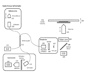
100071 Fig. I is a diagram of an autofocus apparatus according to an
embodiment.
100081 Fig. 2 is a diagram of an autofocus algorithm according to an
embodiment.
DETAILED DESCRIPTION OF THE PREFERRED EMBODIMENTS
100091 This disclosure describes the best mode or modes of practicing the
invention
as presently contemplated. This description is not intended to be understood
in a limiting
sense, but provides an example of the invention presented solely for
illustrative purposes by
reference to the accompanying drawings to advise one of ordinary skill in the
art of the
advantages and construction of the invention. In the various views of the
drawings, like
reference characters designate like or similar parts. It is to be noted that
all fiber optic
systems can be replaced with free space equivalents.
100101 In microscopy, a sample object to be examined is placed on a slide
and is
cover by a slip cover. The objective of a microscope is adjusted so that a
focused view of the
magnified object is obtained. When light traveling in a first medium having a
first refractive
index enters into a second medium having a second reflective index, reflection
occurs at the
boundary between the two media. The amount of light that gets reflected and
the amount of
light that gets transmitted at the boundary depend on the refractive indices
of the two media.
In microscopy, there are typically many different boundaries, e.g. air-glass,
glass-water,
water-glass, and glass-air, and thus there are different reflection intensity
levels
corresponding to these boundaries.
100111 The device according to an embodiment is capable of detecting the
position of
a sample on a microscope. The sample may consist of a specimen mounted between
a
microscope slide and coverslip or specimens within a well plate. The device
tracks the
position of a sample by identifying refractive index boundaries through
Fresnel reflections.
A change in refractive index can correspond to the top and bottom of a
coverslip, the top of a
slide, the bottom of a well plate or the bottom of a well within a well plate.
Using optical
coherence tomography (OCT) these reflections are used to form a depth scan of
the sample
which gives the positions of these surfaces relative to the objective. The
device functions as
an autofocus system by compensating for any variation of the position of the
sample from the
focal plane of the objective.
- 3 -
CA 02944688 2016-09-30
WO 2015/157520
PCT1US2015/025113
100121 The device has 5 major components:
1. Microscope, which is used to image the sample mounted on a Z-axis piezo
stage.
2. Sample arm, which is one of two arms used to create the interference
signal
needed for position detection. This can be introduced before or after the tube
lens on the
microscope.
3. Reference arm, which is the second arm used to create the interference
signal.
This arm is also used to compensate for different path lengths introduced by
different
objectives and to separate physical features for auto-correlated signals.
4. Base unit, which provides the surface detection through the use of OCT.
The
base unit contains the elements needed for obtaining and analyzing the depth
scan from the
sample.
S. Computer, which contains the NI-DAQ card and software used for
obtaining
and analyzing the signal. Based on measurements made in the software a
correction signal.
can be applied to the z-axis piezo stage keep the sample in the focal plane of
the objective.
100131 Fig. 1 shows the microscope and the sample arm with an imaging path
and the
sample arm path. The image path includes a light source 101, such as a
semiconductor laser
diode (SLD), which provides illumination to the sample, which is placed on a
sample stage
102. The sample stage is capable of moving in x, y and z directions. For
example, the
sample stage includes a motorized x-y stage and a z-axis piezo stage. The
objective 103 can
include a range of types and magnification for specific viewing needs. The
tube lens 104
directs the light to a camera 105, such as a CCD, for taking image of the
sample.
100141 In one embodiment, the sample arm includes a 780HP APC Nufern fiber
from
the base unit, a paddle polarization controller, an adjustable APC collimator
106, a piano-
concave lens 107, a dichroic mirror 108 that transmits visible and reflects
IR. The dichroic
mirror can be introduced either between the camera and the tube lens, or
between the tube
lens and the objective. In the latter case the piano-concave lens is not
needed. Fig. I also
shows a shutter 109.
100151 In one embodiment, the reference arm includes a 780HP APC Nufem
fiber
from base unit, a paddle polarization controller, a fixed focus APC collimator
110, a
motorized stage to alter the path length to a retroreflector 111.
100161 In one embodiment, the base unit includes a superluminescent light
source.
For example, the light source preferrably has an output power of 2.5mW, a
central
- 4 -
CA 02944688 2016-09-30
WO 2015/157520
PCT1US2015/025113
wavelength of 930nm and a spectral range of 90mn. The base unit also includes
a 50:50 fiber
coupler 112 that couples to the SLD through its first port, a sample arm
through its second
port, a reference arm through. its third port, and a collimator to a
spectrometer through its
fourth port. In one embodiment, the base unit also includes driving
electronics that provides
a constant current driver for SLD, a servo loop controlled Peltier cooler, a
heater driver, and a
spectrometer image sensor driver. In one embodiment, the spectrometer includes
a fiber
collimator 113, a diffraction grating 114, an objective 115, and a camera 116.
In one
embodiment, the system computer includes a NI-PCIe-6351 DAQ controller,, and
application
software configured to perform the signal detection and controls.
100171 Fig. 2 shows the operation flow of the autofocus system according to
an
embodiment. In step 201, the image is adjusted to be in focus of the camera.
In step 202, the
reference arm is scanned to match the optical path length of the sample arm.
Once peaks are
visible, the reference arm is scanned to bring the strongest peaks into view
on the A.-scan.
Step 203 is the image processing, in. which the signal is &chirped, the DC
spectrum is
removed, and the spectrum is averaged over 40 cycles.
100181 Apodization is then performed. In step 204, the mechanical shutter
is closed
to block the laser in the sample arm after the collimator. In step 205, the
spectrum is
apodized through software.
100191 Dispersion compensation is then performed. In step 206, maximum peak
is
identified on the spectrum. In this case, the position resolution is limited
to pixel width
(3.35 lu.m). In step 207, a parabola is fitted to the max data point and the
two adjacent data
points to provide sub-pixel resolution. In step 209, the peak width is
measured. In step 209,
a numerical quadratic dispersion coefficient is incremented and plotted
against the peak
width. In step 201, the dispersion coefficient is set to give the minimum peak
width.
100201 The sample peak detection is then performed. In step 211, the three
max
peaks are identified. In step 212, the parabola fit method is applied to all
three peaks as
described above.
100211 Peak tracking is then performed. In step 213, the user adjusts the
objective to
focus the image on the camera. User commands the software to track the current
position of
the top of the slide (third peak in A-scan). This position becomes the set
point. In step 214,
the position of the top of the slide is monitored. This position is the
process variable. The
process variable is subtracted from the set point to produce an error value.
- 5 -
CA 02944688 2016-09-30
WO 2015/157520
PCT1US2015/025113
100221 As the peak position changes the error changes to reflect the
displacement of
the sample from the focal plane of the objective. In step 215, a ND control
loop is used to
supply a voltage based on the error to the Z-piezo stage. The voltage changes
the position of
the sample. The voltage is changed to minimize the value of the error.
Minimizing the error
allows the position of the sample to remain in the focal plane of the
objective.
100231 While the present invention has been described at some length and
with some
particularity with respect to the several described embodiments, it is not
intended that it
should be limited to any such particulars or embodiments or any particular
embodiment, but
it is to be construed with references to the appended claims so as to provide
the broadest
possible interpretation of such claims in view of the prior art and,
therefore, to effectively
encompass the intended scope of the invention. Furthermore, the foregoing
describes the
invention in terms of embodiments foreseen by the inventor for which an
enabling
description was available, notwithstanding that insubstantial modifications of
the invention,
not presently foreseen, may nonetheless represent equivalents thereto.
- 6 -
