Note: Descriptions are shown in the official language in which they were submitted.
DEMANDE OU BREVET VOLUMINEUX
LA PRESENTE PARTIE DE CETTE DEMANDE OU CE BREVET COMPREND
PLUS D'UN TOME.
CECI EST LE TOME 1 DE 2
CONTENANT LES PAGES 1 A 154
NOTE : Pour les tomes additionels, veuillez contacter le Bureau canadien des
brevets
JUMBO APPLICATIONS/PATENTS
THIS SECTION OF THE APPLICATION/PATENT CONTAINS MORE THAN ONE
VOLUME
THIS IS VOLUME 1 OF 2
CONTAINING PAGES 1 TO 154
NOTE: For additional volumes, please contact the Canadian Patent Office
NOM DU FICHIER / FILE NAME:
NOTE POUR LE TOME / VOLUME NOTE:
CA 02944903 2016-10-04
WO 2015/164743 PCT/US2015/027515
TUNIO.R SUPPRESSOR AND .ONCOGENE BIOMARKERS PREDICTIVE OF
ANTI-IMMUNE CHECKPOINT INHIBITOR RESPONSE
Cross-Reference to Related Applications
This application claims the benefit of US. ProviSional Application Nos.
62/01.2,689,.
filed on 16 June 2014, and 61/983,602, filed On 24 April 2014; the entire
contents of each
of said .applications are incorporated herein in their entirety .by this
reference.
Statement of WO ts
This .invention was made with government support -under Grant Numbers
1l1090136, 1 00402, CA122794, CA140594, CA163896, CA.166480, CAI 54303,
CA0981.01, CA141576, CA1371.81, CA120964, C.:A.143083, and CA1.63677 awarded
by the
National Institutes of Health. The US. government has certain rights in the
invention. This
statement is included solely to comply with 37 C.F.R. * 401.14(00)(4) and
should not be
taken as an assertion or admission that the .application discloses andfor
claims only one
invention.,
Backeround of the .Invention
Lung squanlous cell carcinoma (SCC) is a COMMOTI type of non-small cell lung
cancer and the second leadin;,, cause of hing, cancer related-death, causing
approximately'
400,000 deaths per year worldwide (Cancer (ienome Atlas Research (2)12) Nature
489:519-525; Siegel et aL 2oi3) CA Cancer .1. ain. 63:11-30). Unlike lung
adenocarcinorna (ADC), for which many. relevant oncogenic mutations have been
defined
and used to develop strategies for targeted therapies, the .genotnic landscape
of lung. SCC is
only now merging. There are .not yet any approved targeted therapies for lung
SCC.
Unfortunately, therapeutic targets in Wag, ADC, such as E(i.R and EML4-ALK, do
not
appear to play major roles in lung SCC (Rekluinan et al. (2012) OM. Cancer
Ras. 18:1167-
I 176). This fact underscores th.e need to develop a preclinical model of lung
squamous cell
carcinoma in which to define and test novel therapeutic approaches.
Currently, the field lacks a mouse model in which introduction of genetic
alterations
found human squamous tuna cancers ieads to tumors of purely squamous
phenotype.
Simultaneous activation of Kras 4 21) (Kras) and inactivation of !IN (also
knol.vn as scrim-
threonine kinase 1 1, SW 1) gives rise tO multiplelung cancer histologies,
:including
squamous ceiI carcinomas et al. (20)7).Na ture 448:807-810); however, KRAS
mutations
- -
CA 02944903 2016-10-04
WO 2015/164743
PCT/US2015/027515
are very rarely .found in human squamous lung. tumors. Recently, it was
reported that
kinase-dead Mira knock-in mice developed spontaneous lung squamous cell
carcinomas
characterized by./kkrt dowThrephitiOn and .marked pulmonary inflammation (Xiao
et al.
(20.13) Cancer Cell 23:527-540), Significant down-regulation of /lb./ Was
found in
ikkalcvgA lung, SCCs and adjacent lung tissues as compartxl to wild-type
lungs.
'Deletion of Uhl alone is unable to drive tumor fbrination I af (2007) Nature
448:807-810). PTEN (Phosphatase and tensin homolog) is another commonly
mut.awd,
deleted, or epigenctically silenced tumor suppressor in human lung cancers
(Salmena et al.
(2008) Cell 133:403-414). .PTEN is ;Altered in 15% of human SCCs (Cancer
Genome Atlas
Research (2012) Nature 489:.51)-525). PTEN neaatively regulates the P13KSAKT
pathway
and P13K pathway gene alterations are found in more than half of human itmn
SCCs
(Cancer Genome .Atlas Research (2012) Nature 489:519-525). In the mouse model,
Pten
deletion alone in airway basal cells: can initiate lung tumor formation, but
with low tumor
incidence, Icing latency, and mixed A-DC and. SCC phenotype (Malkoski e al.
(2013) . .o/.
Carcinag. (c-pub) doi: 0.10021met22030),
One key feature of -minor development that autochthonous genetically
engineered
mouse models provide is a 'physiologically relevant tumor microenvironment.
All of the
models of lung SCC to date, .including the aka knock-in mice and a model
driven by
chronic tuberculosis infection, show marked pulmonary inflammation (Nalhandian
e aL
(2009) Oncagene 28:1928-1938; Xiao et al. (2o13) Cancer (1.W123:527-540),
suggesting.
that an inflammatory rnicroenvironment is central to the development of lung
SCCs. This
is not surprising given that nearly all humans .with lung SCCs have a history
of tobacco use
that drives squamous mctaplasia and the development of SCCs is associated with
in flammatoly diseases and chronic immtmosuppression. Both tumor-associated
macrophages (TAN) and nimor-associated nentrophils (TANs) comprise significan
t
proportions of the inflammatory infiltrates in a wide variety of mouse tumor
models and
human cancers (Murdoch et al. (2008) Nat. Rev. Cancer 8:(l31. Neutrophils were
shown to predominate in iltallan head/neck squamous carcinomas (Trellakis et
al. (2011)
Int. .1 Cancer 129;2183-2193). Neutrophils found in mouse tUrnat'S are
phenotypically
characterized as polymotphonucicar CD1161.,y6G cells, and may be related to a
subtype
of myeloid derived suppressive cells (MDSCs). MDSCs encompass a heterogeneous
population of myeloid cells, which share the ability to suppress T cells
through the
production of arginase, the expression of inducible nitric oxide synthase
(iNOS), and other
CA 02944903 2016-10-04
WO 2015/164743
PCT/US2015/027515
mechanisms (Dunn nu et aL (2)12) Cancer Mumma nother. 61:1155- 1167). In
the
tumor microenvironment, accumulated iN1DSCs are thought to promote tumor
progression
through enhancing. matrix degradation, tumor cell proliferation, metastasis
and angiogenesis
(\Vela et iI. (1989) Proc. .1krat1. Acad. Sci. US.A. 8(,:5859-5863). NIDSCs
have also been
shown to aningonize effector T cell .function, support the generation of
immunosuppressive
T ceil populations and. inhibit the lysis of tumor cells by cytotoxie T cells
or NK cells
(Pumitrti et al. (2012) Cancer Inunanol. immunother. 61:1155-1 I 67.), Some
MDSCs have
neutmphilie features, but the precise relationship between these cells and
normal
pelymorphonnelear leukocytes remains under active investigation.
Polymorphanuclear cells
infiltrating lung cancers are rekrred to herein as 'FANs.
Tumors can also evade immune surveillance by expressing molecules that
maintain
immune tolerance in peripheral tissues, such as Pd-ligand-1 (P13-L1), which
interacts with
the immune receptor Programmed cell. death-1. (PDCD1 or PD-1.) (Barber et al.
(2006)
Nature 439:682-687), The P1)-1/PD-LI interaction inhibits CD8' cytotoxic T
lymphocyte
(CTL) proliferationõ survival and effector function, and can induce apoptosis
of tumor-
infiltrating T cells (Barber et al. (2006).Nantre 439:682-687). P1)-1.IPD-.L1
interactions can
also promote the differentiation of CD4' T cells into FOXP3'. 'Tregs
(Francisco et at (2009)
J. Exp. Med. 206:3015-3029), which are known to further suppress the immune
system and
cause peripheral immune tolerance in lung cancer patients (Adeegbe and
Nishikawa (2)13)
Front. linmunol. 4:190). &topic PD-LI. expression in tumor cells in a
syngeneie transplant
atodel .facilitated the escape of the tumor cells from. CTL control (Iwai et
ïl.(2002) Proc.
.Natl. Acad. Sci. U.A. 99:12293-12297). Consistent with these findings in pre-
clinical
systems, infusing lung cancer patients with blocking anti-PD-l/PDL-1
monoclonal
antibodies has shown efficacy in early stage trials, despite limited activity
of prior
immunotherapies for lung malignancies (Braluner et al. (2012)N Engl. J. =led.
366:2455-
2465; Topalian et al (2012) N. Engl. J Med. 366:2443-2454).
Tumor-propagating cells have the ability to self-renew and differentiate into
the
bulk population of the tumor and are thought to drive both disease recurrence
.and
metastatic spread (Visvader and Lindeman (2012) Cell Stem Cell 10:717-724
Stein cell
antigen-1 (.crsc.a/ or Ly6a) was :reported as a bronchinalvenlar stein cell
MASC) marker in
the distal lung and is also enriched in bronchiolar progenitor cells (Kim et
al. (2005) Cell
121:823-835; Lee et al. (2014) OR 156:440-455). SCAT coils, located at the
bronehioalveolar duct junction (BAD,1), are hyper-proliferative in response to
both
- 3 -
CA 02944903 2016-10-04
WO 2015/164743
PCT/US2015/027515
oncogenic .Kr' and deletion of Pten, suggesting that they are susceptible to
neoplastic
transformation (Kim ei aL (2005)(V/1121:823-835; Tiozzo et aL (2) A. J.
Respir.
0.1t. Gee Med. 180:701412). In addition, SCA I can be tised to enrich for
tumor
propagating veils (TPCs) in the lung adenocarcinomaKrasGua_,p53" (Kras;p53)
model
(Curtis et aL (2010) Cell Stern Cell 7:127-133). In the more proximal lung,
nerve growth
factor receptor (TNFR superfamily, member 16, Nkfk) is a stem cell marker for
the
pseudostratified tracheal epithelium in both human and mouse; .NGFR expression
is
specifically observed in the p63' mouse basal stem cells (Rock et cd. (200))
Proc. Nail
Acad. Sci, USA 106:12771-12775), NUR' basal cells appear to be the cells-of-
origin in.
a SOX2-induced model of esophageal SCC, and .NGFR has been suggested its a
putative
marker for human esophageal SCC TPCs (Huang et aL (2009) 13MC Cancer 9:9; Liu
et ai
(2013) Cell Stem Cell 12:304-315),
Despite these clues as to the .molecular phenotype of a potential tumor
propagating
cell in SCC. no Tpc population able to propagate disease serially has been
identified for
itmg SCC. 'Moreover, since therapies that negatively regulate numune
checkpoint
inhibitors, such as initi-PD-1, anti-PD-L. i. and aini-CTLA4 antibodies, are
both.
significantly toxic in combination and -very expensive, there is a great need
in the art to
identify biomarkers which are predictive of patient responsiveness to such
therapies in
order to appropriately determine an efficacious and cost-effective course of
therapeutic
intervention.
Summary of the Invention
The present invention is based, atleast ìa part, on the discovery that the
presence
activating- oncogenes (e.g., activating KRAS, NRAS, and/or HRAS mutations),
the
presence of inhibiting tumor suppressors (e.g., inhibiting Lkb i and/or Pten
:mutations), and
the. amount (e.g., copy number or level of expression). andfor activity of
such biomarkers,
are predictive of hyperproliferative cell responsiveness to anti-immune
checkpoint inhibitor
therapies.
In one aspect, a method of treating a subject :afflicted with A Caneer,
wherein the
cancer comprises at least one mutation selected from the group consisting (Wan
activating
KRAS mutation, an activating NRAS .mutation, an activating HRAS mutation, an
inhibiting
EKB1 mutation, and an inhibiting PTEN mutation, comprising administering to
the subject
an agent that inhibits the copy number, amount, an!/or activity of arginase 1,
thereby
- 4 -
CA 02944903 2016-10-04
WO 2015/164743
PCT/US2015/027515
treating the subject afflicted With the cancer, is provided. In one
cnibodiment, thc agent is
administered in a pharmaceutically. acceptable formulation. In another
.embodiment, the
agent directly binds arginase I. In still another embodiment, arginase I is
human .arginase
. In yet another embodUnent, the method further comprises administering one or
morc
additional anti-cancer agents.
In another aspect, a method of inhibiting hyperproliferative growth of a
cancer cell
or cells. Wherein the cancer cell or cells comprise at least one mutation
selected from the
group consisting of an activating KRAS mutation, an activating. NRAS mutation,
an
activating 1-IRAS mutation, an inhibiting LKal mutation, and an inhibiting
PTEN mutation,
the .method comprising contacting the cancer cell or cells with an agent that
inhibits the
copy number, amount, andlor activity of arginase I, thereby inhibiting
hyperproliferative
growth of the cancer eel or cells, is provided. In one embodiment, the step of
contacting
occurs in vivo, ev vivo, or in vitro. In another embodiment, the .agent is
.administered in a.
pharmaceutically acceptable formulation. In still another embodiment, the
.agent directly
binds arginase 1 . In yet another embodiment, arginase I is human arginase
1..in another
embodiment, the method further comprises administering one or more additional
anti-
cancer agents.
ha still another aspect, a method of determining whether a subject afflicted
with a
cancer or at risk for developing a cancer, Wherein the cancer comprises at
least one
mutation selected from the group consisting of an activating KRAS mutation, an
activating
NRAS -mutation, an activatini:, HRAS mutation, an inhibiting I.,KB I mutation,
and an
inhibiting MTN mutation., -s.vould 'benefit =from anti-immune checkpoint
inhibitor .therapy,.
the method comprising: a) obtaining a biological sample from the subject; bi)
determining
the presence, copy number, amount, andlor activity of at least one biomarker
listed in Table
I in a subject sample; c) determining the presence, copy number, amount,
andlor activity of
the at least one biomarker in a control; and d) comparing the presence, copy
number,
imount, and/or activity of said at least one biomarker detected in steps to
and e); wherein
the presence or a significant increase in the copy number, amount, and/or
activity a the at
!east one biomarker in the subject sample relative to the control indicates
that the subject
afflicted with the cancer or at risk for developing the cancer would benefit
from anti-
inmate checkpoint inhibitor therapy, is provided. In one embodiment., the
method further
comprises recommendinu, prescribing, or administering anti-immune checkpoint
inhibitor
therapy if the cancer is determined to benefit fro anti-immune checkpoint
inhibitor
- 5 -
CA 02944903 2016-10-04
WO 2015/164743
PCT/US2015/027515
therapy. In another embodiment, the method .further comprises recommending,
prescribing,
or administering anti-cancer therapy other than anti-immune checkpoint
inhibitor therapy if
the cancer is determined to not benefit from mui-iminune checkpoint inhibitor
therapy. In
still another embodiment, the anti-cancer therapy is selected from the group
consisting of
targeted therapy, chemotherapy, radiation therapy, and/or hormonal therapy. In
yet another
embodiment, the control sample is determined from a cancerous or non-cancerous
sample
from either the patient or a member of the same species to which the patient
belongs. In
another embodiment, the control sample comprises cells. in still another
embodiment, the
method further comprises determining responsiveness to anti-immune checkpoint
inhibitor
therapy measured by at least one criteria selected from the group consisting
of clinical
benefit rate, SUrVival until mortality,=patholol.:*al complete .response, semi-
quantitative
measures of pathologic response, clinical complete remission, clinical partial
remission,
clinical stable disease, recurrence-free survival, metastasis free survival,
disease free
survival; circulating tumor cell decrease, circulating marker response, and
REasT criteria,
in yet another aspect, a .method of assessing the .efficacy of an agent for
treating a_
cancer iri a subject, vherein the cancer comprises at least one .mutation
selected from the
group consisting of an activating KRAS mutation, an activating NRAS mutation,
an
activating HRAS mutation, .an inhibiting .L.KB1 mutation, and an inhibitim4
.PTEN mutation,
comprising: a) detecting in a first subject sample and maintained in the
presence of the
agent the presence, copy number, amount and/or activity of at least one
biomarker listed in
Table 1; b) detectina the presence, copy number, amount and/or activity Idle
at least one
biomarker listed in Table .1 i.n a second subject sample and .maintained in
the absence of the
test .compound; and c) comparing the presence, copy number, amount andlor
activity of the
at least one biomarker listed in Table 1 .from steps a) and b), wherein the
presence or a
sipificantly increased copy number, amount, andlor activity of the at least
one biomarker
listed in. Table i n the first subject sample relative to the SCCOild subject
sample, indicates
that the agent treats the cancer in the subject, is provided.
Si:Milady, in another aspect, a method of monitoring the progression of a.
cancer in a
subject, wherein the cancer comprises at least one mutation selected from the
group
consisting of an activating KRAS mutation, an activating NRAS mutationõ an
activating
HRAS mutation, an inhibiting LKB I mutation, and an inhibiting PTE N mutation,
comprisinn: a) d.etecting in a subject sample at a first point in tiine the
presence, copy.
number, amount, andlor activity of at least one biomarker listed in Table 1;
b) repeating
- 6 -
CA 02944903 2016-10-04
WO 2015/164743
PCT/US2015/027515
step a) during at least One subsequent point in time after administration of a
therapeutic
agent; and c) comparing the presence, copy number, amount, and/or activity
detected in
steps a) and b), wherein the presence or a significantly increased copy
.number,
andfor activity of the at least one biomarker listed in Table 1 in the first
subject sample
relative to at: least one subsequent subject sample, indicates. that -the
agent treats the cancer
in the subject, is provided..
For such methods of assessment or monitoring, in one embodiment, the subject
has
undergone treatment, completed treatment, andibris in remission for the cancer
in between
the first point iri time and .the subsequent point in time. In another
embodiment, .the subject
has undergone anti-immune: checkpoint inhibitor therapy in between the .first
point -in time
and the subsequent point in time, In still another embodiment, the first
andlor at least one
subsequent sample is selected from the group consisting of ex vivo and in vivo
samples. In
yet another embodiment, the First andfor at least one subsequent sample is
obtained from an
animal model of the cancer. In another embodiment, the first andfor at least
one subsequent
sample is a portion of a single sample or pooled samples obtained from the
subject
In another aspect, a cell-basixl .rnethod for .identifying an agent that
.inhibits a cancer,
the .inethod comprising: a) contacting a cell expressing at least one
biomarker listed in
Table I with a test agent; and b) determining:, the effect of the test agent
on the copy
number, level of expression, andfor level of activity of the at least One
biomarker i.n Table l
to thereby identify an. agent that inhibits the cancer, is provided. In one
embodiment, the
cells arc isolated from an animal model of a cancer, hi another embodiment,
the cells are
from a subject afflicted with a cancer or wherein the cell comprises at least
one mutatiori.
selected from the group consisting of an activating KRAS mutation, an
activating NRAS
mutation, an activating HRAS mutation, an inhibiting EKB1 mutation, and an
inhibiting
VITEN mutation. still another embodiment, the cells are tmresponsive to
.anti-iramune
checkpoint inhibitor therapy.. In yet .another embodiment, th.e step of
contacting, occurs in
vivo, ex vivo, or in vitro. In another embodiment, the method further
comprises determining
the ability of the test agent to bind to the at least one biomarker listed in
Table 1 betbre or
after determining the effect of the test agent on the copy number, ievel of
expression, or
level of activity of the at least one biomarker listed in Table I.
Certain embodiments can be applied to any .method, assay, and the like oldie
present invention For example, in one enibodiment, the sample comprises cells,
cell lines,
histological slides, paraffin embedded tissue, fresh frozen tissue, fresh
tissue, biopsies,
- 7 -
CA 02944903 2016-10-04
WO 2015/164743
PCT/US2015/027515
bronchoalveolar lavagc IBA.1.,) fluid, blood, plasma, serum, buccal scrape,
saliva,
cerebrospinal fluid, urine, stool, mucus, or bone marrow, obtained from the
subject. In
another embodiment, the 'presence or copy number is assessed by microarray,
quantitative
PCR (qPCR:i, hialt-throughput sequencing, comparative gcnomic hybridization
(C.CiH), or
fhiorescent in situ 'hybridization (FISH). 1 still another embodiment, the
amount of the at
!east one biomarker listed. in Tabie I is assessed by detecting the presence
in the samples of
polynucleotide .molecule encoding the biomarker or a portion of said
polynueleotide
molecule. In yet another embodiment, the polymtcleotide molecule is a mRNA...
CDNA, or
functional variants or -fragments thereof In another embodiment, the step of
detecting
further comprises amplifying the polynucleotide molecule. In still another
entbodiment, the
amount of the at least one biomarker :is assessed by annealing a nucleic acid
probe with the
sample of the polynucleotide encoding the one or more biomarkers or a portion
of said
polynu.cleotide molecule .under stringent hybridization conditions, in yet
another
embodiment, the amount of the at least one biomarker is assessed by detecting
the presence
polypeptide of the at least one biomarker. In another embodiment, the presence
of said
polypcptide is detected using a reagent Nvitich specifically binds with said
polypeptide (e.g.,
a reagent selected front the group consisting of an antibody, an antibody
derivative, and an
antibody fragment). In still another embodiment, the activity of the at least
one biomarker
is assessed by determining the magnitude of cellular proliferation, cell
death, or cylokine
production. In yet another embodiment, the agent or anti-immune checkpoint
inhibitor
therapy is selected from the group consisting of a biockint, antibody, small
molecule,
antisense nucleic acid, interfering. RNA, shRNA, siRNA, a.pta.mer, ribozyme,
dominant-
negative protein, and combinations thereof (e.g., an agent or anti-immune
checkpoint
inhibitor therapy selected .from the group consisting- of inhibitors of PD-
L1, =PID-L2,
C1.'LA-4, arginase I, and combinations thereof. In another embodiment, the
agent or anti-
immune checkpoint inhibitor therapy is an inhibitor of arginase i in
combination with
inhibitors of , =PD-1,1, PD-1õ2, or CTLA-4. In still another embodiment,
the at least
one biomarker is selected from the aroup consisting of I, 2, 3, 4, 5, 6, 7, 8,
9, 10, or more
biomarkers, yet another embodiment, the at least one biomarker is selected
from the
group consisting of KRAS, NRAS, }IRAS, Ý.,KB I, WIEN, arginase I.õ and
combinations
thereof. 11.1 another embodiment, the cancer is selected from the aroup
consisting of luna
cancer, hitla SparlIOUS Cell carcinoma. (SCC), melanoina, cervical cancer, and
pancreatic
cancer. In still another embodiment, the cancer comprises I) at least one
inhibiting '1õ,KB1
- 8 -
CA 02944903 2016-10-04
WO 2015/164743
PCT/US2015/027515
mutation and at least one inhibiting. PTEN mutation or 2) at least one
activating. RAS
mutation sewed from the group consisting of KRAS, NRAS, EIRAS, and any
combination
thereof In yet another embodiment, the subject is a Main Mai (e.g., an animal
model of
cancer or a human).
Brief Description of the Drawings
Figure 1 includes 3 panels, .identified as -panels .A, and. C, which show
that W-
ade:lie inactivation of both Lkb I and Pte in the inou.se lung leads to
squamous
carcinoma, Panel A shows a schematic of hí-allelic inactivation of both Ikbi
and Pte n in.
the .mouse lung by Ad-Cre inhalation, followed by tumor dissociation and
sorting.. Panel B
shows the results of phenotypic quantification of lung tumor histologies ti-om
the indicated
conditional MOUSe models, including Kra..02D (Krav),
Krascil2D,p5311(Kras,p53),
K11.11129 L,kbl" (Kras;Lkb1) and KraPlw.,-p5312'n;Lkbl" (Kray,p53;Lkb
Lkb.ri ;hen" (LH)] , Pte 1kbillfitenfi41 p5/11(Lkbl ;Pten,p53.1, which all -
.rely upon
Ad-Cre inhalation for tumor initiation (mean +/- SE; n = 5 mice per genotype).
Panel C
shows representative HI stained sections derived from tumors arising in the
Lkb.1;.Pten
mouse model, .Arrows .indicate specific features on individual images, which
include (a)
mature squainous cells with aberrant nuclear morphology; (h) large infiltrates
of neutrophils
in SCC nodules; (c) keratinized cells \vial markedly dense eosnloPhilie
eroPlasm
surrounded by epithelial cells; (41) uzell-differentiated SCC with keratin
pearls; (c) SCC
nodules in large airways; and (f) squamous-like tumor cell iymphovascular
invasion. Scale
bar in Panels C(a), C(d),. and. C(e) 200 um; scale bar in Panels C(c) and.
C(f) = 50
scale bar in Panel C(b) 25 gm.
Figure 2 includes 6 panels, .identified as panels A. .B, C, D, E, and F, which
further
show that bi-alielic .inactivation of both Lkb.1 and Pten in the mouse Tung
leads to squamous
cell carcinoma. Panel A shows representative histology of hematoxylin and
eosin (1-1.&E)-
s1ained genetically engineered mouse models of the indicated genotypes.
CrellOtypC
indicated. on images; scale bar = 100 nin for all panels. Panel. B shows the
results of
Kaplan-Meyer survival analysis of Lk:hi:Pim mice following intra-nasal Ad-Cre
instillation. Median survival= 45.7.1 weeks.. Panel C ShOWS the results of end-
point PCR
for LoxP recombination in the Lkbl and hen alleles in the .indicated sorted or
total tumor
cell populations. 'Panels D and E show images of total lungs from illyi,Pren
mice stained.
with Il&E to show both tumor location and tumor burden at 40-50 week time-
points, Note
- 9 -
CA 02944903 2016-10-04
WO 2015/164743
PCT/US2015/027515
that while some tumors appear to arise in the proximal lung at the branch
point from trachea
into the lobe (Panel Da), top arrow), other tti.mors arise appear to be
isolated in the distal
lung and completely surrounded by alveolar epithelitun (Panels DO) and Eta),
bottom
arrows). Panel E(a) corresponds to 45-50 weeks, while Panel E(b) corresponds
to 40-45
weeks post Ad-Cre. Scale bar in Panels Dtb), Fa). and F(b) St)() tin. scale
bar in Panels
D(a), E(a), and E(1)) 5,000 um. Paael F shows Fl&E stained chest all
metastasis of the
lik51,-Pten primary Minor cells. Scale bar lt)l un on both panels.
Figure 3 includes 3 panels, identified as pzinels A, B, and C, which show that
lung
SCCs in abififil;PienII mice closely recapitulate the human disease, Panel A
shows the
results of immunohistochemically stained human and mouse ADC and SCC tumors.
The
SCC canonical markers KRT5, SOX2 and p63, and the ADC canonical marker TTF I ,
were
used to distinguish the tumor types. EpCAM is an epithelial cell marker, and
the
expression patterns of p63, K1T5 and SOX in SCC co-localized with EpCAM
expression,
Scale bar = 100 um for all panels. Panel B shows microarray expression
profiling results of
normal lung and SCC tumors from mouse and human, Up-regulated genes in both
mouse
and human SCC were enriched for a squarnous differentiation signature. Panel C
shows
that down-regulated acmes from the analysis in Panel /3 were enriched for a
normal lung
terminal respiratory unit signature.
Figure 4 includes 2 panels, identified as panels A and B, which further show
that
lung SCCs iix LA-bi"-,Prenll mice closely recapitulate the human disease,
Panel A shows a
schematic of a gene cxpression profile comparison of 34 human SCC tumors with
either
LKB.1 or PTEN alterations to 35 normal human Jun tissues samples, and three
tumor
bealrisz Lkhi,Pien mouse SCC to lungs from age-matched LP mice that never
received Ad-
Cre, There were 489 up-regulated genes and 404 down-regulated genes shared by
the
human and mouse contrasts. Panel B shows that the results of the 893 shared
differentially
expressed genes in human and mouse SCC clustered and displayed by heat map.
Figure 5 includes 6 panels, identified as panels A, B, C, D, E, arid F, which
show
that Lir.b./"-,Pten" lung SCCs display unique gene expression, metabolism, and
downstream signaling pathways, Panel A shows representative flow eytometric
plots for
sorting the epithelial fractions (CD45VD31-EpC:AM.') from 1,P tumor nodules,
Krcts unnor
nodules and normal lung. The RNA from purified epithelial cells was extracted
for gene
expression profile analysis, Panel B shows the results of gene set enrichment
analysis
(GS.E,A) (Subramanian et CIL (2)07) BioMformatics 23:3251-3253) used to
compare the
- I ii -
CA 02944903 2016-10-04
WO 2015/164743
PCT/US2015/027515
gene expression profile of Lkb./.1'ten tumor .cells to transcriptionally well-
defined sub-
classes of human itmg SCC (Wilkerson et al. (2010) Clin, Cancer Res. 1614864-
4875).
GSA was pertbrined using the Java CISEA desktop application on pre.,ranked
gene lists
constructed fi-om RMA. normalized expression. data. The LichiTten model very
closely
recapitulates the expression pattern found in the basal subtype of human SCC.
Panel C
shows the results of GSEA analysis used to discover the molecular pathways
altered. in
likb1,-Pten and Kills tumors. All gene sets Were downloaded from the World
Wide -Web at
broadinstitute.orgigseaimsigdbigeriesetsjsp. AKT and mTOR. gene sets were both
derived.
from Majumder a al. (20(4) Nat, Med. 10:594-()1, The lung-specific KRA.S
mutation
acne set was derived front Barbie. et ctl. (2009)Ni-4'111re 462:108-112.
Compared to Kras
tumorsõAKT and mTOR gene sets are both more significantly enriched in
the.1.1b1;Pren
model. Panel D shows the results of immunohistochemical staining. for Ii&E, p-
AKT and
p-ERK on Ijcbi ;Plea, Kras, Kras,l,kb õ Kras;Pten and Kyvsõp53,-Lkb tinnor
nodules with
low magnification (x20)). Seale bar 1.00 jun for all panels. 'Panel -E shows a
graph of top
metabolites used to cluster normal lung from Kras tumor and I,kb 1 ;Pten.
tumor. 1,-arginine
(reduced) and creatine (increased), two byproducts of L-arginine metabolism,
show that the
aminine metabolism pathway is skewed towards the function of the enzyme
arginasel in
the Lkbl Tten tumors. Panel. F shows the results of real-time RT-PCR. for
arginasel
niRNA expression in the indicated EpCAWCD45-CD3 1' purified populations from
normal
lung, Kras, and.Lkb ;Den tumors, Comparing with normal lung and Kras,
arginasel is
hìghly expressed. in L.01,-Pien EoCAM'CD45-CD3 1- tumors cells (a ¨ 5 for
normal
EpCAM' cells; n 4 for Kras EpCAMT 5 thr.11b1;Pten EpCAM' .eells; data
are
presented as mean +/- SEM; p<).0001 ).
Figure 6 includes 4 panels. .identificd as panels A, .B, C. .and. D, hic.h
further show
that abli";Prenfv'l lung SCCs display unique gene expression, metabolism, and
downstream signaling pathways. Panel A shows the results of EpCA.M.-CD31-CD45-
cells
isolated by FACS and subjected to microarray expression analysis (top line
number of up-
regulated genes; bottom line - number of down-regulated genes). An Euler
diagram
illustrating the gene expression profiles of epithelial cells from LP SCCs,
Kras ADCs, and
normal lung tissues, is shown, Panel B shows a beat map depicting differential
expression
of selected genes in LP SCCs, Kt-as ADCs and normal lung tissues as determined
by
microarray expression profiling. Re.d indicates up-regulation and green
indicates down-
regulation. Panel C shows the results of immunohistoehemical staining for p-
AKT and p-
11
CA 02944903 2016-10-04
WO 2015/164743
PCT/US2015/027515
ERK on LP, Kras, Kras;Lkhl, Kras,Pten and Kras,p53;Lkb.I tumor nodules. Scale
bar
50 pm for all panels. Panel D shows the results of hierarchical clustering by
Ward Niethod
of ClUallti tatiVe metabolomic profiling for LP SCC tumors (S),, Kr s ADC (K)
tumors and
normal lung tissues (N).
Figure 7 includes 7 panels, identified as panels A, B, C. D ., F, and which
show
that tunwr-associaicd neatrophils (-LANs) were the predominant inflammatory
coil
population in Lkbi";Pteea SCC tumors. Panel A shows representative flow
eytometry
plots for Kras' ADC and LP SCC dissociated tumors. Plots are gated on live
single CD45'
cells, Gating was performed as described in the Examples. In Kras tumors,
rumor TAMs
(CD45-VDI IcCDI IbCD103") comprised the majority of CD45+ cells, while in LP
tumors, TANs (CD45'CD1 b.1...y6G') were predominant. Panel B shows
quantification of
inflammatory cell populations in Kras tumors (n 7), .Kras,p53 tumors (n = 8)
and
LA-b1 ;hen tumors (n = 7) by flow cytometry (mean +/- SEM; p<).(001), Panel C
shows
quantification of TANS within right lung lobes from samples with progressively
increasing
weights (shown in Panel A of -Figure 8), indicating different tumor burdens N
= 8 for
normal lunrr control; n 5 for mild disease group (tumor plus surrounding
tissue weight
less than 75) trig); n 5 for severe disease group (tumor plus surrounding
tissue weight
greater than 750 mp,); mean +/- SEM.; mild vs. control p = 0.0034; severe vs.
control
r0,0001. Panels D and E show representative immunohistochemical staining for
WO,
F4/80, and C1M63 in SCCs and ADCs. The mouse slides were LP SCC and Kras
driven
ADCs. MP() staining indicating neutrophiis was only positive in SCC nodules.
F4/80
marks macrophages in mice, while CD163 marks macrophages in humans. Scale bar
= 200
gm for all sub-panels of Panci D; scale bar MO gm for all sub-panels of Panel
E. Panel F
shows representative immunohistochemicall staining, on Lkb Yien,p53 tumors
where
distinct areas of ADC and SCC were adiaeently located. p63 and WO staining
were
restricted to the SCC area. Scale bar 200 p.m for all panels. Panel G shows
the results of
GSEA analysis used to confirm the major immune cell types within Lkhi,..Pien
SCCs and
Ks ADCs (Abbas et aL (2005) Genes ',ninon. 6:319-33 l; Konuma et al. (2011)
bLvp.
Hematol. 39:697-709).
Figure 8 includes 6 panels, identified as panels A,13, C, D, F, and F, which
further
show that TANs were the predominant inflammatory cell population in
1:01";Ptee1l
SCC tumors. Pane! A shows quantification of inflammatory cells by flow
eytometly froni
samples with progressively increasing weights, indicating different tumor
burdens: normal
- 12 -
CA 02944903 2016-10-04
WO 2015/164743
PCT/US2015/027515
lung control (n 8); mild disease group (tumor plus surrounding tissue
veightless than 750
5; severe disease group (tumor phis suo-ounding tissue weight greater than 75(
mg), 11 = S. The illumine cell populations -were gated as described in the
Examples:
Macrophages (TAM), T cehs, B cells, and NK. cells within the tumors decreased
with
increasing tumor burden, Panels B and C correspond to 'Panels D and E of
Figure 7 and
sho-w representative immunohistochemical staining at high magnification of
mouse and
human SCC and ADC for MPO, p63 and F4180 -(mouSc)/C1)I63 (human), Within the
SCC
nodule, MPO TANS are specifically surrounded by 1363' squamous epithelial
cells. Scale
bar 100 pm liar all panels. Panel D shows representative
immunohistochemical staining
for p63, MP and 14/80, :Distinct areas of ADC and .SCC.; are observed in
close proximity
to each other in the lung of Krasj..kb mouse. Magnifications are indicated on
the images.
Scale bar for the top panels = 100 gm.; scale bar for the middle and bottom
panels 1,00-0
Panci E shows the results: of rcal-time WI-1'0Z. for ypo. Argi and (..:.k,-cr2
mR.NA
expression in the indicated CD45' purified. populations. Compared with Kras
and
.Kkas,p53, Mpo, Arg.1 and Crer2 expression level in./..-kb I ;hen tumor C1)-
454. cells µvere
significantly elevated, p values are indicated on each panel. Panel F shows
that CXCL1,
CXCL2, CXCL5, CXCL7 and GCSF levels in BA.L fiuid from Lkb ; Pten
turnorzbearine
mice were detected by ELISA. Compared to levels in normal BAL fluid, the
levels of each
of these eytokines were significant increased. N 9 for normal control lung
BALIF n 7
for LP tumor-bearing BALE; p value is indicated on each panel. 'Data shown in
Panels A.
E, .and F are presented as mean +/- SEM.
Figure 9 includes 7 panels, identified as panels A. 13, C, D, E, F., and G,
which show
that Lkb.1".,Pien" lung SCCs display hallmarks of immune suppression. Panel A
showS
representative .flow eywmetry plots for FOXP3 and C1)8 in total CAB+ T cells
within LP
SCC tumor, unindueed normal 'lung and lung surrounding LP SCC tumors. Panel B
shows
ratios of C1)8' T cells to .FOXP3' Tregs as determined with flow cytornetry; n
8 for
control lung; n 5 for mild disease group; n 5 for severe disease group;
p<0.0001, Panel
C Shows the results of immunohistoeheinical staining for FOXP3 and. confirms
the presence
Tregs in LP SCC nodules. Scale bar ----- 50 wn tbr both panels, Panels D and E
show
quantification the percentage of.P1')-1-positive cetls within the CD8' and
C1)4' T -cell
populations; n 8 101 COMOl lung; n 5 for mild disease group; 5 for severe
disease
group: p<0.0001.. Pane! F shows representative innnunohistocheinical staining
for PD-1,1
on LP SCC nodules. Seale bar .100 Inn for both panels. Panel G(a) shows the
percentage
- -
CA 02944903 2016-10-04
WO 2015/164743
PCT/US2015/027515
of PD-Li positive cells within the EpCAM*CD45-CD31- fraction from LP SCC as
measured by flow eytometry; n 7 for LP tumors; n 5 for normal lung; p<0.000 I
. Panel
G()) ShOWS the results of real-time RI PCR for Pdll niRNA levels in the
indicated
.EpCA.M. purified cells from SCC tumors and normal lung tissue; n = 6 for
normal Itin,g; n
for LP SCC tumors; p 00/13. Data in Panels B. D, E, and G are presented as
inean-q- SEM.
Figures 10 includes 2 panels, identified as panels A and B, which further show
that
Lkb 1"-,Pteni" lung SCCs display hallmarks of immune suppression. Panel A
shows
representative flow eytornetrie plots for T cell and associated
immunoreculatory markers
within 1:01 ; Pten SCC turnors, adjacent tissue and normal lung vi.thout Ad-
Cre inhalation,
Panel B shows that IGEN and 11,6 levels in BM., fluid from Uhl ;Pwn tumor-
bearing
mice were detected by ELISA. Compared with those in BAL. fluids front normal
mice,
TGFp and 11,6 levels were significant increased; a 9 for norinal control lung;
n 7 for LP
tumor-bearing mice. Data are presented as mean 41- SEM, P-values are indicated
on each
panel.
Figure1 t includes 8 panels, identified as panels A. B, C. D. E, F, G, and H.
which
show that /..kb,/"Xteen lung SCC contain SCAI'NOM tumor propagating cells that
ean
serially transplant squamous disease. .Panel A shows NGFR and SCA1 expression
in distal
Krrrg and trachea tis measured by flow cytometry: a. Lung; h. Tracheas; c.
Lung EpCAM
single stained gating control. Dissociated total distal lung, and dissociated
tracheal
epithelium were stained for DAPI, CD45, CD31, EpCAM, SCA I, and NUFR. When
gated
on the DAPITCD31-CD45-EpCAM' epithelial cells, only a small fraction (--M) of
the distal
lung epithelium expressed SCA1 and GFR, while ¨20% of the tracheal epithelium
is
SCAI'NGFR'. Panel B shows NGFR and SCA1 expression in the Lkbl ;Pen tun:tors.
Dissociated tumors were again stained for DAP!, CD45, CD3I , EpCAM, SCA I and
NGFR..
When gated on the DAPI-CD31-CD,45'EpCAM epithelial cells, a large portion of
the cells
express SCAI, and of those ¨17% also express NCiFR. a. unstained tiunor gated
on
FSC/SCC and DAPI-neuative; b. stained 1..kb I ;Pim SCC. Pane! C shows the
results of
flow cytometry thr the various indicated cell populations within dissociated.
Krim, Kras,p53
and 1.161,Pien tumors; data are presented as mean SEM. Panel D shows the
results of
real-time RT-PCR for AV* niRNA expression in the indicated EpCANI purified
populations. Compared with normal lurif:3EpCAM' cells and Kras tumor EpCANI!
cells,
Ner in UN ;Plea SCC EpeAM' cells was significantly increased; n 5 for normal
- !4-
CA 02944903 2016-10-04
WO 2015/164743
PCT/US2015/027515
EpCAM' cells; a 4 for Kras EpCAM cells; n 5 for .1.kbl ,ften EpCAM cells; data
are
presented as mean +/- SEM; p<0.0001. Ptniel E shows a schematic for the FACS
procedure
using NGFR and SCA! makers and ill W1r0 31) culture. The digested tumor eelis
were
initially gated on epithelial cells (C1)45-CD31-EpCAM) and were secondarily
gated by
NGFR and SC,AI expression. Four fractions with different markers (SCA] SCAr
SCAVNGER- and SCAINGER) were collected and co-cultured in Matrinel with
equal amounts of CD45'.CD3 'support' ce:lis that were isolated from the same
primary
timers. Representative flow cytometric plots from Lkbl,Pten, Kras and Kras,-03
tumors
are shown, Panel F shows characterization of tumorspberes ìi 31) cultures.
Represcritative
bright field images of 3D culture colonies clerived from primary mouse tumors
of the
indicated genotypes (top), H&E (middle) and IF (bottom). Fixed and sectioned
tumotspheres were stained with anti-p63 (green), anti-SPC (red) and DAVI.
(blue) to show
squamous and adeno differentiation; scale bar ¨ 100 um for all panels. Imaging
was
performed with a Nikon 90i camera and N1.S-Elements software and processed
with NIS-
Elementsm, and Adobe Photoshoplm. Panel G shows a schematic of the in vivo
serial
transplantation procedure. Panel shows representative H&E stained sections and
flow
cytometric plots of primary, secondary and tertiary Lkb1;.Pten lung tumors.
Seale bar 100
}UM for all panels.
Figure 1.2 includes 6 panels, identified as panels A, B, C, D, E,ad '17, which
further
show that Lkb.ifiln;Rentill lung SCC contain SCAPNGER' tumor propa.gating
cells that can
serially transplant squamous disease. 'Panel A shows representative flow
cytometry plots
thr NGER and SC.A1 expression within the indicated EpCAM'CD45-CD31-
dissociated
tumor cell populations. LP tumor cells showed much higher expression of both
SCA1 and
MIER than either the Kras or Kras,p53 nanors. 'Panel B shows quantification of
SCA1-
and NGER-expressing cells with the EpCAMt1)45-CD3 I- population as assessed by
flow
cytometry. The percentage of SCA.1'NGFR' in LP tumors is much higher than in
Kras or
Kras;p53 tumors; a 23 for Kras tumors; a. 25 for Kras,p53 tumors; n= 34 for
Lk-b ;Pten tumors; p<0.(001. Panel C shows representative immunohistochetnical
staining
for NGFR on !noun SCC and ADC (a) and human SCC nodules (b). 'N'CiFR staining
is
strongly positive on SCC tumors but negative on ADC tumors. In the Uhl
,Pien,p53
tumors, distinct areas of ADC and SCC were adjacently located. NGFR staining
was
restricted to the SCC area (c). Seale bar 50 um for panels of Figures 2(B)a
and 12(B)b;
scale bar 200 um for sub-panels of Panel (B)c. Panel D shows quantification of
- 15 -
CA 02944903 2016-10-04
WO 2015/164743
PCT/US2015/027515
tumorsphercs .derivcd from SCA 1.'NCi.FR' SC.ATNGFR-, SCA I. -NIGFWand
SCAINGFR"
FACS purified .cells that were co-cultured in Matrigel with equal amounts of
CD4.5'CD31
cells from the same primary tumors, Each .fraction was seeded at 5.,() tumor
The colony propagating ability of the SCAINCIER' fraction in LP tumors is
higher than.
that of the other fractions; p 0.0011. Panel E shows quantification of tumor
propagation
ability of FACS isolated SCA l'ISIGFR% SCAI'NG.FR', and SCAl"NGFR LP minor
cells.
The secondary tumors were derived from intra-tracheal transplant, with Minor
formation
latency of-3()-H) weeks. The tertiary minors were derived from intra-thoracie
injection,
with the tumor formation latency of ¨20-30 weeks. 10,000 sorted eel.ls from
each fraction
were injected for each fraction and each experiment. Only SCAl.'NUFR'
populations
could :form tumors and be serially transplanted; p 0.001 for secondary tumors;
p 0.002
for tertiary tumors; Fisher's Exact Test. Panel F shows representative
immunohistochemical staining on tertiary tumors derived .from. SCAVNGFR" LP
tumor
cells after intra-thoracic injection. The tumors retained a squamous histology
and were
positive for all of the squamous markers examined. Scale bar = 100 .an for all
panels.
Data are presented as mean-t-1- SEM in Panels 13 and D.
Figure 13 includes 6 panels, identified as panels A. B, C, D, F. and F. which
show
that SCA1'-NGFR:' tumor propagating cells in .t.,th ./";P t en" lung SCC tumor
express high
levels of PD-LI. Panel A shows a representative histogram of PD-Ll expressing
cells from
a dissociated LP minor gated .on DAPI"EpCAM'CD45"CD31" cells and then for the
4
indicated fractions of SCA1.;NGFR. expressing cells. 'The unstained control
trace in gay is
shown for gating, Panel B shows quantification. of PD-L1 expression level by
flow
cytornetric analysis. PD-L1 expression is higher in .SCAINGFR'' population
than any
other population; n 7 tumors; p 0.004. Panel C shows the results of real-time
RT-PCR
quantification of P// mRNA expression in SCAI'NGFR+, SCA I "NGFR.',
and SCA1'NGFR.- sorted populations; n 7 tumors; p 0.035. Panel D shows the
results
of representative H&E staining confirming that PDX tumors retained squamous
histology;
scale bar tbr bottom. panel 200 um; scale bar for top -panel = 2,000 sin.
Panel E shows
quantification of PD-L1 expression level by flow cytometric analysis of PDX
samples.
Mean fluorescence intensities for PD-L1 antibody on EpCAIVINGFR1' fractions
are higher
than those for EpCAWNGFR7 fractions. The control is unstained dissociated PDX
cells; n
6 tumors; p 0.02. Panel F shows serial sections of formalin fixed. human SCC
tumors
stained with It&E, PD-L1 or NG-FR. PD-L1 is co -localized to the NOFTL' cells
within
-16 -
CA 02944903 2016-10-04
WO 2015/164743
PCT/US2015/027515
these tumors. Scale bar 100 um for all panels. Data are presented as mean +/-
SEM in
Panels B, C, and E.
Figure 14 includes 4 panels, identified as panels A, B, C, and D, which
further
show that SCA1 'NGEW tumor propagating cells in LkbP";Pten" lung SCCs tumor
express high levels of PD-1,1. Panel A shows PD-1,1 expression in distal lung
and trachea,
as measured by flow cytometry, Dissociated total distal lung, and dissociated
tracheal
epithelium were stained for DAM, CD45, CD31, EpCAM, PD-L1, and NCiFR. When
gated
on the DA.PICD31-CD45-EpCANf. epithelial cells, only a small. fraction (-2%)
of the distal
lung epitheliurn expressed PD-L1 and NGER. Nvh ile -40% of the tracheal
epithelium is PD-
Ll'NGER'. Panel B shows the results of real titne RT-PC. for Pdl 1 transcript
from the
various sorted cell populations indicated; data are presented as mean 41- SEM.
Panel C
shows representative /ISLE stained sections of primary patient SCC tumors (F0)
and first-
(F1) and second-generation PDX tumor samples (F2), showing that squamous
morphology
is maintained. Seale bar for left column panels 2,000 um; scale bar kir middle
column
panels = am pin; scale bar for right column panels = 100 mn. Panel D shows
representative flow cytometry plots of PDX tumor analysis. EpCAN1. human cells
were
gated for NGFR' and NGFR- fractions: and PD-L I expression on both fractions
was
assessed by mean fluorescence intensity as depicted in the histogram.
Figure 15 shows a comparison of microarray expression data with a focus on
immune-related genes in EGER (T790111 andL858R) mutant mouse tumors and Kras
(612D) mutant incluse tumors, both normalized. to normal lungs.
Figure 16 shows representative images front arginase l immunohistochemistry on
EGFR T790M and Del 19 (i.e., TD) mutant mouse tumors, ECiFR Dell 9 (i.e.,
DEI,19)
mutant mouse tumorsõ and the indicated Kras mutant mouse tumors. Such mutant
mice are
well .known in the. art (see, for example. Ohasbi et al. (2013).1. Clin.
(Meal. 31:10704080;
ií el al. (2006) Cancer Cell 9:485-495; Li et al. (2007) Cancer Cell 12:81-
93., Zhou et al.
(2009) Nature 4611070-1074),
Figure 17 includes 4 panels, identified as panels A, S. C, and D, which show
the
results of treating Kras mutant mice with the arginase inhibitor, compound
941Y-15775.
After 1 'week of short-term treatment with the arginase inhibitor compound at
30 mg/kg
through once daily pavage, an increase in total T cell counts (Pane/ A), no
change in CD I I e
and CD1lb myeloid. populations (Panel B), a decrease in the ratio of C134 T
cells and an
increase in CD8 T cells in the total T cell population (Panel C), and an
increase in the ratio
- 17 -
CA 02944903 2016-10-04
WO 2015/164743
PCT/US2015/027515
of CD8 to FoxP3 cells (i.e., the ratio of eytotoxic T cells. to regulatory T
cells) (Panel Di
was .determined.
Figure 18 includes 2 panels, identified as panels A .and B, which show the
results of
treating Krasw2D mice with the arginase inhibitor, compound 944Y-15775.
Treatment with
the arginase inhibitor compound at 30 mgtkg through once daily gavage resulted
in
decrease in lung tumor volumes in Kras6121) mice in I week (Panel A). The
graph
represents the percentage tumor volume change compared to ba.selinc tumor
levds. Unt. =
untreated mice; arginase t = arginase inhibitor-treated mice. Panel B shows a
representative
lung '1R1 image; Unt. = untreated and t compound9-treated
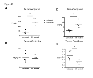
Figure 19 includes .4 panels, identified as panels A. B, C, and. D, whieh show
target
engagement of the arginase inhibitor (compound .9). Levels of serum and tumor
ornithine
and arginine levels in the mice from metabolomics profiling-untreated versus
compound 9-
treated for 3 days at 100 .trigikg are shown in each of Panels A, B, C,4.3nd
D..
For any figure showing a bar histogram. MEW, or other data associated with a
legend, the bars, curve, or other data presented from left to right for each
indication
correspond directly and in order to the boxes from top to bottom of the
lenend.
Detailed 'Description of the Invention
The present invention is based, at least.in part:,. on the discovery that
inactivation of the tumor suppressors,Lkb 1 andrien, in the hingeauses lung
tumors with a.
purely squamous ceII phenotype. These squamons lung tuniors were 100%
penetrant and
recapitulated the genetic, molecular and mieroenvironmental aspects of the
human disease.
With this model, the molecular and genetic mechanisms involved in the
pathogenesis of
lung squamous tumors, including rumor propagating cells, microenvironmental
factors,
immune tolerance, and therapeutic targets were identified. For example,
./..kb./Tiren-null
(LP) tumors expressed the squamous markers KRT5,. p63 and. SOX2, .and
transcriptionally
resembled the basal subtype of human SCC. In contrast to mouse
adenocareinornas, the LP
tumors contained immune populations enriched. for tumor-associated
neutrophils.
SCA.CNCIFR'- fractions were enriched for tumor propagating cells (PCs) that
could
serially transplant the disease in orthotopie assays. TPCs in the LP model and
NCIFW cells
in human SCCs highly expressed PD-L I, suggestion a mechanism of .immune
escape for
TPCs.
- 18
CA 02944903 2016-10-04
WO 2015/164743
PCT/US2015/027515
Accordingly, the present. invention relates., in part, to methods for
predicting
response of a cancer in a subject to anti-immune checkpoint inhibitor therapy
based upon a
determination and analysis of specific biontirkers, such as the presence of
activating
oneogenes (e.g. , activating KRAS, RAS, andior -HRAS mutations), the presence
of
biting tumor suppressors (e.g., inhibiting 11,kb l andfor Pten mutations), and
the amount
(e.g., copy number or level of expression) andior activity of such biomarkers.
In addition,
sueh analyses can be used in order to provide useful .anti-im,mune checkpoint
inhibitor
treatment regimens (e.g., based on predictions of subject survival or relapse,
timing of
adjuvant or neoadjavant treatment, etc.).
Definitions
The artieks "a" .and "an" are used herein to refer to tumor to more than one
(i.e.. to
at least one of the gratnmatical object of the ;article. By way of example,
"an element"
means one element or more than one element.
The term "altered amount" or "altered lever refers to increased or decreased
.eopy
number (e.g., germline andlor somatic) of a hiamarker nucleic, acid, e.g,
increased or
decreased expression level in a cancer sample, as compared to the expression
level or copy
number of the biomarker .nueleic acid in a control sample. The term "altered
amount" of a
biornarker also includes an increased or decreased protein level of a
biomarker protein or
metabolite level of a biomarker metabolite, such as L-arginine or creatine, ìn
Et sample,
a cancer sample, as compared to the corresponding protein or metabolite level
in a normal,
control sample. Furthermore, an altered amount of a biomarker protein may be
determined
by defecting posttranslational modification such as methyiation status of the
marker, which
may affect the expression or activity of the biomarker protein.
The amount of a blomarker in a subject is "significantly" higher or lower than
the
normal amount of the biomarkerõ if the amount of the biomarker is reater or
less,
respectively, than the normal level by art amount greater than the standard
.error of the assay
employed to assess amount, .and preferably at toast 2, 3", 4", 5", 6", 7", 8",
906/0, l00%, 150%, 200%, 300%, 350%, 400%, 500%, 600 ,6, 700%, 800%, 900%,
1000%
or than that amount. Alternately, thc amount of the biumarker in the subject
can be
considered "significantly" higher or lower than the normal amount if the
amount is at least
about two, and preferably at least about three, limn, or five times, higher or
lower,
respectively, than the normal amount of the .biomarker. Such "significance"
can also be
- l9 -
CA 02944903 2016-10-04
WO 2015/164743
PCT/US2015/027515
applied to any other measured parameter described herein, such as for
expression,
cytotoxicity, cell growth, and the like.
The term "altered. level of expression" of .a biomarker refers to an
expression level
or copy number of the biomarker in a test sample, e.g., a sample derived from
a patient
suffering from cancer, that is greater or less than the standard error of the
assay employed
o assess expression or copy number, and is preferably at least twice, and more
preferably
three, four, five or ten or more times the expression level or copy number of
the biomarker
in a control sample (e.g., sample from a healthy subieets not having the
associated disease)
and preferably, the average expression level or copy number of the biamarker
in several
control samples.. The altered level of expression is greater or less than the
standard error of
the assay employed to assess expression or copy number, and is prekrably at
least twice,
and more preferably three, four, five or ten or more times the expression
level or copy
number of the 'biomarker in a control sample (e.g., sample from a healthy
subjects not
having the associated disease) and preferably, the average expression level or
copy -number
of the biomarker in several control samples.
The term "altered_ activity" of a biomarker refers to an activity of the
biomarker
which is increased or decreased in a disease state, e.g., in a cancer sample,
as compared to
the activity of the biomarker in a .normal, control sample Altered activity of
the biomarker
may be the result of, for example, altered expression of the biornarker,
altered protein level
of the biornarker, altered structure of the biomarker, or, e.g., an. altered
interaction with
other proteins involved in the saine or different pathway as the biomarker or
altered
interaction with transcriptional activators or inhibitors.
The term "altered structure of a biomarker refers to the presence of mutations
or
allelic- .variants within a 'biomarkcr nucleic acid or protein, e.g.,
mutations which affect
expression or activity- of the biomarker :nucleic acid or protein, as compared
to the normal
or wild-type gene or protein, 'For example., mutations include, but are not
limited to
substitutions, .deletions, or addition mutations. :Mutations may be present in
the coding or
non-coding region of the biomarker nucleic acid.
-Unless otherwise specified here within, the terms "antibody" and "antibodies"
broadly encompass naturally-occurring forms of antibodies (e.g, 4C1,, 1.gA,
EgM., TgE) and
recombinant antibodies such as single-chain antibodies, chimeric and humanized
antibodies
and multi-specific .antibodies, as ve.11 as fragments and derivatives of all
of the foregoing,
- 20 -
CA 02944903 2016-10-04
WO 2015/164743
PCT/US2015/027515
which fragments and derivatives .have at least an antigenic binding site.
Antibody
derivatives may comprise a protein or chemical moiety conjugated to an
antibody.
The term "antibody" as used herein also includes an "antigen-binding portion"
of an
antibody (or simply "antibody portiors") The terril "anthwn-bindi ng portion",
as used
herein, refers to one or more fragments of an antibody that retain the ability
to specifically
bind to an antigen (e.g., a biomarker polypeptide, fragment thereof, or
biomarker
metabolite). It has been shown that the antigen-binding function of an
antibody can be
performed by fragments of a full-length antibody. Examples of binding
fragments
encompassed ,vithin the term "amigen-binding -portion" of an antibody include
(ì) a Fab
fragment, a monovalent fragment consisting of the VL, VH, Cl..õ and CHI
domains; (ii)
F(a13')2 fragment, a bivalent fragment comprising two Fab fragments linked by
a disulfide
bridge at the hinge region; (iii) a Fd fragment consisting of the VH and CHI
domains; (iv) a
Fv fragment consisting of the VL and VH domains of a single arm of an
antibody, (y)
dAb fragment (Ward et al., (1989) Nature 34i:-5. which consists of a V1-1
domain;
and (vi) an isolated complementarity determining region (C.DIq Furthermore,
although the
two domains of the .E.v fragment, VL and are coded for by separate genes,
they can be
joined, using recombinant methods, by a synthetic linker that enables them to
be made as a
single protein chain in which the VL and regions
pair to form monovalent polypeptides
(known as single chain Fv (scFv); see e.g. ,:Bird et al. (1988) Science
242:423-426; and
Huston et al. 0988) Proc. Nall. Acad. Sci. USA 855879-5883; and Osbourn et at.
1998,
Nature Biotechnology 778) Such single chain antibodies are also intended to
be
encompassed within the term "antigen-binding portion" of an antibody. Any VH
and VL
sequences of specific say ca.n be linked to human immunoglobulin constant
region eDNA
or genomie sequences, in order io generate expression vectors encoding
complete IgG
polypeptides or other isotypes. VH. and VL can also bc used in the generation
of Fab, Fv or
other fragments of immunoglobulins using either protein chemistry or
recombinant DNA
technology. Other forms of single chain antibodies, such as diabodies are also
encompassed. Diabodies are bivalent, bispecifie antibodies in which VH and VI,
domains
are expressed on a single polypeptide chain, but using a linker that is too
short to allow for
pairing, between the two domains on the same chain, thereby forcing the
domains to pair
with complementary domains of another chain and creating two antigen binding
sites (see
e.g., Holliger, P., et al. (193) roc. Natl. Arad. Sel USA 90:6444-6448;
Poljak, R, J., et a/.
(1994) Structure 2:1121-1123).
CA 02944903 2016-10-04
WO 2015/164743
PCT/US2015/027515
Still further, an antibody or antigen-binding portion thereof may be part of
larger
immunoadhesion polypeptides, formed by covalent or noncovalent association of
the
antibody or antibody portion with one or more other proteins or peptides.
Examples of such
immunoadhesion polypeptides include use of the steptavidin .eore region to
make a
tetra/no:fie say polypeptide (Kipriyanov, S.M., et al. (1995).i/town
Antibodies and
Hybridomas 6:934(1) and use of a cysteine residue, biomarker peptide and a C-
terminai
polyhistidine tag to make bivalent and biotinylated say- polypeptidos
(Kipriyanov. SM., et
al. (.1994) Ma Immuno I. 31:1047-1054 Antibody portions, such as Fab and RAO"
.fraiments, can be prepared front whole antibodies using conventional
techniques, such as
papain or .pepsin digestion, respectively, of whole antibodies, .M.orcover,
antibodies,
antibody portions and immunoadhesion polypeptides can be obtained using
standard
recombinant DNA techniques, as described herein.
Antibodies may be poIyclonal or monoclonal; xenogeneie, allotteneic, or
syngencie;
or modified fornis thereof (e.g. humanized, chimeric, etc.). Antibodies may
also be fully
human. Preferably, antibodies of the invention bind specificatly or
substantially
specifically to n biomarker polypeptidc or .64n-tient thereof. The terms
"monoclonal
antibodies" and "monoclonal antibody composition.," as used. 'herein, refer to
a population
of antibody polypeptides that contain only one species of an antigen binding
site capable .of
immunoreacting x.%,ith a 'particular epitope of an antigen, whereas the term
"polyclonal
antibodies" and "polyelonal antibody composition" refer to a population of
antibody
polypeptides that contain multiple species of antigen binding sites capable of
interacting
with a particular antigen. A monoclonal antibody composition typically
displays a single
binding .atfinity for a particular antigen with which it immunoreacts.
Antibodies may also be "humanized", µ;thich is intended to include antibodies
made
by a non-human cell haying variable and constant regions which have been
altered to more
closely resemble antibodies that would be made by a human cell. For example,
by altering,
the .nn-human atnibody amino acid sequence to .incorporate amino acids found
in human
germline iminunoglobulin sequences. The humanized antibodies of the invention
may
include amino acid residues not encoded by human germane inununoglobulin
sequences
(e.g, mutations introduced by random or site-specific inutagencsis in vitro or
by somatic
mutation in vivo), for example in the CDRs. The term "humanized antibody", as
used.
herein, also includes .antibodies in which CDR sequences derived froni the
germline of
- 27 -
CA 02944903 2016-10-04
WO 2015/164743
PCT/US2015/027515
another mammalian species, such as a mouse, have been grafted .onto human
framework
sequences.
The term "assigned score" refers to the numerical value designated for each of
the
biomarkers after being measured in a patient sample. The assigned score
correlates to the
absence, presence or inferred amount of the biomarker in the sample. The
assigned score
can be generated .manuaily (e.g., by visual inspection) or with the aid of
instrunientation .for
image acquisition and analysis. In certain embodiments, the assigned score is
determined
by a qualitative assessment, for example, detection of a fluorescent readout
on a graded
scale, or quantitadve assessment. In one embodiment, an "aggregate score,"
which refers to
the combination of assigned scores from a plurality of measured biomarkers, is
determined.
In one embodiment the aggregate score is a summation of assigned scores. In
another
embodiment, combination of assigned scores involves performing mathematical
operations
on the assigned scores before combining them into an aggregate score. In
certain,
embodiments, the aggregate score is also referred to herein as the predictive
score."
The term "biomarker" refers to a measurable entity of the present invention
that has
been determined to be predictive of anti-immune checkpoint inhibitor therapy
effects on a
cancer. Biomarkers can include, without limitation, nucleic acids, proteins,
and
metabolites, particularly those .relating to oneogene biomarkers (e.g.,
activating, mutations
in oncogene biomarkers) and tumor suppressor biomarkers (e.g., inhibiting
mutations in
tumor suppressors) as shown in Table 1.
A "blocking" .antibody or an ;antibody "antagonist" is one which inhibits or
reduces
at least one biological activity of the antigen(s) it binds. In certain
embodiments, the
blocking antibodies or antagonist antibodies or fragments thereof described
herein
substantially or completely, inhibit a given biological activity of the
antig.en(s).
The term "body fluid" refers to fluids that are excreted or secreted from the
body as
well as fluid that are nomially not (e.g., bronchoalveolar lavage fluid,
amniotic fluid,
aqueous humor, bile, blood and blood plasma, cerebrospinal fluid, cernmen and
earwax,
eowper's fluid or pre-ejacuiatory fluid, chyle, chyme, stool, :female
ejaculate, interstitial
intracelhilar fluid, lymph, menses, breast milk, mucus, pleurai fluid, pus,
saliva,
sebum, semenõ serum, sweat, synovial fluid, tears, urine., vaginal
lubrication., vitreous
humor, vomit).
The terms "cancer" or "tumor" or "hyperproliferative" refer to the presence of
cells
possessing characteristics typical of cancer-causing cells, such as
uncontrolled proliferation,
- -
CA 02944903 2016-10-04
WO 2015/164743
PCT/US2015/027515
immortality, inetastatic potential, rapid growth and proliferation rate, and
certain
characteristic morphological features. in some embodiments, such .cells
exhibit such
characteristics in part or in full due to the expression and activity of
immune checkpoint
inhibitors, such as PD-], PD-U2, andlor CILA-4. Cancer cells are often in
the
form of a tumor, but such cells may exist alone within an animal, or may be a
non-
tumorigenic cancer cell, such as a leukemia cell. As used. herein, the term
"cancer"
includes premalignant as well as malignant cancers. Cancels include, but are
not limited to,
13 .cell cancer, e.g., multiple myelomaõ Waidenstroin's maeroglobulinemia, the
heavy chain
diseases, such as, for example, alpha chain disease, gamma chain disease, and
MU chain
disease, benign .M0110Ciallal gammopathy, and immunocytic amyloidosis,
melanomas,
breast cancer, lung cancer, bronchus cancer, colorectal cancer, prostate
cancer, pancreatic
cancer, stomach cancer, ovarian cancer, urinary bladder cancer, brain or
central lICTVOUS
system cancer, peripheral nervous system cancer, esophageal cancer, cuvical
eamcr,
uterine or endometrial cancer, canter of the oral cavity or pharynx., liver
cancer, kidney
cancer, testicular cancer, biliary tract cancer, small 'bowel or appendix
cancer, salivary
gland cancer, thyroid gland cancer, adrenal gland cancer, osteosareoma,
chondrosarcoma,
cancer of hematologic tissues, and the like. Other non-limiting examples of
types of
cancers applicable to the methods encompassed by the present invention include
human
sarcomas and carcinomas, e.g., fibrosarcoma, myxosarcoma, liposarcoma,
chondrosarcoma,
osteogenic sarcoma., .chordoma, arigiosarcoma, endotheliosarcoma,
lymphangiosarcoma,
iymphangioendotheliosarcoma, synovionia, ruesothelioma, Ewing's tumor,
icionlyosarcoma, rhabdomyosarcoma., colon carcinoma, colorectal cancer,
pancreatic
cancer, breast cancer, ovarian cancer, prostate cancer, squamous
.carcinoma, basal cell
carcinoma, adcnocarcinoma, sweat .gland carcinoma, sebaceous gland carcinoma,
papillary
carcinoma, papillm, adenocarcinornas, eystadenocarcinoma, medullary carcinoma,
bronchogcnic carcinoma, renal eell.carcinoma, hepatoma, bile duct carcinoma,
liver cancer,
ehoriocarcinoma, scminom.a., embryonal carcinoma, 'Wilms.' tumor, cervical
cancer, bone
cancer, brain tumor, testicular cancer, lung carcinoma, small cell lung
carcinoma., bladder
carcinoma, epithelial carcinoma, glioma, astrocytoma, modulloblastoma,
craniopharyngiomaõ ependymoma, pinealoma., hemangioblastoma, acoustic
ileUraina,
oligodendroglioma, .ineningioma, melanoma, neuroblastotna, refinoblastorna;
leukemias,
e.g., acute iymphocytic leukemia and .acute myelocytic leukemia
(inyeloblastic,
promyelocytic, myelomonocytic, monoeytic and erythroleukemia); chronic
leukemia
- 24
CA 02944903 2016-10-04
WO 2015/164743
PCT/US2015/027515
(chronic myelocytic (granulocytic) leukemia and chronic lymphocytic leukemia);
and
polycythcmia v'era, lymphoma (Hodgkin's disease and non-Hodgkin's disease),
multiple
myelomaõ Waldenstrotn's macroglobulinemiaõ and heavy .chain disease, In some
embodiments, cancers are epithiclial in nature and include but are not limited
to, bladder
cancer, breast .cancer, cervical cancer, colon cancer, gynecologic cancers,
renal cancer,
laryngeal cancer, lung cancer, oral cancer, head and neck Caner, ovarian
cancer, pancreatic
cancer, prostate cancer, or skin .cancer. In other embodiments., the cancer is
.hrcast cancer,
prosta.te cancer, lung cancer, or colon cancer. In still other embodiments,
the epithelial
cancer is non-small-cell lung cancer, nonpapillary renal cell carcinoma,
cervical carcinoma,
ovarian carcinoma. (e.g., serous ovarian carcinoma), or breast carcinoma. 'The
epithelial
cancers may be characterized in various other ways including, but not limited
to, serous,
endomcirioid, mucinous, clear cell, Brenner, or undifferentiated.
In some embodiments, lung cancer subtypes arc included. For example, according
to the American Cancer Society, there arc two major types of lung cancer;
small cell lung
cancer (Kik) and non-small cell lung cancer (NW:LC). SUE comprises about 15%
of all
cancers. NSCLE, however, comprises about 85% of all lung cancers and is
divided into
three distinct sub-types: squamous cell carcinoma (about 25-30% of the cases),
large cell
carcinomas (about 10-15%), and adenocarcinomas (about 40%). The cells in these
sub-
types differ in size, shape, and .chemical make-up. These lung cancers are
inclusive of
bronchogcnic carcinoma, bronchial carcinoidsõ ehondroinatous hamartoma,
solitary
pulmonary nodules, pulmonary sarcomas, undifferentiated small cell carcinoma,
undifferentiated large cell carcinoma, and broneholoalveolar carcinomas. Each
such lung
cancer subtype is contemplated for use according to the present invention,
either alone or in
any .combination,
The term "coding region" .refers to regions of a nucleotide SecitiCi3Ce
COMpriSing
codons which are translated into amino acid residues, whereas the term
"noncodina region"
refers to regions of a nucleotide sequence that are not translated into amino
acids (f.g. 5'
and 3' untranslated regions).
The term "complementary" refers to thc.btoad concept Of Sequence
complementarity between regions of two nucleic acid strands or between two
TegiOnS of the
same nucleic acid strand. h is known that an adenine residue of a first
nucleic acid region
is capable of forming specific hydrogen bonds ("base pairina") with a residue
of a second
nucleic acid region .which is antiparallel to the first region if the residue
is thymine or
-
CA 02944903 2016-10-04
WO 2015/164743
PCT/US2015/027515
uracti. Similarly, it is known that a cytosine residue of a first nucleic acid
strand is capable
of base piiiring with a residue of a second nucleic acid strand which is
antiparallei to the
first strand if the residue is guanine. A first region of .a nucleic acid is
complementary to a
second region of the same or a different nucleic acid if, 'When the two
regions are arranged
in an antiparallei fashion, at least one nucleotide residue of the first
region is capable of
base pairing with a residue of the second region. Preferably, the first region
comprises a
first portion and the second region comprises a second portion, whereby, when
the first and
second portions are arranged in an antiparaliel fashion, at least about 50%,
and preferably at
least about 75%, at least about 90%, or at least about 95Vo of the nucleotide
residues of the
first portion are capable abase pairing with nucleotide residues in the second
portion.
lore preferably, all nucleotide residues of the first portion are capable of
base pairing with
nucleotide residues in the second portion,
'The term "control" refers to any reference standard suitable to provide a
comparison
to the expression products in the test sample. In one embodiment, the control
comprises
obtaining a "control sample" from which expression product levels are detected
and
compared to the expression product levels from the test sample. Such a control
sample .may
comprise any suitable sample, .including but not limited to a sample from a
control cancer
patient (can be stored sample or previous sample measurement) with a .known
outcome;
normal tissue or cells isolated from a subject, such as a normal patient or
the cancer patient,
cultured primary cells/tissues isolated from a subject such as a normal
subject or the cancer
patient, adjacent norinal cells/tissues obtained. front the same organ or body
location of the
cancer patient, a tissue or cell sample isolated from a normal subject, or a
primary
cells/tissues obtained from a depository. l.n another preferred embodiment,
the control may
comprise a =femme standard expression product level from any suitable source,
including
but not limited to housekeeping genes, an. expression product level range from
nonnal
tissue (Or other previously analyzed control sample), a previously determined
expression
product level range within a test sample from a group of patients, or a set of
patients with a
certain outcome (for example, survival for one, two, three, four years, etc)
or receiving a
certain treatment (for example, standard of care cancer therapy). It will be
understood by
those of skill in the art that such control samples and reference standard
.expression product
levels can be used in combination as controls in the methods (tithe present
illVentiOn. Jn
one embodiment, the control may comprise nortnal or non-cancerous cell/tissue
sam0e,
another preferred embodiment, the control may comprise an expression level for
a set of
- 26 -
CA 02944903 2016-10-04
WO 2015/164743
PCT/US2015/027515
patients, such as a set of cancer patients, or for a set of cancer patients
receiving a certain
treatment, or for a set of patients with one outcome versus another outcome.
In the former
case, the specific expression product level of each patient can be assigned to
a percentile
level of expression, or expressed as either higher or lower than the mean or
average of the
reference standard expression level. In another preferred embodiment, the
control may
comprise normal cells, cells from patients treated with combination
chemotherapy, and
cells from patients having benign cancer. In another embodiment, the control
may .also
comprise a .measured value for example, average level of expression of a.
particular gene in.
a population compared to the level of expression of a housekeeping gene in the
same
population. Such a population may comprise normal subjects, cancer patients
.vho have not
tmdergone any treatment (i.e., treatment naive), cancer patients undergoing
standard of care
therapy, or patients having benign cancer. In another preferred .embodiment,
the control
comprises a ratio transformation of expression product levels, .including but
riot limited to
determining a ratio of expression product levels of two genes in the test
sample and
comparing it to any suitable ratio of the same two genes in a reference
stmdard;
determining expression product levels of the two or .MOre genes in the test
sample and
determining a difference in expression 'product levels in any suitable
control; and
determining expression product levels of the two or .more genes in the test
sample,
normalizing their expression to expression of housekeeping genes in the test
sample, and
comparing to any suitable control In particularly preferred embodiments, the
control
comprises a control swriple \vhich is of the same lineage .and/or type as the
test sample, hi
another embodiment, the control may comprise expression product levels grouped
as
percentiles within or based on a set of patient samples, such as all patients
with cancer. In
one embodiment a maw' expression product level is established wherein higher
or lower
levels of expression product relative to, for instance, a particular
percentile, are tised as the
basis for predicting outcome. In .another preferred embodiment, a control
expression
product level is established using expression product levels from cancer
control patients,
with a known outcome, and the expression product ievels from the test sample
are
compared to the control expression product le-vel as the basis for predicting
outcome. As
demonstrated by the data below, the methods of the invention are not limited
to use of a
specific cut-point comparing, the level of expression product in the -test
sample -to the
control
- 27 -
CA 02944903 2016-10-04
WO 2015/164743
PCT/US2015/027515
The "copy number" of a biomarker nucleic acid refers to the number of DNA
sequences in a cell. (e.g., germline arid/or somatic) encoding a particular
gene product.
Generally, for a given gene, a mammal has two copies of each gene. The copy
number can
be increased, however, by Rene amplification or duplication, or reduced by
deletion. For
example, germline copy number changes include changes at one or more genomic
loci,
wherein said one or more genomie loci are not accounted. for by the number of
copies in the
normal complement of germline copies in a control (e.g , the normal copy
number in
germline DNA for the same species as that froin which the specific germline
DNA and
corresponding copy number were determined). Somatic copy number changes
include
changes at one or .more genomie Ici,wherein said one or .more genomic loci are
not
accounted for by the number of copies in germline [).NA of a control (e.g.,
copy number in
germline DNA for the same subject as that from which the somatic DNA and
corresponding
copy number were determined),
The "normal" copy number (e.g., germline andicir somatic) of a biomarker
nucleic
acid or "normal" level of expression of a biomarker nucleieacid, protein, or
metabolite is.
the activity/level of expression or copy number in a biological sample, e.g, a
sample
comainine tissue, -whole blood, serum., plasma, buccal serape, saliva,
cerebrospinal fluid,
urine, stool, and bone marrow, from a subject, e.g., a himian, not afflicted
with cancer, or
from a corresponding noncancerous tissue in the same subject who has cancer.
The term "determining a suitable treatment regimen for the subject is taken to
mean the determination of a treatment regimen (i.e., a single therapy or a
combination of
different therapies that are used for the prevention and/or treatment of the
cancer in the
subject) for a subject that is started, modified and/or ended based or
essentially based or at
least partially based. on the results of the analysis according to the present
invention., One
example is determining whether to provide targeted therapy against a cancer to
provide
immunotherapy that generally increases immune responses against the cancer
(e.g., anti
-
immune checkpoint inhibitor therapy). Another example is muting an adiliVallt
therapy
after surgery whose purpose is to decrease the risk of recurrence, another
would be to
modify the dosage of a particular chemotherapy. The determination can, in
addition to the
results of the analysis according to the present invention, be based on
personal
characteristics of the subject -to be treated. In most eases, the actual
determination of the
suitable treatment regimen for the subject will be perthrmed..by the attending
physician or
doctor.
- 28 -
CA 02944903 2016-10-04
WO 2015/164743
PCT/US2015/027515
A molecule is "fixed" or "affixed" to a substrate if it is covalently or non-
covalently
associated µvith the substrate such that the substrate can be rinsed with a
fluid (e.g. standard
saline citrate, pl 74) without a substantial fraction of the Blo lecu e
dissociating from the
substrate.
The term "expression signature" or "signature" refers to a group of two or
more
coordinately expressed biomarkers.. For ex.rmIple, the genes, proteins,
metabolites, and the
like making up this signaturc may be expressed in a specific cell lineage,
state of
differentiation, or during a particular biological response. The biomarkers
can reflect
biological aspects of .the tumors in which .they an expressed, such as the
cell .of origin of
the cancer, the nature of the non-malignant cells in the biopsy., and the
oncogenic
mechanisms responsible :for the cancer. Expression data and gene expression
levels can be
stored on computer readable media, e.g., the .eomputer readable medium used in
conj.unction with a mieroarray or chip reading device. Such expression data
earl he
manipulated to generate expression signatures,
".1-loniolotious" as used herein, refers to nucleotide sequence similarity
between two
regions of the same nucleic acid strand or between regions of two different
nucleic acid
strands. When a nucleotide residue position in both regions is occupied by thc
same
nucleotide residue, then the regions are homologous at that position. A first
region is
homologous to a second re.t_Or if at least one nucleotide residue position of
each region is
occupied by the same residue. Homology between two regions is expressed in
terms of the
proportion of nucleotide, residu.e positions of the two regions that are
occupied by the same
nucleotide residue. By way of ex.ampleõ a region having the nucleotide
sequence 5'-
ATTGCC-.3' and a region haying the nucleotide sequence 5'.-TATGGC-3' Share RV%
homology, Preferably., the first region comprises a first portion and the
SCCOnd region
comprises a. second portion, whereby, at least about 50%, and preferably at
least .about 75%,
at least about )0%, or at least about .95% of the nucleotide residue positions
of each of the
portions are occupied by t.11.e same nucleotide residue. 'More preferably, all
nucleotide
residue positions of each of the portions are occupied. by the same nucleotide
residue.
The term "initmine cell" refers to cells that play a role in the immune
response.
Immune cells are of hematopoietie origin, and include lymphocytes, such as B
cells and. T
rens; .natural kilter cells; inyeloid cells, such as .monocytes, macrophages,
eosinophils, mast
basophils, .and granulocytes.
- 29 -
CA 02944903 2016-10-04
WO 2015/164743
PCT/US2015/027515
The term "immune checkpoint inhibitor" means a group of molecules on the cell
surf:lice of CD4+ andfor CD8 T cells that fine-tune immune responses by down-
modulating or inhibiting an anti-tumor immune response. 111111.111110
Checkpoint proteins are
well known in the art and include, without limitation, CAA-4, PD-I, VISTA, B7-
H2, 137-
1:13, PD-LI, 137-1I4, B7-1116, 2B4, ICS, 'MIEN& PD-12, CD160, p49B, PR-, KIR
family receptors, TIM-1, M1-4, LAG-3, BTLA, SIRPalpha (CD47), CD48, 2E14
(CD244), B7.1, B7.2, 1LT-2, tur-4, TIGHT, and A2aR. (see, for example. WO
2012/177(324). "Anti-immune checkpoint inhibitor therapy" :refers to the use
of agents that
inhibit immune checkpoint inhibitors. Inhibition of one or .more immune
checkpoint
inhibitors can block or otherwise neutralize inhibitory signaling to thereby
.upreeulate an
immune response in order to .more efficaciously treat cancer. Exemplary agents
useful for
inhibiting immune checkpoint inhibitors include antibodies, small molecules,
peptides,
peptidoinimeties, natural !bonds, and derivatives of natural ligands, that can
either bind
and/or inactivate or inhibit immune checkpoint proteins, or fragments
thereof.; as well as
RNA interference, antisense, nucleic acid aptamers, etc. that can downregulate
the
expression and/or activity of immune checkpoint inhibitor nucleic, acids, or
fragments
thereof. Exemplary agents for upreaulating an immune response include
antibodies against
one or more iirminne checkpoint inhibitor proteins block. the .interaction
between the
proteins and its natural receptor(s); a non-activating form of one or more
immune
checkpoint inhibitor proteins (e.g. a dominant negative polypeptide); small
molecules or
peptides that block the interaction between one or more immune checkpoint
inhibitor
proteins and its natural receptor(s); fusion proteins (e.g. the extracellular
portion of an
immune checkpoint inhibition protein fused to the Fc portion of an antibody or
inilmmoglobulin) that bind to its natural receptor(s); nucleic acid molecules
that block
immune checkpoint inhibitor nucleic acid transcription or translation; and the
like. Such
agents can directly block the interaction between the one or more immune
checkpoint
inhibitors and its natural receptor(s) (e.g., antibodies) to prevent
inhibitory signaling and
upregulate an itnniune response. Alternatively, agents can indirectly block
the interaction
between one or more inîrnune checkpoint proteins and its natural receptor(s)
to prevent
inhibitory signaling and upregulate art inimune response. For example., a
soluble version of
an immune checkpoint protein liaand such as a stabilized extract:Hula domain
can binding
to its receptor to indirectly reduce the effective concentration of -the
receptor to bind to an
appropriate ligand. In one embodiment, anti-PD-1 antibodies, anti-PD-L I
antibodies, and
- 30 -
CA 02944903 2016-10-04
WO 2015/164743
PCT/US2015/027515
anti-CTLA-4 antibodies, either alone or in combination or in combination with
anti-ARG
therapeutics described below, are used to inhibit illumine checkpoint
inhibitors.
In some Clilbodiments, arginase I (ARG1) is included within the definition of
"immune checkpoint inhibitor" by virtue of the fact that it has
immunoinhibitory functions.
The term "arginase r refers to a manganese-containing enzyme that catalyzes
the reaction
of arginine and water to ornithine and urea. At least rwo isoforms of
mammalian arginase
exist (types I and II), which differ in their tissue distribution, subcellular
localization,
immunologic erossreactivity and physiolouic function. The type isofortn
encoded by this
gene, is a cytosolic enzyme and expressed predominantly in the liver as a
component of the
urea cycle. Representative human ARG1 cDNA and protein sequences are well-
known in
the art and are publicly available from the National Center for Biotechnology
Information
(NOM), For example, .ARGi isoform 1 is available under accession numbers
NA/1_001244438,1 and N P ()l2'I367 I RGl isoform 2, available under
accession
numbers M1_000045,3 and NP_000036.2, uses an alternate in-frame splice site at
the 5'
end of an exon compared to variant 1 resulting in the same N- and C-termini,
but is shorter
in comparison to isoform 1. Nucleic acid and polypeptide sequences of ARG1
orthologs in
organisms other than humans arc well known and include, for example, mouse
ARG1
(N.M._007842,3 and NP_031508.1), chimpanzee ARG1 (XM_0033114892 and
Xp_003311537.1), monkey ARO] (XM._0011036092 and XP 001103(09.2), dog ARG1
(XM_532053,4, XP_532053.3, XM_003639427.2, and XP_003639475.1), cosy ARG1
(N1\4_001046154.1 and NP _001039619.1), rat ARG1 (N1_)17134.3 and
N.P_058830,2),
and zebrafish ARG1 (NK.)01045197.1 and N11_001038662.1), Representative ARG1
sequences are presented below in Table I. Anti-ARG1 antibodies are well-known
in the art
and include, for example, 16001-I-AP (Pwteinteeh Group), AMAb90545 (Atlas
Antibodies), and PA1783 (Boster Immunoleader). 113 addition, other inhibitors
of A.RGI.
(e.g., small molecules) are known and include, for example, N-hydroxy-L-
arginine and
2(S)-amino-6-boronotiexonic acid (ABI-11). Moreover, assays for measuring
.ARGI amount,
activity, and metabolites are well-known in the art (see, for example, US,
Pat. Publ. 2011-
02(t348). It is to be noted. that the term can further be used to refer to any
combination of
features described herein regarding .ARG1 molecules, For example, any
combination of
sequence composition, percentage identify, sequence length, domain structure,
functional
activity, etc. can be used to describe an ARG1 molecule of the present
invention.
CA 02944903 2016-10-04
WO 2015/164743
PCT/US2015/027515
"PD-1" is an immune checkpoint inhibitor that refers to a member of the
immunoglobulin gene superfamily that fimctions as a coinhibitoly receptor
haying PD-LI
and PD-L2 as known ligimds, PD- I was previously identified -using a
subtraction cloning
based approach to select for proteins involved in apoptotic celldezith, PD-I
is a meniber of
the C28/CIA-4 family of molecules based on its ability to bind to PD-LL Like
CTLA-
PD-1 is rapidly induced on the surface of T-cells in response to anti-C133
(Agata et a I. 25
(1)96) Int. bulimia 8:7(5). In contrast to CTLA-,1, hOWCVCF, PD-1 is also
induced on the
surface of B-cells (in response to anti-IgM). PD-1 is also expressed on a
subset of
thymocytes and myeloid cells (Agata et al, (1996) supra; Nishimura et al.
(1.99) inf.
immoral. 8:773).
The nucleic acid and amino acid sequences Oa representative human PD-1
biornarker is available to the public at the C3enBank database, under
NIV1_00501$.2 and
NP 0050092 (sec also Ishida et al. (1992) 20 EMBO J11:3887: Shinohara el al.
(1994)
(enomies 23:704; U. S. Patent 5,698,520). PD- i has an extracellular region
containing
itrununoglobulin superfamily domain, a transmembrane domain, and an
intracellular regíon
including an immunoreceptor tyrosine-based inhibitory motif (ITIM) (shida et
al. (1992)
EA/1101. 11:3887; Shinohara a al. (1994) Genornics 23:704; and U.S. Patent
5,698,520).
Those features also define a larger family of polypeptides, called the
imnumoinhibitory
receptors, which also includes gp4913. P1R-B, and the killer inhibitory
receptors (KIRs)
(Vivier and Dacron (1997).1mnunal. Today 18:286). it is often assumed that the
tyrosyl
phosphorylated IT1M moti f of these teceptors interaets with SH2-domain
containing
phosphatases, which leads to inhibitoty signals. A subset of these
immunoinhibitory
receptors bind to MFIC polypeptides, for example the KIRs, and CTLAe1 binds to
137-1 and
137-2. It has been proposed that there is a phylogcnetic relationship between
the and
87 genes (H.enry et a I. (1999) immtawl. Today 20(6):285-8). Nucleic acid and
polypeptide
sequences of PD-I orthologs in organisms other than humans are µvell known and
include,
for example, mouse PD-1 NM _008798.2 and NP_032824.1), rat PD-1 (N)0 1 I
06927.1
and NP001 10(J397,1), dog PD-1 (XM_543338.3 and X' 53338.3), cow PD-1
(NN1.001083506.1 and NP001076975.1), and chicken P13-1 (XM2422723.3 and
X42723 .2).
PD-1 polypeptides are inhibitory receptors capable of transmitting an
inhibitory
signal to au immune cell to thereby inhibit immune cell effector function, or
are capable of
promoting costimulation (e.g., by competitive inhibition) of immune cells,
e.g., when
- 37 -
CA 02944903 2016-10-04
WO 2015/164743
PCT/US2015/027515
present in soluble, monomeric form. Preferred PD-1 family members share
sequence
identity with P[)-it and bind to one or more 87 member, e.g,, B7-1, B7-2,
PD-1
andlor other .polypeptides on antigen presenting cells.
The term "P11)-1 activity" includes the ability of a. PD-1 pol.ypeptide to
modulate an
bitory signal in an activated immune cell., e.g,, by engaging a natural PD-1
ligand on an
anti= presenting cell, PD-1 transmits art .inhibitory signal to an immune cell
in a manner
similar to CT1A4. Modulation of an .inhibitory signal in an immune cell
results in
modulation of proliferation of, andior cytokine secretion by, an immune cell.
Thus, the
term "PD-1 activity" includes the ability of a PD- 1 polypeptide to bind its
natural ligand(s),
the ability to .modulatc immune cell costimulatory or inhibitory sistnals, and
the ability to
modulate the immune response.
The term "PD-I ligartd" refers to binding partners of the P11)-1 receptor and
includes
both P13-L1 (Freeman et al. (2000)J. Exp. Med. 1.92;1027) and PD-L2
.(Latchinan et al.
(2001 .)Nat. Immunol. 2261), At least two types of human P13-1 lifAand
polypeptides exist..
PD-1 ligand proteins comprise a sipal sequence, and an lgV domain, an IgC
domain, a
transmernbrane domain, and a short cytoplasmic tail. Both P11)-L.1 (See
:Freeman et al.
(200)) I. Exp.lqed, 192:1027 for sequence data and P11i)-L2 (See Latchman et
al. (2001)
'Nat. framunol, 22(i1. for sequence data) are members of the .137 family of
polypeptides.
Both PD-Lit and PD-L2 are expressed in placenta, spleen, lymph nodes, thymus,
and heart,
Only R11)-1,2 is expressed in pancreas, lung and liver, while only PD-L1 is
expressed in .fetal
liver, Both .PD-1 ligands are upregulated on activated monocytes and dendritic
cons,
although PD-Ii expression is broader. For example, P13-1,1 is known to be
constitutively
expressed and upregulated to higher levels on inurine hernatopoietic cells
T cells, 13
cells, .macrophag.es, dendritic cells (DCs), and bone marrow-derived .mast
cells) and non-
hematopoietie cells (e.g., endothelial, epithelial, and muscle ceils), whereas
PD-L2is
inducibly expressed on DCs, macrophages,. and bone marrow-derived mast cells
(see, Butte
et al. (2ow) Antnuttity 27:111).
PD-1 ligand.s comprise a family of polypeptides haying certain conserved
structural
and functional features. The term "family" when used to refer to proteins or
nucleic acid.
molecules, is intended to mean two or .more proteins or nucleic. acid
.molecules .having
common structural domain or .motif and having sufficient amino acid or
nucleotide
sequence homology, as defined herein. Such thmily members can be naturally or
non-
naturally occurring and can he from either the same or different species. For
example, a
- 33 -
CA 02944903 2016-10-04
WO 2015/164743
PCT/US2015/027515
thraily cm contain a first protein of human origin, as well as other, distinct
proteins of
human origin or alternatively, can contain homologues of non -human origin.
Members of a
family may also have conimon functional characteristics, Pt)- I ligands are
IlleMbOTS of the
B7 :family of polypeptides. The term "B7 family" or -137 polypeptides" as used
herein
includes costimulatory polypeptides that share sequence homology with 137
polypeptides,
t?.g,, with 137-1 (CD()), 137-2 (C1i)8(9), inducible costimulatory ligand
(ICOS-L), 137-B.3,
137-H4, VISTA, E37-116, 13711 (Swallow- et al. 099)) immunity 1 1:423), an/or
PD-1 ligands
(e.g., P[)-L1 or PD-1,2). For exarnpleõ hurnan B7-I and 137-2 share
approximately 26%
amino acid sequence identity when compared using the BLAST program at NCB1
with the
default parameters (BIosurn.62 matrix with gap penalties set at existence I I
and extension I
(see the NCBI website) The term B7 :family also includes variants of these
polypeptides
which are capable of modulating immune cell function. The 137 family of
molecules share
a number of conserved regions, including signal domains, IgV domains and the
IgC
domains, ig,V domains and the IgC domains are art-recognized la superfamily
member
domains. These domains correspond to structural units that have distinct
folding patterns
called 1g folds. la folds are comprised of a sandwich of two l sheets, each
eousisting of
anti-parallel l strands of 5-10 amino acids with a conserved disulfide bond
between the two
sheets in most, but not all, TgC domains fig, TCR, and MEC molecules share
the same
types of sequence patterns and are called the CI-set within the ig
superfamily. Other IgC
domains fall within other sets. lgV domains also share sequence patterns and
are called V
set domains. IgV domains are longer than 1gC, doinains and contain an
additional pair off/
strands.
The term "PD-LI" refers to a specific PD-1 ligand. Two forms of human PD-11,1
molecules have been identified. One form is a naturally occurring PD-LI
soluble
polypeptide, i.e., having a short hydrophilic domain at the COOH-terminal end
and no
transmembrane domain, and is referred to herein as PD-I.I S. 'The second form
is a cell-
associated polypeptide, i.e., haying a transmembrane and cytoplasmic domain,
referred to
herein as PD-L1 M, The nucleic acid and amino acid sequ.ences of
representative human
PD-L1 biomarkers regarding PD-L1M are also available to the public at the
GenBank
database under NIVLO1 41 43.3 and NP_054862.1. PD-L1 proteins comprise a
signal
sequence, and an TgV domain and an :tgC domain, The sinnal sequence is from
about amino
acid ì to about amino acid. 18. The signal sequence is from about amino acid 1
to about
amino acid IS. The IgV domain is from about amino acid .19 to about amino acid
134 and
- 34 -
CA 02944903 2016-10-04
WO 2015/164743
PCT/US2015/027515
the IgV domain is from about amino acid to about amino acid 134. The IgC
domain is
from about amino acid 135 to about amino acid 227 and the ligC. domain of
SEQ1D NO: 6
is shown from about amino acid 135 to about amino acid 227. The hydrophilic
tail of
comprises a hydrophilic tail shown from about amino acid 228 to about amino
acid 245.
The PD-1.1 polypeptide comprises a transmembrane domain shown from about amino
acids
239 to about amino acid 259 and a cytoplasmic domain shown of about 30 amino
acids
from 260 to about amino acid 290, In addition, nucleic acid and polypeptid.c
sequences of
orthologs in organisms other than humans are wen known and include, for
example,
mouse PD-L1 (NMJ21893.3 and NP068693.1), rat PD-LI (NM _001191954,1 and
41001178883.1), dog PD-L1 (X1 5O2 and XP541302.3), cow PD-Li
(KM001163412.1. and NP_0(1156884.1), and chicken PD-.1.,1 (XM__.424811,3 and
XP_42481L3).
'The term "PD-L.2" reRrs to ;another specific PD-1 iigand. PD-L2 is a 87
family
member expressed on various APCs, including dendritic cells, macrophages and
bone-
marrow derived mast cells (Thong et al. (2007) Eur.J. Immunol. 37:24)5). A.PC-
expressed
PD-L2 is able to both inhibit T cell activation through ligation of PD-1 and
costimulate T
cell activation, through a PD-1 independent mechanism (Shin et al. (2005)J.
Exp. Med
201:1531). In addition, ligation of deadline cell-expressed PD-L2 results in
enhanced
dendritic cell cytokine expression and survival (Radhakrishnan et I. (2003) J.
bwmatol.
37!1827; Nguyen et al. (2002) Erp. Med. 19(:1393). The nucleic acid and amino
acid
sequences of representative human PD-L2 blomarkers are u,elI known in the art
and are
also available to the public at the GenBank database under NM 025239.3 and
NP 079515,2, PD-11,2 proteins are characterized by common structural elements.
In some
embodiments, PD-L2 proteins Maude at least one or more of the following
domains: a
sipal peptide domain, a transmembrane domain, anl,c4V domain, an IgC domain,
an
extracellular domain, a transmembrane domain, and a cytoplasmic domain. For
example,
amino acids 1-.19 comprise a signal sequence. As used herein, a "signal
sequence" or
signal peptide" serves to d.irect a polypeptid.e containing such a sequence to
a lipid bilayer,
and is cleaved in secreted and membrane bound polypeptides and includ.es a
peptide
containing about 15 or more amino acids which occurs at the N-terminus of
secretory and
membrane bound polypeptides and which coatains a large number of hydrophobic
amino
acid residues. For example, a signal sequence contains at least about 10-30
amino acid
residues, preferably about 15- 25 amino acid residues, more preferably about
18-20 ainino
- 35 -
CA 02944903 2016-10-04
WO 2015/164743
PCT/US2015/027515
acid residues, and even more preferably about 1.9 amino acid residues, and has
at least
about 35-65%, preferably about 38-50%, and more preferably about 40-45%
hydrophobic
amino acid residues (e,g., valine, leucine, isoleueine or phenylalanine). In
another
embodiment, amino acid residues 220-243 of the native human PD-12 polypeptide
and
amino acid residues. 20.1-243 of the mature polypeptide comprise a
transmembrane domain.
As used herein, the term "transtnembrane domain" includes art amino acid
sequ.enee
about 15 .amino acid residues in length leVhiCh vans the plasma .membrant,
More
preferably, a transmembrane domain includes about at least A, 25. 30, 35, 40,
or 45 amino
acid residues and spans the plasma membrane. Transmembrane domains are rich in
hydrophobic wsidues, and typically have an alpha-helical structure., In a
pmferred
embodiment, at least 50%. 60%, 70%, 80%. 90%, 95% or more of the amino acids
of a
transmembrane domain are hydrophobic, eg, ucines, isoleucines, tyrosines, or
tryptophans. Ti-ansmembrane domains are described in, for example, Zagotta et
al. (1996)
Annu. Rep. NeltrOSCi. 19-,. 235-263, In still another embodiment, .amino acid -
.residues 20-
120 of the native human PD-1,2 polypeptide and amino acid residues 1-1.0i of
the 1mm:re
polypcptide comprise an IgNi domain. Amino acid residues 121- 219 of the
native human
PD-L2 polypeptide and amino acid residues 102-200 (Wale mature polypeptide
comprise an
1t4C domain. As -used herein, 10 and .igC domain.s are recognized in the art
asIg
superfamily member .domains, These domains correspond to structural units that
have
distinct folding patterns ..called ig folds. Is folds are .comprised of a
sandwich of two B
sheets, cath consisting of antiparaliel (3 strands of 5-10 amino acids with a
conserved
disulfide bond between the two Sheets in most, but not all, domains. ligC
domains of
TCR, and WIC molecules share the same types of sequence patterns and are
called the Cl
set within the ig superfamily. Other IgC.- domains fall within other sets. IgV
dotnains also
share sequence patterns and are called V set domains. 1.gV domains are longer
than C-
domains and form an additional pair of strands. In yet another embodiment,
amino acid
residues1-219 of the native human PD-1,2 polypeptide and amino acid residues.
1.-200 of
the mature poly-peptide comprise an extraceitular domain. As used herein, the
term
extraceiluiar domain" represents the N-terminal amino acids which extend as a
Lill from
the surface of a cell. An extracellular domain of the present invention
includes an igV
domain and an NC. domain, and may inelud_e a signal 'peptide domain. in still
another
enabodiment, amino acid residues 244-273 of the native human .PD-L2
polypeptide and
amino acid residues 225-273 of the mature polypeptide comprise a cytoplasmic
domain, .As
-36-
CA 02944903 2016-10-04
WO 2015/164743
PCT/US2015/027515
used herein, the tcmi 'cytoplasmic domain" represents the C-terminal amino
acids which
extend as a tail into the cytoplasm of a cell. In addition, nucleic acid and
polypeptide
sequenees of PD-L2 orthologs in organisms other than humans are well known and
include,
for example, mouse PD-L2 (NfV1 j121396.2 and NP _067371 .1), rat PD-1..2
(NM_001107582.2 arid NP_001101(152.2), dog PD-L2 (X1_847012.2 and
XP_852105.2),
coxv PD-L2 (X114_58(846.5 and X.P_586846,3), and chimpanzee. PD-L2
(XM()0I1,40776.2
and X19 ()()l i40776,1),
The term "P13-L2 activity," "biological activity of PD-L2," or ".functional
activity of
PD-L2," refers to an activity exerted by a PD-L2 protein, polypeptide or
nucleic acid
molecule on a PD-L2-responsive cell or tissue, or on a PD- L2 polypeptide
binding partner,
as determined in vim or in vitro, according to standard techniques. In one
embodirnent, a
PD-L2 activity is a direct activity, such as an association with a PD-L2
binding partner. As
u.sed herein, a "taraea molecule" or partrae is a molecule with svhich a PD-
L2
polypeptide binds or interacts in nature, such that PD-L2-mediated function is
achieved. In
an exemplary embodiment, a PD-L2 target molecule is the receptor RGMb.
Alternatively,
a PD-L2 activity is an indirect activity, such as a cellular signaling
activity mediated by
interaction of the PD- L2 polypeptide with its natural binding partner, e.g.,
RGMb. The
biological activities of PD-1,2 are described herein. For example, the PD-L2
polypeptides
of the present invention can have one or more of the following activities: I)
bind to andfor
modulate the activity of the receptor RGMb, PD- or other PD-L2 natural binding
partners,
2) modulate intra-or intercellular signaling, 3) modulate activation of immune
cells, e.g. , T
lymphocytes, and 4) modulate the immune response of an oranism, e.g., a mouse
or
human organism.
The term "itinnime response" includes T cell mediated andlor B cell mediated
immune responses. Exemplary immune responses include T cell responses, e.g.,
eytokine
production and cellular cytotoxicity. In addition, the term immune response
includes
immune responses that are indirectly effected by T cell activation, e.g.,
antibody production
(Immoral responses) and activation of cytokine responsive cells, e.g.,
macrophages.
The term "immunotherapeutic af..3ent" can include any molecule, peptide,
antibody
or other agent which can stimulate a host immune system to generate an immune
response
to a tumor or cancer in the subject. Various immunotherapentie agents are
useful in the
compositions and methods described herein,
- 37 -
CA 02944903 2016-10-04
WO 2015/164743
PCT/US2015/027515
The term "inhibit" includes the decrease, limitation, or blockage, of, for
example a
particular action, function, or interaction. in some embodiments, cancer is
"inhibited" if at
least one symptom of the cancer is alleviated, terminated, slowed, or
prevented. As used
herein, cancer is also "inhibited" :if recurrence or metastasis of the cancer
is reduced,
slowed, delayed, cw prevented.
The term "interaction", when referring to an inter.action between two
molecules,
refers to the physical contact (e.g., binding) of the molecules with one
another. Generally,
such an interaction results in an activity (which produces a biological
effect) done or both
of said molecules.
An "isolated protein" refers to a protein that is substantially free a other
proteins,
cellular material, separation medium, and culture medium when isolated from
cells or
produced by recombinant DNA techniques, or chemical precursors or .other
chemicals Ivhen
chemically synthesized. An "isolated" or "purified" protein or bioloaicaliy
active portion
thereof is substantially free of cellular material or other contaminating
proteins from the
cell or tissue source from \-vlaich the antibody, polypeptide, peptide or
fusion protein is
derived, or substantially free from chemical 'precursors or other chemicals
when chemically
synthesized. The lanattage "substantially free of cellular material" includes
preparations of
a biomarker polypeptide or fragment thereof, in which the protein is separated
from cellular
components of the .cells from which it is isolated or reeombinantly produced.
in one
embodiment, the language "substantially free &cellular material" includes
preparations of
a biomarker protein or fragment thereof, havina icss than about 3" (by dry
weight). of
non-biomarker protein (also refrred to herein as a "contaminating protilin."),
inore
preferably less than about 20 ,4 of non-hiomarker protein, still more
preferably less than
about l0`.X3 anon-No=11er protein, and .most preferably less than about 5% non-
biomarker protein, When antibody, polypeptide, peptide or fusion protein or
fragment
thereof, e.g.. a biologically active fragment thereof, is recombinantly
produced, it is also
preferably substantially. free of culture medium, Le., culture medium
represents less than
about 20%, more preferably less than about 10%, and most prefera.bly less than
about 5% of
the volume of the protein preparation,
A -kit" is any manufacture (e.g. a package or container) comprising, at least
one
reagent, e.g a probe or small molecule, for specifically detecting arid/or
affeetina the
expression nf a marker of the invention. The k.it may be promoted,
distributed, or sold as a
unit for performing the methods of the present invention. The kit may comprise
one or
- 38 -
CA 02944903 2016-10-04
WO 2015/164743
PCT/US2015/027515
more reagents neeessaty to express a composition useful in the methods of the
present
invention. 1.11 certain embodiments, the kit may further comprise a reference
standard,
a nucleic acid. encoding a protein that does not affect or regulate signaling
pathways
controlling mil growth, division, migration, survival or apoptosis. One
skilled in the art can
envision many such control proteins, including., but not limited 1.0, COMMOTI
molecular tags
LuKiel] fluorescent protein and. beta-galactosid.ase), proteins not classified
in any of
pathway encompassing e(11 growth, division, migration, survival or apoptosis
by
GeneOntology reference, or ubiquitous housekeeping proteins. Reairents in the
kit may be
provided in individual containers or as mixtures of two or more reagents in a
single
container. In addition, instructional .materials which describe the use of the
compositions
within the kit can be included.
The term "ncoadjuvant therapy" refers to a treatment given before the primary
treatment. Examples of neoadjuvant therapy can include chemotherapy, radiation
therapy,
and hormone therapy. For example, in treating breast cancer, neoadjuvant
therapy can
allows patients with large breast cancer to undergo breast-conserving surgery.
The "normal" level of expression of a biamarker is the level of expression of
the
biomarker in cells of a subject, e.g., a human patient, .not afflicted with a
cancer. An "over-
expression" or "significantly higher level of expression" of a biomarker
.refers to .an
expression level in a test sample that is greater than the standard error of
the assay
employed to assess expression, and is preferably at least twice, and more
preferably
2.2, 2.3, 2.4, 2.5, 2.6, 2.7, 2.8, 2.9, 3, 3,5, 4., 4.5, 5, 5.5, 6, 6.5, 7,
7,5, 8, 8,5, 9, 9.5, 10, 10,5,
11, 12,13,14, 15, 16, 1.7, 18, 19, 20 .times or more higher titan the
expression activity or
level of the biomarker in a control sample (e.g., sample from a healthy
subject not having
the hiomarker associated disease) and preferably., the average expression
level of the
biomarker in several control samples. A "significantly lower level of
expression" of a.
biomarker refers to nn expression level in a test sample that is at least
twice, and more
preferably 2:1, 2.2, 2.3, 2.4, 2.5, 2.6, 2,7, 2,8, 2,9, 3, 3.5, 4, 4.5, 5,
5.5, 6, 6.5, 7, 7.5, 8, 8.5,
9, 9,5, 10, 10.5, 11, 12, 13, 14., 15, 16, 17, 18, 19, 20 times or more lower
than the
expression level. of the btomarker in a control sample (e.g., sample from a
healthy subject
not having the biomarker associated disease) and preferably, the average
expression level of
the biomarker in several control samples.
The tertn "oncoacne" refers to a well-known class of proteins that have the
potential
to cause cancer or a nucleic acid encoding same. Non-limiting examples of
oneogenes
- 39 -
CA 02944903 2016-10-04
WO 2015/164743
PCT/US2015/027515
include growth factors or mitogens such as c-Sis; receptor tyrosine kinases
such as EGFR,
PDGFR., and VEGER; cytoplasmic tyrosine kinases such as NH and kinases in the
Sic-family, Syk-ZAP-70 fiunily, and BTK family of tyrosine kinases;
cytoplasmic
serineithreonine kinases and their regulatory subunits such as PIK3CA. MORI,
and RAF
(e.g., RAF-I., A-RAF, B-RAF); regulatory GTPases such as RAS (e.g., '<RAS);
transcription factors such as MYC; and combinations thereof.
By contrast, the term -tumor suppressor refers to a well-known class of
proteins
that have the potential to protect a cell from 'becoming a cancerous cell. Non-
limiting
examples of tumor suppressor genes include the TP53 gene (also known as the
P53 :wile),
which encodes p.53 (also known as protein 53 or tumor protein 53); kinases
such as, e.g.,
tyrosine kinases or serinefthreonine kinases including serinefthreonine kinase
11 (STK] ly,
the RBI. gene, which encodes the Retinoblastoma protein (pRb); LKBI,; PTEN; VI-
IL;
APC; C1195; STS; 'YPEL.3; ST7; STI.4; and combinations thereof.
The term "at least one mutation" in a polypeptide or a gene encoding a
polypeptide
and grammatical variations thereof means a polypeptide or gene encoding a
polypeptide
having one or more allelic, -variants, splice variants, derivative variants,
substitution
variants, dektion variants, truncation variants, and/or insertion variants,
fusion
polypeptides, orthologs, and/or interspeeies homologs. By way of example. at
least one
mutation of a Ras protein would include a Ras protein in which part of all of
the sequence
of a polypeptide or gene encoding the Ras protein. is absent or not expressed
in the cell for
at least one Ras protein produced in the cell. For example, a Ras protein may
be produ.ced.
by a cell in a truncated fOrm and the sequence of the tnincated form may be
wild type over
the sequence of the truncate. A deletion may mean the absence of all or part
of a gene or
protein encoded by a gene. Additionally, some of a protein expressed in or
encoded by a
cell may be mutated while other copies of the same protein .produced in the
same cell may
be wild type. 13y uay another example a mutation in a Ras protein would
include a Ras
protein haying. onc or more amino acid differences in its amino acid sequence
compared
with wild type of the same Ras protein, By way of another example, a
mutation LKBI inclu.des, but is not limited to, an LKBI ha-vine at least one
amino acid.
difference compared to wild type LKB 1.. Mutation may he somatic or germline.
NIutations
in a polypeptide, including, but not limited -to, LKB 1. can lead to
expression of truncated
protein,
- 40
CA 02944903 2016-10-04
WO 2015/164743
PCT/US2015/027515
Mutations in an oneogene that cause imreased activity of the oneogene to
therefore
promote cancer are known as "activating mutations," Such activating mutations
are well
known in the art for many oneogenes and particularly for the activating mutant
oncogenes
described herein. Such mutations can be constitutive (Le., always causing
increased
activity) or inducible. Such mutations can also cause variable increases in
oncogene
activity.
For example, the term '!'RAS" and "?RAS' and "HRAS" refer tospecific
:embers of the rat sarcoma (RAS) superfamily of proteins, V-Ki.las2
Kirsten.rat sarcoma
viral ancogenc homolou (KRAS), v-Ha-ras Harvey rat sarcoma viral oncogene
homolog
(HRAS), and neuroblastoma. RAS viral (v-ras) oneogene homolog (RAS ) are the
.founding
members of the rat sarcoma (RAS) superfamily of small guanosine
triphosphatases
(GTPases) that is known to .comprise >150 members in humans (Coliccili .(2)04)
Scì. STKE
2004:RE1 3). Five subgroups of these small OTPases have been identified and
designated
as the RAS: ras homolog family member (RHO); RAM A. member RA'S oricogene
family
(RA B); RAN, menther RAS (Imogene family (RAN); and ADP-ribosylation factor
(ARF)
families. All small GTPases function as binaty switches that wansition between
GDP-
bound. inactive and GTP-bound, active forms and thereby contribute to
intracellular
signaling that underlies a wide array of cellular activities, including cell
.proliferation,
differentiation, survival, motility, cytoSkeletort rearrangements, and
transformation (Cox
and Der (2010) Small 67Pases 12-27; Lowy et I. (1993) Anna. Rev. Biochent.
62:85'-
891). Somatic point mutations that activate. KRAS, 'I-IRAS, or NRAS have been
identified
in a variety of human tumors, with KR.A.S being the most frequently activated
oncoprotein
in humans. Somatic activating mutations of KRAS are thus present in. >90% of
pancreatic
adenocareinomas, for example (Jaffee et I. (2002) Cancer Cell 2:25-24 Since
members
of the Ras family communicate signals from outside the cell to the .iltidatS,
mutations in
Ras pathway signalling can permanently activate it and cause inappropriate
transmission
inside the cell CVeli in the absence of extracellular signals. Because these
signals result in
cell growth .and division, dysregulated RAS pathway signaling, such as
promoted by
activatin RAS mutations, can ultimately iead co oncogenesis .and cat=
(Ciood.sell et aL
(1999) Oncologist 4: 20-264). Activated mutations in the Ras family (e.g., .H-
Ras, M-Ras
and K-Ras) are found in 20-25% of all human tumors and up to 90% in specific
tumor types
(Downward et aL (2003) Nat. Rev. Cancer 3:11-22; Bos et at. (1989) Cancer Res.
49:4682-
,4689; Kranenburg et al. (2005) Mackin. Mophys. Acta 756:8.142).
-
CA 02944903 2016-10-04
WO 2015/164743
PCT/US2015/027515
Representative human KRAS (DNA and protein sequences are well-known in the
art and are public:1y available from the National Center for Biotechnolm,
Information
(NCI). For example, KRAS isotbrm 1 is available under accession timbers
NM_033360,3 and NP_203524.1 and is composed of six exams, including exon 4a,
which
the shorter transcript variant 2 laCks. This rare variant has a coding
sequence that
terminates in exon 4a and encodes a unique C-terminus, con:wand to isoform 2,
KRAS
isoform 2., available under accession numbers N N1_004985.4 and NP 4ì72 is
composed of five exons and lacks exon 4a which the longer transcript variant 1
includes.
This predominant variant as a cds that terminates in min 4b and encodes
isoform 2,
Nucleic acid and poiypeptide sequences of KRAS orthologs in organisms other
than
humans are well known and include, for example, canine KRAS (NCBI Accession
XM_540523.3, XP_54.1523.3, XM_003432429.1, and XP_00343247,1), chimpanzee
KRAS (WTI Accession X114_0033137)4,1, XP_003313842,1, X114528758.3, and
Xp.528758,3), cow KRAS (KM Accession NM JI011I0001,1 and NP 001103471.i ),
mouse KRAS OWB1 Accession NM_021284.6 and NP 7259.4), rat KRAS (NCBI
Accession Nm..931515.3 and NP. J13703.1), chicken KRAS NOM Accession
NM_001256162.1 and NP ..001243091.1), and zebrafisb KRAS (NCB1 Accession
NM_001003744,1 and NP_001003744. ). Representative KRAS sequences are
presented
below in Table 1. It is to be noted that the term can further be used to refer
to any
combination of features described herein regarding KRAS molecules. For
example, any
combination of sequence composition, percentage identify, sequence length,
domain
structure, functional activity, etc. can be used to describe a KRAS molecule
of the present
invention.
Representative human NRAS CDNA and protein sequences are xveil-known in the
art and are publicly available from the National Center for Biotechnology
Infomation
(NCB!). For example, NRAS sequences are available under accession numbers
NM_002524.4 and NP _002515,1. Nucleic acid and polypeptide sequences of KRAS
orthoious in organisms other than humans are Ikell known and inctude, tbr
example, dog
N-RAS (N1\1_001287065,1 and NP_001273994 ), chimpanzee .NRAS (XM._001149822.3
and XP_001149822.1), co NRAS (M4_001097989.1 and N13_001091458.1), inouse
NRAS (XN4...006501122.1, X11_006501185,1, W..006501119,1, XP..006501182.1,
XM_006501 i 18.1, XP _006501181,1, XIV1_006501120,1, XP _006501183,1,
X4_00650112
X13_006501184,1, X.M._006501123.1, and XP_006501186.1), rat NRAS
- 42 -
CA 02944903 2016-10-04
WO 2015/164743
PCT/US2015/027515
(NN1_08076(.2 and NP_542944.1), chicken NRAS tNMJI01012549.1 and
NEL001012567.1), and zebrafish NRAS NI'vtI31 '5. ind NP_571220.1).
Representative NRAS sequences are presented below in Table I It is to be noted
that the
term can further be used to refer to any combination of features described
herein :regarding
NRAS molecules. For example, any combination of sequence composition,
percentage
identif, sequence iength, domain structure, -functional activity, eie. can be
used to desetibe
NRAS molecule of the present invention.
Representative human HRAS cON A and protein sequences are well-known in the
art and are publicly available from the National Center for Biotechnology
Itiformation
(NCBI). For example, HRAS isofOrm 1 is available under accession timbers
NP005334.1 and NP001123914.1 and is encoded by two different splice 'variants.
Transcript 1 (NM _005343.2) differs from transcript 3 (NM._001130442.1) in the
3' UTR.,
but otherwise eicodes the sanie protein. Transcript 2 (NM_O()5343.2) encodes
an alternate
exon in its 3 coding region and a different 3'UTR from transcripts 1 and 2
resulting in a
shorter isoform 2 (NP:789765.1) compared to isoform t and contains a distinct
C-terminus
as well. Nucleic acid and polypeptide :sequences of HRAS orthologs in
organisms other
than humans arc well known and include, for example, dog HRAS (NCB! Accession
NM_0012870701, NP_001273999.1, NM_001287069.1, and NP_0(1273998.1),
chimpanzee HRAS (NCBI Accession X{ 5217()2.4 and XP_521702.2), monkey HRAS
(NCB' ACCCSSIOTINM_001266421.1 and NP_001.253350.1), cow IIRAS (NCBI Accession
NM _001242347,1. NR001229276.1, NMJ)01242346,1 and NP001229275,1), mouse
HRAS (NCB I Accession NM_008284.2, NP_032310.2, NM_001131)444.1,
N1)001123916.1, N001130443.1, and N1:1_001123915.1), rat HRAS (NCI :Accession
NM.. 001130441 I. NP 001123913.1, NM..901098241.1, and NP..0010-91711.1), and
chicken .HRAS (NCBI Accession M4_205292.1 and NP_990623.1). Representative.
HRAS
sequences are presented below in Table 1. It is to be noted that the term can
further be used
to refer to any combination of features described herein regarding HRAS
molecules. For
example, any combination of sequence composition, percentage identify,
sequence length.
domain structure, functional activity, etc. can be used to describe. an FfRAS
molecule of the
present invention.
The terms "mutant KRAS protein" and "mutant NRAS protein" and "mutant HRAS
protein" and. "KRAS mutation" and "NRAS mutation" and "HRAS mutation" refer to
KRAS, NRAS. and HRAS proteins having at least one mutation, respectively. The
term
- 43 -
CA 02944903 2016-10-04
WO 2015/164743
PCT/US2015/027515
"acti \rating KRAS mutation' refers to a mutation in a KRAS polypeptide that
causes
enhanced KRAS activity relative to the control wild-type KRAS polypeptide
without the
imitation and are well known in the art (see, for example, U.S. Pat. Publ.
2013-0231346 and
U.S. Pat. Publ. 2014-0057798). in certain embodiments, the activating
KRAS,NR.ASõ or
HRAS mutations include G1.2S, G 2V, G) 2, GIZA, GI2C, G12F, G) 2R, G13A, G13C,
Gl3D, V141, G60E, Q61H, Q61K, T74P, E76G, E76K, E76Q and A I 46T. Certain NRAS
mutations include, but are not limited to Cil 2S, Gl2V, Cii2D, Ci 12A. Cil2C,
GI 3A, GI 3D,
G60E Q6 1H, and Q61K. Certain KRAS mutations can occur at positions 12, 1,17
14, 6.1,
and 76 and include, but are not limited to, G125, GI 2V, GI 2D, G12A, G12C,
G.1 2F, GI2R,
Gl3A, Cil3C, GI3D, V I 41, G60E, Q61H, Q61K, T74P, E760, E76K, E76Q and A146T.
Certain HRAS mutations include, but are not limited to, substitution of Gly1.2
with Val
(G.1211, caused by, for example, a GGC to (iTC InUtati011 at codon 12) and
substitution of
G1n61 -with Lys (Q61K caused by, for example, a CAG o AACi mutation at codon
61). Ras
protein mutation may occur at amino acid 12, 13, 14, 59, 60, 61, 76, and/or
14.6. Certain
exemplary mutant KRAS and NRAS polypeptides include, but are not limited to,
allelic
variants, splice variants, derivative variants, substitution variants,
deletion variants, andior
insertion variants, fusion poly-peptides, otthologs, and interspecies
homologs. In certain
einbodiments, a mutant KRAS and NRAS polypeptide includes additional residues
at the
C- or N-terminus, such as, but not limited to, leader sequence residues,
targeting residues,
amino terminal methionnie residues, lysine residues, tag residues and/or
fusion protein
residues.
Mutations in a tumor suppressor that cause reduced activity of the tumor
suppressor
to therefore promote cancer are kilown as "inhibiting mutations," Such
inhibiting
mutations, such as missense, frameshift, nonsense, deletion, addition,
catalytic reduction, or
other :mutations, are Well known in the art for many tumor suppressors and
particularly for
the inhibiting mutant tumor suppressors described herein, Such mutations Call
be
constitutive (i.e., always causing decreased activity) or inducible. Such
mutations can also
cause variable decreases in tumor suppressor activity or be loss-of-timction
mutations.
For example, the term "L-KBI" is synonymous with SerineiThreonine Kinase 11
(STK' .) and is a serinefthreonine protein kinase. 1..KB I is a primary
upstream kinase of
adenine monophosphate-activated protein kinasc (AMPK), a key regulator of
cell metabolism and maintainenance of energy homeostasis. LKBI suppresses
cellular
growth by activating a group of other kinases, comprising AMPK and AMPK-
related
- 44 -
CA 02944903 2016-10-04
WO 2015/164743
PCT/US2015/027515
kinases. Activation of .AMPK by L.KB I suppresses growth and prolit-eration
when energy
and nutrient levels are scarce. The human LKB g elle is defective in patients
with Peutz-
Jeghers syndrome (P.IS). NS is an antosomal dominantly inherited syndrome
characterized
by hamartomatous polyposis of the gastrointestinal tract and mueocutaneous
pigmentation.
Over 145 differen rmline LKB 1minatioas are known and the maj:ority of the
mutations
iead to a truncated protein product, At !east 40 different somatic LKB
mutations are
known in 4 I sporadic tumors and seven cancer cell lines. Mutations occur
particularly in
hmg and colorectal cancer. .N.lost of the somatic 1,KB1 mutations result in
trtmcation of the
protein.
Representative human LKB l NA and protein sequences are weIl-known in the
art and are publicly available from the National Center for Biotechnology
Infonnation
(NCB1), For example, LKB I sequences are available under accession numbers
NAll 00455A and N11_00044(.1. Nucleic: zteid zuld polypeptid.e sequences of
LKB1
ortholons in organisms other than humans are well known and include, fo r
example,
ehnnpanzee LKB1 (X 524028.3 and XP_524028.2), monkey LKB I (Xlv4_00 I 093806.2
and X1)...001093806.1), mouse 1.KB1 (XM...906513439.1, XP...006513502.1,
XM..006513440, I, XP...006513503.1, XM..006513,142.1, X1?..006513505.1,
XM_0065 I 3441,1, XP_0065 I 3504.1, X1 _0065I 3443,1, and XP_0065 135061 ),
rat LKB I
(X10_006240910.1 and X13_006240972.1), chicken LIKB.1 (NM_001045833.1 and
NP_001039298, I), and zchrafish LKB 1 (NNI00 1017839. 1 and NP_00 I 017839,1),
Representative LKB i sequences are presented below in Table 1. It is to be
noted that the
term can further be used to refer to any combination of features described
herein regarding
11,KBI molecules. For example, any combination of sequence composition,
percentage
identify, sequence length, domain structure, functional activity, etc can be
used to describe,
at LKB I molecule of the present. invention.
The term "inhibiting LIKBI mutation" includes any one or more mutations in the
LKB1 gene that reduce or eliminate 11,KBI tumor suppressor activity. Examples
of LKB
mutations include, but are not limited to, C109T (Q3 7Ter), 0595T (El 99Ter),
C108A
(Y36Tet), T 145G (Y4)1i), Gio9T (E57Ter), T200C (1..67P), 4250T (1(84Ter),
0290+36T,
0403C (0135R), 0488A (6 i 631), C508T (Q170Ter), 0580A (D 194N), G580T (D
194Y),
A581T (D l94V), 0595A (E1)91), 0717C (W239C), C7380 (Y246Ter).,
C759A(Y253Ter), C842T (P2811.), 0996A (W332Ter), CI 0620 (F354õ), 6169del
(E57K
frameshift), TTCiT787-790del (1,263-F264 frameShift), C842de1 (1)281R
frameshift), a
- 45 ^
CA 02944903 2016-10-04
WO 2015/164743
PCT/US2015/027515
kinase domain mutation, and combinations thereof In another embodiment, the
deletion,
insertion or mutation ofUB.l is in the catalytic kinase domain. The deletion,
insertion or
mutation of LK13I may be in codons .50-337, in one embodiment, a mutation,
deletion or
insertion in 1.:KB1 causes a truncated. protein. Additional inhibiting ',Kai
mutations are
well known in the art (see, for example, U.S. Pat. Publ. 2013-023 i346 and
U.S. Pat, Publ.
2014-0057798).
"PTEN" is a tumor suppressor known as .Phosphatase.and Tensin
homolog deleted on chromosome Ten and is party of the P1-3 Kinase-AKT.pathway,
The
P1-3 kinase pathway controls a number of cellular fin:idiom 'including cell
growth,
metabolism, differentiation, and. apoptosis, :Many types of cancer are thought
to arise in
response to abnormalities in signal transduction pathways of which the PI-3
kinase pathway
is a major example. The PI-3 kinase pathway comprises a number of enzymes in
addition
to PTEN, including PI-3 kinase and AKT (a serinetthreonine kinase) all of
which are
involved in producing and maintaining intracellular levels of second messenger
molecule
Ptd.fris(3,4,5)P3 (PIP. Homeostasis in the levels of this important second
messenger is
maintained by the interaction between PI-3 kinase and PTEN. Specifically, the
PTEN gene
encodes a lipid phosphatase that regulates signaling through the
phosphaddylinositol. 3-
kinase (1>I-3 kinase) pathway. PTEN dophosphorylates P1P3, the 'produet of Pi-
3 kinase
(for review, see Cantley ec1Ø999) Proc. Nail. Acad. Sci. USA. 9(:4240-4245).
As a.
consequence of PTEN loss and the resultant increase .iri PIP3 levels, signal
propagation
through downstream kinases such as AKT is constitutively elevated. When either
PI-3
kinase or PTEN are 'mutated and/or reduced in activity 1>I1>3 levels are
perturbed and it is
believed that this perturbation acts as a trigger in the .development of
cancer. Such
perturbation of metabolites can be detected according to well known methods in
the art.
Preclinical studies indicate that this indirect 'mode of constitutive kinase
activation in tunior
cells, through loss of the PTEN suppressor gene, creates a kinase dependency
analogous to
that, seen in tumors with direct, activating mutations in the kinase itself.
'Tumors with loss-
of-funetion mutations in PTEN exhibit constitutive activation of AKT.
The PTEN protein comprises, from amino- to carboxy-terminus, a protein
tyrosine
phosphatase catalytic domain that has considerable hoineloa to the croskeleial
protein
tensin, a C2 domain that confers and
'inembrane4argeting, and a PDZ domain-
binding site that contributes to membrane localization and protein stability
(Lee a aL
(1999)(70; Wu er al. 0000 PNAS), The amino-terminal catalytic domain includes
the
- 46 .^
CA 02944903 2016-10-04
WO 2015/164743
PCT/US2015/027515
11.C.(X)5a sequence, which is the signature motif of protein tyrosine
phosphatases. Dual
specificity phosphatase, whose substrate targets include phosphorylated
proteins and
iiiosìtoi phospholipids. PTEN is distinguished by the fact that, unlike other
dual specificity
plaosphatases, it preferentially dephospiaorylate.s phosphoinositides at the
D3 position of the
inositol ring (Mtiehama et aL (1999) Trends Cell Biol., Maehama tI. (1998) J.
BioL
Chem.), PTEN is the product of the tumor suppressor gene PTEN/MM.AC,
tnutations in
which have been correlated with a number of different tumor types, including
those of the
brain, prostate, endometrium, breast, and lung. (see, for example, U.S. Pat.
Pohl. 2012-
(1253(20),
Representative human PTEN CL/NA and protein sequences are weil-known in the
art and are publicly available from tiae National Center for Biotechnology
Info:mint-ion
(NOM). For example, PTEN sequences are available under accession numbers
NAll 000314.4 and NP00(V305.3. Nuckic acid and poiypeptide sequences of PTEN
ortholoes in organisms other than humans are well known and include, thr
example,
chimpanzee PTEN (KM52 l 544 ,4 and XP_521544.3), monkey PTEN (NM()01260965.1
and NP ..001247894.1), doe PTEN (Nlvt..001003192.1 and NP..001003192.1),.mouse
PTEN (Nk1..008960.2 and NP..032986.1), rat PTEN (NM..031606.1 and
NP...113794.1),
chicken PTEN (3(1 421555A and XP_4215552), and zebrafish PTEN (NM_001.00 I
822.2
and NP_001001822.1.). Representative PTEN sequences are presented below in
Table iI. It
is to be noted that the term can further be used to refer to any combination
of features
d.escribed. limit) regarding .PTEN molecules. For example, any combination of
sequence
composition, percentage identifY, sequence length, domain structure,
finictional activity,
etc. can be used to describe a PTEN molecule of the present invention.
The term "inhibiting PTEN mutation" includes any one or more mutations in the
PTEN gene that reduce or eliminate PTEN tumor suppressor activity. Examples of
LIC.BI
mutations include, but are not limited to, missense, nonsense, fiameshift,
deletion, addition,
a kinase domain mutation, and combinations thereof. In another embodiment, the
deletion,
insertion or mutation of PTEN is in the catalytic kinase domain,
An "over-expression" or "significantly higher level of expression" of a
biotnarker
refers to an expression level in a test sample that is greater than the
standard error of the
assay employed to assess expression, and is preferably at least twice, and
more preferably
2.1, 22, 2.3, 2,4, 2.5, 2,6, 2,7, 2,8, 2,9, 3, 3.5, 4, 4.5, 5, 5,5, 6, 6,5, 7,
7.5, 8, 8.5, 9, 9,5, 10,
10.5, 11, 12, 13, .14, 15, 16, 17, 18, 19, 20 times or more higher than the
expression activity
- 47 ^
CA 02944903 2016-10-04
WO 2015/164743
PCT/US2015/027515
or level of the biomarker in a control sample (e.g., sample from a healthy
subject not hming
the omarker associated .disease) and preferably, the average expression
level of the
biomarker in several control samples. A "significantly lower level of
expression" of a.
biomarker refers to an expression leVel in a test sample that :is at .least
mice, and more
preferably 2,1, 2.2, 2.3, 2.4, 2.5, 2.6, 2,7, 2,8, 2,9, 3, 3.5, 4, 4.5, 5,
5.5, 6, 65, 7, 7.5, 8, 8.5,
9, 95, 10, 10.5, 11, 12, 13, 14, 15, 16, 17, 18, 19, 20 times or more lower
than the
expression levei of the biom.arker in a control sample (e.g., sample from a
healthy subject
not having the biomarker associated disease) and preferably, the average
expression level of
the biomarker in several control samples.
The term "predictive" includes the use of a 'biomarker nucleic acid, protein,
andlor
nwtabolite status, e.g., over- or under- activity, emernence, expression,
growth, remission,
feCtIrrefiCe or resistance of minors before, during or after therapy, for
d.eterrnining the
likelihood are-sponse of a cancer to anti-immune checkpoint .inhibitor
treatment (e.g.,
therapeuticc antibodies against PD-I, PD-L-1, andfor CTLA-4). 'Such predictive
use of the
biomarker may be confirmed by, e.g., (1.) increased or decreased copy number
(e.g., by
FISH: FISH plus SKY, single-molecule sequencing, e.g., as described in the art
at least at
Bioteebnol., 86:289-301, or gPC.R.): overexpression or underexpression of a
biomarker
Illfeleie acid (e.g., by !SIT, Northern Blot, or ciPC.R), increased or
decreased biomarker
protein. (e.g., by MC) andfor biornarker metabolite, or increased or decreased
activity
(determined by, for example, modulation of oneogene biomarkers (e.g.,
activating
imitations in oncogene biomarkers) .and tumor suppressor biomarkers (e.g-.,
inhibiting
mutations in tumor suppressors)), e.g., in more than about 5%, 6%, 7%, 8%, 9%,
10%õ
11%, 1.2%, 13%, 14%, 1.5%, 20%, 25%, 30%, 40%, 50%, 60 ,4, 70%, 80%, 90%, 95%,
100%, or .more of assayed human cancers types or cancer samples; (2) its
absolute or
relatively modulated presence or absence in a. biological sample, e.g., a
sample containing
tissue, ..whole blood, serum, plasma., buccal scrape, saliva, cerebrospinal
fluid,..nrinc, stool,
or bone marrow, from a subject, e.g. a human, afflicted with cancer; (3) its
absolute or
relatively modulated presence or _absence in clinical subset of patients with
cancer (e.g.õ
those responding to a. particular anti-inumme checkpoint inhibitor therapy or
those
developing resistance thereto).
The terms "pre-vent," "preventing," "prevention," "prophylactic tivatownt,"
and the
like refer to reducing the probability of developing a disease, disorder, or
condition in a
- 4,S -
CA 02944903 2016-10-04
WO 2015/164743
PCT/US2015/027515
subject, who does not have., bin is at risk of or susceptible to .developing a
disease, disorder,
or condition.
The term "probe" refers to any molecule µvhich is capable of selectively
binding to a.
specifically intended target molecule, for example, a nucleotide transcript or
protein
encoded by or corresponding to a biomarker nucleic acid.. Probes can be either
synthesized
by one sk.ilied in the art, or derived from appropriate biological
preparations. For purposes
of detection of the target molecule, probes may 'he specifically designed to
be labeled, as
described herein. Examples of molecules that can be utilized as probes
include, but are not
limited to, R.A. DNA, proteins, antibodies, Eilld organic molecules.
The term "prognosis" includes a prediction afire probable: course and outcorne
of
cancer or the likelihood of recovery from the disease. In some embodiments,
the use of
statistical algorithms provides a prognosis of cancer in an individual. For
example, the
prognosis can be surgery, development of a clinical subtype of cancer (e.g.,
solid tumors,
such as lung cancer, melanoma, and -.renal cell carcinoma), development of one
or more
factors, development of intestinal cancer, or recovery from the disease.
The term "response to anti-immune checkpoint inhibitor therapy" relates to
any.
response of the hyperproliferative disorder (e.g., cancer) to an anti-immune
checkpoint
inhibitor therapy. such as anti-immune chee.kpoint inhibitor therapy,
preferably to a change
in tumor mass and/or volume after initiation of neoadjuvant or adjuvant
chemotherapy.
Hyperproliferative disorder response May be assessed for example .for efficacy
or in a
neoadjuvant or adjuvant situation, whore the size of a tumor after systemic
intervention can
he compared to the initial size and dimensions as measured by CT, PET,
mammogram,
ultrasound or palpation. Responses may also be assessed by caliper measurement
or
pathological examination of the tumor after 'biopsy or surgical =section.
Response may be
recorded in a quantitative fashion like percentage change in ttmior volume or
in a
qualitative fashion like "pathological complete response" (PCR), "clinical
complete
remission" (ceR), "clinic:al partial remission" i,'IcPR), "clinical stable
disease" (cS)),
"clinical progressive disease" (c_PD) or other qualitative criteria.
Assessment of
hyperproliferative disorder -.response may be done early after the onset of
neoa.d.itivant or
adjuvant therapy, e.g., after a few hours., days, weeks or preferably after a
few months. A
typical endpoint for response assessment is upon termination of neoadjuvain
chemotherapy
or upon surgical removal of residual tumor cells and/or the tumor 'bed.. This
is typically
three months alter initiation of neoadjuvant therapy. In some embodiments,
clinical
- 49 -
CA 02944903 2016-10-04
WO 2015/164743
PCT/US2015/027515
efficacy of the therapeutic treatments described herein may be determined by
measuring the
ciinical benefit rate (CBR). The clinical benefit rate is measured by
determining the sum of
the percentage of patients who are in complete remission (CR), the number of
'patients who
are in partial remission (PR) and the nunther of patients having stabl.c
disease (SD) at a time
point at least 6 months out: from the end of therapy. The Shorthand for this
formula is
CBReeeelt+PR-1-SD over b months. In some embodiments, the CBR for a particular
cancer
therapeutic imen is at least 25%, 30%, 35%, 40%, 45%, 50%, 55%, 60%, 65%, 70%,
75%, 8O 5%, or more. Additional criteria :for evaluating: the response to
cancer
therapies are related to "survival," which includes all of the following:
survival -until
mortality, also known as overall survival (wherein Said mortality. may 'be
either irrespective
of cause or tumor related); "recurrence-free survival" (wherein the term
recurremee shall
include both localized and distant recurrence), metastasis .free survival;
disease free survival
(wherein the term discaso shall include cancer and diseases associated
therewith). The
length of said survival ma.),, bc calculated by reference to a defined start
point (e.g., time of
diagnosis or start of treatment) and end point (e.g., death, recurrence or
metastasis). ln
addition, criteria for efficacy of-treatment can be expanded to include
response to
chemotherapy, probability of survival: probability of metastasis within a
given time period,
and probability of tumor :recurrence. For example, in order to determine
appropriate
threshold values, a particular cancer therapeutic reg,irnen can be
administered to a
population. of subjects and the outcome ean be correlated to biomarker
measurements that
wore determined pri.or to administration of any cancer therapy. The outcome
Measurement
may be pathologic response to therapy given in the ncoadjuvant setting.
.Altematively,
outcome measures, such as overall survival and disease-free survival can be
monitored over
a period of time fOr subjects following cancer therapy for -whom Non:tacker
measurement
values are .known. tn certain embodiments, the doses administered are standard
doses
known in the art for cancer therapeutic agents. The period of time for which
subjects are
monitored can vary. For example, subjects may be monitored for at least 2, 4,
6, 8, .10, 12,
14, 16, 18, 20, 25, 30, 35, 40, 45, 50, 55, of 60 months, Bioinarker
measurement threshold.
values that correlate to outcome of a cancer therapy can be determined using
\yell-known
methods in tile art, such as those described in the Examples section.
The term "resistance" refers to an acquired or natural resistance of a cancer
sample
or a mamma! to a cancer therapy ( i.e. , being nonresponsive to or having
redu.eed or limited
response to the therapeutic treatment), such as having a reduced response to a
therapeutic
-
CA 02944903 2016-10-04
WO 2015/164743
PCT/US2015/027515
treatment by 25% or more, for example, 30%, 40%, 50%, 60%, 70%, 80%, or more,
to 2-
fold, 3-fold, 4-fold, 5-foldõ 10-fold, I 5-fold, 20-fold or more. The
reduction in response
can be measured by comparing with the saint cancer sample or mammal before the
resistance is acquired, or by comparing with a different cancer sample or a
inammal who is
known to have no resistance to the therapeutic treatment, A typical acquired
resistance to
chemotherapy is called. "muitidrug resistance." The nrultidrug resistance can
be mediated
by P-glycoprotein or can be mediated .by other mechanisms, or it can occur
when a mammal
is infe.eted with a multi-dwg-resistant microorganism or a combination of
microorganisms.
The determination of resistance to a therapeutic treatment is routine in the
art and within the
skill of an ordinarily skilled clinician, for example, can be measured by cell
proliferative
assays and cell death assays as described herein as "sensitizing." n some
einbodimentsõ the
term ''reverses resistance' means that the use of a second agent in
.combination Nvith a
primary cancer therapy (e.g., chemotherapeutic or radiation therapy) is able
to produce a
significant decrease in tumor volume at a level of statistical significance
(e.g.,
when compared to tumor volume of tmtreated tumor in the circumstance where the
primary
cancer therapy (ag,õ chemotherapeutic or radiation therapy) alone is unable to
produce
statistically significant decrease in tumor volume compared to tumor vOhnile
of -untreated
tumor. This generally applies to tumor volume -measurements made at a time
when the
untreated tumor is growing log rhythmically.
The terms "response" or "responsiveness" refers to an anti-cancer response,
e.g. in
the sense of reduction of tumor size or inhibiting tumor growth. The terms can
also refer to
an improved prognosis, for ex.ample, as reflected by an increased time to
recurrence, which
is the period to first recurrence censoring for second primary cancer as a
.first event or death
without evidence of recurrence, or an increased overall survival, -which is
the period .from
treatment to death frOM any cause. To respond or to have a response means
there is a
beneficial endpoint attained -when exposed to .a stimulus, .Alternatively, a
negative or
detrimental symptom is minimized, mitigated or attenuated on exposure to a
stimulus. It
will be appreciated that evaluating the likelihood that a tumor or subject -
will exhibit a
favorable response is equivalent to evaluating the likelihood that the tumor
or subject will
not exhibit favorable response (i.e., will exhibit a lack of response or be
non-responsive).
An "RNA interteri4..!, agent" as used herein, is defined as any agent Nvhich
interferes
with or inhibits expression of a target biomarker gene .by RNA interference
(RNA:), Such
RNA interfering agents include, but are not limited to, nucleic acid molecules
including
-51
CA 02944903 2016-10-04
WO 2015/164743
PCT/US2015/027515
RNA molecules which are homologous to the tart:i;.et biomarker gene of the
irkvention, or a.
fragment thereof, short interfering RNA. (siRNA), and small molecules which
interfere with
or inhibit expression of a. target biomarkcr nucleic acid by RNA interference
(MAO.
"RNA interference (RNAi)" is ark evolutionally conserved. process -whereby the
expression or introduction of RNA of a sequence that is identical or highly
similar to a
target 'biomarker nucleic acid. results: in the sequence specific degradation
or specific post-
transcriptional. gelIC silencing (PIGS) of messenger RNA. (m.RNA) transcribed
.from that
targeted gene (see Coburn, and Cullen, B. (2002) of Virology 76049225),
thereby
inhibiting expression of the target biomarker ieleic acid. In one embodiment,
the RNA is
double stranded RNA (dsRNA). This process has been described in plants,
invertebrates,
and mammalian cells. In nature, RNAi is initiated by the dsRN.A-specifie
endonuelease
Dicer, µvhich promotes processive cleavage of long dsRNA into double-stranded
fragments
termed. siRN As. siRNAs are incorporated into a protein complex that
recognizes and
cleaves target mRNAs. RNAi can also be initiated by introducing nucleic acid
molecules,
e.g., srithetie siRN.As or RN.A interfering anent's, to inhibit or silence the
expression of
target blatnatker nucleic acids. As used herein, "inhibition of target
biamarker nucleic acid.
expression" or "inhibition of marker gene expression" includes any decrease in
expression
or protein activity or level of the target biomarker nucleic acid or protein
encoded by the
target biomarker nucleic acid. The decrease may be of at least 30%, 40%, 50%,
60%, 70%,
80%, 90%, 95% or 99% or more as compared to the expression of a target
biornarker
nucleic acid or the activity or level of the protein encoded by a target
biomarker nucleic
acid which has not been targeted by an RNA interfering agent..
The term "sample" used for detecting or determining the presence or level of
at !east
one biornarker is typically whole Wood, plasma, serum, saliva, urine, stool
(e.g., fixes),
tears, and any other bodily fluid (e.g. , as described above under the
definition of "body
fluids"), or a tissue sample (e.g., biopsy) such as a small intestine, colon
sample, or surgical
resection tissue. In certain instances, the method of the present invention
further comprises
obtaining the sample from the individual prior to d.eteeting or determining
the presence or
!eve of at least one marker in the sample.
'The term "sensitize' means to alter cancer cells or tumor cells in a way that
allows
for more effective treatment of the associated cancer with a cancer therapy
(e.g., anti-
immune checkpoint inhibitor, chemotherapeutic, .andior radiation therapy). In
some
embodiments, nomial cells are not affected to an extent that causes the normal
cells to be
-
CA 02944903 2016-10-04
WO 2015/164743
PCT/US2015/027515
unduly injured by the tuiti-immune checkpoint inhibitor therapy. An increased
sensitivity
or a reduced sensitivity to ti therapeutic: treatment is measured according to
a known method
in the art for the particular treatment 1.1:1d methods described herein
bellow, including, but
not limited to, cell proliferative assays (TanigawaN, Kern D H, Kikasa Y,
Morton D L,
Cancer Res 1982; 42: 2159-2'164), cell death assays (Weisenthal L M. Shoemaker
R
114arsden J A, Dill P L, Baker J A, 'N4oran E M, Cancer Res 1984; 94: 161-173;
Weisenthal
M, Lippman 1V1 E, Cancer Treat Rep 1985; 69: 615-632; Weisenthal LIVI,
Kaspers Ci J
L, Pieters R, Twentyman P R, Weisenthal L M, rman A J P, eds. Drug
Resistance in
Leukemia and Lyniphoma. Langhorne, P A: Harwood Academic Publishers, 1.993:
415-
432, Weisenthal L vi, Contra) Gynceol Obstet 1994;19: 82-90), The sensitivity
or
resistance may also be measured in animal. by measuring the tumor size
reduction over a
period of time, for example, 6 month for human and 4-6 uzeeks for mouse. A
composition
or a method sensitizes responsc to a therapeutic treatment if the increase in
treatment
sensitivity or the reduction in resistance is 25% or more, for example, 30%,
40%, 50%,
60%, 70%, 80%, or more, to 2-fo1d, 3-fold, 4-fold, 5-fold, 10-fold, 1.5-fold,
20-fold or
more, compared to treatment sensitivity or resistance in the absence of such
composition or
method. The determination of sensitivity or resistance to a therapeutic
treatment is routine
in the art and within the skill of an ordinarily skilled clinician. It is to
be understood that
any method described herein for enhancing the efficacy of a cancer therapy can
be equally
applied to methods for sensitizing hyperproliferative or otherwise cancerous
cells (e.g.,
resistant cells) to the cancer therapy.
The term "synergistic effeee refers to the combined effect of two or more anti-
immune checkpoint inhibitor agents can be gredter than the sum of the separate
effects of
the anticancer agents alone.
"Short interfering RNA" (siRNA), also :referred to herein as "small
interfering,
RNA" is defined as an agent which functions to inhibit expression of a target
biomarker
nucleic acid, e.g., by RNAi. An siRNA may be chemically synthesized, may be
produced
by in vitro transcription, or may be produced within a host cell. ln one
embodiment, siRNA
is a double stranded RNA (dsRNA) molecule of about 15 to about 40 nucleotides
in length,
preferably about 15 to about 28 nucleotides, more preferably about 19 to about
25
nucleotides in length, and more preferably about 19, 20, 21, or 22 nucleotides
in Icoatb,
and may contain a 3' and/or 5' overhang on each strand. haying a length of
about 0, 1, 2, 3,
4, or 5 nucleotides. The length of the overhang is independent between the two
strands, Le.,
- 53 -
CA 02944903 2016-10-04
WO 2015/164743
PCT/US2015/027515
the length of the overhang on one strand is not dependent on the length of the
overhang, on
the second strand. Preferably; the siRNA is capable of promoting RNA
interference through
degradation or specific post-transcriptional gene silencing (PIGS) of the
target messenger
RNA (in.RNA).
In another embodiment, an siRNA is a small hairpin also called stem loop) RNA
(shRNA), In one embodiment, these shRNAs are composed of a short (e.g., 19-25
nucleotide) antisense strand, Mowed by a 5-9 nucleotide loop., and the
analogous sense
strand. Alternatively, the sense strand may precede the nucleotide loop
stmentre and the
antisensc strand may follow. These shRNAs may be contained in plasmids,
retroviruses,
and lentivinises and expressed from, for example, the poi Hi U.6 promoter, or
another
promoter (see., e.g., Stewart, et (200.3)
RNA Apr;9(4):493-501 incorporated by reference
herein).
RNA interfering agents, e.g., siRNA molecules, may be .administered to a
patient
having or at risk for hayintt cancer, to inhibit expression of a biomarker
gene which is
Niel-expressed in cancer and thereby treat, prevent, or inhibit cmicer in the
sub:tea
The term "subject" refers to any healthy animal, mammal or human, or any
animal,
mammal or human afflicted with a cancer, e.g.., lung, ovarian., pancreatic.
Liver, breast,
prostate, and colon carcinomas, as well as :melanoma and multiple myeloma.
'rho term
"subject" is interchangeable with "patient."
The term "survival," includes all of the following; survival until mortality,
also
known as overall survival (wherein said mortality may be either irrespective a
cause or
tumor related); "recurrence-free surviwir (wherein the term recurrence shall
include both
localized and distant recurrence); metastasis free survival; disease free
survival (wherein
the term disease: shall include .eancer and. diseases associated therewith).
The length of said
survival may be calculated by reference to a defined start point (e.g. time of
diagnosis or
start of treatment) and end point (e.g. death, recurrence or metastasis). In
addition, criteria.
for efficacy of treatment can be expanded to include response 1.0
chemotherapy, probability
of survival, probability of metastasis within a given time period, .and
probability of tumor
recurrence.
'The term "therapeutic effect" refers to a local or systemic effect in
animals,
particularly mammals, and .more particularly humans, caused by a
pharmacologically active
substance,. The term thus means any substance intended for use in the
diagnosis, cure,
mitigation, treatment or prevention of disease or in the enhancement of
desirable physical
- 54
CA 02944903 2016-10-04
WO 2015/164743
PCT/US2015/027515
or mental development and conditions in an animal or human. The phrase
"therapeutically-
effective amount" means that amount of such a substance that produces some
desired local
or systemic effect at a reasonable benefitlrisk ratio applicable to any
treatment. In certain
embodiments, a therapeutically effective amount of a compound will depend on
its
therapeutic index, solubility, and the like. For example, certain compounds
discovered by
the methods of the present invention may be administered in a sufficient
amount to produce
a reasonable benefit/risk ratio .applicable to such treatment.
The terms "therapeutically-effective amount" and "effective moue' as used
herein
means that amount of a compound, material, or composition comprising a
compound of the
present invention xvhich is effective for producing some desired therapeutic
effect in at least
a sub-population of cells in an animal at a reasonable 'benefit/risk ratio
applicable to any
medical treatment. Toxicity and therapeutic efficacy of subject compounds may
be
determined by standard pharmaceutical procedures in cell cultures or
experimental animals,
e.g., for determining' the LD,so and the ED50. Compositions that exhibit large
therapeutic
indices are preferred. In some embodiments, thel.DR, (lethal dosage) can be
.measured and
can be, for example, at least 10%, 20%, 30%, 40%, 50%, 60%, 70%, 80%, 90%,
100%,
200%, 300%, 400%, 500%, 600%, 700%, 800%, 900%, 1000% or more reduced for the
agent relative to no administration of the agent. Similarly, the ED50 (i.e.,
the concentration
which achieves a half-maximal inhibition of symptoms) can be measured and can
be, for
example, at least 10%, 20%, 30%, 40%, 50%, 60%, 70%, 80%, 90%, 100%, 200%,
300%,
400%, 500%, 600%, 700%, 800%, 900%, 1000% or more increased for the agent
relative to
no administration of the agent, .Also, Similarly, the IC10 (i.e., the
concentration -s.vhich
achieves half-maximal .eytotoxie or cytostatie effect on cancer cells) can be
measured and
can be, for example, at least 10%, 20%, 30%, 40%, 50%, 60%, 70%, 80%, 90%,
100%,
200%, 300%, 400%, 500%, 600%, 700%, 800%, 900%,1000% or .more increased for
the
agent relative. to no administration of the agent. In some embodiments, cancer
cell growth
in an assay can be inhibited by at least about 10%, .15%, 20%, 25%, 30%, 35%,
40%, 45%,
50%, 55%, 60'.Y4.), 65%, 70%, 75%, 80%, 85%, 90%, 95%, or even 100%. In
another
embodiment, at !east about a 10% , 15%, 20%, 25%, 30%, 35%, 40%, 45%, 50%,
55%,
ri0%, 65%, 70%, 75%, 80%, 85%, 90%, 95%, or even .100% decrease in a solid
.malignancy
ran be achieved.
A "transcribed polynucieotide" or "nucleotide transcript" is a polynueleotide
an mRNA., hnRNA, a eDNA, or an analog of such RNA or cDNA) which is
complementary
- 55
CA 02944903 2016-10-04
WO 2015/164743
PCT/US2015/027515
to or homologous with all or a portion of a. mature mRNA made by transcription
of
biomarker nucleic acid and normal post -transcriptional processing (e.g,
splicing), if any, of
the RNA transcript, and reverse transcription of the RNA transcript,
There is a known and definite correspondence 'between the amino acid sequence
of a
particular protein and the nucleotide sequences .that can code for the
protein, as defined by
the genetic code (shown below), Likewise, there is a known and definite
correspond.ence
between the nucleotide sequence of a particular nucleic acid and the amino
acid sequence
encoded by that nucleic acid, as defined by the genetic code.
GENETIC CODE
:Marline (Ala., A) GCA, GCC, GCG, <XI
Arginine fArg, R) AGA, ACG, CGA, CGC, CGG, cur
Asparagine (Asn, N) AAC, A.AT
Aspartie acid (Asp, D) GAC, GAT
Cysteiae (Cys, C TGC TGT
Glut:aline acid (Gin, E) GAAõ GAG
(Zilutamine (Gin,. Q) CAA, CAG
Glycine (Gly, G) OGA, GGC, GGG, GGT
Histidine (His, H) CAC, CAT
Isoleucine (He, I) .ATA,. ATCõ ATT
Leucine (Len, L) CIA, CTC, CTO, CTT, TIA, TIG
Lysine (Lys, K) AAA, AAG
Methionine (Met, M) Kru
Phenylalanine TX, TIT
Proline (Pro, P) CCA, CCC, CCG, CCT
Stain (Ser, AGC, AGT, TCA, ICC, ICU, TCT
Threonine (Mr, T) ACA, ACC, ACGõACT
Tryptophan (Tip, W) 'MG
Tyrosine (Tyr, Y.) TAC, TAT
Valinc (Val, V) CETA, GIC, CUG,.Grr
Termination signal .(end) T.AA, TAG, TGA
.An important and -well kuown feature of the genetic code is its redundancy,
whereby, for most of the amino acids used to make proteins, more than one
coding
- 56
CA 02944903 2016-10-04
WO 2015/164743
PCT/US2015/027515
nucleotide triplet may be employed (illustrated above). Thereibre, a number of
different
nucleotide sequences may code for a given amino acid sequence. Such nucleotide
sequemes are considered functionally equivalent since they result in the
production of the
same amino acid sequence in all organisms (although certain organisms may
translate some
sequences more .afficiently than they do others). Moreover, occasionally, a
methylated
variant of a purine or pyrimidine may be found in a given nucleotide sequence.
Such
methylations do not affect the coding relationship between the trinueleotid
codon and the
corresponding amino acid.
In -view oldie foregoing, the nucleotide sequence of a :DNA or RNA encoding a.
biomarker nucleic acid. (or any portion thereof) can be .used to derive the
polypeptide amino
acid sequence, using the genetic code to translate the. DNA or R.Å. into an
amino acid.
sequence. Likewise, for polypeptide amino acid sequence, corresponding
nucleotide
sequences that can encode the polypeptide can be deduced from the genetic.
code (which,
because of its redundancy, will produce multiple nucleic acid sequences for
anyiven
amino acid sequence). Thus, description and/or disclosure herein of a
nucleotide sequence
which encodes a potypeptide should be considered to also include description
and/or
disclosure of the amino acid sequence encoded by the .nucleotide sequence.
Similarly,
description andfor disclosure of a. polypeptide amino acid sequence herein
should be
considered to also include description and/or disclosure of all possible
nucleotide sequences
that can encode the amino acid sequence.
Finally, nucleic acid and amino acid sequence information for the loci and
biomarkers of 'the present invention (e.g, blomarkers listed in Table 1 ) are
well .known in
the art and readily available on publicly available databases, such as the
National Center for
Bioteehnoiogy Information (iNCBI), For example, exemplary nucleic acid and
amino acid
sequences derived from publicly available sequence databases are provided
below.
Table 1.
SEO ID NO: 1 Human ARGI eDNA sequence (transcript variant I)
i. atgagcgcca agtccagaac catagggatt attggagctc ctttctcaaa gggacagcca
61 cgaggagggg tggaagaagg ccctacagta ttgagaaagg ctggtctgct tgagaaactt
121 aaagaacaag taactcaaaa ctttttaatt ttagagtgtg atgtgaagga ttatggggac
181 ctgccctttg ctgacatccc taatgacagt ccctttcaaa ttgtgaagaa tccaaggtct
241 gtgggaaaag caagcgagca gctggctggc aaggtggcag aagtcaagaa gaacggaaga
301 at;cagccwg tgctgggcgg agaccacagt ttggcaattg gaagcaxtc tggccatgcc
361 agggtccacc ctgat;cttgg agtcatc4g gtggatgctc acactgatat caacactcca
421 ctgacaacca caagtggaaa cttgcat-gga caacctgtat ctttcct-cct gaaggaacta
481 aaaggaaaga ttcccgatgt gccaggattc tcctgggtga ctccctgtat atctgccaag
- 57
CA 02944903 2016-10-04
WO 2015/164743
PCT/US2015/027515
541 gatattgtgt atattggctt gagagacgtg gaccctgggg aacactacat tttgaaaact
601 ctaggcatta aatacttttc aatgactgaa gtggacagac taggaattgg caaggtgatg
661 gaagaaacac tcagctatct actaggaaga aagaaaaggc caattcatct aagttttgat
721 gttgacggac tqgacccatc tttcacacca gctactgqca caccagtcgt gg*aggtctg
781 acatacagag aaggtctcta. catcacagaa gaaatctaca aaacagggct actctcagga
841 ttagatataa tggaagtgaa. cccatccctg gggaagacac cagaagaagt aactcgaaca
901 gtgaacacag cagttgcaat aaccttggct tgtttcggac ttgctcggga gggtaatcac
961 aagcctattg actaccttaa cccacctaag taa
SE() ID N.0: 2 Human AI I amino acid sequence (isoform
1 msaksrtigi. igapfskgqp rggveegptv ltkaglleki keqvtgnfli lecdvkdygd
51 infadipnds pfaivknpra vgkaseglag kvaevkknar islviggdhs laigsisaha
121 rvhpdagviw vaahtdistp Itttsgnlhg gpvsflikel kgkipdvpgf swvtpoisak
181 divyigIrdv dpgahyilkt lgikyfsmte vdrigigkvm eatIsyllgt kkrpihisfd
241 vdgldpaftp atgtpvvggl tyreglyite elyktglisg 1.dimevnpsI gktpeevtrt
3. vntavaitla cfglaregnh kpidyinppk
SE0 ID NO: 3 Human ARCH cDNA sequence .(trauscript variant 2).
1 atgagcqcca agtccagaac catagggatt attggagctc ctttctcaaa gggacagcca
61 cgaggagggg tggaagaagg ccctacagta ttgagaaagg ctggtctgct tgagaaactt
121 aaagaacaag agtgtgatgt gaaggattat ggggacctgc cctttgctga catccctaat
181 gacagtccct ttcaaattgt gaagaatcca aggtctgtgg gaaaagcaag cgagcagctg
241 gctggcaagg tggcagaagt caagaagaac ggaagaatca gcctggtgct gggcggagac
301 cacagtttgg caattggaag catctctggc catgccaggg tccaccctga tcttggagtc
361 atctgggtgg atgctcacac tgatatcaac actccactga caaccacaag tggaaacttg
421 catggacaac ctgtatcttt cctcctgaag gaactaaaag gaaagattcc cgatgtgcca
481 ggattctcct gggtgactcc ctgtatatct gccaaggata ttgtgtatat tggcttgaga
541 gacgtggacc ctggggaaca ctacattttg aaaactctag gcattaaata cttttcaatg
601 actgaagtgg acagactagg aattggcaag gtgatggaag aaacactcag ctatctacta
651 ggaaaaaga aaaggccaat tcatctaagt tttgatgttg acggactgga cccatctttc
721 acaccagcta ctggcacacc agtcgtggga ggtctgacat acagagaagg tctctacatc
781 acagaagaaa tctacassac agggctactc tcaggattag atataatgga agtgaaccca
841 tccctgggga agacaccaga agaagtaact cgaacagtga acacagcagt tgcaataacc
901 ttggcttgtt tcggacttgc tcgggagggt aatcacaagc ctattgacta ccttaaccca
951 cctaagtaa
SEQ ID NO: 4 Human ARO] an .acid sequence Iisolorm
1 msakartiai igapfskggp rggvaegptv lrkagliekl kegecdvkdy gdipfadips
61 dapfgivksp ravgkasegl agkvaevkkn grialviggd halaag harvhpdlgv
121 iwidahtdin tpItttagn1 hggpvsfilk elkgkipdvp gfswvtpcis akdivyiglr
181 dvdpgehyil ktlgikyfsm tevdrIgigk vmeetisylI grkkrpihis fdvdgidpsf
241 tpatgtpvvg gltyreglyi teeiyktgll sgldimavnp slgktpeevt rtvntavait
301 lacfglareg nnkpidvinp pk
SE0 ID NO: 5 Human KRAS cDNA sequence (transcript variant 1 )
1 atgactgaat ataaacttgt ggtagttgga gctggtggcg taggcaagag tgccttgacg
51 atacagctaa ttcagaatca ttttgtggac gaatatgatc caacaataga ggattcctac
121 aqgaagcaag tagtaattga tggagaaacc tgtctcttgg atattctcga cacagcaggt
181 caagaggagt acagtgcaat gagggaccag tacatgagga ctggggaggg ctttctttgt
241 gtatttgcca taaataatac taaatcattt gaagatattc accattatag agaacaaatt
301 aaaagagtta aggactctga agatgtacct atggtcctag taggaaataa atgtgatttg
58
CA 02944903 2016-10-04
WO 2015/164743
PCT/US2015/027515
361 acttatagaa aagtagaaaa aaaacaggct caggacttag caagaagtta tggaattcat
421 tttattgaaa aataagcaaa gacaagacag agagtggagg atgcttttta tacattggtg
481 agggagatcc gacaatacag attgasaaaa atcagaaaag aagasaagac tcctggctgt
541 gtaaaaatta aaaaatgcas tataatgtaa
SEQ IT) NO: 6 Human KRAS amino acid sequence (isoform
1 mtsyklvVvg eggVgkselt iglicrAfvd eypt.Ledy rkgvVidgst clIdildrag
61 veysamrdq ymrtgegfic vfainntkaf edihhyreqi krvkdsedvp mvIvokcd1
121 psrtvdtkqa gdlarsygap fietsektrg rvedsfytiv reirqvrIkk iskeektpgc
181 vkikciim
SEO .NO: 7 Human KRAS eDN A sequence (transcript variant 2)
1 atgactgaat ataaacttgt ggtagtsgga gctggtggcg taggcaagag tgccttgacg
61 atacagcsaa ttcagaatca ttttgtggac gaatatgatc caacaasaga ggattcctac
121 aggaagcaag tagtaattga tggagaaacc tgtctcttgg atattctcga cacagcaggt
181 caagaggagt acagtgcaat gagggaccag tacatgaggs ctggggaggg ctttctttgt
241 gtatttgcca taaataatac taaatcattt qaagatattc accattataq agaacaaatt
301 aaaagagtta aggactctqa agatqtacct atggtcctag taqqaaataa atgtgatttg
361 ccttctagea cagtegacac eaaacaggct caggacttag caagaagtta tggaattcct
421 tttattgaaa catcagcaaa gacaagacag ggtgttgatg atgccttcta tacattagtt
481 cgagaaattc qaaaacataa agaaaagatg agcaaagatg gtaaaaagaa gaaaaaqaag
541 tcaaagaaaa agtgtgtaat tatgtaa
SEQ. ID NO: 8 Human KR.AS amino acid sequence (isoform 2)
1 mteykl. ..... aggvgksalt igiignhfvd eydptiedzy rkqvvidget cildildtag
61 geeysamrdq ymrtgegfic vfainntksf edihhyreqi krvkdsedvp mvivgnkcdi
121 psrtvdtkga qdlarsygip fietsaktrq gvddafytiv reirkhkekm skdgkkkkkk
181 sktkcvim
SEC) ID NO: 9 Human NRAS eDNA sequence
1 atgactgagt acaaactggt ggtggttgga gcaggtggtg ttgggaaaag cgcacsgaca
61 atccagctaa sccagaacca cttsgtagat gaatatgatc ccaccataga ggattcttac
121 agaaaaaaag tggttataga tggtgaaacc tgtttgttgg acataatgga tacagatgga
181 aaagaagagt acagtgcaat gagagaacaa taastgagga caggcgaagg cttcctctgt
241 gtatttgcca tcaataatag caagtcattt gcggatatta acctctacag ggagcagatt
301 aagcgagtaa aagactcgga tgatgtacct atggtgctag tgggaaacaa gtgtgatttg
361 ccaacaagga cagStgataa aaaacaagcc caagaactgg ccaagagtta agggattaaa
421 ttcattgaaa actaagcaaa gacaagacag ggtgttgaag atgcttttta cacactggta
481 agagaaatac gccagtaacg aatgaaaaaa ctaaacagca gtgatgatgg gactcagggt
541 tgtatgggat tgccatgtgt ggtgatgtaa
SEO ID NO: 10 Human NRAS Minim) acid sequence
1 mteykivvvg aggvgksalt igliqnhfvd eydptiedsy rkqvvidget cildildtag
61 cleevsamrdq vmrtgegflc vfainnskof adisivreql krvkdsddvp mvIvgnkcd1
121 ptrtvdtkqa helaksygip fletsaktrq gvedafytiv reittlyrmkk Inssadgtqg
181 amgipcvvm
SEC) ID .NO: I Human HRAS cDNA sequence (transcript variant I )
1 atgacggaat ataagctggt ggtggtgggc gccggcggtg tgggcaagag tgcgctgacc
61 atccagatga tccagaaaca ttttgtggac gaatacgaca ccactataga ggattactaa
- 59-
CA 02944903 2016-10-04
WO 2015/164743
PCT/US2015/027515
121 cggaagcagg tggtcattga tggggagacg tgcctgttgg acatcctgga taccgccggc
181 caggaggagt acagcgccat gcgggaccag tacatgcgca ccggggaggg cttcctgtgt
241 gtgtttgcca tcaacaacac caagtcttt gaggacatcc accagtacag ggagcagatc
301 aaacgggtaa aggactcgga taacgtgccc atg*tgctgg tggggaacaa gtatgacctg
361 gctgcacgca ctgtggaatc tcggcaggct caggacctcg cccgaagcta cggcatcccc
421 tacatcgaga cctcggccaa gacccggcag ggagtggagg atgccttcta cacgttggtg
481 cgtgagatcc ggcagcacaa gctgcggaag ctgaaccctc ctgatgagag tggccccggc
541 tgcatgagct gcaagtgtgt gctctcctga
SE() NO: 12 Haman NRAS cDNA sequence (transcript variant 3)
1 atgacggaat ataagctggt ggtggtgggc gccggcggtg tgggcaagag tgcgctgacc
atcca*ctga tccagaacca tttrAtggac *aatacgacc ccactataga agattcctac
121 cggaagcagg tggtcattga tggggagacg tgcctgttgg acatcctgga taccgccggc
181 caggaggagt acagcgccat gcgggaccag tacatgogca ccggggaggg cttcctgtgt
241 gtgtttgcca tcaacaacac caagtctttt gaggacatcc accagtacag ggagcagatc
30.. aaacgggtga aggactcgga tgacgtgccc atggtgctgg tggggaacaa gtgtgacctg
361 gctgcacgca ctgtggaatc tcggcaggct caggacctcg cccgaagcta cggcatcccc
421 tacatcgaga cctcggccaa gacccggcag ggagtggagg atgccttcta cacgttggtg
481 cgtgagatcc ggcagcacaa gctgcggaag ctgaaccctc ctgatgagag tggccccggc
541 tgcatgagct gcaagtgtgt gctctcctga
SE() 113 NO: 13 Human NRAS amino acid sequence (isofonn 1)
1 mteyklvvvg aggvgksalt iglicanhfvd eydptiedsy rkgvvidget clidildtag
El geeysamrdg ymrtgegfic vfainntk,sf edihgyregi krvkdsddvp mvivgnkcdi
121 aartvesrqa qdlar.5ygip yietsaktrq gvadatytIv reirOkIrk 1nppdesgpg
181 cmsckcvls
SE0 ID NO: 14 Human NRAS cDNA sequence (transcript variant 2)
1 atgacggaat ataagctggt ggtggtgggc gccggcggtg tgggcaagag tgcgctgacc
61 atccagctga tccagaacca ttttgtggac gaatacgacc ccactataga ggattcctac
121 cggaagcagg tggtcattga tggggagacg tgcctgttgg acatcctgga taccgccggc
181 caggaggagt acagcgccat gcgggaccag tacatgcgca ccggggaggg cttcctgtgt
241 gtgtttgcca tcaacaacac caagtctttt gaggacatcc accagtacag ggagcagatc
301 aaacgggtga aggactcgga tgacgtgccc atggtgctgg tggggazcaa gtgtgacctg
361 gctgcacgca ctgtggaatc tcggcaggct caggacctcg cccgaagcta cggcatcccc
421 sacatcgaga cctcggccaa gacccggcag ggcagccgct ctggctctag ctccagctcc
481 gggacccsct gggacccccc gggacccatg tga
SE() ID NO: 15 Iiuman NRAS amino acid sequence fisofonn
1 nteyklvvvg aggvgkealt iglignhfvd eydptleday rkgvvidget clIdiIdtag
a geeysamrdg ymrtgegfIc vfainntkef adihqyreqi krykdrAdvp mvIvgnIccdi
121 aartvesrga gdiarsygap yietsaktrg gsr2gassss gtIvedppgpm
SE0 113 NO: 16 Human .1.KB1 cDNA sequence
1 atggaggtgg tggacccgca gcagctgggc atgttcacgg agggcgagct gatgtcggtg
61 ggtatggaca cgttcatcca ccgcatcqac tccaccgagg tcatctacca gccgcgccgc
121 aagcgggcca agctcatcgg caagtacctg atgggggacc tgctggggga aggctcttac
181 ggcaaggtga aggaggtgct ggactcggag acgctgtgca ggagggccgt caagatcctc
241 aagaagaaga agttgcgaag gatccccaac ggggaggcca acgtgaagaa ggaaattcaa
301 ctactgagga ggttacggca caaaaatgtc atccagctgg tggatgtgtt atacaacgaa
361 gagaagcaga aaatgtatat ggtgatggag tactgcgtgt gtggcatgca ggaaatgctg
- 60-
CA 02944903 2016-10-04
WO 2015/164743
PCT/US2015/027515
421 gacagcgtgc cggagaagcg tttcccagtg tgccaggccc acgggtactt ctgtcagctg
481 attgacggcc tggagtacct gcatagccag ggcattgtgc acaaggacat caagccgggg
541 aacctgccgc tcaccaccgg tggcaccctc aaaatctccg acctgggcgt ggccgaggca
601 ctgcacccgt tcgcggcgga cgacacctgc cggaccagcc agggctcccc ggctttccag
661 ccgcccgaga ttgccaacgg cctggacacc ttctccggct tcaaggtgga catctggtcg
721 gctggggcca ccccctacaa catcaccacg ggcctgtacc ccttcgaagg ggacaacatc
701 tacaagttgt ttgagaacat cgggaagggg agctacgcca tcccgggcga ctgtggcccc
$41 ccgctctctg acctgctgaa agggatgctt gagtacgaac cggccaagag gttctccatc
901 cggcagatcc ggcagcacag ctggttccgg aagaaacatc ctccggctga agcaccagtg
961 cccatcccac cgagcccaga caccaaggac cggtggcgca gcatgactgt ggtgccgtac
1021 ttggaggacc tgcacggcgc ggacgaggac gaggacctct tcgacatcga ggatgacatc
1081 atctacactc aggacttcac ggtgcccgga caggtcccag aagaggagqc cagtcacaat
1141 ggacagcgcc ggggcctccc caaggccgtg tgtatgaacg gcacagaggc ggcgcagctg
1201 agcaccaaat ccagggcgga gggccgggcc cccaaccctg cccgcaaggc ctgctccgcc
1281 agcagcaaga tccgccggct gtcggcctgc aagcagcagt ga
SE() ID NO: 17 Human LKB1 amino acid sequence
1 mevvdpcmig mftegelns,qv gmdtfihrid steviyqprr krakligkyl mgdligegsy
61 gkvkevIdse tIcrravkil kkkkIrripn geanvkkeiq lIrrlaknv icilvdvlyne
121 ekgkmymvme ycvcgmgeml dtv-pekrfpv cgahgyfcql idgleyihsg givhkdikpg
lei nillttggti kisdigvaea ihpfaaddtc rtsggspafq ppeiangidt fsgfkvdiws
241 agvtlynitt glypfegdni yklfenigkg syaipgdcgp plsdlIkgml eyepakrfsi
301 rgirghswfr kkhppaeapv pippspdtkd rwrsmtvvpy ledlhgaded edifdieddi
381 iytqdftvpg qvpaeeashn gqrrglpkav cmngteaaqi stksraegra pnparkacsa
421 sskirrlsac kqq
SEO ID NO: 18 Human .PIEN cDNA sequence
1 mtaiikeive xnkrryqedg fdldltyiyp niiamgfpae rlegvyrnni ddvvrfldsk
61 hknhyklyni caerhydtak fncrvagypf edhnppglei ikpfcedidg wiseddnhva
121 aihckagkgr tgvmicayll hrgkflkage aldfygevrt rdkkgvtips qrryvyyysy
181 ilknbldyrp vallfhkmmf etipmtsggt cnpgfvvcql kvklyssnsg ptrredktmy
241 fefpqpipvc gdikveffhk gnkmikkdkm fhfwvncffi pgpeetsekv engslcdgei
301 dsicsierad ndkeylvitl tkndldkank dkanryfspn fkvklyftkt veepsnpeas
381 sstsvtpdvs dnepdhyxys dttdsdpene pfdedqbtqi tkv
SE ID NO: 19 Human PTEN amino acid sequence
1 mtaivs rnkrrygedg fdidltylyp niiamgfpae rlegvyrnni ddvvrfldsk
61 hknhykiynl caerhydtak fncrvagypf edbmppglel ikpfcedidg wIseddnhva
121 aihckagkgr tgvmicayll hrgkflkage aldfygevrt rdkkgvtips qrryvyyysy
181 ilknhldyrp valifhkmmf etipmfsggt cnpgfvvcql kvkiyssnsg ptrredkfmy
241 fefpqpIpvc gdikveffhk gnkmlkkdkm fhfwvntffi pgpeetsekv engslcdqei
301 dsicsierad ndkeylvIti tkndldkank dkanryfspn fkvklyftkt veepsnpeas
381 sstsvtpdvs dnepdhyrys dttdsdpene pfdedghtqi tkv
* Included in Table 1 are RNA nucleic acid molecules (e.g., thymirtes replaced
with
uredines), nucleic acid molecules encoding orthologs of the encoded proteins,
as well as
DNA or RNA nucleic acid sequences comprisina a nucleic acid sequence having at
least
80%, 81%, 82%, 83%, 84%, 85%, 86%, 87%, 88%, 89%, 90%, 91%, 92%, 93%, 94%,
95%, 96%, 97%, 98%, 99%, 99,5%, or more identity across their full length with
the
-61 -
CA 02944903 2016-10-04
WO 2015/164743
PCT/US2015/027515
nucleic acid sequence of any SE Q ID NO listed in Table I, or a portion
thereof. Such
nucleic acid molecules can have a function of the full-lentnb nucleic acid as
described
further herein, but harbor one or more activating oncogene mutations or one or
more
inhibiting tumor suppressor mutations to thereby active oncogenes or inhibed
tumor
suppressors.
*Included in Table 1 arc orthologs of the proteins,: as veil as. polype.ptide
molecules
comprising an amino acid .sequence:haVIna at least .8W4, 8,1%..,.n%.,.%, 84%,
.85%., SOVoi.
87%, 88%, 89%, 90%, 91%, 97%, 93%, 94%, 95%, 96%, 97%, 98%, 99%, 99.5%, or
more
identity across their full length with an imino acid sequence of any SEQ ID NO
listed in
Table I, or a portion thereof. Such polpeptides can have a function of the
full-length
polypeptide as described further herein, but harbor one or nlore activating
oncoscrie
mutations or one or more inhibiting tumor suppressor mutations to thereby
active
oncogenes or inhibed tumor suppressors.
* Included in Table -I are arginase l metabolites and reactants, such as
argininc L.-
arginine), creatineõ ornithine, and urea.
IL Subiects
In one embodiment, the subject for whom predicted likelihood of effitaey of
an.
anti-immune checkpoint inhibitor therapy is determined, is a mammal (=e.g.,
knougc, rat,
primate, rim-human mammal, domestic animal such as dog, eat, cow, horse), and
is
preferably a human.
In another embodiment of the methods of the invention, the subject has not
undergone treatment, such as chemothera.pyõ radiation therapy., targeted
therapy, andlor
anti-immune checkpoint inhibitor therapy. In stilt another embodiment, the
subject has
undergone treatment, such as chemotherapy, radiation therapy, targeted
therapy, andfor
anti-immune checkpoint inhibitor therapy.
In certain embodiments, the subject has had surgery to remove cancerous Or
precancerous tissue. In other embodiments, the cancerous :tissue has.not been
.removed,
e.g, the cancerous tissue may be 'located in an inoperable region of the body,
such as in a
tissue that is essential for life, or in a region 'where a surgical procedure
would cause
considerable risk of harm to the patient.
The methods of the invention can be used to determine the responsiveness to
anti-
- 67 -
CA 02944903 2016-10-04
WO 2015/164743
PCT/US2015/027515
immune checkpoint inhibitor therapies of many different .cancers in subjects
such as those
described above. In one embodiment, the cancers arc solid tumors, such as lung
cancer or
lutig cancer subtypes (e.g:,, squamous cell carcinoma), melanoma, andfor renal
cell
carcinoma. In another embodiment, the .cancer is an epithelial cancer such as,
but not
limited to, brain cancer (e.g., glioblastomas) bladder cancer, breast cancer,
cervical cancer,
colon cancer, gynecologic cancers, renal cancer, laryngeal cancer, lung
cancer, orai cancer,
head and neck cancer, ovarian cancer, pancreatic cancer, prostate cancer, or
skin cancer, ln
still other embodiments, the .cancer is breast cancer, prostate cancer, itmg
cancer, or colon
cancer. In still other embodiments, the epithelial cancer is non-small-cell
lung cancer,
nonpapillary renal cell .carcinoma, cervical carcinoma, ovarian carcinoma.
(e.g., serous
ovarian careinoma)õ or breast carcinoma, The epithelial cancers may be
characterized in
various other ways including., but not limited to, serous, endometrioid,
mucinous, clear
bit-miler, or undifferentiated.
Hl
in SO= embodiments, biomarker all101Mt aild/Or activity measurement(s) in a
sample from a subject is compared to a predetermined control (standard)
sample. The
sample from the subject is typically from a diseased tissue, such as cancer
cells or tissues.
The control sample can be from the same subject or from a different subject.
The control
sample is typically a normal, non-diseased sample. However, in some
embodiments, such
as for staging of disease or thr evaItiatina the e.fficacy of tR.-atment, the
control sample can
be from a diseased. tissue. The control sample can be a combination of samples
from
several different subjects. In some embodiments, the biomarker amount andler
activity
measurementts) from a subject is compared to a pre-determined level. This pre-
determined
level is typically obtained from normal samples. As described herein, a "pre-
determined"
biomarker amount and/or activity measurement(s) may be a biomarker amount
andfor
activity measurement(s) used to, by way of example only, evaluate a subject
that may be
selected for treatment, evaluate a response to an anti-immune checkpoint
inhibitor therapy,
andlor evaluate a response to a combination anti-immune check-point inhibitor
therapy. A
pre-detemtined hioniarker amount and/or activity measurement(s) may be
determined in
populations of patients with or 'without cancer. The pre-determined biomarker
amount
andfor activity measurement(s) can be a single nuather, equally applicable to
every patient,
or the pre-determined .biomarker amount andlor activity measurement(s) can
vary according
- 63 -
CA 02944903 2016-10-04
WO 2015/164743
PCT/US2015/027515
to specific subpopulations of patients. Age, weight, height, and other factors
of a subject
may affect the pre-determined biorriarker amount and/or activity
measurement(s) of the
individual. Furthermore, the pre-determined biornarker amount and/or activity
can be
determined for each subiect idivídualLy. Ln one embodirnent, the amounts
determined
and/or compared in a method .described herein are based on absolute
measurements. In
another embodiment, the aatounts determined. andlor compared in a method
described
herein are based on relative measurements, such as ratios (e.g., expression
antkor activity of
activating oncogene biomarkers to that of wild type oncogene biomarkers,
expression.
and/or activity of inhibiting 0.111101- suppressor biomarkers to that of wil.d
type tUMOT
suppressor biomarkers, and expression and/or activity of a biomarker of
interest normalized
to that of a housekeeping gene).
The pre-determined biomarker amount and/or activity measuremem(s) can be any
suitable standard. For example, the pre-determined biomarker amount andlor
activity
measuremeat(s) can be obtained from the same or a different human for whom a
patient
selection is being assessed, .In one embodiment, the pre-determined biomarker
amount
aid/or activity measurement(*) can be obtained from a previous assessment of
the same
patient. 'in such a manner, the progress of the selection of the patient can
be .monitored over
time.. In addition, the control can be obtained from an assessment of another
human or
multiple humans, e.g., selected groups flaming, if the subject is a human. In
such a
manner, the extent of the selection of the human for .whom selection is being
assessed can
be compared to suitable other huirins, e.g., other humans who are in a similar
situation to
the human of interest, such as .those suffering from similar or the same
condition(s) and/or
of the same ethnic group.
In some embodiments of the present invention the change of 'biomarker amount
andfor activity measurement(s) from the pre-determined level is about 0.5
fold, about 1 .0
fold, about 1.5 fold, .about 2.0 fold, about 2.5 fold, about 3,0 fold, about
3.5 fold,. about 4.0
fold, about 4.5 fold, or about 5.0 fold or greater. In some embodiments, the
fold change is
less than about I , less than about 5, less than about 10, less than about 20,
less than about
30, less than about 40, or less than about 50. In other embodiments, the fold.
change in
biumarker amount andlor activity measurement(s) compared to a predetermined
level is
more than about 1, .inore than about 5, more than about 10, more than about
20, more than
about 30, more than about 40, or more than about 50.
Biological samples can be collected from a variety of sources from a patient
- 64
CA 02944903 2016-10-04
WO 2015/164743
PCT/US2015/027515
including a body fluid sample, cell sample, or a tissue sample comprising
nucleic acids
and/or proteins, "Body fluids" refer to fluids that are excreted or secreted
from the body as
well as fluids that are .normaliy .not (e.g., bronchoalevolar lavage fluid,
amniotic fluid,
aqueous humor, bile, blood and blood plasma, cerebrospinal fluid, cerumen and
earwax,
cowper's fluid or pre-ejaculatory fluid, chyle, chyme, stool, female
ejaculate, interstitial
intracellular fluid, lymph, .triensesõ breast milk, mucus, pleura! fluid.,
pus, saliva,
sebum, semen, serum, sweat, synovial fluid, tears, urine, vaginal lubrication,
vitreous
humor, vomit). In a preferred embodiment, the subject arid/or control sample
is selected
from the group consisting of cells, cell lines, histological slides, paraffin
embedded tissues,
biopsies, whole blood, nipple aspirate, serum, plasma, buccal scrape, saliva,
cerebrospinal
fluid, urine, stool, and bone marrow. in one enthodimentõ the sample is senun,
plasma., or
urine. IN another embodiment, the sample is serum.
The samples can be collected from individuals repeatedly over a longitudinal
period
of time (e.g., once or more on the order of days, weeks, months, annually,
'biannually, etc.),
Obtaining numerous samples from an individual over a period of time can he
used to verify
results from earlier detections andfor to identify an alteration in biological
pattern as a result
of, for example, disease progression, dru!',.,! treatment, etc. For example:
subject samples can
be taken and .monitored every month, every two months, or combinations of one,
two, ot
three month intervals according to the invention In addition, the biomarker
amount and/or
activity measurements, of the subject obtained over time can be conveniently
compared µvith
each other, as well as with those of normal controls during tlIc monitoring
period, thereby
providing the subject's on. values, as an internal, or personal, control for
long-term
monitoring,
Sample preparation arid. separation .e.an involve any of the procedures,
depending. on
the type of sample collected andlor analysis of biomarker measurement(s). Such
procedures include, by way of example only, concentration, dilution,
adjustment of
removal of high abundance polypeptides (e.g., albumin, gamma globulin, and
transfetTin,
et0, addition of preservatives and ealibrants, addition of protease
inhibitors, .addition of
denaturants, desalting, of samples, concentration of sample proteins,
extraction and
purification of lipids.
The sample preparation can also isolate inoleculcs...that:archound in non-
eovalent
complexes to other protein (e.g., carrier proteins). This proms may isolate
those'
molecules bound to a specific carrier protein (e.g., albumin), or use a more
general process,
- 65 -
CA 02944903 2016-10-04
WO 2015/164743
PCT/US2015/027515
such as the release of bound molecules from all carrier proteins via protein
.denaturation, for
example using an acid, followed by removal of the carrier proteins.
Removal of .undesircd proteins (.g. high abundance, uninthrmative, or
undetectable proteins) from a sample can be achieved using high affinity
reagents, high
molecular weight filters, ultracentrifugation and/or eleetrodialysis. High
affinity reagents
include antibodies or other reagents (.e.g., aptamers) that selectively bind
to high abundance
proteins. Sample preparation could also include ion exchange chromatography.,
metal ion
affinity .ehromatography, gel filtration, hydrophobic chromatography,
chromarofocusing,
adsorption chromatography, isoelectric focusing and related techniques.
Molecular weight
filters include membranes that separate molecules on the basis of size and
molecular
Iveight. Such filters may further employ :reverse osmosis, nanofiltration,
ultrafiltration and
mierofiltration.
Ultracentrifugation is a method for removing undesired poiypeptides from a
sample..
Ultracentrifugation is the centrifugation of a sample at about 15,000-60,000
rpm while
monitoring with an optical system the sedimentation (or lack thereof) of
particles.
Eleetrodialysis is a procedure which uses an eleetromembrane or sernipermable
membrane
in a process in which ions are transported through serui-permeable .membranes
from one
solution to another under the influence of a potential gradient. Since the
membranes used.
in electrodialysis .may have the ability to selectively transport ions having
positive or
negative charge, reject ions of the opposite charge, or to allow species to
migrate through a
semipermable membrane based on size arid charge, it renders electiodialysis
useful thr
concentration, removal, or separation of electrolytes.
Separation and purification in the present inve.ntion may include any
procedure
know.n in the art, such as capillary electrophoresis (e.g., in capillary or on-
chip) or
chromatography (e.g., in capillary, column or on a chip). Eleetrophoresis is a
method
which can be used to separate ionic: molecules under the influence of an
electric field.
Electrophoresis can be cond.ueted in a gel, capillary, or in a microchannel on
a chip.
Examples of gels -used for electrophoresis include starch, aeryiamide,
polyethylene oxides,
ap,arose, or combinations thereof. A gel can be modified .by its cross-
iinking, addition of
detergents, or denaturants, immobilization of enzymes or antibodies (affinity
electrophoresis) or substrates (zymography) and incorporation of a .p1-1
gradient. Examples
of capillaries used for electrophoresis include capillaries that interface
with an electrospray.
Capillary electrophoresis (CE) is preferred for separating complex hydrophilic
- 66 -
CA 02944903 2016-10-04
WO 2015/164743
PCT/US2015/027515
molecules and highly charged solutes. CE technology can also be implemented on
microfinidic chips. Depending on the types of capillary and buffers used, CE
can be further
segmented into separation techniques such as capillary zone electrophoresis
(CZE),
capillary isoel.ectric focusing (CLEF), capillary isotachophoresis (clIP).and.
capillary
electrochromatography (CEC). An embodiment to couple CE techniques 1.0
electrospray
ionization involves the use of volatile solutions, .for example, aqueous
mixtures containing a
volatile acid andior base and an organic such as .an alcohol or aectonitrile.
Capillary isotachophoresis (c.ITP) is a technique in which the analytes move
through
the capillary at a constant speed but are nevertheless separated by their
respective
mobilities. Capillary zone electrophoresis (CE), also known as free-solution
CE (FSCE),
is based on differences in the clectrophoretic mobility of the species,
determined by the
charge on the molecule, and the frictional resistance the molecule encounters
during
migration which is often directly proportional to the size of the molecule.
Capillary
isoeleetric focusing (CIEF) allows weakly-ionizable amphoteric molecules, to
be separated
by electmphoresis in a pH gradient. CEC is a hybrid technique between
traditional high
performance liquid chromatography (IIPLC) and C.E.
Separation and purification techniques used in the present invention include
any
chromatography procedures .known in the art. Chromatography can be based on
the
differential adsorption and elution of certain analytes or partitioning of
analytes between
mobile and stationary phases. Different examples of chromatography include,
but not
limited to, liquid chromatography (LC), as chromatography (Cie), high
performance liquid
chromatography WPM, etc.
IV., Biomarker 'Nucleic. Acids and Polypeptides
One aspect of the invention pertains to the use of isolated nucleic acid
molecules
that correspond to biomarker nucleic acids that .encode a hiomarker
polypeptide or a portion
of such a polypeptide. As used herein, the term "nucleic acid molecule" is
intended to
include DNA molecules (e.g., c-DNA or gnomic -DNA) and RNA molecules (e.g., in-
RN A)
and analogs of the DNA or RNA generated using nucleotide analogs. The nucleic
acid
molecule can be single-stranded or double-stranded, but preferably is double-
stranded
DNA.
An "isolated" nucleic acid. molecule is one which is separated from other
nucleic
acid molecules which are present in the natural source of the nucleic acid
molecule.
- 67 -
CA 02944903 2016-10-04
WO 2015/164743
PCT/US2015/027515
Preferably, an "isolated" nucleic acid molecule is free of sequences
(preferably protein-
encoding sequences) ..1,,hich naturally flank the nucleic acid (Le, sequences
located at the 5'
and 3 ends of the nucleic acid) in the genomie DNA of the organism from which
the
nucleic acid is derived. For example, in various embodiments, the isolated
nucleic acid.
molecule can contain less than about 5 kB, 4 kB, 3 kit, 2 kB, kB, 0.5 kB or
0.1 kB of
nucleotide sequences -which naturally flank the nucleic acid molecule in
genomic. DNA of
the cell from which the nucleic acid is derived Moreover, an "isolated"
nucleic acid
molecule, such as a cDN.A inolecide, can be substantially free of other
cellular material or
culture meth= when produced by recombinant techniques, or substantially free
of
chemical precursors or other chemicals when .ehemically synthesized,
A. biomarker nucleic acid molecule of the present invention can be isolated
using
standard molecular biology techniques and the sequence information in the
database
records described herein. Using a.il or a portion of such nucleic: acid
seqnences., nucleic.
acid MOICCUICS of the invention can be isolated using standard hybridization
and cloning
techniques (e.g., as described in Sambrook et al., ed.,11101ecidar Cloning: A
Laboraioly
Manual, 2nd ed., Cold Spring Harbor Laboratory Press, Cold Spring Harbor, NY,
1989).
A nucleic arid .molecule of the invention can be amplified using eDNA, .mRINA,
or
genomic DN.A as a template and appropriate olig,onticleotide primers according
to standard
PCR amplification techniques. The nucleic acid molecules so amplified can be
cloned into
an appropriate vector and characterized by DNA sequence ;analysis.
Furthermore,
oliganucieotides corresponding to aJ1 or a portion of a nucleic: acid molecule
of the
invention can be prepared by standard synthetic techniques, e.g., using an
automated DNA
synthesizer.
Moreover, a nucleic acid molecule of the invention can .comprise only a
portion of a
nucleic acid sequence, wherein the full length nucleic acid sequence comprises
a marker of
the. invention or which encodes a polypeptide corresponding to a marker of the
invention,
Such nucleic acid molecules can be used, for example, as a probe or primer.
The
probefprimer typically is used as one or more substantially purified
oligonucleotidesõ The
oligonucieotide typically comprises a region of nucleotide sequence that
hybridizes under
stringent conditions to at least about 7, preferably about 15, more preferably
about 25, 50,
75, 100, .125, 150, 175, 200, 250, 300, 350, or 400 or MOW COTISCCUtiVC
1110.eakideS of a
biomarker nucleic acid sequence. Probes based on the sequence of a. biomarker
nucleic
acid molecule can bc used to detect transcripts or genomic sequences
corresponding to one
- 68 -
CA 02944903 2016-10-04
WO 2015/164743
PCT/US2015/027515
or more ma.
rkers of the invention. The probe comprises a label group attached thereto,
e.g.,
a radioisotope, a fluorescent compotmd, an enzyme, or an enzyme co-factor.
A biomarker nucleic acid molecules that differ, due to degeneracy of the
genetic
code, from the nucleotide sequence of nucleic acid molecules encoding a
protein which
corresponds to the biomarker, and thus encode the same protein, are aiso
contemplated.
In addition, it will be appreciated by those skilled in the art that DNA
sequence
polymorphisms that lead to changes in the amino acid sequence can exist within
a
population (e.g., the human population). Such genetic polymorphisms can exist
.among
individuals within a population due .to natural ;Allelic, variation. An allele
is one of a group
of genes which occur alternatively at a. given genetic locus, In addition, it
vill be
appreciated that -DNA polyinolphisms that affect RNA expression levels can
also exist that
may affect the overall expression level of that gene (e.g., by affecting
regulation or
deqadation.).
Te term "allele," which is used interchangeably herein with "allelic variant"
refers.
to alternative forms of a gene or =portions thereof. Metes :occupy the same
locus or pOsition
on homologous chromosomes. When a subject has two identical alleles of a gene,
the
subject is said to be homozygous ìr the gene or allele. When a subject has two
different
alleles of a gen.e, the sabiect is said to be heterozygous for the gene or
allele. For example,
bioniarker alleles can differ from each other in a simile nucleotide, or
several nucleotides,
and can include substitutions, deletions, and insertions of nucleotides. An
allele of a gene
can also be a .form of a gime containing one or more mutations.
The .term "allelic 'variant of a polymorphic region of gene" or "allelic
variant", used
interchangeably herein, refers to an alternative form of a gene having one of
several
possible nucleotide sequences thund in that region of the gene in the
population, As .used
herein, allelic variant is meant to encompass functional allelic variants, non-
ftmctional
allelic variants, SNPs, mutations and polymorphi sins.
The term "single nucleotide polymorphism" (SNP) refers to a polymorphic site
occupied by a single nucleotide, which is the site of variation between
allelic sequences.
The site is usu.aliy preceded by and forlowed by hil.?ibly connived sequences
of the allele
(e.g., sequences that vary in less than i1100 or 11000 .members of a
population). A SNP
usually arises due to substitution lone nucleotide for another at the
polymorphic site.
SNPs can also arise from a deletion of a nucleotide or an insertion of a
nucleotide relative
to a reference Typically the polymorphic site is occupied by a base other
than the
- 69 -
CA 02944903 2016-10-04
WO 2015/164743
PCT/US2015/027515
reference base. For example, where the reference allele contains the base "T"
(thymidine)
at the polymorphic site, the altered allele can contain. a "C" (cytidine), "G"
(guanine), or
"A" (adenine) at the polymorphic site. SNP's may occur in protein-coding
nucleic. acid
sequences, in NVIlith case they may give rise to a defective or otherwise
variant protein, or
genetic disease. Such a SNP may alter the coding sequence of the gene and
therefore
specify another amino acid (a. "missense" SNP) or a. SNP may introduce a stop
codon
"nonsense" SNP). When a SNP does not alter the amino acid sequence of a
protein, the
SNP is called "sacra." SNP's may also occur in noncoding regions of the
nucleotide
sequence. This may result in defective protein expression, e.g, as a result of
alternative
spicing-, or it -may have no effect on the function of the protein.
As used herein, the terms "gene" and. "recombinant gene" refer to nucleic acid
molecules comprising an op-en reading frame encoding a polypeptide
corresponding to a.
marker of the invention. Such natural -allelic variations can typi.cally
result in 1-5%
variance in the nucleotide sequence of a given gene. Alternative alleles can
be identified by
sequencing the gene of interest in a number of different individuals, This can
be readily
carried out by using hybridization probes to identify the same genetic- locus
in a variety of
individuals. Any and all such nucleotide variations and resulting amino acid
polymorphisrns or variations that are the result of natural allelic variation
and that do not
alter the functional activity are intended to be within the scope of the
invention.
in another embodiment, a biomarker nucleie acid molecule is at least 7, 15,
20, 25,
30, 40, 60, 80, 100, 150, 200, 250, 300, 350, 400, 450, 550, 650, 700, 800,
900, 1000, 1100.,
1200, 1300, 1400, 1500, 1600, 1.700,1800, 1900, 2000, 2200, 2400, 2(ì00, 2800,
3000,
3500, 4000, 4500, or more nucleotides in length and hybridizes under stringent
conditions
to a nucleic acid molecule corresponding to a marker of the .invention or to a
nucleic acid
molecule encoding a protein corresponding to a Marker of the invention .As
used herein,
the term "hybridizes under stringent conditions" is intended to describe
conditions for
hybridization and %,ashing under which nucleotide sequences at least 60% (65%,
70%,
0%, preferably 85%) id.entical. to each other typically -remain hybridized to
each
other. Such stringent conditions are known to -those skilled in the art and
can be found in.
sections 6..3.1-6.3.6 of Ctorent Protocols in iVioleador Biology, john Wiley &
Sons, N.Y.
(1989). A preferred, non-limiting example of stringent hybridization
conditions are
hybridization in (iX sodium chlorideisodium citrate (SSC) at about 45T,
followed by one
or more washes in 0.2X SSC, 0,1% SDS at 50-65T.
- -
CA 02944903 2016-10-04
WO 2015/164743
PCT/US2015/027515
In addition to naturally-occurring allelic variants of a nucleic acid molecule
of the
invention that can exist in the population, the skilled artisan will further
appreciate that
sequence changes can be introduced by mutation thereby leading to changes in
the amino
acid sequence of the encoded protein, without altering the biological activity
of the proteiri.
encoded thereby. For example, one can make nucleotide substitutions leading to
amino
acid substitutions at "non-essential" amino acid residues. A -non-essential"
amino acid
residue is a residue that can he .altered from the wild-type sequence without
altering the
biological activity, whereas an "essential" amino acid residue is required for
biological.
activity. For exainple, amino acid residues that are not conserved or only
semi-conserved
among homologs of various species may be non-essential for activity and thus -
would be
likely targets fix alteration. Alternatively, amino acid residues that are
conserved among
the homologs of various species (e.g., murine and human) may be essential for
activity and
thus -would not be likely targets for alteration.
Accordingly, another aspect of the invention pertains to nucleic acid
molecules
encoding a polypeptide oldie invention that contain changes in amino acid
residues that are
not essential. for activity. Such polypeptides differ in amino acid sequence
from the
naturally-occurring proteins -which correspond to the markers of the
invention, yet retain
biological activity. ln one embodiment, a biomarker protein has an amino acid
sequence
that is at least about 403 identical, 50%, 60',1/0, 70%, 75%, 80%, 83%, 85%,
87.5%, 90%,
91%, 92%, 93%, 94%, 95%, 96%, 97%, 98%, 99% or identical to the amino acid
sequence
ova biornarker protein described herein.
An isolated nucleic acid molecule encoding ariant protein can be created by
introducing one or more nucleotide substitutions, additiOns or deletions into
the nucleotide
sequence of nucleic acids of the .invention, such that one or more amino acid
residue:
substitutions, .additions, or deletions are .introduced into the encoded
protein. Nhitations can
be introduced by standard techniques, such as site-directed mutagenesis and
PCR-mediated
mutagenesis. Preferably, conservative amino acid substitutions are made at one
or more
predicted non-essential .amino acid residues, A "conservative ainino acid
substitution". is
one in which the atnino acid residue is replaced with au atnino acid residue
having a similar
side chain. Families of amino acid residues having sitnilar side .chains have
been defined in
the art. These ft:unities include amino acids with basic side chains (e.g.,
lysine, argininc,
histidine), acidic side chains (e.g. aspartic acid, giutamic acid), uncharged
polar side chains
(e.g., glycine, aspara,gine, glutamine, serine, thrconine, tyrosine,
cysteine), non-polar side
- 71
CA 02944903 2016-10-04
WO 2015/164743
PCT/US2015/027515
chains (e.g., alanine, valine,leucinc, isolcucine, proline, phenylalanine,
incthioninc,
tryptophan), beta-branched side chains (e.g., threonine, vairte, isoleucine)
atid aromatic
side chains (e.g., tyrosine, phonylalanine, tryptophan, histidine).
Alternatively, mutations
can be introduced randomly along all or part of the coding sequence, such as
by saturation
mutagenesis, and the resultant mutants can be screened for biological activity
to identify
mutants that retain activity. Following inutagenesis, the encoded. protein can
be expressed
recombinantly and the activity of the protein can be determined.
In some embodiments, the present invention further contemplates the ase of
anti-
biomarker antisense nucleic acid molecules, i. e., molecules which are
complementary to a
sense nucleic acid_ of the .invention, complementary to the coding strand
of a. double-
stranded .c.DNA molecule corresponding to a marker of the invention or
complementary to
an mRNA sequence corresponding 1.0 a marker of the invention. Accordingly, an
antisense
nucleic acid molecule of the invention can hydrogen bond to (.e. anneal with)
a. sense
nucleic acid of the invention. The antisense nucleic acid can be complementary
to an entire
coding strand, or to only a portion thereof, e.g., all or part of the protein
coding region (or
open reading frame). An annsense .nucleic acid molecule can also be antisense
to all or part
of a non-coding region of the coding strand of a nucleotide sequence encoding
a
polypeptidc of the invention. The non-codinl.:, regions ("5' and 3'
untranslated regions") are
the 5' and 3' sequences which flank the coding region and are not translated
into amino
acids.
An antisense oligonueleotide can be., for example, about 5, 1.0, 15, 20, 25,
30, 35,
40, 45, or 50 or more nucleotides in length. An antisense nucleic acid can be
constructed
using chemical synthesis and enzymatic ligation reactions using procedures
known in the
art. For example, an antisense nucleic acid (e.g., an antisense
oligonucleotide) can be
chemically synthesized using naturally occurring lux.sleotides or variously
modified
nucleotides designed to increase the biological stability of the molecules or
to increase the
physical stability of the duplex formed between the antisense and SCIISC
nucleic acids, e.g.,
phosphorothioate d.erivatives and..acridine substituted. nucleotides can be
used. Examples of
modified nucleotides which can be used to generate the antisense nucleic acid
include 5-
fluorouraci1, 5-bromouracilõ 5-chlorouracil., 5-iodouracilõ hypaxanthine, xan
thine, 4-
acetylcytosinc 54earboxyhydroxylmethy1) timed, 5-carboxymethytaminomethyl-2-
thiouridine, 5-earboxymethyiaminomethyluracil, dihydrouracil, beta-D-
galactosylqueosine,
inosine, N6-isopenteny1adenine, 1-methylguanine, 1.-methylinosine, 2,2-
dimethyl,guanine,
-72-
CA 02944903 2016-10-04
WO 2015/164743
PCT/US2015/027515
2- methyladenine, 2-methylguanine, 3-methyleytosine, 5-methylcytosine, N6-
adenine, 7-
methylguanine, 5-methylaminomeihyluracil, 5-rnethoxyaminomethyl-2-thiouracil,
beta-D-
mannosylqueosinc, 5 -mothoxycarboxymethyluracil, 5-inethoxyuracii, 2-
methy1thio-N6-
isopentenladenine, uracil-5-oxyacetic acid (v), wybutoxosine, pseudouracii,
queosine, 2-
thiocytosine, 5-methy1-2-thiouracil, 2-thiouracil, 5-methyluracii, uracil-5-
oxyacene acid methylester, uracil-5-oxyacetic acid (v), 5-inethyl-2-
thiouracil, 3-(3-amino-
3-N-2-carboxypropyl) uraeil, (acp3)w, and 2,6-diaminopurine. Alternatively,
the antisense
nucleic acid can be produced biologically using an expression vector into
which a nucleic
acid has been sub-cloned in an antisense orientation (i.e., RNA. transcribed
from the
inserted nucleic acid -will be of at antisense orientation to a target nucleic
acid of interest,
described further in the following subsection).
The antisense nucleic acid molecules of the invention are typically
administered ti-) a
subject or generated in situ such that they hybridize with or bind to cellular
MRNA andior
genomic .DNA encoding a polypeptide corresponding to a selected marker of the
invention
to thereby inhibit expression of the marker, e.g., 'by inhibiting
transcription and/'or
translation. The hybridization can be by conventional nucleotide
complementarily to .forin
a stable duplex, or, for example, in the case of an antisense nucleic acid
molecule which
binds to IY\IA duplexes, through specific interactions in the major groove of
the double
helix. Examples of a route of administration of antiserise nucleic acid
molecules of the
invention includes direct injection at a tissue site or infusion of the
antisense nucleic acid
into a blood- or bone marrow-associated body fluid.. Alternatively, antisense
nucleic acid
molecules can be modified to target selected cells and then administered
systemically. For
example, for systemic administration, antiserise molecules can be modified
such that they
specifically bind to receptors or antigens expressed on a selected cell
surface, e.g. by
linking the antisense .nucleic acid .molecules to peptides or antibodies which
bind to cell
surface receptors or antigens. The antisense nucleic acid molecules can also
he .delivered to
cells usirig the vectors described herein. To achieve sufficient intracellular
concentrations
of the antisense molecules, vector constructs in which the antisense nucleic
acid. molecule is
placed under the control of a. strong poi .11. or poi 111 promoter are
preferred.
An antisense nucleic acid molecule of thc invention can be an a-anomeric
nucleic
acid molecule. An a-anomerie nucleic acid molecule .forms specific double-
stranded
hybrids with complementary RNA. in which, contrary to the usual a-units., the
strands run
parallel to each other (Ciaultier et all, 1987, Nucleic Acids Res, 15:6625-
6641). The
- 73 -
CA 02944903 2016-10-04
WO 2015/164743
PCT/US2015/027515
antisense nucleic acid molecule can also comprise a 1"-o-methylribonucleotide
(Inoue et al.,
1987, Nuckic Acid.; Res. 15:61.31-6148) or a chimeric RNA-DNA analogue
.(frioue et
1987 PEBS Lett. 215:327-33)),
The pre.sent invention also encompasses ribozymes., .RibozytneSare catalytic.
RNA
molecules with ribanuelease activity which are capable of .cleaving a single-
stranded
nucleic acid, such as an mRNA, to which they have a complementary region.
Thus,
ribazymes (e.g., hammerhead ribozymes as described in Haselhoff and Gerlach,
1988,
Nature 334:585-591) can be used to catalytically cleave mRNA. transcripts to
thereby
inhibit .transiation of the protein encoded by the mRNA. A ribozyme having
specificity fin-
a nucleic acid molecule encoding a polypeptide corresponding to a marker of
the invention
can be designed based upon the nucleotide sequence of a cDNA corresponding to
the
marker, For example, a derivative of a retrtrhymena L-19 'VS RNA can be
constructed in
which the nucleotide sequence of the active site is complementary to the
nucleotide
sequence to be cleaved (s.ee Cech et al. U.S. -Patent No. 4,987,071 and. Cech
et al. U.S.
Patent .No. 5,116,742). Alternatively, an mRNA encoding a polypcptide of thc
invention
can be used to select a catalytic RNA having a specific ribonuclease activity
from a pool of
RNA molecules (see, e.g, Bartel and Szostak, 1993, ,kience 261:1411-1418).
The .present invention also encompasses Tlitelek.s. acid molecules which form
triple
helical structures. For example, expression of a biomarker protein can be
inhibited by
targeting nucleotide sequences complementary to the regulatory region of the
gene
encoding the polypeptide (e.g., the promoter andfor enhancer) to form triple
helical
structures that prevent transcription of the gene in .target cells. See
generally Helene (1991)
Anticancer Drug .Des. 6(6):569-84; Helene (1992) Ann. N.Y. Acad. Sci. 66:27-
36; and
Maher (19)2).Bioassays 14(12):807-15,
In various embodiments, the nucleic acid molecules of the present invention
can be
modified at the base moiety, sugar .moiety or phosphate backbone to improve,
e.g., the
stability, hybridization, or solubility of the molecule. For example, the
deoxyribose
phosphate backbone of the nucleic acid molecules can be modified. to generate
peptide
nucleic acid molecules (see 'Hyrup et al., 1996, Bloorganic & Aledicinal
Chemistry 4(1): 5-
23). As used herein, the terms "peptide nucleic acids" or "PNAs" refer to
nucleic acid
mimics, e.g., DNA .mimics, in which the deoxyribose phosphate backbone is
replaced by a.
pseudopeptide backbone .and only the four natural nueleobases are retained.
The neutral
backbone of PNAs has been shown to allow for specific hybridization to DNA and
RNA
- 74
CA 02944903 2016-10-04
WO 2015/164743
PCT/US2015/027515
under conditions oflOW ionie strength. The synthesis of PNA oligomers can he
permed
using standard solid phase peptide synthesis protocols as described in Flyrup
et aL (.1996),
supra; Perry-(YKeefe el a. (1996) Proc. .Kat Acad. Set. USA 93:14670-675:
PNAs can be used in therapeutic and diagnostic applications. For exa.mple, PN
As
can be used as ant/sense or mitigate agents for sequence-specific modulation
of gene
expression by, e.g., inducing transcription or translation arrest or
inhibiting replication,
PN As can also be used, e.g., in the analysis of single base .pair mutations
in a gene by, e.g..
PNA directed .PCR clamping; as artificial restriction enzymes when used in
combination
with other .enzymes, e.g., SI nucleases (Hyrup (1996), supra; or as probes or
primers
DNA sequence and hybridization (Hyrup, 1996, supra: Perry-O'Keefe et aL, 1996,
Proc.
Nt2d. Acad. Set. USA 93:14670-675).
Tn another embodiment. PNAs can be modified, e.g., to enhance their stability
or
ccIlubr uptake, by attaching lipophilic or other helper groups to PNA, by the
formation of
PN A-DNA chimeras, or by the use of liposomes Or other techniques of dnig
delivery
known in the art. For example., PNA-DN.A chimeras can be generated which can
combine
the advantageous properties of PNA and DNA. Such chimeras allow DNA
recognition
etuyntes. RNASE 14 and DNA polymerases, to interact with the DNA portion
while
the PNA portion would provide 'high hindino, affinity and specificity.. .PNA-
DNA chimeras
can be linked .using linkers of appropriate lengths selected in terms abase
stacking,
number of bonds between the nueleobases, and orientation (Hyrup, 1996õvupra).
The
synthesis of PNA-DNA chimeras can be performed as described in Hyrup (1.996),
supra,
and Finn eì al. (1996) .Nucleie Acids Res. 24(17)3357-63. For example, a DNA
Chain can
be synthesized on. a solid support using standard phosphoramidite coupling
chemistry and
modified nucleoside analogs_ Compounds such as 5.`-(4-inethoxytrityl)amino-5'-
dcoxy-
thymidine phosphoramidite can be used as a. link between the PNA and the f end
of DNA
(Mag et aL, 1989, Nucleic Adds Res. 17:5973-88). PNA monomers are then coupled
in a
step-wise manner to produce a chimeric molecule with a 5' PNA segment and a 3'
DNA
segment (Finn et aL, 1996, Nucleic Acids Res. 2407):3357-63). Alternatively,
chimeric
moleeides can be synthesized with a 5' DNA segment and a 3' PNA segment
(Peterser et
al.,1975,Bioargante Med. Chem Lett. 19-111.24).
In other embodiments, the oligonucleotide can include other appended groups
such
as peptides (e.g., lbr targeting host cell receptors in vivo), or agents
facilitating transport
across the cell membrane (see, e.g., Letsinger et aL, 1989, Proc. Natl. .Acad
Sci. USA
- 75
CA 02944903 2016-10-04
WO 2015/164743
PCT/US2015/027515
86:6553-655 Lemaitre et al., 1.987, .Proc Natl. Acad. SO. USA 84:68-652, PCT
Publication No. WO 88/0981 o) or the blood-brain barrier (see, e.g., PCT
Publication. No.
WO 89/1 0134), In addition, oligonticieotides an be modified with
hybridization-triggered
cleavage agents (see, e.g., Krol et ed.. I 988õ Bioll'echniques 58-9')r6:9
intercalating
agents (see, e.g., Zon,1988,Phartn, Res. 5:539-549i, To this end, the
.oligonucleotide can
be conjugated. to another molecule, e.g., a peptide, hybridization triggered.
.cross-linking
agent, transport agent, hybridization-triggered cleayane agent, etc.
Another aspect of the invention pertains to the use of biomarker proteins and
biologically active portions thereof In one embodiment, the native polypeptide
corresponding to a marker can be isolated from cells or tissue sources by an
appropriate
purification scheme using standard protein purification techniques. In another
embodiment,
polypeptides corresponding to a marker of the invention are produced by
recombinant DNA
techniques. Alternative to recombinant expression, a polypeptide corresponding
to a
marker of the invention can be synthesized chemically using standard peptide
s.ynthes.is
techniques.
An "isolated" or "purified" protein or biologically active portion thereof is
substantially free of cellular .inaterial or other contaminating proteins from
the cell or tissue
source from which the protein is derived, or substantially free of chemical
precursors or
other chemicals when chemically synthesized. The language "substantially free
of cellular
material' includes preparations of protein in which the protein is separated
from .cellular
components of the cells from which it is isolated or rceombinzurtly produced.
Thus:, protein
that is substantially :free of cellular material includes preparations of
protein having .less
than about 30%, 20%, 10%, or 5% by dry weight) of heterologous protein (also
referred to
herein as a "contaminating -protein"): When the protein or biologically active
portion
thereof is recombinantly 'produced, it is also preferably substantially free
of culture
medium, i.e., culture medium represents less than .about 20,4,10%, or 5% oldie
volume of
the protein preparation. When the protein is produced by chemicai synthesis,
it is
preferably substantially free of chemical precursors or other chemicals, i.e.,
it is separated
from chemical precursors or other chemicals which are involved in the
synthesis of the.
protein. Accordingly such preparations of the protein have less than about
30%, 20%, 10%,
5% by dry weip.ht) of chemical precursors or compounds other than the
=polypeptide of
interest,
- 76 -
CA 02944903 2016-10-04
WO 2015/164743
PCT/US2015/027515
Biologic:ally active portions of a biornarker polypeptide include polypeptides
comprising amino acid sequences sufficiently identical to or derived .from a
biomarker
protein amino acid sequence described herein, but hich includes fewer amino
acids than
the full length protein, and exhibit at lent One activity of the
corre.sponding full-length
protein.. Typically, biologically active portions comprise a dornain or motif
with at least
one activity of the corresponding protein. A biologically active portion of a
protein of the
invention can be a polypeptidc svhich is, for example, 10. 25, 50, 100 or more
.amino acids
in length. Moreover, other biologically active portions, in other :regions
of the
protein are deleted, can be prepared by recombinant techniques and evaluated
for one or
rnore of the functional activities of the native form of a polypeptide of the
invention.
Preferred polypeptides have an amino acid sequence of a biomarker protein
encoded
by a nucleic acid molecule described herein. Other useful proteins are
substantially
identical (e.g., at least about 40%, preferably 50%, 60%. 70%, 75%, 80%, 83%,
85%, 88%,
90%, 91%, 92%, 93%, 94%, 95%, 96 4, 97%, 98%, or 99%) to one of these
sequences and
retain the functional activity of the protein of the corresponding naturally-
occurring protein
yet differ in amino acid sequence due to natural allelic variation or
mutagenesis.
To determine the percent identity of two amino acid sequences or of two
nucleic
acids, the sequences are aligned for optimal comparison .purposes (e.g., gaps
can be
introduced in the sequence of a first amino acid or .nucleic acid sequence for
optimal
alignment with a second amino or nucleic acid sequence). The amino acid
residues or
nucleotides a corresponding amino acid positions or nu.cleotide positions at
then
compared. When a position in the first sequence is occupied by the same amino
acid
residue or nucleotide as the corresponding position in the second sequence,
then the
molecules are identical at that position. The percent identity between the two
sequences is
a function of the number of identical positions shared by the sequences (ì.i,
% identity
of identical positions/total of positions (e.g., overlapping positions) x100).
In one
embodiment the two sequences are the saine length.
The determination of percent identity between two sequences can be
accomplished
Mina a mathematical algorithm. A preferred, non-limiting example of a
mathematical
algorithm utilized for the comparison of two sequences is the algorithm of
Karlin and
Altschul (1990) PrOC. Nad. Acad. Sol. USA 87:2264-2268, modified as in Karlin
and
Altschul (993) Pc, IL Acad. Sci. USA 90:5873-5877. Such .an algorithm is
incorporated into the NBLAST and XBIAST programs of Altschul, et al. (1990) J.
MoL
- 77 -
CA 02944903 2016-10-04
WO 2015/164743
PCT/US2015/027515
Ilia 215:403-410. BLAST nucleotide searches can be performed with the N BLAST
program, score 100, wordiength 12 to obtain nucleotide sequences homologous to
a
nucleic acid molecules of the invention. BLAST protein seamhes can be
performed -with
the .XB.L.AST program, score = 50,1vordlength 3 to obtain amino .acid
sequences
homologous to a protein molecules of the invention. To obtain gapped.
alignments for
comparison purposes, Gapped BLAST can be utilized as d.escribed. in Altschul
eit a/. (1997)
Nucleic Aekk Res. 253389-3402. Alternatively, PSI-Blast can be u.s.(,.'d tc.i
perform an
iterated search which detects distant relationships between molecules. When
utilizing
BLAST, Gapped BLAST, and PSI-Blast programs, the default parameters of the
.respective
programs (e.g., XBLAST and NBLAST) can be used. See
http://www.nebi,nlinnillgov,
Another preferred, non-limiting example of a mathematical algorithm utilized
for the
comparison of sequences is the alg.orithin of .Myers and Miller, (1988) Comput
Ap!)1
4:11-7. Such an algorithm is incorporated into the ALIGN program (version 2.0)
which is
part cif the GCG sequ.ence alignment software package. When utilizing the
ALIGN
program for comparing amino acid sequences, a PA.M120 weight residue table, a
gap length
penalty of 12, and a gap 'penalty of 4 can be used. Yet another useful
algorithm .for
identifying regions of local sequence similarity and alignment is the :RASTA
algorithm as
described in .Pearson ..and Lipman (1988) Proc. Nail. Acad. Sc. USA 85:2444-
2448. When
using the FAST.A. algorithm for comparing nucleotide or amino acid sequences,
a PAM120
weight residue table can, for example, be used with a k-tuple value of 2.
The paeent identity between two sequences can be determined u.sing techniques
similar to those described above, with or without allowing gaps. In
calculating percent
identity, only exact matches are counted.
The invention also provides chimeric or tbsion proteins corresponding to a
biomarker protein, .As used herein,. a 'chimeric protein" or "iiision protein"
coinprises all
or part (preferably a biologically active part) of a polypeptide corresponding
to a marker of
the invention operably linked to a heterologous polypeptide (Le., a
polypepticle other than
the polypeptid.e corresponding to the marker). Within the fusion protein, the
term
"operably Tinker is intended to indicate that the polypeptid.e of the
invention and the
hcterologous polypeptide arc fused in-frame to each other. The heterologous
polypeptide
can be fused to the amino-terminus or the earboxykerminus of the 'polypeptide
of the
invention.
- 78 -
CA 02944903 2016-10-04
WO 2015/164743
PCT/US2015/027515
Otte useful fusion protein is a GST fusion protein in which a polypeptide
corresponding to a marker of the invention is fused to the carboxyl terminus
of GST
sequences: Such fusion proteins ean facilitate the purification of a
.recornbiranit =polypeptide
of the invention,
in another embodiment, the fusion protein contains a heterologous signal
sequence,
immunoglObuiin fusion protein, toxin, or other useful protein sequence.
Chimeric and
fusion proteins of the invention can he produced by standard recombinant DNA
techniques.
111 another embodiment, the fusion gene can be synthesized by conventional
techniques
including automated DNA synthesizers. Alternatively, PCR amplification of gene
fragments can be earried out using anchor primers which give rise to
complementary
overhangs between two consecutive acne fragments which can subsequently be
annealed
and re-amplified to generate a chimeric gene sequence (see, e.g., Ausubel et
al.slipra).
Moreover, many expression vectors arc commercially available that already
encode a fusion
moiety (e.g., a GST polypeptide), A nucleic acid encoding a polypeptide of the
invention
can be cloned into such an expression vector such that the fusion moiety is
linked in-frame
to the polypeptide of the invention.
A signal sequence can be used to facilitate secretion and isolation of the
secreted
protein or other proteins of interest. Signal sequences are typically
characterized by a core
of hydrophobic amino acids which are generally cleaved from the mature protein
during
secretion in one or more cleavage events. Such signal peptides contain
processing sites that
allow cleavage of the signal sequence from the inature proteins as they pass
thromth the
secretory pathway. Thus, the invention pertains to the described polypeptides
having a
signal sequence, as well as to polypeptides from which the signal sequence has
been
proteollytically .eleaved (i.e., the cleavage products). trt one ethbodiment,
a nucleic acid
sequence encoding a signal sequence can be operably linked in an expression
vector to a
protein of interest, such as a protein which is ordinarily not secreted or is
otherwise clìfficuit
to isolate. The signal sequence directs secretion of the protein., such as
from a eukaryotic
host into Which the expression vector is transformed, and the signal sequence
is
subsequently or concurrently cleaved. The protein can then be readily purified
frorn the
extracellular medium by art recognized methods_ Alternatively, the signal
sequence can be
linked to thc protein of interest using a sequence which facilitates
purification, such as xvith
GST domain,
- 79 -
CA 02944903 2016-10-04
WO 2015/164743
PCT/US2015/027515
The present invention also pertains to variatw of the biorparker polypeptides
described herein. Such variants have an altered amino acid sequence .which can
function as
either agonists ttni metes) or as antagonists. Variants can be generated by
inutagenesis,
e.g., discrete point mutation or tnincation. An agonist can retain
substantially the game, or
a subset, of the biological activities of the naturally occurring form of the
protein. An
antaaonist of a protein can inhibit one or more of the activities of the
naturally occurrina
tem of the protein by, for example, competitively binding to a downstream or
upstrearn.
member of a cellular signaling cascade µvnich includes the protein of
interest. Thus,
specific biological effects can be elicited by treatment ,vith a -variant of
limited function.
Treatment of a subject with a variant having a subset of the biological
activities of the
naturally occurring :form of thc protein can have fewer side effects in a
subject relative to
treatment with the naturally occurring .form of the protein.
Variants of a. biomarker protein which function as either agonists Onimetics)
or as
antagonists can be identified by screening combinatorial libraries of mutants,
e.g,
truncation mutants., of the protein of the invention for agonist or antagonist
activity. In one
embodiment, a variegated library of-variants is generated by combinatorial
mutagenesis at
the nucleic acid levet and is encoded by a variegated gene library. A
variegated library of
variants can be produced by, for example, enzymatically lige* a mixture of
synthetic
olisonueicotides into gene sequences such that a d.egenerate set of potential
protein
sequences is expressible as individual polypeptides, or alternatively, as a
set of larger fusion
proteins (e.g., f4z)r phage display). There are a variety of methods which can
be used to
produce libraries of potential variants of .the polypeptides of the invention
from a
degenerate oligonucleotide sequence. Methods for synthesizing degenerate
oligonueleotides are known in the art Om Narang,
1983, Tetrahedron 39;3; 1 takura
I. .1984, An. Rev. Biochent .rtakura
el al., 1984, Science 198:105(; lke et al.,
1.983 ;Wade .Acid Res. 11:477).
In addition, libraries of fragments of the coding sequence of a polypeptide,
corresponding to a marker of the invention can be used. to generate a
varieaated population
of poiypeptides for screening .and subsequent selection of variants, For
example, a library
of coding sequence fragments can be generated by treating a double stranded
PCR fragment
idle coding sequence of interest -with a nuclease under conditions wherein
nicking occurs
only about once per molecule, d.enaturing the double stranded DNA, retraturing
the DNA to
form double stranded DNA which can include sensetantisense pairs from
different nicked
-
CA 02944903 2016-10-04
WO 2015/164743
PCT/US2015/027515
products, removing single stranded portions from reformed duplexes by
treatment with s1.
nuclease, and ligating the resulting fragment libraty into an expression
vector. By this
method., an c.xpression library CWI be derived which encodes amino terminal
and .internal
fragments of various sizes of the protein of interest.
&WM" techniques are known in the art for screening gene products or
combinatorial libraries made by point mutations or truncation, and. for
screening (DNA
libraries for gene products having a selected. property, The .most widely used
techniques,
which are amena.ble to high throughput analysis, for screening large gene
libraries typically
include cloning the gene library into replicable expression vectors,
transforming appropriate
cells with the resulting library of vectors, and expressing the combinatorial
genes under
conditions in which detection of a desire.d activity facilitates :isolation of
the 'vector
encoding the gene whose product was detected. Recursive ensemble mutagenesis
(REM), a
technique which crihnces the Frequency of functional mutants: in the
libraries, can be used
in combination with the screening assays to identify N=,ariants of a protein
of the invention
(Arkin and Yourvan, 1)92, Proc.hi-211. Sci. OA 891811-7815; Delgave et A,
1993, Protein Engineering 6(3):327- 331).
The 'production and use of biomarker nucleic acid andlor biomarker
'polypeptide
molecules described 'herein can be facilitated by using standard recombinant
techniques, in.
some embodiments, such techniques use vectors, preferably expression vectors,
containing
a nucleic acid encoding, a biomarker polypeptidc or a portion of such a
polypeptide. As
used herein, the term "vector" refers: to a nucleic acid. molecule capable of
trnsportinu
another nucleic acid to .which it has been linked. One type of -vector is a
"plasmid", which
refers to a .circular double stranded 'DNA loop into which additional DNA
segments can be.
ligated. Another type of vector is a viral vector, wherein additional DNA
segments can be
ligated into the viral genorne. Certain vectors are capa.ble. of .autonomous
replication in a
host cell into .which they are introduced (e.g., bacterial vectors having a
bacterial origin of
replication and cpisomal mammalian vectors). Other vectors (e.g., non-
episornal
mammalian vectors). are integrated into the genome of a host cell upon
introduction into the
host ceil, and thereby atv. replicated along with the host genome. Ntoreover,
certain vectors,
namely .expression vectors, are capable of directing the expression of genes
to which they
are operably linked. In general, expression vectors of utility in recombinant
DNA
techniques are often in the thrin of plasmids (vectors). However, the present
invention is
intended to include such other forms of expression vectors, such as viral
vectors (e.g.,
- 81
CA 02944903 2016-10-04
WO 2015/164743
PCT/US2015/027515
replication defentive retrovinises, adenoviruses and adeno-associated
viruses), which serve
equivalent functions.
The recombinant expression -vectors of the invention comprise a nucleic acid
of the
invention in a form suitable for expression of the nucleic acid :in a host
cell, This means
that the recombinant expression vectors include one or more regulatory
sequences, selected
on the basis of the host cells to be used fìr expression, -which is (Tenably
linked to the
nucleic acid sequence to be expressed. Within a recombinant expression vector,
"operably
linked" is intended to mean that the :nucleotide sequence of interest is
linked to the
regulatory sequences) ì.n a manner which allows for expression of the
nucleotide. sequence
(e.g., in an in vitro transcription/translation system or in a host cell when
the vector is
introduced into the host cell). The term "regulatory sequence" is intended to
include
promoters, enhancers and other expression control elements (e.g.,
polyadenylation signals).
Such renniatory sequences are described, for cxample, in C.joeddei, Methods
nEnzymology:
Gene Expression Technolov vol.185, Academic Press, San Diego, CA (1994
Regulatory
sequences include those which direct constitutive expression of a nucleotide
sequence in
many types of host cell and those which direct expression of the nucleotide
sequence only
in certain host cells (e.g., tissue-specific regulatory sequences), it will be
appreciated by
those skilled in the art that the design of the expression vector can depend
on such factors
as the choice of the host cell to be transformed, the level of expression of
protein desired,
and the like. The expression vectors of the invention can be introduced into
host cells to
thereby produ.ce proteins or peptides, including fusion proteins or peptides,
mcoded by
nucleic acids as described herein.
The recombinant expression vectors for .use in the invention can be designed
for
expression of a polypeptide corresponding to a .marker of the invention in
prokaryotic
E. coil) or cukary one cells insect cells fusing: baculovirus expression
vectors}, yeast
cells or mammalian cells). Suitable host cells are discussed further in Gonad,
supra.
Alternatively, the recombinant expression vector can he transcribed and
translated in vitro,
for example using T7 promoter regulatory sequences and T7 polymerase.
Expression of proteins in prokatyotes is most often carried out in E. mei with
vectors containing constitutive or inducible promoters directing the
.expression of either
fusion or non-fusion proteins. Fusion vectors add a .trumber of amino acids to
a protein
encoded therein, usually to the amino terminus of the recombinant protein.
Such fusion
vectors typically serve three purposes: 1) to increase expression of
recombinant protein; 2)
- 87 -
CA 02944903 2016-10-04
WO 2015/164743
PCT/US2015/027515
to increase the solubility of the recombinant protein; and 3) to aid in t,he
purification of the
recombinant protein by acting as a ligand in affinity purification. Often, in
fusion
expression vectors, a proteolytic cleavage site is introduced at the junction
of the fusion
moiety and the :recombinant protein to enable separation of the recombinant
protein from
the fusion moiety subsequent to purification of the fusion protein. Such
enzymes, and their
cognate recognition sequences, include Factor Xa, thrombin and enierokinase,
Typical
fusion expression vectors include pCiEX (Pharmacia Biotech Inc.õ Smith and
Johnson, 1988,
Gene 67:31-40), pMAI, (New England Biolabs, Beverly, MA) and pR1T5 (Pharmacia,
Piscataway, NI vhìch filse glutathione S-transferase (GST)õ maltose E binding
protein, or
protein A, respectively, to the target recombinant protein.
Examples of suitable inducible non-fusion E coli expression vectors include
rare
(Amann ei al., 1988, Gene 69:301-315) and pET i ld (Studier et al., p, 60-89,
In Gene
.I.4ression Technology: :Ile:Mods in Enzymology val.185, Academic Press, San
Diego, CA,
1991), Target btomarkernucleic acid expression from the pTre vector relies on
host RNA
polyinerase transcription from a hybrid trp-lac fusion promoter. Target
biomarker nucleic
acid expression from the pET 1 Id vector relies 011 transcription from a T7
4110-lac fusion
promoter mediated by a co-expressed viral RNA polymerase (T7 gni). This viral
polymerase is supplied by host strains BL2 1 (1)E3) or HMS174(DE3) from a
resident
prophage harboring a T7 gal gene under the transcriptional control of the
lae1JV 5
promoter,
One strategy to maximize recombinant protein expression in E coil is to
express the
protein in a host bacterium an impaired capacity to proteolytically cleave
the
recombinant protein (Gottesman, p. 119-128, In Gene Expression Technology:
Methods ift
Enzymology-vol. 185, Academic Press, San Diego, CA, 1990. Another strategy is
to alter
the nucleic acid SepenCe of the nucleic acid to be inserted into an expression
vector so that
the individual cations for each amino acid arc those preferentially utilized
in E. coif (Wada
et al., 1992, Nucleic Achis Res. 20:2111-2118). Such alteration of nucleic
acid sequences
of the invention can be carried out by standard DNA synthesis 14:x111741es,
ln another embodiment, the expression vector is a yeast expression vector.
Examples of vectors for expression in yeast S. cerevisiae include pYc.:pSeci
(Baldari et al,
1987, EMBO (:229-234), pMFa (KO= and Herskowitz, 1982, Cell 30:933-943),
p1RY88 (Schultz et al., 1987. Gene 54:1 13-123), pYES2 (Invitrogen
Corporation, San
Diego, CA), and pPicZ Orwitrogen Corp, San Diego, CA).
- 83 -
CA 02944903 2016-10-04
WO 2015/164743
PCT/US2015/027515
Aiternatively5 the expression vector is a baculovirus expression vector.
Bactilovirus
vectors available for expression of proteins in cultured insect cells (e.g.,
Sf 9 cells) include
the 'pAc series (Smith .tcii., 1983,11a Cell Biol. 3:215(-2165) and the OIL
series
(Lucklow and Summers, 1989, Virology 17):31-39).
In yet another embodiment, a nucleic acid of the invention .is expressed in
mammalian cells using a mammalian expression vector. Examples of mammalian
expression vectors include pCDIVIS (Seed, 1987, Natztre 329:840) and p-1T2PC
(Kaufman
et al., 1987, EMBO 6:1e7-.195). When used in mammalian cells, the expression
vector's
control .Itinctions are often provided by viral regulatory delinelliS. For
example, commonly
Used promoters are derived from polyoma, Adenovirus 2, cytomegalovirus and
Simian
Virus 40. For other suitable .expression systems for both prokaryotic and
eukaryotic cells
see chapters 16 and 1.7 of Sambrook et al., supra
In another embodiment, the recombinant mxninalian expression vector is capable
of
directing expression of the nucleic acid preferentially in a particular cal
type (e.g., tissue-
specific regulatory elements are used to express the nucleic acid). Tissue-
specific
retwlatory elements are known in the art. Non-limiting examples of suitable
tissue-specific
promoters include the albumin 'promoter (liver-specific; Pinkert et al., 1987,
Genes Der
1:268-277), lymphoid-specifics. promoters (Calame and Eaton, 1988,
Ativ.immunot 43:235-
275), in particular promoters of T cell receptors (Winoto and Baltimore, 1989,
.1EMBO J
8:729-733) and immunoglobulins (Banelji et al., .1983, Cell 33:729-740; Queen
and
Baltimore, 1983, Cell 33:741-748), neuron-specific promoters (e.g., the
neurofilament
promoter; Byrne and. Ruddleõ 1989, Proc. Nag Acad. Set. USA 86:5473-5477),
pancreas-
specific promoters (Ediund et aL, 1985, Science 230:912-916), and mammary
gland-
specific promoters (e.g., milk whey promoter; U.S, Patent No, 4,873,316 and
European
Application Publication No. 264,16(). Developmentaliyreulated. promoters are
also
encompassed, fbr example the murine hox promoters (Kessel and Ciruss, 1990,
Science
249:374-379) and the a-fetoprotein promoter (Camper and Tilghman: 1989, Gem
Dev,
3:537-54).
The invention .further provides a recombinantexpression vettor comprising a
DNA
molecule cloned into the expression -vector in an atitisense orientation. That
is, the DNA
molecule is operably linked to a regulatory sequence in a .manner which allows
for
expression (by transcription of the DNA molecule) of an RNA molecule Nvhich is
antisense
to the raRNA. encoding a polypcptide of the invention. Regulatory sequences
operably
- 84
CA 02944903 2016-10-04
WO 2015/164743
PCT/US2015/027515
!inked to a nucleic acid cloned in the antisensc orientation can be chosen
which direct tlie
continuous expression of the antisense RNA molecule in a variety of cell
types, for instance
viral promoters andfor enhancers, or regulatory sequences can be chosen Which
direct
constitutive, tissue-specific or cell type specific expression of antisense
R.A. The
antisense expression vector can be in the form of a recombinant plasmid,
phagemid, or
attenuated .virus in which antisense nricleic acids are produced under the,
control of a high
efficiency regulatory region, the activity of which can be dc,termined by the
cell type into
which the vector is introduced. For a discussion of the regulation of gene -
expression using
antisense genes (see Weintraub et al., 1986, Thetis' in Genetics, Vol. .1(1)),
Another aspect of the invention pertains to host cells into which a
recotribinant
expression vector of the invention has been introduced. The terms "host cell"
and
"recombinant host cell" are used interchangeably herein. it is understood that
such terms
refer not only to the particular subject cell but to the progeny or potential
progcny of ;.,,,uch a
cell. Because certain modifications may OCCUr in succeeding generations due to
either
mutation or environmental influences, such progeny may not, in Met, be
identical to the
parent cell, but are stilt included within the scope oldie term as used
herein.
A host cell. can be any prokaryotic (ag., E.eon) or eukaryotic (ag,
insect cells,
yeast or .maminalian
Vector DNA can be introduced into prokaryotic or enkalyone cells via
conventional
transformation or transfection techniques. As used herein, the
temisltansformation" and
"transfection" are intended to -.refer to a.variety of art-recognized
techniques for introducing
foreign nucleic acid into a host MI, including calcium phosphate or calcium
chloride co-
precipitation. DEAE-dextran-mediated transfection, lipofection, or
clectroporation..
Suitable methods for transforming or transfeeting host cells can be found in
Sambrook, (ur
.cd, (supra), and other laboratory manuals.
For stable transfeetion of mammalian cells, it isinown that, depending. upon
the
expression vector and transThetion technique .used, ortlyil. WO fraction of
cells .may
integrate the liDretan DNA into their genome. In order to identify and select
these
integrants, a gene that encodes a selectable marker (e.g., for resistance to
antibiotics) is
generally introduced into the host cells along with the gene of interest
Preferred selectable
markers include those which confer resistance to drugs, such as 6418,
hygromvein and.
methotrexate. Cells stably transfeeted with the introduced nucleic acid can he
i.d.entified by
-
CA 02944903 2016-10-04
WO 2015/164743
PCT/US2015/027515
drug selection (e.g., cells that have incomorated the selectable marker gene
vili survive,
while the other cells die).
/ Analyzing .Biornarker Nucleic .Acids and Potypeptides
Biomarker nue leic:aeidsandlor biomark.er polypeptides en be analyzed xcording
to the methods described 'herein and techniques known to the skilled artisan
to identify such
genetic or expression alterations useful for the present invention including,
but not limited
to, 1) an alteration in the level of a biomarker transcript or polypeptide, 2)
a deletion or
addition of one or more .nucleotides frorn a biomarker gene, 4) a substitution
cif one or more
nucleotides of a biornarker gene, 5) aberrant modification of a biomarker
gene, such as an
expression regulatory region, and the like.
a. Methods for Detection of Copy Number
Methods of evaluating the copy number of a biomarker nucleic. acid arc well
known
to those of skill in the art. The presence or absence of chromosomal pin or
loss can be
evaluated simply by a determination of copy number of the regions or markers
identified
herein,
hi one embodiment, a biological sample is tested for the presence of copy
number
changes in genomic loci containing the genomic marker. A copy number of at
least 3, 4, 5,
6, 7, 8, 9, or 1.0 is predictive of poorer outcome of anti-immune checkpoint
inhibitor
treatment.
Methods of evaluating the copy number of a bioniarker locus include, but are
not
limited to, hybridization-based assays. Hybridization-based assays iildlide.,
but are not
limited to, traditional "direct probe" methods, such as Southern blots, in
situ hybridization
(e.g., FISH and FISH plus SKY) methods, and "comparative probe" methods, such
as
comparative genornic hybridization (CCM), e.g., eDN.A-based or olimonucleotide-
based
= The methods can be used in a wide variety of forma is including, but not
limited to,
substrate (e.g. membrane or glass) bound methods or array-based approaches.
In one embodiment, evaluating the biomarker gene copy -number in a sample
involves a Southern Blot, =In a Southern Biot, the genomie DNA (typically
fragmented and
separated on an electrophoretic gel) is hybridized to a probe specific for the
target region.
Comparison of the intensity of the hybridization signal from the probe for the
target region
with control probe signal from analysis of normal. tNnomic DNA (e.g., a non-
amplifted
portion of the same or related cell, tissue, organ, etc.) provides an estimate
of the relative
- 86 -
CA 02944903 2016-10-04
WO 2015/164743
PCT/US2015/027515
copy number of the target nucleic acid. Alternatively, a Northern blot may be
utilized for
evaluating the copy number of encoding nucleic acid in a sample. In a Northern
blot,
niRNA is hybridized to a probe specific for the target region, Comparison of
the intensity
of the hybridization signal from the probe for the target region with control
probe signal
from analysis of normal RNA (e.g., a non-amplified portion of the same or
related cell,
tissue, organ, etc.) provides an estimate of the relative copy number of the
target nucleic
acid. Alternatively, other methods well known in the art to detect RNA can be
used, such
that higher or lower expression relative to an appropriate control (e.g., a
non-amplified
portion oldie same or related cell tissue, organ, etc.) provides an estimate
of .the relative
copy miter of the target nucleic acid.
An alternative means for determining genoinic copy number is m $141
hybridization
Angerer (1987) Meth. It7nzymol 152: 649), Generally, ./u situ hybridization
comprises
the following steps: (1) fixation of tissue or biolo0eal structure to be
analyzed; (2)
prehybridization treatment of the biological structure to increase
accessibility of target
DNA, and to reduce nonspecific binding; (3) hybridization of the mixture of
nucleic acids
to the nucleic acid in the biological structure or tissue; (4)
posktybridization washes to
remove nucleic acid fragments not bound in the hybridization and (5) detection
of the
hybridized nucleic acid fragments. The reagent used in each of these steps and
the
conditions for MSC vary depending on the particular application. In a typical
if,xìt
hybridization assay, cells are fixed to a solid support, typically a glass
slide. If a nucleic
acid is to be probed, the cells are typically denatured -sk.-ith 'heat or
aIkaIi. The cells are then
contacted with a hybridization solution at a moderate temperature to permit
annealing of
labeled probes speeffic to the nucleic acid sequence encoding the protein. The
targets (e.g.,
cells) are then typically- washed at a predetermined stringency or at an
.inereasing stringency
until an .appropriate signal to noise ratio is obtained. The probes are
typically labeled, e.g.,
with radioisotopes or fluorescent reporters. In one embodiment, probes are
sufficiently
long so as to specifically hybridize with the target nucleic acid(s) under
stringent
conditions. -Probes generally range in length from about 200 bases to about
1000 bases, In
some applications it is necessary- to block the 'hybridization capacity of
repetitive sequences.
Thus, in some embodiments, tRN.A, human genomic DNAõ or Cot-i DNA is used to
block
non-specific hybridization.
An alternative means for determining genomic copy number is comparative
genomie hybridization. In general, genomic DNA is isolated from normal
reference cells,
- 87 -
CA 02944903 2016-10-04
WO 2015/164743
PCT/US2015/027515
as uzell as .from test cells (e.g., tumor cells) and amplified, if necessary.
The two nucleic
acids are differentially labeled and then hybridized in Mit to metaphase
chromosomes of a
reference cell. The repetitive sequences in both the reference and test DNAs
are either
removed or their hybridization capacity is reduced by sonic means, for example
by
prehybridization with appropriate blocking nucleic acids. andfor including
such blocking
nucleic acid sequences hr said repetitive sequences during said hybridization.
The bound,
labeled DNA sequenc(,.'s ax then rendered in a visualizable thnn, if
ntxessary,
Chromosomal regions in the test cells which are at increased or decreased copy
number can
be identified by detecting regions where the ratio of signal from .the two
DNAs is altered.
For example, those regions that have decreased in copy number in the test
cells -will Show
relatively lower signal from the test DNA. than the reference compared to
other regions of
the genome. Regions that have been increased in copy number in the test cells
will show
relatively higher signal from the test DNA. Where there are chromosomal &idiom
or
multiplications, differences in the ratio of the signals from the two labels
will be detected
and the ratio will. provide a ineasure of the copy number. In another
embodiment of C(H,
array CG H (aCGH), the immobilized chromosome element is replaced Nvith a
collection of
solid support bound target nucleic acids on an array, allowing .for a large or
complete
percentage of the genome to be represented in the collection of solid support
bound targets.
Target nucleic acids may .comprise cDNAs, genomic DNAs, oligonucleotides
(e.g., to
detect: single nucleotide polymorphisms) and the like, .Array-based COH may
also be
performed with sini:de-eolor labeling (as opposed to -- the control and the
possible
tumor sample with two different dyes and mixing them prior to hybridization,
which will
yield a ratio due to competitive hybridization of probes on the arrays). In
single color
CGH, the control is labeled and hybridized to one: array and absolute signals
are read, and
the possible tumor sample is Iabele.d and hybridized to a second array (with
identical
content) and absolute signals are read. Copy number difference is calculated
based on
absolute signals from the to arrays. Ndethods of preparing immobilized
chromosomes or
arrays and performing comparative genomie hybridization are well known in the
art (see,
e.g., U.S. Pat. Nos: 6,335,167; 6,197,501; 5,830,645; and 5,60,549 and
Albertson (1984)
EMBO J. 3: 1227-1234; Pinkel (1'.)88) Proc. Nod. /lead, Set. USA 85 9138-
9.1.42; EPO
Pub. No. 430.402: Methods in Molecular Biology, Vol. 33: In situ Hybridization
Protocols,
Choo, ed.., Humana Press, Totowa, NI (1)94), etc) in another embodiment, the
- 88 -
CA 02944903 2016-10-04
WO 2015/164743
PCT/US2015/027515
hybridization protocol of Pinkel, t a/. (1998) Nature Genetics 20: 207-21.1,
or of
Kallioniemi (1992) Proc. Nc 1.,4caScì USA 89:5321-5325 (1992) is used.
in still another embodiment, amplcation-based assays can be =used to measure
copy number. In such amplification-based assays, the nucleic acid sequences
act as a
template in an amplification reaction. (e.g., Polymerase Chain Reiction
(PCR.). In a
quantitative amplification, the amount of amplification product wìlì be
proportional to the
amount of templatc in the original sample. Comparison to appropriate controls,
e.g. healthy
tissue, provides a measure of the copy number.
Methods of "quantitative" amplification are well known to those of skill in
the art.
For example, quantitative PCR involves simultaneously co-amplifying a known
quantity of
a control sequence 'using the same primers. This provides an internal standard
that inay be
used to .calibrate the PCR. reaction, Detailed protocols for .quantitative PCR
are provided in
ei al, (1990) PCR Protocols, A Guide to Methods and Applications, Academic
Press,
Inc. N.Y.). .1V1easurement of DNA copy mtmber at mierosatellite 100i using
quantitative
KR analysis is described in Ginzoniter, et al. (200(i) Cancer Research 60:5405-
5409. The
known nucleic, acid sequence for the genes is sufficient to enable one of
skill in the art to
routinely select primers to amplify. any portion of the gene. Fluorogenic
quantitative PCR
may also be used in the methods of the .invention. fitiorogenic
quantitative PCR.,
quantitation is based on amount of fluorescence signals, e.g., TaqMan and SYBR
green.
Other suitable amplification methods include, but are not limited to, ligase
chain
reaction (LCR) (see Wu and Wallace (1989) Geoomics 4; 560, Landegren, d al.
(1988)
Science 241.:1077, and Barringer et al. (1990) Gene 89:117), transcription
amplification
(Kwoh, et al. (1989) Proc. .Nati. _Acad. Set. USA 8(: 1173), self-sustained
sequence
replication (Citiatelli, et47/, (1990) Proc. Nal, Acad. Sci. USA 87: 1874),
dot Pelt, and linker
adapter PCR, etc.
Loss of heterozygosity (LOH) and major copy proportion (MCP). mapping (Wang,
LC., et al. (20(g) Cancer Res 64(1):64-71; $ey.rnour, A. B. et al.
(..1994)17ancer Res 54,
2761.-4; -Hahn, S. A., d al. (1995) Cancer Res 55, 4670-5; Kimura, M,, d al.
(199() Genes
Chromosomes' Cancer 17, 88-93; Li et at, (2008) itifieBioitiform. 9, 2(4-219)
may also be
used to identify regions of amplification or deletion.
b. Methods for Detection of B.iomarker Nucleic Acid Expression
Biotnarker expression may be assessed by any of a wide -variety- of weil known
methods for detecting expression of a transcribed molecule or protein, Non-
limiting
- 89 -
CA 02944903 2016-10-04
WO 2015/164743
PCT/US2015/027515
examples of such methods include immunological methods for detection of
secreted, cell-
surface, cytoplasmic, or nuclear proteins, protein purification methods,
protein function or
activity assays, nucleic acid hybridization methods, nucleic acid reverse
transcription
methods, and nucleic acid .amplification inethods,
In preferred embodiments, activity of a particular gene is characterized by a
pleasure of gene transcript (e.g. mRNA), by a .ineasure of the quantity of
translated protein,
or by a measure of gene product activitN,,. Marker expression can be monitored
in a variety
of ways, :including by detecting .n1RNA IONTIS, protein levels, or protein
activity, any of
winch can be measured using standard techniques. Detection can .involve
quantification of
the level of gene expression (e.g.. genomie DNA, eDNA, nrRNA, protein, or
enzyme
activity), or, alternatively, earl be a qualitative assessment of the level of
,gene expression, in
particular in comparison with a control level. 'The type of level being
detected will be clear
from the context.
in another embodiment, detecting or determining expression level's of a
biomarker
and :functionally similar homologs thereof, including a fragment or gen.etie
alteration.
thereof (e.gõ, in regulatory or promoter regions thereof) comprises detecting,
Of determining
RNA 1m...els for the marker of hitereSt. In one embodiment, one or inore cells
from the
subject to be tested are obtained .and RNA is isolated .from the cells. In a
preferred
embodiment, a sample of breast tissue .cells is obtained from the subject.
In one embodiment, RNA is obtained from a single cell. For example, a cell can
be
isolated from a tissue swriple by laser capture mierodissection (I...CM).
Using this
technique, a cell ean be isola.ted from a tissue section, including a stained
tissue section,
thereby assuring that the desired cell is isolated (see, e.g., Bonner et al,
(1997) Science 278:
.1481; Emmett-Buck et at. (199) Science 274998; Fend et at, (1999) Am. S.
Path. 154: 61
and 'Murakami et at. (20M) Kidney Int. 58:134(i). For ex.ample, Murakann et
al., supra,
describe isolation of a .cell from a previously immunostained tissue section.
It is also be possible to obtain cells from a subject and culture the cells in
Afro, such
as to obtain a larger population of cells from WiliCh RNA can be extracted.
Methods for
establishing cultures of non-transformed cells, i.e, primary, cell cultures,
are known in the.
art.
When 'isolating RNA from tissue samples:oreells from individuals, it may be
important to prevent any further changes in gone expression after the tissue
or cells has
been removed from the subject. Changes in expression levels are known to
change rapidly
-
CA 02944903 2016-10-04
WO 2015/164743
PCT/US2015/027515
followirw perturbations, e.g., heat shock or activation with
lipopolysaccharide (P,S) or
other reagents, In addition, the RNA in the tissue and cells may quickly
become degraded.
Accordingly, in a preferred eMbodiment, the tissue or cells obtained from a
subject is snap
frozen as soon as possible.
RNA can be extracted from the tissue sample by a variety of methods, e.g., the
gu;,,midium thiocyanate lysis folioxved by Csei centrifugation (Chirgwin et
at, 1979,
Biochemistry 18:5294-5299). RNA from single cells can be obtained as
d.escribed in
methods for preparing' .cIDNA libraries from single cells., such as those
described in Dulac,
C. (1998) Cum Top. De-v.. Biol. 36, 245 and Jena et al, (.l996) J. Immanol.
:Methods
190:199. Caw to avoid RNA degradation must be taken, e.g., hy inclusion of
RNAsin.
The RNA sample can then be enriched in particular species.ir'i one embodiment,
poly(A)+ RNA is isolated from the RNA sample. In general, such purification
takes
adymtage of the poly-A tails on MRNA. in particular and as noted above, poly-T
oligonueleotides may he immobilized within on a solid. support to serve as
affinity ligands
for mRNA, Kits for this purpose are commercially available, e.g., the
.MessageMaker kit
(Life 'Technologies, Grand Island, NY).
In a preferred embodiment, the RNA population is enriched marker sequences.
Enrichment can be undertaken, e.g., by primer-specific cDNA synthesis, or
multiple rounds
of linear amplification based on cDNA synthesis and tem.plate-direeted ìn
vitro
transcription (see, e.g., Wang ct al. 0989) PNAS 86, 9717; Dulac et al.,
supra, and Sena et
al., supra).
The population of RNA, enriched or :not in particular spectaor sequences, can
further be amplified. As defined herein, an-amplification process" is designed
to
strengthen, increase, or augment a molecule xvithin the RNA. For example,
where RNA is
mRN.A, an amplification process such as RT-PCR can be utilized to amplify the
mRNA,.
sueh that a signal is detectable or detection is enhanced. Such an
amplification process is
beneficial particularly when the biological, tissue, or tumor sample is of a
small size or
-volume.
Various amplification and detection methods.ean be used. For example, it is -
Within
the scope of the present invention to reverse transcribe mRNA into cDNA
followed by
polymerase chain reaction (RI-P12R); or, to use a single enzyme for both steps
as described
in. U.S. Pat. No. 5,322,770, or -reverse transcribe mRNA into eDNA lbllowed by
symmetric
- 91
CA 02944903 2016-10-04
WO 2015/164743
PCT/US2015/027515
gap ligase chain reaction (RT-AGLCR) as described by R. L. .Niarshall, et al,,
.PCR.
Methods and Applications 4: 80-84 (1994). Real time PCR may also be used.
Other known amplification methods which .can be utilized herein include but
are not
limited to the so-called "NASBA" or "3SR" technique described in PNAS USA 87:
1874-
1878 (1990) and also described in Nature 350 No. (i3.13): 91-92 (1991): Q-
beta.
amplification as described in published European 'Patent Application (EPA) No.
4544610;
strand displacement amplification (as described. in CI, T. -Walker et al,,
Cliri Chem. 42: 9-13
(NW and European Patent Application No. (84315; target inediated
aniplifieation, as
described by PCT Publication W09322461; PCR; ligase chain reaction (LCR) (see,
Wu and Wallace, Genomies 4, .560 (1989), Landegren et a, Science 241, 1077
(1988));
self-sustained sequence replication (SS.R) (ee, e.g., Guatelli et al., Proc.
Nat. Aead, Sci.
USA, 87, 1.874 (1990)); and transcription amplification (seeõ e.g., Kw oh et
al., Proc., Natl.
Sci. USA 8(, 1173 (1989)).
Many techniques arc known ni the state of heart for. determining absolute and
relative levels of gene expression, commonly used techniques_ suitable for use
in the present.
invention include Northern analysis, RNase protection assays (RPA),
microarrays and PCR-
based techniques, such as quantitative PCR and .ditTerential display PCR, For
example,
Northern blottino, :involves running a preparation of RNA on a denaturing
agarose gel, and
transferring it to a suitable support, such as activated cellulose,
nitrocellulose or glass or
nylon membranes. Radiolabeled .eDNA or RNA is then hybridized to the
preparation,
washed and .nalyzed by autoradiography,
M situ hybridization visualization may also be employed, wherein a
radioaCtively
labeled antisense RNA probe is hybridized with a thin section of a biopsy
.sample, Washed,
cleaved with RNase and exposed to a sensitive emulsion ti.ir autoradiography.
The samples
may be stained with hematoxylin to demonstrate the histological composition of
the
sample., and .dark field imaging with a suitable light filter shows the
developed emulsion.
Non-radioactive labels such as digoxigenin may also be used.
Alternatively, inRNA expression can be detected on a DNA array, chip or a
microarray. Labeled nucleic acids of a test sample obtained from a subject may-
be
hybridized to a solid surface .eomprising biomarker -DNA,. Positive
hybridization signal is
obtained with the sample containing biomarker transcripts. Methods of
preparing DNA
arrays and their use are known in the art (see, e.g., U.S. Pat. Nos:
6,618,6796;
6,379,897; 6,664,377; 6,451,5M; 548,257; U.S. 20030157485 and Schena et al.
(1995)
- 92 -
CA 02944903 2016-10-04
WO 2015/164743
PCT/US2015/027515
Science 20, 467.470; &rho.Id et al. (1999) Trends In &ocher& Sci, 24, 1(8-173;
and
Lennon et al. (20())) Drug l)/..covey Today 5, 59-65, which are herein
incorporated by
reference in their entirety), Serial Analysis of Gene Expression (SAGE) can
also be
performed (See for ex.ample U.S.. .Patent Application 200302.15858).
To monitor inRNA levels, for example, rtiRN.A is extracted from the biological
sample to be tested, reverse transcribed., and fluerescently-labeled eDNA
probes are
generated. Tilt microarrays capable of hybridizing to .m.arker cDNA are then
probed with
the labeled .cDNA probes, the slides scanned and fluoresecnee intensity
measured. This
intensity correlates with .the hybridization intensity and expression lev-els.
Types of probes that can be used in the methods described herein include eDNA,
riboprobes, synthetic obgonucleotides and ge.nomic probes. The type of probe
usedIvill
generally be dictated by the particular situation, such as riboprobes for in
sin" hybridization,
and cDNA for Northern blotting, -for example. In one embodiment, the probe is
directed to
nucleotide regions unique to the RNA. The probes may be as short as is
required to
differentially recopize marker mR.N.A transcripts, and may be as short as, for
example, 15
bases; however, probes of at least 17, 18, 19 or 20 ar more bases can be used.
In one
embodiment, the primers and 'probes hybridize specifically under stringent
conditions to a
DNA. fragment having the nucleotide sequence corresponding, to the .marker. As
herein
used, the terrii "stringent conditions" means hybridization will occur only if
there is at least
95% identity in nucleotide sequences. In another embodiment, hybridization
under
".stringent conditions" occurs when there is at least 97% identity between the
sequences.
The form of labeling of the probes may be any that is .appropriate, such as
.the use of
radioisotopes, for example, ''21) and 35S. Labeling with radioisotopes m.ay be
achieved,
-whether the probe is synthesized chemically or biologically, by the .use of
suitably labeled
bases.
In one embodiment, the biological sample contains potypeptide molecules from
the
test subject. Alternatively, the biological sample can contain mRNA molecules.
from .the
test subject or gnomic -DNA molecules from the tcst subject.
In another embodiment, the method.s further involve obtaining a control
bioiotticai
sample froin a control subject, contacting the control sample µvith a compound
or agent
capable of detecting marker polypeptide, mRNA, genomic DNA, Of fragments
thereof, such
that the presence of the. marker polypeptide, mRNA, genotnie DNA, or fragments
thereof,
is detected in the biological sample, and comparing the presence of the marker
polypeptide,
- 93 -
CA 02944903 2016-10-04
WO 2015/164743
PCT/US2015/027515
mRNA, gnomic DN.A, or fragments thereof, in the control sample with the
presence of the
marker polypeptide, MRNA, genomic DNA, or framents thereof in the test sample.
c. Methods for Detection of Biomarker Protein Expression
The activity or level of a bioniarker protein can be detected andlor
quantified by
detecting or quantifYing the expressed polypeptide. The polypeptide can be
detected and
quantified by any of a number of means well known to those of skill in the
art. Aberrant
levels of polypeptide expression of the polypeptides encoded by a biom.afker
nucleic acid
and :functionally similar ologs thereof, including a fra.ginent or genetic
alteration
thereof (e.g, in regulatory or promoter regions thereof) are associated with
the likelihood of
response of a cancer to an anti-immune checkpoint inhibitor therapy. Any
method known
in the art for detectinii polypeptides can be used. Such methods include, but
are not limited
to, immunodiffusion, immunoelectrophoresis, radioimmunoassay (RIA), enzyme-
linked
immunosorbent assays (ELISAs), immunoiluoreseent assays. V.iestern blotting,
binder-
ligand assays, immunohistochemical techniques, agglutination, complement
assays, high
performance liquid. ehroinatography (IIPLC), thin layer chromatography (TLC),
hyperdiffusion chromatography, and the like (e.g, Basic and Clinical
Immunology, Sites
and Terr, eds.: Appleton and Lent,z,e, Norwalk., Conn. pp 217-262, 1991 which
is
incorporated by reference). Preferred are binder-ligand .inuminoassay method.s
reacting antibodies with an epitope or epitopes and .eompetitively displacing
a labeled
polypeptide or derivative thereof
For example, ELISA and R1A procedures may be conducted such that a desired
biomarker protein standard is labeled (cvith a radioisotope such as 1251 or
3'5S, or an
assayable enzyme, such as h.orseradish peroxidase or alkaline phosphatase),
and, together
with the unlabelled sample, brought into .contaet with the .corresponding
antibody, whereon
a second antibody is used to bind the first, and radioactivity or the
immobilized enzyme
assayed (competitive assay). A.Itematively, the hiomarker protein in the.
sample is allowed
to react with the corresponding immobilized antibody, radioisotope- or enzyme-
labeled
antt-biomarker proteinantibody is allowed to react with the system, and
radioactivity or the
enzyme assayed (EL1SA-sandwich assay). Other conventional methods may also be
employed as suitable.
The above techniques may be conducted essentially as a "one-step" or "two-
step"
assay, A "one-step" assay involves contacting antigen with immobilized
antibody apd,
without washing, .contaeting the mixture with labeled antibody. A "two-step"
assay.
- 94
CA 02944903 2016-10-04
WO 2015/164743
PCT/US2015/027515
involves washing before .contaeting, the mixture with labeled antibody. Other
conventional
methods may also be employed Ets suitable.
In one embodiment, a method tbr measuring biomarker protein levels comprises
the
steps of: contacting a biological specimen With an antibody or variant (e.g.,
fragment)
thereof which selectively binds the biomarker protein, and .detecting whether
said antibody
or variant thereof is bound to said sample and thereby measuring the levels of
the
biomarker protein.
'Enzymatic and radiolabeling of biomarker protein andlor the antibodies .may
he
effected by conventional means., Such MeallS Will generally include covalent
linking of the
enzyme to the anti= or the antibody in question, such as by glutaraidehyde,
specifically so
as not to adversely affect the activity of the enzyme, by which is meant that
the enzyme
must still be capable of interacting \vial its substrate, although it is not
necessary for all of
the enzyme to be active, provided that enough remains active to permit the
assay to be
effected. Indeed, some techniques for binding enzyme are non-specific (such as
-using
famialdehyde), and will only yield a proportion of active enzyme.
It is usually .destrable to immobilize one component of the assay system on a
support, thereby allowing other components of the system to be brought into
contact -with
the component and readily removed without laborious and time-consuming labor.
It is
possible for a second phase to be immobilized away from the first, but one
phase is usually
sufficient
it is possible to immobilize the enzyme itself oh a support, but if solid-
phase
enzyme is required, then this is generally best achieved by binding to
antibody and affixing
the antibody to a support, models and systems for which are well-known in the
art. Simple
polyethylene may provide a stiitable support,
.Enzymes employable for labeling are not particularly limited, but may be
selected
from the members of the oxidase group, for example. These catalyze production
of
hydrogen peroxide:by reaction with their substrates, and glucose oxidise is
often used for
its good stability, ease of availability and cheapness, as well as the ready
availability of its
substrate (glucose). Activity of the oxidase may be assayed by measuring the
concentration
of hydrogen peroxide formed after reaction of the enzyme-labeled antibody with
the
substrate under controlled_ conditions well-known in the art
Other techniques may be used to detect biomarker protein according to a
practitioner's preference based upon the present disclosure. One such
technique is Western
- 95
CA 02944903 2016-10-04
WO 2015/164743
PCT/US2015/027515
blotting. (Towbin et at., Proc. Nat. Acad. Sci. 76:4350 (1979)), wherein a
suitably -treated
sample is run on an SDS-PAGE gel before being transferred to a solid support,
such as a
nitrocellulose .filter. Anti-biomarker protein antibodies (unlabeled) are then
brought into
contact with the support and assayed by a secondary immunological reagent,
such as
labeled protein A or anti-immunoglobulin (suitable labels including 121,
horseradish
peroxidase and alkaline phosphatase). Chromatographic detection may also be
used,
=Immunohistoehemistry may be used to detect expression of biomarker protein,
e.g.
in a biopsy sample, A suitable antibody is brought into contact with, for
example, a thin
layer of cells, washed, and then contacted with a second, labeled antibody.
Labeling may
be by fluorescent markers, enzymes, such as petoxidase, avidni, or
radiolabelling. The
assay is scored visually, using microscopy.
biomarker protein antibodies, such as intrabodies, may also be used for
imaging purposes, for c.x.ample, to detect the presence of biomarker.protein
in cells and
tissues of a subject. Suitable labels include radioisotopes, iodine
(1251,121I), carbon (14C),
sulphur (35s), tritium OP, indium (1121n), and technetium (99:triTc),
fluorescent labels, such
as fluorescein and rhodamine, and biotin.
For n vivo imaging purposes, antibodies are not detectable, as such, from
outside
the body, and so must be labeled, or otherwise modified, to permit detection.
Markers for
this purpose may be any that do not substantially interfere with the antibody
binding, but
which allow external detection. Suitable markers may include those that may be
detected
by X-radiogi-aphy, NMR orN11-2.1. For X-iarliouraphie techniques, suitable
markers include
any radioisotope .that emits detectable radiation but that is not overtly
'harmful .to the
subject, such as barium or cesium, for example. Suitable markers fir MOIR and
NIRl.
generally. include those -with a detectable characteristic spin, such as
deuterium, which may
be incorporated into the antibody by suitable labeling of nutrients for the
relevant
hybridoma, for example.
The size of the subject, and the imaging system used, will determine ilic
quantity of
imaging moiety needed to produce diagnostic images. ln the case of a
radioisotope moiety,
for a human subject, the quantity of -radioactivity injected -witi normally
range from about 5
to 20 millicuries of technetium-99. The labeled antibody or antibody fragment
will then
preferentially accumulate at the location of cells which contain biomarker
protein. The
labeled antibody or antibody fragment can then be detected using known
techniques.
CA 02944903 2016-10-04
WO 2015/164743
PCT/US2015/027515
Antibodies that may he used to detect blomarker protein Mel u.de atty
antibody,
whether .natural or synthetic, full length or a fragment thereof, monoclonal
or polyclonal,
that binds sufficiently- strongly and specifically to the biotniu-ker protein
to be detected. An
antibody may have a Kdof at most a.bout 10-6M, 10-7M, 104M, 10-9M, 1 e.M., 10-
liM, 10-
M. 1'he phrase "specifically binds" refers to binding of, for example, an
antibody to an
epitope or antigen or antigenic determinant in such a .manner that binding
.can be displaced
or competed with a second preparation of id.enticat or similar epitope,
antigen or antigenic
determinant. An antibody ma.y bind preferentially to the biomarker protein
relative to other
proteins, such as related proteins.
Antibodies are commercially available or may be prepared according to methods
known in the art.
Antibodies and. derivatives thereof that may be used encompass polyclonal or
monoclonal antibodies, chimeric, human, humanized, primatized (CDR-grafted),
veneered
or single-chain antibodies as well as functional fragments. Le., biomarker
protein binding
fragments, of a.ntibodies. For example, antibody fraDnents capable of binding
to a
biomarker protein or portions thereof, includin.g, but not limited to, Fv,
Fab, Fab' and F(ab).
2 fragments can be used. Such fragments can be produced by enzymatic cleavage
or by
recombinant techniques. For example, papain or pepsin cleavage can generate
.Fab or .F.:(ali)
2 fragments, respectively.. Other proteases \vial the requisite substrate
specificity can also
be used to generate Fab or F(ab') 2 fragments. Antibodies can also he produced
in a -variety
of truncated forms: using antibody genes in Which one or more stop codons have
been
introduced upstream of .the natural stop site. For .exampleõ a chimeric gene
encoding a F(ab)
2 heavy Chain portion can be designed to include DNA sequences encoding the
CH, domain
and hinge region of the heavy chain.
Synthetic and engineered antibodies are described in, e.g., Cabilly et al.,
U.S.. Pat.
No. 4,81.6,567 Cabilly et al., -European Patent No. 0,125,023 Bi; Boss et al.,
U.S. Pat. No.
4,816,397; Boss et al., .European Patent No. 0,120,694 Bl; Neuberger, M.. S.
et al., WO
86/01533; Neuberger, M. S. et. al., European .Patent No, 0,1.94,276 81;
Winter, U.S. Pat.
No. 5,225,539; Winter, European Patent No. 0,239,400 Bl; Queen et al.,
European Patent
No. 04512.16 Bl; and Padlan, E. A. ct al.,. EP 0519596 Al . See also, 'Newman,
R.. et al.,
BioTechnology, 10:1455-1.460 (1992), regarding pritnatized antibody, and
.Ladner et al.,
U.S. Pat, No, 4,946,778 and Bird, R. E. et al,, Science, 2.42: 423-426 (1988))
regarding
single-chain antibodies. Antibodies produced from a library, e.g., phage
display library,
- 97
CA 02944903 2016-10-04
WO 2015/164743
PCT/US2015/027515
may also he used.
In some erithodiments, agents that specifi.cally bind to a biongirke.r protein
other
than antibodies are used, such as peptides. Peptides that specifiCally bind to
a biomarker
protein. can be .identified by any means known in the art. For example,
specific peptide
binders of a biomarker protein can be screened for using peptide phagc
.display libraries,
d. 'Methods for Detection of Bionlarker Metabolite, Expression
Biomarker metabolites, such as those s.hol.vn in Table 1 can be detected in
numerous
ways according to vrell-known techniques. For example, such metabolites, as
well as
biumarker proteins, cm be detected using mass spectrometry .methods, such as
MALDIITOF (time-of-flight), SELDFTOF, liquid ehromatography-mass spectwinetry
(LC-MS),. gas chromatography-mass spectronwtry (GC-MS), high performance
liquid
chromatography-mass spectrometry (lPLC-MS), capillary electrophoresis-mass
spectrometry, nuclear magnetic resonance spectrometry, or tandem ..mass
spectrometry (e.g.,
MS,/MS, NISIMS/MS, ESI-MSIMS, etc.), See for example, U.S, Patent Application
Nos:
200301 99001, 20030134304, 2003007761(3, which are herein incorporated by
reference.
,Nlass spectrometry methods are well known in the art and. have been used to
quantify andfor identifY biomolecides, such as chemical metabolites and
proteins (see, ag,
Li et al, (2000) Tibtech 18, 15.1-160; Rowley et at (2000) gethods 20, 383-
397; Kuster and.
Mann (1998) Cuff. ()pin. Structural 8, 393-400). Further, .mass
spectrometric
techniques have been developed that permit at .least partial de ?IMO
sequencing of isolated
proteins (see, , Chait et al, (1993) Science 262, 89-92, Kceutth et al.
(19)9) Pmc. Natl.
Acad. S. Md. 96,7131-71.36; reviewed in Bergman (2000) LYS 88, 133-44).
In certain embodiments, a gas phase ion spectrophotometer is used. In other
enibodiments, laser-desorptionfionization .mass spectrometry is used to
analyze the sample..
Modem laser desorptionlionization mass spectrometry ("LDI-MS") can be
practiced in two
main .variations: matrix assisted laser desorptiontionization ("MALD1.") mass
spectrometry
and surface-enhanced laser desorptionlionization ("SELDY). in MALDI, the
analyte is
mixed with a solution containing a matrix., and a drop of the liquid. is
placed on the surface
of a. substrate. The matrix solution then co-crystallizes with the biological
molecules. The
substrate is inserted into the inass spectrometer. Laser energy is directed to
the substrate
surface where it desorbs and ionizes the biological molecules without
significantly
fragmenting them. However, .MALDI has limitations as an analytical tool. It
does not
provide means for fractionating the sample, and the matrix material can
interfere with
- 98 -
CA 02944903 2016-10-04
WO 2015/164743
PCT/US2015/027515
detection, especially for km molecular weight analytes (see, e.g., Hellenkamp
et al., U.S.
Pat No. 5,118,937 and Beavis and Chat, U.S. Pat. No. 5,045,694).
SELDI, the substrate surface is modified so that it is an active participant
in the
desorption process. In one variant, the surface is derivatized with adsorbent
andlor capture
reagerm that selectively bind the protein of interest. In another variant, the
surface is
d.erivatized with energy absorbing molecules that are not desorbed when struck
with the
laser, In .another variant, the surfacc is derivatized with molecules that
bind the protein of
interest and that contain a photolytic bond that is broken upon application of
the laser. In
each of these methods:, the derivatizing agent generally is localized .to a
specific location on
the substrate surface where the sample is applied (see, e.g., Hutchens and
Yip, U.S. Pat. No.
5,719,060 and Hutchens arid Yip. WO 98/593(1). The two methods can be combined
by,
for example, using a SEMI. affinity surface to capture an analyte and adding
matrix
-
containing liquid to the captured analyte to provide the energy ;absorbing
material,
For additional information regarding mass spectrometers, see, e.g., -
Principles. of
Instrumental. Analysis, 3rd edition., Skoog, Satmders College Publishing,
.Philadelphia.
1985; and Kirk-Othiner Encyclopedia of Chemical Technology, 4th. ed. Vol.
15 (John
Wiley & Sons, New York 1995), pp. 1071-1094.
Detection of the presence of a marker or other substances will typically
involve
detection of signal intensity. For example, in certain embodiments, the signal
strength of
peg values from spectra of a first sample and a second sample can be compared
(e.g.,
visually or by computer analysis) to determine the relative amounts of
particular
biamolecules. Software programs such as the Biomarker Wizard program
.(Ciphergen
Siosystems, Inc., Fremont, Calif) can be .used to aid in analyzing mass
spectra. The mass
spectrometers and. their techniques arc well knoxyri to those of skill in the
art.
Any person skilled in the art understands, any of the components of a :mass
spectrometer (e.g., desorption source, mass analyzer, detect, MO and .varied
sample
preparations Can be .combined .with other suitable .components or preparations
described
herein, or to those known in the art. For example, in some embodiments a
control sample
=
may contain heavy atoms (e.g. C) thereby permitting the test sample to be
mixed vith the
known control sample in the same mass spectrometry run. In some embodimentsõ
internal
controls, such as phenylalanine-d8 andlor vatine-d8 can be run with the
samples.
In one embodiment, a laser desorption time-of-flittht .CF0F) mass spectrometer
is
used. In laser desorption mass spectrometry, a substrate with a bound marker
is introduced
- 99 -
CA 02944903 2016-10-04
WO 2015/164743
PCT/US2015/027515
into an inlet system.. The marker is dcsorbed and ionized into the gas phase
by laser from
the ionization source. The ions generated are .collected by an. ion optic
assembly, and then
in a tirne-of-flight .mass analyzer, ions are accelerated through a short high
voltage field. and
let drift into a high vacuum .chamber. At the fir end of the high vacuum
chamber, the
accelerated ions strike a sensitive detector surface at a different time.
Since the time-of-
flight is a function of the mass of the ions, the elapsed time between ion
.formation and ion
detector impact can be used to identify the presence or absence of molecules
of specific
mass to charge ratio.
In some embodiments the .relative amounts of one or more biomolecules present
in a
first or second sample is determined, in part, by executing an algorithm -with
a
programmablc digital computer. The algorithm identifies at least one peak
value in the first
mass spectrum and the second mass spectrum. The algorithm then compares the
signal
strength of the peak value of the first mass: spectrum to the signal. strength
oldie peak value
of the second mass spectnim of the mass spectrum.. The relative signal
strengths are an
indication of the amount of the bionwlecule that is present in the first and
second samples.
A standard containing a known amount of a, biornolecule can be analyzed as the
second
sample to provide better quantification of the amount of the biornolecute
present. in the first
sample.. In certain embodiments, the identity of the biomelocules in the first
and second
sample can also be determined,
e. Methods for Detection of Biomarker Structural .Alterations
The ftillowing illustrative methods can be used to identify the presence of a
structural alteration in a bioniarker nucleic acid and/or biomarker
polypeptide molecule in
order to, for example, identify oneogene biomarkers (e.g., activating
mutations in oncogene
biomarkers) and tumor suppressor biomarkers (e.g., inhibiting mutations in
tumor
suppressors).
In certain embodiments., detection of the alteration involves the use of a
probe/primer in a polymerase chain reaction (PCK) (see:, u/:.s. Pat. Nos.
083,1.95 ..zind
4,683,202), such as anchor PCR or RACE PCR, or, alternatively, in a ligation
chain
reaction (LCR) (see, e.g.,Landegran et aL (1988) Science 241:1077-'1080; and
Nakazawa
et al. (1994) Proc. Natl, Acad., Set USA 91:360-3641, the latter of which can
be particularly
useful for detecting point mutations in a biomarker nucleic acid such as a
bioinarker gene
(see Abravaya et al. (1995) Nucleic Acids 'Res. 23:(i75-682). This method can
include the
steps of collecting a sample fees from a subject, isolating nucleic. acid
(e.g., genornic,
- 100 -
CA 02944903 2016-10-04
WO 2015/164743
PCT/US2015/027515
mRNA or both) .from the cells of the sample, contacting the nucleic acid
sample with one or
more primers which specifically hybridize to a biomarker gene under conditions
such that
hybridization and amplification of the biomarker gene (if present) occurs, and
detecting the
presence or absence of an amplification product, or detecting the size of the
amplification
product and comparing the length to a control sample. it is anticipated that
PCR and/Or
LCR may be desirable!: to use as a preliminary amplification step in
conjunction with any of
the techniques used for µ1(.1teeting mutations described herein.
Alternative amplification methods include: self sustained sequence replication
(Guatelli, J. C. et at (1990) Proc. Natl. Acad., Sci. US.A 87:1874-1878),
transcriptional
amplification system (Kwoh, D. Y et at (1989) Proc. Nati. Acad. Sei, USA
86:1173-1177),
Q-Beta Replicase (Lizardiõ P. M. et at (1988) Bio-Teehnology 6:11)7), or any
other
nucleic acid amplification method, followed by the detection of the amplified
molecules
using teChniques well known to those of skill in the art. These detection
schemes are
especially useful for -the detection of nucleic acid molecules if such
molecules are present in
very low numbers.
an alternative embodiment, mutations in a biomarker nucleic acid from a sample
cell can be identified by alterations in restriction enzyme cleavage
patterns.. For example,
sample and control. DNA. is isolated, amplified (optionally), digested with
one or more
restriction endonucleases, and .fragment length sizes are determined by gel
electrophoresis
and compared. Differences in fragment length sizes between sample and control
DNA
indicates mutations in the sample DNA. Moreover, the usc of sequence specific
ribozymes
(see, thr example, U.S. Pat. No. 5,498,531) can be used to score for the
presence of specific
mutations by development or loss of a ribozyme cleavage site.
in other embodiments, genetic mutations in biomarker nucleic acid can be
identified
by hybridizing a sample and control nucleic acids, e.g., DNA or RNA, to high
density
arrays containing hundreds or thousands of oligonucleotide probes (Cronin, M..
T. et at
(1.996) Hum. Mutat, 7:244-25.5; Kozal, vì. J. et J. (1996) Nat. INI.ed. 2:753-
759), For
example, biomarker genetic mutations can be identified in two dimensional
arrays
containing light-generated DNA probes as described in Cronin et al. (1996)
supra.. Briefly,
a first hybridization array of probes can be used to scan through long
stretches of DNA in a
sample and control to identify base changes between the sequences by making
linear arrays
ofsequcntial, overlapping probes.. This step allows the identification of
point mutations.
This step is followed by a second hybridization array that allows the
characterization of
-101 -
CA 02944903 2016-10-04
WO 2015/164743
PCT/US2015/027515
specific mutations by .using smaller, specialized probe arrays complementary
to all variants
or mutations detected. Each mutation array is composed of parallel probe sets,
orte
complementary to the \vild-type gene tuld the other complementary to the
mutant gene.
Such biomarker genetic mutations can be identified in a variety of contexts,
including, for
example, germline and somatic mutations.
In yet another embodiment, any of a V aricty of sequencing reactions known in
the
art can be used to directly sequence a biomarker genc and detect mutations by
comparing
the sequence of the sample biomarker with thc corresponding wild-type
(control) sequence.
Examples of sequencing reactions include those based on techniques developed
by :M.axam
and ìi1hert ( 977) Proc. Nall. Acad.. Sci. USA 74:5(0 or Sanger (1977) Proc.
Natl. Acad
Sci. USA 74:5463. It is also contemplated that any of a variety of automated
sequencing
procedures can be utilized when performing the diagnostic assays (Naeve (1995)
Biotechniquem 19:448-53), including sequencing by mass spectrometry (sc. e.g.,
PCT
International Publication No. WO 94116101; Cohen et al. (1996) Adv.
Chromatogr. 36:127-
.15 162; mid Griffin et al. (1993) Appl. Biochem. Motechrtat 38:147-159).
Other .methods for detecting mutations in a biomarker gene include methods in.
which 'protection from cleavage agents is used to detect mismatched bases in
:RNA/RNA or
RNA/DNA hoteroduplex.es (M.yers et al. (1985) Science 230:1242). In general,
the art
technique of "mismatch cleavage" staris by providing beteroduplexes fbmied by
hybridizing..(labeled) RNA or DNA containing the wild-type biomarker sequence
with
potentially mutant RNA or DNA obtained .from a tissue sample. 'The double-
stranded
duplexes are treated with an agent which cleaves single-stranded regions of
the duplex such
as which will exist due to base pair mismatches between the control and sample
strands.
For instance, RNA/DNA duplexes can be treated -with RNase and DNAIDNA hybrids
.25 treated with S1 nuclease to enzymatically digest the mismatched
regions. In other
embodiments, either DN.A/DN.A or RNA/DNA duplexes can be treated with
hydroxylamine
or osmium tetroxide and with piperidine in order to digest mismatched regions.
After
digestion of the mismatched regions, the resulting material is then separated
by size on
denaturing polyacrylamid.e gels to determine the site of mutation. See,, for
example, Cotton
et al. (1988) Proc. Nati.. Acad.. Set USA 5:4397 and Saleeba eit al. (1992)
Methods
Enzyinol, 217:286-295. In a preferred embodiment, the control. DNA or RNA can
be
labeled for detection,
- 1.02 -
CA 02944903 2016-10-04
WO 2015/164743
PCT/US2015/027515
in still another embodiment, the mismatch cleavage reaction employs one or
more
proteins that recognize mismatched base pairs in double-stranded DNA (so
called "DNA
mismatch repair" enzymes) in defined systems for detecting- and mapping point
mutations
in biomarker e.DNAs obtained from samples of cells. For example, the -auitY.
enzyme of E.
coil cleaves A at GIA mismatches and the thymidine DNA glycosylase from HeLa
cells
cleaves T at GT mismatches (Hsu et (1994) (:.arcinogenesis 15:1657- 1662).
According
to an exemplary .embodiment, a probe based on a biomarker sequence, e.g., a
wild-type
biomarker treated with a DNA. mismatch repair enzyme, and the cleavage
products, if any,
can be detected from electrophoresis protocols or the like (e.g, U.S. Pat No.
5,459,039.)
in other embodiments, alterations in electrophomtie mobility can be used to
identify
mutations in biomarker genes. For example., sinl.,9e strand confonnation
polymorphism
(SSCP) may be used to detect differences in electrophoretic mobility between
mutant and
Nvild type nucleic acids (()rita. et al. (1989) Proc Nad. Acad. Set USA 86:27W
see aìso
Cotton (1993) Mutat. Res. 285:T25-144 and Hayashi (1992) Genet. ..4na1. .Teeh.
App. 9:73-
'15 79). Single-stranded DNA frainnents of sample and control biomarker
nucleic acids will be
denatured and allowed to renature. The secondary structure of single-stranded
nucleic acids
varies according to sequence, the resulting alteration in electrophoretic
mobility enables the
detection of even a single base Change. The DNA fragments may be labeled or
detected
with labeled probes. The sensitivity of the assay may be enhanced by using RNA
(rather
than DNA), in which the secondary structure is .more sensitive to a change in
sequence. In
-prekrred embodiment, the subject method utilizes heteroduplex analysis to
separate
double stranded beteroduplex molecules on the basis of .changes in
electrophoretic mobility
(Keen et al. (19)1) Trends Genet. 7:5).
In yet another embodiment the movement a mutant or wild-type fragments in
polyaoylarnide gels containing a gradient of denaturant is assayed using
denaturing
gradient gel clectrophoresis (DGGE) (Myers et aL (1985) Naiure 313:495). When
DGGE
is used as the method of analysis, DNA will be modified to ensure that it does
not
completely denature, for ex.ample by adding a GC clamp of approximately 40 bp
of 'high-
trichina GC-rich DNA by KR. In a .further embodiment, a temperature gradient
is used. in
place of a denaturing gradient to identify differences in the mobility of
control and sample
DNA (Rosenbaum and Reissner (1987) Biophys. Chem. 2(i5:12753).
Examples of other techniques for detecting point mutations inclu.de, but are
not
limited to, selective oligonucleotide hybridization, selective amplification,
or select-ive
- i.)3-
CA 02944903 2016-10-04
WO 2015/164743
PCT/US2015/027515
primer extension. For example, Oligonucleotide primers may be prepared in
which the
known mutation is placed .centrally and then hybridized to target DNA under
conditions
which permit: hybridization only if a perfect match is found (Saiki et al.
(1986) Nature
324:163; Saiki et al. (1989) Proc.. Natl. Aead: Sa USA M:6230). Such allele
specific.
oligonucleotides are hybridized to .PC.R. amplified target DNA or a number of
different
mutations when the oiigonueleotides am. attached. to the hybridizing membrane
and
hybridized with labeled target DNA.
Alternatively, allele specific amplification technology which depends on
selective
PCR amplification may be used in conjunction with the instmt invention.
Oligonucleotides
1( used as primers for specific amplification may carry the mutation of
interest in the center of
the molecule (so that amplification depends on differential hybridization)
((ìibbs et al.
(1.989).Nucleic Acids Res. 17:2437-2448) or at the extreme 3 end of one primer
where,
under appropriate conditions., mismatch can prevent, or reduce polymerase
extension
(Pros.sner (1993) fibtech l 1:238). In addition it may be desirable to
introduce a novel
1.5 restriction site in the region of the mutation to create cleavage-based
detection (Gasparini el
a (19)2) Ala Cell Probes 6:1). It is anticipated. that in certain embodiments
amplification
may also be performed using Taq hose for amplification (Barany (1991) Poe..
Natl. Acad.
Sei USA 88:189). In such cases, ligation will occur only if there is a perfect
match at the 3'
end of the 5' sequence making it possible to detect the presence of a known
mutation at a
20 specific site by looking for the presence or .absence of amplification.
3, Anti-Cancer Therapies and Combination Therapies
The efficacy of anti-immune checkpoint inhibitor therapy is predicted
according to
biomarker amount and/or activity associated with a cancer in a subject ace-
c)rcling to the
.25 methods described herein. In one embodiment, such anti-immune
checkpoint inhibitor
therapy or .combinations of therapies (e.g., anti-PD-1, anti-PD-I.1, anti-PD-
1..2, and anti-
CTI,A4 therapies) can be administered once a subject is indicated as being a
likely
responder to anti-immune eheek-point inhibitor therapy. ln another embodiment,
such anti-
inumine checkpoint inhibitor -therapy can be avoided once a stibject is
indicated as not
30 being a likely responder to anti-immune checkpoint inhibitor therapy and
an alternative
treatment re6men, such as targeted andfor untaigeted anti-eaneer therapies can
be
administered. Conibination therapies are also contemplated and can comprise,
fiat. example,
one or more chemotherapeutic agents and radiation, one or more
chemotherapeutic agents
-104 -
CA 02944903 2016-10-04
WO 2015/164743
PCT/US2015/027515
and immunotherapy, or one or more chemotherapeutic agents, radiation and
chemotherapy,
each combination of which can he with or without anti-immune checkpoint
inhibitor
therapy.
The term "targeted therapy" refers to administration of agents that
selectively
interact with a chosen biomolecule to thereby neat cancer..
Immunotherapy is one form of targeted therapy that may comprise, for ex.ample,
the
use of cancer vaccines andfor sensitized antigen presenting cell& For
example., an oncolytie
vinis is a virus that is able to infect and lyse cancer cells, while leaving
normal cells
unharmed, making them potentially useful in cancer therapy. Replication of
oncolytic
viruses both facilitates tumor cell destruction and also produces dose
amplification at the
minor site. They may also act as vectors for anticancer genes, allowing them
to be
specifically .delivered to the tumor site. The immunotherapy can involve
passive immunity
for short-term protection of a host, achieved by the ;administration of pre-
formed antibody
directed against a cancer antigen or disease antigen (e.g.. administration of
a monoclonal.
1.5 antibody, optionally linked to a chemotherapeutic agent or toxin, to a
tumor antigen). For
example, anti-VEGF and inTOR. inhibitors are known to be effective in treatine
renal cell.
carcinoma. Immunotherapy ran also focus on using the evtotoxic lymphocyte-
recognized
epitopes of cancer cell lines. Alternatively, antisense polynueleotides,
ribomnes, 'RNA
interference molecules, triple helix polynueleofides and the like, can be used
to selectively
modulate biomolecules that are linked to the initiation, progression, andfor
pathology of a
tumor or cancer.
The .term "untargeted therapy" refero to administration of agents that do not
selectively interact with a chosen biomoleculi yettreareancer. Representative
examples Of
untarinted therapies include, -without limitation, chemotherapy, gene therapy,
and radiation
therapy.
In one embodiment, chemotherapy is used. Chemotherapy includesihe
administration of a chemotherapeutic agent_ Stith a chemotherapeutic
agent.ntay be, but is
not limited to, those selected from atnong the followinu groups olcompoond.s:
platinum
compounds, cytotoxic antibiotics, antimetaboiifies, anti-mitotie agents,
alkylatina agents,
arsenic compounds, DNA topoisomerase inhibitors, taxanes, nucleoside
analogues, plant
alkaloids, and toxins; and synthetic derivatives thereof. Exemplary compounds
include, but
are not limited to, alkylatina agents:. cisplatin, treosulfanõ and
trollasfatnide; plant .alkaloids:
vinblastine, paclitaxel, docetaxol; DNA topoisomerase inhibitors: teniposide,
erisnatol., and
- 105 -
CA 02944903 2016-10-04
WO 2015/164743
PCT/US2015/027515
mitornyein; anti-folates: methotrexate, mycophenolic acid,=zuld hydroxyurea;
pyrimidine
analogs: 5.-fluorouracii, doxifluridine, and cytosine arahinoside, purine
analogs:
mereaptopurine and thioguanine; DNA antimetabolites: 2-deoxy-5-f1uorouridine,
aphidìcoi in glyeinate, and pyrazoloimidazole; and antimitotic agents:
halichondrin,
eolehicine, and rhizoxin. Compositions comprising. One or more
.chemothcrapeutic agents
(e.g, FLAG, CHOP) may also be used. FLAG comprises .fludarabinc, cytosine
arabinoside
(Ara-C' and G-CSF. CHOP comprises cyclophosphamide, vincristine, doxorubicin,
.and
prednisone. In another embodiments, .P ARP (e.g., P.ARP-1 andior PARP-2)
inhibitors are
used and such inhibitors are well known in .the an (e.g, Olaparib, A.BT-888,
BS1.-201,
BGP-15 (N-Gene Research Laboratories, be); INO-1001 .(Inotek Pharmaceuticals
Ine.,);
R134 (Soriano et ai., 2001; Pacher eI a1., 2002b); 3-aminobenzamide
(Trevigen); 4-amino-
1;8-naphthalin-Ude, .crrevigen); 6(5H)-plicnanthridinone (Trevigen); benzamide
(US. Pat.
Re. 36,397); and 'NU1.025 (Bowman et al.), The mechanism of action is
generally related. to
the ability of PARP inhibitors to bind 'PAR!) and decrease its activity. PA-RP
catalyzes the
1.5 conversion of .beta.-nicotinamide .adenine dinucleotide (NAD+) into
nicotinamide and
poly-ADP-ribose (PAR). Both poly (ADP-ribose) and PARP have been linked to
regulation of transcription,. cell proliferation, genomic stability; and
earcinogenesis
(Bouchard V. J. etal. Experimental Hematology, Volume 31, Number 6, June 2003,
pp.
446-454(9); tierces Z.; \Vans Z.-Q. Mutation Research/Fundamental and
'Molecular
Mechanisms of Mutagenesis. Volume 477, Number 1, 2 Jun. 2001, pp. 97-
11.0(14)).
Poiy(ADP-ribose) polymerasc 1 (PARP1) is a key molecule in the repair of DNA
single-
strand breaks (SSBs) (de Murcia J. et al. 1997. 'Proc Acad Sei USA. 94:7303-
7307;
Schreiber V, Dantzer F. Attie J C. i.e Murcia G (2006) Nat Rev Mol Cell Biol
7:517-528;
Wang Z Q, et. al. (1997) Genes Dev 11:2347-2358). Knockout of SSB repair by
inhibition
of P.AR.P.1 function .induces DN.A double-strand breaks (DSBs) that can
trigger synthetic
lethality in cancer cells with defective homology-directed DSB repair (Bryant
HE, et al.
(2005) Nature 434:913-917; Farmer H, et al, (2005) Nature 434:917-921). The
foregoing
examples of chemotherapeutic agents are illustrative, and are not intended to
be limiting.
In another embodiment, radiation therapy is used. The radiation used in
radiation
therapy can be ionizing radiation. Radiation therapy can also be gamma rays. X-
rays, or
proton beams. .Examples of .radiation therapy illehldC, but are not limited
to, external-beam
radiation therapy, interstitial implantation of radioisotopes (1-125,
palladium, iridium),
radioisotopes such as strontium-89, thoracic radiation therapy,
intraperitoneal P-32
-
CA 02944903 2016-10-04
WO 2015/164743
PCT/US2015/027515
radiation therapy, andlor total abdominal and pelvic radiation therapy. For a
general
overview of radiation therapy, see Hellman. Chapter 1.6: Principles of Canc.er
Management:
Radiation Therapy, 6th edition, DeVita et al., eds., J. B., Lippencott
Company,
Philadelphia. The radiation therapy can be achninistered as external beam
radiation or
teletherapy Wherein the radiation is directed &MI a remote source. The
radiation treatment
can also be ;administered as internal therapy or brachytherapy wherein a
radioactive source
is place d inside the body close to cancer cells or a tiLMOF mass. Also
encompassed is the use
of photodynamie therapy comprisMg the administration of photosensitizers. SUCh
as
hematoporphyrin and its. derivatives, Vertoporfin (BPD-MA), phthalocyanine,
photosensitizer Pc, demetboxy-hypocrellin A; and 2BA-2-DMHA..
In another embodiment, hormone therapy is 'used. 'Hormonal therapeutic
treatments
can comprise, for example, hormonal agonists, hormonal antagonists (e.g.,
flit:amide,
bicalinamide, tamoxiten, r.aloxifenc, icuproiide acetate (LUPRON), LII-RH
antagonists),
inhibitors of hormone biosynthesis and processinu, and steroids (e.g.,
dexamethasone,
retinoids, deltoids, betamethasone, cortisol, cortisone., prednisoneõ
dehydrotestosteroneõ
glucoeorticoids, mirteralocorticoids, estrogen, testosterone, progestins),
vitamin A
derivatives (e.g all-trans retinote acid (ATRA)); vitamin D3 analogs;
antigestagens (e.g.,
mifepristone, onapristonc), or antiandrogens (e.g., cyproterone acetate).
In another embodiment, byperthermia, a procedure in which boiy. tissue is
exposed
to high temperatures (up to 10(i*F.) is used. Heat may help shrink tumors by
damaging
cells or depriving them of substances they need to live. Hyperthermia therapy
can be
regional, and whole-body hyperthermia.õ using external and internal heating
devices.
Hypertherrnia is almost always used with other forms of therapy (e.g.,
radiation therapy,
chemotherapy, and 'biological therapy) to try to .incrcase their
effectiveness. Local
.25 hyperthermia refers to heat that is applied to a very small area, suCh
as a tumor. The area
may be heated externally with high-frequency waves aimed at a tumor from a
device
outside the body. To achieve internal heating, one of several types of sterile
probes may be.
used, including thin, heated wires or hollow tubes filled with warm -water;
implanted
microwave antennae; and -radiofrequency electrodes. In regional hyperthermia,
an organ or
a limb is heated. Magnets and devices that produce high energy are placed over
the .region
to be heated. 1.n another approach., called perfusion, some of the patient's
blood is removed,
heated, .and then pumped (perfused) into the region that is to be heated
internally. 'Whole-
body heating is used to treat metastatic cancer that has spread throughout the
body. It can
- 107 -
CA 02944903 2016-10-04
WO 2015/164743
PCT/US2015/027515
be accomplished using winrnwater blankets, hot vgax, inductive coils (like
those in electric
blankets), or thermal chambers (similar to large incubators). Hyperthermia
does not cause
any marked increase in -radiation side effects or complications. Heat applied
directly to the
skin, however, can cause discomfort or even significant local pain in about
hallthe patients
treated, it can also cause blisters, which generally heal rapidly.
In still another embodiment, photodynamic therapy (also called PDT,
photoradiation
therapy, phototherapy, or photochemotherapy) is used for the treatment of some
types of
cancer. It is based on the discovery that certain chemicals .known as
photosensitizing agents
can kill one-celled organisms when .the organisms are exposed .to a particular
type of light,
PDT destroys .cancer cells through the use of a fixed-fixalueney laser light
in .combination
with a photosensitizing agent. In PDT, the photosensitizing at,,!ent is
injected into the
bloodstream and absorbed by cells ali over the body. The agent remains in
cancer cells for
a longer time than it does in normal cells. When the treated cancer cells are
exposed to
laser light., the photosensitizing agent absorbs the light and produces an
active form of
oxygen that destroys the treated cancer cells. Light exposure inust be timed
carefully so
that it mous when most of the photosensitizing agent has left healthy cells
but is still
present in the cancer cells. The laser light used in PDT can be directed
through a fiber-
optic (a very thin glass strand), 'The fiber-optic is placed close to the
cancer to deliver the
proper amount of light. The fiber-optic can be directed through a bronchoscope
into the
lungs for the treatment of lung cancer or through an endoscope into the
esophagus for the
treatment of esophageal cancer. An advantage of PDT is that it causes
damage to
healthy tissue. However, 'because the laser light currently in use cannot pass
through more
than about 3 centimeters of tissue (a little more than one and an eighth
inch), 'PDT is mainly.
used to tmt tumors on or just under the skin or on the lining- of internal
organs,
Photodynamic therapy makes the skin and eyes sensitive to light for (' cvecks
or :more after
treatment. Patients are advised to avoid .direct sunlight and bright indoor
14.,õht for at -least 6
weeks. if patients must go outdoors, they need to wear protective clothing,
including
sunglasses. Other tetnporary side effects of -PDT are -related to the
treatment of specific
areas and can include coughing, trou.ble swallowing, abdominal pain, and
painful breathing
or shortness of breath. In December 1995, the U.S. Food and Drug
Administration (FDA)
approved a photosensitizing agent called porfuner sodium., or Pilot:0-th*, to
'relieve
symptoms of esophageal cancer that is causing an obstruction and for
esophageal cancer
that cannot be satisfactorily treated lasers
alone. In January 1998, the FDA approved
-108 -
CA 02944903 2016-10-04
WO 2015/164743
PCT/US2015/027515
porftmer sodium for the treatment of early nonsmall eclt lung cancer in
patients for whom
the .usual treatments for lung cancer arc not appropriate. The National Cancer
Institute and
other institutions are supporting clinical trials (research studies) to
evaluate the use of
pliotodynamic therapy for several types of cancer,. including cancers of the
bladder, brain,
larynx, and oral cavity.
in yet another embodiment, laser therapy is usect to harness high-intensity
light to
destroy cancer cells. This technique is often used to relieve symptoms of
cancer such as
bleeding or obstruction, especially when the cancer eatmot be cured by other
treatments, It
may also be used to treat cancer by shrinking or .destroying tumors. The term
laser" stands
for light amplification by stimulated emission of radiation. Ordinary light,
such as that
from a light bulb, has many wavelengths and spreads in all directions. Laser
light, on the
other hand, has a specific wavelength and is focused in a narrow beam.. This
type of high-
intensity light contains a lot of energy, Lasers are very powerful and may be
used. to cut
through steel or to shape diamonds. 'Lasers also can be used for very precise
surgical work.,
such as repairing a damaged retina in the eye or cutting through tissue iìu
place of a
scalpel). Althouub there ait several different kinds of lasers, only thtee
kinds have gained
wide use itmedieine: Carbon dioxide (CO2) laser¨This type of laser can
ferrIOVe thin
layers from the skin's surface without penetrating the deeper layers. This
technique is
particularly useful in treating minors that have not spread deep into the skin
and certain
precancerous conditions.. As an alternative to traditional scalpel surgery,
the CO2 laser is
also able to cut the skin. The laser is used in this way to remove skin
cancers.
Neodynnum:yttrium-aluminum-garnet (Nd.:YAG) Light
from this laser can penetrate
deeper into tissue than light from the other types of lasers, and it can cause
blood to clot
quickly. It can be carried through optical fibers to less accessible puts of
the body. This
type of laser is sometimes used to treat throat cancers. .Arson laser¨This
laser can pass
through only superficial layers of tissu.e and is therefore useful in
dermatology and in eye
surgery. It also is used with light-sensitive dyes to treat tumors in a
procedure known as
photod.ynamic therapy (P)T). Lasers lia-ve several advantageS over standard
surgical tools,
including: Lasers are more precise than scalpels. Tissue near an incision is
protected, since
there is little contact with surrounding skin or other tissue. Ihe heat
produced by lasers
sterilizes the surgery site, thus reduein.g the risk of infection. Less
operating time may he
needed because the precision of the laser .allows for a smaller incision.
'Healing time is
often shortened; since laser heat seals blood vessels, there is less bleeding,
swelling, or
CA 02944903 2016-10-04
WO 2015/164743
PCT/US2015/027515
scarring, Laser surgery may be -less complicated. For example, with fiber
optics, laser light
can be directed to parts of the body without making a large incision. More
procedures may
be done on an outpatient basis, Lasers .can be used in two ways to treat
cwicer; by
shrinking or destroying a tumor with heat, or by activating a chemical¨known
as a.
photosensitizing agent¨that destroys cancer cells. In PDT, a photosensitizing
agent is
retained in cancer cells and can be stimulated by light to cause a. reaction
that kilis .cancer
cells, C.02 and Nd:YAG 'lasers are. used to shrink or destroy tumors. They may
be used
with endoscopes, tubes that .allow physicians to see into certain areas of the
body, such as
the bladder. The light from some lasers can be transmitted throutth a flexible
endoseope
fitted .with fiber optics. This allows physicians to see and work in parts of
the body that
could not otherwise be reached except by surety and therefore allows very
precise aiminti,
or the -laser beam.. Lasers also May be used with low-power microscopes,
giving the doctor
a. clear view or thc site being treatcdõ Used with other instruments, laser
systems can
produce a cutting area as small as 200 microns in diameter¨less than thc.
width of a very
fine thread. Lasers are used to treat many types of cancer. Laser surgery is a
standard
treatment ftir certain stages of glottis (vocal cord), cervical, skin: lung,
vaginal, vulvar, and
penile cancers, :In addition to its use to destroy the cancer, laser surgery
is also used to help
relieve symptoms caused .by cancer (palliative care), For example, lasers may
be used to
shrink or destroy a tumor that is blocking a patient's trachea (windpipe),
making it easier to
breathe. it is also sometimes used for palliation in .colorectal and. anal
cancer. Laser
-
induced interstitial thermotherapy (LITT) is one of the. most recent
developments in laser
therapy. LiTr uses the same idea as a cancer treatment called hyperthermia;
that heat may
help shrink tumors by damaging cells or depriving them of substances they need
to live. In
this treatment, lasers are directed to interstitial areas (areas between
organs) in the body,
The laser light thCil raises the temperature of the tumor, which damages or
destroys cancer
The duration and/or dose of treatment with anti-immune checkpoint inhibitor
therapies may vary according to the particular anti-immune checkpoint
inhibitor agent or
combination thereof (e.g., anti-ARGI agents like small molecule inhibitors in
combination
with inhibitors of P1)-1, PD-L PD-L2, CTLA.-4, and the like). An appropriate
treatment
time for a particular cancer therapeutic agent will be appreciated by the
skilled artisan. The
invention contemplates the continued assessment of optimal treatment schedules
fbr each
cancer therapeutic agent, where the phenotype of the cancer of the subject as
detemiined by
- 110 -
CA 02944903 2016-10-04
WO 2015/164743
PCT/US2015/027515
the methods of the invention is a factor in determining optimal treatment
doses and
schedules.
Any .means for the introduction of a polynucleotide into mammals, human or non-
human, or cells thereof may be adapted to the practice of this invention for
the delivery of
the various constructs of the invention into the intended recipient In one
embodiment of
the nwention, the 'DNA constructs are delivered to cells by transfection,
i.e., by delivery of
"naked" DNA or in a complex with a colloidal dispersion system. A colloidal
system
includes macromolecule complexes, nanoeapsules, microspheres, beads, and lipid-
ba.sed
systems including oil-in-water emulsions, micelles, mixed micelles> and
liposomes. The
preferred colloidal system of this invention is a lipid-complexed or Liposome-
formulated.
DNA_ tu the former approach, prior to formulation of DN.A, e.g., with lipid, a
plasmid
containing a transgene bearing the desired DNA constructs may first be
experimentally
optimized for cx.prcssion. (e.g., inclusion of an introit in the 5'
untranslated region and
elimination of unnecessary sequences (Feigner, et al, Ann NY Acad. Sci 126-
139, 1995.)
1.5 Formulation of DNA, e.g. with various lipid or liposome materials, may
then be .effected
using known methods and .materials and delivered to the .recipient mammal.
See, e.g,.
Canonic et al, .Ain J Respir Cell lqol Riot 1(24-29> 09,1; Tsan et at, Am .1
Physiol 268;
Alton et al., Nat Genet. 5:135-142, 1993 and U.S. patent N. 5,679,647 by
Carson et al.
The targeting of liposomes can he classified based on. anatomical and
mechanistic
factors. Anatomical classification is based on the level of selectivity, for
example, organ--
specific, and organelle-specific. Nlechailisfic targeting can bc
distinguished
based upon whether it is passive or active. Passive targeting utilizes the
natural.tendeney of
liposomes to distribute to cells of the reticulo-endothelial system (RES) in
organs, which
contain sinusoidal capillaries. Active targeting, on thc other hand, involves
alteration of the
.25 liposome by coupling the liposome to a. specific ligand such as a.
monoclonal antibody,
sugar, glycolipid, or protein, or by changing the .composition or size of the
liposome in
order to achieve targeting to organs and cell types other than the naturally
occurring sites of
The surface of the targeted delivery system may be modified ilia variety
ofways
In the case of a liposomal targeted delivery 'system, lipid groups can be
inemporated into
the lipid bilayer of the liposame in order to maintain the targeting ligand in
stable
association with the liposomal bilayer. Various linking groups can be used for
joining the
- t 1 ì -
CA 02944903 2016-10-04
WO 2015/164743
PCT/US2015/027515
lipid chains to the targeting ligand. Naked .DNA or DNA associated with a
delivery
vehicle, e.g., liposomes, can he administered to several sites in a subject
(see below).
Nucleic acids can be delivered in any desired vector. These include viral or
non-
viral vectors, :including adenovirus vectors, adeno-associated virus vectors,
retrovirus
-- vectors, ientivirus vectors, and plasmid vectors. Exemplaty types of
viruses include EISV
(herpes simplex virus), AAV (ad eno associated virus), .FITV (human
immunodeficiency
virus), BlV (bovine immunodeficiency virus), and .N.11.N. (muritte 'leukemia
virus). Nucleic
acids can be administered in any desired format that provides sufficiently
efficient delivery
levels, including in virus particles, in liposomes, in nanopartieles, and
complexed to
polymers,
The nucleic acids encoding a protein or nucleic acid of interest may- be in a
plasmid.
or viral vector, or other vector as is known in the art .Suen vectors are
welliknown and any
can he selected for a particular application, In one embodiment of the
invention, the gcne
defive.ty vehicle comprises a promoter and a demethylasc coding sequence..
Preferred
-- promoters are tissue-specific promoters and promoters which are activated
by cellular
proliferation, such as the thymidine kinase and thymidylatc synthase
promoters. Other
preferred 'promoters include promoters which are activatable by infection with
a -virus, such
as the a- and. Vnterfaron .promoters, and promoters which are activatable by a
hormone,
such as estrogen. Other promoters. Which can be used include the iNioloney
virus LIP, the
-- CMV promoter, and the mouse albumin promoter. A promoter may be
constitutive or
inducible.
in another embodiment, naked poly nucleotide molecules are pet' as gene
delivery
vehicles, as described in WO 90/1.1092 and U.S. Patent 5,580,859: Such gene
.delivery
vehicles can be either growth factor DNA or RNA. and, in certain embodiments,
.are linked
-- to 'killed adenovirus. Curiel et al,. H.rnn. Gene. Ther. 3:147-154, l92.
Other vehicles
which Can .optionally he used include DNA-ligand (Wu et al., J. Biol. Chem.
264:16985-1087, 1989), lipid-DNA combinations (Felt:.!ner et al., Proc. Natl.
Acad.
USA 847413 7417, l989), liposomes (Wang et al., Proc. Natl. Aead. Sei. 847851-
7855,
1987) and mieroprojectiles (Williams et al., Pro. Natl. Acad. Sei, 882726-
273(>, l9!),
A gene delivery vehicle can optionally comprise viral sequences such as a
viral
origin of replication or 'packaging signal. These viral sequences can be
selected from
viruses such as astrovinis, corona-virus, orthomyxovirtis, papovavirus,
paramyxovirus,
pamovirus, picornavirus, poxvirus, retrovints, togavirus or adenovirus. In a
preferred
- i 1 2 -
CA 02944903 2016-10-04
WO 2015/164743
PCT/US2015/027515
embodiment, the growth factor gene delivery vehicle is a recombinant
retroviral vector.
Recombinant retroviruses and various uses thereof have been described in
numerous
references including, for example, Mann et al:, Cell 33.1.53, 1983, Cane and
Mulligwi,
Proc. -Natl. Acad.. Sci. USA 81:6349, .1984, Miller et Human Gene Therapy
1:5-14,
1.990, U.S. Patent Nos, 4,405,71.2,4,861,719, and 4,980,289, and PCT
.Applieation. Nos.
WO 89/02,468, WO 89/05,349, and .WO 90/02,886, Numerous oviral gene delivery.
vehicles can be utilized in the present invention, including for example those
described in
EP 0415731 ; WO 90/07936; WO 94/03622; WO 93/25698; WO 93/25234; U.S. .Patent
No. 5,219,740; WO 9311230; WO 9310218; Vile and Hart, Cancer Res. 53:3860-
3864,
1993; Vile and Hart, Cancer Res. 53:962-967, 1993; Ram et at,. Cancer Res.
53:83-88,
1993; Takamiya et al., S. Neurosei. Res. 33:493-503, 1992; Baba et al.., J.
Neurosuru.
79:729-735, 1993 (U.S. Patera No. 4,777,127, GB 2,200,651, EP 0,345,242 and
W091/02885).
Other viral vector systems that can be used to deliver 0 polynueleotide of the
1.5 invention have been derived from herpes virus, ag, Herpes Simplex Virus
(U.S..Patent.No.
5,631,236 by Woo et al, issued May. 20, 1997 and WO 00/08191 by Neurovex),
vaccinia
virus (Ridgeway (1988) Ridgeway., "Ntammallan expression vectors," In:
Rodriguez R
Denhardt D T, ed. Vectors: A survey of molecular cloning vectors and their
uses,
Stoneham: Butterworth,; Baichwal and Sugden (1986) "Vectors for gene transfer
derived
from animal DNA viruses: Transient and stable expression of transferred
genes," lir
Kuchetlapati R, ed. Gene transfer. New Yorke PIC1111131 Press; Coupar et al
(1988) Ciene,
68:1-10), and several RNA viruses, Preferred viruses include an al.phavirus.,
a poxivirus., an
arena virus, a vaccinia virus, a polio vim, and the like. They offer several
attractive
features for various mammalian cells (Friedmann (.1989) Science, 2.44:1275-
1281;
.25 Ridgeway, 1.988, supra; Baichwal and Sugden, 1986, supra Coupar et al.,
1988; Horwieh et
.(1990) J.Virol., 64:642-(50).
in other embodiments, target DNA in the genome can be manipulated using well-
known methods in the art. For example, the target DNA in the genome can be
manipulated
by deletion, insertion, .and/or mutation are retroviral insertion, artificial
chromosome
techniques, gene insertion, .random inscnion with tissue specific promoters,
gene targeting,.
transposable elements and/or any other method for .introdueing feign DNA or
producing
modified DNA/modified nuclear DNA. Other modification techniques inclu.de
deleting
- 113 -
CA 02944903 2016-10-04
WO 2015/164743
PCT/US2015/027515
DNA sequences from a genome and/or altering. nuclear DNA sequences. Nuclear
DNA.
sequences, for example, may be altered by site-directed mutagenesis.
In other embodiments, recombinant biomarker polypeptides, and fragments
thereof,
can be administered to subject. In sonic embodnuents, fusion proteins can be
constructed.
and administered which have enhanced biological properties. In addition, the
biomarker
polypeptides, and .fragtrient thereof, can be modified according to well-known
pharmacological methods in the art (e.g., pegylation, glycosylation,
oligomerization, etc.) in
order to further enhance desirable biological activities, such as increased
bioavailability and
decreased proteolytie degradation.
Clincal Efficacy
Clinical efficacy can be measured by any method .known in the art.. For
example,
the response to a therapy, such as anti-iinnume checkpoint inhibitor
therapies, Mates to any
response of the canter, e.g., a tumor, to the therapy, preferably to a change
in tumor mass
and/or volume after initiation of neoadjuvant or adjuvant chemotherapy. Tumor
response
may be assessed in a neoadjuvant or adjuvant situation where the size of a
tumor after
systemic, intervention can be compared to the initial size and dimensions as
.measured by
CT. PET, mammogram, ultrasound or palpation and the caularity of a tumor can
be
estimated histologically and compared to the cellularity of a tumor biopsy
taken before
initiation of treatment. Response may also he assessed by caliper measurement
or
pathologic:al examination of the tumor after biopsy or surgical. resection.
'Response may be
recorded in a quantitative :fashion like percentage change in tumor volume or
eellularity or
using a semi-quantitative scoring system such as residual cancer burden
($yrnmans et aY.. J.
OncoL (2007) 25:4414-4422) or 'NIA Her-Payne score (Qg.ston et al., (2(03)
Breast
.25 (Edinburgh,. Scotland) 12:320-327) in a qualitative fashion iike
"pathological complete
response" (pCR)õ "clinical complete remission" (cCR), "clinical partial
remission" (OR),
"clinical stable disease" (eSD), "clinical progressive disease" (c131)) or
other qualitative
criteria. Assessment of tumor response may be performed early atter the onset
of
neoadjuvant or adjuvant therapy, e.g., after a few hours, days, weeks or
preferably after a
few months. A typical endpoint for response assessment is upon tennination of
ncoadjuvant chemotherapy or .upon surgical removal of residual tumor cells
and/or the
tumor bed.
- 114-
CA 02944903 2016-10-04
WO 2015/164743
PCT/US2015/027515
M some embodiments, clinical efficacy of the therapeutic treatments described
herein may be determined by measuring the clinical benefit rate (CM), The
.clinieal
benefit rate is measured by determining the sum of the percentage of patients
who are in
complete reinission (CR), the number of patients who are in partial remission
(PR) and the
number of patients having stable disease (SD) at a time point at least 6
months out from the
end of therapy. The shorthand hr this formula is CBReeeCR-PR-i-SD over 6
months. In
some embodiments, the CBR for a particular anti-immune checkpoint inhibitor
therapeutic
regimen is at least 25%, 30%, 35%, 40%, 45%, 50%, 55%, 60%, 65%, 70%, 75%,
80%,
85%, or more.
Additional criteria for evaluating the response to anti-immune checkpoint
inhibitor
therapies are related to "survival," which includes all of the following:
survival until
mortality, also known as overall survival (Nyherein said mortality may be
either irrespective
of cause or tumor related); "recurrence-free survival" (Wherein the term
recurrence shall
include both localized and distant recurrence); metastasis free survival;
disease free survival
.1.5 (wherein the term disease shall include cancer and diseases associated
therewith). The
length of said survival .may be calculated by rthrence to a defined start
point (eg., time of
diagnosis or start of treatment) and end point (e.g., death, .recurrence or
metastasis). In
addition, criteria for efficacy of treatment can be expanded to include
response to
chemotherapy, probability of survival, probability of metastasis within a
given time period,
and probability of tumor recurrence.
For example, in order to determine .appropriate threshold values, a particular
anti-
immune checkpoint inhibitor therapeutic regimen can be administered to a
population of
subjects and the outcome can be .correlated to biomarker measurements that
were
determined prior to administration of any anti-immune checkpoint inhibitor
therapy.. The
outcome measurement may be pathologic response to therapy given in the
neoadjuvant
setting. Alternatively., outcome measures, such as overall survival and
.disease-free survival
can be monitored over a period of time for subjects following anti-immune
.checkpoint
inhibitor therapy for -whom No:marker measurement values are known, In certain
embodiments, the same doses of anti-immune checkpoint inhibitor agents are
.administered
to each subject. tn related embodiments, the doses administered are standard
doses known
in the art for anti-immune checkpoint inhibitor agents. The period of time for
which
subjects are monitored can -vary. For example, subjects may be monitored for
at least 2, 4,
6, 8, 10, 12, 14, 16, 18, 20, 25, 30, 35, 40, 45, 50, 55, or 60 months.
Biomarker
- 115 -
CA 02944903 2016-10-04
WO 2015/164743
PCT/US2015/027515
measurement threshold values that correlate to outcome of an an checkpoint
inhibitor therapy can be determined using methods such as those described in
the .Examples
section.
5, Further Uses and Methods of the Present Invention
The compositions described herein cart be used in a variety of diagnostic,
prognostic, and therapeutic applications.
a. Screening -Methods
One aspect of the present invention .relates to screening assays, including.
non-cell
based assays. in one embodiment, the assays provide a rnethod for identifying
whether a
cancer is likely to respond to anti-immune checkpoint inhibitor therapy andlor
whether an
agent can inhibit the growth of or kill a cancer cell that is unlikely to
respond to anti-
immune checkpoint inhibitor therapy.
In one embodiment, the invention relates to assays for screening test .agents
which
bind to, or modulate thc biological activity of, at least one biomarker listed
in Table 1. In
one embodiment, a method for identifying such an agent entails determining the
ability of
the agent to modulate e.g. inhibit, the at least one biomarker listed in Table
1.
ln one embodiment, an. assay is a cell-free or cell-based assay, comprising
contacting at least one biomarker listed in Table I, with a test agent, and
determining the
ability of the test agent to modulate (e.g.. inhibit) the enzymatic activity
of the biomarker,
such as -by measuring direct binding of substrates or by measuring indirect
paratneter5,=== as
described below.
In another embodiment, an assay is a cell-free or cell-based assay, comprising
contacting at least one metabolite biomarker listed in Table with a test
agent, and
determining the ability of the test agent to sequester the availability of the
metabolite
biomarker to signal or otherwise be sensed, such as by measuring direct
binding of
substrates or by .measuring indirect parameters as described below.
For example., in a direct binding assay, biomarker protein (or their
respective target
polypeptides or .amlecules) can be coupled with a radioisotope or enzymatic
label such that
binding can be determined by detecting the labeled protein or molecule in a
complex. For
123 35 4
exam i
example, the targets can bc labeled wth I, S. C, or "H, either directly or
iadirectly,
and the radioisotope detected by direct counting of radioemmission or by
scintillation
counting. Alternatively, the targets. can be enzymatically labeled with, for
example,
- 116 -
CA 02944903 2016-10-04
WO 2015/164743
PCT/US2015/027515
horseradish peroxidase, alkaline phosphatase, or luciferase, and the enzymatic
label
detected by determination of conversion of an appropriate substrate to
product.
Determining the interaction between biomarker and substrate can also be
accomplished
using standard binding or enzymatic analysis assays. In onc or more
embodiments of the
above described assay methods, it may be desirable to immobilize polypeptid.es
or
molecules to theiiitate separation of complexed from uncomplexed. forms
()folic or both of
the proteins Or 'molecules, as well as to accommodate automation of the assay,
'Binding of a. test agent to a target can be accomplished in any vessel
suitable for
containing the reactants. Non-limiting examples of such vessels include
mierotiter plates,
test tubes, .and micro-centrifuge tubes, immobilized fortns of the antibothes
of the present
invention can also include antibodies hound to a solid =phase like a porous,
mieroporous
(with an average pore diameter less than about one micron) or macroporous
(with an
avuage pore diameter of morc than about 10 microns) matuial, such as a
membrane,
cellulose, nitrocellulose, or glass fibers; a bead, such as that made of
agarose or
polyaerylamide or latex; or a surface of a dish, plate, or well, such as one
made of
polystyrene.
in an alternative embodiment, determinin4 the ability of the agent to modulate
the
interaction between the biomarker and a substrate or a biomark.er metabolite -
and its natural
binding partner can be accomplished by determining the ability of the test
agent to
modulate the activity of a polypeptide or other pro-duct that functions
downstream or
upstream of its position within the pathway (e.g., feedback loops).
The present invention further pertains to novel agents identified by the above-
described screening assays. Accordingly, it is within the scope of this
invention to further
use an agent identified as described herein in an appropriate animal .model.
For example:,
an agent identified as described herein can be used in an animal model to
determine the
efficacy, toxicity, or side effects of treatment with such an agent.
.Alternatively, an
antibody identified as described hercin can be used in an animal model to
determine the
mechanism of action of such an agent,
b. Predictive Medicine
The present invention also pertains to the field of predictive medicine in -
which
diagnostic assays, prognostic assays, and monitoring, clinical trials are used
for prognostic
(predictive) purposes to thereby treat an individual prophylactically.
Accordingly, one
aspect of the present invention relates to diagnostic assays for determining
the amount.
- 1 7 -
CA 02944903 2016-10-04
WO 2015/164743
PCT/US2015/027515
and/or activity level of a biomarker listed in Table 1 in the context of a
biological sample
(e.g., blood, serum, cells, or tissue) to thereby determine \kthether an
individual afflicted
with a cancer is likely to respond to ati-inunune checkpoint inhibitor
therapy, whether in
an original OT recurrent cancer. Such assays can be used for prognostic or
predictive
purpose to thereby prophylactically treat an individual prior to the onset or
after recurrence
of a. disorder characterized by or associated xvith biomarker polypeptide,
nucleic acid
expression or activity. The skilled artisan will appreciate that any method
can use one or
more (e.g., combinations) of biomarkers listed in Table
Another aspect of the present invention pertains to monitoring the influence
of
l 0 agents (e.g., drugs, compounds, and small nucleic acid-based molecules)
on the expression
or activity of a bioniarker listed in Table I. These and other agents are
described in further
detail in the following sections.
The skilled artisan also
appreciated that, in certain embodiments, the methods
of the present invention implement a computer program and computer system.,
For
.15 example, a computer program can 'be used to perform die .alapritiuns
described herein A
computer system can also store and manipulate data generated by the .methods
of the
present invention which comprises a plurality of biomarker signal
changes/profiles which
can be used by a computer system in implementing the methods of this
invention, in
certain embodiments, a computer system receives biomarker expression data;
(ii) stores the
20 data; and tiii) compares the data in any number of ways described herein
(e.g., analysis
relative to appropriate controls) to determine the state of informative
biomarkers from
cancerous or pre-cancerous tissue. In other embodiments, a computer system (i)
compares
the determined expression biomarker level to a threshold value; and tiì)
outputs an
indication of .svhether said 'biomarker level is significantly. modulated
(e.g., above or below)
.25 the threshold value, or a phenotype based on said indication.
In certain embodiments, such computer systems are also .considered part of the
present invention. Numerous types of .computer systems can be used to
implement the
analytic methods of this invention according to knowledge possessed by a
skilled. artisan in
the .bioinfbrmatics andior computer arts. Seveml software components can be
loaded into
30 memory during operation of such a computer system. The software
components can
comprise both software components that are standard irì the art and components
that are
special to the present invention (e.g., CHIP software described in Lin et al.
(2004)
I -
CA 02944903 2016-10-04
WO 2015/164743
PCT/US2015/027515
Biollybrmatics 20, 1233-1240; :radial basis machine learning algorithms (RBM)
known in
the art).
The methods of the invention can also be programmed or modelled in
mathematical software packages that allow symbolic entryofeqtiations and high-
level
specifi.eation of processing, including specific algorithms to be .used,
thereby freeing a user
of the need to procedurally program indiyidtial equations and aiaorithms.
.Such packages
include, e.g.. Matiab from .Mathworks (Natick, Nlass.). Alathem.atica from
Wolfram
Research (Champaign, Hi) or S-Plus from MathSoft (Seattle, Wash.).
In certain embodiments, the computer comprises a database for storage of
biomarker data. Stich stored profiles can be accessed and used to perform
.eomparisons of
interest at a later point in time. For example, biomarker expression profiles
of a sample
derived from the non-cancerous tissue of a subject andior profiles generated
froni.
population-ba.sed distributions of informative loci of interest in relevant
populations of the
same species can be stored and later compared to that of a sample derived from
the
1.5 cancerous tissue of the subject or tissue suspected of 'being cancerous
of the subject.
In addition to the exemplary program structures and computer systems
.described
herein, other, alternative program structures and computer systems will be
readily apparent
to the skilled artisan. Such alternative systems, which do .not depart from
the above
described computer system and programs structures either in spirit or in
scope, are therefore
intended to be .comprehended .within the accompanying claims.
e. Diaanostie Assays
The present :invention provide.s, in part, methods, systems, and code for
accurately
classifying whether a biological sample is associated with a cancer that is
likely to respond
to anti-immune checkpoint inhibitor therapy, tu some embodiments, the present
.invention
is -useful for classifying a snmple from a soOject) as associated \vial or
at risk for
responding to or not responding to anti-immune checkpoint inhibitor therapy
using a
statistical algorithm and/or .empirical data (e.g., the amount or activity of
a biomarker listed
in Table l).
An exemplary method for detecting the amouneoractivity of a .biomarker listed
in
Table iõ and thus useful for .classifying whether a sample is likely or
unlikely to respond to
anti-immune checkpoint inhibitor therapy involves obtaining a biological
sample :from a
test subject and contacting the biological sample -with .an agent, such as a
protein-binding
agent like an antibody or antigen-binding fra,gment thereof, or a nucleic acid-
binding agent
- 119 -
CA 02944903 2016-10-04
WO 2015/164743
PCT/US2015/027515
Ii ke an oligonueleotide, .capable of detecting the amount or activity of the
biomarker in the.
biological sample. In some embodiments, at least one antibody or antigen-
binding
fragment thereof is used, wherein two, three, four, five, six, seven, eight,
nine, ten, or .more
such antibodies or antibody fragments can be used in combination (e.g., in.
sandwich
ELISAs) or in serial. 1.11 certain instances, the statistical algorithm is a
single learning
statistical classifier system. 'For example, a situtle learning statistical
classifier system can
be used to classify a sample as a based upon a prediction or probability value
and the
presence or level attic biomarker. The use of a single learning statistical
classifier system
typically classifies the sample as, for example, a likely anti-immune
checkpoint inhibitor
therapy responder or progressor sample with a sensitivity, specificity,
positive predictive
value, negative predictive value, and/or overall accuracy of at least about
75%, 76%,. 77%,
78%, 79%, 80 4, 8 2%, 83%, 84%, 85%, 86%, 87%, 88%, 89%, 90%, 91%, 97%,
93%, 94%, 95%, 96%, 97%, 98'Yi;, or 99%
Other suitable statistical algorithms Ate won known to those .of skill in the
art.
For ex.ample, learnin it statistical classifier systems, include a machine
learning:algorithmic
technique capable of adapting to complex data sets (e.g., panel of markers a
interest) and
making decisions based ulxin such data sets. In some embodiments, a single
learning
statistical classifier system such as a classification tree (e.g., random
forest) is u.sed.
other embodiments, a combination of 2, 3, 4, 5, 6, 7, 8, 9, l 0, or more
learning statistical
classifier systems are used, preferably in tandem. Examples of learning
statistical classifier
systems include, but are not limited to, those i.ising inductive Teaming
decision/classification trees such as random fOrests, classification and
regression trees
(C&RT), 'boosted trees, etc.), 'Probably Approximately Correct (PAC) learning,
connectionist learning (e.g., neural networks (NN), artificial neural networks
(ANN), near
.25 fuzzy networks (NFN), network structures, pereeptrons such as multi-
layer peree.ptrons,
multi-layer feed-forward networks, applications of neural. networks, Bayesian
learning in
belief networks, etc), reinforcement learning (e.g., passive learning in a
known
environment such. as naive learning, adaptive dynamic learning, .and temporal
difference
Teaming, passive learning in an unknown environment, active learning in .an
unknown
environment, learning action-value functions, applications of reinforcement
learning, etc.),
and genetic algorithms and evolutionary programming. Other learning
statistical classifier
systems include support .vector machines (e.g., Kernel methods), muitivariate
adaptive
regression splines (MARS), Levenbetg-Marquardt algorithms, Gauss-Newton
algorithms,
-120 -
CA 02944903 2016-10-04
WO 2015/164743
PCT/US2015/027515
mixtures of Ciaussians, gradient descent algorithms, and luirning vector
.quantization
(LVQ). In certain embodiments, the method of the present invention further
.comprises
sending the siunple classification results to a clinician, e.g., an
oncologist,
in another embodiment, the diagnosis of a subject is :followed by
administf.s.ring to
the individual a therapeutically effective amount of a defined treatment based
upon the
diagnosis.
In one .embodiment, the methods further involve obtainina.a control biological
sample (e.g., biological sample from a subjeet who does not ha.ye a cancer or
whose eanc.er
is susceptible to anti-immune checkpoint inhibitor therapy), a biological
sample from the
I() subject during remission, or a biological sample -from the subject
during treatment for
developing a cancer progressing despite anti-immune checkpoint inhibitor
therapy.
d. Prognostic Assays
The diagnostic method.s described herein can furthermore be utilized to
identify
subjects having or at risk of developing a cancer that is likely or unlikely
to be responsive
to anti-immune checkpoint inhibitor therapy. The assays described here.inõ
such as the
preceding diagnostic assays or the .following assays, Can be utilized to
.identify a subject
having or at risk of developing a. disorder associated with a .misregulation
of the amount or
activity of at least one biomarker described in Table .1, such as in cancer.
.Alternatively, the
prognostic assays can be utilized to identify a subject having or at risk for
developing a
disorder associated with a misregulation of the at least one biomarker
described in Table 1,
such as in cancer. Furthermore., the prognostic: assays described herein en be
used to
determine whether a subject can be administered an agent (e.g., an agonist,
antagonist,
peptidomimetic, polypeptide, peptide, nucleic: acid, small molecule, or other
drug
candidate) to treat a disease or disorder associated with the aberrant
biomarker expression
or activity.
e. Treatment Methods
The compositions described herein (including dual binding antibodies and
derivatives and conjugates thereof) can be used in a variety of in vitro and
in vivo
therapeutic applications using the lbrinuiations and/or combinations
d.escribed. hrein. In
one embodiment, anti-immune checkpoint inhibitor agents can be used to treat
cancers
determined to be responsive thereto. 'For example, antibodies that bl.ock the
interaction
between PD-L i. PD-U, and/or CILA-4 and their receptors (e.g , P[)-Li binding
to P1.)-1,
- 121 -
CA 02944903 2016-10-04
WO 2015/164743
PCT/US2015/027515
PD-L2 binding to PD- , irid the like) can be used to treat cancer in subjects
identified as
likely responding. thereto.
6. Pharmaceutical .Compositions
In another aspect, the present invention prm.rides pharmaceutical:1Y
acceptable
compositions which .comprise a therapeutically-effective amount of an agent
that modulates
(e.g., d.ecreases) biomarker expression and/or activity, formulated together
INith one or
more pharmaceutically acceptable carriers (additives) and/or diluents. As
described in
detail below, the pharmaceutical compositions of the present invention may be
specially
1( formulated for administration in solid or liquid form, including those
adapted for the
following: (1) oral administration, for example,. drenches (aqueous or non-
aqueous
solutions or suspensions), tablets, boluses, powders, granules, pastes; (2)
parenteral
administration, for example, by subcutaneous, intramuscular or intravenous
injection as, for
example, a sterile solution or suspension; (3) topical application, for
example, as a cream,
1.5 ointment or spray applied to the skin; (4) intravaginally or
intrarectally, for example, as a
pessaiy, cream or .foam; or (5) aerosol, for example, as an aqueous aerosol,
tiposomat
preparation or solid particles containing the compound.
The .phrase "therapeutically-effective amount" as used herein means that
amount of
an agent that modulates (e.g., inhibits ) biornarker expression and/or
acdvity, or expression
2) and/or aetivit3,' of the complex, or composition comprising an agent
that modulates (e.g.,
inhibits) omarker expression .and/or activity, or exprcssioiì andlor activity
Idle complex,
vviiich is effective for producing some desired therapeutic effect, e.g.,
cancer treatment, at a
reasonable benefit/risk ratio.
The phrase "pharmaceutically acceptable" is employed herein to refer to those
25 agents, materials, compositions, and/or dosage forms whiell are, within
the scope of sound
medical judgment, suitable .for use in contact with rhe tissues of human
beings and animals
without excessive toxicity, irritation, allergic response, or other problem or
complication,
commensurate -with a reasonable benefit/1.A ratio.
The phrase "pharmaceutically-acceptable carrier" as used herein means a
30 pharmaceutically-acceptable material, composition or vehicle., such as a
liquid or solid
filler, diluent, excipient, solvent Of encapsulating .material, involved in
carrying or
transporting the subject chemical from one organ, or portion of the body, to
another organ,
or portion of the body. -Lich carrier must be "acceptable" in the sense of
'being compatible
-1.22 -
CA 02944903 2016-10-04
WO 2015/164743
PCT/US2015/027515
with the other ingredients of the .formulation and not injurious to the
subject. Some
examples of materials which cart serve as pharmikeutically-acceptable catTiers
include: (1)
sugars, such as lactose, glucose and sucrose; (2) starches, such as corn
starch and potato
starch; (3) cellulose, and its derivatives, such as sodium .earboxymethyl
cellulose, ethyl
cellulose and cellulose acetate; (4) powdered tragacanth; (5) malt; (6)
gelatin; (7) talc; (8)
excipients, suelt as cocoa butter and suppository waxes; (9) oils, such as
peanut oil,
cottonseed oil, safflol,ver oil, sesame oil., olive oil, corn oil. and soybean
oil; .00) glycols,
such as propylene glycol.; (11) polyols, such as glycerin, sorbitolõ mannitol
and
polyethylene glycol; (12) esters, such as ethyl oleate and ethyl lataate; (13)
agar; (14)
1( buffering agents, such as magnesium hydroxide and aluminum hydroxide;
(15) algink acid;
06) pyrogen-free water; (17) isotonic saline; (18) Ringer's solution; (19)
ethyl alcohol; (20)
phosphate buffer solutions; and (21) other lion4oxic compatible substances
employed in
pharmaceutical formulations.
The term "pharmaceutically-acceptable salts" refers to the relatively non-
toxie,.
.15 inorganic and organic acid addition salts of the agents that modulates
(e.g., inhibits)
biomarker expression and/or activity, or expression andfor activity of the
complex
encompassed by the illVentiOn. These salts can be prepared in situ during the
final isolation
and purification of the agents, or by separately reacting a purified agent in
its free 'base form
with a suitable organic or inorganic acid, arid isolwhig the salt thus formed.
Representative
20 salts include the hydrobromide, hydrochloride, sulfate, bisulfate,
phosphate, nitrate, acetate,
valuate, oleate, pahnitate, stearate, laurate, benzoate, lactate, phosphate,
tosylate, citrate,
maleate, .fumarate, succinate, tartrate, napthylate, mesylate, glueoheptonate,
lactobionateõ
and lauryisulphonate salts and the like (See, for example. Berge et aL (1977)
"Pharmaceutical Salts", J. Pharm. Sc. 66:1-19),
25 In other
cases, the agents useful in the methods of the present invention may contain
one or more acidic .functional groups and, thus, are capable of forming
phamiaceutically-
acceptable salts with pharmaceutically-acceptable bases. The term
"pharmaceutically-
acceptable salts" in these instances refers to the relatively non-toxic,
inorganic and. organic
base addition salts of agents that modulates (e.g., inhibits) biomarker
expression and/or
30 activity, or .expression and/or activity of the .complex. These salts
can likewise be prepared
in situ during the .final isolation and purification of the agents, or by
separately reacting the
purified agot in its free acid forni with a suitable base, such as the
hydroxide, carbonate or
bicarbonate of a pharmaceutically-acceptable metal cation, s,vith ammonia., or
with a
- 123-
CA 02944903 2016-10-04
WO 2015/164743
PCT/US2015/027515
pharmaceutically-acceptable organic primary, secondary or ternary amine.
Representative
alkali or alkaline earth salts include the lithium. soditun, potassium,
calcium, magnesium,
and aluminum salts and the like. Representative organic amines useful for the
liirmation of
base addition salts include ethylamine, diethylamine, ethylenediamine,
ethanolamine,
diethanolamine, piperazine and the like (see, for example, Berge et al.,
supra).
\Vetting agents, emulsifiers zuid lubricants, such as sodium lautyl sulfate
and
magnesium stearate, as well as coloring agents, release agents, coating
agclits, sweetening,
flavoring and perfuming agents, preservatives and antioxidants can also 'Lic
present in the
compositions.
Examples of pharmaceutically-acceptable antioxidants 'Maude: (1) water soluble
antioxidants, such as ascorbic acid, cysteine hydrochloride, sodium bisulfate,
sodium
metabisultite, sodium sulfite and the like; (2) oil-soluble antioxidants, such
as ascorhyl
palatitate, butylated hydroxyanisoie (BHA), but' lated hydroxytoluene (BHT),
lecithin,
propylgaìlate, alpha-tocopherol, and the like; and (3) metal chanting agents,
such as citric.
1.5 acid, ethylenediatnine tetraacetic acid (EDTA), sorbitol, tartaric
acid, phosphoric acid, and
the like.
Formulations useful in the methods of the present invention include those
suitable
for oral, nasal, topical (ineludinf4 buccal .and sublingual), rectal, vaginal,
aerosol andlor
parenteral administration. The formulations may COTIVelliently be presented in
unit .dosage
form and may be prepared by any methods well known in the art of pharmacy. The
amount
of active ingredient which can be combined with a carrier material to produce
a single
dosage form will vary depending upon the host being treated, the particular
mode of
administration. The amount of active ingredient, which can be combined with a
carrier
material to produce a single dosage .form will generally be that amount of the
.compotind
which produces a therapeutic effect. Generally, out of one. hundred per cent,
this amount
will range from about .1. per cent to about ninety-nine percent of active
ingredient,
preferably from about 5 per cent to about 70 per cent, most preferably from
about 1.0 per
cent to about 30 per cent
Methods of preparing these formulations or compositionsinciude the step of
bringing, into association an agent that modulates (e.g., inhibits) btomarker
expression
andfor activity., with the carrier and, optionally, 0Ele or 'more accessory
ingredients. 111
V,eneral, the formulations .are prepared by unithrinly and intimately bringing
into association
-1.24 -
CA 02944903 2016-10-04
WO 2015/164743
PCT/US2015/027515
a agent with liquid carriers, or finely divided solid .carriers, or both, and
then, if necessary,
shaping the product.
Formulations suitable for oral administration may be in the .form of capsules,
cachets, pills, tablets, lozenges (using a flavored basis, usually sucrose and
acacia or
tragacanth), powders, granules, or as a solution or a suspension in an aqueous
or non-
aqueous liquid, or as an oii-in-water or water-in-oil liquid. emulsion, or as
an elixir or syrup,
or as .pastilles (using an inert base, such as gelatin and glycerin, or
sucrose and acacia)
and/or as mouth washes and the like, each containing a predetermined amount of
a agent as
an active ingredient .A compound may also be administered as a bolus,
electuary or paste.
in solid dosage forms for oral administration (capsules, tablets, pills,
&laves,
powders, granules and the like), the active ingre.dient is mixed with one or
more
pharmaccutically-nceptable .cairiers, such as sodium citrate or &calcium
phosphate, andlor
any of the following: (1) fillers or extenders, such as starches, lactose.,
sucrose, gineose,
mannitol, .andior silicie acid.; (2) binders, such as, for example,
carboxymethylceitulose,
1.5 alginates, gelatin, polyvinyl pyrrolidoneõ sucrose and/;r acacia; (3)
humectantsõ such as
glycerol; (4) disintegrating agents, such as agar-agar, calcium carbonate,
potato ot tapioca
starch, alginie acid, certain silicates, and. sodium carbonate; (5) solution
retarding agents,
such as paraffin; (6) absoiption accelerators, such as quaternary ammonium
compounds; (7)
wetting agents, such as, for example, acetyl alcohol and glycerol
monostearate; (8)
absorb-ents, such as kaolin and bentonite clay; ()) lubricants, such a talc,
calcium stearate,
magnesiunt stearate, Said polyethylene glycols, sodium lauryl sulfate, and
iniXtUfeS
thereof; and (10) coloring agents. in the ease of capsules, tablets and pills,
the
pharmaceutical .compositions may also comprise buffering. agents. Solid
.compositions of a
similar type may also be employed as fillers in soft and hard-filled .gelatin
capsules using
such excipients as lactose or :milk sugars, as well as high molecular weight
polyethylene
glycols and the like.
.A. tablet may be made by compression or molding, optionally with one or more
accessory ingredients. Compressed tablets may. be prepared using binder (for
example.,
gelatin or hydroxypropylinethyl cellulose), lu.bricant, inert diluent,
preservative,
disintegrant (for example., sodium starch glycolate or cross-linked sodiuni
carbo.xymethyl
cellulose), surface-active or dispersing agent. Molded tablets may be .mad.e
by .molding, in a.
suitable machine a mixture of the powdered. peptide or peptidomimetie
moistened vith an
inert liquid diluent.
-1.25 -
CA 02944903 2016-10-04
WO 2015/164743
PCT/US2015/027515
Tablets, and other solid dosage forms, such as dragees, capsules, pills and
granules,
may optionally he scored or prepared with coatings and shells, such as enteric
coatings and
other coatings well known in the pharmaceaticall-thrmulating art. They may
also be
formulated so as to provide slow or controlled release of the active
ingredient therein -using,
for example, hydroxypropylmethyl cellulose varying proportions to provide the
desired
release profile, other polymer matrices, liposomes andlor mierospheres. They
may he
sterilized by, for example, filtration through a baetaria-retaining filter, or
by incorporating
sterilizing agents in the :form of sterile solid compositions, which can be
dissolved in sterile
water, or some other sterile injectable medium immediately before use. These
compositions
may also optionally contain pacifying agents atid.may be of a composition
that they
release the active ingredient(s) only, or preferentially, in a certain portion
of the
gasnointestinal tract, optionally, in a delayed manner. Examples of embedding
compositions, xvIii.th can be .used .ineltide polymeric substances and waxes.
The active
ingredient can also be in micro-encapsulated thrtn, if appropriate, with one
or more of the
above-described on ipi ents
Liquid dosage forms ibr oral administration include pharmaceutically
acceptable
emulsions, microemulsions, solutions, suspensions, syrups and elixirs. In
addition to the
active ingredient, the liquid dosage forms .may contain inert diluents
commonly used in the
art, such as, fOr example, water or other solvents, solubilizing agents and
emulsifiers, such
as ethyl alcohol, isopropyl alcohol, ethyl carbonate, ethyl acetate, benzyl
alcohol, benzyl
benzoate, propylene glycol, 1,3-butyl-tam glycol, oils (in particular,
cottonseed, groundnut,
corn, germ, olive, castor and sesame oils), glycerol, tetrahydrofuryl alcohol,
polyethylene
glycols and fatty acid esters of sorbitan, and mixtures thereof
Besides inert diluents, the oral compositions .ean also include adjuvants such
as
wetting agents, eimilsiing .and suspending agents, sweetening, flavoring,
coloring,
perfuming and preservative agents.
Suspensions, addition to the active agent may .contain suspending agents as,
for
example, ethoxylated. isosteatyl alcohols, polyoxyethylene sorbitoi and
sorbitan esters,
microerystailine cellulose, aluminum metahydroxide, bentonite, agar-agar and
tragacanth,
and mixtures thereof
Formulations for rectal or vaginal adminigration=may be presented as a.
suppository,
which may be prepared by mixing one or more :agents with one or more suitable
nonirritating excipients or carriers comprising, for example, cocoa butter,
polyethylene
-1.26 -
CA 02944903 2016-10-04
WO 2015/164743
PCT/US2015/027515
glycol, a suppository wax or a salicylate, and which is solid at room
temperature, but liquid
at body temperature and, therefore, will melt in the rectum or .vaginal cavity
and release the
active agent.
Formulations which are suitable for Vaginal administration
alSo..includepessaries.,.
tampons, creams., gels, pastes, foams or spray .formulations .containingsach.
carriers .as are
known in he art to be appropriate,
'Dosage fiarms for the topical or transdermal administration of an ngent that
modulates (e.g., inhibits) biomarker expression an/or activity include
powders, sprays,
OintillentS, pastes, ereants, lotions, gels, solutions, patches and inhalants.
The active
component may be mixed under sterile conditions with a pharmaceutically-
acceptabIe
carrier, and with any preservatives, buffers, or propellants which niay be
required.
The ointments, pastes, creams and gels may contain, in addition to a agent,
excipients, such as animal and .vegetable fats, oils, waxes, paraffins,
starch, tragacanth,
cellulose derivatives, polyethylene glycols, silicones, bentonites, silicie
acid, talc and zinc
.15 oxide, or mixtures thereof.
Powders and sprays can contain, in addition to an auent that modulates (e.g,
inhibits) biamarker expression andlor activity, excipicnts such as lactose,
talc, silleic acid,
aluminum hydroxide, calcium silicates and polvamide powder, or mixtures of
these
substances. Sprays can additionally contain customary propellants, such as
ehlorolluorohydrocarbons and .volatile unsubstituted hydrocarbons, such as
butane and
propane.
The agent that modulates (e.g., inhibits) hiomarker expression and/or
activity, can
be alternatively administered by aerosol. This is .accomplished by preparing
an aquebils
aerosol, liposomal preparation or solid particles containing the compound. A
nonaqueous
.25 (e.g., fluorocarbon propellant) suspension could be used. Sonic
nebulizers are preferred
because they minimize exposing the agent to shear, which can result in
degradation of the
compound.
Ordinarily, an aqueous aerosol is made by formulating an aqueous solution or
suspension of the agent together \vith conventional pharmaceutically
acceptable carriers and.
stabilizers. The carriers and stabilizers vary with the requirements of the
particular
compound, but typically include .aonionie surfactants (Tweens, Pluronics, or
polyethylene
ttlycol),
innocuous proteins like serum albumin, sorbitan esters, oleic acid, lecithin,
amino
- 127-
CA 02944903 2016-10-04
WO 2015/164743
PCT/US2015/027515
acids such as glycine, buffers, salts, sugars or sugar alcohols. Aerosols
generally are
prepared from isotonic solutions.
Transderinal patches have the added advantage of providing controlled delivery
of a
agent to the body. Such dosage forms can be made by dissolving or dispersing
the agent in.
the proper m.edium. _Absorption enhancers can also be used to increase the
flux of the
peptidoinimetic across the skin. 'The rate of such flux can be controlled by
either providing
a rate controlling membrane or dispersing the peptidomimetic in a polymer
matrix or geL
Ophthalmic fonnulationsõ eye ointments, powders,. solutions and the like, are
also
contemplated as. being within the scope of this. invention,
Pharmaceutical compositions of this invention suitable for parenteral
administration
comprise one or more agents in conibination with one or more pliannaceutically-
acceptable
sterile isotonic aqueous or nonaqueous solutions, dispersions, su.spensions or
emulsions, or
sterile powders which may be reconstituted into sterile injectable solutions
or dispersions:
just prior to use, which may contain antioxidants, buffers, bacteriostats,
solutes which
1.5 render the formulation isotonic with the blood of the intended
recipient or suspending or
thickening agents.
Examples of suitable aqueous and nonaqueous carriers which may be employed in
the .pharmaceutical compositions of the .invention .include water, ethanol,
polyols (such as
glycerol, propylene glycol, polyethylene glycol, and the like), and suitable
mixtures thereof,
vegetable oils, such as olive oil, and injectable organic esters, such as
ethyl oleate. Proper
fluidity can be maintained, for example, by the In.:e of coating materials,
such as lecithin, by
the .maintenance of the required particle size in the case of dispersions, and
by the use of
surfactants.
These compositions may also contain .adjuvants such as preservatives, wetting
agents, emulsifying agents and dispersing agents. Prevention of the action of
microorganisms may be ensured by the inclusion of various antibacterial and
antiftingal
agents, for example, paraben, .ehlorobutanol, phenol sorbie acid, and the
like. It may also be
desira.ble to include isotonic agents, such as sugars, sodium chloride., and
the like into the
conrpositions. In addition, prolonged absorption of the injectable
pharmaceutical form may
be brought about by the inclusion of agents which delay absorption such as
aluminwn
monostearate and gelatin.
In some cases, in order to prolong the effect of a drug, it is desirable to
slow the
absorption of the drug from subcutaneous or intramuscular injection. This may
be
-
CA 02944903 2016-10-04
WO 2015/164743
PCT/US2015/027515
accomplished by the .use of a liquid suspension of crystalline or amorphous
material having
poor water solubility. The rate of absorption of the drug then depends upon
its rate of
dissolution, which, in turn, may depend upon crystal size and crystalline
form.
Alternatively, delayed absorption of a parenterally-administered dnig form is
accomplished
by dissolving or suspending the .drug in an oil vehicle.
injecta.ble depot forms are made by thrilling mieroencapsuie matrices of an
agent
that modulates (e.g., inhibits) hiomarker expression and/or activity, ìrt
biodegradable
polyiners such as polylacide-polylyeolide, Depending on the ratio of drug to
polymer,
and the nature .of the particular polymer employed, the rate of drug release
can be
controlled. Examples of other biodegradable polymers .include
poly(orthoesters) and
poly(anhydrides). Depot injectable formulations are also prepared by
entrapping the drug
in liposomes or mieroemulsions, which are compatible with body tissue.
When the agents of the present invention are administued as pharmaceuticals,
to
humans and animals., they can be given per se or as a pharmaceutical
composition
1.5 containing, for example, 0.1. to 99.5% (more preferably,. 0.5 to 90%)
of active ingredient in
combination with a pharmaceutically acceptable carrier.
Actual dosage levels of the active ingredients in the pharmaceutical
compositions of
this invention may be determined by the methods of the present invention so as
to obtain an
amount of the achve ingredient, which is effective to achieve the desired
therapeutic
response for a particular subject, composition, and mode of administration,
without being
toxic to the subject.
The nucleic. acid .molechles of the invention can be inserted into vectors and
used as
gene therapy vectors. Gene therapy vectors can he delivered .to a subject y,
for example,
intravenous injection, local administration (see U.S, Pat, No, 5,328,470) or
by. stereotactic
injection (see e.g., Chen at al. (1994) Proc. Nail. Acad. Sct LISA 91:3054
3057). The
pharmaceutical preparation of the gene therapy vector can include the gene
therapy vector
in an acceptable diluent, or can comprise a slow release matrix in. which the
gene delivery
vehicle is imhedde& Alternatively, where the complete gene delivery vector can
be
produced intact .from recombinant cells, e.g., rerroviral vectors, the
pharmaceutical
preparation can include Ile or more cells which produce the gene delivery
system.
The present invention also encompasses kits for detecting and/or modulating
oncogene bicimarkers (e.g , activating mutations in oncogene bioniarkers).and
tumor
suppressor .biomarkers (e.g., inhibiting mutations in tumor suppressors)
described herein. A
-
CA 02944903 2016-10-04
WO 2015/164743
PCT/US2015/027515
kit of the present invention may also include instructional materials
disclosing or describing
the .use of the kit or an antibody of the disclosed invention in a method of
the disclosed
invention as provided herein. A kit may also include additional components to
fticilitate the
particular application for which the it is designed. For example,. a kit may
.additionally
contain means of detecting the lab-el (e.g., enzyme substrates for enzymatic
labels., filter sets
to detect fluorescent labels, appropriate secondary labels suCh as a sheep
anti-mouse-HRP,
etc.) and reagents necessary for controls (e.g., control biological samples or
metabolite
standards). A kit may additionally include buffers and other reagents
recognized for use in
a method of the disclosed invention, Non-limiting examples include agents to
reduce non-
specific binding, such as a carrier protein or a. detergent,
Other embodiments of the, present invention are described in the following
Examples. The present invention is further illustrated by the .following
examples .which
should not be construed as further Limiting.
.15 EXAMPLES
Example 1: Materials and Methods for Examples 2-7
a.Mouse cohorts and human samples
Mouse cohorts of Lkb Lthfil?fi;Ptertfl':n;p53/111; ISL:Krascilm
1,SL:Ki-ascil2D,p531141, ISL.:Kra:0 ISL:Kraew;Pieteft and
ISL:Ki-asG/21);LKB.e'fl,p5/11 were all maintained in virus-free conditions on
a mixed
1291EVB backaround.. Nu/Nu mice were purchased from Charles River Laboratories
International Inc. All the .mice were housed in a BI.,2 lab at Dana Farber
Cancer Institute.
All care and treatment of experimental animals vvere in accordance with
Harvard Medical
SehooliDana-Farber Cancer Institute institutional animal cam and use committee
(IACUC)
.25 guidelines. Mice were given Ad-Cre via intranasal .infections at 6-8
weeks old. Mice were
monitored for signs of lung tumor onset, and enthanized for gross and
histological analysis
and tumor isolation upon signs of distress. Patient slides were provided by
the pathology
department of Brigham and Women's -Hospital.. Ail human samples and clinicai
information were obtained under institutional Review Board approved protocols
02480
and 07-01201. .FrO.Zeil. PDX tissues were purchased from thc Van Andel
Institute (Grand
Rapids, Mt).
- 130 -
CA 02944903 2016-10-04
WO 2015/164743
PCT/US2015/027515
b. Flow cvtometry analvsis and sorting
Tumors were dissected from the lungs of primary mice And tumor tisSue=was
prepared as described in C7urtis et aL (2010) Gll Stern Cell 7:127-133 and
Akbay a
(2013) Cancer Diseov. 3:1355-130. Single cell suspensions were stained using
rat-anti-
mouse antibodies. Detailed antibody information and gating strategy can be
found in
Akbay at (2013) Cancer Discov. 3:1355-1363 and as l'oliows:
Gating strategv and antibodv list for flow cytometry
Cells Gating markers
Tarnor Associated CD45TD1 1 eTD I 1b-
acrophages (TAN4s) CD" 03'
Tumor Associated
Neutrophils (TANs) CD45" CD1lb"Ly6G'.
T cells CD45"CD3'
Treg cells CD45CD3TOXP3'
B cells CD45'-CDI9'
-NIK cells CD45'-DX5'-NI(p46'CD3-
Anti-murine antibodies
Antigen Clone Antigen Clone
CD103 2E7 F4180 B.M8
CD 1 1 b M1/70 FOX13 RIK-16s
CD! lc N418 LAG-3 631501
CD31 MEC 13.3 146C HK L.4
.4
CD3f. 145-2C11 Ly6G 1A8
CD4 RM4-5 NGFR EP1039Y
CD44 ivi7 PD-1 29F,1Al2
CD45 30-F II PD-L1 10E902
CD8 53-6.7 SCA 1 D7
CILA-4 UC10-4139 TIM-3 RMT3-23
EpCAM Cì,8 CDI6/CD32 2.462
- 131 -
CA 02944903 2016-10-04
WO 2015/164743
PCT/US2015/027515
Anti-human antibodies
Antigen C1one
CD3 'WM59
CD45 H139
EpCAM EBA-
NGFR ME20.4
PD-[I MIN
All antibodies were incubated for -15-20 .minutes at 1;100 dilutions for
primary
antibodies and 1:200 for secondary antibodies. Cell sorting was performed
with. a BD
FACS Aria 11, and data were analyzed with Flok software (Tree Star). For
sorting NGFR
and =SCA1 fractions, cells \vac first gated on FSC/SCC and DApr cells, then on
EpC,A.M.-
PECyrCDµ15-APC-CD31.-APC7, and finally On SCAl-FITC and GFR-PE fractions,
c, _______________________________
FACS-sorted mouse cells were resuspended in MTECIPlus containing 20 nglinl
EC& and FGF2, mixed 1:1 with growth factor-reduced iNlatrigel (BD
Bioseiences), and
pipettcd into a 12-well OA tn -Transwell insert (Falcon). MIEC/Plus medium
(700 0) was
added to the lower .chaniberlind refreshed every other day. Intratraeheal
transplants were
performed as described .(Curtis et al. (20.H)) Cell Stem Cell 7:127434
Intrathoracie
injections were pertbrined as described (.longs= et al. (2008) Cancer (.7ell
1326i-27D.
d. Gene expression profile analysis
Arrays were performed in Dana-Farber Cancer -Institute facility on. Affymetrix
mouse Gene I AST slides. Array quality was assessed using the RiBioconductor
package
(available on the World Wide -Web at bioconductor.org), Raw CEL fiics from
0133A
Affy-metrix arrays were processed. using the robust multiarray average (RMA)
algorithm
(Irizarry et al. (2003) Nucleic Ad& Res. 31:c15), To identify genes
correlating with the
phenotypic, groups, the limma and SAM algorithms (Smyth (2004) Stat. Appl.
Genet Mal,
Biol. 3Artic1c3 (e-pub)) were used to fit a statistical linear model to the
data. The data
were then tested for differential gene expression in the three groups: Normal
EpC.AM-',
Kras tumor EpCAM' and LP tumor EpCAM''. For the analysis of the EpCAMTCD45''
immune cell fractions, LP tumor EpC.A1111., Kra. tumor CD45' and LP tumor
CD45' were
contrasted. The yermSelect fimetion in R was used to contrast the
differentially expressed
- 13) -
CA 02944903 2016-10-04
WO 2015/164743
PCT/US2015/027515
genes for each cell type. Multiple hypothesis testing as COITected for using
the Benjamini
and Hochberg method (BM (Benjamini and Hochberg (.1995) R. Statist Sac. B
57:289-
300), and signillcantly differentially expressed genes are. reported (Table
2). The array raw
data and 1og2 RMA. signal WM, uploaded to GEC) under GEO accession number:
GSE54353. For the mouse and human comparative analysis fold changes of gene
expression itt the LP mice as compared to nonnal lung and human squamous cell
cancers
with PTEN or LKB1 abnormalities compared to normal lung (available on the
World Wide
Web at tcga-datanei.nih.govitegal) were analyzed using SAMR using the two
class
coniparison function (available on the World Wide Web at
stat.stanford,edul¨tibs/SAM/).
For GSEA analysis (Subramanian et at (2005) Proc, Nail Acad. Sci. U.S.A.
102:15545-
15550) either the LP tumor EpCA1Vr vs Krac tumor EpC.AM' rank ordered gene
list, or the
LP tumor CD45 vs Kras tumor CD45+ rank ordered gene liSt Was used,
c. Quantitative RT PCR
RNA-sorted tumor populations was isolated on Qiagett RNeasy kit, (DNA was
made using the SuperScript HI kit (Invitrogen). Relative gene expression was
assayed with
Tagman assays performed on the StepOnePhsTm Real-Time PCR System (Applied
Biosystems).
?0 f Histology and immunohistochemistrv
Mice -were sacrificed with CO2 and the right lobe was dissected and snap4rozen
for
biochemical analysis. The remainder of the lungs was inflated with neutral
buffered 1.0%1
fon-mini overnight at room temperature and then transferred to 70% ethanol,
embedded in
paraffin, and sectioned at 5 gm. Hematoxylin and eosin (H&E) stains Were
performed in
the Department of Pathology in Brigham and Women's Hospital, PD-L
immunohistochemistry was performed using an automated immunostainer (Ventana,
Tucson. AZ) on patient slides and manually on the mouse slides at 10 mglini
concentration
using anti-PD-L1 antibody as described in Chen et al. (2013) ain. Cancer Res.
19:3462-
3473. Antibodies used for other markers are listed below:
Si l
Antibodies Companies Cat. ID
p-AKT Cell Signaling 4060
P-ERK.112 Cell Signaling 4376
MPO Novus R-1073
-133 -
CA 02944903 2016-10-04
WO 2015/164743
PCT/US2015/027515
F4/80 :eStoscience 14-4801-82
CD163 'Lc-tea -NCL-C)163
=ProteinTech 2I())- 1-AP
Cytokeratin 5 =Epitomics 1988-1
TIT I DAKO M3575
p63 .A.hcam ab53039
SOX 2 (C70B1) Cell Signaling 3728S
NGFR Epitomics 1812-
FOXP3 eBioscienee 14-5773
SPC Millipore AB3786
g. BALE collection and cytokine measurement
One milliliter of PBS was injected into the .trachea to inflate: the lungs,
which were
then aspirated and frozen. Cytokine concentrations in BALFs we-re -measured
with ELBA
kits for mouse CXCL , CXCL2, CCXL5, CXCL7, íC SF, TCiF-fil. (R&D Systems) and
11.6 (BD bioscienees).
h. Metabolomics profiles analysis
Metabolite extraction and targeted mass spectrometry analysis for metabolomics
1.0 profiles were conducted as described in Yuan et ai. (201'2) Nat.
Pratoc, 7:872-881. Briefly,
the frozen tUMOIS were smashed in :cold 8(% HA:C.-grade methanol on d.ty ice
twice,. and
then the extractions were Speed Vaellyophilized to a pellet using no heat. The
data were
normalized and analyzed with MetaboAnalyst 2.0 (Xia et aL (2012) Mieleic Acids
Rem
40:W. 127-W133). In hierarchical. cluster analysis, each sample begins as a
separate cluster
and the algorithm proceeds to combine them until all samples belong to one
:cluster.
Clustering result shown as a heat map (distance mea.sure using Pearson and
clustering
algorithm using ward).
i. Statistical analysis
Statistical analyses were carried out usirig.G:raphPadPritilit AI1 numerical
data are
presented as mean standard error of the mean :(SEM). Grouped analysis was
performed
using -hvo-way ANOVA. Column analysis was using one-way ANOVA or t-test. A p-
value less than 0.05 was considered statistically significant.
- 134-
CA 02944903 2016-10-04
WO 2015/164743
PCT/US2015/027515
j. Gene expression at
Expression data from wild type and EGFR. trOggenie mice-were Obtained from
Weaver et at. (21)12) Cancer Res. 72:921-933. :Expression data from Kras
mutant mìce
were obtained. from Chen et al. (201.0) Cancer Res. 70:9827-9836. The data
were
converted into log2 values.
k. Mouse husbandry and breeding
AH .EGFR transgenie .mice carlying tetracycline inducible human :EGER cDNA
were previously generated, crossed with CC)-RITA mice expressing reverse
tetracycline
0 activator from lung Clara cell CCI 0 promoter, and maintained in mixed
background.
I.)ouble positive panteny were fed \yith doxycyc line diet starting at 5-6
weeks of age for the
induction of tumors and maintained on doxycyline throughout the study. EGER
and.
KrasG12.D mice were maintained in mixed (C57B116,.FVB, and S129) background
and
given adeno -virus expressing Cre recombinase (5 x 10⁶ titer) intranasally at
5 weeks of age
1.5 for induction of recornbination and tumor .formation. All breedings and
in vivo experiments
were performed with the approval of the DEC' Animal Care and Use Committee.
n. Arainase inhibitor compounds and. treatment
The argillaSC inhibitor (R)-2-Amino-6-borono-2-(2-(piperidin- l -
yl)ethyDhexanoic
20 acid, also known as compoundflY-1575/compound 9, was obtained from
MedChem
Express ..t.nd is well known in the art (see, for example, Van Zandt ei
cil.(20:13)J Ada
Chem. 56:2568-2584 The compound was administered to mice at 30 mg/kg in
phosphate-
buffered saline. (PBS) via orai gavage once d.aiiy.
.25 m. Immune eell analysis
Total lung cell and tumor infiltrating immune cell characterization was
performed as
described in Akbay et al. (2013) Cancer Diseov. 3:1355-1363.
n. v1Ri tumor volume qu.antification
30 Animals were anesthetized. with 1.5-2% isoflurane (halo; Abbott) in
1.00'.!iii
oxygen. Both cardiac and respiratory gating was applied to minimize motion
effects.
Acquisition of the magnetic resonance signal was synchronized \yith the
cardiac and
respiratory cycles. MRi protocols optimized for assessing pulmonary parenchyma
and
vessels in normal mice were adapted for operation at 7 Testa (BioSpee; Bruker
BioSpin).
35 Tumor volume pantifications were performed using 3D-Slicer software as
described in
- 135-
CA 02944903 2016-10-04
WO 2015/164743
PCT/US2015/027515
Weaver a I. (2012) Cancer .Res. 72:921 -933.
Example 2: tkb.1";Pten" mice develop lung squamous ccII carcinomas that
recapitulate the human disease
In order ul examine the possibility that Lkb and Pten loss would lead to lung
SCC
formation, 6-8 week-old nth I" Pico?" (Uhl ,Th`en or LP) mice were
administered
Adenovirus-Cre (Ad-ere) via intranasal instiìiation (Figure IA). In contrast
to other lung-
specific genetic mouse models described to date, including.Kras6721)(Kras),
.KrasG1211..p..53" (Kras,p53), KrasGaD (Kra,a01) and
KraFG12D,p53/111,1kb.efl
(Kras,p5311b1), in -which the pred.ominant phenotypes are adenocarcinoma or
mixed
adenosquamous cell. carcinoma, 100% of the LP mice developed typical lung
squanious cell
carcinom.as (SCC) with a 40-50 week latency (Figures. 1B and 2A-2B). Small
malignant
nodules with squamous characteristics Al= evident at different time-points
after Ad-Cre
infection ranging from 30 to 40 weeks.. Both I:kb 1 and Rem were confirmed. to
.be
homozyaously deleted. by -PCR on genomie DNA from sorted tumor cells (Figure
2C). The
LP tumors were verified as recapitulating human SCC 'pathology! within the
tumor nodules,
mature squamous cells growing in a solid configuration with aberrant .nuelear
.morphology
(Figure 1Ca), large infiltrates of neutrophils (Figure 1C.b), and keratinized
cells or
individual cells with markedly dense eosinophilic cytoplasm were all observed
(Figure
2) I.Ce). Tumors showed hallmarks of well-differentiated SCC, including
invading fibrous
soma with prominent keratinization (keratin pearls) (Figure RAD. In some
eases, SCC
nodules were visible in airways (Figure ire), and at later time points showed
lymphovascular invasion (Figure .1Cf). 'Tumors arose in both the proximal
(Figure 2D, top
arrow) and distal lung (Figure 2D-2E, bottom arrows), though many nodules
appeared. to be
surrounded completely by alveolar epithelium. Lovi freque.ney metastatic
lesions were
visible in the chest a1l of these mice (r78) (F4..,,ure 2F).
To confirm the phenotype of the LP tumors, immunohistochemisuy was performed
for markers use.d clinically to distinguish human lung ADC from lung SCC 7:1T7
(also
known as NKX2-1).and .S0X2 are genomically amplified in lung adenocarcinomas
and
squamons cell carcinomas, respectively, and routinely used as histologic
markers (Bass et
at (2).at, Genet. 41:1238-1242; Weir et al. (2007) .Nature 450:893-898). In
addition,
.positive staining for the markers p63 and keratin-5/6 (K.13/() appear to
robustly classify
SCCs from ADCs (Fatima et aL (2012) Diagn. cytopathol. 40:943-948). Similar to
the
-136 -
CA 02944903 2016-10-04
WO 2015/164743
PCT/US2015/027515
human SCC samples, the LP tumor nodules displayed high expression of p63, KRT5
arid
SOX2, while TTF1 staining was negative (Figure 3A). 'The expression patterns
of p63.
1RT5 and SOX2 in SCC co-localized with the expression of epithelial cell
adhesion
molecule (EpCAM) (Fignre 3A). In contrast,.Kras4 )-driven marine ADC and human
ADC tissues were p63-, KRT5- and SOX2-negative, while TTFI staining .was
strongly
positive, confinnirui their ADC phenotype (Figure 3A), Together these data
indicate that
LP tumors strongly resemble human =SCC by their expression of the classic
squamous
markers p63, -KRT5/6 and SOX2, .and. hallmarks of squamous differentiation
such as keratin
deposition.
Next, the transcriptional landscapes of the LP tumors to those .found ìri
primary
human tumors were compared. To do this, the gene expression profile of 34
human =SCC
tumors with either LIC81 or PTEN alterations from the Cancer Gnome .Atlas
(Cancer
Genii= Atlas 'Research (2.012) Nature 489:519-525) was compared to 35 normal
human
lung tissues samples to generate a list of qenotype-specific SCC genes, In
parallel, the acne
expression profiles of ÝP tumors from thrce. independent mice were compared to
profiles of
normal lung from three age-matched LP .mice that never received Ad-Cre. In the
human
comparison, 8237 genes were significantly ditirentially expressed in the SCCs
versus
normal human lung with a corrected .p value (901h percentile FDR) of zero.
Compared to
normal, 3658 genes were .up-regulated in tumors and 4579 down-regulated. In
the smaller
mouse dataset, 2236 genes were differentially expressed with .916 up-regulated
and 1320
d.own-regulated..(Figure 4A), Comparison of Me mouse and human gene sets
yielded. 8)3
gems that s.vere significantly differentially expressed in both human tuniors
with LKB
and/or PTEN alterations and LP mouse SCCs (Figure 413). Among the shared up-
regulated
genes were several known squamous-associated genes, including SOXZ PO,
NOW113,.
/IRAS and several keratins (KRT5/KRT6). Gene ontogeny analysis demonstrated
enrichment for genes implicated in squamous differentiation (11=162 x
Figure. 3B).
In contrast, the shared down-regulated genes were enriched for terminal
respiratory unit
differentiation, consistent with the idea that SCC more closely resembles
proximal lima
cells than distal epithelia (v4,07 x Figure 30,
- 137-
CA 02944903 2016-10-04
WO 2015/164743
PCT/US2015/027515
Example 3: Lktill";Pten" lung SCCs display unique gene expression, metabolism
and downstream signaling pathways
In order to characterize the gene expression profiles specific to the tumor
.cells
within the mouse Lkbl;Pten SCC.s, fluorescence activated cell sorting (FACS)
was used to
enrich for the epithelial cells (CD45-CD31-EpeAM') from LP SCC tumor nodules,
Kras
driven tumor nodules and normal lung (Figure 54 The gene expression profiles
of these
three epithelial cell fractions were then contrasted. (figure 6A.; p<0,001),
Remark.ably, aIl
of the gives differentially expressed 'between norma1 epithelial and LP tumor
cells are
likewise differentially expressed when comparing &as tumor to LP tumor cells..
This
1( result indicates that .Kras tumors retain some gene expression
reminiscent of the normal
distal lung epithelial cell, from Which they likely arise. In contrast, LP
tumor cells do not
resemble ADC or normal distal lung..cells, and instead have markers expressed
by tracheal
basal cells as discussed below.
The genes that were differentially expressed in the LP tumor cells when
compared
to both Kras tumor cells and normal lung were then focused on. In this
comparison,. 408
genes -were up-regulated and 297 genes -were down-regulated with a tog fold
change >1.8
and an adjusted p -value '8.()i Tabic 2.). Selected genes that can be
organized by
funetionifamily are illustrated by a heat map (Figure 613). Gene sets that
were up-regulated
in LP tumors include the keratin family members, including Kra, which were
observed by
MC, and other squamous keratins, such as Krt6a, Krt6b and Kra 4, .Also :highly
up-
regulated in LP tumors were the transcription factors Sox2 and p63õ,
consistent with theIIIC
results, and Slug and Pax). Among the secreted proteins and cytokines produced
by these
tumors were several Crei family members, including 0v/3. Cxd.7 and Creis, and
members
of the Wi, imp and interleukin super-families. Several enzymes that were
'highly
expressed in LP cells included Serpin family members and arginasel . Lastly,
genes for
proteins and receptors known to be localized to the cell membrane that were
highly
expressed in LP cells. included Seal, Ater, Eilr and PdJI. Ngir in particular
was of interest
because it is 'known to be a stem cell marker in the tracheal epithelium.,
and. Pal expression
suggested a meehanism of imrnu.ue evasion for LP tumor cells, .Cienes down-
regulated in
1,P tumors included Tgfrb3 and surfactants.
Gene set enrichment analysis (6-SEM. was then used to query the pathways and
molecular phenotypes specific to the LP tumors (Subramanian et al,
207).11iainfarrnaties
23:3251-3253). To do this, a rank-ordered gene list derived from the contrast
of LP
- 13$ -
CA 02944903 2016-10-04
WO 2015/164743
PCT/US2015/027515
EpCAN1'- cells to Kras EpCAM:' cells as used. The gene list as first queried
for
enrichment of the four knoWli transcriptionally defined sub-classes of human
lung SCC
(Wilkerson et al. (MO) OM. Cancer Res. 16:4864-4875), It was found that the LP
model
very closely recapitulates the expression pattern :found in the basal subtype
of human SCC
(Figure 513; p<0,0001 NESA .9), Gene sets enriched in the LP tumor cells
compared to
Kras. tumor cells includ.ed those positively reaulated by AKTI and inTOR, whik
a lung
specific -KRAS-associated gene set was enriched in the Kras cells (Figure 5C;
p<0,000I for
AKTI and niTOR: p41,012 for KRAS). In addition, compared to tumors driven by
Kras,
.Kras,101., Kra,s7Pten and .Kras,p53;11-bi, the LP tumors have much stronger p-
AKT but
weaker p-ERK staining (Figures 6C-61)), Together these data indicate that the
oncogenic
sipaling =pathways activated in the thbUten tumors predominantly involve AKT
and.
inTOR, while those in Kras tumors involve downsmmin mediators of RAS signaling
such
as .MEK .and ERK,
To address potential metabolic diffelences.beNteenSCC., ADC and normal lung,
the
1.5 metabolites in each tissue were profiled., In addition:to the
transcriptional differences
observed among the samples, metabolic profiles of LP tumors, Kim tumors and
normal
murine lungs were unique. The metabolic profiles of both Kras and LP tumors
clustered
completely separately fi=orn normal lung.. Furthermore, metabolites in Kr s
timiors and LP
tumors segregated the tumor types into two distinct .clusters (Figure 61) and
Table 3).
Among the metabolites most significantly changed in LP .cells relative to
normal lung were
L-argininc (reduced) and creatine (increased) (Figure 5E), whieh were expected
due to the
increased expression of arginase I in these cells. Which was confirmed by real-
time RT-
PCR .(Figure 5F; p<0.001),
.25 Example 4: ./..kbit";Pten" lung SCCs are enriched for iumer-associated
neutrophils (TANS)
As noted histologically, the LP SCC lesions contained large neutrophilie
infiltrates,
suggesting that the immune mieroenvironment was distinct from the typical
tumor
associated macrophage (TANI)-rich microenviroaments observed in most mouse
Kras and
Kras,p53 driven ADC models. To better undentand the role of the inflammatory
mieroenvironment in lung SCC versus Kras ADC, immune cells (CD45') from LP
SCCs
and Kras or Kras,p53 ADCs were compared by flow cytometry, Kras and Kras,p53
tumors TAMs (alveolar macrophages, CD415+C1I leCD1 lb-CD103-) predominated;
- 139 -
CA 02944903 2016-10-04
WO 2015/164743
PCT/US2015/027515
however, within the LP tumors, the CD45 population contained significantly
fewer
macrophages and more .TANs (CD45+CD1.1bly6G') (Figurs 7A-7/3; p<0.0001.). TANs
may pr011iate tumorigenesis by stimulating angiogenesis and
inimunosuppre.ssion in the
tumor microenvironment (Dumitru et al. (2( 13) Sem-in. Cancer Biol. 23:14-1-
148).
Interestingly, the prevalence of TANs increased with tumor burden, as lung
lobes uith
higher weight (indicative of higher tumor burden) showed. substantially more
TANs (Figure
7C; p<0,0001), In contrast, the absolute counts of T cells., 13 cells, NK
cells and TAms
decreased with increasing lobe weight (Figure 8A), sugge.sting a selective
recruitment
arid/or proliferation of TANs during SCC tumor progression.
To tbrther confirm the presence of TANs in LP tumor nodules in sin", staining
tbr
myeloperoxidase (WO, a marker that is highly expressed by TANs in tumor
bearing mice
(Your' et al. (2012)J. Lagoa Biol. 91:167-181), was performed. Within the l.")
SCCs,
MPO'. TANs appeared to be specifically localized to squamous lesions
surrounded by 03+
epithelial cells (Figures 7D and 813), Conversely, macrophas, identified. by
F4/80'
.15 staining in mouse tissue were distributed widely within or around. SCC
and. ADC lesions
(Figures 7D and 813). Similar patterns were observed in human samples. WO
staining
was strongly positive in 13/15 human 'primary SCC samples examined, while only
4/12
human primary .ADC showed staining (Table 4; p 0.007), 114acropliages (Cal (3'
in
human tissue) were scattered in both human ADCs and SCCs (Figures 7E and 8C).
These
differences in .cell infiltrates between adenocarcinoma and squamous cell
carcinoma lesions
were clearly evident .in tumors obtained from the. mixed histology plil'11,-
Piengifi;Lkly/W
1p53;11bUten or PL,P) mouse model. In this model, distinct areas of ADC and
SCC were
sometimes observed in close proximity to each other in the lung. Confirming
their
histologic identity, the squamous areas in PEP .inice expressed high levels of
p63, while the
.25 acinar areas were negative. Importantly, staining for .M.P0 was
specific to the SCC area of
the tumor,. suggesting .that T.ANs are specifically recruited to SCC lesions
(Figure 7F).
Similarly, the enrichment for T.ANs specifically in the areas of SCC tumors,
but not in the
adjacent ADC tumors, was also observed in the Kras,141, I mouse model, which
also has
the mixed ADC and. SCC histology (Figure 8D),
To further explore the differences between C1.')45.= fractions within SCC and
ADC
tuna tumors, CD45'-EpCAM' cells were isolated from LP and KraS tumors, and
microarray
analysis was performed, By contrasting the gene expression profiles of LP
CD45" cells
against LP EpCAM' cells and Kras CD454 cells, a list of 156 genes
significantly enriched
- 140 -
CA 02944903 2016-10-04
WO 2015/164743
PCT/US2015/027515
in LP CD45'' .eells (Table 5; used p<0.025) as constructed. Among the genes
highly
expressed by these cells was Ly6(ì, further confirming the TAN phenotype You
et al
(2012) J. Leukne. BiaL 91:167-181 ), By gPCR, it was also confirmed that Mpo,
atginasel
and Cxer2 are enrìcl'ed ìn SCC lesions from I,P mice, while their expression
).vas negligible
in 'Cray- and Knis;p53-tumor derived C 45"- cells p<0.001 and p-----0.(011.
respectively; Figure 8E). Immunologic signatures that were enriched in the
ranked ordered
list of LP vs Kras CD45' genes were also queried, By using two sets of
independently
derived signatures comparing monocyteitnaeropha.ges to neutrophils (Abbas et
ill (2005)
Genes &man. 6:319-331; Konuma et at (2011) f,..kp. &mewl 39:6)7-709), a clear
enrichment for neutrophil signatures in the LP CD45.'" cells was tband, while
macrophage
signatures ).'ere siv,nificantly enriched in the .Kra. CD.45' cells (Figure
7G) (7(0.)01 ).
From expression profiling of sorted mouse lung cancer cells, it has been
demonstrated that lung cancers driven by Kras have higher !evei of arginase,
therbeby also
providing an means to stratify lung cancer patient for arginase inhibitors,
'Similarly, it is
believed based on both experiinental evidence and theory that arginase
expression is
upregutated in cancers having an activating. NRAS "natation and/or an
activating,IIRAS
mutation,
Elevated expression of the ehemokine receptor Cver2 suggests one mechanism
through which the LP EpC.ANI' cells are able to specifically recruit TANs. an
of the
CXC-Ligand family members have neutrophil chemoattractant activity (De Filippo
a aL
(2013 )Blood 12 l A930-4937), and appeared to be up-regulated at the
transcriptional level
in the EpCAM microarray. Therefore, the protein concentrations of these
cytokines,
including CXCL1, CXCL2, CXCL5 and CXCL7, vvere assessed in bronchoalvcolar
lavage
(BAL) fluid from Lkbi,Pien tumor-bearing mice. Compared with levels observed
.BAL
fluid isolated from normal mice, all these chernokines were significantly
elevated in BAL
fluid from LP tumor-bearing mice (Figure 8F; p<0.002), suggesting a mechanism
through
which TANs are recruited and stimulated by these tumors. In addition, GCSF,
another
essential regulator of neutrophil trafficking (Semerad at. (20)2) immunity
17:413-423),
was also elevated. in LP BAL fluid (Figure 817; p 0.0002). Together, these
data confirm
that in contrast to inurinc 1Cras and Kras,p53 ADC models that contain
predominantly
macrophages, tuna SCCs show accumulation of TANS, indicating that distinct
oncouenic
drivers in NSCLC sculpt the immune microenvironment in different ways..
-141 -
CA 02944903 2016-10-04
WO 2015/164743
PCT/US2015/027515
Example 5: Lktill";Pten" lung SCCs display hallmarks of immune suppression
The types of T cells present in LP SCC tumors were evaluated by flow
eytometry.
Compared to T cell populations isolated frinn normal lung and peri-tumoral
areas, the T
cells NV/thin LP tunlors were significantly enriched for Tregs as determined
by FOXP3
staining (Figure 9A). The ratio of C18 T cells to FOXP.3' Tregs within the
tumor and
surrounding tissues decreased with increasing tumor burden, indicating that
the ieVetS of
immunosuppression rose with disease progression (Figure 9B; p<0.)001). The
accumulation of Tregs in LP tumors was fluffier confirmed by
immunohistoehemical
staining for FOXP3 in LP nodules (Figure '..)C), In addition, T cells in LP
tumors highly
expressed the negative T cell co-stimulatory molecule prot:Tammed cell death
protein 1
(PD-1) and increased percentages of PD-1 positive T cells (both CIA. and C1
A')
correlated with increased tumor burden (Figures 9D-9E; p<0.0001). In COIltraSt
to the
increased PD-1 expression on T ceils, lymphocyte-activation gene 3 (LAW) and T-
cell
immunoglobulin domain and mucin domain 3 (TI13), two other known immune-
modulating proteins, showed modestly increased expression (Figure 10A).
Cytokines in
BAL fluids, including TGFli and 1L6, were further evaluated. Compared to
levels in
normal Itin4, these c-5,tokines were significantly increased in BAL fluids
from LP tumor-
hearing mice (Figure 10B; p<0.0009). Previous reports have shown that TUFO and
IL6
promote tumor growth, regulate Tres cell development and cause
immunosuppression
(Flavell al, (20/ 0) Nat. Rev. Immunol. 10:554-564
Since high levels of Pcili wore observed in the LP EpCAIVI" cells by
microarray,
and published work suggests that PD-LI can induce Tregs (Francisco et al.
(2009)
.Ated. 21)6:3015-3029), the expression of this immunomodulating protein was
limber
explored. PD-Li expression was first observed on LP turnor cells by
immonohistochernistry (Figure 9F). High cell surface expression of .PD-Lì on
EpCAM'CD45- cells from LP tumors was confirmed by flow cytometry (Figure 9G I
;
p<0,0001). Pdi 1 expression was further confirmed with real-time PCR on
EpCAMTD45-
cells from normal lung and. LP tumors, and 6-fold. more Pal mR.N A was
observed in the
LP tumor cells when compared to levels ih normai distal lung epithelium
(Figure 9(12,
p=0.0013). The increased numbers of Tregs, together with the high levels of PD-
I and PD-
Li on immune and tumor cells, respectively, indicated that unintine
suppression plays an
important role dUritif4 lUng SCC tumorigenosis.
- 142 -
CA 02944903 2016-10-04
WO 2015/164743
PCT/US2015/027515
Example 6: A SCAl.'-NC FR+ phenotype is enriched in Lk.binin;.Piett" lung SCC
Tumor Propag.ating Cells (TPCs)
it was determined if LP SCCs contained. distinct tumor propagating cell (TPC)
populations. The analysis was begun with two known stem cell makers, -SCA1 and
NG-FR,
which mark BASCs and tracheal basal cells, respectively. NGFR. expression was
examined
I3ASCs and SCA I. expression on basil.] cells. it was found that while nearly
100% of
.NGFR'' basal ea's ex.pressed SCA1, only ¨25% of SC. A BASCs express NGFR
(Figure
.11A). Within the .EpCAfCl5 cell populations, LP nimor cells showed high
expression
of SCA] and NGFR, and LP tumors harbored a unique population of SCA
1.'1,.1GFR' cells
that comprised an average of 175% of the LP EpCAIVI'. cells. Interestingly,
this population
was nearly absent in both .Kras and Kras,p53 ADC models (Figures 12A-12.13
(p<0.0)01)
and] 113-11C), NO' transcript was 30-fold more abundant in. LP tumor cells
than in normal
Inn or Kras epithelia] fractions (Figure 11 D, p<0,0001), NGFR also
specifically stained.
the LP SCC -tumor lesions but was not detectable in Krartumor lesions by IfIC
(Fiqure
1.5 12C1.). Likewise, in human primary lung SCCs. NGFR staining was
observed in 11113
samples examined, x.,11lIc only. 2/12 human primal), ADC sections had
deteetabie NGFR
staining (Figure 12C2 and Table 4; p 0.004 In p53;PterGabl (PLP) tumors where
the
ADC anti SCC lesions were juxtaposed, NOM staining as specific to the SCC side
of the
tissue section (Figurel2C3).
F.ACS was used to fractionate LP EpC.A.M'CD31-CD45' tumor cells according to
SCA1 and NGFR expression for .functional comparison of TPC capacity.. First, a
surrogate
in vitro assay for tumor propagation was used.. Four distinct populations,
SCAINGFR%
SCA.VNGFR" and SCA I -NGFR7 cells, were collected and co-cultured in
Matrigel %Oh CD4.5+CD3l 'support' cells isolated .from the primary Minors. The
tumor
colony forming .ability of Kras and Kms,p53 sorted ttmicir fractions was also
evaluated in
the 3D Matrigel system (F.ipre 114 In agreement with previous in vim results
described.
in. Curtis et al, (2010) Cell Stern Cell 7:127-133 and validating this assay
for TPC .capacity,
SCA1' cells from Kras,p53 tumors were enriched for tumor colony formation
.ability
(p,-,0.002(), but the same was not true for Kras tumors. SCAl.'NGER'- ceils
from LP
tumors formed the most tumor .colonies ìu 31) cultures, suggesting that they
arc the fractiou.
enriched for tumor propagation. (figure 12D; p 0,0010, The .morphology and
histology
of the LP tumor colonies were distinct from those found. in Km or Kras,p53
tumor cultures
(Figure I IF). By immunoflooreseence, the 1.1ch1;Pteii tumor colonies
expressed the
- 143 -
CA 02944903 2016-10-04
WO 2015/164743
PCT/US2015/027515
Nuarnous marker p63, but not the adenocarcinoma associated Surfactant protein
C (SPC),
while both the /Cras and Kras,p53 derived colonies expressed SPC (Figure 11
F).
To determine if SCA1+NGFR cells from LP tumors were enriched thr tumor
propaaating ce1 Characteristics in vivo, three major fractions of the EpCANF
cells from
primary LP tumors, SCAF'NCIFR', SCA 1.'NGFIZT and SCAI -NIGER' were
transplanted
orthotopicaily into .inuntuw-compromiscd. mouse recipients immediately
thllowing FACS
purification (Figure I 10). The ft-action of SCAFNGFR' was not tested due to
its
reproducibly small abundance. Of the four mice that received SCAF+NGFR:'
cells, all
developed typical SCC with p63% S)X2' and RT5' staining within 30-40 weeks
(Figures.
.12E-12F). To assess the presence of self-renming TPcs within these turnors,
the
secondary tumors were dissected, dissociateLl, sorted for NOFR. and SC.A1 and
the three
major -fractions were transplanted for tertiary tumor .formation. All mice
transplanted with
SCAi 'NCiFRi. developed tertiary SCC within 11-27 weeks (Figure 1.2E; p 0S101
for
secondary; p 0.002. for tertiary; Fisher's Exact Test). All primary, secondary
and tertiary
iS nimors shared the same histological and. FACS characteristics (Figures I
1}1).
Together these data .demonstrate that LP tumors contained a distinct
population of
SCAI NGFR minor propagating cells that could transplant disease retaining
squamous
characteristics,
Example 7: Tumor propagating cells express high levels of PD41
Little is known about hOW tumor propagating cells escape immunologic clearance
and clonally expand to thrm malignant tumor nodules. To address this
qitestion., the PD-Ll
level on LP tumor cell fractions was further quantified. By gating thc four
SCAI;NGFR.
fractions and analyzing the percentage ofP[)-Ll ells in each fraction, a dear
enrichment
for .P.D-L cells was found within the SCA I 'NU:R.' fraction (Figure I3A).
Within a
group of 7 mice, an average of 69% of SCAI cells expressed PD-1,1 on their
cell
surface, while only 39% of .SCAlGFR. or 32% of SCAI-NGFR." cells were PD-L1'
(Figure 13B; p 0.004). Likewise, hy real-time .RT PcR for PM. within the
sorted LP
tumor fractions. SCAYNCIFR'- cells had 7-foid .n'ioiv'dìJ mRNA than SCAINGFR-
cells
and about .2-fold more than SCA:I'M:1FR- or SCAINCIFR" cells (Figure 13C; p =
Flow eytometty and real time RT-PCR were also used to assess PD-Li levels in
BASCs
and basal cells, NC-1FR' BASCs expressed the inost PD-L1 in the distal lung,
while PD-L1
expression was uniformly high by flow eytometry in the trachea (Figures 14A-
14B).
-144 -
CA 02944903 2016-10-04
WO 2015/164743
PCT/US2015/027515
In order 10 explore the relationship between NGFR and PD-L1 expression in
patient
tissue, human lung SCC tissues that were passaged in immunocompromised mice as
Patient-Derived Xenografts (PDXs) were used (Figure I4C). Hematoxylin and
eosin
(H&E)-stained sections from the PDX samples showed that the squamous histology
of the
tumors was retained in the xeriograft model (Figure 13D). PDX samples were
dissociated
and stained with antibodies directed ;against human CD31, C1)45, =EpCA.M,NGFR
and PD-
Li (Figure 14D), The amount of PD-LI staining on both EpCAIVINCIFR' and
EpCAM'NGFR. fractions was analyzed. Using 6 different PDX samples, P1J-L1
staining
was 4.2-fold higher in the NGFR' fraction relative to the NM. fraction of the
human
EpCAIVr tumor cells (Figure 13E; p eee 0,02). This trend was further confirmed
with
independent human lung SCC tumor samples by staining serial sections for NGFR
and PD-
. Clearly, PD-L1 is co4ocalized to the NGFR' cells within these tumors,
indicating that
the majority of NGFR cells co-express PD-L I (Figure I3F). Therefore, in lung
SCC. P[).-
L1 =is most abundantly expressed on tumor cells that express NGFR, and if
these cells are
analogous to the NGFR' cells in mouse tumors, they will also be enriched for
TPC activity.
Example 8: Arginase inhibitors modulate immune cell populations and function
to
treat cancer in a variety of tumor models
As described above, arginase 1 expression is significantly increased in LP
tumor
cells. Similarly, arginase I expression is enriched in Kras Gi2D mutant mice,
as opposed
to ECiFR mutant mice, as measured by both genc expression and
immunohistoehernical
analyses (Figure.s 15-16). The Masi, Kras, and Kras3 images shown in Figure 16
are
from different tumors obtained from different Kras Gl2D mutant mice. in
general, Kras
mutant tumors (e.g., obtained from Kras (i121) mutants, Kras GI 2D Lkbi
mutants, and
Kras Cil2D p53 LIM mutants) express arginase I at a higher level than EGFR
mutant
tumors. Pten, Lcb 1. and p53 tumors also express high levels of arginase 1
(sec, for
example, Xu et al. (2014) Cancer Cell 25:590-6()4).
Treatment of the Kras mutant mice with the arginasc inhibitor, compound 9/FIY-
15775, altered immune cell populations and .funetion. For example, after 1
week of short-
term treatment with the arginase inhibitor compound at 30 ing/kg through once
daily
garage, an increase in total T cell counts (Figure 1:7A).õ no change in CDlic
and CD! lb
myeloid populations (Figure 17B), a decrease in the ratio th CD4 cels and an
increase in
CDS T cells in the total T cell population (Figure 17C), and an increase in
the ratio of CD8
- 145-
CA 02944903 2016-10-04
WO 2015/164743
PCT/US2015/027515
to Fox P3 cells (i.e., the ratio of cytotoxic T cells to regulatory T cells)
(Figure I7D) as
determined. Moreover, treatment of Keras612D mice with .compound 9/11Y-15775
resulted in
decreases in lung tumor volumes in the mice within -1 week (Figure 18),
Thus, bi-allelic inactivation of both Lkb.1 and Pten in the mouse lung, leads
to .ftilly
penetrant squamous cell .carcinoma. When compared to lung Kras driven ADC
.models, the
immune mieroenvironment of these .SCCs was =idled for TANs. Furthermore, it
was
demonstrated that SCA CNC-FR'. tumor cells are enriched for tumor propagating
ability and
express high levels of the immune-evasion molecule PD-L.1. These tumors very
closely
recapitulate the gene expression profiles of the basal subtype of human lung
SCC,
indicating that these mice can serve as a valuable model for understanding
progression and
maintenance of basal lung SCCs. This SCC model are useful for the
investigation of the
molecular mechanisms of human .SCC earcinogenesis and allow for further pre-
clinical and
co-clinical investigation of novel therapies aimed at eradicating lung
tumors:.
'Despite the fact that lung adenocareinoma (ADC) and squ.amons cell carcinoma.
(SCC) occur at relatively equal frequencies world-wide, developing a genetic
inodel of lung
SCC has been challenging, The Ad-Cre inhalation method may specifically target
more
distal lung progenitors, thus selecting for tumor cells-of-origin that
predispose towards an
ADC phenotwe, Several shiclies have targeted deletion of squamous tumor
suppressors,
such as P ten, or activation of squamous oncogenes, such as Sos2. Despite
these efforts,
only partial SCC differentiation was observed in either model (1,ai et aL
(2010) PLoS One
5:e-11022, NlalkoSki et al. (2013) APAI`. carcinog: (c-pub) doi
:10.1002/mc/22030). By
contrast, it is demonstrated herein that deletion of both Iten and .1.kb.1,
via the traditional
Ad-Cre inhalation system., is able to produce lung tumors of purely squamous
phenotype.
Lk/ I ;Pim tumor lesions appeared to grow. into the distal lung, suggesting
that if basal cells
are the cells-of-origin, they are able to migrate more distally to propagate
disease. When
the expression of stem cell markers in norTnal lung tissue was examined, of
basal
cells were SCAC., while ¨25% of BASCs were NUR.. These data suggest an
alternate
possibility that a rare subset of -NUM' BASCs could serve as distal cells-of-
origin for these
tumors. SPC-CreER and CCSP-CreER both failed to produce tumors WIIC11 used
with the
Lkb.1;Ptert õ indicating that distal lung cells may not be the primary
targets of
oneogenic transformation in this .model. Further examination of the cells of
origin for these
tumors, including the use of basal-cell specific Cre strains or repetitive
injury that targets
-146 -
CA 02944903 2016-10-04
WO 2015/164743
PCT/US2015/027515
particular .celi populations, will help elucidate. Willa lung ..cells can sem
as precursors for
these squamous tumors.
As expected., the loss of both Rid and Pten in these tumors activated the AKT
and
niTOR pathways, likely driving cellular proliferation and tumorigencsis. The
deletion of
these genes as also associated µvith the up-regulation of specific cytokines
and other
Min:tune modulating proteins, leading to a unique turnor microenviroament.
Compared to
Kras tumor cells, the LP EpC,ANI. cells expressed very high levels of the
chemokines
CXCL3 and CXCL5, the BAL fluid contained .elevated CXCLI, CXCL2õ CXCL5 and
CXCL7. The CXC chemokine fanììlv controls the migration and adhesion of
monocytes
and netamphils, mediating its effects on target cells by interacting w.ith
CXCR2 (Pold ei aL
(2)04) Cancer Res. 64: J.853-1860). CXCL5,. is also known as epithelial-
derived
nentrophil-activating peptide 78 (,FA-78, and its expression is associated
kvith PI3K/AKT
and RaEMEK/ERK activation Hst e aL (2012) Oncogene 32:4436-4447), Recent
findings in tumor bearing mice and cancer patients indicate that the increased
metabolism
1.5 of L-arginine by TANs producing arginasei can inhibit cell lymphocyte
responses (Raber
a al. (2012).Thuminol. invat 41:614-634), and it is likely that this mechanism
is in play in
the LP tumors. Furthermore, strontg 1qP0 staining in patient SCC tissues was
observed,
suggesting that activated TANS are a key component of SCC in both mouse and
human.
In addition to expression of the TAN-attracting cytokines, the LP EpCAM7
expressed high levels of the immune evasion molecule P[)-L1. Recently, there
has been
much exc itelitellt surrotindint, the potential of nu-going molecules such as
PD-L I to
'reawaken.' the immune system and cause tumor destruction. In the phase I
study of
niyolumab, a fully humanized monoclonal antibody to PD-I. PD-Ll. expression
was
determined by IHC in pre-treatment tumor biopsies of various tumor types (n
42). 36%
of patients cvhose tumors Showed. P13-L1 expression achieved objective
response to
nivolumab treatment (f25), while none of the patients with PD-L1 negative
tumors showed
uny.objective response ()/1.7), although some achieved prolonged stable
disease cropalian
ei aL (201 2).W. Engl. J. MM. 366:2443-2454). These data indicate. that PD-L i
expression
influences response to anti-PD-I antibody therapy, With the accumulation of
clinical d.ata,
the actual correlation between PD-L1 expression and response to anti-PD-1.
therapy should.
become clearer.
intriguingly, the TPCs within the SCC model showed enrichment for P[)-Li
expression, suggesting that TPCs have unique immune evasion properties.
Strikingly, it
-
CA 02944903 2016-10-04
WO 2015/164743
PCT/US2015/027515
was found that the SCA.1*NGFRI.' =cell population had enhanced tumor-
propagating
activity compared to other tumor cell populations. The studies described
herein compared
three minor cell populations from murine SCC, and the SCAINGFW population
could not
be assessed due to low abundance. Thus, there could be additional TPCs to
characterize in
LA* 1,11en .SCCs. Furthermore, while PM analyses showed that NGFR* human SCC
cells
are enriched hr PD-L1, the identity of TPCs in human SCC has not been
established with a.
functional assay. SCAT as a Tpc marker is not useful .for human cell studies,
and markers
in addition to NGFR may be :required to enrich for propagating activity from
primary
patient SCCs. It may be possible to first debulk tumors WW1 a more generally.
targeted
inhibitor or surgery, .and then prevent tumor recurrence and/or metastasis
through
administration of anti-PD-1 therapy to target TPCs. Together these data
demonstrate the
potential of immunotherapy for the treatment of lung SCC and lay the
groundwork for
further investigation into the response of both cancer cells and the itninune
microenvironment to such treatments.
Incorporation by Reference
Alt publications, patents, and :patent applications :mentioned herein are
hereby
ineolporat-ed by reference in their entirety ris if each. individual
.publication, patent or patent
application was specifically and individually indicated to be incorporated by
reference. In
case of conflict, the present application, including any definitions herein,
will control.
Also incorporated by reference in their entirety ale any poiynucleotide and
polypeptide sequences which reference an accession number correlating to an
entry in a.
public database, such as those maintained by The institute for Genornic
Research .(TIGR)
on the .worid .wide xveb andlor the National Center for Biotechnology
Information (NCB
on the w,orid wide Web.
Equivalents
Those skilled in the art will recognize, or be able to ascertain U.Sirla 110
more than
routine experimentation, many equivalents to the specific embodiments of the
invention
described herein, Such equivalents are intended to be encompassed by the
following
claims.
- 148 -
CA 02944903 2016-10-04
WO 2015/164743
PCT/US2015/027515
Table 2
Genes with significantly altered expression in EpiCam+ cells: LP tumor vs
Kras tumor vs
Normal lung EpCam+ cells (log2 fold>1; or log2
fah:1<i)
log2 fold
log2 fold change
change for for
LKI31PTEN LK81PTE
Symbol Description GenBank p-value - Norma! N - Kras
transrnembrane protease,
10530986Trnprsslig serine llg 5,24E-08 \\..."...w .:õ.:õ...
iiiiiiiiiiiiiiiiitaa
, ,
4833423E24 WEN cDNA 4833423E24 =MgggggNii iiigggggg
10484520 Rik gene 4,55E-08
:::::::::::7001::::::::::::::::::::::::::::73n:::::::=&
serine or cysteine)
..........................................................
..........................................................
.........................................................
peptidase inhibitor, clade
õõ=¨...............õõ=¨.............õõ=¨...............õ--=
10349138 Serpinbli B (ovalbumin), member 11 1.45E-07 OR4MCM AMMOSIMM
.0: "..\,:tssmsssmmtnsss
10432774 Krt6b keratin 613 4,71E-07
::::::::::::: \
,::=:=:=:=:::::::;:::::::::::::::::;:;:;:;:;:;:::;:;:;:::;,:::;:::;:;:;:;:;:;::
:::::
10454154 Dsg3 desmogiein 3 146E-08
MM...4ZllllllllllllIilfiBMlllllll
10424662 Psca prostate
stem cell antigen 2,09E-04 NMPAE:::ungOnm
transmembrane protease, inii :Mggggggffl
10531009 Tmorssllbnl serine 11b N terminal like 3,97E-08 llllll =
98Rml:Meag4ME:
aldo-keto reductase family
10407435 Akr1c18 1, member C18 1,21E-05 inigN.:,:k0EM
M::444.4N:N::
chloride channel calcium
..........................................................
10502575 Clca4 activated 4 3,93E-05
expressed sequence
..........................................................
10475517 , AA467197 , AA467197 , 2,97E-
06 1104111111ilililittilillililililili
transmembrane protease,
..........................................................
10530974 Tmprss11a serine lla 4,10E-08
113432780 Krt6a keratin SA 1.75E-06
Mllll6l255llllWilliiiiiiiia$850iiiiiiiiii
small proline-rich protein
.........................................................
10499896 Sprr3 3 3,97E..08
death associated protein-
..........................................................
10472235 Dalai' like 1 41.0E,08
=
¨ --------------
õ ,..õ
::::::::::::::::_:=:=::::_:=:=::_:=:=::_:=:=:::::::::::::::::
:::::::::::::,.,.....,:_.,,,,,,....,,,,,::::::::::
10432785 Krt5 keratin 5 2,28t -ui,
.......... . ..
................... . ..........
10499952 Crctl cysteine-rich C-terminal 1 1.23E-
06 iiiiiiiiiiiiiiit Min:
,mssssssssssssss.sssssssssssss:
small proline-rich protein
.........................................................
10493870 Sprr2f 2F 1.40E-06
MONSUMN RESMO Mg
kallikrein related-
-----------------------------
.........................................................
10552488 K1k10 peptidase 10 1.72E-07
llManit{M llillllll5MallEll
transmembrane protease,
.........................................................
10530960 Tmprssild serine lld 3,97E,.08aGn
........ . .................¨
õ:õ:õ......õ
small proiine-rich protein
.........................................................
10493864 Sprr2d 213 2.11E -07
:i:i:i:i:i:i:i:ii,5:.:674mi:i:i:
10589703 itt lactotransferrin 4.12E-05
;.:.:.:.:.:.:.:.:.......:.................:.:.::::::::::i.::::::::::::::::.:...
....:...............:.:.:.::::::::::i.
small proline-rich protein -----------------------------
10493850 Sprr2a1 2A1 4,07E-07
aaa..:.:.s.--aa:::::.:=,=,=,=,=,=,=,=,==¨=¨=,=,=,=,====
-
CA 02944903 2016-10-04
WO 2015/164743
PCT/US2015/027515
10391052 , Krt14 , keratin
,
Cxc15.chernokine (C-X-C motif)
.........õ.....................................................................
..............................-
10523120 (ENA78) ligand 5 1.25E-06
tripartite motif -containing
-------------- --------------
10584604 Trim29 29 1.46E-08
,
small proline-rich protein
..........................................................
10493858 Sprr2a1 2A1 1.58E-07 iMi.i.:::5ii43'2.-
i.i.i.i.i.i.i.i.i.:':':':':':':':'S'''O''1'5-:':':':':':':':':':':':'
serine(or cysteine)
.........õ.....................................................................
..............................¨
...........................................................
............................ ---------------
peptidase inhibitor, clade
.............,
10349157 Serpinb2 B, member 2 5,23E-08
glycoprotein 2 (z.ymogen
.........õ.....................................................................
..................................
.........................................................
10567366 .Gp2 granule membrane) 4.50E-06
Mai538 =asAlsin
10450525 G m9573 predicted gene 9573 6.49E-07
iiiiiiiiiiiiiii$36$:miii:i:::::::::::::::::%34t:::::::::::::::
::.*_,::::::::::::::::::.::::::::::::::::::_,::::::_,,,:_,:::::::::::::::::::
10523128 Ppbp .(Cxci7) pro -platelet basic protein 7.62E-
05 mii::WA,.4:::im:E:H:%limg::::::::::::::
-------------------------------------------------------------------------------
------------------mtitrtrr.iTTm
chemokine (C-x-c motif)
........................................................,
10523138 Cxci3 ligand 3 3.67E-05 MR i4VEgMMI4Ø6:7MAl
10391066 Krt17 keratin 17 9,92E-08
Iiiiiiiiiii54PRiiiiiiiiiiiiiiii iigiAilAM
10407416 Calmi3 calmodulin-like 3 3.97E-
08WW.:::::::::::::::::iiiiiiiMaa6:::::::iiiii
..:.:.:.:.:.:
Ly6/Plaur domain
-----------------------------
10550980 Lypd3 containing 3 4.55E-08 S 157
::47=
cell wait biogenesis 43 C-
..........................................................
.õ.õ.õ.õ.õ.õ.õ.õ.õ.õ.õ.õ.õ.õ.õ.õ.õ.õ.õ.õ.õ.õ.õ.õ.õ.õ.õ.õ.,.......
terminal homolog (5.
:.:.:.:.:.:.:.:.:.:.:.:.:.:.:.:.:.:.:.:.:.:.:.:.:.:.:.:.:..:.:.:.:.:.:.:.:.:.:.
:.:.:.:.:.:.:.:.:.:.:.:.:.:.:.:.:.:..:
.........................................................,
10522411 Cw1143 cerevisiae) 947E-08
iiiiiiiiiiiiiiii5344MMNi4AMOR
10368343 Argl arginase, liver 4.29E-04 gg000$Mg
chloride channel calcium
1050'2638 , Cica5 , activated 5 1.5.2E-07
,
'interferon activated gene
..........................................................
10360398 ifi202b 2028 6.13E-08
gg50.tTA5M:EM4MgiV::::'...ii
---------------------------------- --- - - -i*i n =
:m.iftim.m.:
oncoproteininduced
...............................................................................
....................................
10417568 Oit1. transcript 1 2.33E-06 497 4583
,
transglotaminase 3, E
..........................................................
...........................................................
10476042 Tgm3 polypeptide 2.89E-
079:48MiN:N4:766.0::::::::i
.....,...,..,...,..,..,...,..,...:...:,......,...........................,,,,,,
.........
serine (or cysteine)
...........................................................
peptidase inhibitor, clade
10408557 Seroinbl.a B,. member la 4.99E-08 49 51376
,
10531724 Plac8 placenta -specific 8 9.61E-06
10399407 Vsnll yisinin-like J. 8,26E-08
M4743immg5i4k.kgm
transmembrane protease,
-----------------------------
..........................................................
10531022 .1-mprsslle serine lle 4-08E-06
iiiiiiiiiiiiiiiATOPiiiiiiiiiiiiiiiiiiigil
...,, ,,.,.:.:.,..,...,...
10415431 rVicptl mast cell protease J. .3,80E-ij
:...:i:...::::::::::::4&y.:::::::::::::::,1::::.:i::::::::::::::::::
,.....:::::::::::::.....,..........,,....,..4
..:
10457669 Dsc3 desmocollin 3 1,55E-07 45$3
4453
transmembrane protease,
õ:õ............................................................................
..................................õ
...........................................................
10530998 Tmorssilf serine llf /.28E-07
iiiiiiiiiiiiiii4im5miiiiiiiiiiiiiiiiii4i4aaiiiiiiiiiiiiii.
.18100110.10 MEN cDN.A 1810011010
.........................................................õ
.........................................................:
10577641 Rik gene 3.72E-05 4535 S87
-------------------------------------------------------------------------------
-------------------------------------------------------------------------------
-------------------------------------------------------------------------------
----------- 'imi.****fftt.,.mtt
10358124 Pkpl. plakophilin J. 3,87E-07
iiiiiiiiiiiii4.57.90iiiiiiiiiiiiiiiiiiiiiiiii4an.iiiiiiiiiiiiii:
..
,....:,,,,::::::::::::::::::::::::::::::::::::,..:,.........:::::::::::::::::::
:,.
C-type lectin domain
..........................................................
.........................................................,
-----------------------------
--------------------------
10542129 Clec2g family 2, member g 4.45E-07
NAS4.7.MMMM4.5I8.Mililiii
- 150-
CA 02944903 2016-10-04
WO 2015/164743 PCT/US2015/027515
..........................................................
G protein-coupled
10445251 Gpr110 receptor 110 2.04E-04 =4539NM NM*458:=
2610528A11 WEN cDNA 2610528A11
10419034 Rik gene 3,54E-07 H14516mow.432
dehydrogenase/reductase
10472538 Dhrs9 (SDR family) member 9 3.82E-04 MN:4$09M:
3.928
10439282 Csta cystatin A 1.88E-06 =4:498: Nm4,388::=
anterior gradient 2 .
............................
10395365 Agr2 (Xenopus laevis) 1.64E-06 kg *40,
1.11.11.11.11.11.11.11.141$0....i...111.11.11.11.11.11.11.11=
transformation related
10434808 Tprg protein 63 regulated 2.28E-07 M4A6.1=
4.017
:.........,..õõ.õ........:
interferon induced .
............................
10570434 Ifitml transmembrane protein 1 1,28E-07
4.45 479
kallikrein related-
10552475 K1k12 peptidase 12 1.48E-05 4.435
430M:
interieukin 1 family,
10469786 111f9 member 9 2,30E-06
Pn,442.6iiiiiiiiiiiiiiiiSii2Baiiiiiiiiiiiiiiii
:::::::::::::::: =::: :::::::::::::::
=;::::::::::::::::::...,,,.............::::::::::::::::::,
:::::::::::::::::::::::::K:K:::::::::::::::::::::::::::::::K:
10449807 Ephx3 epoxide hydrolase 3 2.73E-06
:::::::::::::4,42 mg436.
keratinocyte
differentiation associated
10552064 Krtdap protein 4.80E-06 M::4.892= 3,919
predicted pseudogene ..
10347925 6m7609 7609 3.17E-04 ii:N:+368 3.391
.
small praline-rich protein
10499899 Sprrla 1A 9.91E-04ME=k3.6:
4.02
.. .,
10561025 Cnfn cornifelin 3.13E-07
:''''''''''41:,352:':,:::::,::,::,::,:,'''-4,147 -
peptidoglycan recognition
10493834 Pglyrp4 protein 4 4.13E-05 4,302..-- 4:147
...:
. .. ---- :-
10390748 Tns4 tensin 4 1.60E-06 4,302 3.896
hydroxysteroid (17-beta)
10575833 Hsdl7b2 dehydrogenase 2 1.04E-05 4-.273 n
3.874
G protein-coupled
10498361 Gpr87 receptor 87 8.26E-08
gg::==:=..4.=:2.4.nw..44.p.7.mR
grainyhead-like 3
10517401 GrhI3 (Drosophila) 9.61E-06 iN14227m 3.892
glucosaminyl (N-acetyl)
10466521 Gcntl transferase 1, core 2 3.91E-04
ON:=::,,,wegim 2.871
small proline-rich protein
. .....
. ......
. .....
10493873 Soil2g 26 2.59E-06 Pg4496='' 4.057
i.ii.i.i.i.i.i.i=
::::::::::::::, = ::::: = : , =
:::::::::::::::::: ::::,:i:
10517609 Cda cytidine deaminase 1.30E-07 :::4=,19'
4,054 -
10439500 Upklb uroplakin 1B 5.25E-06 4.168
MP:4 .PMN
interferon regulatory
10569102 Irf7 factor 7 1.40E-05 &1S &331
..
interferon induced
10558769 Ifitml transmembrane protein 1 8.51E-08 N:N*146m:
ii:i::.:.:.:.5....055...:.1111111
LAG1 homolog, ceramide
10554034 Lass3 synthase 3 7.30E-08 gn:::44I:m
3.793::::
10575034 Cdh3 cadherin 3 2,61E-07 :::::=2k:':
9,831
- 151 -
CA 02944903 2016-10-04
WO 2015/164743 PCT/US2()15/027515
7 7'77'77'777'77.777777777777777
ectonucleotide
pyrophosphatase/phosph
.............................
.............................
10368317 Enpp3 odiesterase 3 1,39E-07 4,083
11111111111111.4.M11111111111111111.
extracellular matrix .
10500204 Ecml protein 1 5,00E-05 ii 4= .063 3,996
, -,
2'-5` oligoadenylate
10524631 Oasil synthetase-like 1 1.15E-06 . 4.041
3.937
.....
arachidonate
10387838 Alox12e lipoxygenase, epidermal 1.06E-06 4.03
10606609 Tspan6 tetraspanin 6 6.36E-05 4.027 ,
i..........n.....ii.....400.....
10574560 Ces2f carboxylesterase 2F 5.05E-07 4.017
3,949
interferon-induced
protein with
tetratricopeptide repeats ......
10462623 Ifitl 1 1.95E-06 .. 4= .016
.........4226.......,
solute carrier family 5
(sodium/glucose .....
10515115 Slc5a9 cotransporter), member 9 1.67E-
07 ii. 4.008 4.18
interferon induced .....
10569020 Ifitrri6 transrnembrane protein 6 7.30E-
08 ........: 3.989 4,204
-...
kallikrein related- ..........
10552462 K1k14 peptidase 14 5.27E-06 ..........1
1994. 3486
10394119 Zfp750 zinc finger protein 750 2.45E-06 li 3.955
, 3.667
interferon activated gene .:
..
10360391 1fi203 203 1.65E-05 ....i. 3.952 3,739
transcription factor AP-2, ..
10408798 Tfap2a alpha 1.26E-07 11 3.95 3>593
ribonuciease L (2', 5', :
oligoisoadenyiate
10350742 Rnasel synthetase-dependent) 6.15E-05
..........iiiii::::....3,944 3,734
arachidonate 12-
............................
10377490 Aloxl2b.......:.:.:.:.:.:.:.:.:.:.:.:.:.:.:
lipoxygenase, 12R type 1.45E-07 i.........M.3.M5
3,933
-
transmembrane protein
10547056 Tmem40 40 6.31E-06
......IIIIIIIIIIIIMPA.... 3.44 .
thioesterase superfarhily .........................
10494016 Them5 member 5 1.19E-04
......IIIIIIIIIIIII1.410$1.IIIIIIIIII.Ii. 3,43
.........-::::::::::::::::¨.....................
aldehyde dehydrogenase 0..
10564417 Aldhla3 family 1, subfamily A3 1.64E-06 .
...............iii..: 3.895 I,j.,.?,.
...
10582879 Csprs component of Sp100-rs 1.29E-04
...i.................... 3.865 2.879 1
.---- ¨ ;
-......
.....................................................--
........,................................
10459633 3.63E-05 ..... 3.856
............i.........:-....-3$9
10375265 AtplOb_ ....
,........:i
ATPase, class V, type 108 6.40E-06 .. 3.843 3.85
.....................i
,>
gltitathione S-transferase,
10587331 Gstal alpha 1 (Ya) 4,37E-05
....................idj....... 3,846 -
extracellular proteinase
..............
:..
.......................*:::::::
10379731 Expi inhibitor 1.91E-04 i......
3.81.3 a.6ovil
. '
........
dual oxidase maturation
10486988 Duoxal factor 1. 2,48E-07 if.. 3= .799
3.663
interferon-induced
10502791 1644 protein 44 2.80E-05 .. 3.797 laos
- S2-
CA 02944903 2016-10-04
WO 2015/164743 PCT/US2()15/027515
ras homolog gene family,
10486197 Rhov member V 4.64E-07 3,787 3>915 A
small proline-rich protein ...
10499891 Sprrlb 18 1.32E-06 3,78.1 3.65
paired-like homeodomain ..:
10409551 Pitx1 transcription factor 1 3,83E-07
..........1111111111111....1178 3.43
transmembrane protein ......................
............................
.....................
...........................
10440019 Tmem45a 45a /.73E-05
i=....iiiiiii......=....1iiiiiiiigiiii.....:.
:::::::::::::::.=....i....iii=.4.... === 4: iiiii=....iiiiiiiiiii...*:==
:::::::::.........,.....¨.... :::::::::::::,....¨:
transmembrane channel-
.................................................
...............................................................................
......................:
............iii........:. ,
............................................................iiiiiii............
................:
10556616 , Tme5 , like gene family 5 3.46E-05
iiiiiiiiiii.iii.:,,79:........ Iggc.,:iiiimi
transmembrane protein
............................................................iiiiiii............
...............iiiiiii...........................iii.............
........iii
..............................................................
.........
10592044 Tmem45b 45b 1.85E-05 3S73 3.567
10442381 Prss27 protease, serine, 27 5.70E-06
11:. 3,713 4.,.. 2.9.M.......
kallikrein related-
10552480 Kikli peptidase 11. 4.10E-08 1.... 3,697NA*2.38M
,
leucine rich repeat ..
10469984 irrc26 containing 26 2.40E-05
....1................... 3.681. HA4A0.M
10391061 Krt16 keratin 1.6 1.80E-05 gg:::::...i3..81
3,437
peptidyl arginine ..................*:...........iimiii......::.
.......:...:.:::.:
................,:.ii::::::::ii,.:.......i... .... .
.......................iii
10517825 Padil deirninase, type 1 4.85E-07
..........1675
....,
cell adhesion molecule
10540298 Chll with homology to L1CAM 4,45E-07
1.,66.7_,. ............
G-protein-coupled
10600024 Gpr50 receptor 50
.............................
.. . .
::::::::::::::::::........:...........::::::::::::::::::::
1.45E-05 ii. 3,663
.......,,,,,,,.....352.............
,
..........................,,.......................................,
,,,, ........ , õ, ::::::::
10587383 Cc1109 CD109 antigen 5.38E-vo
.........................:4, '0.,... i:i.:::::::::::::::::::::::
3.6
::::i*:::::::i::...............:'
10404783 Edni. endotilelin 1 5-08E-04
................i.iii.i.............::::8fil 2-499
FXYD domain-containing
.........................................
10562223 Fxyd3 ion transport regulator 3 2.57E-05 3.603
3,037
..
plasminogen activator,
10570855 Plat tissue, 1.78E-04
111111111111111111459 3,652
...................¨ .
10557862 Itgarn integrin alpha M 8.24E-05
::::::::::::::::1581 3,374
..........õ .
.....,,,,,,,..
10533720 Niacrl niacin receptor 1 2.82E-06
..............::::::::..i........g4,4g..... 4,513
fascin horriolog 1, actin
bundling protein
(Strongylocentrotus 1........
10527158 Fscn1 purpuratus) 1.76E-05
..ii..ii.ii.ii....i.iii..........3,572 3>272
5100 calcium binding
:::::::........:...:....
10493824 5100a7a protein A7A 1.16E-04 1,...................
3,569 3.153
interleukin 1 receptor
10469816 Illrn antagonist 1.12E-06
ig.....õ..:.....3,.548......A 3A09
potassium voltage-gated
............................
............................
channel, subfamily H (eag-
............................
10352798 Kcrthl related), member 1 1..64E-06
i.i.i.i.i.i.i.i.,..t.....$..4...7......i.....i.A....i....1= 3,136 ,
10395849 Pax9 paired box gene 9 4.71E-06
......:::::::::::::::::::::3517M 1442
..............,.............................................z.
10534395 Cldn4 claudin 4 4.35E-04 ........W.A.V.qMii.i.ii
2.714 .
..............:....:....:
10486681 Tgm5 transglutaminase 5 2-22E-05
ji............::::::::::::::::::::::::::4:44.M. 3-088
V-set domain containing T
..........................................................
.............................
. 10494761 Vtcol cell activation inhibitor 1 3.59E-06
3.464 iiiiiiiiiiiiiiii.4.b.,5.... 4.iiiiiiiiiiiiiiiii
- 153 -
CA 02944903 2016-10-04
WO 2015/164743
PCT/US2015/027515
10570741 Defbl defensin beta 1 1.16E-07 3A55 4,552
arachidonate 15-
10387855 Alox15 lipoxygenase 3,41E-07 3A5 3,725 .
predicted pseudogene
10347915 Gm7609 7609 7,05E-05 3R35 2.456
,
10487588 IIla interleukin 1 alpha 4.46E-04
3,434
family with sequence
10441244 Fam3b similarity 3, member 8 6,33E-05 3A22
2.806
10578904 Cpe carboxypeptidase E 7.62E-06
3,418 1668::::::i'..
...
glutathione 5-transferase
10463836 Gstol omega 1 3.96E-04 3,395 4,585
10379636 Slfn4 schlafen 4 2.21E-06 3,394 3,325 ,
10396402 Prkch protein kinase C, eta 1.54E-05 3,359
3.475 ,
10493979 Rptn repetin 7.82E-063.355 3,603........
. .
.......;
10448409 Prss22 protease, serine, 22 8.15E-05 ::
1355 2,888
10458534 Pcdh1 protocadherin 1 2.60E-05 8.853 2.835
10403229 Itgb8 integrin beta 8 6.22E-07 :=: 3,345 :::::
3,556¨
guanylate binding protein
10531987 Gbp4 4 3.87E-05 1345 :,.. 3,682
tripartite motif-containing
..................
..............
10450800 Trim15 15 5.83E-05 3.33 oiii,,.3154 a
cytochrome P450, family
.............................
..............................
..............................
..............................
...............................
4, subfamily a,
.................................
......
.......................
......
...........
...
...........
10507152 Cyp4a12b polypeptide 128 5.63E-04 3,326
apolipoprotein B mRNA
editing enzyme, catalytic
10547621 Apobecl polypeptide 1 1.18E-07 3.291
10514338 Mir31 microRNA 31 1.69E-05 3,291 2.937
10530772 Nmu neuromedin U 2.37E-05 3.289 2.858
.,
phytanoyl-CoA
hydroxylase interacting
10416273 Phyhip protein 1.85E-05 3.286 3,137
10582985 Caspl caspase 1 9.71E-06 3,2B 3,265
---------------------------7777TiT,';-------------
10482500 Rnd3 Rho family GTPase 3 3.75E-07
3.239
mai, T cell differentiation
10487447 Mall protein-like' 164E-04 3.232 3.267
10578203 Cldn23 claudin 23 8.46E-07 3.229 1677
elongation of very long
chain fatty acids
(FEN1/Elo2, SUR4/E103,
10595392 Elov14 yeast)-like 4 1.67E-07 3,213 3.143 .
ELOVL family member 6,
elongation of long chain
10495993 Elov16 fatty acids (yeast) 2.38E-063.168
3.3-s4
, , ,
beta-site APP-cleaving
......................
10437210 Bace2 enzyme 2 6.93E-06 3.149
1111.11.11.11.11.11.1319.6......./1.:1.:1.:1.:1.:1.:1.:1.:1=
10494500 Ankrd35 ankyrin
repeat domain 35 1.69E-06 Eg3.$22Mi iiiM3.415 _
- 154 -
DEMANDE OU BREVET VOLUMINEUX
LA PRESENTE PARTIE DE CETTE DEMANDE OU CE BREVET COMPREND
PLUS D'UN TOME.
CECI EST LE TOME 1 DE 2
CONTENANT LES PAGES 1 A 154
NOTE : Pour les tomes additionels, veuillez contacter le Bureau canadien des
brevets
JUMBO APPLICATIONS/PATENTS
THIS SECTION OF THE APPLICATION/PATENT CONTAINS MORE THAN ONE
VOLUME
THIS IS VOLUME 1 OF 2
CONTAINING PAGES 1 TO 154
NOTE: For additional volumes, please contact the Canadian Patent Office
NOM DU FICHIER / FILE NAME:
NOTE POUR LE TOME / VOLUME NOTE: