Note: Descriptions are shown in the official language in which they were submitted.
TITLE
[0001] Colorimetric Detection of Nucleic Acid Amplification
SEQUENCE LISTING
[0003] The instant application contains a Sequence Listing which has been
filed
electronically in ASCII format. Said ASCII copy, created on April 14, 2015, is
named
28770PCT_CRF_sequencelisting.txt and is 5,524 bytes in size.
BACKGROUND OF THE INVENTION
Field of the invention
[0005] The invention relates to methods and compositions for colorimetric
detection of
nucleic acid amplification reaction products. In particular, the invention
relates to accelerated
colorimetric detection of nucleic acid amplification reaction products, using
a reaction mix
including one or more halochromic agents.
Description of the Related Art
[0006] Some current methods for the detection of specific nucleic acid
sequences and nucleic
acid biomarkers involve fluorescence methods. DNA primers are designed to
amplify nucleic
acid sequences from a sample using nucleic acid amplification schemes such as
PCR
(polymerase chain reaction) and LAMP (loop-mediated amplification). Typically,
the
resulting amplicons are detected and quantified through fluorescence
techniques using an
intercalating fluorophore or molecular probe. However, these techniques
require
sophisticated instrumentation, including optical components, an excitation
source, and one or
more sensors for detection of the fluorescent emission. These instruments are
potentially
large, cumbersome, and expensive. Alternatively, the amplicons can be
colorimetrically
visualized using agarose gels or lateral flow assays. However, these
techniques require
1
CA 2944994 2019-02-07
CA 02944994 2016-10-05
WO 2015/164770 PCT/US2015/027556
additional steps, which increase the time to result, and in some cases need
instrumentation
such as a gel box.
SUMMARY OF THE INVENTION
[0007] Disclosed herein are methods and kits for colorimetric detection of an
amplification
reaction product. The methods include contacting the sample with a reaction
mix under
conditions such that an amplification reaction occurs and produces an
amplification reaction
product if the sample contains a target nucleic acid template molecule. The
reaction mix
includes an enzyme for catalyzing the amplification reaction, and a
halochromic agent. In
some embodiments, the reaction mix includes more than one halochromic agent.
In some
embodiments, the reaction mix also includes a buffer having a buffering
capacity equivalent
to Tris buffer at a concentration between 1 mM-19 mM in a solution having a
starting pH of
8Ø If the target nucleic acid template molecule is present, the
amplification reaction
changes the starting pH of the reaction mix to cause a detectable colorimetric
change of the
halochromic agent, thereby indicating the presence of the target nucleic acid.
In some
embodiments, the detectable colorimetric change is quantified at a cell path
length of 50 Jim.
If the target nucleic acid template molecule is not present, the amplification
reaction does not
generate an adequate number of protons to sufficiently change the starting pH
of the reaction
mix to cause a detectable colorimetric change of the halochromic agent,
thereby indicating
that the amplification reaction product has not been produced.
[0008] The kit includes an enzyme for catalyzing an amplification reaction, a
halochromic
agent, and optionally a buffer having a buffering capacity equivalent to Tris
buffer at a
concentration between 1 mM-19 mM in a solution having a starting pH of 8Ø
The kit
further includes instructions for use comprising instructions for contacting a
sample with a
reaction mix including the buffer and the enzyme and the halochromic agent
under conditions
that an amplification reaction occurs and produces an amplification reaction
product if the
sample contains a target nucleic acid template molecule, the reaction mix
having a starting
pH. If the target nucleic acid template molecule is present, the amplification
reaction
changes the starting pH of the reaction mix to cause a detectable colorimetric
change of the
halochromic agent, thereby indicating the presence of the target nucleic acid.
If the target
nucleic acid template molecule is not present, the amplification reaction does
not generate an
adequate number of protons to sufficiently change the starting pH of the
reaction mix to
cause a detectable colorimetric change of the halochromic agent, thereby
indicating that the
amplification reaction product has not been produced.
2
CA 02944994 2016-10-05
WO 2015/164770 PCT/US2015/027556
BRIEF DESCRIPTION OF THE SEVERAL VIEWS OF THE DRAWINGS
[0009] These and other features, aspects, and advantages of the present
invention will
become better understood with regard to the following description, and
accompanying
drawings, where:
[0010] FIG. 1 shows the DNA sequence of a template nucleic acid molecule
target region
from Schistosoma mansom (SEQ ID NO: 23), according to an embodiment.
[0011] FIG. 2 is a graph indicating pH measurements for positive and negative
isothermal
amplification reactions, according to an embodiment.
[0012] FIG. 3 is a graph showing the detection of color (hue) of positive and
negative
isothermal amplification reactions at the reaction endpoints, according to an
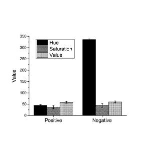
embodiment.
[0013] FIG. 4 shows the results of a gel electrophoresis assay of positive and
negative
isothermal amplification reaction products, according to an embodiment.
[0014] FIG. 5 shows the normalized hue values for amplification reactions
using various Tris
buffer concentrations, according to an embodiment.
[0015] FIG. 6 shows the normalized hue values for amplification reactions
using varying
amounts of additional hydronium ion equivalents, according to an embodiment.
[0016] FIGs. 7A, 7B, 7C, and 7D show the normalized hue values for
amplification
reactions using various halochromic agent concentrations, according to an
embodiment.
[0017] FIG. 8 shows the compatibility of different polymerases with visual
detection of
LAMP amplification, according to an embodiment.
[0018] FIGs. 9A and 9B show the normalized hue values for amplification
reactions using
varying channel depths, according to an embodiment.
[0019] FIG. 10 shows the normalized hue values over time for SDA, according to
an
embodiment.
[0020] FIG. 11 shows the normalized hue values over time for PCR, according to
an
embodiment.
[0021] FIGs. 12A and 12B show the normalized contrast changes for
amplification reactions
using combinations of halochromic agents, according to an embodiment.
[0022] FIG. 13 shows the normalized contrast changes over time for different
DNA template
concentrations, according to an embodiment.
DETAILED DESCRIPTION OF THE INVENTION
[0023] Disclosed herein are compositions and methods for colorimetric
detection of nucleic
acid amplification reaction products. In some embodiments, amplified reaction
products are
3
CA 02944994 2016-10-05
WO 2015/164770 PCT/US2015/027556
detected by a visual color change observation or by measuring absorbance or
fluorescence of
the color change of a halochromic agent in the amplification reaction mix.
Definitions
[0024] Temis used in the claims and specification are defined as set forth
below unless
otherwise specified.
[0025] The term "colorimetry" or "colorimetric" refers to techniques of
quantifying or
otherwise observing colored compound concentrations in solution. "Colorimetric
detection"
refers to any method of detecting such colored compounds and/or the change in
color of the
compounds in solution. Methods may include visual observation, absorbance
measurements,
or fluorescence measurements, among others.
[0026] The term "halochromic agent" refers to a composition that changes color
upon some
chemical reaction. In particular, a halochromic agent can refer to a
composition that changes
color with a pH change. Different halochromic agents may change colors over
different pH
transition ranges.
[0027] The term "transition pH range" or "pH transition range" refers to a pH
range over
which the color of a particular sample or compound changes. A specific
transition pH range
for a sample may depend on a halochromic agent in the sample (see above).
[0028] The term "nucleic acid amplification" or "amplification reaction"
refers to methods of
amplifying DNA, RNA, or modified versions thereof. Nucleic acid amplification
includes
several techniques, such as an isothermal reaction or a thermocycled reaction.
More
specifically, nucleic acid amplification includes methods such as polymerase
chain reaction
(PCR), loop-mediated isothermal amplification (LAMP), strand displacement
amplification
(SDA), recombinase polymerase amplification (RPA), helicase dependent
amplification
(HDA), multiple displacement amplification (MDA), rolling circle amplification
(RCA), and
nucleic acid sequence-based amplification (NASBA). The term "isothermal
amplification"
refers to an amplification method that is performed without changing the
temperature of the
amplification reaction. Protons are released during an amplification reaction:
for every
deoxynucleotide triphosphate (dNTP) that is added to a single-stranded DNA
template during
an amplification reaction, one proton (F1' ) is released.
[00291 The term "sufficient amount" means an amount sufficient to produce a
desired effect,
e.g., an amount sufficient to modulate protein aggregation in a cell.
[0030] The term percent "identity," in the context of two or more nucleic acid
or polypeptide
sequences, refer to two or more sequences or subsequences that have a
specified percentage
4
CA 02944994 2016-10-05
WO 2015/164770 PCT/US2015/027556
of nucleotides or amino acid residues that are the same, when compared and
aligned for
maximum correspondence, as measured using one of the sequence comparison
algorithms
described below (e.g., BLASTP and BLASTN or other algorithms available to
persons of
skill) or by visual inspection. Depending on the application, the percent
"identity" can exist
over a region of the sequence being compared, e.g., over a functional domain,
or,
alternatively, exist over the full length of the two sequences to be compared.
[0031] For sequence comparison, typically one sequence acts as a reference
sequence to
which test sequences are compared. When using a sequence comparison algorithm,
test and
reference sequences are input into a computer, subsequence coordinates are
designated, if
necessary, and sequence algorithm program parameters are designated. The
sequence
comparison algorithm then calculates the percent sequence identity for the
test sequence(s)
relative to the reference sequence, based on the designated program
parameters.
[0032] Optimal alignment of sequences for comparison can be conducted, e.g.,
by the local
homology algorithm of Smith & Waterman, Adv. Appl. Math. 2:482 (1981), by the
homology alignment algorithm of Needleman & Wunsch, J. Mol. Biol. 48:443
(1970), by the
search for similarity method of Pearson & Lipman, Proc. Nat'l. Acad. Sci. USA
85:2444
(1988), by computerized implementations of these algorithms (GAP, BESTFIT,
FASTA, and
TFASTA in the Wisconsin Genetics Software Package, Genetics Computer Group,
575
Science Dr., Madison, Wis.), or by visual inspection (see generally Ausubel et
al., infra).
[0033] One example of an algorithm that is suitable for deteimining percent
sequence
identity and sequence similarity is the BLAST algorithm, which is described in
Altschul et
al., J. Mol. Biol. 215:403-410 (1990). Software for performing BLAST analyses
is publicly
available through the National Center for Biotechnology Information
(www.ncbi.nlm.nih.gov/).
[0034] It must be noted that, as used in the specification and the appended
claims, the
singular forms "a," "an" and "the" include plural referents unless the context
clearly dictates
otherwise.
Compositions of the invention
[0035] Disclosed herein are compositions and methods for accelerated and
efficient
calorimetric detection of nucleic acid amplification reaction products. In an
embodiment, a
calorimetric assay is used to visually detect the presence of an amplified
nucleic acid
product, which eliminates the need for expensive and sophisticated
instrumentation.
CA 02944994 2016-10-05
WO 2015/164770 PCT/US2015/027556
[0036] In some embodiments, the calorimetric detection of amplification
products is
achieved by amplifying a target nucleic acid template molecule to obtain the
amplification
reaction product. The amplification reaction includes a reaction mix. In an
embodiment, the
reaction mix includes a nucleic acid template molecule, one or more enzymes
for catalyzing
the amplification reaction, and one or more halochromic agents for
calorimetric detection. In
a further embodiment, the reaction mix also includes a buffer having a
buffering capacity
equivalent to Tris buffer at a concentration between 1 mM-19 mM in a solution
having a
starting pH of 8Ø In further embodiments, the reaction mix also includes a
plurality of
nucleic acid primers, deoxynucleotide triphosphates (dNTPs), suitable salts
for the enzyme,
and other non-buffered chemicals that enable nucleic acid amplification.
[0037] During the amplification reaction, one proton is released for each dNTP
that is
incorporated into a nucleic acid template molecule. Thus, the pH of the
reaction mix
decreases throughout the amplification reaction. In an embodiment, if the
target nucleic acid
is present, the amplification reaction changes the starting pH of the reaction
mix to cause a
detectable calorimetric change of the halochromic agent, thereby indicating
the presence of
the target nucleic acid, and if the target nucleic acid is not present, the
amplification reaction
does not generate a sufficient number of protons to change the starting pH of
the reaction mix
sufficient to cause a detectable calorimetric change of the halochromic agent,
thereby
indicating that the amplification reaction product has not been produced. In
an embodiment,
the halochromic agent (or pH indicator) in the reaction mix has a transition
pH range for a
calorimetric change of the halochromic agent that is narrower than an expected
pH change
between (1) a starting pH of the reaction mix before the amplification
reaction is performed,
and (2) an ending pH of the reaction mix after the amplification reaction has
been performed.
[0038] In an embodiment, the halochromic agent is a calorimetric agent or a
fluorescent
agent. Suitable halochromic agents include phenol red, bromocresol purple,
bromothymol
blue, neutral red, naphtholphthalein, cresol red, cresolphthalein,
phenolphthalein, methyl red,
and thymolphthalein, among others. A wide range of concentrations of these
halochromic
agents can be used in the reaction mix. Different halochromic agents have
different transition
pH ranges. In some embodiments, the halochromic agent has a transition pH
range between
pH 5-10, between pH 6-9, or between pH 6.5-8.8. In another embodiment, the
halochromic
agent is at a concentration between 25-100 iuM in the reaction mix. In another
embodiment,
the halochromic agent is at a concentration between 50-260 ittM. In some
embodiments, a
combination of two or more halochromic agents is used in the reaction mix,
which increases
6
CA 02944994 2016-10-05
WO 2015/164770 PCT/US2015/027556
the normalized color contrast change of the reaction mix by being of
complementary colors at
the beginning and similar colors at the end of the amplification reaction. In
a further
embodiment, the combination of halochromic agents comprises phenol red and
bromothymol
blue. In a further embodiment, the combination of halochromic agents comprises
cresol red
and bromothymol blue.
[0039] In one example, Phenol red is a halochromic agent that has a transition
pH range from
around 6.4-8Ø At the upper limit of the transition pH range, phenol red is
red, and at the
lower limit of the transition pH range, phenol red is yellow. A reaction mix
containing phenol
red will change color from red to yellow throughout the amplification
reaction, as long as the
starting pH of the reaction mix is around or above 8.0, and the ending pH of
the reaction mix
is within the transition pH range or around or below 6.4.
[0040] In some embodiments, the starting pH of the reaction mix is set by
adding an acid or a
base to the reaction mix until the desired starting pH is reached. The ending
pH of the
reaction mix is determined by performing a sample amplification reaction and
measuring the
ending pH (for example, with a micro-pH electrode). In an embodiment, the
halochromic
agent for an amplification reaction is selected so that the transition pH
range lies in between
the starting pH and ending pH. In a further embodiment, the halochromic agent
is selected so
that the transition pH range is nearer to the starting pH than the ending pH.
The halochromic
agent can also be selected based on the particular enzyme used for catalyzing
the
amplification reaction. Near the ending pH, the enzyme in the reaction mix
teiminates
polymerization of the amplification reaction as the pH decreases to
unfavorable H-
concentrations. In an embodiment, additional hydronium ions or hydronium ion
equivalents
are added to the reaction mix via the sample. For example, between 4.8 x 10⁻⁹
and 4.8 x 10⁻¹⁸
additional hydronium ion equivalents per 10 p1 reaction mix can be tolerated
for the
amplification reaction to proceed. In a further embodiment, between 4.8 x 10¹⁰
and 4.8 x 1 -
18, 4.8 x 10⁻¹² and 4.8 x 10⁻¹⁸, or 4.8 x 10⁻¹⁵ and 4.8 x 10⁻¹⁸ can be
tolerated.
[0041] Generally, the enzyme will catalyze amplification reactions within a pH
range that
encompasses or is close to the transition pH range of the selected halochromic
agent. Various
enzymes can be used for the reaction, and different enzymes catalyze
amplification reactions
at different pH ranges. For example, Bst polymerase is believed to catalyze
amplification
reactions within the pH range of 6.6-9Ø The preferred starting pH for Bst
polymerase is
greater than 7, more preferably greater than 8.2, and more preferably at 8.8.
Other examples
of a preferred starting pH for Bst polymerase are found in U.S. Pat. No.
5,830,714, filed April
7
17, 1996. In an embodiment, phenol red is coupled with Bst polymerase in a
reaction mix,
since the pH range at which Bst polymerase is active (6.6-9.0) encompasses the
transition pH
range of phenol red (6.4-8.0). In another embodiment, methyl red is coupled
with U exo-
Klenow fragment (polymerase for Helicase Dependent Amplification, HDA) in a
reaction
mix, since a starting pH at which U exo-Klenow fragment is active (around 7.5)
is higher
than the transition pH range of methyl red (4.8-6.2).
[0042] Other than Bst or Bst 2.0 polymerase, other enzymes capable of being
used for
catalyzing the amplification reaction include the polymerase from Thermus
aquaticus (TAQ),
DNA polymerases I-IV, Kapa Polymerase, RNA polymerases I-V, 17 RNA Polymerase,
a
reverse transcriptase, any DNA polymerase or RNA polymerase, a helicase, a
recombinase, a
ligase, a restriction endonuclease, and a single-strand binding protein. In
some embodiments,
an isothermal amplification reaction uses an enzyme that is a strand
displacement
polymerase, such as phi29-DNA-Polymerase, Klenow DNA-Polymerase, Vent DNA
Polymerase, Deep Vent DNA Polymerase, Bst DNA Polymerase, 9oNm(TM) DNA
Polymerase, U exo-Klenow fragment, or mutants and variants thereof. In some
embodiments,
suitable salts for the enzyme are also added to the reaction mix. In certain
embodiments, the
starting pH of the reaction mix is set based on an optimal pH for the specific
enzyme used for
catalyzing the amplification reaction. In an embodiment, the pH of the entire
DNA sample is
between pH 3 and pH 11.
[0043] In other embodiments, a fluorescent halochromic agent is used to detect
protons
released during amplification. The halochromic agent may change optical
properties (such as
amplitude and emitted wavelength) as the pH of the reaction mix changes during
the
amplification reaction. Fluorescent halochromic agents include fluorescein,
pyranine, and
pHrodo dye (Life Technologies, Carlsbad CA).
100441 The base and/or acid added to the reaction mix maintains the starting
pH of the
reaction mix around or above an upper limit of the transition pH range of the
halochromic
agent. For example, an acid such as hydrochloric acid (HC1) or sulfuric acid
(H2SO4), or a
base such as sodium hydroxide (NaOH) or potassium hydroxide (KOH), can be
added to the
reaction mix. In some embodiments, the acid or base sets the starting pH of
the reaction mix
between pH 6-10, between pH 7-8, or between pH 8-8.6. In an embodiment, the
reaction mix
is capable of offsetting the starting pH of the reaction mix by less than 0.1
pH units. In
another embodiment, the reaction mix has a starting pH lower than 2 pH units
above the
8
CA 2944994 2019-02-07
CA 02944994 2016-10-05
WO 2015/164770 PCT/US2015/027556
upper limit of the transition pH range of the halochromic agent. In further
embodiments, the
reaction mix has a starting pH lower than 1 pH unit, 0.5 pH units, or 0.1 pH
units above the
upper limit of the transition pH range of the halochromic agent. In a further
embodiment,
noise from non-specific amplification is minimized by setting the pH
transition range
sufficiently separated from the starting pH of the reaction mix, so that any
color change is
only achieved by a specific and sustained amplification.
[0045] In an embodiment, the reaction mix does not require any additional
buffering agent
for the amplification reaction, since a buffering agent could prevent large
changes in pH from
occurring during the amplification reaction. In another embodiment, the
reaction mix
contains a minimal amount of buffering agent, such that the buffering capacity
of the reaction
mixture is less than the expected change in pH during amplification. In some
embodiments,
the buffer is at a concentration between 1 mM and 3 mM. In a further
embodiment, the buffer
is at a concentration of 1 mM. In certain embodiments, the buffer used is Tris
buffer
(formulated to pH 8.8), HEPES (pH 7-9), or TAPS (pH 7-9). In another
embodiment, the
buffer used is a buffer having a buffering capacity equivalent to a Tris
buffer at a
concentration between 1 mM-19 mM in a solution having a starting pH of 8Ø
This broad
range of suitable buffer concentrations allows the reaction mix to resist
unwanted starting pH
changes during reaction setup, unlike reaction setups with minimal (<ImM) Tris
buffer
equivalents (see US 13/799,995, filed March 13, 2013). These unwanted changes
in pH come
about due to hydronium or hydroxide ion equivalents added to the reaction via
the sample
reagents. As colorimetric detection and enzyme kinetics depend on the starting
pH, the
presence of buffer capacity in the reaction mix high enough to avoid starting
pH change, but
low enough to allow color change upon amplification, become important. In a
further
embodiment, the pH of the reaction mix is between pH 7.5-8.8. Table 1 shows
various
buffers having buffering capacities equivalent to a Tris buffer at a
concentration between 1
mM-19 mM in a solution having a starting pH of 8Ø The buffer capacity (13)
is defined as
the equivalents of acid or base needed to change the pH of 1 Liter of buffer
by I pH unit.
This can be calculated as: 13 = 2.3* C * (K5*[H30]/(K5 + [H30])2); where C is
the buffer
concentration, Ka is the dissociation constant for the buffer and [F130] is
the hydronium ion
concentration of the buffer (which is calculated from the reaction starting
pH). The buffer
capacity of 1 mM - 19 mM Tris (in a solution having a starting pH of 8.0) was
found to range
from 0.000575 to 0.010873. The starting pH of the buffer was considered to be
in the range
of 7.5 - 8.8 to be compatible with the reaction biochemistry (polymerase
function, nucleic
9
CA 02944994 2016-10-05
WO 2015/164770
PCT/US2015/027556
acid melting, etc.). In other embodiments, the buffer has a buffering capacity
equivalent to a
Tris buffer at a concentration between 1.5 mM - 19 mM, 2 mM - 19 mM, 3 mM - 19
mM, 4
mM- 19 mM, 5 mM - 19 mM, 6 mM - 19 mM, 7 mM - 19 mM, or otherwise, in a
solution
having a starting pH of 8Ø In other embodiments, the buffer has a buffering
capacity
equivalent to a Tris buffer at a concentration between 1.92 mM -36.29 mM, 3 mM
-36.29
mM, 4 mM - 36.29 mM, 5 mM - 36.29 mM, or otherwise, in a solution having a
starting pH
of 8.8. In other embodiments, the buffer has a buffering capacity equivalent
to a Tris buffer
at a concentration between 1.48 mM - 27.92 mM, 2 rnM - 27.92 mM, 3 mM - 27.92
mM, 4
mM - 27.92 mM, 5 mM - 27.92 mM, or otherwise, in a solution having a starting
pH of 7.5.
Table 1: Buffer Capacity Table
Buffer Full Chemical Name pKa at 25 C Starting MM Cone Max
Cone
Reaction pH (mM) (mM)
8.8 1.92 36.29
tris(hydroxymethyl)meth
Tris 8.06 8.0 1.00 19.00
ylamine
7.5 1.48 27.92
N- 8.8 1.19 22.55
Tris(hydroxymethyl)meth
8.0 1.27 23.94
TAPS y1-3- 8.43
aminopropanesulfonic
acid 7.5 2.66 50.25
8.8 1.29 24.46
N,N-bis(2-
Bicine 8.35 8.0 1.17 22.15
hydroxyethyl)glycine
7.5 2.31 43.59
8.8 1.67 31.63
N-tris(hydroxymethyl)
Tricine 8.15 8.0 1.03 19.48
methylglycine
7.5 1.67 31.63
3-[N- 8.8 4.17 78.90
Tris(hydroxymethyl)meth
8.0 1.19 22.45
TAP SO ylamino] -2- 7.635
hydroxypropanesulfonic
acid 7.5 1.02 19.37
4-(2-hydroxyethyl)-1- 8.8 5.74 108.45
HEPES p iperaz ineethane sul fon i c 7.48 8.0 1.40
26.54
acid 7.5 1.00 18.92
N- 8.8 6.79 128.39
tris(hydroxymethyl)meth
TES 7.4 8.0 1.56 29.46
y1-2-aminoethanesulfon ic
acid 7.5 1.01 19.16
3-(N- 8.8 10.46 197.77
morpholino)propanesulfo
MOPS 7.2 8.0 2.12 40.03
nic
acid 7.5 1.12 21.26
1,4- 8.8 27.91 500.00
piperazinediethanesulfoni
PIPES 6.76 8.0 4.86 91.88
c acid
ac id 7.5 1.92 36.29
8.8 16.28 300.00
SSC Saline Sodium Citrate 7.0 8.0 3.03
57.20
7.5 1.37 25.90
100461 In an embodiment, a magnesium compound is added to the reaction mix,
because
magnesium promotes nucleotide incorporation into the template and influences
the activity of
the polymerase. In a further embodiment, the concentration of a magnesium
compound (such
as magnesium sulfate) in the reaction mix is at least 0.5 mM, at least 1 mM,
at least 2 mM, or
at least 4 mM. In an embodiment, the concentration of added magnesium ion is
dependent on
the concentration of dNTPs, nucleic acid template, and primers. In an
embodiment, the ratio
of dNTPs to magnesium sulphate in the reaction mix is less than 1:2, less than
1:3, less than
1:4 or less than 1:5.
100471 In some embodiments, monovalent cations are added to the reaction mix.
Monovalent
cations include potassium, ammonium, and quaternary ammonium, among others.
Monovalent cations can affect the melting characteristics of the nucleic acid
template and
improve the efficiency of the enzyme. In an embodiment, potassium is in the
reaction mix at
a concentration of less than 50 mM, or less than 15 mM. In another embodiment,
quaternary
ammonium salts are in the reaction mix at a concentration of greater than 2mM,
greater than
5mM, or greater than 8mM. In another embodiment, an ammonium compound (such as
ammonium chloride) is in the reaction mix at a concentration of less than
15mM, or less than
mM. Ammonium (NH4) has some buffering capability, thus the final concentration
of
ammonium compounds in the reaction mix should be minimized while maintaining
optimal
amplification yield.
[0048] In an embodiment, the concentrations of other reagents of the reaction
mix are kept at
amounts as generally used in amplification reactions. See Notomi T et. al.
Nucleic Acids Res.
2000 Jun 15; 28(12): E63; Nature Protocols 2008, Loop-mediated isothermal
amplification
(LAMP) of gene sequences and simple visual detection of products, 2008 3(5):
pg 880. In an
embodiment, the Bst or Bst 2.0 enzyme is used, and the amount of enzyme is at
least 0.8 Unit
per microliter of combined fluid. In this embodiment, Betaine is also present
in the reaction
mix at a concentration between 0-1.5 M or 0.8M-1 M, and the total
concentration of primers
is between 3.6pM and 6.2pM. In some embodiments, any of the following reagents
is present
in the reaction mix: Tris buffer (pH 8.8) at 20 mM, KC1 at 10 mM, MgSO4 at 8
mM,
(NH4)2SO4 at 10 mM, TweenTm 20 at 0.1%, Betaine at 0.8 M, dNTPs at 1.4 mM
each, MnC12
at 0.5 mM, FIP at 1.6
11
CA 2944994 2019-08-08
CA 02944994 2016-10-05
WO 2015/164770
PCT/US2015/027556
p,M, F3 at 0.2 iuM, B3 at 0.2 iuM, primers at a total concentration of 5.2
itiM
(2*(1.6+0.8+0.2), and Bst / Bst 2.0 at 8 U per 10pL.
[0049] The above reagent concentrations have been found to provide good
amplification
yield and low buffering capacity so that a halochromic pH sensor can be used
to detect
protons released during the amplification reaction. In some embodiments, the
concentrations
of reaction mix reagents depend on the enzyme selection. In further
embodiments, guidance
regarding appropriate reagent concentrations is available from the enzyme
manufacturers. In
an embodiment, the ratio of the sample volume to the reaction mix volume is
such that the
sample is diluted between 5% and 40% when the reaction mix is added.
[0050] In some embodiments, amplification reaction reagents are stored
separately before
being added to a reaction mix, since some reagents have specific required
conditions for
stability. For example, the enzyme may be stored long term in a moderately
buffered solution
separate from the other reagents to ensure stability of the enzyme. Upon
mixing with the
remaining reagents in the reaction mix, the buffering agent becomes
sufficiently diluted so as
not to significantly mask a pH change. In addition, primers for specific genes
of interest may
be provided in a separate solution or in a lyophilized form.
[0051] In some embodiments, the amplification reaction is performed within a
microtube. In
other embodiments, the amplification reaction is performed within a fluidic or
microfluidic
structure. In some embodiments, the fluidic or microfluidic structure is a
well, chamber, or
channel that receives the reagents and the nucleic acid sample separately, and
then mixes the
components together. In another embodiment, the fluidic or microfluidic
structure is a well,
chamber, or channel that receives the pre-mixed reaction mix. In a further
embodiment, the
fluidic or microfluidic structure possesses a long optical path for
colorimetric observation, or
a fluorescent/ absorbance excitation source and detector. In another
embodiment, the fluidic
or microfluidic structure receives the reagents in a lyophilized form, and
subsequently
receives the nucleic acid sample and hydration solution. In an embodiment, a
chamber
fluidic or microfluidic structure has a channel depth ranging between 50 ium-
400 ium or
greater. In a further embodiment, colorimetric observation is accomplished for
channel
depths (path length) of 50 j.tm, 50 jim-400 i.tm, or 50 tun or greater.
[0052] Some embodiments include a kit for calorimetric detection of an
amplification
product. The kit may include one or more halochromic agents, one or more
enzymes for
catalyzing an amplification reaction, and instructions for contacting a sample
with a reaction
mix including the buffer and the enzyme and the halochromic agent under
conditions that an
12
CA 02944994 2016-10-05
WO 2015/164770 PCT/US2015/027556
amplification reaction occurs and produces an amplification reaction product
if the sample
contains a target nucleic acid template molecule, the reaction mix having a
starting pH, and if
the target nucleic acid template molecule is present, the amplification
reaction changes the
starting pH of the reaction mix to cause a detectable colorimetric change of
the halochromic
agent, thereby indicating the presence of the target nucleic acid, and if the
target nucleic acid
template molecule is not present, the amplification reaction does not generate
a sufficient
number of protons to change the starting pH of the reaction mix sufficient to
cause a
detectable colorimetric change of the halochromic agent, thereby indicating
that the
amplification reaction product has not been produced. In another embodiment,
the
instructions are for contacting a nucleic acid template molecule with the
halochromic agent
and enzyme in a reaction mix, under conditions that result in (1) an
amplification reaction
that amplifies the nucleic acid template molecule to produce an amplification
reaction
product, and (2) generation of a sufficient number of protons so that an
ending pH of the
reaction mix is sufficiently low to produce a detectable colorimetric change
of the
halochromic agent, thereby indicating that the amplification reaction product
has been
produced. In further embodiments, the kit also includes an acid or base,
dNTPs, primers, and
monovalent cations. In a further embodiment, the kit includes the following
reagents at the
following concentrations:
= Bst or Bst 2.0 polymerase, at least 0.8 Unit per microliter;
= Betaine at 0.8 M;
= Primers at 3.6 iuM total;
o FIP and B1P primers at 1.6 iLtM
o F3 and B3 at 0.2 1\4
= Magnesium sulfate at 8 111M;
= Ammonium sulfate at 10 mM;
= Potassium chloride at 10mM;
= Sodium hydroxide to set the starting pH of the reaction mix;
= Tween20 at 0.1%;
= dNTP's at 1.4 mM each;
= Phenol red at 50 iuM.
In a further embodiment, the kit includes LoopF and LoopB primers at 0.8 iuM
each.
13
CA 02944994 2016-10-05
WO 2015/164770 PCT/US2015/027556
Methods of the invention
[0053] The amplification reaction amplifies nucleotides from a nucleic acid
template. In
some embodiments, the amplification reaction is an isothermal amplification
reaction, such as
a strand displacement reaction. In a further embodiment, a strand displacement
reaction is
provided by a polymerase with strand displacement activity under reaction
conditions such
that strand displacement is possible. Examples of strand displacement
reactions include
strand displacement amplification (SDA), multiple displacement amplification
(MDA),
rolling circle amplification (RCA) or loop mediated isothermal amplification
(LAMP). In
other embodiments, the amplification reaction includes other non-isothermal
amplification
reactions such as polymerase chain reaction (PCR).
[0054] In certain embodiments, the amplification reaction performed is LAMP.
In a LAMP
reaction, a double- or single-stranded DNA template in dynamic equilibrium at
an elevated
temperature is amplified using two or three pairs of primers. The primers are
designed based
on the DNA template, using primer design software such as LAMP Designer
(Premier
Biosoft, Palo Alto, CA). In the first step of the LAMP reaction, the F2 region
of the FIP
(Forward Inner Primer) anneals to the single stranded DNA at the respective
complementary
(F2c) position. Next, a polymerase with strand displacement activity
incorporates dNTPs
along the template from the 3' end of F2. The incorporation of nucleotides
releases protons,
reducing the pH of the reaction mix. Then, the F3 forward primer anneals to
the F3c region
upstream of the F2 region and on the template. The F3 forward primer begins
amplifying the
template strand, which releases further protons and displaces the FIP-
incorporated strand that
was synthesized previously. This single strand contains an Fl sequence (within
the target
sequence) along with its complementary Flc sequence (within the FIP). This
forms a stem-
loop as Flc anneals to Fl at the 5' end. At the same time, the BIP (Backward
Inner Primer)
anneals to the other end of the strand and nucleotides extend from B2,
releasing more
protons. The backward primer B3 then binds to the B3c region, downstream of
the B2 region,
displaces the BIP-amplified strands and promotes extension to create the
double strand. This
displaced strand now contains a B1 sequence (within the target sequence) along
with its
complementary Blc sequence (within the BIP), forming another stem loop in the
3' end. The
structure now has two stem-loop structures at each end from which continuous
displacement
and extension occur to amplify the template. The LAMP reaction can be
amplified by adding
further Forward and Backward Loop primers to produce more amplicons with stem
loop
structures.
14
CA 02944994 2016-10-05
WO 2015/164770 PCT/US2015/027556
[0055] The LAMP procedure can take place at a fixed temperature, minimizing
the need for
any expensive thermocycling equipments. Typically, isothermal methods require
a set
temperature, which is determined by the selected reagents. For example,
enzymes function
best between 60-65 C in LAMP methods.
[0056] Colorimetric detection of the nucleic acid amplification reaction
product can be
performed in real-time throughout the amplification reaction, or after the
performance of the
amplification reaction. Detection of the colorimetric change of the reaction
mix can be
associated with a digital indication of a presence or absence of the
amplification reaction
product. In other words, a visual observation of the color change of the
reaction mix can
provide information regarding whether the amplification reaction product is
present or
absent. In certain embodiments, detection of a colorimetric change of the
reaction mix
indicates that the exponential or plateau phase of the amplification reaction
has been
obtained.
[0057] In some embodiments, detection of the amplification reaction product is
accelerated
relative to an amplification reaction that uses a reaction mix without a
halochromic agent. In
further embodiments, the colorimetric change of the reaction mix is detected
in less than 60
minutes from a starting time of the amplification reaction. Accelerated
detection of the
amplification reaction product is obtained because the halochromic agent (a
weak acid or
base) in the reaction mix absorbs protons generated during the amplification
reaction, and
recombination of the free protons acts to accelerate the detection of the
amplification
reaction. The reaction can be designed so that minimal amplification is
required to generate a
pH transition sufficient for the halochromic agent to change color.
Conventional
amplification techniques that use fluorescent intercalating dyes, molecular
beacons,
hybridization probes, dye-based detection, UV-Vis, or other detection methods
require a
certain threshold amount of amplification to occur before an amplification
signal is
detectable. However, the methods of the present invention require a relatively
smaller
threshold amount of amplification before a color change of the halochromic
agent is
detectable, and therefore the detection of an amplification reaction product
is accelerated
relative to conventional amplification methods.
[0058] In some embodiments, the amplification reaction product is detected
visually by
observation of a color change of the reaction mix. In a further embodiment,
the human eye is
used for the visual detection. In another embodiment, a camera, a computer, or
some other
optical device is used for the visual detection or for imaging the reaction
mix. Imaging
CA 02944994 2016-10-05
WO 2015/164770 PCT/US2015/027556
programs include Photoshop (Adobe, San Jose CA), ImageJ (National Institutes
of Health,
Bethesda MD), and MATLAB (MathWorks, Natick MA). In another embodiment, the
amplification reaction product is detected by measuring fluorescence of the
reaction mix,
using fluorescence spectroscopy methods. In another embodiment, the
amplification reaction
product is detected by measuring absorbance of the reaction mix, using
absorption
spectroscopy methods. In a further embodiment, the endpoint or overall change
in absorbance
or fluorescence of the reaction mix is measured at a given wavelength or set
of wavelengths.
EXAMPLES
[0059] Below are examples of specific embodiments for carrying out the present
invention.
The examples are offered for illustrative purposes only, and are not intended
to limit the
scope of the present invention in any way. Efforts have been made to ensure
accuracy with
respect to numbers used (e.g., amounts, temperatures, etc.), but some
experimental error and
deviation should, of course, be allowed for.
[0060] The practice of the present invention will employ, unless otherwise
indicated,
conventional methods of protein chemistry, biochemistry, recombinant DNA
techniques and
pharmacology, within the skill of the art. Such techniques arc explained fully
in the
literature. See, e.g., T.E. Creighton, Proteins: Structures and Molecular
Properties (VV.H.
Freeman and Company, 1993); A.L. Lehninger, Biochemistry (Worth Publishers,
Inc., current
addition); Sambrook, et al., Molecular Cloning: A Laboratory Manual (2nd
Edition, 1989);
Methods In Enzymology, (S. Colowick and N. Kaplan eds., Academic Press, Inc.);
Remington 's Pharmaceutical Sciences, 18th Edition (Easton, Pennsylvania: Mack
Publishing
Company, 1990); Carey and Sundberg Advanced Organic Chemistry .3rd Ed. (Plenum
Press)
Vols A and B(1992).
Example 1: Colorimetric Detection of a Nucleic Acid Amplification Reaction
Product
[0061] In an assay for colorimetric detection of a nucleic acid amplification
reaction product,
the following reagents were mixed together to produce a 2X reagent mix:
= Magnesium Sulphate (Sigma Aldrich) at 16 mM
= Ammonium Sulphate (Sigma Aldrich) at 20 mM
= Potassium Chloride (Sigma Aldrich) at 20mM
= Sodium hydroxide (Sigma Aldrich) at a concentration that sets the
starting pH of the
reagent mix to 8.8 pH
16
[0062] The reagent mix was adjusted to an initial pH of 8.8 to enable
efficient initial
polymerization. The reagent mix was autoclaved for 1 hour for sterilization.
The following
ingredients were then added (in a sterile form) to the reagent mix to generate
the reaction
mix:
= TweenTm20 (Sigma Aldrich) at 0.1% (v/v)
= dNTPs (NEB) at 1.4 mM each
= Phenol Red (Sigma Aldrich) at 50 itiM
= Bst polymerase (NEB) at 0.8 Unit per microliter (the enzyme storage
buffer
contributing 1 mM Tris buffer, 5 mM KCI, 0.01 mM EDTA, 0.1 mM DTT, 0.01 %
TritonTm X-100 (v/v) and 5% Glycerol ((w/v) to the reaction mix)
= Betaine (Sigma Aldrich) at 0.8 M
[00631 Primers and a nucleic acid template were added to the reaction mix. The
primers were
designed for LAMP and included two pairs of primers (solubilized in 1X Tris
EDTA buffer)
at a total concentration of 3.6 M as described above. Primer F3 has the
sequence:
GATCTGAATCCGACCAACCG (SEQ ID NO: 1); primer B3 has the sequence:
AACGCCCACGCTCTCGCA (SEQ ID NO: 2); the primer FIP has the sequence:
AAATCCGTCCAGTGGTTTTTTTGAAAATCGTTGTATCTCCG (SEQ ID NO: 3); and the
primer BIP has the sequence:
CCGAAACCACTGGACGGATTTTTATTTTTAATCTAAAACAAACATC (SEQ ID NO:
4). The nucleic acid template molecule was purified from Schistosoma mansoni.
FIG. 1
shows the SM1-7 target region of the nucleic acid template molecule (see
Hamburger et al,
Detection of Schistosoma mansoni and Schistosoma haematobium DNA by Loop-
Mediated
Isothermal Amplification: Identification of infected Snails from Early
Prepatcncy, Am .1 Trop
Med Hyg, 2010). The positive test reactions contained template DNA, and the
negative
control reactions contained water. The reaction mixes had a starting in the
range of 7.5 -
8.5. The reaction mixes were heated in micro-tubes to 63 C on a thermocycler
to allow
template amplification. After a predetermined reaction period of 45 minutes,
during which
sufficient template amplification occurred, the resultant color of the
reaction mix was visually
observed.
[0064] During the amplification process, the pH of the reaction mix was
reduced from 7.5-
8.5 to around 6.6 in a repeatable fashion. FIG. 2 is a graph showing the pH
measurements for
repeated positive (test) and negative (negative control) amplification
reactions. The
17
CA 2944994 2019-08-08
CA 02944994 2016-10-05
WO 2015/164770 PCT/US2015/027556
halochromic agent used was Phenol red, which has a transition pH range of 6.8 -
8.2. Phenol
red changes color over this transition pH range from red to yellow (when the
pH is lowered
from the upper pH limit to the lower pH limit). In the assay, the reaction mix
changed color
from red (at pH 8.0) to yellow (at pH 6.6) in response to the pH change during
nucleic acid
amplification. FIG. 3 is a graph showing the difference in contrast value
using HSV (hue,
saturation, value) of images of the reaction mixes of a positive and negative
amplification
reaction at the reaction endpoints. The color change is quantitatively
demonstrated in the hue
variable. To confirm that the color change was due to target DNA
amplification, endpoint
reactions were analyzed using gel electrophoresis to verify the presence of
amplicons (FIG.
4).
[00651 Using this method, amplification of a DNA template can be easily
observed, either at
the reaction end-point or in real-time throughout the reaction, by visually
observing the color
change in the reaction mix, or by measuring the absorbance or fluorescence of
the reaction
mix. This mechanism generates much larger contrast in comparison to other
colorimetric
detection techniques and can be imaged without the need of expensive optical
instrumentation.
Example 2: Detection of LAMP Amplification Using a Visual Halochromic
Agent
[00661 LAMP reactions were performed with a reaction mix comprising of: 10 mM
(NH4)2SO4, 15 mM KC1, 0.1 mM EDTA, 0.1 mM DTT, 0.01 % Triton X-100 (v/v), 5 %
Glycerol, 8 mM MgSO4, 1.4 mM each dNTPs, 0.1% v/v Tween-20, 0.8 M Betaine.
Three
primer pairs, specific to different targets, were added to a final
concentration of 1.611M each
for FIP/BIP, 0.2 uM each for F3 /B3, 0.4 LM each for LoopB/F. The final
reaction volume is
[iL and was held at 63 C for different incubation times.
[0067] In FIG. 5, the final Tris buffer concentration of the reaction mix was
varied from 0.34
mM to 19 mM (by varying amount of Tris buffer formulated to pH 8.8). Reactions
were
performed with primers for lambda phage DNA, 5 ng of lambda DNA (New England
Biolabs), 0.8 U/ulBst 2.0 DNA polymerase (New England Biolabs) and 0.2 mM
Neutral Red
(Sigma Aldrich). The reaction tubes were then imaged and the Normalized Hue
value was
calculated for the color of the reaction mix. The Normalized Hue value was
defined as the
difference in Hue values between a positive and a no-template negative
reaction. A color
change, indicated by a change in the Normalized Hue value above the
visualization threshold
(dotted line), was observed for buffer concentrations as high as 19mM Tris.
This indicates
18
CA 02944994 2016-10-05
WO 2015/164770 PCT/US2015/027556
that reaction mix with buffer capacities equivalent to >1mM and <19mM Tris
allow enough
pH change for visual color change detection.
[0068] In FIG. 6, the tolerance of this visual detection method to excess
hydronium ions
added to the reaction mix was evaluated. This tolerance is important to allow
the use of a
wide variety of DNA samples which can add a range of hydronium or hydroxide
ion
equivalents to the reaction. Reactions were performed with 2mM final Tris
buffer
concentration, 5 ng lambda DNA target, 0.8 15/4 Bst DNA polymerase and 0.2 mM
Neutral
Red halochromic agent. The change in Normalized Hue value indicates that this
visual
detection chemistry works with 4.8 x 10⁻⁹ till 4.8x10⁻¹⁸ additional hydronium
ion equivalent
per 10 uL reaction.
[0069] In FIGs. 7A-7D, the compatibility of different pH indicators and
amplification targets
with visual detection of LAMP amplification was evaluated. The reactions were
performed
with final Tris buffer concentration in the range of 1.2 - 1.3 mM and 0.8
11/1AL Bst DNA
polymerase. Three different indicator were tested with 5 ng lambda DNA target:
50 tM
Phenol Red, 260 !AM Cresol Red and 160 [tM Bromothymol Blue (FIG. 7A). High
contrast
change in the normalized hue value was observed for all indicators tested.
[0070] Concentration sweeps were also performed for these indicators
Bromothymol Blue
(FIG. 7B top left), Cresol Red (FIG. 7B top right), Neutral Red (FIG. 7B
bottom left) and
Phenol Red (FIG. 7B bottom right) with Lambda target, which demonstrated the
wide range
of concentrations that are compatible with the chemistry. LAMP assays using
130 ng
Schistosorna mansoni gDNA with 501AM Phenol Red (FIG. 7C) and Human GAPDH mRNA
with 0.2 mM Neutral Red (FIG. 7D) were also tested visual detection of these
targets was
demonstrated at end-point.
[0071] In FIG. 8, the compatibility of different polymerases with visual
detection of LAMP
amplification was evaluated. The reactions were performed with 1.3 mM final
Tris buffer
concentration, 5 ng lambda DNA target and 0.2 mM Neutral Red. 0.8 U411 of two
different
polymerases, Bst 2.0 and Gspm 2.0 (OptiGene), were used. High contrast color
change was
observed for both polymerases after 60 minutes of incubation (FIG. 8).
Table 2: Sequences Used
Lambda FIP SEQ ID NO: 5
Lambda BIP SEQ ID NO: 6
Lambda F3 SEQ ID NO: 7
Lambda B3 SEQ ID NO: 8
19
CA 02944994 2016-10-05
WO 2015/164770 PCT/US2015/027556
Lambda Loop F SEQ ID NO: 9
Lambda Loop B SEQ ID NO: 10
Schistosoma F3 SEQ ID NO: 1
Schistosoma B3 SEQ ID NO: 2
Schistosoma FIP SEQ ID NO: 3
Schistosoma BIP SEQ ID NO: 4
GAPDH F3 SEQ ID NO: 11
GAPDH B3 SEQ ID NO: 12
GAPDH FIP SEQ ID NO: 13
GAPDH BIP SEQ ID NO: 14
GAPDH Loop F SEQ ID NO: 15
GAPDH Loop B SEQ ID NO: 16
Example 3: Visual Detection of LAMP Amplification in Sub-Millimeter Path
Lengths
[0072] LAMP reactions were performed as in Example 1 with 1.3 mM final Tris
buffer
concentration (buffer formulated to pH 8.8), 0.8 U/jil of Bst 2.0 DNA
Polymerase, 5 ng
lambda DNA template and 0.2 mM Neutral Red or 160 jiM Bromothymol Blue. Both
the
positive and the no-template negative reactions were added after amplification
to flow
chambers with varying channel depths (FIG. 9A for Neutral Red and FIG. 9B for
Bromothymol Blue). These flow chambers were machined in acrylic with channel
depths
ranging from 50 jim to 400 jtm. High contrast color difference (above the
visual detection
threshold; dotted line) between the positive and the negative reactions was
observed for
channel depths of 50 jtm and above. This demonstrates that this visual
detection chemistry is
amenable for use in reaction chambers with sub-milimeter path lengths (depths)
and above.
Such reaction chambers can be used to reduce the amount of reagents used and
to allow
multiple reactions to take place in a certain footprint (eg. in a microfluidic
cartridge).
Example 4: Detection of Strand Displacement Amplification (SDA) Using a
Visual Halochromic Agent
[0073] SDA reactions were performed using a reaction mix comprising of: 1.3 mM
final Tris
buffer concentration (buffer formulated to pH 8.8), 10 mM (NH4)2SO4, 50 mM KC1
(adjusted to pH 8.5), 8 mM MgSO4, 4.4 mM each dATP, dGTP, dTTP, 0.8 mM dCTP-aS
(TriLink Biotechnologies), 0.1% ITN Tween-20, 0.8 M Betaine, 0.32 U/jil Bst
DNA
CA 02944994 2016-10-05
WO 2015/164770
PCT/US2015/027556
polymerase (New England Biolabs), 0.2U/uL BSoBI (New England Biolabs) and 0.2
mM
Neutral Red halochromic agent. Primers designed for human BRCA1 (SDAf: SEQ ID
NO:
17; SDAr: SEQ ID NO: 18; BF: SEQ ID NO: 19; BR: SEQ ID NO: 20) were added to
the
reaction at 0.511M final concentration each. 5 ng of HeLa gDNA was added to a
final
reaction volume of 25 lit and was held at 65 C for different incubation
times. A change in
Normalized Hue value over time (FIG. 10) indicates that this visual detection
chemistry
works with SDA.
Example 5: Detection of PCR Amplification Using a Visual Halochromic Agent
[0074] PCR reactions were performed using a reaction mix comprising of: 50 mM
KO and 2
mM MgC12 (pH adjusted 8.5), 0.5 mM each dNTP, 5U Taq DNA polymerase (New
England
Biolabs) and 0.2 mM Neutral Red halochromic agent. Total carry-over Tris-HC1
concentration from enzyme storage buffer and primers (Forward: SEQ ID NO: 21;
Reverse:
SEQ ID NO: 22) was 1.15 mM in the final reaction mix. Primers were designed
for
Escheriehia cell 16s rRNA gene and added to the reaction at 0.5 [iM final
concentration
each. 10 ng of E.coli gDNA was added to a final reaction volume of 25 1AL and
was initially
held at 95 C hold for 2 min, followed by 50 cycles of 95 C for 10 sec, 55 C
for 30 sec, 68
C for 30 sec. A change in Normalized Hue value over time (FIG. 11) indicates
that this
visual detection chemistry works with PCR.
Example 6: Increase in Visual Detection Contrast with Combination of
Halochromic Agents
[0075] LAMP reactions were performed as in Example 1 with 1.3 mM final Tris
buffer
concentration (buffer formulated to pll 8.8), 0.8 U/[il of Bst 2.0 DNA
Polymerase and 5 ng
lambda DNA template. The color change contrast was evaluated for Phenol Red at
50 [1M
concentration and combination of Phenol Red and Bromothymol Blue at 50 .IM and
16011M
concentrations respectively (FIG. 12A). The color change contrast was also
evaluated for
Cresol Red at 260 [tM concentration and combination of Cresol Red and
Bromothymol Blue
at 260 [EM and 160 RM concentrations respectively (FIG. 12B). The contrast
values were
calculated from the RGB values of images of the reaction mix using the
formula: 0.299R +
0.587G + 0.114B. The normalized contrast change was defined as the difference
between
positive and negative reaction contrast values normalized to the background.
The increase in
the normalized contrast change with the use of the halochromic agent
combination
demonstrates the utility of such combinations.
21
Example 7: Real-time Color Monitoring of Amplification for Quantification.
Using Visual Halochromic Agents
[0076] LAMP reactions were performed as in Example 1 with 1.3 mM final Tris
buffer
concentration (buffer formulated to pH 8.8), 0.8 U/1.11 of Bst 2.0 DNA
Polymerase, Phenol
Red and Bromothymol Blue at 50 uM and 160 p.M concentrations respectively and
varying
lambda DNA template concentrations. Color change contrast was evaluated for
lambda DNA
target at 0.5 fg/til, 0.05 pg/[il and 0.5 pg/p.1 final concentrations. The
contrast values were
calculated from the RGB values of images of the reaction mix as described in
Example 5.
The results (FIG. 13) indicate that the higher DNA concentrations led to a
detectable change
in visual contrast earlier than the lower DNA concentrations. Hence, we
demonstrate the
ability to distinguish between different target concentrations with the real-
time color
monitoring of this chemistry.
[0077] While the invention has been particularly shown and described with
reference to a
preferred embodiment and various alternate embodiments, it will be understood
by persons
skilled in the relevant art that various changes in form and details can be
made therein
without departing from the spirit and scope of the invention.
22
CA 2944994 2019-02-07