Note: Descriptions are shown in the official language in which they were submitted.
CA 02946222 2016-10-18
WO 2015/162211 1
PCT/EP2015/058817
Title
Method for automated generation of genetically modified T cells
Field of the invention
.. The invention relates to the automated generation of modified T cells
Background
The clinical manufacture of gene-modified T cells is currently a complex
process that
generally starts with obtaining the patient's peripheral blood mononuclear
cells (PBMC).
Current protocols feature a leukapheresis step, trading off an initially more
cumbersome
process (as opposed to a smaller volume blood draw) for an increased cell
yield. PBMC are
often enriched for T cells and activated prior to gene modification with viral
or nonviral
vectors. The modified T cells are then expanded in order to reach the cell
numbers required
for treatment, after which the cells are finally formulated and/or
cryopreserved prior to
reinfusion. The cell product must be subjected to a number of quality control
assays and has
to meet all release criteria and Good Manufacturing Practices (GMP)
guidelines. Thus far,
adoptive cell transfer (ACT) using gene-modified T cells has mainly been
carried out by
investigators who have developed their manufacturing process for small scale
clinical trials by
using the devices and infrastructure at hand. Such individualized therapies
are complex:
.. the cell manufacturing process is labor intensive, as it comprises many
(open) handling steps
(e.g., density gradient cell processing, gene modification, washing, feeding
and so on) that
require interventions from committed skilled operators who have undergone
extensive
training. The failure rate can be high owing to the high skill and time
demands on clean room
personnel to make these complex products. Moreover, dedicated infrastructure
with clean
rooms and all required instruments must be in place, qualified and functional
to ensure aseptic
and sterile containment. These requirements restrict such clinical
manufacturing to a limited
number of institutions worldwide. This in turn confines the number of runs and
therefore the
number of patients that can be served at any given time. Such unfavorable
commercial
distribution models impede investment and therefore the broad development of
these
promising therapies for the patients that need them (Kaiser AD, Cancer Gene
Therapy (2015),
1-7).
Therefore, there is a need in the art for a method of generating gene-modified
T cells for
clinical use which is more robust and independent from the skills of the
operators.
CA 02946222 2016-10-18
WO 2015/162211 2
PCT/EP2015/058817
Summary of the invention
Generally, it is difficult to automate biological processes, especially when
multiple processes
must be combined in order to generate a complex product such as gene-modified
T cells.
Therefore, surprisingly it was found that the implementation of an automated
process of
generating genetically modified T cells in a device suitable for cell
processing in a closed
GMP-compliant environment (a closed and sterile cell culture system) is robust
and leads to
equal or even higher amounts of genetically modified T cells suitable for
clinical application
compared to non-automated processes. The invention discloses how to obtain
better
transduction efficiency and robust manufacturing of clinically relevant
numbers of gene-
modified T cells thanks to fewer manipulations inherent to the automation and
the more
"gentle" handling of the cells.
Cell processing in a closed GMP-compliant system may be performed e.g. with
the
CliniMACS Prodigy and associated tubing sets (Miltenyi Biotec GmbH, Germany).
The
CliniMACS Prodigy offers a flexible platform for cell processing applications
enabling the
magnetic separation of different cell types as well as cell processing
protocols. Details of the
sample processing system are also disclosed in W02009/072003.
The method of the present invention comprises the automated cell preparation,
selection
(separation) of T cells, T cell subsets or T cell progenitors, activation of
said cells, expansion
.. of said cells, transduction of said cells, and formulation (wash) of said
cells, e.g. for
subsequent clinical use.
Brief description of the drawings
FIG 1: Results from a representative automated T cell enrichment
FIG 2: Automated T cell activation
FIG 3: Schematic representation of the software architecture allowing
automated
manufacturing of gene modified T cells
FIG 4A, B, C: Impact of culture shaking during the manufacturing of gene-
engineered T cell
FIG 5: Relationship between density of the cell culture and effect of the type
of shaking
applied to the culture
CA 02946222 2016-10-18
WO 2015/162211 3
PCT/EP2015/058817
FIG 6: In process monitoring of an automated manufacturing run
FIG 7A, B: Transduction efficiency in manual versus automated conditions
FIG 8: Robustness of automated T cell manufacturing
FIG 9A, B: Automated manufacturing using shaking conditions on day 0
FIG 10: Composition of the cell culture during automated manufacturing
Detailed description of the invention
The current state of the art for the manufacturing of gene-modified T cells
consists in using a
large number of devices to perform small steps of the manufacturing of gene-
modified T cells.
Many steps require manual interventions increasing the risks of error. Here,
all steps are
performed in a single device, exemplarily, the CliniMACS Prodigy is used,
using a single
use closed and sterile tubing set and programmed software. Surprisingly the
method of the
invention leads to higher transduction efficiency of the manufactured T cells
and a higher
transgene expression by the gene modified T cells compared to the manual
process (FIG 8).
Moreover a large number of highly viable T cells can be generated robustly
over less than 2
weeks (FIG 8 and 6). These advantages linked to the method of the invention
disclosed herein
rely on a highly maintained environment (temperature and gas) during the
entire process as
the cells do not need to be removed from an incubator for sampling for example
(which would
otherwise lead to a strong drop in temperature of the culture) and thanks to
the gentle
processing and handling of the cells in the tubing set and the absence of
manual pipetting
and/or use of syringes that create shear forces that are difficult to control
in intensity and to
normalize and are harmful to the cells and the process.
In one aspect the present invention provides an automated process (method) for
generation of
genetically modified T cells, T cell subsets and/or T cell progenitors
comprising the steps:
a) providing a cell sample comprising T cells, T cell subsets and/or T cell
progenitors
b) preparation of the cell sample by centrifugation
c) magnetic separation of the T cells, T cell subsets and/or T cell
progenitors
CA 02946222 2016-10-18
WO 2015/162211 4
PCT/EP2015/058817
d) activation of the enriched T cells, T cell subsets and/or T cell
progenitors using modulatory
agents
e) genetic modification of the T cells, T cell subsets and/or T cell
progenitors
f) expansion of the genetically modified T cells, T cell subsets and/or T cell
progenitors
in a cultivation chamber
g) washing of the cultured T cells
characterized in that all steps are performed in a closed and sterile cell
culture system.
Said magnetic separation of the T cells, T cell subsets and/or T cell
progenitors may be
performed by using antigen-binding molecules specific for a cell surface
marker on the
surface of the T cells, T cell subsets and/or T cell progenitors such as
markers
CD2, CD3, CD4, CD8 CD25, CD28, CD27, CD45RA, CD45RO, CD62L, CD95, CD127,
CD137, alpha/beta TCR, gamma/delta TCR, CCR7, PD-1 or Lag3.
Said modulatory agents may be selected from the group consisting of agonistic
antibodies,
.. cytokines, recombinant costimulatory molecules and small drug inhibitors.
Preferentially,
said modulatory agents are anti-CD3 and anti-CD28 antibodies or fragments
thereof coupled
to beads or nanostructures. More preferentially, the modulatory agents are a
nanomatrix, the
nanomatrix comprising a) a matrix of mobile polymer chains, and b) attached to
said matrix
of mobile polymer chains anti-CD3 and anti-CD28 antibodies or fragments
thereof, wherein
the nanomatrix is 1 to 500 nm in size. The anti-CD3 and anti-CD28 antibodies
or fragments
thereof may be attached to the same or to separate matrices of mobile polymer
chains. If the
anti-CD3 and anti-CD28 antibodies or fragments thereof are attached to
separate matrices of
mobile polymer chains, fine-tuning of nanomatrices for the stimulation of the
T cells is
possible. The nanomatrix may be biodegradable.
In addition sterile filtration of said small nanomatrices as disclosed e.g. in
W02014/048920A1 is possible which is an important feature for long term T cell
in vitro
expansion under conditions which are compliant with rigorous GMP standards,
i.e. in a closed
and sterile cell culture system.
Said genetic modification of T cells, T cell subsets and/or T cell progenitors
may be
performed by transduction, transfection or electroporation.
Preferably, transduction is performed with lentiviruses, gamma- , alpha-
retroviruses or
adenoviruses or with electroporation or transfection by nucleic acids (DNA,
mRNA, miRNA,
antagomirs, ODNs), proteins, site-specific nucleases (zinc finger nucleases,
TALENs,
CA 02946222 2016-10-18
WO 2015/162211 5
PCT/EP2015/058817
CRISP/R), self replicating RNA viruses (e.g. equine encephalopathy virus) or
integration-
deficient lentiviral vectors.
More preferentially, said genetic modification of T cells, T cell subsets
and/or T cell
progenitors may be performed by transducing said cells with lentiviral
vectors.
Said expansion of the genetically modified T cells, T cell subsets and/or T
cell progenitors
may be performed by adding a suited cell medium for cell culture expansion
such as
TexMACS GMP Medium (Miltenyi Biotec GmbH) to said cultivation chamber.
Said activation, genetic modification and/or said expansion of T cells, T cell
subsets and/or T
cell progenitors may be performed by shaking conditions. Preferentially the
shaking is
performed during expansion of T cells, T cell subsets and/or T cell
progenitors. Preferentially,
the shaking (rotating) in the cultivation chamber takes place sporadically or
periodically by
rotating the cultivation chamber (centrifugation chamber) every 1-120 seconds,
more
preferably every 15-60 seconds and most preferably every 30 seconds, with
centrifugal forces
between larger (>) 0 and maximum 70 x g (1 to 1000 rpm in a chamber having a
radius of 6
cm) in one or two directions, more preferentially between 0.2 and 17 x g (50
to 500 rpm in a
chamber having a radius of 6 cm) in one or two directions, most preferentially
at at 6 x g (300
rpm in a chamber having a radius of 6 cm) in two directions. Importantly, the
shaking
conditions can be adapted during the culture (typically increased with
increased cell density)
to best support the T cell expansion.
.. Said activation may be performed by using cell densities between 0.2e6 / ml
cells to 4e6 / ml
cells to be activated and preferably between 0.5e6 / ml cells to 2e6 / ml and
most preferably
1e6 cells / ml. Alternatively, said activation may be performed by using high
cell densities
between 4e6 / ml cells to 2e7 / ml cells to be activated and preferably
between 4e6 / ml cells to
1e7/ml.
.. Conventionally T cells are activated and expanded at low density under low
T cell density (i.e
< 1e6 T cells / ml or <2e6 PBMC cells / ml). Normally, high T cell densities
(>3e6 T cells / ml
or 5e6 PBMC cells /m1) cannot be activated properly. Therefore surprisingly,
synergistic
effects can be observed when high T cell, T cell subsets and/or T cell
progenitor densities are
activated and then expanded under shaking conditions (possibly before or after
genetic
modification of said cells) within the process of the present invention. This
rapidly leads to
very high cell numbers of genetically-modified cells (see FIG 9). Due to this
unexpected
synergistic effect of the combination of activating high cell numbers e.g.
with a soluble
nanomatrix as mentioned above and the shaking condition during the expansion,
the
automated process allows to generate high numbers of modified T cells, T cell
subsets and/or
CA 02946222 2016-10-18
WO 2015/162211 6
PCT/EP2015/058817
T cell progenitors for use in therapy in a reduced time compared to methods
known in the art
(i.e. 8 days instead of 14-28).
Said genetically modified T cells, T cell subsets and/or T cell progenitors
may be genetically
modified to express a chimeric antigen receptor (CAR), a T cell receptor
(TCR), or any
accessory molecule, on their cell surface.
For final formulation, the expanded and genetically modified T cells, T cell
subsets and/or T
cell progenitors are washed by centrifugation and replacement of culture
medium with a
buffer appropriate for subsequent applications such as infusion of the
generated cell
composition into a patient.
When required, genetically-modified T cells, T cell subsets and/or T cell
progenitors can be
separated from non-modified T cells e.g. using again the magnetic separation
technology
integrated into the closed and sterile cell culture system used.
In another aspect, the invention provides a substantially pure composition of
genetically
modified T cells, T cell subsets and/or T cell progenitors obtainable by the
method of the
present invention (see FIG 10).
In a further aspect the invention provides a pharmaceutical composition of
genetically
modified T cells, T cell subsets and/or T cell progenitors obtainable by the
method of the
present invention.
Exemplarily the CliniMACS Prodigy and associated tubing sets (Miltenyi Biotec
GmbH,
Germany) are used herein as a closed cell sample processing system on which an
automated
process was implemented. This system is disclosed in W02009/072003 in detail.
But it is not
intended to restrict the use of the method of the present invention to the
CliniMACS Prodigy
system
The CliniMACS Prodigy System is designed to automate and standardize complete
cellular
product manufacturing processes. It combines CliniMACS Separation Technology
(Miltenyi
Biotec GmbH, Germany) with a wide range of sensor-controlled, cell processing
capabilities.
Prominent features of the device are:
= disposable CentriCultTM Chamber enabling standardized cell processing and
cultivation
= Cell enrichment and depletion capabilities, alone or combined with
CliniMACS Reagents
(Miltenyi Biotec GmbH)
CA 02946222 2016-10-18
WO 2015/162211 7
PCT/EP2015/058817
= Cell cultivation and cell expansion capabilities thanks to temperature
and controlled CO2
gas exchange.
= Final product formulation in pre-defined medium and volume
= the possibility to program the device using Flexible Programming Suite
(FPS) and GAMP5
compatible programming language for customization of cell processing
= Tailor-made tubing sets for a variety of applications
The step of separation of T cells, T cell subsets and/or T cell progenitors
may comprise one,
several (two or more) or a combination of positive enrichment steps, i.e.
separation of T cells,
T cell subsets and/or T cell progenitors (direct magnetic labeling). T cells
may be selected for
CD4+ and/or CD8+ T cells by using antigen binding molecules coupled to
particles such as
magnetic beads specific for CD4 and CD8, respectively. A subpopulation of T
cells such as
naïve and central memory T cells may be separated e.g. by using the marker
CD62L.
The step of separation of T cells, T cell subsets and/or T cell progenitors
may also comprise
negative enrichment (direct labeling of non-T cells) of T cells or of the
depletion of cellular
subsets to be removed from the preparation. For example B cells may be removed
from
lymphoma patient material via the CD19 marker. Inhibitory cells such as
regulatory T cells
(CD25 high), monocyte (CD14) can be removed as well using the markers CD25 and
CD14,
respectively.
Viral transduction of the T cells can be enhanced by the use of transduction
enhancer reagents,
especially transduction enhancer reagents selected from the group of
polycationic reagents
(polybrene, protamine sulphate, poly-L-lysine, peptides with a net positive
charge),
poloxamers, adhesion molecules such as fibronectin or modified fibronectin
(RetroNectin), or
protein targeting domains such as antibodies, antibody complexes, magnetic
particles.
The transduction enhancers can be provided in solution, coated on the
cultivation chamber or
coated on a carrier substance present in suspension/solution within the
cultivation chamber.
The centrifugation chamber and the cultivation chamber may be identical. The
centrifugation
chamber and the cultivation chamber can be used in various conditions: for
example, for
separation or transduction, high rotational speed (i.e. high g-forces) can be
applied, whereas
for example, culturing steps may be performed with slow rotation or even at
idle state. In
another variant of the invention, the chamber changes direction of rotation in
an oscillating
manner that results in a shaking of the chamber and maintenance of the cell in
suspension.
CA 02946222 2016-10-18
WO 2015/162211 8
PCT/EP2015/058817
Accordingly, in the process of the invention, T cell activation, gene
modifying and/or
cultivation steps can be performed under steady or shaking conditions of the
centrifugation or
the cultivation chamber.
FIG 1 shows the results from a representative automated T cell enrichment. A
leukapharesis
of 8e9 total cells is connected by sterile welding to a tubing set fitted onto
the CliniMACS
Prodigy . The cells are washed and labeled with CD4 and CD8 CliniMACS reagent.
Labeled
T cells are specifically isolated from the rest of the cells by magnetic
enrichment. Cells before
and after enrichment are labeled with fluorochrome-bound antibodies against
CD3, CD14 and
CD20 and analyzed by flow cytometry. The Top dot plot shows the composition of
the cells
before enrichment and the bottom dot plot represents the purity (94.7%) of the
T cells after
enrichment.
FIG 2 to shows an automated T cell activation. On day 0, 1e8 enriched T cells
were
automatically sampled into the chamber of a tubing set on the CliniMACS
Prodigy device.
The same day, the T cells are incubated with the activation reagent of MACS
GMP TransAct
CD3/CD28 Kit (Miltenyi Biotec GmbH) which leads to the upregulation of early
activation
markers CD25 and CD69. The figure represents the results from a representative
flow
cytometric analysis gated on live T cells before activation (top) and 24 hours
after providing
the activation reagent (bottom) and shows a strong upregulation of CD25 and
CD69.
FIG 3 shows a schematic representation of the software architecture allowing
automated
manufacturing of gene modified T cells: In order to perform automated
manufacturing of gene
engineered T cells, meaning in order to be able to automate a complicated
biological process
it is important to create a software capable of accepting parameters such as
number of cells,
flow speed, volume, temperature, % CO2, motion of culture, time of incubation,
medium
exchange etc. For development purposes, the program must be flexible, however,
for clinical
use, the numbers of input parameters must be reduced and in process changes
must be
abrogated. Therefore we describe a program in which culture parameters, time
and days when
actions must take place is first set up in a so called activity matrix. The
activity matrix
provides guidance for the program running in the background. The background
program
functions as a cultivation loop (central box) controlling basic functions of
the culture where
"satellite programs such as "Transduction", "Reagent wash", "Feed" can be
activated at
defined time. Upon completion of the satellite programs, the central
cultivation loop is
resumed. Cultivation loop and satellite program parameters are defined in the
activity matrix
(input part not shown) at the initiation of the manufacturing process.
Although the creation of
CA 02946222 2016-10-18
WO 2015/162211 9
PCT/EP2015/058817
a program is important to perform automated procedures of the process as
disclosed herein the
implementation of such a program can be performed by a skilled person in the
art without
inventive input. However the parameter input and development of the program
(such as
shaking modes, times and frequency) must be specifically implemented in order
to obtain a
robust and functional automated process (meaning a process capable of
generating reliable
and reproducible results with highly variable input material such as cells
from patient from
different medical indications).
FIG 4 shows the impact of culture shaking during the manufacturing of gene-
engineered T
cell. After automated enrichment of CD62L positive cells on the CliniMACS
Prodigy , 1e8
enriched T cells were introduced into the chamber, activated with the MACS GMP
TransAct
CD3/CD28 Kit (Miltenyi Biotec GmbH), gene modified with a lentiviral vector
encoding for
a chimeric antigen receptor directed against CD20. The first 4-5 days where
carried out under
steady state cultivation conditions. The culture was then subjected to 3
different types of
sporadic shaking modes. A) type 1, every 30 seconds (sec), 100 rpm in one
direction for 2
.. seconds, B) Type 1 from day 5 to day 9. On day 9 Type 2 is activated (every
30 sec, 300 rpm
in one direction for 2 seconds) or C) Type 1 from day 5 to day 9. On day 9
Type 3 is activated
(every 30 sec, 300 rpm in two directions for 2 seconds). The X axis represents
days of culture.
The left y axis displays the T cell expansion (squares), the cell density
(circles, 1e6 cells per
ml) and the total cell count (triangles, x 1e8 cells). The right y-axis
displays the shaking speed
of the indicated type. As can be seen in C), varying the parameters of the
shaking conditions
lead to increased cell production.
FIG 5 shows the relationship between density of the cell culture and effect of
the type of
shaking applied to the culture. Results from several experiments performed in
similar
conditions as described in FIG 4, it can be observed that a more robust
shaking type (e.g. type
3) will yield better results when subjected to a culture with a density higher
than 2e6 cells per/
ml and preferably higher than 4e6 cells / ml. The X-axis, represents days
after the culture has
been set to 250 ml, in all cases, 8 days after onset of culture and initial T
cell activation. The
y-axis represents T cell density (1e6 cells / m1).
FIG 6 shows an in process monitoring of an automated manufacturing run. In
order to ensure
the T cells are cultured in optimal conditions it is important to be able to
sample the culture
during the manufacturing run to monitor critical parameters. The automated
process described
here allows the user to take at any time a sample of the culture medium into
dedicated
sampling pouches. Parameters such as cell density, glucose, pH etc. can then
be measured
remotely. The figure represents in process monitoring values of a typical run
performed in the
CA 02946222 2016-10-18
WO 2015/162211 10
PCT/EP2015/058817
CliniMACS Prodigy using the GMP TexMACS medium (Miltenyi Biotec GmbH). The X-
axis represents time in days. The left y axis shows the values of glucose
(triangle, in g/m1), pH
values (open lozenge). The right Y-axis represents viability (closed lozenge,
in %) and
shaking speed of the experiment (doted line, in rpm).
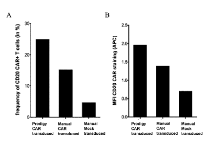
FIG 7 shows the transduction efficiency in manual versus automated conditions.
In similar
conditions as described in FIG 4. 1e8 T enriched T cells were stimulated with
MACS GMP
TransAct CD3/CD28 Kit and transduced with 1e8 transducing unit of lentiviral
vector
encoding or an anti-CD20 chimeric antigen receptor on day 1. In parallel to
the automated
manufacturing process a manual manufacturing run was carried out. 7 days after
transduction
a sample was analyzed by flow cytometry to determine A) the frequency of
transduced T cells
and B) the mean fluorescence intensity of the CAR expression. As can be seen,
the percentage
of cells expressing the transgene as well as the level of transgene expression
is higher in T
cells transduced during the automated manufacturing process.
FIG 8 shows the robustness of automated T cell manufacturing. All lines
represent
independent automated manufacturing runs performed with different donors.
Experiments are
performed as described in FIG 4. The X- axis depicts time in days and the Y-
axis the absolute
cell count determined on the different days. As can be seen, the automated
manufacturing
process is very robust and leads to very comparable results from individual
runs.
FIG 9 shows the automated manufacturing using shaking conditions on day 0.
CD4/CD8
positive cells were isolated out of apheresis and cultivated in different
settings on the
CliniMACS Prodigy platform. A higher density of 4e8 cells enriched T cells
were seeded in
100m1 (A) or 200m1 (B) total volume on day 0 and culture was immediately
carried out under
type 3 shaking conditions. The cells in 100m1 were diluted on day 2 and a
medium exchange
was performed every day beginning on day 4 until end of cultivation. On day 6
the culture
started with 200m1 (B) was stopped, the culture beginning with 100m1 (A) was
terminated on
day 8. Surprisingly, results show that it is possible to activate and expand T
cells without a
steady state phase during activation at the beginning of the culture. In such
dynamic
conditions, it is possible to very rapidly generate large numbers of T cells
(i.e. 2.8e9 T cells on
day 8 FIG 9A, versus 1.8e9 total cells on day 8 in FIG 4C).
FIG 10 shows a composition of the cell culture during automated manufacturing.
A buffy coat
from a healthy donor was connected to the tubing set T5520 (Miltenyi Biotec
GmbH)
installed on the CliniMACS Prodigy , the naïve and central memory T cell
subsets were
enriched using the CliniMACS CD62L reagent. 1e8 CD62L enriched cells were
placed in the
culture chamber, activated, transduced on day 1 with a lentiviral vector
encoding the green
CA 02946222 2016-10-18
WO 2015/162211 11
PCT/EP2015/058817
fluorescent protein (GFP) and expanded using the method described in this
invention. The
figure represents the frequency of the indicated cell subsets (from the bottom
of the bars to the
top: T cells, B cells, Monocytes, NK cells, NK T cells, and granulocytes). As
can be seen
after 11 days of culture and in the final harvest sample, the cell product is
composed of over
95% of T cells.
Embodiments
In one embodiment of the invention, a patient sample, for example, comprising
T cells, T cell
subsets and/or T cell progenitors of interest are introduced into the chamber
of a closed and
sterile culture system such as the CliniMACS Prodigy . The sample is
centrifugated,
preferentially using optical density phase detection, excess erythrocytes are
removed, the cell
sample is washed using e.g. the CliniMACS Buffer (Miltenyi Biotec GmbH) to
avoid cell
aggregation, and magnetically labeled with a magnetic cell separation reagent
such as
CliniMACS CD4 and CD8 Reagent (Miltenyi Biotec GmbH). After labeling, cells
are washed,
magnetically enriched via an integrated magnetic cell selection column and
then returned to a
cell culture chamber.
In the cell culture chamber, the T cells can be activated upon steady or
shaking culture
conditions with one or a combination of reagents capable of inducing T cell
proliferation such
as agonistic antibodies (e.g. anti-CD3 and anti-CD28), cytokines (e.g. IL-lb,
IL-2, IL-4, IL-6,
IL-7, IL-9, IL-10, IL-12, IL-15, IL-17, IL-21, IL-22, IL-23, IL-35, TGF-b, IFN
alpha, IFN
gamma, TNF alpha) recombinant proteins, costimulatory molecules, lectins,
ionophores,
synthetic molecules, antigen presenting cells (APCs), artificial APCs or
feeders. These
activation reagents can be provided in solution, coated on the cultivation
chamber or coated
on a carrier substance present in suspension/solution within the cultivation
chamber or on
large particles.
T cells can be cultivated upon steady or shaking culture conditions. After a
period of culture,
viral vector is added to the culture chamber and the cells are transduced.
Following a further
cell culture period, the cells can be transduced again or washed extensively
and harvested
(formulated). Prior to in vivo transfer of the gene-modified T cell products
the cells can be
.. washed, concentrated and resuspended in a buffer compliant with clinical
requirements for in
vivo infusion. All steps mentioned above are performed automatically.
In one embodiment of the invention the T cells, T cell subsets and/or T cell
progenitors are
labeled by binding antibody-coupled magnetic beads to a cell surface marker
present on the
CA 02946222 2016-10-18
WO 2015/162211 12
PCT/EP2015/058817
surface of the T cell, T cell subsets and/or T cell progenitors and enriching
the labeled cells by
magnetic separation (positive enrichment).
In another embodiment of the invention the T cells, T cell subsets and/or T
cell progenitors
are enriched by binding antibody-coupled magnetic beads to a cell surface
marker not present
.. on the surface of the T cells or defined cellular subsets and depleting the
labeled cells by
magnetic separation (negative enrichment).
In a further embodiment of the invention in addition to the first enrichment
of T cells, T cell
subsets and/or T cell progenitors the genetically modified T cells, T cell
subsets and/or T cell
progenitors are enriched in a second enrichment step by magnetic labeling of
the genetically
modified T cells, T cell subsets and/or T cell progenitors and magnetic
separation before or
after cultivation to obtain higher frequency of the genetically modified T
cells, T cell subsets
and/or T cell progenitors in the finally achieved cell composition obtained by
the present
method. E.g. if the genetically modified cell is a T cell expressing a CAR or
TCR, then the
second separation step may be performed by using an antigen-binding molecule
coupled to a
.. magnetic particle specific for the recombinantly expressed CAR or TCR on
the cell surface of
the genetically modified T cell.
In a preferred embodiment of the invention a cell sample, e.g. whole blood
from patient,
comprising T cells, T cell subsets and/or T cell progenitors is provided. Said
sample is
.. connected to a closed and sterile cell culture system, e.g. the sample is
connected via tubing
sets to the CliniMACS Prodigy device. The cell sample is prepared by
centrifugation in a
centrifugation chamber of the device, resulting in the separation of
erythrocytes and platelets
from other cells including T cells, T cell subsets and/or T cell progenitors.
Magnetic
separation of T cells, T cell subsets and/or T cell progenitors is performed
by using antibodies
coupled to magnetic particles specific for markers of T cells, T cell subsets
and/or T cell
progenitors such as CD2, CD3, CD4, CD8 CD25, CD28, CD27, CD45RA, CD45RO,
CD62L,
CD95, CD127, CD137, alpha/beta TCR, gamma/delta TCR, CCR7, PD-1 or Lag3. by
conducting the labeled cells through a magnet unit with separation column of
the device
resulting in an enrichment of said T cells, T cell subsets and/or T cell
progenitors. After
moving the separated T cells, T cell subsets and/or T cell progenitors to the
cultivation
chamber (which may be identical to the centrifugation chamber) of the device,
said cells are
set at a given density of 0.5e6 / ml cells to 2e6/ ml activated by using
modulatory agents, e.g.
nanomatrices which consist of mobile polymer chains having attached thereto
anti-CD3 and
ant-CD28 antibodies or fragments thereof and which are in size between 1 to
500 nm. After
CA 02946222 2016-10-18
WO 2015/162211 13
PCT/EP2015/058817
said activation of T cells, T cell subsets and/or T cell progenitors said
cells are genetically
modified in the cultivation chamber of the device, e.g. they are transduced
with a lentiviral
vector comprising a polynucleotide sequence encoding for a CAR. After genetic
modification
of the T cells, T cell subsets and/or T cell progenitors said cells are
expanded in the
cultivation under shaking conditions. Shaking may be performed by sporadic or
periodical
centrifugation of the cultivation chamber (in this case the cultivation
chamber is identical to
the centrifugation chamber) under conditions which allow the cells to be in
suspension (and as
disclosed herein). Finally, the cultured cells are washed by centrifugation,
thereby allowing
the replacement of culture medium with a buffer appropriate for subsequent
applications such
as infusion of the generated cell composition to a patient.
In one embodiment of the invention, a higher purity of transduced T cells,
e.g. T cells
expressing a transgene such as a CAR or TCR on their cell surface, is obtained
at the end of
the manufacturing process thanks to an additional cell selection step that
specifically enriches
the gene-modified T cells, this is preferably carried out using magnetic
particles coated with
antibodies directed against the surface molecule encoded by the transgene. The
step of
enrichment is preferably carried out by using again the magnetic separation
unit of the device
in an automated manner and is done before final formulation.
Preferentially, a selection agent that can be completely removed from the
surface of the
.. selected cells after this second enrichment and before application to a
patient or downstream
use is used.
In another embodiment of the invention, it is possible to start the automated
manufacturing
process with higher cell densities by activating the T cells under suspension
conditions. When
sufficient numbers of target T cells, T cell subsets and/or T cell progenitors
can be obtained
from the starting material, it is possible to start the automated
manufacturing process with a
high cell density of 4e6 to 1e7 T cells directly under shaking conditions,
e.g. using a sporadic
or periodical centrifugation of the cultivation chamber (in this case the
cultivation chamber is
identical to the centrifugation chamber) under conditions which allow the
cells to be in
suspension for activation of the cells upon onset of the culture. T cells can
be further modified
using lentiviral vector and expanded under suspension. In this embodiment of
the invention,
preferentially, the shaking conditions are maintained during the activation,
genetic
modification and expansion steps of the process as disclosed herein to keep
the high density
CA 02946222 2016-10-18
WO 2015/162211 14
PCT/EP2015/058817
cell culture in suspension.The advantage of such alternative is the
possibility to obtain large
cell numbers in a shorter period of time (typically 1 week versus 10-14 days).
In one embodiment of the invention the step of genetic modification of the T
cells, T cell
subsets and/or T cell progenitors may be performed by using lentiviral
vectors. Lentiviral
vectors with the VSVG pseudotype enable efficient transduction under automated
manufacturing method. However the method is entirely suitable for the use of
any type of
lentiviral vector (with e.g. measles virus (ML-LV), gibbon ape leukaemia virus
(GALV),
feline endogenous retrovirus (RD114), baboon endogenous retrovirus (BaEV)
derived
pseudotyped envelopes). Other viral vectors such as gamma or alpha retroviral
vectors can be
used. Transduction enhancer reagents can be added when necessary using the
automated
manufacturing described in this invention.
Definitions
Unless defined otherwise, technical and scientific terms used herein have the
same meaning
as commonly understood by one of ordinary skill in the art to which this
invention belongs.
The terms "closed cell sample processing system" and 'closed and sterile (cell
culture)
system" can be used interchangeably.
The term "closed cell sample processing system" as used herein refers to any
closed system
which reduces the risk of cell culture contamination while performing
culturing processes
such as the introduction of new material, e.g. by transduction, and performing
cell culturing
steps such as proliferation, differentiation, activation, and/or separation of
cells. Such a
system allows to operate under GMP or GMP-like conditions ("sterile")
resulting in cell
compositions which are clinically applicable. Herein exemplarily the CliniMACS
Prodigy
(Miltenyi Biotec GmbH, Germany) is used as a closed cell sample processing
system. This
system is disclosed in W02009/072003. But it is not intended to restrict the
use of the method
of the present invention to the CliniMACS Prodigy .
The process of the invention may be performed in a closed and sterile system
(a closed cell
sample processing system), comprising a centrifugation chamber comprising a
base plate and
cover plate connected by a cylinder, pumps, valves, a magnetic cell separation
column and a
tubing set. The blood samples or other sources comprising T cells, T cell
subpopulations
and/or T cell progenitors may be transferred to and from the tubing set by
sterile docking or
sterile welding. A suitable system is disclosed in W02009/072003.
CA 02946222 2016-10-18
WO 2015/162211 15
PCT/EP2015/058817
The closed cell sample processing system may comprise a plurality of tubing
sets (TS) where
cells are transferred between TS by sterile docking or sterile welding.
Different modules of the process may be performed in different functionally
closed TS with
transfer of the product (cells) of one module generated in the one tubing set
to another tubing
set by sterile means. For example, T cells, T cell subsets and/or T cell
progenitors can be
magnetically enriched in a first tubing set (TS) TS100 by Miltenyi Biotec GmbH
and the
positive fraction containing enriched T cells is welded off the TS100 and
welded onto a
second tubing set TS730 by Miltenyi Biotec GmbH for further activation,
modification,
cultivation and washing.
The terms "automated method" or "automated process" as used herein refer to
any process
being automated through the use of devices and/or computers and computer
softwares..
Methods (processes) that have been automated require less human intervention
and less
human time. In some instances the method of the present invention is automated
if at least one
step of the present method is performed without any human support or
intervention.
Preferentially the method of the present invention is automated if all steps
of the method as
disclosed herein are performed without human support or intervention other
than connecting
fresh reagents to the system. Preferentially the automated process is
implemented on a closed
cell sample processing system such as CliniMACS Prodigy as disclosed herein.
The closed cell sample processing system may comprise a) a sample processing
unit
comprising an input port and an output port coupled to a rotating container
(or centrifugation
chamber) having at least one sample chamber, wherein the sample processing
unit is
configured to provide a first processing step to a sample or to rotate the
container so as to
apply a centrifugal force to a sample deposited in the chamber and separate at
least a first
component and a second component of the deposited sample; and b) a sample
separation unit
.. coupled to the output port of the sample processing unit, the sample
separation unit
comprising a separation column holder, a pump, and a plurality of valves
configured to at
least partially control fluid flow through a fluid circuitry and a separation
column positioned
in the holder, wherein the separation column is configured to separate labeled
and unlabeled
components of sample flown through the column.
.. Said rotating container may also be used as a temperature controlled cell
incubation and
cultivation chamber (CentriCult Unit = CCU). This chamber may be flooded with
defined gas
mixes, provided by an attached gas mix unit (e.g. use of pressurized air/ N2 /
CO2 or
N2/CO2/02).
CA 02946222 2016-10-18
WO 2015/162211 16
PCT/EP2015/058817
All agents may be connected to the closed system before process initiation.
This comprises all
buffers, solutions, cultivation media and supplements, MicroBeads, used for
washing,
transferring, suspending, cultivating, harvesting cells or immunomagnetic cell
sorting within
the closed system. Alternatively, such agents might by welded or connected by
sterile means
at any time during the process.
The cell sample comprising T cells, T cell subsets and/or T cell progenitors
may be provided
in transfer bags or other suited containers which can be connected to the
closed system by
sterile means.
The term "providing a cell sample comprising T cells, T cell subsets and/or T
cell progenitors"
means the provision of a cell sample, preferentially of a human cell sample of
hematologic
origin. Normally, the cell sample may be composed of hematologic cells from a
donor or a
patient. Such blood product can be in the form of whole blood, buffy coat,
leukapheresis,
PBMCs or any clinical sampling of blood product. It may be from fresh or
frozen origin.
The term "preparation of the cell sample by centrifugation" as used herein
refers to the
separation of cells from other components (e.g. non-cell components) of the
cell sample
provided by centrifugation. The centrifugal step may comprise one, more or all
of the
following aspects: gradient separation, erythrocyte reduction, platelet
removal and cell
washing.
The term "washing" means the replacement of the medium or buffer in which the
cells are
kept. The replacement of the supernatant can be in part (example 50% of the
medium is
removed and 50% fresh medium is added) this often is applied for dilution or
feeding
purposes, or entirely. Several washing steps can be combined in order to
obtain a more
profound replacement of the original medium in which the cells are kept. A
washing step
often involves pelleting the cells by centrifugation forces and removing the
supernatant. In the
method of the present invention, cells are pelleted by rotation of the chamber
at e.g. 300xg
and the supernatant is removed during rotation of the chamber. Medium is added
during
rotation or at steady state.
The term "shaking conditions" as used herein refers to any means that allow to
keep the cells
of the cell culture in suspension. The shaking may be performed by rotating
(or sporadic
centrifugation) a cultivation chamber of a closed and sterile cell culture
system, and wherein
said rotation is performed periodically as disclosed herein. The shaking may
also be
performed e.g. by using a whipping equipment, a propelling device or a flow of
liquid (e.g
channels) integrated into the closed and sterile cell culture system used
which prevent
sedimentation of the cells.
CA 02946222 2016-10-18
WO 2015/162211 17
PCT/EP2015/058817
The term "marker" as used herein refers to a cell antigen that is specifically
expressed by a
certain cell type. Preferentially, the marker is a cell surface marker so that
enrichment,
isolation and/or detection of living cells can be performed. The markers may
be positive
selection markers such as CD4, CD8 and/or CD62L or may be negative selection
markers (e.g.
depletion of cells expressing CD14, CD16, CD19, CD25, CD56).
The term "expression" as used herein is defined as the transcription and/or
translation of a
particular nucleotide sequence driven by its promoter in a cell.
The term "particle" as used herein refers to a solid phase surface such as
colloidal particles,
microspheres, nanoparticles, or beads. Methods for generation of such
particles are well
known in the field of the art. The particles may be magnetic particles. The
particles may be in
a solution or suspension or they may be in a lyophilized state prior to use in
the present
invention. The lyophilized particle is then reconstituted in convenient buffer
before contacting
the sample to be processed regarding the present invention.
The term "nanostructure" as used herein refers to nano-sized structures which
do not fall
under the scope of the term "particle" but allow for polyclonal stimulation of
T cells when
coupled to modulatory agents such as anti CD3- and/or anti CD28-antibodies or
fragments
thereof.
The nanomatrix as disclosed in W02014/048920A1 or as given in the MACS GMP
TransAct
CD3/CD28 Kit (Miltenyi Biotec GmbH, Order no. 170-076-140) is a specific
nanostructure.
The term "nanomatrix" as used herein refers to a nanomatrix comprising
a) a matrix of mobile polymer chains; and
b) attached to said matrix of mobile polymer chains one or more stimulatory
agents which
provide activation signals to the T cells; thereby activating and inducing the
T cells to
proliferate, wherein the nanomatrix is 1 to 500 nm, preferentially 10 to 200
nm, in size.
Stimulatory agents may be anti-CD3 and/or anti-CD28 antibodies or fragments
thereof.
These polymers consists of mobile (motile), preferentially highly mobile
(motile) chains, so
the matrix is characterised by the absence of a solid surface as the
attachment point for the
stimulating agents such as antibodies, and which is in strong contrast to
currently used beads
or microspheres which regularly have an inflexible, stiff surface.
The matrix consists of a polymeric, preferentially biodegradable or
biocompatible inert
material which is non-toxic to cells. Preferentially the matrix is composed of
hydrophilic
polymer chains, which obtain maximal mobility in aqueous solution due to
hydration of the
CA 02946222 2016-10-18
WO 2015/162211 18
PCT/EP2015/058817
chains. The mobile matrix is the only or at least main component of the
nanomatrix regardless
the agents which are attached thereto.
The mobile matrix may be of collagen, purified proteins, purified peptides,
polysaccharides,
glycosaminoglycans, or extracellular matrix compositions. A polysaccharide may
include for
.. example, cellulose ethers, starch, gum arabic, agarose, dextran, chitosan,
hyaluronic acid,
pectins, xanthan, guar gum or alginate. Other polymers may include polyesters,
polyethers,
polyacrylates, polyacrylamides, polyamines, polyethylene imines,
polyquaternium polymers,
polyphosphazenes, polyvinylalcohols, polyvinylacetates, polyvinylpyrrolidones,
block
copolymers, or polyurethanes. Preferentially the mobile matrix is a polymer of
dextran.
Expamers (Stage Cell Therapeutics, Germany) are another example for a
nanostructure. Here,
a soluble StrepTactin protein oligomer is functionalized with activating
primary ligand such
as anti-CD3 and CD28 Fab fragments for polyclonal stimulation of T cell. The
StreptTactin
backbone allows for a reversible and modular functionalization via association
of low-affinity
Fab fragments.
The term "antigen-binding molecule" as used herein refers to any molecule that
binds
preferably to or is specific for the desired target molecule of the cell, i.e.
the antigen. The term
"antigen-binding molecule" comprises e.g. an antibody or antibody fragment.
The term
"antibody" as used herein refers to polyclonal or monoclonal antibodies, which
can be
generated by methods well known to the person skilled in the art. The antibody
may be of any
species, e.g. murine, rat, sheep, human. For therapeutic purposes, if non-
human antigen
binding fragments are to be used, these can be humanized by any method known
in the art.
The antibodies may also be modified antibodies (e.g. oligomers, reduced,
oxidized and
labeled antibodies).
The term "antibody" comprises both intact molecules and antibody fragments,
such as Fab,
Fab', F(a1302, Fv and single-chain antibodies. Additionally, the term "antigen-
binding
molecule" includes any molecule other than antibodies or antibody fragments
that binds
preferentially to the desired target molecule of the cell. Suitable molecules
include, without
limitation, oligonucleotides known as aptamers that bind to desired target
molecules,
carbohydrates, lectins or any other antigen binding protein (e.g. receptor-
ligand interaction).
The linkage (coupling) between antibody and particle or nanostructure can be
covalent or
non-covalent. A covalent linkage can be, e.g. the linkage to carboxyl-groups
on polystyrene
beads, or to NH2 or 5H2 groups on modified beads. A non-covalent linkage is
e.g. via biotin-
avidin or a fluorophore- coupled-particle linked to anti-fluorophore antibody.
CA 02946222 2016-10-18
WO 2015/162211 19
PCT/EP2015/058817
The terms "specifically binds to" or "specific for" with respect to an antigen-
binding
molecule, e.g. an antibody or fragment thereof, refer to an antigen-binding
molecule (in case
of an antibody or fragment thereof to an antigen-binding domain) which
recognizes and binds
to a specific antigen in a sample, e.g. CD4, but does not substantially
recognize or bind other
.. antigens in said sample. An antigen-binding domain of an antibody or
fragment thereof that
binds specifically to an antigen from one species may bind also to that
antigen from another
species. This cross-species reactivity is not contrary to the definition of
"specific for" as used
herein. An antigen-binding domain of an antibody or fragment thereof that
specifically binds
to an antigen, e.g. the CD4 antigen, may also bind substantially to different
variants of said
antigen (allelic variants, splice variants, isoforms etc.). This cross
reactivity is not contrary to
the definition of that antigen-binding domain as specific for the antigen,
e.g. for CD4.
A potent sorting technology is magnetic cell sorting. Methods to separate
cells magnetically
are commercially available e.g. from Invitrogen, Stem cell Technologies, in
Cellpro, Seattle
or Advanced Magnetics, Boston. For example, monoclonal antibodies can be
directly coupled
to magnetic polystyrene particles like Dynal M 450 or similar magnetic
particles and used e.g.
for cell separation. The Dynabeads technology is not column based, instead
these magnetic
beads with attached cells enjoy liquid phase kinetics in a sample tube, and
the cells are
isolated by placing the tube on a magnetic rack. However, in a preferred
embodiment for
enriching, sorting and/or detecting T cells, T cell subsets and/or T cell
progenitors from a cell
sample monoclonal antibodies are used in conjunction with colloidal
superparamagnetic
microparticles having an organic coating by e.g. polysaccharides (Magnetic-
activated cell
sorting (MACS ) technology (Miltenyi Biotec, Bergisch Gladbach, Germany)).
These
particles (nanobeads or MicroBeads) can be either directly conjugated to
monoclonal
antibodies or used in combination with anti- immunoglobulin, avidin or anti-
hapten-specific
MicroBeads. The MACS technology allows cells to be separated by incubating
them with
magnetic nanoparticles coated with antibodies directed against a particular
surface antigen.
This causes the cells expressing this antigen to attach to the magnetic
nanoparticles.
Afterwards the cell solution is transferred on a column placed in a strong
magnetic field. In
this step, the cells attach to the nanoparticles (expressing the antigen) and
stay on the column,
while other cells (not expressing the antigen) flow through. With this method,
the cells can be
separated positively or negatively with respect to the particular antigen(s).
In case of a
positive selection the cells expressing the antigen(s) of interest, which
attached to the
magnetic column, are washed out to a separate vessel, after removing the
column from the
CA 02946222 2016-10-18
WO 2015/162211 20
PCT/EP2015/058817
magnetic field. In case of a negative selection the antibody used is directed
against surface
antigen(s), which are known to be present on cells that are not of interest.
After application of
the cells/magnetic nanoparticles solution onto the column the cells expressing
these antigens
bind to the column and the fraction that goes through is collected, as it
contains the cells of
interest. As these cells are non-labeled by an antibody coupled to
nanoparticles, they are
"untouched". The procedure can be performed using direct magnetic labeling or
indirect
magnetic labeling. For direct labeling the specific antibody is directly
coupled to the magnetic
particle. Indirect labeling is a convenient alternative when direct magnetic
labeling is not
possible or not desired. A primary antibody, a specific monoclonal or
polyclonal antibody, a
combination of primary antibodies, directed against any cell surface marker
can be used for
this labeling strategy. The primary antibody can either be unconjugated,
biotinylated, or
fluorophore-conjugated. The magnetic labeling is then achieved with anti-
immunoglobulin
MicroBeads, anti-biotin MicroBeads, or anti-fluorophore MicroBeads. The above-
described
processes can also be performed in a closed cell sample processing system such
as
CliniMACSO (Miltenyi Biotec GmbH, Germany) or CliniMACS Prodigy (Miltenyi
Biotec
GmbH, Germany).
The term "substantially pure cell composition of genetically modified T cells,
T cell subsets
and/or T cell progenitors "as used herein refers to a cell composition
comprising at least 70%,
more preferentially at least 90%, most preferentially at least 95% of
genetically modified T
cells, T cell subsets and/or T cell progenitors in the cell composition
obtained by the method
of the present invention. The transduction frequency of the cell product
depends on the type
of vector used to carried out the gene modification as well as the nature of
the transgene.
Increased frequency of gene-modified T cells can be obtained by including an
additional
selection step directed towards, at least a part, of the transgene.
"Chimeric antigen receptor" or "CAR" refer to engineered receptors, which
graft an antigen
specificity onto cells, for example T cells. The CARs of the invention
comprise an antigen
binding domain also known as antigen targeting region, an extracellular spacer
domain or
hinge region, a transmembrane domain and at least one intracellular signaling
domain or a
least one co-stimulatory domain and at least one intracellular signaling
domain.
The term "genetically modified cell" means containing and/or expressing a
foreign gene or
nucleic acid sequence which in turn modifies the genotype or phenotype of the
cell or its
progeny. Especially, the term refer to the fact that cells can be manipulated
by recombinant
methods well known in the art to express stably or transiently peptides or
proteins, e.g. CARs
CA 02946222 2016-10-18
WO 2015/162211 21
PCT/EP2015/058817
which are not expressed in these cells in the natural state. Genetic
modification of cells may
include but is not restricted to transfection, electroporation, nucleofection,
transduction using
retroviral vectors, lentiviral vectors, non-integrating retro- or lentiviral
vectors, transposons,
designer nucleases including zinc finger nucleases, TALENs or CRISPR/Cas.
The genetically modified T cells, T cell subsets and/or T cell progenitors
obtainable by the
methods disclosed herein may be used for subsequent steps such as research,
diagnostics,
pharmacological or clinical applications known to the person skilled in the
art.
The genetically modified T cells, T cell subsets and/or T cell progenitors can
also be used as a
pharmaceutical composition in the therapy, e.g. cellular therapy, or
prevention of diseases.
The pharmaceutical composition may be transplanted into an animal or human,
preferentially
a human patient. The pharmaceutical composition can be used for the treatment
and/or
prevention of diseases in mammals, especially humans, possibly including
administration of a
pharmaceutically effective amount of the pharmaceutical composition to the
mammal.
Pharmaceutical compositions of the present disclosure may be administered in a
manner
appropriate to the disease to be treated (or prevented). The quantity and
frequency of
administration will be determined by such factors as the condition of the
patient, and the type
and severity of the patient's disease, although appropriate dosages may be
determined by
clinical trials.
The term "therapeutic effective amount" means an amount which provides a
therapeutic
benefit for the patient.
The composition of genetically modified T cells, T cell subsets and/or T cell
progenitors
obtained by the method of the present invention may be administered either
alone, or as a
pharmaceutical composition in combination with diluents and/or with other
components such
as cytokines or cell populations. Briefly, pharmaceutical compositions of the
present
invention may comprise the genetically modified T cells, T cell subsets and/or
T cell
progenitors of the present invention as described herein, in combination with
one or more
pharmaceutically or physiologically acceptable carriers, diluents or
excipients. Such
compositions may comprise buffers such as neutral buffered saline, phosphate
buffered saline
and the like; carbohydrates such as glucose, mannose, sucrose or dextrans,
mannitol; proteins;
polypeptides or amino acids such as glycine; antioxidants; chelating agents
such as EDTA or
glutathione; adjuvants (e.g., aluminum hydroxide); and preservatives.
CA 02946222 2016-10-18
WO 2015/162211 22
PCT/EP2015/058817
Examples
Example 1: Automated manufacturing of gene-modified T cells using several
closed sterile
tubing sets
A leukapheresis bag (100-200 ml) from a donor is connected, by sterile welding
to the Tubing
set TS 100 (Miltenyi Biotec GmbH) installed on the CliniMACS Prodigy device.
CliniMACS buffer as well as CliniMACS CD4 and CD8 reagents (Miltenyi Biotec
GmbH)
are also connected to the same Tubing set. An enrichment program is launched.
The tubing
set is automatically primed with buffer, then the leukaphereis product is
transferred to the
chamber of the tubing set where it is washed 3 times with CliniMACS buffer in
order to
remove serum and platelets. A red cell reduction is also performed to remove
excess
erythrocytes. The CliniMACS CD4 and CD8 reagents are transferred to the cells
into the
chamber for magnetic labeling of the CD4 and CD8 positive cells. After 30 min
incubation at
room temperature, the magnetically labeled cells are automatically transferred
onto a column
placed in a magnetic field. The labeled cells are trapped and the non-labeled
cells are eluted in
a non-target cell fraction bag. The column with the labeled cells is rinsed
several times after
which the labeled cells are eluted into the target cell fraction bag. A sample
pouch integrated
in the tubing set allows to obtain a sample of 1-2 ml that is analyzed
remotely for cell counts
and cell purity by flow cytometry (FIG 12). Part of the enriched cells are
transferred (via
sterile welding connection) into another Tubing set (TS730) newly installed on
a CliniMACS
Prodigy device. The tubing set is also connected to MACS GMP TexMACS medium
supplemented with IL-2, MACS GMP TransAct CD3/CD28 Kit (all Miltenyi Biotec
GmbH)
and 1e8 enriched T cells. The activation program is started. The enriched
cells are washed and
resuspended in medium, the culture is set at 37 C and with a 5% CO2 gas
supply. Upon
equilibration of the culture, the activation reagent is automatically added to
the culture. 24h
later, a sample is analyzed for the upregulation of the activation markers
CD25 and CD69
(FIG 2). A bag containing the viral vector is sterile welded onto the tubing
set using for
instance a Terumo sterile welder, the user acknowledges the prompt in the
software asking for
confirmation that the viral vector has been connected and the viral vector is
transferred into
the chamber containing the activated T cells. After 5 days of culture the T
cells are transferred
into a 5 liter bag and transferred onto the Wave Bioreactor TM (Life
Technologies), another
device enabling T cell expansion. The cells are cultured for an additional 7
days in suspension.
The bag of expanded cells is connected back onto a fresh tubing set on the
CliniMACS
Prodigy . The CliniMACS Prodigy allows the automated concentration of the
cells from 5L
down to 100 ml and the re-buffering of the cells in a solution suitable for
human infusion.
CA 02946222 2016-10-18
WO 2015/162211 23
PCT/EP2015/058817
Example 2: Automated manufacturing of gene-modified T cells using a single
closed sterile
tubing set
A buffy coat bag of 100-200 ml from a donor is connected, by sterile welding
to the Tubing
set TS520 (Miltenyi Biotec GmBH) installed on the CliniMACS Prodigy device.
CliniMACS buffer as well as CliniMACS CD62L reagent, MACS GMP CD3/CD28 Kit and
MACS GMP TexMACS medium containing 10 ng/ml IL-7 and IL-15, are also connected
to
the T5520 (all Miltenyi Biotec GmbH). The activity matrix is then filled with
activities (e.g.
transduction, feed, wash, final formulate) and their parameters (e.g. day,
volume, temperature)
of the automated manufacturing run. The programmed software is then launched
(FIG 3). As
in example 1 the leukapheresis is washed. Labeling takes place at 4-8 C (this
is important in
order to enrich CD62L positive cells as CD62L is shed from the surface of
cells at room
temperature). Labeled cells are enriched and eluted into a reapplication bag
which is part of
the tubing set. The program asks for a sample to be taken (using sampling
pouches included
in the tubing set). Once the cell density determined, this information is
indicated in the
program as well as the required number of cells to be transferred back into
the chamber (e.g.
1e8). The required volume of enriched cells containing 1e8 enriched cells is
then
automatically transfered into the cultivation chamber. The activation reagent
is then added to
the culture. On day 1, the bag containing the Lentiviral vector in 10 ml is
connected to the
tubing set (this is performed extemporaneously due to the short half life of
viral vectors). The
viral vector, in this case a lentiviral vector encoding a CD20 CAR (comprising
the 4-1BB and
CD3zeta signaling domains) is then transferred onto the activated T cells. The
T cells remain
in culture at 37 C and in an atmosphere enriched with 5% CO2 for an additional
2 days. On
day 5 the spent culture medium is automatically washed away and replaced with
fresh
medium. The culture is now set on type 1 shaking (low shaking) in order to
gently resuspend
the T cells using a sporadic slow shaking of the chamber. Half the culture
medium is
exchanged every other day in order to feed the cells with fresh medium. On day
9 the shaking
speed is increased to a type 3 shaking (more vigorous resuspension). On days
11 ¨ 13, the
manufacturing process reaches an end, the cells are washed several time with a
solution
suitable for human infusion (i.e. final formulation buffer) and harvested in a
bag for further
clinical handling (FIG 4 and 6). Either for direct infusion or for
cryopreservation. The
manufactured gene-modified T cells were analyzed for their cell composition
(FIG 10), their
percent of transduced cells and the level of transgene expression per
transduced cells (FIG 7A
and B respectively).
CA 02946222 2016-10-18
WO 2015/162211 24
PCT/EP2015/058817
Example 3: Automated manufacturing of gene-modified T cells starting with high
density
culture activated under shaking conditions
Starting from a leukapheresis of 100-200 ml, CD4 and CD8 T cells are enriched
similarly to
Example 2 using the tubing set T5520 installed onto the CliniMACS Prodigy . In
this
example however a high density of 4e6 enriched T cells / ml are transferred
into the chamber
of the closed sterile tubing set. The enriched T cells were seeded in 100m1
(FIG 9A) on day 0
and activation was immediately carried out under type 3 shaking conditions
using MACS
GMP TransAct CD3/CD28 kit and 200IU IL-2 in MACS GMP TexMACS medium. The cells
in 100 ml were diluted on day 2 and a medium exchange was performed every day
beginning
on day 4 until end of cultivation. The cultivation was ended on day 8.
Surprisingly, results
show that it is possible to activate and expand T cells without a steady state
phase during
activation at the beginning of the culture. In such dynamic conditions, it is
possible to very
rapidly generate large numbers of T cells (i.e. 2.8e9 T cells on day 8 FIG 8A,
versus 1.8e9
total cells on day 8 in FIG 4C).
Example 4: Automated manufacturing of gene-modified T cells starting from
frozen
leukapheresis
A 100 ml bag of frozen leukapheresis from a donor was thawed and transferred
into a 3L
MACS GMP Cell Differentiation bag (Miltenyi Biotec GmbH product) and diluted
with
MACS GMP TexMACS medium into 2L. Thawed cells were rested for 48 hours at 37 C
in
5% CO2, the bag of cells was then connected to a closed sterile tubing set
installed on the
CliniMACS Prodigy for automated cell concentration. A similar manufacturing
process as
described on Example 2 was then carried out (using CD62L enriched T cells). On
day 12, the
gene modified T cells were final formulated in 100 ml CliniMACS Buffer and
transferred into
the harvest bag. The entire bag was connected by sterile welding to the closed
sterile tubing
set TS100 installed on a CliniMACS Prodigy. There, the gene-modified T cells
where
labeled with anti-IgG1 microbead in order to permit isolation of gene-modified
T cells from
the non-modified T cells. All steps from the diluted and rested thawed cells
to the
manufacturing and isolation of gene-modified T cells was performed in an
automated manner
using several different tubing sets and several different types of programs.