Note: Descriptions are shown in the official language in which they were submitted.
METHOD FOR REMOVING BACTERIA FROM BLOOD
USING HIGH FLOW RATE
CROSS-REFERENCE TO RELATED APPLICATIONS
[0001] The present application claims priority to US Provisional Patent
Application No.
61/984,013, filed April 24, 2014.
BACKGROUND OF THE INVENTION
[0002] Bloodstream infection, or bacteremia, is a major challenge in the
Intensive Care Unit
(ICU). Bacteremia can quickly lead to septic shock, meningitis, endocarditis,
osteomyelitis and
other metastatic complications. Staphylococcus aureus, P. aeruginosa and
Enterobacteriacea
are the most common bacteria responsible for bacteremia and nosocomial
infections. Severity of
outcome for bacteremic patients is correlated to both the bacterial load and
duration of
bacteremia. For example, a quantitative RT-PCR study of E. coli and S. aureus
bacteremia
patients showed that when the number of rDNA increased to over 1,238
copies/ml, mortality
increased from 14.3% to 42.9% and septic shock increased from 31.4% to 85.7%.
It was also
found that a high blood concentration of N. meningitides is correlated with
prolonged
hospitalization, limb or tissue loss, the need for dialysis, and patient
mortality. Another study
showed that the severity of Pneumococcal pneumonia correlated with bacterial
load in the blood:
the mortality for patients with over 1000 S. pneumoniae DNA copies/ml of blood
was 25.9% vs.
6.1% for patients exhibiting less than 1000 copies/ml. In yet another study, a
follow-up positive
blood culture between 48 and 96 hours after initial diagnosis was shown to be
the strongest
predictor of complicated S. aureus bacteremia. Compounding the difficulty of
effective
bacteremia treatment is the often delayed administration of appropriate
antibiotic therapy. For
each hour of delay in treatment the mortality risk increases over 7%.
[0003] The conventional strategy for combating bacterial infections is to
administer active
drugs that specifically kill bacteria while minimizing damage to host tissue.
This is a major
1
Date Recue/Date Received 2021-09-01
CA 02946294 2016-10-19
WO 2015/164198 PCTIUS2015/026340
challenge as some of the more effective antibiotics available today are quite
toxic. For example,
vancomycin is nephrotoxic, and may soon be contraindicated for patients
undergoing
extracorporeal oxygenation. Even if new antibiotics are successfully developed
to address
current drug resistance, new superbugs will continue to emerge. Clearly, new
strategies for
combating infection are needed, in addition to drug discovery.
[00041 Drug-resistant pathogens arc a growing threat to the healthcare system.
The CDC has
recently warned of the emergence of carbapenem-resistant Enterobacteriacea
(CRE; "super
bugs"). The mortality rate for CRE bacteremia can be as high as 50%.
Resistance of CREs to
even the strongest available antibiotics leaves clinicians with few treatment
options. The
incidence of hospital-acquired CRE infections has increased 400% over the last
10 years.
Currently, CRE bacteremias are mostly nosocomial infections, but there is
concern that the
incidence of community acquired CRE could increase. Today, the only strategy
is to reduce
CRE infections is through education and prevention.
[0005] There is a need for a safe, broad-si ectrum. technology that can
quickly reduce bacterial
load, and shorten the duration of bacteremia. The present invention satisfies
this and other needs
by providing a high-surface-area extracorporeal affinity adsorption media that
can quickly and
safely remove pathogens from whole blood or whole serum.
BRIEF SUMMARY OF THE INVENTION
[0006] The present invention provides methods that can quickly reduce
bacterial load, and
shorten the duration of bacteremia even without first identifying the type of
bacteria present in
the blood.
[0007] In some aspects, provided herein is an ex vivo method for removing
bacteria from a
sample taken from. a subject who is suspected of being infected with bacteria.
The method
comprising, consisting essentially of or consisting of: contacting a sample
taken from the subject
with an adsorption media to allow the formation of an adhering complex,
wherein the adhering
complex comprises bacteria and the adsorption media; and separating the sample
from the
adhering complex to produce the sample with a reduced amount of bacteria.
Typically, the
adsorption media is contained within a column, a container or cartridge.
2
CA 02946294 2016-10-19
WO 2015/164198 PCTIUS2015/026340
100081 in some embodiments, the sample is selected from the group consisting
of whole blood,
serum and plasma. In other embodiments, the sample is whole blood.
100091 In some embodiments, the adsorption media is a solid substrate of high
surface area
having a hydrophilic surface that is free of a polysaccharide adsorbent. In
some instances, the
solid substrate comprises a plurality of rigid polymer bead. In some
embodiments, the rigid
polymer bead is a member selected from the group consisting of polyurethane,
polymethylmethacrylate, polyethylene or co-polymers of ethylene and other
monomers,
polyethylene imine, polypropylene, and polyisobutykne. In other embodiments,
the solid
substrate comprises one or a plurality of hollow fibers or yarn.
[0010] In some embodiments, the hydrophilic surface is a cationic surface. In
other
embodiments, the hydrophilic surface is a neutrally charged surface.
[00111 In some embodiments, the bacteria in the sample are reduced by about
20% to about
99.9%. In other embodiments, the bacteria in the sample are reduced by about
20% to about
40%.
100121 in some embodiments, the bacterium is a gram-negative bacterium. In
other
embodiments, the bacterium is a gram-positive bacterium. In other embodiments,
the bacteria is
selected from the group consisting of Escherichia coil, Klebsiella pneumoniae,
carbapenem-
resistant Escherichia coil, carbapenem-resistant Klebsiella pneumoniae, and
extended spectrum
beta-lactamase Klebsiella pneumoniae, Enterococcus faccium, Acinetobacter
baumannii, and
methicillin-resistant Staphylococcus aureus (MRSA). In yet other embodiments,
the bacterium
is selected from the group consisting of Staphylococcus aureus, methicillin-
resistant
Staphylococcus aureus (MRSA), and Escherichia coll.
100131 In some embodiments, the cationic surface of the adsorption media forms
an adhering
complex with bacteria selected from the group consisting of Escherichia coil,
Klebsiella
pneumoniae, carbapenem-resistant Escherichia coil, carbapenem-resistant
Klebsiella
pneumoniae, and extended spectrum beta-lactamase Klebsiella pneumoniae,
Enterococcus
faecium, Acinetobacter baumannii, and methicillin-resistant Staphylococcus
aureus (MRSA). In
other embodiments, the neutrally charged surface forms an adhering complex
with bacteria
3
CA 02946294 2016-10-19
WO 2015/164198 PCTIUS2015/026340
selected from the group consisting of Staphylococcus aureus, methicillin-
resistant
Staphylococcus aureus (MRSA), and Escherichia coll.
100141 in some aspects, provided herein is an ex vivo method for removing
bacteria from a
sample taken from a subject who is suspected of being infected with bacteria,
wherein the
bacteria are known to have a low affinity or no affinity for heparan sulfate.
The method
comprising, consisting essentially of or consisting of: contacting a sample
taken from a subject
with an adsorption media to allow the formation of an adhering complex,
wherein the adsorption
media is a solid substrate of high surface area having at least one
polysaccharide adsorbent on
the surface thereof and separating the sample from the adhering complex to
produce the sample
with a reduced amount of bacteria. The adhering complex comprises bacteria and
the
adsorption media. Typically, the adsorption media is contained within a
column, a container or
cartridge. In certain aspects, the sample exits the column, the container or
the cartridge, and the
adhering complex remains behind.
[0015] In some embodiments, the sample is selected from the group consisting
of whole blood,
scrum and plasma. In other embodiments, the sample is whole blood.
[0016] In some embodiments, the solid substrate comprises a plurality of rigid
polymer bead.
In some instances, the rigid polymer bead is a member selected from the group
consisting of
polyurethane, polymethylmethacrylate, polyethylene or co-polymers of ethylene
and other
monomers, polyethylene imine, polypropylene, and polyisobutylene. In other
embodiments, the
solid substrate comprises one or a plurality of hollow fibers.
[0017] In some embodiments, the at least polysaccharide absorbent is a member
selected from
the group consisting of heparin, heparan sulfate, hyaluronic acid, sialic
acid, carbohydrates with
mannose sequences, and chitosan. In other embodiments, the at least
polysaccharide absorbent is
heparin or heparan sulfate. In some instances, the at least polysaccharide
absorbent is heparin.
[0018] In some embodiments, the beads are coated with about 0.27 mg to about
10 mg heparin
per gram of bead. In other embodiments, the bead is coated with 2 0.5 mg
heparin per gram of
bead.
4
CA 02946294 2016-10-19
WO 2015/164198 PCTIUS2015/026340
100191 in some embodiments, the bacteria in the sample are reduced by about
20% to about
99.9%. In other embodiments, the bacteria in the sample are reduced by about
20% to about
40%.
[00201 In some embodiments, the bacteria are gram-negative bacteria. in other
embodiments,
the bacteria are gram-positive bacteria. In yet other embodiments, the
bacteria is selected from
the group consisting of Escherichia coif, Klebsiella pneumoniae, Acinetobacter
baumannii,
Enterococcus faecium, carbapenem-resistant Escherichia coif, carbapenem-
resistant Klebsiella
pneumoniae, and extended spectrum beta-lactamase Klebsiella pneumoniae.
100211 In some aspects, provided herein is an ex vivo method for removing
bacteria from a
sample taken from a subject undergoing dialysis or extracorporeal oxygenation.
The method
comprising, consisting essentially of, or consisting of: contacting a sample
taken from a subject
with an adsorption cartridge comprising adsorption media, wherein the
adsorption cartridge is in
series with a dialysis cartridge or oxygenator to allow the formation of an
adhering complex and
separating the sample from the adhering complex to produce the sample with a
reduced amount
of bacteria. The adhering complex comprises bacteria and adsorption media.
Typically, the
adsorption media is contained within a column, a container or cartridge. In
certain aspects, the
sample exits the column, the container or the cartridge, and the adhering
complex remains
behind.
100221 In some embodiments, the sample has a total blood volume of less than
200 ml.
[0023] In some embodiments, the adsorption cartridge has a column height
between 1 cm-50
cm. In some embodiments, the adsorption cartridge has a column diameter
between 1 cm-50 cm.
[0024] In some embodiments, the adsorption cartridge is proximal to the
subject compared to
the dialysis cartridge. In other embodiments, the adsorption cartridge is
distal to the subject
compared to the dialysis cartridge.
100251 These and other aspects, objects and advantages will become more
apparent when read
with the figures and the detailed description which follow.
5
CA 02946294 2016-10-19
WO 2015/164198 PC1'11152015/026340
BRIEF DESCRIPTION OF THE DRAWINGS
[0026) FIGS. 1A-B show a comparison of the adsorption media and human blood.
FIG. lA
shows the adsorption media and FIG. 1B shows an image of a human blood smear.
[0027) FIG. 2 shows a size comparison of bacteria, e.g., Staphylococcus aureus
and
Chlamydia, and viruses, e.g., pox virus, herpes virus, influenza virus, and
picornavirus (polio).
[00281 FIG. 3 illustrates a cross-section of the adsorption media containing
beads with a
diameter (d) and a cell with a diameter (a).
[00291 FIG. 4 illustrates the minimum bead size as a function of linear flow
and adsorption
cartridge column height for a rigid media subject to forced convection.
DETAILED DESCRIPTION OF THE INVENTION
MOM The present invention is based, in part, on the discovery of an
adsorption media that is
effective for removing a significant amount of bacteria (e.g., gram-negative
bacteria and gram-
positive bacteria, including bacteria with no known affinity or low affinity
for heparan sulfate)
from blood (e.g., whole blood and blood serum). In addition, the adsorption
media can be used
in extracorporeal treatments involving high volumetric flow rates and high
linear flow rates.
Typically, the adsorption media is contained within a column, a container or
cartridge. In
certain aspects, the sample exits the column, the container or the cartridge,
and an adhering
complex remains behind.
[0031] A first aspect of the present invention provides a method for the
removal of bacteria
from blood, such as mammalian blood, by contacting the blood with a solid
substrate. The
inventors have found that the surface architecture of the solid substrate is
effective for removing
pathogens such bacterial pathogens or viruses.
[0032] The substrate of the present invention possesses sufficiently large
interstitial
dimensions to permit a high flow rate of blood over the substrate without a
large pressure drop.
For instance, as blood is taken from a mammalian patient, it is passed over
the substrate at a flow
rate whereby the delivery of adsorbates to the surface of the adsorbent bed is
characterized
primarily by forced convection. Substrates suited for convection transport,
generally rely on
6
CA 02946294 2016-10-19
WO 2015/164198 PCT/US2015/026340
macroscopic "channels" or visible interstices between solid, essential
nonporous material, such
as particles, beads, fibers, yarn, reticulated foams, or optionally spiral-
wound dense membranes.
100331 This is in contrast to highly porous adsorbent media (e.g., porous
silica, Sephadexe,
crosslinked polystyrene and other size exclusion media), and many other
microporous media that
use the much slower process of molecular diffusion. Adsorption substrates that
depend on
diffusion transport arc generally composed of porous materials with
microscopic pores and an
extremely high internal surface area.
I. Definitions
100341 The term "extracorporeal therapy" includes a medical procedure that is
conducted
outside the body i.e., ex vivo. In some instances, extracorporeal therapies
include methods in
which a bodily fluid such as blood is taken from the individual and desired
products such as, but
not limited to, oxygen, blood-anticoagulants, anesthetics, and the like are
added to the body fluid
before it is returned to the individual. In other instances, an extracorporeal
therapy includes
removing undesired products like naturally occurring toxins, poisons or
viruses from the body or
the body fluids. Non-limiting examples of extracorporeal therapies include
apheresis,
autotransfusion, hemodialysis, hemofiltration, plasmapheresis, extracorporeal
circulation (ECC),
extracorporeal life support (ECLS) extracorporeal membrane oxygenation (ECMO),
and
cardiopulmonary bypass.
100351 The term "high flow rate" or "high flow condition" includes a flow rate
or velocity of
blood that is above the diffusion limit.
100361 The term "adsorption media" includes a material to which a cell,
organism, virus,
pathogen, polypeptidc, polynucleotidc, chemical molecule, biological molecule
can adhere to the
surface thereof and be removed from a sample such as blood.
100371 The term "adhering complex" includes a complex of at least two
molecules wherein the
first molecule is attached (e.g., linked, coupled or bound) to a surface such
as a substrate and the
second molecule is attached to the first molecule. For example, a pathogen or
virus can adhere
to heparin to form an adhering complex. Typically, in the methods of the
present invention, the
adhering complex remains behind and the sample is cleansed of the patthogen or
virus.
7
CA 02946294 2016-10-19
WO 2015/164198 PCTIUS2015/026340
100381 The term "high surface area" includes the property of having a large
specific surface
area to volume ratio.
100391 The term "adsorbent" includes a solid substrate with a chemical
compound, a biological
molecule, or a material that is attached (e.g., linked, coupled or bound)
thereto. In certain
instances, the adsorbent is the solid substrate itself. in one embodiment, an
adsorbent is a
polymer resin with a polysaccharide such as heparin bound thereto. The
substrate can be a
polymer bead, fiber or yarn.
[00401 The term "rigid polymer bead" refers to a bead, granule, pellet,
sphere, particle,
microcapsule, sphere, microsphere, nanosphere, microbead, nanobead,
microparticle,
nanoparticle, and the like that is made from a polymer resin. A polymer bead
is useful as a
substrate.
[00411 The term "fiber" or "yam" is useful as a soild substrate. The fiber or
yarn can be made
of a synthetic polymer or a natural polymer or a mixture thereof. In certain
instances, an
originally porous hollow fiber or yarn is rendered dense or non-porous before,
during or after
binding heparin or other adsorbents to the outer and/or inner surfaces
thereof.
[0042] The term "carbohydrate" refers to a molecule containing carbon,
hydrogen and oxygen
atoms, and usually with the empirical formula C2(H20)y, where x and y are
different numbers.
Examples of carbohydrates includes monosaccharides, disaccharides,
oligosaccharides, and
polysaccharides.
[0043] The term "polysaccharide" refers to a molecule of monosaccharide units
joined
together by glycosidic bonds, and having an empirical formula of C2(F120)y,
where x is between
200 to about 3000.
[0044] The term "hydrophilic surface" includes a surface with a water contact
angle less than
90 when the surface is flat.
[0045] The term "low affinity to heparan sulfate" in the context of a
bacteria, refers to the low
binding affinity of the bacteria for heparan sulfate. In some embodiments, the
binding affinity is
determined using standard assays, such as an enzyme-linked immunosorbent assay
(ELISA) for
heparan sulfate. In other embodiments, the binding affinity is determined
based on a predictive
8
CA 02946294 2016-10-19
WO 2015/164198 PCTIUS2015/026340
analysis, such as an analysis of putative heparan sulfate binding proteins
expressed by the
pathogen, e.g., bacteria. The term "no affinity for heparan sulfate" refers to
a bacteria having no
binding affinity for or a lower than detectable affinity for heparan sulfate,
or no known binding
to heparan sulfate. In some instances, having no affinity for heparan sulfate
includes having no
predicted binding affinity for heparan sulfate.
IL Detailed Description of Embodiments
A. Binding of Bacterial Pathogens by Convection Transport
[00461 The binding of bacterial pathogens to the essentially nonporous
adsorption substrate of
the present invention during convection transport is particularly effective
under the relatively
high-flow conditions typically employed in the safe operation of
extracorporeal blood circuits,
e.g. when measured by linear flow velocity, >8 cm/min, preferably about >30
cm/min, and more
preferably about 30-1,000 cm/min.
100471 in some embodiments, the adsorption media removes pathogens from whole
blood in
extracorporeal circuits with a linear flow rate of about 8 cm/min to about
1,000 cm/min, e.g.,
about 8 cm/min to about 30 crn/min, about 25 cm/min to about 100 cm/min, about
50 cm/min to
about 200 cmJmin, about 100 cm/min to about 1000 cm/min, about 200 cm/min to
about 1000
cm/min, about 400 cm/min to about 1000 cm/min, about 500 crn/min to about 1000
cm/min,
about 600 cm/min to about 1000 cm/min, about 100 cm/min to about 500 cm/min or
about 300
cm/min to about 800 cm/min. In certain instances, the flow rate is about 10,
15, 20, 25, 30, 35,
40, 45, 50, 55, 60, 65, 70, 75, 80, 85, 90, 95 100 cm/min or about 25-40
cm/min.
[0048I In other embodiments, the adsorption media removes pathogens from whole
blood in
extracorporeal circuits with a volumetric flow rate, around 50 mUminute to
about 5 L/minute,
e.g., 50 mL/min, 100 mL/min, 150 mL/min, 200 mL/min, 250 mL/min, 300 mL/min,
350
mL/min, 400 mL/min, 500 mL/min, 550 mL/min, 600 mL/min, 650 mL/min, 700
mL/min, 750
mL/min, 800 mL/min, 850 mL/min, 900 mL/min, 950 mL/min, 1.0 L/min, 1.5 I./min,
2.0 I./min,
2.5 L/min, 3.0 L/min, 3.5 L/min, 4.0 L/min, 4.5 L/min, and 5 L/min. In som.e
embodiments, the
flow rate is preferably >150 mUminute.
[0049] Highly porous adsorbent media, in contrast, requires much lower flow
rates of less than
1 mi./minute to about less than 50 mUminute. Additionally, the residence time
on the
9
CA 02946294 2016-10-19
WO 2015/164198 PCTIUS2015/026340
adsorption substrate (e.g., amount of time the adsorbate (e.g., bacteria) is
in contact with the
adsorbent media) needs to be much longer for a media requiring diffusive
transport of adsorbates
to the adsorbent site within the media, compared to a media using forced
convection of
adsorbates to the binding sites, which are not compatible with standard
extracorporeal blood
systems.
[0050] Typically, it is recognized that "residence time" on the adsorption
column needs to be
longer for a media requiring diffusive transport of adsorbates to the
adsorbent site within the
media, when compared to the lower residence time needed to convey an adsorbate
to the binding
site (on an essentially nonporous media) by forced convection. However, there
are practical
.. limits to the dimensions of a safe and effective adsorbent cartridge,
column, filter, etc., especially
with respect to the maximum hold-up volume of blood it can contain, and the
flow velocity of
blood or serum past the adsorption media. For this reason average flow rate
through the
adsorption device is considered to be a design variable.
[0051] Substrates that rely on forced convection transport are generally more
suitable for high-
flow rates, while substrates that rely on the much slower diffusion transport
are much less
effective when high flow rates and shorter residence times are required. For
this reason, in an
extracorporeal blood purification device, it is preferred that an adsorbate
quickly diffuses
through the pores within the adsorbent media. When blood is pumped through
circuits fabricated
from man-made materials, it is a general practice to employ relatively high
blood flow rates in
order to prevent stagnation and reduce the risk of clotting. On the other
band, extremely high
flow rates may be avoided because they can expose blood cells to high shear
rates and
impingement damage that can rupture or otherwise damage blood cells. The
present invention,
therefore, provides a method and device for removing bacterial pathogens from
blood using the
preferred characteristics of convection transport and its desirable, more-
rapid kinetics. This is
.. achieved by passing/flowing blood over an essentially non-microporous
substrate (e.g., a solid
substrate), which is capable of binding the desired cytokine, pathogen or
bacteria to remove them
from the blood.
100521 Adsorption media provided herein can be used in traditional
extracorporeal blood
circulation with flow rates >50 mL/min, and preferably between about 150
mUminute to
5L/minute. If measured by linear flow velocity, >8 cm/min, preferably about
>24 cm/min and
CA 02946294 2016-10-19
WO 2015/164198 PCT/US2015/026340
more preferably about 24-329 cm/min, or more. For example, the flow rate can
be 25, 50, 75,
100, 125, 150,175, 200, 225, 250, 275, 300, 325, 350, 375, 400, 425, 450, 475,
500, 525, 550,
575, 600, 625, 650, 675, 7(X), 725, 750, 775, 800 cm/min or more. Such high
flow rates create
short residence times within the adsorption column and convection transport
dominates over
Brownian diffusive transport. This is particularly important for binding
larger particles such as
viruses, bacteria and parasites and other proteins and pathogens that diffuse
slowly.
[00531 The main adsorption sites available for removing bacterial pathogens
lie at the surfaces
within the interstices of the media bed, container or cartridge through which
the blood flows or is
delivered by forced convection. To treat blood, the interstitial channels need
to be large enough
to allow the transport of red blood cells, which are an average 6 microns in
diameter. To allow a
packed adsorption cartridge to be placed into an extracorporeal circuit with
high blood flow rate,
the interstitial channels can be several times larger than the diameter of red
blood cells. This can
prevent or substantially eliminate high shear rates that lead to hemolysis
while simultaneously
minimizing pressure drop in the blood that flows through the packed bed or
cartridge.
Additionally, the media is preferably rigid to minimize deformation that can
clog the filter
cartridge by compaction. Based on these preferences, an optimized rigid media
balances
interstitial channel size and total surface area, e.g., for efficient removal
of pathogens and/or
cytokines in high-flow extracorporeal blood circuits.
[00541 The claimed methods are intended to be applied primarily in
extracorporeal therapies or
procedures, and also implantable devices.
[00551 Whole blood and blood serum. from mammals can be used in the present
invention.
The amount of blood or blood serum that can be used in the claimed methods is
not intended to
be limited. It can range from less than l mL to above I L, up to and including
the entire blood
volume of a patient or subject when continuous recirculation back to the
patient is employed.
One or more passes through the adsorption bed may be used if needed. The blood
may be
human or animal blood.
[00561 In some embodiments, bacteria or pathogens in the sample, e.g., whole
blood or blood.
serum, is reduced by about 20% to about 90%, e.g., about 20%, 30%, 40%, 50%,
60%, 70%,
80%, 90% or 99.9%. In other embodiments, bacteria in the sample is reduced by
about 20% to
11
CA 02946294 2016-10-19
WO 2015/164198 PCTIUS2015/026340
about 40%, e.g., about 20%, 25%, 30%, 35%, or 40% or about 5, 10, 15, 20, 25,
30, 35, 40, 45,
50, 55, 60, 65, 70, 75, 80, 85, 90, 95 or 99.9% reduction of the bacteria or
pathogen.
100571 in some embodiments, the bacteria in the sample is a gram-negative
bacteria, such as
any bacteria that does not retain crystal violet dye. Non-limiting examples of
a gram-negative
bacteria are Klebsiella pneumoniae, Acinetobacter baumannii, Pseudomonas
aeruginosa,
Escherichia coil, Salmonella, Shigella, Stenotrophomonas maltophilia,
Moraxella, Borrelia,
Burkolderia, Campylobacter, Chlamydia, Hemophilus, Helicobacter,
Stenotrophomonas, Vibrio,
Leginella, other Enterobacteriaceae, and drug-resistant strains thereof. In
other embodiments,
the bacteria in the sample is a gram-positive bacteria, such as any bacteria
that retains crystal
violet dye. Non-limiting examples of a gram-positive bacteria are Actinomyces,
Bacillus,
Enterococcus, Lactobacillus, Listeria monocytogenes, Mycobacterium, Nocardia,
Propionibacteriaum, Staphylococcus aureus, Staphylococcus epidermidis,
Staphylococcus
saprophyticus, Streptomyces, Streptococcus pneumoniae, Streptococcus
pyrogenes,
Streptococcus viridans, Enterococci, Clostridium difficile, Enterococcus
faecium, Enterococcus
faecalis, and drug-resistant strains thereof.
[0058] In some embodiments, the methods provided herein are used to remove
gram-negative
bacteria from a whole blood or blood serum sample. In other embodiments, the
methods are
used to remove gram-positive bacteria from the sample. In yet other
embodiments, the
adsorption media described herein having a polysaccharide absorbant on its
surface is used to
remove bacteria such as Escherichia coli, Klebsiella pneumoniae, Acinetobacier
baumannii,
.Enterococcus faecium, carbapenem-resistant .Escherichia coli, carbapenem-
resistant Klebsiella
pneumoniae, and/ or extended spectrum beta-lactamase Klebsiella pneumonia from
the sample.
[0059] In some embodiments, the absorption media having a neutrally charged
hydrophilic
surface is used to remove Staphylococcus aureus, methicillin-resistant
Staphylococcus aureus
(MRSA), and/or Escherichia coli from a whole blood or blood serum. sample. In
other
embodiments, the adsorption media having a cationic surface (hydrophilic
surface) is used to
remove Escherichia coli, Klebsiella pneumoniae, carbapenem-resistant
Escherichia coli,
carbapenem-resistant Klebsielkz pneumoniae, and extended spectrum beta-
lactamase Klebsiella
pneumoniae, Enterococcus .faecium, Acinetobacter baumannii, and methicillin-
resistant
Staphylococcus aureus (MRSA) from the sample.
12
CA 02946294 2016-10-19
WO 2015/164198 PCTIUS2015/026340
B. Adsorption Media
[0060] Various materials, in shape and composition, can be used as a substrate
in the present
invention. All suitable adsorbent substrates provide high surface area while
promoting the
conveyance of adsorbates to the adsorbent sites that bind them (primarily) by
forced convective
transport. Useful substrates for creating the adsorption media include non-
porous rigid beads,
particles, or packing, reticulated foams, a rigid monolithic bed (e.g. formed
from sintered beads
or particles.), a column packed with woven or non-woven fabric, a column
packed with a yarn or
solid or hollow non-microporous monofilament fibers, a spiral wound cartridge
formed from flat
film or dense membrane, or a combination of media such as a mixed bead/fabric
cartridge. In
.. some embodiments, a suitable substrate for use in the present invention is
one that is initially
microporous, but becomes essentially non-porous when the surface is treated
before, during or
after the creation of adsorption sites.
[0061] One useful substrate is in the form, of solid beads or particles. The
beads can be made
of materials that are sufficiently rigid to resist deformation or compaction
under the encountered
flow rates. In some embodiments, sufficient substrate rigidity is the absence
of a significant
increase in pressure drop across the adsorption bed during about one hour of
flow of water or
saline at typical clinical flow rates. For instance, a suitable substrate
rigidity is a <10-50%
increase in pressure drop relative to the initial pressure drop (e.g.,
measured within the first
minute of flow) when measured at a similar flow rate, e.g., of saline.
[0062] The adsorbent substrate beads may be made from a number of different
biocompatible
materials, such as natural or synthetic polymers or non-polymeric materials
including glasses,
ceramics and metals, that are essentially free of leachable impurities. Some
exemplary polymers
including polyurethane, polymethylm.ethacrylate, polyethylene or co-polymers
of ethylene and
other monomers, polyethylene imine, polypropylene, and polyisobutylene.
Examples of useful.
substrates include nonporous Ultra High Molecular Weight Pol.yEthylene
(UITMWPE). Other
suitable beads are polystyrene, high density and low density polyethylene,
silica, polyurethane,
and chitosan.
[0063] The substrate such as beads, fiber, yarn and the like can. be prepared
with a surface
roughness or topography to increase the adsorption surface area. For example,
it is possible to
13
CA 02946294 2016-10-19
WO 2015/164198 PCT/US2015/026340
increase the surface area by increasing the surface area to volume ratio. As
is shown in FIG. 1A,
an uneven and unulating surface produces more binding sites for the bacteria
and pathogens.
Typically a free form, shape, or geometry produces more surface area and is
advantageous. FIG.
lA shows UHMWPE beads as received out of a reactor.
100641 Methods for making beads are known in the art. For instance, suitable
polyethylene
beads and other polyolefin beads are produced directly during the synthesis
process. In some
instances, the beads are processed to the required size and shape. Other
polymers may need to
be ground or spray dried and classified, or otherwise processed to create
beads of the desired size
distribution and shape.
100651 In some aspects, the adsorption media of the present invention provides
a surface to
attach a polysaccharide adsorbent that can bind a bacterial pathogen. In some
embodiments, the
adsorption media includes a solid substrate with a high surface area having at
least one
polysaccharide adsorbent on the surface thereof.
[0066] In other aspects, an adsorption media of the present invention provides
a hydrophilic
surface without a polysaccharide adsorbent ("a naked surface"). In some
embodiments, the
adsorption media includes a solid substrate with a high surface area and a
hydrophilic cationic
surface. In other embodiments, the adsorption media includes a solid substrate
with a high
surface area and a hydrophilic neutral surface.
100671 The solid substrate can be made of, for example, but not limited to,
polyethylene,
polystyrene, polypropylene, polysulfone, polyacrylonitrile, polycarbonate,
polyurethane, silica,
latex, glass, cellulose, crosslinked agarose, chitin, chitosan, crosslinked
dextran, crosslinked
alginate, silicone, fluoropolymer, and other synthetic polymers. The solid
substrate with a high
surface area can be a plurality of adsorbent monolayers, filters, membranes,
solid fibers, hollow
fibers, particles, or beads. Optionally, the solid substrate can be present in
other forms or shapes
providing a large surface area.
[00681 In certain instances, the solid substrate is a plurality of rigid
polymer beads such as
polyethylene, polystyrene, polypropylene, polysulfone, polyacrylonitrile,
polycarbonate,
polyurethane, silica, latex, glass, cellulose, crosslinked agarose, chitin,
chitosan, crosslinked
14
CA 02946294 2016-10-19
WO 2015/164198 PCT/US2015/026340
dextran, crosslinked alginate, silicone, fluoropolymer, and synthetic polymer
beads. Preferably,
the rigid polymer beads are polyethylene beads.
100691 The size of the solid substrate can be selected according to the volume
of the test
sample used in the assay or other parameters. In some embodiments, each bead
of the plurality
of rigid polymer beads has an average outer diameter of about 1 gm to about 1
mm, e.g., 1 gm, 2
gm, 3 pm, 4 gm, 5 gm, 6 gm, 7 gm, 8 p.m, 9 gm, 10 gm, 15 gm, 20 gm, 25 gm, 30
gm, 35 gm,
45 p.m, 55 p.m, 60 gm, 65 gm, 70 gm, 75 p.m, 80 p.m, 85 p.m, 90 gm, 95 gm, 100
p.m, 200 p.m,
300 gm, 400 gm, 500 p.m, 600 p.m, 700 gm, 800 p.m, 900 p.m., or 1 mm. In other
embodiments,
the each bead of the plurality of rigid polymer beads has an average diameter
of about 10 p.m to
about 200 gm, e.g., 10 gm, 15 gm, 20 p.m, 25 p.m, 30 gm, 35 p.m, 45 gm, 55 gm,
60 gm, 65 gm,
70 gm, 75 gm, 80 gm, 85 p.m, 90 gm, 95 gm, 100 gm, 105 gm,110 p.m, 115 gm, 120
gm, 125
p.m, 130 gm, 135 gm, 140 gm,145 gm, 150 gm,155 gm, 160 p.m, 165 p.m, 170 gm,
175 p.m, 180
p.m, 185 gm, 190 gm 195 gm, or 200 pm.
[0070] In some embodiments, useful beads have a size ranging from about 100
microns (gm)
.. to 500 gm, or more in diameter, e.g., 100 gm, 150 gm, 200 gm, 250 gm, 300
gm, 350 gm, 400
p.m., 450 gm, 500 gm, or more, in diameter. The average size of the beads can
be from about
150 gm to about 450 pm in diameter, e.g., 150 gm, 200 gm, 250 p.m, 300 p.m,
350 gm, 400 p.m.,
or 450 p.m in diameter. For example, polyethylene beads from Polymer
Technology Group
(Berkeley, CA) having an average diameter of 300 gm are suitable for the
present invention.
[0071] In some embodiments, the substrate is a barrier membrane, e.g., a non-
porous film.
Alternatively, a microporous membrane may be rendered non-porous by filling
the pores with
essentially non-porous material, e.g., a polymer. The membrane in the form of
a sheet or a solid
or hollow fiber may be arranged within a housing or a container.
[00721 The adsorption media can be in a vessel such as a column, cartridge,
tube, centrifuge
tube, bed, and the like, or any vessel wherein the cells of the blood that are
not captured onto
polysaccharide bound adsorption media can be removed without disturbing the
bacterial
pathogen attached to the media.
[0073] The substrate is typically provided packed within a housing or
container, such as a
column, that is designed to hold the substrate within the container and permit
the blood or serum.
CA 02946294 2016-10-19
WO 2015/164198 PCTIUS2015/026340
to flow over the surface of the substrate or bed. The substrate may be
arranged within the
container to maximize the binding of the adsorbates to the absorbent sides of
the substrate. The
housing or container can have a macroporous surface structure that provides a
large surface area
to the blood or serum.
100741 A column or other housing shape can be packed with either woven or non-
woven
hcparinized fabric or the heparin, heparan sulphate or optional non-heparin
adsorption sites may
be attached, e.g. by covalent, ionic or other chemical or physical bonds,
after the housing has
been filled with the substrate media. By controlling the fiber denier and
density of the fabric
during weaving or knitting or during the creation of a non-woven web, the
interstitial pore size
can be controlled. Useful non-woven fabrics may be in the form of felts, melt-
blown, or
electrostatically spun webs, having a random orientation held together by
entanglement of the
fibers and/or adhesion or cohesion of intersecting fibers. Useful woven
fabrics have a more
defined and non-random structure.
[0075] A column or housing can be packed with fibers or yarns made from
fibers.
Polyethylene, and other fibers, can be drawn into thin hollow or solid
monofilament fibers or
multifilament yarns, which can be packed into cartridges in the same way that
hollow fiber
membranes, are installed within conventional hemodialysis cartridges or blood
oxygenators. In
the present invention, originally porous hollow fibers are rendered dense or
non-porous before,
during or after binding heparin or other adsorbents to the outer and/or inner
surfaces. Dyneema
Purity from Royal DSM is a high-strength solid fiber made of UHMWPE. Ultra-
high-
molecular-weight polyethylene (UHMWPE, UHMW) is a subset of the thermoplastic
polyethylene. Dyneema can be heparinized and packed into a cartridge to
provide a high-
surface area support for the removal of cytokines, bacteria and pathogens.
[0076] A spiral wound cartridge contains a thin film or membrane that is
tightly wound
together with optional spacer materials to prevent contact of adjacent
surfaces. The membrane
can be made from polymers such as polyurethane, polyethylene polypropylene,
polysulfone,
polycarbonate, PET, PBT, and the like.
[0077] As noted above, in certain instances, for use in the methods of the
invention, the size of
the channels or interstitial space between. individual beads for
extracorporeal blood filtration are
optimized to prevent a high-pressure drop between the inlet and outlet of the
cartridge, to permit
16
CA 02946294 2016-10-19
WO 2015/164198 PCT/US2015/026340
safe passage of the blood cells between the individual beads in a high flow
environment, and to
provide appropriate interstitial surface area for binding of the
polysaccharide adsorbent to the
cytokines or pathogens in the blood. For example, in a close packed bed of Mk-
micron, roughly
spherical beads, an appropriate interstitial pore size is approximately 68
microns in diameter.
100781 in some embodiments, the rigid beads of the adsorption media have an
average
diameter as is listed in Table 5. In some embodiments, the non-bead substrates
of the adsorption
media such as woven yarns or fibers have a macroscopic pore size as set forth
in Table 6.
C. Methods for Making Adsorption Media
100791 The surface of the solid substrate described herein can be
functionalized to allow the
covalent attachment of the polysaccharide adsorbent described herein, in some
embodiments,
the surface of the solid substrate has at least one chemical group, such as an
amine group.
[00801 Polysaccharides such as heparin or heparan sulfate or other
polysaccarides can be
linked onto the surface of the adsorption media by covalent end-point
attachment (e.g., covalent
attachment through the terminal residue of the heparin molecule). Covalent
attachment as
compared to non-covalent attachment advantageously provides better control of
the orientation
of the immobilized molecules and their surface density. In particular, the end-
point attachment
of these long chain carbohydrates provides a spacer function that leads to a
higher concentration
of accessible carbohydrate oligomers available for pathogen binding. In fact,
certain pathogens
attach to full-length heparin (e.g., heparin with a mean molecular weight of
more than 10 kDa)
coated surfaces much more efficiently than to conventional surfaces coated
with heparin
fragments, as is generally employed in the art.
[00811 In some embodiments, the immobilized full-length heparin molecules have
a mean
molecular weight of more than 10 kDa. In other embodiments, the immobilized
heparin
molecules have a mean molecular weight of more than 15 kDa. In another
embodiment, the
immobilized heparin molecules have a mean molecular weight of more than 21
kDa. In yet
another embodiment, the immobilized heparin molecules have a mean molecular
weight of more
than 30 kDa. Preferably, the immobilized heparin molecules have a mean
molecular weight
within the range of 15-25 kDa. The mean molecular weight may also be higher,
such as in the
range of 25-35 kDa.
17
[0082] In some embodiments, the surface concentration of the heparin adsorbent
on the solid
substrate is in the range of 1 [tg/cm2to 20 [tg/cm2, e.g., 1 [tg/cm2, 2
jig/cm2, 3 jig/cm2, 4 jig/cm2,
jtg/cm2, 6 jig/cm2, 7 jig/cm2, 8 jig/cm2, 9 jig/cm2, 10 jig/cm2, 11 jig/cm2,
12 jig/cm2, 13 jig/cm2,
14 jig/cm2, 15 jtg/cm2, 16 jig/cm2, 17 jig/cm2, 18 jig/cm2, 19 jig/cm2, and 20
jig/cm2. In other
5 embodiments, the surface concentration of the heparan adsorbent on the
solid substrate is in the
range of 5 jig/cm2to 15 jig/cm2, e.g., 5 jig/cm2, 6 jig/cm2, 7 jig/cm2, 8
jig/cm2, 9 jig/cm2, 10
jig/cm2, 11 jig/cm2, 12 jig/cm2, 13 jig/cm2, 14 jig/cm2, and 15 jig/cm2.
[0083] The amount of polysaccharide adsorbent per gram substrate can vary. In
one particular
embodiment, if beads are used, the amount of polysaccharide, such as heparin
per gram bead is
determined by the number of layers used and also the size of the beads. The
larger the bead, the
less polysaccharide, such as heparin per gram of bead is achieved. One
preferred amount is
2.0 0.5 mg heparin/g bead per the MBTH method (Larm et al., Biomater Med
Devices Art/
Organs, 1983, 11:161-173 and Riesenfeld and Rosen, Anal Biochem, 1990, 188:383-
389).
[0084] Covalent attachment of full-length heparin molecules to a surface can
be achieved by
the reaction of an aldehyde group of the heparin molecule with a primary amino
group present on
the surface of the adsorption media. An inherent property of all carbohydrates
is that they have a
hemiacetal in their reducing end. This acetal is in equilibrium with the
aldehyde form and can
form Schiffs bases with primary amines. These Schiffs bases may then be
reduced to stable
secondary amines. In some embodiments, full-length heparin is surface
immobilized onto the
solid substrate by covalent conjugation. In other embodiments, full-length
heparin is covalently
attached to said adsorption media via a stable secondary amino group.
[0085] In certain instances, various methods of making adsorbents and the
adsorbents per se
are disclosed in U.S. Patent No. 8,663,148 and U.S. Patent App. Publication
Nos.
US2009/0136586, US2010/0249689, US2011/0184377, and US2012/0305482.
[0086] In some embodiments, the adsorption media is hydrophilized prior to
attachment of the
polysaccharide, such as heparin, or other compounds. Methods for preparing the
hydrophilic
surface of the substrate include acid etching, plasma treating, and exposure
to strong oxidizers.
For instance, a polymeric surface such as a polyethylene (PE) bead can be
etched with an
oxidizing agent, such as potassium permanganate, ammonium peroxidisulfate and
the like, to
18
Date Recue/Date Received 2021-09-01
CA 02946294 2016-10-19
WO 2015/164198 PCT/US2015/026340
introduce hydrophilic properties together with some reactive functional groups
(e.g., a sulfonyl
group, a hydroxyl group, a carboxyl group, a carbonyl group, or carbon double
bonds). The
surface can be etched with plasma or corona. For example, PE beads can be
etched with an
potassium permanganate in sulfuric acid to produce beads with a hydrophilic
surface containing
hydroxyl groups and carbon double bonds.
D. Mixtures of Adsorption Media
100871 in certain instances, the methods of the invention prepares the
adsorption bed from a
mixture of heparinized media which is antithrombogenic and another media which
is inherently
thrombogenic. By assembling an adsorption cartridge with both heparinized
surfaces and, for
example, hydrophilic surfaces (cationic or neutral surfaces), bacterial
pathogens can all be safely
removed from blood or other biological fluid. For example, the heparinized
media can be from
1% to 99% of the adsorption bed and the and the inherently thrombogenic
substrate can be from
99% to 1% of the adsorption bed.
[0088] In some embodiments of the present invention, the adsorption media
provides an
antithrombogenic surface that is in intimate contact with, or in close
proximity to a thrombogenic
surface. This adsorption media can prevent clinically significant thrombus
formation that would
otherwise occur if the inherently thrombogenic surface were used alone.
[0089] In the case of adsorption media in the form beads or particles, a
preferred application of
this invention is to blend the different adsorption media together before
packing them into a
cartridge or other housing. This provides intimate contact among the various
surface chemistries
on adjacent beads while permitting efficient manufacturing of adsorption
cartridges or filters. A
related approach is to layer the different media in a 'parfait-type'
arrangement within the housing
such that the blood contacts the different media in series or parallel flow.
One arrangement of
the different media within a cartridge is to position unblended
antithrombogenic media at the
entrance and/or the exit of the cartridge, with an optionally blended region
containing the more
thrombogenic media interposed between the entrance and exit regions.
[0090] In the case of media in fiber form, a mixed woven, knitted, or nonwoven
structure can
be prepared by methods well known in the textile industry to form fabric from.
the mixed fiber.
Alternatively, a yarn can be prepared from finer multifilament yarn or
monofilament made from
19
CA 02946294 2016-10-19
WO 2015/164198 PCTIUS2015/026340
two or more fibers with different surface chemistries, as long as one fiber
type contains a surface
that actively prevents blood clotting on contact. The mixed-fiber yarn can
then be used to
prepare fabric for blood contact. Hollow fiber or solid fiber adsorption media
can be blended
and used to make cartridges that resemble hollow-fiber dialyzers or
oxygenators. For membrane
or film-type adsorption media of the type that is used in a spiral-wound
adsorption cartridges,
two or more surface chemistries may be used in close proximity to each other
such that the blood
must contact both surface chemistries (nearly) simultaneously. This can be
done with a regular
or random array of the various binding groups within the surface layer of the
membrane film, or
by forming a flow path for blood between two closely-spaced membrane films,
one of which is
antithrombogenic.
E. Extracorporeal Blood Filter
[0091] In certain aspects, methods provided herein can be used in a device
comprising
adsorption media for extracorporeal removal of pathogens from mammalian blood,
e.g., human
blood. For instance, the device can be a conventional device for
extracorporeal treatment of
blood and scrum from patients, e.g. a subject suffering from renal failure.
[00921 Local blood flow patterns in blood contacting medical devices for
extracorporeal
circulation are known to influence clot formation via shear activation and
aggregation of
platelets in stagnant zones. The device containing the adsorption media
provided herein may, for
example, have one or more of the following properties: a) a blood flow in the
range of 150-
5,000 nil/min, or if measured by linear flow velocity of >8 cm/min; b) low
flow resistance; c)
large surface area of substrate having carbohydrates immobilized thereto, e.g.
about 0.1-1 m2; d)
a stable coating (e.g., no clinically significant leakage of carbohydrate to
the blood in contact
therewith); e) proper hemodynamic properties in the device (e.g., no stagnant
zones); and 0
optimal biocompatibility.
[0093] Non-limiting examples of a device for use according to the methods of
the present
invention include an extracorporeal membrane oxygenation (ECMO) device, a
pediatric
hemoflow dialyzer which is an extracorporeal blood filtration device for
removing cytokine
molecules or other extracorporeal device that can accommodate high flow rates.
CA 02946294 2016-10-19
WO 2015/164198 PCTIUS2015/026340
100941 The methods of the present invention can be employed either before or
after other
conventional treatments, such as administration of antibiotics.
100951 In some embodiments, the methods are performed in a continuous loop
such that, the
sample, e.g., whole blood, is extracted from the body and processed according
to the method
provided herein, and then the resulting sample (e.g., sample containing a
reduced amount of
bacterial pathogen) is reintroduced into the body, thereby forming a loop
comprising part of the
bloodstream of the patient.
[00961 In other embodiments, the methods provided herein can be combined with
other
techniques to filter or treat mammalian blood. For example, a cartridge that
is based on
convection kinetics can then be used in series with conventional
extracorporeal circuits such as
cardiopulmonary bypass (CPB), hemodialysis, extracorporeal blood oxygenation
and zonation
(EB00), and the like.
100971 The various aspects of the invention are further described in the
following examples.
These examples arc not intended to be limiting. For instance, in the present
examples heparin is
used. However, other carbohydrates and polysaccharide adsorbents may be used
alone or in
addition to the heparin-coated substrates exemplified below.
III. EXAMPLES
[00981 The following examples are offered to illustrate, but not to limit, the
claimed invention.
Example I. Removal of Bacteria with Low or Undetectable Affinity for Heparan
Sulfate
100991 This example illustrates the use of heparin coated beads to remove
bacterial pathogens
with low affinity or undetectable affinity for heparan sulfate from whole
blood.
[01001 It has been reported in the literature that over 50 different pathogens
target heparan
sulfate proteoglyeans found on syndecans as an initial attachment site during
their pathogenesis.
Surprisingly, surface bound heparin can function as a surrogate to heparan
sulfate binding
organisms.
[01011 Our studies have shown that heparinized adsorption media can remove
high
concentration of S. aureus and MRSA from whole blood. Also, the study showed
that the
21
CA 02946294 2016-10-19
WO 2015/164198 PCTIUS2015/026340
bacteria attached to the heparinized surface were not killed, and thus did not
release potential
inflammatory toxins and their byproducts into the blood. Thus, the heparin-
bound media can be
used in an extracorporeal device to effectively and safely remove circulating
bacteria including
drug-resistant strains from infected blood.
.. {01021 This example tests both known heparan sulfate binding pathogens and
pathogens either
unknown or unexpected to bind to heparin. Additionally, it was discovered that
hydrophilic
controls, either cationic or neutrally charged, can function as an effective
surface to bind
pathogens. Neutrally charged surfaces in general were not as effective as
heparinized surfaces in
removing pathogens, but it is feasible that a pathogen reduction technology
could be developed
using generic hydrophilic surfaces. Hydrophilic cationic surfaces showed
reasonable ability to
remove pathogens as well.
[0103] This example illustrates that an adsorption media comprising a surface-
bound heparin
can be used to remove expected heparan sulfate binding pathogens such as, S.
aureus,
methicillin-resistant S. aureus (MRSA), E. faecalis, vancomycin-resistant E.
laecalis, HSV-1 and
HSV-2, and Candida albicans.
[01041 This example illustrates that an adsorption media comprising a surface-
bound heparin
can be used to remove low (e.g., zero) affinity heparan sulfate-binding
pathogens, such as,
E.coli, carbapenem-resistant E. coli, K. pneumoniae, carbapenem-resistant K.
pneumoniae,
extended spectrum. beta-lactamase K. pneumoniae, E. faecium, A. baumannii, and
S. pne,umonia,
from blood.
[0105] In particular, an adsorption media comprising a neutral hydrophilic
surface can remove,
for example, S. aureus, methicillin-resistant S. aureus (MRSA.), and E.coli.
Also, an adsorption
media comprising a cationic hydrophilic surface can remove, for example,
E.coli, K.
pneumoniae, carbapenem-resistant K. pneumoniae, extended spectrum beta-
lactamasc K.
pneumoniae, E. faecium, A. baumannii, and methicil.lin-resistant S. aureus
(MR.SA).
[01061 S. aureus or methicilfin-resistant S. aureus (MR.SA) bacteremia exhibit
a natural
affinity towards heparin and heparin sulfate (HS). An affinity adsorption
technology has been
developed that relies on this natural mechanism to remove bacteria from blood.
The primary
ligand is end-point attached heparin, an analogue of heparan sulfate. Not only
does the heparin
22
CA 02946294 2016-10-19
WO 2015/164198 PCTIUS2015/026340
provide the mechanism of action to remove bacteria from whole blood, it also
provides an anti-
thrombogenic surface that enhances the safety of the extracorporeal circuit.
101071 The targeting of carbohydrates and proteoglycans for initial attachment
is a common
mechanism of most pathogens. For instance, influenza viruses will bind to
sialic acid, a
carbohydrate found in many glycoproteins. Many gram negative bacteria have
mannose binding
adhesins located on the tips of fimbriac. Other carbohydrates that have shown
to be targeted by
bacteria include L-fucose, galactose, and various glucosamines or
galactoamines. The common
theme of pathogens binding to carbohydrates is the ubiquitous nature of the
glycocalyx on cell
surfaces.
[0108] The bacteria that have been targeted in this example include E. coil,
Klebsiella
pneumoniae, and their carbapenem-resistant strains, and also P. aeruginosa.
There are many
different adhesins reported for gram negative bacteria. The most studied are
Fimbriae of Type 1,
Type 3, Type P. and Type S and also outer membrane protein A (OmpA). Type 1
fimbriae and
OmpA have been implicated in the attachment to endothelial cells. Type I
fimbriae mediate
attachment to mannose (mannose-sensitive) and are expressed in the majority of
Enterobacteriacea. Other fimbriae have adhesins for different carbohydrates
and are considered
mannose-resistant. Typically, several types of fimbriae are expressed
simultaneously.
[0109] In addition, it has been shown that mannose-sensitive adhesins are
present on the
bacterial cell surface even when fimbriae are not expressed. Type I fimbriae
have been shown
to interact with human brain microvascular endothelial cells suggesting that
fimbriae can be
expressed in blood. Drug resistant strains of Klebsiella pneumoniae express a
higher
concentration of both Type 1 and Type 3 fimbriae.
[01101 A heparinized surface to target removal of S. aureus, MRSA, S.
pneumoniae, E.
faecalis, E. jitecium, herpes simplex virus, specific exotoxins, and other HS
targeting pathogens
was investigated. In vitro studies have confirmed the affinity of many of
these pathogens and
toxins for heparinized media.
[0111] The second adsorption media developed was a mannose functionalized
surface to target
gram negative bacteria, such as E. coil, K. pneumoniae, and A. baumannii. In
vitro studies
confirmed that mannose media can bind these pathogens. It was demonstrated
that MRSA had
23
CA 02946294 2016-10-19
WO 2015/164198 PCTIUS2015/026340
no affinity to the mannose media. However, the heparinized media was also very
effective at
removing these gram negative bacteria that were not expected to have a high
affinity for heparin.
These results were unexpected, and therefore it is not possible to predict
based on literature alone
which bacteria can be removed from blood by a heparinized surface.
Results
A. Results
101121 The first report of successful removal of bacteria from whole blood was
published in
2011 (Mattsby-Baltzer etal., J. Microbiol. Biotechnol., 2011, 21(6), 659-664).
In this study, it
was shown that a high concentrations of S. aureus and MRSA were removed from
whole blood
using the heparinized media. In addition, it was demonstrated using PCR that
the bacteria were
not killed when they attach to the heparinized surface and therefore did not
release potential
inflammatory toxins/byproducts into the bloodstream. The use of the
heparinized media creates
a very broad spectrum device that can safely remove circulating bacteria from
blood, regardless
of drug resistance.
101131 The heparin adsorption media does not function by adding any detectable
chemical
substances to the treated blood or blood products. Instead it uses (non-
leaching) covalently-
bound, end-point-attached heparin as a ligand in a rapid adsorption process
not limited by
diffusion.
[0114] As discussed herein, S. aureus and MRSA can be removed from whole blood
using the
heparinized media. Several strains of S. aureus and MRSA were tested in this
study. The results
are shown in Table 1. S. aureus and several strains of MRSA were removed in
high yield from
whole blood. Depending on the strain, up to 85% of MRSA. bacteria were removed
by the
heparinized media.
Table 1. Removal of S. Aureus and MR.SA Strains From Whole Blood.
S. Aureus and MRSA Strains tested
SABOOT MRSA485 MRSA251 MRSA860
A, Removed in 62% 85% 59% 70%
one pass
24
CA 02946294 2016-10-19
WO 2015/164198 PCTIUS2015/026340
101151 In an in vitro blood study, 85% of MRSA was removed by a single pass
through the
media (Table 2).
Table 2. Removal of both drug susceptible and drug resistant pathogens
Bacteria % Reduction Capacity (CFU/g)
Cram Positive Bacteria
MRSA 91.57% 3.69E+05
S. pnettinonute 53.06% 1.73E+05
E. faecalis 99.04% 2.12E+06
.E. fuecalis(VRE) 91.25% 1.88E+06
E. faecium 56.38% 1.72E+06
[01161 The starting concentration of bacteria was 5 x 10⁶ CFUlmL. In addition
to binding
MRSA, PCR analysis indicated that the heparinized surface was not
bactericidal. This is an
important finding that indicates cellular components of (dead) bacteria, which
can be
inflammatory and toxic to the recipient, are not released into the blood when
bacteria attach to
the media.
101171 Additional studies were performed to test the affinitiy of various
pathogens for the
heparinized media. In these studies, 2.5 ml filter syringes were filled with
heparinized media or
control media to test the removal of various gram negative and gram positive
bacteria. The
bacteria were cultured using standard methods and diluted in defibrinated
horse blood. The
blood was then passed over the saline rinsed media a total of 3 times, and
then plated for CFU
counts. The targeted CFU/m1 concentration was typical for antimicrobial
testing and ranged
between 105 and 106 CFU/ml.
[01181 A summary table reporting the removal of pathogens using the
heparinized media is
shown in Table 2.
B. Unexpected Results
101191 Several pathogens reported in the literature with either little, no
affinity, or unknown
affinity to heparin or heparin sulfate were tested using the same protocols
used for the heparin
CA 02946294 2016-10-19
WO 2015/164198 PCT/US2015/026340
bind pathogens. Table 3 lists these bacteria and the results. Surprisingly,
many gram negative
bacteria and their drug resistant strains were removed in high concentration
from blood.
Table 3. Unexpected removal of gram negative bacteria using a heparinized
surface
Gram Negative Bacteria % Reduction Capacity (CFU/g)
K..pnennioniae (CRE) 99.94% 4.66E+05
K. pneumoniue 36.57% 4.90E+05
E. coli (CRE) 99.93% 8.56E+05
E. coli 99.75% 2.04E+06
A. baumannii 79.13% 4.83E+05
Conclusion
101201 The results show that heparinized media has an extremely high capacity
to remove a
broad spectrum of bacteria from blood. Unexpectedly, several bacteria with
either no known
affinity or had little affinity to heparin or heparin sulfate were also
removed. Therefore, there is
little predictability regarding the affinity that many pathogens may or may
not have towards
heparinized surface chemistry. The adsorption of several gram positive
bacteria, including
reported heparin binding pathogens, suggests that these pathogens bind
specifically to the
heparinized surface. Without being bound to any particular theory, it is
believed that hydrophilic
surfaces, such as neutral or cationic surfaces on the adsorption media, can be
used to remove
bacteria with no known affinity (or low affinity) to heparin or heparan
sulfate. Alternatively, the
binding of the above listed gram negative bacteria may be through interaction
of specific sites or
via non-specific binding. The surface topography of the adsorption media may
be important to
this binding.
Example 2. Adsorption media with a hydrophilic Surface
[01211 This example shows the adsorption media comprising a hydrophilic
surface which can
be used to removed bacteria from whole blood or serum.
[01221 The adsorption media described herein contains a surface topography
that enables its
binding to pathogens, such as those with no affinity or low affinity to
heparin (FIG. I A).
26
CA 02946294 2016-10-19
WO 2015/164198 PCT/US2015/026340
Without being bound by any particular theory, it is believed that a rough,
uneven or ungulating
surface may contribute to the affinity of the bacteria to the adsorption
media.
101231 FIG. 1B shows an image of a human blood smear for comparison. FIG. 2
shows a size
comparison of bacteria, e.g., Staphylococcus aureus and Chlamydia, and
viruses, e.g., pox virus,
herpes virus, influenza virus, and picornavirus (polio).
Example 3. Blood filters for use in high linear flow rate extracorporeal
therapies
101241 This example provides an exemplary design of an extracorporeal filter
cartridge that is
used to accommodate high linear flow rates.
101251 An extracorporeal blood filter can be designed to operate safely at
specific flow rates
used with common pump systems. If the pressure drop across a blood filter is
too high,
hemolysis can occur. Typically, dialysis systems operate with pressures below
34 kPa to avoid
the risk of hemolysis.
101261 For a cartridge filled with packed adsorbent media, the pressure drop
across the
cartridge depends on the flow rate, particle size, particle modulus, height of
the packed media,
and viscosity of blood. If a filter media is not sufficiently rigid, then
compression of the media
can occur with increased blood flow resulting in a reduced porosity that can
lead to unsafe
pressures.
[01271 The first variable to determine is the minimum particle size allowable
for specific
column heights and linear flow rates. Typical flow rates of dialysis systems
are between 100 and
400 milmin which equates to a linear flow rate of roughly 8 and 30 cm/min
depending on the
cartridge diameter. Typical volumetric flow rates of cardiopulmonnary bypass
(CPB) and
extracorporeal membrane oxygenators (ECMO) can be up to 5000 mhimin. Thus,
depending on
the cartridge width, the linear flow rate could be as high as 1000 crn/min. If
a cartridge is made
wider, the linear flow rate can be decreased to reduce pressure.
101281 In determining the minimum particle size based on linear flow rate and
particle size, it
is necessary not to exceed pressures that can cause hemolysis. The Blake-
Kozeny equation
describes the pressure drop across packed media of rigid solids.
27
CA 02946294 2016-10-19
WO 2015/164198 PCTIUS2015/026340
AP =pki __ EA:
r'
101291 where ft is the viscosity of blood; Ko is a constant; dr, is the
diameter of the particle; c is
the interstitial bed porosity or void volume; L is the height of the packed
media; and u is the
linear flow rate.
[0130] The equation can be solved for dp
Ko
= _________ L tz
r = AP E2
"
[0131] If 34 kPa is the maximum allowable pressure, then the following
variables are used to
determine particle size as a function of flow rate and column height.
1.1.= 4 cp viscosity of blood
Ko = 150 constant
(can range from 0.3 -0.5 depending on packing
E= 0.36 efficiency)
AP = 34 kPa Maximum allowable pressure
255 mmHg
4.9 PSI
(1 ¨ 8.78
Eo
4140 1.76E-05
[01321 The minimum bead diameter for a given linear velocity and column height
are given in
Table 4.
Table 4. Low Volumetric Flow Rates
bead diameter (microns)
I (column height in cm)
u (cm/min) 3 5 10 20 30
1 22 28 39 56 68
3 37 48 68 96 118
5 48 62 88 124 152
7 57 74 104 147 180
28
CA 02946294 2016-10-19
WO 2015/164198 PCT/1152015/026340
Table 4. Low Volumetric Flow Rates
bead diameter (microns)
L (column height in cm)
u (cm/min) 3 10 20 30
9 65 83 118 167 205
11 72 92 131 185 226
13 78 100 142 201 246
15 83 108 152 216 264
17 89 115 162 230 281
19 94 121 172 243 297
21 99 128 180 255 312
23 103 133 189 267 327
25 108 139 197 278 341
27 112 145 205 289 354
29 116 150 212 300 367
31 120 155 219 310 380
[0133) However, the size of blood cells can also be taken into account, as the
effective pore
size cannot be too small to block passage of blood cells. Macrophages are the
largest cells in the
blood and are about 21 microns, so it is important that these cells are
allowed to pass through the
filter media (FIG. 3).
[0134] The throat size represented by "a" in FIG. 3, i.e., the smallest
opening between beads in
a packed media, is described more fully below. The neck size can be calculated
by the following
equation.
a ¨
" 3
[0135] The minimum neck size must then be at least 21 microns. Therefore, the
minimum
bead size is:
dpnun ______________________________________
&V
1
10136] where dpulia= 136 microns
101371 Thus, the minimum size allowable is 136 microns. Table 5 represents
useful linear
.. flow rates and column heights for beads equal to or greater than 136 um in
diameter.
29
CA 02996294 2016-10-18
WO 2015/164198 PCT/1JS2015/026340
Table 5. Bead Size in Relation to Linear Flow and Column Height
bead diameter (microns) bead diameter (microns)
(column height in cm) I. (column height in cm)
u (cm/mm) 3 5 10 20 30 u (cm/min) 3 5 10 20
30
1 136 136 136 136 136 1 136 136
136 136 136
3 136 136 136 136 136 76 188
243 343 485 594
136 136 136 136 152 151 265 342
484 684 838
7 136 136 136 147 1.80 226 324
418 592 837 1025
9 136 136 136 167 205 301 374
483 683 966 1183
11 136 136 136 185 226 376 418
540 763 1079 1322
13 136 136 142 201 246 451 458
591 836 1182 1448
136 136 152 216 264 526 494 638
903 1277 1564
17 136 136 162 230 281 601 529
682 965 1365 1671
19 136 136 172 243 297 676 561
724 1023 1447 1773
21 136 136 180 255 312 751 591
763 1079 1525 1868
23 136 136 189 267 327 826 620
800 1131 1600 1959
136 139 197 278 341 901 647 835
1181 1671 2046
27 136 145 205 289 354 976 674
870 1230 1739 2130
29 136 150 212 300 367 1051 699
902 1276 1805 2210
31 136 155 219 310 380 1126 723
934 1321 1868 2288
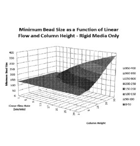
[0138] FIG. 4 represents a plot of Table 5. The plot shows the minimum bead
size on the y
axis, the linear flow rate on the x axis and the column height on the z axis.
FIG. 4 has 6 distinct
5 shades of grey as the bead size cut-off is 136 microns. Therefore,
shades representing beads
below that size are not represented. (e.g. 0-50 and 50-100).
[0139j The data was used to determine the minimum pore opening size of non-
bead material
such as woven yarns or fibers. The following table (Table 6) provides the
corresponding
minimum size of pore opening in relation to column height and linear flow
rate.
CA 02946294 2016-10-19
WO 2015/164198 PCT/U52015/(12634(1
Table 6. Macroscopic Pore Sizes for Non-Bead Material
Macroscopic Pore Size Macroscopic Pore Size
L (column height in cm) L (column height in cm)
u (cm/min) 3 5 10 20 30 u (cm/min) 3 5
10 20 30
1 21 21 21 21 21 1 21 21 21 21 21
3 21 21 21 21 21 76 29 38 53 75 92
5 21 21 21 21 24 151 41 53 75 106 130
7 21 21 21 23 28 226 50 65 92 129 159
9 21 21 21 26 32 301 58 75 106 149 183
11 21 21 21 29 35 376 65 83 118 167 205
13 21 21 22 31 38 451 71 91 129 183 224
15 21 21 24 33 41 526 76 99 140 197 242
17 21 21 25 36 43 601 82 106 149 211 259
19 21 21 27 38 46 676 87 112 158 224 274
21 21 21 28 39 48 751 91 118 167 236 289
23 21 21 29 41 51 826 96 124 175 247 303
25 21 22 30 43 53 901 100 129 183 258 317
27 21 22 32 45 55 976 104 135 190 269 329
29 21 23 33 46 57 1051 108 140 197 279 342
31 21 24 34 48 59 1126 112 144 204 289 354
101 40] If an adsorption media is compressible, the macroscopic pore size will
decrease as a
function of flow rate due to the shear stress of flowing blood. A compressible
media can be
"pre-compressed" to achieve the minimum pore size as calculated in Table 6 for
a desired flow
rate. For a loosely packed compressible media, the macroscopic pore size must
not decrease
below the values in the Table 6 under flow conditions, otherwise the pressure
of the system will
increase that could lead to heinolysis and macrophages would also be filtered
out.
[01411 In addition to the determining particle size andlor macroscopic pore
size, the diameter
(e.g., inner diameter) of the extracorporeal filter cartridge can determined.
Table 7 provides
useful cartridge diameters necessary to achieve the needed linear flow rate at
a specific
volumetric flow rate.
31
CA 02946294 2016-10-19
WO 2015/164198 PCT/US2015/026340
Table 7. Cartridge Diameters
Diameter of Cartridge (cm) Diameter of Cartridge (cm)
____________ Desired Volumetric Flow Rate (ml/min) Desired Volumetric Flow
Rate (mi/min)
u (cm/min) 50 100 150 300 500 1000 u (cm/min) SOO 1000 2000 3000
4000 5000
14.
1 1 20.0 24.5 34.6 44.7 63.2 1 44.7 63.2 89.4 109.5 126.5 141.4
3 8.2 11.5 14.1 20.0 25.8 36.5 76 5.1 7.3 10.3 12.6 14.5 16.2
6.3 8.9 11.0 15.5 20.0 28.3 151 3.6 5.1 7.3 8.9 10.3 11.5
7 5.3 7.6 9.3 13.1 16.9 23.9 226 3.0 4.2 5.9 7.3 8.4 9.4
9 4.7 6.7 8.2 11.5 14.9 21.1 301 2.6 3.6 5.2 6.3 7.3 8.2
11 4.3 6.0 7.4 10.4 13.5 19.1 376 2.3 3.3 4.6 5.6 6.5 7.3
13 3.9 5.5 6.8 9.6 12.4 17.5 451 2.1 3.0 4.2 5.2 6.0 6.7
15 3.7 5.2 6.3 8.9 113 16.3 526 1.9 2.8 3.9 4.8 53 6.2
17 3.4 4.9 5.9 8.4 10.8 15.3 601 1.8 2.6 3.6 4.5 5.2 5.8
19 3.2 4.6 5.6 7.9 10.3 14.5 676 1.7 2.4 3.4 4.2 4.9 5.4
21 3.1 4.4 5.3 7.6 9.8 13.8 751 1.6 2.3 3.3 4.0 4.6 5.2
23 2.9 4.2 5.1 7.2 9.3 13.2 826 1.6 2.2 3.1 3.8 4.4 4.9
25 2.8 4.0 4.9 6.9 8.9 12.6 901 1.5 2.1 3.0 3.6 4.2 4.7
27 2.7 3.8 4.7 6.7 8.6 12.2 976 1.4 2.0 2.9 3.5 4.0 4.5
29 2.6 3.7 4.5 6.4 8.3 11.7 1051 1.4 2.0 2.8 3.4 3.9 4.4
31 2.5 3.6 4.4 6.2 8.0 11.4 1126 1.3 1.9 2.7 3.3 3.8 4.2
[0142] Another factor to consider is the total blood volume used with an
extracorporeal device.
For instance, the total volume removed from the body during an extracorporeal
circulation
treatment is typically no more than 8-10% of the patient's blood. For an
average adult, this
equates to 500 ml of blood. A typical dialysis cartridge and tubing blood
volume can range from
250-300 ml. If a dialysis cartridge is used in series with an adsorption
cartridge, then the blood
volume of the adsorption cartridge should be no more than 200 ml. The
practical dimensions for
an adsorption cartridge of the present invention is provided in Table 8.
Table 8. Blood Volume of Packed Cartridge (ml) - 0.36 void volume
ratio
Column Height (cm)
Diameter 3 5 10 20 30
1 0.84834 1.4139
2.8278 5.6556 8.4834
5 21.2085 3534i5
70.695 141.39 212,085
84-834 141-39 282.78 565-56 84834
190.8765 318,1275 6:46-255 12,72-51 19138,765
:9:5336 565-56 1131,12 2262-24 3393-36
32
CA 02946294 2016-10-19
WO 2015/164198 PCTIUS2015/026340
101431 This example provides exemplary embodiments of the adsorption media and
adsorption
cartridge describe above. The adsorption media can be used in extracorporeal
therapies with
volumetric flow rate of up to 5000 milmin and linear flow rates of up to IMO
crn/min.
Example 4. Blood filters for removal of Hepatitis C virus and Hepatitis B
virus
101441 This example provides an extracorporeal filter cartridge that is used
to remove Hepatitis
C virus and Hepatitis B virus. In this example, the adsorption media is mixed.
The mixed
media comprises a 70:30 ratio of heparinzed polyethylene beads : protein A
fixed to a cellulose
gel.
101451 The heparinzed PE beads have covalent end-point attachment of nitrous
acid degraded
heparin onto arninated PE beads. The heparinized PE beads contain 2.6 mg
heparin/g beads.
101461 Covalent end-point attachment of nitrous acid degraded heparin onto
aminated PE
beads is prepared using 0.1 M acetate buffer pH 4.0 (100 ml) and nitrous acid
degraded heparin
(1.6 g). After shaking for 15 mM, NaBH3CN (100 mg) dissolved in 0.1 M acetate
buffer pH 4.0
(10 ml) is added. The reaction mixture is shaken for 24 h at room temperature
and additional
NaBH3CN (100 mg) dissolved in 0.1 M acetate buffer pH 4.0 (10 ml) is added,
and shaking is
continued for another 24 h at room temperature to produce the covalent end-
point attachment of
heparin.
[0147] In 0.5 mL of 0.05 M borate buffer (pH 10.0) is dissolved 4 mg of
protein A (Sigma),
and 0.01 N NaOH/water is added so as to bring the pH to 10 and make a total
volume of 1.0 mL
(protein A solution). This protein solution (total amount) is added to 1 mL of
an epoxy-activated
cellulose gel and the mixture is shaken at 37 C for 16 hours and washed with
a sufficient
amount of PBS (10 mM phosphate buffer supplemented with 150 mM sodium
chloride) to
provide (]CL 2000m-Protein A.
[0148] The mixed adsorption media is used to remove Hepatitis C virus and
Hepatitis B virus
from blood.
[0149] It is understood that the examples and embodiments described herein are
for illustrative
purposes only and that various modifications or changes in light thereof will
be suggested to
persons skilled in the art and are to be included within the spirit and
purview of this application
33
and scope of the appended claims.
34
Date Recue/Date Received 2021-09-01