Note: Descriptions are shown in the official language in which they were submitted.
CA 02946747 2016-10-21
WO 2015/164506
PCT/US2015/027099
METHODS OF ENHANCING STEM CELL ENGRAFTMENT
STATEMENT REGARDING SPONSORED RESEARCH
[0001] The invention described and claimed herein was made in part utilizing
funds
supplied by AHA NSDG, Grant # 105DG4260005.
TECHNICAL FIELD
[0002] This disclosure relates to methods of enhancing stem cell engraftment.
More
specifically, it relates to compositions and methods of using biologically
active compounds to
enhance stem cell engraftment.
BACKGROUND
[0003] The growing prevalence of peripheral arterial disease (PAD) is an
increasing global
concern as the population ages. PAD is an atherosclerotic disease associated
with diabetes,
hypertension, hypercholesterolemia, and coronary artery disease. Currently,
PAD affects 12-
14% of the general population, and its incidence is accelerating because of
the increase in the
elderly population. More than 10 million people in the United States have PAD.
The two
major clinical stages of PAD ¨ intermittent claudication and critical limb
ischemia (CLI) ¨
result from insufficient blood supply to lower extremities, but the clinical
outcome is more
severe in the latter stage. Conventional treatments for PAD, such as
angioplasty, stent
deployment, and peripheral bypass surgery, are less effective when PAD
progresses and
causes obstruction of arterioles. In these
cases, patients may develop untreatable
claudication, rest pain, and ulcers that can progress to gangrene and other
infections requiring
amputation of a lower limb. Although surgical advancements have improved the
lives of
some PAD patients, many are not treated surgically because of the risk of
complications.
New therapeutic approaches are needed to promote vascular growth, reduce
functional
impairment of ischemic legs, and improve quality of life.
[0004] Exogenous prostacyclin (PGI2 or PGI2) replacement therapy offers a
therapeutic
alternative for patients who are poor candidates for surgical
revascularization, such as high-
risk patients (e.g., the elderly). Clinical studies have shown that PGI2
therapy is efficacious,
but because PGI2 is an unstable compound with a circulating half-life of 1-2
minutes, this
approach requires continuous intravenous or intraarterial infusion, which is
associated with
side effects and several potential complications. While continuous intravenous
PGI2 therapy
is effective, this approach is inconvenient for PAD patients, as PGI2 must be
administered by
using a continuous pump with an indwelling catheter. This delivery system is
cumbersome
and greatly reduces the patient's quality of life. Moreover, significant
adverse events are
I
CA 02946747 2016-10-21
WO 2015/164506
PCT/US2015/027099
associated with this delivery system; infection at the infusion site can lead
to life-threatening
complications. In addition, continuous infusion of PGI2 is a financial burden.
Although
stable PGI2 analogues have been developed and used clinically, most still
require continuous
intravenous or subcutaneous infusion. An oral formulation of treprostinil was
recently
approved for pulmonary arterial hypertension (PAH) by the U.S. Food and Drug
Administration (FDA), but its efficacy is minimal and must be used in
combination with
other agents and it has not been tested for PAD.
[0005] We have shown that a localized delivery approach in which a micro-
osmotic pump
is used to directly administer PGI2 analogue Carbaprostacyclin (CPGI2) to
ischemic
hindlimbs of mice may overcome the disadvantages of systemic PGI2 therapy.
Local CPGI2
delivery alleviates hindlimb ischemia by improving perfusion and promoting
arteriolar
growth. However, there are side effects and potential complications associated
with this
therapeutic method as well.
[0006] A new approach to effectively deliver PGI2 is urgently needed for
treating PAD
patients. As such, there exists a need for improved compositions of local PGI2
delivery and
methods of using same.
BRIEF DESCRIPTION OF THE DRAWINGS
[0007] For a more complete understanding of the present disclosure and
advantages
thereof, reference will now be made to the accompanying drawings/figures in
which:
[0008] Figure 1 illustrates a schematic of biosynthesis of prostanoids (e.g.,
prostaglandins,
such as prostaglandin D2 (PGD2), E2 (PGE2), F2 (PGF2), and 12 (PGI2)
(prostacyclin), or
thromboxane A2 (TXA2)) through coupling reactions of upstream cyclooxygenases
(COXs)
and downstream individual synthases;
[0009] Figure 2A displays laser Doppler images of local treatment of mouse
ischemic
limbs with carbaprostacyclin (CPGI2) as compared to control (saline);
[0010] Figure 2B displays a graph of a quantitative analysis of perfusion
recovery of mouse
ischemic limbs with CPGI2 treatment as compared to saline treatment;
[0011] Figure 3 displays live images of distinct arterial growth of mouse
ischemic limbs
treated with CPGI2, wherein more intraarteriolar connections (solid line
arrows) and
corkscrew extensions of arterioles (dashed line arrows) developed in the CPGI2-
treated
versus the saline-treated group;
[0012] Figure 4A displays a histogram of mean blood vessel size distribution
in a
quantitative micro-CT analysis;
2
CA 02946747 2016-10-21
WO 2015/164506
PCT/US2015/027099
[0013] Figure 4B displays micro-CT images of microvascular network in CPGI2-
treated
and saline-treated ischemic legs; the red dashed circles show the vasculature
of the thigh
muscle where CPGI2 or saline was administered;
[0014] Figure 5A displays western blot images of COX-1-10aa-PGIS and COX-1
expression in human mesenchymal stem cells (hMSC or hMSCs);
[0015] Figure 5B displays a graph of PGI2 production levels in hMSCs
engineered to
overexpress PGI2 (PGI2-hMSCs) versus control;
[0016] Figure 5C displays endothelial cell tube formation incubated with PGI2-
hMSC
conditioned medium;
[0017] Figure 5D displays endothelial cell tube formation incubated with
control medium;
[0018] Figure 6A displays a schematic representation of the lentiviral vector
encoding
herpes virus thymidine kinase (HSV1-tk), mCherry fluorophore, and firefly
luciferase
reporter genes;
[0019] Figure 6B displays representative in vitro bioluminescent imaging (BLI)
images of
hMSCs transduced with lentiviruses;
[0020] Figure 6C displays a representative photomicrograph and its
corresponding
fluorescence image showing the expression of red mCherry fluorescent protein
in transduced
hMSCs;
[0021] Figure 6D displays a graph of high efficiency lentiviral transduction
in hMSCs as
confirmed by flow cytometry analysis;
[0022] Figure 7A displays representative BLI images of NOD-SCID mice 3 days
after
PGI2-hMSCs or 3.1-hMSCs were injected into the gastrocnemius muscle of the
ischemic
hindlimb;
[0023] Figure 7B displays a quantitative analysis of the BLI images of Figure
7A;
[0024] Figure 8A displays BLI images of NOD-SCID mice over a 14 day period
after
PGI2-hMSCs or 3.1-hMSCs were injected into the gastrocnemius muscle of the
ischemic
hindlimb;
[0025] Figure 8B displays a quantitative analysis of the BLI images of Figure
8A;
[0026] Figure 9A displays BLI images of NOD-SCID mice over a 5 day period
after a
hMSCs injection combined with daily cicaprost or CW501516 treatments;
[0027] Figure 9B displays a quantitative analysis of the BLI images of Figure
9A;
[0028] Figure 10A displays a graph of systolic blood pressure in mice at 3
days after
injection with PGI2-hMSC and 3.1-hMSC;
3
CA 02946747 2016-10-21
WO 2015/164506
PCT/US2015/027099
[0029] Figure 10B displays a graph of mean arterial pressure in mice at 3 days
after
injection with PGI2-hMSC and 3.1-hMSC;
[0030] Figure 11A displays a graph of functional recovery of ischemic
hindlimbs in mice at
21 days after injection with PGI2-hMSC and 3.1-hMSC;
[0031] Figure 11B displays a graph of functional recovery of ischemic
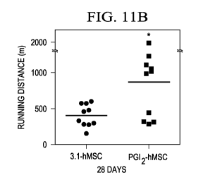
hindlimbs in mice at
28 days after injection with PGI2-hMSC and 3.1-hMSC;
[0032] Figure 12 displays endogenous Ki67+ cells spread within the hMSC
injection area;
[0033] Figure 13 displays confocal images indicating of endogenous
proliferating (Ki67+)
cells only rarely seen in regions further away from both 3.1-hMSC and PGI2-
hMSC injection
site;
[0034] Figure 14A displays representative confocal images of endogenous
Ki67+Sca-1+ and
Ki67+Sca-rcells;
[0035] Figure 14B displays a quantitative analysis of Ki67+Sca-1+cells
surrounding PGI2-
hMSCs injection sites as compared to 3.1-MSC sites;
[0036] Figure 14C displays a quantitative analysis of Ki67+Sca-1cells
surrounding PGI2-
hMSCs injection sites as compared to 3.1-MSC sites;
[0037] Figures 15A-F display H19 RNA levels along with cell viability in C2C12
myoblasts in various coculture environments;
[0038] Figures 15G-I display H19 RNA levels along with cell viability in C2C12
myoblasts after specific knock down with H19 siRNA (H19 KD) compared to
negative
control siRNA;
[0039] Figures 16A-F display H19 RNA levels along with cell viability in
primary
myoblasts;
[0040] Figure 16G-I display H19 RNA levels along with cell viability in
primary myoblasts
after specific knock down with H19 siRNA (H19 KD) compared to negative control
siRNA;
and
[0041] Figure 16J displays representative images of H19 RNA fluorescence in
situ
hybridization in gastrocnemius muscle sections at 3 days after 3.1-hMSC or PG2-
hMSC
injections.
SUMMARY
[0042] Disclosed herein is an effective amount of a composition comprising a
stem cell, a
stem cell engraftment enhancer, and a carrier fluid, for use in the treatment
of an individual
having a disease or at risk of developing a disease, wherein the disease is a
vascular-associated
disease and/or a muscular disease.
4
CA 02946747 2016-10-21
WO 2015/164506
PCT/US2015/027099
[0043] Also disclosed herein is a composition comprising PGI2-overexpressing
human
mesenchymal stem cells (PGI2-hMSCs), and a carrier fluid; wherein an effective
amount of
the composition is administered via a single treatment stream as an
intramuscular injection to
an individual having a disease or at risk of developing a disease, wherein the
disease is a
vascular-associated disease and/or a muscular disease, and wherein stem cell
engraftment is
enhanced in said individual by greater than about 200%, when compared to stem
cell
engraftment in an individual treated with a composition lacking the stem cell
engraftment
enhancer.
[0044] Further disclosed herein is a composition comprising: human mesenchymal
stem
cells (hMSCs), Iloprost, and a carrier fluid; wherein the composition is
administered to an
individual having a disease or at risk of developing a disease, wherein the
disease is a vascular-
associated disease and/or a muscular disease, and wherein the composition is
administered via
multiple treatment streams comprising: a stem cell treatment stream, and a
stem cell
engraftment enhancer treatment stream; wherein the stem cell treatment stream
comprises
hMSCs and is administered via an intramuscular injection; and wherein the stem
cell
engraftment enhancer treatment stream comprises Iloprost and is administered
via inhalation.
[0045] Further disclosed herein is a composition for stem cell engraftment,
wherein the
composition for stem cell engraftment comprises a stem cell, wherein the stem
cell comprises
human mesenchymal stem cells (hMSCs), endothelial progenitor cells (EPCs),
hematopoietic
stem cells (HSCs), cardiac progenitor cells (CPCs), satellite cells, or
combinations thereof, a
stem cell engraftment enhancer, wherein the stem cell engraftment enhancer
comprises
prostacyclin (PGI2), a PGI2 precursor, a peroxisome proliferator-activated
receptor 13/6
isoform (PPAR6) agonist, a cAMP inducer, a phosphodiesterase inhibitor, an
endothelin
receptor antagonist, a nitrous oxide modulating agent, a prostacyclin receptor
(IP) agonist, a
non-prostanoid IP receptor agonist, or combinations thereof, and a carrier
fluid.
[0046] The foregoing has outlined rather broadly the features and technical
advantages of the
present invention in order that the detailed description of the invention that
follows may be
better understood. Additional features and advantages of the invention will be
described
hereinafter that form the subject of the claims of the invention. It should be
appreciated by
those skilled in the art that the conception and the specific embodiments
disclosed may be
readily utilized as a basis for modifying or designing other structures for
carrying out the same
purposes of the present invention. It should also be realized by those skilled
in the art that such
CA 02946747 2016-10-21
WO 2015/164506
PCT/US2015/027099
equivalent constructions do not depart from the spirit and scope of the
invention as set forth in
the appended claims.
DETAILED DESCRIPTION
[0047] It should be understood at the outset that although an illustrative
implementation of
one or more embodiments are provided below, the disclosed systems and/or
methods may be
implemented using any number of techniques, whether currently known or in
existence. The
disclosure should in no way be limited to the illustrative implementations,
drawings, and
techniques below, including the exemplary designs and implementations
illustrated and
described herein, but may be modified within the scope of the appended claims
along with
their full scope of equivalents.
[0048] Disclosed herein are embodiments of compositions for stem cell
engraftment,
designated a CSCE, and methods of using the same. For purposes of the
disclosure herein,
engraftment may be defined as (i) a process by which transplanted stem cells
are retained
within a tissue and/or (ii) a process by which upon transplantation of stem
cells within a
tissue, beneficial effects of stem cell transplantation (e.g., tissue healing;
tissue repair; up-
regulating lnc-RNA H19 in a host cell environment; host cell stimulation;
improved exercise;
etc.) are retained within the tissue, even when the stem cells themselves or a
portion thereof
are not retained within the tissue. In some embodiments, the CSCE may be used
for targeted
delivery of stem cells in specific body areas, wherein the stem cells may
engraft and provide
a repair function (e.g., a tissue repair function). In other embodiments, the
CSCE may be
used for targeted delivery of prostacyclin (PGI2 or PGI2) in specific body
areas, such as for
example ischemic areas. While the current disclosure will be discussed in
detail in the
context of compositions for stem cell engraftment, it should be understood
that other
compositions for cell engraftment can comprise other types of cells, such as
for example cells
that have been engineered to produce prostacyclin (e.g., fibroblasts,
endothelial cells, etc.).
The cells can comprise any cells compatible with the disclosed methods and
materials.
[0049] In an embodiment, the CSCE comprises a stem cell, a stem cell
engraftment
enhancer (designated a SEE), and a carrier fluid. In some embodiments, the
stem cell may
produce the SEE (e.g., PGI2). In other embodiments, the SEE may be supplied
exogenously.
Although the CSCEs will be discussed in detail in the context of peripheral
arterial disease
(PAD), it should be understood that treatment for other diseases is also
contemplated,
wherein enhanced engraftment of stem cells in the presence of a SEE may be
useful.
[0050] As will be apparent to one of skill in the art, with the help of this
disclosure, other
suitable ingredients/components may be used in the CSCE, and each
ingredient/component of
6
CA 02946747 2016-10-21
WO 2015/164506
PCT/US2015/027099
the CSCE may perform more than one function (e.g., stem cells may be both the
stem cell
component as well as the SEE, wherein the stem cells may be engineered to
express or
overexpress the SEE). Each of the components of the CSCE as well as methods of
using
same will be described in more detail herein.
[0051] In an embodiment, stem cells may comprise stem cells and/or progenitor
cells. In
an embodiment, stem cells comprise natural stem cells, induced pluripotent
stem cells,
engineered adult stem cells, or combinations thereof As will be appreciated by
one of skill
in the art, and with the help of this disclosure, natural stem cells refer to
stem cells that are
present in an organism (e.g., human) and may be isolated and used without
further
modification. Further, as will be appreciated by one of skill in the art, and
with the help of
this disclosure, induced pluripotent stem cells refer to stem cells (e.g.,
human adult stem
cells) that have been modified (e.g., genetically modified) to provide
pluripotent stem cells.
Nonlimiting examples of stem cells suitable for use in the present disclosure
include human
mesenchymal stem cells (hMSC or hMSCs), endothelial progenitor cells (EPCs),
hematopoietic stem cells (HSCs), cardiac progenitor cells (CPCs), satellite
cells (e.g.,
myosatellite cells, skeletal muscle progenitor cells, etc.), or combinations
thereof
[0052] In an embodiment, the stem cells comprise hMSCs. Human mesenchymal stem
cells offer advantages as vehicles for therapeutic gene transfer. Stem cell
therapy is emerging
as a novel and promising therapeutic approach for PAD. Clinical studies in PAD
patients
have shown that hMSCs are attractive candidates for stem cell-based strategies
for tissue
repair and gene therapy. hMSCs can be easily isolated and expanded to large
numbers in
vitro or ex vivo. Furthermore, hMSCs show low immunogenicity after allogeneic
transplantation and provide paracrine factors for repairing damaged tissue. In
addition,
hMSCs accumulate at sites of injury to protect against inflammation and
promote
revascularization. These unique properties make hMSCs an excellent choice for
exogenous
gene delivery. hMSCs can be modified to express therapeutic genes before being
administered directly to damaged tissues. This combined hMSC-gene therapy
approach
eliminates the need for repetitive or continuous gene delivery because hMSCs
are able to
self-renew.
[0053] In an embodiment, hMSCs may be engineered to augment production of
specific
desired proteins, thereby enhancing the therapeutic benefits provided by
native hMSCs. In an
embodiment, hMSCs may be engineered to produce PGI2, thereby offering a novel,
targeted
PGI2 replacement therapy for treating PAD, as will be described in more detail
later herein.
7
CA 02946747 2016-10-21
WO 2015/164506
PCT/US2015/027099
[0054] EPCs generally comprise a population of rare cells that circulate in
the blood or reside
in vasculatures. EPCs have the ability to differentiate into endothelial cells
(e.g., cells that
make up the lining of blood vessels). In an embodiment, EPCs may be engineered
to augment
production of specific desired proteins, thereby enhancing the therapeutic
benefits provided
by native EPCs. In an embodiment, EPCs may be engineered to produce PGI2.
[0055] HSCs generally comprise a heterogeneous population of blood cells. HSCs
are
derived from mesoderm and have the ability to give rise to all the other blood
cells. In an
embodiment, HSCs may be engineered to augment production of specific desired
proteins,
thereby enhancing the therapeutic benefits provided by native HSCs. In an
embodiment,
HSCs may be engineered to produce PGI2.
[0056] CPCs generally comprise a population of resident cardiac stem cells.
CPCs are
thought to account for the physiological turnover of cardiac myocytes and
vascular
endothelial cells. In an embodiment, CPCs may be engineered to augment
production of
specific desired proteins, thereby enhancing the therapeutic benefits provided
by native
CPCs. In an embodiment, CPCs may be engineered to produce PGI2.
[0057] Satellite cells generally comprise small mononuclear progenitor cells
with virtually
no cytoplasm found in mature muscle. Satellite cells are precursors to
skeletal muscle cells,
able to give rise to satellite cells or differentiated skeletal muscle cells.
[0058] In an embodiment, the stem cells may be included within the CSCE in a
suitable
amount. In an embodiment the stem cells may be present within the CSCE in an
amount of
from about 5 million cells/mL to about 600 million cells/mL, alternatively
from about 10
million cells/mL to about 500 million cells/mL, or alternatively from about 25
million
cells/mL to about 400 million cells/mL, based on the total volume of the CSCE.
In an
embodiment the stem cells may be present within the CSCE in an amount of about
200
million cells/mL, based on the total volume of the CSCE. For purposes of the
disclosure
herein, the term "about," when used in conjunction with a percentage or other
numerical
amount, means plus or minus 10% of that percentage or other numerical amount.
For
example, the term "about 400 million cells," would encompass 400 million cells
plus or
minus 40 million cells.
[0059] In an embodiment, the CSCE can comprise a SEE. Generally, the SEE can
enhance
(e.g., increase) (a) an ability of the stem cells to engraft (e.g., be
retained) in a tissue upon
transplantation into the tissue and/or (b) retention of beneficial effects of
stem cell
transplantation (e.g., tissue healing; tissue repair; up-regulating lnc-RNA
H19 in a host cell
environment; host cell stimulation; improved exercise; etc.) in the tissue,
even when the stem
8
CA 02946747 2016-10-21
WO 2015/164506
PCT/US2015/027099
cells themselves or a portion thereof are not retained within the tissue. For
purposes of the
disclosure herein, a host cell refers to a cell present in a location (e.g.,
tissue location) where
the stem cells are transplanted. Without wishing to be limited by theory,
engraftment plays a
role in co-stimulation of the host cells to proliferate and regenerate due to
the stem cells being
retained long enough to stimulate host cells and the new growth of muscle and
blood vessels.
[0060] In an embodiment, the SEE may comprise PGI2; PGI2 stable precursors or
analogues
(e.g., Cicaprost, Iloprost, Beraprost, Carbaprostacyclin, Trepostinil,
Epoprostenol, etc.); a
peroxisome proliferator-activated receptor 13/6 isoform (PPAR6) agonist (e.g.,
GW501516,
also known as GW-501,516, GW1516, GSK-516, Endurobol, etc.); a cAMP inducer
(e.g.,
forskolin, also known as coleonol, 8-bromo-cAMP, etc.); a phosphodiesterase
inhibitor (e.g.,
sildenafil citrate (VIAGRAg), tadalafil (CIALISg), vardenafil (LEVITRAg),
etc.); an
endothelin receptor antagonist (e.g., bosentan (TRACLEER ), ambrisentan
(LETAIRIS ),
macitentan (OPSUMIT ), etc.); a nitrous oxide modulating agent (e.g.,
nitrates, or soluble
GMP cyclase inducers, such as for example riociguat (ADEMPAS)); a prostacyclin
receptor
(IP) agonist; a non-prostanoid IP receptor agonist (e.g., selexipag); and the
like; or
combinations thereof While the current disclosure will be discussed in detail
in the context
of SEE comprising PGI2 and/or a PGI2 precursor, it should be understood that
other classes
of compounds (e.g., a PPAR6 agonist, a cAMP inducer, a phosphodiesterase
inhibitor, an
endothelin receptor antagonist, a nitrous oxide modulating agent, an IP
agonist, a non-
prostanoid IP receptor agonist, etc.) may be used to enhance stem cell
engraftment, thereby
enhancing a repair function that such stem cells might exhibit.
[0061] In an embodiment, the SEE may be a biologically or pharmacologically
active
compound. For purposes of the disclosure herein, a biologically active
compound can be
defined as a compound that interacts in some fashion with any living cell,
tissue, and/or
organism. For example, PGI2, PGI2 precursors or analogues, PPAR6 agonists,
cAMP
inducers, phosphodiesterase inhibitors, endothelin receptor antagonists,
nitrous oxide
modulating agents, IP agonists, and non-prostanoid IP receptor agonists are
biologically
active compounds.
[0062] In an embodiment, the SEE comprises PGI2. PGI2, a member of the
prostaglandin
family, is synthesized from arachidonic acid (AA) in a multistep process
involving the
enzymes cyclooxygenase-1 (COX-1) or cyclooxygenase-2 (COX-2) and prostacyclin
synthase (PGIS). As a vasodilatory drug, PGI2 has multiple favorable
properties for treating
9
CA 02946747 2016-10-21
WO 2015/164506
PCT/US2015/027099
PAD. In addition to mediating vascular homeostasis, PGI2 inhibits thrombosis
and platelet
aggregation.
[0063] The function of PGI2 is primarily mediated by the PGI2 receptor (IP) on
the cell
surface. The role of PGI2 as an endogenous anti-thrombotic and vasodilative
agent was
confirmed with the experimental data generated in IP receptor-knockout mice.
The IP-
deficient mice developed without vascular problems in normal situations.
However, an
increased thrombotic tendency was observed in the IP-deficient mice when
endothelial
damage was induced. These findings indicate that the anti-thrombotic system
mediated by
PGI2 is activated in response to vascular injury to minimize the effects of
vascular injury. It
has been reported that defects in the IP receptor of platelets has
pathogenetic significance for
developing atherosclerosis at an early age. The evidence was derived from a 10
year-old
human diagnosed with an occluded left popliteal artery who also had a defect
of her IP
receptor. This defect appears to be genetically determined and to contribute
to the
development of atherosclerosis.
[0064] In an embodiment, PGI2 may enhance functional benefits of human stem
cell
therapy. Accumulating evidence indicates a critical role for PGI2 in
controlling stem cell
recruitment and survival and in promoting angiogenesis. Patients with critical
limb ischemia
(CLI) have reduced numbers of circulating progenitor cells; however, after 4
weeks of
treatment with a PGI2 analogue, such patients show increased levels of
progenitor cells and
pain relief Human outgrown EPCs may produce PGI2 and endogenous secretion of
PGI2 by
EPCs may facilitate vascular regeneration. In contrast, inhibiting PGI2
production in EPCs
may reduce their proliferation, survival, and angiogenic capacity in ischemic
hindlimbs.
PGI2 signaling promotes the migration and recruitment of EPCs to injured
vessels. Impaired
function of EPCs is associated with decreased endogenous PGI2 synthesis and
signaling.
PGI2 may have the ability to enhance the natural abilities of stem cells. The
cell-protective
property of PGI2 in vivo may attenuate cell loss by stimulating their
plasticity to adapt to
unfavorable environments.
[0065] In an embodiment, increasing or enhancing PGI2 biosynthesis in
stem/progenitor
cells may enhance the beneficial effects of stem cell therapy. Generally,
biosynthesis, also
known as biogenesis or anabolism, is a multi-step, enzyme-catalyzed process,
wherein
substrates are converted into more complex products. In biosynthesis, simple
compounds are
modified, converted into other compounds, or joined together to form
macromolecules.
[0066] The recent discovery that COX-2 inhibitors may be linked to heart
disease has
greatly increased the interest in understanding the biology of COX enzymes,
which convert a
CA 02946747 2016-10-21
WO 2015/164506
PCT/US2015/027099
lipid molecule, AA, into different prostanoids (part of the eicosanoid family)
having diverse
and/or opposite biological functions. Figure 1 shows a schematic of the
biosynthesis of
prostanoids.
Biosynthesis of prostanoids generally comprises prostaglandins and
thromboxane, formed via the COX pathway from arachidonic acid (AA) in three
catalytic
(tri-catalytic) steps (represented by some of the thin line arrows in Figure
1). AA may
traverse across an endoplasmic reticulum (ER) membrane (e.g., from a first or
cytoplasmic
side of the ER membrane to a second or luminal side of the ER membrane) and be
converted
in catalytic step 1 to prostaglandin G2 (PGG2) by COX isoform-1 (COX-1) and/or
COX-2,
wherein COX-1 and COX-2 may be located on the luminal side of the ER membrane.
In
catalytic step 2, PGG2 may be further converted to prostaglandin endoperoxide
(prostaglandin
H2 (PGH2)) by COX-1 and/or COX-2. PGH2 may traverse across the ER membrane
(e.g.,
from the luminal side of the ER membrane to the cytoplasmic side of the ER
membrane). In
catalytic step 3, PGH2 may be further isomerized to biologically active end-
products
(prostaglandin D2 (PGD2), E2 (PGE2), F2 (PGF2), and 12 (PGI2 (prostacyclin) or
thromboxane
A2 (TXA2) by individual synthases (PGD2 synthase (PGDS), PGE2 synthase (PGES),
PGF2
synthase (PGFS), and PGI2 synthase (PGIS), or TXA2 synthase (TXAS),
respectively, as
depicted in Figure 1) in tissue specific manners, wherein such individual
synthases may be
located on the cytoplasmic side of the ER membrane. Prostanoids act as local
hormones in
the vicinity of their production site to regulate hemostasis and smooth muscle
functions.
Unlike the stable level of COX-1 expression, COX-2 expression is inducible and
it responds
to the stimuli of pro-inflammatory and other pathogenic factors. TXA2 produced
from PGH2
by TXA2 synthase (TXAS) has been implicated in various pathophysiological
conditions as a
proaggregatory and vasoconstricting mediator. PGI2 is the main AA metabolite
in vascular walls
and has opposing biological properties to TXA2, representing the most potent
endogenous
vascular protector acting as an inhibitor of platelet aggregation and a strong
vasodilator on
vascular beds. PGE2 exhibits a variety of biological activities in
inflammation. Aspirin and
non-steroidal anti-inflammatory drugs (N SAID) inhibit both COX-1 and COX-2
activities to
reduce the production of all prostanoids, which leads to thinning of the blood
by reducing TXA2
production and the suppression of inflammation through decreasing PGE2
production. The
selective COX-2 inhibiting drugs exhibit anti-inflammatory effects similar to
aspirin and
NSAIDs, but they may also promote strokes and heart attacks by decreasing the
production of
PGI2, and increasing the production of TXA2. This may occur because, when the
COX-2
enzyme was specifically inactivated by COX-2 inhibitors, the PGH2 produced by
COX-1 was
11
CA 02946747 2016-10-21
WO 2015/164506
PCT/US2015/027099
still available to be converted into other prostanoids such as TXA2 by TXAS,
leading to an
increased risk of thrombosis and vasoconstriction.
[0067] Recently, PGI2 has also been determined to be a ligand for the nuclear
hormone
receptor peroxisome proliferator-activated receptor (PPAR). Three PPAR-
isoforms, PPARa,
13/6 and y have been cloned and implicated in the regulation of the expression
of genes
involved in lipid metabolism. In both skeletal and cardiac muscle cells it has
been
demonstrated that the metabolic conversion of fatty acids is under control by
PPARs. PGI2
and PGI2 agonists (e.g., carbaprostacyclin, iloprost, etc.), can effectively
induce DNA binding
and transcriptional activation by PPAR. PGI2, acting as a ligand for PPAR,
induces
increased expression of PPAR 6 in the arterial wall after a balloon injury,
suggesting that PGI2
effects vasodilation and anti-platelet aggregation through the IP receptor and
PPAR. It has
also been speculated that PGI2, as a ligand for PPAR, induces anti-
inflammatory activity in
vascular diseases, such as atherosclerosis.
[0068] In an embodiment, peroxisome proliferator-activated receptor-beta/delta
(PPAR)
can be a potential regulator of PGI2 signaling. In the search for endogenous
targets for PGI2
signaling, PPAR 6 was found to colocalize with COX-2/PGIS and actively respond
to PGI2
agonists. PPAR 6 is a ligand-activated nuclear hormone receptor that is
ubiquitously
expressed in various tissues. It forms heterodimers with retinoid X receptor,
which binds to
the peroxisome proliferator response element in the promoter region of target
genes to control
transcription. Emerging evidence suggests that PPAR 6 plays a critical role in
stem cell
survival and neovascularization. Accordingly, activation of PPAR 6 by PGI2 may
promote
stem cell¨mediated vascular regeneration in ischemic hindlimbs. Inhibition of
PPAR 6 by
selective antagonists or specific siRNA in human progenitor cells may reduce
PGI2-induced
regenerative ability and blood vessel formation. PGI2, in partnership with
PPAR,
accelerates embryo implantation and blastocyst hatching. In addition to its
pro-survival and
pro-angiogenic roles, PPAR 6 is important in adaptive responses to
environmental changes.
As a
metabolic sensor, PPAR 6 regulates several metabolic genes involved in
cellular
homeostasis. PPAR 6 may play a critical role in mitochondrial function. In an
embodiment,
PGI2-PPAR 6 axis may affect the ability of stem cells to adjust to
environmental changes
(e.g., may affect the viability of stem cells introduced to certain body
areas, such as for
example ischemic areas), thus might affect the ability of stem cells to
engraft.
12
CA 02946747 2016-10-21
WO 2015/164506
PCT/US2015/027099
[0069] In an embodiment, the SEE comprises a PPAR6 agonist, such as for
example
GW501516, also known as GW-501,516, GW1516, GSK-516, Endurobol, etc.
[0070] In an embodiment, the carrier fluids that may be used in the CSCE
include any
carrier fluid suitable for delivery of stem cells in vivo. In an embodiment,
the carrier fluid
comprises a pharmaceutically acceptable carrier. For purposes of the
disclosure herein, a
"pharmaceutically acceptable carrier" is meant to encompass any carrier that
does not
interfere with effectiveness of a biological activity of any active ingredient
(e.g., stem cell,
stem cell engraftment enhancer) and that is not toxic to an individual to
which it is
administered. "Pharmaceutically acceptable" as used herein adheres to the U.S.
Food and
Drug Administration guidelines.
[0071] In an embodiment, the CSCE may comprise an aqueous carrier fluid. In an
embodiment, the aqueous carrier fluid comprises deionized water and a variety
of additives
that may promote the viability and health of the stem cells of the CSCE. In an
embodiment,
the carrier fluid comprises a saline solution (e.g., phosphate buffer saline).
[0072] Nonlimiting examples of additive suitable for use in the carrier fluid
in the present
disclosure include nutritional supplements, growth factors, proteins (e.g.,
human serum
albumin or HSA), and the like, or combinations thereof In an embodiment, the
carrier fluid
may be included within the CSCE in a suitable amount.
[0073] In an embodiment, PGI2 may be delivered by stem cells that may be
engineered
(e.g., programmed) to overexpress PGI2, e.g., express high levels of PGI2 or
express PGI2
levels that are higher than the PGI2 levels expressed by the same stem cells
prior to being
engineered. A system that increases PGI2 biosynthesis in cells of the ischemic
areas would
help prevent the adverse events caused by conventional PGI2 delivery methods.
As will be
appreciated by one of skill in the art, and with help of this disclosure,
effective and stable
biosynthesis of PGI2 requires an increase in the expression of COX-1 or COX-2
in
conjunction with PGIS, as illustrated in Figure 1.
[0074] In an embodiment, the SEE may comprise a PGI2 precursor. In an
embodiment, the
PGI2 precursor may comprise a triple catalytic enzyme, a PGI2-overexpressing
stem cell
(PGI2-SC), a DNA sequence encoding for the triple catalytic enzyme, a cDNA
sequence
encoding for the triple catalytic enzyme, a host cell containing an
expressible DNA sequence
encoding for the triple catalytic enzyme, a vector comprising a DNA sequence
encoding for
the triple catalytic enzyme, a plasmid comprising a DNA sequence encoding for
the triple
13
CA 02946747 2016-10-21
WO 2015/164506
PCT/US2015/027099
catalytic enzyme, a fusion gene encoding for the triple catalytic enzyme, a
synthetic PGI2
analogue, and the like, or combinations thereof
[0075] Nonlimiting examples of synthetic PGI2 analogues suitable for use in
the present
disclosure include Iloprost, Carbaprostacyclin, Treprostinil, Cicaprost,
Beraprost,
Epoprostenol, and the like, or combinations thereof
[0076] In an embodiment, stem cells such as hMSCs may be engineered to
overexpress an
active triple catalytic enzyme to promote PGI2 expression (e.g., release
PGI2). In such
embodiment, the PGI2 overexpression by hMSCs may provide a means for local
PGI2
delivery in body areas such as ischemic areas (e.g., ischemic tissue) and may
concurrently
enhance the natural ability of hMSCs to mediate repair in ischemic tissue.
Although local
delivery of prostacyclin and/or prostacyclin analogues (e.g.,
carbaprostacyclin (CPGI2) may
alleviate hindlimb ischemia by improving perfusion and promoting arteriolar
growth, this
approach is not clinically practical because an invasive catheter-connected
pump carrying a
prostacyclin and/or prostacyclin analogues solution is generally
subcutaneously implanted.
In an embodiment, a triple catalytic enzyme may enhance the expression of PGI2
in stem
cells, such as for example hMSCs, EPCs, HSCs, CPCs, satellite cells, or
combinations
thereof
[0077] Recent studies of the structure and function relationship of COX
enzymes and PGIS
have advanced knowledge of the molecular mechanisms involved in the
biosynthesis of PGI2 in
native cells. Crystallographic studies of detergent-solubilized COX-1 and COX-
2 suggest that
the catalytic domains of the proteins lie on the lumina' side of the
endoplasmic reticulum (ER)
and are anchored to the ER membrane by hydrophobic side chains of amphipathic
helices A-D.
These hydrophobic side chains of the putative membrane anchor domains also
form an entrance
to the substrate-binding channel and potentially form an initial docking site
for the lipid
substrate, AA. Recent progress in the topology and structural studies of human
PGIS and TXAS
have led to the proposal of models in which PGIS and TXAS have catalytic
domains on the
cytoplasmic side of the ER, opposite the orientation of COXs. In this
configuration, the
substrate channels of all three enzymes, COX, PGIS and TXAS, open at or near
the ER
membrane surface. The coordination between COXs and PGIS or TXAS in the
biosynthesis of
TXA2 and PGI2 may be facilitated by the enzyme's anchoring in the lipid
membrane. The
physical distances between COXs and PGIS are very small. Consequently, it
should be possible
to create a single protein molecule containing COX and PGIS sequences with
minimum
alteration of both enzymes' folding and membrane topologies by extending the N-
terminal
membrane anchor domain of PGIS using a transmembrane sequence linked to the
COX-1 or
14
CA 02946747 2016-10-21
WO 2015/164506
PCT/US2015/027099
COX-2, which then adopts the functions of both enzymes of COX and PGIS. In
this case, AA
could be directly converted into the vascular protector, PGI2, with
concurrently decreasing the
production of the unwanted PGE2 and TXA2.
[0078] In an embodiment, the triple catalytic enzyme may be characterized by a
formula
COX-linker-ES, wherein COX comprises a cyclooxygenase (COX) amino acid
sequence,
such as for example COX-1 or COX-2; wherein ES comprises an eicosanoid-
synthesizing
(ES) enzyme amino acid sequence; wherein the linker comprises from about 10 to
about 22
amino acid residues of a transmembrane sequence; wherein the linker may be
disposed
between the COX and the ES; and wherein the linker may directly connect the
COX to the
ES. In an embodiment, the triple catalytic enzyme comprises a hybrid protein
or hybrid
peptide.
[0079] In some embodiments, the linker (e.g., linker peptide) may function as
a
transmembrane linker in a cell, such that folding ability and function of each
enzyme (e.g.,
COX, ES) of the triple catalytic enzyme may be substantially unaltered
compared to the
folding ability and function of respective native enzymes. As will be
appreciated by one of
skill in the art, and with the help of this disclosure, the linker is a
peptide, since it comprises a
relatively short sequence of amino acids. For purposes of the disclosure
herein, the terms
"linker" and "linker peptide" can be used interchangeably.
[0080] In an embodiment, the linker (e.g., linker sequence) comprises His-Ala-
Ile-Met-
Gly-Val-Ala-Phe-Thr-Trp (SEQ ID NO. 1) or His-Ala-Ile-Met-Gly-Val-Ala-Phe-Thr-
Trp-
Val-Met-Ala-Leu-Ala-Cys-Ala-Ala-Pro-Pro-Leu-Val (SEQ ID NO. 2). In
certain
embodiments, the linker sequence comprises residues 1-11, 1-12, 1-13, 1-14, 1-
15, 1-16, 1-
17, 1-18, 1-19, 1-20 or 1-21 of SEQ ID NO. 2. In some embodiments, the linker
peptide
provides approximately 10 A separation between the catalytic sites of the COX
and the ES
enzyme. In an embodiment, the connected enzymes (e.g., COX, ES) are preferably
capable of
substantially normal folding and enzymatic activity compared to the native
folding and
enzymatic activity of the native COX and ES enzymes.
[0081] In an embodiment, the triple catalytic enzyme may be characterized by a
faster
turnover rate when compared to a mixture of the native COX and ES enzymes. The
hybrid
protein (e.g., COX-linker-ES) does not only possess the individual enzymes'
activities, but has
a faster turnover rate as compared to a mixture of separate COX and ES
enzymes.
[0082] In an embodiment, the ES may comprise PGIS or a downstream synthase
thereof In
an embodiment, the PGIS downstream synthase may comprise prostaglandin E
synthase
(PGES), prostaglandin D synthase (PGDS), or prostaglandin F synthase (PGFS).
In an
CA 02946747 2016-10-21
WO 2015/164506
PCT/US2015/027099
embodiment, the triple catalytic enzyme may be characterized by formulas COX-
linker-
PGIS, COX-linker-PGES, COX-linker-PGDS, or COX-linker-PGFS, wherein COX
comprises COX-1 or COX-2. In such embodiment, the triple catalytic enzyme
combines the
enzymatic functions of COX (e.g., COX-1, COX-2) and ES (e.g., PGIS, PGES,
PGDS,
PGFS) in a single hybrid protein.
[0083] In an embodiment, the triple catalytic enzyme may be characterized by a
formula
COX-linker-PGIS, wherein COX comprises COX-1 or COX-2. In an embodiment, the
COX-
linker-PGIS may adopt the functions of COX and PGIS. In an embodiment, the COX-
linker-
PGIS may be able to continually convert AA into prostaglandin G2 (catalytic
step 1),
prostaglandin H2 (catalytic step 2) and prostacyclin (PGI2; catalytic step 3),
wherein the
catalytic steps have been described previously herein. Such conversion of AA
into PGI2 may
be even faster than coupling reactions using unlinked, co-expressed COX and
PGIS.
[0084] In an embodiment, the triple catalytic enzyme may be characterized by a
formula
COX-1¨linker¨PGIS. In an embodiment, the triple catalytic enzyme may catalyze
the three
catalytic steps (e.g., three key reactions) in the biosynthesis of PGI2,
thereby enhancing the
expression of PGI2 (e.g., increasing the production of PGI2). In such
embodiment, the triple
catalytic enzyme links COX-1 to PGIS and catalyzes three key reactions for
efficient
production of PGI2 from AA.
[0085] In an embodiment, the COX-1¨linker¨PGIS protein may comprise an 130 kDa
recombinant protein, wherein the recombinant protein may be constructed by
linking together
human cyclooxygenase (COX) isoform-1 (COX-1) and PGIS via a linker. In such
embodiment, the linker may comprise from 10 to 22 amino acid residues of a
transmembrane
sequence, as previously described herein. In an embodiment, the COX-
1¨linker¨PGIS protein
may comprise COX-1-10aa-PGIS, wherein the linker comprises a 10 amino acid
(10aa)
transmembrane sequence (e.g., SEQ ID NO. 1). As will be appreciated by one of
skill in the
art, and with the help of this disclosure, some COX-2 inhibitors inhibit COX-2
activity but not
COX-1 activity. Thus, the introduction of the COX-1¨linker¨PGIS hybrid protein
to vascular
systems is expected to help overcome the damage of the vascular functions
caused by COX-2
inhibitors. In an embodiment, the triple catalytic enzyme may be characterized
by a formula
COX-1-10aa-PGIS.
[0086] In some embodiments, the triple catalytic enzyme may be chemically
synthesized.
In other embodiments, the triple catalytic enzyme may be recombinantly
produced. The
triple catalytic enzyme and methods of producing and/or using same are
described in more
16
CA 02946747 2016-10-21
WO 2015/164506
PCT/US2015/027099
detail in U.S. Publication No. 20100015120 Al, which is incorporated by
reference herein in
its entirety.
[0087] In an embodiment, PGI2 may be delivered by stem cells (SCs) that may be
engineered (e.g., programmed) to express high levels of PGI2. Stem cells that
overexpress
PGI2 may be referred to as PGI2-SCs, such as for example PGI2-hMSCs, PGI2-
EPCs, PGI2-
HSCs, PGI2-CPCs, PGI2-(satellite cells), etc. In an embodiment, the PGI2
precursor
comprises a PGI2-SC.
[0088] In an embodiment, the COX-linker-ES (e.g., COX-1¨linker¨PGIS) may be
introduced in stem cells via any suitable transfection methods, such as
nucleofection.
Nucleofection is a nonviral transfection technique. As will be appreciated by
one of skill in the
art, and with the help of this disclosure, stable expression of COX-linker-ES
may be confirmed
via a variety of biochemical methods, such as for example Western Blot,
genomic PCR, RT-
PCR, and the like, or combinations thereof
[0089] In an embodiment, SCs (e.g., PGI2-SCs) comprise a DNA sequence encoding
for a
COX, a transmembrane linker peptide, and an ES. In some embodiments, COX
comprises
COX-1. In other embodiments, COX comprises COX-2. In an embodiment, ES
comprises
PGIS. In an embodiment, the linker directly connects the COX to the ES. In an
embodiment,
SCs (e.g., PGI2-SCs) comprise a DNA sequence encoding for the triple catalytic
enzyme, and
such DNA sequence may be referred to as a "fusion gene."
[0090] Generally, stem cells may be transfected by introducing a plasmid
expressing the triple
catalytic enzyme that links COX to ES (e.g., COX-1-10aa-PGIS). Such plasmid
may comprise
a promoter and an antibiotic resistance gene for selection of stable cell
lines. Nonlimiting
examples of promoters suitable for use in the present disclosure include a
human
cytomegalovirus promoter; endothelial-specific promoters (e.g., tie gene
promoter, Tie2 gene
promoter also known as Tek gene promoter, ICAM-2 (intercellular adhesion
molecule-2)
promoter, Flk-1 (fetal liver kinase-1) promoter, Flt-1 (fms-like tyrosine
kinase) promoter,
thrombomodulin promoter, vWf (von Willebrand factor) promoter, VE-cadherin
promoter,
etc.); cardiomyocyte specific promoters (e.g., alpha-MHC (myosin heavy chain)
promoter;
troponin promoter); smooth muscle cell specific promoters (e.g., SM22alpha
promoter); human
muscle specific promoter; human muscle creatinine kinase promoter; human a-
skeletal actin
promoter; human desmin promoter; human troponin I promoter; and the like; or
combinations
thereof Nonlimiting examples of antibiotic resistance genes suitable for use
in the present
disclosure include a neomycin resistance gene (e.g., resistant to antibiotic
G418); a puromycin
17
CA 02946747 2016-10-21
WO 2015/164506
PCT/US2015/027099
resistance gene; an ampicillin resistance gene; a kanamycin resistance gene; a
blasticidin
resistance gene; a hygromycin resistance gene; a gentamicin resistance gene; a
spectinomycin
resistance gene; a streptomycin/spectinomycin resistance gene; and the like;
or combinations
thereof
[0091] After transfection (e.g., nucleofection), transfected cells may be
grown (e.g., cultured)
for selection for a time period of from about 1 week to about 4 weeks. Then,
cell clusters may
be selected for further subculture, propagated and examined for PGI2 and/or
COX-linker-ES
expression. Subcultures that overexpress PGI2 comprise PGI2-SCs.
[0092] In some embodiments, a vector may comprise a DNA sequence encoding for
the
triple catalytic enzyme. In certain embodiments, the vector comprises an
expression vector,
such as for example a retrovirus, a lentivirus, an adenovirus, an adeno-
associated virus, etc.
In such embodiments, the DNA sequence encoding for the triple catalytic enzyme
may be
introduced into SCs (e.g., a host cell) via transduction.
[0093] In an embodiment, the SCs comprise a host cell containing an
expressible DNA
sequence encoding for the triple catalytic enzyme.
[0094] In some embodiments, the triple catalytic enzyme may be produced by a
host cell
containing an expressible DNA sequence encoding for the triple catalytic
enzyme. In an
embodiment, the host cell may be transfected with a vector comprising the DNA
sequence
encoding for the triple catalytic enzyme to produce host cell containing an
expressible DNA
sequence encoding for the triple catalytic enzyme. In some embodiments, the
host cell
comprises a SC. In other embodiments, the host cell does not comprise a SC. In
such
embodiments, the host cell may produce the triple catalytic enzyme. The host
cell may be
cultured under conditions suitable for expression of the DNA sequence encoding
the triple
catalytic enzyme, and then the triple catalytic enzyme may be recovered. In an
embodiment,
the triple catalytic enzyme comprises enzymatically active cyclooxygenase,
transmembrane
linker, and enzymatically active prostacyclin synthase.
[0095] In an embodiment, a cDNA may comprise a sequence encoding for the
triple catalytic
enzyme. In such embodiment, the cDNA may be used for COX gene therapy.
[0096] In an embodiment, the CSCE may be prepared via any suitable method or
process.
The components of the CSCE (e.g., stem cells, SEE, carrier fluid) may be
combined using any
mixing device compatible with the composition, e.g., that does not alter or
destroy the CSCE
components, such as the cells, etc. In an embodiment, the stem cells and/or
SEE may be
suspended in a saline solution comprising HSA. More details regarding stem
cell preparation
for administering as a treatment are available in Cytotherapy, 2010, 12(5), pp
684-691; and
18
CA 02946747 2016-10-21
WO 2015/164506
PCT/US2015/027099
JAMA, 2011, 306(19), pp 2110-2119; each of which is incorporated by reference
herein in its
entirety.
[0097] In an embodiment, the CSCE may be used for the treatment of an
individual having a
disease or at risk of developing a disease, wherein the disease can be a
vascular-associated
disease and/or a muscular disease, and wherein the CSCE may be a
pharmaceutical
composition.
[0098] In an embodiment, the CSCE may be used for the treatment of an
individual having a
vascular-associated disease or at risk of developing a vascular-associated
disease, wherein the
CSCE may be a pharmaceutical composition. In an embodiment, the vascular-
associated
disease may comprise PAD, peripheral vascular disease, thrombosis, ischemia,
CLI, heart
attack, acute myocardial infarction, congestive heart failure, pulmonary
arterial hypertension,
acute lung injury, stroke, inflammation in an organ or vessel of a vascular
system, chronic
kidney disease, leukemia, bone marrow transplant, metabolic diseases,
diabetes, and the like, or
combinations thereof
[0099] In an embodiment, the CSCE may be used for the treatment of an
individual having a
muscular disease or at risk of developing a muscular disease, wherein the CSCE
may be a
pharmaceutical composition.
[00100] In an embodiment, a method of treating an individual having a disease
or at risk of
developing a disease, wherein the disease can be a vascular-associated disease
and/or a
muscular disease, may comprise administering to the individual an effective
amount of the
CSCE, wherein the CSCE may be a pharmaceutical composition, to enhance stem
cell
engraftment in said individual, thereby ameliorating, deterring and/or
preventing the disease
in said individual. For purposes of the disclosure herein, an "effective
amount" of CSCE may
be defined as an amount of CSCE that produces a therapeutic response or
desired effect (e.g.,
increase PGI2 levels in a body area) in some fraction of individuals to which
it is administered.
[00101] In an embodiment, a method of treating an individual having a vascular-
associated
disease or at risk of developing a vascular-associated disease may comprise
administering to
the individual an effective amount of the CSCE, wherein the CSCE may be a
pharmaceutical
composition, to enhance stem cell engraftment in said individual, thereby
ameliorating,
deterring and/or preventing the vascular-associated disease in said
individual.
[00102] In an embodiment, a method of treating an individual having a muscular
disease or at
risk of developing a muscular disease may comprise administering to the
individual an
effective amount of the CSCE, wherein the CSCE may be a pharmaceutical
composition, to
19
CA 02946747 2016-10-21
WO 2015/164506
PCT/US2015/027099
enhance stem cell engraftment in said individual, thereby ameliorating,
deterring and/or
preventing the muscular disease in said individual.
[00103] In an embodiment, a method of treating an individual having a disease
or at risk of
developing a disease, wherein the disease can be a vascular-associated disease
and/or a
muscular disease, may comprise administering to the individual a
pharmaceutical composition
comprising an effective amount of the CSCE, to enhance stem cell engraftment
in said
individual, thereby ameliorating, deterring and/or preventing the disease in
said individual.
[00104] In an embodiment, a method of treating an individual having a vascular-
associated
disease or at risk of developing a vascular-associated disease may comprise
administering to
the individual a pharmaceutical composition comprising an effective amount of
the CSCE, to
enhance stem cell engraftment in said individual, thereby ameliorating,
deterring and/or
preventing the vascular-associated disease in said individual.
[00105] In an embodiment, a method of treating an individual having a muscular
disease or at
risk of developing a muscular disease may comprise administering to the
individual a
pharmaceutical composition comprising an effective amount of the CSCE, to
enhance stem
cell engraftment in said individual, thereby ameliorating, deterring and/or
preventing the
muscular disease in said individual.
[00106] In an embodiment, the CSCE may be a pharmaceutical composition. For
purposes of
the disclosure herein, a pharmaceutical composition generally refers to any
composition that
may be used on or in a body to prevent, deter, diagnose, alleviate, treat,
and/or cure a disease in
humans or animals.
[00107] In an embodiment, the stem cell engraftment in an individual treated
with a CSCE
may be enhanced by greater than about 200%, alternatively by greater than
about 300%,
alternatively by greater than about 400%, or alternatively by greater than
about 500%, when
compared to stem cell engraftment in an individual treated with an otherwise
similar
composition lacking the SEE. In an embodiment, the stem cell engraftment in an
individual
treated with a CSCE may be enhanced by from about 200% to about 500%, when
compared
to stem cell engraftment in an individual treated with an otherwise similar
composition
lacking the SEE. For purposes of the disclosure herein, stem cell engraftment
may be defined
as retention of the stem cells by a tissue, subsequent to administering stem
cells to an
individual.
[00108] In some embodiments, the components of the CSCE may be administered at
the same
time and via a single treatment stream (e.g., a single injection, such as
intramuscular injection,
intra-arterial injection, etc.). In other embodiments, the components of the
CSCE may be
CA 02946747 2016-10-21
WO 2015/164506
PCT/US2015/027099
administered at the same time and via multiple treatment streams. In yet other
embodiments,
the components of the CSCE may be administered at different times and via
multiple treatment
streams.
[00109] In an embodiment, a method of treating an individual having a disease
or at risk of
developing a disease, wherein the disease can be a vascular-associated disease
and/or a
muscular disease, may comprise administering to the individual an effective
amount of the
CSCE via a single treatment stream (e.g., single stream CSCE treatment). In
some
embodiments, the single stream CSCE treatment may comprise PGI2-SCs and a
carrier fluid.
[00110] In an embodiment, a method of treating an individual having a vascular-
associated
disease or at risk of developing a vascular-associated disease may comprise
administering to
the individual an effective amount of the CSCE via a single treatment stream
(e.g., single
stream CSCE treatment). In some embodiments, the single stream CSCE treatment
may
comprise PGI2-SCs and a carrier fluid.
[00111] In an embodiment, a method of treating an individual having a muscular
disease or at
risk of developing a muscular disease may comprise administering to the
individual an
effective amount of the CSCE via a single treatment stream (e.g., single
stream CSCE
treatment). In some embodiments, the single stream CSCE treatment may comprise
PGI2-SCs
and a carrier fluid.
[00112] In other embodiments, the single stream CSCE treatment may comprise
SCs, a SEE,
and a carrier fluid; wherein the SEE may be a biologically active compound and
wherein the
SEE may be exogenously supplied to the SCs. In some embodiments, the single
stream CSCE
treatment may comprise SCs, a SEE comprising PGI2 and/or PGI2 precursor, and a
carrier
fluid; wherein the SCs do not overexpress PGI2; and wherein the PGI2 may be
exogenously
supplied to the SCs in the form of PGI2 and/or PGI2 precursor. In other
embodiments, the
single stream CSCE treatment may comprise SCs, a SEE comprising a PPAR6
agonist, and a
carrier fluid; wherein the PPAR6 agonist may be exogenously supplied to the
SCs.
[00113] In yet other embodiments, the single stream CSCE treatment may
comprise
engineered SCs, a SEE, and a carrier fluid, wherein the SEE may be a
biologically active
compound. In such embodiments, the single stream CSCE treatment may comprise
PGI2-SCs,
a SEE comprising PGI2 and/or PGI2 precursor, and a carrier fluid; wherein the
SCs
overexpress PGI2; and wherein PGI2 may also be exogenously supplied to the
PGI2-SCs in
the form of PGI2 and/or PGI2 precursor.
21
CA 02946747 2016-10-21
WO 2015/164506
PCT/US2015/027099
[00114] In an embodiment, a method of treating an individual having a disease
or at risk of
developing a disease, wherein the disease can be a vascular-associated disease
and/or a
muscular disease, may comprise administering to the individual an effective
amount of the
CSCE via multiple treatment streams. In such embodiment, the CSCE may be
administered
via a stem cell treatment stream and via a SEE treatment stream; wherein the
stem cell
treatment stream comprises stem cells and a carrier fluid; wherein the SEE
treatment stream
may comprise a SEE and a carrier fluid; and wherein the SEE may be a
biologically active
compound. For example, the CSCE may be administered via a stem cell treatment
stream and
via a PGI2 treatment stream; wherein the stem cell treatment stream comprises
stem cells and a
carrier fluid; and wherein the PGI2 treatment stream may comprise a PGI2
and/or PGI2
precursor and a carrier fluid. In an embodiment, the stem cell treatment
stream may be
administered prior to, concurrent with, and/or subsequent to administering the
SEE treatment
stream. In some embodiments, the SEE treatment stream may be administered
concurrent
with and subsequent to administering the stem cell treatment stream.
[00115] In an embodiment, a method of treating an individual having a vascular-
associated
disease or at risk of developing a vascular-associated disease may comprise
administering to
the individual an effective amount of the CSCE via multiple treatment streams.
In such
embodiment, the CSCE may be administered via a stem cell treatment stream and
via a SEE
treatment stream; wherein the stem cell treatment stream comprises stem cells
and a carrier
fluid; wherein the SEE treatment stream may comprise a SEE and a carrier
fluid; and wherein
the SEE may be a biologically active compound. For example, the CSCE may be
administered via a stem cell treatment stream and via a PGI2 treatment stream;
wherein the
stem cell treatment stream comprises stem cells and a carrier fluid; and
wherein the PGI2
treatment stream may comprise a PGI2 and/or PGI2 precursor and a carrier
fluid. In an
embodiment, the stem cell treatment stream may be administered prior to,
concurrent with,
and/or subsequent to administering the SEE treatment stream. In some
embodiments, the SEE
treatment stream may be administered concurrent with and subsequent to
administering the
stem cell treatment stream.
[00116] In an embodiment, a method of treating an individual having a muscular
disease or at
risk of developing a muscular disease may comprise administering to the
individual an
effective amount of the CSCE via multiple treatment streams, as disclosed
herein.
[00117] In embodiments when PGI2-SCs may be administered to an individual,
upon
engraftment of such stem cells, the PGI2-SCs may consistently produce PGI2,
which may
then be secreted into surrounding areas/tissues.
22
CA 02946747 2016-10-21
WO 2015/164506
PCT/US2015/027099
[00118] In an embodiment, the PGI2-SCs may be locally injected (e.g.,
intramuscular
injection) in ischemic areas or tissues, such as for example ischemic heart
tissue, ischemic
kidney tissue, ischemic limb tissue, ischemic lung tissue, ischemic brain
tissue, ischemic
pancreas tissue, and the like. In such embodiment, the PGI2-SCs may
consistently release
PGI2 and may engraft into the ischemic tissue, thereby enhancing tissue
vascularization and
restoring blood flow into at least a portion of the ischemic tissue. In an
embodiment, the
PGI2-SCs may comprise a vehicle for direct delivery of PGI2 to ischemic
tissue.
[00119] In an embodiment, a method of treating an individual having a disease
or at risk of
developing a disease, wherein the disease can be a vascular-associated disease
and/or a
muscular disease, comprising administering to the individual an effective
amount of a CSCE
can further comprise up-regulating a long non-coding RNA H19 (lnc-RNA H19) in
a host
environment.
[00120] In an embodiment, a method of treating an individual having a muscular
disease or
at risk of developing a muscular disease comprising administering to the
individual an
effective amount of a CSCE can further comprise up-regulating a long non-
coding RNA H19
(lnc-RNA H19) in a host environment.
[00121] In an embodiment, a method of treating an individual having a vascular-
associated
disease or at risk of developing a vascular-associated disease comprising
administering to the
individual an effective amount of a CSCE can further comprise up-regulating a
long non-
coding RNA H19 (lnc-RNA H19) in a host environment. In such embodiment, up-
regulating
the long non-coding RNA H19 in the host environment can promote host cell
growth (e.g.,
can promote endogenous progenitor cell activity under a hostile environment,
such as for
example under tissue damage and/or ischemia). As will be appreciated by one of
skill in the
art, and with the help of this disclosure, a host environment refers to a
cellular environment in
a location (e.g., tissue location) where the stem cells are transplanted.
Generally, long non-
coding RNAs (lncRNAs) are an array of non-protein coding transcripts over 200
nucleotides
long and have emerged as critical transcriptional or post-transcriptional
regulators of cellular
activity. The lncRNA H19 is a maternally imprinted gene that is abundantly
expressed
during embryonic development. After birth, H19 expression is reduced except in
skeletal
muscle. While up-regulation of H19 in myoblasts has been proposed to promote
differentiation and myogenesis, the cytoprotective properties of H19 on
progenitor cells have
not been elucidated.
[00122] Without wishing to be limited by theory, H19 up-regulation can promote
progenitor
cell (e.g., myogenic progenitor cell) survival under hypoxia, as supported by
targeted H19
23
CA 02946747 2016-10-21
WO 2015/164506
PCT/US2015/027099
knock down leading to an increase in nonviable cells. Further, without wishing
to be limited
by theory, H19 may act as an early regulatory element in augmenting cellular
adjustment to
environmental stress, thereby mobilizing protection mechanisms and increasing
resistance to
stress. Further, without wishing to be limited by theory, H19 may promote
cellular
proliferation by modulating downstream target genes.
[00123] In an embodiment, paracrine effects (e.g., paracrine signaling) of
CSCE (e.g., PGI2-
hMSCs) on progenitor cells can be achieved by modulating lncRNA H19.
Generally,
paracrine signaling is a form of cell-cell communication in which a cell
produces a signal to
induce changes in nearby cells, altering the behavior or differentiation of
those cells.
[00124] In an embodiment, CSCE (e.g., PGI2-hMSCs) can induce up-regulation of
H19
RNA levels in target cells (e.g., host environment, host cells, host cell
environment etc.). In
such embodiment, the up-regulation of H19 RNA in target cells can be
accompanied by a
simultaneous reduction in progenitor cell death.
[00125] In an embodiment, a method of treating an individual having a vascular-
associated
disease or at risk of developing a vascular-associated disease may comprise
administering to
the individual an effective amount of PGI2-hMSCs, to enhance stem cell
engraftment in said
individual, thereby ameliorating, deterring and/or preventing the vascular-
associated disease.
In such embodiment, the vascular-associated disease may comprise PAD and the
PGI2-hMSCs
may be administered by local injection (e.g., intramuscular injection) into
the ischemic tissue.
[00126] In an embodiment, a method of treating an individual having a vascular-
associated
disease or at risk of developing a vascular-associated disease may comprise
administering to
the individual an effective amount of PGI2-hMSCs, to enhance stem cell
engraftment in said
individual, thereby ameliorating, deterring and/or preventing the vascular-
associated disease.
In such embodiment, the vascular-associated disease may comprise PAD and the
PGI2-hMSCs
may be administered by intra-arterial injection.
[00127] In another embodiment, a method of treating an individual having a
vascular-
associated disease or at risk of developing a vascular-associated disease may
comprise
administering to the individual an effective amount of hMSCs along with an
effective amount
of a PGI2 precursor, to enhance stem cell engraftment in said individual,
thereby
ameliorating, deterring and/or preventing the vascular-associated disease.
In such
embodiment, the vascular-associated disease may comprise PAD; the hMSCs may be
administered by local injection (e.g., intramuscular injection) into the
ischemic tissue; and the
PGI2 precursor may comprise a PGI2 analogue, such as for example iloprost. In
an
embodiment, iloprost may be administered by inhalation.
24
CA 02946747 2016-10-21
WO 2015/164506
PCT/US2015/027099
[00128] In yet another embodiment, a method of treating an individual having a
vascular-
associated disease or at risk of developing a vascular-associated disease may
comprise
administering to the individual an effective amount of hMSCs along with an
effective amount
of a PGI2 precursor, to enhance stem cell engraftment in said individual,
thereby
ameliorating, deterring and/or preventing the vascular-associated disease.
In such
embodiment, the vascular-associated disease may comprise diabetes; the hMSCs
may be
administered by local injection (e.g., intramuscular injection) into ischemic
limb tissue; and
the PGI2 precursor may comprise a PGI2 analogue, such as for example iloprost.
In an
embodiment, iloprost may be administered by inhalation.
[00129] In an embodiment, the method of treating an individual having a
disease or at risk of
developing a disease, wherein the disease can be a vascular-associated disease
and/or a
muscular disease, as disclosed herein advantageously displays improvements in
one or more
outcomes when compared to a treatment method with an otherwise similar
composition
lacking the SEE. In an embodiment, stem cell engraftment in an individual
treated with a
CSCE may be increased when compared to stem cell engraftment in an individual
treated
with an otherwise similar composition lacking the SEE.
[00130] In an embodiment, stem cell engraftment in an individual treated with
a CSCE may
be increased when compared to stem cell engraftment in an individual treated
with an
otherwise similar composition lacking the PGI2 and/or the PGI2 precursor. In
an
embodiment, PGI2-SCs (e.g., PGI2-hMSCs) may advantageously display an enhanced
ability
to promote angiogenesis when compared to otherwise similar stem cells that
lack the ability
to overexpress PGI2.
[00131] In an embodiment, the method of treating an individual having a
disease or at risk of
developing a disease, wherein the disease can be a vascular-associated disease
and/or a
muscular disease, as disclosed herein may have several advantages over current
standard
PGI2 therapies. In an embodiment, when PGI2-SCs (e.g., PGI2-hMSCs) are used as
a
vehicle for PGI2 delivery, the treatment method may advantageously deliver
PGI2 directly to
ischemic tissue. In an embodiment, when PGI2-SCs (e.g., PGI2-hMSCs) are used
as a
vehicle for PGI2 delivery, the treatment method may advantageously and
consistently
provide a high level of PGI2 to ischemic tissues. In an embodiment, when PGI2-
SCs (e.g.,
PGI2-hMSCs) are used as a vehicle for PGI2 delivery, the treatment method may
advantageously enhance the capability of stem cells (e.g., hMSCs) to repair
the damaged
tissue.
CA 02946747 2016-10-21
WO 2015/164506
PCT/US2015/027099
[00132] In an embodiment, when PGI2-SCs (e.g., PGI2-hMSCs) are used as a
vehicle for
PGI2 delivery, the treatment method as disclosed herein may advantageously and
effectively
alleviate tissue ischemia and improve functional recovery. The PGI2-SCs (e.g.,
PGI2-
hMSCs) as disclosed herein may advantageously provide a way to specifically
increase the
biosynthesis of the vascular protector PGI2 in ischemic tissue, and as such is
believed to be
an important development in pharmacology.
[00133] In an embodiment, PGI2-SCs (e.g., PGI2-hMSCs) may advantageously allow
for
direct in vivo synthesis of the potent vascular protector, PGI2, from AA with
a high
efficiency, which may be used to prevent and rescue patients from vascular-
associated
diseases (e.g., PAD, peripheral vascular disease, thrombosis, ischemia, CLI,
heart attack, acute
myocardial infarction, congestive heart failure, pulmonary arterial
hypertension, acute lung
injury, stroke, inflammation in an organ or vessel of a vascular system,
chronic kidney disease,
leukemia, bone marrow transplant, metabolic diseases, diabetes, etc.) and/or
muscular diseases
through specifically increasing PGI2 production in target areas, such as for
example in
ischemic tissue.
[00134] In an embodiment, the method of treating an individual having a
disease or at risk of
developing a disease, wherein the disease can be a vascular-associated disease
and/or a
muscular disease, with a CSCE (e.g., PGI2-SCs, PGI2-hMSCs, etc.) as disclosed
herein may
advantageously allow for improved recovery (e.g., tissue healing, tissue
repair, etc.) of such
individual when compared to recovery of an individual treated with an
otherwise similar
composition lacking the SEE, and for maintained the improved recovery after
stopping
therapy with the CSCE. As described herein, benefits achieved with PGI2-hMSCs
are
superior to those seen with control hMSCs or iloprost, indicating that the
combination of
hMSCs and PGI2 can advantageously result in greater tissue healing and repair.
Without
wishing to be limited by theory, functional recovery is not induced by
prolonged presence of
PGI2-hMSCs but rather by an improved mobilization of the host response.
[00135] In an embodiment, the method of treating an individual having a
disease or at risk of
developing a disease, wherein the disease can be a vascular-associated disease
and/or a
muscular disease, with a CSCE (e.g., PGI2-SCs, PGI2-hMSCs, etc.) as disclosed
herein may
advantageously confer pro-survival benefits to host cells (e.g., proliferating
myogenic
progenitor cells). In such embodiment, the method can comprise up-regulating
the lnc-RNA
H19 in host cell environment (e.g., host cell stimulation).
[00136] In an embodiment, the method of treating an individual having a
disease or at risk of
developing a disease, wherein the disease can be a vascular-associated disease
and/or a
26
CA 02946747 2016-10-21
WO 2015/164506
PCT/US2015/027099
muscular disease, with a CSCE (e.g., PGI2-SCs, PGI2-hMSCs, etc.) as disclosed
herein may
advantageously retain benefits/effects of stem cell transplantation even when
not all the
transplanted stem cells are retained at a stem cell transplantation location.
In such
embodiment, the individual may advantageously display an improved ability to
exercise even
when not all the transplanted stem cells are retained at a stem cell
transplantation location.
Additional advantages of the CSCE and treatment methods of using same may be
apparent to
one of skill in the art viewing this disclosure.
EXAMPLES
[00137] The embodiments having been generally described, the following
examples are given
as particular embodiments of the disclosure and to demonstrate the practice
and advantages
thereof It is understood that the examples are given by way of illustration
and are not intended
to limit the specification or the claims in any manner. Unless otherwise
specified, the
following methodology was used for performing the experiments detailed in the
following
examples herein.
[00138] Transfection of human mesenchymal stem cells with a plasmid expressing
a
novel triple-catalytic hybrid enzyme. Human mesenchymal stem cells (hMSCs,
between
passages 3-4, Lonza, Switzerland) were transfected by electroporation (Human
MSC
Nucleofector Kit, Lonza) to introduce a plasmid pcDNA 3.1 (Invitrogen,
Carlsbad, CA) or a
pcDNA 3.1 expressing a triple-catalytic hybrid enzyme that links
cyclooxygenase-1 (COX-1)
to prostacyclin synthase (PGIS, the hybrid enzyme [COX-1-10aa-PGIS]). After
nucleofection,
cells were grown under G418 (200 g/m1) selection, and confluent cell
monolayers were
harvested for evaluation of stable expression of the transgene COX-1-10aa-
PGIS. hMSCs
containing pcDNA 3.1 were referred to as 3.1-hMSCs and those containing pC0X-1-
10aa-
PGIS were referred to as PGI2-hMSCs.
[00139] Genomic PCR, western blot, and enzyme immunoassays. Genomic DNA was
isolated and purified from native hMSCs, 3.1-hMSCs, and PGI2-hMSCs according
to the
manufacturer's protocol (DNeasy Blood and Tissue Kit, QIAGEN, Germantown, MD).
PCR
was performed using total DNA (200ng/sample), COX-1-10aa-PGIS specific
primers, and
platinum Taq DNA polymerase (Invitrogen). Cell lysates prepared from hMSCs,
3.1-hMSCs,
and PGI2-hMSCs were used to assess the expression of fusion protein (COX-1-
10aa-PGIS) by
Western blot. In brief, 5 1..tg of protein was fractionated by SDS-PAGE (4%-
20% gradient gel,
Bio-Rad, Hercules, CA) and transferred onto a polyvinylidene fluoride (PVDF)
membrane.
The membrane was incubated with a primary antibody against COX-1 (Cayman
Chemical,
27
CA 02946747 2016-10-21
WO 2015/164506
PCT/US2015/027099
Ann Arbor, MI) followed by an HRP-conjugated anti-mouse secondary antibody
(Sigma, St.
Louis, MO). Protein signals were detected by using the ECL system (Thermo
Scientific,
Rockford, 1L). To verify equal loading of each protein sample, we stripped the
membranes and
re-probed them with 13-actin monoclonal antibody (Sigma).
[00140] The secretions of PGI2 in the supernatants from native hMSCs, 3.1-
hMSCs, and PGI2-
hMSCs were measured by using the 6-keto prostaglandin F 1 a enzyme immunoassay
(6-keto
prostaglandin F la ETA kit, Cayman Chemical) according to the manufacturer's
instructions.
Briefly, the supernatant was collected after incubating cells (4 x 10⁴, N=5)
with arachidonic
acid (20p.m in MSC cell basal medium) for 20 minutes at 37 C. The absorbance
was read
using a microplate reader (Safire II, Tecan, Triangle Park, NC), and the
concentration (pg/ml)
of 6-keto prostaglandin Fla was calculated for each sample by using XFluor4
Safire II, V4.62n
software.
[00141] Lentiviral transduction of hMSCs. Lentiviral transduction was
performed on
hMSCs and quantitative flow cytometry was used to assess the transduction
efficiency.
Because the lentiviral particles contain triple reporter genes, including
herpes virus lthymidine
kinase (HSV1-tk), mCherry fluorophore, and firefly luciferase, transduced
cells were tracked
by using multiple types of imaging modalities.
[00142] Mouse unilateral hindlimb ischemia model. All animal procedures were
conducted
according to the University of Texas Health Science Center Animal Welfare
Committee
guidelines in accordance with the National Institutes of Health Guide for the
Care and Use of
Laboratory Animals. 20 NOD/SCID mice (NOD/ShiLtSz- PrkdeeldIJ, 11-12 weeks
old;
Jackson Laboratory, Bar Harbor, ME) were randomly divided into 2 treatment
groups (n=10
each): 3.1-hMSCs, or PGI2-hMSCs. To create unilateral hindlimb ischemia, the
left femoral
artery was surgically ligated in mice anesthetized by isoflurane inhalation (2-
4% isoflurane in
oxygen). Specifically, 2 adjacent sutures were placed on the femoral artery,
proximal to the
origin of the femoral bifurcation, to interrupt flow. The incision was closed,
and the mice were
returned to their cages. At 24 hours after surgery, 4.5x10⁵ 3.1-hMSCs or PGI2-
hMSCs were
injected into the gastrocnemius muscle of the ischemic hindlimbs.
[00143] Laser Doppler perfusion Imaging. Serial measurements of perfusion were
performed with the use of a laser Doppler image device (Perimed AB, Germany)
at 24 hours
and 3, 5, 7, and 14 days after cell injections in the cell-treated groups and
at the same time
points in iloprost-treated mice. Perfusion was expressed as the perfusion
ratio in the ischemic
compared to the contralateral, non-manipulated leg.
28
CA 02946747 2016-10-21
WO 2015/164506
PCT/US2015/027099
[00144] Running endurance. At 21 and 28 days, all mice were challenged with
acute
exercise in a run-to-exhaustion study. Before running, mice were acclimated to
the treadmill
(Eco 3/6, Columbus Instruments, Columbus, OH, inclination +5 ) for 1-2 hours
and to the
motor sound for 15 minutes. At the start, the belt was set at a slow speed (6
meters/min), and
the treadmill velocity was increased 2 meters every 2 minutes for the initial
12 minutes and
held constant (18 m/min) thereafter. Exhaustion was defined as the point when
mice spent
more than 10 consecutive seconds on the shock grid without trying to reengage
the treadmill.
Maximal running time and distances were recorded.
[00145] Immunofluorescence and hematoxylin &eosin staining. Mice were
euthanized by
CO2 inhalation at 3 days (n=4 mice/group) after cell delivery, and the
gastrocnemius muscle of
the ischemic hindlimb was excised and processed. Cross sections of muscle
tissue (6 um) were
incubated overnight at 4 C with the following primary antibodies individually
or in
combination: anti¨Ki67 (Abcam, Cambridge, MA) and anti- Sca-1 (Biolegend, San
Diego,
CA). The sections were then incubated with corresponding secondary antibodies:
Alexa Fluor-
647 donkey or anti-rabbit IgG, Alexa Fluor-488 goat anti-rabbit IgG, or Alexa
Fluor-488
donkey anti-rat IgG (all from Invitrogen). Nuclei were counterstained with
DAPI. A confocal
laser scanning microscope (Leica TCS SP5II, Buffalo Grove, IL) was used to
obtain
fluorescence images of stained sections. Image processing and quantitative
analysis were
performed by using the ImageJ software (http://imagej.nih.gov/ij/). To
quantify Ki67+Sca-1+
and Ki67+Sca-1- cells, a total 10 high power fields were analyzed.
[00146] Bioluminescence imaging. Bioluminescence imaging was performed using
the
Xenogen IVIS 200 system (Xenogen, Alameda, CA). Mice were intraperitoneally
injected with
D-luciferin (150 mg /kg) and imaged for 15 seconds at 2 minute intervals until
maximum
photon levels were reached. The mice were scanned at 1 hour (n=2 mice/group)
and 14 days
(n=5 mice/group) after cell injections. Imaging signals were quantified in
units of maximum
photons/s/cm2/steridian (photons/s/cm2/sr). The minimal non-invasive
visualized value was set
at lx106 p/s/cm2/sr.
[00147] Human MSC and myoblast coculture. Mouse primary myoblasts from
NOD/SCID
mice (11-12 weeks old) were isolated. Primary myoblasts (4x10⁴ cells) or mouse
C2C12
myoblasts (5x10⁴ cells, ATCC, Manassas, VA) were seeded in 24-well tissue
culture plates and
cultured with growth medium [GM; DMEM medium (ATCC) containing 10% FBS (ATCC)
and 1% penicillin-streptomycin (Lonza)] in a 5% CO2 incubator at 37 C for 24
hours.
Transwell inserts (0.4 um pore size; BD Biosciences; San Jose, CA) containing
either 3.1-
29
CA 02946747 2016-10-21
WO 2015/164506
PCT/US2015/027099
hMSCs or PGI2-hMSCs (5x10⁴ cells/well in C2C12 coculture or 4x10⁴ cells/well
in primary
myoblast coculture) were placed into each well. The cells were cocultured in
GM in a hypoxic
incubator (1.5% 02, New Brunswick Galaxy 14 S, Eppendorf, Enfield, CT) for 24
or 48 hours.
In parallel, C2C12 cells were grown in the absence of any treatment or in the
presence of
iloprost (100 nM). the inserts were removed and the myoblasts were processed
for viability
assays, RT-qPCR, or other analyses.
[00148] Cell viability assay. Trypan blue exclusion assay was used to obtain
counts of viable
and nonviable myoblasts. The assay was performed according to the online
protocol provided
by Life
Technologies (http ://www. lifetechnolo gies . c om/us/en/home/referenc
es/gibc o-c ell-
culture-b as ics/cell-culture-protoco ls/trypan-blue- exc lus ion. html).
All experiments were
performed in quadruplicate in 3 independent experiments.
[00149] RT-ca'CR. After the treatments described above, myoblasts were washed
with cold
DPBS and harvested for RNA isolation (RNase Plus Micro Kit, QIAGEN). Total RNA
(2 g)
was reverse transcribed by using high capacity RNA-to-cDNA kit (Invitrogen)
and T100
thermal cycler (Bio-rad, Hercules, CA). qPCR was performed by using TaqMan
Gene
Expression Master Mix (Invitrogen) and 7900HT Fast Real-Time PCR System (Life
Technologies, Grand Island, NY). H19 specific primers/probes and 18S rRNA
endogenous
control (VIC/MGB Probe) were purchased from Life Technologies. The relative
expression of
RNA was calculated using RQ Manager 1.2.1 (the AACt method). All experiments
were
performed in triplicate in 3 independent experiments.
[00150] H19 knock down. RNAiMAX transfection reagent, silencer select H19
siRNAs
(n253569, n253570), a negative control set, and opti-MEM medium were purchased
from
Invitrogen. H19 siRNA was transfected into myoblasts according to the
manufacturer's
procedures. Negative control siRNA was transfected in parallel. Mouse C2C12
myoblasts
(2x 10⁴/well) and primary myoblasts (4x10⁴ cells) were seeded and cultured
with GM in a 5%
CO2 incubator at 37 C for 24 hours (day 1) prior to transfection. H19 siRNA
(n253569,100
pmol/siRNA) was transfected into myoblasts at day 2 and then H19 siRNAs
(n253569 and
n253570, 100 pmol/per siRNA) at day 3 to ensure sufficient knock down. Six
hours after the
second transfection, the cells were transferred to a hypoxic incubator (1.5%
02) for an
additional 42 hours before being harvested for analyses. All experiments were
performed in
triplicate in 3 independent experiments.
[00151] H19 RNA fluorescence in situ hybridization (RNA-FISH). Mice were
euthanized
by CO2 inhalation at 3 days (n=4 mice/group) after cell delivery. Cryosections
of the
gastrocnemius muscle from cell-treated ischemic hindlimbs were used for RNA-
FISH
CA 02946747 2016-10-21
WO 2015/164506
PCT/US2015/027099
according to the protocol provided by Biosearch Technologies Inc (Petaluma,
CA). An H19
probe labeled with fluorescein was designed against mouse H19 transcript by
using the online
Stellaris FISH Probe Designer version 4Ø The nucleotide sequences containing
miR-675-3p
and miR-675-5p were excluded from the design. An Olympus BX-51 microscope with
an oil
immersion lens (100x1.4 NA) and an Olympus DP70 digital camera were used to
obtain the
images. The light source was an X-cite 120PC Mercury lamp (EXFO).
[00152] Statistical analysis. The data were expressed as the mean standard
error of mean
(SEM). To determine statistical significance among the three independent
groups, a one-way
analysis of variance was used. To determine statistical significance between
two groups, a two-
tailed t test (Graph Pad Prism 5) was used. P<0.05 was considered
statistically significant.
EXAMPLE 1
[00153] The outcomes of local delivery of CPGI2, a prostacyclin analogue, to
ischemic
hindlimbs were investigated. More specifically, blood perfusion in ischemic
hindlimbs was
monitored in conjunction with CPGI2 treatment. An osmotic pump was implanted
and a
catheter was used to deliver the prostacyclin analogue CPGI2 or saline
(vehicle) to the
surface of the ischemic anterior thigh muscle for up to 14 days. Blood
perfusion was
measured before ligation of femoral artery, 24 hours after drug treatment, and
up to 14 days
thereafter, and the results are shown in Figures 2A and 2B, wherein "NI"
denotes "non-
ischemic legs;" "Isch" denotes "ischemic legs;" and "Pre-S" denotes "before
surgery."
Figure 2A displays representative laser Doppler images that illustrate
perfusion of ischemic
(left) legs versus nonischemic contralateral limbs. Figure 2B displays a
quantitative analysis
of ischemic hindlimb perfusion recovery following CPGI2 treatment. At 7 days,
blood flow
recovery was significantly better in mice treated with CPGI2 than in those
treated with
vehicle (58.80 5.74% versus 40.60 3.14%, respectively; *P<0.05; n=5/group;
as shown in
Figure 2B). At 14 days, blood flow recovery in the CPGI2-treated group was
significantly
superior to that in the vehicle group (88.40 8.71% versus 54.60 6.67%,
respectively;
*P<0.01; n=5/group; as shown in Figure 2B).
EXAMPLE 2
[00154] The outcomes of local delivery of CPGI2, a prostacyclin analogue, to
ischemic
hindlimbs were investigated. More specifically, arteriolar growth in ischemic
hindlimbs was
monitored in conjunction with CPGI2 treatment. Dynamic changes in the
microvascular
morphology of the limb region distal to the ligation where CPGI2 was applied
were
microscopically examined in live mice. At 7 days after treatment, structural
remodeling at
the arteriolar level in the CPGI2 group was more distinct than in the saline
group, as shown in
31
CA 02946747 2016-10-21
WO 2015/164506
PCT/US2015/027099
Figure 3. Figure 3 displays representative microscopic images that demonstrate
increased
vascular remodeling in ischemic legs treated with CPGI2 versus those treated
with saline.
More intra-arteriolar connections (solid line arrows, as shown in Figure 3)
and corkscrew
extensions of arterioles (dashed line arrows, as shown in Figure 3) developed
in CPGI2-
treated versus saline-treated groups. Arteriolar networks were identified by
their branching
out from a large feeder femoral artery or from a saphenous branch of
descending genicular
artery. Quantitative confocal analysis indicated that a number of microvessels
ranging in size
from 15-50 um in diameter was significantly higher in the CPGI2 group than in
the vehicle
group (38.00 2.41/high-power field [HPF] versus 18.69 2.12/HPF; P<0.01).
EXAMPLE 3
[00155] The outcomes of local delivery of CPGI2, a prostacyclin analogue, to
ischemic
hindlimbs were investigated. More specifically, remodeling of microvascular
network in
ischemic hindlimbs was monitored in conjunction with CPGI2 treatment. High-
definition,
volumetric, quantitative micro-CT was used to assess the overall microvascular
geometry of
ischemic and contralateral non-ischemic legs 14 days after femoral occlusion
and constant
local administration of CPGI2 or saline. Supporting the perfusion data in
Example 1, it was
found that a vascular volume of CPGI2-treated legs was significantly higher
than that of
saline-treated legs (41.28 2.22 versus 27.11 2.85 mm3; P<0.05). In
addition, CPGI2-
treated legs had significantly more blood vessels (0.16 0.014 versus 0.09
0.011 1/mm;
P<0.05) and less distance between vessels (6.60 0.52 versus 10.15 1.14 mm;
P<0.05)
than did saline-treated legs. These findings suggest better development of the
vascular
system in the CPGI2-treated group than in the saline-treated group. Similar
global
morphometric analyses were used to evaluate contralateral non-ischemic legs.
No significant
differences were found between the CPGI2-treated and saline-treated groups in
any of the 4
morphologic variables.
[00156] To further verify the pro-arteriogenic effect of CPGI2, a quantitative
histogram was
generated by using micro-CT to illustrate frequency and distribution of blood
vessel size in
CPGI2-treated legs in comparison to saline-treated or contralateral non-
ischemic legs.
Relative to saline-treated ischemic legs, CPGI2-treated ischemic legs showed a
significant
increase in small vessels, with vessel diameter bins ranging from 40-60 um
(P<0.05; as
shown in Figure 4A). Figure 4A displays a histogram of mean blood vessel size
distribution
showing a marked increase in arterioles between 40-60 um in the CPGI2-treated
group.
Representative micro-CT images of vessel remodeling in CPGI2-treated and
saline-treated
32
CA 02946747 2016-10-21
WO 2015/164506
PCT/US2015/027099
limbs showed that vascular remodeling is more prominent in the region of CPGI2
delivery
than in the similar anatomic location of saline delivery, as seen in Figure
4B. Figure 4B
displays representative micro-CT images of the microvascular network in CPGI2-
treated and
saline-treated ischemic legs. Referring to Figure 4B, the red dashed circles
show more
vasculature in the thigh muscle where CPGI2 was administered as compared to
the similar
area where saline was administered. Contralateral non-ischemic legs were
similarly
evaluated and no significant differences in vessel distribution were found
between the
CPGI2-treated and saline-treated groups. Together, these data indicate that
increased vessel
formation is an important means by which CPGI2 improves perfusion in ischemic
legs.
CPGI2 treatment positively affects remodeling of the microvascular network in
ischemic
hindlimbs. Quantitative micro-CT analyses indicate CPGI2 augments vascular
growth.
EXAMPLE 4
[00157] The ability of specifically engineered stem cells to overexpress PGI2
was
investigated. More specifically, hMSCs were engineered to overexpress PGI2 and
the ability
of PGI2-hMSCs to promote angiogenesis was investigated. Although local
delivery of
CPGI2 alleviated hindlimb ischemia by improving perfusion and promoting
arteriolar
growth, as described in Examples 1, 2, and 3, this approach is not clinically
practical because
a catheter-connected pump carrying the CPGI2 solution was subcutaneously
implanted on the
mouse's back. A triple catalytic enzyme (e.g., COX-1-10aa-PGIS) that links COX-
1 to PGIS
and catalyzes 3 key reactions for the efficient production of PGI2 from
arachidonic acid (AA,
as illustrated in Figure 1) was created, and the procedure is described in
more detail in U.S.
Publication No. 20100015120 Al. The effective and stable biosynthesis of PGI2
requires an
increase in the expression of COX-1 or COX-2 in conjunction with PGIS. COX-1-
10aa-
PGIS was introduced into hMSCs via nucleofection, to produce PGI2-hMSCs. The
stable
expression of COX-1-10aa-PGIS in PGI2-hMSCs was confirmed by western blot, as
shown
in Figure 5A. Figure 5A displays western blots showing the overexpression of
COX-1-10aa-
PGIS fusion protein (130 IcD) in PGI2-hMSCs and endogenous COX-1 protein
levels in
hMSCs.
[00158] To evaluate the production of PGI2 from engineered cells (e.g., PGI2-
hMSCs), we
used an enzyme immunoassay to measure the metabolite 6-keto PGF la in the
supernatant of
cells that had been treated with arachidonic acid (20 nm) for 20 min. Compared
with native
hMSCs (containing no vector) and 3.1-hMSCs (containing pcDNA3.1, the vector
used to
construct pcDNA COX-1-10aa-PGIS), the concentration of 6-keto PGFla was 5-fold
higher
in PGI2-hMSCs (**P<0.01, as seen in Figure 5B). Figure 5B displays a graph
showing PGI2
33
CA 02946747 2016-10-21
WO 2015/164506
PCT/US2015/027099
production, which was notably higher in the supernatant of PGI2-hMSCs than in
control
cells.
[00159] To assess whether PGI2-hMSCs promote paracrine-related angiogenesis,
endothelial progenitor cells we mixed either with conditioned medium (CM) from
PGI2-
hMSCs or 3.1-hMSCs. CM from PGI2-hMSCs markedly stimulated endothelial cell
tube
formation, as seen in Figure 5C, as compared to endothelial cell tube
formation in CM from
3.1-hMSCs as seen in Figure 5D, indicating that the paracrine effects of PGI2
release
included protective vascular activities. Figure 5C displays a representative
image of
endothelial cell tubes incubated with CM from PGI2-hMSC, while Figure 5D
displays a
representative image of endothelial cell tubes incubated with CM from 3.1-
hMSC.
Collectively, these findings confirmed successful establishment of hMSCs that
consistently
secrete high levels of PGI2 (PGI2-hMSCs).
EXAMPLE 5
[00160] The ability of stem cells to engraft in ischemic tissues in the
presence of PGI2 was
investigated. More specifically, the ability of PGI2-hMSCs to engraft in
ischemic hindlimbs
was investigated. Hindlimb ischemia was created in NOD-SCID mice by performing
unilateral surgical ligation using 2 adjacent sutures to interrupt the left
femoral artery
proximal to the origin of the femoral bifurcation. To evaluate the feasibility
of administering
cells locally, 4x10⁵ PGI2-hMSCs or equal numbers of vehicle (3.1-hMSCs) were
injected
directly into the gastrocnemius muscle of ischemic legs (n=5/group). The
injected cells
ubiquitously expressed human herpes simplex virus type 1-thymidine kinase,
mCherry
fluorophore protein, and firefly luciferase reporter genes driven by the human
ubiquitin
promoter, as shown in Figures 6A-6D. Figure 6A displays a diagrammatic
representation of
the lentiviral vector encoding herpes virus thymidine kinase (HSV1-tk),
mCherry
fluorophore, and firefly luciferase genes; Figure 6B displays representative
in vitro
bioluminescent imaging (BLI) images of hMSCs transduced with the lentiviral
vector,
wherein cells were consecutively diluted in a 6-well plate; it displays the
positive relationship
between bioluminescent intensity and cell numbers. Figure 6C displays a
representative
photomicrograph and its corresponding fluorescence image showing the
expression of red
mCherry fluorescent protein in transduced hMSCs; and Figure 6D displays a flow
cytometry
analysis graph confirming high efficiency of lentiviral transduction in hMSCs.
hMSCs were
efficiently transduced with a lentiviral vector containing triple fusion
reporters (>99%).
Direct correlation between the numbers of hMSCs and luciferase activity was
confirmed, as
shown in Figures 6A-6D. Luciferase catalyzes light-emitting photochemical
reactions of
34
CA 02946747 2016-10-21
WO 2015/164506
PCT/US2015/027099
luciferin in live cells, allowing for whole-body imaging to track the
distribution and
engraftment of transplanted cells.
[00161] In vivo BLI was performed using the Xenogen IVIS 200 system (Xenogen,
Alameda, CA). For imaging, NOD/SCID mice were intraperitoneally injected with
D-
luciferin (150 mg/kg) and imaged for 15 sec, at 2 min intervals, until maximum
photon levels
were reached. The mice were scanned at 1, 3, 5, 7 and 14 day post-injection of
3.1-hMSCs or
PGI2-hMSCs. Imaging signals were quantified in units of maximum
photons/s/cm2/steridian
(photons/s/cm2/sr).
[00162] Signal measurement in whole-body images showed that bioluminescence
was
detected only in ischemic hindlimbs. Transplanted cells were not detected in
other organs or
tissues, as shown in Figures 7A and 8A. Figure 7A displays representative in
vivo BLI
images of NOD-SCID mice 3 days after PGI2-hMSCs or 3.1-hMSCs were injected
into the
gastrocnemius muscle of the ischemic hindlimb. At 3 days after the cell
injections, a
markedly higher bioluminescent intensity was detected in ischemic hindlimbs
that received
PGI2-hMSCs than in those that received 3.1-hMSCs, as shown in Figure 7B (12.20
3.05 x
107 versus 2.49 0.58 x 10⁷ p/s/cm2/sr; PGI2- hMSCs versus 3.1-hMSCs,
n=5/group,
*P<0.05). Figure 7B displays a quantitative analysis of the BLI images in
Figure 7A. These
data showed a significant increase in the acute retention of PGI2-hMSCs in
ischemic
hindlimbs. Human mesenchymal stem cells engineered to secrete PGI2 show
enhanced
retention in ischemic hindlimbs.
[00163] Figure 8A displays in vivo BLI images of NOD-SCID mice over a 14 day
time
period after PGI2-hMSCs or 3.1-hMSCs were injected into the gastrocnemius
muscle of the
ischemic hindlimb. The BLI signal was the strongest at 3 days after the cell
injections, and it
started to decay after day 3. At all time points over the first 7 days, a
markedly higher
bioluminescent intensity was detected in ischemic hindlimbs that received PGI2-
hMSCs than
in those that received 3.1-hMSCs, as shown in Figure 8B (n=5/group, *P<0.05).
Figure 8B
displays a quantitative analysis of the BLI images in Figure 8A. These data
showed a
significant increase in the acute retention of PGI2-hMSCs in ischemic
hindlimbs.
EXAMPLE 6
[00164] The ability of stem cells to engraft in ischemic tissues in the
presence of SEE was
investigated. More specifically, the ability of hMSCs to engraft in ischemic
hindlimbs in the
presence of different SEEs was investigated. The studies were conducted in as
described in
Example 5, unless otherwise specified.
CA 02946747 2016-10-21
WO 2015/164506
PCT/US2015/027099
[00165] To investigate the signaling pathways involved in prostacyclin-induced
cell
retention after hindlimb ischemia, hMSCs were preconditioned with either
prostacyclin
receptor agonist Cicaprost (100nM) or PPARI3/6 agonist GW501516 (100 nM) for 4
days in
vitro. Cells were then injected into the gastrocnemius of ischemic hindlimbs
of NOD/SCID
mice. After cell injection, mice were either treated daily with Cicaprost
(oral gavage,
LD=0.3mg/kg/day; HD=1 mg/kg/day) or GW501516 (intra peritoneal (IP), 5
mg/kg/day).
LD = low dose and HD = high dose. In vivo BLI was performed on cell-drug
treated mice.
The mice were scanned at 1, 3, and 5 day post-injection of cells and drug
treatment, and the
data is shown in Figure 9A.
[00166] Signal measurement in whole-body images showed that bioluminescence
was
detected only in ischemic hindlimbs. Transplanted cells were not detected in
other organs or
tissues, as shown in Figure 9A.
[00167] Figure 9A displays in vivo BLI images of NOD-SCID mice at various time
points
(e.g., day 1, day3, and day 5) after hMSCs were injected into the
gastrocnemius muscle of the
ischemic hindlimb and while SEE drug treatment was ongoing. At 3 days after
the cell
injections, a markedly higher bioluminescent intensity was detected in mice
that were treated
with either Cicaprost or GW501516, when compared to the mice that received no
drug
treatment (e.g., control mice), as shown in Figure 9B (n=5/group, *P < 0.05).
Figure 9B
displays a quantitative analysis of the BLI images in Figure 9A. These data
showed a
significant increase in the acute retention of hMSCs in ischemic hindlimbs in
the presence of
either Cicaprost or GW501516, when compared to control mice that received no
drug
treatment. Exogenously supplied prostacyclin analogues improve hMSC retention
in mouse
hind limb ischemia model.
EXAMPLE 7
[00168] The systemic effects and safety of local PGI2-hMSCs treatment in mice
was
investigated. More specifically, the systolic blood pressure and mean arterial
pressure in
mice treated with PGI2-hMSCs was investigated. The major side effect of
systemic infusion
of PGI2 and its analogues is hypotension because PGI2 is a vasodilator. To
ensure that local
injection of PGI2-hMSCs does not induce hypotension, a non-invasive tail cuff
method was
used to measure systemic blood pressure 3 days after cell injection. Two
physiologic
parameters (systolic blood pressure and mean arterial pressure) were recorded
by using a
volume pressure recording sensor and a tail occlusion cuff (Coda 6; Kent
Scientific Corp.).
Hypotensive effects were not detected in PGI2-hMSC-treated mice, as shown in
Figures 10A
and 10B. Figure 10A displays a graph of systolic blood pressure in PGI2-hMSC-
treated and
36
CA 02946747 2016-10-21
WO 2015/164506
PCT/US2015/027099
3.1-hMSC-treated groups (N=5 mice/group), showing that the blood pressure was
similar at 3
days after cell injection. Figure 10A displays a graph of mean arterial
pressure in PGI2-
hMSC-treated and 3.1-hMSC-treated groups (N=5 mice/group), showing that the
arterial
pressure was similar at 3 days after cell injection. Local PGI2-hMSCs
treatment did not
significantly alter mouse blood pressure parameters.
EXAMPLE 8
[00169] Functional recovery of ischemic hind limbs after PGI2-hMSC was
investigated.
More specifically, the ability of PGI2-hMSC-treated and 3.1-hMSC¨treated to
run on a
treadmill was investigated. Treadmill running was used to quantitatively
measure the
functional recovery of ischemic hindlimbs after cell therapy. PGI2-hMSC-
treated and 3.1-
hMSC¨treated NOD/SCID mice were challenged with acute exercise (run-to-
exhaustion).
Before running, cell-treated mice were acclimated to the treadmill (Eco 3/6,
Columbus
Instruments, inclination +5 ) for 1-2 hours and to the motor sound for 15
minutes. At trial
start, the belt was set at a slow speed (6 meters/min), and the treadmill
velocity was increased
2 meters every 2 minutes for the initial 12 minutes and held constant (18
meters/min)
thereafter. Exhaustion was defined as mice spending more than 10 consecutive
seconds on a
shock grid without trying to reengage the treadmill. The maximal running time
and distances
were recorded, and the data is shown in Figures 11A and 11B. The mice were
assessed at 21
and 28 days after cell delivery. Figure 11A displays a graph of functional
recovery of
ischemic hindlimbs in mice at 21 days after injection with PGI2-hMSC and 3.1-
hMSC,
wherein the functional recovery is expressed as total distance run by a mouse
(n=10/group,
*P < 0.05). Figure 11B displays a graph of functional recovery of ischemic
hindlimbs in
mice at 28 days after injection with PGI2-hMSC and 3.1-hMSC, wherein the
functional
recovery is expressed as total distance run by a mouse (n=10/group, *P <
0.05). The mice
that were treated with PGI2-hMSC performed overall better at both 21 days and
28 days than
the mice that were treated with 3.1-hMSC.
EXAMPLE 9
[00170] The ability of cells to accumulate at the site of PGI2-hMSC injections
was
investigated. More specifically, the ability of endogenous Sca-l+Ki67+ and Sca-
l-Ki67+ cells
to accumulate at the site of PGI2-hMSC injections in ischemic hindlimbs was
investigated.
[00171] To determine if PGI2-hMSCs induced an endogenous cellular response, we
examined their effects in situ during hindlimb ischemia. Immunofluorescence
staining of
gastrocnemius muscle obtained 3 days after cell delivery showed the presence
of cells
37
CA 02946747 2016-10-21
WO 2015/164506
PCT/US2015/027099
positive for the proliferation-associated protein Ki67. Ki67+ cells tended to
localize toward
areas adjacent to the location of PGI2-hMSCs (Figure 12). Figure 12 displays
endogenous
Ki67+ cells spread within the hMSC injection area at day 3. hMSCs contain red
fluorescent
mCherry protein. Ki67+ cells were observed only rarely in regions further away
from PGI2-
hMSCs (>250 m distance, Figure 13). Figure 13 displays confocal images
indicating that
endogenous proliferating (Ki67+) cells were rarely detected in tissue that was
located more
than 250 ILtm away from the hMSC injection site at day 3 in both 3.1-hMSC and
PGI2-hMSC¨
treated mice.
[00172] Notably, most Ki67+ cells expressed stem cell antigen-1 (Sca-1), a
common marker
on stem/progenitor cells. A similar anatomical distribution of endogenous
Ki67+ and Sca-1+
cells was found in 3.1-hMSC¨treated mice, although the number of cells
positive for the
markers was less than that seen with PGI2-hMSC treatment (Figure 14A). Figure
14A
displays representative confocal images illustrating the distribution of
endogenous Ki67+Sca-
1+ and Ki67+Sca- rcells.
[00173] When cell numbers were quantified in areas (125x125 [tm2) surrounding
injected
hMSCs, the accumulation of Ki67+Sca-l+cells was over 2-fold higher in PGI2-
hMSC¨treated
mice as compared to 3.1-hMSC¨treated mice (P<0.01; Figure 14B). Figure 14B
displays a
quantitative analysis indicating significantly higher numbers of Ki67+Sca-
l+cells (**P<0.01)
surrounding PGI2-hMSCs injection sites as compared to 3.1-MSC sites.
Similarly, in the
same areas, the number of Ki67+Sca-l-cells was also 2-fold higher in PGI2-hMSC
treatment
than in 3.1-hMSC treatment (P=0.083, Figure 14C). Figure 14C displays a
quantitative
analysis indicating no statistical difference between the two sites in the
number of Ki67+Sca-
rcells (P=0.083).
[00174] Thus, although PGI2-hMSCs did not incorporate into host tissues to
generate
functional new cells long-term, their early retention within ischemic beds
yielded higher
numbers of proliferating endogenous progenitor cells than did treatment with
control 3.1-
hMSCs. Moreover, Ki67+ resident cells were located next to PGI2-hMSCs,
suggesting that
PGI2-hMSCs may affect host cell proliferation or survival through paracrine
effects.
EXAMPLE 10
[00175] The ability of PGI2-hMSCs to promote endogenous progenitor cell
survival was
investigated. More specifically, the ability of PGI2-hMSCs promote survival by
upregulating
long noncoding RNA H19 in proliferating C2C12 myoblasts under hypoxia was
investigated.
[00176] To gain insight into the paracrine effects of PGI2-hMSCs, in vitro co-
culture
mechanistic studies were conducted under hypoxic conditions (1.5% 02) that
mimic the low-
38
CA 02946747 2016-10-21
WO 2015/164506
PCT/US2015/027099
oxygen tension seen in ischemic hindlimbs. Myogenic progenitor cells
(myoblasts) were
used to assess the paracrine effects of PGI2-hMSCs because greater exercise
performance was
observed at days 2 land 28 and muscle regeneration at day 14 in PGI2-
hMSC¨treated mice.
Emerging evidence has shown that long noncoding RNAs (lncRNAs) contribute
significantly
to cellular functions such as proliferation, survival, and differentiation.
Specifically, the
lncRNA H19 has been identified as an important factor in regulating muscle
development.
Thus, the effect of PGI2-hMSCs on H19 transcript levels in myoblasts under
hypoxic stress
by coculturing proliferating C2C12 cells with PGI2-hMSCs or 3.1-hMSCs (1.5%
02) was
assessed. At 24 hours, a 3-fold increase was found in H19 RNA in C2C12 cells
cocultured
with PGI2-hMSCs as compared to those cocultured with 3.1-hMSCs (P<0.01, Figure
15A).
Figure 15A indicates that H19 transcripts were significantly increased in
C2C12 myoblasts
after coculture with PG2-hMSCs in a hypoxic incubator for 24 hours.
[00177] Coculturing with PGI2-hMSCs had no obvious effects on C2C12 cell
growth
(13.61 0.41x10⁴ vs 13.45 0.50x10⁴ myoblasts cocultured with PGI2-hMSC vs 3.1-
hMSC,
respectively, Figure 15B) but caused a significant reduction of non-viable
cells at this time
point (trypan blue positive cells, 6.40 0.71x10³ vs 11.38 1.22x10³; P<0.05;
Figure 15C).
Figure 15B indicates that the number of viable C2C12 myoblasts was not
different after 24
hours of coculture with PGI2-hMSCs or 3.1-hMSCs, but PGI2-hMSC coculture
significantly
reduced nonviable cells compared with 3.1-hMSC coculture at 24 hours (Figure
15C).
[00178] At 48 hours, H19 RNA levels in C2C12 cells cocultured with PGI2-hMSCs
returned
to levels similar to those cocultured with 3.1-hMSCs (Figure 15D). Figure 15D
indicates that
H19 RNA levels were comparable in C2C12 myoblasts after 48 hours of coculture
with PG2-
hMSCs or 3.1-hMSCs in a hypoxic incubator.
[00179] However, PGI2-hMSC coculture led to a marked growth of C2C12 myoblasts
at 48
hours; significantly higher numbers of viable cells were detected in myoblasts
cocultured
with PGI2-hMSCs than in those cocultured with 3.1-hMSCs (18.75 0.34x10⁴ vs
13.56 0.48x10⁴ cells; P<0.01; Figure 15E). Figure 15E indicates that coculture
for 48 hours
with PGI2-hMSCs induced a significant increase of viable C2C12 cells.
Moreover, nonviable
myoblasts remained significantly lower in the PGI2-hMSC cocultured group than
in the 3.1-
hMSC group (4.12 0.27x10³ vs 7.25 0.71x10³ cells; P<0.01; Figure 15F). Figure
15F
indicates that coculture for 48 hours with PGI2-hMSCs induced a significant
decrease of
nonviable cells.
[00180] Because of the reduction in C2C12 cell death and the concomitant
increase of H19
RNA levels after 24 hours of coculture with PGI2-hMSCs, the effects of H19 RNA
on cell
39
CA 02946747 2016-10-21
WO 2015/164506
PCT/US2015/027099
survival and proliferation were examined under hypoxia by knocking down H19
RNA in
proliferating myoblasts. To achieve this, siRNA transfection was performed
twice at 24-hour
intervals and 2 silencer select small interfering RNAs (siRNAs) that target 2
regions of H19
during the second round of siRNA transfection were used simultaneously. A 50%
reduction
of H19 RNA levels as compared to negative control siRNA (P<0.01; Figure 15G)
was
observed. Figure 15G indicates that H19 RNA levels were significantly reduced
in C2C12
myoblasts after specific knock down with H19 siRNA (H19 KD) compared to
negative
control siRNA.
[00181] H19 silencing caused a small, albeit significant, reduction in the
number of viable
cells at the end of 3 days of siRNA treatment (7.72 0.07 vs 6.67 0.10x10⁴
cells in negative
control siRNA-transfected vs H19 siRNA-transfected myoblasts; P<0.01; Figure
15H).
Figure 15H indicates that H19 silencing significantly reduced the numbers of
viable cells. A
concurrent increase of myoblast death was also detected after H19 siRNA
treatment
(4.12 0.27 vs 6.70 0.45x10³ cells, negative control siRNA vs H19 siRNA-
transfected cells;
P<0.01; Figure 151). Figure 151 indicates that H19 silencing increased cell
death. Together,
in vitro data clearly suggest that the secretome of PGI2-hMSCs provides
cytoprotection by
upregulating H19 RNA levels in progenitor cells and that silencing H19 affects
cell
survival/growth.
EXAMPLE 11
[00182] The ability of PGI2-hMSCs to induce H19 lncRNA upregulation was
investigated.
More specifically, the ability of PGI2-hMSCs to induce endogenous H19 lncRNA
upregulation in primary myoblasts was investigated.
[00183] The effect of the secretome of PGI2-hMSC (e.g., protective effect) on
host
progenitor cells was investigated. Thus, the in vitro coculturing assays were
repeated as
described in Example 10 with primary myoblasts isolated from the same strain
of NOD/SCID
mice used in functional assessments. Supporting the above results, a
significant 2-fold
increase was found in H19 transcript levels in myoblasts cocultured with PGI2-
hMSCs as
compared with those cocultured with 3.1- hMSCs for 24 hours (P<0.05; Figure
16A). Figure
16A indicates that H19 RNA levels increased significantly in primary myoblasts
cocultured
with PGI2-hMSCs for 24 hour in a hypoxic incubator as compared with 3.1-hMSC
coculture.
[00184] Similarly, coculturing did not affect the number of viable cells
(Figure 16B) but
significantly reduced the number of nonviable cells after 24 hours of PGI2-
hMSC coculture
(3.57 0.27x10³ vs 5.00 0.35x10³ cells; myoblasts cocultured with PGI2-hMSC vs
3.1-
hMSC, respectively, P<0.02; Figure 16C). PGI2-hMSC coculture did not increase
myoblast
CA 02946747 2016-10-21
WO 2015/164506
PCT/US2015/027099
growth (Figure 16B) but significantly reduced nonviable cells (Figure 16C)
compared with
3.1-hMSC coculture at 24 hours.
[00185] At 48 hours, the level of H19 RNA was comparable (Figure 16D), but
there was an
18% increase in viable cells (7.67 0.10x10⁴ vs 6.47 0.10x10⁴ cells; P<0.01;
Figure 16E) and
a 27% decrease in nonviable cells (5.30 0.30x10³ vs 7.25 0.32x10³ cells;
P<0.01; Figure
16F) in PGI2-hMSC¨cocultured myoblasts as compared with those cocultured with
3.1-
hMSCs. Figure 16D indicates that H19 RNA levels were comparable in myoblasts
cocultured for 48 hours with PGI2-hMSCs or 3.1-hMSCs. Coculture with PGI2-
hMSCs for
48 hours induced a significant increase in viable myoblasts (Figure 16E) and a
significant
decrease in nonviable cells (Figure 16F) compared with coculture with 3.1-
hMSCs.
[00186] The siRNA approach described in Example 10 was used to evaluate the
survival or
growth benefit of H19 in primary myoblasts. Downregulation of H19 RNA (40%,
P<0.02)
caused a 23% reduction of viable cells (4.89 0.16x10⁴ vs 3.97 0.09x10⁴ cells;
negative
control siRNA vs H19 siRNA-transfected myoblasts; P<0.01; Figure 16H) and a
34%
increase of nonviable cells (3.85 0.32x10³ vs 5.87 0.27x10³ cells; P<0.01;
Figure 161). H19
silencing significantly reduced the number of viable cells (Figure 16H) and
increased cell
death (Figure 161).
[00187] After confirming that PGI2-hMSCs trigger H19 upregulation in host
myoblasts in
vitro during low oxygen tension, the effect of PGI2-hMSCs was examined on
endogenous
H19 RNA expression in ischemic hindlimbs by using fluorescence in situ
hybridization
(RNA-FISH). A mouse H19 fluorescent oligonucleotide probe was used to detect
single H19
RNA molecules. H19 sequences that contain miR-675-3p and miR-675-5p were
excluded in
the probe design to avoid non-specific binding. RNA-FISH studies showed an
increase of
H19 RNA levels in the cytoplasm and within the nucleus of endogenous cells
surrounding
PGI2-hMSC injection sites as compared with 3.1-hMSC injection sites at 3 days
after cell
administration (Figure 16G). These results demonstrate endogenous H19 RNA
upregulation
induced by PGI2-hMSCs. Figure 16G indicated that H19 silencing by siRNA
significantly
reduced H19 levels in primary myoblasts.
[00188] Figure 16J displays representative images of H19 RNA fluorescence in
situ
hybridization in gastrocnemius muscle sections at 3 days after 3.1-hMSC or PG2-
hMSC
injections. A fluorescein-tagged H19 FISH probe that specifically targets
endogenous H19
RNA was used, resulting in intense intracellular green fluorescent particles.
A higher
expression of host H19 RNA was found in PGI2-hMSC¨treated muscles than in 3.1-
hMSC-
41
CA 02946747 2016-10-21
WO 2015/164506
PCT/US2015/027099
treated muscles. All sections were counterstained with DAPI to localize
nuclei. *P<0.05;
**P<0.01.
ADDITIONAL DISCLOSURE
[00189] The following are nonlimiting, specific embodiments in accordance with
the present
disclosure:
[00190] A first embodiment, which is an effective amount of a composition
comprising a
stem cell, a stem cell engraftment enhancer, and a carrier fluid, for use in
the treatment of an
individual having a disease or at risk of developing a disease, wherein the
disease is a vascular-
associated disease and/or a muscular disease.
[00191] A second embodiment, which is an effective amount of a composition
comprising a
stem cell, a stem cell engraftment enhancer, and a carrier fluid, for use in
the treatment of an
individual having a vascular-associated disease or at risk of developing a
vascular-associated
disease.
[00192] A third embodiment, which is an effective amount of a composition
comprising a
stem cell, a stem cell engraftment enhancer, and a carrier fluid, for use in
the treatment of an
individual having a muscular disease or at risk of developing a muscular
disease.
[00193] A fourth embodiment, which is the composition of any of the first and
the second
embodiments wherein the vascular-associated disease comprises peripheral
arterial disease,
peripheral vascular disease, thrombosis, ischemia, critical limb ischemia,
heart attack, acute
myocardial infarction, congestive heart failure, pulmonary arterial
hypertension, acute lung
injury, stroke, inflammation in an organ or vessel of a vascular system,
chronic kidney disease,
leukemia, bone marrow transplant, metabolic diseases, diabetes, or
combinations thereof
[00194] A fifth embodiment, which is the composition of any of the first
through the fourth
embodiments wherein the stem cell comprises human mesenchymal stem cells
(hMSCs),
endothelial progenitor cells (EPCs), hematopoietic stem cells (HSCs), cardiac
progenitor cells
(CPCs), satellite cells, or combinations thereof
[00195] A sixth embodiment, which is the composition of any of the first
through the fifth
embodiments wherein the stem cell overexpresses prostacyclin (PGI2).
[00196] A seventh embodiment, which is the composition of any of the first
through the
sixth embodiments wherein the stem cell engraftment enhancer comprises PGI2, a
PGI2
precursor, a peroxisome proliferator-activated receptor 13/6 isoform (PPAR6)
agonist, a cAMP
inducer, a phosphodiesterase inhibitor, an endothelin receptor antagonist, a
nitrous oxide
42
CA 02946747 2016-10-21
WO 2015/164506
PCT/US2015/027099
modulating agent, a prostacyclin receptor (IP) agonist, a non-prostanoid IP
receptor agonist,
or combinations thereof
[00197] An eighth embodiment, which is the composition of the seventh
embodiment
wherein the PGI2 precursor comprises a triple catalytic enzyme, a PGI2-
overexpressing stem
cell (PGI2-SC), a DNA sequence encoding for the triple catalytic enzyme, a
cDNA sequence
encoding for the triple catalytic enzyme, a host cell containing an
expressible DNA sequence
encoding for the triple catalytic enzyme, a vector comprising a DNA sequence
encoding for
the triple catalytic enzyme, a plasmid comprising a DNA sequence encoding for
the triple
catalytic enzyme, a fusion gene encoding for the triple catalytic enzyme, a
synthetic PGI2
analogue, or combinations thereof
[00198] A ninth embodiment, which is the composition of the eighth embodiment
wherein
the synthetic PGI2 analogue is selected from the group consisting of Iloprost,
Carbaprostacyclin, Treprostinil, Cicaprost, Beraprost, and Epoprostenol.
[00199] A tenth embodiment, which is the composition of the eighth embodiment
wherein
the triple catalytic enzyme is characterized by a formula COX-linker-ES,
wherein COX
comprises a cyclooxygenase (COX) amino acid sequence; ES comprises an
eicosanoid-
synthesizing (ES) enzyme amino acid sequence; and the linker comprises from
about 10 to
about 22 amino acid residues of a transmembrane sequence; wherein the linker
is disposed
between the COX and the ES, and wherein the linker directly connects the COX
to the ES.
[00200] An eleventh embodiment, which is the composition of the any of the
eighth through
tenth embodiments wherein the triple catalytic enzyme is characterized by a
formula COX-1-
10aa-PGIS; wherein COX-1 is cyclooxygenase isoform-1; the linker comprises a
10 amino
acid (10aa) transmembrane sequence; and PGIS is prostacyclin synthase.
[00201] A twelfth embodiment, which is the composition of any of the first
through the
eleventh embodiments further comprising a PGI2-overexpressing human
mesenchymal stem
cell (PGI2-hMSC).
[00202] A thirteenth embodiment, which is the composition of any of the first
through the
twelfth embodiments administered via an intramuscular injection.
[00203] A fourteenth embodiment, which is the composition of any of the first
through the
thirteenth embodiments having PGI2-SCs and a carrier fluid wherein the
composition is
administered via a single treatment stream.
43
CA 02946747 2016-10-21
WO 2015/164506
PCT/US2015/027099
[00204] A fifteenth embodiment, which is the composition of any of the first
through
fourteenth embodiments comprising stem cells, a PPAR6 agonist, and a carrier
fluid wherein
the composition is administered via a single treatment stream.
[00205] A sixteenth embodiment, which is the composition of any of the first
through the
thirteenth embodiments administered via multiple treatment streams comprising:
a stem cell treatment stream and a stem cell engraftment enhancer treatment
stream,
wherein the stem cell treatment stream comprises stem cells and a carrier
fluid; and
wherein the stem cell engraftment enhancer treatment stream comprises a stem
cell
engraftment enhancer and a carrier fluid.
[00206] A seventeenth embodiment, which is the composition of the sixteenth
embodiment
wherein the stem cell treatment stream comprises hMSCs and wherein the stem
cell
engraftment enhancer treatment stream comprises PGI2, a PGI2 precursor, or
both.
[00207] An eighteenth embodiment, which is the composition of any of the first
through the
seventeenth embodiments wherein a stem cell engraftment in an individual
treated with the
composition is enhanced by greater than about 200%, when compared to stem cell
engraftment in an individual treated with a composition lacking the stem cell
engraftment
enhancer.
[00208] A nineteenth embodiment, which is the composition of any of the first
through the
eighteenth embodiments wherein the composition up-regulates a long non-coding
RNA H19
in a host environment.
[00209] A twentieth embodiment, which is the composition of the nineteenth
embodiment
wherein up-regulating the long non-coding RNA H19 in the host environment
promotes host
cell growth.
[00210] A twenty-first embodiment, which is a composition comprising PGI2-
overexpressing human mesenchymal stem cells (PGI2-hMSCs), and a carrier fluid;
wherein
an effective amount of the composition is administered via a single treatment
stream as an
intramuscular injection to an individual having a disease or at risk of
developing a disease,
wherein the disease is a vascular-associated disease and/or a muscular
disease, and wherein
stem cell engraftment is enhanced in said individual by greater than about
200%, when
compared to stem cell engraftment in an individual treated with a composition
lacking the
stem cell engraftment enhancer.
[00211] A twenty-second embodiment, which is a composition comprising PGI2-
overexpressing human mesenchymal stem cells (PGI2-hMSCs), and a carrier fluid;
wherein
44
CA 02946747 2016-10-21
WO 2015/164506
PCT/US2015/027099
an effective amount of the composition is administered via a single treatment
stream as an
intramuscular injection to an individual having a vascular-associated disease
or at risk of
developing a vascular-associated disease, and wherein stem cell engraftment is
enhanced in
said individual by greater than about 200%, when compared to stem cell
engraftment in an
individual treated with a composition lacking the stem cell engraftment
enhancer.
[00212] A twenty-third embodiment, which is a composition comprising PGI2-
overexpressing human mesenchymal stem cells (PGI2-hMSCs), and a carrier fluid;
wherein
an effective amount of the composition is administered via a single treatment
stream as an
intramuscular injection to an individual having a muscular disease or at risk
of developing a
muscular disease, and wherein stem cell engraftment is enhanced in said
individual by greater
than about 200%, when compared to stem cell engraftment in an individual
treated with a
composition lacking the stem cell engraftment enhancer.
[00213] A twenty-fourth embodiment, which is a composition comprising human
mesenchymal stem cells (hMSCs), Iloprost, and a carrier fluid; wherein the
composition is
administered to an individual having a disease or at risk of developing a
disease, wherein the
disease is a vascular-associated disease and/or a muscular disease, and
wherein the composition
is administered via multiple treatment streams comprising: a stem cell
treatment stream, and a
stem cell engraftment enhancer treatment stream; wherein the stem cell
treatment stream
comprises hMSCs and is administered via an intramuscular injection; and
wherein the stem cell
engraftment enhancer treatment stream comprises Iloprost and is administered
via inhalation.
[00214] A twenty-fifth embodiment, which is a composition comprising human
mesenchymal stem cells (hMSCs), Iloprost, and a carrier fluid; wherein the
composition is
administered to an individual having a vascular-associated disease or at risk
of developing a
vascular-associated disease via multiple treatment streams comprising: a stem
cell treatment
stream, and a stem cell engraftment enhancer treatment stream; wherein the
stem cell treatment
stream comprises hMSCs and is administered via an intramuscular injection; and
wherein the
stem cell engraftment enhancer treatment stream comprises Iloprost and is
administered via
inhalation.
[00215] A twenty-sixth embodiment, which is a composition comprising human
mesenchymal stem cells (hMSCs), Iloprost, and a carrier fluid; wherein the
composition is
administered to an individual having a muscular disease or at risk of
developing a muscular
disease via multiple treatment streams comprising: a stem cell treatment
stream, and a stem
cell engraftment enhancer treatment stream; wherein the stem cell treatment
stream comprises
CA 02946747 2016-10-21
WO 2015/164506
PCT/US2015/027099
hMSCs and is administered via an intramuscular injection; and wherein the stem
cell
engraftment enhancer treatment stream comprises Iloprost and is administered
via inhalation.
[00216] A twenty-seventh embodiment, which is a composition for stem cell
engraftment,
wherein the composition for stem cell engraftment comprises a stem cell,
wherein the stem
cell comprises: human mesenchymal stem cells (hMSCs), endothelial progenitor
cells
(EPCs), hematopoietic stem cells (HSCs), cardiac progenitor cells (CPCs),
satellite cells, or
combinations thereof; a stem cell engraftment enhancer, wherein the stem cell
engraftment
enhancer comprises: prostacyclin (PGI2), a PGI2 precursor, a peroxisome
proliferator-
activated receptor 13/6 isoform (PPAR6) agonist, a cAMP inducer, a
phosphodiesterase
inhibitor, an endothelin receptor antagonist, a nitrous oxide modulating
agent, a prostacyclin
receptor (IP) agonist, a non-prostanoid IP receptor agonist, or combinations
thereof; and a
carrier fluid.
[00217] A twenty-eighth embodiment, which is the composition of the twenty-
seventh
embodiment wherein the stem cells overexpress PGI2.
[00218] While embodiments of the invention have been shown and described,
modifications
thereof can be made by one skilled in the art without departing from the
spirit and teachings
of the invention. The embodiments described herein are exemplary only, and are
not
intended to be limiting. Many variations and modifications of the invention
disclosed herein
are possible and are within the scope of the invention. Where numerical ranges
or limitations
are expressly stated, such express ranges or limitations should be understood
to include
iterative ranges or limitations of like magnitude falling within the expressly
stated ranges or
limitations (e.g., from about 1 to about 10 includes, 2, 3, 4, etc.; greater
than 0.10 includes
0.11, 0.12, 0.13, etc.). For example, whenever a numerical range with a lower
limit, RI, and
an upper limit, Rii, is disclosed, any number falling within the range is
specifically disclosed.
In particular, the following numbers within the range are specifically
disclosed: R=Ri +k*
(Rii-Ri), wherein k is a variable ranging from 1 percent to 100 percent with a
1 percent
increment, i.e., k is 1 percent, 2 percent, 3 percent, 4 percent, 5 percent,
..... 50 percent, 51
percent, 52 percent......, 95 percent, 96 percent, 97 percent, 98 percent, 99
percent, or 100
percent. Moreover, any numerical range defined by two R numbers as defined in
the above is
also specifically disclosed. Use of the term "optionally" with respect to any
element of a
claim is intended to mean that the subject element is required, or
alternatively, is not required.
Both alternatives are intended to be within the scope of the claim. Use of
broader terms such
46
CA 02946747 2016-10-21
WO 2015/164506
PCT/US2015/027099
as comprises, includes, having, etc. should be understood to provide support
for narrower
terms such as consisting of, consisting essentially of, comprised
substantially of, etc.
[00219] Accordingly, the scope of protection is not limited by the description
set out above
but is only limited by the claims which follow, that scope including all
equivalents of the
subject matter of the claims. Each and every claim is incorporated into the
specification as an
embodiment of the present invention. Thus, the claims are a further
description and are an
addition to the embodiments of the present invention. The discussion of a
reference in the
Detailed Description of the Embodiments is not an admission that it is prior
art to the present
invention, especially any reference that may have a publication date after the
priority date of
this application. The disclosures of all patents, patent applications, and
publications cited
herein are hereby incorporated by reference, to the extent that they provide
exemplary,
procedural or other details supplementary to those set forth herein.
47