Note: Descriptions are shown in the official language in which they were submitted.
CA 02946877 2016-10-24
WO 2015/167983 PCT/US2015/027715
1
METHODS AND SYSTEMS FOR POINT-OF-CARE COAGULATION ASSAYS BY
OPTICAL DETECTION
FIELD OF THE INVENTION
This invention relates to an optical system. and method for detecting
coagulation of
plasma or blood comprising a standard optical reference, a sample handling
structure, a light
source and an optical detection unit.
BACKGROUND
Coagulation assays are important tools to monitor a patient's risk of bleeding
or
thrombosis, both of which could lead to final consequences if intervention
does not occur
promptly and appropriately. This is especially critical in emergency and
operation rooms, as
a patient's hemostasis health status needs to be understood before proper
hemetherapy is
administered. Among all the coagulation assays, prothrombin time (PT) and
activated partial
.. thromboplastin time (APTT) assays are currently the most popular
coagulation tests used in
clinics and hospitals.
Instruments performing PT and APTT assays usually contain blood sample
preparation mechanisms such as coagulation reagents and optical spectroscopy
measurement
units. Despite the advantages such as high throughput and good accuracy, these
assays have
some disadvantages that prevent its application for point-of-care tests.
First, (1) due to the
complex sample preparation and measurement process, the sampling-to-result
time ranges
from days to weeks. Such a slow turnaround time cannot meet the near real-time
requirement
in emergency rooms or other near-patient use. Secondly, (2) a large volume of
blood, i.e.,
more than a milliliter of blood is required with these instruments for proper
sample handling
and accurate measurement.
Fluorescence-based technologies with state-of-the-art microfluidic sample
preparation,
such as lab-on-a-chip immunoassay, were developed to solve the above
shortfalls. A popular
method in the recognized art is to use thrombin or plasmin (both factors are
generated during
coagulation reaction pathways) specific substrates containing immunoreactive
fragments.
Upon exposure to thrombin or plasmin, the substrates are cleaved and the
immanoreactive
fragments are released from the substrate, which generates a fluorescence
signal as an
CA 02946877 2016-10-24
WO 2015/167983 PCT/US2015/027715
2
indicator of the kinetics of coagulation process. These technologies suffer
from poor
reliability due to the low efficiency of the chemical reaction and the
stability of the
immunoreactive fragments. Additionally, the requirement by the industry for
quality control
in chemical production, instrument manufacturing, and final usage increases
the cost of these
prior art coagulation assays.
The invention disclosed herein was developed to successfully solve the
problems
identified in prior art coagulation assays of slow turn-around-time, large
sample size
requirement, excessive production costs, lack of reagent stability, and
inability of prior art
coagulation assays to meet the near real-time requirement in emergency rooms
or other near-
patient use for immediate coagulation assay results.
SUMMARY OF THE INVENTION
The fluorescence based and other coagulation assays according to the invention
described below can be used widely in various clinical situations. Centralized
large
instruments or point-of-care instruments can be developed around these methods
to achieve
high throughput coagulation assays. Various assays specific to certain factors
involved in the
coagulation cascade, for example, can be realized with this technology.
More importantly, compact point-of-care devices according to the invention
described
herein can be developed for emergency room, surgical suites, intensive care
units or a
physician's office. The rapid response and small sample size requirement of
the disclosed
invention allow the technology to be used for continuous monitoring of
coagulation kinetics,
e.g., when hemotherapy is required. In the meantime, the invention can be used
with existing
immunoassay systems and/or microfinidic systems that are currently used for
the diagnosis of
heart diseases and cancers of patients, without the need for extensive new
instrument
development.
In one aspect, the invention is directed to an assay system comprising a
reaction
chamber for holding a sample, an excitation light source, an optical reference
for providing an
optical signal, and an optical receiver. The optical reference is positioned
to absorb the
excitation light and generates the optical signal to the optical receiver.
2a
In accordance with an aspect of the present invention there is provided
an assay system, comprising:
a reaction chamber for holding a sample;
an optical reference, wherein the optical reference comprises a
fluorescence element;
an excitation light source on one side of the reaction chamber for
directing an excitation light in a first direction through the reaction
chamber to
the optical reference positioned on the other side of the reaction chamber
which
absorbs the excitation light having passed through the reaction chamber, the
optical reference emitting an emission light responsive to the absorption of
the
excitation light, the emission light directed through the reaction chamber in
a
second direction; and
an optical detector positioned on the same side of the reaction chamber
as the excitation light source for detecting a calibrated optical signal from
said
optical reference, said optical signal conveyed via said emission light having
passed through said reaction chamber in the second direction to said optical
detector,
wherein the fluorescence element and the sample are positioned to
provide varying light energy that reaches or leaves the optical reference in
response to kinetics of a coagulation process of the sample such that said
optical
signal includes variation of a fluorescence signal to indicate the kinetics of
the
coagulation process.
In accordance with a further aspect of the present invention there is
provided an assay system, comprising:
a reaction chamber for holding a sample;
an excitation light source for directing an excitation light to the reaction
chamber via an optical reference, wherein the optical reference comprises a
florescence element;
an emission light responsive to the absorption of the excitation light by
the optical reference; and
an optical detector positioned on the same side of the optical reference
as the excitation source for detecting a calibrated optical signal from said
optical
reference, said optical signal conveyed via said emission light, wherein an
increase in said optical signal is indicative of coagulation,
CA 2946877 2020-04-05
2b
wherein the fluorescence element and the sample are positioned to
provide varying light energy that reaches or leaves the optical reference in
response to kinetics of a coagulation process of the sample such that said
optical
signal includes variation of a fluorescence signal to indicate the kinetics of
the
coagulation process.
In accordance with a further aspect of the present invention there is
provided a method for detecting coagulation, comprising:
(i) providing an optical configuration system comprising an optical
reference for generating an optical signal, wherein the optical reference
comprises a florescence element;
(ii) providing a reaction chamber for holding a fluid;
(iii) transmitting excitation light from an excitation light source
positioned on a first side of the reaction chamber through said fluid in said
reaction chamber in a first direction to the optical reference positioned on a
second side of the reaction chamber opposite to the first side of the reaction
chamber which absorbs the excitation light and transmits an emission light;
(iv) transmitting the emission light in (iii) from said optical reference
on the second opposite side of the reaction chamber through said fluid in said
reaction chamber in a second direction to the first side of the reaction
chamber;
(v) providing an optical detector positioned on the first side of said
reaction chamber for detecting a calibrated optical signal from said optical
reference, said optical signal conveyed via said emission light having passed
through said fluid in said reaction chamber; and
(vi) detecting and comparing said optical signal to a pre-determined
standard for determining coagulation time in said system,
wherein the fluorescence element and the sample are positioned to
provide varying light energy that reaches or leaves the optical reference in
response to kinetics of a coagulation process of the sample such that said
optical
signal includes variation of a fluorescence signal to indicate the kinetics of
the
coagulation process.
In accordance with a further aspect of the present invention there is
provided an assay system comprising:
a reaction chamber for holding a sample;
CA 2946877 2020-04-05
2c
an excitation light source for directing an excitation light through an
optical reference comprising a florescence element, into the reaction chamber,
a
first portion of said excitation light directed through the reaction chamber
and a
second portion of said excitation light directed back into said optical
reference at
an interface between the optical reference and the reaction chamber, wherein
the
optical reference absorbs the second portion of the excitation light the
optical
reference emitting an emission light responsive to the absorption of the
second
portion of the excitation light; and
an optical detector for detecting an optical signal from said optical
reference, said optical signal conveyed via said emission light,
wherein the fluorescence element and the sample are positioned to
provide varying light energy that reaches or leaves the optical reference in
response to kinetics of a coagulation process of the sample such that said
optical
signal includes variation of a fluorescence signal to indicate the kinetics of
the
coagulation process.
In accordance with a further aspect of the present invention there is
provided method for detecting coagulation, comprising:
(i) providing an optical configuration system comprising an optical
reference, wherein the optical reference comprises a florescence element, for
generating an optical signal, wherein the optical reference comprises a
florescence element;
(ii) providing a reaction chamber for holding a sample;
(iii) transmitting excitation light from an excitation light source
through an optical reference into the reaction chamber, a first portion of
said
excitation light directed through the reaction chamber and a second portion of
said excitation light directed back into said optical reference at an
interface
between the optical reference and the reaction chamber, wherein the optical
reference absorbs the second portion of the excitation light the optical
reference
emitting an emission light responsive to the absorption of the second portion
of
the excitation light;
(iv) transmitting the emission light in (iii) from said optical reference
to an optical detector, wherein said optical detector is positioned to detect
a
calibrated optical signal from said optical reference, said optical signal
conveyed
CA 2946877 2020-04-05
2d
via said emission light having passed through said sample in said
reaction chamber; and
(v) comparing said optical signal to a pre-determined
standard for
determining coagulation time in said system;
wherein the fluorescence element and the sample are positioned to
provide varying light energy that reaches or leaves the optical reference in
response to kinetics of a coagulation process of the sample such that said
optical
signal includes variation of a fluorescence signal to indicate the kinetics of
the
coagulation process.
CA 2946877 2020-04-05
CA 02946877 2016-10-24
WO 2015/167983 PCT/US2015/027715
3
The reaction chamber according to the invention is positioned to inhibit or
enhance the
signal generated from the optical reference and detected by the optical
receiver. In one
embodiment, the reaction chamber holds a sample in the absence of a
colorimetric reagent.
The excitation light source provides a specific wavelength ranging, for
example but
not limited to, from 20 nal to 5000 urn, 50 nm to 2000 rim, or 100 run to 1000
um.
The optical reference according to the assay system is selected from the group
consisting of, for example, fluorescence doped glass, stained glass, dyed
glass, and materials
showing 1;?,amari effect. The reaction chamber comprises a lumen, a planar
first wall, and a
planar second wall, In one embodiment of the reaction chamber, the planar
second wall is
opposite and parallel to the planar first wall.
In one embodiment of the invention, the planar first wall and the planar
second wall
are each optically transparent to light in the wavelength range of, for
example, about 20 rim to
about 5000 mu, or alternatively, in the wavelength range of about 20 11111 to
about 2000 ran.
in various embodiments of the invention, the reaction chamber is positioned
between
the optical reference and the optical receiver and excitation light source, or
the optical
reference is positioned between the reaction chamber and the optical receiver
and excitation
light source, alternatively the optical reference is positioned between the
excitation light
source and the reaction chamber, and the reaction chamber is positioned
between the optical
reference and the optical receiver.
The assay system further comprises an optical receiver that includes a light
detector
for detecting emission light emitted from the light source or the optical
reference, or for
detecting reflected or secondary light. In one embodiment, the optical
receiver module and
light source module are integrated.
In one embodiment, each of the first and second planar walls of the reaction
chamber
comprise a luminal surface and the first planar luminal surface is coated with
one or more
reactants. The reaction chamber may further comprise a sample inlet port and a
reaction fluid
outlet port. The first inlet port may feature a v-shape.
In another aspect, the invention is directed to a method for detecting
coagulation. In
one embodiment of this aspect of the invention, the method requires:
(1) providing a system comprising an optical reference consisting of a device
for
generating a calibrated optical signal;
CA 02946877 2016-10-24
WO 2015/167983 PCT/US2015/027715
4
(ii) providing a reaction chamber comprising a chamber for holding a fluid,
the
chamber comprising a planar first wall and a planar second wall that is
opposite and parallel
to the planar first wall, and. a lumen for holding a fluid, the first and
second planar walls of the
reaction chamber comprising a lurninal surface and the first planar luminal
surface is coated
with one or more reactants, and an inlet, for example, a V-shaped inlet for
introducing a body
fluid sample into the reaction chamber;
(iii) transmitting excitation light from a light source through the fluid
in the
reaction chamber to an optical reference;
(iv) measuring emission light from the optical reference transmitted
through the
fluid in the reaction chamber to an optical detector;
(v) comparing the measured emission light to a pre-determined standard for
determining coagulation time in the system.
In another embodiment, the method requires
(i) providing a system comprising an optical reference consisting of a
device for
generating a calibrated optical signal;
(ii) providing a reaction chamber comprising a chamber for holding a fluid,
the
chamber comprising a planar first wall and a planar second wall that is
opposite and parallel
to the planar .first wall, and a lumen for holding a fluid, the first and
second planar walls of the
reaction chamber comprising a lumina' surface and the first planar luminal
surface is coated
with one or more reactants, and an inlet, for example, a V-shaped inlet for
introducing a body
fluid sample into the reaction chamber;
(iii) transmitting excitation light from a light source through the optical
reference to
the fluid in the reaction chamber;
(iv) measuring reflected emission light from the optical reference
transmitted to an
optical detector; and
(v) comparing the measured emission light to a pre-determined standard for
determining coagulation time in the system.
In yet another embodiment, the method requires
providing a system comprising an optical reference consisting of a device for
generating a calibrated optical signal;
CA 02946877 2016-10-24
WO 2015/167983 PCT/US2015/027715
(ii) providing a reaction chamber comprising a chamber for holding a
fluid, the
chamber comprising a planar first wall and a planar second wall that is
opposite and parallel
to the planar first wall, and a lumen for holding a fluid, the first and
second planar walls of the
reaction chamber comprising a luminal surface and the first planar luminal
surface is coated
5 with one or more reactants, and an inlet, for example, a V-shaped inlet
introducing a body
fluid sample into the reaction chamber;
WO transmitting excitation light from a light source through the
optical reference;
(iv) the optical reference generates a secondary light which passes
through the
reaction chamber;
(v) measuring the secondary light by the optical detector; and.
(vi) comparing the measured secondary light to a predetermined
standard for
determining coagulation time in the system.
The foregoing and other objects, features, and advantages of the invention
will
become apparent from the following, more particular description of the
preferred
embodiments of the invention.
BRIEF DESCRIPTION OF THE DRAWINGS
The invention is described with particularity in the appended claims. The
further
advantages of the invention described herein may be better understood by
referring to the
following description taken in conjunction with the accompanying drawings.
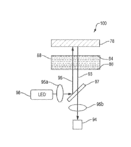
Figure .1 illustrates a double absorbance optical configuration for the
coagulation
system to absorb both excitation light from a light source and returning light
from optical
reference during coagulation of a plasma or blood sample according to one
embodiment of the
invention.
Figure 2 illustrates a reflection optical configuration for the coagulation
system to
entrap excitation light at the interface between sample and optical reference
to enhance optical
signal generated in the optical reference during coagulation of a plasma or
blood sample
according to another embodiment of the invention.
Figure 3 illustrates a transmission configuration for the coagulation system
to absorb
the light emitted from the optical reference excited by the light source
during coagulation of a
plasma or blood sample according to another embodiment of the invention.
CA 02946877 2016-10-24
WO 2015/167983 PCT/US2015/027715
6
Figure 4 illustrates that the diqance between optical reference and sample
fluid (d) can
vary from 0 to a large value, typically U to 200 mm with all the
configurations described with
respect to Figure 1, 2 and 3.
Figures 5A4i illustrate a method to make the optical reference with various
compositions, including (A) dope optical agents, such as but not limited to
fluorescence
molecules, particle, dyes, inside a substrate material such as, but not
limited to plastics, glass,
and silicon; (B) chemically assemble a layer of optical agents on the first
surface of the
substrate; (C) chemically assemble a layer of optical agents on the opposite
surface of the
substrate; (p) coating a layer of optical agents on the first surface of a
substrate by either
physical or chemical method; (E) coating a layer of optical agents on the
opposite surface of
the substrate by either physical or chemical method.
Figures 6A-G illustrate exemplary configurations for integrating the optical
reference
with the reaction chamber of the sample handling device: (A) embedding the
optical
reference in the first wall of the integral reaction chamber; (B) bending a
flat optical reference
to form the first wall of the reaction chamber with the cavity of the reaction
chamber in the
bottom part; (C) bonding the optical reference to the remaining portions of
the reaction
chamber illustrated in 63 except the bottom portion; (P) placing the optical
reference outside
the enclosed reaction chamber as a separate part; (E) embedding the optical
reference within
the wail opposite to the first wall of the integral reaction chamber; (F)
bonding the fiat optical
reference to form the opposite wall of the reaction chamber with the cavity of
the reaction
chamber in the remaining portion; (G) bonding the optical reference to the
remaining portions
of the reaction chamber illustrated in 6F except the first wall,
Figure 7A is a view of the bottom of a microfluidie plate with a reaction
chamber
illustrating one exemplary configuration of the reaction chamber having a
fluidic inlet and
.. outlet for the sample, and dry reagent pre-stored in the chamber, and
Figure 73 illustrates a
side view of Figure 7A;
Figures 8A-D illustrate a liquid handling device including an exemplary
configuration
of the reaction chamber with two fluidic inlets; one for sample and one for
reagent separately,
and a fluid outlet. (A) is a perspective view of the reaction chamber; (B) is
a top view of the
reaction chamber; (C) is a cross-section of Figure 813; (D) is another cross-
section of Figure
8B. Figures 8E-81-1 illustrate sequential tilling of the reaction chamber with
reagent from time
CA 02946877 2016-10-24
WO 2015/167983 PCT/US2015/027715
7
= 0 to time = 3, and Figures 8I-8L illustrate the sequential filling of the
reaction chamber with
sample fluid from time 4 to time = 7.
Figure 9 illustrates a cross-section of the reaction chamber filled with
reagent and
sample fluid.
Figure 10.A and 1013 are top and bottom views, respectively, of an exemplary
mierofluidic device including a plurality of reaction Chambers in accordance
with the
invention.
Figure 11 shows an embodiment of the invention based on the optical
configuration
shown in Figure 1 and exemplary assay result. (A) in this specific embodiment,
an LED is
used as light source, a fluorescence doped glass is used as optical reference,
and a quantitative
fluorescence detector is used as an optical detection unit. (B) illustrates
fluorescence signal
from an assay group having abnormal plasma (b) and control group having normal
plasma
(a) according to one embodiment of the double absorbance configuration of the
coagulation
system according to the invention shown in (A). The abnormal assay group
results show
.15 delayed signal change compared to that from the normal control assay
group.
Figure 12 shows an embodiment of the invention based on Figure 2 optical
configuration and exemplary assay result. (A) in this specific embodiment, an
LED is used as
light source, a fluorescence doped glass is used as optical reference, and a
quantitative
fluorescence detector is used as an optical detection unit; (B) illustrates
fluorescence signal
.. from an assay group having coagulated plasma (a) and control group having
uncoa.gulated
plasma (b) according to one embodiment of the double absorbance configuration
of the
coagulation system according to the invention shown in A. The assay results
shows enlarged
signal change from the coagulated plasma (a) compared to that from plasma
without
coagulation (b);
Figure 13A-D shows an exemplary mathematical method to process the optical
data to
obtain quantitative coagulation time.
DESCRIPTION
In one aspect, the invention relates to a system for detecting coagulation of
a patient
plasma or blood sample in a reaction chamber, for example, a chamber of a
microfluidic
device. The system includes an optical reference part, such as hut not limited
to a standard
CA 02946877 2016-10-24
WO 2015/167983 PCT/US2015/027715
8
fluorescence element, such as but not limited to a fluorophore-doped glass, a
polymer film or
sheet containing intrinsic fluorescence that is used to generate a
fluorescence reference signal.
The positioning of the fluorescence element and the coagulating blood/plasma
sample is
configured to vary the light energy that reaches andlor leaves the optical
reference. With such
configuration, the system according to the invention de-couples the
fluorescence signal from
chemical reactions. The variation of the .fluorescence signal indicates the
kinetics of the
.plasma/blood sample coagulation process.
The coagulation detection system according to the invention is used for
performing
coagulation assays, for example, with fluorescence detection. As a point-of-
care (POC)
coagulation immunoassay system, the sample preparation can be implemented in a
microfluidic cartridge, allowing small sample volume, i.e., less than a
milliliter, preferably less
than 100 microliters, and low manufacturing cost. The invention can be used
.fbr types of wet
chemical assays where a change in adsorption, turbidity during the assay is
used for detection
and quantification of an analyte in a sample. Typical wet chemical assays are
immunochemical, enzymatic, clotting assays, affinity based, and nucleic acid
based assays.
Different optical detection methods may be used in various embodiments such as
but not
limited to turbidity, absorption, reflectance, fluorescence intensity, time
resolved
fluorescence, 1\TIR and others. Compared to traditional coagulation assay
tools such as optical
spectroscopy or lab-on-a-chip assay systems, the coagulation system according
to the invention
has at a minimum the following advantages:
(1) the enhanced portability of the system and fast turnaround time allowing
point-of-
care applications;
(2) the system's handling of a sample requires only a small amount, ne, less
than a
milliliter of patient blood or plasma, preferably below 100 microliters;
(3) no indicators like those typically required in state-of-the-art
fluorescence assays
such as fluorophore reagents or calorimetric reagents need to be added into
the assay. This
simplifies the assay protocol by reducing the assay handling steps which would
otherwise
require immunoreactive reagents, intra-assay chemicals, and chemical
reactions. The
_fluorescence signal generated according to the invention is only a function
of the coagulation
reaction and requires no fluorophores added into the sample, resulting in
lower cost and lower
background interference;
CA 02946877 2016-10-24
WO 2015/167983 PCT/US2015/027715
9
(4) the decoupling of fluorescence signal and chemical reaction, together with
using a
standard fluorescence element, allows easy and reliable quality control;
(5) the system according to the invention described herein can be realized in
any
fluorescence system, various liquid handling systems including mierolluidics,
robotic, and
manual liquid transportation systems allowing rapid and cost-effective
adoption and
integration with other biomarker detection systems, such as, but not limited
to solid phase
immunoassays for the quantification of other analytes in blood such as cardiac
markers like
troponin / or markers providing additional infbrmation to clotting parameters
such as D-Dimer.
D-dimer tests are ordered, along with other laboratory tests and imaging
scans, to help rule
out the presence of a thrombus;
(6) the cost of the cartridge which includes the various embodiments of the
optical
system according to the invention is sufficiently low to be disposable which
reduces the risk of
cross-contamination. The cartridge can be manufactured preferably in polymers
such as
polystyrene or cycloolefines, by manufacturing methods, preferably injection
molding or hot
embossing;
(7) different wavelengths may be used for the light source and signal
detection thereby
reducing background interference. The light source may be selected from but is
not limited to
the group consisting of a laser, a mercury arc lamp, and an LED. Wavelengths
range from, for
example, about 20riM to about 5000nivl, about 50riM to about 2000nIVI, about
100nm to about
1. 000riM ,
Optical Configuration
Various optical configurations, with different arrangements of optical
reference and
sample reaction chamber, are disclosed for various turbidity assays, e.g.
blood coagulation
assays. The schematics of each configuration according to embodiments of the
invention are
.. illustrated in Figures 1, 2 and 3, and the operation principles are
described below.
Double Absorbance Optical Configuration
As shown in Figure 1, according to one embodiment of the invention, a double
absorbance optical configuration system 100 has a fluorescence module 98, a
reaction
chamber 88, and a fluorescence reference 78. In one embodiment, a fluorescence
module 98
integrates both light source 96 and fluorescence detection unit 94, for
example, but not limited
to a detection system to measure time resolved fluorescence (TRF) using an LED
(360ntn),
CA 02946877 2016-10-24
WO 2015/167983 PCT/US2015/027715
for example, for excitation and a photodetector, such as a photo diode or a
multi pixel photon
counter (MPPC, to quantify the fluorescence emission.
With continued reference to Figure 1, according to one embodiment of the
invention,
the double absorbance optical configuration system 100 has a light source 96,
an optical
5 detection unit 94, an optical reference 78 and a reaction chamber 88.
During operation, the
light 95 from the light source 96 and the returning light 93 from the optical
reference 78 both
transmit through the sample in the reaction chamber 88 and is absorbed due to
turbidity
change of the sample, plasma or blood, for example. The source or excitation
light 95 and
returning or emission light 93 can have same or different wavelengths. The
optical reference
10 78 can be realized with various optical technologies, such as but not
limited to generic
photometry, fluorescence, Raman spectroscopy time-resolved fluorescence, and
surface
enhanced Raman spectroscopy. In one embodiment, a fluorescence-based method is
used for
blood coagulation time measurement with light source being LED, optical
reference being a
fluorescence glass, returning light being emission from the fluorescence
element, the sample
being plasma, and the optical detection unit being a fluorescence detector, In
this
embodiment, when the plasma coagulates in the reaction chamber, the
fluorescence signal
read at the optical detection unit is reduced due to increased optical
absorbance by the
coagulated plasma.
With continued reference to Figure 1, the reaction chamber 88 encloses a
plasma or
blood sample and reagent(s) for a particular target coagulation assay. The
reaction chamber
88 comprises a first wall 86 and a second wall 84 that is opposite to the
first wail and is
positioned between the optical reference 78 and the light source 96 and
detector 94, The first
wall 86 is optically transparent to a light of specified wavelength and is
closer to the
fluorescence module 98 than the second wall 84. The second wall 84 is
optically transparent
to the light with specified wavelengths and is positioned opposite and
parallel to the first wall
86 and closer to the fluorescence reference 78 than the first wall 86. A
reagent added to the
plasma or blood sample in the reaction chamber 88 enables the coagulation
reaction in the
reaction chamber 88.
The optical reference 78 is, for example, but not limited to, a fluorescence-
doped
glass, or fluorophores immobilized on the surface of the second, opposite wall
84 of the
reaction chamber 88. In the double absorbance optical configuration
embodiment, the
CA 02946877 2016-10-24
WO 2015/167983 PCT/US2015/027715
11
fluorescence reference 78 is positioned on the side of the reaction chamber 88
that is opposite
to the fluorescence module 98 as illustrated in Figure 1. The purpose of the
optical reference
78 is to provide a calibrated optical signal at a specific wavelength.
During operation, once the plasma or blood sample coagulation process starts,
more
and more fibrin is formed thereby increase the turbidity of the plasma or
blood sample in the
reaction chamber 88. As a result, the transmission of the excitation light 95
through the
sample is reduced and the excitation of fluorescence molecules on the optical
reference 78 is
inhibited. In addition, the reduced emission light 93 from the optical
reference 78 is absorbed
further when it passes through the sample in the reaction chamber 88 to the
fluorescence
module 98 where it is detected and measured by the fluorescence detector 94.
The combined
effect of the two absorbance processes, i.e., the first absorbance as the
excitation light 95
passes through the reaction chamber 88 to the fluorescence reference 78, and
the second
absorbance as the emission light 93 passes from the optical reference 78
through the reaction
chamber 88, is expected to produce a signal change detected by the optical
detector 94. The
signal change indicates the coagulation process of the sample in the reaction
chamber 88. As
a result, a decrease of the fluorescence signal detected by the optical
detector 94 in this double
absorbance optical configuration indicates that the coagulation process has
begun. The
relative change of the signal with time gives information about the
coagulation process
(kinetics, slope), For the proper calculation of the different coagulation
parameters such as
PT, APTT, the maximum and minimum signal is determined.
Reflection Optical Signal
Figure 2 illustrates a reflection optical configuration of the turbidity
system 100'
according to another embodiment of the invention in which the fluorescence
reference 78 is
positioned between the reaction chamber 88 and the fluorescence module 98.
With continued reference to Figure 2, the reflection optical configuration
system 100',
like the double absorbance optical configuration system 100' described above,
comprises a
fluorescence module 98, a reaction chamber 88, and a fluorescence reference
78. In one
embodiment of the reflection optical configuration, the fluorescence module 98
integrates
both light source 96 and fluorescence detection unit 94, for example, but not
limited to a
fluorescence reader from Horiba Instruments Inc. (Kyoto, Japan) that has an
LED (360nm)
light source and a MPPC (a multi pixel photon counter ) detector.
CA 02946877 2016-10-24
WO 2015/167983 PCT/US2015/027715
12
The reaction chamber 88 encloses a plasma or blood sample, and reagent(s) for
a
specified target coagulation assay and typically has a plurality of planar
walls, at least two of
which are parallel and opposite. The optical reference 78 is positioned
between the reaction
chamber 88 and the excitation light source 96, and optical receiver 94. For
example, the
reaction chamber 88 comprises a first wall 86 and a second wall 84 opposite to
the first wall
86. In a preferred embodiment the first wall 86 and the second wall 84 are
parallel to one
another. Alternatively the first wall and the second wall may be placed at an
angle to one
another, for example, at a 45" angle. In the reflection optical configuration,
the first wall 86
is optically transparent to a light of specified wavelength and is positioned
closer to the
fluorescence reference 78 than the second wall 84. The second wall 84 is
positioned opposite
and parallel to the first wall 86 and further away from the fluorescence
reference 78 than the
first wail 86. The second wall 84 may or may not be optically transparent. A
reagent added
to the plasma or blood sample in the reaction chamber 88 enables the
coagulation reaction in
the reaction chamber 88.
The optical reference 78 is, for example, but not limited to, a fluorescence-
doped
glass, or fluorophores immobilized on the surface of the first wall 86 of the
reaction chamber
88. In this embodiment, the fluorescence reference 78 is positioned between
the reaction
chamber 88 and the fluorescence module 98 as illustrated in Figure .2. The
purpose of the
fluorescence reference 78 is to provide a calibrated fluorescence signal.
During operation, once the plasma or blood sample coagulation process starts,
more
and more fibrin is formed thereby increase the turbidity of the plasma or
blood sample in the
reaction chamber 88.
As illustrated in Figure 2, in the reflection optical configuration, the
excitation light 95
first reaches the optical reference 78 and then transmits through the sample
in the reaction'
chamber 88. In other words, one portion of the excitation light 95 excites
fluorescence of the
optical reference 78 before transmission through the reaction chamber 88,
while the remaining
portion of the excitation light 95 transmits through the sample in the
reaction chamber 88. As
coagulation of the plasma or blood sample in the reaction chamber 88 initiates
and
propagates, and the quantity of fibrin increases in the sample, the energy
distribution of the
two light portions, i.e., transmitted and reflected light, is varied due to
the change of the
sample's transmission property. Namely, the transmission of the excitation
light 95 is
CA 02946877 2016-10-24
WO 2015/167983 PCT/US2015/027715
13
inhibited, and more light is trapped at the interface of the fluorescence
reference 78 and the
first wall 86 of the reaction chamber 88 to excite more fluorophores. As a
result, an increase
of the fluorescence signal detected by the optical detector 94 in this
configuration indicates
that the coagulation process has begun. The clotting time can he determined
by, for example,
the slope of the clotting curve which is calculated by the first derivative of
the clotting curve
(maximum value of the first derivative is giving the start time for
coagulation) as shown in
Figure 12. Maximum (start of the reaction, time point zero) and minimal signal
(coagulation
completed) are needed to determine the clotting time.
Transmission Optical Configuration
Figure 3 illustrates yet another optical configuration of the system 100". The
optical
reference 78 is arranged between the light source 96 and sample reaction
chamber 88, and the
reaction chamber 88 is placed between optical reference 78 and optical
detection unit 94.
During operation, the optical reference 78 is excited by the source light 96
and emits a
secondary light 93, such as fluorescence signal. The secondary light 93 passes
through the
reaction chamber 88 and is absorbed due to turbidity change of the sample. The
optical
detector 94 reads the signal of the secondary light 93 from the optical
reference 78. The
quantitative value of the signal represents the kinetics of the coagulation
reaction.
Figure 4 illustrates that the distance (d) between the optical reference 78
and the
sample in the lumen 83 of the reaction chamber 88 can vary from about 0 to
about 200 rum
with each configuration described above with respect to Figure 1, 2 and 3.
Figure 5 illustrates exemplary configurations of the optical reference 78.
Using optical
reference with fluorescence properties as a non-limiting example, the optical
agents 61 can be
made by embedding fluorescence molecules, particles or other carriers into a
plastic, glass or
silicon material substrate 78 (Figure 5A). Alternatively, the optical
fluorescence agents can be
chemically or physically coated on the surface of the substrate, either on the
top 60 or bottom
62 surface, i.e., first surface 60 or second surface 62 opposite the first
surface, or both. For
example, as illustrated in Figure 5(B) a layer of optical agents 61 may be
chemically
assembled on the first surface 60 of the substrate by physical means or
chemical means; in
Figure 5(C) a -layer of optical agents 61 may be chemically assembled on the
second surface
62 of the substrate by physical or chemical means; in Figure 5(D), a layer of
optical agents 61
may be coated on the first surface 60 of the substrate by chemical or physical
means; or, in
CA 02946877 2016-10-24
WO 2015/167983 PCT/US2015/027715
14
Figure 5(e), a layer of optical agents 61 may be coated on the second surface
62 of the
substrate by chemical or physical means.
Figure 6 illustrates various exemplary arrangements of the reaction chamber 88
and
the optical reference 78. The optical reference 78 can be an integral part of
the reaction
chamber 88, for example, by being embedded in the top or bottom part of the
enclosed wall
64 of the reaction chamber 88, or, alternatively, the optical reference can he
a separate part
placed outside above or outside below the reaction chamber 88 to form suitable
optical
configurations according to the invention. Preferably the long axis of the
optical reference 78
is perpendicular to the excitation light. Alternatively the excitation light
may he at an angle to
the long axis of the optical reference 78.
Figure 6A illustrates an exemplary planar optical reference 78 embedded in the
first
wail 65 of the enclosing wall 64 of the reaction chamber 88, according to one
embodiment.
Alternatively, Figure 6B illustrates a planar optical reference 78 bonded to
and forming the
first wall 65 of the reaction chamber 88 with the lumen 83 of the reaction
chamber 88 on the
inside of the first wall 65 of the reaction chamber 88. in a preferred
embodiment the long axis
of the optical reference 78 is perpendicular to the light source or
alternatively at an angle up to
about 45 .
In another embodiment, illustrated in Figure 6C, the optical reference 78
forms three
walls, 65, 65", 65" of the reaction chamber 88 while only the second wall 65',
opposite wall
65, is not a portion of the optical reference 78.
In still another embodiment, illustrated in Figure 60, optical reference 78 is
positioned
as an element separate from any wall of the reaction chamber 88 and with the
long axis of the
optical reference 78 parallel to at least one wall of the reaction chamber 88;
illustrated in
Figure 6.E, the optical reference 78 is embedded in the second wall 65' of the
reaction
chamber 88; illustrated in Figure 6F, the optical reference 78 is planar and
bonded to the
second wall 65' of the reaction chamber 88; illustrated in Figure 6G, the
optical reference 78,
forms three walls, 65', 65", 65", with only the first wall 65 opposite to wall
65' not a
portion of the optical reference 78.
Sample preparation cartridge
According to the embodiments of the coagulation systems .100, 100' and 100"
illustrated in Figures 1, 2, and 3, sample preparation in this invention can
be realized in
CA 02946877 2016-10-24
WO 2015/167983 PCT/US2015/027715
various ways, from manual pippeting to an automatic fluidic control system.
Non-limiting
examples of microfluidic devices and methods applicable to the coagulation
assay systems
described above are given below. These devices and methods are not limited to
assays for
coagulation and can be used for a variety of wet chemical assays where
metering, reagent
5 addition, mixing, incubation and quantification of the assay reaction
product is needed.
Typical assays are using enzymatic reaction to measure metabolites such as
lactate or
creatinine or turbidimetric assays. Examples of such turbidimetrie assays are
agglutination
assays such as latex agglutination where mono-disperse immune particles are
eomplexing in
the presence of an analre, which can be monitored by a change in turbidity.
0 Flow chamber with dry reagent
Referring now to Figures 7A and 7/3, in one embodiment, a reaction chamber 88
of a
liquid handling device 120 with a defined volume is formed by a rinerochannel
plate 90
covered with a lid 91. The reaction chamber is used to meter the sample
volume, one fluid
inlet 68 is used to introduce the sample, e.g., plasma, from the bottom 60b of
the reaction
15 chamber 88, and one fluid outlet 66 at the bottom 60b of the reaction
chamber 88 is used to
discharge the excessive liquid from the lumen 83 of the reaction chamber 88.
Dry reagent,
such as lyophilized PT/APTI' reagent, biotin and etc., is pre-stored in the
reaction chamber
88, unifeamly coated on the lumina! surface of the first wall 86, for example.
When the
plasma fills the reaction chamber 88, the dry reagent starts to dissolve and
then diffuses into
the sample along the vertical direction, he., from the bottom 60b of the
chamber 88 toward the
top 60a of the chamber. The dry reagent has a relatively large contact area
with the liquid
sample and the diffusion distance along the vertical direction is relatively
short. This
confifration provides a homogeneous coagulation process across the lateral
plane of the
reaction chamber S. During operation, once the chamber 88 is filled with
sample, the assay
process starts and the fluorescence signal acquisition begins to ibilow the
reaction kinetics.
Flow chamber with liquid reagent
Figures 8A-8D illustrate one embodiment of the invention illustrating a liquid
handling device 120 IM investigation of a sample fluid. The liquid handling
device 120
comprises a reaction chamber 88, two inlet ports and channels 66 and 68 to
deliver sample
fluid and reagent fluid into the lumen 83 of the reaction ch2mher,
respectively, and an outlet
channel 64 fur venting of the reaction chamber 88 during filling. The device
120 may contain
CA 02946877 2016-10-24
WO 2015/167983 PCT/US2015/027715
16
one or more fluidic structures 68a and 64a, for example, to provide a
controlled and bubble
free filling of the reaction chamber lumen 83.
According to one embodiment of the liquid handling device 120 illustrated in
Figures 8A-8.1), the reaction chamber 88 of the device 120 is firstly filled
with a metered
amount of a liquid reagent via a first inlet 66. A bubble-free liquid filling
can be achieved by a
capillary stop feature 64a next to the outlet 64, in Figure 8A., for example,
a cylindrical
groove is acting as a capillary stop 64A. A capillary stop is defined either
by a sudden channel
opening and by the curvature of the feature 64a or by making the outlet area
64 hydrophobic.
Figures SE to 8H illustrate the sequential filling of reagent into the
reaction chamber 88 at
.. different points of time from time=0, to t1me=3, After a metered amount of
reagent has filled
into the lumen 83 of the reaction chamber 88, a metered amount of sample fluid
(such as
plasma and whole blood) is filled into the chamber lumen 83 via the second
inlet 68 as
illustrated in Figures 81 to 8L.
Additional features of the embodiment shown on Figures 8A-8D follows. The
liquid
handling device 120 is oriented in the horizontal direction, i.e. the top view
of the liquid
handling device 120 is as shown on Figure 8B. The v-Shape 68a at the inlet
channel 68 has,
for example, an opening angle of 30 . 'ibis v-Shape 68a could have an angle
ranging from 00
to 1800, typically 15" to 120". It is also noted that, according to this
embodiment of the liquid
handling device 1202 the second inlet 68 and the outlet 64 is positioned on
the top side 60a of
.. the liquid handling device 120, while the first inlet 66 is positioned on
the bottom side 60b of
the liquid handling device 120, Other arrangements of the inlets and outlets
on the top and
bottom sides of the liquid handling structure are also possible and are not
limited by the
illustrated embodiment. The flow rate for sample and reagent may range from
about 0.5ill/s
to 2000/s, typically 2p1is to 104.1/s.
Figure 9 illustratively exemplifies the reaction chamber 88 after filling of
reagent and
sample have been completed. Two layers are shown: a layer of reagent and a
layer of sample
fluid. The sample layer is spread above the reagent layer across the whole
surface of the fluid
in the lumen 83 of reaction chamber 88. Therefore, it generates a large
contact area between
the two liquids, namely reagent and sample. With this large contact area, the
mixing and
thereby the reaction of the reagent and sample liquids is highly efficient.
CA 02946877 2016-10-24
WO 2015/167983 PCT/US2015/027715
17
In the illustrated embodiments in Figure 8, a v-shaped geometry of the inlet
structure
68 is used to support an even distribution of the sample fluid into the
reaction chamber. .As
illustrated in Figures Sc and 8d, the sample inlet 68 is connected to the top
60a of the reaction
chamber 88 whereas the reagent inlet 66 is positioned in opposite to the
bottom 60b of the
reaction chamber 88.
Referring to Figure 10A, a top view of an embodiment of a mieroftuidic device
50
having four reaction chambers 88 a-d is illustrated. In the illustrated
embodiment, the
reaction chambers 88 a-d are positioned toward one side of the .nneroiluidic
device 50 but
could be positioned in the microlluidie card at other positions.
Figure 10B illustrates a bottom view of the microfluidic card 50 including a
plurality
of channels 67 that are in fluid communication with the reaction chambers 88.
Exemplification/Proof of Principle
The embodiments of the coagulation systems 100, 100, 100" discussed above and.
their associated assay methods for detecting coagulation of a blood or plasma
sample were
evaluated with controlled plasma samples and reagents for PT and APTT assays.
In the
e)tampic of the double absorbance configuration described above with respect
to Figure 1
and illustrated in Figure I. IA, the fluorescence module applied in the method
was a PMT-
based Time Resolved Fluorescence (TRF) unit, fluorescence reference 78 was a
Europium-
doped glass, which contained precisely-controlled amount of europium and did
not have
photo bleach during excitation, and an LED 96 was used as a light source. A
filter 95A was
placed between the LED 96 and a dichroie mirror 97. A second filter 951.3 was
placed
between the detector 94 and the dichroie mirror 97, The plasma samples
included normal
control plasma (a) and high abnormal control plasma (b) from Instrumentation
Laboratory
Company (Orangeburg, NY). Coagulation was initiated by introduction of a
coagulation
initiater.
Referring to Figure 1 J.B, the intensity of fluorescence signal emanating from
the
fluorescence reference and transmitted to the fluorescence detector in the
double absorbance
coagulation system described above with respect to Figures 1 and l IA, is
represented by
curve (a) for normal control and a curve (b) for abnormal control plasma, in
both normal and
abnormal plasma samples, the fluorescence signal decreased as the coagulation
initiated,
CA 02946877 2016-10-24
WO 2015/167983 PCT/US2015/027715
18
propagated, and reached a stable value when coagulation was completed. The
abnormal
plasma takes a longer time to start and finish the coagulation process than
the normal plasma.
Referring to Figure 12A, an embodiment of the invention using reflection
configuration described above with respect to Figure 2 is realized. As
illustrated in Figure
12A, an LED 96 was used as the light source, a fluorescence-doped glass was
used as the
optical reference 78, and a quantitative fluorescence detector was used as the
optical detector
unit 94õA. filter 95A was placed between LED 96 and a dichroic mirror 97. A
second .filter
95B was placed between the detector 94 and the diehroic mirror 97. The plasma
samples
included a normal plasma sample (a) to which a coagulation reagent was
introduced and a
control normal sample (1)) to which water (no coagulation reagent) was
introduced. The
optical signal obtained from plasma with coagulation (a) and plasma without
coagulation (b)
is illustrated in figure 1213, The fluorescence signal increased on sample (a)
and reached
stable value as coagulation initiated and propagated and reached a stable
value when
coagulation was completed. The control (b) used the same plasma sample but
with the
addition of deionized water (no coagulation happened).
Figure 13 shows an exemplary method to process the optical data to obtain the
quantitative coagulation time, in the four steps, the original data is first
normalized (Fig. 13A)
and filtered (Fig. 1313) to eliminate redundant data and noise. A first order
derivative (Fig.
13C) of the original data is implemented to identify the time spot when the
quickest change of
optical signal locates. The peak position of the first-order derivative (Fig,
13D) is used as the
coagulation start time. Other methods can be used to quantitatively study the
coagulation
process as well.
Various modifications and other implementations of what is described and
illustrated
herein will occur to those of ordinary skill in the art without departing from
the scope and
spirit of the invention. The invention is not to he defined only by the
preceding illustrative
descriptions or drawings.