Note: Descriptions are shown in the official language in which they were submitted.
CA 02948188 2016-11-04
WO 2015/175609 PCT/US2015/030489
1
METHOD AND DEVICE FOR MEASURING DENTIN PERMEABILITY
FIELD OF THE INVENTION
The invention relates to a method and device that allows visual recordation of
hydraulic
conductance through a sectioned piece of dentin; and which can be used to
measure the
effectiveness of treatments to reduce the permeability of dentin.
BACKGROUND OF THE INVENTION
A large portion of US households (60%) have at least one family member that
suffers from
hypersensitive teeth. Products, such as toothpaste, targeting consumers
suffering from tooth
sensitivity are the fastest growing segment of the oral care market; however,
the results provided
by sensitivity toothpastes leave a majority of users (60%) unsatisfied, with
57% of sufferers
willing to try products other than toothpaste.
Tooth sensitivity can be activated by hot and cold drinks and certain acidic
or hypertonic foods.
This sensitivity often occurs when gum tissue recedes from the necks of teeth,
exposing root
surfaces that are not covered by hard enamel but by soft cementum. The
cementum is so thin and
soft that it can be removed by tooth brushing, thereby exposing the sensitive
underlying dentin.
The dentin can also become exposed under restorations. The pain that patients
feel from exposed
dentin is called dentin sensitivity, as reported in Pashley, Arch Oral Biol 39
(Suppl) 735-805
(1994).
Dentin sensitivity, whether on exposed surfaces at the necks of teeth or under
restorations, is
reportedly due to minute fluid shifts across dentin in response to painful
stimuli (Brannstrom,
Oper Dent 9: 59-68, 1984). This theory, called the hydrodynamic theory,
(Brannstrom and
Astrom, Int Dent J 22: 219-j226, 1972), assumes that fluid can move within or
through the
microscopic tubules making up dentin, at a rate that activates mechanoreceptor
nerves in the
dentin pulp. When the surface of the tooth is eroded, the dentinal tubules
become exposed to the
external environment. The exposed dentinal tubules provide a pathway for
transmission of fluid
flow to the pulpal nerves and this is induced by changes in temperature,
pressure and ionic
gradients. By blocking the tubules, the external stimuli have a diminished
effect, and less pain
will be felt. A number of agents have been previously screened for their
ability to occlude
CA 02948188 2016-11-04
WO 2015/175609 PCT/US2015/030489
2
dentinal tubules, including potassium oxalate (Greenhill and Pashley, J Dent
Res 60: 686-698,
1981).
Hypersensitive teeth can cause pain and discomfort when subjected to changes
in temperature,
pressure, or chemical action. Exposure of the dentin frequently leads to
hypersensitivity. Dentin
exposure may occur due to recession of the gums, periodontal disease and
improper dental care.
Hypersensitive teeth are commonly treated with a nerve desensitizer such as
potassium nitrate or
potassium chloride. Alternatively, hypersensitive teeth may also be treated
with an ingredient
intended to occlude the tubuli, such as strontium acetate, strontrium
chloride, stannous fluoride,
ferric oxalate or potassium oxalate. Application of the therapeutic ingredient
may be via a non-
prescription preparation, such as a dentifrice or rinse, or via products
professionally applied or
prescribed.
There have been various in vitro methods that have been developed to measure
the effectiveness
of various treatments and compositions to treat dentin so that its less
permeable. An effective
method should reproduce the microenvironment in and around the tubule orifice,
provide the
correct geometry, appropriate surface chemistry, and relevant fluid
composition and movement.
One method was developed by Pashley (J. Periodontology, vol. 55, no. 9, pg.
522, September
1984). Pashley utilizes sectioned dentin discs of predetermined thickness
which are placed
between two o-rings within a two part chamber. Positive pressure is used to
drive a testing fluid
through the bottom chamber to the dentin section; the amount of fluid passing
through the dentin
is then measure to determine hydraulic conductance. The system was used to
measure the effect
of any desensitizing toothpastes had on reducing the permeability of the
dentin.
One of the issues with the Pashley device has been with visualization. The
upper portion of the
device is usually covered so direct visualization of fluid moving through
dentin is not possible.
Further, as the dentin is covered by the top portion of the device in order to
treat the dentin, for
example by brushing with toothpaste or application of a tooth whitening strip)
after a baseline
measurement has been taken the top portion must be removed and then
reattached, which could
damage the dentin and/or alter the results.
Therefore, what is needed is a device allowing for direct manipulation of a
dentin section while
the dentin section is secured within the testing device, and which allows the
dentin section to be
visible during testing.
CA 02948188 2016-11-04
WO 2015/175609 PCT/US2015/030489
3
SUMMARY OF THE INVENTION
A method of visually recording the hydraulic conductance of a dentin section
is provided that
comprises preparing a dentin section; providing a test cell having a bottom
component with an
inner chamber; positioning at least a portion of the dentin section over the
bottom component
inner chamber; providing a fluid under pressure to the inner chamber of the
bottom component;
and visually recording hydraulic conductance of fluid through the dentin
section.
A method of visually comparing the effect of oral care compositions on the
hydraulic
conductance of dentin sections is provided that comprises preparing a dentin
section; providing a
test cell having a bottom component with an inner chamber and a top component,
wherein the top
component comprises an opening having beveled walls; positioning at least a
portion of the
dentin section between the bottom component inner chamber and top component
opening,
providing a fluid under pressure to the inner chamber of the bottom component;
visually
recording hydraulic conductance of fluid through the untreated dentin section;
treating at least a
portion of the dentin section with an oral care composition; visually
recording hydraulic
conductance of fluid through the treated dentin section; and comparing the
visually recorded
hydraulic conductance of the treated and untreated dentin sections.
A method of visually comparing the effect of two or more oral care
compositions on the
hydraulic conductance of dentin sections is provided that comprises preparing
a first dentin
section; providing a test cell having a bottom component with an inner chamber
and a top
component, wherein the top component comprises an opening having beveled
walls; positioning
at least a portion of the first dentin section between the bottom component
inner chamber and top
component opening, providing a fluid under pressure to the inner chamber of
the bottom
component; treating at least a portion of the first dentin section with a
first oral care composition;
visually recording hydraulic conductance of fluid through the first dentin
section; preparing a
second dentin section; providing a test cell having a bottom component with an
inner chamber
and a top component, wherein the top component comprises an opening having
beveled walls;
positioning at least a portion of the second dentin section between the bottom
component inner
chamber and top component opening, providing a fluid under pressure to the
inner chamber of
the bottom component; treating at least a portion of the second dentin section
with a second oral
care composition; visually recording hydraulic conductance of fluid through
the second dentin
section; and comparing the visually recorded hydraulic conductance of the
denting section treated
CA 02948188 2016-11-04
WO 2015/175609 PCT/US2015/030489
4
with the first oral care composition and the second dentin section treated
with the second oral
care composition.
BRIEF DESCRIPTION OF THE DRAWINGS
FIG. 1 is a top view of a test cell bottom component for use in an embodiment
of the present
invention.
FIG. 2 is a sectional view of FIG. 1 along the 2-2 plane.
FIG. 3 is a perspective view of a test cell top component for use in an
embodiment of the present
invention.
FIG. 4 is a top view of a test cell top component for use in an embodiment of
the present
invention.
FIG. 5 is a sectional view of FIG. 4 along the 5-5 plane.
FIG. 6 is an exploded view of a test cell for use in an embodiment of the
present invention.
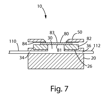
FIG. 7 is a sectional view of FIG. 6 in an assembled state along the 7-7
plane.
FIG. 8A is a perspective view of a test cell for use in an embodiment of the
present invention.
FIG. 8B is a sectional view of FIG. 8A along the 8B-8B plane.
FIG. 9 is a schematic drawing of a system layout for visually recording dentin
permeability
according to an embodiment of the present invention.
FIG. 10 is a sequence of pictures illustrating the hydraulic conductance of a
dentin section
according to an embodiment of the invention.
FIG. 11A and 11B show pictures illustrating the hydraulic conductance of
dentin sections
according to an embodiment of the invention.
CA 02948188 2016-11-04
WO 2015/175609 PCT/US2015/030489
FIG. 12 shows pictures illustrating the hydraulic conductance of dentin
sections according to an
embodiment of the invention.
5
DETAILED DESCRIPTION OF THE INVENTION
Dental sensitivity is believed to be a result of rapid flow of fluid through
dentinal tubulii caused
by pressure changes, which are in turn a result of thermal or osmotic insults.
The effectiveness
of various oral care compositions at reducing sensitivity can be linked to
their ability to block or
reduce such fluid movement. As such, the methods of the present invention are
designed to
quantitatively compare, and visually record, the performance of oral care
compositions and
materials in blocking or inhibiting hydraulic conductance through dentinal
tubulii.
All percentages and ratios used hereinafter are by weight of total
composition, unless otherwise
indicated. All percentages, ratios, and levels of ingredients referred to
herein are based on the
actual amount of the ingredient, and do not include solvents, fillers, or
other materials with which
the ingredient may be combined as a commercially available product, unless
otherwise indicated.
All measurements referred to herein are made at 25 C unless otherwise
specified.
By "oral care composition", as used herein, is meant a composition, which in
the ordinary course
of usage, is not intentionally swallowed for purposes of systemic
administration of particular
therapeutic agents, but is rather retained in the oral cavity for a time
sufficient to contact dental
surfaces or oral tissues. Examples of oral care compositions include
dentifrice; mouth rinse;
mousse; foam, mouth spray; lozenge; chewable tablet; chewing gum; dental
strips, such as tooth
whitening strips, sensitivity strips, or breath freshening dissolvable strips;
floss and floss
coatings; or denture care or denture adhesive product. The oral care
composition may also be
incorporated onto strips or films for direct application or attachment to oral
surfaces.
The term "dentifrice", as used herein, includes tooth or subgingival -paste,
gel, or liquid
formulations unless otherwise specified. The dentifrice composition may be a
single phase
composition or may be a combination of two or more separate dentifrice
compositions. The
dentifrice composition may be in any desired form, such as deep striped,
surface striped,
CA 02948188 2016-11-04
WO 2015/175609 PCT/US2015/030489
6
multilayered, having a gel surrounding a paste, or any combination thereof.
Each dentifrice
composition in a dentifrice comprising two or more separate dentifrice
compositions may be
contained in a physically separated compartment of a dispenser and dispensed
side-by-side.
The term "dispenser", as used herein, means any pump, tube, or container
suitable for dispensing
compositions such as dentifrices.
The term "teeth", as used herein, refers to natural teeth as well as
artificial teeth or dental
prosthesis.
The term "visually recorded", as used herein refers to visual observation of
the appearance and
rate of appearance of liquid. Using a camera or other visual recordation
device, this may be
performed in real time or captured via still or video photography for
incorporation and replay in
hardcopy or digital media; may also include quantitative analysis of the size
of liquid droplets or
area occupied by liquid droplets.
The term "hydraulic conductance", as used herein refers to convective liquid
movement,
specifically pressure driven movement of liquid. Mathematically it is
described by the
volumetric flow rate (Q) divided by the area of the flow window (A) and the
pressure drop across
the thickness of the dentin section (AP) as shown below and as described in
the literature (see J
Dent Res 1981, 60(3):pp 686-698 for an example)
Q
Ly, = _________________________________________
v A(AP)
Where Lp is hydraulic conductance, (A) is the defined area of liquid flow, and
(AP) is the
pressure drop across the thickness of the dentin section, and (Q) is
volumetric flow rate.
In the present invention hydraulic conductance rates are measured through a
cross-section of
dentin to evaluate and visually record the effect of treatment with an oral
care composition.
Examples of treatment include application via toothbrush, oral rinse, dental
strip or patch, swab
applicator, manually rubbing with a finger, and application via burnishing
tool such as a prophy
cup. The method may comprise one or more of the following steps: dentin
section preparation,
mounting the section in a test cell, measurement and visual recording of
baseline hydraulic
conductance under pressure, treatment with one or more oral care compositions,
and
CA 02948188 2016-11-04
WO 2015/175609 PCT/US2015/030489
7
measurement and visually recording of hydraulic conductance post treatment. A
comparison is
then made of the visually recorded results between treated and untreated
and/or different
treatments. Results may also be reported in terms of percentage reduction in
rate.
Dentin Section Preparation
Dentin sections of human molars can be obtained in as close proximity to the
enamel junction as
possible without residual enamel appearing on the surface of the cross-
section. Sections may be
cut and sanded to a thickness of between about 0.1 mm to about 2 mm, however
sections are
typically cut and sanded to a thickness between about 0.8 mm to about 1 mm,
although larger or
smaller specimens may be used. Thicker sections are typically marred by
residual enamel on one
surface or the appearance of pulpal horns on the other. Section thickness down
to about 0.1 mm
may be used, but sections thinner than about 0.4 mm can be very fragile and
more difficult to
work with. In certain embodiments, a constant section thickness is used for
multiple samples, for
example about 0.8 mm, so that the pressure drop per unit thickness remains
consistent from
sample to sample. To remove the smear layer resulting from the
cutting/sectioning/sanding
process, samples may be sonicated in de-ionized (DI) water for a period of
time, such as about 6
minutes on each side, followed by acid-etching in an ultrasonic bath operated
at 15 kz, such as
the Bronson Model 1510 Ultrasonic Cleaner (Fischer Scientific, Pittsburgh, PA)
in 10 ml of 6.0%
citric acid for a certain amount of time, such as about 2 minutes on each side
to remove the smear
layer deposited by cutting and sanding. Samples may then be rinsed with DI
water followed by
immersion in commercial phosphate-based pH 7 calibration buffer containing a
microbial growth
inhibitor, such as BDH pH 7 Reference Standard Buffer (VWR p/n BDH5052,
Radnor, PA).
Water may be used as a short-term storage solution, but refrigeration in a
dilute thymol solution
may be used for long-term storage.
FIGS. 1 through 6 are views of a test cell 10 that may be used in the present
invention. FIG. 1 is
a top view of the bottom component 20 of test cell 10, while FIG. 2 is a
vertical sectional view of
FIG. 1 along the 2-2 plane. Bottom component 20 of the test cell includes
bottom surface 22, top
surface 24, indent 26, inner chamber 30, fastener blind holes 32 (or other
suitable mechanism for
engaging fasteners), inlet channel 34, and flush channel 36. In certain
embodiments, inlet
channel 34 and flush channel 36 may be positioned opposite or substantially
opposite each other.
Inlet channel 34 and flush channel 36 each have an inner end, 34a and 36a,
respectively, the inner
openings, 34a and 36a, joining inlet channel 34 and flush channel 36 to the
inner chamber 30.
CA 02948188 2016-11-04
WO 2015/175609 PCT/US2015/030489
8
The inner openings, 34a and 36a, further define the point from which inlet
channel 34 and flush
channel 36 extend outwardly from the test cell 10.
The bottom component 20 comprises an opening 40 at the top of the inner
chamber 30 for
accessing the inner chamber 30. Inlet channel 34 is positioned in fluid
communication with inner
chamber 30. Flush channel 36 is also positioned in fluid communication with
inner chamber 30.
Inlet channel 34 and flush channel 36 can be, optionally, as shown in FIG. 7,
threaded to receive
the compatibly threaded ends of inlet and flush tubes 110 and 112,
respectively.
FIG. 3 is a perspective view of the top component 50 for test cell 10; FIG. 4
is a top view of the
top component 50 for test cell 10; while FIG. 5 is a vertical sectional view
of FIG. 4 along the 5-
5 plane. The top component 50 of the test cell includes bottom surface 52,
upper surface 54,
optional fastener through-holes 60 (or other suitable mechanism for engaging
fasteners), and top
component opening 70. Top component opening 70 is defined by walls 78 on top
component 50.
The walls 78 may be beveled towards the top component opening 70 at an angle
"a" as
determined by the XY plane, having its vertex at the intersection of the wall
78 and upper surface
54 of the top component, wherein the angle a can be from about 30 to 85 or
from about 50 to
about 75 . The beveled walls 78 allow easy access to oral care compositions
and devices to
apply oral care compositions, which can be used to treat the dentin, such as
whitening strips and
toothbrushes.
As shown in FIG's 6 and 7, in certain embodiments, top component 50 and bottom
component 20
are shaped to fit one in the other so as to permit a secure engagement between
the two
components, to form a test cell 10. FIG. 7 is a cross-sectional view of the
test cell shown in FIG.
6, in an assembled state, with inlet and flush tubes 110 and 112 connected
with inlet channel 34
and flush channel 36, respectively. The top component 50 and bottom component
20
components may be formed from machined glasses; woods; metals, such as
stainless steel;
plastics, such as polymethyl methacrylate (PMMA) or polycarbonate (PC); or a
combination of
these materials. In one embodiment, top component 50 and/or bottom component
20 are formed
from (e.g., by machining) optically clear or transparent PMMA, such as that
available from
MacMaster-Can- (Catalogue #8560K912 or #8560K265) of Robbinsville, N.J. The
advantage of
using a clear (e.g., optically clear or transparent) material in forming test
cell 10 is that clear
materials allow "line of sight" into the cell or otherwise makes the contents
of the cell visible to
the unaided eye to, for example, help in visually determining whether all air
in the form of air
bubble(s) has been purged from the portion of inner chamber 30 below the
dentin section 80. An
CA 02948188 2016-11-04
WO 2015/175609 PCT/US2015/030489
9
air bubble below the dentin section 80 decreases the area of dentin section 80
through which fluid
can flow through, which may result in an inconsistent measurement of
permeability through the
dentin.
As shown in FIG's 6 and 7 the test cell may be assembled by placing first
washer 82 in indent 26
of bottom component 20. Second washer 84 is positioned under top component 50.
In certain
embodiments, indent 26 of bottom component 20 is machined to fit the width
dimensions of any
washer(s) used (such as washer 82) so as to reduce, minimize or prevent any
displacement of the
washer(s): i) as the components of the test cell are being secured for use
(e.g., testing and/or fluid
hydraulic conductance measurement); and/or ii) during actual use (e.g.,
testing and/or fluid
hydraulic conductance measurement). Second side 81 of dentin section 80 is
placed on first
washer 82. The dentin section 80 is centered over the opening in the first
washer 82 with the
enamel side (first side 83) corresponding to the occlusal surface of the tooth
facing up, i.e. facing
the second washer 84 and top component 50, making sure that the section 80
either completely
spans the opening or sufficiently contacts enough of the perimeter such that
the section 80 is held
securely in place. Washer 84 is placed on first side 83 of dentin section 80.
To complete sealing
of the cell, top component 50 is fastened onto bottom component 20, using
fasteners, for example
the fasteners may be screws which pass through optional fastener through-holes
60 of top
component 50 and are anchored in/by fastener blind holes 32 of bottom
component 20 having
screw holes suitable for engaging the screws so that the screws adjustably
tighten and seal the top
component 50 on to bottom component 20. In this embodiment the test cell,
including top
component 50 and bottom component 20 is referred to as test cell 10. Fasteners
may be formed
of materials such as stainless steel.
Alternatively, the assembly of top component 50 on to bottom component 20 can
be
accomplished by the use of other adjustable fastening mechanisms, such as
nails, dowels, clamps,
straps, bolts (e.g., screw-type), or any other fastening mechanism suitable
for providing a leak
proof (or substantially leak proof) seal and allow for ready disassembly and
assembly.
Optionally, the fastening mechanism can operate by friction or interference
fit so long as the
friction or interference fit can withstand the fluid pressures used in the
present invention.
The test cell 10 used in the present invention differs from "Pashley" type
flow-through cells
reported in the external literature in that it includes a top component 50,
which allows sealing the
dentin section 80 in the test cell while maintaining access to one of the
dentin section 80 surfaces.
The area defined by the first washer 82 underneath the dentin section 80
remains constant
CA 02948188 2016-11-04
WO 2015/175609 PCT/US2015/030489
throughout the conditioning, baseline measurement, and post-treatment
hydraulic conductance
measurement. This is a significant advantage in comparison to devices which
require
disassembly between baseline and post-treatment measurements in order to apply
a treatment to
the dentin section surface, because quantitative hydraulic conductance is a
strong function of the
5 specific hydraulic conductance window selected. Our experience has shown
that quantitative
precision is improved when the same hydraulic conductance window is utilized
to compare pre-
and post-treatment measurements to show hydraulic conductance reduction. A
second advantage
to having a top component 50, as used in the present invention is the
unobstructed view of the
dentin section surface. After excess moisture has been removed, still or video
photography may
10 be used to visually record images of liquid droplets coalescing on the
dentin section surface.
In certain embodiments of the present invention, as illustrated in FIG's 8A
and 8B, a test cell 310
may be used that does not include a top component. The assembled test cell 310
comprises a
bottom component 320 having an opening 340 at the top of an inner chamber 330
for accessing
the inner chamber 330. Inlet channel 334 is positioned in fluid communication
with inner
chamber 330. Flush channel 336 is also positioned in fluid communication with
inner chamber
330. In this embodiment the test cell 310 also comprises a washer 383 and a
dentin section 380.
Second side 382 of dentin section 380 faces the washer 383. The dentin section
380 is centered
over the opening in the washer 383 with the enamel side (first side 381)
corresponding to the
occlusal surface of the tooth facing up, making sure that the dentin section
380 either completely
spans the opening or sufficiently contacts enough of the opening perimeter
such that the section
380 is held securely in place. The washer 383 forms a liquid-tight seal
between the bottom
component 320 and the dentin section 380 establishing fluid communication
between the bottom
component 320 and dentin section 380. The water tight seal can be produced by
a free-standing
adhesive substance, or by a combination of a self-adhesive washer in
combination with a
waterproof adhesive substance present on the washer first surface 387 (faces
the bottom
component) and the washer second surface 389 (faces the dentin section).
In certain
embodiments the second surface 389 may include a notch 385, so that a dentin
section 380 may
rest, at least partially, within the washer 383. In this embodiment adhesive
would be present
within the notch. An example of a suitable adhesive substance includes
silicone-based bonding
agents such as Dow Coming Number 700 Silicone Sealant (McMaster-Can- p/n
7425A51). An
example of a suitable material from which washers may be cut is Buna-N rubber
(McMaster-Carr
p/n 86795K21). After cleaning with liquid dishwashing detergent, the washer
may be coated on
CA 02948188 2016-11-04
WO 2015/175609 PCT/US2015/030489
11
both sides with the adhesive substance or may be clamped or pressed in place
until the sticky
material forms a water-tight seal between the dentin and bottom component.
The "washers" that may be used in the present invention may have at least one
flat side for
contacting a dentin section, bottom component, or top component . Washers may
be made of,
silicon, rubber or soft plastic. Examples of such silicon, rubber or soft
plastic materials, include,
but are not limited to, butadiene rubber, butyl rubber, chlorosulfonated
polyethylene,
epichlorohydrin rubber, ethylene propylene diene monomer, ethylene propylene
rubber,
fluoroelastomer, nitrite rubber, perfluoroelastomer, polyacrylate rubber,
polychloroprene,
polyisoprene, polysulfide rubber, sanifluor, silicone rubber and styrene
butadiene rubber) and
thermoplastics (including, but not limited to, thermoplastic elastomer;
thermoplastic polyolefin,
thermoplastic polyurethane, thermoplastic etheresterelastomers, thermoplastic
polyamide(s), melt
processible rubber thermoplastic vulcanizate) and mixtures thereof. In one
embodiment, the
washers may be rubber "0"-rings supplied by McMaster-Can- (Catalogue
#4061T114) of
Robbinsville, N.J.
The permeability of dentin section 80 may be measured using test cell 10 in
the present invention
in the following manner. Once the two-part test cell 10 is assembled, pressure
is used to initiate
and maintain fluid (e.g., distilled water) flow in inlet channel 34. In the
embodiment shown in
FIG. 7, fluid flows into the bottom component 20 through inlet channel 34, and
into the portion
of inner chamber 30 below dentin section 80. Initially, flush channel 36 is
kept open so that
residual air in the form of air bubble(s) located in the portion of inner
chamber 30 below dentin
section 80 flows into flush channel 36 and exits test cell 10. When the
residual air has been
removed, flush channel 36 is closed. When flush channel 36 is closed, fluid
pressure rises in the
portion of inner chamber 30 below dentin section 80. This increased fluid
pressure initiates fluid
hydraulic conductance in (across or through) the dentin tubule orifices in
dentin section 80. Fluid
hydraulic conductance continues through top component opening 70 of top
component 50.
FIG. 9 is a schematic flow chart drawing, explaining the equipment lay-out for
use in the method
of measuring the permeability of dentin according an embodiment of the present
invention.
Though this is one possible lay-out of for the equipment, it is to be
understood that other possible
lay-outs would also be useful in the method of measuring the permeability of
dentin according to
the present invention.
CA 02948188 2016-11-04
WO 2015/175609 PCT/US2015/030489
12
The schematic flow chart drawing includes, in fluid communication: pressure
generating device
200; fluid source 210; flow meter 230; tubes 110, 112, 114, and 116; and valve
240. Tube 116
connects pressure generating device 200 to valve 240. Tubes 110, 112, 114, and
116, may be
metal or plastic. In one embodiment, the tubes are as 1/16" x 1/32" Tefzel or
PTFE Tubing (Idex
Corp., Lake Forest, Ill.). The flow chart also includes a camera 300 for
visually recording
hydraulic conductance of dentin samples.
Fluid source 210 could be plastic, metal or glass. For example, fluid source
210 could be a one-
liter media bottle supplied by Kimble Chase Life Science and Research Products
LLC, Vineland,
N.J., with a GL-45 Q-type Bottle cap 3way 1/4-28 fitting ports (Fisher
Scientific #00945Q-3).
Fluid 212 may be water, distilled water, or de-ionized water (DI). In certain
embodiments, Fluid
212 may consist of a mixture of proteins and salts, which to varying degrees,
may approximate
physiological pulpal fluid. Examples of simulated pulpal fluid include whole
bovine plasma such
as Sigma-Aldrich product P4639, Hartmanns solution, Lactated Ringers solution
(Sigma-Aldrich,
St. Louis, MO), or protein-containing simulated pulpal solution, described
below. A visible dye,
such as FD&C Blue #1 (cas # 3844-45-9) or fluorescent dye, such as Rhodamin B
(cas#81-88-9),
may also be used to improve contrast between fluid and dentin or otherwise
enhance the visual
effect of fluid appearance and movement.
Pressure generating devices, include pumping mechanisms (or sources of
pressure) such as, static
fluid pressure, piston pumps, rotary piston pumps, diaphragm pumps, gear
pumps, or double-
action piston pumps.
Pressurized inert gas flows from pressure generating device 200 through valve
240, and into the
headspace above fluid 212 in fluid source 210. Tube 114 and valve 240 are in
fluid
communication with fluid source 210, and are used for venting fluid source
210, if necessary.
The pressurization of fluid source 210 causes fluid 212 to exit fluid source
210 through fittings
213 on the union adjacent to the fluid source 210 and inlet tube 110. The
fittings may comprise
any combination of fasteners which allow a reversible interruption of a secure
liquid seal, such as
a threaded Upchurch plastic bolt/union combination (IDEX Corp. p/n P-760, Lake
Forest, IL). In
certain embodiments, in addition to the pressure exerted on the fluid 212 by
the pressure
generating device 200, the fluid source 210 can be positioned above the test
cell 10 (represented
by up and down arrow in FIG. 9); for example the difference in height between
the fluid source
210 and test cell 10 may be between about 5 cm to about 100 cm or from about
15 cm to about
CA 02948188 2016-11-04
WO 2015/175609 PCT/US2015/030489
13
70 cm. The difference in height can be used to exert controlled fluid
pressure, which in turn
induces liquid flow through dentinal tubules during treatment phase. The
liquid flow is
important in order to mimic natural physiological conditions in the tubuli
which exist during
treatment, including indigenous mineral transport and resistance to diffusive
penetration of
therapeutic agents. The fluid in inlet tube 110 passes through flow meter 230,
and enters test cell
through flow inlet channel 34. A bubble for measuring the flow rate may be
introduced to the
inlet tubing 110 just downstream from the fluid source 210 by releasing the
pressure via
disconnecting the fittings 213 on the union adjacent to the fluid source 210,
and raising the inlet
tubing 110 above the fluid source 210 until a bubble is visible. Flush tube
112 is connected to
10 and, as earlier noted, in fluid communication with flush channel 36 of
test cell 10. Effluent valve
250 is located on flush tube 112 to bleed residual air (or, air bubbles)
located in the portion of
inner chamber 30 below dentin section 80 at the start of a dentin permeability
measurement.
Fluid exits test cell 10 via through top component opening 70 of top component
50.
In certain embodiments, the flow rate meter 230 is a high precision flow
meter. When used to
describe the flow rate meter, the phrase "high precision" means a flow meter
having an
instrument resolution of below about 0.5 microliter per minute, or optionally
below about 0.5
nanoliters. The flow meter can be a manual or digital flow meter. Flow meter
230 acts as a
measuring device suitable for measuring and/or determining hydraulic
conductance through
dentin section 80. In certain embodiments, the flow rate meter is calibrated
to measure fluid flow
rates of from about 0 to about 400 microliter per minute, optionally from
about 0 to about 200
microliter per minute, or optionally from about 0 to about 150 microliter per
minute. Examples
of manual flow rate meters that can be used include those supplied by Gilmont
Instruments
(Barrington, Ill.), including the direct reading flowmeter Gilmont Flowmeter
GF2000 and the
correlated flowmeter Gilmont Flowmeter GF3000. Examples of digital flow rate
meters that can
be used include the Sensirion SLG1430-025 flowmeter supplied by The Sensirion
Co. (Westlake
Village, Calif.) and such flow meters supplied by Bronkhorst High-Tech
(Bethlehem, Pa.) as the
thermal liquid mass flowmeter Micro-FLOW series LO1 Digital Mass Flow Meter.
In some
embodiments, a second flow rate meter may be used with flow rate meter 230 to
confirm that the
fluid flow rate in the system of the present invention falls within the range
that flow rate meter
230 is calibrated to measure (as described above). In other embodiments, one
flow rate meter
(manual) could be used to verify the more accurate reading of a second,
digital flow rate meter.
CA 02948188 2016-11-04
WO 2015/175609
PCT/US2015/030489
14
Tubes 110, 112, 114, and 116, may be metal or plastic. In one embodiment, the
tubes are Tube
Tefzel (Natural 1/16 * 0.040 * 50 ft), available from Upchurch -- IDEX health
and Science,
Bristol, Conn.
After preparation and mounting, each dentin section may undergo a sequence
involving (1)
formation of pellicle (2) conditioning (3)
baseline hydraulic conductance measurement
(4) treatment (5) post-treatment hydraulic conductance measurement. Steps
(4) & (5) may be
repeated. In some cases, the method of the present invention may conclude with
an acid
challenge and final hydraulic conductance measurement step.
Pellicle Formation
In some cases, formation of a biological pellicle along the surface of the
dentin and inner tubuli
surfaces may be desired to more closely mimic physiological systems. To form a
pellicle, mount
a dentin section in a test cell, as shown in FIG's 6 and 7, and pass filtered
saliva or other protein-
containing mimic of pulpal fluid through inlet tubing 110 and exiting outlet
tubing 112 to flush
air bubbles from liquid chamber 30 of the bottom component 20 prior to
stopping outlet tubing
112 and application of fluid pressure to force protein-containing fluid
through the dentin section
to contact all tubuli surfaces. Deposition of a robust pellicle may require
prolonged contact
between solution and dentin of about 0.5 to about 12 hours or more.
Conditioning
In certain embodiments the dentin section may be conditioned, wherein an oral
care composition
is used to directly treat a portion of the dentin section, for example by
brushing the enamel
surface with a sonic powerbrush (e.g. Oral B Triumph Professional Care, The
Procter & Gamble
Co., Cincinnati, OH). To produce accurate flow reduction measurements a stable
background
flow is often used. Otherwise, it is difficult to ascertain how much of the
change in flow is a
result of treatment vs. background flow variation, which is an inherent
problem in working with
dentin. We've found empirically that vigorously brushing the dentin surface
has the effect of
stabilizing background hydraulic conductance. For example, it may be difficult
to get repeat
measurements of hydraulic conductance to stabilize within 5% unless this
conditioning procedure
is followed. Further, lightly brushing at a defined period of time before each
flow measurement
is made improves reproducibility and consistency. For example, lightly
brushing with a
dampened manual toothbrush (ADA standard reference toothbrush from Ranir Corp
p/n
101044100, Grand Rapids, MI or CVS Pharmacy p/n 29470A) for 30 seconds just
prior to each
CA 02948188 2016-11-04
WO 2015/175609 PCT/US2015/030489
evaluation of flow measurement may limit the amount of drift in background
flow and enable a
more reproducible evaluation of hydraulic conductance.
Baseline Hydraulic Conductance Measurement
5 To establish a reference point for the % flow reduction measurement, flow
rate of a fluid 212 can
be measured by visually monitoring progress of an air bubble adjacent to a
ruler, atop a light box,
as discussed previously.
Treatment
10 A dentin section or portion thereof may be treated using one or more
oral care compositions, for
example by using a dentifrice. A quantity of dentifrice can be dispensed to an
ADA standard
reference toothbrush, which is then used to apply the dentifrice to the dentin
section. Following
application the oral care composition is then rinsed, for example by using a
laboratory wash
bottle, direct a stream of DI water or other fluid with composition similar to
fluid 212 around the
15 dentin section to rinse away residual oral care composition.
In certain embodiments a dentin section may be treated with a coated whitening
strip. For
treatment a section of the whitening strip is removed, such as by using a
circular tool (ex. arch
punch), to punch out a coated whitening strip disk having a smaller diameter
than the top
component opening 70. Treat for a timed period, remove the whitening strip
disk, and rinse the
dentin section with water.
Post-treatment Hydraulic Conductance Measurement
As described previously under "Baseline Flow Measurement," hydraulic
conductance
comparative results can be expressed as % reduction in hydraulic conductance
as per the equation
shown below:
%Reduction = 100 (Qp ¨ Qb)
Qb
Where Qp = post-treatment hydraulic conductance, and Qb = baseline hydraulic
conductance.
Hydraulic conductance may also be visually recorded by placing a suitable
camera or recorder,
preferably one having a zoom or magnification capability of at least about
100x and video
CA 02948188 2016-11-04
WO 2015/175609 PCT/US2015/030489
16
capability of at least about 30 frames per second, such as the EVOS-XL digital
camera (Electron
Microscopy Sciences p/n 6500-XL, Hatfield, PA), or Pro-Scope HR2 (Electron
Microscopy
Sicences p/n 68350-65-2), or USB Digital Microscope (Trait Technology Co.
Limited, Shenzhen,
PRC, p/n T-Microscope-1011) above the mounted dentin section prior to
application of pressure
to the fluid source. Still photographs and video sequences can be acquired pre-
and post-
treatment to aid in communicating concepts associated with liquid hydraulic
conductance and
reduction in hydraulic conductance. Multiple cameras may be utilized at
various angles and
various times to capture additional visual perspectives. One example of the
many suitable
photographic configurations involves a USB camera, such as the Pro-Scope HR2,
mounted
directly above and at right angles to the surface of the dentin section,
utilizing lighting directed
from the camera or from the same angle as the camera, capturing still images
or video at 30
frames per second for up to about 10 minutes, for example for about 10
seconds, 30 seconds, one
minute, or five minutes. Magnification and camera proximity, as illustrated in
FIG. 9, may be
chosen such that the observation frame includes the entire dentin section 80
visible through the
opening 70 of top component 50, as well as the edges of the top component 50
surrounding the
opening 70. The dentin section surface is typically wiped dry with a
laboratory tissue or lint-free
cloth prior to focusing and optimizing image contrast. Image or video
acquisition is typically
initiated at or just prior to application of liquid pressure, which in turn
causes liquid flow through
the dentin section and the appearance of liquid on the surface of the dentin
section. Liquid
pressures from about 0.5 psi to about 100 psi, about 5 psi to about 90 psi,
about 10 psi to about
70 psi, or about 20 psi to about 50 psi, may be consistent with pain-inducing
conditions described
in the literature (see D.H. Pashley et. al., "Dental Pain Evoked by
Hydrostatic Pressures Applied
to Exposed Dentin in Man: A Test of the Hydrodynamic Theory of Dentin
Sensitivity", Journal
of Endodontics, Vol 20(3), 1994, pp. 130-134). In certain embodiments, a
reduced pressure of
about 1 to about 10 psi applied across about a 0.8 to about a 1.0 mm section
of the dentin may
extend the time-frame of liquid appearance, such that the viewer has
sufficient time to understand
and comprehend the phenomenon.
EXAMPLES
The following procedure was used to prepare the test cell and dentin sections
for testing.
Preparation of Cell Components
CA 02948188 2016-11-04
WO 2015/175609 PCT/US2015/030489
17
Inlet and flush tubes were fitted to the inlet and flush channels of test cell
bottom component by
fitting tubing (1/16" x 1/32" Tefzel or PTFE Tubing; Idex Corp., Lake Forest,
Ill.) through the
channel. This was accomplished by forcibly drawing out the tubing to expose a
narrowed
section, cutting the tubing at approximately the location where the outer
diameter is smallest,
inserting the narrowed end within the desired channel (inlet or flush),
grasping the protruding
end, pulling about 3-4 inches of tubing through the hole, and trimming the
protruding end. Test
cell components, including washers, were washed with an SLS-based detergent
and thoroughly
rinsed.
Mounting Dentin Sections
Dentin sections were centered over the opening of flat first washers (1/16"
buna-N material) with
the occusal side up, i.e. facing the top component, making sure that the
dentin section spans the
opening with at least 1 mm overlap at each location around the perimeter. To
allow for a dentin
section treatment protocol involving brushing and/or application of a strip or
smear-layer, the test
cell was assembled with a second washer (also 1/16" buna-N material) and the
top component.
To form a pellicle the fluid source was filled with Hartmanns solution.
Hartmanns solution was
prepared by combining the following with approximately 1.8 liters of DI water
in a 2 L
volumetric flask at room temperature, and then mixing until the salts were
visibly dissolved:
6.8 g Lactic acid
0.59 g CaC12
0.75 g KC1
11.7 g NaC1
A sufficient quantity of 50% NaOH (1-2 mL) was then added drop-wise to bring
the solution to
pH 7 before bringing the flask to volume with additional DI water.
Once filled with Hartmanns solution, 30 psi of pressure was applied to the
fluid source, via
regulated laboratory gas service, and the effluent valve on the flush tube
opened to momentarily
(1 to 3 seconds) flush the inner chamber and pulpal side of the dentin
section. The effluent valve
was then closed, pressure released from the fluid source, and the Hartmann's
solution replaced
with simulated pulpal fluid. Simulated pulpal fluid (SPF) was prepared by
addition of 1.2 g of
Bovine Serum Albumin, such as p/n A2153 from Sigma-Aldrich, to a sufficient
volume of
Hartmanns solution, with mixing, to obtain a visually homogeneous solution
volume of 100 mL.
CA 02948188 2016-11-04
WO 2015/175609 PCT/US2015/030489
18
Simulated pulpal solution was allowed to flow through the dentin section under
30 psi pressure
for 15 minutes to ensure contact with all dentin surfaces.
The pressure was reduced by means of valve 240, allowing the SPF to flow under
a modest head
pressure produced by elevation of the fluid source by 30 cm for 1 hour. During
this time, 200 pl
of pooled human saliva was pipetted and allowed to sit directly atop the
dentin section.
At the end of the one hour period, the saliva was rinsed with an indirect
stream of room-
temperature Hartmanns solution using a washbottle. The SPF was replaced with
Hartmann's
and the inner chamber flushed for several seconds with Hartmanns solution
under a pressure of
30 psi.
In preparation for measurement of baseline hydraulic conductance, the dentin
section was
conditioned by brushing the enamel surface with a sonic powerbrush (e.g. Oral
B Triumph
Professional Care, The Procter & Gamble Co. Cincinnati, OH) for two minutes,
pausing to rotate
the dentin section every thirty seconds, initially and periodically wetting
the brush with
Hartmanns solution. During the conditioning procedure and for 5 minutes
thereafter, Hartmanns
solution was continually forced through the dentin under 30 psi of pressure.
After measurement
of hydraulic conductance (see hydraulic conductance measurement), the sample
was subjected to
a mechanical challenge by brushing the dentin surface with a conventional
(manual) toothbrush
such as an ADA standard reference toothbrush (Ranir Corp p/n 101044100, Grand
Rapids, MI or
CVS Pharmacy p/n 29470A), wetted with Hartmanns solution, for 2 minutes before
obtaining a
second hydraulic conductance measurement. If necessary, the conditioning
procedure was
repeated until hydraulic conductance measurements before and after the
mechanical challenge
varied by less than 7%. This was done to demonstrate a stable baseline
hydraulic conductance,
enabling meaningful comparison of hydraulic conductance before and after
treatment, even when
the treatment involved direct contact with the dentin, e.g. tooth brushing.
Quantitative Hydraulic Conductance Measurement
The hydraulic conductance of Hartmanns solution through the dentin section was
measured prior
to and following one or more therapeutic treatments by visually monitoring
progress of an air
bubble adjacent to a ruler, atop a light box. The bubble was introduced to the
inlet tubing just
downstream from the fluid source by releasing the pressure via the valve in
Figure 8,
disconnecting the fitting (IDEX corp p/n P-760) on the union adjacent to the
fluid source, and
CA 02948188 2016-11-04
WO 2015/175609 PCT/US2015/030489
19
elevating the inlet tubing above the fluid source until about a 2 inch to
about 5 inch bubble was
visible. The fluid source fitting was retightened, 30 psi pressure reapplied
and hydraulic
conductance measured. At least four time vs. linear distance data points were
obtained by
visually monitoring progress of an air bubble adjacent to the ruler atop a
light box and
periodically recording bubble position vs. elapsed time as measured by
stopwatch. Bubble
translation was converted to volume by multiplying by the appropriate
conversion factor, e.g.
12.6 ul/in for 1/32 ID tubing. Using linear regression, i.e. the SLOPE
function in Microsoft excel
software, the hydraulic conductance rate was calculated from the slope of time
vs. volume data.
Separately, video photography of fluid movement was also obtained prior to and
following one
or more therapeutic treatments with a digital microscope (USB Digital
Microscope, p/n T-
Microscope-1011, Trait Technology Co. Limited, Shenzhen, PRC). The microscope
was placed
directly above and at right angles to the surface of the dentin section,
utilizing lighting directed
from the LED bulbs surrounding the camera lens. Magnification (approximately
100X) and
camera proximity were chosen such that the entire dentin section exposed
through the opening of
the top component of the test cell, including edges of the top component
surrounding the opening
were within the observation frame. The dentin surface was wiped dry with a
laboratory tissue
prior to focusing the camera, after which lighting, image hue, and contrast
were optimized to give
an accurate and sharp image. Video acquisition was typically initiated at 15
frames per second
just prior to application of 5 pounds per square inch (psi) of liquid
pressure, and concluded
between 8 and 20 seconds after application of liquid pressure. VirtualDub
software (Avery Lee,
v. 1.9.11) and Camtasia Studio software (TechSmith Corp, ver 7.1.0, Okemos,
MI) were used to
crop and overlay video tracks, respectively, to enable side-by-side
comparisons.
EXAMPLE 1: WHITENING STRIP
As shown in FIG. 10, a frame sequence showing progressive appearance of liquid
atop an
untreated dentin surface and FIG. 11A, after preparation via the etching and
conditioning
procedures, a dentin section was subjected to baseline hydraulic conductance
and video
photography measurements detailed above. The dentin section was then subjected
to three
treatments with a polyethylene-backed whitening strip coated with about 0.2 mm
of adhesive gel,
formulated to interact with dentin. Each treatment procedure was preceded by
replacement of the
fluid source with simulated pulpal solution, including flushing the bottom
component of the test
cell through the flush channel, closing the effluent valve on the flush
channel, and application of
CA 02948188 2016-11-04
WO 2015/175609 PCT/US2015/030489
modest fluid pressure to the test cell by elevating the fluid source 30 cm
above the cell. Using an
arch punch applied to an inverted strip (gel side up), a disk was made having
a diameter matching
that of the exposed dentin surface (9.5 mm). The disk was applied to the
dentin section gel side
down, and centered to ensure direct contact between the gel of the whitening
strip and the dentin
5 section. The dentin sections were treated for 10 minutes. Following
treatment, the disk was
removed, and the dentin section rinsed with Hartmanns solution.
Measurement of post-treatment hydraulic conductance was conducted after each
treatment
procedure, and was preceded by replacement of the fluid source with Hartmanns
solution,
10 including flushing the bottom component of the test cell through the
flush channel, closing the
effluent valve on the flush channel, and application of 30 psi fluid pressure
to the test cell. A
video sequence of the post-treatment flow was also captured in the same
treated section after 3
treatment procedures.
15 As shown in FIG. 11A and 11B, the method was able to show the effect of
three successive
applications of a whitening strip on the hydraulic conductance of the dentin
section. The effect
was visually recorded demonstrating reduction in hydraulic conductance of the
whitening strip
treated dentin section as compared to the untreated dentin section. In order
to accommodate the
2-dimensional format of FIG's 11A and 11B, a still frame was extracted from
video photography
20 of each of the treated sections. However, the full video sequence may
also be utilized in cases
wherever the capability to display digital media is available. At a collection
rate of 15 frames per
second, frame 100 corresponds to 6.7 seconds after pressure application. In
this illustrative
example, a single dentin section was used. However, it may be desirable to
collect quantitative
hydraulic conductance data from several dentin sections to ascertain section-
to-section variability
and to ensure that the rate of fluid appearance in the photographed section is
not atypically high
or low before treatment, and to ensure that the flow reduction after treatment
is likewise not
atypically high or low.
Comparative Results: Quantitative Hydraulic Conductance
Comparative results for hydraulic conductance were calculated as % reduction
in hydraulic
conductance as per the equation shown below:
CA 02948188 2016-11-04
WO 2015/175609 PCT/US2015/030489
21
%Reduction =100 (QP ¨Qb)
(2)
Qb
Where Qp = post-treatment hydraulic conductance, and Qb = baseline hydraulic
conductance.
EXAMPLE 2: COMPARISON
The method is also able to visually record the effects of various oral care
compositions and
treatments on hydraulic conductance of dentin sections; allowing a viewer to
distinguish the
effectiveness of such oral care compositions and treatments on reducing or
blocking dentin
hydraulic conductance.
Treatment
After preparation via the etching and conditioning procedures, a dentin
section was subjected to
baseline hydraulic conductance and video photography measurements detailed
above prior to
multiple treatments with a commercially-available antisensitivity dentifrice
containing calcium
phosphosilicate (Sensodyne Repair & Protect Whitening toothpaste,
GlaxoSmitKline, lot 313E
L2, Philadelphia, PA). Each treatment procedure was preceded by replacement of
the fluid
source with simulated pulpal solution, including flushing the bottom component
of the test cell
through the flush channel, closing the effluent valve on the flush channel,
and application of
modest fluid pressure to the test cell by elevating the fluid source 30 cm
above the test cell.
Treatment was initiated by applying a pea-sized (0.20 cm3) quantity of
dentifrice onto an ADA
standard reference toothbrush (Ranir Corp p/n 101044100, Grand Rapids, MI or
CVS Pharmacy
p/n 29470A). The bristle portion having the dentifrice was dipped in
Hartmann's solution and
the toothbrush gently tapped to remove excess liquid. The dentin section was
brushed with the
loaded toothbrush for about 30 seconds in a circular manner using gentle
pressure (approximately
25 g), with the brush handle angled approximately 30 with respect to the
dentin section surface
in order to clear the top component and make contact with the dentin section.
The dentin section
was then rotated counterclockwise 90 , the brush momentarily rewetted in
Hartmanns solution
(without rinsing away the toothpaste) and the dentin section brushed an
additional 30 s. A 0.20
cm3 quantity of dentifrice was then reapplied to the bristle portion of the
toothbrush, the dentin
section rotated counterclockwise 90 and brushed again for 30 seconds. The
dentin section was
rotated counterclockwise another 90 , the brush momentarily rewetted, and
dentin section
brushed for a final 30 seconds. Using a laboratory wash bottle, a stream of
Hartmanns solution
was directed around the dentin section to rinse away residual dentifrice until
the surface was
CA 02948188 2016-11-04
WO 2015/175609 PCT/US2015/030489
22
visibly clean. Measurement of post-treatment hydraulic conductance was
conducted after each
treatment procedure, and was preceded by replacement of the fluid source with
Hartmanns
solution, including flushing the bottom component of the test cell through the
flush channel,
closing the effluent valve on the flush channel, and application of 30 psi
fluid pressure to the test
cell. A video sequence of the post-treatment flow was also captured in the
same treated section
after 3 treatment procedures.
FIG. 12 contains images which illustrate the relative efficacy of the
whitening strip treatment
described in EXAMPLE 1 (shown on the right in FIG. 12) in comparison to the
dentifrice
treatment in EXAMPLE 2 (shown on the left in FIG. 12). VirtualDub software
(Avery Lee, v.
1.9.11) was used to crop images from video photography of the treated
sections, enabling side-
by-side comparisons. The picture-in-picture functionality in Camtasia Studio
software was
utilized to overlay video tracks of cropped images. In order to accommodate
the 2-dimensional
format of Figures 11A and 11B, frame 22 was extracted from the video sequence.
The full video
sequence may also be utilized in cases wherever the capability to display
digital media is
available. The side-by-side cropped overlay comparison is one of a number of
comparisons that
may be constructed from the video data, and is a particularly effective method
of drawing the
viewer's attention to any contrast evident in post-treatment flow of two or
more sections.
The dimensions and values disclosed herein are not to be understood as being
strictly limited to
the exact numerical values recited. Instead, unless otherwise specified, each
such dimension is
intended to mean both the recited value and a functionally equivalent range
surrounding that
value. For example, a dimension disclosed as "40 mm" is intended to mean
"about 40 mm."
Every document cited herein, including any cross referenced or related patent
or application and
any patent application or patent to which this application claims priority or
benefit thereof, is
hereby incorporated herein by reference in its entirety unless expressly
excluded or otherwise
limited. The citation of any document is not an admission that it is prior art
with respect to any
invention disclosed or claimed herein or that it alone, or in any combination
with any other
reference or references, teaches, suggests or discloses any such invention.
Further, to the extent
that any meaning or definition of a term in this document conflicts with any
meaning or
definition of the same term in a document incorporated by reference, the
meaning or definition
assigned to that term in this document shall govern.
CA 02948188 2016-11-04
WO 2015/175609 PCT/US2015/030489
23
While particular embodiments of the present invention have been illustrated
and described, it
would be obvious to those skilled in the art that various other changes and
modifications can be
made without departing from the spirit and scope of the invention. It is
therefore intended to
cover in the appended claims all such changes and modifications that are
within the scope of this
invention.