Note: Descriptions are shown in the official language in which they were submitted.
CA 02948279 2016-08-02
WO 2015/120179 PCT/US2015/014664
ULTRASONIC DATA COLLECTION
CROSS-REFERENCE TO RELATED APPLICATION
[0001] This application claims priority to U.S. Prov. Appl. No. 61/936,232
filed
February 5, 2014, which is hereby incorporated by reference in its entirety.
BRIEF DESCRIPTION OF THE DRAWINGS
[0002] FIGS. 1-51 illustrate features according to one or more embodiments of
the
invention.
DETAILED DESCRIPTION OF THE PREFERRED EMBODIMENT
[0003] This patent application is intended to describe one or more embodiments
of
the present invention. It is to be understood that the use of absolute terms,
such as "must,"
"will," and the like, as well as specific quantities, is to be construed as
being applicable to
one or more of such embodiments, but not necessarily to all such embodiments.
As such,
embodiments of the invention may omit, or include a modification of, one or
more features or
functionalities described in the context of such absolute terms.
[0004] Embodiments of the invention may be operational with numerous general
purpose or special purpose computing system environments or configurations.
Examples of
-1-
CA 02948279 2016-08-02
WO 2015/120179 PCT/US2015/014664
well-known computing systems, environments, and/or configurations that may be
suitable for
use with the invention include, but are not limited to, electronic medical
devices, personal
computers, server computers, hand-held or laptop devices, multiprocessor
systems,
microprocessor-based systems, set top boxes, programmable consumer
electronics, network
PCs, minicomputers, mainframe computers, distributed computing environments
that include
any of the above systems or devices, and the like.
[0005] Embodiments of the invention may be described in the general context of
computer-executable instructions, such as program modules, being executed by a
computer
and/or by computer-readable media on which such instructions or modules can be
stored.
Generally, program modules include routines, programs, objects, components,
data
structures, etc. that perform particular tasks or implement particular
abstract data types. The
invention may also be practiced in distributed computing environments where
tasks are
performed by remote processing devices that are linked through a
communications network.
In a distributed computing environment, program modules may be located in both
local and
remote computer storage media including memory storage devices.
[0006] Embodiments of the invention may include or be implemented in a variety
of
computer readable media. Computer readable media can be any available media
that can be
accessed by a computer and includes both volatile and nonvolatile media,
removable and
non-removable media. By way of example, and not limitation, computer readable
media may
comprise computer storage media and communication media. Computer storage
media
include volatile and nonvolatile, removable and non-removable media
implemented in any
method or technology for storage of information such as computer readable
instructions, data
structures, program modules or other data. Computer storage media includes,
but is not
limited to, RAM, ROM, EEPROM, flash memory or other memory technology, CD-ROM,
digital versatile disks (DVD) or other optical disk storage, magnetic
cassettes, magnetic tape,
magnetic disk storage or other magnetic storage devices, or any other medium
which can be
used to store the desired information and which can accessed by computer.
Communication
media typically embodies computer readable instructions, data structures,
program modules
-2-
CA 02948279 2016-08-02
WO 2015/120179 PCT/US2015/014664
or other data in a modulated data signal such as a carrier wave or other
transport mechanism
and includes any information delivery media. The term "modulated data signal"
means a
signal that has one or more of its characteristics set or changed in such a
manner as to encode
information in the signal. By way of example, and not limitation,
communication media
includes wired media such as a wired network or direct-wired connection, and
wireless media
such as acoustic, RF, infrared and other wireless media. Combinations of the
any of the
above should also be included within the scope of computer readable media.
[0007] According to one or more embodiments, the combination of software or
computer-executable instructions with a computer-readable medium results in
the creation of
a machine or apparatus. Similarly, the execution of software or computer-
executable
instructions by a processing device results in the creation of a machine or
apparatus, which
may be distinguishable from the processing device, itself, according to an
embodiment.
[0008] Correspondingly, it is to be understood that a computer-readable medium
is
transformed by storing software or computer-executable instructions thereon.
Likewise, a
processing device is transformed in the course of executing software or
computer-executable
instructions. Additionally, it is to be understood that a first set of data
input to a processing
device during, or otherwise in association with, the execution of software or
computer-
executable instructions by the processing device is transformed into a second
set of data as a
consequence of such execution. This second data set may subsequently be
stored, displayed,
or otherwise communicated. Such transformation, alluded to in each of the
above examples,
may be a consequence of, or otherwise involve, the physical alteration of
portions of a
computer-readable medium. Such transformation, alluded to in each of the above
examples,
may also be a consequence of, or otherwise involve, the physical alteration
of, for example,
the states of registers and/or counters associated with a processing device
during execution of
software or computer-executable instructions by the processing device.
[0009] As used herein, a process that is performed "automatically" may mean
that the
process is performed as a result of machine-executed instructions and does
not, other than the
establishment of user preferences, require manual effort.
-3-
CA 02948279 2016-08-02
WO 2015/120179 PCT/US2015/014664
[0010] This invention disclosure discusses some potential optional advantage
over the
existing calibration method for certain types of ultrasonic scanner (which, in
this document,
may be referred to as the BladderScan product. An embodiment tries to solve
three problems
involved in the calibration process. First, the method detects the amount of
misalignment
between the transducer rotational and dome centers, which has been known to
degrade the
calibration results, and minimizes its negative impact on the calibration
procedures. Second,
the method tried to minimize the misalignment between image frames acquired by
two-way
scans caused by the gear backlash and/or machining errors. Third problem an
embodiment
tries to tackle is to enable the calibration without the use of water taffl(
and external
ultrasound target.
[0011] An embodiment proposes optional advantages over the existing
calibration
method for such ultrasonic scanners.
[0012] BladderScan product measures bladder volume by acquiring and analyzing
three-dimensional (3D) cone-like ultrasound data. One of the most important
factors in
influencing the accuracy of volume measurement results with BladderScan is the
geometrical
structure, which is determined by a set of calibration parameters, of the
acquired 3D
ultrasound data. It was found that the calculation of calibration parameters
with the existing
algorithm is sensitive to the amount of misalignment between the transducer
rotational and
dome centers caused by the assembly errors. Thus, the first problem that the
proposed
method proposes to solve is to detect the amount of misalignment between the
rotational and
dome centers and minimize its negative impact on the calibration process.
[0013] Different from the BladderScan 9400, where only one-directional scan is
taken, the next-generation BladderScan product performs two-way scan, where a
data frame
can be acquired by either moving the motor in a clockwise or anticlockwise
direction or in
both directions in succession. One of the advantages of two-way scan is that
the frame rate of
real-time B-mode is doubled compared to one-way scan given that the motor
speed is the
same for both cases. Also, the data acquisition time for 3D volume can be
reduced by half.
On the other hand, due to the potential machining errors and gear backlash,
the data frames
-4-
CA 02948279 2016-08-02
WO 2015/120179 PCT/US2015/014664
acquired by 2 different scans would not be automatically aligned with each
other, leading to
the misalignment of real-time B-mode imaging and the negative impact on the
volume
measurement accuracy. So the second problem an embodiment intends to solve is
to align
the ultrasound images acquired by two-way scans.
[0014] For the current BladderScan 9400 calibration process, a spiral-liked
ultrasound
target installed in a plastic water taffl( is used. An operator needs to fill
the tank with enough
amount of water in performing the calibration and empty the taffl( after the
completion, which
is time-consuming. Also, as discussed above, one assumption of the current
calibration
algorithm is the perfect alignment between the rotational and dome centers.
Since the
ultrasound target is placed in a water tank, there is also a requirement that
the dome center
should be aligned with the center of the spiral-liked target. Similar to the
misalignment
between the transducer rotational and dome centers, the small misalignment
between the
centers of the dome and the ultrasound target would also cause the incorrect
Phi offset value
and sometimes fail the calibration. Thus, by removing the need of water tank
from the
calibration process, we can eliminate one error source contributing to the
inaccurate
calibration results. So the third problem we want to solve is to enable the
calibration without
the use of any external fixtures, e.g., water tank and ultrasound target.
[0015] Three different approaches have been explored. The
first approach (i.e.,
Algorithm I) deterministically calculates the amount of misalignment between
the rotational
and dome centers, estimates the Phi firing offset values for two-way scan, and
detects the
potential failure of a DCM. The second approach (i.e., Algorithm II) tries to
solve the
calibration parameters in a recursive optimization manner, where the optimal
parameters are
estimated by minimizing the difference between a sphere constructed by the
calculated dome
geometries and a perfect sphere. The
third approach utilizes the cross-correlation in
estimating the Phi offset and gear backlash values.
-5-
CA 02948279 2016-08-02
WO 2015/120179 PCT/US2015/014664
Algorithm I
Overview
[0016] Figure 1 shows the high-level block diagram of the proposed algorithm.
The
first step of the algorithm is to detect the distance between the rotational
and dome centers
(i.e., the amount of misalignment) of a BladderScan's data collection module
(DCM). After
detecting the misalignment as well as the corresponding plane with the maximum
amount of
misalignment, the algorithm rotates the Theta motor to the plane perpendicular
to that plane.
This process is more clearly illustrated in Fig. 2(a), where the blue circle
represents the dome
(viewing from the top of the probe). Instead of always performing the
calibration in the first
Theta plane, where the in-plane misalignment between the rotational and dome
centers is
potentially present, the proposed algorithm performs the calibration on the
plane with the
least amount of in-plane misalignment (Fig. 2(b)) so that the negative
influence of the
misalignment would be minimized. After the rotation of Theta motor, the
algorithm
determines the Phi firing offsets for forward and backward scans based on the
symmetricity
information of a data plane. With the calculated Phi firing offsets, the
calibration algorithm
commands the DCM to acquire a new set of 3D data, which is used for the
detection of
potential skewed spine of the DCM. Finally, the central scanlines from all
data planes are
compared in checking the similarity between them. If the difference between
those scanlines
is below a pre-defined cutoff value, then the calibration process succeeds.
Otherwise, the
calibration fails due to the severe skewness of the spine.
Step 1. Detection of misalignment
[0017] Figure 3 gives the illustration of the misalignment detection
algorithm. The
blue circle represents the dome (viewing from the top of the probe) and the
blue dot is the
dome center. The red dot represents the rotational center and the red solid
line stands for the
first scanline in the first Theta plane. The length of the red solid line
represents the distance
between the rotational center and the wall of the dome. The red circle was
formed by rotating
the red solid line 360 degree around the rotational center (i.e., red dot).
-6-
CA 02948279 2016-08-02
WO 2015/120179 PCT/US2015/014664
[0018] Assuming that there is no misalignment between the rotational and dome
centers, i.e., the red and blue dots are at the same location, then the red
and blue circles
should also be overlapped with each other. It means that the distance from the
rotational
center (i.e., red solid line) should be the same for all Theta planes.
However, due to the
misalignment between the rotational and dome centers, the red and blue circles
would be no
longer overlapped with each other as shown in Fig. 3. The distance between the
rotational
center and the dome would be varied for different planes. In Fig. 3, the
shortest and longest
distance between the rotational centers and the dome is denoted as S2 and S3,
respectively.
[0019] Theoretically, the distances Si, S2 and S3 can be estimated based on
the
reverberation patterns present in the reflected ultrasound echo as discussed
in Algorithm II.
However, the calculation is relatively sensitive to noise. Based on the
relative displacement
between the lines Si, S2 and S3, the distance between the rotational (red dot)
and dome (blue
dot) centers can be estimated via the following equations:
d1 = disp(S2, Si) (1)
d2 = disp(53, Si) (2)
d = (abs(di) + abs(d2))/2 (3)
[0020] where disp( , ) calculates the displacement between 2 signal and
converts it to
the distance, abs() obtains the absolute value, dl and d2 represent the
distance difference
between 51 and S2 and between 51 and S3, respectively, and d is the amount of
misalignment between the rotational and dome centers.
[0021] After obtaining the amount of misalignment between the rotational and
dome
centers, we also know which of plane has the maximum amount of in-plane
misalignment.
So the algorithm can command the DCM to rotate the Theta motor to the plane
perpendicular
to the one with maximum in-plane misalignment (Fig. 2(a)) and perform the rest
of the
calibration procedures.
Step 2. Determination of forward scan Phi firing offset, which is the blind
spot from
home (vertical) to the angle at which data begins to be collected by the
transducer
-7-
CA 02948279 2016-08-02
WO 2015/120179 PCT/US2015/014664
[0022] The purpose of the forward scan Phi firing offset value is to determine
the first
scanline (i.e., first ultrasound transmit) position during the forward scan in
ensuring that the
forward scan frame is symmetrical. The forward scan Phi firing offset value
can be
iteratively determined based on the symmetricity information of data frame as
shown in Fig.
4. Starting with a default Phi firing offset value, a forward scan data is
acquired and the
asymmetricity of the data is estimated. Based on the amount of asymmetricity,
the Phi firing
offset value is adjusted and updated, after which a forward scan data is
acquired again with
the updated Phi firing offset value. This procedure is repeated multiple times
as shown in
Fig. 4 until the blue dot curve crosses the zero value, suggesting that the
optimal Phi firing
offset value is found as the amount of data asymmetricity is minimal. And this
forward scan
Phi firing offset value can be saved as one of the scan parameters and also
used for the rest of
calibration process.
Step 3. Determination of backward scan Phi firing offset
[0023] After ensuring that the forward scan frame is symmetrical, the
algorithm tries
to match the backward scan to the forward scan frame. The reason why the Phi
firing offset
for the backward scan frame is not determined through the same steps as shown
in Fig. 4 is
that there is potential gear backlash that would make the forward and backward
scan frames
misaligned with each other. To compensate the gear backlash, different
approach is used in
determining the backward scan Phi firing offset value.
[0024] The procedures that determine the Phi firing offset value for backward
scan is
shown in Fig. 5. The blue color lines represent the scanlines from the forward
scan and the
red color lines the scanlines of the backward scan. In order to align the
forward and
backward scans, we need to align the last scanline of the forward scan (blue)
with the first
scanline of the backward scan (red). As the space interval between 2
consecutive scanlines is
reasonably the same as the motor runs in a constant speed, so the alignment
between the
forward and backward scan planes would be achieved by aligning the last
scanline of the
forward and the first scanline of the backward scans.
-8-
CA 02948279 2016-08-02
WO 2015/120179 PCT/US2015/014664
[0025] To minimize the time in searching the best match between the 2
scanlines
from the forward and backward scans, a much higher density of scanlines are
formed by
transmitting and receiving the ultrasound signal more frequently as shown in
Fig. 5. The
ultrasound signature of each scanline would be varied between each other as
the ultrasound
signal is reflected back from different regions of dome while the motor moves.
When 2
scanlines are reflected from the same spot of the dome, the correlation
between them should
be maximized. By computing the correlation between the last scanline of the
forward scan
and every scanlines (high density) collected during the backward scan, the
best-matched
scanline from the backward scan can be identified and the timing of the
scanline can be used
for the Phi firing offset value for the backward scan.
Step 4. Detection of skewed spine
[0026] One of the major sources contributing to the inaccurate bladder volume
measurement is the skewed spine shown in Fig. 6(b), where TXU stands for the
transducer
that moves in and out of the paper. It is optionally advantageous to detect
the potential
skewed spine in a DCM and preferably compensate the volume measurement
inaccuracy.
[0027] After determining the Phi firing offset value for the forward and
backward
scans, assuming that the spine of a DCM is not considerably skewed, the
central scanlines
from all image planes should be largely similar as they are reflected from the
reasonably
same spot of the dome as shown in Fig. 7(a). On the other hand, due to the
skewed spine in
Fig. 7(b), the central scanlines from different planes would intersect with
the dome at
different locations, where a larger skewed spine would lead to a bigger
difference between
central scanlines. When the amount of difference between scanlines (measured
by the
correlation coefficient) exceeds a pre-defined cut-off value, which can be
determined by
correlating it with the volume inaccuracy, the calibration algorithm should
notify customers
about the failure of a DCM as shown in Fig. 1.
-9-
CA 02948279 2016-08-02
WO 2015/120179 PCT/US2015/014664
Algorithm II
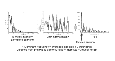
[0028] For this algorithm, we use the principal that the gap size between the
consecutive patterns is determined as the distance between the transducer and
the dome
surfaces. By combining the gap size information with the transducer position
information
obtained from the Phi/Theta tables and skew angle/offset values, the location
of a sample
point that corresponds to the dome surface at each scan line, i.e., dome
geometry, can be
estimated. This process can be repeated using various skew angle/offset values
until we
obtain dome geometry that is closest to a perfect sphere. The skew
angle/offset value
information that corresponds to the best matching dome geometry is used as
calibration
parameters. As this algorithm relies on the distance between the transducer
and the dome
surface, intentional offset value between the transducer rotation and dome
center is desirable
to avoid undetectable motion errors due to the symmetry (Fig. 9). For example,
if transducer
rotation and dome center is the same, erroneous offset value in transducer Phi
motion cannot
be detected because transducer-dome distance is constant regardless of Phi
offset. Number of
planes and scan lines acquired could vary depending on the system. Followings
are the
detailed procedures for the proposed calibration algorithm (Fig. 8).
Step 1. Collect ultrasound data
[0029] As the first step of the calibration, ultrasound data is collected in
the air (i.e.,
without water taffl( and ultrasound target).
Step 2. Estimate gap size between air scan patterns
[0030] Gap size between the consecutive air scan reverberation patterns is
proportional to the transducer-dome distance (Fig. 10). Gap size can be
detected in B-mode
image by detecting air scan pattern. As a more advance method, dominant
spatial frequency
of the reverberation pattern can be detected to estimate gap size more
accurately (Fig. 11).
Step 3. Estimate dome geometry
[0031] If we assume that there is no error in transducer motion, 3D location
of the
transducer for each scan line can be derived from the Phi and the rotation
information. Then,
the location of a sample point that corresponds to the dome surface in each
scanline can be
-10-
CA 02948279 2016-08-02
WO 2015/120179 PCT/US2015/014664
estimated by using the gap size information at the scanline. An example of
this dome
geometry estimation results is shown in Fig. 12. In the same way, dome
geometry can be
estimated when transducer motion error exists. This dome geometry estimation
process is
repeated many times by varying transducer skew angles and offset values.
Step 4. Select the best matching skew angle/offset values
[0032] A best fitting sphere can be determined for each of the estimated dome
geometries (Fig. 13). Based on the assumption that dome shape is perfectly
spherical, the
dome geometry that resulted in the smallest deviation (e.g., mean squared
error) from its
fitting sphere is selected as the best matching case. The corresponding skew
angle and offset
values to the best matching case can be considered as the calibration result.
Algorithm III
[0033] One of the main purposes of the calibration process is to estimate
appropriate
phi offset and firing delay values to make the orientation of the B-mode image
correct. This
can be done by comparing a B-mode image with another one at the same scan
plane after
180 theta rotation as shown in Fig. 14. To increase the sensitivity of the
off-angle detection,
an intentional offset between phi rotation and dome center (Fig. 9) would be
desirable.
[0034] Some small errors in the phi motion itself, e.g., gear backlash, cannot
be
detected using the above procedure. To estimate the phi offset and firing
delay values to
compensate the gear backlash, an additional step is optionally advantageous.
This process is
basically the same with Fig. 14, except for comparing images acquired with
different phi
motions without moving the theta motor. The detailed step-by-step procedure
for the simple
air scan calibration is as follows (Fig. 15):
a. Phi offset calibration
[0035] Step 1: Collect air scan data (B-mode) in a scan plane.
[0036] Step 2: Collect another air scan data in the same plane with the same
phi
motion after 180 theta rotation.
-11-
CA 02948279 2016-08-02
WO 2015/120179 PCT/US2015/014664
[0037] Step 3: Estimate the phi angle difference between the two B-mode
images.
Cross correlation of the two images can be used as a simple estimation method.
[0038] Step 4: If the estimated phi angle difference is small enough, use the
current
phi offset/firing delay value for calibration. Otherwise, adjust the phi
offset/firing delay
according to the difference, then repeat steps 1-4.
b. Backlash calibration
[0039] Step 1: Collect air scan data (B-mode) in a scan plane with forward phi
motion.
[0040] Step 2: Collect another air scan data in the same plane with backward
phi
motion.
[0041] Step 3: Estimate the phi angle difference between the two B-mode
images.
Cross correlation of the two images can be used as a simple estimation method.
[0042] Step 4: If the estimated phi angle difference is small enough, use the
current
phi offset/firing delay value for backlash calibration. Otherwise, adjust the
phi offset/firing
delay according to the difference, then repeat steps 1-4.
Method for real-time C-mode using position sensor
[0043] Three-dimensional ultrasound has a limit in the achievable volume rate
because of the delay times for sound wave travel and/or mechanical transducer
motion. With
the current ultrasound bladder scanners that have the same limitation, severe
motion blur
occurs if a probe does not stay still during scanning. For this reason, most
bladder scanners
do not provide real time imaging modality for probe aiming. Some recently
introduced
bladder scanners support real time B-mode by restricting the transducer motion
within one
plane for higher frame rates. This B-mode is useful, but still inconvenient
because B-mode
imaging plane is perpendicular to the plane of probe motion. Instead, as an
ideal aiming
guide, this invention introduces real-time C-mode bladder imaging and two
methods to
implement it. The first method uses probe translation/rotation information
derived from
position sensors to compensate for probe motion. The other method uses a new
user interface
to make the user interpret the motion-blurred data more efficiently.
-12-
CA 02948279 2016-08-02
WO 2015/120179 PCT/US2015/014664
[0044] Three-dimensional ultrasound has a limit in the achievable volume rate
because of the delay times for sound wave travel. With mechanical 3D probes,
the limitation
becomes stricter due to the additional delay for transducer motion, which is
the case of the
current ultrasound bladder scanners that typically require 2-3 seconds for
volume scanning.
With this low volume rate, probe motion by an operator could produce severe
motion blur in
the ultrasound data. On this account, the majority of the current ultrasound
bladder scanners
have not provided real time imaging modality. This caused inconvenience in
aiming a probe.
Recently, some new bladder scanners provide real time B-mode by restricting
the transducer
motion within one plane for higher frame rates, but the B-mode is still far
from the ideal
imaging modality for probe aiming because it provides only partial information
on the
bladder location and shape. In addition, B-mode is not easy to use because
imaging plane is
perpendicular to the plane of probe motion. In contrast, C-mode is very
intuitive because its
imaging plane is parallel to the probe motion. However, C-mode is difficult to
implement in
real time with low volume rate probes because it requires full 3D volume data.
[0045] An embodiment of the present invention introduces new methods for real
time
C-mode bladder imaging as ideal probe aiming guides. The first method uses
sensors, e.g.,
inertial measurement unit, magnetic and optical sensors, etc., that can be
used for
measuring/deriving probe location and orientation information in combination
with
ultrasound data. Summarized real time C-mode process with a position sensor is
as follows:
a. Acquire ultrasound and probe location sensor data synchronized with each
other in
real time.
b. Detect sample points that correspond to the bladder wall at each scan line
from the
ultrasound data acquired for the most recent 1-5 seconds.
c. Derive relative location information of the bladder wall sample points from
the
probe.
d. Derive absolute probe location and orientation information from the
position
sensor data at each scan line; derive the probe translation and rotation
information at
each scan line.
-13-
CA 02948279 2016-08-02
WO 2015/120179 PCT/US2015/014664
e. Convert the relative bladder wall sample locations to the absolute
locations by
compensating for the probe translation and rotation at each scan line.
f. Optimize the probe motion compensation based on the absolute bladder wall
locations; fine tune the probe motion parameters to make the congregated
sample
points a bladder-like shape.
g. Calculate projections of the motion-compensated bladder wall sample points
to the
plane perpendicular to the sightline of the probe; estimate the bladder shape
from the
viewpoint of the probe. Generate C-mode image from the projection.
h. Check the location of the bladder center in the C-mode. If it has been well
centered for a certain period of time, turn on the on-target indicator (or
automatically
start volume measurement).
i. (Optional) Detect probe motion in the latest volume data. If there is no
large
motion, calculate and display bladder volume instantaneously using the data in
the
buffer when requested for responsiveness.
[0046] The second method does not use position sensor, but uses a new user
interface
(UI) utilizing the fact that human eye can perceive accurate object position
by estimating its
motion if the blurred object looks like a comet with tail. This new UI can be
implemented by
making the bladder walls that correspond to the more recent data have a deeper
color or lower
transparency. Although this new C-mode still has motion blurs, it would
provide all the
information necessary for probe aiming. Summarized real time C-mode process
with the new
UI is as follows:
a. Acquire ultrasound and probe location sensor data synchronized with each
other in
real time.
b. Detect sample points that correspond to the bladder wall at each scan line
from the
ultrasound data acquired for the most recent 1-5 seconds.
c. Derive relative location information of the bladder wall sample points from
the
probe.
-14-
CA 02948279 2016-08-02
WO 2015/120179 PCT/US2015/014664
d. Generate C-mode images from the bladder wall information. Make the more
recent information less transparent or have deeper color to make the motion-
blurred
C-mode look like a comet with tail.
e. Check the location of the bladder center in the C-mode. If it has been well
centered for a certain period of time, turn on the on-target indicator (or
automatically
start volume measurement).
[0047] a. Difficult probe aiming with ultrasound bladder scanners: No
ultrasound
bladder volume scanner has ever provided a real time C-mode that shows bladder
shapes
from the viewpoint of the probe head. Different from the B-mode that requires
training to be
accustomed to, C-mode is very intuitive because image plane is parallel with
the plane of
probe motion, which is similar to seeing a bladder through a virtual window on
the skin
surface.
[0048] b. Probe motion during bladder volume measurement (position sensor
method): Start button on the probe causes small probe motion that could
increase inaccuracy
in bladder volume measurement. In real time C-mode, the device can
automatically start
volume measurement when the bladder is right on the target without any button
push by an
operator. In addition, accelerometer monitors probe motion during the volume
data
acquisition. If large motion is detected, the device can flash a warning sign
or automatically
repeat data collection until the probe stands still.
[0049] c. Probe-console alignment problem (position sensor method, optional):
By
tracking the absolute orientation of the probe, orientation of the C-mode
display can be
automatically aligned with the probe orientation. Thus, operator can perform
examination
from both sides of the patient regardless of the console location without any
confusion or
manual display adjustment.
-15-
CA 02948279 2016-08-02
WO 2015/120179 PCT/US2015/014664
Method 1: Real-time C-mode using position sensor
Detailed real-time C-mode process with position sensor:
Step 1: Data acquisition
[0050] Ultrasound and probe position/rotation sensor data synchronized with
each
other in real time are acquired (Fig. 17.1). Inertial measurement unit (IMU)
that consists of
three-axis accelerometers, magnetometer sand gyroscopes is an example of the
position
sensor. An optical sensor with markers, a magnetic sensor with a transmitter,
or any
combinations of IMU, optical and magnetic sensors can be used for position
tracking. Data
buffer should be large enough to store data acquired for the most recent 1-5
seconds.
Step 2: Bladder wall detection
[0051] Sample points that correspond to the bladder wall are detected at each
scan
line from the ultrasound data acquired for the most recent 1-5 seconds.
BVI9400 algorithm or
any new algorithm can be used for this process. From the detection results,
relative 3D
location information of bladder wall sample points is derived. At this stage,
bladder wall
location is relative from the probe at each scan line, i.e., probe motion is
not compensated and
may have motion blur (Fig. 17.2).
Step 3: Probe location/orientation estimation
[0052] From the position sensor data, probe location and orientation
information is
estimated at each scan line.
Step 4: Probe motion compensation
[0053] Relative bladder wall locations are converted into the absolute
locations by
compensating for the probe translation and rotation at each scan line (Fig.
between 17.2 and
17.3).
Step 5: Fine tuning of the motion compensation (optional)
[0054] Output of the position/rotation sensor is not stable sometimes,
especially with
an IMU. For example, a small offset in accelerometer output could cause
several inches of
error in the estimate translation value. Thus, an additional step to stabilize
the motion
compensation result would be desired. One of the possible approaches is
estimating the
-16-
CA 02948279 2016-08-02
WO 2015/120179 PCT/US2015/014664
sensor offset values to make the resulting bladder wall sample points to form
a sphere-like
shape (or, alternatively, as close as possible each other). If there is
another sensor that can
measure the location of the probe, e.g., optical sensors, it can be used to
compensate for the
accelerometer/gyroscope errors.
Step 6: C-mode image generation
[0055] To make a C-mode, or to estimate the bladder shape from the viewpoint
of the
probe, projections of the motion-compensated bladder wall sample points to the
plane
perpendicular to the sightline of the probe are calculated (Fig. 17.3). By
detecting the outline
of the projected sample points, C-mode image can be generated (Fig. 17.4).
Scan lines that
pass through the pubic bone can be also detected in this procedure using the
9x or similar
algorithm.
Step 7: On-target indicator
[0056] From the generated C-mode, it can be determined whether the bladder is
centered enough or not. If it is well centered for a certain period of time,
e.g., 2 seconds, an
on-target indicator can be turned on (Fig. 17.5). This on-target indicator can
be also used for
triggering bladder volume calculating process.
Step 8: Probe motion detection (optional)
[0057] Optionally, accelerometer data can be used for detecting probe motion
for the
most recent 1-2 seconds. Based on this information, integrity of the most
recent volume data
can be checked, which means bladder volume calculation can be done using the
data already
in the data buffer. This enables a responsive bladder volume display.
Method 2: Real-time C-mode with an improved user interface
[0058] If position sensors are not used, motion blur in C-mode is unavoidable.
Even
in this case, however, a new user interface (UI) can make the blurred C-mode
less annoying
and more usable. For example, in a radar display that typically has a very low
frame rate
(Figure 18); more recent data are displayed in a brighter color. So, user can
track object
positions more accurately by putting more weight on the brighter information.
-17-
CA 02948279 2016-08-02
WO 2015/120179 PCT/US2015/014664
[0059] Another example is hockey puck enhancement technology that looks like a
comet with a tail (Figure 19). Although the comet tail is a kind of motion
blur, it does not
confuse the object position, but rather helps in estimating the next position
of the puck. This
is because human visual system naturally perceives the direction and speed of
the motion
from the comet tail-like object shape.
[0060] For real-time C-mode, a similar approach can be applied. The new UI is
basically similar with the 9x C-mode, i.e., plotting the location of the
detected bladder wall
locations on the x-y plane. However, the new UI displays the more recent data
less
transparently (or in a darker color, etc.) to make the bladder trajectory like
a comet with a tail.
In this mode, human eye focuses on the "comet head" and naturally tracks its
motion based
on the shape of the comet tail (Figure 20). Although this new C-mode looks
different from
the traditional C-mode, it provides all the necessary information for aiming
in an intuitive
way.
Detailed real-time C-mode process with a new UI:
Step 1: Data acquisition
[0061] Ultrasound and probe position/rotation sensor data synchronized with
each
other in real time are acquired (Figure 22.1). Data buffer should be large
enough to store data
acquired for the most recent 1-5 seconds.
Step 2: Bladder wall detection
[0062] Sample points that correspond to the bladder wall are detected at each
scan
line from the ultrasound data acquired for the most recent 1-5 seconds.
BVI9400 algorithm or
any new algorithm can be used for this process. Through this process, all the
scan lines are
classified into two groups; 1) scan lines that pass through the bladder and 2)
the others
(Figure 22.2). Scan lines that pass through the pubic bone can be also
detected in this
procedure using the 9x or similar algorithm.
Step 3: C-mode image generation
[0063] In the comet tail mode, scan lines that pass through the bladder are
plotted as
dots in the x-y plane (Figure 22.3). Dots from the more recently acquired data
are less
-18-
CA 02948279 2016-08-02
WO 2015/120179 PCT/US2015/014664
transparent to make the C-mode have a comet tail-like shape. Pubic bone
information can be
overlaid on the comet tail image.
Step 4: On-target indicator (Optional)
[0064] From the generated C-mode, it can be determined whether the bladder is
centered enough or not. If it is well centered for a certain period of time,
e.g., 2 seconds, an
on-target indicator can be turned on (Figure 22.4). The on-target indicator
can be also used
for triggering bladder volume calculating process.
Calibration method using plate target
[0065] Typical ultrasound bladder scanners use a single-element transducer
that
moves mechanically in a dome-shaped probe head. For this type of devices,
precise
calibration of transducer motions is optionally advantageous for accurate
volume
measurement. An ultrasound target with a known shape, e.g., spiral or string,
in water tank is
typically used for this purpose. One of the problems of the typical
calibration method is that
there could be a parallax issue. An embodiment of the present invention solves
this problem
and provides other benefits including smaller calibration fixture and better
reliability by
utilizing intensity information of the beam reflected from a plate target,
instead of using the
spiral/string target location/shape information.
[0066] An embodiment of the present invention provides a method for abnormal
transducer motion detection of a mechanical three-dimensional one-channel
ultrasound probe
used for bladder volume measurement. This type of probe has a moving
transducer in the
dome-shaped probe head filled with coupling/lubrication fluid such as mineral
oil. In this
probe, transducer motion is characterized by rotations about two axes, phi and
theta, as
shown in Fig. 23. As transducer motions can be inaccurate for several reasons,
e.g., skew
angles of phi/theta rotation axes, gear backlash, wear and tear, etc., it
needs to be precisely
measured and calibrated for accurate volume measurement, which has been
commonly done
using an external calibration target, e.g., spiral or string, immersed in a
water tank.
[0067] One of the problems of the typical calibration method is that there
could be a
parallax issue. Fig. 24(a) shows a probe with a crooked transducer. In this
case, ultrasound
-19-
CA 02948279 2016-08-02
WO 2015/120179 PCT/US2015/014664
beam does not go straight down unlike the intention of the device. To
compensate for this
error, typical calibration methods try to match the location of a calibration
target on the two
ultrasound images obtained before and after 180-deg rotation about the theta
axis. If the
transducer is not crooked as in Fig. 24(b), the device can accurately find the
phi angle that
makes the beam go straight down. However, with a skewed transducer, actual
calibration
result is still not accurate due to the parallax error as shown in Fig. 24(c).
With this parallax,
desired calibration result in Fig. 24(d) is difficult to achieve. The parallax
problem can be
relieved if a calibration target is far from the transducer, but then poor
lateral resolution of the
ultrasound in the far field would affect the calibration accuracy, and
calibration fixture could
become too bulky.
[0068] As a new calibration method that does not have parallax problem, an
embodiment uses a reflective plate target, e.g., metal surface, instead of a
typical string or
spiral target. By using the intensity information of the reflected beam from
the plate target,
instead of the target location information that is typically used, an
embodiment provides a
more accurate way of doing calibration without any parallax problem, as well
as other
benefits like smaller fixture and better reliability.
[0069] a. Parallax in calibration: An embodiment of the present invention uses
a plate
target that does not have parallax problem.
[0070] b. Bulky calibration fixture: Small fixture can be used for the plate
calibration
because a plate target can be very close to the probe. So, an embodiment
enables use of
several different types of calibration fixtures, e.g. a calibration cup or a
small rubber block
with a plate target in it, instead of traditional bulky water bath.
[0071] c. Small tolerance in probe - target alignment: A plate target doesn't
have to
be well aligned with the probe for calibration using the proposed method. This
means plate
calibration could be more reliable than traditional methods as some
misalignment or an error
in calibration fixture doesn't affect the calibration result.
-20-
CA 02948279 2016-08-02
WO 2015/120179 PCT/US2015/014664
A. Phi offset calibration
[0072] One of the main purposes of the calibration process is to estimate
appropriate
phi offset and firing delay values to make the orientation of the B-mode image
correct. This
can be done by comparing a beam peak intensity profiles with another one in
the same scan
plane after 180 theta rotation as shown in Fig. 25. For example, for a system
that doesn't
have any phi error, the maximum peak intensity angles in the first and second
profiles, cOpeakl
and mpeak2, respectively, would have the relationship, cOpeakl ¨ 180 -
cOpeak2. For a system
where actual phi angle of the beam is skewed by cOoffset from the correct
direction, two angles
of maximum peak would meet the following equation: 2m
offset ¨ cOpeakl ¨ (180 - cOpeak2). By
utilizing this relationship between peak intensity angles, phi offset can be
calibrated through
the following procedure:
Step 1: Collect ultrasound data (RF, IQ or B-mode) in a scan plane.
Step 2: Calculate the maximum ultrasound intensity profile from the data.
Step 3: Collect another ultrasound data in the same plane with the same phi
motion
after 180 theta rotation.
Step 4: Calculate the second maximum intensity profile from the second data.
Step 5: Estimate the phi angle difference between the two profiles. Peak
detection, or
cross correlation method for better precision, can be used after flipping the
send
profile.
Step 6: If the estimated phi angle difference is small enough, use the current
phi
offset/firing delay value for calibration. Otherwise, adjust the phi
offset/firing delay
according to the difference. If necessary, then repeat steps 1-6.
[0073] One of the virtues of the plate calibration is that the relationship
between peak
angles is valid regardless of the angle of the surface target, thus target can
be skewed from
the probe as in Fig. 24. So, plate calibration fixture including probe holder
can be made more
easily with less precision compared to typical ones.
-21-
CA 02948279 2016-08-02
WO 2015/120179 PCT/US2015/014664
B. Gear backlash calibration
[0074] If a probe does a two-way scanning by rotating the phi motor in both
directions, there could be small amount of misalignment between the forward
and backward
scanning mainly due to the gear backlash. This backlash can be estimated with
a similar
method used for phi offset estimation as shown in Fig. 26. In this case, two
peak phi angles,
copeakforward and cOpeak backward would meet the following equation: 2c0
backlash ¨ cOpeak_forward --
peak backward. Detailed calibration procedure is as follows:
Step 1: Collect ultrasound data (RF, IQ or B-mode) in a scan plane with
forward phi
motion.
Step 2: Calculate the maximum ultrasound intensity profile from the data.
Step 3: Collect another ultrasound data in the same plane with backward phi
motion.
Step 4: Calculate the second maximum intensity profile from the second data.
Step 5: Estimate the phi angle difference between the two profiles. Cross
correlation
of the two profiles can be used as an estimation method.
Step 6: If the estimated phi angle difference is small enough, use the current
backlash
value for calibration. Otherwise, adjust the backlash according to the
difference. If
necessary, then repeat steps 1-6.
C. Estimation of skew angle perpendicular to the scan plane
[0075] While geometrical errors in the direction of phi motion (Type I error)
can be
compensated by adjusting phi offset / backlash, there is another type of error
(Type II error)
that is perpendicular to the scan plane. In reality, a geometrical error is
likely to be a
composition of these two different types of errors. The type II error is
difficult to compensate
physically by controlling the motor motion or firing delay, but information on
the type II
error can be used to detect a faulty probe or to compensate for bladder volume
in software.
With a plate target, the type II error can be estimated according to the
following procedure:
Step 1: Collect ultrasound cone data (RF, IQ or B-mode) on a plate target with
forward phi
motion.
-22-
CA 02948279 2016-08-02
WO 2015/120179 PCT/US2015/014664
[0076] Among all the scanlines that covers a cone, at least one scanline is
perpendicular to the plate target. (This is because the plate can be thought
as a tangent plane
of the cone. The scanline that crosses the point of contact is perpendicular
to the plate target.
The number of scanlines could be increased by interpolation to get a better
angular
precision.) However, there could be an exceptional case where a hole, like an
eye of
hurricane, that none of the scanlines passes through exists due to the type II
error. In this
case, none of the scanlines could be perpendicular to the plate target. To
avoid this situation,
the plate target needs be tilted from the surface seen straight from the
probe. For example, if
expected maximum type II error is 5 degrees, the plate target should be tilted
by at least 5
degrees.
Step 2: Find a scan plane perpendicular to the plate. Then, find the
relationship between the
incidence angle and peak intensity.
[0077] The scanline that is perpendicular to the plate target can be found by
finding
the scanline that has the largest peak intensity. The scan plane that contains
the scanline
should be perpendicular to the plate. In this scan plane, thanks to the
perpendicularity, we
can derive the relationship between the incidence angle (phi angle) and the
peak intensity
from the plate target.
Step 3: Collect ultrasound data in a scan plane, then collect another one in
the same plane
after 180-deg theta rotation. In this case, make sure that plate target is
tilted at least as much
as expected maximum type II error from the surface seen straight from the
probe in the
direction perpendicular to the scan plane.
[0078] Instead of this separate data collection step, we could make the first
scan plane
overlap the last scan plane with 180-deg theta angle difference in Step 1 for
convenience. Or,
the data used in the phi offset calibration can be used again if probe and
plate target have not
moved.
Step 4: Calculate incidence angles of the maximum intensity beam for two plane
data
acquired in Step 3 using the incidence angle ¨ peak intensity relationship
derived in Step 2.
Calculate type II error by dividing the difference between the incidence
angles by two.
-23-
CA 02948279 2016-08-02
WO 2015/120179 PCT/US2015/014664
[0079] If there's no type II error, two planes should overlap perfectly, then
two
incidence angles should be the same after phi offset calibration. In the
presence of a type II
error, two planes have an angular gap that causes differences between the two
incidence
angles. As we tilted the target more than the maximum type II error in Step 3,
two scan
planes are tilted into the same direction about the scan plane. Thus, type II
error can be
calculated simply subtracting one incidence angle from the other followed by a
division by
two.
D. Simulation Example
[0080] To show an example of the proposed method, a 13-plane peak intensity
profile
data were simulated using Matlab. The first plane overlaps the 13th plane with
180-deg theta
angle difference. In this simulation, we assumed that plate target is
intentionally skewed by 5
degrees towards southeast, and the probe has 3 degrees of phi offset and one
degree of type II
error.
[0081] Figure 27 shows two intensity profiles in plane #1 and #13 (flipped for
comparison). The 6-deg difference between the two peaks caused by the 3-deg
phi offset is
clearly observable in the figure. For accurate estimation of the phi offset,
intensity profiles
can be interpolated to improve the angular resolution. Note that peak
intensities are different
each other. This implies that type II error is not zero in this case. Backlash
was not tested in
this simulation, but it can be estimated using the same principal to the phi
offset estimation.
[0082] Figure 28 shows peak intensities of reflected beam at 1040 scanline
locations
(80 scanlines x 13 planes). In this figure, the scanline with the maximum
intensity is on the
10th plane. So, we can assume that the 10th plane is perpendicular to the
plate target. (For
better accuracy, we can actually interpolate the 10th plane with the 11th
plane, to find the
plane exactly perpendicular to the plate.) From the peak intensity profile in
this plane, we
can derive the relationship between the beam incidence angle and peak
intensity as shown in
Fig. 29. This profile was smoothed with interpolation for better accuracy.
[0083] In Fig. 27, peak intensities in planes 1 and 13 were 2.32 and 3.16,
respectively.
These values correspond to the incidence angles of 6.05 and 4.03 degrees in
Fig. 6,
-24-
CA 02948279 2016-08-02
WO 2015/120179 PCT/US2015/014664
respectively. By dividing the difference between the angles by two, we can get
the type II
error; i.e, (6.05 ¨ 4.03) / 2= 1.01 degree. This value well matches to the
simulation
parameter, type II error of one degree.
Ball-and-Socket Hemispherical Scan Mechanism
[0084] An embodiment includes a reliable hemispheric scan mechanism for use
in,
for example, the Bladderscan and Aortascan product line.
1. Description of an embodiment:
a. Purpose
[0085] This mechanism was invented for the purpose of supporting and pointing
directive sending and receiving devices in various desired directions, thus
mapping out a two
dimensional region of interest, within a hemispherical region.
b. Drawings
[0086] An embodiment of the invention is illustrated in Figures 30-38 below.
For
clarity, motors and small pinion gears (figure 34) are not shown.
c. Description of the Parts
[0087] An embodiment of the invention consists of three principal components,
a
spherical "eyeball" transducer holder with integral "latitude" gear teeth
(figure 35), a gimbal
ring with integral "longitude" gear teeth (figure 36) and a support frame with
a longitude ring
groove (figure 37).
[0088] The longitude motor (for clarity, not shown) is attached to the support
frame
and the latitude motor (for clarity, not shown) is attached to the gimbal
ring. The inside
surface of the gimbal ring is formed with a spherical contour. The transducer
holder sphere is
held in place within the gimbal ring component. It has an external surface
with a spherical
contour matching that of the gimbal ring.
[0089] When the transducer holder and the gimbal ring are driven by two
electric
motors, through pinion gears (for clarity, not shown), a directive transducer
may be pointed
in any desired direction within a hemispherical region.
-25-
CA 02948279 2016-08-02
WO 2015/120179 PCT/US2015/014664
d. Use
[0090] In operation, two motors independently and simultaneously move and
position
the inner transducer holder and the outer gimbal ring so as to point a
transducer toward any
latitude and longitude coordinate, within a hemispherical region, which the
application
requires.
e. Features
[0091] Hemispherical scan mechanisms typically use two motors and two
associated
gear mechanism in order to point a transducer device in various directions
within a
hemispherical region. Two common prior art mechanisms are the alt-azimuth
mount often
used to support telescopes (figure 30) and the gimbal mount (figure 31) often
used to support
compasses and gyroscopes.
[0092] The alt-azimuth and gimbal mount have deficiencies which an embodiment
of
the invention circumvents. In particular, the prior art devices are large,
delicate and complex.
This makes them comparatively heavy, expensive, less reliable and less
tolerant of damage
through misuse.
[0093] An embodiment of the invention circumvents these deficiencies by
combining
two novel ideas:
1. It uses a ball-and-socket support mechanism (figure 32) instead of the more
common trunion bearings (figure 33)
2. It integrates the two required drive gears into their associated gimbal
ring and
transducer holder parts.
Features
[0094] As a result of the novel features, my invention has the following
advantages
over hemispherical scanning mechanisms found in the prior art.
= it requires relatively few components
= the few components are relatively easy to fabricate
= the few components are individually rugged and thus damage resistant
= the few components are inexpensive to fabricate
-26-
CA 02948279 2016-08-02
WO 2015/120179 PCT/US2015/014664
= the few components do not extend very far vertically or laterally. This
makes
the mechanism, as a whole, compact and rugged.
= the few components do not extend very far vertically or laterally. This
allows
space for a shock mount spring feature
= the few components do not extend very far vertically or laterally. This
allows
space for more electronic circuitry in a probe hand piece
= the few components do not extend very far vertically or laterally. This
allows
space for better electrical shielding of electronic circuitry in a probe hand
piece
= many spatial directions can be sampled within a hemisphere, without the
transducer electrical connection cable being repeatedly wrapped and
unwrapped around one or more motor shaft axis.
= The electrical connection to the transducer device can be made relatively
short, thus reducing signal interference opportunities
f. Testing
[0095] A computer CAD model of an embodiment has been created that allows
examination of the relative motion of the various components, as they would
move in actual
use.
Spherical Spiral Path Scan Mechanism
2. Description of an embodiment:
a. Purpose
[0096] An embodiment of the invention achieves the purpose of supporting and
pointing directive sending and receiving devices in various desired
directions, thus mapping
out a two dimensional region of interest, within a hemispherical region.
b. Drawings
[0097] An embodiment of the invention is illustrated in Figures 41-49 below.
-27-
CA 02948279 2016-08-02
WO 2015/120179 PCT/US2015/014664
c. Description of the Parts
[0098] An embodiment of the scanning mechanism consists of five principal
components, which when driven by a motor, move a directive transducer so as to
point in
many directions within a hemispherical region.
[0099] The components are: a gimbal mount, with 1) inner ring and 2) outer
shell, to
support the transducer holder 3) a cup, with spiral grooves to guide the
transducer pointing
direction along a spiral path 4) a transducer holder, with a short pin
extending down from the
holder body that engages with a spiral groove in the grooved cup body to force
pointing of
the transducer along a defined spiral path 5) a slotted cup to move the spiral
groove guided
pin in a spiral direction 6) an optional shuttle feature (not shown in a
figure) that allows the
pin to cross spiral grooves at an acute angle without chance of changing
direction at the
groove intersections.
d. Use
[00100] In operation, motor torque is applied to a shaft that
extends downward
from the slotted cup component. This torque rotates the slotted cup. As the
slotted cup
rotates, the captured pin feature, extending downward from the bottom of the
transducer
holder, is forced to follow a spiral slot containing the captured pin. As the
captured pin
moves within the spiral slot, the transducing device is then necessarily
pointed in a direction
away from the pin and co-axial with its axis. In this manner, a spiral shaped
scan path is
traveled by any energy beam being sent and-or received by the transducer
device mounted in
or on the mechanism inner gimbal ring.
e. Novel Features
[00101] Hemispherical scan mechanisms typically use two motors and
two
associated gear mechanism in order to point a transducer device in various
directions within a
hemispherical region. An embodiment of the invention, not described in the
prior literature,
points a transducer mechanism in many directions covering a region of interest
within a
hemisphere, without using any gear mechanisms. Also, by employing a spiral
scan path, the
-28-
CA 02948279 2016-08-02
WO 2015/120179 PCT/US2015/014664
mechanism may require only one motor to scan many points, in two spatial co-
ordinates,
within a hemispherical region.
[00102] The spiral path scan plan is useful because the method
substantially
shortens the angular path that the mechanism must traverse, in order to scan a
grid of points,
in a hemispherical region. Drastically shortening the angular scan path allows
a much faster
scan rate and-or a large reduction in mechanism power consumption.
[00103] An additional novel aspect of an embodiment is the optional
incorporation of a crisscrossed spiral groove feature. The crisscrossed groove
allows the
spiral transducer beam pointing path to spiral both outward, from the center
region, and
inward, from the perimeter region, without changing direction or speed of
rotation of the
rotating elements. This allows maintenance of a high angular scan speed, while
simultaneously reducing drive power requirements.
f. Features
[00104] As a result of the novel features, an embodiment of the
invention has
the following features distinguishable over hemispherical scanning mechanisms
found in the
prior art.
= it has relatively few components
= the few components are relatively simple in form
= the few components are individually rugged and thus damage resistant
= the few components are inexpensive to fabricate
= the few components do not extend very far above the drive motor. This
makes
the mechanism, as a whole, compact and rugged.
= the few components do not extend very far above the drive motor. This
allow
space for a shock mount spring feature
= the few components do not extend very far above the drive motor. This
allow
space for more electronic circuitry in a probe hand piece
-29-
CA 02948279 2016-08-02
WO 2015/120179 PCT/US2015/014664
= the few components do not extend very far above the drive motor. This
allow
space for better electrical shielding of electronic circuitry in a probe hand
piece
= use of only one motor reduces cost
= use of only one motor reduces size
= use of only one motor reduces weight
= for a given number of spatial sample points, the total spiral scan path
length is
short. This allows sampling of many spatial directions in a very short time.
= fast scanning times reduce motion artifact
= fast scanning times allow smooth real-time scan imaging
= the spiral scan path does not impose frequent acceleration and
deceleration of
the transducer device. This reduces motor power.
= the spiral scan path does not impose frequent acceleration and
deceleration of
the transducer device. This reduces mechanism vibration
= the spiral scan path does not impose frequent acceleration and
deceleration of
the transducer device. This reduces mechanism wear and tear, thus enhancing
reliability.
= many spatial directions can be sampled within a hemisphere without the
transducer electrical connection cable being repeatedly wrapped and
unwrapped around one or more motor shaft axis.
= The electrical connection to the transducer device can be made relatively
short, thus reducing signal interference opportunities
= If axial symmetric transformer primary and secondary windings are used to
couple transducer signals into and-or out of the rotating sub-assembly, a
crisscross spiral groove feature may be used. The crisscross spiral groove
feature provides the opportunity to hugely increase scanning speed while, at
the same time, reducing drive power requirements.
-30-
CA 02948279 2016-08-02
WO 2015/120179 PCT/US2015/014664
g. Testing
[00105] A computer CAD model has been created that allows
examination of
the relative motion of the various components, as they would move in actual
use.
[00106] Figure 41 ¨ Transducer holder with indexing groove following
pin
[00107] Figure 42 -Inner Gimbal Ring
[00108] Figure 43 ¨ Outer Gimbal Yoke
[00109] Figure 44 ¨ Groove Cup
[00110] Figure 45 - Slot Cup
[00111] Figure 46 ¨ Mechanism Cross Section
[00112] Figure 47 ¨ Mechanism Isometric View
[00113] Figure 48 ¨ Top view of a crisscross spiral scan path
[00114] An oblong groove follower won't "de-rail" or follow the
wrong path at
a groove crossing point. Any hysteretic "backlash" can be calibrated out
[00115] Figure 49 ¨ Oblong groove following pin maintains travel
direction
Wireless Disposable Video Laryngoscope with interface to Generic Computing
Display
[00116] An embodiment of the invention is a single patient use
disposable
video laryngoscope blade that communicates wirelessly and displays images on a
generic
tablet computer (or other device) executing proprietary application software.
This
architecture could disrupt the laryngoscope product topology by reducing both
the system
capital cost and complexity of use and maintenance. In this system
architecture, the display
component would not be a proprietary, dedicated device, but instead leverage
existing generic
mobile computing devices in the hospital environment. There is recent enabling
regulatory
precedent for a tablet to be treated as office equipment, and only the
application software to
be registered as a medical device.
[00117] Optionally advantageous attributes of an embodiment that
enable this
technology include:
1. Wireless communications protocol that can stream video in real time with
limited
delay or interruptions in signal transmission to facilitate intubation
procedure.
-31-
CA 02948279 2016-08-02
WO 2015/120179 PCT/US2015/014664
2. Lower power wireless communication protocol allows device to operate on a
single primary battery, without charging or wired power connection.
3. Prolific communications protocol that enables medical device to display on
commercially available computing displays without the need for a dedicated
monitor
component of the medical device.
4. Reliable wireless communications protocol and band between medical device
and
the computer.
5. Rapid pairing method to link the medical device and that display, such as
scanning
a bar / QR code on the medical device package, RFID tag, or NFC tag in order
to
exclusively associate the medical device with the display.
6. Low cost chip on flex circuitry to enable low cost of goods sold device
with a
target cogs of $12-15.
7. A latching power button to ensure the medical device is not allowed for
reuse.
[00118] This architecture could disrupt the laryngoscope product
topology by
reducing both the system capital cost and complexity of use and maintenance.
= Reduces cost
o No dedicated monitor, no rechargeable battery, no charger, no stand,
no cables
= Reduces maintenance
o No charging or management of state of charge, no sterilization
between uses, reduced ER equipment footprint
[00119] From the foregoing, it will be appreciated that specific
embodiments of
the personalized feed system have been described herein for purposes of
illustration, but that
various modifications may be made without deviating from the spirit and scope
of the
invention. Accordingly, the invention is not limited except as by the appended
claims.
-32-