Note: Descriptions are shown in the official language in which they were submitted.
REPLACEMENT MITRAL VALVE WITH ANNULAR FLAP
[0001]
BACKGROUND
Field
[0002] Certain embodiments disclosed herein relate generally to
prostheses for
implantation within a lumen or body cavity. in particular, certain embodiments
relate to
expandable prostheses such as replacement heart valves, such as for the mitral
valve, that are
configured to atraumatically grasp intralumenal tissue.
Background
[0003] Human heart valves, which include the aortic, pulmonary,
mitral and
tricuspid valves, function essentially as one-way valves operating in
synchronization with the
pumping heart. The valves allow blood to flow downstream, but block blood from
flowing
upstream. Diseased heart valves exhibit impairments such as narrowing of the
valve or
regurgitation, which inhibit the valves' ability to control blood flow. Such
impairments
reduce the heart's blood-pumping efficiency and can be a debilitating and life
threatening
condition. For example, valve insufficiency can lead to conditions such as
heart hypertrophy
and dilation of the ventricle. Thus, extensive efforts have been made to
develop methods and
apparatuses to repair or replace impaired heart valves.
[0004] Prostheses exist to correct problems associated with
impaired heart valves.
For example, mechanical and tissue-based heart valve prostheses can be used to
replace
impaired native heart valves. More recently, substantial effort has been
dedicated to
developing replacement heart valves, particularly tissue-based replacement
heart valves that
can be delivered with less trauma to the patient than through open heart
surgery.
Replacement valves are being designed to be delivered through minimally
invasive
procedures and even percutaneous procedures. Such replacement valves often
include a
-1-
Date Recue/Date Received 2021-10-14
CA 02948379 2016-11-07
WO 2015/179423 PCT/US2015/031612
tissue-based valve body that is connected to an expandable frame that is then
delivered to the
native valve's annulus.
[0005] These replacement valves are often intended to at least partially
block
blood flow. however, a problem occurs when blood flows around the valve on the
outside of
the prosthesis. For example, in the context of replacement heart valves,
paravalvular leakage
has proven particularly challenging. An additional challenge relates to the
ability of such
prostheses to be secured relative to intralumenal tissue, e.g., tissue within
any body lumen or
cavity, in an atraumatic manner. Further challenges arise when trying to
controllably deliver
and secure such prostheses in a location such as at a native mitral valve.
SUMMARY OF THE INVENTION
[0006] Embodiments of the present disclosure are directed to a
prosthesis, such as
but not limited to a replacement heart valve. According to some embodiments, a
prosthesis
can be configured to be deployed within a body cavity and prevent axial flow
of fluid around
an exterior of the prosthesis. The prosthesis can include an expandable frame
configured to
radially expand and contract for deployment within the body cavity, and an
annular flap
positioned around an exterior of the expandable frame. Further embodiments are
directed to
methods of delivering a prosthesis, e.g. a replacement heart valve, and
methods of using a
prosthesis to create a barrier to fluid flow exterior to the prosthesis (e.g.,
to prevent
paravalvular leakage).
[0007] In some embodiments, the prosthesis can include an expandable
frame
having a proximal end and a distal end and a longitudinal axis extending
therethrough. In
some embodiments, the frame can be designed to radially expand and contract
for
deployment within the body cavity. The prosthesis can include an annular flap
positioned
around and secured to an exterior of the frame. The annular flap may have a
distal edge
secured at or near the distal end of the frame and extending to a proximal
edge secured at an
intermediate location on the frame between the proximal and distal ends. The
prosthesis can
include a valve body positioned within an interior of the expandable frame. In
some
embodiments, the valve body can include an inner skirt secured to the interior
of the
expandable frame and a plurality of leaflets designed to allow flow in a first
direction and
prevent flow in a second opposite direction. In some embodiments, an opening
is defined at
-2-
CA 02948379 2016-11-07
WO 2015/179423 PCT/US2015/031612
or near the distal end of the frame between the annular flap and the valve
body which can
provide access for fluid to flow into a space between the annular flap and the
valve body. In
some embodiments, the fluid flow into the space can cause the annular flap to
move from a
first configuration wherein the flap is closer to the frame to a second
configuration wherein
the flap is spaced further away from the frame to increase the surface area of
the prosthesis
and create a barrier to fluid flow exterior to the frame when deployed within
the body cavity.
BRIEF DESCRIPTION OF THE DRAWINGS
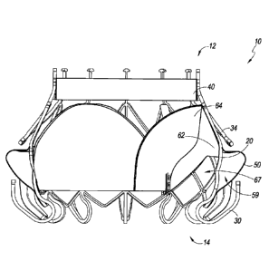
[0008] FIGURE lA is a proximal oriented, perspective view of an
embodiment of
a prosthesis illustrating a frame, a plurality of anchors, a band, a flap, and
a valve body.
[0009] FIGURE 1B is a distal oriented, perspective view of the
prosthesis of
FIGURE 1A.
[0010] FIGURE 2 is a front elevation view of the prosthesis of FIGURE 1.
[0011] FIGURE 3 is a front elevation view of another embodiment of a
prosthesis.
[0012] FIGURE 4 is a front elevation view of an embodiment of a frame.
[0013] FIGURE 5 is a perspective view of an embodiment of an annular
flap.
[0014] FIGURE 6 is a front elevation view of the annular flap of FIGURE
5.
[0015] FIGURE 7 is a perspective view of an embodiment of a valve body.
[0016] FIGURE 8 is a front perspective view of the valve body of FIGURE
7.
[0017] FIGURE 9 is a front elevation of an embodiment of a prosthesis
illustrating a frame, a plurality of anchors, a band, a flap, and a valve
body.
[0018] FIGURE 10 is a front elevation another embodiment of a
prosthesis.
[0019] FIGURE 11A is a partial cross-sectional view of the prosthesis of
FIGURE 1 with the annular flap in a first configuration.
[0020] FIGURE 11B is a partial cross-sectional view of the prosthesis of
FIGURE
11A with the annular flap in a first configuration.
[0021] FIGURE 12A is a partial cross-sectional view of the prosthesis of
FIGURE 1 with the annular flap in a first configuration, the valve body being
removed.
[0022] FIGURE 12B is a partial cross-sectional view of the prosthesis of
FIGURE
12A with the annular flap in a first configuration.
-3-
[0023] FIGURE 13A-15 illustrate schematic representations of the
prosthesis of
Figure 3 positioned within a heart, with FIGURES 13A-13C illustrating the
prosthesis in situ
with distal anchors contacting the ventricular side of a mitral valve annulus,
FIGURES 14A-
14B illustrating the prosthesis in situ with distal anchors not contacting the
ventricular side of
the mitral valve annulus, and FIGURE 15 illustrating the prosthesis in situ
with distal anchors
not extending between the chordae tendineae.
DETAILED DESCRIPTION OF THE PREFERRED EMBODIMENTS
[0024] The embodiment of Figures 1A-4 illustrates a prosthesis 10.
The
prosthesis 10 can have components, features, and/or functionality similar to
those described
in any of U.S. Publication Nos. 2014/0277390, 2014/0277422, and 2014/0277427.
With reference first to the
embodiments of Figures 1A-4, the prosthesis 10 can include a frame 20, anchors
30, 34, a
band 40, an annular flap or sail 50 and a valve body 60. The prosthesis 10 can
include a
proximal end 12 and a distal end 14 with openings defined at both ends 12, 14
such that fluid
can flow therethrough. In some embodiments, the proximal end 12 can be placed
in the left
atrium while the distal end 14 can be placed in the left ventricle such that
prosthesis 10 can
function as a replacement for a mitral valve. As will be discussed in greater
detail below and
as discussed in U.S. Publication Nos. 2014/0277390, 2014/0277422, and
2014/0277427, the
prosthesis 10 can allow blood flow in a first direction from the proximal end
12 to the distal
end 14 while preventing blood flow in a second direction from the distal end
14 to the
proximal end 12. For example, during diastole the valve body 60 may be open to
allow blood
flow from the proximal end 12 to the distal end 14, and during systole the
valve body 60 may
be closed to prevent blood flow from the distal end 14 to the proximal end 12.
[0025] With reference now to the embodiment of Figure 4, the
embodiment
illustrates an expandable frame 20 of the prosthesis 10 which can have a
proximal end 22 and
a distal end 24. In some embodiments, such as the illustrated embodiment, the
frame 20 can
include an intermediate portion 26 which has a greater diameter than the
diameter of the
frame 20 at the proximal and/or distal ends 22, 24 when the frame 20 is in an
expanded
configuration. In some embodiments, such as the illustrated embodiment, the
frame 20 can
include an intermediate portion 26 which has a greater cross-sectional area
than the cross-
-4-
Date Recue/Date Received 2021-10-14
CA 02948379 2016-11-07
WO 2015/179423 PCT/US2015/031612
sectional area of the frame 20 at the proximal and/or distal ends 22, 24 when
the frame 20 is
in an expanded configuration. The frame 20 can be designed to expand radially
and contract
for deployment within a body cavity, such as at a heart valve location such as
the mitral
valve. For example, as described in greater detail in U.S. Publication Nos.
2014/0277390,
2014/0277422, and 2014/0277427, the frame 20 can include a plurality of struts
which define
a plurality of foreshortening cells. In some embodiments, the frame 20 can be
designed to
radially and contract radially from a longitudinal axis 28 extending through
the frame 20. As
illustrated in the embodiments of Figures 1-4, the proximal end 22 can define
a proximal
opening 23 and the distal end 24 can define a distal opening 25.
[0026] With
continued reference to the embodiments of Figures 1A-4 which
illustrates the prosthesis 10, in some embodiments the prosthesis 10 can
include one or more
distal anchors 30. The distal anchors 30 can be positioned along or proximate
a distal end 24
of the frame 20 and can be connected to the frame 20. The distal anchors 30
can be designed
such that when the frame 20 is in an expanded configuration an end or tip 32
of each distal
anchor 30 is positioned radially outward from the frame 20 and extends
generally in a
proximal direction. In some embodiments, the prosthesis 10 can include one or
more
proximal anchors 34. The
proximal anchors 34 can be positioned along or proximate a
proximal end 22 of the frame 20 and can be connected to the frame 20. The
proximal
anchors 34 can be designed such that when the frame 20 is in an expanded
configuration an
end or tip 36 of each proximal anchor 34 is positioned radially outward from
the frame 20
and extends generally in a distal direction. In some embodiments, one or more
anchors 30,
34 can include cushions 38, 39 covering one or more of such anchors.
[0027] In some
embodiments, the cushion 38 can be formed from two separate
pieces of material such as an inner portion positioned within a covering such
that the
covering forms a layer surrounding the inner portion. For example, the inner
portion can be
wholly contained within the covering. In some embodiments, the inner portion
can be
formed of a foam material such that the inner portion is at least somewhat
compliant and the
covering can be formed of a biocompatible, fabric material. The embodiment of
Figures 1A,
1B and 2 illustrates cushions 38 on alternating distal anchors 30, the
cushions 38 extending
partially from an end or tip of the anchor 30 towards the connection between
the anchor 30
-5-
CA 02948379 2016-11-07
WO 2015/179423 PCT/US2015/031612
and the frame 20. Use of cushions 38 on alternating distal anchors 30 can
maintain a smaller
form factor while in the prosthesis 10 is in a contracted state for delivery.
As such, for
embodiments having twelve distal anchors 30, a total of six distal anchors 30
can have
cushions 38 and a total of six distal anchors 30 may not have a cushion 38.
The cushions 38
can advantageously increase contact area of the anchors 30 on tissue. This can
reduce trauma
between the anchor 30 and such tissue. Moreover, this can facilitate growth of
tissue in
and/or around the anchor 30 in embodiments where the cushions 38 are formed of
a material
which encourages tissue growth. The cushions 38 on anchors 30 adjacent anchors
30 without
a cushion 38 can also beneficially reduce any potential trauma caused by
adjacent anchors 30
without a cushion 38.
[0028] The embodiment of Figure 3 illustrates cushions 38, 39 on all
distal
anchors 30. As shown some of the distal anchors 30 include thicker cushions 38
than other
distal anchors 30. The cushions 38, 39 can extend the majority or entirety of
the length of the
anchor 30 from an end or tip of the anchor 30 towards the connection between
the anchor 30
and the frame 20. As shown, two distal anchors 30 include thicker cushions 38
with a distal
anchor 30 having a thinner cushion 39 positioned therebetween. As such, for
embodiments
having twelve distal anchors 30, a total of eight distal anchors 30 can have
thicker cushions
38 and a total of four distal anchors 30 can include thinner cushions 39. The
thicker cushions
38 can be formed of an inner portion and a cover layer, with the inner portion
being formed
from a compliant material, such as foam, and the covering can be formed of a
biocompatible,
fabric material. As shown, the inner portion can be positioned only around a
portion of the
anchor 30 whereas the covering can extend the majority or entirety of the
length of the anchor
30. The thinner cushion 39 can be a cover layer with a thinner inner portion
or without an
inner portion. The inner portion and/or the covering can be formed of a
material which
encourages tissue growth.
[0029] Other configurations of cushions 38, 39 can also be used. For
example, in
some embodiments, the cushions 38, 39 can be included on proximal anchors 34.
In some
embodiments, the cushions 38, 39 can be positioned on other portions of the
frame 20 such
as, but not limited to, one or more of the struts forming the frame 20. The
cushions 38, 39
can advantageously increase contact area of the prosthesis 10 on tissue. This
can reduce
-6-
trauma between the frame 20 and such tissue. Moreover, this can facilitate
growth of tissue
in and/or around the frame 20 in embodiments where the cushions 38, 39 are
formed of a
material which encourages tissue growth. In some embodiments, the covering of
cushions
38, 39 can extend from the annular flap 50 and be formed from materials
similar to those of
the annular flap 50. The covering of cushions 38, 39 can cover a majority or
the entirety of
the distal anchors 30 as shown in Figure 3. In some embodiments, the cushions
38, 39 can be
attached to the distal anchors 30 via circumferential stitching about a
longitudinal axis of the
distal anchor 30.
[0030] With reference to the embodiments of Figures 1A-3, in some
embodiments
the prosthesis 10 can include a band 40 along or proximate the proximal end 22
of the frame
20. The band 40 can include features and perform functions similar to those
described in
U.S. Patent Application No. 13/403,929 filed February 23, 2012, titled
REPLACEMENT
VALVE AND METHOD, published as U.S. Publication No. 2012/0215303.
[0031] With reference to the embodiments of Figures 1A-3, 5 and 6,
the
prosthesis 10 can include an annular flap 50 which can be positioned around
and secured to
an exterior of the frame 20. The annular flap 50 can have a distal edge 52
secured at or
proximate the distal end 24 of the frame 20 and extend to a proximal edge 54
secured at or
proximate an intermediate location, such as the intermediate portion 26, on
the frame 20
between the proximal and distal ends 22, 24. In some embodiments, the distal
edge 52 of the
annular flap 50 can be provided with a shape that generally corresponds to the
shape of the
frame 20. This can facilitate the securement of the flap 50 to the frame 20.
For example, as
illustrated in the embodiments of Figures 1A-3, 5 and 6, the distal edge 52
can include a
generally triangular pattern 56 which follows the generally triangular, zig-
zag or undulating
pattern of the struts of frame 20 along the distal end 24 of frame 20. Other
shapes and/or
patterns 56 can be used along the distal edge 52 of the annular flap 50. In
some
embodiments, the distal edge 52 of the annular flap 50 can have no pattern. In
some
embodiments the distal edge 52 does not follow the pattern of the struts of
the frame 20
and/or can have a different pattern from that of the struts.
-7-
Date Recue/Date Received 2021-10-14
CA 02948379 2016-11-07
WO 2015/179423 PCT/US2015/031612
[0032] In some embodiments, such as the embodiments of Figures 1A-3, 5
and 6,
the annular flap 50 can have a flange 58. The flange 58 can extend generally
radially outward
in a direction generally orthogonal to the longitudinal axis 28 extending
through the frame
20. In some embodiments, the flange 58 can also project proximally and/or
distally. The
flange 58 can be used to further prevent or inhibit backflow of fluids around
the prosthesis
10. In some embodiments, the flange 58 can be formed from a first layer of
resilient material,
such as polyethylene terephthalate (PET) or any other biocompatible material,
which extends
radially outward from the frame 10. In some embodiments, a second layer of
resilient
material, such as PET or any other biocompatible material, can extend from the
first layer in a
distal direction towards a distal end 24 of the frame 20. In some embodiments,
the first and
second layers can be connected together using a suitable mechanism such as
adhesives or
sutures. In some embodiments, the annular flap 50 can be formed from a single
layer of
resilient material. In some embodiments, the first and/or second layers can be
formed from a
deformable material. In some embodiments, the first and/or second layers can
be formed
from a material which is wholly or substantially fluid impermeable. The
annular flap 50 can
also include other structures, such as wires formed from resilient materials
such as nitinol, to
allow at least portions of the annular flap 50 to retain a particular shape.
These structures
may be positioned on an inner surface of the annular flap 50.
[0033] In some embodiments, the flange 58 can be formed when the annular
flap
50 is in an expanded configuration. When the flap is in an expanded
configuration, such as
illustrated in the embodiment of Figure 6, the radius of the annular flap 50
can decrease distal
of the flange 58. As will be described in further detail below, the annular
flap 50 can have a
first, collapsed or deflated configuration in which the flap 50 is closer to
the frame 20 to a
second, expanded or inflated configuration in which the flap 50 is spaced
further away from
the frame 20. The expanded configuration can increase the surface area of the
prosthesis 10
and create a barrier to fluid flow exterior to the frame 20 when deployed
within a body cavity.
The transition from the first configuration to the second configuration, and
from the second
configuration to the first configuration, can be triggered by blood flow into
and out of the
interior region of the flap 50, as described further below.
-8-
CA 02948379 2016-11-07
WO 2015/179423 PCT/US2015/031612
[0034] With reference to the embodiments of Figures 1A-3 and 7-10, the
prosthesis 10 can include a valve body 60 positioned within an interior of the
frame 20. In
some embodiments, the valve body 60 can include an inner skirt 62 secured to
the interior of
the frame 20. The valve body 60 can include a plurality of leaflets 64 which
can be designed
to allow flow in a first direction, such as a proximal to distal direction,
while preventing flow
in a second direction, such as a distal to proximal direction. In some
embodiments, the
leaflets 64 have a curved, proximal edge which is fixed to the inner skirt 62
and a distal edge
which freely moves. In such embodiments, movement of the distal edges towards
and away
from each other can allow the valve body 60 to open and close depending on the
direction of
flow. Accordingly, the valve body 60 can function as a one-way valve such as a
mitral valve.
In some embodiments, the leaflets 64 are secured to the inner skirt 62. The
leaflets 64 and
the inner skirt 62 can be manufactured from the same material or from
different materials.
For example, the inner skirt 62 can be manufactured from a more rigid material
than the
leaflets 64. In some embodiments, the distal end 66 of the inner skirt 62 can
be secured at or
proximate the distal end 24 of the frame 20. In some embodiments, such as is
illustrated in
the embodiments of Figures 9 and 10, the distal end 66 of the inner skirt 62
can be positioned
slightly proximal of the distal end 24 of the frame 20. This can allow
facilitate blood flow
around the outside of the inner skirt 62 and into the annular flap 50. The
inner skirt 62 can
include one or more openings or cutouts 67 positioned along a distal end 66 of
the inner skirt
62. This can further facilitate blood flow around the outside of the inner
skirt 62. In some
embodiments, the valve body 60 can include arms 68 to further secure the valve
body 60 to
the frame 20.
[0035] Reference is now made to the embodiments of Figures 11A-B and 12A-
B
which illustrate two configurations of the annular flap. It should be noted
that the
embodiment of Figures 12A-B is similar to the embodiment of Figure 11A-B with
the valve
body 60 removed. As shown in the embodiments of Figures 11A and 12A, in a
first
configuration the annular flap 50 is positioned closer to the frame 20. In the
event that fluid
flows in a second direction, such as a distal to proximal direction, at least
a portion of the
fluid can enter into an opening between the frame 20 and the annular flap 50,
such as opening
or cutout 67 formed along the distal end 66 of the inner skirt 62, and collect
within a space 59
-9-
CA 02948379 2016-11-07
WO 2015/179423 PCT/US2015/031612
such that the annular flap 50 takes on the second configuration as shown in
the embodiments
of Figures 11B and 12B. As shown in the embodiments of Figure 1B, the frame 20
can be
positioned within the space 59 between the annular flap 50 and the valve body
60. This
effect can be enhanced if the valve body 60 is designed to prevent fluid flow
in the second
direction (e.g., distal to proximal), such that a substantial portion of fluid
is forced around
and into the annular flap 50. The annular flap 50 can revert back to the first
configuration
when fluid flows in a first direction, such as a proximal to distal direction,
such that fluid is
expelled from within the space 59. In some embodiments, the space 59 can be
formed
between the inner skirt 62 and the flap 50. For example, both the inner skirt
62 and the flap
50 can be connected to the frame 20 along this region, such as along a
proximal edge 54 of
the flap 50, such that the inner skirt 62 and flap 50 serve as a barrier to
flow of fluid outward
from space 59.
[0036] Reference is now made to Figure 13A-15 which illustrate schematic
representations of an embodiment of a replacement heart valve 10 positioned
within a native
mitral valve of a heart 100. A portion of the native mitral valve is shown
schematically and
represents typical anatomy, including a left atrium 102 positioned above an
annulus 106 and
a left ventricle 104 positioned below the annulus 106. The left atrium 102 and
left
ventricle 104 communicate with one another through a mitral annulus 106. Also
shown
schematically in Figures 13A-15 is a native mitral leaflet 108 having chordae
tendineae 110
that connect a downstream end of the mitral leaflet 108 to the papillary
muscle of the left
ventricle 104. The portion of the replacement heart valve 10 disposed upstream
of the
annulus 106 (toward the left atrium) can be referred to as being positioned
supra-annularly.
The portion generally within the annulus 106 is referred to as positioned
intra-annularly. The
portion downstream of the annulus 106 is referred to as being positioned sub-
annularly
(toward the left ventricle). In the illustrated embodiment, only a part of the
foreshortening
portion is positioned intra-annularly or sub-annularly, and the rest of the
replacement heart
valve 10 is supra-annular.
[0037] As shown in the situations illustrated in Figures 13A-14, the
replacement
heart valve 10 can be disposed so that the mitral annulus 106 is between the
distal anchors 30
and the proximal anchors 34. In some situations, the prosthesis 10 can be
positioned such
-10-
CA 02948379 2016-11-07
WO 2015/179423 PCT/US2015/031612
that ends or tips 32 of the distal anchors 30 contact the annulus 106 as
shown, for example, in
Figures 13A-13C. In some situations, the prosthesis 10 can be positioned such
that ends or
tips 32 of the distal anchors 30 do not contact the annulus 106 as shown, for
example, in
Figures 14A-14B. In some situations, the prosthesis 10 can be positioned such
that the distal
anchors 30 do not extend around the leaflet 108 as shown in Figure 15. While
Figures 13A-
15 are described separately below, it should be understood that one or more of
the situations
illustrated in Figures 13A-15 may be present when the prosthesis 10 is
positioned at the
implantation location, such as a native mitral valve. For example, in some
situations the
prosthesis 10 may be positioned such that some distal anchors 30 may contact
the annulus
106 while other distal anchors 30 may not.
[0038] With reference first to the situations illustrated in Figures 13A-
14B, the
replacement heart valve 10 can be positioned so that the ends or tips 32 of
the distal anchors
30 are on a ventricular side of the mitral annulus 106 and the ends or tips of
36 the proximal
anchors 34 are on an atrial side of the mitral annulus 106. The distal anchors
30 can be
positioned such that the ends or tips 32 of the distal anchors 30 are on a
ventricular side of
the native leaflets beyond a location where chordae tendineae 110 connect to
free ends of the
native leaflets. The distal anchors 30 may extend between at least some of the
chordae
tendineae 110 and, in some situations such as those shown in Figures 13A-13C,
can contact
or engage a ventricular side of the annulus 106. It is also contemplated that
in some
situations, such as those shown in Figure 14A and 14B, the distal anchors 30
may not contact
the annulus 106, though the distal anchors 30 may still contact the native
leaflet 108. In some
situations, the distal anchors 30 can contact tissue of the left ventricle 104
beyond the annulus
106 and/or a ventricular side of the leaflets.
[0039] During delivery, the distal anchors 30 (along with the frame 20)
can be
moved toward the ventricular side of the annulus 106 with the distal anchors
30 extending
between at least some of the chordae tendineae 110 to provide tension on the
chordae
tendineae 110. The degree of tension provided on the chordae tendineae 110 can
differ. For
example, little to no tension may be present in the chordae tendineae 110 as
shown in Figure
13C where the leaflet 108 is shorter than or similar in size to the distal
anchors 30. A greater
degree of tension may be present in the chordae tendineae 110 as shown in
Figures 13A and
-11-
CA 02948379 2016-11-07
WO 2015/179423 PCT/US2015/031612
13B where the leaflet 108 is longer than the distal anchors 30 and, as such,
takes on a
compacted font' and is pulled proximally. An even greater degree of tension
may be present
in the chordae tendineae 110 as shown in Figures 14A and 14B where the
leaflets 108 are
even longer relative to the distal anchors 30. As shown in Figures 14A and
14B, the leaflet
108 is sufficiently long such that the distal anchors 30 do not contact the
annulus 106.
[0040] The proximal anchors 34 can be positioned such that the ends or
tips 36 of
the proximal anchors 34 are adjacent the atrial side of the annulus 106 and/or
tissue of the left
atrium 102 beyond the annulus 106. In some situations, some or all of the
proximal anchors
34 may only occasionally contact or engage atrial side of the annulus 106
and/or tissue of the
left atrium 102 beyond the annulus 106. For example, as shown in Figures 13A
and 13B, the
proximal anchors 34 may be spaced from the atrial side of the annulus 106
and/or tissue of
the left atrium 102 beyond the annulus 106. The proximal anchors 34 could
provide axial
stability for the prosthesis 10. In some situations such as those shown in
Figures 13A and
14A, some or all of the proximal anchors 34 may not contact the annular flap
50. This may
occur when the annular flap 50 is in a collapsed configuration although it may
also occur
when the annular flap 50 is in an expanded configuration. In some situations
such as those
shown in Figures 13B, 13C and 14B, some or all of the proximal anchors 34 may
contact the
annular flap 50. This may occur when the annular flap 50 is in an expanded
configuration
although it may also occur when the annular flap 50 is in a collapsed
configuration. It is also
contemplated that some or all of the proximal anchors 34 may contact the
atrial side of the
annulus 106 and/or tissue of the left atrium 102 beyond the annulus 106
[0041] With continued reference to the situations illustrated in Figures
13A-14B,
the annular flap 50 can be positioned such that a proximal portion 51 of the
annular flap 50 is
positioned along or adjacent an atrial side of the annulus 106. The proximal
portion 51 can
be positioned between the atrial side of the annulus 106 and the proximal
anchors 34. The
proximal portion 51 can extend radially outward such that the annular flap 50
is positioned
along or adjacent tissue of the left atrium 102 beyond the annulus 106. The
annular flap 50
can create a seal over the atrial side of the annulus 106 when the flap 50 is
in the expanded
state.
-12-
CA 02948379 2016-11-07
WO 2015/179423 PCT/US2015/031612
[0042] The flap 50 can transition from the collapsed state to the
expanded state
during systole when pressure in the left ventricle 104 increases. This
increased pressure
within the left ventricle 104 can cause blood within the left ventricle 104 to
he directed to
areas of lower pressure, such as the aorta (not shown) and the left atrium
102. As noted
above, during systole the valve body 60 may be closed to prevent blood from
flowing back
into the left atrium 102. A substantial portion of blood can forced around the
frame 20 and
valve body 60 and into the annular flap 50 such that the flap 50 can expand.
Sealing along an
atrial side of the annulus 106 can be particularly effective. The left atrium
102 can be at a
lower pressure in comparison to the pressure of the space 59 between the
annular flap 50 and
the valve body 50, which is closer to the pressure of the left ventricle 104.
The existence of
such a pressure differential between the left atrium 102 and the space 59
during systole can
allow the flap 50 to apply a greater force to surrounding tissue within the
left atrium 102.
During diastole, where blood flows from the left atrium 102 towards the left
ventricle 104,
the flap 50 can transition from the expanded state back to the collapsed
state.
[0043] In some situations such as those shown in Figure 13A and 14A, the
annular flap 50 may not contact the wall of the heart 100. This may occur when
the annular
flap 50 is in a collapsed configuration although it may also occur when the
annular flap 50 is
in an expanded configuration. In some situations such as those shown in Figure
13B, 13C
and 14B, the annular flap 50 may contact the wall of the heart 100. This may
occur when the
annular flap 50 is in an expanded configuration although it may also occur
when the annular
flap 50 is in a collapsed configuration. As shown in Figure 13A-14B, the
annular flap 50 can
also assist in filling gaps which exist between the leaflet 108 and the frame
20 (portions of
which are illustrated in dashed lines).
[0044] In some situations such as that shown in Figure 15, the leaflet
108 may not
be captured between the frame 20 (portions of which are shown in dashed lines)
and the
distal anchors 30. As shown, the anchor 30 may be positioned along an atrial
surface of the
leaflet 108. The anchor 30 may also be positioned along an inner surface of
the annulus 106.
It is also contemplated that the anchor 30 may exert a force against the
leaflet 108 such that
the leaflet 108 is pushed radially outward, relative to the longitudinal axis
28, towards a wall
of the heart 100. In such situations, the flap 50 can create a seal intra-
annularly and/or along
-13-
CA 02948379 2016-11-07
WO 2015/179423 PCT/US2015/031612
an atrial side of the leaflet 108. In alternative situations (not shown), the
flap 50 can create a
seal along a ventricular side of the annulus 106. For example, the replacement
heart valve 10
may be disposed in the mitral annulus such that a portion of the annular flap
50 is positioned
on the ventricular side of the native annulus 106.
[0045] As noted
above, although the in vivo situations of Figure 13A-15 have
been described separately, it should be understood that one or more of these
situations may be
present when a prosthesis is positioned at the implantation location, such as
a native mitral
valve. For example, one or more of the distal anchors 30 may not capture the
leaflet 108
whereas the remaining anchors 30 may capture the leaflet 108. As another
example, when
the prosthesis 10 is positioned within the native mitral valve, the annular
flap 50 can contact
the wall of the heart 100 along one or more portions of an outermost
circumference of the
proximal portion 51 and may not contact the wall of the heart 100 along other
portions of the
outermost circumference of the proximal portion 51. For example, the annular
flap 50 may
contact the wall of the heart 100 along an approximately 180 degree portion of
the outermost
circumference of the proximal portion 51 and may not contact the wall of the
heart 100 along
the remaining, approximately 180 degree portion of the outermost circumference
of the
proximal portion 51.
[0046]
Replacement heart valves can be delivered to a patient's heart mitral valve
annulus in various ways, such as by open surgery, minimally-invasive surgery,
and
percutaneous or transcatheter delivery through the patient's vasculature.
In some
embodiments, the replacement heart valve can be delivered transapically or
transfemorally.
[0047] Although
this invention has been disclosed in the context of certain
preferred embodiments and examples, it will be understood by those skilled in
the art that the
present invention extends beyond the specifically disclosed embodiments to
other alternative
embodiments and/or uses of the invention and obvious modifications and
equivalents thereof.
In addition, while a number of variations of the invention have been shown and
described in
detail, other modifications, which are within the scope of this invention,
will be readily
apparent to those of skill in the art based upon this disclosure. It is also
contemplated that
various combinations or sub-combinations of the specific features and aspects
of the
embodiments may he made and still fall within the scope of the invention.
Accordingly. it
-14-
should be understood that various features and aspects of the disclosed
embodiments can be
combined with or substituted for one another in order to form varying modes of
the disclosed
invention. Thus, it is intended that the scope of the present invention herein
disclosed should
not be limited by the particular disclosed embodiments described above, but
should be
determined only by a fair reading of the claims that follow.
[00481
-15-
Date Recue/Date Received 2021-10-14